Microarray and bioinformatic analyses suggest models for carbon metabolism in the autotroph Acidithiobacillus ferrooxidans Corinne Appia-Ayme a,1 , Raquel Quatrini b,1 , Yann Denis a , François Denizot a , Simon Silver c , Francisco Roberto d , Felipe Veloso b , Jorge Valdés b , Juan Pablo Cárdenas e , Mario Esparza f , Omar Orellana f , Eugenia Jedlicki f , Violaine Bonnefoy a , David S. Holmes b, ⁎ a Laboratoire de Chimie Bactérienne, IBSM, CNRS, Marseille, France b Andrés Bello University and Millennium Institute for Fundamental and Applied Biology, Santiago, Chile c University of Illinois, Chicago, USA d Idaho National Laboratory, Idaho Falls, USA e University of Santiago, Santiago, Chile f ICBM, Faculty of Medicine, University of Chile, Santiago, Chile Abstract Acidithiobacillus ferrooxidans is a chemolithoautotrophic bacterium that uses iron or sulfur as an energy and electron source. Bioinformatic analysis of the A. ferrooxidans draft genome sequence was used to identify putative genes and potential metabolic pathways involved in CO 2 fixation, 2P-glycolate detoxification, carboxysome formation and glycogen utilization. Microarray transcript profiling was carried out to compare the relative expression of the predicted genes of these pathways when the microorganism was grown in the presence of iron versus sulfur. Several gene expression patterns were confirmed by real-time PCR. Genes for each of the above-predicted pathways were found to be organized into discrete clusters. Clusters exhibited differential gene expression depending on the presence of iron or sulfur in the medium. Concordance of gene expression within each cluster suggested that they are operons. Most notably, clusters of genes predicted to be involved in CO 2 fixation, carboxysome formation, 2P-glycolate detoxification and glycogen biosynthesis were upregulated in sulfur medium, whereas genes involved in glycogen utilization were preferentially expressed in iron medium. These results can be explained in terms of models of gene regulation that suggest how A. ferrooxidans can adjust its central carbon management to respond to changes in its environment. Keywords: Calvin cycle; Glycogen; Glycolate; Carboxysome; Genome of Acidithiobacillus ferrooxidans 1. Introduction The acidophilic bacterium Acidithiobacillus ferroox- idans is an obligate chemolithoautotroph. It fixes CO 2 using energy and reducing power derived from the oxidation of iron or sulfur. CO 2 fixation in both chemo- lithoautotrophs and photoautotrophs occurs via the ⁎ Corresponding author. E-mail address: [email protected] (D.S. Holmes). 1 These authors contributed equally to the work.

Welcome message from author
This document is posted to help you gain knowledge. Please leave a comment to let me know what you think about it! Share it to your friends and learn new things together.
Transcript
Microarray and bioinformatic analyses suggest models for carbonmetabolism in the autotroph Acidithiobacillus ferrooxidans
Corinne Appia-Ayme a,1, Raquel Quatrini b,1, Yann Denis a, François Denizot a,Simon Silver c, Francisco Roberto d, Felipe Veloso b, Jorge Valdés b,
Juan Pablo Cárdenas e, Mario Esparza f, Omar Orellana f,Eugenia Jedlicki f, Violaine Bonnefoy a, David S. Holmes b,⁎
a Laboratoire de Chimie Bactérienne, IBSM, CNRS, Marseille, Franceb Andrés Bello University and Millennium Institute for Fundamental and Applied Biology, Santiago, Chile
c University of Illinois, Chicago, USAd Idaho National Laboratory, Idaho Falls, USA
e University of Santiago, Santiago, Chilef ICBM, Faculty of Medicine, University of Chile, Santiago, Chile
Abstract
Acidithiobacillus ferrooxidans is a chemolithoautotrophic bacterium that uses iron or sulfur as an energy and electron source.Bioinformatic analysis of the A. ferrooxidans draft genome sequence was used to identify putative genes and potential metabolicpathways involved in CO2 fixation, 2P-glycolate detoxification, carboxysome formation and glycogen utilization. Microarraytranscript profiling was carried out to compare the relative expression of the predicted genes of these pathways when themicroorganism was grown in the presence of iron versus sulfur. Several gene expression patterns were confirmed by real-time PCR.Genes for each of the above-predicted pathways were found to be organized into discrete clusters. Clusters exhibited differential geneexpression depending on the presence of iron or sulfur in the medium. Concordance of gene expression within each cluster suggestedthat they are operons. Most notably, clusters of genes predicted to be involved in CO2 fixation, carboxysome formation, 2P-glycolatedetoxification and glycogen biosynthesis were upregulated in sulfur medium, whereas genes involved in glycogen utilization werepreferentially expressed in iron medium. These results can be explained in terms of models of gene regulation that suggest how A.ferrooxidans can adjust its central carbon management to respond to changes in its environment.
Keywords: Calvin cycle; Glycogen; Glycolate; Carboxysome; Genome of Acidithiobacillus ferrooxidans
⁎ Corresponding author.E-mail address: [email protected] (D.S. Holmes).
1 These authors contributed equally to the work.
1. Introduction
The acidophilic bacterium Acidithiobacillus ferroox-idans is an obligate chemolithoautotroph. It fixes CO2
using energy and reducing power derived from theoxidation of iron or sulfur. CO2 fixation in both chemo-lithoautotrophs and photoautotrophs occurs via the
C. Appia-Ayme et al.
Calvin-Benson-Bassham reductive pentose phosphatecycle (Calvin cycle). The genes and biochemicalreactions of the Calvin cycle are highly conservedbetween organisms facilitating their discovery andprediction in novel organisms both by DNA sequenceanalysis and by experimentation.
Early studies showed a relationship between the rateof iron and sulfur oxidation and the rate of CO2
fixation in A. ferrooxidans [1,2]. Several enzymes ofthe Calvin cycle have been detected in A. ferrooxidansincluding the key enzyme D-ribulose-1,5-bisphosphatecarboxylase/oxygenase (RuBisCO) [3]. RuBisCO cat-alyzes the formation of two molecules of 3-phospho-glyceric acid (PGA) from ribulose bisphosphate andCO2. A plant-type hexadecameric form of the enzyme,consisting of eight copies each of the small (CbbS) andlarge (CbbL) subunits, has been purified from A.ferrooxidans. This enzyme was shown to have Km
values for CO2 and ribulose bisphosphate that aresimilar to those of RuBisCO from plants and greenalgae but are four- to fivefold lower than those typicalof bacteria [4].
Two structurally distinct forms of RuBisCO (I andII), with different catalytic properties, are present inautotrophs [5]. Form I is composed of both large(catalytic) and small subunits in a hexadecamericstructure, and form II is composed exclusively ofmultiples of the large subunit. It has been suggestedthat form I provides the cell with fixed carbon whereasform II functions primarily as a terminal electronacceptor, assisting in the maintenance of the redoxbalance of the cell [6]. In A. ferrooxidans (strain Fe1),two sets of identical genes, originally termed rbcLS1and rbcLS2 but more correctly known as cbbLS1 andcbbLS2, encoding the large and small subunits of form IRuBisCO have been cloned and characterized [7–9].Co-transcription of the large and small subunit geneswas shown to occur when A. ferrooxidanswas grown oniron, but sequence identity between the two copies doesnot allow to distinguish between their expression.Located upstream of cbbLS1, and divergently tran-scribed from this gene cluster, is a well conserved LysR-type transcriptional regulator gene (cbbR) known tobind specifically to overlapping promoter elements inthe intergenic sequence between cbbR and cbbL1. Inaddition, the presence of a putative CbbR binding siteupstream of the second set of the cbb genes (cbbLS2)suggests that both RuBisCO form I gene clusters areunder the control of the CbbR regulator [9].
Many CO2-fixing microorganisms, including che-molithoautotrophic bacteria and cyanobacteria, containpolyhedral inclusion bodies known as carboxysomes
[10]. These structures were originally isolated fromHalothiobacillus neapolitanus (previously Thiobacillusneapolitanus) [11] and found to contain substantialamounts of RuBisCO type I [12]. In addition toRuBisCO, up to seven additional polypeptides areassociated with the carboxysome; five of these havebeen identified as shell proteins [13]. These polypep-tides are encoded by genes within the cso gene cluster,an apparent operon that also includes cbbL and cbbSRuBisCO subunits in several Thiobacillus species [14]and in cyanobacteria [15]. Additionally, low levels ofcarbonic anhydrase (CA) appear to co-purify with theparticles from Synechococcus [16] and Synechocystis[17]. Recently, the carboxysomal shell protein, CsoS3from H. neapolitanus, was shown to constitute a novelevolutionary lineage of CAs (ε class) [18]. The locationof the carboxysomal CA in the shell suggests that itcould supply the active sites of RuBisCO in thecarboxysome with the high concentrations of CO2
necessary for optimal RuBisCO activity.In the obligate, chemolithoautotroph T. neapolitanus
carboxysome synthesis appears to be regulated by theconcentration of CO2 in the growth medium, with largequantities appearing under CO2-limiting conditions[19]. In the facultative organism T. intermedius, theobserved number of carboxysomes under mixotrophicgrowth conditions varied in approximate proportion tothe specific activity of RuBisCO, suggesting that thesynthesis of these structures is under metabolic control[20]. Whether the regulation of carboxysome formationis linked to the control of RuBisCO synthesis is yet to bedetermined.
A deleterious side product of the Calvin cycle is 2-phosphoglycolate (2P-glycolate) which is typicallymetabolized to innocuous compounds in CO2 fixingorganisms. No information regarding this pathway hasbeen published for A. ferrooxidans. Information regard-ing other aspects of CO2 fixation in A. ferrooxidans isalso lacking. For example, nothing is known regardingthe reduction of Calvin cycle intermediates, nor howribulose 1,5-bisphosphate (RuBP) is regenerated. Inaddition, there is a lack of information as to how carbonderived from the Calvin cycle is channelled intosubsequent pathways for intermediate metabolism.Challenged by these deficiencies, we undertook abioinformatic analysis of potential pathways involvedin CO2 fixation, carboxysome formation, 2P-glycolatedetoxification and in the synthesis and utilization ofglycogen in A. ferrooxidans. In addition, microarraytranscript profiling of genes in these pathways wascarried out in order to advance our understanding of
C. Appia-Ayme et al.
how these pathways might be regulated when cells aregrown in either iron or sulfur.
2. Materials and methods
Bioinformatic analysis of candidate genes, from theTIGR draft genome, and metabolic reconstruction of A.ferrooxidans ATCC 23270 (type strain) were carried outas previously described [21]. Microarray transcriptprofiling was carried out as described in the accompa-nying paper [22]. Sequences deposited in GenBank:cbbRa: B49698; cbbL1b: RKBCLT, S18315; cbbS1c:B41323, S18316.
3. Results and discussion
Putative genes were identified in the genome of A.ferrooxidans predicted to be involved in the Calvincycle, 2P-glycolate detoxification, the formation ofcarboxysomes and glycogen metabolism (Table 1).Several of these candidate genes potentially encodeproteins that exhibit conserved motifs and predictedfolds characteristic of the proposed function (Table 1).The relative level of expression of these genes in cellsgrown in either iron (Fe) or sulfur (S) medium wasevaluated by microarray analysis and confirmed inseveral cases by real-time PCR (Table 1).
3.1. Calvin cycle
Two enzymes that are unique to the Calvin cycleare ribulose 1,5-bisphosphate carboxylase/oxygenase(RuBisCO) and phosphoribulokinase (PRK).RuBisCO catalyzes the first step of the Calvincycle, the carboxylation of ribulose 1,5-bisphosphate(RuBP) with CO2. Phosphoribulokinase (PRK) cata-lyzes the last step of the cycle, which is the re-generation of the CO2 acceptor molecule, RuBP, viathe phosphorylation of ribulose 5-phosphate with ATP.All other steps of the Calvin cycle are catalyzed byenzymes common to other pathways of intermediarymetabolism. Briefly, these steps fulfill (i) the reduc-tion of 3-phosphoglyceric acid, (ii) the formation offructose-6-phosphate and (iii) the regeneration of theCO2 acceptor RuBP. For convenience, the reactions ofthe Calvin cycle can be grouped into four mainevents: CO2 uptake, CO2 fixation, intermediatereduction and regeneration of the CO2 acceptormolecule. We have identified candidate genes in A.ferrooxidans for each of these functions (Table 1).The key genes and reactions are depicted in themetabolic scheme presented in Fig. 1. Also included
in Fig. 1 is an indication of whether the expression ofeach gene is enhanced when cells are grown in mediacontaining iron versus sulfur.
The first step of carbon fixation is carboxylation ofthe acceptor molecule, ribulose-1,5-bisphosphate(RuBP), by ribulose-1,5-bisphosphate carboxylase/oxy-genase (RuBisCO; EC 4.1.1.39). Since the substrate forRuBisCO is CO2, a carbonic anhydrase (CA; EC4.2.1.1) that catalyzes the reversible hydration of CO2
is needed to ensure rapid conversion of cytosolic HCO3−
to CO2 at concentrations that support optimal RuBisCOactivity. HCO3
− is accumulated in the cytosol by theoperation of a number of active CO2 and HCO3
−
transporters.
3.2. 2P-Glycolate detoxification
RuBisCO is a bifunctional carboxylase/oxygenaseable to utilize both CO2 and O2 as substrates dependingon their availability. While the carboxylase reactioninitiates CO2 fixation, the oxygenase reaction initiatesthe C2 oxidative carbon cycle that results in thepoisonous intermediary, 2-phosphoglycolate (2P-glyco-late) [23]. Unless this product is dephosphorylated by a2-phosphoglycolate phosphatase (PGP; EC 3.1.3.18) toyield glycolate, the accumulation of the compound as adead-end metabolite results in the inhibition of triose-phosphate isomerase [24]. Further breakdown ofglycolate by a peroxisomal glycolate oxidase (GOX;EC 1.1.3.15) allows carbon from 2-phosphoglycolate tobe recycled into the Calvin cycle.
Candidate genes predicted to encode enzymes thatparticipate in the detoxification of 2P-glycolate weredetected in the genome of A. ferrooxidans (Table 1) anda scheme illustrating the possible carbon-salvaging 2P-glycolate of this organism is shown in Fig. 1. All thegenes in this pathway were upregulated in cells grown insulfur medium.
3.3. Carboxysome formation
Obligate chemolithotrophic sulfur-oxidizing bacte-ria, nitrifying bacteria and cyanobacteria, have apolyhedral protein microcompartment that containsRuBisCO together with a carboxysomal carbonicanhydrase (CA). CA converts an accumulated cytosol-ic pool of HCO3 into CO2 within the carboxysome,elevating the CO2 around the active site of RuBisCO.Carboxysomes are not present in facultative auto-trophs, despite the fact that when these organisms growas photoautotrophs they use the Calvin cycle to fixCO2. Although the detailed biochemical mechanism by
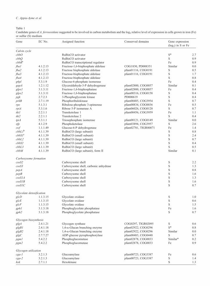
Table 1Candidate genes of A. ferrooxidans suggested to be involved in carbon metabolism and the log2 relative level of expression in cells grown in iron (Fe)or sulfur (S) medium
Gene EC No. Assigned function Conserved domains Gene expression(log2) in S or Fe
Calvin cyclecbbO RuBisCO activator S⁎ 2.7cbbQ RuBisCO activator S 0.9cbbRa RuBisCO transcriptional regulator Fe 0.9fba1 4.1.2.13 Fructose-1,6-bisphosphate aldolase COG1830, PD008351 Similar 0.0fba2 4.1.2.13 Fructose bisphosphate aldolase pfam01116, COG0191 S 0.5fba3 4.1.2.13 Fructose-bisphosphate aldolase pfam01116, COG0191 S 1.7fba4 4.1.2.13 Fructose-bisphosphate aldolase S 0.8g6pl 5.3.1.9 Glucose-6-phosphate isomerase Fe 0.4gapA 1.2.1.12 Glyceraldehyde-3-P dehydrogenase pfam02800, COG0057 Similar 0.1glpx1 3.1.3.11 Fructose-1,6-bisphosphatase pfam02800, COG0057 Fe 0.4glpx2 3.1.3.11 Fructose-1,6-bisphosphatase pfam00316, COG0158 S 0.4pgk 2.7.2.3 3-Phosphoglycerate kinase PD000619 S 0.4prkB 2.7.1.19 Phosphoribulokinase pfam00485, COG3954 S 0.7rpe 5.1.3.1 Ribulose-phosphate 3-epimerase pfam00834, COG0036 Fe 0.5rpiA 5.3.1.6 Ribose 5-P isomerase A pfam06026, COG0120 S 0.7tkt1 2.2.1.1 Transketolase 1 pfam00456, COG3959 Fe 1.1tkt2 2.2.1.1 Transketolase 2 S 0.4tpiA 5.3.1.1 Triosephosphate isomerase pfam00121, COG0149 Similar 0.0xfp 4.1.2.9 Phosphoketolase pfam03894, COG3957 S 2.6zwf 1.1.1.49 Glucose-6-P dehydrogenase pfam02781, TIGR00871 Fe 1.4cbbL1b 4.1.1.39 RuBisCO (large subunit) S 0.8cbbS1c 4.1.1.39 RuBisCO (small subunit) S 2.4cbbL2 4.1.1.39 RuBisCO (large subunit) S 0.7cbbS2 4.1.1.39 RuBisCO (small subunit) S 0.4cbbL3 4.1.1.39 RuBisCO (large subunit) S 0.5cbbM 4.1.1.39 RuBisCO (large subunit), form II Similar 0.1
Carboxysome formationcsoS2 Carboxysome shell S 2.2csoS3 Carboxysome shell, carbonic anhydrase S 1.3pepA Carboxysome shell S 0.8pepB Carboxysome shell S 1.6csoS1A Carboxysome shell S 1.3csoS1B Carboxysome shell S 1.6csoS1C Carboxysome shell S 0.7
Glycolate detoxificationglcD 1.1.3.15 Glycolate oxidase S 1.0glcE 1.1.3.15 Glycolate oxidase S 0.6glcF 1.1.3.15 Glycolate oxidase S 1.5gph1 3.1.3.18 Phosphoglycolate phosphatase Fe 1.6gph2 3.1.3.18 Phosphoglycolate phosphatase S 0.7
Glycogen biosynthesisglgA 2.4.1.21 Glycogen synthase COG0297, TIGR02095 S 0.6glgB1 2.4.1.18 1,4-α-Glucan branching enzyme pfam02922, COG0296 S⁎ 0.8glgB2 2.4.1.18 1,4-α-Glucan branching enzyme pfam02922, COG0296 Similar 0.0glgC 2.7.7.27 ADP-glucose pyrophosphorylase pfam00483, COG0448 S 0.7pgm1 5.4.2.2 Phosphoglucomutase pfam02878, COG0033 Similar⁎ 0.2pgm2 5.4.2.2 Phosphoglucomutase pfam02878, COG0033 Fe 0.9
Glycogen utilizationcga-1 3.2.1.3 Glucoamylase pfam00723, COG3387 Fe 0.6cga-2 3.2.1.3 Glucoamylase pfam00723, COG3387 S 1.4hxk 2.7.1.1 Hexokinase S 1.3
C. Appia-Ayme et al.
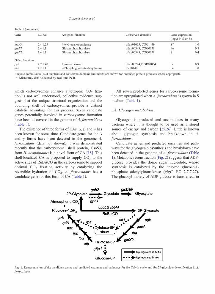
Table 1 (continued)
Gene EC No. Assigned function Conserved domains Gene expression(log2) in S or Fe
malQ 2.4.1.25 4-α-Glucanotransferase pfam03065, COG1449 S⁎ 1.0glgP1 2.4.1.1 Glucan phosphorylase pfam00343, COG0058 Fe 0.8glgP2 2.4.1.1 Glucan phosphorylase pfam00343, COG0058 S 0.4
Other functionspyk 2.7.1.40 Pyruvate kinase pfam00224,TIGR01064 Fe 0.9eno 4.2.1.11 2-Phosphoglycerate dehydratase PR00148 Fe 1.0
Enzyme commission (EC) numbers and conserved domains and motifs are shown for predicted protein products where appropriate.⁎ Microarray data validated by real-time PCR.
C. Appia-Ayme et al.
which carboxysomes enhance autotrophic CO2 fixa-tion is not well understood, collective evidence sug-gests that the unique structural organization and thebounding shell of carboxysomes provide a distinctcatalytic advantage for this process. Seven candidategenes potentially involved in carboxysome formationhave been discovered in the genome of A. ferrooxidans(Table 1).
The existence of three forms of CAs, α, β and γ hasbeen known for some time. Candidate genes for the βand γ forms have been detected in the genome A.ferrooxidans (data not shown). It was demonstratedrecently that the carboxysomal shell protein, CsoS3,from H. neapolitanus is a novel form of CA [18]. Thisshell-localized CA is proposed to supply CO2 to theactive sites of RuBisCO in the carboxysome to supportoptimal CO2 fixation activity by catalyzing thereversible hydration of CO2. A. ferrooxidans has acandidate gene for this form of CA (Table 1).
Fig. 1. Representation of the candidate genes and predicted enzymes and paferrooxidans.
All seven predicted genes for carboxysome forma-tion are upregulated when A. ferrooxidans is grown in Smedium (Table 1).
3.4. Glycogen metabolism
Glycogen is produced and accumulates in manybacteria where it is thought to be used as a storedsource of energy and carbon [25,26]. Little is knownabout glycogen synthesis and breakdown in A.ferrooxidans.
Candidate genes and predicted enzymes and path-ways for the glycogen biosynthesis and breakdown havebeen detected in the genome of A. ferrooxidans (Table1). Metabolic reconstruction (Fig. 2) suggests that ADP-glucose provides the donor sugar nucleotide, whosesynthesis is catalyzed by the enzyme glucose-1-phosphate adenylyltransferase (glgC; EC 2.7.7.27).The glucosyl moiety of ADP-glucose is transferred, in
thways for the Calvin cycle and for 2P-glycolate detoxification in A.
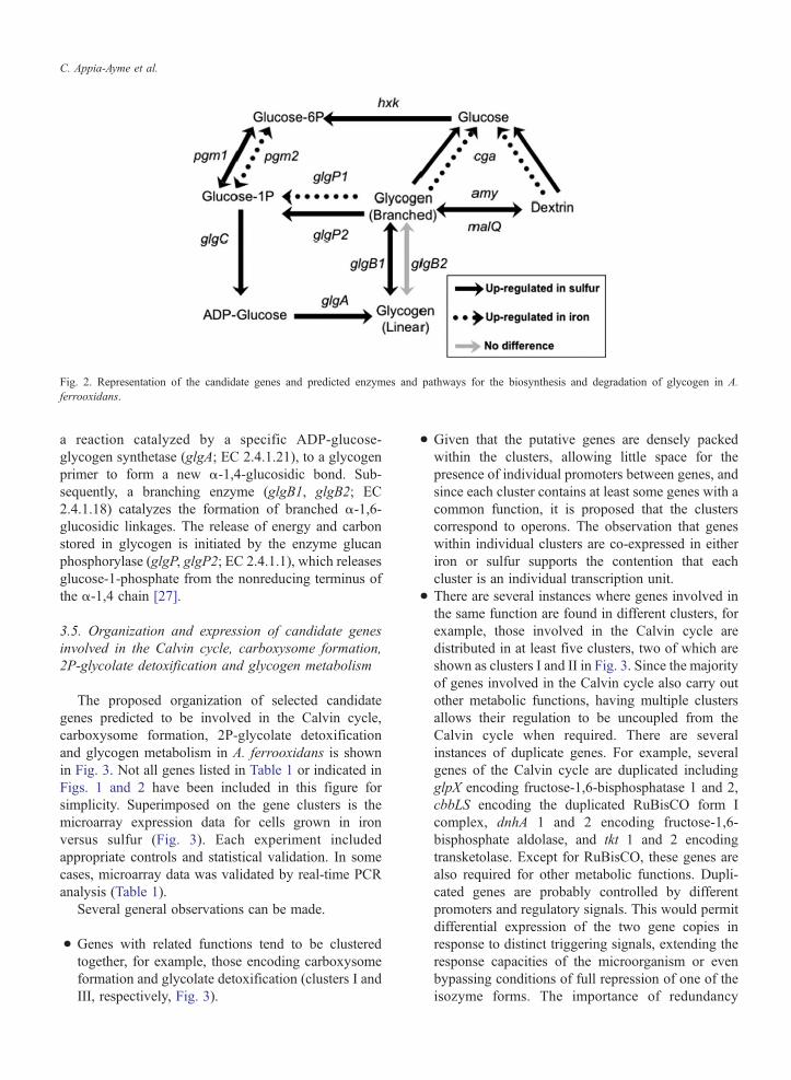
Fig. 2. Representation of the candidate genes and predicted enzymes and pathways for the biosynthesis and degradation of glycogen in A.ferrooxidans.
C. Appia-Ayme et al.
a reaction catalyzed by a specific ADP-glucose-glycogen synthetase (glgA; EC 2.4.1.21), to a glycogenprimer to form a new α-1,4-glucosidic bond. Sub-sequently, a branching enzyme (glgB1, glgB2; EC2.4.1.18) catalyzes the formation of branched α-1,6-glucosidic linkages. The release of energy and carbonstored in glycogen is initiated by the enzyme glucanphosphorylase (glgP, glgP2; EC 2.4.1.1), which releasesglucose-1-phosphate from the nonreducing terminus ofthe α-1,4 chain [27].
3.5. Organization and expression of candidate genesinvolved in the Calvin cycle, carboxysome formation,2P-glycolate detoxification and glycogen metabolism
The proposed organization of selected candidategenes predicted to be involved in the Calvin cycle,carboxysome formation, 2P-glycolate detoxificationand glycogen metabolism in A. ferrooxidans is shownin Fig. 3. Not all genes listed in Table 1 or indicated inFigs. 1 and 2 have been included in this figure forsimplicity. Superimposed on the gene clusters is themicroarray expression data for cells grown in ironversus sulfur (Fig. 3). Each experiment includedappropriate controls and statistical validation. In somecases, microarray data was validated by real-time PCRanalysis (Table 1).
Several general observations can be made.
• Genes with related functions tend to be clusteredtogether, for example, those encoding carboxysomeformation and glycolate detoxification (clusters I andIII, respectively, Fig. 3).
• Given that the putative genes are densely packedwithin the clusters, allowing little space for thepresence of individual promoters between genes, andsince each cluster contains at least some genes with acommon function, it is proposed that the clusterscorrespond to operons. The observation that geneswithin individual clusters are co-expressed in eitheriron or sulfur supports the contention that eachcluster is an individual transcription unit.
• There are several instances where genes involved inthe same function are found in different clusters, forexample, those involved in the Calvin cycle aredistributed in at least five clusters, two of which areshown as clusters I and II in Fig. 3. Since the majorityof genes involved in the Calvin cycle also carry outother metabolic functions, having multiple clustersallows their regulation to be uncoupled from theCalvin cycle when required. There are severalinstances of duplicate genes. For example, severalgenes of the Calvin cycle are duplicated includingglpX encoding fructose-1,6-bisphosphatase 1 and 2,cbbLS encoding the duplicated RuBisCO form Icomplex, dnhA 1 and 2 encoding fructose-1,6-bisphosphate aldolase, and tkt 1 and 2 encodingtransketolase. Except for RuBisCO, these genes arealso required for other metabolic functions. Dupli-cated genes are probably controlled by differentpromoters and regulatory signals. This would permitdifferential expression of the two gene copies inresponse to distinct triggering signals, extending theresponse capacities of the microorganism or evenbypassing conditions of full repression of one of theisozyme forms. The importance of redundancy
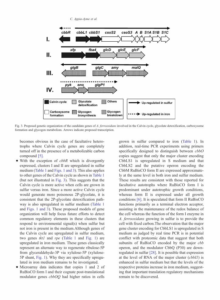
Fig. 3. Proposed genetic organization of the candidate genes of A. ferrooxidans involved in the Calvin cycle, glycolate detoxification, carboxysomeformation and glycogen metabolism. Arrows indicate proposed transcription.
C. Appia-Ayme et al.
becomes obvious in the case of facultative hetero-trophs where Calvin cycle genes are completelyturned off in the presence of a metabolizable carboncompound [5].
• With the exception of cbbR which is divergentlyexpressed, clusters I and II are upregulated in sulfurmedium (Table 1 and Figs. 1 and 3). This also appliesto other genes of the Calvin cycle as shown in Table 1(but not illustrated in Fig. 3). This suggests that theCalvin cycle is more active when cells are grown insulfur versus iron. Since a more active Calvin cyclewould generate more poisonous 2P-glycolate, it isconsistent that the 2P-glycolate detoxification path-way is also upregulated in sulfur medium (Table 1and Figs. 1 and 3). These proposed models of geneorganization will help focus future efforts to detectcommon regulatory elements in these clusters thatrespond to environmental signal(s) when sulfur butnot iron is present in the medium.Although genes ofthe Calvin cycle are upregulated in sulfur medium,two genes tkt1 and rpe (Table 1 and Fig. 1) areupregulated in iron medium. These genes classicallyrepresent an alternate way to regenerate ribulose-5Pfrom glyceraldehyde-3P via xylulose-5P (xylulose-5P shunt, Fig. 1). Why they are specifically upregu-lated in iron medium remains to be investigated.
• Microarray data indicates that copies 1 and 2 ofRuBisCO form I and their cognate post-translationalmodulator genes cbbOQ had higher ratios in cells
grown in sulfur compared to iron (Table 1). Inaddition, real-time PCR experiments using primersspecifically designed to distinguish between cbbOcopies suggest that only the major cluster encodingCbbLS1 is upregulated in S medium and thatCbbLS2 and the putative operon encoding theCbbM RuBisCO form II are expressed approximate-ly at the same level in both iron and sulfur medium.These results are consistent with those reported forfacultative autotrophs where RuBisCO form I ispredominant under autotrophic growth conditions,whereas form II is expressed under all growthconditions [6]. It is speculated that form II RuBisCOfunctions primarily as a terminal electron acceptor,assisting in the maintenance of the redox balance ofthe cell whereas the function of the form I enzyme inA. ferrooxidans growing in sulfur is to provide thecell with fixed carbon.The observation that the majorgene cluster encoding for CbbLS1 is upregulated in Smedium as judged by real time PCR is in potentialconflict with proteomic data that suggest that bothsubunits of RuBisCO encoded by the major cbboperon, and the modulator CbbQ (P30) are down-regulated in sulfur [28]. It is possible that expressionat the level of RNA of the major cluster (cbbI1) isenhanced in sulfur medium but that the levels of therespective proteins increase in iron medium, suggest-ing that important translation regulatory mechanismsremain to be discovered.
C. Appia-Ayme et al.
• Genes involved in glycogen biosynthesis and theglycogen branching/debranching system tend to beupregulated in sulfur medium (Table 1 and cluster III,Fig. 3), whereas those proposed to be required forglycogen breakdown are upregulated in iron medium(Table 1 and cluster IV, Fig. 3). The other two genes,pyk and eno, associated with the glycogen break-down gene glgP1 in cluster IV are predicted to beinvolved in sugar metabolism, suggesting that thiscluster is involved in the recovery of energy andcarbon from glycogen. Theoretically, sulfur shouldyield more energy than iron. Therefore, perhaps, it ismetabolically favorable to fix CO2 and to channelsome of the fixed carbon to stored glycogen whensulfur is available as an energy source. The glycogencan be broken down later to yield carbon and energyin leaner times, for example, when only iron isavailable as an energy source.
Note added in proof
The complete sequence of A. ferrooxidans has nowbeen released (TIGR. org).
Acknowledgements
This work was supported in part by Fondecyt1010623 and 1050063, Conicyt/CNRS, “Geomex” and“Puces à ADN” from the Centre National de laRecherche Scientifique and NSF. RQ was the recipientof an American Society for Microbiology AntorchaFellowship and was supported by scholarships from theDAAD and from the Fundación Ciencia para la Vida,Chile. CAA was supported by a CNRS post-doctoralfellowship. We thank the Institute for Genome Research(TIGR) for the use of their draft genome sequence of A.ferrooxidans.
References
[1] Silver, M., Can. J. Microbiol., 16 (1970), 845–849.[2] Tabita, R., Lundgren, D.G., J. Bacteriol., 108 (1971), 328–333.[3] Gale, N.L., Beck, J.V., J. Bacteriol., 94 (1967), 1052–1059.[4] Holuigue, L., Herrera, L., Phillips, O.M., Young, M., Allende,
J.E., Biotechnol. Appl. Biochem., 9 (1987), 497–505.
[5] Tabita, F.R., Microbiol. Rev., 52 (1988), 155–189.[6] Gibson, J.L., Tabita, F.R., Arch. Microbiol., 166 (1996),
141–150.[7] Pulgar, V., Gaete, L., Allende, J., Orellana, O., Jordana, X.,
Jedlicki, E., FEBS Lett., 292 (1991), 85–89.[8] Kusano, T., Sugawara, K., Inoue, C., Curr. Microbiol., 22 (1991),
35–41.[9] Kusano, T., Takeshima, T., Inoue, C., Sugawara, K., J. Bacteriol.,
173 (1991), 7313–7323.[10] Cannon, G.C., Bradburne, C.E., Aldrich, H.C., Baker, S.H.,
Heinhorst, S., Shively, J.M., Appl. Environ. Microbiol., 67(2001), 5351–5361.
[11] Shively, J.M., Ball, F., Brown, D.H., Saunders, R.E., Science,182 (1973), 584–586.
[12] Cannon, G.C., Shively, J.M., Arch. Microbiol., 134 (1983),52–59.
[13] English, R.S., Lorbach, S.C., Qin, X., Shively, J.M., Mol.Microbiol., 12 (1994), 647–654.
[14] Cannon, G.C., Baker, S.H., Soyer, F., Johnson, D.R., Bradburne,C.E., Mehlman, J.L., Davies, P.S., Jiang, Q.L., Heinhorst, S.,Shively, J.M., Curr. Microbiol., 46 (2003), 115–119.
[15] Badger, M.R., Hanson, D., Price, G.D., Funct. Plant Biol., 29(2002), 161–173.
[16] Price, G.D., Coleman, J.R., Badger, M.R., Plant Physiol., 100(1992), 784–793.
[17] So, A.K., Espie, G.S., Plant Mol. Biol., 37 (1998), 205–215.[18] So, A.K., Espie, G.S., Williams, E.B., Shively, J.M., Heinhorst,
S., Cannon, G.C., J. Bacteriol., 186 (2004), 623–630.[19] Beudeker, R.F., Cannon, G.C., Kuenen, J.G., Shively, J.M., Arch.
Microbiol., 124 (1980), 185–189.[20] Purohit, K., McFadden, B.A., Shaykh, M.M., J. Bacteriol., 127
(1976), 516–522.[21] Quatrini, R., Jedlicki, E., Holmes, D.S.J., Indust. Microbiol.
Biotech., 32 (2005), 606–614.[22] Quatrini, R., Appia-Ayme, C., Denis, Y., Ratouchniak, J., Veloso,
F., Valdes, J., Lefimil, C., Silver, S., Roberto, F., Orellana, O.,Denizot, F., Jedlicki, E., Holmes, D.S., Bonnefoy, V., Hydro-metallurgy, 83 (2006), 263–272 (this volume) doi:10.1016/j.hydromet.2006.03.030.
[23] Lorimer, G.H., Andrews, T.J., Tolbert, N.E., Biochemistry, 12(1973), 18–23.
[24] Wolfenden, R., Biochemistry, 9 (1970), 3404–3407.[25] Preiss, J., Annu. Rev. Microbiol., 38 (1984), 419–458.[26] Preiss, J., Romeo, T., Adv. Microb. Physiol., 30 (1989),
183–238.[27] Fletterick, R.J., Madsen, N.B., Annu. Rev. Biochem., 49 (1980),
31–61.[28] Ramírez, P., Guiliani, N., Valenzuela, L., Beard, S., Jerez, C.A.,
Appl. Environ. Microbiol., 70 (2004), 4491–4498.
Related Documents