ORIGINAL PAPER Heat and phosphate starvation effects on the proteome, morphology and chemical composition of the biomining bacteria Acidithiobacillus ferrooxidans Daniela A. Ribeiro • Danilo A. Maretto • Fa ´bio C. S. Nogueira • Ma ´rcio J. Silva • Francisco A. P. Campos • Gilberto B. Domont • Ronei J. Poppi • Laura M. M. Ottoboni Received: 3 August 2010 / Accepted: 19 October 2010 / Published online: 4 November 2010 Ó Springer Science+Business Media B.V. 2010 Abstract Acidithiobacillus ferrooxidans is a Gram neg- ative, acidophilic, chemolithoautotrophic bacterium that plays an important role in metal bioleaching. During bioleaching, the cells are subjected to changes in the growth temperature and nutrients starvation. The aim of this study was to gather information about the response of the A. ferrooxidans Brazilian strain LR to K 2 HPO 4 star- vation and heat stress through investigation of cellular morphology, chemical composition and differential prote- ome. The scanning electron microscopic results showed that under the tested stress conditions, A. ferrooxidans cells became elongated while the Fourier transform infrared spectroscopy (FT-IR) analysis showed alterations in the wavenumbers between 850 and 1,275 cm -1 , which are related to carbohydrates, phospholipids and phosphopro- teins. These findings indicate that the bacterial cell surface is affected by the tested stress conditions. A proteomic analysis, using 2-DE and tandem mass spectrometry, enabled the identification of 44 differentially expressed protein spots, being 30 due to heat stress (40°C) and 14 due to K 2 HPO 4 starvation. The identified proteins belon- ged to 11 different functional categories, including protein fate, energy metabolism and cellular processes. The upregulated proteins were mainly from protein fate and energy metabolism categories. The obtained results pro- vide evidences that A. ferrooxidans LR responds to heat stress and K 2 HPO 4 starvation by inducing alterations in cellular morphology and chemical composition of the cell surface. Also, the identification of several proteins involved in protein fate suggests that the bacteria cellular homesostasis was affected. In addition, the identification of proteins from different functional categories indicates that the A. ferrooxidans response to higher than optimal tem- peratures and phosphate starvation involves global changes in its physiology. Keywords Acidithiobacillus ferrooxidans Á Scanning electron microscopy Á Fourier transform infrared spectroscopy Á Proteome Á Stress Introduction Acidithiobacillus ferrooxidans is a Gram-negative, chemo- lithoautotrophic, acidophilic bacterium that thrives opti- mally around pH 2.0 and 30°C. It derives energy from the oxidation of ferrous iron or reduced sulphur compounds, and is used industrially in metal bioleaching, a process in which metal sulphides are converted to water-soluble metal sulphates (Rawlings 2005). During the bioleaching process, A. ferrooxidans is often subjected to changes in the ideal growth pH and temperature, and to nutrients starvation (Rawlings 2005). These changes can affect the bacterial D. A. Ribeiro Á M. J. Silva Á L. M. M. Ottoboni (&) Centro de Biologia Molecular e Engenharia Gene ´tica (CBMEG), Universidade Estadual de Campinas (UNICAMP), CP 6010, Campinas, SP 13083-875, Brazil e-mail: [email protected] D. A. Maretto Á R. J. Poppi Instituto de Quı ´mica, Universidade Estadual de Campinas (UNICAMP), CP 6154, Campinas, SP 13083-970, Brazil F. C. S. Nogueira Á G. B. Domont Departamento de Bioquı ´mica, Instituto de Quı ´mica, Universidade Federal do Rio de Janeiro (UFRJ), Rio de Janeiro, RJ 21941-909, Brazil F. A. P. Campos Departamento de Bioquı ´mica e Biologia Molecular, Universidade Federal do Ceara ´ (UFC), CP 6039, Fortaleza, CE 60455-900, Brazil 123 World J Microbiol Biotechnol (2011) 27:1469–1479 DOI 10.1007/s11274-010-0599-9

Welcome message from author
This document is posted to help you gain knowledge. Please leave a comment to let me know what you think about it! Share it to your friends and learn new things together.
Transcript
ORIGINAL PAPER
Heat and phosphate starvation effects on the proteome,morphology and chemical composition of the biomining bacteriaAcidithiobacillus ferrooxidans
Daniela A. Ribeiro • Danilo A. Maretto • Fabio C. S. Nogueira •
Marcio J. Silva • Francisco A. P. Campos • Gilberto B. Domont •
Ronei J. Poppi • Laura M. M. Ottoboni
Received: 3 August 2010 / Accepted: 19 October 2010 / Published online: 4 November 2010
� Springer Science+Business Media B.V. 2010
Abstract Acidithiobacillus ferrooxidans is a Gram neg-
ative, acidophilic, chemolithoautotrophic bacterium that
plays an important role in metal bioleaching. During
bioleaching, the cells are subjected to changes in the
growth temperature and nutrients starvation. The aim of
this study was to gather information about the response of
the A. ferrooxidans Brazilian strain LR to K2HPO4 star-
vation and heat stress through investigation of cellular
morphology, chemical composition and differential prote-
ome. The scanning electron microscopic results showed
that under the tested stress conditions, A. ferrooxidans cells
became elongated while the Fourier transform infrared
spectroscopy (FT-IR) analysis showed alterations in the
wavenumbers between 850 and 1,275 cm-1, which are
related to carbohydrates, phospholipids and phosphopro-
teins. These findings indicate that the bacterial cell surface
is affected by the tested stress conditions. A proteomic
analysis, using 2-DE and tandem mass spectrometry,
enabled the identification of 44 differentially expressed
protein spots, being 30 due to heat stress (40�C) and 14
due to K2HPO4 starvation. The identified proteins belon-
ged to 11 different functional categories, including protein
fate, energy metabolism and cellular processes. The
upregulated proteins were mainly from protein fate and
energy metabolism categories. The obtained results pro-
vide evidences that A. ferrooxidans LR responds to heat
stress and K2HPO4 starvation by inducing alterations in
cellular morphology and chemical composition of the cell
surface. Also, the identification of several proteins
involved in protein fate suggests that the bacteria cellular
homesostasis was affected. In addition, the identification of
proteins from different functional categories indicates that
the A. ferrooxidans response to higher than optimal tem-
peratures and phosphate starvation involves global changes
in its physiology.
Keywords Acidithiobacillus ferrooxidans � Scanning
electron microscopy � Fourier transform infrared
spectroscopy � Proteome � Stress
Introduction
Acidithiobacillus ferrooxidans is a Gram-negative, chemo-
lithoautotrophic, acidophilic bacterium that thrives opti-
mally around pH 2.0 and 30�C. It derives energy from the
oxidation of ferrous iron or reduced sulphur compounds,
and is used industrially in metal bioleaching, a process in
which metal sulphides are converted to water-soluble metal
sulphates (Rawlings 2005). During the bioleaching process,
A. ferrooxidans is often subjected to changes in the ideal
growth pH and temperature, and to nutrients starvation
(Rawlings 2005). These changes can affect the bacterial
D. A. Ribeiro � M. J. Silva � L. M. M. Ottoboni (&)
Centro de Biologia Molecular e Engenharia Genetica (CBMEG),
Universidade Estadual de Campinas (UNICAMP), CP 6010,
Campinas, SP 13083-875, Brazil
e-mail: [email protected]
D. A. Maretto � R. J. Poppi
Instituto de Quımica, Universidade Estadual de Campinas
(UNICAMP), CP 6154, Campinas, SP 13083-970, Brazil
F. C. S. Nogueira � G. B. Domont
Departamento de Bioquımica, Instituto de Quımica,
Universidade Federal do Rio de Janeiro (UFRJ),
Rio de Janeiro, RJ 21941-909, Brazil
F. A. P. Campos
Departamento de Bioquımica e Biologia Molecular,
Universidade Federal do Ceara (UFC), CP 6039,
Fortaleza, CE 60455-900, Brazil
123
World J Microbiol Biotechnol (2011) 27:1469–1479
DOI 10.1007/s11274-010-0599-9
physiology and as a consequence, the efficiency of
bioleaching.
Acidithiobacillus ferrooxidans respond to high temper-
atures by synthesizing several heat shock proteins (HSPs)
(Jerez 1988, Xiao et al. 2009). This bacterium is also able
to acquire thermotolerance (Hubert et al. 1995), which
indicates that it has developed protective mechanisms to
deal with heat stress. This fact is particularly important
since temperature is one of the main factors affecting
metals solubilization during bioleaching (Modak et al.
1996). Indeed, Modak et al. (1996) reported that a tem-
perature-adapted strain of A. ferrooxidans was more effi-
cient during pyrite bioleaching than non-adapted ones.
Regarding phosphate starvation, Seeger and Jerez
(1993) showed a reduction in the A. ferrooxidans growth
rate as well as in its capacity to oxidize ferrous iron and to
fix CO2. Seeger et al. (1996) observed a protein phos-
phorylation increase in A. ferrooxidans cells submitted to
phosphate starvation. Also, the chaperones DnaK and
GroEL are phosphorylated when this bacterium is sub-
jected to phosphate starvation (Seeger et al. 1996), sug-
gesting the activation of a general stress response.
Moreover, the lipopolysaccharides production is altered in
phosphate starved A. ferrooxidans cells (Farah et al. 2005),
which may affect bioleaching since lipopolysaccharides
are part of the polysaccharide matrix involved in ore
colonization.
Little is known about the A. ferrooxidans response to
phosphate starvation and heat stress. Thus, in this work,
three different approaches, namely, scanning electronic
microscopy, FT-IR spectroscopy and proteome analysis,
were used to investigate morphological changes, alterations
in the chemical composition and in the protein profile of
A. ferrooxidans LR cells subjected to such conditions.
These analyses rely on the possibility of bacterial cell
shape being affected by stress, which indicates that mor-
phological changes are correlated to adaptive mechanisms
that enable the cells to survive (Pianetti et al. 2009).
Additionally, FT-IR, a method which measures the overall
composition of a sample by detecting the molecular
vibrations and other motions of chemical bonds, can be
used to characterize modifications in cells grown in dif-
ferent conditions. Finally, the proteome analysis explore
the alterations in the protein profile of the cell.
Materials and methods
Bacterial strain and growth conditions
The A. ferrooxidans Brazilian strain LR (Garcia Jr 1991)
was used for the experiments. The bacteria were grown at
250 rpm in modified T&K liquid medium (Tuovinen and
Kelly 1972) containing: 0.4 g/l K2HPO4�3H2O, 0.4 g/l
MgSO4�7H2O, 0.4 g/l (NH4)2SO4 and 33.4 g/l FeS-
O4�7H2O, pH 1.8, adjusted with sulfuric acid. The bacteria
were grown under control conditions (30�C and presence of
K2HPO4 in the medium), under heat stress (40�C) and
under phosphate-limiting conditions (absence of K2HPO4
in the medium), as described by Knegt et al. (2008). The
bacteria were grown until 50% of ferrous iron oxidation in
the medium. The growth curve experiments were per-
formed in 250 ml Erlenmeyer flasks containing 100 ml of
T&K medium, pH 1.8, inoculated with 0.75 9 109 cells,
on a rotatory shaker at 250 rpm. Bacterial growth was
monitored by ferrous iron titration with potassium
dichromate.
Scanning electron microscopy (SEM)
Sample preparation for SEM was carried out in duplicate as
described by Shi and Xia (2003), with minor modifications.
Briefly, strain LR was grown at 30�C in the presence of
K2HPO4 (control), at 40�C and in the absence of K2HPO4,
as described above. Cells were harvested, washed and
resuspended in water, pH 1.8. The suspension (approxi-
mately 1 9 106 cells) was filtered through a 0.45 lM
Millix membrane (Millipore, Ireland). The cells in the
Millix membrane were immersed in 2.5% glutaraldehyde/
0.1 M sodium cacodylate buffer for 4 h, washed three
times with 0.1 M sodium cacodylate buffer, treated for 3 h
with 1% OsO4 in 0.1 M sodium cacodylate buffer and
washed three times with sodium cacodylate buffer and
deionised water. Cells were dehydrated by an ethanol series
(30, 50, 70, 90 and 100% ethanol) and stored in 100%
ethanol. Cells were critical-point-dried in a CO2 atmo-
sphere (Balzers Critical Point Dryer), mounted on alu-
minium stubs and gold-coated for 3 min in a Sputter Coater
(Balzers, SCD050). They were examined under a JEOL
5800LV scanning electron microscope. Cell dimensions
were measured using the ImageJ software (available at
http://rsb.info.nih.gov/ij). The average cellular dimensions
were calculated using 100 individual cells per treatment.
The statistical significance of the observed differences in
length, width and area was verified using one-way
ANOVA.
Fourier transform infrared spectroscopy (FT-IR)
Acidithiobacillus ferrooxidans cells that received different
growth treatments were frozen in liquid nitrogen and
lyophilized. The infrared spectra of the cells were recorded
in an ABB-Bomem MB Series Fourier spectrometer with a
diffuse reflectance attachment. Spectra were collected
using a scan range of 400–3,800 cm-1 at a resolution of
4 cm-1. The data were imported into a PLS-Toolbox 4.21
1470 World J Microbiol Biotechnol (2011) 27:1469–1479
123
for Matlab 6.5, and a baseline correction of the data was
performed. In order to decrease concentration effects on the
model, the first derivative spectra were used for the prin-
cipal component analysis (PCA) model development. The
data were also mean-centred.
Protein extraction
Acidithiobacillus ferrooxidans whole cell protein extracts
were obtained as described by Smolka et al. (2003).
Briefly, bacterial cells (*100 mg, wet weight) were
washed three times in 1 ml of washing buffer (10 mM Tris,
pH 8.8, 3 mM KCl, 50 mM NaCl, 5 mM EDTA and 1 mM
PMSF) and centrifuged for 2 min at 5,400 9 g in a
microcentrifuge (Beckman). The cells were lysed in 200 ll
of 10 mM Tris (pH 8.8), 0.5% (w/v) SDS, 5 mM EDTA
and 1 mM PMSF. Dithiothreitol was added to a final
concentration of 100 mM, and the samples were boiled for
3 min and centrifuged for 2 min at 5,4009g. The protein
concentration in the samples was determined using the Bio-
Rad protein assay kit (Bio-Rad, Hercules, CA, USA). The
protein samples were stored at -80�C.
Two-dimensional gel electrophoresis (2-DE)
2-DE gels and image analysis were performed as described
by Vasconcelos et al. (2005). Briefly, approximately
200 lg of total protein was loaded onto a rehydrated 11-cm
Immobiline DryStrip pH 3–10 or pH 4–10 (GE Healthcare,
USA). Isoelectric focusing was carried out in an Ettan
IPGPhor isoeletric focusing system (GE Healthcare, USA).
The second-dimension separation was carried out on a 15%
acrylamide gel containing SDS. Gels were stained with
0.1% Coomassie PhastGel Blue R-350. The Image Master
2D Platinum 7.0 (GE Healthcare, USA) was used to detect
and quantify protein spots in the gels. The experiments
were performed in triplicate. The statistical significance of
the observed spots differences was determined using the
Student t test (P value B 0.05).
Mass spectrometry
The differentially displayed protein spots were excised
from the acrylamide gels, destained and digested with
sequencing-grade modified trypsin (Promega, Madison,
WI, USA), as described by Vasconcelos et al. (2005).
Matrix-assisted laser desorption ionization-time of flight-
tandem mass spectrometry (MALDI TOF-TOF-MS/MS)
acquisition was performed in an ABI 4700 Proteomics
Analyzer (Applied Biosystems) using 3,5 dimethoxy-4-
hydroxycinnamic acid as the matrix. The obtained tandem
mass spectra were searched against the NCBInr database
and the A. ferrooxidans ORFs database (J. Craig Venter
Institute) using the Mascot (version 2.1) MS/MS ion search
tool (http://www.matrixscience.com). Cysteine residues
were reduced and alkylated by iodoacetamide to carboxy-
amidomethyl cysteine, and methionine residues were
modified to methionine sulphoxide wherever necessary.
Peptide mass tolerance in the searches was 100 ppm for
MS and 0.6 Da for MS/MS spectra. Peptides were con-
sidered identified when the score value exceeded the
identity or extensive homology threshold value calculated
by Mascot.
Results and discussion
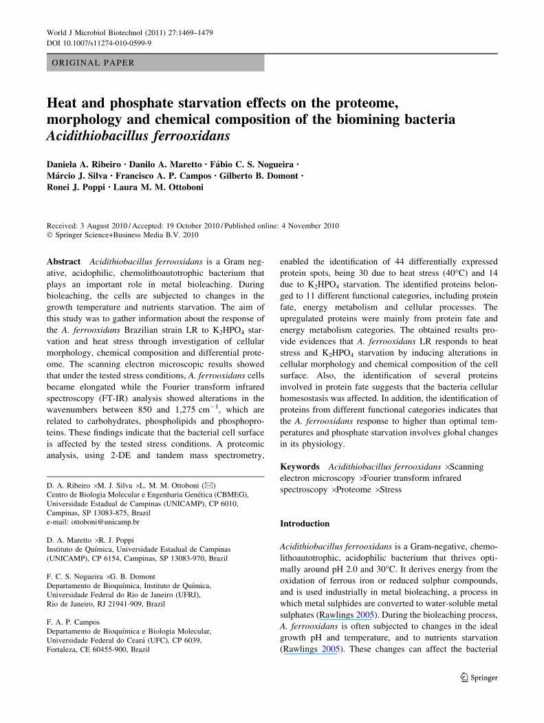
Growth at 40�C and in the absence of K2HPO4
As shown in Fig. 1, the lag phase was pratically the same
for A. ferrooxidans LR control (30�C and presence of
K2HPO4) cells and cells submitted to stress (40�C and
absence of K2HPO4). During the log phase, the control
cells were able to oxidize the available ferrous iron in 28 h,
whereas the cells submitted to heat stress oxidized the
ferrous iron in 40 h and cells submitted to phosphate lim-
iting conditions in 48 h. These results indicate that growth
was differentially inhibited by heat and phosphate
limitation.
Morphological changes in A. ferrooxidans due
to temperature elevation and phosphate starvation
Temperature elevation and phosphate starvation are two
environmental factors that affect microbial survival and
growth. In order to investigate the effects of these condi-
tions on A. ferrooxidans LR cell morphology, the bacteria
were grown at 30�C in the presence of K2HPO4 (control),
at 40�C or in the absence of K2HPO4. After growth, the
Fig. 1 Growth curves of A. ferrooxidans LR at 40�C (squares),
whitout K2HPO4 in the culture medium (circles) and at control
conditions -30�C and presence of K2HPO4 (triangles). The errorbars show the standard deviation
World J Microbiol Biotechnol (2011) 27:1469–1479 1471
123
cells were analysed by SEM (Fig. 2). Table 1 shows the
means of the measurements for cellular dimensions from
each experiment. The mean cell length and area increased
significantly under both heat (40�C) stress (Flength = 417.2,
Farea = 388.9, 199 df, P \ 0.01) and K2HPO4 starvation
(Flength = 525.6, Farea = 343.8, 199 df, P \ 0.01). The
mean cell width was also significantly altered by K2HPO4
starvation (Fwidth = 24.3, 199 df, P \ 0.01), but it was not
significantly changed during growth at 40�C (Fwidth = 1.7,
199 df, P = 0.1903).
As shown in Table 1, the most affected parameter was
the cell length, which increased from 1.05 lm in control
cells to 1.9 lm in cells grown at 40�C and 1.77 lm in cells
grown in the absence of K2HPO4. Elongation is a wide-
spread phenomenon in bacteria exposed to temperatures
near their upper limit, and has been reported in a number of
microbes, including Pseudomonas pseudoalcaligenes (Shi
and Xia 2003). In cells undergoing phosphate starvation,
however, the morphological changes seem to be species
specific, since Pseudomonas putida forms small, almost
spherical cells (Eberl et al. 1996), while Vibrio sp. form
filaments (Holmquist and Kjelleberg 1993). In agreement
with our work, Seeger and Jerez (1993) showed that A.
ferrooxidans strain R2 also forms filaments under phos-
phate starvation conditions. The mechanisms responsible
for the elongation that occurs when bacterial cells are
subjected to non-optimal temperatures and to phosphate
starvation are not clear. According to van der Veen et al.
(2010), a possible explanation for bacteria cell elongation
is that cell division is blocked due to DNA damage and
induction of SOS response.
The morphological alterations observed in A. ferrooxi-
dans LR cells grown at 40�C and under phosphate
starvation suggest possible alterations in the membrane, as
well as in the cell wall. These external components of the
bacterial cell are composed mainly of phospholipids and
polysaccharides (Yu and Irudayaraj 2004). To investigate
whether these compounds were affected by the tested
stress conditions, A. ferrooxidans LR cells were analysed
by FT-IR.
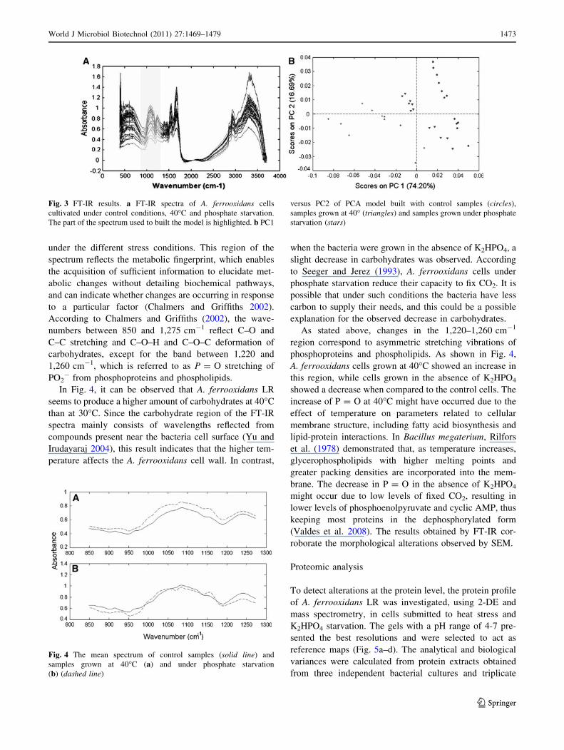
FT-IR analysis of A. ferrooxidans cells grown at 40�C
and in the absence of K2HPO4
The FT-IR technique was used to evaluate differences
among A. ferrooxidans LR cells grown under control
conditions (30�C with K2HPO4), at 40�C and in the
absence of K2HPO4. Typical FT-IR spectra for A. ferro-
oxidans LR are shown in Fig. 3a. The obtained spectra
were analysed using principal component analysis (PCA).
In order to investigate qualitative differences between the
samples in the principal component (PC) space, PCA was
performed on the first-derivative spectra. Several models
were developed based on the first derivative of the full
spectra. Since none of these models yielded a good sepa-
ration between the groups, the spectra were divided into
different regions, and models were built using the mean-
centred first derivative spectra as a data input in the PCA.
The region between 850 and 1,275 cm-1, highlighted in
Fig. 3a, presented the best results. Figure 3b shows the
scores of a PCA model built by combining all of the
samples. In this model, three different groups were
observed, showing that the different growth conditions
caused distinct differences in the A. ferrooxidans LR cells.
Figure 4 shows the mean spectra between 850 and
1,275 cm-1 for control samples and for samples grown
Fig. 2 SEM images of
A. ferrooxidans LR cells.
Bacterial samples were cultured
until 50% of oxidation of Fe2?.
a Control. b 40�C. c Phosphate
starvation. Bar = 1 lm.
Magnification: 20,000 (a);
15,000 (b, c)
Table 1 The mean cell size of A. ferrooxidans cells grown under normal conditions (control), at 40�C and under phosphate starvation
Control Temperature (40�C) Phosphate starvation
Length (lm) Width (lm) Area (lm2) Length (lm) Width (lm) Area (lm2) Length (lm) Width (lm) Area (lm2)
Mean 1.05 0.50 0.45 1.90 0.49 0.86 1.77 0.47 0.77
SD (±) 0.20 0.05 0.09 0.36 0.07 0.19 0.24 0.05 0.14
The values represent the means and standard deviations of the measurements obtained from 100 cells in two independent experiments
1472 World J Microbiol Biotechnol (2011) 27:1469–1479
123
under the different stress conditions. This region of the
spectrum reflects the metabolic fingerprint, which enables
the acquisition of sufficient information to elucidate met-
abolic changes without detailing biochemical pathways,
and can indicate whether changes are occurring in response
to a particular factor (Chalmers and Griffiths 2002).
According to Chalmers and Griffiths (2002), the wave-
numbers between 850 and 1,275 cm-1 reflect C–O and
C–C stretching and C–O–H and C–O–C deformation of
carbohydrates, except for the band between 1,220 and
1,260 cm-1, which is referred to as P = O stretching of
PO2- from phosphoproteins and phospholipids.
In Fig. 4, it can be observed that A. ferrooxidans LR
seems to produce a higher amount of carbohydrates at 40�C
than at 30�C. Since the carbohydrate region of the FT-IR
spectra mainly consists of wavelengths reflected from
compounds present near the bacteria cell surface (Yu and
Irudayaraj 2004), this result indicates that the higher tem-
perature affects the A. ferrooxidans cell wall. In contrast,
when the bacteria were grown in the absence of K2HPO4, a
slight decrease in carbohydrates was observed. According
to Seeger and Jerez (1993), A. ferrooxidans cells under
phosphate starvation reduce their capacity to fix CO2. It is
possible that under such conditions the bacteria have less
carbon to supply their needs, and this could be a possible
explanation for the observed decrease in carbohydrates.
As stated above, changes in the 1,220–1,260 cm-1
region correspond to asymmetric stretching vibrations of
phosphoproteins and phospholipids. As shown in Fig. 4,
A. ferrooxidans cells grown at 40�C showed an increase in
this region, while cells grown in the absence of K2HPO4
showed a decrease when compared to the control cells. The
increase of P = O at 40�C might have occurred due to the
effect of temperature on parameters related to cellular
membrane structure, including fatty acid biosynthesis and
lipid-protein interactions. In Bacillus megaterium, Rilfors
et al. (1978) demonstrated that, as temperature increases,
glycerophospholipids with higher melting points and
greater packing densities are incorporated into the mem-
brane. The decrease in P = O in the absence of K2HPO4
might occur due to low levels of fixed CO2, resulting in
lower levels of phosphoenolpyruvate and cyclic AMP, thus
keeping most proteins in the dephosphorylated form
(Valdes et al. 2008). The results obtained by FT-IR cor-
roborate the morphological alterations observed by SEM.
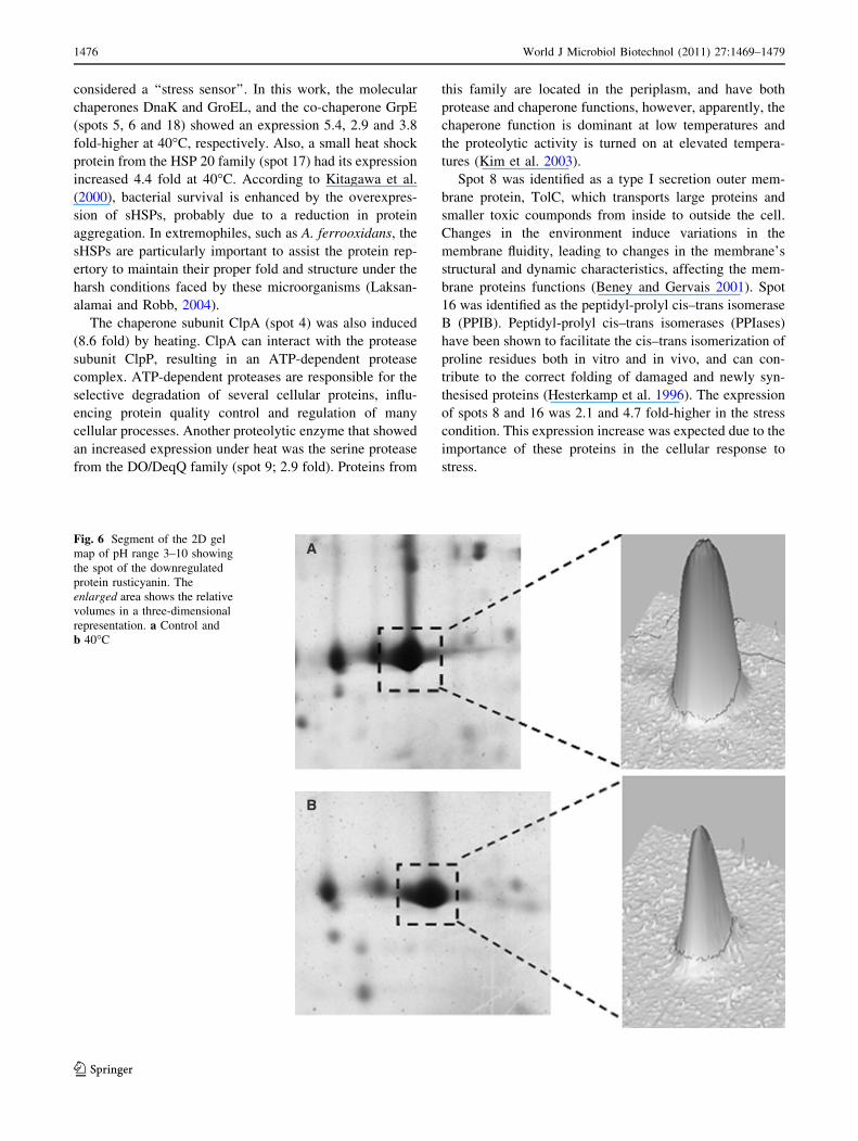
Proteomic analysis
To detect alterations at the protein level, the protein profile
of A. ferrooxidans LR was investigated, using 2-DE and
mass spectrometry, in cells submitted to heat stress and
K2HPO4 starvation. The gels with a pH range of 4-7 pre-
sented the best resolutions and were selected to act as
reference maps (Fig. 5a–d). The analytical and biological
variances were calculated from protein extracts obtained
from three independent bacterial cultures and triplicate
Fig. 3 FT-IR results. a FT-IR spectra of A. ferrooxidans cells
cultivated under control conditions, 40�C and phosphate starvation.
The part of the spectrum used to built the model is highlighted. b PC1
versus PC2 of PCA model built with control samples (circles),
samples grown at 40� (triangles) and samples grown under phosphate
starvation (stars)
Fig. 4 The mean spectrum of control samples (solid line) and
samples grown at 40�C (a) and under phosphate starvation
(b) (dashed line)
World J Microbiol Biotechnol (2011) 27:1469–1479 1473
123
2-DE gels. For protein comparison, gel images of each
treatment were calibrated with the correspondent control
gel image using the Image Master 2D Platinum software.
Spots showing at least a twofold change in their expression
level were considered for analysis, resulting in 44 differ-
entially expressed proteins on the tested stress conditions.
Differentially expressed proteins in A. ferrooxidans
cells submitted to heat stress
Thirty protein spots were significantly changed by a tem-
perature shift from 30 to 40�C. Under these conditions, 12
proteins were upregulated and 18 were downregulated
(Table 2; Fig. 5a, b).
Upregulated proteins
The upregulated proteins belong to the energy metabolism
and protein fate categories, and includes a subunit of the
enzyme NADH-quinone oxidoreductase (NDH-1; spot 1),
which catalyses electrons transfer in the NADH-quinone
oxireductase complex in a proton pump associated reac-
tion. In bacteria this complex usually includes 14 subunits.
In A. ferrooxidans, this biochemical pathway is important,
since this bacterium gains energy by a reverse electron flow
from Fe(II) to NADH (Valdes et al. 2008). The expression
of NDH-1 was 2.8 fold-higher at 40�C, suggesting that
proton translocation across the membrane raised during
heat stress.
Fig. 5 The reference gel of
A. ferrooxidans cells cultivated
under different conditions.
a control of the temperature
experiment; b 40�C; c control of
the phosphate starvation
experiment; d phosphate
starvation. Total protein
(200 lg) was subjected to 2-DE
electrophoresis (11-cm IPG
strips with pH range 4–7; 15%
SDS–PAGE). Proteins were
visualised by Coomassie
Brilliant Blue staining. Circlesrepresent differential proteins
with at least twofold expression.
The enlarged areas show the
relative volumes in a three-
dimensional representation
1474 World J Microbiol Biotechnol (2011) 27:1469–1479
123

The overexpression of proteins involved in energy
metabolism (spots 10, 12 and 22) could have occurred due
to the necessity to generate ATP for the synthesis of pro-
teins that can deal with heat stress, such as the heat shock
proteins (HSPs).
Most of the overexpressed proteins from the protein fate
category were HSPs and molecular chaperones (Table 2).
Heat shock proteins expression is induced when bacteria
are exposed to several stress conditions, such as heating,
starvation, presence of toxic elements like heavy metals,
for example, and others. Jerez (1988) showed that DnaK
and GroEL are part of the A. ferrooxidans response to heat
shock. Seeger et al. (1996) demonstrated that these proteins
are induced and have their level of phosphorylation
increased when A. ferrooxidans cells are exposed to several
stress conditions, suggesting that these chaperones could be
Table 2 Proteins regulated by heat stress in A. ferrooxidans LR cells
Spot
number
Protein ID (JCVI) Functional
categoryaTheorical
MW (kDa)/pI
Matched
peptides
Total ion
score
Expression Fold changesb
(P value)
1 NADH-quinone oxidoreductase, G
subunit
AFE_0480 EM 83.9/6.3 14 89 Up 2.8 (0.006763)
2 TonB-dependent receptor AFE_1054 TBP 85.2/6.53 11 100 Down 4.5 (0.000289)
3 DNA ligase, NAD-dependent (ligA) AFE_2283 DM 74.4/6.9 15 66 Down 3.5 (0.02)
4 ATP-dependent Clp protease- subunit
(ClpA)
AFE_2518 PF 79.4/4.4 5 75 Up 8.6 (0.000263)
5 Chaperone protein DnaK AFE_0440 PF 68.3/5.0 25 245 Up 5.4 (0.000603)
6 Chaperonin, 60 kDa (GroEL) AFE_2496 PF 58.7/5.4 22 248 Up 2.9 (0.003833)
7 Pyruvate kinase barrel domain protein AFE_2290 U 63.4/6.9 16 33 Down 3 (8.185E-07)
8 Type I secretion outer membrane
protein, TolC
AFE_2463 PF 48.0/6.4 4 116 Up 2.1 (0.001984)
9 Serine protease, DO/DeqQ family AFE_1685 PF 49.7/7.0 8 166 Up 2.9 (9.74E-05)
10 Fructose -1-6-bisphosphatase AFE_2837 EM 37.3/5.5 11 217 Up 3.8 (0.001046)
11 Carboxymethyleneutenolidase AFE_0887 CP 26.4/6.22 10 70 Down 4.7 (0.000755)
12 Phosphoglycerate kinase pgk AFE_3083 EM 42.1/6.45 15 199 Up 3.1 (0.002303)
13 Mrp protein AFE_0444 U 38.2/5.07 4 91 Down 3.0 (0.0092623)
14 Translation elongation factor P (efp) AFE_2190 PS 20.8/4.98 3 70 Down 2.0 (0.022795)
15 Ribose 5-phosphate isomerase A (rpiA) AFE_2410 EM 23.3/6.25 8 324 Down 2.3 (0.000239)
16 Peptidyl-prolyl cis–trans isomerase
B (ppiB)
AFE_1106 PF 18.4/6.95 3 209 Up 4.7 (1.348E-06)
17 Heat shock protein, Hsp20 family AFE_1437 PF 16.7/5.43 9 155 Up 4.4 (0.000103)
18 Co-chaperone GrpE AFE_0439 PF 18.9/4.12 7 84 Up 3.8 (0.002834)
19 FeS cluster assembly scaffold protein
IscU
AFE_2366 BC 15.2/5.33 4 59 Down 3.8 (2.132E-06)
20 Ribulose biphosphate carboxylase,
rubisco
AFE_1395 EM 13.9/5.9 5 250 Down 2.0 (5.232E-05)
21 Ribulose bisphosphate cbbS1 AFE_0058 EM 12.6/6.41 5 70 Down 2.0 (0.000239)
22 Glycine cleavage system H protein AFE_2067 EM 13.6/5.29 4 120 Up 2.5 (0.000772)
23 ACT domain protein AFE_2600 U 18.4/4.29 5 220 Down 2.3 (0.000407)
24 Transcription elongation
factor GreA (greA)
AFE_2401 T 17.4/4,20 4 87 Down 2.5 (0.0007724)
25 Ferredoxin AFE_2063 EM 13.6/4.11 4 159 Down 2.6 (0.000263)
26 Thioredoxin AFE_1113 EM 12.7/4.25 5 164 Down 6.5 (1.770E-05)
27 Ccarboxysome shell peptide AFE_1400 CP 9.9/5.23 6 273 Down 4.8 (1.215E-07)
28 Carboxysome shell peptide AFE_1402 CP 11.4/5.2 10 188 Down 3.2 (1.668E-07)
29 Thioredoxin (trx) AFE_2383 EM 12.1/5. 59 5 290 Down 3.0 (0.002663)
30 Conserved hypothetical protein AFE_2669 U 9.4/6.86 4 90 Down 2.5 (0.000610)
a BC biosynthesis of cofactors, prosthetic groups and carriers, CP cellular process, DM DNA metabolism, EM energy metabolism, PF protein
fate, PS protein synthesis, T transcription, TBP transport and binding proteins, U unknown function and enzymes of unknown specificityb Fold changes of expression levels of differential proteins after heat stress (40�C)
World J Microbiol Biotechnol (2011) 27:1469–1479 1475
123
considered a ‘‘stress sensor’’. In this work, the molecular
chaperones DnaK and GroEL, and the co-chaperone GrpE
(spots 5, 6 and 18) showed an expression 5.4, 2.9 and 3.8
fold-higher at 40�C, respectively. Also, a small heat shock
protein from the HSP 20 family (spot 17) had its expression
increased 4.4 fold at 40�C. According to Kitagawa et al.
(2000), bacterial survival is enhanced by the overexpres-
sion of sHSPs, probably due to a reduction in protein
aggregation. In extremophiles, such as A. ferrooxidans, the
sHSPs are particularly important to assist the protein rep-
ertory to maintain their proper fold and structure under the
harsh conditions faced by these microorganisms (Laksan-
alamai and Robb, 2004).
The chaperone subunit ClpA (spot 4) was also induced
(8.6 fold) by heating. ClpA can interact with the protease
subunit ClpP, resulting in an ATP-dependent protease
complex. ATP-dependent proteases are responsible for the
selective degradation of several cellular proteins, influ-
encing protein quality control and regulation of many
cellular processes. Another proteolytic enzyme that showed
an increased expression under heat was the serine protease
from the DO/DeqQ family (spot 9; 2.9 fold). Proteins from
this family are located in the periplasm, and have both
protease and chaperone functions, however, apparently, the
chaperone function is dominant at low temperatures and
the proteolytic activity is turned on at elevated tempera-
tures (Kim et al. 2003).
Spot 8 was identified as a type I secretion outer mem-
brane protein, TolC, which transports large proteins and
smaller toxic coumponds from inside to outside the cell.
Changes in the environment induce variations in the
membrane fluidity, leading to changes in the membrane’s
structural and dynamic characteristics, affecting the mem-
brane proteins functions (Beney and Gervais 2001). Spot
16 was identified as the peptidyl-prolyl cis–trans isomerase
B (PPIB). Peptidyl-prolyl cis–trans isomerases (PPIases)
have been shown to facilitate the cis–trans isomerization of
proline residues both in vitro and in vivo, and can con-
tribute to the correct folding of damaged and newly syn-
thesised proteins (Hesterkamp et al. 1996). The expression
of spots 8 and 16 was 2.1 and 4.7 fold-higher in the stress
condition. This expression increase was expected due to the
importance of these proteins in the cellular response to
stress.
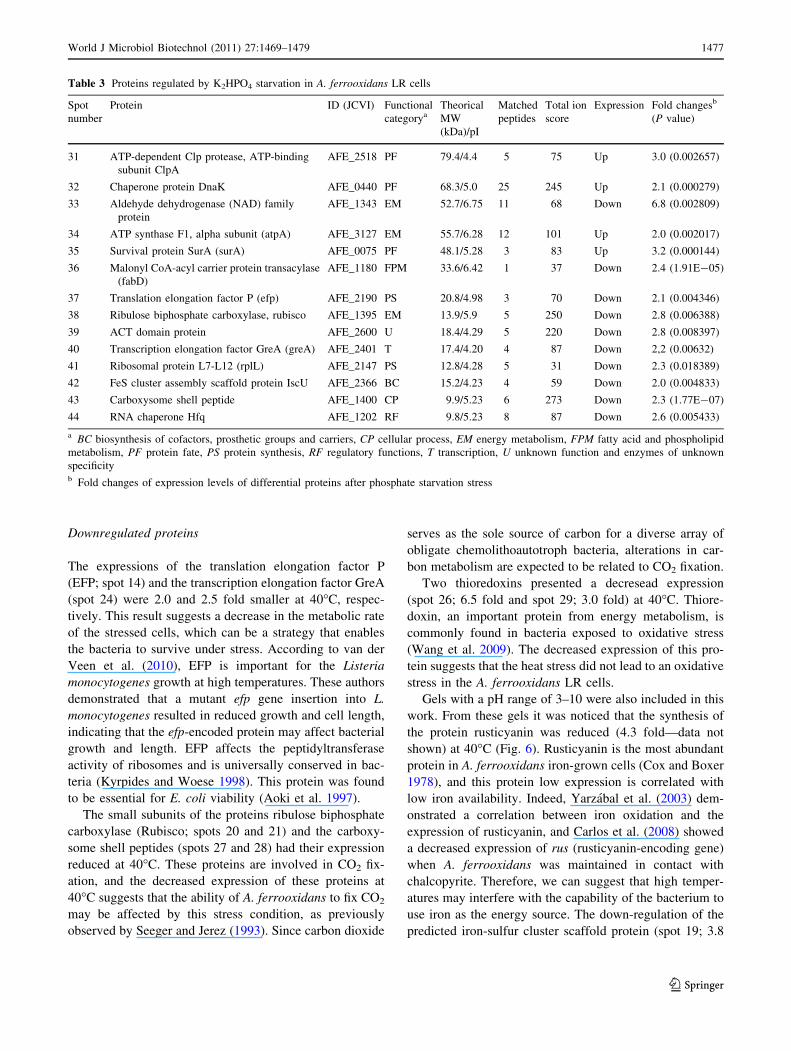
Fig. 6 Segment of the 2D gel
map of pH range 3–10 showing
the spot of the downregulated
protein rusticyanin. The
enlarged area shows the relative
volumes in a three-dimensional
representation. a Control and
b 40�C
1476 World J Microbiol Biotechnol (2011) 27:1469–1479
123
Downregulated proteins
The expressions of the translation elongation factor P
(EFP; spot 14) and the transcription elongation factor GreA
(spot 24) were 2.0 and 2.5 fold smaller at 40�C, respec-
tively. This result suggests a decrease in the metabolic rate
of the stressed cells, which can be a strategy that enables
the bacteria to survive under stress. According to van der
Veen et al. (2010), EFP is important for the Listeria
monocytogenes growth at high temperatures. These authors
demonstrated that a mutant efp gene insertion into L.
monocytogenes resulted in reduced growth and cell length,
indicating that the efp-encoded protein may affect bacterial
growth and length. EFP affects the peptidyltransferase
activity of ribosomes and is universally conserved in bac-
teria (Kyrpides and Woese 1998). This protein was found
to be essential for E. coli viability (Aoki et al. 1997).
The small subunits of the proteins ribulose biphosphate
carboxylase (Rubisco; spots 20 and 21) and the carboxy-
some shell peptides (spots 27 and 28) had their expression
reduced at 40�C. These proteins are involved in CO2 fix-
ation, and the decreased expression of these proteins at
40�C suggests that the ability of A. ferrooxidans to fix CO2
may be affected by this stress condition, as previously
observed by Seeger and Jerez (1993). Since carbon dioxide
serves as the sole source of carbon for a diverse array of
obligate chemolithoautotroph bacteria, alterations in car-
bon metabolism are expected to be related to CO2 fixation.
Two thioredoxins presented a decresead expression
(spot 26; 6.5 fold and spot 29; 3.0 fold) at 40�C. Thiore-
doxin, an important protein from energy metabolism, is
commonly found in bacteria exposed to oxidative stress
(Wang et al. 2009). The decreased expression of this pro-
tein suggests that the heat stress did not lead to an oxidative
stress in the A. ferrooxidans LR cells.
Gels with a pH range of 3–10 were also included in this
work. From these gels it was noticed that the synthesis of
the protein rusticyanin was reduced (4.3 fold—data not
shown) at 40�C (Fig. 6). Rusticyanin is the most abundant
protein in A. ferrooxidans iron-grown cells (Cox and Boxer
1978), and this protein low expression is correlated with
low iron availability. Indeed, Yarzabal et al. (2003) dem-
onstrated a correlation between iron oxidation and the
expression of rusticyanin, and Carlos et al. (2008) showed
a decreased expression of rus (rusticyanin-encoding gene)
when A. ferrooxidans was maintained in contact with
chalcopyrite. Therefore, we can suggest that high temper-
atures may interfere with the capability of the bacterium to
use iron as the energy source. The down-regulation of the
predicted iron-sulfur cluster scaffold protein (spot 19; 3.8
Table 3 Proteins regulated by K2HPO4 starvation in A. ferrooxidans LR cells
Spot
number
Protein ID (JCVI) Functional
categoryaTheorical
MW
(kDa)/pI
Matched
peptides
Total ion
score
Expression Fold changesb
(P value)
31 ATP-dependent Clp protease, ATP-binding
subunit ClpA
AFE_2518 PF 79.4/4.4 5 75 Up 3.0 (0.002657)
32 Chaperone protein DnaK AFE_0440 PF 68.3/5.0 25 245 Up 2.1 (0.000279)
33 Aldehyde dehydrogenase (NAD) family
protein
AFE_1343 EM 52.7/6.75 11 68 Down 6.8 (0.002809)
34 ATP synthase F1, alpha subunit (atpA) AFE_3127 EM 55.7/6.28 12 101 Up 2.0 (0.002017)
35 Survival protein SurA (surA) AFE_0075 PF 48.1/5.28 3 83 Up 3.2 (0.000144)
36 Malonyl CoA-acyl carrier protein transacylase
(fabD)
AFE_1180 FPM 33.6/6.42 1 37 Down 2.4 (1.91E-05)
37 Translation elongation factor P (efp) AFE_2190 PS 20.8/4.98 3 70 Down 2.1 (0.004346)
38 Ribulose biphosphate carboxylase, rubisco AFE_1395 EM 13.9/5.9 5 250 Down 2.8 (0.006388)
39 ACT domain protein AFE_2600 U 18.4/4.29 5 220 Down 2.8 (0.008397)
40 Transcription elongation factor GreA (greA) AFE_2401 T 17.4/4.20 4 87 Down 2,2 (0.00632)
41 Ribosomal protein L7-L12 (rplL) AFE_2147 PS 12.8/4.28 5 31 Down 2.3 (0.018389)
42 FeS cluster assembly scaffold protein IscU AFE_2366 BC 15.2/4.23 4 59 Down 2.0 (0.004833)
43 Carboxysome shell peptide AFE_1400 CP 9.9/5.23 6 273 Down 2.3 (1.77E-07)
44 RNA chaperone Hfq AFE_1202 RF 9.8/5.23 8 87 Down 2.6 (0.005433)
a BC biosynthesis of cofactors, prosthetic groups and carriers, CP cellular process, EM energy metabolism, FPM fatty acid and phospholipid
metabolism, PF protein fate, PS protein synthesis, RF regulatory functions, T transcription, U unknown function and enzymes of unknown
specificityb Fold changes of expression levels of differential proteins after phosphate starvation stress
World J Microbiol Biotechnol (2011) 27:1469–1479 1477
123
fold) suggests a decrease in the capacity of the cells to
acquire iron from the medium, suggesting that the stress is
interfering with the energy metabolism. Also, the synthesis
of the iron-sulfur protein ferredoxin (spot 25; 2.6 fold) and
the TonB-dependent receptor (spot 2; 4.5 fold) showed a
decrease in A. ferrooxidans cells grown at 40�C. These
results suggest that growth at 40�C could have affected the
iron metabolism in A. ferrooxidans LR.
Differentially expressed proteins in A. ferrooxidans
cells submitted to phosphate starvation
Fourteen proteins had their expression changed in the
absence of K2HPO4 (Table 3; Fig. 5c, d). Among the
proteins whose synthesis increased were the molecular
chaperone DnaK (spot 31; 3.0 fold) and the chaperone
subunit ClpA (spot 32; 2.6 fold), suggesting that the
absence of K2HPO4 in the medium activates a stress
response in A. ferrooxidans, despite the bacteria capability
to accumulate polyphosphate granules in high amounts
(Alvarez and Jerez 2004). This is emphasized by the
increased synthesis of the survival protein SurA (spot 35;
4.6 fold). In E. coli, SurA is a periplasmic protein required
for the proper assembly of several outer membrane proteins
(Lazar and Kolter 1996). This result may also indicate that
phosphate starvation damages the outer membrane pro-
teins. In fact, outer membrane proteins have previously
been found to change due to phosphate starvation (Jerez
et al. 1992, Seeger and Jerez 1993) or alterations in other
growth conditions, such as pH (Amaro et al. 1991).
The translation elongation factor P (spot 37; 2.1 fold),
the transcription elongation factor GreA (spot 40; 2.2 fold)
and the ribosomal protein L7-L12 (spot 41; 2.3) had their
synthesis decreased in the absence of K2HPO4. This result
may be attributed to a decrease in the metabolic rate of the
cells submitted to this stress. A metabolic rate decrease was
also observed for cell submitted to heat stress. The proteins
Rubisco (spot 38; 2.8 fold) and carboxysome shell peptide
(spot 43, 2.3 fold) also had their synthesis decreased
indicating that the carbon metabolism was also affected by
phosphate starvation.
Acknowledgments This work was supported by grant 02/07642-3
from Fundacao de Amparo a Pesquisa do Estado de Sao Paulo
(FAPESP). DAR received a fellowship from Coordenacao de Aper-
feicoamento de Pessoal de Nıvel Superior (CAPES). LMMO received
a research fellowship from Conselho Nacional de Desenvolvimento
Cientıfico e Tecnologico (CNPq).
References
Alvarez S, Jerez CA (2004) Copper ions stimulate polyphosphate
degradation and phosphate efflux in Acidithiobacillus ferroox-idans. Appl Environ Microbiol 70:5177–5182
Amaro AM, Chamorro D, Seeger M, Arredondo R, Peirano I, Jerez
CA (1991) Effect of external pH perturbations on in vivo protein
synthesis by the acidophilic bacterium Thiobacillus ferrooxi-dans. J Bacteriol 173:910–915
Aoki H, Dekany K, Adams SL, Ganoza MC (1997) The gene
encoding the elongation factor P protein is essential for
viability and is required for protein synthesis. J Biol Chem 272:
32254–32259
Beney L, Gervais P (2001) Influence of the fluidity of the membrane
on the response of microorganisms to environmental stresses.
Appl Microbiol Biotechnol 57:34–42
Carlos C, Reis FC, Vicentini R, Madureira DJ, Ottoboni LMM (2008)
The rus operon genes are differentially regulated when Acidi-thiobacillus ferrooxidans LR is kept in contact with metal
sulfides. Curr Microbiol 57:375–380
Chalmers JM, Griffiths PR (2002) Handbook of vibrational spectros-
copy, vol 5. Wiley, Chichester
Cox JC, Boxer DH (1978) The purification and some properties
of rusticyanin, a blue copper protein involved in iron (II)
oxidation from Thiobacillus ferrooxidans. Biochem J 174:
497–502
Eberl L, Givskov M, Sternberg C, Morller S, Christiansen G, Molin S
(1996) Physiological responses of Pseudomonas putida KT2442
to phosphate starvation. Microbiol 142:51–63
Farah C, Vera M, Morin D, Haras D, Jerez CA, Guiliani N (2005)
Evidence for a functional quorum-sensing type AI-1 system in
the extremophilic bacterium Acidithiobacillus ferrooxidans.
Appl Environ Microbiol 71:7033–7040
Garcia O Jr (1991) Isolation and purification of Thiobacillusferrooxidans and Thiobacillus thiooxidans from some coal and
uranium mines of Brazil. Braz J Microbiol 20:1–6
Hesterkamp T, Hauser S, Lutcke H, Bukau B (1996) Escherichia colitrigger factor is a prolyl isomerase that associates with nascent
polypeptide chains. Proc Natl Acad Sci 93:4437–4441
Holmquist L, Kjelleberg S (1993) Changes in viability, respiratory
activity and morphology of the marine Vibrio sp. strain S14
during starvation of individual nutrients and subsequent recov-
ery. FEMS Microbiol Ecol 12:215–224
Hubert WA, Leduc LG, Ferroni GD (1995) Heat and cold shock
responses in different strains of Thiobacillus ferrooxidans. Curr
Microbiol 31:10–14
Jerez CA (1988) The heat shock response in meso- and thermoacid-
ophilic chemolithotrophic bacteria. FEMS Microbiol Lett 56:
289–294
Jerez CA, Seeger M, Amaro AM (1992) Phosphate starvation affects
the synthesis of outer membrane proteins in Thiobacillusferrooxidans. FEMS Microbiol Lett 98:29–34
Kim DY, Kim DR, Ha SC, Lokanath NK, Lee CJ, Hwang HY, Kim
KK (2003) Crystal structure of the protease domain of a heat-
shock protein HtrA from Thermotoga maritime. J Biol Chem
278:6543–6551
Kitagawa M, Matsumura Y, Tsuchido T (2000) Small heat shock
proteins, IbpA and IbpB, are involved in resistances to heat and
superoxide stresses in Escherichia coli. FEMS Microbiol Lett
184:165–171
Knegt FHP, Mello LV, Reis FC, Santos MT, Vicentini R, Ferraz FC,
Ottoboni LMM (2008) ribB and ribBA genes from Acidithioba-cillus ferrooxidans: expression levels under different
growth conditions and phylogenetic analysis. Res Microbiol 159:
423–431
Kyrpides NC, Woese CR (1998) Universally conserved translation
initiation factors. Proc Natl Acad Sci USA 95:224–228
Laksanalamai P, Robb FT (2004) Small heat shock proteins from
extremophiles: a review. Extremophiles 8:1–11
Lazar S, Kolter R (1996) SurA assists the folding of Escherichia coliouter membrane proteins. J Bacteriol 178:1770–1773
1478 World J Microbiol Biotechnol (2011) 27:1469–1479
123
Modak JM, Natarajan KA, Mukhopadhyay S (1996) Development of
temperature-tolerant strains of Thiobacillus ferrooxidans to
improve bioleaching kinetics. Hydrometallurgy 42:51–61
Pianetti A, Battistelli M, Citterio B, Parlani C, Falcieri E, Bruscolini F
(2009) Morphological changes of Aeromonas hydrophila in
response to osmotic stress. Micron 40:426–433
Rawlings DE (2005) Characteristics and adaptability of iron and
sulfur-oxidizing microorganisms used for the recovery of metals
from minerals and their concentrates. Microb Cell Fact 4:1–15
Rilfors L, Wieslander A, Stahl S (1978) Lipid and protein compo-
sition of membranes of Bacillus megaterium variants in the
temperature range of 5 to 70 degrees C. J Bacteriol 135:
1043–1052
Seeger M, Jerez CA (1993) Responses of Thiobacillus ferrooxidans to
phosphate limitation. FEMS Microbiol Rev 11:37–42
Seeger M, Osorio G, Jerez CA (1996) Phosphorylation of GroEL,
DnaK and other proteins from Thiobacillus ferrooxidans grown
under different conditions. FEMS Microbiol Lett 138:129–134
Shi B, Xia X (2003) Morphological changes of Pseudomonaspseudoalcaligenes in response to temperature selection. Curr
Microbiol 46:120–123
Smolka MB, Martins D, Winck FV, Santoro CE, Castellari RR,
Ferrari F et al (2003) Proteome analysis of the plant pathogen
Xylella fastidiosa reveals major cellular and extracellular
proteins and a peculiar codon bias distribution. Proteomics 3:
224–237
Tuovinen OH, Kelly DP (1972) Biology of Thiobacillus ferrooxidansin relation to the microbiological leaching of sulphide ores.
Z Allg Mikrobiol 12:311–346
Valdes J, Pedroso I, Quatrini R, Dodson RJ, Tettelin H, Blake R II,
Eisen JA, Holmes DS (2008) Acidithiobacillus ferrooxidansmetabolism: from genome sequence to industrial applications.
BMC Genomics 9:597–621
van der Veen S, van Schalkwijk S, Molenaar D, De vos W, Abee T,
Wells-Bennik MHJ (2010) The SOS response of Listeriamonocytogenes is involved in stress resistance and mutagenesis.
Microbiol 156:374–384
Vasconcelos AR, Nogueira FCS, Abreu EFM, Goncalves EFP, Souza
AS, Campos FAP (2005) Protein extraction from cowpea tissues
for 2D electrophoresis and MS analysis. Chromatographia
62:447–450
Wang Y, Zhang X, Liu Q, Ai C, Mo H, Zeng J (2009) Expression,
purification and molecular structure modeling of thioredoxin
(Trx) and thioredoxin reductase (TrxR) from Acidithiobacillusferrooxidans. Curr Microbiol 59:35–41
Xiao S, Chao J, Wang W, Fang F, Qiu G, Liu X (2009) Real-time
PCR analysis of the heat-shock response of Acidithiobacillusferrooxidans ATCC 23270. Folia Biol 55:1–6
Yarzabal A, Duquesne K, Bonnefoy V (2003) Rusticyanin gene
expression of Acidithiobacillus ferrooxidans ATCC 33020 in
sulfur- and in ferrous iron media. Hydrometallurgy 71:107–114
Yu C, Irudayaraj J (2004) Spectroscopic characterization of micro-
organisms by Fourier transform infrared microspectroscopy.
Biopolymers 77:368–377
World J Microbiol Biotechnol (2011) 27:1469–1479 1479
123
Related Documents


