ORIGINAL ARTICLE Metabolomic study of Chilean biomining bacteria Acidithiobacillus ferrooxidans strain Wenelen and Acidithiobacillus thiooxidans strain Licanantay Patricio Martı ´nez • Sebastia ´n Ga ´lvez • Norimasa Ohtsuka • Marko Budinich • Marı ´a Paz Corte ´s • Cristia ´n Serpell • Kenji Nakahigashi • Akiyoshi Hirayama • Masaru Tomita • Tomoyoshi Soga • Servet Martı ´nez • Alejandro Maass • Pilar Parada Received: 21 December 2011 / Accepted: 29 June 2012 / Published online: 21 July 2012 Ó The Author(s) 2012. This article is published with open access at Springerlink.com Abstract In this study, we present the first metabolic profiles for two bioleaching bacteria using capillary elec- trophoresis coupled with mass spectrometry. The bacte- ria, Acidithiobacillus ferrooxidans strain Wenelen (DSM 16786) and Acidithiobacillus thiooxidans strain Licanantay (DSM 17318), were sampled at different growth phases and on different substrates: the former was grown with iron and sulfur, and the latter with sulfur and chalcopyrite. Metabolic profiles were scored from planktonic and sessile states. Spermidine was detected in intra- and extracellular samples for both strains, suggesting it has an important role in biofilm formation in the presence of solid substrate. The canonical pathway for spermidine synthesis seems absent as its upstream precursor, putrescine, was not present in samples. Glutathione, a catalytic activator of elemental sulfur, was identified as one of the most abundant metab- olites in the intracellular space in A. thiooxidans strain Licanantay, confirming its participation in the sulfur oxi- dation pathway. Amino acid profiles varied according to the growth conditions and bioleaching species. Glutamic and aspartic acid were highly abundant in intra- and extracellular extracts. Both are constituents of the extra- cellular matrix, and have a probable role in cell detoxifi- cation. This novel metabolomic information validates previous knowledge from in silico metabolic reconstruc- tions based on genomic sequences, and reveals important biomining functions such as biofilm formation, energy management and stress responses. Keywords Bioleaching Á Metabolomics Á Biomarker Á Capillary electrophoresis Á Mass spectrometry Á CE–MS Á Acidithiobacillus Á Thiooxidans Á Ferrooxidans Á Wenelen Á Licanantay Á Spermidine 1 Introduction Several extremophiles have been isolated from mining operations (Johnson et al. 2001; Okibe et al. 2003; Tyson et al. 2005), and their role in the dynamics and evolution of minerals has previously been discussed (Santelli et al. 2009). They are known to have a relevant role in hydro- metallurgic extraction processes: their presence is linked to enhanced extraction of metals such as copper, nickel, cobalt, zinc and uranium. This process, termed ‘‘biomin- ing’’ or ‘‘bioleaching’’, is an example of industrial bio- technology as an empirical process, governed by trial Electronic supplementary material The online version of this article (doi:10.1007/s11306-012-0443-3) contains supplementary material, which is available to authorized users. P. Martı ´nez Á S. Ga ´lvez Á N. Ohtsuka Á P. Parada (&) BioSigma S.A., Loteo Los Libertadores, Lote 106, Colina, Chile e-mail: [email protected] P. Martı ´nez e-mail: [email protected] M. Budinich Á M. P. Corte ´s Á C. Serpell Á S. Martı ´nez Á A. Maass Laboratory of Bioinformatics and Mathematics of the Genome, Center for Mathematical Modeling (UMI 2807, CNRS) and Center for Genome Regulation, University of Chile, Avda. Blanco Encalada 2120, 7th Floor, Santiago, Chile K. Nakahigashi Á A. Hirayama Á M. Tomita Á T. Soga Institute for Advanced Biosciences, Keio University, Tsuruoka, Yamagata, Japan S. Martı ´nez Á A. Maass Department of Mathematical Engineering and Center for Mathematical Modeling (UMI 2807, CNRS), Faculty of Mathematical and Physical Sciences, University of Chile, Avda. Blanco Encalada 2120, 7th Floor, Santiago, Chile 123 Metabolomics (2013) 9:247–257 DOI 10.1007/s11306-012-0443-3

Welcome message from author
This document is posted to help you gain knowledge. Please leave a comment to let me know what you think about it! Share it to your friends and learn new things together.
Transcript
ORIGINAL ARTICLE
Metabolomic study of Chilean biomining bacteriaAcidithiobacillus ferrooxidans strain Wenelenand Acidithiobacillus thiooxidans strain Licanantay
Patricio Martınez • Sebastian Galvez • Norimasa Ohtsuka • Marko Budinich •
Marıa Paz Cortes • Cristian Serpell • Kenji Nakahigashi • Akiyoshi Hirayama •
Masaru Tomita • Tomoyoshi Soga • Servet Martınez • Alejandro Maass •
Pilar Parada
Received: 21 December 2011 / Accepted: 29 June 2012 / Published online: 21 July 2012
� The Author(s) 2012. This article is published with open access at Springerlink.com
Abstract In this study, we present the first metabolic
profiles for two bioleaching bacteria using capillary elec-
trophoresis coupled with mass spectrometry. The bacte-
ria, Acidithiobacillus ferrooxidans strain Wenelen (DSM
16786) and Acidithiobacillus thiooxidans strain Licanantay
(DSM 17318), were sampled at different growth phases
and on different substrates: the former was grown with iron
and sulfur, and the latter with sulfur and chalcopyrite.
Metabolic profiles were scored from planktonic and sessile
states. Spermidine was detected in intra- and extracellular
samples for both strains, suggesting it has an important role
in biofilm formation in the presence of solid substrate. The
canonical pathway for spermidine synthesis seems absent
as its upstream precursor, putrescine, was not present in
samples. Glutathione, a catalytic activator of elemental
sulfur, was identified as one of the most abundant metab-
olites in the intracellular space in A. thiooxidans strain
Licanantay, confirming its participation in the sulfur oxi-
dation pathway. Amino acid profiles varied according to
the growth conditions and bioleaching species. Glutamic
and aspartic acid were highly abundant in intra- and
extracellular extracts. Both are constituents of the extra-
cellular matrix, and have a probable role in cell detoxifi-
cation. This novel metabolomic information validates
previous knowledge from in silico metabolic reconstruc-
tions based on genomic sequences, and reveals important
biomining functions such as biofilm formation, energy
management and stress responses.
Keywords Bioleaching � Metabolomics � Biomarker �Capillary electrophoresis � Mass spectrometry � CE–MS �Acidithiobacillus � Thiooxidans � Ferrooxidans �Wenelen �Licanantay � Spermidine
1 Introduction
Several extremophiles have been isolated from mining
operations (Johnson et al. 2001; Okibe et al. 2003; Tyson
et al. 2005), and their role in the dynamics and evolution of
minerals has previously been discussed (Santelli et al.
2009). They are known to have a relevant role in hydro-
metallurgic extraction processes: their presence is linked to
enhanced extraction of metals such as copper, nickel,
cobalt, zinc and uranium. This process, termed ‘‘biomin-
ing’’ or ‘‘bioleaching’’, is an example of industrial bio-
technology as an empirical process, governed by trial
Electronic supplementary material The online version of thisarticle (doi:10.1007/s11306-012-0443-3) contains supplementarymaterial, which is available to authorized users.
P. Martınez � S. Galvez � N. Ohtsuka � P. Parada (&)
BioSigma S.A., Loteo Los Libertadores, Lote 106, Colina, Chile
e-mail: [email protected]
P. Martınez
e-mail: [email protected]
M. Budinich � M. P. Cortes � C. Serpell � S. Martınez �A. Maass
Laboratory of Bioinformatics and Mathematics of the Genome,
Center for Mathematical Modeling (UMI 2807, CNRS)
and Center for Genome Regulation, University of Chile,
Avda. Blanco Encalada 2120, 7th Floor, Santiago, Chile
K. Nakahigashi � A. Hirayama � M. Tomita � T. Soga
Institute for Advanced Biosciences, Keio University,
Tsuruoka, Yamagata, Japan
S. Martınez � A. Maass
Department of Mathematical Engineering and Center for
Mathematical Modeling (UMI 2807, CNRS), Faculty of
Mathematical and Physical Sciences, University of Chile,
Avda. Blanco Encalada 2120, 7th Floor, Santiago, Chile
123
Metabolomics (2013) 9:247–257
DOI 10.1007/s11306-012-0443-3
testing. In 1947, the phenomenon of metal dissolution in
acid media was attributed to microorganismal action (Col-
mer and Hinkle 1947). Biomining is described as the
extraction of metal from sulfide ores or concentrates by the
action of acidophilic bioleaching microorganisms that cat-
alyze mineral ore oxidation. These microorganisms are
naturally present in the minerals’ native flora. The industrial
process is designed to provide an environment with optimal
growth conditions in order to optimize microorganismal
action. Dump and heap bioleaching operations using this
technology are located in SBL Radomiro Tomic in Chile,
Cerro Verde in Peru, Morenci in USA, among others.
The use of specific microorganisms and/or their deriv-
atives is relatively new to this industry. It has been shown
that these enhance conventional processes of acid irrigation
several fold, which eventually translates into economical
benefits for companies (BioSigma US Patent No.
7,601,530; 7,700,343; 7,837,760 among others). Studies of
their physiology are crucial for understanding how they
interact with the mineral surface and how they can be
optimized to improve mineral dissolution.
Acidophilic prokaryotes used for metal recovery from
sulfide minerals include members of the Bacteria and
Archaea domains. We have isolated several microorgan-
isms with the aim of using them in biomining processes for
copper extraction. The two selected isolates show improved
oxidizing activity when compared to standard international
strains: these are A. ferrooxidans, strain Wenelen DSM
16786, and A. thiooxidans, strain Licanantay DSM 17318
(Sugio et al. 2009; Ohata et al. 2010). The former is an iron
and sulfur oxidizing microorganism, while the latter is
strictly sulfur oxidizing. Detailed studies of both chemo-
lithotrophic bacteria have been undertaken since their iso-
lation in 2003 (Levican et al. 2008), and continue to be a
matter of interest in other ‘‘omics’’ analyses.
An important complication when studying biomining
microorganisms is genetic transformation. Notwithstanding,
there are some reports of successful transformations, which
are considered random phenomena (Liu et al. 2000; Kusano
et al. 1992). This explains why, despite advances made in
past years, little is known regarding the specifics of their
metabolism. Genomic sequences analyzed with bioinfor-
matics tools have provided insights into their metabolism
(Valdes et al. 2008; Cardenas et al. 2010; Quatrini et al.
2009). The information gathered from these sequencing
projects complement other ‘‘omics’’ data, such as gene
expression microarray experiments, mass spectrometry
based metabolite detection and proteomics (Ishii et al. 2007).
The latest advances in metabolomics, particularly the
quantitative metabolic response, are attributable to high-
throughput techniques, which separate and detect cellular
compounds. One such technique, named capillary electro-
phoresis Time-of-Flight Mass Spectrometry, CE–TOFMS,
has several advantages including: high resolution, quantifi-
cation of charged low molecular weight compounds, and
suitability for different organisms (Soga et al. 2003, 2006;
Sato et al. 2004). However, there are limitations related to
metabolomic coverage (Ohashi et al. 2008).
In this paper, we report the first metabolomic study of
bioleaching microorganisms. Different mineral substrates
were tested as energy sources for both bioleaching iso-
lates, A. ferrooxidans strain Wenelen and A. thiooxidans
strain Licanantay. The aim of the study is to reveal
information about the metabolic pathways of these two
bioleaching bacteria. In addition, we compare their
growth in ideal conditions (pure media energy sources—
iron and sulfur) to their growth under more realistic
conditions (chalcopyrite and ore impurities). Finally, we
compare cells attached to solid substrate versus free ones,
as results could reveal information on contact and non-
contact bioleaching.
High-throughput data analysis highlighted differences
between the metabolic profiles of the bacteria when faced
with different energy sources. Similar conclusions are
drawn when comparing different cell populations. Standard
metabolite analysis reveals that specific metabolites are
abundant and can be secreted to the extracellular space.
2 Materials and methods
2.1 Strains and growth conditions
Two isolates obtained from mining environments, A. fer-
rooxidans, strain Wenelen (DMS 16786), and A. thiooxi-
dans, strain Licanantay (DMS 17318), were used in this
study (Sugio et al. 2009; Ohata et al. 2010).
Acidithiobacillus ferrooxidans strain Wenelen, an iron/
sulfur oxidizing bacteria, was grown in KDM media con-
taining (NH4)2SO4 0.99 g/l, NaH2PO4 *2H2O 0.145 g/l,
MgSO4 *7H2O 0.10 g/l, KCl 0.10 g/l, CaCL2 0.021 g/l,
KH2PO4 0.052 g/l with either FeSO4 6 g/l, 1 % sulfur or 1 %
concentrate (composed mainly of chalcopyrite, CuFeS2)
obtained from a Chilean mine. For sulfur oxidizing A. thio-
oxidans strain Licanantay, KDM was supplemented either
1 % sulfur or 1 % concentrate from a Chilean mine. The
mineral was sterilized 3 times by autoclave at 120 �C for
30 min. Both strains were cultivated in bioreactors at 30 �C
with a pH of 1.8 under all conditions. Liquid cultures were
stirred at 150 rpm with an aeration flow of 0.5 VVM (vol-
ume per volume per minute).
2.2 Metabolite extraction protocol
Two reactors were managed under the same conditions for
each microorganism in order to obtain biological replicates.
248 P. Martınez et al.
123
Samples were taken at three time points (T1, T2 and T3)
corresponding to the exponential, early stationary, and late
stationary phase, respectively (Supplementary Fig. SF1).
Our protocol is a modified version of the Soga et al.
(2002) protocol.
For solid substrate growing conditions (sulfur and
chalcopyrite), 200 ml of the culture were filtered using a
vacuum pump with 2 filters in tandem: the upper filter had
a 5 lm pore size to retain cells attached to the substrate
(sessile cells), and the lower filter (0.2 lm pore size)
retained free cells (planktonic cells). For soluble substrate
(iron) only the lower filter was used.
To clean samples, we performed two washes with 10 ml
of acidic water (pH 1.8), followed by two additional
washes with distilled water.
Filters were immersed in a methanol solution (5 ml)
with three internal standards: methionine sulfone, 2-(N-
morpholino) ethane sulfonic acid (MES) and D-camphor-
10-sulfonic acid (CSA).
Cells were sonicated for 30 s and then incubated for
10 min in order to quench enzymatic reactions. 4 ml of the
homogenate were mixed with 1.6 ml water and 4 ml
chloroform, and then vortexed and centrifuged for 5 min at
3,222 RCF at 4 �C. The aqueous phase, containing intra-
cellular metabolites, was collected and subjected to cen-
trifugal filters with a 5 kDa cut-off (millipore).
Sample solutions were centrifuged at 9,520 RCF at 4 �C
for 2 or 3 h. Next, the filtrate was evaporated by the cen-
trivac system until samples were dry.
Samples were stored at -80 �C until CE–MS analysis.
For supernatant analysis, samples were collected from
the reactor at the same time points (T1, T2 and T3). The
two filtering steps consisted of: a 0.2 lm millipore filter to
remove cells, and then, a second filtration using a centrif-
ugal filter (5 kDa). Samples were dried and stored at
-80 �C until CE–MS analysis.
Controls for all assays were generated following the
same protocol without cells.
2.3 Analytic conditions for metabolome analysis
The setup conditions for all runs were performed as
described by Hirayama et al. (2009) with some modifica-
tions. Three technical replicates were carried out for each
sample.
2.4 Instruments
All CE–TOFMS experiments were performed using a CE
capillary electrophoresis system equipped with TOFMS,
1100 isocratic HPLC pump, G1603A CE–MS adapter kit,
and G1607A CE–electrospray ionization (ESI)–MS sprayer
kit (Agilent Technologies). System control and data acqui-
sition were performed using Agilent G2201AA ChemStation
software and Analyst QS for CE and TOFMS, respectively.
In addition, the original Agilent SST316Ti stainless steel
(Fe/Cr/Ni/Mo/Ti; 68:18:11:2:1) ESI needle was replaced
with a platinum needle to avoid poor robustness and needle
corrosion (Soga et al. 2009).
2.5 CE–TOFMS conditions for cationic metabolite
analysis
Cationic metabolites were separated with a fused-silica
capillary (50 lm i.d. 100 cm total length) filled with
1 mol/L formic acid as the reference electrolyte. Sample
solution was injected at 50 mbar for 3 s (ca. 3 nL), at
30 kV. The capillary and sample trays were maintained at
20 �C and below 5 �C, respectively. Sheath liquid was
composed of methanol/water (50 % v/v) with 0.1 lmol/L
hexakis (2,2-difluorothoxy)phosphazene (Hexakis) deliv-
ered at a rate of 10 lL/min. ESI–TOFMS was operated in
the positive ion mode. Capillary voltage was set at 4 kV
and the nitrogen gas flow rate at 10 psig (heater tempera-
ture 300 �C). In TOFMS, the fragmentor, skimmer, and
octapole radio frequency voltage (Oct RFV) were set at 75,
50, and 125 V, respectively. An automatic recalibration
function was performed according to the mass of two ref-
erence standards: 13C isotopic ion of protonated methanol
dimer ([2CH3OH ?H]?, m/z 66.06371) and protonated
Hexakis ([M ? H]?, m/z 622.02896), which provided the
lock mass for exact mass measurements (acquired at a rate
of 1.5 cycles/s over a 50 to 1,000 m/z range).
2.6 CE–TOFMS conditions for anionic metabolite
analysis
Anionic metabolites were separated using a cationic-poly-
mer-coated SMILE(?) capillary (Nacalai Tesque) with
50 mmol/L ammonium acetate (pH 8.5) as the reference
electrolyte. Sample solution was injected at 50 mbar for
30 s (ca. 30 nL) at -30 kV. Ammonium acetate (5 mmol/
L) diluted in 50 % methanol/water (50 % v/v) containing
0.1 lmol/L Hexakis, was used as sheath liquid at 10 lL/
min. ESI–TOFMS was operated using the negative ion
mode. The capillary voltage was set at 3.5 kV. In TOFMS,
the fragmentor voltage, skimmer voltage, and Oct RFV
were set at 100, 50, and 200 V, respectively. An automatic
recalibration function was performed according to the mass
of two reference standards: 13C isotopic ion of deproto-
nated acetate dimer ([2CH3COOH-H]-, m/z 120.03841)
and Hexakis ? deprotonated acetate ([M ? CH3COOH-
H]-, m/z 680.03554). Other conditions were identical to
those used in cationic assay.
Metabolomic study of two Chilean biomining bacteria 249
123
2.7 Standard metabolites
A mix of 112 metabolites (Supplementary Table ST1), with
known m/z and migration times, was used as a standard for
sample identification and quantification. All standards were
of analytical grade and obtained from Wako, Aldrich or
Sigma.
2.8 Data processing
Preliminary raw data analysis for experimental conditions
was performed with the MasterHands Program (Sugimoto
et al. 2010a, b). Once conditions were adjusted, data in wiff
format were converted to mzXML and exported to the
MeltDB platform (Neuweger et al. 2008). The platform
was adapted for CE–MS dataset storage, as it was origi-
nally designed for GC/MS and LC–MS data management.
Peak detection was performed using the XCMS software
(Smith et al. 2006). Peak detection parameters, ‘‘signal to
noise ratio’’ threshold and peak ‘‘full width at half maxi-
mum’’, were optimized for the anionic and cationic sam-
ples. Global normalizations were performed using spiked
internal standards for standard runs and biological samples
(Ishii et al. 2007). Since migration time variations are
significant in capillary electrophoresis (Soga et al. 2006),
we aligned chromatograms along the time axis in order to
compare CE–MS runs. We adjusted retention times for
each chromatogram by an ad-hoc methodology. First, we
located internal sample standards, selecting the largest
peak area for matching m/z. Then, a sample retention time
correction was performed by linear adjustment using
internal standards as reference. Next, we searched for
compatible m/z and retention times for the remaining 112
standards; if a standard fit, it was regarded as present. A
quadratic model was used to re-correct retention times
using all present standards. Finally, we performed the last
round of standard detection and a final quadratic correction
for retention times using all localized standards (Supple-
mentary Table ST2).
We excluded peaks found in control samples in order to
remove peaks lacking a biological origin.
Relative peak areas detected in standard runs were used
to derive metabolite concentrations in biological sample
runs. Intracellular concentration was calculated using the
estimated weight of a cell (2.80 9 10-13 g), its volume
(4.96 9 10-16 l) and cell concentration (Ishii et al. 2007).
For estimation purposes, all cells were considered free
cells.
2.9 Multivariate data analysis
Principal component analysis of the complete dataset was
performed using the R software. These analyses considered
detected peaks as variables and their abundance as values.
Zero abundance values were assigned to conditions where
peaks were not detected. Data was centered and scaled
before these analyses.
3 Results and discussion
The ultimate goal of this study is to expand knowledge on
key active metabolic pathways in A. ferrooxidans strain
Wenelen and A. thiooxidans strain Licanantay. We com-
pared their growth under ideal conditions (pure media and
energy sources—iron or sulfur) to more realistic conditions
and energy sources (chalcopyrite and ore impurities). We
also contrasted free and attached cells, as this comparison
could highlight differences between contact and non-contact
bioleaching.
The data analysis is divided into two sections: first, we
evaluate the complete dataset, including peaks representing
known and unknown metabolites, and then we outline dis-
coveries based on metabolites identified for each condition.
3.1 Complete dataset analysis
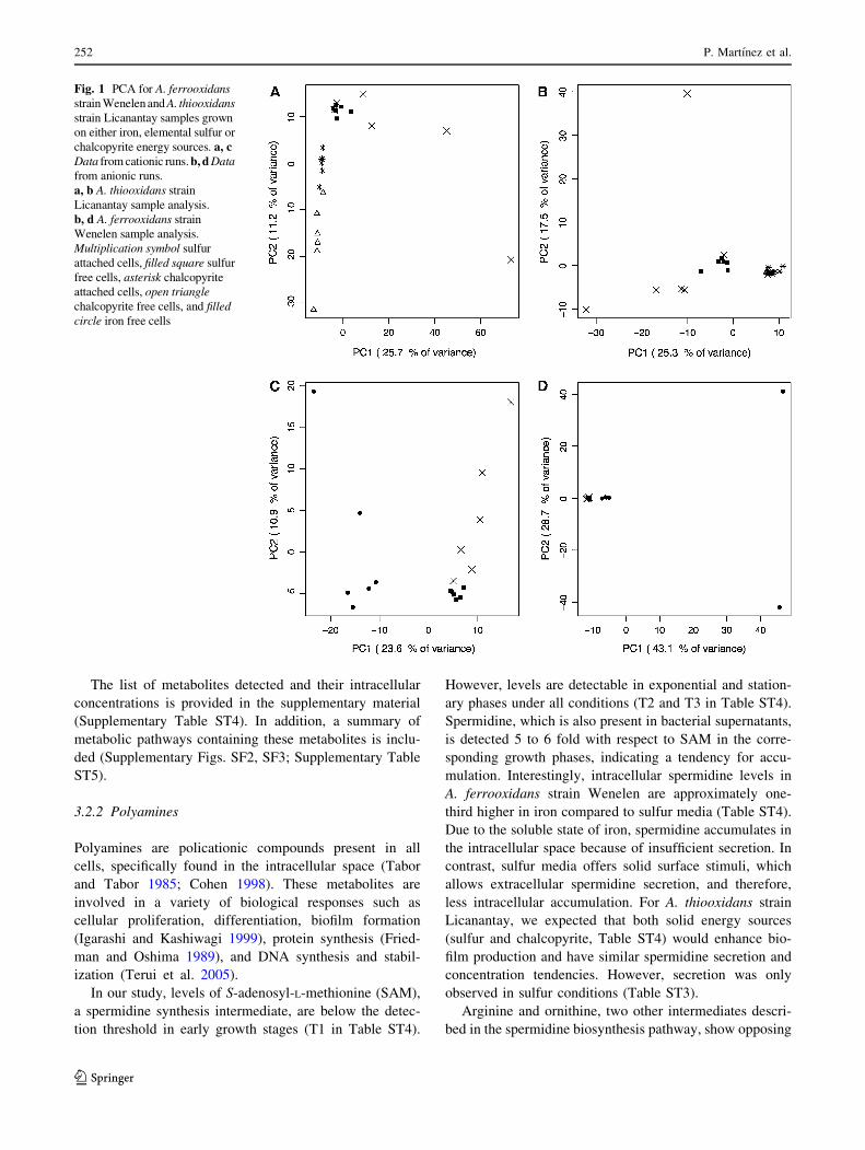
Tables 1 and 2 summarize the complete dataset after peak
detection and control peak subtraction for A. ferrooxidans
strain Wenelen and A. thiooxidans strain Licanantay sam-
ples, grown on their respective energy sources. In general,
fewer peaks were detected in attached cell samples com-
pared to free cell samples for both bacteria. The presence of
more mineral residues in attached cell samples may interfere
with the ionization process. The area of the detected peaks
was smaller for free cell samples for A. thiooxidans strain
Licanantay grown in chalcopyrite, in both anionic and cat-
ionic modes. This behavior, however, was not observed for
samples grown in sulfur.
Principal component analysis (PCA) was performed
using the complete dataset to determine if the metabolomic
data allowed for separation of the different conditions.
Principal components were calculated using all detected
peaks (for each sample) as variables and their abundance as
values.
3.1.1 Cationic CE–MS runs
Figure 1c shows clear separation between sulfur and iron
conditions for A. ferrooxidans strain Wenelen samples.
Separation observed in sulfur samples indicates that the
two filters used in the extraction protocol allow cell pop-
ulations (free and attached cells) to be distinguished.
Similarly, positive values in PC1 are strongly associated to
unique peaks detected in sulfur samples. This shows that
250 P. Martınez et al.
123
unique features are present under both conditions and can
be used as possible biomarkers for each condition. For A.
thiooxidans strain Licanantay sample separation was
observed for different energy sources (Fig. 1a). Attached
and free cell samples were separated in chalcopyrite
growing conditions, but not in sulfur.
3.1.2 Anionic CE–MS runs
For A. ferrooxidans strain Wenelen anionic runs, PCA
failed to clearly separate the various growth conditions
(Fig. 1d). This is due to the presence of two outlying
chromatograms originating from Fe cultures. These chro-
matograms are associated to cultures in late exponential
growth, where on average, the number of detected peaks is
more than three times the average for all other samples. This
phenomenon suggests an increased adduct abundance.
Acidithithiobacillus thiooxidans strain Licanantay sam-
ples grown in sulfur and chalcopyrite conditions can be
differentiated as shown in Fig. 1b. However, as with cationic
runs, attached and free cell samples were not separated.
Differences in samples obtained during different growth
stages were not clearly reflected by PCA of CE–MS runs in
either mode.
3.2 Annotated standard metabolites analysis
In cationic and anionic chromatograms, a search for a set of
112 standard metabolites (Supplementary Table ST1) was
made and those detected were annotated. The intracellular
concentrations of these annotated metabolites in free cells
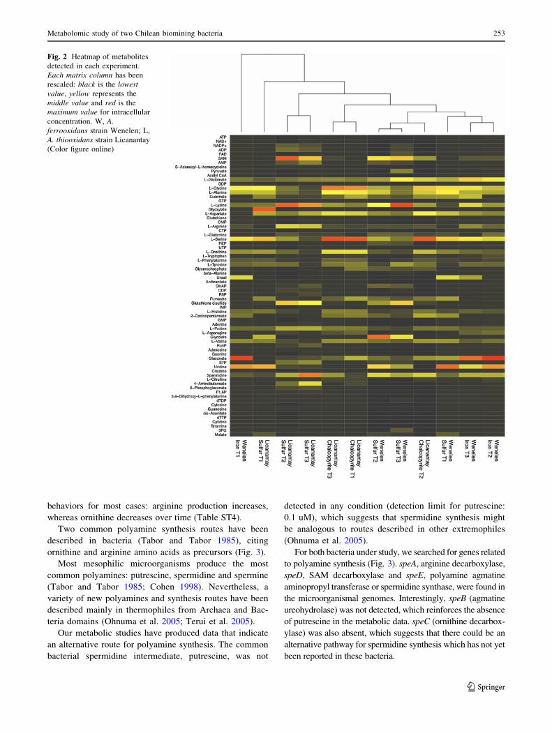
were calculated (Fig. 2).
As expected, sulfur and iron conditions in A. ferrooxi-
dans strain Wenelen are grouped in T2 and T3, respec-
tively, exhibiting differing metabolic profiles according to
growth media. Samples in T1 show different behavior,
most likely because fewer metabolites are detected as a
result of low cell concentration (Fig. 2). Also, A. thiooxi-
dans strain Licanantay in chalcopyrite shares characteris-
tics with A. ferrooxidans strain Wenelen in sulfur
conditions, suggesting similarities in sulfur processing.
Spermidine appears ubiquitously across different con-
ditions. Glutathione disulfide, which is of particular
importance for sulfur assimilation (Rohwerder and Sand
2003), is present in both organisms under sulfur growth.
Gluconate characterizes ferrous growth of A. ferrooxidans
strain Wenelen, and moreover, glutamate, tryptophan and
phenylalanine show particular profiles depending on the
growth media.
3.2.1 Metabolic pathways
Metabolic pathways were reconstructed and characterized.
We paid special attention to the polyamine synthesis
pathway because it seems active in every energy condition:
we analyzed the presence of spermidine in the supernatant,
which could have a significant role in this pathway (Sup-
plementary Table ST3).
Metabolites present in glutathione pathways, which are
related to elemental sulfur oxidation, were also analyzed
given their activity in sulfur conditions.
Glutamate, aspartate and various metabolites involved in
energy processes were detected in supernatants. Then, we
analyzed the amino acid profiles for each microorganism.
Table 1 Average number of peaks and their normalized areas for
A. thiooxidans strain Licanantay sample runs grown on either chal-
copyrite (Cpy) or elemental sulfur (S0)
Energy
source
Sample
type
Anionic
mode
Cationic
mode
Cpy Free cells
Average number of peaks 142 ± 28 58 ± 104
Average normalized area
[9103]
1.99 ± 0.54 1.84 ± 0.41
Attached cells
Average number of peaks 133 ± 12 442 ± 53
Average normalized area
[9103]
1.24 ± 0.24 3.88 ± 1.03
S0 Free cells
Average number of peaks 161 ± 65 520 ± 214
Average normalized area
[9103]
4.54 ± 3.07 3.04 ± 1.55
Attached cells
Average number of peaks 73 ± 25 360 ± 42
Average normalized area
[9103]
2.27 ± 1.11 1.97 ± 0.42
Table 2 Average number of peaks and their normalized areas for
A. ferrooxidans strain Wenelen sample runs grown on either Fe or
elemental sulfur (S0)
Energy
source
Sample
type
Anionic
mode
Cationic
mode
Fe?2 Free cells
Average number of peaks 143 ± 32 423 ± 38
Average normalized area
[9103]
4.92 ± 4.66 2.31 ± 0.19
S0 Free cells
Average number of peaks 126 ± 8 244 ± 71
Average normalized area
[9103]
1.17 ± 0.27 2.34 ± 1.39
Attached cells
Average number of peaks 126 ± 8 192 ± 49
Average normalized area
[9103]
1.01 ± 0.24 1.83 ± 1.09
Metabolomic study of two Chilean biomining bacteria 251
123
The list of metabolites detected and their intracellular
concentrations is provided in the supplementary material
(Supplementary Table ST4). In addition, a summary of
metabolic pathways containing these metabolites is inclu-
ded (Supplementary Figs. SF2, SF3; Supplementary Table
ST5).
3.2.2 Polyamines
Polyamines are policationic compounds present in all
cells, specifically found in the intracellular space (Tabor
and Tabor 1985; Cohen 1998). These metabolites are
involved in a variety of biological responses such as
cellular proliferation, differentiation, biofilm formation
(Igarashi and Kashiwagi 1999), protein synthesis (Fried-
man and Oshima 1989), and DNA synthesis and stabil-
ization (Terui et al. 2005).
In our study, levels of S-adenosyl-L-methionine (SAM),
a spermidine synthesis intermediate, are below the detec-
tion threshold in early growth stages (T1 in Table ST4).
However, levels are detectable in exponential and station-
ary phases under all conditions (T2 and T3 in Table ST4).
Spermidine, which is also present in bacterial supernatants,
is detected 5 to 6 fold with respect to SAM in the corre-
sponding growth phases, indicating a tendency for accu-
mulation. Interestingly, intracellular spermidine levels in
A. ferrooxidans strain Wenelen are approximately one-
third higher in iron compared to sulfur media (Table ST4).
Due to the soluble state of iron, spermidine accumulates in
the intracellular space because of insufficient secretion. In
contrast, sulfur media offers solid surface stimuli, which
allows extracellular spermidine secretion, and therefore,
less intracellular accumulation. For A. thiooxidans strain
Licanantay, we expected that both solid energy sources
(sulfur and chalcopyrite, Table ST4) would enhance bio-
film production and have similar spermidine secretion and
concentration tendencies. However, secretion was only
observed in sulfur conditions (Table ST3).
Arginine and ornithine, two other intermediates descri-
bed in the spermidine biosynthesis pathway, show opposing
Fig. 1 PCA for A. ferrooxidansstrain Wenelen and A. thiooxidansstrain Licanantay samples grown
on either iron, elemental sulfur or
chalcopyrite energy sources. a, cData from cationic runs. b, d Datafrom anionic runs.
a, b A. thiooxidans strain
Licanantay sample analysis.
b, d A. ferrooxidans strain
Wenelen sample analysis.
Multiplication symbol sulfur
attached cells, filled square sulfur
free cells, asterisk chalcopyrite
attached cells, open trianglechalcopyrite free cells, and filledcircle iron free cells
252 P. Martınez et al.
123
behaviors for most cases: arginine production increases,
whereas ornithine decreases over time (Table ST4).
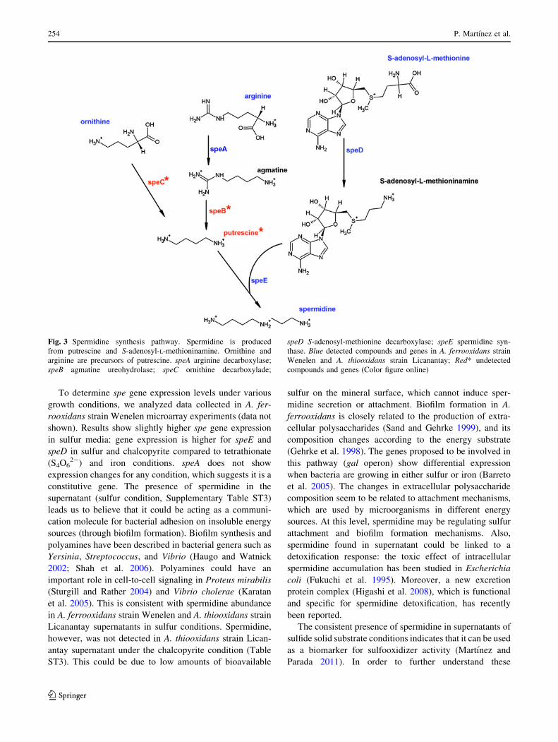
Two common polyamine synthesis routes have been
described in bacteria (Tabor and Tabor 1985), citing
ornithine and arginine amino acids as precursors (Fig. 3).
Most mesophilic microorganisms produce the most
common polyamines: putrescine, spermidine and spermine
(Tabor and Tabor 1985; Cohen 1998). Nevertheless, a
variety of new polyamines and synthesis routes have been
described mainly in thermophiles from Archaea and Bac-
teria domains (Ohnuma et al. 2005; Terui et al. 2005).
Our metabolic studies have produced data that indicate
an alternative route for polyamine synthesis. The common
bacterial spermidine intermediate, putrescine, was not
detected in any condition (detection limit for putrescine:
0.1 uM), which suggests that spermidine synthesis might
be analogous to routes described in other extremophiles
(Ohnuma et al. 2005).
For both bacteria under study, we searched for genes related
to polyamine synthesis (Fig. 3). speA, arginine decarboxylase,
speD, SAM decarboxylase and speE, polyamine agmatine
aminopropyl transferase or spermidine synthase, were found in
the microorganismal genomes. Interestingly, speB (agmatine
ureohydrolase) was not detected, which reinforces the absence
of putrescine in the metabolic data. speC (ornithine decarbox-
ylase) was also absent, which suggests that there could be an
alternative pathway for spermidine synthesis which has not yet
been reported in these bacteria.
Fig. 2 Heatmap of metabolites
detected in each experiment.
Each matrix column has been
rescaled: black is the lowestvalue, yellow represents the
middle value and red is the
maximum value for intracellular
concentration. W, A.ferrooxidans strain Wenelen; L,
A. thiooxidans strain Licanantay
(Color figure online)
Metabolomic study of two Chilean biomining bacteria 253
123
To determine spe gene expression levels under various
growth conditions, we analyzed data collected in A. fer-
rooxidans strain Wenelen microarray experiments (data not
shown). Results show slightly higher spe gene expression
in sulfur media: gene expression is higher for speE and
speD in sulfur and chalcopyrite compared to tetrathionate
(S4O62-) and iron conditions. speA does not show
expression changes for any condition, which suggests it is a
constitutive gene. The presence of spermidine in the
supernatant (sulfur condition, Supplementary Table ST3)
leads us to believe that it could be acting as a communi-
cation molecule for bacterial adhesion on insoluble energy
sources (through biofilm formation). Biofilm synthesis and
polyamines have been described in bacterial genera such as
Yersinia, Streptococcus, and Vibrio (Haugo and Watnick
2002; Shah et al. 2006). Polyamines could have an
important role in cell-to-cell signaling in Proteus mirabilis
(Sturgill and Rather 2004) and Vibrio cholerae (Karatan
et al. 2005). This is consistent with spermidine abundance
in A. ferrooxidans strain Wenelen and A. thiooxidans strain
Licanantay supernatants in sulfur conditions. Spermidine,
however, was not detected in A. thiooxidans strain Lican-
antay supernatant under the chalcopyrite condition (Table
ST3). This could be due to low amounts of bioavailable
sulfur on the mineral surface, which cannot induce sper-
midine secretion or attachment. Biofilm formation in A.
ferrooxidans is closely related to the production of extra-
cellular polysaccharides (Sand and Gehrke 1999), and its
composition changes according to the energy substrate
(Gehrke et al. 1998). The genes proposed to be involved in
this pathway (gal operon) show differential expression
when bacteria are growing in either sulfur or iron (Barreto
et al. 2005). The changes in extracellular polysaccharide
composition seem to be related to attachment mechanisms,
which are used by microorganisms in different energy
sources. At this level, spermidine may be regulating sulfur
attachment and biofilm formation mechanisms. Also,
spermidine found in supernatant could be linked to a
detoxification response: the toxic effect of intracellular
spermidine accumulation has been studied in Escherichia
coli (Fukuchi et al. 1995). Moreover, a new excretion
protein complex (Higashi et al. 2008), which is functional
and specific for spermidine detoxification, has recently
been reported.
The consistent presence of spermidine in supernatants of
sulfide solid substrate conditions indicates that it can be used
as a biomarker for sulfooxidizer activity (Martınez and
Parada 2011). In order to further understand these
Fig. 3 Spermidine synthesis pathway. Spermidine is produced
from putrescine and S-adenosyl-L-methioninamine. Ornithine and
arginine are precursors of putrescine. speA arginine decarboxylase;
speB agmatine ureohydrolase; speC ornithine decarboxylade;
speD S-adenosyl-methionine decarboxylase; speE spermidine syn-
thase. Blue detected compounds and genes in A. ferrooxidans strain
Wenelen and A. thiooxidans strain Licanantay; Red* undetected
compounds and genes (Color figure online)
254 P. Martınez et al.
123
phenomena, we searched for spermidine and polyamine
transporters in the A. ferrooxidans strain ATCC 23270
genome, however, no candidates were found. Nevertheless,
we identified genes, described in other bacteria such as E.
coli and V. cholerae, that are associated to spermidine and
putrescine transport (potD and potF; Igarashi and Kashiwagi
1999). The transport system for these polyamines is similar
to ABC systems (secretion system type I), which has been
well described in the literature. After analysis and compar-
ison of the nucleotide and amino acid sequences of these
genes in A. ferrooxidans ATCC 23270 and Wenelen strains,
we detected functional candidates that contain ATP binding
motifs (data not shown). We believe this could be a new
secretion and/or incorporation model for spermidine in
A. ferrooxidans which has not yet been identified. This
finding confirms that the presence of spermidine has a
functional significance for the bacterium, where the regu-
lation is related to specific growth stages and sulfur presence.
3.2.3 Glutathione pathways
Glutathione related pathways seem more active in sulfur
than in iron and chalcopyrite conditions for A. ferrooxidans
strain Wenelen and A. thiooxidans strain Licanantay
(Supplementary Table ST4). This observation is supported
by Rohwerder and Sand (2003), who show that elemental
sulfur activation provides a catalytic role for reduced glu-
tathione (GSH). GSH is only detected in A. ferrooxidans
strain Wenelen in the early exponential phase (T1), prac-
tically in the same amount as oxidized glutathione (GSSG).
GSH is undetectable for either T2 or T3 time points for
both bacteria, but two of the three constitutive amino acids,
glutamate and glycine, are present (cysteine is undetectable
under this technique). GSSG, in contrast, shows a con-
centration increase throughout the growth phases. These
observations should be taken with caution, as it is likely
that reduced glutathione could be oxidized during the
extraction and/or identification protocol. Further analyses
are needed to corroborate these observations.
Also, glutathione appears in more significant quantities
in A. thiooxidans strain Licanantay than in A. ferrooxidans
strain Wenelen.
3.2.4 Amino acids and metabolites involved in energy
processes
Acidithiobacillus thiooxidans strain Licanantay shows a
differential amino acid metabolic profile in chalcopyrite and
sulfur conditions (Supplementary Table ST4). It seems that
the presence of chalcopyrite triggers differential protein
expression, most likely associated to cell attachment, energy
source usage or detoxification mechanisms. A similar
phenomenon is observed when comparing iron and sulfur
conditions in A. ferrooxidans strain Wenelen (data not
shown).
Because of the detection of glutamate and aspartate in
supernatants (Supplementary Table ST3), we examined if
these were related to structural polymers outside the cell
(i.e. poly-glutamate and poly-aspartate). However, these
polymers were undetectable in these cases (data not
shown).
There is an increased presence of metabolites involved in
energy processes (NADP?, ADP, AMP, CDP and dTDP) in
A. thiooxidans strain Licanantay. This is concordant with the
fact that sulfur processing is more effective in A. thiooxidans
than in A. ferrooxidans. In addition, dihydroxyacetone
phosphate and sedoheptulose 7-phosphate, involved in bio-
film formation pathways, are enhanced in A. thiooxidans
strain Licanantay when compared to A. ferrooxidans strain
Wenelen (Supplementary Table ST4).
4 Conclusions
This study introduces the first metabolomic analysis of two
extremophilic biomining bacteria: A. ferrooxidans strain
Wenelen and A. thiooxidans strain Licanantay. Principal
component analysis indicates that metabolic profiles were
different under each condition and therefore, metabolic
results within each experiment are correlated.
Together with previous metabolic in silico reconstruc-
tion, our results show that several processes such as biofilm
formation, carbon and amino acid usage, energy related
compounds and oxidative stress response appear to be of
importance for the bioleaching bacteria under study.
Attached and free cells have different metabolic profiles.
Some interesting information emerged from the detailed
analysis: spermidine, glutathione, and certain amino acids
are the most abundant metabolites detected by our method
(polar metabolites), which suggests that they have a poten-
tially important role in the physiology of biomining bacteria.
We believe that there is a relation between spermidine
secretion and its role in biofilm formation in sulfur condi-
tions and that an alternative polyamine synthesis pathway is
present in these biomining bacteria. Further, we propose the
use of spermidine as a biomarker for sulfooxidizer activity.
Glutathione abundance and glutamate over-production in
sulfur conditions, support previous knowledge about their
participation in sulfur oxidation. Amino acid profiles vary
between different growth conditions, suggesting differential
protein synthesis possibly related to cell attachment, energy
source or cell detoxification. Sugars are abundant in A.
thiooxidans strain Lincanantay growing in sulfur media, and
are likely involved in biofilm formation.
Metabolomic study of two Chilean biomining bacteria 255
123
Unfortunately, directed functional mutagenesis has not
been achieved for this type of bacteria. This complicates
the study of relevant metabolic pathways identified in this
work such as the spermidine synthesis pathway. However,
the detection of potential new intermediates for this met-
abolic pathway and additional genomic analysis could be
useful for elucidating new functional activities and syn-
thesis. In order to enhance knowledge of metabolic pro-
cesses related to bioleaching microorganisms, studies
should focus on improving metabolic extraction methods.
Also, additional metabolomic studies of organisms present
in bioleaching processes are necessary to understand the
effect of changing conditions (i.e. energy source, pH,
toxicity, etc.). These studies should be conducted using a
variety of detection techniques in order to include polar and
non-polar metabolites. Our present study is the first step in
consolidating metabolic knowledge for these biomining
microorganisms, which will contribute to their global
understanding and future applications.
Acknowledgments The authors would like to thank Dr. Masahiro
Sugimoto for his bioinformatic support, and Mr. Kenichi Iida for his
invaluable help. We also thank Asako Suzuki, Maki Ohishi, Hiromi
Onuma, Ayako Momose and Chieko Kikuchi from the Institute for
Advanced Biosciences, Keio University for metabolome analysis. We are
grateful to Tsuruoka City and Yamagata Prefecture of Japan, for their
financial and organizational support. This work was supported by Bio-
Sigma ‘S.A.’. The authors thank the company for authorizing the sub-
mission of the manuscript for publication. This study was partially
supported by FONDAP-CGR No. 15090007 and BASAL-CMM Grants.
Open Access This article is distributed under the terms of the
Creative Commons Attribution License which permits any use, dis-
tribution, and reproduction in any medium, provided the original
author(s) and the source are credited.
References
Barreto, M., Jedlicki, E., & Holmes, D. S. (2005). Identification
of a gene cluster for the formation of extracellular polysaccha-
ride precursors in the chemolithoautotroph Acidithiobacillusferrooxidans. Applied and Environmental Microbiology, 71(6),
2902–2909.
Cardenas, J. P., Valdes, J., Quatrini, R., Duarte, F., & Holmes, D. S.
(2010). Lessons from the genomes of extremely acidophilic
bacteria and archaea with special emphasis on bioleaching
microorganisms. Applied and Environmental Microbiology,88(3), 605–620.
Cohen, S. S. (1998). A guide to the polyamines. New York: Oxford
University Press.
Colmer, A. R., & Hinkle, M. E. (1947). The role of microorganisms in
acid mine drainage: a preliminary report. Science, 106, 253–256.
Friedman, S. M., & Oshima, T. (1989). Polyamines of sulfur-
dependent archaebacteria and their role in protein synthesis.
Journal of Biochemistry, 105, 1030–1033.
Fukuchi, J., Kashiwagi, K., Yamagishi, M., Ishihama, A., & Igarashi,
K. (1995). Decrease in cell viability due to the accumulation of
spermidine in spermidine acetyltransferase-deficient mutant of
Escherichia coli. The Journal of Biological Chemistry, 270,
18831–18835.
Gehrke, T., Telegdi, J., Thierry, D., & Sand, W. (1998). Importance
of extracellular polymeric substances from Thiobacillus ferro-oxidans for bioleaching. Applied and Environmental Microbiol-ogy, 64(7), 2743–2747.
Haugo, A. J., & Watnick, P. I. (2002). Vibrio cholerae CytR is a
repressor of biofilm development. Molecular Microbiology, 45,
471–483.
Higashi, K., Ishigure, H., Demizu, R., et al. (2008). Identification of a
spermidine excretion protein complex (MdtJI) in Escherichiacoli. Journal of Bacteriology, 190, 872–878.
Hirayama, A., Kami, K., Sugimoto, M., et al. (2009). Quantitative
metabolome profiling of colon and stomach cancer microenvi-
ronment by capillary electrophoresis time-of-flight mass spec-
trometry. Cancer Research, 69, 4918–4925.
Igarashi, K., & Kashiwagi, K. (1999). Polyamine transport in bacteria
and yeast. The Biochemical Journal, 344, 633–642.
Ishii, N., Nakahigashi, K., Baba, T., et al. (2007). Multiple high-
throughput analyses monitor the response of E. coli to pertur-
bations. Science, 316, 593–597.
Johnson, D. B., Rolfe, S., Hallberg, K. B., & Iversen, E. (2001). Isolation
and phylogenetic characterization of acidophilic microorganisms
indigenous to acidic drainage waters at an abandoned Norwegian
copper mine. Environmental Microbiology, 3, 630–637.
Karatan, E., Duncan, T. R., & Watnick, P. I. (2005). NspS, a predicted
polyamine sensor, mediates activation of Vibrio cholerae biofilm
formationbynorspermidine. Journal ofBacteriology,187, 7434–7443.
Kusano, T., Sugawara, K., Inoue, C., Takeshima, T., Numata, M., &
Shiratori, T. (1992). Electrotransformation of Thiobacillusferrooxidans with plasmids containing a mer determinant.
Journal of Bacteriology, 174, 6617–6623.
Levican, G., Ugalde, J. A., Ehrenfeld, N., Maass, A., & Parada, P.
(2008). Comparative genomic analysis of carbon and nitrogen
assimilation mechanisms in three indigenous bioleaching bacte-
ria: predictions and validations. BMC Genomics, 9, 581.
Liu, Z., Guiliani, N., Appia-Ayme, C., Borne, F., Ratouchniak, J., &
Bonnefoy, V. (2000). Construction and characterization of a
recA mutant of Thiobacillus ferrooxidans by marker exchange
mutagenesis. Journal of Bacteriology, 182, 2269–2276.
Martınez, P., & Parada, P. (2011). Biomarcador de espermidina para
deteccion de actividad sulfooxidante. CL Patent No. 3066-2011.
Neuweger, H., Albaum, S. P., Dondrup, M., et al. (2008). MeltDB: a
software platform for the analysis and integration of metabolo-
mics experiment data. Bioinformatics, 24, 2726–2732.
Ohashi, Y., Hirayama, A., Ishikawa, T., Nakamura, S., Shimizu, K.,
Ueno, Y., et al. (2008). Depiction of metabolome changes in
histidine-starved Escherichia coli by CE–TOFMS. MolecularBioSystems, 4, 135–147.
Ohata, A., Manabe, M., & Parada, P. (2010). Sulfur-oxidizing
bacteria and their use in bioleaching processes for sulfured
copper minerals. U.S. Patent No. 7,700,343.
Ohnuma, M., Terui, Y., Tamakoshi, M., et al. (2005). N1-aminopro-
pylagmatine, a new polyamine produced as a key intermediate in
polyamine biosynthesis of an extreme thermophile, Thermusthermophilus. The Journal of Biological Chemistry, 280,
30073–30082.
Okibe, N., Gericke, M., Hallberg, K. B., & Johnson, D. B. (2003).
Enumeration and characterization of acidophilic microorganisms
isolated from a pilot plant stirred-tank bioleaching operation.
Applied and Environmental Microbiology, 69, 1936–1943.
Quatrini, R., Appia-Ayme, C., Denis, Y., Jedlicki, E., Holmes, D., &
Bonnefoy, V. (2009). Extending the models for iron and sulfur
oxidation in the extreme acidophile Acidithiobacillus ferrooxi-dans. BMC Genomics, 10, 394.
Rohwerder, T., & Sand, W. (2003). The sulfane sulfur of persulfides is the
actual substrate of the sulfur-oxidizing enzymes from Acidithioba-cillus and Acidiphilium spp. Microbiology, 149, 1699–1709.
256 P. Martınez et al.
123
Sand, W., & Gehrke, T. (1999). Analysis and function of the EPS
from the strong acidophile Thiobacillus ferrooxidans. In J.
Wingender, T. R. Neu, & H.-C. Fleming (Eds.), Microbialextracellular polymeric substances: characterization, structureand function (pp. 127–141). Berlin: Springer.
Santelli, C. M., Edgcomb, V. P., Bach, W., & Edwards, K. J. (2009).
The diversity and abundance of bacteria inhabiting seafloor lavas
positively correlate with rock alteration. Environmental Micro-biology, 11, 86–98.
Sato, S., Soga, T., Nishioka, T., & Tomita, M. (2004). Simultaneous
determination of the main metabolites in rice leaves using
capillary electrophoresis mass spectrometry and capillary elec-
trophoresis diode array detection. The Plant Journal: for Celland Molecular Biology, 40, 151–163.
Shah, P., Marquat, M., Quin, L. R., & Swiatlo, E. (2006). Cellular
location of polyamine transport protein PotD in Streptococcuspneumoniae. FEMS Microbiology Letters, 261, 235–237.
Smith, C. A., Want, E. J., O’Maille, G., Abagyan, R., & Siuzdak, G.
(2006). XCMS: processing mass spectrometry data for metab-
olite profiling using nonlinear peak alignment, matching, and
identification. Analytical Chemistry, 78, 779–787.
Soga, T., Ueno, Y., Naraoka, H., Ohashi, Y., Tomita, M., & Nishioka,
T. (2002). Simultaneous determination of anionic intermediates
for Bacillus subtilis metabolic pathways by capillary electro-
phoresis electrospray ionization mass spectrometry. AnalyticalChemistry, 74, 2233–2239.
Soga, T., Ohashi, Y., Ueno, Y., Naraoka, H., Tomita, M., & Nishioka, T.
(2003). Quantitative metabolome analysis using capillary electro-
phoresis mass spectrometry. Journal of Proteome Research, 2,
488–494.
Soga, T., Baran, R., Suematsu, M., et al. (2006). Differential
metabolomics reveals ophthalmic acid as an oxidative stress
biomarker indicating hepatic glutathione consumption. TheJournal of Biological Chemistry, 281, 16768–16776.
Soga, T., Igarashi, K., Ito, C., Mizobuchi, K., Zimmermann, H. P., &
Tomita, M. (2009). Metabolomic profiling of anionic metabolites
by capillary electrophoresis mass spectrometry. AnalyticalChemistry, 81, 6165–6174.
Sturgill, G., & Rather, P. N. (2004). Evidence that putrescine acts as
an extracellular signal required for swarming in Proteusmirabilis. Molecular Microbiology, 51, 437–446.
Sugimoto, M., Koseki, T., Hirayama, A., et al. (2010a). Correlation
between sensory evaluation scores of Japanese sake and
metabolome profiles. Journal of Agriculture and Food Chemis-try, 58, 374–383.
Sugimoto, M., Wong, D. T., Hirayama, A., Soga, T., & Tomita, M.
(2010b). Capillary electrophoresis mass spectrometry-based
saliva metabolomics identified oral, breast and pancreatic
cancer-specific profiles. Metabolomics, 6, 78–95.
Sugio, T., Miura, A., Parada, P., Badilla, R. (2009). Bacteria strain
Wenelen DSM 16786, use of said bacteria for leaching of ores or
concentrates containing metallic sulfide mineral species and
leaching processes based on the use of said bacteria or mixtures
that contain said bacteria. U.S. Patent No. 7,601,530.
Tabor, C. W., & Tabor, H. (1985). Polyamines in microorganisms.
Microbiology and Molecular Biology Reviews, 49, 81–99.
Terui, Y., Ohnuma, M., Hiraga, K., Kawashima, E., & Oshima, T.
(2005). Stabilization of nucleic acids by unusual polyamines
produced by an extreme thermophile, Thermus thermophilus.
The Biochemical Journal, 388, 427–433.
Tyson, G. W., Lo, I., Baker, B. J., Allen, E. E., Hugenholtz, P., &
Banfield, J. F. (2005). Genome-directed isolation of the key
nitrogen fixer Leptospirillum ferrodiazotrophum sp. nov. from an
acidophilic microbial community. Applied and EnvironmentalMicrobiology, 71, 6319–6324.
Valdes, J., Pedroso, I., Quatrini, R., et al. (2008). Acidithiobacillusferrooxidans metabolism: from genome sequence to industrial
applications. BMC Genomics, 9, 597.
Metabolomic study of two Chilean biomining bacteria 257
123
Related Documents