This is an Open Access article distributed under the terms of the Creative Commons Attribution Non-Commercial License (http://creativecommons.org/licenses/by-nc/3.0/) which permits unrestricted non-commercial use, distribution, and reproduction in any medium, provided the original work is properly cited. Case Report Journal of Epilepsy Research pISSN 2233-6249 / eISSN 2233-6257 Mesial Temporal Lobe Epilepsy in Congenital Toxoplasmosis: A Case Report Woo Kyo Jeong 1 , Byung-Euk Joo 1,2 , Ji-hye Seo 1,2 , Jun Kyu Mun 1 , Juhyeon Kim 1 , Dae-Won Seo 1,2 1 Department of Neurology, Samsung Medical Center, Sungkyunkwan University School of Medicine, Seoul, Korea; 2 Neuroscience Center, Samsung Medical Center, Sungkyunkwan University School of Medicine, Seoul, Korea Received March 3, 2015 Accepted April 28, 2015 Corresponding author: Dae-Won Seo Department of Neurology, Samsung Medical Center, Sungkyunkwan University, 81 Irwon-Ro, Gangnam-gu, Seoul 135-710, Korea Tel. +82-2-3410-3595 Fax. +82-2-3410-0052 E-mail; [email protected] Toxoplasmosis is a rare disease caused by intracellular protozoan parasite, Toxoplasma gondii. Though most patients with toxoplasmosis are asymptomatic, congenital toxoplasmosis in the fetus can cause ocular involvement such as chorioretinitis and central nervous system disease including intracerebral calcification, nystagmus, hydrocephalus and microcephaly. Also, these brain lesions can cause seizure secondarily. Our patient was diagnosed with congenital toxoplasmosis, based on toxoplasma-specific serologic test with typical clinical symptoms, including chorioretinitis, nystagmus, hydrocephalus and cerebral palsy. Her brain imaging findings revealed not only the multifocal encephalomalacia, but also multifocal cerebral calcification including intracerebral calcification in left perihippocampal region. Her epileptogenic zone was defined as mesial temporal lobe including hippocampus on left side by seizure semiology, electroencephalogram and neuroimaging including single photon emission computed tomography and 18F-Fluorodeoxyglucose positron-emission tomography. Her seizures were refractory to multiple anti-epileptic drugs. We report a patient with congenital toxoplasmosis who showed intractable mesial temporal lobe epilepsy. (2015;5:25-28) Key words: Congenital toxoplasmosis, Temporal lobe epilepsy, Intracerebral calcification Introduction Toxoplasmosis is caused by the obligate intracellular protozoan parasite, Toxoplasma gondii, for which preferred primary host is a cat and human may become infected as an intermediate host. 1 Most people with toxoplasmosis are asymptomatic. However, if a pregnant woman is infected, the parasite can enter the fetal circulation through placenta and may cause congenital toxoplasmosis in the fetus. Congenital toxoplasmosis may present with a broad spectrum of clinical manifestations, ranging from mild chorioretinitis to severe central nervous system (CNS) involvement, including hydrocephalus, mental retardation, seizures and intracerebral calcifications. 2,3 Seizure is one of the main presenting symptoms in patients with CNS infection of congenital acquired toxoplasmosis and many pa- tients with congenital toxoplasmosis took anti-epileptic drugs (AED) constantly. However, there have been few reports of detailed descrip- tion of seizure semiology and besides few reported cases of a patient with congenital toxoplasmosis who was suffering from temporal lobe epilepsy with hippocampal sclerosis (HS), which is one of the most common epileptic syndromes and is considered medically intractable but surgically remediable in any causes. 4,5 Hereby we described a patient with congenital toxoplasmosis who showed intractable mesial temporal lobe epilepsy. Case A 34-year-old left handed Arabian female was presented with our hospital with medically intractable seizure. The patient was born pre- term with fetal infection by Toxoplasma gondii. She had hydro- cephalus and right ventriculoperitoneal (VP) shunting was performed at 1 month old. She experienced convulsive seizures since 8 months old. Her development was delayed than other children. She started walk at 3 years and right hemiparesis was observed. She also com- plained of decreased visual acuity in both eyes. On past history, she had no history of febrile seizure, metabolic disease or head trauma and her family also had no history of seizure. In a neurological examination, contracture and weakness of right arm and leg was found compatible with cerebral palsy. Her mental status was evaluated using Mini-Mental State Examination with the help of a translator and her score was 22, suggesting memory im-

Welcome message from author
This document is posted to help you gain knowledge. Please leave a comment to let me know what you think about it! Share it to your friends and learn new things together.
Transcript

This is an Open Access article distributed under the terms of the Creative Commons Attribution Non-Commercial License (http://creativecommons.org/licenses/by-nc/3.0/) which permits unrestricted non-commercial use, distribution, and reproduction in any medium, provided the original work is properly cited.
Case ReportJournal of Epilepsy Research
pISSN 2233-6249 / eISSN 2233-6257
Mesial Temporal Lobe Epilepsy in Congenital Toxoplasmosis: A Case ReportWoo Kyo Jeong1, Byung-Euk Joo1,2, Ji-hye Seo1,2, Jun Kyu Mun1, Juhyeon Kim1, Dae-Won Seo1,2
1Department of Neurology, Samsung Medical Center, Sungkyunkwan University School of Medicine, Seoul, Korea;2Neuroscience Center, Samsung Medical Center, Sungkyunkwan University School of Medicine, Seoul, Korea
Received March 3, 2015Accepted April 28, 2015
Corresponding author: Dae-Won SeoDepartment of Neurology, Samsung Medical Center, Sungkyunkwan University, 81 Irwon-Ro, Gangnam-gu, Seoul 135-710, KoreaTel. +82-2-3410-3595Fax. +82-2-3410-0052E-mail; [email protected]
Toxoplasmosis is a rare disease caused by intracellular protozoan parasite, Toxoplasma gondii. Though
most patients with toxoplasmosis are asymptomatic, congenital toxoplasmosis in the fetus can cause
ocular involvement such as chorioretinitis and central nervous system disease including intracerebral
calcification, nystagmus, hydrocephalus and microcephaly. Also, these brain lesions can cause seizure
secondarily. Our patient was diagnosed with congenital toxoplasmosis, based on toxoplasma-specific
serologic test with typical clinical symptoms, including chorioretinitis, nystagmus, hydrocephalus and
cerebral palsy. Her brain imaging findings revealed not only the multifocal encephalomalacia, but also
multifocal cerebral calcification including intracerebral calcification in left perihippocampal region. Her
epileptogenic zone was defined as mesial temporal lobe including hippocampus on left side by seizure
semiology, electroencephalogram and neuroimaging including single photon emission computed
tomography and 18F-Fluorodeoxyglucose positron-emission tomography. Her seizures were refractory
to multiple anti-epileptic drugs. We report a patient with congenital toxoplasmosis who showed intractable
mesial temporal lobe epilepsy. (2015;5:25-28)
Key words: Congenital toxoplasmosis, Temporal lobe epilepsy, Intracerebral calcification
Introduction
Toxoplasmosis is caused by the obligate intracellular protozoan
parasite, Toxoplasma gondii, for which preferred primary host is a cat
and human may become infected as an intermediate host.1 Most
people with toxoplasmosis are asymptomatic. However, if a pregnant
woman is infected, the parasite can enter the fetal circulation
through placenta and may cause congenital toxoplasmosis in the
fetus. Congenital toxoplasmosis may present with a broad spectrum
of clinical manifestations, ranging from mild chorioretinitis to severe
central nervous system (CNS) involvement, including hydrocephalus,
mental retardation, seizures and intracerebral calcifications.2,3
Seizure is one of the main presenting symptoms in patients with
CNS infection of congenital acquired toxoplasmosis and many pa-
tients with congenital toxoplasmosis took anti-epileptic drugs (AED)
constantly. However, there have been few reports of detailed descrip-
tion of seizure semiology and besides few reported cases of a patient
with congenital toxoplasmosis who was suffering from temporal lobe
epilepsy with hippocampal sclerosis (HS), which is one of the most
common epileptic syndromes and is considered medically intractable
but surgically remediable in any causes.4,5
Hereby we described a patient with congenital toxoplasmosis who
showed intractable mesial temporal lobe epilepsy.
Case
A 34-year-old left handed Arabian female was presented with our
hospital with medically intractable seizure. The patient was born pre-
term with fetal infection by Toxoplasma gondii. She had hydro-
cephalus and right ventriculoperitoneal (VP) shunting was performed
at 1 month old. She experienced convulsive seizures since 8 months
old. Her development was delayed than other children. She started
walk at 3 years and right hemiparesis was observed. She also com-
plained of decreased visual acuity in both eyes. On past history, she
had no history of febrile seizure, metabolic disease or head trauma
and her family also had no history of seizure.
In a neurological examination, contracture and weakness of right
arm and leg was found compatible with cerebral palsy. Her mental
status was evaluated using Mini-Mental State Examination with the
help of a translator and her score was 22, suggesting memory im-
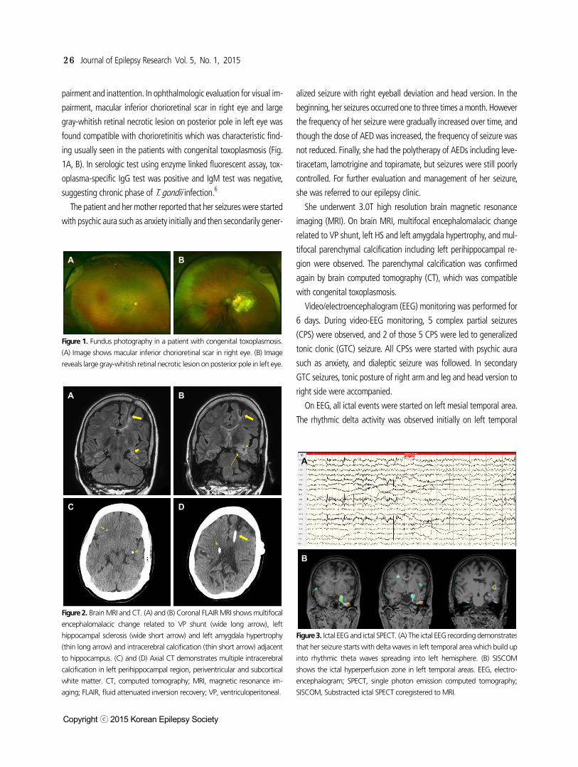
26 Journal of Epilepsy Research Vol. 5, No. 1, 2015
Copyright ⓒ 2015 Korean Epilepsy Society
A B
Figure 1. Fundus photography in a patient with congenital toxoplasmosis.
(A) Image shows macular inferior chorioretinal scar in right eye. (B) Image
reveals large gray-whitish retinal necrotic lesion on posterior pole in left eye.
A B
C D
Figure 2. Brain MRI and CT. (A) and (B) Coronal FLAIR MRI shows multifocal
encephalomalacic change related to VP shunt (wide long arrow), left
hippocampal sclerosis (wide short arrow) and left amygdala hypertrophy
(thin long arrow) and intracerebral calcification (thin short arrow) adjacent
to hippocampus. (C) and (D) Axial CT demonstrates multiple intracerebral
calcification in left perihippocampal region, periventricular and subcortical
white matter. CT, computed tomography; MRI, magnetic resonance im-
aging; FLAIR, fluid attenuated inversion recovery; VP, ventriculoperitoneal.
A
B
Figure 3. Ictal EEG and ictal SPECT. (A) The ictal EEG recording demonstrates
that her seizure starts with delta waves in left temporal area which build up
into rhythmic theta waves spreading into left hemisphere. (B) SISCOM
shows the ictal hyperperfusion zone in left temporal areas. EEG, electro-
encephalogram; SPECT, single photon emission computed tomography;
SISCOM, Substracted ictal SPECT coregistered to MRI.
pairment and inattention. In ophthalmologic evaluation for visual im-
pairment, macular inferior chorioretinal scar in right eye and large
gray-whitish retinal necrotic lesion on posterior pole in left eye was
found compatible with chorioretinitis which was characteristic find-
ing usually seen in the patients with congenital toxoplasmosis (Fig.
1A, B). In serologic test using enzyme linked fluorescent assay, tox-
oplasma-specific IgG test was positive and IgM test was negative,
suggesting chronic phase of T. gondii infection.6
The patient and her mother reported that her seizures were started
with psychic aura such as anxiety initially and then secondarily gener-
alized seizure with right eyeball deviation and head version. In the
beginning, her seizures occurred one to three times a month. However
the frequency of her seizure were gradually increased over time, and
though the dose of AED was increased, the frequency of seizure was
not reduced. Finally, she had the polytherapy of AEDs including leve-
tiracetam, lamotrigine and topiramate, but seizures were still poorly
controlled. For further evaluation and management of her seizure,
she was referred to our epilepsy clinic.
She underwent 3.0T high resolution brain magnetic resonance
imaging (MRI). On brain MRI, multifocal encephalomalacic change
related to VP shunt, left HS and left amygdala hypertrophy, and mul-
tifocal parenchymal calcification including left perihippocampal re-
gion were observed. The parenchymal calcification was confirmed
again by brain computed tomography (CT), which was compatible
with congenital toxoplasmosis.
Video/electroencephalogram (EEG) monitoring was performed for
6 days. During video-EEG monitoring, 5 complex partial seizures
(CPS) were observed, and 2 of those 5 CPS were led to generalized
tonic clonic (GTC) seizure. All CPSs were started with psychic aura
such as anxiety, and dialeptic seizure was followed. In secondary
GTC seizures, tonic posture of right arm and leg and head version to
right side were accompanied.
On EEG, all ictal events were started on left mesial temporal area.
The rhythmic delta activity was observed initially on left temporal

Woo Kyo Jeong. Epilepsy in congenital toxoplasmosis 27
www.kes.or.kr
area, and then led to rhythmic theta waves in the same area, spread-
ing into left hemisphere. Intermittent slowing in left temporal area
was also seen in interictal EEG monitoring and no epileptiform dis-
charges were seen.
She underwent ictal and interictal 99m-Technetium (Tc) hexame-
thylpropyleneamineoxime single photon emission computed tomog-
raphy (HMPAO-SPECT). Ictal hyperperfusion was seen in the bilateral
frontotemporal areas, more pronounced on the left side. Interictal
SPECT, taken after at least 24 hour seizure freedom, showed hypo-
perfusion in the left frontotemporal area. Substracted ictal SPECT
coregistered to MRI (SISCOM) shows the ictal hyperperfusion zone in
left mesial temporal areas. Brain 18F-Fluorodeoxyglucose positron-
emission tomography (FDG-PET) demonstrated focal hypermetabolism
in left medial temporal cortex compatible with postictal status and
hypometabolism in the left fronto-parieto-temporal cortex. Wada
test was performed to determinate the lateralization of hemispheric
speech and memory dominance and it revealed right hemisphere lan-
guage and memory dominance, predictive of good outcome after left
anteromesial temporal resection. Considering that her seizure was
medically intractable and epileptogenic focus was determined to be
in left temporal lobe by presurgical evaluation, we decided to per-
form epilepsy surgery with left anteromesial temporal resection in-
cluding calcified lesion to achieve seizure control.
Discussion
This is a case report of medically intractable mesial temporal lobe
epilepsy in a patient with congenital toxoplasmosis, diagnosed by
toxoplasma-specific serologic test with typical symptoms and signs
including chorioretinitis, growth retardation, cerebral palsy and hy-
drocephalus.
In patients with congenital toxoplasmosis, it is well known that in-
fection may invade tissues in central nervous system and can cause
diffuse and multifocal parenchymal necrosis forming calcifications,
which are main characteristics of this disease. In addition, the dis-
tribution and severity of calcifications vary according to the date of
maternal infection and also correlate with neurologic signs and
symptoms.7,8 In our case, multifocal small parenchymal calcifications
including a calcified nodule in left perihippocampal area was found
in brain imaging findings, and HS and amygdala hypertrophy on left
temporal lobe were also observed. Our patient presented predom-
inantly dialeptic seizure accompanied by psychic aura during ictal
events, which was a commonly observed finding in TLE. TLE was con-
firmed with ictal and interictal EEG findings, and neuroimaging in-
cluding SPECT and SISCOM.
HS is the most common finding in mesial temporal lobe epilepsy,
closely associated with medically intractable epilepsy. Though the
cause of HS remains unknown, many hypotheses including febrile
seizures, genetic susceptibility or acquired damage such as in-
flammation, are considered. Also, it is well known that recurrent seiz-
ures may cause hippocampal neuronal damage in animals similar to
hippocampal sclerosis, which may be considered as a mechanism in
patients with dual pathology, coexistence of HS with extrahippo-
campal lesion.5,9 Our patient had no history of febrile seizure and no
family history of epilepsy, but on brain imaging study, HS and amyg-
dala hypertrophy were demonstrated concomitant with a calcified
nodule in perihippocampal region by congenital toxoplasmosis. In
this case, the pathogenesis of HS, which may be the cause of the TLE,
can be considered by several different mechanisms. One of possible
mechanism of HS is that seizure due to a calcified nodule constitutes
an initial precipitating illness, leading to the development of HS. The
kindling phenomenon of epilepsy is known as a process in which re-
peated low-level stimuli result in seizure development until a plateau
is reached. This phenomenon is one of well-known model of epi-
leptogenesis and also has been closely related to mesial temporal
lobe epilepsy in respect of inducing progressive neuronal loss in hip-
pocampal formation by repetitive seizure.10,11 This hypothesis has
been suggested in several case reports of other disease such as
neurocysticercosis. Another possible mechanism is that hippocampus
may be impacted by the inflammatory reaction and resulting gliosis
around the calcified nodule. There was a case report of a patient with
neurocysticercosis and intractable mesial temporal lobe epilepsy.12 In
that case, calcified mass near the hippocampus and abundant corpo-
ra amylacea were found, suggesting that degenerating cysticercus
induced inflammatory injury to hippocampus. Of course, both mecha-
nisms could contribute to HS in same patient. Also, the possibility
that a calcified nodule by congenital toxoplasmosis and HS may co-
exist purely by chance cannot be excluded.
TLE accompanied with HS is usually medically intractable, and hip-
pocampal damage is known to progress in patient with medically in-
tractable TLE. So it needs to be very careful when managing with
seizure in patients with congenital toxoplasmosis, accompanied with
calcification in the perihippocampal area. Comprehensive evaluation
including video-EEG monitoring, SPECT, FDG-PET and MRI might be
needed for defining epilepsy and localizing epileptogenic foci accu-
rately, and be necessary to consider the surgical treatment for man-

28 Journal of Epilepsy Research Vol. 5, No. 1, 2015
Copyright ⓒ 2015 Korean Epilepsy Society
aging seizure if possible.
In Korea, there have been many surveys on seroprevalence of tox-
oplasmosis, which are in the range from 1.9 to 7.7% in the 1980-
90s.13-15 These result was significantly lower than those of the other
endemic countries of 30-70%.16 However, with increase of the num-
ber of domestic cats and change of eating habits to meat preference,
seroprevalence of toxoplasmosis in Korea has increased to 13-
25%.17,18 Furthermore, considering high prevalence of T. gondii in
the stray cats in Korea,19,20 these data imply that the number of pa-
tients with congenital toxoplasmosis like our case may increase, thus
careful prenatal and neonatal monitoring for congenital toxoplas-
mosis is necessary and essential in Korea.
In our opinion, the association of congenital toxoplasmosis with
mesial temporal lobe epilepsy has been poorly reported so far, and al-
so patients with congenital toxoplasmosis showing seizure who un-
derwent video EEG monitoring and appropriate neuroimaging study
were reported rarely.
We report a case with medically intractable mesial temporal lobe
epilepsy with congenital toxoplasmosis, in which presented both HS
and a calcified nodule in perihippocampal area. There may be several
hypotheses for the observed HS in patients with congenital tox-
oplasmosis, and limits to define a cause-effect relationship between
the two conditions among several mechanisms. When managing
seizure disorder in patients with congenital toxoplasmosis, neurolo-
gists should be aware of this association, and the surgical treatment
should be considered for managing seizure when patients with con-
genital toxoplasmosis presented with medically intractable TLE and a
calcified nodule in perihippocampal area.
Acknowledgments
There is no funding and financial or material support.
References
1. Kaye A. Toxoplasmosis: diagnosis, treatment, and prevention in con-genitally exposed infants. J Pediatr Health Care 2011;25:355-64.
2. Kravetz JD, Federman DG. Toxoplasmosis in pregnancy. Am J Med 2005;118:212-6.
3. Jones J, Lopez A, Wilson M. Congenital toxoplasmosis. Am Fam Physician 2003;67:2131-8.
4. Wiebe S, Blume WT, Girvin JP, Eliasziw M, Effectiveness, Efficiency of
Surgery for Temporal Lobe Epilepsy Study G. A randomized, controlled trial of surgery for temporal-lobe epilepsy. N Engl J Med 2001;345: 311-8.
5. Engel J, Jr. Introduction to temporal lobe epilepsy. Epilepsy Res 1996; 26:141-50.
6. Montoya JG. Laboratory diagnosis of Toxoplasma gondii infection and toxoplasmosis. J Infect Dis 2002;185 Suppl 1:S73-82.
7. Lago EG, Baldisserotto M, Hoefel Filho JR, Santiago D, Jungblut R. Agreement between ultrasonography and computed tomography in detecting intracranial calcifications in congenital toxoplasmosis. Clin Radiol 2007;62:1004-11.
8. Diebler C, Dusser A, Dulac O. Congenital toxoplasmosis. Clinical and neuroradiological evaluation of the cerebral lesions. Neuroradiology 1985;27:125-30.
9. Cendes F, Cook MJ, Watson C, et al. Frequency and characteristics of dual pathology in patients with lesional epilepsy. Neurology 1995;45: 2058-64.
10. Goddard GV. Development of epileptic seizures through brain stim-ulation at low intensity. Nature 1967;214:1020-1.
11. Bertram E. The relevance of kindling for human epilepsy. Epilepsia 2007;48 Suppl 2:65-74.
12. Chung CK, Lee SK, Chi JG. Temporal lobe epilepsy caused by intra-hippocampal calcified cysticercus: a case report. J Korean Med Sci 1998;13:445-8.
13. Choi WY, Nam HW, Youn JH, Kim WS, Kim WK. Toxoplasma antibody titers by indirect latex agglutination test in patients of Kangnam St. Mary's Hospital and Cheju Medical Center. Kisaengchunghak Chapchi 1989;27:171-5.
14. Choi WY, Nam HW, Youn JH, et al. Detection of antibodies in serum and cerebrospinal fluid to Toxoplasma gondii by indirect latex aggluti-nation test and enzyme-linked immunosorbent assay. Kisaengchunghak Chapchi 1992;30:83-90.
15. Song KJ, Shin JC, Shin HJ, Nam HW. Seroprevalence of toxoplasmosis in Korean pregnant women. Korean J Parasitol 2005;43:69-71.
16. Tenter AM, Heckeroth AR, Weiss LM. Toxoplasma gondii: from ani-mals to humans. Int J Parasitol 2000;30:1217-58.
17. Hong SJ, Chong CK, Lee K, et al. Maintained seroprevalence of tox-oplasmosis among the residents of Jeju island, Korea. Korean J Parasitol 2011;49:309-11.
18. Yang Z, Cho PY, Ahn SK, et al. A surge in the seroprevalence of tox-oplasmosis among the residents of islands in Gangwha-gun, Incheon, Korea. Korean J Parasitol 2012;50:191-7.
19. Kim HY, Kim YA, Kang S, et al. Prevalence of Toxoplasma gondii in stray cats of Gyeonggi-do, Korea. Korean J Parasitol 2008;46:199-201.
20. Lee SE, Kim NH, Chae HS, et al. Prevalence of Toxoplasma gondii in-fection in feral cats in Seoul, Korea. J Parasitol 2011;97:153-5.
Related Documents