HAL Id: hal-00143965 https://hal.archives-ouvertes.fr/hal-00143965 Submitted on 3 May 2007 HAL is a multi-disciplinary open access archive for the deposit and dissemination of sci- entific research documents, whether they are pub- lished or not. The documents may come from teaching and research institutions in France or abroad, or from public or private research centers. L’archive ouverte pluridisciplinaire HAL, est destinée au dépôt et à la diffusion de documents scientifiques de niveau recherche, publiés ou non, émanant des établissements d’enseignement et de recherche français ou étrangers, des laboratoires publics ou privés. Effectiveness of prenatal treatment for congenital toxoplasmosis: a meta-analysis of individual patients’ data Rodolphe Thiébaut, Sandy Leproust, Geneviève Chêne, Ruth Gilbert To cite this version: Rodolphe Thiébaut, Sandy Leproust, Geneviève Chêne, Ruth Gilbert. Effectiveness of prenatal treat- ment for congenital toxoplasmosis: a meta-analysis of individual patients’ data. The Lancet, Elsevier, 2007, 369 (9556), pp.115-122. <hal-00143965>

Welcome message from author
This document is posted to help you gain knowledge. Please leave a comment to let me know what you think about it! Share it to your friends and learn new things together.
Transcript
HAL Id: hal-00143965https://hal.archives-ouvertes.fr/hal-00143965
Submitted on 3 May 2007
HAL is a multi-disciplinary open accessarchive for the deposit and dissemination of sci-entific research documents, whether they are pub-lished or not. The documents may come fromteaching and research institutions in France orabroad, or from public or private research centers.
L’archive ouverte pluridisciplinaire HAL, estdestinée au dépôt et à la diffusion de documentsscientifiques de niveau recherche, publiés ou non,émanant des établissements d’enseignement et derecherche français ou étrangers, des laboratoirespublics ou privés.
Effectiveness of prenatal treatment for congenitaltoxoplasmosis: a meta-analysis of individual patients’
dataRodolphe Thiébaut, Sandy Leproust, Geneviève Chêne, Ruth Gilbert
To cite this version:Rodolphe Thiébaut, Sandy Leproust, Geneviève Chêne, Ruth Gilbert. Effectiveness of prenatal treat-ment for congenital toxoplasmosis: a meta-analysis of individual patients’ data. The Lancet, Elsevier,2007, 369 (9556), pp.115-122. <hal-00143965>

Individual patient data meta-analysis of prenatal treatment effect for
congenital toxoplasmosis.
The SYROCOT (Systematic Review on Congenital Toxoplasmosis) Study Group*
*Membership at end of report
Correspondence to: Rodolphe Thiébaut, MD, PhD
INSERM E0338 & U593 - ISPED
Université Bordeaux 2 Victor Segalen
146 rue Léo Saignat - 33076 Bordeaux
FRANCE
Tel: (33) 5 57 57 45 21
Fax: (33) 5 56 24 00 81
email: [email protected]
Running title: Prenatal treatment for congenital toxoplasmosis
Word count: 3630 w, 34 references, abstract 260w
Conflict of interest statement: We declare that we have no conflict of interest
Contributors: with membership at the end of the report
1
Abstract
Background. Despite three decades of prenatal screening for congenital toxoplasmosis in
some European countries, uncertainty remains about the effectiveness of prenatal anti-
toxoplasma treatment.
Methods. We conducted a systematic review of cohort studies based on universal screening
for congenital toxoplasmosis. We did a meta-analysis using individual patient data to examine
the effect of the timing and type of prenatal treatment on mother to child transmission of
infection and clinical manifestations before one year of age. Analyses were adjusted for
gestational age at maternal seroconversion and other covariates.
Findings. We included 25 cohorts in the review. In 1438 treated mothers identified by
prenatal screening, we found weak evidence that treatment started within 3 weeks of
seroconversion reduced mother to child transmission compared with treatment started after 8
or more weeks (Odds ratio [OR] 0.48, p=0.05). Among 550 infected infants identified by
prenatal or neonatal screening, we found no evidence that prenatal treatment significantly
reduced the risk of clinical manifestations in infected live born infants (OR for treated vs not
treated 1.11, 95% Confidence interval [CI]: 0.61, 2.02). Increasing gestational age at
seroconversion was strongly associated with an increased risk of mother-to-child transmission
(OR 1.15, 95%CI: 1.12, 1.17) and a decreased risk of intracranial lesions (OR 0.91, 95%CI:
0.87, 0.95), but not with eye lesions (OR 0.97, 95%CI: 0.93, 1.00).
Interpretation. Further evidence from observational studies is unlikely to change these
results and would not reduce biases due to confounding. Only a large randomized controlled
clinical trial would provide clinicians and their patients with valid evidence of the potential
benefit of prenatal treatment.
2
Introduction
Toxoplasma gondii is a common parasitic infection acquired by ingesting oocysts
excreted by cats and contaminating soil or water, or by eating tissue cysts which remain
viable in undercooked meat of infected animals 1,2. Mother to child transmission of the
parasite occurs only when infection is acquired for the first time during pregnancy. The risk of
transmission rises steeply with gestational age at maternal infection 3. Overall, about one third
of infected mothers give birth to an infant with congenital toxoplasmosis 3,4. Most children
with congenital toxoplasmosis are developmentally normal 5 but up to four per cent die or
have evidence of permanent neurological damage or bilateral visual impairment during the
first years of age 6,7.
Toxoplasma infection in pregnancy is usually asymptomatic. Consequently, infected
mothers can only be detected by serological testing. Prenatal testing for toxoplasmosis is
routinely offered in many European countries in order to treat infected mothers with
antibiotics to reduce the risk of mother to child transmission and, if fetal infection has
occurred, to reduce impairment in the child 8. There is no consensus about the most effective
screening strategy or the best type of treatment. Uncertainty about the benefits of prenatal
treatment 9 and concerns about adverse treatment effects and the infrastructure and costs
required to implement prenatal screening have led to diverse policies including no screening,
neonatal screening 6,10,11 and prenatal screening with monthly or 3-monthly re-testing
schedules 4,8,12,13. In those countries where prenatal screening applies, recommendations for
treatment may differ. In most centers, including those in France, spiramycin is prescribed
immediately after diagnosis of maternal infection and changed to a pyrimethamine-
sulphonamide combination if fetal infection is diagnosed or if infection is acquired in late
3
pregnancy 4. In contrast, in Austria, mothers are initially treated with pyrimethamine-
sulphonamide (after 15 weeks of gestation), and changed to spiramycin if fetal diagnosis is
negative 4.
So far, two systematic reviews have evaluated the effect of prenatal treatment on
mother to child transmission 9,14. No randomized controlled trials were found and we know of
no subsequent trial. Meta-analysis of the effect of prenatal treatment was not possible in these
reviews due to differences in the analytic methods and the way aggregate data were presented.
However, new observational data have been published since then: three analyses of
retrospective cohort studies 12,13,15,16 and the results of a large prospective, multicentre cohort
study 4,7. None of these studies reported a significant effect of treatment on mother to child
transmission but none could exclude clinically important effects. The findings for the effect of
prenatal treatment on the risk of clinical manifestations of congenital toxoplasmosis
(intracranial and ocular lesions) have been inconsistent 7,12,13,16.
Our aim was to estimate the effects of timing and different type of prenatal treatment
on the risk of congenital toxoplasmosis and its clinical manifestations during infancy, based
on a systematic review using individual patient data to undertake a meta-analysis.
4

Methods
Study selection
Any cohort study of women identified during pregnancy by universal screening for
T.gondii infection was eligible for inclusion provided the following data were recorded. For
the analysis of mother to child transmission: sample dates for the last negative and first
positive specific antibody tests; date prenatal treatment was started; date of birth or last
menstrual period; and congenital infection status based on specific antibody tests beyond 11
months postnatal age. Exact dates for testing and treatment were required to reduce
inaccuracy in the measurement of the timing of treatment and the gestational age at maternal
seroconversion. For analysis of clinical manifestations in infancy, we included studies
meeting the above criteria and those based on neonatal screening for congenital toxoplasmosis
provided at least one ophthalmoscopy or intracranial imaging examination was recorded
during the first year of life. We excluded studies of mothers enrolled before 1985 as diagnosis
using IgM was widely used only after this time.
Searches and study selection
We searched MEDLINE®, EMBASE® and PASCAL® from 1980 to 2002 (figure 1) and
updated searches using Current Contents® (based on Medline®) in November 2005 (full
details of searches are reported elsewhere 17). We also searched reference lists of identified
papers and contacted researchers in the field with the results of the initial searches to ask for
any additional studies. There was no language restriction. Two reviewers (RT and RG)
independently scanned abstracts for potentially eligible studies, and selected studies using a
checklist of inclusion criteria. Datasets were requested and individual patient data were
examined by four reviewers (RT, SL, SDC, RG) before inclusion was confirmed.
5

Analyses
We performed separate meta-analyses of the effect of prenatal treatment on mother-to-child
transmission and on clinical manifestations in infancy. This was because there were
differences in the cohorts and pregnancies that could be included in each analysis. To avoid
selection bias due to referred cases, we excluded mothers that had prenatal diagnosis or
initiation of treatment before documented seroconversion.
Mother-to-child transmission
Study population
Analyses of the effect of prenatal treatment on mother-to-child transmission were confined to
mothers who seroconverted during pregnancy and who were identified by prenatal screening.
Neonatal screening cohorts with retrospective testing were excluded because the diagnosis of
maternal infection is less specific than in prenatal cohorts 18. Primary analyses were further
restricted to treated mothers because delayed detection of untreated mothers, who were
clustered in late pregnancy, introduced potential selection bias.
Treatment exposure
We compared the risk of transmission according to the time interval (categorized by
quartiles) between seroconversion and the initiation of any type of prenatal treatment. We also
examined the effect of the type of this first treatment (spiramycin alone or pyrimethamine-
sulphonamide combination [P-S]). Patients who changed from spiramycin to pyrimethamine-
sulfadiazine after prenatal diagnosis of fetal infection were considered as treated with
spiramycin. We did not analyse specific dosages or types of pyrimethamine and
sulphonamide treatment due to lack of power.
6

Outcome
Congenital infection status was based on serologic tests for IgG and IgM antibodies in
live-born infants. The presence of congenital infection was defined by the persistence of
specific IgG antibodies beyond 11 months of age. The absence of congenital infection was
defined by undetectable IgG after 2 months of age in the absence of anti-toxoplasma
treatment 18. In the case of stillbirth or termination of pregnancy, congenital infection status
was positive given a PCR test of amniotic fluid result or any detection of the parasite in fetal
tissues, and negative if all tests were negative.
Clinical manifestations in children
Study population
We confined the analyses to European cohorts of live born children with congenital
toxoplasmosis identified by prenatal or neonatal screening. Studies from South America were
excluded because ocular disease is more frequent and more severe than in Europe 19. In
addition, North and South American studies (mainly based on neonatal screening) were
excluded because they used computed tomography (CT) scan to screen for intracranial
lesions. This method is more sensitive than cranial ultrasound (US) scan 20,21.
When analysing the risk of intracranial lesions, patients who presented with ocular
lesions alone were excluded (and vice versa when ocular lesions was the outcome) to avoid
bias in favour of no effect.
Treatment exposure
We compared the effect of timing and type of prenatal treatment strategies, defined as:
no treatment, spiramycin alone started within 5 weeks, or 5 or more weeks after
7

seroconversion, pyrimethamine-Sulphonamides and spiramycin followed by pyrimethamine-
Sulphonamides. The 5-week threshold was based on the median treatment delay.
Outcome
Clinical manifestations were defined as ocular (retinochoroiditis or microphthalmia)
and/or intracranial lesions (intracranial calcification or ventricular dilation) detected by
cranial ultrasound screening during the first year of life. Infants that had no ophthalmic
examination or cranial ultrasound scan were excluded from analyses for that specific
outcome. Other (rare) clinical manifestations were not systematically assessed or reported and
were not taken into account in analyses.
Statistical analyses
The gestational age at seroconversion was considered in all the analyses. This variable
is defined by the dates of last negative and first positive tests and is therefore interval-
censored. Other factors considered in the analysis were the latitude of the centre (representing
the geographical variation of the epidemiology of Toxoplasma gondii) and the period of the
study distinguishing studies predating PCR for prenatal diagnosis (<1991), studies based on
the same standard prospective data collection form as used in the EMSCOT studies 4,7
(>1994), and studies in the interim period (1991-1994). We did not examine the effect of
maternal age or gender of the child as these data were absent from most databases. We
examined whether the treatment effect was modified according to explanatory variables using
the Likelihood ratio test for interaction between variables.
We used logistic models. The addition of a random effect, to allow a differential
baseline risk (of transmission or clinical manifestation) according to centre, did not improve
8

the model likelihood. Results are therefore presented using fixed effect logistic regression.
Model parameters were estimated using an integrated maximum likelihood method to take
into account the interval censoring of the gestational age at seroconversion and the timing of
treatment initiation (further details given in the appendix)7,22. Consequently, the uncertainty
relating to these variables was included in all estimations of model parameters. We used
information from mothers who were IgG negative at the first positive IgM test, and from the
child’s postnatal serology (presence of IgM, IgG titre) to modify the probability of
seroconversion at each possible date between the last negative and first positive date 7. In
sensitivity analyses, the results were robust to each of the functions used to estimate the
gestational age at seroconversion (data not shown). Women with no IgG negative test date
during pregnancy were excluded from the analyses of mother to child transmission but
included in the analyses of clinical manifestations by assuming that the last IgG negative test
occurred at conception.
9

Results
Systematic literature search and included studies
We found no randomized controlled trials. 26 observational cohorts were included in
the review comprising a total of 1745 infected mothers and 691 infected live born infants.
Three cohorts from the same study12 had relevant data but declined to participate (96 mothers,
43 infected infants). Investigators for 4 further studies 23-26 accounting for 288 mothers and 49
infected infants did not respond (Figure 1). Studies of prenatal screening varied from a
monthly to three-monthly re-testing schedule for susceptible mothers (Table 1). Details of the
prenatal treatment regimens are published elsewhere 4,7,12,13,15. The crude risk of mother to
child transmission shown in Table 1 varies between cohorts due mainly to differences in
gestational age at maternal seroconversion. Among the 1745 infected mothers, the risk of fetal
death, whether due to therapeutic termination (n=22) or stillbirth (n=13), was very low (2%).
Four cohorts from outside Europe (2 in Brazil, 1 in Colombia, and 1 in Massachusetts;
N=141 infected infants), mainly based on neonatal screening, were excluded. The crude risk
of ocular lesions diagnosed the first year of life was much higher in the South American
cohorts (47%; 18/38) than in Europe (14%; 79/550), and intermediate in the Massachusetts
cohort (27%; 28/103). The crude risk of intracranial lesions detected by CT scan was much
higher in the cohorts from North (19%, 19/103) and South America (53%, 20/38) than those
from Europe (9%, 49/550), where cranial ultrasound was used.
Mother-to-child transmission
Overall, there were 1721 infected mothers with 506 infected fetus/children from 20
cohorts. 24 women (and one infected child) were excluded because they started prenatal
treatment before the date of positive serology (potential referred cases). The estimation of
10
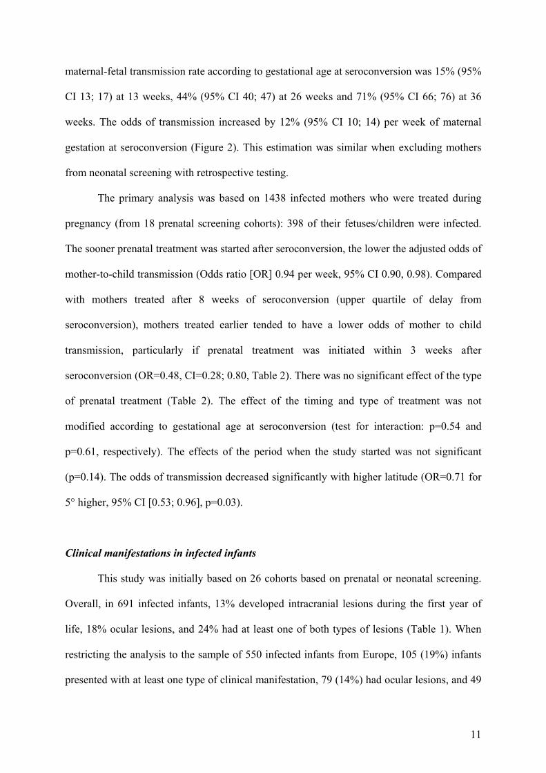
maternal-fetal transmission rate according to gestational age at seroconversion was 15% (95%
CI 13; 17) at 13 weeks, 44% (95% CI 40; 47) at 26 weeks and 71% (95% CI 66; 76) at 36
weeks. The odds of transmission increased by 12% (95% CI 10; 14) per week of maternal
gestation at seroconversion (Figure 2). This estimation was similar when excluding mothers
from neonatal screening with retrospective testing.
The primary analysis was based on 1438 infected mothers who were treated during
pregnancy (from 18 prenatal screening cohorts): 398 of their fetuses/children were infected.
The sooner prenatal treatment was started after seroconversion, the lower the adjusted odds of
mother-to-child transmission (Odds ratio [OR] 0.94 per week, 95% CI 0.90, 0.98). Compared
with mothers treated after 8 weeks of seroconversion (upper quartile of delay from
seroconversion), mothers treated earlier tended to have a lower odds of mother to child
transmission, particularly if prenatal treatment was initiated within 3 weeks after
seroconversion (OR=0.48, CI=0.28; 0.80, Table 2). There was no significant effect of the type
of prenatal treatment (Table 2). The effect of the timing and type of treatment was not
modified according to gestational age at seroconversion (test for interaction: p=0.54 and
p=0.61, respectively). The effects of the period when the study started was not significant
(p=0.14). The odds of transmission decreased significantly with higher latitude (OR=0.71 for
5° higher, 95% CI [0.53; 0.96], p=0.03).
Clinical manifestations in infected infants
This study was initially based on 26 cohorts based on prenatal or neonatal screening.
Overall, in 691 infected infants, 13% developed intracranial lesions during the first year of
life, 18% ocular lesions, and 24% had at least one of both types of lesions (Table 1). When
restricting the analysis to the sample of 550 infected infants from Europe, 105 (19%) infants
presented with at least one type of clinical manifestation, 79 (14%) had ocular lesions, and 49
11

(9%) had intracranial lesions. The odds of clinical manifestations during infancy decreased
with older gestational age at seroconversion (OR=0.96, p=0.01; Table 3). However, when the
type of lesion was considered, we found a marked reduction in the odds of intracranial lesions
with gestational age at seroconversion (p<0.0001), whereas the decline in odds with ocular
lesions was less significant (p=0.04) (Figures 3 a and b).
The adjusted odds of any clinical manifestations in infants of treated mothers
compared with untreated mothers was not significantly different (OR=1.11, p=0.74). We
further analysed the treatment effect by distinguishing the type and the timing of treatment
initiation (Table 3). We found no evidence of reduced odds of clinical manifestations in
infants born to mothers treated with spiramycin throughout pregnancy or with P-S alone,
compared with untreated mothers (all confidence intervals including 1). However, infants
born to mothers treated with spiramycin followed by P-S had a higher odds of any clinical
manifestations compared with those treated with P-S alone (OR=1.29, CI=1.42; 9.34). The
effect of prenatal treatment was not modified by gestational age at seroconversion (test for
interaction, p=0.56). An intent-to-treat analysis of prenatal treatment (patients switching their
treatment being considered in the spiramycin group) did not show any significant effect of
treatment (p=0.39). Compared to the untreated group, there was no significant difference in
the odds of clinical manifestations in infants of mothers treated with spiramycin within 5
weeks of seroconversion (OR=1.18, 95%CI=0.58; 2.40), after 5 or more weeks after
seroconversion (OR=1.22, 0.63; 2.38), or those treated by P-S (OR=0.63, 0.25; 1.61). The
odds of clinical manifestations was not significantly different according to the period of the
study (p=0.08) or the latitude of the centre (OR=1.31 for 5° higher, p=0.13).
12

Discussion
Summary of the results
We found weak evidence for an increased risk of mother-to-child transmission the
later prenatal treatment was started after maternal seroconversion. This result may reflect a
true protective effect of early treatment or confounding due to selective treatment of mothers
at high risk of fetal infection whose infection was diagnosed late (ie. outside the standard
monthly or 3 monthly re-testing schedule). We found no evidence that prenatal treatment
significantly reduced the risk of clinical manifestations in infected live-born infants.
Gestational age at seroconversion was strongly associated with mother-to-child transmission
and with the risk of intracranial lesions but marginally with eye lesions.
Strengths and limitations
To our knowledge, this is the first meta-analysis of the effect of prenatal treatment for
congenital toxoplasmosis. Almost all eligible cohorts were included but three cohorts with
appropriate data, representing 96 infected mothers (43 infected children), declined to
participate. Four further cohorts were unlikely to have been eligible because of selection bias
and enrolment before 1985 23-26. Analysis of individual patient data made it possible to
examine the effect of systematic differences in treatment schedules within and between
cohorts and we used a statistical method to minimize bias and reflect uncertainty due to the
interval censored variables (gestational age at seroconversion and timing of prenatal
treatment).
The main limitation is that our results for prenatal treatment may be partly explained
by biases in the way the cohort studies were designed and conducted. Although we adjusted
for the strong confounding effects of gestational age at seroconversion, we can not exclude
13

effects due to unmeasured confounders.27 In the analysis of mother to child transmission, we
included only prenatal screening cohorts as all used multiple tests on repeated samples to
confirm maternal infection. We excluded neonatal screening cohorts as retrospective testing
of a single stored prenatal sample could result in mislabelling of uninfected women as
infected, thereby reducing the risk of transmission in untreated women. We further restricted
the primary analysis of prenatal screened cohorts to treated women because of potential biases
causing untreated women to have a lower risk of mother to child transmission than treated
women who seroconverted at the same gestational age. One possible explanation for this
finding is that women were less likely to be treated after a long delay from seroconversion
and shortly before delivery, unless there were signs of fetal infection or complications. Such
indication bias could have increased the risk of transmission in women treated after a long
delay and could partly explain the weak association between early treatment and the risk of
mother to child transmission.
There were several sources of potential bias in the analyses of clinical manifestations,
which included neonatal and prenatal screening cohorts. Firstly, although the criteria for
congenital infection were similar across all cohorts, neonatal screening is less sensitive than
prenatal screening. Insensitivity is associated with the gestational age at maternal
seroconversion and, as it is most marked in the first half of pregnancy when intracranial
lesions are more likely, could reduce the observed effect of prenatal treatment28. We
minimised this problem by adjusting all analyses for gestational age at seroconversion. A
further source of bias is the large uncertainty in the estimated gestational age at
seroconversion in neonatal screened untreated cohorts compared with prenatal screened
cohorts. Whether such error would bias in favour of under- or overestimating the treatment
effect is difficult to predict. Thirdly, bias could have been introduced if the accuracy of cranial
14
ultrasound or ophthalmic examinations differed between neonatal and prenatal screened
cohorts. This seems unlikely as it is standard practice in European centres to perform a single
cranial ultrasound in early infancy (repeated if abnormalities are detected) and to perform at
least two ophthalmic assessments, one in early infancy and the other at one year. Prenatal
centres stated that their protocol was to examine children 3 to 6 monthly, while neonatal
centres reported 3 monthly examinations. We could not verify these practices as most datasets
did not record each examination. Fourthly, indication bias is likely to explain the apparently
harmful effect of changing treatment from spiramycin to pyrimethamine-sulphonamide
compared with no treatment. This finding may be because clinicians performed prenatal
diagnosis or changed treatment more readily if they detected fetal or maternal complications.
Such a response would also overestimate the benefits of treatment for mothers who remained
on spiramycin. We minimised this problem by conducting an ‘intention to treat’ analysis.
Fifthly, inclusion of ocular lesions detected at older ages could diminish the treatment effect
if, as seems likely, the impact of prenatal treatment is greatest on lesions detected soon after
birth. Sixthly, treatment effects in both analyses could be diminished by poor compliance with
treatment. Unfortunately, data on compliance were not recorded in any cohort. A major
limitation of ours and all published cohort studies to-date is the lack of information on the
clinical consequences of intracranial lesions for subsequent development.
We could not investigate the potential impact of missing data on these results. As is
commonly the case in studies based on routine practice, investigators recorded their caseload
of patients undergoing follow up not all seroconverting women who were eligible for follow
up. Hence, we could only identify cases with missing outcome data in the prospective
EMSCOT study: 15% of infants born to infected women, and 19% of all infants classified as
infected had insufficient follow up to meet the reference criteria for congenital infection status
4,7. Infection status was imputed for these cases based on prenatal PCR results, postnatal IgM
15
tests, and the postnatal age when last IgG positive 4,7. In the remaining cohorts, we excluded
only 21 mother-child pairs due to missing infection status and relied on the investigators’
classification of infection status. Similarly, for most cohorts, we did not have data on dates
and results of all postnatal ophthalmic and cranial ultrasound examinations, and relied on the
investigators’ classification of findings. Information on type or timing of treatment were
rarely missing (less than 10 women) and were imputed according to the protocol performed in
the given centre.
We excluded cohorts from America in the meta-analysis because of differences in the
burden of the disease, the risk of clinical manifestations 19, the parasite strain 29,30, and the
way in which intracranial lesions were measured (CT versus ultrasound scan) 20,21. Further
studies are required to compare outcomes in treated and untreated mothers within South
America and other endemic tropical areas.
Policy implications
From our results, it is unclear whether prenatal treatment has any effect on
transmission or the presence of clinical manifestations. However, confidence intervals were
wide and consistent with a beneficial effect and with no effect. Further evidence from
observational studies is unlikely to change these results. Valid evidence of any benefit of
prenatal treatment should be obtained through a large randomized controlled clinical trial.
16

*Members of the Systematic Review On Congenital Toxoplasmosis (SYROCOT)
Writing Committee: Rodolphe Thiébaut, Sandy Leproust, Geneviève Chêne, Ruth Gilbert on behalf of
the SYROCOT investigators
Investigators of cohorts contributing to SYROCOT: A Prusa, M Hayde, A Pollak (University
Children’s Hospital, Vienna, Austria), M Wallon, F Peyron (Hôpital de la Croix Rousse, Lyon,
France), S Romand, P Thulliez (Institut de Puericulture, Paris, France), W Buffolano, A. Romano
(Universita di Napoli, Naples, Italy), J Franck, H Dumon (Hôpital de la Timone, Marseille, France), P
Bastien, E Issert (CHU de Montpellier, Montpellier, France); M-H Bessieres (Hôpital de Rangueil,
Toulouse, France), N Ferret, P Marty (Hôpital de l’Archet, Nice, France), C Chemla, I Villena
(Hôpital Maison Blanche, Reims, France), H Pelloux, H Fricker-Hidalgo, C Bost-Bru (Centre
Hospitalier Universitaire de Grenoble, Grenoble, France), E Semprini, V Savasi (Milan, Italy), M Paul
(University Medical Sciences, Poznan), G Malm, B Evengard (Huddinge Hospital, Stockholm,
Sweden), E Petersen, D Schmidt (Statenseruminstitut, Copenhagen, Denmark), T Kortbeek (National
Institute of Public Health and the Environment, Bilthoven, The Netherlands), J Logar (Medical
Faculty, University of Ljubljana, Ljubljana, Slovenia), S Szenasi (Albert Szent-Györgyi Medical
University, Szeged, Hungary), B Stray-Pedersen, P Jenum (University of Oslo, Rikshospitalet,
National Insitute of Public Health, Norway), M Lappalainen (Helsinki University Central Hospital,
Finland), E Lago, E Neto (Hospital Sao Lucas da PUCRS, Porto Alegre, Brazil), L Bahia-Oliveira
(Universidade Estadual do Norte Fluminense, Campos, Brazil), R Eaton, H-W Hsu (Massachussets
State Laboratory Institute, Boston, USA), J Gomez-Marin (Universidad del Quindio, Armenia,
Colombia)
Study design and coordination: Geneviève Chêne, Ruth Gilbert, Luuk Gras, Rodolphe Thiébaut
Data management: Kathy Freeman, Tan Hooi Kuan (EMSCOT study), Sabrina Di Costanzo, Sandy
Leproust, Rodolphe Thiébaut
Statistical analysis: Sabrina Di Costanzo, Sandy Leproust, Rodolphe Thiébaut
Funding source
The research was part of The Eurotoxo project which is financed by the European Commission
(Contract No. QLG4-CT-2002-30262).
17

References
1. Cook AJ, Gilbert RE, Buffolano W, et al. Sources of toxoplasma infection in pregnant women: European multicentre case-control study. European Research Network on Congenital Toxoplasmosis. BMJ 2000;321:142-7.
2. Remington J, McLeod R, Thulliez P, Desmonts G. Toxoplasmosis. In: J. S. and Klein J, ed. Infectious Diseases of the Fetus and Newborn Infant. Philadelphia: WB Saunders, 2001: 205-346.
3. Dunn D, Wallon M, Peyron F, Petersen E, Peckham C, Gilbert R. Mother-to-child transmission of toxoplasmosis: risk estimates for clinical counselling. Lancet 1999;353:1829-33.
4. European Multicentre Study on Congenital Toxoplasmosis. Effect of timing and type of treatment on the risk of mother to child transmission of Toxoplasma gondii. BJOG 2003;110:112-20.
5. Salt A, Freeman K, Prusa A, et al. Determinants of response to a parent questionnaire about development and behaviour in 3 year olds: European multicentre study of congenital toxoplasmosis. BMC Pediatr 2005;5:21.
6. Guerina NG, Hsu HW, Meissner HC, et al. Neonatal serologic screening and early treatment for congenital Toxoplasma gondii infection. The New England Regional Toxoplasma Working Group. N Engl J Med 1994;330:1858-63.
7. Gras L, Wallon M, Pollak A, et al. Association between prenatal treatment and clinical manifestations of congenital toxoplasmosis in infancy: A cohort study in 13 European centres. Acta Paediatrica 2005;94:1721-31.
8. Raeber PA, Biedermann K, Just M, Zuber P. [Prevention of congenital toxoplasmosis in Europe]. Schweiz Med Wochenschr Suppl 1995;65:96S-102S.
9. Wallon M, Liou C, Garner P, Peyron F. Congenital toxoplasmosis: systematic review of evidence of efficacy of treatment in pregnancy. BMJ 1999;318:1511-4.
10. Neto EC, Anele E, Rubim R, et al. High prevalence of congenital toxoplasmosis in Brazil estimated in a 3- year prospective neonatal screening study. Int J Epidemiol 2000;29:941-7.
11. Evengard B, Petersson K, Engman ML, et al. Low incidence of toxoplasma infection during pregnancy and in newborns in Sweden. Epidemiol Infect 2001;127:121-7.
12. Foulon W, Villena I, Stray-Pedersen B, et al. Treatment of toxoplasmosis during pregnancy: a multicenter study of impact on fetal transmission and children's sequelae at age 1 year. Am J Obstet Gynecol 1999;180:410-5.
13. Gilbert R, Dunn D, Wallon M, et al. Ecological comparison of the risks of mother-to-child transmission and clinical manifestations of congenital toxoplasmosis according to prenatal treatment protocol. Epidemiol Infect 2001;127:113-20.
14. Eskild A, Oxman A, Magnus P, Bjorndal A, Bakketeig LS. Screening for toxoplasmosis in pregnancy: what is the evidence of reducing a health problem? J Med Screen 1996;3:188-94.
15. Gilbert RE, Gras L, Wallon M, Peyron F, Ades AE, Dunn DT. Effect of prenatal treatment on mother to child transmission of Toxoplasma gondii: retrospective cohort study of 554 mother-child pairs in Lyon, France. Int J Epidemiol 2001;30:1303-8.
16. Gras L, Gilbert RE, Ades AE, Dunn DT. Effect of prenatal treatment on the risk of intracranial and ocular lesions in children with congenital toxoplasmosis. Int J Epidemiol 2001;30:1309-13.
17. Thiébaut R, Gilbert RE, Gras L, Chêne G. Timing and type of prenatal treatment for congenital toxoplasmosis (Protocol for a Cochrane Review). The Cochrane Library. Oxford: Update Software, 2003.
18

18. Lebech M, Joynson DH, Seitz HM, et al. Classification system and case definitions of Toxoplasma gondii infection in immunocompetent pregnant women and their congenitally infected offspring. European Research Network on Congenital Toxoplasmosis. Eur J Clin Microbiol Infect Dis 1996;15:799-805.
19. Holland GN. Ocular toxoplasmosis: a global reassessment. Part I: epidemiology and course of disease. Am J Ophthalmol 2003;136:973-88.
20. Grant EG, Williams AL, Schellinger D, Slovis TL. Intracranial calcification in the infant and neonate: evaluation by sonography and CT. Radiology 1985;157:63-8.
21. Blankenberg FG, Loh NN, Bracci P, et al. Sonography, CT, and MR imaging: a prospective comparison of neonates with suspected intracranial ischemia and hemorrhage. AJNR Am J Neuroradiol 2000;21:213-8.
22. Gomez G, Espinal A, S WL. Inference for a linear regression model with an interval-censored covariate. Stat Med 2003;22:409-25.
23. Mayer HO, Stunzner D, Grubbauer HM, Faschinger C, Wocheslander E, Moser M. Nachuntersuchung von Kindern nach Toxoplasmosefrischinfektion in der Schwangerschaft. Follow-up of children after toxoplasmosis infection in pregnancy. Zentralbl Gynakol 1986;108:1482-6.
24. Ghidini A, Sirtori M, Spelta A, Vergani P. Results of a preventive program for congenital toxoplasmosis. J Reprod Med 1991;36:270-3.
25. Ndong Obame T, Ayadi A. The acquired and congenital toxoplasmosis in the Sfax area (Tunisia). Bull Soc Fr Parasitol 1997;15:141-7.
26. Patissier G, Flori P, Varlet MN, Patural H, Hafid J, Tran manh sung R. Depistage de la toxoplasmose congenitale signification des IgM anti-toxoplasmiques : Etude a partir du suivi clinique et biologique de 155 patientes en cours de grossesse. Rev Prat Gynecol Obstet 2001;52:33-7.
27. Thiébaut R, Leroy V, Alioum A, et al. Biases in observational studies of the effect of prenatal treatment for congenital toxoplasmosis. Eur J Obstet Gynecol Reprod Biol 2005;124:3-9.
28. Wallon M, Dunn D, Slimani D, Girault V, Gay-Andrieu F, Peyron F. Diagnosis of congenital toxoplasmosis at birth: what is the value of testing for IgM and IgA? Eur J Pediatr 1999;158:645-9.
29. Dubey JP, Graham DH, Blackston CR, et al. Biological and genetic characterisation of Toxoplasma gondii isolates from chickens (Gallus domesticus) from Sao Paulo, Brazil: unexpected findings. Int J Parasitol 2002;32:99-105.
30. Vallochi AL, Muccioli C, Martins MC, Silveira C, Belfort R, Rizzo LV. The genotype of Toxoplasma gondii strains causing ocular toxoplasmosis in humans in Brazil. Am J Ophthalmol 2005;139:350-1.
31. Logar J, Petrovec M, NovakAntolic Z, et al. Prevention of congenital toxoplasmosis in Slovenia by serological screening of pregnant women. Scand J Infec Dis 2002;34:201-4.
32. Szenasi Z, Ozsvar Z, Nagy E, et al. Prevention of congenital toxoplasmosis in Szeged, Hungary. Int J Epidemiol 1997;26:428-35.
33. Bahia-Oliveira L, Abreu A, Azevedo-Silva J, F. O. Toxoplasmosis in southeastern Brazil: an alarming situation of highly endemic acquired and congenital infection. Int J Parasitol 2001;31:115-44.
34. Gomez Marin JE. Evaluación del tratamiento de la toxoplasmosis gestacional en una cohorte colombiana. Infectio 2005;9:16-23.
19
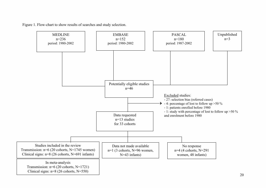
Figure 1. Flow
TransmissiClinical si
StudiTransmission: Clinical signs
-chart to show results of searches and study selection.
20
MEDLINE n=236
period: 1980-2002
EMBASE n=152
period: 1980-2002
PASCAL n=180
period: 1987-2002
Potentially eligible studies n=46
Unpublished n=3
Data requested n=13 studies
for 33 cohorts
Excluded studies: - 27: selection bias (referred cases) - 4: percentage of lost to follow up >50 % - 1: patients enrolled before 1980 - 1: study with percentage of lost to follow up >50 % and enrolment before 1980
In meta-analysis on: n=6 (20 cohorts, N=1721) gns: n=8 (26 cohorts, N=550)
es included in the review n=6 (20 cohorts, N=1745 women) : n=8 (26 cohorts, N=691 infants)
Data not made available n=1 (3 cohorts, N=96 women,
N=43 infants)
No response n=4 (4 cohorts, N=291
women, 48 infants)

Figure 2. Risk of mother to child transmission of T. gondii according to gestational age at
maternal seroconversion. Dotted lines are bounds of 95% confidence interval. SYROCOT
Study, N=1721.
0,00
0,10
0,20
0,30
0,40
0,50
0,60
0,70
0,80
0,90
1,00
0 5 10 15 20 25 30 35 40
Gestational age at seroconversion (weeks)
Pro
babi
lity
of c
onge
nita
l inf
ectio
n.
21

Figure 3. Risk of clinical manifestations in children infected by T. gondii according to
gestational age at maternal seroconversion. Dotted lines are bounds of 95% confidence
interval. SYROCOT Study. Europe only.
A. Risk of intracranial lesions (N=473)
0
0,1
0,2
0,3
0,4
0,5
0,6
0,7
0,8
0,9
1
0 5 10 15 20 25 30 35 40
Gestational age at seroconversion (weeks)
Pro
babi
lity
of in
tracr
ania
l les
ions
22

B. Risk of eye lesions (N=526)
0
0,1
0,2
0,3
0,4
0,5
0,6
0,7
0,8
0,9
1
0 5 10 15 20 25 30 35 40
Gestational age at seroconversion (weeks)
Pro
babi
lity
of e
ye le
sion
s
23

Table 1. Characteristics of cohorts included in the systematic review.
Clinical manifestation (% of infected infants)
Country Cohort region (study reference) Recruitment
Period
Screening
$
Infected
Mothers
Infected live born
children (%) Any Ocular Intracranial
Netherlands
South Netherlands 13 1987-1988 PN 2m 52 12 (23.1) 3 (25.0) 2 (16.7) 1 (8.3)
Norway Oslo 12 1992-1994 PN 3m 33 17 (51.5) 6 (35.3) 3 (17.7) 4 (23.5)
Finland Helsinki 12 1988-1994 PN 3m 12 4 (33.3) 2 (50.0) 1 (25.0) 2 (50.0)
Slovenia Ljubljana 31 1994-1996 PN 3m 19 3 (15.8) 0 (0.0) 0 (0.0) 0 (0.0)
Hungary Hungary 32 1987-1994 PN 3m 10 4 (40.0) 1 (25.0) 1 (25.0) 0 (0.0)
France Nice (unpublished) * 1998-2002 PN 1m 44 15 (34.1) 1 (6.7) 1 (6.7) 0 (0.0)
Grenoble 7 1996-2000 PN 1m 24 6 (25.0) 2 (33.3) 1 (16.7) 1 (16.7)
Lyon 7 1996-2000 PN 1m 167 43 (25.7) 10 (23.3) 9 (20.9) 3 (7.0)
Lyon 13 1987-1995 PN 1m 583 179 (30.7) 33 (18.4) 23 (12.9) 16 (8.9)
Marseille 7 1996-2000 PN 1m 67 20 (29.9) 2 (10.0) 2 (10.0) 0 (0.0)
Nice 7 1996-2000 PN 1m 30 8 (26.7) 4 (50.0) 2 (25.0) 2 (25.0)
Paris 7 1996-2000 PN 1m 197 65 (33.0) 8 (12.3) 8 (12.3) 1 (1.5)
Reims 7 1996-2000 PN 1m 26 8 (30.8) 2 (25.0) 2 (25.0) 0 (0.0)
Toulouse 7 1996-2000 PN 1m 68 22 (32.4) 3 (13.6) 2 (9.1) 1 (4.6)
24
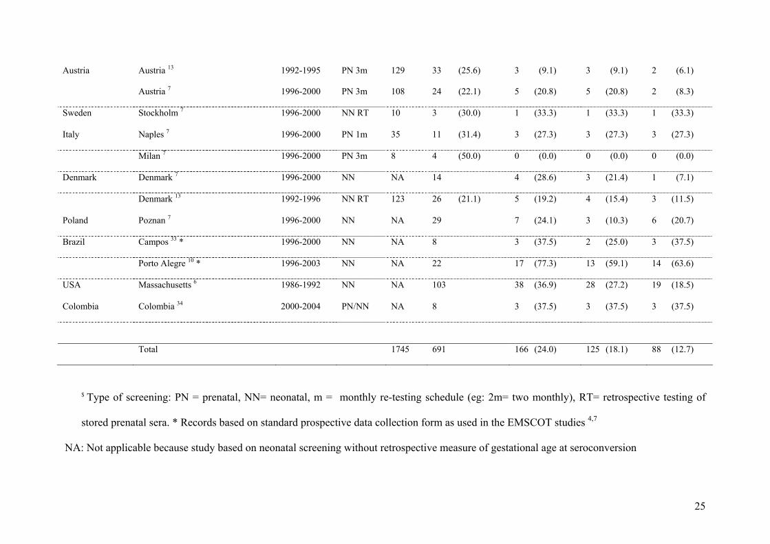
Austria
Austria 13 1992-1995 PN 3m 129 33 (25.6) 3 (9.1) 3 (9.1) 2 (6.1)
Austria 7 1996-2000 PN 3m 108 24 (22.1) 5 (20.8) 5 (20.8) 2 (8.3)
Sweden Stockholm 7 1996-2000 NN RT 10 3 (30.0) 1 (33.3) 1 (33.3) 1 (33.3)
Italy Naples 7 1996-2000 PN 1m 35 11 (31.4) 3 (27.3) 3 (27.3) 3 (27.3)
Milan 7 1996-2000 PN 3m 8 4 (50.0) 0 (0.0) 0 (0.0) 0 (0.0)
Denmark Denmark 7 1996-2000 NN NA 14 4 (28.6) 3 (21.4) 1 (7.1)
Denmark 13 1992-1996 NN RT 123 26 (21.1) 5 (19.2) 4 (15.4) 3 (11.5)
Poland Poznan 7 1996-2000 NN NA 29 7 (24.1) 3 (10.3) 6 (20.7)
Brazil Campos 33 * 1996-2000 NN NA 8 3 (37.5) 2 (25.0) 3 (37.5)
Porto Alegre 10 * 1996-2003 NN NA 22 17 (77.3) 13 (59.1) 14 (63.6)
USA Massachusetts 6 1986-1992 NN NA 103 38 (36.9) 28 (27.2) 19 (18.5)
Colombia Colombia 34 2000-2004 PN/NN NA 8 3 (37.5) 3 (37.5) 3 (37.5)
Total 1745 691 166 (24.0) 125 (18.1) 88 (12.7)
$ Type of screening: PN = prenatal, NN= neonatal, m = monthly re-testing schedule (eg: 2m= two monthly), RT= retrospective testing of
stored prenatal sera. * Records based on standard prospective data collection form as used in the EMSCOT studies 4,7
NA: Not applicable because study based on neonatal screening without retrospective measure of gestational age at seroconversion
25

Table 2. Adjusted effect of the timing and type of prenatal treatment on the risk of mother to child transmission in European prenatal screening
centers in the sub-sample of treated mothers (N=1438 mothers, 398 infected fetus/children).
OR 95% CI p
Timing of prenatal treatment initiation* 0.05
< 3 weeks after seroconversion (n=312) 0.48 [0.28 ; 0.80]
3 ≤ and < 5 weeks after seroconversion (n=442) 0.64 [0.40 ; 1.02]
5 ≤ and < 8 weeks after seroconversion (n=360) 0.60 [0.36 ; 1.01]
≥ 8 weeks after seroconversion (n=325) Ref
Type of treatment (Spiramycin vs. P-S) 0.79 [0.55 ; 1.13] 0.19
Gestational age at maternal seroconversion (per week) 1.15 [1.12 ; 1.17] <10-4
Latitude (for 5° higher) 0.71 [0.53 ; 0.96] 0.03
Start of study period 0.14
After 1994 0.39 [0.15 ; 1.05]
Between 1991 and 1994 0.46 [0.17 ; 1.21]
Before 1991 Ref
26

* Contribution of woman time to each category given in brackets. Model adjusted for gestational age at maternal seroconversion estimated by the
integrated maximum likelihood method
OR: Odds ratio, 95% CI: 95% confidence interval, P-S: Pyrimethamine-Sulphonamides
27
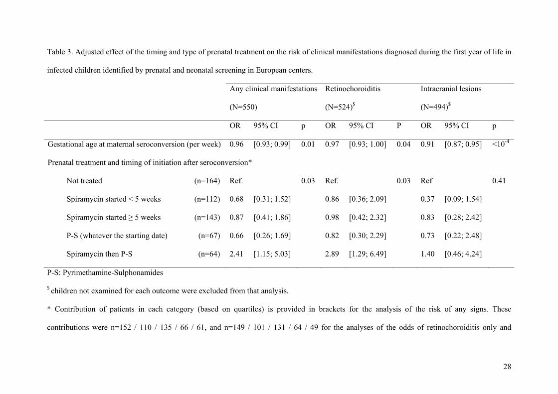
Table 3. Adjusted effect of the timing and type of prenatal treatment on the risk of clinical manifestations diagnosed during the first year of life in
infected children identified by prenatal and neonatal screening in European centers.
Any clinical manifestations Retinochoroiditis
(N=550) (N=524)$
Intracranial lesions
(N=494)$
OR OR95% CI 95% CI p P OR 95% CI p
Gestational age at maternal seroconversion (per week) 0.96 [0.93; 0.99] 0.01 0.97 [0.93; 1.00] 0.04 0.91 [0.87; 0.95] <10-4
Prenatal treatment and timing of initiation after seroconversion*
Not treated (n=164) Ref. 0.03 Ref. 0.03 Ref 0.41
Spiramycin started < 5 weeks (n=112) 0.68 [0.31; 1.52] 0.86 [0.36; 2.09] 0.37 [0.09; 1.54]
Spiramycin started ≥ 5 weeks (n=143) 0.87 [0.41; 1.86] 0.98 [0.42; 2.32] 0.83 [0.28; 2.42]
P-S (whatever the starting date) (n=67) 0.66 [0.26; 1.69] 0.82 [0.30; 2.29] 0.73 [0.22; 2.48]
Spiramycin then P-S (n=64) 2.41 [1.15; 5.03] 2.89 [1.29; 6.49] 1.40 [0.46; 4.24]
P-S: Pyrimethamine-Sulphonamides
$ children not examined for each outcome were excluded from that analysis.
* Contribution of patients in each category (based on quartiles) is provided in brackets for the analysis of the risk of any signs. These
contributions were n=152 / 110 / 135 / 66 / 61, and n=149 / 101 / 131 / 64 / 49 for the analyses of the odds of retinochoroiditis only and
28

29
intracranial lesions only, respectively. Models were also adjusted for gestational age at maternal seroconversion, period of the study (<1991,
1991-1994, >1994), and latitude of centre.
Related Documents