Welcome message from author
This document is posted to help you gain knowledge. Please leave a comment to let me know what you think about it! Share it to your friends and learn new things together.
Transcript
VMIC 2017The Veterinary Medicine International Conference 2017Volume 2017
Conference Paper
Teratogenic Effect of CongenitalToxoplasmosis in Chicken EmbryoLucia Tri Suwanti1, Mufasirin2, Hani Plumeriastuti2, and Erma Safitri3
1Veterinary Parasitology Department of VeterinaryMedicine Faculty Surabaya, 60115, Indonesia2Veterinary Pathology Department of Veterinary Medicine Faculty, Surabaya, 60115, Indonesia3Veterinary Reproduction Department of Veterinary Medicine Faculty, Surabaya, 60115,Indonesia
AbstractThis research is designed to observe the teratogenic effect of Toxoplasma gondii
infection in chick embryos, based on the number of somites, embryo length and thedevelopment of embryonic brain vesicles. Methods in the research: Chicken eggswere infected with 1 x 10³ tachyzoites of T. gondii. The eggs were incubated in eggshatching box. Observation of somite performed on embryonated eggs 24 hours afterincubation and the embryonic development of vesicles performed 72 hours afterincubation then the length of each embryo were measured. Results: Revealed thatthere was a significant difference in the number of somites (p < 0.1), T. gondii infectionreduced the number of somites. While in the number of brain vesicles in 3 - daysold chicken embryos, although there was no significant difference, the size decliningemerged. The length of the embryos both at 24 or 72 hours old showed that T. gondiiinfection reduced the length (p < 0.1). Conclusions: T. gondii infection influences thedevelopment of chicken embryos in the declining of length and the decreasing ofsomite embryo number.
Keywords: IGF-I crossbreed mare serum pregnant; Follicle; Mus musculus.
1. Introduction
Toxoplasmosis is a zoonotic disease that caused by the parasitic protozoan Toxoplasma
gondii [1-4]. Humans, livestock and poultry were infected by sporulating oocysts thatpollute the environment [5-6]. A human can also be infected by eating undercookedmeat [7-8]. In pregnant mammals, both livestock and human, T. gondii infection canbe transmitted transplacentally [9] and may risk the fetus to get fetal absorption,abortion, stillbirth, infant death and congenital abnormalities born weak, dependingon the time of gestation getting an infection [10-12]. T. gondii infection, in livestock, isalso given rise to problems such as fetal pathology and abortion [12]. T. gondii infectionis a major cause of abortion goats and sheep in several countries including Australia
How to cite this article: Lucia Tri Suwanti, Mufasirin, Hani Plumeriastuti, and Erma Safitri, (2017), “Teratogenic Effect of Congenital Toxoplasmosisin Chicken Embryo” in The Veterinary Medicine International Conference 2017, KnE Life Sciences, pages 596–602. DOI 10.18502/kls.v3i6.1187 Page 596
Corresponding Author:
Lucia Tri Suwanti
Received: 03 October 2017
Accepted: 10 October 2017
Published: 29 November 2017
Publishing services provided
by Knowledge E
Lucia Tri Suwanti et al. This
article is distributed under the
terms of the Creative
Commons Attribution License,
which permits unrestricted
use and redistribution
provided that the original
author and source are
credited.
Selection and Peer-review
under the responsibility of the
VMIC Conference Committee.
VMIC 2017
and the United States. The frequency of occurrence of abortion and fetal death in sheepinfected with T. gondii is quite high [13-14]. Studies on the incidence of toxoplasmo-sis in chickens and chicken eggs have been done by several researchers. The studyincludes serological and parasitological study and the results of these studies indicatethe incidence of toxoplasmosis in chickens and eggs is quite high [15-17]
The risk of infection due to chicken embryos in eggs rarely have been reported. Theaim of this research is to reveal the teratogenic effects of T. gondii infection in chickenembryos, based on the number of somites, the development of the embryonic brainvesicles and length of the embryo. It is expected that this study can be used as aresearch model of T. gondii infection teratogenic effects in mammals, including humanembryos
2. Materials and methods
Forty chicken eggs were divided into two groups. The first groups as a control and theother infected with T. gondii. Isolates of T. gondii was RH strain. Infection proceduresappropriated with [18-19]. Infection dose was 1 × 10³ tachyzoites. tachyzoites injectedinto chorioallantois and then closed with paraffin and masking tape. After infection, allof the eggs were incubated in hatching box.
Teratogenic effects based on embryonic development, include: the number ofsomites in the embryo, the length of embryos and brain vesicle formation IsolationEmbryos was done by using whole- mounts [20]. Ten embryonated chicken eggs ofeach group were observed at 24 hours and then the other at 72 hours after incubation.Embryonated chicken eggs were opened by eggshell peeling.
The egg was dipped in 0.9% saline buffer and simultaneity poured the contents ofthe egg. The red part in hollow eggswas containing an embryo. Embryonic membraneswere cut using a glass slide and embryowas raised and put on the glass. Embryoswereobserved under a microscope. The embryonic length was measured from the anteriorend to the posterior end of the embryo ages 24 and 72 hours after infection, a numberof somites were calculated on embryo 24 hours old and the development of embryonicvesicles was done embryonated chicken eggs age of 72 hours.
The number and length of somite chicken embryos were analyzed by t test, whilefor the development of embryonic vesicles descriptively presented.
DOI 10.18502/kls.v3i6.1187 Page 597
VMIC 2017
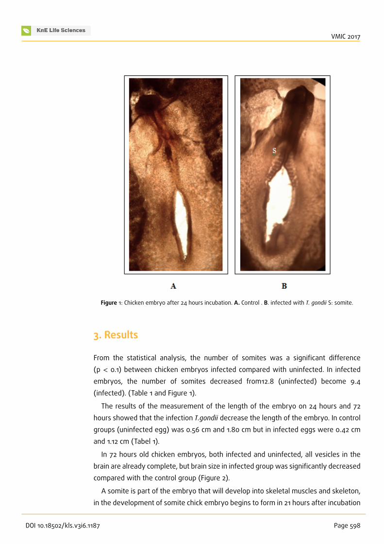
Figure 1: Chicken embryo after 24 hours incubation. A. Control . B. infected with T. gondii S: somite.
3. Results
From the statistical analysis, the number of somites was a significant difference(p < 0.1) between chicken embryos infected compared with uninfected. In infectedembryos, the number of somites decreased from12.8 (uninfected) become 9.4(infected). (Table 1 and Figure 1).
The results of the measurement of the length of the embryo on 24 hours and 72hours showed that the infection T.gondii decrease the length of the embryo. In controlgroups (uninfected egg) was 0.56 cm and 1.80 cm but in infected eggs were 0.42 cmand 1.12 cm (Tabel 1).
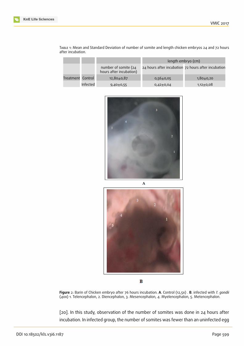
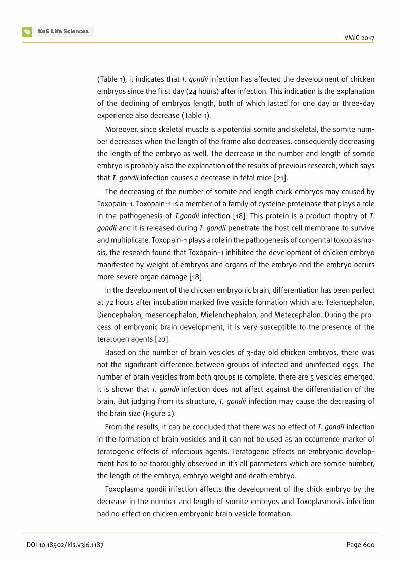
In 72 hours old chicken embryos, both infected and uninfected, all vesicles in thebrain are already complete, but brain size in infected group was significantly decreasedcompared with the control group (Figure 2).
A somite is part of the embryo that will develop into skeletal muscles and skeleton,in the development of somite chick embryo begins to form in 21 hours after incubation
DOI 10.18502/kls.v3i6.1187 Page 598
VMIC 2017
T 1: Mean and Standard Deviation of number of somite and length chicken embryos 24 and 72 hoursafter incubation.
length embryo (cm)
number of somite (24hours after incubation)
24 hours after incubation 72 hours after incubation
Treatment Control 12,80±0,87 0,56±0,05 1,80±0,20Infected 9,40±0,55 0,42±0,04 1,12±0,08
Figure 2: Barin of Chicken embryo after 76 hours incubation. A. Control (12,5x) . B. infected with T. gondii(40x) 1. Telencephalon, 2. Diencephalon, 3. Mesencephalon, 4. Myelencephalon, 5. Metencephalon.
[20]. In this study, observation of the number of somites was done in 24 hours afterincubation. In infected group, the number of somites was fewer than an uninfected egg
DOI 10.18502/kls.v3i6.1187 Page 599
VMIC 2017
(Table 1), it indicates that T. gondii infection has affected the development of chickenembryos since the first day (24 hours) after infection. This indication is the explanationof the declining of embryos length, both of which lasted for one day or three-dayexperience also decrease (Table 1).
Moreover, since skeletal muscle is a potential somite and skeletal, the somite num-ber decreases when the length of the frame also decreases, consequently decreasingthe length of the embryo as well. The decrease in the number and length of somiteembryo is probably also the explanation of the results of previous research, which saysthat T. gondii infection causes a decrease in fetal mice [21].
The decreasing of the number of somite and length chick embryos may caused byToxopain-1. Toxopain-1 is a member of a family of cysteine proteinase that plays a rolein the pathogenesis of T.gondii infection [18]. This protein is a product rhoptry of T.
gondii and it is released during T. gondii penetrate the host cell membrane to surviveandmultiplicate. Toxopain-1 plays a role in the pathogenesis of congenital toxoplasmo-sis, the research found that Toxopain-1 inhibited the development of chicken embryomanifested by weight of embryos and organs of the embryo and the embryo occursmore severe organ damage [18].
In the development of the chicken embryonic brain, differentiation has been perfectat 72 hours after incubation marked five vesicle formation which are: Telencephalon,Diencephalon, mesencephalon, Mielenchephalon, and Metecephalon. During the pro-cess of embryonic brain development, it is very susceptible to the presence of theteratogen agents [20].
Based on the number of brain vesicles of 3-day old chicken embryos, there wasnot the significant difference between groups of infected and uninfected eggs. Thenumber of brain vesicles from both groups is complete, there are 5 vesicles emerged.It is shown that T. gondii infection does not affect against the differentiation of thebrain. But judging from its structure, T. gondii infection may cause the decreasing ofthe brain size (Figure 2).
From the results, it can be concluded that there was no effect of T. gondii infectionin the formation of brain vesicles and it can not be used as an occurrence marker ofteratogenic effects of infectious agents. Teratogenic effects on embryonic develop-ment has to be thoroughly observed in it’s all parameters which are somite number,the length of the embryo, embryo weight and death embryo.
Toxoplasma gondii infection affects the development of the chick embryo by thedecrease in the number and length of somite embryos and Toxoplasmosis infectionhad no effect on chicken embryonic brain vesicle formation.
DOI 10.18502/kls.v3i6.1187 Page 600
VMIC 2017
References
[1] Astrid M. Tentera, Anja R. Heckerotha, and Louis M. Weissb. Toxoplasma gondii:from animals to humans. Int J Parasitol. 2000 November; 30(12-13): 1217–1258.
[2] Sonar S. S. and Brahmbhatt M.N. Toxoplasmosis: An Important Protozoan Zoonosis.Veterinary World Vol.3(9):436-439
[3] Francisco Mimica, Claudia Muñoz-Zanzi, Marisa Torres y Oslando Padilla. Toxoplas-mosis, a parasitic zoonoses prevalent in Chile: count and challenges
[4] Ahmad S, Tasawar Z. Toxoplasmosis In Four Caprine Breeds: A Future Risk OfZoonosis. The Journal of Animal & Plant Sciences, 26(2): 2016, Page: 388-394
[5] Karen Shapiro* John Largier, Jonna A. K. Mazet, William Bernt, John R. Ell, AnnC. Melli, Patricia A. Conrad. Surface Properties of Toxoplasma gondii Oocysts andSurrogate Microspheres. Applied and Environmental Microbiology, Feb. 2009, p.1185–1191
[6] Emily L Lilly and Caroline D Wortham. High prevalence of Toxoplasma gondii oocystshedding in stray and pet cats (Felis catus) in Virginia, United States. Lilly andWortham Parasites & Vectors 2013, 6:266
[7] Dubey JP, Hill DE, Jones JL, Hightower HW, Kirkland E, Roberts JM, Marcet PL,Lehmann T, Vianna MCB, Miska K, Sreekumar C, Kwok OCH, Shen SK, and GambleHR. Prevalence Of Viable Toxoplasma Gondii In Beef, Chicken, And Pork FromRetail Meat Stores In The United States: Risk Assessment To Consumers. Journalof Parasitology. Oct 2005 : Vol. 91, Issue 5, pg(s) 1082- 1093.
[8] Berger-Schocha E, Herrmann DC, Schares G, Müller N, Bernet D, Gottsteina B, FreyaCF. Prevalence and genotypes of Toxoplasma gondii in feline faeces (oocysts) andmeat from sheep, cattle and pigs in Switzerland. d. Vet. Parasitol. 2010(6): 1-8.
[9] Hill D, Dubey JP. Toxoplasma gondii: transmission, diagnosis and prevention. ClinMicrobiol Infect 2002; 8: 634–640
[10] Dubey JP, Lindsay DS, and Speer CA. Structures of Toxoplasma gondii tachyzoites,bradyzoites, and sporozoites and biology and development of tissue cysts. J CMR1998; 11(2): 267-299
[11] Weissmann J.. Presumtive Toxoplasma gondii abortion in sheep. J Can Vet 2003;44(4): 322-324
[12] Dubey JP and Jones JL. Toxoplasma gondii infection in human and animals in theUnited States.Internas. J. Parasitol.. 2010; 38:1257-1278.
[13] Duncanson P, Terry RS, Smith JE, and Hide G, High levels of congenital transmissionof Toxoplasma gondii in a commercial sheep flock. Int J Parasitol. 2001; 31(14):1699-703
DOI 10.18502/kls.v3i6.1187 Page 601
VMIC 2017
[14] Rahdar M, Samarbaf-Zadeh AR, Arab L. Evaluating the Prevalence of Toxoplasmagondii in Meat and Meat Products in Ahvaz by PCR Method. Jundishapur J Microbiol.2012;5(4):570-573.
[15] Mufasirin, Suprihati E dan Suwanti LT. Studi Toxoplasmosis pada Telur Ayam BurasYang Dijual Sebagai Campuran Jamu di Kota Surabaya dan kabopaten SidoarjoMenggunakan Uji Dot Blot. J. Penelit. Med Eksakta. 2003; 4:113-119.
[16] Mufasirin dan Suwanti LT. Deteksi Toxoplasma gondii Pada Telur Ayam Buras YangDijual Sebagai Campuran Jamu Di Kota Surabaya Menggunakan Uji Biologis. MediaKedokteran Hewan. 2008 24(1); 5-9
[17] Suwanti LT. Weight Reduction of Fetuses of Toxoplasma gondii-infected Mice.Proceeding International Conference “Animal Health and Human Safety” UPMMalaysia. 2009, pp 197-200
[18] Que, XC, Wuderlich A, Joiner KA, and Reed SL. Toxopain-1 is Critical for Infection ina Novel Chicken Embryo Model of Congenital Toxoplasmosis. IAI. 2004; 72(5):2915-2921
[19] Furuta PI, Mineo TWP, Carrasco AOT, Godoy GS, Pinto AA, Machado RZ. Neosporacaninum infection in birds: experimental infections in chicken and embryonatedeggs. Parasitology (2007), 134, 1931–1939
[20] Luqman, EM, Soenardihardjo BP, dan Mahaputra L. Peranann Choline Esterase(ChE) pada Pembentukan Vesikel Otak Embrio Ayam yang Terpapar InsektisidaKarbofuran. Media Kedokteran Hewan. 2007; 23(3):145-150
[21] Suwanti LT, Suprihati E, dan Mufasirin.. Deteksi Kista Jaringan Toxoplasma gondii
pada beberapa organ ayam. Lemlit. Unair. Surabaya. 2003. 1-21
DOI 10.18502/kls.v3i6.1187 Page 602
Related Documents