The mechanical properties of adipose tissue 30 th June 2009 1/30 Fleck The mechanical response of porcine adipose tissue Kerstyn Comley and Norman Fleck 1 , Department of Engineering, Cambridge University, Cambridge, CB1 2PZ, UK Submitted to the ASME Journal of Biomechanical Engineering 1 Corresponding author

Welcome message from author
This document is posted to help you gain knowledge. Please leave a comment to let me know what you think about it! Share it to your friends and learn new things together.
Transcript

The mechanical properties of adipose tissue
30th June 2009 1/30 Fleck
The mechanical response of porcine adipose tissue
Kerstyn Comley and Norman Fleck1,
Department of Engineering, Cambridge University, Cambridge, CB1 2PZ, UK
Submitted to the ASME Journal of Biomechanical Engineering
1 Corresponding author

The mechanical properties of adipose tissue
30th June 2009 2/30 Fleck
Abstract Subcutaneous adipose tissue has been tested in compression and in shear over a wide range of strain
rates, from quasi-static to 5700 s-1. In the quasi-static regime the tissue was subjected to fully
reversed cyclic loading. A symmetric tensile-compressive response was observed with lock-up at
tensile and compressive strains of 25%. Uniaxial compressive tests at high strain rates (1000 s-1 –
5700 s-1) were carried out using a split Hopkinson pressure bar with polycarbonate bars. Over the
full range of strain rates from quasi-static to high strain rate, the shape of the stress versus strain
response is invariant: the stress level scales with the initial modulus E. A one term Ogden energy
density function is adequate to describe the data. The harmonic response of the tissue was also
measured in compression-compression and in oscillatory shear over a frequency range from zero to
100 Hz and the complex modulus was determined. The real part of the compression modulus, E', is
close to the modulus estimated from the quasi-static tests, and the damping factor tan δ is
approximately 0.22 over the frequency range employed.
Keywords: Adipose tissue; High strain rate test; Hopkinson bar test; DMA; Rheology; Uniaxial
compression

The mechanical properties of adipose tissue
30th June 2009 3/30 Fleck
Introduction
Drug delivery devices load subcutaneous adipose tissue over a range of strain rates, depending upon
the characteristics of the device. For example, a needle and syringe will strain the tissue at
substantially slower rates than a liquid jet injection. Recently, Shergold and Fleck [1] have
developed an penetration model for the dermis. In order to apply this model to adipose tissue it is
necessary to measure the uniaxial response, preferably over a wide range of strain and strain rates.
The purpose of this paper is to measure the uniaxial response of adipose tissue over the practical
range from 10-4 s-1 to 5000 s-1. Both monotonic and cyclic tests are performed, and the large strain
response is determined in addition to the viscoelastic characteristics at low strain level.
Microstructure of Adipose Tissue Adipose tissue, commonly known as fat, is a connective tissue comprising lipid-filled cells called
adipocytes. The lipid is a triacylglyceride whose molecular weight is in the order of 900 g mol-1.
The adipocytes are of diameter 80 μm and are supported by two collagen-based structures: collagen
mesh, containing primarily type I and IV collagen, surrounding each cell, and a type I collagen fibre
network termed the interlobular septa. Additional structures such as blood vessels exist within the
tissue. The intervening space is filled with ground substance. 60-80% by weight of adipose tissue is
lipid, 5-30% is water and the remaining 2-3% is protein [2]. Histology of adipose tissue suggests
that it is approximately isotropic in structure and is thereby isotropic in mechanical properties. The
large liquid content enforces material incompressibility [3].
The poro-elastic nature of various biological tissues has been observed by many researchers, see for
example Barry and Aldis [4] and Mow et al. [5]. The degree to which a soft tissue is poro-elastic
depends upon the volume fraction of liquid contained within open channels. When this is large,
such as for bone, there is a substantial difference in mechanical response in a fully drained test
(allowing for liquid flow and egress from the specimen) compared to an undrained test (with no
liquid flow). The important feature of poro-elasticity is the fact that the fully drained stiffness is
much less than the undrained stiffness. In adipose tissue, the ground substance (which fills the open
channels) accounts for only a few percent of the volume fraction and there is negligible free fluid
available to endow the tissue with poro-elastic properties. This has been confirmed by preliminary
compression tests on porcine adipose tissue. On subjecting the tissue to a uniaxial compressive
strain of 50% the volume of liquid expressed was less than 1% of the overall volume. We note in
passing that the finite permeability of adipose is an insufficient indicator of poro-elastic behaviour.
The measured apparent permeability is very low for adipose tissue and is in the range 1 – 18 x 10-13
The mechanical properties of adipose tissue
30th June 2009 4/30 Fleck
m4 s-1 N-1 [6, 7].
Review of methods used to measure the constitutive response of soft tissue
The possible asymmetry between tensile and compressive behaviours
The experimental measurement of the constitutive properties of biological tissues tends to focus on
either the compressive or tensile properties of the tissue. For example, Miller-Young [8] measured
the stress versus strain response of calcaneal fat in unconfined uniaxial compression at strain rates
up to 35 s-1. Samani et al. [3] used an indentation method to measure the compressive modulus of
breast tissue and Zheng and Mak [9] have attempted to determine the properties of soft limb tissue
by manual indentation of live subjects. Although there are no data reported in the literature for the
tensile properties of adipose tissue, uniaxial tensile data exist for other soft biological tissues. For
example, Huang et al. [10] performed tensile tests and compression tests on cartilage. When testing
soft tissues there is considerable practical difficulty in identifying the zero strain datum; the forces
generated at low levels of strain are low and consequently a pre-stretch or pre-tension can be
generated within the tissue during insertion into a test machine. This raises the question as to
whether the transition point between tension and compression has been accurately identified.
Sophisticated methods which attempt to overcome these difficulties have been presented; see for
example Mansour [11]. However, these methods do not guarantee that the point of zero strain has
been identified. The problem of identifying the zero strain datum is resolved in the current study by
performing large amplitude fully reversed strain excursions.
Compressive testing at high strain rates
The split Hopkinson pressure bar (SHPB) is an established technique used to measure the behaviour
of many engineering materials at strain rates above about 100 s-1. Van Sligtenhorst [12] and Song et
al. [13] have compared the SHPB response with the quasi-static response for muscle tissue: they
observe an elevation in stress level by a factor of 1000 when ε& is increased from 10-3 s-1 to 103 s-1.
However, no data are available on the high strain rate behaviour of adipose tissue. The paucity of
data is due in part to the difficulty of applying the split Hopkinson pressure bar method to soft
tissues where the measured stresses are low, typically less than 5 MPa. In order to increase the
measurement sensitivity modifications are needed to the standard steel bar set-up. Song et al. [13]
measured the properties of porcine muscle with aluminium pressure bars and Shergold et al. [14]
used magnesium bars to measure the stress versus strain response of pig skin. In the present study a
modified SHPB using polycarbonate (PC) pressure bars is employed in order to measure the
The mechanical properties of adipose tissue
30th June 2009 5/30 Fleck
compressive behaviour of adipose tissue at strain rates between 1000 s-1 to 5700 s-1. PC is attractive
because it has a low modulus of 3 GPa and thereby gives a sensitive response.
Constitutive modelling of soft biological tissues It is generally recognised that the stress versus strain response of connective tissue has a
characteristic J – shape, such that stiffening occurs at strains above about 30%, Purslow [15].
Additionally, the stress level increases with increasing applied strain rate. Since the response is
strain rate dependent a strain energy density function does not exist for the material. Despite this
deficiency rubber elasticity models provide a useful phenomenological description of the shape of
the stress versus strain curve for a given value of strain rate. The Mooney-Rivlin model has
frequently been used to model soft tissue [16], however, Shergold et al. [14] argue that the Ogden
[17] model for an incompressible, isotropic, hyper-elastic solid describes a wide range of strain
hardening characteristics for the dermis. The one-term Ogden strain energy density function is
given as:
)( 323212 −++= ααα λλλ
αμφ (1)
where φ is the strain energy density per undeformed unit volume, λ is the stretch ratio, α is the
strain hardening exponent and μ is the shear modulus.
Shergold et al. [14] suggest that since the characteristic J-curve is due to alignment of collagen
fibres and the degree of fibre alignment is insensitive to strain rate the strain hardening exponent α
is strain-rate independent. In contrast the shear modulus is expected to increase with strain rate as it
is determined by the rate dependence of the collagen fibres and the surrounding matrix. It will be
shown below that the one term Ogden model is adequate to describe the uniaxial compression data
of porcine adipose tissue.
Outline of the study The mechanical response of subcutaneous adipose tissue is examined over a wide range of strain
rates, from quasi-static to 5700 s-1. The quasi-static tests entail large amplitude, fully reversed
loading in order to probe the non-linear uniaxial response and to determine the degree of asymmetry
between the tensile and compressive responses.
Monotonic uniaxial compression tests are also carried out at strain rates ranging from 10-4 s-1 to

The mechanical properties of adipose tissue
30th June 2009 6/30 Fleck
5700 s-1. The uniaxial compressive tests at high strain rates (1000 s-1 – 5700 s-1) employ a modified
split Hopkinson pressure bar with polycarbonate bars. Additionally, low strain amplitude harmonic
tests are performed in compression-compression and in oscillatory shear in order to extract the
complex axial modulus and shear modulus as a function of frequency from zero to 100 Hz.
Experimental method
Specimen preparation It is difficult to acquire fresh human subcutaneous adipose tissue for ethical, immunological and
practical reasons. Since porcine adipose tissue has similar morphology, histology and cell kinetics
to human adipose tissue [18] it has been chosen as a suitable substitute. Fresh porcine skin tissue
was supplied by a local abattoir, Dalehead Foods, Linton. Samples of dermis and subcutaneous fat
were removed from the jowl of the pigs to a depth of 20 mm immediately after slaughter. The
samples were stored in Phosphate Buffered Saline (PBS) at room temperature prior to testing,
which always commenced within 3 hours of slaughter.
Circular cylindrical specimens of adipose tissue were cut to a radius a = 10 mm, with the axis
aligned with the normal to the skin using a sharp punch. The specimens were trimmed to a length, l
= 3 mm for the high strain rate tests and l = 8 mm for the low strain rate tests. End trimming
ensured that the end faces were flat and parallel.
Monotonic compression tests were performed over three regimes:
1. Low strain rate (below 2 s-1), using a screw driven tensile test machine;
2. Intermediate strain rate (20 s-1 – 260 s-1) ), using a servo hydraulic tensile test machine;
3. High strain rate (above 1000 s-1), using a split Hopkinson pressure bar;
Low strain rate compression tests Cylindrical specimens were compressed between smooth 15 mm thick nylon platens at strain rates
of ε& < 0.2 s-1 using a screw-driven tensile test machine. The force generated in the specimens was
recorded via a 5 N load cell capable of measuring 20 mN to within 1 %. The initial length of each
specimen was taken as the displacement at which a force was first detectable (on the order of 10
mN). Force, displacement and the rate of displacement were converted into nominal engineering
The mechanical properties of adipose tissue
30th June 2009 7/30 Fleck
stress, strain and strain rate. Monotonic compression tests were conducted at a strain rate in the
range of 2 x 10⁻³ s-1 to 0.2 s-1, and were conducted at a strain level of up to 0.4.
Intermediate strain rate compression tests
Specimens were compressed at strain rates from ε& = 20 s-1 to 260 s-1 between smooth nylon platens
using a servo-hydraulic test machine and a purpose built load cell [19]. The load cell comprised an
aluminium beam, fitted with four strain gauges in a standard Wheatsone bridge arrangement. The
stiffness and sensitivity of the beam were 20.46 N mm-1 and 141 µε N-1, respectively [20]. The
displacement of the machine platens was measured using a Linear Resistance Displacement
Transducer (LRDT) mounted as an integral part of the test machine. Care was taken to subtract off
the machine (and load cell) compliance from the measured displacement response. The outputs
from the beam transducer and LRDT was recorded on an oscilloscope and converted into nominal
engineering stress, strain and strain rate.
High strain rate compression tests
High strain rate compression tests, above 1000 s-1, were conducted using a Split Hopkinson
Pressure Bar (SHPB), see for example Follansbee [21]. The theory of the SHPB is summarised as
follows. The input and output bars each has a cross-sectional area Ab and modulus Eb. A specimen of
length l and cross-sectional area Ah is placed between them (see Fig. 1a). A striker bar is fired down
a gun barrel to impact the input bar. The velocity of the striker bar is measured by light sensors
within the gun barrel. On impact, a compression wave (incident wave, i) propagates along the input
bar at the speed of sound in the bar. After the incident wave has reached the distal end of the input
bar, the distal end begins to compress the specimen. A compression wave is transmitted through the
specimen into the output bar (transmitted wave, t), while the remainder of the incident wave is
reflected back into the input bar as a tensile wave (reflected wave, r). Strain gauges mounted on the
bars measure the axial strain associated with these longitudinal waves.
A full derivation of equations used to calculate the average stress σs(t), strain εs(t) and strain rate
)(tsε& within the specimen is given by Wang et al. [22]. The stress and strain are responses are given
by
)()( tA
AEt t
h
bbs εσ = (2)
and
The mechanical properties of adipose tissue
30th June 2009 8/30 Fleck
∫=t
rb
s dttlc
t0
2 )()( εε (3)
where cb is the longitudinal wave speed in the pressure bar. It is clear from Eq. (2) that the measured
strain level in the output bar, tε is dependent upon the ratio of the stress in the specimen, σs(t) to
the modulus of the bar, Eb. If the specimen is very soft (as in the case of adipose tissue) the use of
metallic pressure bars results in a low level of transmitted strain and this may be difficult to
distinguish from the inherent noise of the test equipment. Also, the input force cannot be precisely
measured, which leads to difficulty identifying the force equilibrium point [23].
The strain sensitivity can be increased by using polymeric bars, which have substantially lower
elastic moduli than metals. The improvement in sensitivity of the system enables lower stress levels
to be measured, and permits a straightforward examination of the force equilibrium in the specimen,
in turn giving confidence that the specimen is subjected to uniform deformation with negligible
influence of internal wave reflections. In addition, polymers exhibit lower wave speeds than metals
and so the specimen can be subjected to a longer test time and thence to larger strains prior to the
influence of waves reflected from the ends of the bars [24]. The pressure bars used in this
investigation were of diameter 12 mm and were made from cast Polycarbonate. The input and
output bars were of length Lb = 1.1 m and the striker bar was of length 0.25 m. Electrical resistance
strain gauges were bonded at mid-length of the input and output bars, as shown in Fig. 1a. The
gauges were placed in diametrically opposed pairs in order to check for bending.
Use of polycarbonate bars in a Split Hopkinson Pressure Bar Polymeric bars typically exhibit viscoelastic rather than elastic behaviour which leads to a number
of challenges for the analysis of the SHPB data. However, it is clear from the values published by
Wang et al. [22] that the magnitude of dispersion and attenuation effects are minor for PC bars.
Deshpande and Fleck [25] argue that the viscoelastic elastic effects are negligible in PC bars such
that they can be assumed to behave in an elastic manner. A series of checks have been conducted to
confirm this. From the results it is evident that at low levels of strain (~7 mε) within the PC bars the
stress waves propagate along the bars with minimal dispersion. As a final validation exercise the
stress versus strain response of Divinycell HD250 PVC foam was measured using PC bars and with
magnesium pressure bars.
A separate SHPB set-up was used to run the validation checks, which comprised an input bar of
length 2.2 m and a striker of length 0.25 m (Fig. 1b). Both bars were of diameter 12 mm. Pairs of

The mechanical properties of adipose tissue
30th June 2009 9/30 Fleck
resistance strain gauges were axially bonded at either end of the input bar, diametrically opposite to
each other in order to check for bending. In order to avoid end effects the strain gauges were
mounted 0.25 m from the end of the bars, which satisfies the criterion of more than 10 bar diameters
from the end of the bar [21]. The sensing circuitry and the calibration method were validated by
performing a dummy test using steel bars with no specimen present; the measured strain histories
were found to be in good agreement with the predicted values (as given by elastic analysis). The
following checks were made to confirm that at low strain levels the PC bars behaved in an elastic
manner.
Attenuation of waves in bar
The harmonic solution for the propagation of a longitudinal wave at displacement u in a viscoelastic
solid takes the form [26]
( )x i t kxu Ae eβ ω± ±= (4)
where β is the wave attenuation, ω is the angular frequency and k is the wave number. A minus sign
corresponds to waves propagating in the positive x-direction. The level of attenuation is quantified
by the parameter
1 2ln( )
gxε ε
β = (5)
where ε1 and ε2 are the maximum strains measured by the first and second strain gauges,
respectively, as shown in Fig. 1b; xg = 1.3 m is the distance between the strain gauges. A typical
strain response at the two locations of the input bar is given in Fig. 2a. The average measured level
of attenuation was =β 0.012 m-1. This level of attenuation is considered to be insignificant for
selected values of striker velocities, v, in the range 4 – 13 m s-1.
Measurement of dispersion via the evolution of pulse shape along bar
The strain gauge measurements revealed that the strain pulse progressing along the input PC bar is a
well-defined square wave, see the typical example shown in Fig. 2a. The viscoelastic tail, as
observed by Zhao [23] for PMMA, is not significant for the case of PC, and the waveshape does not
change significantly as the wave propagates along the bar. These are typical characteristics of an
elastic bar.
The mechanical properties of adipose tissue
30th June 2009 10/30 Fleck
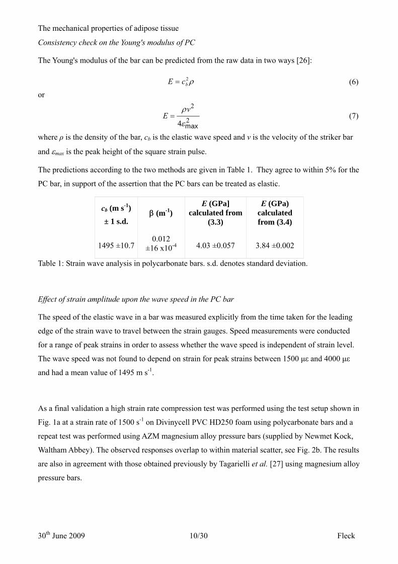
Consistency check on the Young's modulus of PC
The Young's modulus of the bar can be predicted from the raw data in two ways [26]:
ρ2bcE = (6)
or 2
24 max
vE
ρ
ε= (7)
where ρ is the density of the bar, cb is the elastic wave speed and v is the velocity of the striker bar
and εmax is the peak height of the square strain pulse.
The predictions according to the two methods are given in Table 1. They agree to within 5% for the
PC bar, in support of the assertion that the PC bars can be treated as elastic.
cb (m s-1)
± 1 s.d. β (m-1)
E (GPa] calculated from
(3.3)
E (GPa) calculated from (3.4)
1495 ±10.7 0.012
±16 x10-4 4.03 ±0.057 3.84 ±0.002
Table 1: Strain wave analysis in polycarbonate bars. s.d. denotes standard deviation.
Effect of strain amplitude upon the wave speed in the PC bar
The speed of the elastic wave in a bar was measured explicitly from the time taken for the leading
edge of the strain wave to travel between the strain gauges. Speed measurements were conducted
for a range of peak strains in order to assess whether the wave speed is independent of strain level.
The wave speed was not found to depend on strain for peak strains between 1500 με and 4000 με
and had a mean value of 1495 m s-1.
As a final validation a high strain rate compression test was performed using the test setup shown in
Fig. 1a at a strain rate of 1500 s-1 on Divinycell PVC HD250 foam using polycarbonate bars and a
repeat test was performed using AZM magnesium alloy pressure bars (supplied by Newmet Kock,
Waltham Abbey). The observed responses overlap to within material scatter, see Fig. 2b. The results
are also in agreement with those obtained previously by Tagarielli et al. [27] using magnesium alloy
pressure bars.

The mechanical properties of adipose tissue
30th June 2009 11/30 Fleck
Dynamic mechanical analysis (DMA) and oscillatory shear tests
Cyclic tensile and compression tests Using the set-up described in section 2.2.1 additional, fully reversed loading experiments were
conducted to compare the tensile and compressive behaviours of the tissue. The upper and lower
faces of the specimens were bonded to the platens using a cyanoacrylate adhesive (Loctite® Super
GlueTM Control Liquid). A periodic triangular displacement profile was used to cycle the specimens
between a tensile and compressive strain at a strain rate of approximately 0.07 s-1. Prior to testing a
pilot study was conducted to examine whether the imposed mechanical constraint associated with
bonding the tissue to the displacement platens resulted in an increase in the measured stress during
compression. No significant difference was observed between bonded and unbonded specimens.
Measurement of complex compression modulus, 20 Hz – 100 Hz An experimental rig was used to measure the oscillatory uniaxial compressive properties of adipose
tissue. Circular cylindrical specimens of diameter d = 11mm and length l =11 mm were compressed
between two nylon platens. The lower platen was attached to a V201 moving coil shaker (Link
Dynamic Systems, Herts, UK), and this in turn was driven by a 'pink noise' generator with a
frequency cut off of 500 Hz. The amplitude of displacement used in the tests was in the range 4 μm
to 50 μm, giving a strain amplitude of 0.1 – 1 %. The upper platen was attached to a piezoelectric
force sensor with a sensitivity of 115 pC N-1 and a natural frequency of >10 kHz (9205, Kistler,
Winterhur, Switzerland), and the transducer was rigidly attached to the test frame. Piezoelectric
accelerometers (Endevco, San Juan Capistrano, USA) were also fastened to the faces of each platen.
At 100 Hz the accelerometers had a sensitivity of 0.1388 pC s2 m-1. Following the suggestion of
Yamashita [28] a compressive pre-strain of approximately 15 % was applied to ensure stable
measurements of the complex modulus. Prior to testing checks were made to confirm that the
viscoelastic stress versus strain response was linear for strain amplitudes of up to 1 %.
The dynamic response and measurement accuracy of the apparatus was verified by performing a
dummy test on a compliant aluminium-ring specimen; this circular ring was fitted with a pair of
resistance strain gauges and thereby performed as an independent load cell. The stiffness of the load
cell was measured as 1.5 N mm-1 from an independent test in a screw-driven tensile test machine.
The natural frequency of the ring was calculated to be above 4 kHz. The measured stiffness was
constant and equal to the quasi-static value to within ±5 % over a frequency range of 20 Hz – 100
Hz.
The mechanical properties of adipose tissue
30th June 2009 12/30 Fleck
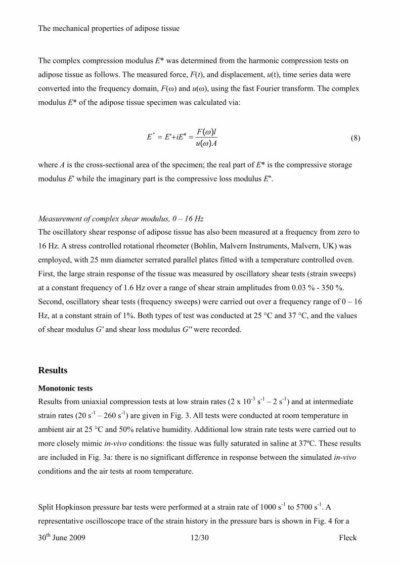
The complex compression modulus E* was determined from the harmonic compression tests on
adipose tissue as follows. The measured force, F(t), and displacement, u(t), time series data were
converted into the frequency domain, F(ω) and u(ω), using the fast Fourier transform. The complex
modulus E* of the adipose tissue specimen was calculated via:
AulFEiEE
)()('*
ωω
=′′+= (8)
where A is the cross-sectional area of the specimen; the real part of E* is the compressive storage
modulus E' while the imaginary part is the compressive loss modulus E''.
Measurement of complex shear modulus, 0 – 16 Hz The oscillatory shear response of adipose tissue has also been measured at a frequency from zero to
16 Hz. A stress controlled rotational rheometer (Bohlin, Malvern Instruments, Malvern, UK) was
employed, with 25 mm diameter serrated parallel plates fitted with a temperature controlled oven.
First, the large strain response of the tissue was measured by oscillatory shear tests (strain sweeps)
at a constant frequency of 1.6 Hz over a range of shear strain amplitudes from 0.03 % - 350 %.
Second, oscillatory shear tests (frequency sweeps) were carried out over a frequency range of 0 – 16
Hz, at a constant strain of 1%. Both types of test was conducted at 25 °C and 37 °C, and the values
of shear modulus G' and shear loss modulus G'' were recorded.
Results
Monotonic tests
Results from uniaxial compression tests at low strain rates (2 x 10⁻³ s-1 – 2 s-1) and at intermediate
strain rates (20 s-1 – 260 s-1) are given in Fig. 3. All tests were conducted at room temperature in
ambient air at 25 °C and 50% relative humidity. Additional low strain rate tests were carried out to
more closely mimic in-vivo conditions: the tissue was fully saturated in saline at 37ºC. These results
are included in Fig. 3a: there is no significant difference in response between the simulated in-vivo
conditions and the air tests at room temperature.
Split Hopkinson pressure bar tests were performed at a strain rate of 1000 s-1 to 5700 s-1. A
representative oscilloscope trace of the strain history in the pressure bars is shown in Fig. 4 for a

The mechanical properties of adipose tissue
30th June 2009 13/30 Fleck
strain rate of 1840 s-1. The strain history in the input bar is the characteristic square pulse expected
for a linear, elastic impact event between striker and input bar. It is clear from the large value of
strain (on the order of 1 mε) detected in the output bar that PC is a suitable choice in terms of
sensitivity. Results for the uniaxial compression of adipose tissue, using a split Hopkinson pressure
bar at strain rates of 1000 s-1 to 5700 s-1 are reported in Fig. 3c.
Dynamic mechanical analysis (DMA) and oscillatory shear results at high strain rates The measured quasi-static stress versus strain response of adipose tissue under fully reversed
loading is shown in Fig. 5a. The results are presented as the averaged stress over four cycles for a
representative specimen. The levels of force measured in the central linear portion of the curve are
on the order of 50 mN, making it difficult to identify the point of zero strain. Therefore, the zero
strain datum was taken as mid-way between the points of strain at which the stress equals ± 1 kPa.
The data takes the form of a hysteresis loop with the central portion of the curve spanning a strain
range of 50 %. This portion is approximately linear with a tangent modulus of 1 kPa. At strains
greater than approximately ± 25 % strain the tissue 'locks up'. The shapes of the tensile and
compressive portions are compared in Fig. 5b and are almost symmetric. This contrasts with the
pronounced asymmetry of other biological tissues such as cartilage and bone [10] [29].
Cyclic compression tests were performed from zero to peak load at 20 Hz – 100 Hz at a strain
amplitude of up to 1%. A linear visco-elastic response was observed (not shown), with a
compressive modulus E' equal to 1.8 kPa ± 0.8 kPa, and a loss modulus E'' equal to 0.4 kPa ± 0.2
kP, both independent of frequency. Consequently, the damping factor is δtan =0.22.
The fully reversed, shear viscoelastic response was measured at 0.01 – 16 Hz and at a temperature
of 25 ºC, using a shear strain amplitude of 1 %. Preliminary tests at 1 Hz confirmed that the stress
response is linear up to a strain amplitude of 5%. It was found that the complex shear modulus is
independent of frequency and is given by G’ = 530 Pa ± 250 Pa and G’’ = 110 Pa ± 60 Pa.
Additional tests conducted at 37 ºC suggest that the tissue becomes softer and less viscous with
increasing temperature: G' and G'' reduce to 187 Pa and 57 Pa, respectively. The damping factor is
δtan =0.30, which is slightly above the tensile value of 0.22, see above. However, the variation
between samples is of similar magnitude to the drop in values with increasing temperature. The

The mechanical properties of adipose tissue
30th June 2009 14/30 Fleck
measurements of the shear and viscous modulus (G', G'') are consistent with those measured by
Patel [30], who gives of G' =1.1 kPa and G'' = 0.6 kPa during tests on adipose at 37 ºC and
Geerlings [31], who recorded values of G’ = 7.5 kPa and G’’ = 2.5 kPa during rheological tests on
adipose at 20 ºC.
Discussion
Assessment of the high strain rate results Application of the SHPB for soft solids requires an assessment of the axial equilibrium and radial
inertia within the specimen. Davis and Hunter [32] have demonstrated that a specimen achieves
axial equilibrium once an elastic stress wave has passed along its length approximately three times.
The time taken for the stress wave to traverse the specimen length is given by l/cs, where l is the
length of the specimen and the elastic wave speed in the specimen sc depends upon the bulk
modulus k according to ρ/kcs = [26]. The density ρ of adipose tissue is 920 kgm-3. Saraf et al.
[33] have estimated the bulk modulus of biological tissue to be approximately 0.5 GPa, giving a
wave speed of approximately 700 ms-1. For a specimen length of 3 mm the time taken to reach axial
equilibrium is just over 13 μs. The finite rise time of the data acquisition system was on the order of
50 μs, and corresponds to an axial strain of about 5% (see Fig. 6a). It is concluded that axial
equilibrium is achieved at an early stage of the test.
Consideration of radial equilibrium for a compression wave propagating along a bar of finite
diameter reveals dispersion effects, see Pochhammer [34] and Chree [35]. A representative input
pulse has been assessed for dispersion using Bancroft’s [36] solution to the Pochhammer Chree
equations. The level of dispersion was found to be negligible.
A contribution to the axial stress at high strain rates arises from the finite radial inertia of the
specimen. In a high strain rate test on muscle Song et al. [13] reported that at low strains (< 20 %
strain) the magnitude of radial inertia is comparable to the measured stress response. A
representative measure of the stress versus strain response of adipose tissue at ε& = 3500 s-1 is given
in Fig. 6b. We note that there is an initial transient in the axial stress with a peak value of 0.5 MPa
over an initial compression phase of 10 % strain. We argue that this transient is due to the radial
acceleration of material elements (as the velocity of the front face rises from zero to the order of 10
m/s).
The mechanical properties of adipose tissue
30th June 2009 15/30 Fleck
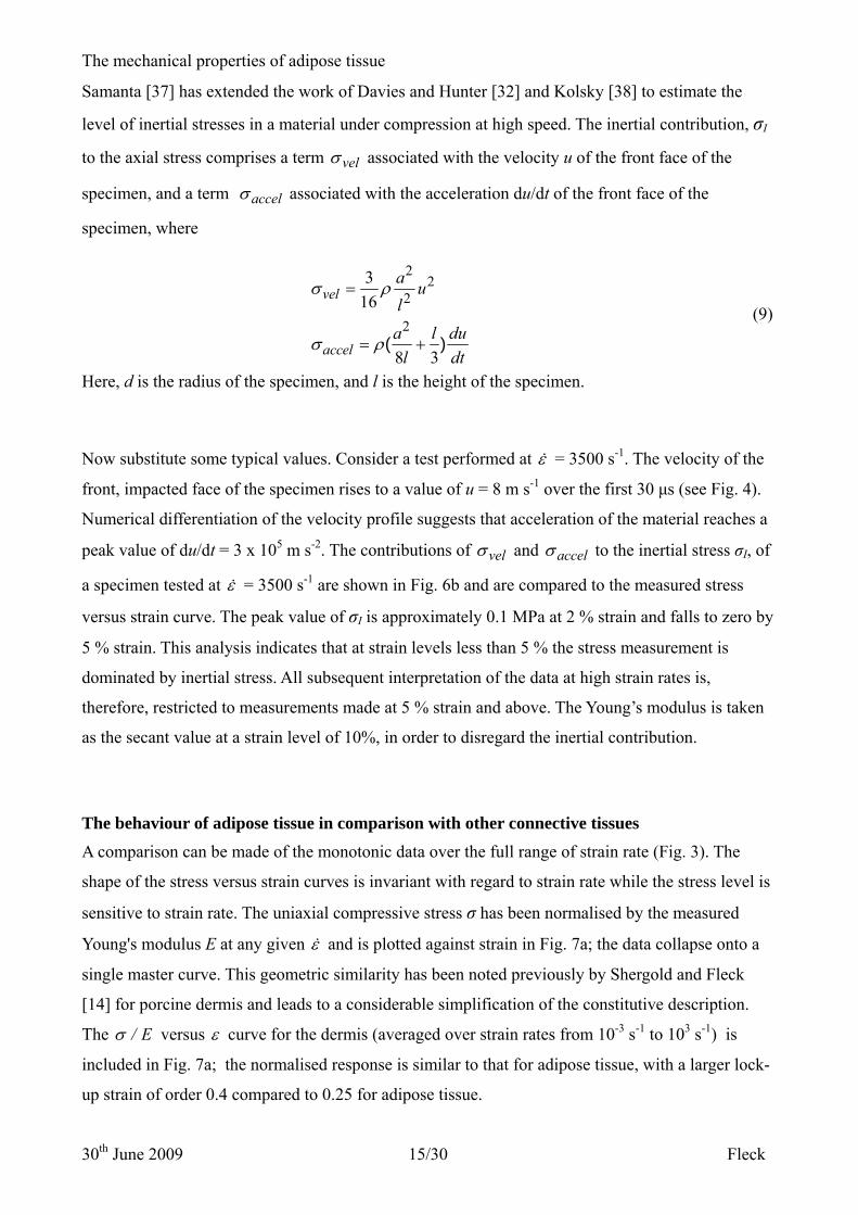
Samanta [37] has extended the work of Davies and Hunter [32] and Kolsky [38] to estimate the
level of inertial stresses in a material under compression at high speed. The inertial contribution, σI
to the axial stress comprises a term velσ associated with the velocity u of the front face of the
specimen, and a term accelσ associated with the acceleration du/dt of the front face of the
specimen, where
22
2
2
316
8 3( )
vel
accel
a ul
a l dul dt
σ ρ
σ ρ
=
= +
(9)
Here, d is the radius of the specimen, and l is the height of the specimen.
Now substitute some typical values. Consider a test performed at ε& = 3500 s-1. The velocity of the
front, impacted face of the specimen rises to a value of u = 8 m s-1 over the first 30 μs (see Fig. 4).
Numerical differentiation of the velocity profile suggests that acceleration of the material reaches a
peak value of du/dt = 3 x 10⁵ m s-2. The contributions of velσ and accelσ to the inertial stress σI, of
a specimen tested at ε& = 3500 s-1 are shown in Fig. 6b and are compared to the measured stress
versus strain curve. The peak value of σI is approximately 0.1 MPa at 2 % strain and falls to zero by
5 % strain. This analysis indicates that at strain levels less than 5 % the stress measurement is
dominated by inertial stress. All subsequent interpretation of the data at high strain rates is,
therefore, restricted to measurements made at 5 % strain and above. The Young’s modulus is taken
as the secant value at a strain level of 10%, in order to disregard the inertial contribution.
The behaviour of adipose tissue in comparison with other connective tissues
A comparison can be made of the monotonic data over the full range of strain rate (Fig. 3). The
shape of the stress versus strain curves is invariant with regard to strain rate while the stress level is
sensitive to strain rate. The uniaxial compressive stress σ has been normalised by the measured
Young's modulus E at any given ε& and is plotted against strain in Fig. 7a; the data collapse onto a
single master curve. This geometric similarity has been noted previously by Shergold and Fleck
[14] for porcine dermis and leads to a considerable simplification of the constitutive description.
The E/σ versus ε curve for the dermis (averaged over strain rates from 10-3 s-1 to 103 s-1) is
included in Fig. 7a; the normalised response is similar to that for adipose tissue, with a larger lock-
up strain of order 0.4 compared to 0.25 for adipose tissue.

The mechanical properties of adipose tissue
30th June 2009 16/30 Fleck
The dependence of E upon ε& is shown in Fig. 7b. For strain rates in the range 2 x 10⁻³ s-2 to 10 s-1
the Young’s modulus E equals approximately 1 kPa and is insensitive to strain rate. This measured
value is in good agreement with previous measurements for adipose tissue, as follows: Samani [3]
used indentation to measure an elastic modulus of 1.9 kPa for breast tissue and Nightingale [39]
measured a value of 5 kPa for human abdominal subcutaneous tissue using acoustic radiation force
impulse imaging (ARFT). For strain rates in the range of 10 s-1 to 5700 s-1 the Young's modulus is
sensitive to strain rate and increases by three orders of magnitude from E = 2 kPa at ε& = 10 s-1 to
approximately E = 4 MPa at ε& = 2000 s-1. A similar sensitivity has been observed for muscle by
Van Sligtenhorst et al. [12] and Song et al. [13]. The dependence of E upon ε& for porcine dermis is
taken from Shergold et al. [14] and is included in Fig. 7b (and Table 2). We conclude that the
dermis is stiffer by more than two orders of magnitude at low strain rates but has a comparable
response at high strain rates.
A constitutive description using the Ogden model
A modified one-term Ogden [17] strain energy density function φ is used to describe the
constitutive behaviour of adipose tissue. The principal values of nominal stress σi (i = 1, 2, 3) are
related to the principle stretches λi by
pdd
ii −=
λφσ
(10)
where p is the hydrostatic pressure.
During uniaxial compression the specimen is assumed to be in a state of uniaxial stress with the z-
axis of a Cartesian co-ordinate system aligned with the direction of the applied load. Therefore,
0== yx σσ (11)For an incompressible solid in which the volume is conserved the principle stretch ratios are related
by
zyx λ
λλ 1== (12)
And λi is related to strain εi, by
ii λε ln−= (13)Therefore, Eq. (1) can be re-written as
The mechanical properties of adipose tissue
30th June 2009 17/30 Fleck
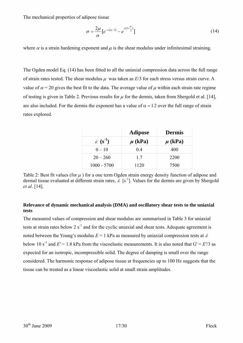
][)()( 2
112 αεαε
αμσ
+−− −= ee (14)
where α is a strain hardening exponent and μ is the shear modulus under infinitesimal straining.
The Ogden model Eq. (14) has been fitted to all the uniaxial compression data across the full range
of strain rates tested. The shear modulus μ was taken as E/3 for each stress versus strain curve. A
value of α = 20 gives the best fit to the data. The average value of μ within each strain rate regime
of testing is given in Table 2. Previous results for μ for the dermis, taken from Shergold et al. [14],
are also included. For the dermis the exponent has a value of α = 12 over the full range of strain
rates explored.
Adipose Dermis
ε& (s-1) μ (kPa) μ (kPa) 0 – 10 0.4 400
20 – 260 1.7 2200 1000 - 5700 1120 7500
Table 2: Best fit values (for μ ) for a one term Ogden strain energy density function of adipose and dermal tissue evaluated at different strain rates, ε& [s-1]. Values for the dermis are given by Shergold et al. [14].
Relevance of dynamic mechanical analysis (DMA) and oscillatory shear tests to the uniaxial tests The measured values of compression and shear modulus are summarised in Table 3 for uniaxial
tests at strain rates below 2 s-1 and for the cyclic uniaxial and shear tests. Adequate agreement is
noted between the Young’s modulus E = 1 kPa as measured by uniaxial compression tests at ε&
below 10 s-1 and E' = 1.8 kPa from the viscoelastic measurements. It is also noted that G' ≈ E'/3 as
expected for an isotropic, incompressible solid. The degree of damping is small over the range
considered. The harmonic response of adipose tissue at frequencies up to 100 Hz suggests that the
tissue can be treated as a linear viscoelastic solid at small strain amplitudes.

The mechanical properties of adipose tissue
30th June 2009 18/30 Fleck
Young's modulus E 1 kPa ± 0.1 kPa Compression modulus E' 1.8 kPa ± 0.8 kPa Shear modulus G' 0.53 kPa ± 0.25 kPa Loss modulus E'' 0.4 kPa ± 0.2 kPa Loss modulus G'' 0.11 kPa ± 0.06 kPa
Table 3: Measured values of compression and shear modulus from uniaxial tests at strain rates
below 2-1 and from cyclic uniaxial and shear tests.
Concluding remarks
The constitutive properties of adipose tissue have been measured over a wide range of strain rate
from 2 x 10⁻³ s-1 to 5700 s-1. The shape of the compressive stress versus strain curve is similar
across all strain rates, differing only by a scale factor which is conveniently given by the Young’s
modulus. This allows for a major simplification in description of the constitutive response. A one
term Ogden strain energy density model can be used to adequately describe the data over the strain
rates tested. This model comprises two parameters, the strain hardening exponent α (independent of
strain rate) and the shear modulus μ (which scales with strain rate). It has also been shown that at
small strain amplitudes, up to frequencies of 100 Hz the tissue behaves as a linear viscoelastic solid.
Acknowledgements
The authors are grateful for financial support by the EPSRC and by Novo Nordisk A/S, Denmark.

The mechanical properties of adipose tissue
30th June 2009 19/30 Fleck
References
[1] Shergold, O. A. and Fleck, N. A., 2004 Mechanisms of deep penetration of soft solids, with
application to the injection and wounding of skin Proc. R. Soc. London ser A, 460, pp. 3037-
3058
[2] Greenwood, M. R. C. and Johnson, P. R., 1983 The adipose tissue, In: Weiss, L.(eds)
Histology, cell and tissue biology, Edition 5, Macmillan
[3] Samani, A., et al., 2003, Measuring the elastic modulus of ex vivo small tissue samples, Phys.
Med. Biol., 48, pp. 2183-2198
[4] Barry, S. I. and Aldis, G. K., 1990, Comparison of models for flow induced deformation of soft
biological tissue, J. Biomech., 23, pp. 647-654
[5] Mow, V. C., et al., 1980 Biphasic creep and stress relaxation of articular cartilage in
compression: theory and experiments, J. Biomech. Eng., Trans. ASME, 102, pp. 73-84
[6] Guyton, A. C., et al. 1966, Interstitial fluid pressure. 3. Its effect on resistance to tissue fluid
mobility, Circ. Res., 19, pp. 412-9
[7] Reddy, N. P. and Cochran, G. V. 1981, Interstitial fluid flow as a factor in decubitus ulcer
formation, J. Biomech., 14, pp. 879-81
[8] Miller-Young, J. E., et al. 2002, Material properties of the human calcaneal fat pad in
compression: Experiment and theory, J. Biomech., 35, pp. 1523-1531
[9] Zheng, Y. P. and Mak, A. F. T. 1999, Extraction of quasi-linear viscoelastic parameters for
lower limb soft tissues from manual indentation experiment, J. Biomech. Eng., 121
[10] Huang, C.-Y., et al. 2005, Anisotropy, inhomogeneity, and tension-compression nonlinearity of
human glenohumeral cartilage in finite deformation, J. Biomech., 38, pp. 799-809
[11] Mansour, J. M., et al. 1993, A method for obtaining repeatable measurements of the tensile
properties of skin at low strain, J. Biomech., 26, pp. 211-6
[12] Van Sligtenhorst, C., et al. 2006, High strain rate compressive properties of bovine muscle
tissue determined using a split Hopkinson bar apparatus, J. Biomech.;39;1852-1858
[13] Song, B., et al. 2007, Dynamic and quasi-static compressive response of porcine muscle, J.
Biomech., 40, pp. 2999-3005
[14] Shergold, O., et al. 2006, The uniaxial stress versus strain response of pig skin and silicone
rubber at low and high strain rates, Int J. Impact Eng., 32,9, pp. 1384-1402

The mechanical properties of adipose tissue
30th June 2009 20/30 Fleck
[15] Purslow, P. 1991 Notch-Sensitivity of Nonlinear Materials J Mat Sci, 26 pp. 4468-4476
[16] Miller, K. and Chinzei, K. 1997, Constitutive modelling of brain tissue: Experiment and
theory, J. Biomech., 30, pp. 1115-1121
[17] Ogden, R. W. 1972, Large Deformation Isotropic Elasticity - Correlation of Theory and
Experiment for Incompressible Rubberlike Solids, Proc. R. Soc London Ser A, 326, pp. 565
[18] Douglas, W. R. 1972, Pigs and Men and Research - Review of Applications and Analogies of
Pig, Sus Scrofa, in Human Medical-Research, Space Life Sciences, 3, pp. 226
[19] Doebelin, E. O. 1966, Measurement systems: Application and design, McGraw-Hill
[20] Fleck, N. A. 1983, Some aspects of clip gauge design, Strain, 19, pp. 17-22
[21] Follansbee, P. S. 1985, The Hopkinson bar, In: H. Kuhn and D. Medlin, E.(eds) ASM
Handbook: Volume 8 mechancial testing and evaluation ASM International
[22] Wang, L. et al. 1992, Generalization of split Hopkinson bar technique to use viscolelastic
bars, Int. J. Impact Eng., 15, pp. 669-686
[23] Zhao, H. et al. 1997, On the use of viscoelastic split Hopkinson pressure bar, Int J. Impact
Eng., 19, pp. 319-330
[24] Sawas, O. et al. 1998, Dynamic characterisation of compliant materials using an all-
polymeric split Hopkinson bar, Exp Mech, pp. 204-210
[25] Deshpande, V. S. and Fleck, N. 2000, High strain rate compressive behaviour of aluminium
alloy foams, Int. J. Impact Eng., 24, pp. 277-298
[26] Kolsky 1953 Stress waves in solids, Oxford
[27] Tagarielli, V. L. et al. 2008, The high strain rate response of PVC foams and end-grain balsa
wood, Composites Part B: Eng. Marine Composites Sandwich Struc., 39, pp. 83-91
[28] Yamashita, J. et al. 2001, The use of dynamic mechanical analysis to assess the viscoelastic
properties of human cortical bone, J Biomed Mater Res, 58, pp. 47-53
[29] Knothe Tate, M. L., R. Steck, et al. (2009), Bone as an inspiration for a novel class of
mechanoactive materials. Biomat 30(2): 133-140
[30] Patel, P. N. et al. 2005, Rheological and recovery properties of poly(ethylene glycol) diacrylate
hydrogels and human adipose tissue, J. Biomed. Mat. Res. Part A, 73A, pp. 313-31
[31] Geerligs, M., G. W. M. Peters, et al. (2008), Linear viscoelastic behavior of subcutaneous
adipose tissue. Biorheol 45(6): 677-688

The mechanical properties of adipose tissue
30th June 2009 21/30 Fleck
[32] Davies, E. D. H. and Hunter, S. C. 1963, The dynamic compression testing of solids by the
method of the split Hopkinson pressure bar, J. Mech. Phys. Solids, 11, pp. 155-179
[33] Saraf, H. et al., 2007, Mechanical properties of soft human tissues under dynamic loading, J.
Biomech., 40, pp. 1960-1967
[34] Pochhammer, L. 1876, J. Reine Angew. Math., 81, pp. 324
[35] Chree, C. 1889, Trans. Camb. Phil. Soc., 14, pp. 250
[36] Bancroft, D. 1941, Phys. Rev., 59, pp. 588
[37] Samanta, S. K. 1971, Dynamic deformation of aluminium and copper at elevated
temperatures, J. Mech. Phys. Solids, 19, pp. 117-122
[38] Kolsky, H. 1949, An investigation of the mechanical properties of materials at very high rates
of loading, Proc. R. Soc. London, Ser. B, 62, pp. 676-700
[39] Nightingale, K. et al. 2003, Shear-wave generation using acoustic radiation force: In vivo and
ex vivo results, Ultrasound Med. Biol., 29, pp. 1715-1723
The mechanical properties of adipose tissue
30th June 2009 22/30 Fleck
The mechanical response of porcine adipose tissue
Kerstyn Comley and Norman Fleck FIGURES
Submitted to the ASME Journal of Biomechanical Engineering
Table 1: Strain wave analysis in polycarbonate bars.
Table 2: Best fit values (for μ ) for a one term Ogden strain energy density function of adipose and
dermal tissue evaluated at different strain rates, ε& (s-1). Values for the dermis are given by Shergold
et al. [14].
Table 3: Measured values of compression and shear modulus for uniaxial tests at strain rates below
2 s-1 and cyclic uniaxial and shear tests.
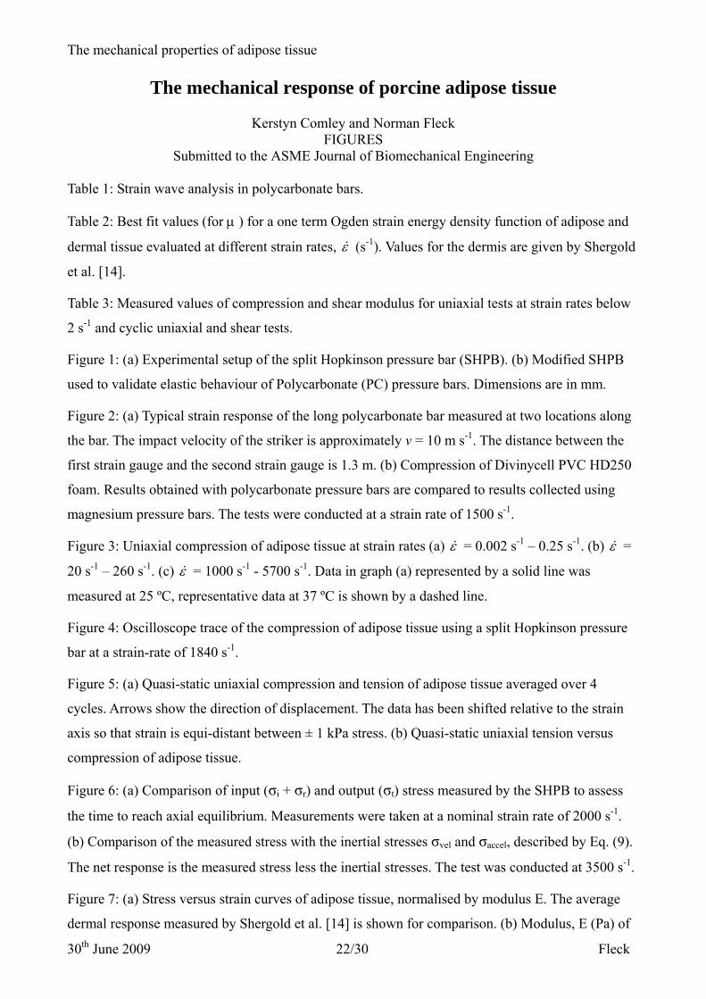
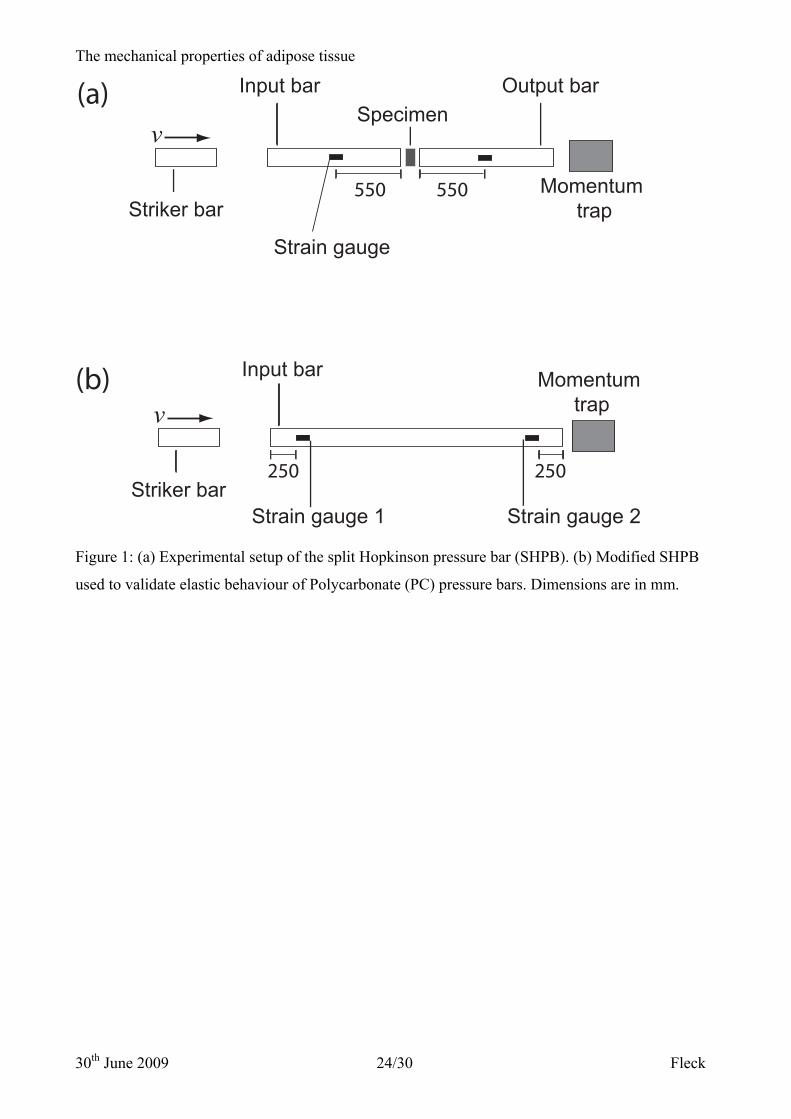
Figure 1: (a) Experimental setup of the split Hopkinson pressure bar (SHPB). (b) Modified SHPB
used to validate elastic behaviour of Polycarbonate (PC) pressure bars. Dimensions are in mm.
Figure 2: (a) Typical strain response of the long polycarbonate bar measured at two locations along
the bar. The impact velocity of the striker is approximately v = 10 m s-1. The distance between the
first strain gauge and the second strain gauge is 1.3 m. (b) Compression of Divinycell PVC HD250
foam. Results obtained with polycarbonate pressure bars are compared to results collected using
magnesium pressure bars. The tests were conducted at a strain rate of 1500 s-1.
Figure 3: Uniaxial compression of adipose tissue at strain rates (a) ε& = 0.002 s-1 – 0.25 s-1. (b) ε& =
20 s-1 – 260 s-1. (c) ε& = 1000 s-1 - 5700 s-1. Data in graph (a) represented by a solid line was
measured at 25 ºC, representative data at 37 ºC is shown by a dashed line.
Figure 4: Oscilloscope trace of the compression of adipose tissue using a split Hopkinson pressure
bar at a strain-rate of 1840 s-1.
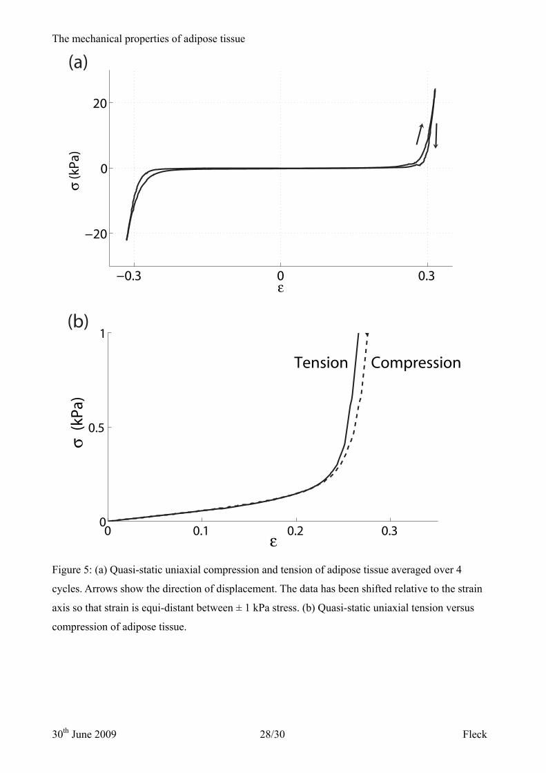
Figure 5: (a) Quasi-static uniaxial compression and tension of adipose tissue averaged over 4
cycles. Arrows show the direction of displacement. The data has been shifted relative to the strain
axis so that strain is equi-distant between ± 1 kPa stress. (b) Quasi-static uniaxial tension versus
compression of adipose tissue.
Figure 6: (a) Comparison of input (σi + σr) and output (σt) stress measured by the SHPB to assess
the time to reach axial equilibrium. Measurements were taken at a nominal strain rate of 2000 s-1.
(b) Comparison of the measured stress with the inertial stresses σvel and σaccel, described by Eq. (9).
The net response is the measured stress less the inertial stresses. The test was conducted at 3500 s-1.
Figure 7: (a) Stress versus strain curves of adipose tissue, normalised by modulus E. The average
dermal response measured by Shergold et al. [14] is shown for comparison. (b) Modulus, E (Pa) of

The mechanical properties of adipose tissue
30th June 2009 23/30 Fleck
adipose tissue under uniaxial compression versus strain rate (s-1). Data shown by triangular markers
were measured using a screw-driven tensile test machine, circular markers by a hydraulic test
machine and the crosses represent data collected using the SHPB. Diagonal crosses represent the
modulus of dermis recorded by Shergold et al. [14].
The mechanical properties of adipose tissue
30th June 2009 24/30 Fleck
Striker bar
Input barSpecimen
Output bar
Momentum trap
v
Strain gauge
550 550
Striker bar
Input bar Momentum trapv
Strain gauge 1
250 250
Strain gauge 2
(a)
(b)
Figure 1: (a) Experimental setup of the split Hopkinson pressure bar (SHPB). (b) Modified SHPB
used to validate elastic behaviour of Polycarbonate (PC) pressure bars. Dimensions are in mm.

The mechanical properties of adipose tissue
30th June 2009 25/30 Fleck
0 0.02 0.04 0.06 0.08 0.1 0.12−5
0
5
10
15
ε
σ [M
Pa]
PC bars
Mg bars
0 0.5 1 1.5−2
0
2
4
6
8
Time [ms]
Stra
in [m
ε]
strain gauge 1 strain gauge 2
(a)
(b)
Figure 2: (a) Typical strain response of the long polycarbonate bar measured at two locations along
the bar. The impact velocity of the striker is approximately v = 10 m s-1. The distance between the
first strain gauge and the second strain gauge is 1.3 m. (b) Compression of Divinycell PVC HD250
foam. Results obtained with polycarbonate pressure bars are compared to results collected using
magnesium pressure bars. The tests were conducted at a strain rate of 1500 s-1.

The mechanical properties of adipose tissue
30th June 2009 26/30 Fleck
Figure 3: Uniaxial compression of adipose tissue at strain rates (a) ε& = 0.002 s-1 – 0.25 s-1. (b) ε& =
20 s-1 – 260 s-1. (c) ε& = 1000 s-1 - 5700 s-1. Data in graph (a) represented by a solid line was
measured at 25 ºC, representative data at 37 ºC is shown by a dashed line.

The mechanical properties of adipose tissue
30th June 2009 27/30 Fleck
0 0.5 1
−4
0
4
Time (ms)
ε (m
ε)
Input bar
Output bar
Figure 4: Oscilloscope trace of the compression of adipose tissue using a split Hopkinson pressure
bar at a strain-rate of 1840 s-1.
The mechanical properties of adipose tissue
30th June 2009 28/30 Fleck
0 0.1 0.2 0.30
0.5
1
ε
σ (k
Pa)
Tension Compression
−0.3 0 0.3
−20
0
20
ε
σ (k
Pa)
(a)
(b)
Figure 5: (a) Quasi-static uniaxial compression and tension of adipose tissue averaged over 4
cycles. Arrows show the direction of displacement. The data has been shifted relative to the strain
axis so that strain is equi-distant between ± 1 kPa stress. (b) Quasi-static uniaxial tension versus
compression of adipose tissue.

The mechanical properties of adipose tissue
30th June 2009 29/30 Fleck
0 0.1 0.2 0.3−0.5
0
0.5
1
1.5
ε
σ (M
Pa)
measured stressnet response
σvel
σaccl
0 100 200 300−1
0
1
2
3
time (μs)
σ (M
Pa)
σi + σr
σt
(a)
(b)
Figure 6: (a) Comparison of input (σi + σr) and output (σt) stress measured by the SHPB to assess
the time to reach axial equilibrium. Measurements were taken at a nominal strain rate of 2000 s-1.
(b) Comparison of the measured stress with the inertial stresses σvel and σaccel, described by Eq. (9).
The net response is the measured stress less the inertial stresses. The test was conducted at 3500 s-1.

The mechanical properties of adipose tissue
30th June 2009 30/30 Fleck
Figure 7: (a) Stress versus strain curves of adipose tissue, normalised by modulus E. The average
dermal response measured by Shergold et al. [14] is shown for comparison. (b) Modulus, E (Pa) of
adipose tissue under uniaxial compression versus strain rate (s-1). Data shown by triangular markers
were measured using a screw-driven tensile test machine, circular markers by a hydraulic test
machine and the crosses represent data collected using the SHPB. Diagonal crosses represent the
modulus of dermis recorded by Shergold et al. [14].
Related Documents