Volume 43 Number 3 July - September 2021 Editor Dr. Tanuj Kanchan Joint Editor Dr. Manish Nigam Publication Quarterly ISSN : 0971 - 0973 e- ISSN : 0974-0848

Welcome message from author
This document is posted to help you gain knowledge. Please leave a comment to let me know what you think about it! Share it to your friends and learn new things together.
Transcript
Volume 43Number 3July - September2021
EditorDr. Tanuj Kanchan
Joint EditorDr. Manish Nigam
Publication QuarterlyISSN : 0971 - 0973e- ISSN : 0974-0848
Journal of Indian Academy of Forensic Medicine(JIAFM)
The Official Publication of Indian Academy of Forensic Medicine
National Advisory BoardDr. A J Patowary (Assam) Dr. Gaurav Sharma (Haryana) Dr. RK Singh (Chhatisgarh)
Dr. A K Srivastava (U.P.) Dr. K. Ravindran (Puducherry) Dr. S K Verma (New Delhi)
Dr. Adarsh Kumar (New Delhi) Dr. K H Chavali (Chattisgarh) Dr. S R Kochar (Rajasthan)
Dr. Aditya Sharma (Himachal Pradesh) Dr. K R Nagesh (Karnataka) Dr. Sanjay Gupta (Gujarat)
Dr. Akhilesh Pathak (Gujarat) Dr. Kusa Kumar Shaha (Puducherry) Dr. Sanjoy Das (Uttarakhand)
Dr. Anil Aggrawal (New Delhi) Dr. L Fimate (Manipur) Dr. S C Mahapatra (Odisha)
Dr. B Shantha Kumar (Tamil Nadu) Dr. M K Mohanty (Odisha) Dr. Shailesh Mohite (Maharashtra)
Dr. B D Gupta (MP) Dr. O P Murty (New Delhi) Dr. S S Oberoi (Punjab)
Dr. C B Jani (Gujarat) Dr. P P Mukhopadhyay (West Bengal) Dr. T K Bose (West Bengal)
Dr. Cyriac Job (Kerala) Dr. Parmod K Goyal (Punjab) Dr. Tulsi Mahto (Jharkhand)
Dr. Dasari Harish (Chandigarh) Dr. Pooja Rastogi (U.P.) Dr. V Khanagwal (Haryana)
Dr. Francis N P Monteiro (Karnataka) Dr. Prateek Rastogi (Karnataka) Dr. V V Pillay (Kerala)
Dr. G Pradeep Kumar (Karnataka) Dr. R S Bangal (Maharashtra) Dr. Yogendra Bansal (Chandigarh)
International Advisory Board
Dr. B L Meel, South Africa Dr. K P Saha, BangladeshDr. B N Yadav, Nepal Dr. K P Shubhakar, UK Dr. Clifford Perera, Sri Lanka Dr. Leandro Duarte De Carvalho, BrazilDr. D N Vieira, Portugal Dr. Magdy A Kharoshah, KSADr. Dan Dermengiu, Romania Dr. Michael S Pollanen, Canada Dr. Derrick J Pounder, UK Dr. Peter Vanezis, UKDr. George Paul, Singapore Dr. R K Gorea, KSADr. Imran Sabri, KSA Dr. Roger W Byard, Australia Dr. John Clark, UK Dr. Serap Annette Akgür, Turkey
Editorial Team
Dr. Raghvendra Singh Shekhawat (AIIMS, Jodhpur)Dr. Vikas P Meshram (AIIMS, Jodhpur)
Editor
Dr. Tanuj KanchanDept. of Forensic Medicine & Toxicology
All India Institute of Medical SciencesJodhpur, Rajasthan
Mobile: +91-9448252394Email: [email protected]
Published by:Dr. Tanuj Kanchan, Editor, JIAFM and Dr. Manish Nigam, Joint Editor, JIAFM
on behalf of the Indian Academy of Forensic Medicine
About the Journal (Print ISSN: 0971-0973 Electronic ISSN:0974-0848): JIAFM is a peer reviewed medical journal published quarterly by the Editor of the Academy on behalf of the Indian Academy of Forensic Medicine.
Aim and Scope of the Journal: The Journal covers all technical, medico-legal and clinical aspects including the ethical and social issues related to the subject specialty of Forensic Medicine and Toxicology and allied specialities. The journal promotes dissemination of original research findings.
Abstracting and Indexing: The journal is included in Scopus, Index Copernicus, IndMED, Index Medicus for South East Asia Region, Indian Citation Index, JIAFM is a UGC Approved Journal (No. 28596). Journal issues are available online at: www.iafmonline.in; http://indmed.nic.in; and www.indianjournals.com
Research ethics and Authorship:JIAFM follows the ICMJE's Recommendations for the Conduct, Reporting, Editing and Publication of Scholarly Work in Medical Journals. JIAFM take issues of copyright infringement, plagiarism or any other act of academic dishonesty very seriously, and encourages the authors to ensure that the submitted manuscripts are their original work and free of any plagiarism.
Copyrights: The entire contents of the JIAFM are protected under Indian and International copyrights. The journal, however, grants to all users a free, irrevocable, worldwide, perpetual right of access to and a license to copy, use, distribute, perform and display the work publicly and to make and distribute derivative works in any digital medium for any reasonable non-commercial purpose, subject to proper attribution of authorship and ownership of the rights. No part of this publication may be reprinted or publish without the prior permission of the Editor, JIAFM. Submission of all manuscripts to the journal is understood to imply that it is not being considered for publication elsewhere. Submission of multi authored papers implies that the consent of each author has been obtained. In this journal, every effort has been made NOT to publish inaccurate or misleading information. However editorial and advisory board accept NO liability in consequences of such statement. The opinions expressed in the articles are those of the authors only.
Subscription Information:JIAFM is published quarterly, and following are its annual subscription rates:Individual: ₹1000 (In India) and USD 200 or equivalent (Rest of the world)Institutions: ₹7500 (In India) and USD 400 or equivalent (Rest of the world)Subscription orders and payments should be made in favour of “Editor IAFM”, payable at Jodhpur, Rajasthan. All communications in this regard should be made with the Editor at the address given below.
Claims for missing issue(s):A copy will be sent free to the member/ subscriber provided the claim is made within 2 months of publication of the issue & a self-addressed envelope of the size 9” x 12” is sent to the Editor. (Those who want the journal to be dispatched by 'Registered Post' must affix postage stamps of ₹ 50).
Editorial OfficeDr. Tanuj Kanchan (Editor, JIAFM)Room No. 3050,Department of Forensic Medicine & ToxicologyAll India Institute of Medical Sciences, JodhpurBasni Industrial Area, Phase-2, Jodhpur-342005, RajasthanMobile: +91-9448252394Email: [email protected]
The Journal of Indian Academy of Forensic Medicine (JIAFM)
Contents
Editorial Postmortem biochemistry: Current perspectives and the road ahead 196-197Raghvendra Singh Shekhawat, Vikas Meshram, Tanuj Kanchan
Original ArticlesValidation of age-related changes in contusions by gross examination and objective analyses 198-203Nisha Nandakumar, KPrasannan, NishaTR
A comprehensive study on infection control and liquid waste management in mortuaries 204-208Mopuri Venkateswarlu, T. Mohit Kumar Moses, Kattamreddy Ananth Rupesh, G.Chandra Deepak, G. Janaki Ramudu
Variations in position of mandibular foramen with age and its efficacy in sex estimation 209-211Deepali P Mohite, Prakash M Mohite, Alka H Hande, Devendra Palve
Seroprevalence of human immunodeficiency virus, hepatitis B virus, and hepatitis C virus 212-217among the forensic autopsy cases in South India Jamshid Parakkattil, Vinod Ashok Chaudhari, Ambika Prasad Patra, Rakesh Singh, Rahul Dhodapkar
The pattern of homicide in Haryana – A retrospective study 218-220 Naveen Yadav, Jitender Kumar Jakhar, Gaurav Kaushik, Lait Chopra, Mahender Singh, SK Dhattarwal
Socio-demographic determinants of victims of sexual assault in Mumbai 221-226 Arun Kumar Jaiswani, Rajesh C Dere, Narendra B Kumar, Hemant G Kukde
Profile of medico-legal autopsy cases performed during COVID-19 pandemic lockdown 227-231 at mortuary of Civil Hospital and B. J. Medical College in Ahmedabad, Gujarat Patel Ankur P, Vaghela Raghurajsinh D, Trivedi Jayjeet M, Madhavi Ajay R
An analysis of medicolegal autopsies in a tertiary care centre in West Bengal - 232-234A morgue-based study Shobhan Roy, Shagun Thakur, Sumanta Malick, Vikas Gurbani
ndPerception of 2 year MBBS students about online zoom classes during COVID-19 pandemic: 235-237A questionnaire-based study Amit Kumar Singh, Anju Singh
238-241Association between suicides among females and phase of uterine cycle during autopsy at a tertiary care centre in Bengaluru north Udaya Shankar B S, Sujatha P L, Shivakumar B C, Vijaya C
The toxicological profile of acute phosphorus poisoning 242-245Surangama Chakraborty, Rajan S
Journal of Indian Academy of Forensic Medicine
Volume 43 Number 3 July - September 2021
246-248A comprehensive study on insecticide poisoning patients brought to a tertiary government hospital in north eastern region of India Antara Debbarma, Juthika Debbarma
Autopsy analysis of craniocerebral injuries at a tertiary healthcare centre 249-253Bandu Waman Ramteke, Shibanand Nepal Karmakar, Nilesh Keshav Tumram
Estimation of time since death from morphological changes in red blood cells of human 2 54-257cadaver: An autopsy-based studyAditya Anand, K.K. Banerjee, Anil Kohli, Vinod Kumar Arora
Investigation of sexing accuracy of second and seventh cervical vertebras in adult Iranian 258-264population by using CT scan imagesReza Saadat Mostafavi, Azadeh Memarian, Arezoo Amiri, Omid Motamedi
Review ArticleMental Healthcare Act (MHCA 2017)–A review from Forensic perspective 265-268 Sravan J S, Atul S. Keche, Vivek Kumar Chouksey, Poovaragavan V
Medical Certification of Cause of Death (MCCD) with special reference to deaths due 269-272to Coronavirus Disease 2019 (COVID–19) Toshal Wankhade, Manish Shrigiriwar, Mandar Ramchandra Sane
Case ReportsLadder-rung tears of aorta - An unusual presentation of medico-legal significance 273-275 Vikas Meshram, Ashish Saraf, Vaibhav Gupta, Tanuj Kanchan, Raghvendra Singh Shekhawat
Fatal colorectal injury by compressed air through anal insufflation 276-277 Mohd Kaleem Khan, Kashif Ali
A second autopsy revealed the silent killer: A case of fatal carbon monoxide poisoning 278-280 from gas geyser Dipak H. Vora, Tikendra Dewangan, Prabhakaran S, Kalpesh A. Shah, Dharmesh S. Patel, H.T.Khubchandani
A fatal case of self-inflicted abdominal stab wound 281-283 Mitul M Sangma, AJ Patowary, Daunipaia Slong
Fatality due to unusual femoral vein rupture in an injectable drug user 284-287 Abhishek Yadav, Aravindan V, Abilash S, Sudhir K Gupta
Firearm Injuries-Unusual cases 288-291 Mandar Karmarkar, Juthikaa Deherkar
PerspectivePediatric Forensic Examination in Domestic Violence cases- Problems and solutions thereof in global perspective 292-295 Bondarchuk Hanna, Gunas Valery, Perebetyuk Anatoliy , Fomina Lyudmila, Adarsh Kumar
Clothes and the evidences they carry: A perspective on its forensic examination 296-299 Varsha Warrier, Tanuj Kanchan
Volume 43 Number 3 July - September 2021
Postmortem biochemistry: Current perspectives and the road ahead
Thanato-biochemistry or postmortem biochemistry is a comprehensive speciality study of biochemistry in which fluids and tissues retrieved from the human corpse are subjected to qualitative and quantitative estimation of desirable biochemical parameters. The field of postmortem biochemistry has started evolving in the last few decades. The conventional autopsy practice relies on the morphological findings observed during necropsy. At many occasions, the aid of pathology and forensic science laboratories is taken for histochemical examination and chemical analysis, respectively. The analysis of the biochemical parameters from the post mortem body samples can offer intensive help in assessing the derangements in the functional, biological and molecular parameters, which are not observed in the traditional method of autopsy conduction.
There are various forensically significant conditions, including myocardial ischemia, sepsis, inflammation, infection, anaphylaxis, where the biochemical analysis of specimens from the dead bodies can aid in the determination of the cause of
1death. The presence of beta-hydroxybutyrate and acetone in the post mortem fluid samples can be a valued finding in deaths
2, 3related to alcohol intoxication and diabetes.
Depending upon the situation, the sample requirements may include a variety of body fluids and tissues like heart-whole blood (right ventricle), peripheral whole blood, jugular vein whole blood, pericardial fluid gastric contents, urine vitreous humor, cerebrospinal fluid, synovial fluid, liver/other tissues and scene residues. Samples like heart-whole blood and jugular vein whole blood can only be subjected to qualitative toxicological analysis. The vitreous humor sample finds a special mention in the literature and is the preferred sample compared to the blood samples. The vitreous fluid is relatively less affected by the changes of decomposition. Additionally, due to the anatomical protection of eye sockets, vitreous fluid is less susceptible to the issues of microbial contamination and diffusion of analytes from the abdominal and thoracic cavities. Other than the postmortem interval estimation, the vitreous humour fluid has been utilized in postmortem diagnosis of
4saltwater drowning, heat shock and chronic alcohol abuse. Interestingly, recently, the vitreous humor fluid has been studied
5to estimate postmortem interval at the crime scene.
The postmortem biomarkers may vary significantly between cases due to various factors such as pre-existent illnesses, the cause of mortality, complications, the period of survival, and cadaveric changes in the distribution and localization of
6analytes. The issues like contamination, late recovery, and hemolysis remain to pose trouble for interpreting results. Additionally, most of the reference values of analytes are those from the live subjects and not for the hemolyzed samples retained from the dead individuals. Samples like vitreous humour, pericardial fluid, etc., are seldom available from the live subjects to determine their reference values which is a key
7to method validation. The qualitative interpretation of endogenous biochemical substrates can become more challenging in forensic autopsies where there is the involvement of xenobiotics like sodium chloride, insulin and various poisons, which may mimic the chemical constitution of
8endogenous substances.
The recent work on thanatobiochemistry has concentrated on various biomarkers like C reactive proteins, Ferritin, T3 (fT3), T4 (fT4), Thyroglobulin, S100 calcium-binding protein B, Neuron-Specific Enolase, GFAP, Glial fibrillary acidic protein, Human liver-type fatty acid-binding protein, and catecholamines for qualitative evaluations in deaths associated
9with short and long agonal periods. Newer diagnostic advancements like electrochemiluminescent immunoassay, lateral flow immunoassay, inductively coupled plasma-mass spectrometry, liquid-chromatography-mass spectrometry, l ow/h igh- reso lu t ion l iqu id ch romatography-mass spectrometryography-mass spectrometry, hollow fibre liquid-phase microextraction coupled with liquid chromatography, mass spectrometry, electrochemiluminometric assay, chemiluminescence immunoassay, CD-linked antibody immunosorbent assay, etc have introduced a new revolution in the postmortem quantitative estimation of analytes.
Since the infrastructural requirements for qualitative and quantitative analysis of postmortem samples require both machines and human resources, intradepartmental collaboration plays a vital role. The need of the hour is to do more research in postmortem biochemistry, which will help in availing credible postmortem reference values of biological samples.
References1. Han SQ, Qin ZQ, Deng KF, Zhang JH, Liu NG, Zou DH, et al.
[Research Advances in Postmortem Chemistry]. Fa Yi Xue Za Zhi. 2015;31(4):287-92, 97
2. Palmiere C, Mangin P, Werner D. Postmortem distribution of 3-
196
Corresponding Author
Tanuj Kanchan (Editor-in-chief; Journal of Indian Academy of Forensic Medicine)
Email: [email protected], [email protected]
Mobile: +91 9448252394
Raghvendra Singh Shekhawat, Vikas Meshram, Tanuj KanchanDepartment of Forensic Medicine and Toxicology, All India Institute of Medical Sciences, Jodhpur, India
EDITORIAL
J Indian Acad Forensic Med. 2021 Jul-Sep; 43(3): 196-197doi:10.5958/0974-0848.2021.00050.6
beta-hydroxybutyrate. J Forensic Sci. 2014;59(1):161-6.10.1111/1556-4029.12265
3. Palmiere C. Postmortem diagnosis of diabetes mellitus and its c o m p l i c a t i o n s . C r o a t M e d J . 2 0 1 5 ; 5 6 ( 3 ) : 1 8 1 -93.10.3325/cmj.2015.56.181
4. Donaldson AE, Lamont IL. Biochemistry changes that occur after death: potential markers for determining post-mortem interval. PloS one. 2013;8(11): e82011-e.10.1371/journal.pone.0082011
5. Musile G, Agard Y, De Palo EF, Shestakova K, Bortolotti F, Tagliaro F. Thanatochemistry at the crime scene: a microfluidic paper-based device for ammonium analysis in the vitreous humor. Anal Chim Acta. 2019;1083: 150-6.10.1016/j.aca.2019.07.033
6. Maeda H, Zhu BL, Ishikawa T, Quan L, Michiue T. Significance of postmortem biochemistry in determining the cause of death. Leg
M e d ( T o k y o ) . 2 0 0 9 ; 1 1 S u p p l 1 : S 4 6 -9.10.1016/j.legalmed.2009.01.048
7. Belsey SL, Flanagan RJ. Postmortem biochemistry: Current applications. J Forensic Leg Med. 2016;41: 49-57.10.1016/ j.jflm.2016.04.011
8. Shekhawat RS, Rathore M. Proceedings: 15th Asia-Pacific Federation for Clinical Biochemistry and Laboratory Medicine (APFCB) Congress 2019 from 17-20 November, 2019, Jaipur, India. Indian Journal of Clinical Biochemistry. 2019;34(1):1-233.10.1007/s12291-019-00859-4
9. Rosato E, Bonelli M, Locatelli M, de Grazia U, Tartaglia A, Savini F, et al. Forensic Biochemical Markers to Evaluate the Agonal P e r i o d : A L i t e r a t u r e R e v i e w. M o l e c u l e s . 2 0 2 1 ; 26(11).10.3390/molecules26113259
197
J Indian Acad Forensic Med. 2021 Jul-Sep; 43(3)
AbstractThe determination of age of injuries has been a longstanding issue in Forensic Medicine. There is paucity of work in this field and standardized methodology. Estimation of age of wounds by visual inspection alone is subjective and susceptible to variation in perception. This study intends to record, document and interpret the age of wounds from available history, gross examination by naked eye and results of objective analyses by magnified digital photograph, examination under Wood's lamp and histological evaluation, to devise a method for retrospective evaluation of the age of contusions. This is an autopsy based prospective study for a period of 1year, involving 50 consecutive cases of contusions, conducted on dead bodies brought to the Department of Forensic Medicine. The data obtained was analyzed by SPSS v18. Comparison of different components, significance of association, level of correlation between various variables were determined, and sensitivity and specificity of various methods of analysis in determining the age of wounds was established. On gross examination, contusions were predominantly red when <24hours old, bluish black on day2,a greenish colour appeared at the earliest on day 3,and yellow on day 7. There was co-existence of yellow and green colours on 8-9days and all contusions on day10 were yellowish. There was positive correlation between the period of survival with histopathological findings and also with colour by magnification of digital photograph which increased till 5-6 days. The association between colour of contusion could be established precisely when examined under Wood's lamp illumination and survival period reached maximum on 5-6days. Histology of contusions <24hours showed red blood cells, day2 showed neutrophils, lymphocytes on day 3, macrophages from day 4, pigments from day 5, collagen fibres from 6 days, complete re-epithelisation from day 7, fibroblasts from day 8, which increased in density on day 9 and10.The age of contusions was determined, and sensitivity and specificity of various methods were assessed. It was concluded that an array of subjective and objective analyses can be used to establish the age of wound.
KeywordsWound age; Contusions; Gross examination; Digital photography; Histopathology; Ultraviolet; Wood's-lamp-illumination
Validation of age-related changes in contusions by gross examination and objective analyses
J Indian Acad Forensic Med. 2021 Jul-Sep; 43(3): 198-203doi:10.5958/0974-0848.2021.00051.8
ORIGINAL ARTICLE
Introduction
The evaluation of any tissue injury is an essential component in the practice of Forensic Medicine whereby furnishing a final word on the age of injuries in cases such as assault, abuse etc. and the interpretation has significant medicolegal implications which may include the incrimination or exclusion of a suspect as the perpetrator of a crime, time of occurrence of the event or crime and if possible discriminate if all injuries found on the body may not have been inflicted by the same assailant or even at the same time. Estimation of age of wound by visual inspection alone is subjective and susceptible to variation in perception, but previous studies have shown that it may be possible to determine the age of wound by complementing direct observation with other objective analyses.
Materials and Methods
The objectives of the study were to estimate the time of contusions by gross examination, Wood's lamp examination, magnification of digital photography and histopathological changes and to validate the age of contusion obtained by above methods with the age of injury as per available history.
The current study is an autopsy based prospective study conducted on dead bodies brought to the department of Forensic Medicine, over a period of 1year spanning from February 2015 to February 2016. The study involves 50 consecutive cases of contusions of known age.
The inclusion criteria was injuries with known age as recorded in the inquest and the exclusion criteria was dead bodies in a state of decomposition and cases where there is no definite history regarding the age of the injury or its time of infliction.
A diagnostic evaluation of contusion on the dead bodies in which the time of sustaining the injury is known through inquest details and treatment records and correlating the time since injury to the findings from different methods of analyses was done.
The gross changes of the surface injuries, namely contusions, were observed and documented. The injuries were then
198
Corresponding Author
Nisha Nandakumar (Assistant Professor)
Email: [email protected]
Mobile: +91-9895584681
Article Historyth thReceived: 4 September, 2020; Revision received on: 16 June, 2021
thAccepted: 10 July, 2021
1 2 3Nisha Nandakumar , KPrasannan , NishaTR1 Department of Forensic Medicine and Toxicology, KMCT Medical College, Manassery, Calicut, Kerala2 Department of Forensic Medicine and Toxicology, Government Medical College Calicut, Kerala3 Department of Pathology, Government Medical College Alapuzha, Kerala
photographed by digital camera and magnified 3times and details were evaluated. Conventional images are sometimes impaired by spurious light reflectance from skin caused by electronic flash. So, examination under Wood's lamp illumination was done.
The apparatus used was Gadget's 60X Magnifying Loupe with LED and UV-A light No:9592 which uses UV-A light of wavelength 320-400nm(~365nm). Wood's lamp is UV-A lamp which helps in clearly visualizing the skin lesion and injuries thereby helps in deciphering the unclear injuries and the margins. The peripheral portion from the margin of the contusion was excised in full depth and subjected to histopathology examination to assess the age of the wound.
The data obtained were analysed using SPSS v18. The frequency of occurrence of different variables were obtained and classified into groups. The cross tabulations were prepared for comparison of different variables to be assessed simultaneously. Comparison of the different components were done by statistical methods and interpreted. The significance of association(p value ≤0.05) was estimated for all by Chi square test and Kappa agreement test was applied to determine the level of correlation between various variables under study. Sensitivity and specificity of various methods of analyses to determine the age of wounds were established.
Results
Survival period frequency has been depicted in Table 1. Table 2 shows the comparison of contusion colours observed using different methodologies. On gross examination,75% of contusions with survival period <24hours were red in colour. All the 2-day old and 83.3% 3-day old contusions were bluish or bluish black in colour. 66.7% of 4days old,100% of 5days old,75% and 66.7% of 6days and 7days old contusions were greenish. The earliest appearance of greenish colour was on day3 and of yellowish colour was on day7after sustaining injury.8days old contusions showed bluish, greenish and yellowish discoloration(33.3% each) on gross examination. 9days old contusions were greenish(50%) and yellowish(50%) in colour and 100% of contusions with period of survival of 10days or more were yellowish
Correlating colour of magnified digital photograph and survival period, 75% of the contusions with survival period of 1day were reddish and all 2days old contusions showed bluish black colour. 33.3% of 3days old, 66.6% of 4days old and 80% of 5days old contusions were greenish. 83.3% of 7days old contusions were greenish and 16.7% were yellowish in colour. 66.7% of 8days old contusions were greenish.75% contusions on day9 and 100% of contusions with 10days or more survival period showed yellowish colour. The earliest appreciation of greenish colour in a contusion was on day2, and yellow was on day7.The agreement between the colour of the wound by magnification of digital
photograph with that of survival period was maximum(88.9%) at 5-6days then declined. Kappa agreement test showed a moderate agreement(0.527).In the current study, the exact colour of the contusion could be precisely madeout by Wood's lamp examination where 2% and 4% of greenish contusions were bluish black and yellowish respectively. The correlation between colour of wound by Wood's lamp examination and survival
J Indian Acad Forensic Med. 2021 Jul-Sep; 43(3)
199
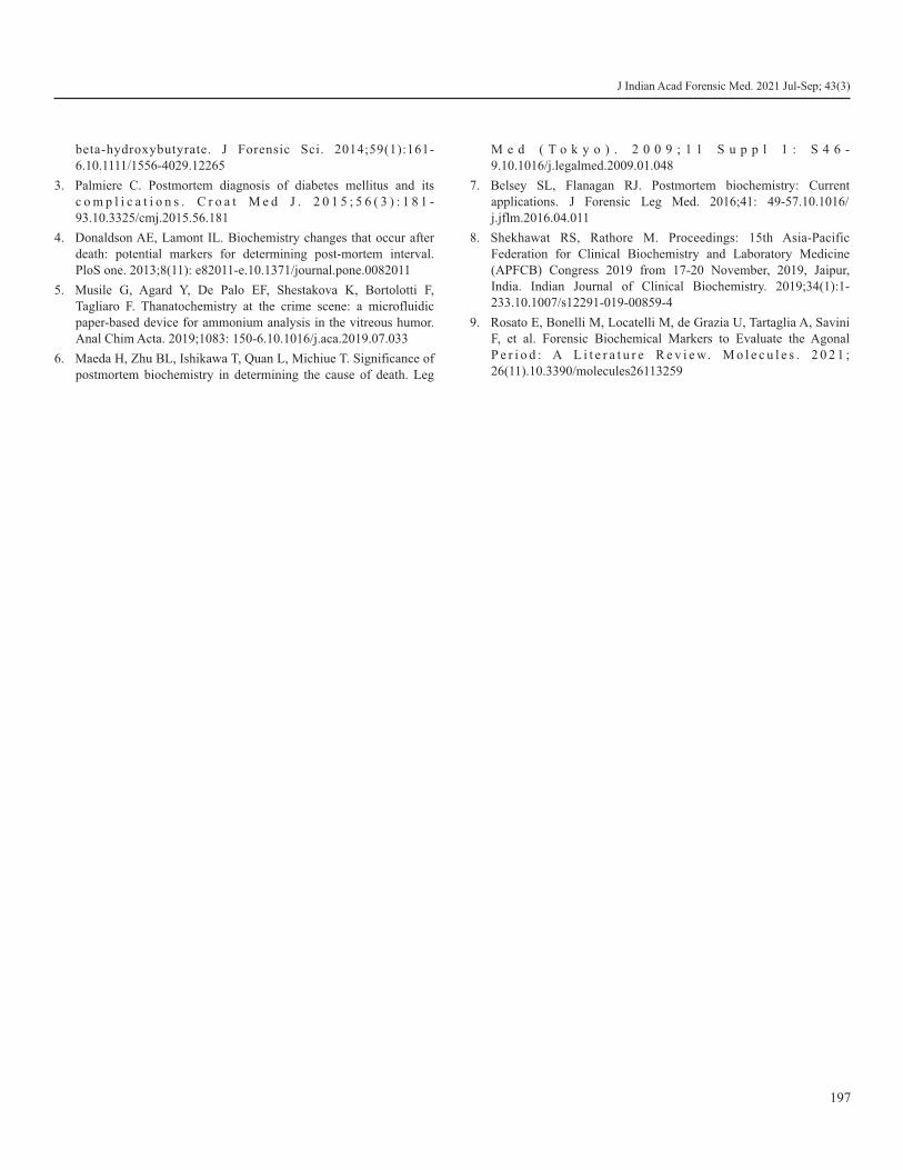
Figure 1: Red Contusion
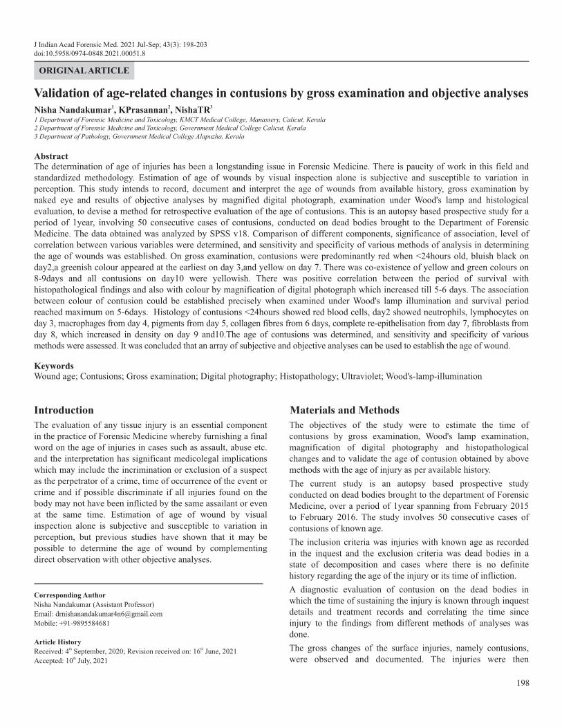
Figure 2: Bluish black contusion
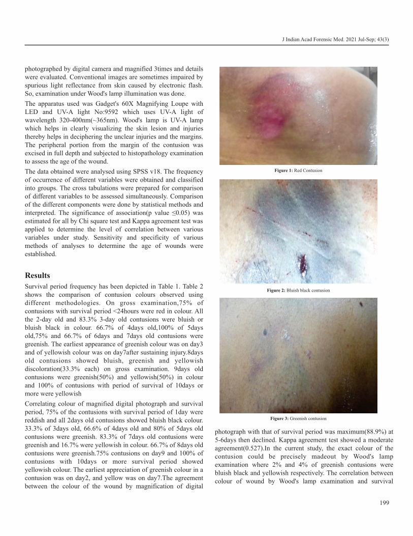
Figure 3: Greenish contusion
period was 100% at 5-6days and then declined. Kappa agreement test showed good agreement(0.627).
Histological analysis of contusions for correlation with survival period showed that 87.5% of contusions with survival <24hours
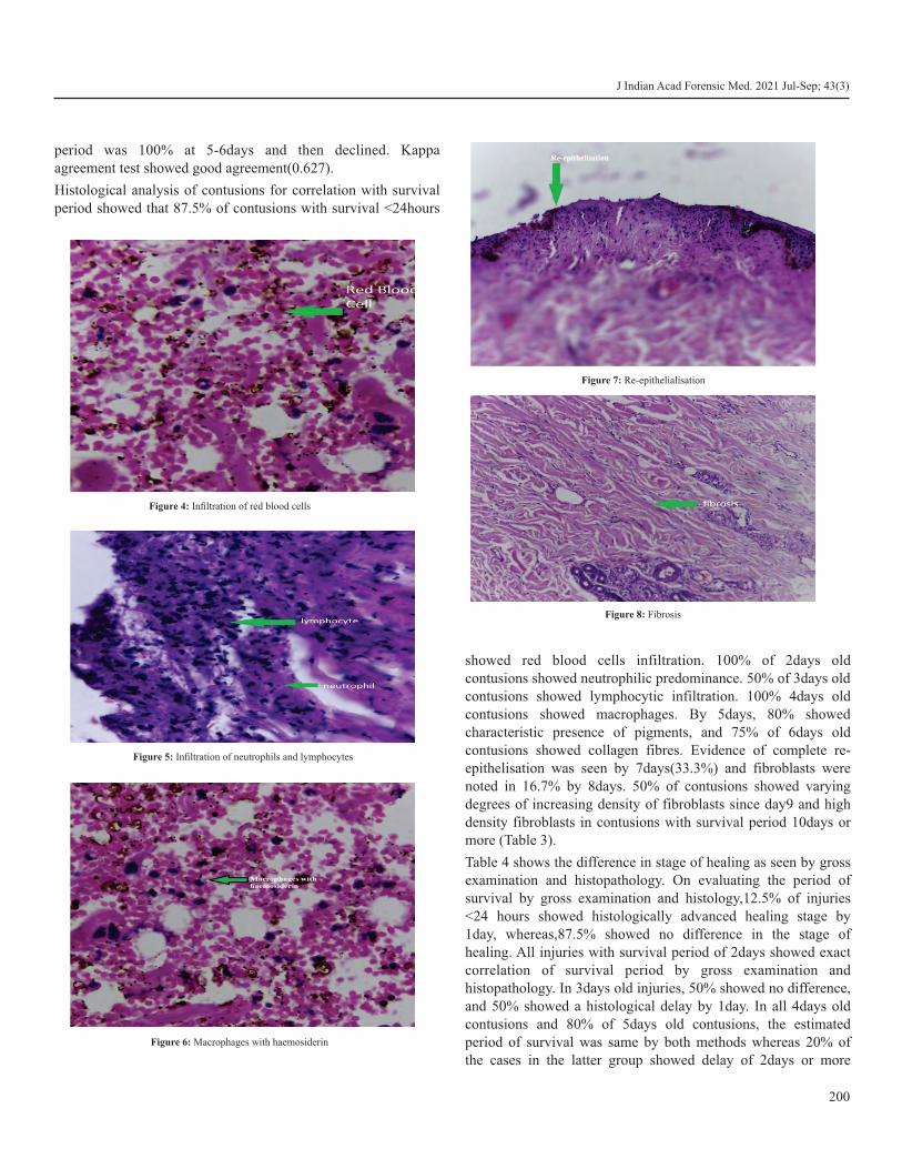
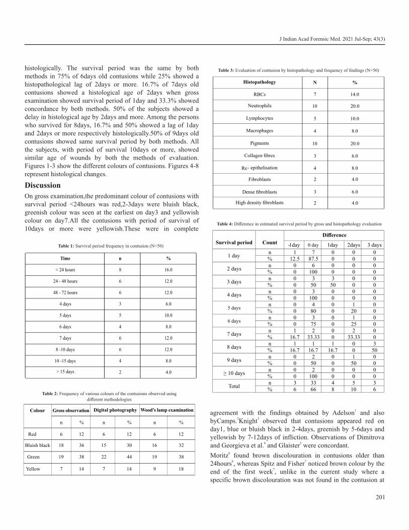
showed red blood cells infiltration. 100% of 2days old contusions showed neutrophilic predominance. 50% of 3days old contusions showed lymphocytic infiltration. 100% 4days old contusions showed macrophages. By 5days, 80% showed characteristic presence of pigments, and 75% of 6days old contusions showed collagen fibres. Evidence of complete re-epithelisation was seen by 7days(33.3%) and fibroblasts were noted in 16.7% by 8days. 50% of contusions showed varying degrees of increasing density of fibroblasts since day9 and high density fibroblasts in contusions with survival period 10days or more (Table 3).
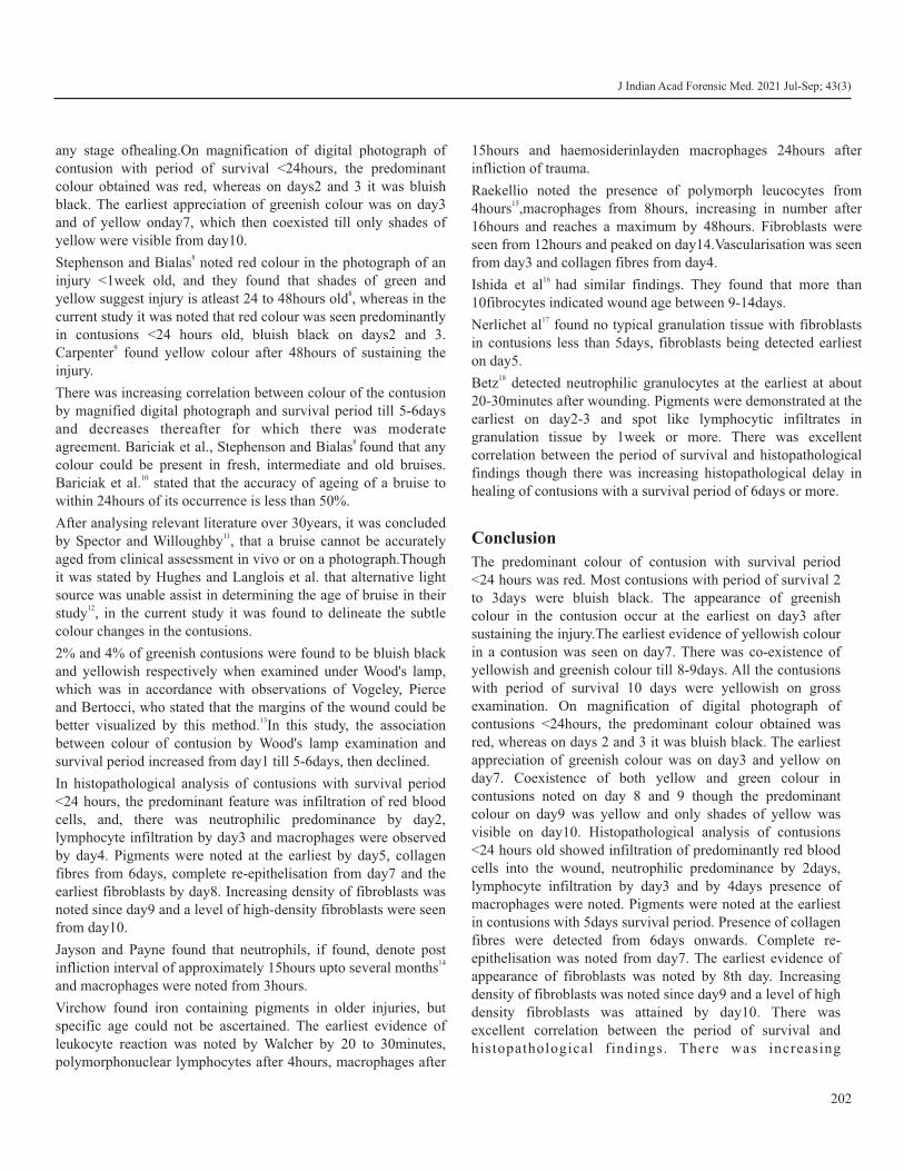
Table 4 shows the difference in stage of healing as seen by gross examination and histopathology. On evaluating the period of survival by gross examination and histology,12.5% of injuries <24 hours showed histologically advanced healing stage by 1day, whereas,87.5% showed no difference in the stage of healing. All injuries with survival period of 2days showed exact correlation of survival period by gross examination and histopathology. In 3days old injuries, 50% showed no difference, and 50% showed a histological delay by 1day. In all 4days old contusions and 80% of 5days old contusions, the estimated period of survival was same by both methods whereas 20% of the cases in the latter group showed delay of 2days or more
J Indian Acad Forensic Med. 2021 Jul-Sep; 43(3)
Figure 7: Re-epithelialisation
Figure 8: Fibrosis
200
histologically. The survival period was the same by both methods in 75% of 6days old contusions while 25% showed a histopathological lag of 2days or more. 16.7% of 7days old contusions showed a histological age of 2days when gross examination showed survival period of 1day and 33.3% showed concordance by both methods. 50% of the subjects showed a delay in histological age by 2days and more. Among the persons who survived for 8days, 16.7% and 50% showed a lag of 1day and 2days or more respectively histologically.50% of 9days old contusions showed same survival period by both methods. All the subjects, with period of survival 10days or more, showed similar age of wounds by both the methods of evaluation. Figures 1-3 show the different colours of contusions. Figures 4-8 represent histological changes.
Discussion
On gross examination,the predominant colour of contusions with survival period <24hours was red,2-3days were bluish black, greenish colour was seen at the earliest on day3 and yellowish colour on day7.All the contusions with period of survival of 10days or more were yellowish.These were in complete
1agreement with the findings obtained by Adelson and also 2 3byCamps. Knight observed that contusions appeared red on
day1, blue or bluish black in 2-4days, greenish by 5-6days and yellowish by 7-12days of infliction. Observations of Dimitrova
4 5and Georgieva et al. and Glaister were concordant.6Moritz found brown discolouration in contusions older than
6 724hours , whereas Spitz and Fisher noticed brown colour by the 7end of the first week , unlike in the current study where a
specific brown discolouration was not found in the contusion at
J Indian Acad Forensic Med. 2021 Jul-Sep; 43(3)
201
Time n %
< 24 hours 8 16.0
24 - 48 hours 6 12.0
48 - 72 hours 6 12.0
4 days 3 6.0
5 days 5 10.0
6 days 4 8.0
7 days 6 12.0
8 -10 days 6 12.0
10 -15 days 4 8.0
> 15 days 2 4.0
Table 1: Survival period frequency in contusion (N=50)
Colour Gross observation Digital photography Wood’s lamp examination
n % n % n %
Red 6 12 6 12 6 12
Bluish black 18 36 15 30 16 32
Green 19 38 22 44 19 38
Yellow 7 14 7 14 9 18
Table 2: Frequency of various colours of the contusions observed using different methodologies
Histopathology N %
RBCs 7 14.0
Neutrophils 10 20.0
Lymphocytes 5 10.0
Macrophages 4 8.0
Pigments 10 20.0
Collagen fibres 3 6.0
Re- epithelisation 4 8.0
Fibroblasts 2 4.0
Dense fibroblasts 3 6.0
High density fibroblasts 2 4.0
Table 3: Evaluation of contusion by histopathology and frequency of findings (N=50)
Survival period Count
Difference
-1day 0 day 1day 2days 3 days
1 dayn 1 7 0 0 0% 12.5 87.5 0 0 0
2 daysn 0 6 0 0 0% 0 100 0 0 0
3 daysn 0 3 3 0 0% 0 50 50 0 0
4 daysn 0 3 0 0 0% 0 100 0 0 0
5 daysn 0 4 0 1 0% 0 80 0 20 0
6 daysn 0 3 0 1 0% 0 75 0 25 0
7 daysn 1 2 0 2 0% 16.7 33.33 0 33.33 0
8 daysn 1 1 1 0 3% 16.7 16.7 16.7 0 50
9 daysn 0 2 0 1 0% 0 50 0 50 0
≥ 10 daysn 0 2 0 0 0% 0 100 0 0 0
Totaln 3 33 4 5 3% 6 66 8 10 6
Table 4: Difference in estimated survival period by gross and histopathology evaluation
any stage ofhealing.On magnification of digital photograph of contusion with period of survival <24hours, the predominant colour obtained was red, whereas on days2 and 3 it was bluish black. The earliest appreciation of greenish colour was on day3 and of yellow onday7, which then coexisted till only shades of yellow were visible from day10.
8Stephenson and Bialas noted red colour in the photograph of an injury <1week old, and they found that shades of green and
8yellow suggest injury is atleast 24 to 48hours old , whereas in the current study it was noted that red colour was seen predominantly in contusions <24 hours old, bluish black on days2 and 3.
9Carpenter found yellow colour after 48hours of sustaining the injury.
There was increasing correlation between colour of the contusion by magnified digital photograph and survival period till 5-6days and decreases thereafter for which there was moderate
8 agreement. Bariciak et al., Stephenson and Bialas found that any colour could be present in fresh, intermediate and old bruises.
10Bariciak et al. stated that the accuracy of ageing of a bruise to within 24hours of its occurrence is less than 50%.
After analysing relevant literature over 30years, it was concluded 11by Spector and Willoughby , that a bruise cannot be accurately
aged from clinical assessment in vivo or on a photograph.Though it was stated by Hughes and Langlois et al. that alternative light source was unable assist in determining the age of bruise in their
12study , in the current study it was found to delineate the subtle colour changes in the contusions.
2% and 4% of greenish contusions were found to be bluish black and yellowish respectively when examined under Wood's lamp, which was in accordance with observations of Vogeley, Pierce and Bertocci, who stated that the margins of the wound could be
13better visualized by this method. In this study, the association between colour of contusion by Wood's lamp examination and survival period increased from day1 till 5-6days, then declined.
In histopathological analysis of contusions with survival period <24 hours, the predominant feature was infiltration of red blood cells, and, there was neutrophilic predominance by day2, lymphocyte infiltration by day3 and macrophages were observed by day4. Pigments were noted at the earliest by day5, collagen fibres from 6days, complete re-epithelisation from day7 and the earliest fibroblasts by day8. Increasing density of fibroblasts was noted since day9 and a level of high-density fibroblasts were seen from day10.
Jayson and Payne found that neutrophils, if found, denote post 14infliction interval of approximately 15hours upto several months
and macrophages were noted from 3hours.
Virchow found iron containing pigments in older injuries, but specific age could not be ascertained. The earliest evidence of leukocyte reaction was noted by Walcher by 20 to 30minutes, polymorphonuclear lymphocytes after 4hours, macrophages after
15hours and haemosiderinlayden macrophages 24hours after infliction of trauma.
Raekellio noted the presence of polymorph leucocytes from 154hours ,macrophages from 8hours, increasing in number after
16hours and reaches a maximum by 48hours. Fibroblasts were seen from 12hours and peaked on day14.Vascularisation was seen from day3 and collagen fibres from day4.
16 Ishida et al had similar findings. They found that more than 10fibrocytes indicated wound age between 9-14days.
17Nerlichet al found no typical granulation tissue with fibroblasts in contusions less than 5days, fibroblasts being detected earliest on day5.
18Betz detected neutrophilic granulocytes at the earliest at about 20-30minutes after wounding. Pigments were demonstrated at the earliest on day2-3 and spot like lymphocytic infiltrates in granulation tissue by 1week or more. There was excellent correlation between the period of survival and histopathological findings though there was increasing histopathological delay in healing of contusions with a survival period of 6days or more.
Conclusion
The predominant colour of contusion with survival period <24 hours was red. Most contusions with period of survival 2 to 3days were bluish black. The appearance of greenish colour in the contusion occur at the earliest on day3 after sustaining the injury.The earliest evidence of yellowish colour in a contusion was seen on day7. There was co-existence of yellowish and greenish colour till 8-9days. All the contusions with period of survival 10 days were yellowish on gross examination. On magnification of digital photograph of contusions <24hours, the predominant colour obtained was red, whereas on days 2 and 3 it was bluish black. The earliest appreciation of greenish colour was on day3 and yellow on day7. Coexistence of both yellow and green colour in contusions noted on day 8 and 9 though the predominant colour on day9 was yellow and only shades of yellow was visible on day10. Histopathological analysis of contusions <24 hours old showed infiltration of predominantly red blood cells into the wound, neutrophilic predominance by 2days, lymphocyte infiltration by day3 and by 4days presence of macrophages were noted. Pigments were noted at the earliest in contusions with 5days survival period. Presence of collagen fibres were detected from 6days onwards. Complete re-epithelisation was noted from day7. The earliest evidence of appearance of fibroblasts was noted by 8th day. Increasing density of fibroblasts was noted since day9 and a level of high density fibroblasts was attained by day10. There was excellent correlation between the period of survival and histopathological findings. There was increasing
J Indian Acad Forensic Med. 2021 Jul-Sep; 43(3)
202

histopathological delay in healing of contusions subjects with survival period 6days or more. There was increasing correlation between colour of the contusion assessed by magnification of digital photography with that of survival period till 5-6 days, when it reaches maximum and then decreases thereafter. By Woods lamp examination, the exact colour of the contusion could be precisely made out. The association between colour of contusion when examined under Woods lamp illumination and survival period increased from day1 to reach maximum on 5-6 days.
Ethical clearance: A prior approval was obtained from the Institutional Ethics Committee
Conflict of interest: None to declare
Source of funding: None to declare
References1. Adelson L. The pathology of homicide. Springfield: Charles C
Thomas; 1974:382-6.
2. Camps FE. Gradwohl's legal medicine, 3rd Ed. Bristol: John Wright and Son; 1976:264-7.
3. Bernard Knight, PekkaSaukko. Forensic Pathology, 3rded. Great Britain: Arnold;2004:138.
4. Dimitrova T, Georgieva L, Pattichis C, Neofytou M. Qualitative visual image analysis of bruise age determination : a survey. ConfProcEng Med Biol Soc.2006.
5. Glaister J. The medico-legal aspects of wounds. In: Medical jurisprudence and toxicology, 11th ed. Edinburgh: E & SLivinstone;1962:220-34.
6. Moritz AR. The Pathology of Trauma. Lea and Febiger, Philadelphia;1942:13-19.
7. Spitz WU, Fisher RS. Medico legal investigation of death. Springfield, Illinois: Charles C Thomas;974.
8. Stephenson T, Bialas Y. Estimation of the age of bruising. Arch Dis Child. 1996;74: 53-55.
9. Carpenter RF. The prevalence and distribution of bruising in babies. Arch Dis Child. 1999;80:363-6.
10. Bariciak E, Plint A, Gaboury I et al. Dating of bruises in children: an assessment of physician accuracy. Paediatrics.2003;112:804-7.
11. Spector WG, Willoughby DA. The pharmacology of inflammation. London: University of London Press;1968.
12. VK Hughes, PS Ellis, NEI Langlois, The practical application of reflectance spectrophotometry for the demonstration of haemoglobin and its degradation in bruises. J ClinPathol. 2004; 57(4) :355-359.
13. Vogeley E, Pierce M C, Bertocci G. Experience with Wood lamp illumination and digital photography in the documentation of bruises on human skin. Arch PaediatrAdolesc Med.2002 Mar;156(3):265-8.
14. Jason Payne-James, Anthony Busuittil, William Smock. Forensic Medicine: Clinical and Pathological Aspects. London: Greenwich Medical Media Ltd;2003:84.
15. Raekallio J. Histological estimation of the age of injuries. In: Perper JA, Wecht CH, eds. Microscopic diagnosis in forensic pathology. Springfield, Illinois : Charles C Thomas;1980:3-16.
16. Ishida Y, Kimura A. Takayasu T, Eisenmenger W, Kondo T. Detection of fibrocytes in human skin wounds and its application for wound age determination. Int J Legal Med. 2009; 123(4):299-304.
17. Betz P, Nerlich A, Wilske J, Tubel J, Penning R, Eisenmenger W. Time dependent appearance of myofibroblasts in granulation tissue of human skin wounds. Int J Legal Med.1992;105(2):99-103.
18. Betz P. Histological and enzyme histochemical parameters for the age estimation of human skin wounds. Int J Leg Med.1994;107:60-68.
J Indian Acad Forensic Med. 2021 Jul-Sep; 43(3)
203
AbstractThe present study was carried out to assess the biohazard status of waste generated in mortuary complex of a tertiary care hospital. Different methods were employed for isolating microbiota from surface, air, and sewers of mortuary complex. Antimicrobial resistance patterns of all isolated organisms were studied. Disinfection trials were carried out with different concentrations of household bleach, sodium hypochlorite and formaldehyde for liquid waste, surfaces, and air quality, respectively. It was established that 3.33% sodium hypochlorite was most effective for surface sterilization; formaldehyde fumigation once in week was found most satisfactory for maintaining air quality and equal quantities of 10% household bleach was found economically feasible for pre-treatment of hazardous liquid waste generated in mortuaries.
KeywordsBio medical waste management; Antibiotic resistance; Microbiological surveillance; Safe mortuary practices.
A comprehensive study on infection control and liquid waste management in mortuaries
ORIGINAL ARTICLE
Introduction
Mortuaries are one of the important hotspots in generating high grade infectious biomedical waste. It is very pertinent that large quantities of solid and liquid hazardous waste are generated in a mortuary setting during the process of handling, depositing, and dissecting of dead bodies as part of professional work. Improper handling and managing such waste is a threat for health care workers, community and environment at large. It is very much necessary to quantify the risk in first place and to come up with economically feasible solutions to reduce the risk of any ill effects to all of us working in mortuaries.
Materials and Methods
Place of the study: ACSR Government Medical College and DSR Govt. General Hospital is a tertiary care institution in Nellore city catering to an average of thousand autopsies in a calendar year. Mortuary complex is situated in a separate building within the premises of the institution which includes main autopsy theatre with 4 autopsy tables, cold storage room, decomposed bodies room, central reception facility, inquest room, viscera room, histopathological examination E room, morticians and doctor's rooms with washrooms. Surface disinfection is routinely carried out with commonly available
detergents. However, hospital grade phenolic compounds or calcium hypochlorite are occasionally used for that purpose. The study was conducted to perform a qualitative analysis of microbiological contamination in mortuary premises and liquid waste generated from mortuary and to study antimicrobial resistance patterns of bacterial cultured from samples collected from surface, air and sewers in mortuary.
For surface contamination: Ten surfaces were identified based on the frequency of personnel touching those surfaces. Multiple samples for each surface were collected for identifying aerobic bacteria and anaerobic bacteria using sterile swabs and under strict aseptic precautions. Swabs were transported to the central microbiology facility and inoculated within half an hour. Blood agar, MacConkey agar and nutrient agar were used for inoculating samples for aerobic bacteria and Robertson cooked meat broth media is used for inoculating samples for anaerobic bacteria. The plates inoculated for growth of aerobic bacteria were incubated in aerobic environment at 37 degrees centigrade in BOD incubator for 24 hours. Candle jar method was used to create anaerobic environment for growth of anaerobic bacteria at room temperature and the total incubation period was 5 days; followed by identification of species of bacteria grown and their antimicrobial resistance patterns were studied using Kirby Bauer Disc Diffusion method. The Muller Hinton Agar plates used were incubated at 37 degrees centigrade for 16-18 hours in BOD incubator. The standard used for the test and interpretation of results is based on 2019 CLSI Antimicrobial susceptibility testing. The zones of complete growth inhibition were measured by ruler. The results were reported as Susceptible(S), Intermediate (I) and Resistant(R).
The swabs were collected from the following sites - autopsy table 1, autopsy table 2, stretcher 1, stretcher 2, table, bin,
204
Corresponding Author
Dr. T. Mohit Kumar Moses (Assistant Professor)
E-mail: [email protected]
Mobile: +91-9949161819
Article Historyth thReceived:18 September, 2020; Revision received on: 07 August, 2021
thAccepted: 12 August, 2021
1 2 1 3 4Mopuri Venkateswarlu , T. Mohit Kumar Moses , Kattamreddy Ananth Rupesh , G.Chandra Deepak , G. Janaki Ramudu1 Department of Forensic Medicine and Toxicology, ACSR GMC, Nellore.
2 Department of Forensic Medicine and Toxicology, Andhra Medical College, Visakhapatnam.
3 Department of Forensic Medicine and Toxicology, All India Institute of Medical Sciences, New Delhi.
4 Department of Microbiology, SV Medical College, Tirupati.
J Indian Acad Forensic Med. 2021 Jul-Sep; 43(3): 204-208doi:10.5958/0974-0848.2021.00052.X
instruments, tap, door handle inside, door handle outside, floor of decomposed bodies room, dead body freezer inside, dead body freezer outside and floor of autopsy theatre. All the swabs which were collected, were packed, labelled, and immediately sent to microbiology laboratory under strict aseptic precautions. After carrying our disinfection with serially increasing concentrations of freshly prepared lab grade sodium hypochlorite. Swabs were repeated to finally arrive at no growth detected on culture and thereby standards were prescribed. Four different concentrations of sodium hypochlorite 0.025%, 0.05%, 0.5% and 3.33% were used for this purpose. Each specific concentration was used for 15 days and repeat swabs were collected at the end of every two weeks. The simple formula of C V = C V is used for preparing 1 1 2 2
different concentrations of sodium hypochlorite i.e., concentration multiplied by volume of one solution being equated with other.
For air contamination: Settle plate method was employed for assessing the air quality inside the autopsy theatre. A total of 8 blood agar petri dishes were used for this purpose, four test plates and four control plates were placed at the four corners of the room as per 1/1/1 scheme for one hour. After qualitative analysis of species of bacteria grown upon culture, their antimicrobial resistance patterns were studied. Disinfection of the autopsy theatre was carried out by fumigation with formaldehyde. Formaldehyde gas is generated by adding 150 g of kmno4 to 280 ml formalin for every thousand cubic feet of room volume. Room was sealed for 48 hours and then complete aeration was done and fumigant vapour was nullified by ammonia vapour. Temporary arrangements were made for conducting autopsies in the adjoining room during the fumigation protocol. Fumigation was carried out every fortnight for one month and the settle plate method was employed for assessing air quality. Later, fumigation protocol was carried out once in a week for two weeks and settle plate method was employed once again to assess the air quality. Based on the results of the fumigation protocols, standards were prescribed.
For Liquid waste generated from mortuary: Five samples of water 5ml each were collected from sewer lines of mortuary at 100m, 200m, 300m, 400m and 500m from the autopsy tables. Qualitative analysis of species of bacteria grown upon and their antimicrobial resistance patterns were studied. All water samples collected were prior to sewage treatment only. Methods routinely used for bacteriology of water were employed for qualitative and quantitative reporting. Presumptive coliform count method (Multiple tube method) using purple MacConkey broth was employed. Disinfection with serially increasing concentrations of common bleach was engaged in laboratory setting to see for maximum disinfection and standards were prescribed.
Results
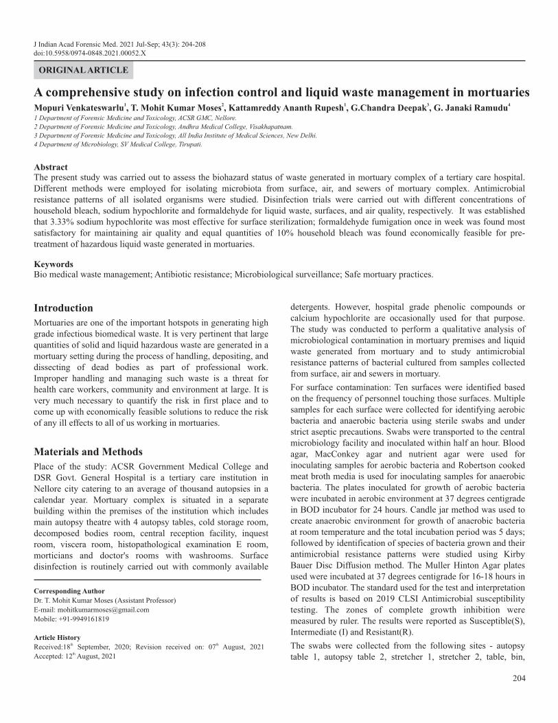
This study was aimed to detect aerobic and anaerobic pathogenic bacteria by way of gram staining and culture and thereby also study the resistance pattern of those organisms. Fungal growth and aerobic spore forming bacilli were not further studied as it was not planned as part of the study. All the swabs received in the microbiology laboratory were processed without any delay. Before culture direct gram stain, pink coloured rod-shaped organisms in single and violet-coloured spherical shaped organisms in pairs and groups were observed abundantly. Occasionally, purple organisms with drum stick appearance and rarely gram-positive bacilli were also observed. Swabs are inoculated on to culture plates (Blood agar and Robertson cooked meat broth medium) and incubated for 24
o ohours at 37 C -38 C.
All plates including settle plates and surface swab inoculated plates showed polymicrobial growth after 24 hours. Clostridial growth was identified in Robertson cooked meat broth media after 5 days. Within the media, meat turned in to black and emanated foul odour. This was inoculated in to blood agar and incubated at room temperature for 24 hours under anaerobic conditions using candle jar method.
205
J Indian Acad Forensic Med. 2021 Jul-Sep; 43(3)
Site Organisms grown
Autopsy table 1 EF, EC, PA, ES, CS, KP, SA, AB
Autopsy table 2 EF, EC, PA, ES, KP, SA, AB
Stretcher 1 EF, EC, CS, KP, SA, AB
Stretcher 2 EF, EC, PA, ES, CS, KP, SA, AB
Bin EF, EC, PA, AB
Instruments EF, EC, PA, ES, CS
Tap EF, EC, PA, ES, CS, KP, SA, AB
Door handle inside EF, EC, PA, ES, CS, KP, SA, AB
Door handle outside EF, EC, PA, ES
Settle plate 1 KP, SA, AB
Settle plate 2 SA, AB
Settle plate 3 KP, SA, AB
Settle plate 4 KP, SA, AB
Floor of room for decomposed bodies
EF, EC, PA, ES, KP, SA, AB
Dead body freezer inside PA, ES, CS, KP, SA, AB
Dead body freezer outside EC, PA, ES, KP, SA, AB
Floor of autopsy theatre EF, EC, PA, ES, CS, KP, SA, AB
Settle plate controls (1-4)Nil. These were part of ensuring quality
control of media.
Table 1: Profile of microorganisms grown at various sites in mortuary
Enterococcus faecium, (EF); Staphylococcus aureus, (SA); Klebsiella pneumoniae, (KP); Acinetobacter baumannii, (AB); Pseudomonas aeruginosa, (PA); Enterobacter species (ES); Clostridia Species (CS); E Coli (EC)
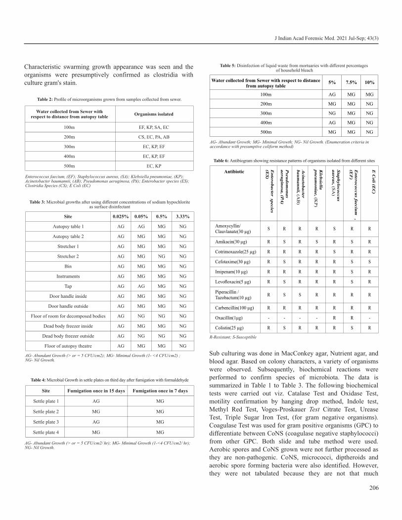
Characteristic swarming growth appearance was seen and the organisms were presumptively confirmed as clostridia with culture gram's stain.
Sub culturing was done in MacConkey agar, Nutrient agar, and blood agar. Based on colony characters, a variety of organisms were observed. Subsequently, biochemical reactions were performed to confirm species of microbiota. The data is summarized in Table 1 to Table 3. The following biochemical tests were carried out viz. Catalase Test and Oxidase Test, motility confirmation by hanging drop method, Indole test, Methyl Red Test, Voges-Proskauer Test Citrate Test, Urease Test, Triple Sugar Iron Test, (for gram negative organisms). Coagulase Test was used for gram positive organisms (GPC) to differentiate between CoNS (coagulase negative staphylococci) from other GPC. Both slide and tube method were used. Aerobic spores and CoNS grown were not further processed as they are non-pathogenic. CoNS, micrococci, diptheroids and aerobic spore forming bacteria were also identified. However, they were not tabulated because they are not that much
206
J Indian Acad Forensic Med. 2021 Jul-Sep; 43(3)
Water collected from Sewer with respect to distance from autopsy table
Organisms isolated
100m EF, KP, SA, EC
200m CS, EC, PA, AB
300m EC, KP, EF
400m EC, KP, EF
500m EC, KP
Table 2: Profile of microorganisms grown from samples collected from sewer.
Enterococcus faecium, (EF); Staphylococcus aureus, (SA); Klebsiella pneumoniae, (KP); Acinetobacter baumannii, (AB); Pseudomonas aeruginosa, (PA); Enterobacter species (ES); Clostridia Species (CS); E Coli (EC)
Site 0.025% 0.05% 0.5% 3.33%
Autopsy table 1 AG AG MG NG
Autopsy table 2 AG MG MG NG
Stretcher 1 AG MG MG NG
Stretcher 2 AG MG NG NG
Bin AG MG MG NG
Instruments AG MG MG NG
Tap AG AG MG NG
Door handle inside AG MG MG NG
Door handle outside AG MG MG NG
Floor of room for decomposed bodies AG NG NG NG
Dead body freezer inside AG MG MG NG
Dead body freezer outside AG NG NG NG
Floor of autopsy theatre AG MG MG NG
Table 3: Microbial growths after using different concentrations of sodium hypochlorite as surface disinfectant
AG- Abundant Growth (> or = 5 CFU/cm2); MG- Minimal Growth (1- <4 CFU/cm2) ; NG- Nil Growth.
Site Fumigation once in 15 days Fumigation once in 7 days
Settle plate 1 AG MG
Settle plate 2 MG MG
Settle plate 3 AG MG
Settle plate 4 MG MG
Table 4: Microbial Growth in settle plates on third day after fumigation with formaldehyde
AG- Abundant Growth (> or = 5 CFU/cm2/ hr); MG- Minimal Growth (1-<4 CFU/cm2/ hr); NG- Nil Growth.
Water collected from Sewer with respect to distancefrom autopsy table
5% 7.5% 10%
100m AG MG MG
200m MG MG NG
300m NG MG NG
400m AG MG NG
500m MG MG NG
AG- Abundant Growth; MG- Minimal Growth; NG- Nil Growth. (Enumeration criteria in accordance with presumptive coliform method)
Table 5: Disinfection of liquid waste from mortuaries with different percentages of household bleach
Antibiotic
En
tero
bacte
rsp
ecie
s (E
S)
Pse
ud
om
on
as
aeru
gin
osa
, (PA
)
Acin
eto
bacte
r b
au
man
nii,
(AB
)
Kle
bsie
lla
pn
eu
mon
iae,
(KP
)
Sta
ph
ylo
coccu
s a
ure
us,
(SA
)
En
tero
coccu
s faeciu
m,
(EF
)
E C
oli (E
C)
Amoxycyllin/ Clauvlanate(30 µg)
S R R R S R R
Amikacin(30 µg) R S R S R S R
Cotrimoxazole(25 µg) R R R R S R R
Cefotaxime(30 µg) R S R R R S S
Imipenam(10 µg) R R R R R S R
Levofloxacin(5 µg) R S R R R S R
Piperacillin / Tazobactum(10 µg)
R S S R R R R
Carbencillin(100 µg) R R R R R R R
Oxacillin(1µg) - - - - R R -
Colistin(25 µg) R S R R R S R
Table 6: Antibiogram showing resistance patterns of organisms isolated from different sites
R-Resistant; S-Susceptible
clinically significant. Due to unavailability of cefoxitin at the time of study, oxacillin was used to differentiate between MSSA and MRSA. The organisms were resistant to oxacillin and several other drugs but they were not included in the table to reduce the unnecessary volume of information. Colistin was selected at the end only as an experimental trial after several drugs tested to be resistant.
After fumigating with formaldehyde, settle plate method was used on the third day to assess disinfection. Fumigation was performed fortnightly initially and once in a week later and results were compared (Table 4). Liquid waste from the sewer was treated with equal quantities of 5% household bleach, 7.5% household bleach and 10% household bleach and was allowed to settle for half an hour and cultures were done from that solution to assess disinfection capabilities of bleach of different concentrations (Table 5 & 6).
Discussion
A thorough search of available literature for microbiological surveillance of autopsy theatres and liquid waste management in mortuaries revealed very few results. However, methods employed for routine operation theatre sterilization can be extrapolated for the purpose of prescribing guidelines for autopsy theatres. After analysing the results of the study, it is not an exaggeration to state that unless a proper infection control plan is worked out for mortuaries, they will become hot spots for emerging superbugs. Because of availability or rich nourishment for bacteria hospital sewers containing untreated liquid hazardous wastes may become sites for resistance transfer between species.
E Coli and Klebsiella demonstrated high degree of multidrug 1resistance in untreated hospital sewage in a study by Kabir.
Our study also demonstrated multi drug resistance in Klebsiella and E Coli isolated from sewers. Settle plates from autopsy theatres showed growth of Staph aureus in our study and the same organisms were identified by a study conducted by Javed
2et al. Enterococcus faecium, (EF) Staphylococcus aureus, (SA) Klebsiella pneumoniae, (KP) Acinetobacter baumannii, (AB) Pseudomonas aeruginosa, (PA) Enterobacter species (ES) Clostridia Species (CS) E Coli (EC) were detected from cultures of sewer lines in our study which is similar to
3 Numberger D et al. Unfortunately, autopsy theatres are not considered on par with surgical OTs. However, some sort of minimum standards of infection control are to be followed for mortuary complexes as well.
Conclusion
In the light of glaring evidence of high risk for everyone who work in mortuaries, it is high time we take a pledge for clean
and safe mortuary practices. Bio medical waste management protocols should be drawn up for all mortuary complexes along with a contingency plan for infection control. Unless the work environment is made more ergonomic and safer it would be exceedingly difficult to find doctors, morticians and ancillary staff ready to work in mortuaries in near future.
4-13Recommendations
The following Ten Commandments are to be followed for welfare of all of us.
1. All surfaces inside mortuary complex should be mopped with 3.33% sodium hypochlorite solution at the end of daily work and if possible, between cases as well.
2. All major rooms in mortuary complex are to be fumigated with at least formaldehyde once a week. Since formaldehyde is being held as a carcinogen it is advisable to search for a cheap yet effective fumigant.
3. Liquid waste generated from mortuary is to be ideally pre-treated in an effluent treatment facility before release from the hospital premises. If such facilities are not available a pre-treatment with 10% household bleach before releasing into municipal sewage is to be followed during the transition period.
4. All personnel in mortuary are to be trained in infection control practices. A proper disinfection action plan includes assessment, cleaning, washing, disinfection, and evaluation. A customized plan may be developed for each setting or the general guidelines in this document may be followed.
5. All mortuary personnel should be vaccinated for vaccine preventable diseases with great caution.
6. Every autopsy complex should have three or more dissection theatres separately earmarked for decomposed cases, infective cases, and routine cases etc.
7. The design of autopsy complex shall have arrangements for air conditioning and proper exhausts are to be fitted preferably close to the ground.
8. All care should be taken to ensure that solid biomedical waste like linen, casts, slabs, catheters etc. are properly segregated at source into respective bins.
9. A septic tank like arrangement is better for allowing the liquid waste generated in mortuaries which can be connected to a pre-treatment facility before letting off the effluents into municipal sewage.
10. It is always advisable to have a team of doctors including microbiologist, hospital administrator and autopsy surgeon for proper mortuary management with respect to infection control.
207
J Indian Acad Forensic Med. 2021 Jul-Sep; 43(3)
Ethical clearance: A prior approval was obtained from the Institutional Ethics Committee
Conflict of interest: None to declare
Source of funding: None to declare
References1. Kabir Y. Multidrug Resistant bacteria in the hospital sewage water
of Dhaka city, Bangladesh. In: Conference of the Bangladesh Society of Biochemistry and Molecular Biology (BSBMB)At: University of Rajshahi, Bangladesh. 2012.
2. Javed R, Hafeez, M, Zubair MS, Anwar M, Tayyib SH. Microbiological surveillance of operation theatres and icus of a tertiary care hospital, lahore. Biomedica. 2008; 24:99–102.
3. Numberger D, Ganzert L, Zoccarato L, Mühldorfer K, Sauer S, Grossart HP, et al. Characterization of bacterial communities in wastewater with enhanced taxonomic resolution by full-length 16S rRNA sequencing. Sci Rep. 2019; 9(1):1–14.
4. Singh A, Joshi HS, Katyal R, Singh R, Singh H. Biomedical Waste Management Rules, 2016: A Brief Review. Int J Adv Integr Med Sci. 2017;2(4):201–4
5. Central government. The Water (Prevention and Control of Pollution) Cess Act. 1988; 1974 (6). Available from: http://dpcc.delhigovt.nic.in/actcess.htm
6. Essential environmental health standards in health care [Internet]. Who.int. 2008 [cited 6 August 2020]. Available from: https://www.who.int/publications/i/item/9789241547239
7. Kumari R, Srivastava K, Wakhlu A, Singh A. Establishing
biomedical waste management system in Medical University of India - A successful practical approach. Clin Epidemiol Glob Heal [ I n t e r n e t ] . 2 0 1 3 ; 1 ( 3 ) : 1 3 1 – 6 . Av a i l a b l e f r o m : http://dx.doi.org/10.1016/j.cegh.2012.11.004
8. Napoli C, Marcotrigiano V, Montagna MT. Air sampling procedures to evaluate microbial contamination: A comparison between active and passive methods in operating theatres. BMC Public Health [Internet]. 2012; 12(1):1. Available from: BMC Public Health
9. Squire JNT. Biomedical Pollutants in the Urban Environment and Implications for Public Health: A Case Study. ISRN Public Health. 2013; 2013:1–5.
10. Biswal S. Liquid biomedical waste management: An emerging concern for physicians. Muller J Med Sci Res. 2013; 4(2):99.
11. Wiafe S, Nooni I, Appiah Boateng K, Nlasia MS, Fianko S. Clinical Liquid Waste Management in Three Ghanaian Healthcare Facilities – a Case Study of Sunyani Municipality. Br J Environ Sci. 2016; 4(1):11–34.
12. Von Wintersdorff CJH, Penders J, Van Niekerk JM, Mills ND, Majumder S, Van Alphen LB, et al. Dissemination of antimicrobial resistance in microbial ecosystems through horizontal gene transfer. Front Microbiol. 2016; 7:1–10.
13. Donde OO. Wastewater management techniques: A review of advancement on the appropriate wastewater treatment principles for sustainability. Environ Manag Sustain Dev [Internet]. 2 0 1 7 ; 6 ( 1 ) : 4 0 . A v a i l a b l e f r o m : http://dx.doi.org/10.5296/emsd.v6i1.10137
208
J Indian Acad Forensic Med. 2021 Jul-Sep; 43(3)
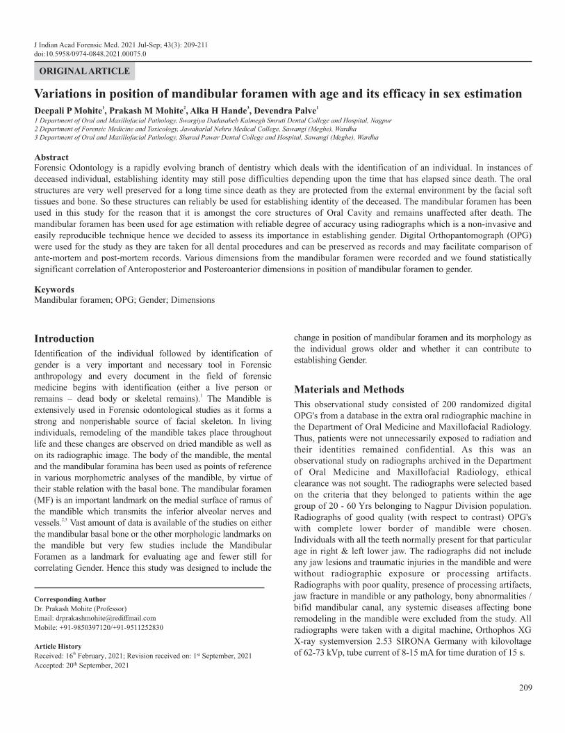
AbstractForensic Odontology is a rapidly evolving branch of dentistry which deals with the identification of an individual. In instances of deceased individual, establishing identity may still pose difficulties depending upon the time that has elapsed since death. The oral structures are very well preserved for a long time since death as they are protected from the external environment by the facial soft tissues and bone. So these structures can reliably be used for establishing identity of the deceased. The mandibular foramen has been used in this study for the reason that it is amongst the core structures of Oral Cavity and remains unaffected after death. The mandibular foramen has been used for age estimation with reliable degree of accuracy using radiographs which is a non-invasive and easily reproducible technique hence we decided to assess its importance in establishing gender. Digital Orthopantomograph (OPG) were used for the study as they are taken for all dental procedures and can be preserved as records and may facilitate comparison of ante-mortem and post-mortem records. Various dimensions from the mandibular foramen were recorded and we found statistically significant correlation of Anteroposterior and Posteroanterior dimensions in position of mandibular foramen to gender.
KeywordsMandibular foramen; OPG; Gender; Dimensions
Variations in position of mandibular foramen with age and its efficacy in sex estimation
ORIGINAL ARTICLE
Introduction
Identification of the individual followed by identification of gender is a very important and necessary tool in Forensic anthropology and every document in the field of forensic medicine begins with identification (either a live person or
1remains – dead body or skeletal remains). The Mandible is extensively used in Forensic odontological studies as it forms a strong and nonperishable source of facial skeleton. In living individuals, remodeling of the mandible takes place throughout life and these changes are observed on dried mandible as well as on its radiographic image. The body of the mandible, the mental and the mandibular foramina has been used as points of reference in various morphometric analyses of the mandible, by virtue of their stable relation with the basal bone. The mandibular foramen (MF) is an important landmark on the medial surface of ramus of the mandible which transmits the inferior alveolar nerves and
2,3 vessels. Vast amount of data is available of the studies on either the mandibular basal bone or the other morphologic landmarks on the mandible but very few studies include the Mandibular Foramen as a landmark for evaluating age and fewer still for correlating Gender. Hence this study was designed to include the
change in position of mandibular foramen and its morphology as the individual grows older and whether it can contribute to establishing Gender.
Materials and Methods
This consisted of 200 randomized digital observational studyOPG's from a database in the extra oral radiographic machine in the Department of Oral Medicine and Maxillofacial Radiology. Thus, patients were not unnecessarily exposed to radiation and their identities remained confidential. As this was an observational study on radiographs archived in the Department of Oral Medicine and Maxillofacial Radiology, ethical clearance was not sought. The radiographs were selected based on the criteria that they belonged to patients within the age group of 20 - 60 Yrs belonging to Nagpur Division population. Radiographs of good quality (with respect to contrast) OPG's with complete lower border of mandible were chosen. Individuals with all the teeth normally present for that particular age in right & left lower jaw. The radiographs did not include any jaw lesions and traumatic injuries in the mandible and were without radiographic exposure or processing artifacts. Radiographs with poor quality, presence of processing artifacts, jaw fracture in mandible or any pathology, bony abnormalities / bifid mandibular canal, any systemic diseases affecting bone remodeling in the mandible were excluded from the study. All radiographs were taken with a digital machine, Orthophos XG X-ray systemversion 2.53 SIRONA Germany with kilovoltage of 62-73 kVp, tube current of 8-15 mA for time duration of 15 s.
Corresponding Author
Dr. Prakash Mohite (Professor)
Email: [email protected]
Mobile: +91-9850397120/+91-9511252830
Article History
Received: 16th February, 2021; Revision received on: 1st September, 2021 Accepted: 20th September, 2021
1 2 3 1Deepali P Mohite , Prakash M Mohite , Alka H Hande , Devendra Palve1 Department of Oral and Maxillofacial Pathology, Swargiya Dadasaheb Kalmegh Smruti Dental College and Hospital, Nagpur2 Department of Forensic Medicine and Toxicology, Jawaharlal Nehru Medical College, Sawangi (Meghe), Wardha3 Department of Oral and Maxillofacial Pathology, Sharad Pawar Dental College and Hospital, Sawangi (Meghe), Wardha
J Indian Acad Forensic Med. 2021 Jul-Sep; 43(3): 209-211doi:10.5958/0974-0848.2021.00075.0
209
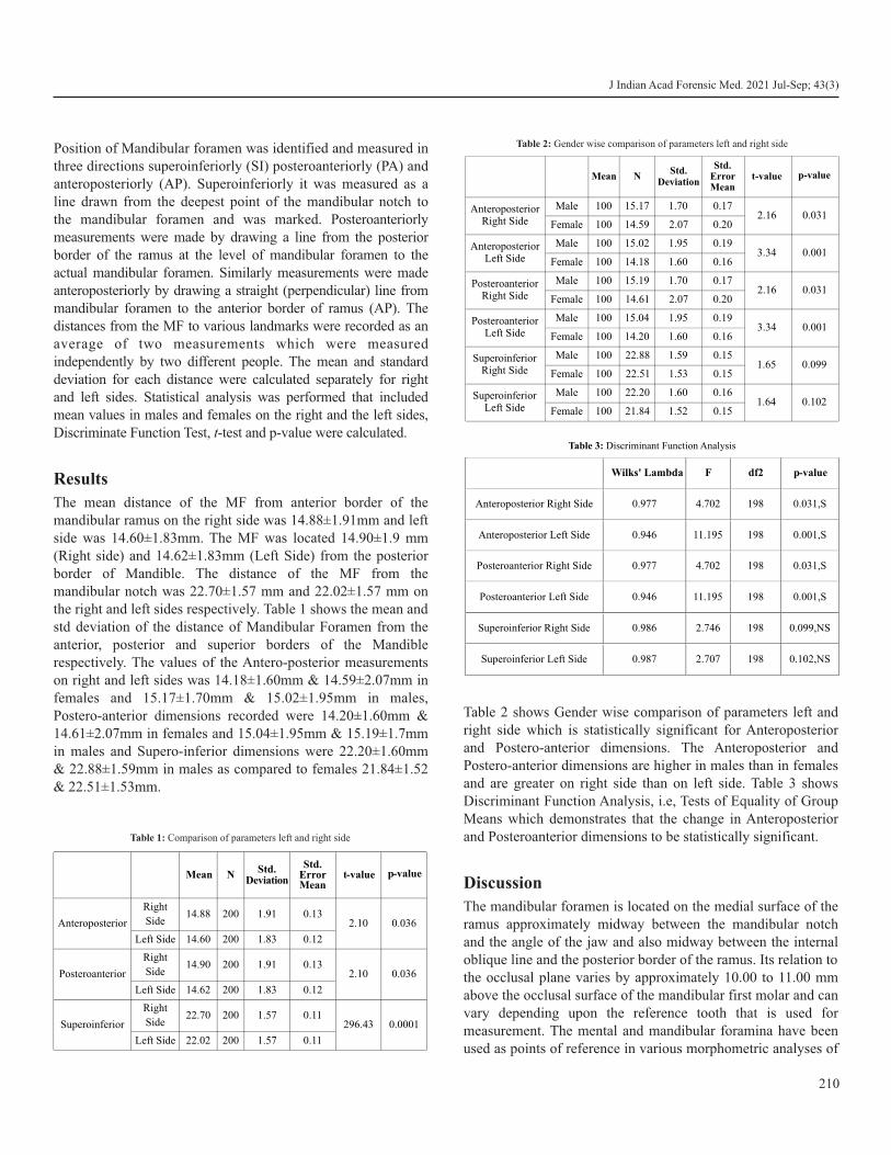
Position of Mandibular foramen was identified and measured in three directions superoinferiorly (SI) posteroanteriorly (PA) and anteroposteriorly (AP). Superoinferiorly it was measured as a line drawn from the deepest point of the mandibular notch to the mandibular foramen and was marked. Posteroanteriorly measurements were made by drawing a line from the posterior border of the ramus at the level of mandibular foramen to the actual mandibular foramen. Similarly measurements were made anteroposteriorly by drawing a straight (perpendicular) line from mandibular foramen to the anterior border of ramus (AP). The distances from the MF to various landmarks were recorded as an average of two measurements which were measured independently by two different people. The mean and standard deviation for each distance were calculated separately for right and left sides. Statistical analysis was performed that included mean values in males and females on the right and the left sides, Discriminate Function Test, t-test and p-value were calculated.
Results
The mean distance of the MF from anterior border of the mandibular ramus on the right side was 14.88±1.91mm and left side was 14.60±1.83mm. The MF was located 14.90±1.9 mm (Right side) and 14.62±1.83mm (Left Side) from the posterior border of Mandible. The distance of the MF from the mandibular notch was 22.70±1.57 mm and 22.02±1.57 mm on the right and left sides respectively. Table 1 shows the mean and std deviation of the distance of Mandibular Foramen from the anterior, posterior and superior borders of the Mandible respectively. The values of the Antero-posterior measurements on right and left sides was 14.18±1.60mm & 14.59±2.07mm in females and 15.17±1.70mm & 15.02±1.95mm in males, Postero-anterior dimensions recorded were 14.20±1.60mm & 14.61±2.07mm in females and 15.04±1.95mm & 15.19±1.7mm in males and Supero-inferior dimensions were 22.20±1.60mm & 22.88±1.59mm in males as compared to females 21.84±1.52 & 22.51±1.53mm.
Table 2 shows Gender wise comparison of parameters left and right side which is statistically significant for Anteroposterior and Postero-anterior dimensions. The Anteroposterior and Postero-anterior dimensions are higher in males than in females and are greater on right side than on left side. Table 3 shows Discriminant Function Analysis, i.e, Tests of Equality of Group Means which demonstrates that the change in Anteroposterior and Posteroanterior dimensions to be statistically significant.
Discussion
The mandibular foramen is located on the medial surface of the ramus approximately midway between the mandibular notch and the angle of the jaw and also midway between the internal oblique line and the posterior border of the ramus. Its relation to the occlusal plane varies by approximately 10.00 to 11.00 mm above the occlusal surface of the mandibular first molar and can vary depending upon the reference tooth that is used for measurement. The mental and mandibular foramina have been used as points of reference in various morphometric analyses of
J Indian Acad Forensic Med. 2021 Jul-Sep; 43(3)
Mean N Std.Deviation
Std. Error Mean
t-value p-value
Anteroposterior
Right
Side14.88 200 1.91 0.13
2.10 0.036
Left Side 14.60 200 1.83 0.12
Posteroanterior
Right
Side14.90 200 1.91 0.13
2.10 0.036
Left Side 14.62 200 1.83 0.12
Superoinferior
Right
Side22.70 200 1.57 0.11
296.43 0.0001
Left Side 22.02 200 1.57 0.11
Table 1: Comparison of parameters left and right side
Mean NStd.
Deviation
Std. Error Mean
t-value p-value
Anteroposterior Right Side
Male 100 15.17 1.70 0.172.16 0.031
Female 100 14.59 2.07 0.20
AnteroposteriorLeft Side
Male 100 15.02 1.95 0.193.34 0.001
Female 100 14.18 1.60 0.16
PosteroanteriorRight Side
Male 100 15.19 1.70 0.172.16 0.031
Female 100 14.61 2.07 0.20
PosteroanteriorLeft Side
Male 100 15.04 1.95 0.193.34 0.001
Female 100 14.20 1.60 0.16
SuperoinferiorRight Side
Male 100 22.88 1.59 0.151.65 0.099
Female 100 22.51 1.53 0.15
SuperoinferiorLeft Side
Male 100 22.20 1.60 0.161.64 0.102
Female 100 21.84 1.52 0.15
Table 2: Gender wise comparison of parameters left and right side
Table 3: Discriminant Function Analysis
Wilks' Lambda F df2 p-value
Anteroposterior Right Side 0.977 4.702 198 0.031,S
Anteroposterior Left Side 0.946 11.195 198 0.001,S
Posteroanterior Right Side 0.977 4.702 198 0.031,S
Posteroanterior Left Side 0.946 11.195 198 0.001,S
Superoinferior Right Side 0.986 2.746 198 0.099,NS
Superoinferior Left Side 0.987 2.707 198 0.102,NS
210
the mandible, by virtue of their stable relation with the basal 3bone. It has been established that the mandibular foramen
undergoes a shift in position from its location at birth into adulthood. This shift in its position is in a vertical plane and this study aimed to assess if there was a change in location with respect to gender of an individual and whether this information may have Forensic implication. In our study, the results showed that differences between gender for mandibular foramen at AP and PA was highly significant, which is similar to the study by
2 3 4Rashid et al, Samanta PP, andLinganna CS. In our study the mandibular foramen shows a shift with age which is similar to
5the study by Lim MY et al. The observation found in our study was a posterior shift because we studied adult population
5whereas in the study by Lim MY et al the population studied were children. When compared between genders the mean values superoinferiorly were higher in males than females
6 which is similar to the study conducted by Shendakar AT etal.7A similar finding was also reported by Direk F et al who used
Multi detector computed tomography to assess mandible and suggested that the ramus dimensions are higher in males. The posteroanterior and anteroposterior dimensions were higher for males in our study, this can be attributed to the stronger masticatory muscles in males imparting greater stability to the
8 9 ramus of the mandible. Sairam V et al, Lasemi Eet al,11Bhardwaj D et al and 12 Jalili MR studied mandibles through
OPG and concluded that superoinferior dimensions were higher in males. This difference was statistically significant between sexes, thus indicating a strong sexual dimorphism.
The difference in dimensions measured for the right and left sides showed values that were almost similar, with a non-significant difference and this applies for both the male and the female groups which is in accordance with study by C. Lavanya Varma et
13 14al and Ashkenazi. Thus the various parameters of the ramus of the mandible can be used for personal identification and also to identify gender, as they serve as stable landmarks and show gradual and steady modifications with age. These dimensional changes are greater in males than in females.
Conclusion
The different measurements of the ramus of the mandible can be used for personal identification to a reliable degree of accuracy and can also be used to identify gender, as they serve as stable landmarks and show gradual and steady modifications with age.
Ethical clearance: A prior approval was obtained from the Institutional Ethics Committee
Conflict of interest: None to declare
Source of funding: None to declare
References1. Popa FM, Ştefǎnescu CL, Corici PD. Forensic value of mandibular
anthropometry in gender and age estimation. Rom J Leg Med. 2009;17(1):45–50.
2. Rashid SA, Ali J. Sex determination using linear measurements related to the mental and mandibular foramina vertical positions on d i g i t a l p a n o r a m i c i m a g e s . J B a g h C o l l D e n t . 2011;23(Special):59–64.
3. Samanta PP, Kharb P. Morphometric analysis of mandibular foramen and incidence of accessory mandibular foramina in adult human mandibles of an Indian population. Rev Arg Anat Clin. 2013;5(2):60–6.
4. Linganna CS, N HRM. Orthopantomograph : a possible predictor of age and gender. 2015;14(March):470–3.
5. Lim MY, Lim WW, Rajan S, Nambiar P, Ngeow WC. Age-related changes in the location of the mandibular and mental foramen in children with Mongoloid skeletal pattern. Eur Arch Paediatr Dent [ I n t e r n e t ] . 2 0 1 5 ; 1 6 ( 5 ) : 3 9 7 – 4 0 7 . Av a i l a b l e f r o m : http://dx.doi.org/10.1007/s40368-015-0184-x.
6. Shendakar AT, Kharat R, et al. Estimation of age in the Living Municipal Employees in the age group of 25-45 years by physical and radiological examination. J Indian Acad Forensic Med. 2010;32(2):113–21.
7. Direk F, Uysal II, Kivrak AS, Unver Dogan N, Fazliogullari Z, Karabulut AK. Reevaluation of Mandibular Morphometry According to Age, Gender, and Side. J Craniofac Surg. 2018;29(4):1054-1059.
8. Sairam V, Geethamalika MV, Kumar PB, Naresh G, Raju GP. Determination of sexual dimorphism in humans by measurements of mandible on digital panoramic radiograph. ContempClin Dent. 2016;7(4):434-439.
9. Lasemi E, Motamedi MHK, Talaeipour AR, et al. Panoramic Radiographic Relationship of the Mandibular Foramen to the Anterior Border of the Ramus and Occlusal Plane as an Aid in Inferior Alveolar Nerve Block. AnesthProg. 2019;66(1):20-23.
10. Poongodi V, Kanmani R, Anandi MS, Krithika CL, Kannan A, Raghuram PH. Prediction of age and gender using digital radiographic method: A retrospective study. J Pharm Bioallied Sci. 2015;7(Suppl 2):S504-S508.
11. Bhardwaj D, Kumar JS, Mohan V. Radiographic evaluation of mandible to predict the gender and age. J Clin Diagn Res. 2014;8(10):ZC66-ZC69.
12. Jalili MR, Esmaeelinejad M, Bayat M, Aghdasi MM. Appearance of anatomical structures of mandible on panoramic radiographs in Iranian population. Acta Odontol Scand. 2012;70(5):384-389.
13. Varma CL, Haq I, Rajeshwari T. Position of Mandibular Foramen In South Indian Mandibles. Anatomica Karnataka,2011: 5(1); 53-56.
14. Ashkenazi M, Taubman L, Gavish A. Age-Associated Changes of the Mandibular Foramen Position in Anteroposterior Dimension and of the Mandibular Angle in Dry Human Mandibles. Anat Rec 2011:294(8); 1319–1325.
J Indian Acad Forensic Med. 2021 Jul-Sep; 43(3)
211
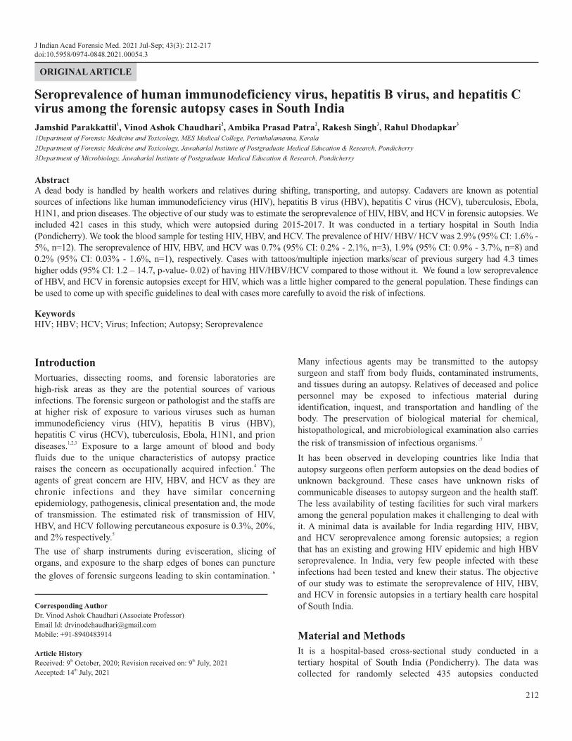
AbstractA dead body is handled by health workers and relatives during shifting, transporting, and autopsy. Cadavers are known as potential sources of infections like human immunodeficiency virus (HIV), hepatitis B virus (HBV), hepatitis C virus (HCV), tuberculosis, Ebola, H1N1, and prion diseases. The objective of our study was to estimate the seroprevalence of HIV, HBV, and HCV in forensic autopsies. We included 421 cases in this study, which were autopsied during 2015-2017. It was conducted in a tertiary hospital in South India (Pondicherry). We took the blood sample for testing HIV, HBV, and HCV. The prevalence of HIV/ HBV/ HCV was 2.9% (95% CI: 1.6% - 5%, n=12). The seroprevalence of HIV, HBV, and HCV was 0.7% (95% CI: 0.2% - 2.1%, n=3), 1.9% (95% CI: 0.9% - 3.7%, n=8) and 0.2% (95% CI: 0.03% - 1.6%, n=1), respectively. Cases with tattoos/multiple injection marks/scar of previous surgery had 4.3 times higher odds (95% CI: 1.2 – 14.7, p-value- 0.02) of having HIV/HBV/HCV compared to those without it. We found a low seroprevalence of HBV, and HCV in forensic autopsies except for HIV, which was a little higher compared to the general population. These findings can be used to come up with specific guidelines to deal with cases more carefully to avoid the risk of infections.
KeywordsHIV; HBV; HCV; Virus; Infection; Autopsy; Seroprevalence
Seroprevalence of human immunodeficiency virus, hepatitis B virus, and hepatitis C virus among the forensic autopsy cases in South India
ORIGINAL ARTICLE
Introduction
Mortuaries, dissecting rooms, and forensic laboratories are high-risk areas as they are the potential sources of various infections. The forensic surgeon or pathologist and the staffs are at higher risk of exposure to various viruses such as human immunodeficiency virus (HIV), hepatitis B virus (HBV), hepatitis C virus (HCV), tuberculosis, Ebola, H1N1, and prion
1,2,3diseases. Exposure to a large amount of blood and body fluids due to the unique characteristics of autopsy practice
4raises the concern as occupationally acquired infection. The agents of great concern are HIV, HBV, and HCV as they are chronic infections and they have similar concerning epidemiology, pathogenesis, clinical presentation and, the mode of transmission. The estimated risk of transmission of HIV, HBV, and HCV following percutaneous exposure is 0.3%, 20%,
5and 2% respectively.
The use of sharp instruments during evisceration, slicing of organs, and exposure to the sharp edges of bones can puncture
–6the gloves of forensic surgeons leading to skin contamination.
Many infectious agents may be transmitted to the autopsy surgeon and staff from body fluids, contaminated instruments, and tissues during an autopsy. Relatives of deceased and police personnel may be exposed to infectious material during identification, inquest, and transportation and handling of the body. The preservation of biological material for chemical, histopathological, and microbiological examination also carries
–7the risk of transmission of infectious organisms.
It has been observed in developing countries like India that autopsy surgeons often perform autopsies on the dead bodies of unknown background. These cases have unknown risks of communicable diseases to autopsy surgeon and the health staff. The less availability of testing facilities for such viral markers among the general population makes it challenging to deal with it. A minimal data is available for India regarding HIV, HBV, and HCV seroprevalence among forensic autopsies; a region that has an existing and growing HIV epidemic and high HBV seroprevalence. In India, very few people infected with these infections had been tested and knew their status. The objective of our study was to estimate the seroprevalence of HIV, HBV, and HCV in forensic autopsies in a tertiary health care hospital of South India.
Material and Methods
It is a hospital-based cross-sectional study conducted in a tertiary hospital of South India (Pondicherry). The data was collected for randomly selected 435 autopsies conducted
212
Corresponding Author
Dr. Vinod Ashok Chaudhari (Associate Professor)
Email Id: [email protected]
Mobile: +91-8940483914
Article Historyth thReceived: 9 October, 2020; Revision received on: 9 July, 2021
thAccepted: 14 July, 2021
1 2 2 3 3Jamshid Parakkattil , Vinod Ashok Chaudhari , Ambika Prasad Patra , Rakesh Singh , Rahul Dhodapkar1Department of Forensic Medicine and Toxicology, MES Medical College, Perinthalamanna, Kerala
2Department of Forensic Medicine and Toxicology, Jawaharlal Institute of Postgraduate Medical Education & Research, Pondicherry
3Department of Microbiology, Jawaharlal Institute of Postgraduate Medical Education & Research, Pondicherry
J Indian Acad Forensic Med. 2021 Jul-Sep; 43(3): 212-217doi:10.5958/0974-0848.2021.00054.3
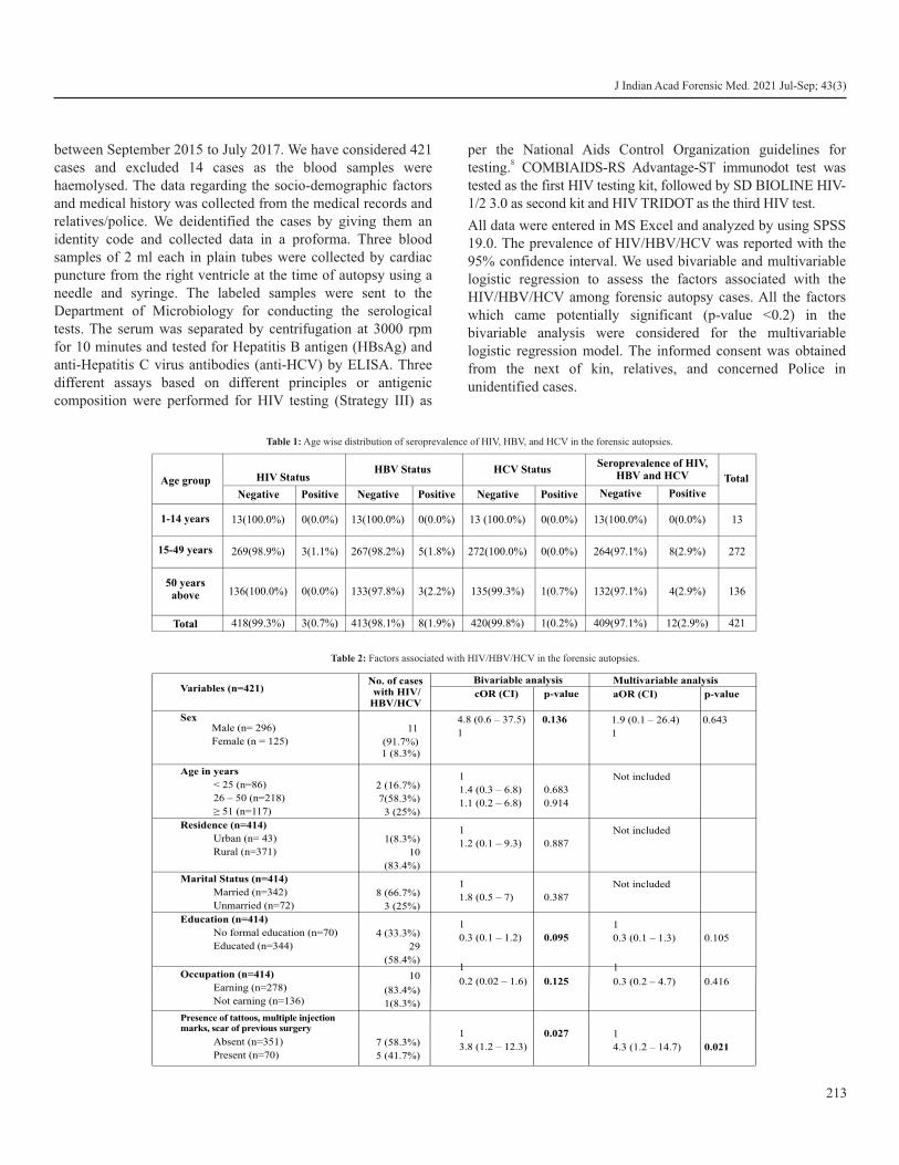
between September 2015 to July 2017. We have considered 421 cases and excluded 14 cases as the blood samples were haemolysed. The data regarding the socio-demographic factors and medical history was collected from the medical records and relatives/police. We deidentified the cases by giving them an identity code and collected data in a proforma. Three blood samples of 2 ml each in plain tubes were collected by cardiac puncture from the right ventricle at the time of autopsy using a needle and syringe. The labeled samples were sent to the Department of Microbiology for conducting the serological tests. The serum was separated by centrifugation at 3000 rpm for 10 minutes and tested for Hepatitis B antigen (HBsAg) and anti-Hepatitis C virus antibodies (anti-HCV) by ELISA. Three different assays based on different principles or antigenic composition were performed for HIV testing (Strategy III) as
per the National Aids Control Organization guidelines for 8testing. COMBIAIDS-RS Advantage-ST immunodot test was
tested as the first HIV testing kit, followed by SD BIOLINE HIV-1/2 3.0 as second kit and HIV TRIDOT as the third HIV test.
All data were entered in MS Excel and analyzed by using SPSS 19.0. The prevalence of HIV/HBV/HCV was reported with the 95% confidence interval. We used bivariable and multivariable logistic regression to assess the factors associated with the HIV/HBV/HCV among forensic autopsy cases. All the factors which came potentially significant (p-value <0.2) in the bivariable analysis were considered for the multivariable logistic regression model. The informed consent was obtained from the next of kin, relatives, and concerned Police in unidentified cases.
213
J Indian Acad Forensic Med. 2021 Jul-Sep; 43(3)
Age group HIV StatusHBV Status HCV Status
Seroprevalence of HIV, HBV and HCV Total
Negative Positive Negative Positive Negative Positive Negative Positive
1-14 years 13(100.0%) 0(0.0%) 13(100.0%) 0(0.0%) 13 (100.0%) 0(0.0%) 13(100.0%) 0(0.0%) 13
15-49 years 269(98.9%) 3(1.1%) 267(98.2%) 5(1.8%) 272(100.0%) 0(0.0%) 264(97.1%) 8(2.9%) 272
50 years above 136(100.0%) 0(0.0%) 133(97.8%) 3(2.2%) 135(99.3%) 1(0.7%) 132(97.1%) 4(2.9%) 136
Total 418(99.3%) 3(0.7%) 413(98.1%) 8(1.9%) 420(99.8%) 1(0.2%) 409(97.1%) 12(2.9%) 421
Table 1: Age wise distribution of seroprevalence of HIV, HBV, and HCV in the forensic autopsies.
Variables (n=421)No. of cases with HIV/HBV/HCV
Bivariable analysis Multivariable analysis
cOR (CI) p-value aOR (CI) p-value
SexMale (n= 296)
Female (n = 125)
11
(91.7%)1 (8.3%)
4.8 (0.6 – 37.5)
1
0.136 1.9 (0.1 – 26.4)
1
0.643
Age in years
< 25 (n=86)
26 – 50 (n=218)
≥ 51 (n=117)
2 (16.7%)
7(58.3%)
3 (25%)
1
1.4 (0.3 – 6.8)
1.1 (0.2 – 6.8)
0.683
0.914
Not included
Residence (n=414)
Urban (n= 43)
Rural (n=371)
1(8.3%)
10
(83.4%)
1
1.2 (0.1 – 9.3) 0.887
Not included
Marital Status (n=414)
Married (n=342)
Unmarried (n=72)
8 (66.7%)
3 (25%)
1
1.8 (0.5 – 7) 0.387
Not included
Education (n=414)
No formal education (n=70)
Educated (n=344)
4 (33.3%)
29
(58.4%)
1
0.3 (0.1 – 1.2) 0.095
1
0.3 (0.1 – 1.3) 0.105
Occupation (n=414)
Earning (n=278)
Not earning (n=136)
10
(83.4%)
1(8.3%)
1
0.2 (0.02 – 1.6) 0.125
1
0.3 (0.2 – 4.7) 0.416
Presence of tattoos, multiple injection marks, scar of previous surgery
Absent (n=351)
Present (n=70)
7 (58.3%)
5 (41.7%)
1
3.8 (1.2 – 12.3)
0.027 1
4.3 (1.2 – 14.7) 0.021
Table 2: Factors associated with HIV/HBV/HCV in the forensic autopsies.
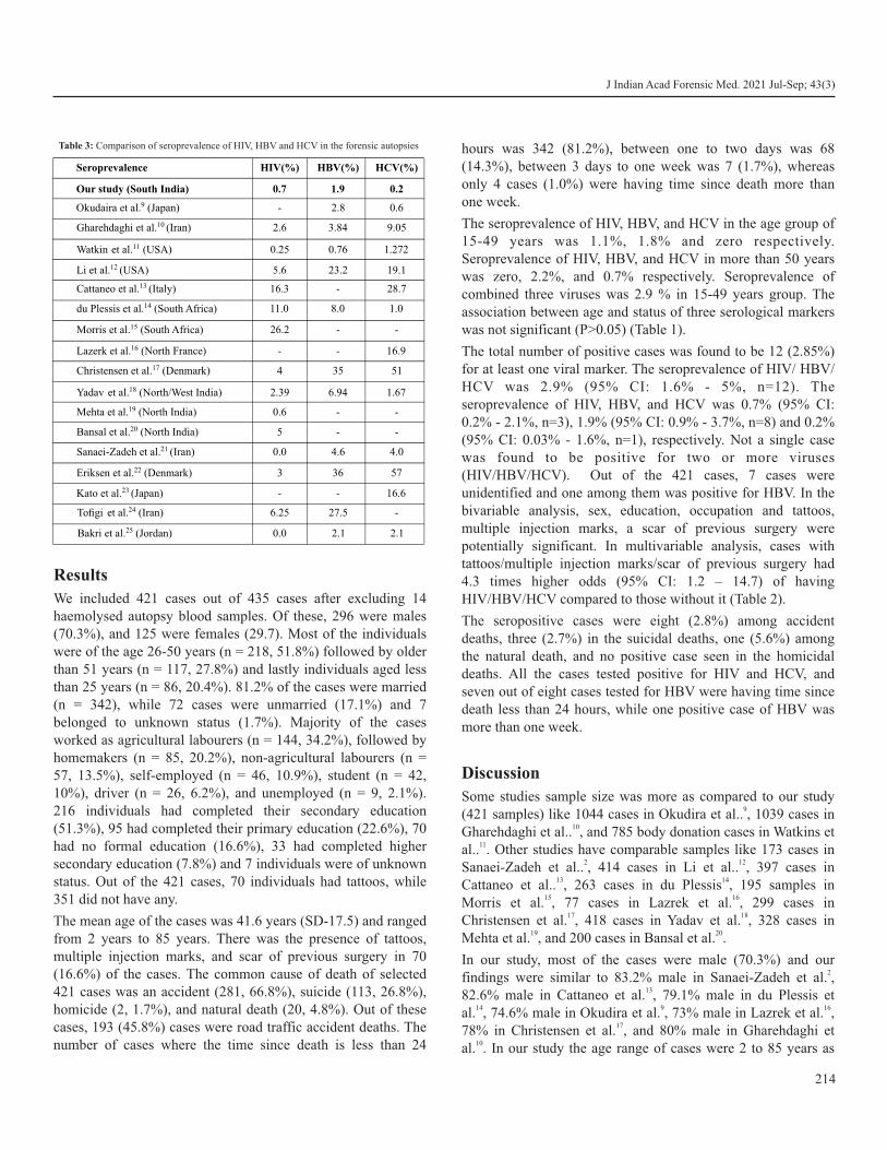
Results
We included 421 cases out of 435 cases after excluding 14 haemolysed autopsy blood samples. Of these, 296 were males (70.3%), and 125 were females (29.7). Most of the individuals were of the age 26-50 years (n = 218, 51.8%) followed by older than 51 years (n = 117, 27.8%) and lastly individuals aged less than 25 years (n = 86, 20.4%). 81.2% of the cases were married (n = 342), while 72 cases were unmarried (17.1%) and 7 belonged to unknown status (1.7%). Majority of the cases worked as agricultural labourers (n = 144, 34.2%), followed by homemakers (n = 85, 20.2%), non-agricultural labourers (n = 57, 13.5%), self-employed (n = 46, 10.9%), student (n = 42, 10%), driver (n = 26, 6.2%), and unemployed (n = 9, 2.1%). 216 individuals had completed their secondary education (51.3%), 95 had completed their primary education (22.6%), 70 had no formal education (16.6%), 33 had completed higher secondary education (7.8%) and 7 individuals were of unknown status. Out of the 421 cases, 70 individuals had tattoos, while 351 did not have any.
The mean age of the cases was 41.6 years (SD-17.5) and ranged from 2 years to 85 years. There was the presence of tattoos, multiple injection marks, and scar of previous surgery in 70 (16.6%) of the cases. The common cause of death of selected 421 cases was an accident (281, 66.8%), suicide (113, 26.8%), homicide (2, 1.7%), and natural death (20, 4.8%). Out of these cases, 193 (45.8%) cases were road traffic accident deaths. The number of cases where the time since death is less than 24
hours was 342 (81.2%), between one to two days was 68 (14.3%), between 3 days to one week was 7 (1.7%), whereas only 4 cases (1.0%) were having time since death more than one week.
The seroprevalence of HIV, HBV, and HCV in the age group of 15-49 years was 1.1%, 1.8% and zero respectively. Seroprevalence of HIV, HBV, and HCV in more than 50 years was zero, 2.2%, and 0.7% respectively. Seroprevalence of combined three viruses was 2.9 % in 15-49 years group. The association between age and status of three serological markers was not significant (P>0.05) (Table 1).
The total number of positive cases was found to be 12 (2.85%) for at least one viral marker. The seroprevalence of HIV/ HBV/ HCV was 2.9% (95% CI: 1.6% - 5%, n=12). The seroprevalence of HIV, HBV, and HCV was 0.7% (95% CI: 0.2% - 2.1%, n=3), 1.9% (95% CI: 0.9% - 3.7%, n=8) and 0.2% (95% CI: 0.03% - 1.6%, n=1), respectively. Not a single case was found to be positive for two or more viruses (HIV/HBV/HCV). Out of the 421 cases, 7 cases were unidentified and one among them was positive for HBV. In the bivariable analysis, sex, education, occupation and tattoos, multiple injection marks, a scar of previous surgery were potentially significant. In multivariable analysis, cases with tattoos/multiple injection marks/scar of previous surgery had 4.3 times higher odds (95% CI: 1.2 – 14.7) of having HIV/HBV/HCV compared to those without it (Table 2).
The seropositive cases were eight (2.8%) among accident deaths, three (2.7%) in the suicidal deaths, one (5.6%) among the natural death, and no positive case seen in the homicidal deaths. All the cases tested positive for HIV and HCV, and seven out of eight cases tested for HBV were having time since death less than 24 hours, while one positive case of HBV was more than one week.
Discussion
Some studies sample size was more as compared to our study 9(421 samples) like 1044 cases in Okudira et al.. , 1039 cases in
10Gharehdaghi et al.. , and 785 body donation cases in Watkins et 11al.. . Other studies have comparable samples like 173 cases in
2 12Sanaei-Zadeh et al.. , 414 cases in Li et al.. , 397 cases in 13 14Cattaneo et al.. , 263 cases in du Plessis , 195 samples in
15 16Morris et al. , 77 cases in Lazrek et al. , 299 cases in 17 18Christensen et al. , 418 cases in Yadav et al. , 328 cases in
19 20Mehta et al. , and 200 cases in Bansal et al. .
In our study, most of the cases were male (70.3%) and our 2findings were similar to 83.2% male in Sanaei-Zadeh et al. ,
1382.6% male in Cattaneo et al. , 79.1% male in du Plessis et 14 9 16al. , 74.6% male in Okudira et al. , 73% male in Lazrek et al. ,
1778% in Christensen et al. , and 80% male in Gharehdaghi et 10al. . In our study the age range of cases were 2 to 85 years as
214
J Indian Acad Forensic Med. 2021 Jul-Sep; 43(3)
Seroprevalence HIV(%) HBV(%) HCV(%)
Our study (South India) 0.7 1.9 0.2
Okudaira et al.9 (Japan) - 2.8 0.6
Gharehdaghi et al.10 (Iran) 2.6 3.84 9.05
Watkin et al.11 (USA) 0.25 0.76 1.272
Li et al.12 (USA) 5.6 23.2 19.1
Cattaneo et al.13 (Italy) 16.3 - 28.7
du Plessis et al.14 (South Africa) 11.0 8.0 1.0
Morris et al.15 (South Africa) 26.2 - -
Lazerk et al.16 (North France) - - 16.9
Christensen et al.17 (Denmark) 4 35 51
Yadav et al.18 (North/West India) 2.39 6.94 1.67
Mehta et al.19 (North India) 0.6 - -
Bansal et al.20 (North India) 5 - -
Sanaei-Zadeh et al.21 (Iran) 0.0 4.6 4.0
Eriksen et al.22 (Denmark) 3 36 57
Kato et al.23 (Japan) - - 16.6
Tofigi et al.24 (Iran) 6.25 27.5 -
Bakri et al.25 (Jordan) 0.0 2.1 2.1
Table 3: Comparison of seroprevalence of HIV, HBV and HCV in the forensic autopsies
compared to age range of cases 2 to 78 the years in study of 2Sanaei-Zadeh , all cases in 16-50 years in the study of Cattaneo
13et al. , 3 months to 79 years (average age, 36 years) in the 14 16study of du Plessis , 1 month to 89 years in Lazrek et al. The
17median age was 38 years in Christensen et al. . In our study, 51.8% of cases were in 21-50 years of age similar to 70.4%
2cases in 20-49 years in the study of Sanaei-Zadeh . But, the most frequent age group was more than 60 years (258, 24.9%)
10in Gharehdaghi et al. Most commonly married cases 342 (81.2%) were included in our study similar to 61.8% married
10cases were included in Gharehdaghi et al. The most common occupation was an agricultural laborer (34.2%) in our study as
10compared to self-employed (31.4%) in Gharehdaghi et al. The majority cases had education up to secondary school (51.3%) in our study as compared to education lower than primary school
10 (41.3%) in another study. Tattoos, multiple injection marks, scar of previous surgery were observed in 70 (16.6%) cases in this study compared to drug abuse (18.1%), sign of nonmedical
10injection mark (8.7%), and tattoos (12.7%) in Gharehdaghi et al.
The most common cause of death was an accident (66.8%) in our 21study similar to a head injury (54.91%) in Sanaei-Zadeh et al. ,
10 trauma 319 (30.7%) in Gharehdaghi et al. But, suicide (35%) 16was a common cause of death in Lazrek et al. Maximum
samples (342, 81.2%) were collected within 24 hours of death in our study. Sample carried out within 1 to 38 days of death as per
13Cattaneo et al. The median time since death of sample collection 22was four days in Eriksen et al.. The peripheral blood collected
from the cadavers where the time since death varied from six 19hours to three days. The time since death was less than 24 hours
10in 74.3% cases in Gharehdaghi et al.
The total number of positive cases was found to be 12 (2.85%) for at least one viral marker (HIV, HBV, and HCV). Eighteen (2.3%) cases were positive for these three infections and it was
11 similar to our findings. But, Gharehdaghi et al. observed that 161 samples (15.49%) were positive for at least one of the infections
10(HBV, HCV, and HIV). A high HIV seroprevalence among 12autopsy cases was reported in Li et al.. (USA) and du Plessis et
14al.. (South Africa) was 5.6% and 11% respectively. The differences were due to the selection of the study population. Our cases were representing the general population. Studies from West and South India showed a seroprevalence of HIV 2.39% and
18,190.6%. A study conducted in Iran and Jordan did not show any 21,25HIV positive cases among the autopsies (Table 3).
An estimated adult (15-49 years) HIV prevalence was of 0.22% [0.16-0.30] in India (2017). In India, around 21.40 lakh people living with HIV (PLHIV) and 97% of the total PLHIV belonged to the 15+ year age group. Females constituted 42% of estimated PLHIV (15+ years). In 2017, 87580 people were newly infected with HIV and 69110 PLHIV died from AIDS-related causes in the same year. Tamil Nadu had 1.42 lakh PLHIV. While Tamil Nadu (0.22%, 0.14-0.31) had point
26prevalence like the national average. Our study showed a low seroprevalence of HIV in forensic autopsies when compared to the previous studies, but found to be higher than that of the general population.
The HBV seroprevalence among the autopsy cases in the USA (23.2%) and South Africa (8%) was high. A study reported a
12,14,18seroprevalence of 6.94% in West India. Okudaira et al. and Bakri et al. reported HBV seroprevalence in forensic autopsies
9,25as 2.8% and 2.1% in Japan and Jordan respectively (Table 3). Globally in 2015, viral hepatitis caused 1.34 million deaths. About 257 million people had chronic HBV infection. Seventy-one million people had chronic HCV infection with 1% prevalence. About 2.7 million had chronic HBV infection and 2.3 million had been infected with HCV among the 36.7 million PLHIV in 2015. Highest hepatitis B prevalence was observed in the WHO Western Pacific region (6.2%), WHO African region (6.1%) WHO South-East Asia region (2.0%). The highest HCV prevalence observed in the Eastern Mediterranean region
27(2.3%) followed by the European Region (1.5%). The present study showed a low seroprevalence of HBV of 1.9% among the autopsy cases tested compared to the previous studies. There was no single case with a combination of HIV, HBV, and HCV. A high HCV seroprevalence was reported from various studies conducted in the USA (19.1%), Iran (4.04), Jordan (2.1%), and
12,18,21,25West India (1.67%) (Table 4). The prevalence of anti-HCV positivity in India was reported around 1% and ranging from 0.09% in the rural population in Maharashtra to 7.89% in
28a tribal population of Arunachal Pradesh. The seroprevalence
of HCV in the present study is less compared to that of the previous studies and the general population as well.
12Li et al. observed a higher positive rate in the age between 30-1439 years for both HBV and HCV. du Plessis et al.. found more
HIV seroprevalence (11% to 19%) and HBV seroprevalence (8% to 9%) in the age group of 15-49 years. A maximum number of HIV cases were found in the age group of 31-40
20 15years (n=4, 9.52%). Morris et al. also observed a rise in HIV seroprevalence from 26.2% to 30% among the 15-49 years age group. Since 15-49 years is the reproductive age group, the rise in seroprevalence found in various studies displays a concern regarding the spread of infection in the living, but our study showed the rise is not significant.
Similar to other studies the males outnumbered the females in the study population. Eleven male cases were positive for viral
12markers out of 12 cases tested positive. Li et al. reported a similar seroprevalence of HIV among males (5.7%) and females (5.2%), but HCV infection in males (21.8%) was twice when compared to females (10.3%) and no significant difference was present among the male and female HBV seroprevalence. A study conducted in Italy showed males (82.6%) were more than, females (17.3%), and study in Iran
215
J Indian Acad Forensic Med. 2021 Jul-Sep; 43(3)
J Indian Acad Forensic Med. 2021 Jul-Sep; 43(3)
216
13,21also reported more males (83.2%) than females (16.8%). The high representation of male deaths that warrant a medicolegal investigation can be attributed to the presence of more seroprevalence in males.
The HIV seroprevalence is found comparatively high among the urban population when compared to the rural population. Urbanization, lower level of education, low income, and mobile occupation are associated with a high prevalence of HIV
29infection. Laborers representing both agriculture as well as the non-agriculture sector have a fair representation in our study population. The seroprevalence of any one of the viral markers is 2.8% and 5.3% respectively.
It is a known fact that tattooing is associated with a high risk of HBV and HCV infection, especially if not done by
30,31professionals. A study conducted in the USA showed significantly increased seroprevalence of HIV, HBV, and HCV among intravenous drug users (IDU). Among this 83.6% were infected with one of these viruses. HIV-1 was seen in 25.5% and
12both HBV and HCV were seen in 47.3%. Similarly, the study in Italy also had a high number of drug-related deaths in their study population, where 81 out of 107 drug overdose cases had markers
13 17of infection. Christensen et al.. (Denmark) assessed the seroprevalence of HIV, HBV, and HCV in postmortem blood samples of drug-related deaths had an anti-HIV prevalence of 4% (9/214), an anti-HBc prevalence of 35% (74/209), and anti-HCV prevalence of 51% (110/215). These results showed the overestimation of seroprevalence of HIV, HBV, and HCV among forensic autopsies due to the increased representation of high-risk groups such as drug-related deaths and IDU.
One of the limitations of our study could be the use of rapid test kits used for the diagnosis of HIV/HBV/HCV, as their sensitivity and specificity can affect the estimate of prevalence. Further studies at a larger scale might be required to generalize the findings. Further studies can be conducted to generate evidence with a larger sample size.
Conclusion
We found a low seroprevalence of HBV, and HCV in forensic autopsies except for HIV, which was a little higher compared to the general population. Cases with tattoos, multiple injection marks or scar of previous surgery have higher odds of having HIV/HBV/HCV. These findings can be used to come up with specific guidelines to deal with autopsy cases more carefully to avoid the risk of infections.
Ethical clearance: A prior approval was obtained from the Institutional Ethics Committee
Conflict of interest: None to declare
Source of funding: None to declare
References1. Kadam SS, Akhade S, Desouza K. Autopsy practice, potential
sources of occupational hazards: A Review for Safety and Prevention. J Indian Acad Forensic Med. 2015;37(2):196-201.
2. Sanaei-Zadeh H, Taghaddosinejad F, Amoei M, Bayatmakou K, Fahim P. Autopsies on bodies without antemortem risk factors for HCV, HBV and HIV infections: are they safe? Pathology. 2002;34(6):582-3.
3. Sharma BR, Reader MD. Autopsy room: a potential source of infection at work place in developing countries. Am J Infec Dis. 2005;1(1):25-33.
4. Nolte KB, Yoon SS. Theoretical risk for occupational blood-borne inections in forensic pathologists. Infect Control Hosp Epidemiol. 2003;24(10):772-3.
5. Butsashvili M, Kamkamidze G, Kajaia M, Morse DL, Triner W, DeHovitz J, et al.. Occupational exposure to body fluids among health care workers in Georgia. Occup Med. 2012;62(8):620–6.
6. Weston J, Locker G. Frequency of glove puncture in the post mortem room. J Clin Patho. 1992;45(2):177–8.
7. Murty OP, Kohli A, Millo T, Rani M, Verma SK, Sikary AK, et al.. Uniform guidelines for postmortem work in India: Faculty development on Standard Operative Procedures (SOP) in forensic medicine and toxicology. J Forensic Med Toxicol. 2013;30:1–138.
8. National guidelines for HIV testing (2015). National AIDS Control Organization, Ministry of Health and Family Welfare, Government o f I n d i a . http://www.naco.gov.in/sites/default/files/National_Guidelines_for_HIV_Testing_21Apr2016.pdf
9. Okudaira M, Tsuda F, Ikawa N, Takamatsu J, Tokudome S, Kurosu K, et al.. Hepatic histopathologic range compared with virological studies of hepatitis viruses among autopsy cases in Tokyo. Hepatol Res. 2001;20(3):320-34.
10. Gharehdaghi J, Hassan M, Khorasgani A, Ghadiani MH, Kazemifar AM, Solhi H. Prevalence of HCV, HBV, and HIV Seropositivity among cadavers referred to autopsy Hall of legal medicine Bureau of Tehran, Iran. Adv Prev Med. 2017:1–4.
11. Watkins BP, Haushalter RE, Bolender DL, Kaplan S, Kolesari GL. Postmortem blood tests for HIV, HBV, and HCV in a body donation program. Clinical Anatomy: The Official Journal of the American Association of Clinical Anatomists and the British Association of Clinical Anatomists. 1998;11(4):250-2.
12. Li L, Zhang X, Constantine NT, Smialek JE. Seroprevalence of parenterally transmitted viruses (HIV-1, HBV, HCV, and HTLV-I/II) in forensic autopsy cases. J Forensic Sci. 1993;38(5):1075–83.
13. Cattaneo C, Nuttall PA, Molendini LO, Pellegrinelli M, Grandi M, Sokol RJ. Prevalence of HIV and hepatitis C markers among a cadaver population in Milan. J Clin Pathol. 1999;52(4):267–70.
14. du Plessis R, Webber L, Saayman G. Bloodborne viruses in forensic medical practice in South Africa. Am J Forensic Med Pathol. 1999;20(4):364–8.
15. Morris N, Du Toit-Prinsloo L, Webber L, Saayman G. The prevalence of HIV in Pretoria's medico-legal laboratory cases, RSA, in 2009. Occup Health South Afr. 2014;20(2):6–10.
217
J Indian Acad Forensic Med. 2021 Jul-Sep; 43(3)
16. Lazrek M, Goffard A, Schanen C, Karquel C, Bocket L, Lion G, et al.. Detection of hepatitis C virus antibodies and RNA among medicolegal autopsy cases in Northern France. Diagn Microbiol Infect Dis. 2006;55(1):55–8.
17. Christensen PB, Kringsholm B, Banner J, Thomsen JL, Cowan S, Stein GF, et al.. Surveillance of HIV and viral hepatitis by analysis of samples from drug related deaths. Eur J Epidemiol. 2006;21(5):383–7.
18. Yadav A, Pathak D, Alam F, Vyas N. Seroprevalence of HIV, HBV and HCV among the cadaver population – A Jaipur based study. Med Leg Update. 2014;14(1):75-9.
19. Mehta S, Singh V, Kaur B, Aggarwal OP. Pre-testing screening for HIV before conducting post-mortem examinations. JK Science. 2012; 14:70-2.
20. Bansal MK, Naik SK, Gupta P, Rani Y, Sherwal BL. Post mortem and the risk of HIV infection. Indian J. Forensic Med. Toxicol. 2014;8(2):63-7.
21. Sanaei-Zadeh H, Amoei M, Taghaddosinejad F. Seroprevalence of HIV, HBV and HCV in forensic autopsies, of presumed low risk, in Tehran, the capital of Iran. J Clin Forensic Med. 2002;9(4):179–81.
22. Eriksen MB, Jakobsen MA, Kringsholm B, Banner J, Thomsen JL, Georgsen J, et al.. Postmortem Detection of Hepatitis B, C, and Human Immunodeficiency Virus Genomes in Blood Samples from Drug-Related Deaths in Denmark. J Forensic Sci . 2009;54(5):1085–8.
23. Kato H, Maeno Y, Seko-Nakamura Y, Monma-Ohtaki J, Sugiura S, Takahashi K, et al.. Identification and phylogenetic analysis of hepatitis C virus in forensic blood samples obtained from injecting
drug users. Forensic Sci Int. 2007;168(1):27–33.
24. Tofigi H, Ghorbani M, Akhlaghi M, Yaghmaei A, Mostafazadeh B, Farzaneh E, et al.. Incidence of hepatitis B and HIV virus at cadaver of IV drug abusers in Tehran. Acta Med Iran. 2011;49(1):59.
25. Bakri FG, Al-Abdallat IM, Ababneh N, Al Ali R, Idhair AKF, Mahafzah A. Prevalence of blood-borne viral infections among autopsy cases in Jordan. Qatar Med J. 2016;2016(2):14.
26. National AIDS Control Organization & ICMR-National Institute of Medical Statistics (2018). HIV Estimations 2017: Technical Report. New Delhi: NACO, Ministry of Health and Family W e l f a r e , G o v e r n m e n t o f I n d i a . A v a i l a b l e from:http://naco.gov.in/sites/default/files/HIV%20Estimations%202017%20Report_3.pdf
27. Global Hepatitis Report 2017. Geneva: World Health Organization; 2017. License: CC BY-NC-SA 3.0 IGO. Available from: https://apps.who.int/iris/bitstream/handle/10665/255016/9789241565455-eng.pdf ; j sess ionid= 4F8BFF16A 9EF6927926 21F74FC23E323?sequence=1
28. Mukhopadhya A. Hepatitis C in India. J Biosci. 2008;33(4):465-73.
29. Kartikeyan S, editor. HIV and AIDS: basic elements and priorities. Dordrecht: Springer; 2007.
30. Jafari S, Copes R, Baharlou S, Etminan M, Buxton J. Tattooing and the risk of transmission of hepatitis C: a systematic review and meta-analysis. Int J Infect Dis. 2010;14(11):928-40.
31. Tohme RA, Holmberg SD. Transmission of hepatitis C virus infection through tattooing and piercing: A critical review. Clin Infect Dis. 2012;54(8):1167-78.
AbstractHomicide is an utmost crime in human society. Land dispute, women and revenge are the usual causative agent behind every homicide. The present retrospective study is conducted on 100 cases brought for autopsy in the mortuary of the Department of Forensic Medicine, PGIMS, Rohtak from 1st January 2017 to 10th November 2019. The commonest age group of victims was 31-40 years and same in age group 41-50 years. Male were victimized 5 times more than the females. Most incidents occurred in monsoon season i.e., July to September in between 6 PM to 12 PM midnight. Hard and blunt weapon was most common type of weapon used in 26 % cases. Brain was the most common organ involved leading to death in 43 cases. Previous enmity followed by property dispute were the most common motive behind the homicide.
KeywordsMotive; Firearm; Axe; Knife; Danda; Homicide
The pattern of homicide in Haryana – A retrospective study
ORIGINAL ARTICLE
Introduction1 Homicide is killing of a human being by another human being.
Homicide may be considered as destruction of human life by the act, agency, procurement or culpable omission of some other
2 person. There are two important elements to commit murder, which include Mens rea which means pre-planning and Actus reus which means the actual execution. Actus rea and Mens rea
3should work together to constitute the Crime. As per global study on homicide 2019 by UNODC, there were 6.1 homicide victims per 100,000 populations worldwide, compared with a rate of 7.4 in 1993. Globally around 464,000 people killed in homicide in year 2017 as a result of interpersonal violence which equates to
41271 deaths every single day.
According to National Crime Record Bureau, 29017 murders were reported in India in year 2018 with a crime rate of 2.2 Percent. Percentage share of violent crimes were 0.9% of the total
Indian Penal Code crimes. In our Haryana 1140 people killed in
5homicide in year 2018 with a crime rate of 3.9 percent.
Materials and Methods
The present study is a retrospective study conducted in the Department of Forensic Medicine, Pt. B. D. Sharma PGIMS, Rohtak. Data of 100 cases of homicide brought to the mortuary of PGIMS, Rohtak for post-mortem examination was collected from the departmental record. The cases in which manner was
suspicious were not included in the study. The details of all 100 cases namely; sex, age, season, time of assault, survival period, motive, weapon, cause of death, a vital organ involved and manner of death were collected from the departmental record with maintaining the confidentiality and keeping ethical issues in mind.
Results
In present study record of 100 cases was studies. Out of 100 cases, 84 were males and 16 were female. Majority of the assaults occurred during monsoon season (June to September) i.e., 37 cases followed by post monsoon season (October to November) i.e., 26 and 19 cases in summer season (March to May). Only 18 cases of assault occurred in winter season (December to February). As far as time of altercations concerned, majority of assaults i.e., 38 cases occurred from 6 PM to 11:59 midnight followed by 25 cases in time frame 12:00 noon to 5:59 PM and 20 cases during 6:00 AM to 11:59 AM. Only 8 cases incurred between 12:00 midnight to 5:59 AM and 9 cases were there in which time frame was not known.
Age-wise, maximum cases belonged to the age group 31 to 40 and 41 to 50 years i.e., 27% each group followed by 21 to 30 years' age group i.e., 15 %. (Table 1). As depicted in Table no. 2, most of the cases survived for more than 3 days i.e., 31 % cases. The previous enmity is the most common motive behind the homicide i.e., 34 % cases followed by a property dispute i.e., 22 % cases as shown in Table 3. As shown in Table no. 4, Iron rod and danda are the most common weapon used i.e. 26 % cases followed by 21% cases of the firearm. Table no 5 shows that the most common cause of death is injuries described and their complication i.e., 35% case followed by head injuries and their complication i.e., 31 % cases. As depicted in Table no. 6, the brain was the most common organ involved i.e., 43 % cases.
218
Corresponding Author
Dr Mahender Singh, (Resident Doctor)
Email address: [email protected]
Mobile: +91-9859595997
Article Historyth stReceived: 28 October, 2020; Revision received on: 21 August, 2021thAccepted: 24 August, 2021
1 2 2 2 2 2Naveen Yadav , Jitender Kumar Jakhar , Gaurav Kaushik , Lait Chopra , Mahender Singh , SK Dhattarwal1 General Hospital, Rewari
2 Department of Forensic Medicine, Pt. B. D. Sharma PGIMS, Rohtak, Haryana
J Indian Acad Forensic Med. 2021 Jul-Sep; 43(3): 218-220doi:10.5958/0974-0848.2021.00055.5
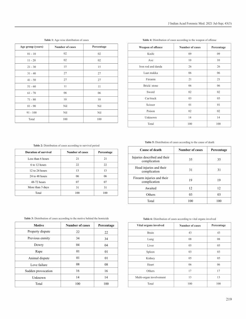
219
J Indian Acad Forensic Med. 2021 Jul-Sep; 43(3)
Age group (years) Number of cases Percentage
01 - 10 02 02
11 - 20 02 02
21 - 30 15 15
31 - 40 27 27
41 - 50 27 27
51 - 60 11 11
61 - 70 06 06
71 - 80 10 10
81 - 90 Nil Nil
91 - 100 Nil Nil
Total 100 100
Table 1: Age-wise distribution of cases
Duration of survival Number of cases Percentage
Less than 6 hours 21 21
6 to 12 hours 22 22
12 to 24 hours 13 13
24 to 48 hours 06 06
48-72 hours 07 07
More than 3 days 31 31
Total 100 100
Table 2: Distribution of cases according to survival period
Motive Number of cases Percentage
Property dispute 22 22
Previous enmity 34 34
Dowry 04 04
Rape 01 01
Animal dispute 01 01
Love failure 08 08
Sudden provocation 16 16
Unknown 14 14
Total 100 100
Table 3: Distribution of cases according to the motive behind the homicide
Weapon of offence Number of cases Percentage
Knife 09 09
Axe 10 10
Iron rod and danda 26 26
Laat mukka 06 06
Firearm 21 21
Brick/ stone 06
06
Sword 02 02
Car/truck 03 03
Scissor 01 01
Poison 02 02
Unknown 14 14
Total 100 100
Table 4: Distribution of cases according to the weapon of offense
Cause of death Number of cases Percentage
Injuries described and theircomplication 35 35
Head injuries and theircomplication 31 31
Firearm injuries and theircomplication 19 19
Awaited 12 12
Others 03 03
Total 100 100
Table 5: Distribution of cases according to the cause of death
Vital organs involved Number of cases Percentage
Brain 43 43
Lung 08 08
Liver 05 05
Spleen 03 03
Kidney 05 05
Heart 06 06
Others 17 17
Multi-organ involvement 13 13
Total 100 100
Table 6: Distribution of cases according to vital organs involved
Discussion
In this study, males outnumbered the females and the overall male to female ratio is 5.25:1. because in India female is less likely involved in altercations outside the households. Findings of our study are consistent with Sharma et al, Sonawane et al., Gupta et
6-12al., Kiran et al., Mada et al., Mohanty et al.,and Hugar et al.
In the present study, maximum cases belong to the age group 31 to 40 and 41 to 50 years. Similar findings were noticed by some
7 9 authors in their studies- Sonawane et al. and Kiran et al while 20 to 29 age group was most common in the study conducted
by Sharma et al, Gupta et al., Mada et al., Mohanty et al., and 6,8,10,11,12Hugar et al. The above findings can easily be explained
by the fact that 31 to 40 years of age group is most susceptible to frustration in life because of many factors like failure of love affair, stressful marital life, financial crunches and pressure of making a good career after completion of studies etc.
This study showed that most common assaults occurred in monsoon season i.e., from June to September while Sharma et
6 11 al and Mohanty et al found most of the homicide occurred in summers. As we all know that the Haryana is primarily an agricultural state and Rabi group of crops are ready for harvesting by end of march month. After the harvesting and sale of crop, people have the enough money in their hands and they also do not have agricultural work till next season of Kharif. In this free time and having enough money, they try to resolve the disputes of finance and land and involved in various illegal activities like physical assault and in extreme cases like homicide. This could be the reason for maximum homicides in monsoon season. The present study depicts that most of the quarrel occurred from 6 PM to 11:59 midnight similar findings
11were observed by Mohanty et al. Most of the victims survived for more than 3 days, while maximum deaths occurred on spot
12in a study conducted by Hugar et al. This is because the study includes the cases of assault that are brought to a trauma center for treatment. Revenge for a previous fight is the most common motive behind the homicide, similar findings were observed by
7-9,12Sonawane et al., Gupta et al., Kiran et al., Hugar et al. An iron rod and danda are the most common weapon used. Our finding is consistent with the finding of Sonawane et al. while the sharp weapon was most common in studies conducted by
, 7-9,11, 12Gupta et al Kiran et al, Mohanty et al and Hugar et al. This is because danda and rod are the most common weapon which are easily available at the site of occurrence of crime. The most common cause of death is injuries described and their complication and brain is the most common organ involved.
Conclusion
Homicides are caused by the person of criminal intent. But sometimes, homicide occurs due to sudden heat of the moment with the help of nearby available weapons. If the accused would
have been thought about the consequences for a small period of time, this type of incident can be prevented.
Ethical clearance: A prior approval was obtained from the Institutional Ethics Committee
Conflict of interest: None to declare
Source of funding: None to declare
References1. Reddy KSN, Murty OP. The Essentials of Forensic Medicine and
Toxicology. 34th ed. New Delhi: Jaypee Brother Medical Publisher (p) Ltd; 2017.
2. Vij K. Textbook of Forensic Medicine and Toxicology, Principles and Practice. 6th ed. New Delhi: Elsevier; 2014.
3. Mukherjee JB, Karmakar RN (ed). Forensic Medicine and Toxicology. 5th ed. Kolkata: Academic Publishers (p) Ltd; 2018.
4. Ummary E. GLOBAL STUDY ON HOMICIDE [Internet]. U n o d c . o rg . [ c i t e d 2 0 2 1 D e c 7 ] . Av a i l a b l e f r o m : h t t p s : / / w w w . u n o d c . o r g / d o c u m e n t s / d a t a - a n d -analysis/gsh/Booklet1.pdf
5. National Crime Records Bureau. Crime In India 2018. Gov.in. [ c i t e d 2 0 2 1 D e c 7 ] . A v a i l a b l e f r o m : https://ncrb.gov.in/sites/default/files/Crime%20in%20India%202018%20-%20Volume%201.pdf.
6. Sharma G, Deepshika, Jain A, Khanagwal P, Punia J, Lathwal S et al. Homicide in District Sonipat in 2018 AD and comparison with all Haryana data. Indian J Appl Res. 2019;9(2):10-12.
7. Sonawane SS, Sukhdeve RB, Tyagi S, Kolle SR. Autopsy evaluation of homicidal deaths in western Mumbai region- 2 years prospective study. Sch J App Med Sci. 2017;5(12A):4840-4846.
8. Gupta N, Aggarwal NK, Verma SK. Pattern of homicidal deaths in North East Delhi and NCR. Indian J Forensic Med Toxicol. 2018;35(1):70-75.
9. Kiran M, Shetty M, Shetty SS, Shetty V. Analysis of homicidal deaths in a tertiary care center of a non-metropolitan city of South India: A retrospective study. J. Indian Acad. Forensic Med. 2017;39(4):384-386.
10. Mada P, Krishna PH. A Comprehensive study on homicidal deaths in Hyderabad. J. Indian Acad. Forensic Med. 2013;35(4):0971-0973.
11. Mohanty S, Mohanty SK, Patnaik KK. Homicide in southern India—A five-year retrospective study. Forensic Medicine and Anatomy Research. 2013;1(2):18-24.
12. Hugar BS, Chandra G, Harish S, Jayanth SH. Pattern of Homicidal Deaths. J. Indian Acad. Forensic Med. 2010; 32(3):194-198.
220
J Indian Acad Forensic Med. 2021 Jul-Sep; 43(3)
AbstractThere has been a drastic increase in the reporting of sexual assault cases in India after the implementation of the Criminal law amendment act, 2013. Various studies on such critical issues have been conducted in past in India. Unfortunately, none of the existing researches focuses on bringing reforms in the prevailing rape law by reconsidering the age of statutory rape. This study was conducted from 2014 to 2016 on 220 victims of sexual assault to identify sociodemographic characteristics and to highlight the issue of statutory rape in India. 90% of victims of sexual assault were females. The mean age of victims was 16.75 years. 68.2% of victims were from the lower middle class. The majority of sexual assault cases took place in the assailant's house. The assailant was known to the victim in 88.6% of cases and was mostly boyfriend (36.4%). Victims under 12 years of age are at a comparatively higher risk of being sexually assaulted by an unknown person. The age of consent for sexual intercourse in India is 18 years while worldwide consensus points to 16 years. With the increase in usage of the internet and social networking sites, young children are having early exposure to sex and sexual content existing on the web. In such a scenario, young children caught in the act would go to jail as it would amount to technical rape as consent would be of no value. This study will create community awareness about the extent of the problem and help the policymakers to make necessary amendments to the existing rape law.
KeywordsSexual assault; Victim; Socio-demographic; Statutory rape
Socio-demographic determinants of victims of sexual assault in Mumbai
ORIGINAL ARTICLE
Introduction
Sexual assault is a form of sexual violence, which is often used synonymously with rape. It includes touching an individual's body against the consent in a sexual way, forceful sexual intercourse, attempted rape, anal and oral sexual acts, molestation of child and breast/ genitals fondling. The victim is someone who has suffered harm and has been subjected to a non-consensual sexual act. Such someone not only needs care
1but also, psychological and moral support. It is important to recognize that the majority of the sexual assaults are committed by persons known to victims. Historically, such offence committed by non-strangers wasn't taken seriously. Sexual assault committed by the individual familiar to the victim causes more damage than those committed by strangers. Responders must remember that the reactions of the victim to sexual assault are affected by many factors, one of which is the foregoing victim and offender relationship. They must also understand that a lot of variables may affect the relevance of certain types of evidence to a selected case, including whether an assault was committed by an unknown person, a known person who denies sexual contact with the victim, or a known
2person who claims they had a consensual act.
The sex-related offence is not uncommon in India with rape th 3,4being the 4 most common crime against women. India
thranked 94 in the ranking of reported cases per 100,000 5population in 2010. Mumbai is a metropolitan city and is
considered the business capital of India and caters to a large population. In the year 2011, the population of Mumbai was 12,442,373 as per the Census of India. A population of 5,206,473 resides in 1,135,514 slums of Mumbai. This is about
6 41.84% of Mumbai's population. The vast population of India is composed of people having diverse creeds, customs and colours. A very large part of Mumbai's population belongs to migrants from various parts of India and thus the city has a wide cultural and ethnic diversity. With such a diverse population, the socio-demographic factors analyzed in the present study will be similar to a certain extent across other regions of the country.
Materials and Methods
A single centre, cross-sectional, observational study of sexual assault cases examined in the Department of Forensic Medicine and Toxicology at a government-run tertiary health care centre between August 2014 and July 2016. Such examinations are only being done at various government-run tertiary care centres in Mumbai. Our centre is the largest amongst others catering to the population under the jurisdiction of 31 police stations. 220 cases of sexual assault were included in the study.
During this 2 year study, 808 victims of sexual offences were
221
Corresponding Author
Dr. Arun Kumar Jaiswani (Assistant Professor),
Email: [email protected]
Mobile: +91-9827959335
Article Historyth rdReceived: 9 November, 2020; Revision received on: 3 August 2021thAccepted: 9 August 2021
1 2 2 2Arun Kumar Jaiswani , Rajesh C Dere , Narendra B Kumar , Hemant G Kukde1 Department of Forensic Medicine and Toxicology, Pt Jawaharlal Nehru Memorial Medical College, Raipur, Chhattisgarh2 Department of Forensic Medicine and Toxicology, Lokmanya Tilak Municipal Medical College, Sion, Mumbai, Maharashtra
J Indian Acad Forensic Med. 2021 Jul-Sep; 43(3): 221-226doi:10.5958/0974-0848.2021.00056.7
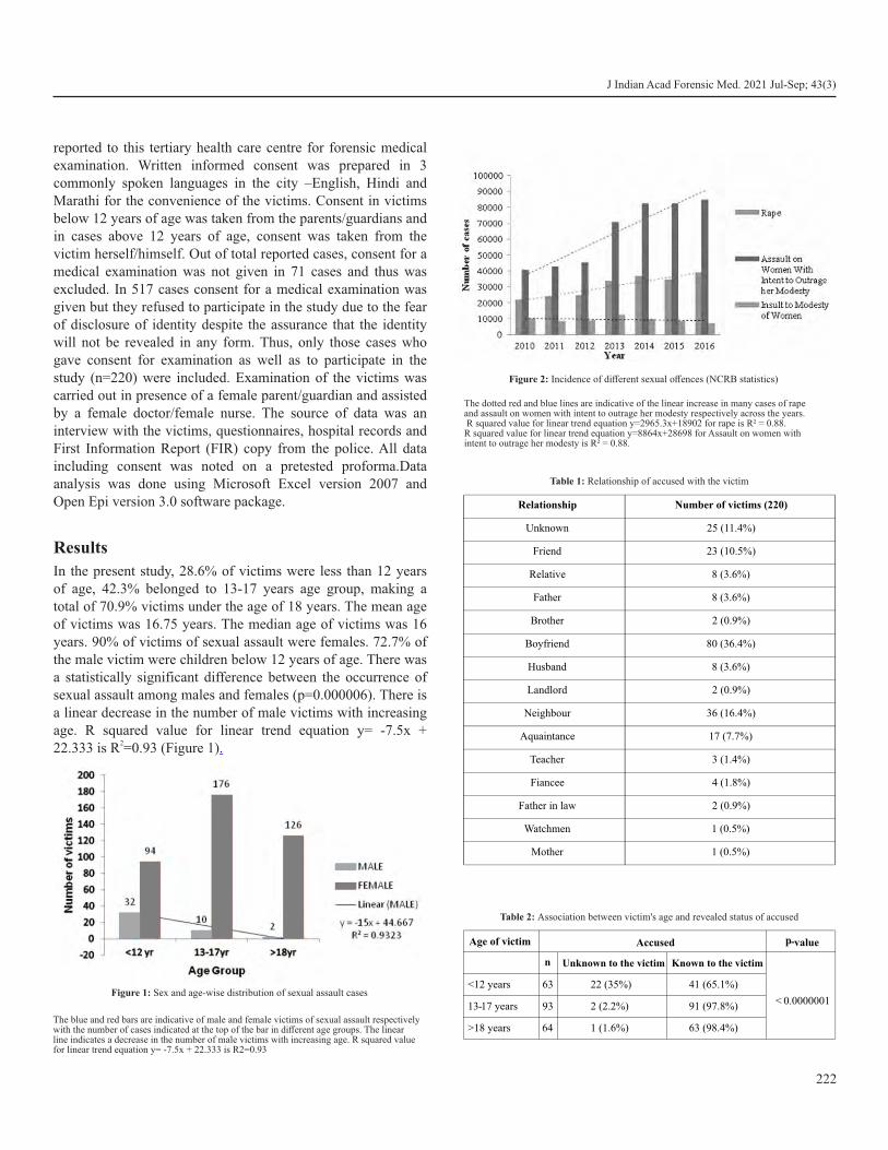
reported to this tertiary health care centre for forensic medical examination. Written informed consent was prepared in 3 commonly spoken languages in the city –English, Hindi and Marathi for the convenience of the victims. Consent in victims below 12 years of age was taken from the parents/guardians and in cases above 12 years of age, consent was taken from the victim herself/himself. Out of total reported cases, consent for a medical examination was not given in 71 cases and thus was excluded. In 517 cases consent for a medical examination was given but they refused to participate in the study due to the fear of disclosure of identity despite the assurance that the identity will not be revealed in any form. Thus, only those cases who gave consent for examination as well as to participate in the study (n=220) were included. Examination of the victims was carried out in presence of a female parent/guardian and assisted by a female doctor/female nurse. The source of data was an interview with the victims, questionnaires, hospital records and First Information Report (FIR) copy from the police. All data including consent was noted on a pretested proforma.Data analysis was done using Microsoft Excel version 2007 and Open Epi version 3.0 software package.
Results�In the present study, 28.6% of victims were less than 12 years of age, 42.3% belonged to 13-17 years age group, making a total of 70.9% victims under the age of 18 years. The mean age of victims was 16.75 years. The median age of victims was 16 years. 90% of victims of sexual assault were females. 72.7% of the male victim were children below 12 years of age. There was a statistically significant difference between the occurrence of sexual assault among males and females (p=0.000006). There is a linear decrease in the number of male victims with increasing age. R squared value for linear trend equation y= -7.5x +
222.333 is R =0.93 (Figure 1) .
222
J Indian Acad Forensic Med. 2021 Jul-Sep; 43(3)
Figure 1: Sex and age-wise distribution of sexual assault cases
The blue and red bars are indicative of male and female victims of sexual assault respectively with the number of cases indicated at the top of the bar in different age groups. The linear line indicates a decrease in the number of male victims with increasing age. R squared value for linear trend equation y= -7.5x + 22.333 is R2=0.93
Figure 2: Incidence of different sexual offences (NCRB statistics)
The dotted red and blue lines are indicative of the linear increase in many cases of rape and assault on women with intent to outrage her modesty respectively across the years. R squared value for linear trend equation y=2965.3x+18902 for rape is R² = 0.88. R squared value for linear trend equation y=8864x+28698 for Assault on women with intent to outrage her modesty is R² = 0.88.
Relationship Number of victims (220)
Unknown 25 (11.4%)
Friend 23 (10.5%)
Relative 8 (3.6%)
Father 8 (3.6%)
Brother 2 (0.9%)
Boyfriend 80 (36.4%)
Husband 8 (3.6%)
Landlord 2 (0.9%)
Neighbour 36 (16.4%)
Aquaintance 17 (7.7%)
Teacher 3 (1.4%)
Fiancee 4 (1.8%)
Father in law 2 (0.9%)
Watchmen 1 (0.5%)
Mother 1 (0.5%)
Table 1: Relationship of accused with the victim
Age of victim Accused p-value
n Unknown to the victim Known to the victim
< 0.0000001
<12 years 63 22 (35%) 41 (65.1%)
13-17 years 93 2 (2.2%) 91 (97.8%)
>18 years 64 1 (1.6%) 63 (98.4%)
Table 2: Association between victim's age and revealed status of accused
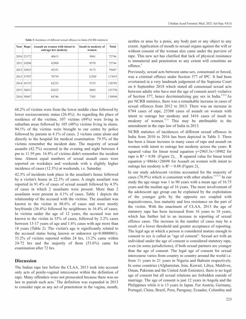
68.2% of victims were from the lower middle class followed by lower socioeconomic status (26.4%). As regarding the place of residence of the victims, 107 victims (49%) were living in suburban areas followed by 105 (48%) victims living in slums. 94.1% of the victims were brought to our centre by police followed by parents in 4.1% of cases. 2 victims came alone and directly to the hospital for medical examination. 79.5% of the victims remember the incident date. The majority of sexual assaults (42.7%) occurred in the evening and night between 4 pm to 11.59 pm. 16.8% of victims didn't remember the incident time. Almost equal numbers of sexual assault cases were reported on weekdays and weekends with a slightly higher incidence of cases (12.3%) at weekends, i.e. Saturday.
42.3% of incidents took place in the assailant's house followed by a victim's house in 22.3% of cases. A single assailant was reported in 91.4% of cases of sexual assault followed by 4.5% of cases in which 2 assailants were present. More than 2 assailants were present in 4.1% of cases. Table 1 depicts the relationship of the accused with the victims. The assailant was known to the victim in 88.6% of cases and were mostly boyfriends (36.4%) followed by neighbours in 16.4% of cases. In victims under the age of 12 years, the accused was not known to the victim in 35% of cases, followed by 2.2% cases between 13-17 years of age and 1.6% cases with age more than 18 years (Table 2). The victim's age is significantly related to the accused status being known or unknown (p<0.0000001). 33.2% of victims reported within 24 hrs, 13.2% came within 24-72 hrs and the majority of them (53.6%) came for examination after 72 hrs.
Discussion
The Indian rape law before the CLAA, 2013 took into account only acts of penile-vaginal intercourse within the definition of rape. Many offenders were not prosecuted because there was no
7 law to punish such acts. The definition was expanded in 2013 to consider rape as any act of penetration in the vagina, mouth,
urethra or anus by a penis, any body part or any object to any extent. Application of mouth to sexual organs against the will or without consent of the woman also came under the purview of rape. This new act has clarified that lack of physical resistance is immaterial and penetration to any extent will constitute an
8offence.
Previously, sexual acts between same-sex, consensual or forced, was a criminal offence under Section 377 of IPC. It had been overturned in a very landmark judgement of the Supreme Court on 6 September 2018 which stated all consensual sexual acts between adults who have met the age of consent aren't violative
9,10,11of Section 377, hence decriminalizing gay sex in India. As per NCRB statistics, there was a remarkable increase in cases of sexual offences from 2012 to 2013. There was an increase in 8784 cases of rape, 25388 cases of assault on women with intent to outrage her modesty and 3416 cases of insult to
12,13modesty of women. This may be attributable to the amendment in the rape law of India in 2013.
NCRB statistics of incidences of different sexual offences in India from 2010 to 2016 has been depicted in Table 3. There has been a linear increase in many cases of rape and assault on women with intent to outrage her modesty across the years. R squared value for linear trend equation y=2965.3x+18902 for rape is R² = 0.88 (Figure 2) R squared value for linear trend . equation y=8864x+28698 for Assault on women with intent to outrage her modesty is R² = 0.88 (Figure 2).
In our study adolescent victims accounted for the majority of 14,15,16cases (70.9%) which is consistent with other studies. In our
study the age range was 1 to 90 years with a mean age of 16.75 years and the median age of 16 years. The more involvement of the adolescent age group can be explained by the exploitation of the younger girls by the opposite sex coupled with inquisitiveness, less maturity and less resistance on the part of the victim. With the enactment of CLAA, 2013 the age of statutory rape has been increased from 16 years to 18 years, which has further led to an increase in reporting of sexual offence cases. The increase in the number of cases may be a result of a lower threshold and greater acceptance of reporting. The legal age at which a person is considered mature enough to consent to sex is called as “age of consent”. Sexual act with an individual under the age of consent is considered statutory rape, even (in some jurisdictions), if both sexual partners are younger than the age of consent. The legal age of consent for sexual intercourse varies from country to country around the world i.e. from 11 years to 21 years in Nigeria and Bahrain respectively. In some countries (Afghanistan, Iran, Kuwait, Libya, Maldives, Oman, Pakistan and the United Arab Emirates), there is no legal age of consent but all sexual relations are forbidden outside of marriage. The age of consent is just 12 years in Angola and the Philippines while it is 13 years in Japan. For Austria, Germany, Portugal, China, Brazil, Peru, Paraguay, Ecuador, Columbia and
J Indian Acad Forensic Med. 2021 Jul-Sep; 43(3)
223
Year Rape Assault on women with intent to outrage her modesty
Insult to modesty of women
Total
2010 22172 40613 9961 72746
2011 24206 42968 8570 75744
2012 24923 45351 9173 79447
2013 33707 70739 12589 117035
2014 36735 82235 9735 128705
2015 34651 82422 8685 125758
2016 38947 84746 7305 130998
Table 3: Incidence of different sexual offences in India (NCRB statistics)
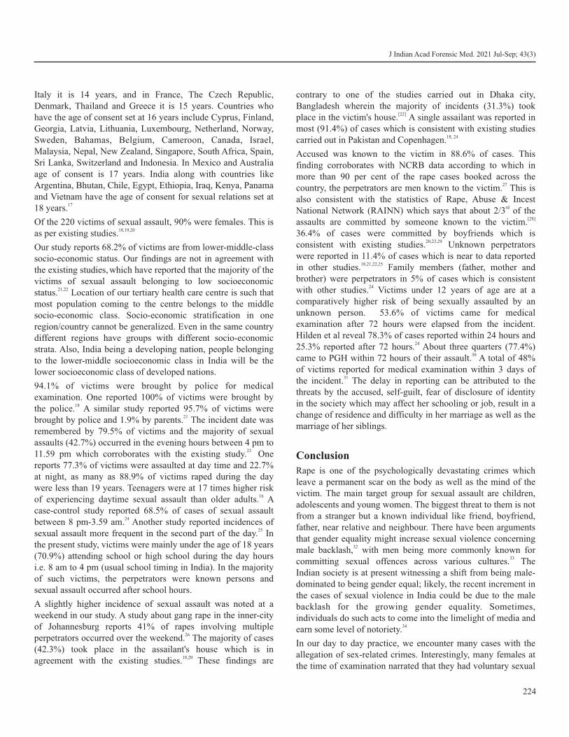
J Indian Acad Forensic Med. 2021 Jul-Sep; 43(3)
224
Italy it is 14 years, and in France, The Czech Republic, Denmark, Thailand and Greece it is 15 years. Countries who have the age of consent set at 16 years include Cyprus, Finland, Georgia, Latvia, Lithuania, Luxembourg, Netherland, Norway, Sweden, Bahamas, Belgium, Cameroon, Canada, Israel, Malaysia, Nepal, New Zealand, Singapore, South Africa, Spain, Sri Lanka, Switzerland and Indonesia. In Mexico and Australia age of consent is 17 years. India along with countries like Argentina, Bhutan, Chile, Egypt, Ethiopia, Iraq, Kenya, Panama and Vietnam have the age of consent for sexual relations set at
1718 years.
Of the 220 victims of sexual assault, 90% were females. This is 18,19,20as per existing studies.
Our study reports 68.2% of victims are from lower-middle-class socio-economic status. Our findings are not in agreement with
the existing studies, which have reported that the majority of the victims of sexual assault belonging to low socioeconomic
21,22status. Location of our tertiary health care centre is such that most population coming to the centre belongs to the middle socio-economic class. Socio-economic stratification in one region/country cannot be generalized. Even in the same country different regions have groups with different socio-economic strata. Also, India being a developing nation, people belonging to the lower-middle socioeconomic class in India will be the lower socioeconomic class of developed nations.
94.1% of victims were brought by police for medical examination. One reported 100% of victims were brought by
18the police. A similar study reported 95.7% of victims were 21brought by police and 1.9% by parents. The incident date was
remembered by 79.5% of victims and the majority of sexual assaults (42.7%) occurred in the evening hours between 4 pm to
23 11.59 pm which corroborates with the existing study. One reports 77.3% of victims were assaulted at day time and 22.7% at night, as many as 88.9% of victims raped during the day were less than 19 years. Teenagers were at 17 times higher risk
16of experiencing daytime sexual assault than older adults. A case-control study reported 68.5% of cases of sexual assault
24between 8 pm-3.59 am. Another study reported incidences of 25sexual assault more frequent in the second part of the day. In
the present study, victims were mainly under the age of 18 years (70.9%) attending school or high school during the day hours i.e. 8 am to 4 pm (usual school timing in India). In the majority of such victims, the perpetrators were known persons and sexual assault occurred after school hours.
A slightly higher incidence of sexual assault was noted at a weekend in our study. A study about gang rape in the inner-city of Johannesburg reports 41% of rapes involving multiple
26perpetrators occurred over the weekend. The majority of cases (42.3%) took place in the assailant's house which is in
18,20 agreement with the existing studies. These findings are
contrary to one of the studies carried out in Dhaka city, Bangladesh wherein the majority of incidents (31.3%) took
[22]place in the victim's house. A single assailant was reported in most (91.4%) of cases which is consistent with existing studies
18, 24carried out in Pakistan and Copenhagen.
Accused was known to the victim in 88.6% of cases. This finding corroborates with NCRB data according to which in more than 90 per cent of the rape cases booked across the
27country, the perpetrators are men known to the victim. This is also consistent with the statistics of Rape, Abuse & Incest
rdNational Network (RAINN) which says that about 2/3 of the [28] assaults are committed by someone known to the victim.
36.4% of cases were committed by boyfriends which is 20,23,29consistent with existing studies. Unknown perpetrators
were reported in 11.4% of cases which is near to data reported 18,21,22,25in other studies. Family members (father, mother and
brother) were perpetrators in 5% of cases which is consistent 24with other studies. Victims under 12 years of age are at a
comparatively higher risk of being sexually assaulted by an unknown person. 53.6% of victims came for medical examination after 72 hours were elapsed from the incident. Hilden et al reveal 78.3% of cases reported within 24 hours and
2425.3% reported after 72 hours. About three quarters (77.4%) 30came to PGH within 72 hours of their assault. A total of 48%
of victims reported for medical examination within 3 days of 31the incident. The delay in reporting can be attributed to the
threats by the accused, self-guilt, fear of disclosure of identity in the society which may affect her schooling or job, result in a change of residence and difficulty in her marriage as well as the marriage of her siblings.
Conclusion
Rape is one of the psychologically devastating crimes which leave a permanent scar on the body as well as the mind of the victim. The main target group for sexual assault are children, adolescents and young women. The biggest threat to them is not from a stranger but a known individual like friend, boyfriend, father, near relative and neighbour. There have been arguments that gender equality might increase sexual violence concerning
32male backlash, with men being more commonly known for 33committing sexual offences across various cultures. The
Indian society is at present witnessing a shift from being male-dominated to being gender equal; likely, the recent increment in the cases of sexual violence in India could be due to the male backlash for the growing gender equality. Sometimes, individuals do such acts to come into the limelight of media and
34earn some level of notoriety.
In our day to day practice, we encounter many cases with the allegation of sex-related crimes. Interestingly, many females at the time of examination narrated that they had voluntary sexual
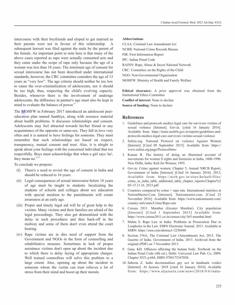
J Indian Acad Forensic Med. 2021 Jul-Sep; 43(3)
225
intercourse with their boyfriends and eloped to get married as their parents were not in favour of this relationship. A subsequent lawsuit was filed against the male by the parent of the female. An important point to note here is that many of the above cases reported as rape were actually consented acts and they came under the scope of rape only because the age of a woman was less than 18 years. The minimum age of consent for sexual intercourse has not been described under international standards, however, the CRC committee considers the age of 13 years as “very low”. The age criteria should neither be too low to cause the over-criminalization of adolescents, nor it should be too high, thus, respecting the child's evolving capacity. Besides, whenever there is the involvement of underage adolescents, the difference in partner's age must also be kept in
35mind to evaluate the balance of power.
The OHFW in February 2017 introduced an adolescent peer-Meducation plan named Saathiya, along with resource material about health problems. It discusses relationships and consent. Adolescents may feel attracted towards his/her friend or any acquaintance of the opposite or same-sex. They fall in love very often and it is natural to have feelings for someone. They must remember that such relationships are based on respect, transparency, mutual consent and trust. Also, it is alright to speak about your feelings with the concerned individual that too respectfully. Boys must acknowledge that when a girl says 'no',
36,37they mean no.
To conclude we propose:
(I) There's a need to revisit the age of consent in India and should be reduced to 16 years.
(ii) Legal consequences of sexual intercourse below 18 years of age must be taught to students. Inculcating the students of schools and colleges about sex education with special mention to the punishments will create awareness at an early age.
(iii) Proper and timely legal aid will be of great help to the victims. Many victims and their families are afraid of the legal proceedings. They also get demoralized with the delay in such procedures and thus back-off in the midway and some of them don't even attend the court hearing.
(iv) Rape victims are in dire need of support from the Government and NGOs in the form of counselling and rehabilitative measure. Sometimes in lack of proper assistance victims don't open up about the incident due to which there is delay laying of appropriate charges. Well trained counsellors will solve this problem to a large extent. Also, opening up about the incident to someone whom the victim can trust relieves a lot of stress from their mind and boost up their morale.
Abbreviations
CLAA: Criminal Law Amendment Act
NCRB: National Crime Records Bureau
FIR: First Information Report
IPC: Indian Penal Code
RAINN: Rape, Abuse & Incest National Network
CRC: Committee on the Rights of the Child
NGO: Non-Governmental Organisation
MOHFW: Ministry of Health and Family Welfare
Ethical clearance: A prior approval was obtained from the Institutional Ethics Committee
Conflict of interest: None to declare
Source of funding: None to declare
References1. Guidelines and protocols medico legal care for survivors victims of
sexual violence [Internet]. Gov.in. [cited 16 January 2016]. Available from: https://main.mohfw.gov.in/reports/guidelines-and-protocols-medico-legal-care-survivors-victims-sexual-violence
2. Safeta.org. National Protocol on violence Against Women [Internet] [Cited 09 September 2015]. Available from: https:// www.safeta.org/page/ProtocolIntro
3. Kumar R. The history of doing an illustrated account of movements for women S rights and feminism in India, 1800-1990. New Delhi, India: Kali for Women; 1993.
4. Gov.in. Crime against women; Chapter 5, Annual NRCB Report, Government of India [Internet] [Cited 16 January 2016]. 2013; Available from: https:/ /ncrb.gov.in/si tes/default /f i les/ crime_in_india_table_additional_table_chapter_reports/Chapter%205-15.11.16_2015.pdf
5. Countries compared by crime > rape rate. International statistics at NationMaster.Com [Internet]. Nationmaster.com. [Cited 23 November 2016]. Available from: https://www.nationmaster.com/ country-info/stats/Crime/Rape-rate
6. Census 2011. Mumbai (Greater Mumbai). City population [Internet] [Cited 1 September 2015] Available from: https://www.census2011.co.in/census/city/365-mumbai.html
7. Mehta S. Rape Law in India: Problems in Prosecution Due to Loopholes in the Law. SSRN Electronic Journal. 2013. Available at SSRN: https://ssrn.com/abstract=2250448
8. Section 376A, The Criminal Law (Amendment) Act, 2013. The Gazette of India. Government of India. 2013. Archived from the original (PDF) on 7 November 2013.
9. Gaur, KD. Offences affecting the human body. Textbook on the Indian Penal Code (4th ed.), Delhi: Universal Law Pub. Co, 2009; Chapter XVI; p.684, ISBN 9788175347038.
10. Saberin Z. India decriminalises gay sex in landmark verdict [Internet]. Al Jazeera. 2018 [cited 16 January 2016]. Available from: https:/ /www.aljazeera.com/news/2018/9/6/india-
J Indian Acad Forensic Med. 2021 Jul-Sep; 43(3)
226
decriminalises-gay-sex-in-landmark-verdict
11. Ghosh D. Love, Equally: Homosexuality No Longer A Crime, Says Supreme Court [Internet] [Cited 6 September 2018]. Available from: https://www.ndtv.com/india-news/homosexuality-no-longer-a-crime-in-india-supreme-court-ends-controversial-section-377-1912202
12. Crimes in India. National Crime Records Bureau. Cases Reported & Rate of Cognizable Crimes (IPC) under Different Crime Heads During 2015 and Decadal & Quinquennial Percentage Changes [Internet] [Cited 10 August 2016]. Available from: https://ncrb.gov.in.
13. Crimes in India. National Crime Records Bureau. IPC crimes 2014-2016 [Internet] [Cited 10 August 2016]. Available from: https://ncrb.gov.in
14. Lakew Z. Alleged cases of sexual assault reported to two Addis Ababa hospitals. East Afr Med J [Internet]. 2001;78(2):80–3. Available from: http://dx.doi.org/10.4314/eamj.v78i2.9093
15. Hwang KR, Han KH, Shin MS, Kim JH, Shin JJ, Song KJ, et al. A study of sexual assault: Based on data from Boramae One-stop Service Center. J Womens Med. 2010;3(3):96-101.
16. Akinlusi FM, Rabiu KA, Olawepo TA, Adewunmi AA, Ottun TA, Akinola OI. Sexual assault in Lagos, Nigeria: a five year retrospective review. BMC Women's Health. 2014 Dec 23;14(1).
17. Age of consent laws by country [Internet] [Cited 10 August 2016]. Available from: https://www.ageofconsent.net/world
18. Maqsood M et al. Incidence of alleged sexual assault cases in Lahore: It's medicolegal and social aspects. J. Fatima Jinnah Med. Univ. [Internet]. 2014 [cited 16 January 2016];8(1). Available from: https://jfjmu.com/index.php/ojs/article/view/123
19. Bandyopadhay S, Ghosh S, Adhya S, Pal K, Dalai CK. A study on sexual assault victims attending a tertiary care hospital of eastern India. OSR-JDMS [Internet]. 2013;6(6):16–9. Available from: http://dx.doi.org/10.9790/0853-0661619
20. Tamuli RP, Paul B, Mahanta P. A statistical analysis of alleged victims of sexual assault – a retrospective study. J Punjab Acad Forensic Med Toxicol 2013;13(1):7-13.
21. Sarkar SC, Sharma RK, Bhardwaj DN, Dogra TD. A study on victims and accused of sexual offences in South Delhi. Indian J Forensic Med Toxicol. 2010;4(2):90–3.
22. Al-Azad M, Raman Z, Ahmad M, Wahab M, Ali M, Khalil M. Socio-Demographic Characteristics Of Alleged Sexual Assault (Rape) Cases In Dhaka City. JAFMC Bangladesh. 2012 Apr 16;7(2).
23. Geidam A, Njoku A, Bako B. Prevalence and Nature of Sexual Assault among Female Students in a Tertiary Institution in
Maiduguri, Nigeria – A Cross Sectional Study. Int. J. Health Res O8U. 2011 Oct 7;3(4).
24. Hilden M, Schei B, Sidenius K. Genitoanal injury in adult female victims of sexual assault. Forensic Sci. Int. 2005 Nov;154(2–3).
25. Grossin C, Sibille I, Lorin de la Grandmaison G, Banasr A, Brion F, Durigon M. Analysis of 418 cases of sexual assault. Forensic Sci. Int. 2003 Jan;131(2–3).
26. Vetten L, Haffejee S. GANG RAPE: A study in inner-city Johannesburg. SA Crime Q [Internet]. 2016;(12). Available from: http://dx.doi.org/10.17159/2413-3108/2005/v0i12a1017
27. Crimes in India. National Crime Records Bureau. Offenders relation to victims of Rape-2016 [Internet] [Cited 10 August 2016]. Available from: https://ncrb.gov.in
28. Perpetrators of sexual violence: Statistics [Internet] [Cited 6 September 2018]. Available from:https://www.rainn.org/ statistics/perpetrators-sexual-violence
29. Jain R, Mathur P, Kothari N. Medicolegal evaluation of sex assault cases admitted at Sardar Patel Medical College & P.B.M. Hospital, Bikaner, India. Medico-Legal Update 8 (2008): 11-15.
30. Teerapong S, Lumbiganon P, Limpongsanurak S, Udomprasertgul V. Physical health consequences of sexual assault victims. J Med Assoc Thai. 2009 Jul;92(7).
31. Chu LC, Tung WK. The clinical outcome of 137 rape victims in Hong Kong. Hong Kong Med J. 2005 Oct;11(5).
32. Martin K, Vieraitis LM, Britto S. Gender equality and women's absolute status: a test of the feminist models of rape: A test of the feminist models of rape. Violence Against Women [Internet]. 2006;12(4):321–39. Available from: http://dx.doi.org/ 10.1177/1077801206286311
33. Sanday PR. The Socio-Cultural Context of Rape: A Cross-Cultural Study. J Soc Issues 1981 Oct;37(4).
34. Kalra G, Bhugra D. Sexual violence against women: Understanding cross-cultural intersections. Indian J Psychiatry.2013; 55(3):244-9.
35. Legal minimum ages and realization of adolescent's right in Latin America and the Caribbean [Internet] [Cited 12 February 2017]. Available from: https://www.unicef.org.
36. Ghosh A. Same-sex attraction is OK; boys can cry, girl's no means no. [Internet] [Cited 21 February 2017]. Available from: https://indianexpress.com.
37. Homosexual attraction is OK; 'NO' means no: Health Ministry rises above Indian stereotypes [Internet] [Cited 21February 2017]. Available from: https://www.financialexpress.com
AbstractOn 24th March 2020, the Government of India ordered a nationwide lockdown for 21 days, starting from 25th March, limiting the movement of India's entire 1.3 billion population as a preventive measure against the COVID-19 pandemic in India. The Lockdown eventually extended in 4 phases, spanning entire 68 days till 31st May 2020. The first of its kind lockdown impacted the lives of all the countrymen in different aspects, viz. personal, social, psychological and economic aspects. The different studies carried out in the past had proven that the profile of Medico-legal autopsy cases in particular geographical regions reflects the crime prevailing in the society. It helps to assess the social and economic profile of the deceased. It is necessary for understanding the nature of crime in a particular area. It helps identify the demographic needs and take necessary preventive measures to stop the crime or unnatural deaths using spreading awareness and psychological counselling if needed. This study was carried out retrospectively on death cases brought for medico-legal autopsies at the mortuary of Civil Hospital and B. J. Medical College, Ahmedabad between the Covid-19 pandemic lockdown period of 25th March 2020 to 31st May 2020.
KeywordsCOVID-19 pandemic; Lockdown; Autopsy; Natural deaths; Unnatural deaths; Road traffic accidents.
Profile of medico-legal autopsy cases performed during COVID-19 pandemic lockdown atmortuary of Civil Hospital and B. J. Medical College in Ahmedabad, Gujarat.
ORIGINAL ARTICLE
Introduction
Around December 2019, Chinese authorities reported acute respiratory syndrome cases in Wuhan City, Hubei province, China. This disease was called coronavirus disease 2019
1(COVID-19) and was identified to be caused by SARS-CoV-2. st thAs per statistics from 31 December 2019 to 24 March 2020,
the day before India went for Lockdown, Total 5, 13, 128 cases of COVID-19 had been reported worldwide, of which 19,193 resulted in death. At that time, India had reported around 500
2cases of COVID-19 with no casualties. The novel disease was characterized by asymptomatic transmission, sometimes intense severity and even risk of death (especially in case co-morbidities exist), lack of control options (no vaccine or effective antiviral therapy was available) and finally, lack of
3widespread diagnostic testing. To reduce transmission of COVID-19, the Government of India announced a strict nationwide lockdown, the first of its kind in the history of
thindependent India, from 25 March 2020. This Lockdown restricted people from stepping out of their homes. All transport services–road, air and rail–were suspended, except for transportation of essential goods, fire, police and emergency services. Educational institutions, industrial establishments and hospitality services were also suspended. Services such as food
shops, banks and ATMs, petrol pumps, other essentials, and manufacturing were exempted. The Home Ministry stated that anyone who fails to follow the restrictions could face a year in
4jail. Initially, Lockdown was announced for 21 days. But, due to the large population of our country and the number of cases rising every day, Government extended the Lockdown in 3
st stmore phases for a total of 68 days till 31 May 2020. From 1 June onwards, the Unlock process started in a phased manner. This 68 days lockdown created a tremendous impact on the economic, social, psychological and personal life of all citizens in one or another way. Reports from various State Crime Records Bureau suggests a significant drop in deaths and crimes in general but, The National Commission for Women (NCW) registered an increase of at least 2.5 times in domestic violence complaints since the nationwide Lockdown, according
5to official data. Studying the profile of medico-legal autopsy cases in mortuary helps to understand the nature of various crimes, the manner and mode of death during a particular time
6,7,8in a particular region. Our study aims to identify and compare the characteristics of cases referred for autopsy during the lockdown period for the COVID-19 pandemic versus the cases referred during the same period in 2019.
Material and Methods
The present study is a retrospective study of medico-legal autopsies performed at the mortuary of Civil Hospital and B. J. Medical College, Ahmedabad between the Covid-19 pandemic
th stlockdown period of 25 March 2020 to 31 May 2020. The cases were distributed based on different age groups, sex, and cause of death. The age-wise distribution consisted of age less
227
Corresponding Author
Dr. Ajay R. Madhavi (Resident Doctor)
Email: [email protected]
Mobile: +91- 9725385864
Article Historyst rdReceived: 31 August, 2020; Revision received on: 23 August, 2021thAccepted: 26 August, 2021
1 2 3 4 Patel Ankur P , Vaghela Raghurajsinh D ,Trivedi Jayjeet M ,Madhavi Ajay R .Forensic Medicine Department, B. J. Medical College, Ahmedabad, Gujarat
J Indian Acad Forensic Med. 2021 Jul-Sep; 43(3): 227-231doi:10.5958/0974-0848.2021.00057.9
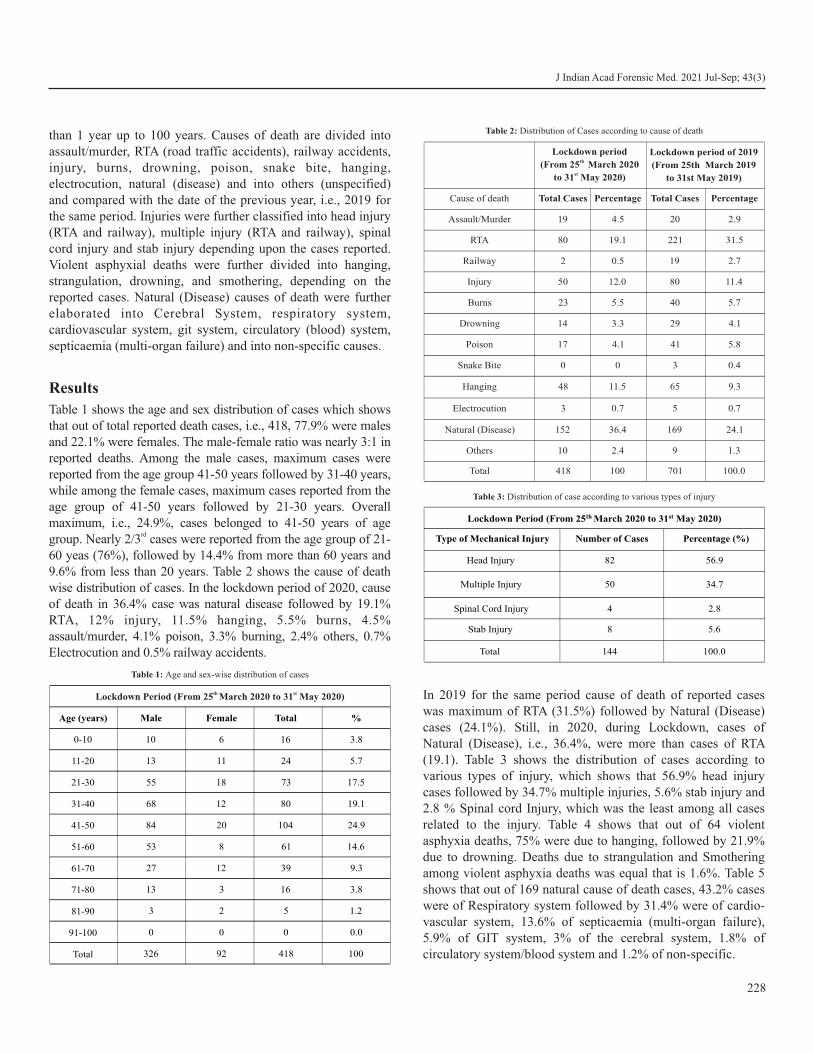
than 1 year up to 100 years. Causes of death are divided into assault/murder, RTA (road traffic accidents), railway accidents, injury, burns, drowning, poison, snake bite, hanging, electrocution, natural (disease) and into others (unspecified) and compared with the date of the previous year, i.e., 2019 for the same period. Injuries were further classified into head injury (RTA and railway), multiple injury (RTA and railway), spinal cord injury and stab injury depending upon the cases reported. Violent asphyxial deaths were further divided into hanging, strangulation, drowning, and smothering, depending on the reported cases. Natural (Disease) causes of death were further elaborated into Cerebral System, respiratory system, cardiovascular system, git system, circulatory (blood) system, septicaemia (multi-organ failure) and into non-specific causes.
Results
Table 1 shows the age and sex distribution of cases which shows that out of total reported death cases, i.e., 418, 77.9% were males and 22.1% were females. The male-female ratio was nearly 3:1 in reported deaths. Among the male cases, maximum cases were reported from the age group 41-50 years followed by 31-40 years, while among the female cases, maximum cases reported from the age group of 41-50 years followed by 21-30 years. Overall maximum, i.e., 24.9%, cases belonged to 41-50 years of age
rdgroup. Nearly 2/3 cases were reported from the age group of 21- 60 yeas (76%), followed by 14.4% from more than 60 years and 9.6% from less than 20 years. Table 2 shows the cause of death wise distribution of cases. In the lockdown period of 2020, cause of death in 36.4% case was natural disease followed by 19.1% RTA, 12% injury, 11.5% hanging, 5.5% burns, 4.5% assault/murder, 4.1% poison, 3.3% burning, 2.4% others, 0.7% Electrocution and 0.5% railway accidents.
In 2019 for the same period cause of death of reported cases was maximum of RTA (31.5%) followed by Natural (Disease) cases (24.1%). Still, in 2020, during Lockdown, cases of Natural (Disease), i.e., 36.4%, were more than cases of RTA (19.1). Table 3 shows the distribution of cases according to various types of injury, which shows that 56.9% head injury cases followed by 34.7% multiple injuries, 5.6% stab injury and 2.8 % Spinal cord Injury, which was the least among all cases related to the injury. Table 4 shows that out of 64 violent asphyxia deaths, 75% were due to hanging, followed by 21.9% due to drowning. Deaths due to strangulation and Smothering among violent asphyxia deaths was equal that is 1.6%. Table 5 shows that out of 169 natural cause of death cases, 43.2% cases were of Respiratory system followed by 31.4% were of cardio-vascular system, 13.6% of septicaemia (multi-organ failure), 5.9% of GIT system, 3% of the cerebral system, 1.8% of circulatory system/blood system and 1.2% of non-specific.
228
J Indian Acad Forensic Med. 2021 Jul-Sep; 43(3)
Lockdown period th
(From 25 March 2020stto 31 May 2020)
Cause of death Total Cases Percentage Total Cases Percentage
Assault/Murder 19 4.5 20 2.9
RTA 80 19.1 221 31.5
Railway 2 0.5 19 2.7
Injury 50 12.0 80 11.4
Burns 23 5.5
40 5.7
Drowning 14 3.3
29 4.1
Poison 17 4.1 41 5.8
Snake Bite 0 0 3 0.4
Hanging 48 11.5 65 9.3
Electrocution 3 0.7 5 0.7
Natural (Disease) 152 36.4 169 24.1
Others 10 2.4 9 1.3
Total 418 100 701 100.0
Lockdown period of 2019
(From 25th March 2019
to 31st May 2019)
Table 2: Distribution of Cases according to cause of death
Lockdown Period (From 25th March 2020 to 31st May 2020)
Type of Mechanical Injury Number of Cases Percentage (%)
Head Injury 82 56.9
Multiple Injury 50
34.7
Spinal Cord Injury 4 2.8
Stab Injury 8 5.6
Total 144 100.0
Table 3: Distribution of case according to various types of injury
th stLockdown Period (From 25 March 2020 to 31 May 2020)
Age (years) Male Female Total %
0-10
11-20
21-30
31-40
41-50
51-60
61-70
71-80
81-90
91-100
Total
10 6 16 3.8
13 11 24 5.7
55 18 73 17.5
68 12 80 19.1
84 20 104 24.9
53 8 61 14.6
27 12 39 9.3
13 3 16 3.8
3 2 5 1.2
0 0 0 0.0
326 92 418 100
Table 1: Age and sex-wise distribution of cases
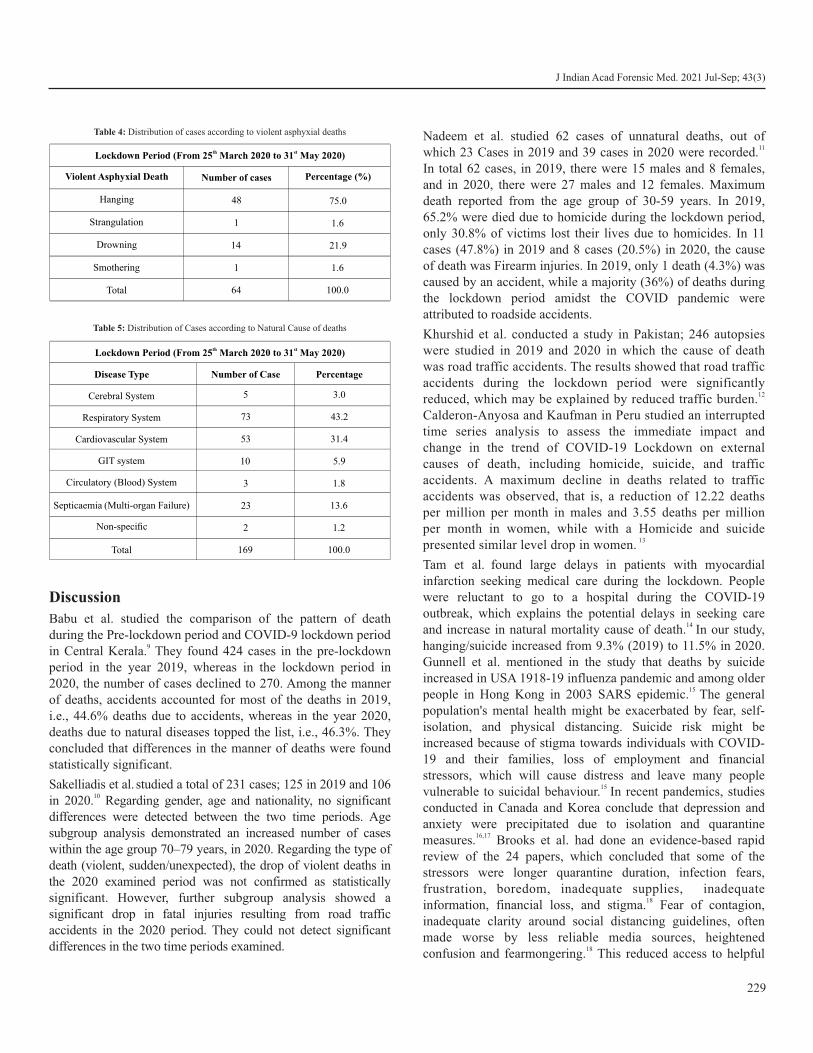
Discussion
Babu et al. studied the comparison of the pattern of death during the Pre-lockdown period and COVID-9 lockdown period
9in Central Kerala. They found 424 cases in the pre-lockdown period in the year 2019, whereas in the lockdown period in 2020, the number of cases declined to 270. Among the manner of deaths, accidents accounted for most of the deaths in 2019, i.e., 44.6% deaths due to accidents, whereas in the year 2020, deaths due to natural diseases topped the list, i.e., 46.3%. They concluded that differences in the manner of deaths were found statistically significant.
Sakelliadis et al. studied a total of 231 cases; 125 in 2019 and 106 10in 2020. Regarding gender, age and nationality, no significant
differences were detected between the two time periods. Age subgroup analysis demonstrated an increased number of cases within the age group 70–79 years, in 2020. Regarding the type of death (violent, sudden/unexpected), the drop of violent deaths in the 2020 examined period was not confirmed as statistically significant. However, further subgroup analysis showed a significant drop in fatal injuries resulting from road traffic accidents in the 2020 period. They could not detect significant differences in the two time periods examined.
Nadeem et al. studied 62 cases of unnatural deaths, out of 11 which 23 Cases in 2019 and 39 cases in 2020 were recorded.
In total 62 cases, in 2019, there were 15 males and 8 females, and in 2020, there were 27 males and 12 females. Maximum death reported from the age group of 30-59 years. In 2019, 65.2% were died due to homicide during the lockdown period, only 30.8% of victims lost their lives due to homicides. In 11 cases (47.8%) in 2019 and 8 cases (20.5%) in 2020, the cause of death was Firearm injuries. In 2019, only 1 death (4.3%) was caused by an accident, while a majority (36%) of deaths during the lockdown period amidst the COVID pandemic were attributed to roadside accidents.
Khurshid et al. conducted a study in Pakistan; 246 autopsies were studied in 2019 and 2020 in which the cause of death was road traffic accidents. The results showed that road traffic accidents during the lockdown period were significantly
12reduced, which may be explained by reduced traffic burden. Calderon-Anyosa and Kaufman in Peru studied an interrupted time series analysis to assess the immediate impact and change in the trend of COVID-19 Lockdown on external causes of death, including homicide, suicide, and traffic accidents. A maximum decline in deaths related to traffic accidents was observed, that is, a reduction of 12.22 deaths per million per month in males and 3.55 deaths per million per month in women, while with a Homicide and suicide
13presented similar level drop in women. Tam et al. found large delays in patients with myocardial
infarction seeking medical care during the lockdown. People were reluctant to go to a hospital during the COVID-19 outbreak, which explains the potential delays in seeking care
14 and increase in natural mortality cause of death. In our study, hanging/suicide increased from 9.3% (2019) to 11.5% in 2020. Gunnell et al. mentioned in the study that deaths by suicide increased in USA 1918-19 influenza pandemic and among older
15 people in Hong Kong in 2003 SARS epidemic. The general population's mental health might be exacerbated by fear, self-isolation, and physical distancing. Suicide risk might be increased because of stigma towards individuals with COVID-19 and their families, loss of employment and financial stressors, which will cause distress and leave many people
15 vulnerable to suicidal behaviour. In recent pandemics, studies conducted in Canada and Korea conclude that depression and anxiety were precipitated due to isolation and quarantine
16,17measures. Brooks et al. had done an evidence-based rapid review of the 24 papers, which concluded that some of the stressors were longer quarantine duration, infection fears, frustration, boredom, inadequate supplies, inadequate
18information, financial loss, and stigma. Fear of contagion, inadequate clarity around social distancing guidelines, often made worse by less reliable media sources, heightened
18confusion and fearmongering. This reduced access to helpful
J Indian Acad Forensic Med. 2021 Jul-Sep; 43(3)
229
th stLockdown Period (From 25 March 2020 to 31 May 2020)
Violent Asphyxial Death Number of cases Percentage (%)
Hanging 48 75.0
Strangulation 1 1.6
Drowning 14 21.9
Smothering 1 1.6
Total 64 100.0
Table 4: Distribution of cases according to violent asphyxial deaths
th stLockdown Period (From 25 March 2020 to 31 May 2020)
Disease Type Number of Case Percentage
Cerebral System 5 3.0
Respiratory System 73 43.2
Cardiovascular System 53 31.4
GIT system 10 5.9
Circulatory (Blood) System 3 1.8
Septicaemia (Multi-organ Failure) 23 13.6
Non-specific 2 1.2
Total 169 100.0
Table 5: Distribution of Cases according to Natural Cause of deaths
J Indian Acad Forensic Med. 2021 Jul-Sep; 43(3)
230
but "non-essential" psychiatric services and ultimately increased 19suicide cases.
Venter et al. has conducted an observational retrospective audit of the patient register and studied 4 300 trauma presentations secondary to interpersonal violence and Road traffic collisions (RTC) between February - June 2019 and 2020 vs 3 239 presentations in February - June 2020. Significant reduction (40% decline) was observed in the number of RTCs, while declines in the volume of trauma cases secondary to interpersonal violence and of overall trauma cases in 2020, was not statistically
20 significant. Zsilavecz et al had studied trauma pattern in during COVID-19 Lockdown in South Africa, which also concluded that as a significant decrease in Motor vehicle accidents, pedestrian-
21 vehicle accidents and interpersonal violence. Yasin et al. conducted a narrative review on the effects of the COVID-19 pandemic and reported the reduction in overall absolute numbers
22of RTCs and their deaths and injuries globally.
Conclusion
The present study was conducted at mortuary of Civil Hospital and B. J. Medical College, Ahmedabad to know the profile of medico-legal autopsy cases performed during covid-19
th stpandemic lockdown i.e., from 25 March 2020 to 31 May 2020 on total cases comprising 418 medico-legal autopsies. A maximum number of cases were recorded in the age group of 21-50 years. Male cases are more in number as compared to female. Death due to Natural causes (Disease) cases are more in number. On comparing our study data with the previous year 2019 data of the same period, we found that RTA and Railway accidents deaths are drastically reduced as all road and rail services were suspended. Unnatural deaths due to injury, burns, drowning, poisoning and snake bites are reduced to more than half in number in Lockdown compared to previous year. In Injury cases, head Injuries were more in number. Hanging cases were more common amongst violent asphyxial deaths.
Ethical clearance: A prior approval was obtained from the Institutional Ethics Committee
Conflict of interest: None to declare
Source of funding: None to declare
References1. World Health Organisation. Coronavirus (Covid-19) Pandemic.
Available from: https://www.who.int/emergencies/diseases/novel-coronavirus-2019
2. Worldometer. Covid-19 Coronavirus Pandemic Statistics. Available from: https://www.worldometers.info/coronavirus/
3. Ministry of Health and Family Welfare, Government of India, New Delhi. Guidelines of Clinical Management of Covid-19 patients in
India. Available from: https://www.mohfw.gov.in/ pdf/ Revised NationalClinicalManagementGuidelineforCOVID1931032020.pdf
4. Ministry of Home Affairs. Order regarding Lockdown on 24/03/2020. Available from: https://www.mha.gov.in/ sites/default/files/Guidelines_0.pdf
5. The Hindu - Newspaper. Covid-19 Lockdown - Rise in domestic violence, police apathy: NCW. Available from: https:// www.thehindu.com/news/national/covid-19-lockdown-spike-in-domestic-violence-says-ncw/article31238659.ece
6. Biswas G. Review of Forensic Medicine and toxicology. Jaypee Brothers Medical Publishers. 3rd Edition. p. 98.
7. Bardale R. Principal of Forensic Medicine and Toxicology. Jaypee Brothers Medical Publishers. 3rd Edition. p. 119.
8. Kannan K, Mathiharan K. Textbook of Medical Jurisprudence and Toxicology. 24th Ed. LexisNexis Butterworth's publication: 2012. p. 293, 295, 297,360.
9. Babu SS, Raveendran R, Ka A. Comparison of pattern of death during Pre-lockdown period and COVID 19 lockdown period in Central Kerala – An Autopsy Study. Asian J Med Sci [Internet]. 2021;12(7):17–21. Available from: ht tp: / /dx.doi .org/ 10.3126/ajms.v12i7.36436
10. Sakelliadis EI, Katsos KD, Zouzia EI, Spiliopoulou CA, Tsiodras S. Impact of Covid-19 lockdown on characteristics of autopsy cases in Greece. Comparison between 2019 and 2020. Forensic Sci Int [Internet]. 2020;313(110365):110365. Available from: http://dx.doi.org/10.1016/j.forsciint.2020.110365
11. Nadeem, S., Sarwar, S., Rehman, H., Iftikhar, H., Saleem, N., Tariq, F. Pattern of unnatural deaths during COVID-19 Lockdown in comparison with deaths reported during 2019 in Sahiwal city. J. Rawalpindi Med. Coll. 2021; 25 COVID-19 Supplement-1, 84-88
12. Khurshid A, Sohail A, Khurshid M, Shah MU, Jaffry AA. Analysis of road traffic accident fatalities in Karachi, Pakistan: an autopsy-based study. Cureus. 2021 Apr;13(4):6-15. DOI:10.7759/ cureus.14459
13. Calderon-Anyosa RJ, Kaufman JS. Impact of COVID-19 lockdown policy on homicide, suicide, and motor vehicle deaths in Peru. Prev. Med. 2021;143(4):83-6. https:// doi.org/ 10.1016/ j.ypmed.2020.106331
14. Tam CF, Cheung KS and Lam S. Impact of Coronavirus Disease 2019 (COVID-19) Outbreak on ST-Segment-Elevation Myocardial Infarction Care in Hong Kong, China. Circ Cardiovasc Qual Outcomes. 2020; 13(4):e006631. https://doi.org/10.1161/ CIRCOUTCOMES.120.006631
15. Gunnell D, Appleby L, Arensman E, Hawton K, John A, Kapur N, et al. Suicide risk and prevention during the COVID-19 pandemic. The Lancet Psychiatry. 2020;7(6):468-71. https://doi.org/ 10.1016/S2215-0366(20)30171-1
16. Hawryluck L, Gold WL, Robinson S, Pogorski S, Galea S, Styra R, SARS control and psychological effects of quarantine, Toronto, Canada. Emerg Infect Dis. 2004; 10:1206-12. PMID:15324539
17. Jeong H, Yim HW, Song Y-J, et al., Mental health status of people isolated due to Middle East Respiratory Syndrome. Epidemiol Health2016;38:e2016048. doi:10.4178/epih.e2016048 pmid:28196409
18. Brooks SK, Webster RK, Smith LE, Woodland L, Wessely S, Greenberg N, et al. The psychological impact of quarantine and how to reduce it: rapid review of the evidence. Lancet. 2020; 395(10227):912-920.
19. Venkatesh A and Edirappuli S. Social distancing in covid-19: what are the mental health implications? BMJ Clinical Research ed. 2020; 369:m1379
20. Venter A, Lewis C, Saffy P, Chadinha L. Locked down: Impact of COVID-19 restrictions on trauma presentations to the emergency department. S. Afr. Med. J. 2021;111(1):52-6. https://doi.org/ 10.7196/SAMJ.2021.v111i1.15289
21. Zsilavecz A, Wain H, Bruce JL, Smith MTD, Bekker W, Laing GL, Lutge E, Clarke DL. Trauma patterns during the COVID-19 Lockdown in South Africa expose the vulnerability of women. S Afr Med J. 2020 Oct 28;110(11):1110-1112. DOI: 10.7196/ SAMJ.2020.v110i11.15124. PMID: 33403988.
22. Yasin YJ, Grivna M, Abu-Zidan FM. Global impact of COVID-19 pandemic on road traffic collisions. World J Emerg Surg. 2021 Sep 28;16(1):51. DOI: 10.1186/s13017-021-00395-8. PMID: 34583713
J Indian Acad Forensic Med. 2021 Jul-Sep; 43(3)
231
AbstractMedico legal autopsy is a statutory requirement in all cases of sudden, suspicious and unnatural deaths. Profiling of these autopsies helps one to understand the pattern seen among them and guide the investigating authorities and the health promotion & prevention programmes to direct their measures in this respect. The present study is a retrospective, cross-sectional, record-based study carried out in a mortuary under Dept. of Forensic Medicine & Toxicology at Purulia GMCH. Out of the total 1137 medicolegal autopsies, 225 cases were of brought-dead cases. Among the unnatural deaths, the most frequent cases were due to hanging (282 cases, 24.8%) followed by road traffic accidents (176 cases, 15.47%) and burns (99 cases, 8.7%). According to the gender, 761 cases (66.93%) were of males, 368 cases (32.36%) were females and in 8 cases (0.7%) the gender could not be ascertained (foetuses) on autopsy examination alone. Majority of the cases belonged to the age group of 21-30 years (261 cases, 22.95%) followed by 31-40 years (216 cases,18.99%) and 41-50 years (165 cases, 14.51%). Death due to road traffic injuries is a preventable occurrence and proper safety riding measures should be observed by all. Although legislation is in place for sale and purchase of poisonous substances, but still it has to be well implemented.
KeywordsAutopsy profile; Hanging; Medico legal autopsy; Road traffic injuries
An analysis of medicolegal autopsies in a tertiary care centre in West Bengal - A morgue-based study
ORIGINAL ARTICLE
Introduction
Medico legal autopsy is a statutory requirement in all cases of sudden, suspicious and unnatural deaths. Profiling of these autopsies helps one to understand the pattern seen among them and guide the investigating authorities and the health promotion & prevention programmes to direct their measures in this respect. The study will also help to find out the profile at current place of study and compare it with the rest of the states and in the country. The present study is a humble attempt to provide information regarding the type of causes of unnatural deaths and their respective epidemiology in this central district of West Bengal.
Materials and Methods
The present study is a retrospective, cross-sectional, record-based study carried out in a mortuary under Dept. of Forensic Medicine & Toxicology at Purulia GMCH. All the medicolegal autopsies that have presented to the above said department
st stduring the period 1 Jan'19 to 31 Dec'19 were included in this study. The study parameters are age, sex and type of cases. The
data thus collected is analysed in the form of tables and frequency distribution and compared with other similar studies.
Results
A total of 1137 medicolegal autopsies were conducted at morgue attached to Dept. of Forensic Medicine and Toxicology, Purulia GMCH during the period of 01 Jan'19 to 31 Dec'19. Out of these, 225 cases were brought-dead cases. Among the unnatural deaths, the most frequent cases were due to hanging (282 cases, 24.8%) followed by road traffic accidents (176 cases, 15.47%) and burns (99 cases, 8.7%). The other cases of unnatural deaths in decreasing order of frequency are railway injuries (96 cases, 8.44%), poisonings (63 cases,5.54%), drowning (61 cases, 5.36%), assault/trauma (37 cases, 3.25%) lightning strokes (31 cases,2.72%), electrocution (26 cases, 2.28%), snake bites (25 cases, 2.19%), foetal autopsies (13 cases, 1.14% cases) and skeleton examinations (3 cases, 0.26%). According to the gender, 761 cases (66.93%) were of males, 368 cases (32.36%) were females and in 8 cases (0.7%) the gender could not be ascertained (foetuses) on autopsy examination alone.
Majority of the cases belonged to the age group of 21-30 years (261 cases, 22.95%) followed by 31-40 years (216 cases,18.99%) and 41-50 years (165 cases, 14.51%). Among the rest of the cases, the age-wise incidence in decreasing order of frequency is as follows: - 51-60 years (153 cases,13.45%), 11-20 years (149 cases, 13.10%) 61-70 years (81 cases, 7.12%), 1-10 years (39 cases,3.43%), 71-80 years (35 cases, 3.07%),
232
Corresponding Author
Dr Vikas Gurbani (Associate Professor)
Email: [email protected]
Mobile: +91-7507828000
Article Historyth thReceived: 30 November, 2020; Revision received on: 30 July, 2021rdAccepted: 03 August, 2021
1 2 1 2 Shobhan Roy , Shagun Thakur , Sumanta Malick , Vikas Gurbani1 Department of F.M.T., Deben Mahata (Purulia) Government Medical College, Purulia, West Bengal.2 Department of F.M.T., Kalinga Institute of Medical Sciences (KIMS) & PBMH, Bhubaneswar
J Indian Acad Forensic Med. 2021 Jul-Sep; 43(3): 232-234doi:10.5958/0974-0848.2021.00058.0
below 1 year (21 cases, 1.84%) and 81-90 years (17 cases, 1.49%). Majority of the cases were of heart disease (92 cases, 40.89%). This was followed by cases of Heart and Lung Disease (45 cases, 20%), lung disease (34 cases, 15.11%), Liver Disease (23 cases, 10.22%), Septicaemia (12 cases, 5.33%), Brain disease (4.89%) and Pregnancy complications (8 cases, 3.56%). Based on the position of knot, out of 282 cases, 273 cases (96.8%) were of atypical hanging and 9 cases (3.2%) were of typical hanging. On the basis of degree of suspension, 275 cases (97.5% were of complete hanging and 7 cases (2.5%) were of partial hanging.
Discussion
In the present study, hanging has been found as the most common form of unnatural death seen. A study has reported hanging as the second most common cause in their study whereas in other authors, have found hanging as the third most
3common unnatural cause of death in their respective studies. 1-
The reason for higher incidence of hanging cases was found to be mental depression. The cause of the depression could not be substantiated in the present study. The number of hanging cases is substantial also because the place of study happens to be a referral centre for the nearby regions and hence, all cases from nearby centres are referred here. The cases of Road Traffic Injuries were found to be the second most common in the study. Many research studies, and have reported death due to the effects of R.T.I.s as the most common cause of unnatural death.
3-7 The proximity of the present institute to state and national highways and also the institute being a referral centre for the region (district headquarter), a higher incidence of road traffic injuries cases is seen. Poisoning cases constitute the third most common finding in the present study. Committing suicide by poison is still a prevalent problem in our country and globally also, as it is one of easiest methods to execute. Some authors
have reported poisoning as the first most common finding in their study whereas others have reported poisoning cases as the second most common finding in their respective studies. 5-7,8,9
In our search for the literature on the present topic, all the studies have found a higher incidence in males as compared to females. The higher incidence in males can be attributed to 1-9
the fact that male are the bread-earners in most of the families and hence for that they have to go out for work. Therefore, they are more vulnerable to the outside forces in play which can be fatal. The stress of earning a livelihood and providing to the dependents is also a burden carried by them. All the other studies in our search have reported the highest incidence in the
1-9age group of 21-30 years. The reason for this higher incidence in this age group could be because this age group is the most economically productive age-group and also this age-group is more prone to high-risk taking due to biological hormones.
They are also more prone to stress due to adapting to the responsibilities of life.
In our study, it is seen that all the brought dead cases had died due to natural causes. In another study, it has been reported that in 67.74% of the brought dead cases, history of major past
10illness was present. In one research on brought dead cases to 11hospital, 51.5% cases were due to unnatural deaths. In the
present study, 97.5% cases were of complete hanging and 96.8% cases were of atypical hanging. In another study, the authors have reported hanging was complete in 75.00% cases
12and was atypical in 68.75% cases.
Conclusion
The highest incidence of unnatural deaths is seen in the age group of 21-30 years and hence the preventive strategies and measures should be directed towards this age group. The preventive strategies include programmes directed towards awareness about mental health. Death due to road traffic injuries is a preventable occurrence and proper safety riding measures should be observed by all and the condition of roadways should also be improved. Although legislation is in place for sale and purchase of poisonous substances, but still it has to be well implemented.
Ethical clearance: A prior approval was obtained from the Institutional Ethics Committee
Conflict of interest: None to declare
Source of funding: None to declare
References1. Bodupally RK, Sheikh N. Comprehensive and Analytical Study of
Medico Legal Autopsies at Tertiary Care Center. Indian J. Forensic Med. Toxicol. 2019;13(1):139
2. Bhabhor R, Parmar A. Profile of medicolegal autopsies at a tertiary centre in bhavnagar region. J. Indian Acad Forensic Med. 2018; 40(4): 383-386.
3. Kumar P, Singh R, Buri S, Pal C, Saini O, Kumar S. Profile of medicolegal autopsy cases conducted at a tertiary care centre in Bikaner, Rajasthan: A one-year retrospective study. Int J Med Biomed Studies [Internet]. 2020Aug.25 [cited 2021Sep.29];4(8). Available from: https://www.ijmbs.info/index.php/ ijmbs/ article/view/1363
4. Radhakrishna KV et al. Profile of medicolegal autopsies conducted at tertiary medicolegal centre in southwestern India. Int J Health Biomed Res. 2015; 3 (2): 70-75
5. Murali MCM, Yadukul S, Kiran J. Profile of Medico-legal Autopsies Done at Sri Devaraj URS Medical College, Kolar for a Period of 3 Years. Med. Leg. 2017; 17 (2) : 197
6. Junaidi KA, Pujar SS, Honnungar RS, Jirli PS, Koulapur VV, Ali K, Pushpa MG. Profile of Medicolegal Autopsy Cases at Tertiary
233
J Indian Acad Forensic Med. 2021 Jul-Sep; 43(3)
Care Centre in Belagavi, Karnataka. A One Year Retrospective Study. Med. Leg.2020; 20(1): 170-174
7. Manish K, Junaidi KA , Deepak S, Umesh SR. Profile of Medicolegal Autopsy Cases Conducted at Tertiary Care Centre, Kalaburagi, Karnataka. J. Forensic Med. Toxicol. 2020 :14(3), 269-274.
8. Batra AK, Dongre AP. A preliminary analysis of medicolegal autopsies performed over five years in a rural health district of Maharashtra state of India. J. Forensic Med. Toxicol. 2003. 20(1): 41-46.
9. Gannur DG, Nuchhi UC, Yoganarasimha K. A Study of Demography of Medico Legal Autopsies in Bijapur. J. Forensic Med. Toxicol. 2013; 7(1): 37-41.
10. Saiyed MZG, Jani RC, Jani CB. Brought dead cases at a tertiary care hospital in Ahmedabad with reference to dispensing with the need of medico legal post-mortem examination: Provisions, Protocol & Procedure. J Indian Acad Forensic Med. 2021;43(1): 60-64.
11. Noone P, Khan F. An Approach to Brought Dead Cases To Hospital-An Autopsy Based Study. J Indian Acad Forensic Med. 2017;39(3): 255-260
12. Guntheti BH, Sheikh K, Singh UP. Profile and Pattern of Hanging Cases at a Tertiary Care Hospital, Khammam; Telangana. J Indian Acad Forensic Med. 2016; 38(1): 67-71.
J Indian Acad Forensic Med. 2021 Jul-Sep; 43(3)
234
AbstractProgress is impossible without change, and those who cannot change their minds cannot change anything” by George Bernard Shaw. Using Zoom technology to deliver lecture, connect students and enable anytime, anyplace learning is increasing in COVID-19 pandemic, however keeping students engaged in online learning is difficult in medical education. The study aimed to determine the student's perception regarding online classes in Forensic medicine. We used zoom video lectures which can be accessed by students from anywhere. The study was conducted at Mayo Institute of Medical Sciences, Barabanki. It is a cross-sectional questionnaire-based study. Analysis was done on percentage wise distribution of various parameters used. The study was conducted among 152 study participants, out of which 82(53.95%) were male and 70(46.06%) were female. Majority of the study participants 95(62.5%) felt online classes help them in understanding the topic adequately. 120(78.9%) respondants perceived that only teacher talks during online classes.140(92.1%) respondents feel major limitation of online class is in regard to practicals. Overall opinion of majority of participants is positive and in favour of online classes to avoid exposure to COVID -19 pandemic. However, practical /clinical exposure will have to be ensured in the training of Indian medical graduates. Normally student's perception regarding online zoom classes in forensic medicine was determined to be positive. Practical class learning is indispensible in medical education, which will have to be incorporated as situation permit.
KeywordsOnline classes; Student perceptions; COVID-19 pandemic; Percentage wise distribution.
ndPerception of 2 year MBBS students about online zoom classes during COVID-19 pandemic: A questionnaire-based study
ORIGINAL ARTICLE
Introduction
Coronavirus pandemic has not only impacted human life but 1also impacted medical education and residency all over.
Educational institutions were shut down because of the lock-down; Medical Colleges started online classes to ensure
2continuity of education. India is not new to online education. Teachers and Colleges are putting extra effort to engage students in online classes by rescheduling timetables, discussions online and taking feedback by means of questionnaire at the end of lecture series. Tutorials are assigned online to encourage self-study.
Materials and MethodsstA Cross-sectional study was conducted during 01 April and
th nd30 September 2020 among 2 year MBBS students of Mayo institute of medical sciences, Barabanki with the objective to
ndevaluate the perception among 2 year MBBS students in relation to online classes and to modify the teaching methods if
required. Online classes were conducted once every week for 6 months in forensic medicine Department, MIMS, Barabanki . Free version of zoom platform was used which allows maximum of 80 students at a time and for time period of 40 minutes. Student's perceptions were collected by online prevalidated questionnaire at the end of this lecture series. Participation was on voluntary basis. Participants were asked to choose their response from the two alternatives. Responses were obtained on google form online due to COVID-19 pandemic. Only participants who attended 75% of online classes were included. Data was entered into Microsoft excel sheet and analyzed using SPSS version 15. Data was summarized using percentages with the confidence level of 95%.
ResultsndThere were total of 152 2 year MBBS student participants in
the online survey who fulfilled the criteria, out of which 82(53.95%) were male and 70(46.06%) were female. A majority of respondents, that is 95(62.5%) felt online classes help them in understanding the topic while 57(37.5%) of them feel online classes are of no use. It is evident from Table 1. Most of the participants 145(95.4%) could easily be able to use the class link and did not need any assistance in joining zoom online classes. While 7(4.6%) of the respondents had difficulty due to poor internet connectivity. In response to quality of teaching, 120(78.9%) study participants perceive it as one way
235
Corresponding Author
Dr Anju Singh (Professor)
Email: [email protected]
Mobile: +91-8765444501
Article Historyth thReceived: 20 November, 2020; Revision received on: 20 July 2021 thAccepted: 25 July, 2021
1 2 Amit Kumar Singh , Anju Singh1 Dept. of Forensic Medicine and Toxicology, Mayo Institute of Medical Sciences, Barabanki- 225001
2 Dept. of Forensic Medicine and Toxicology, Autonomous State Medical College, Ayodhya- 224133
J Indian Acad Forensic Med. 2021 Jul-Sep; 43(3): 235-237doi:10.5958/0974-0848.2021.00059.2
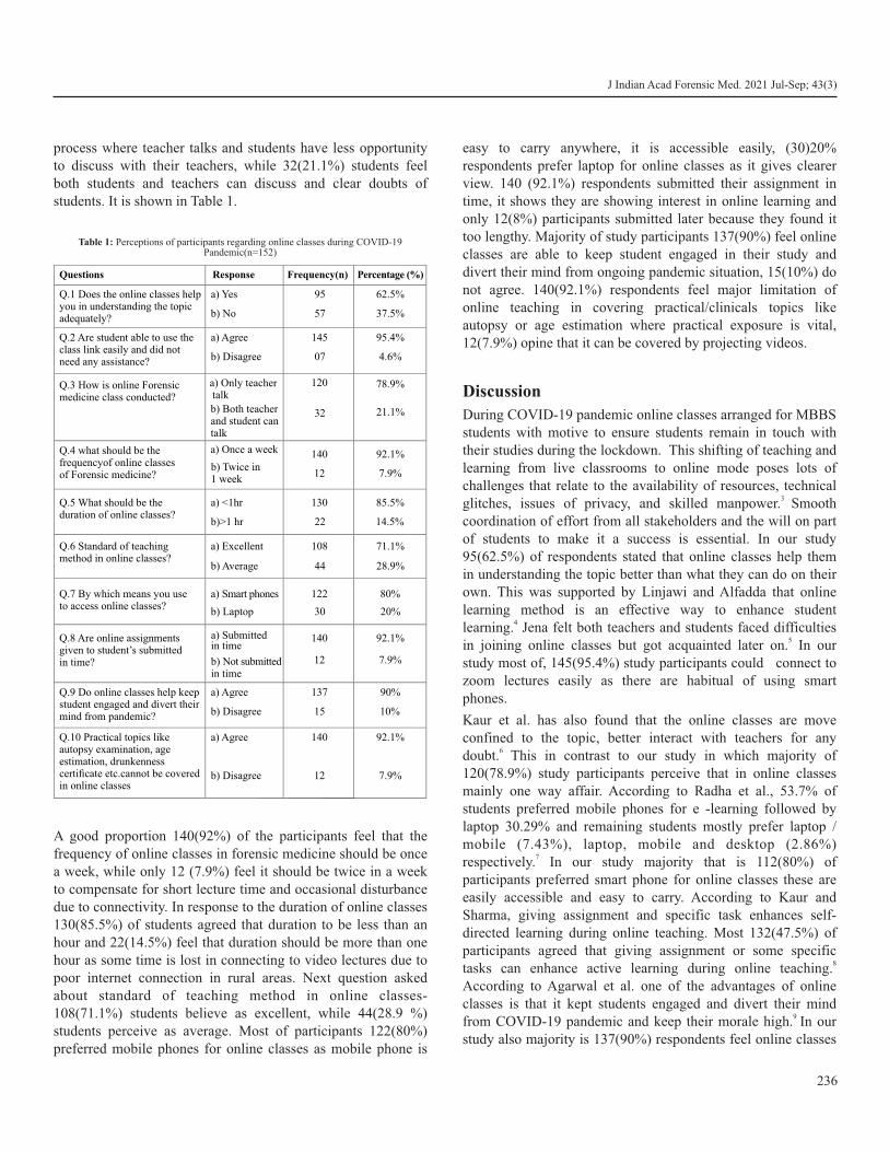
process where teacher talks and students have less opportunity to discuss with their teachers, while 32(21.1%) students feel both students and teachers can discuss and clear doubts of students. It is shown in Table 1.
A good proportion 140(92%) of the participants feel that the frequency of online classes in forensic medicine should be once a week, while only 12 (7.9%) feel it should be twice in a week to compensate for short lecture time and occasional disturbance due to connectivity. In response to the duration of online classes 130(85.5%) of students agreed that duration to be less than an hour and 22(14.5%) feel that duration should be more than one hour as some time is lost in connecting to video lectures due to poor internet connection in rural areas. Next question asked about standard of teaching method in online classes-108(71.1%) students believe as excellent, while 44(28.9 %) students perceive as average. Most of participants 122(80%) preferred mobile phones for online classes as mobile phone is
easy to carry anywhere, it is accessible easily, (30)20% respondents prefer laptop for online classes as it gives clearer view. 140 (92.1%) respondents submitted their assignment in time, it shows they are showing interest in online learning and only 12(8%) participants submitted later because they found it too lengthy. Majority of study participants 137(90%) feel online classes are able to keep student engaged in their study and divert their mind from ongoing pandemic situation, 15(10%) do not agree. 140(92.1%) respondents feel major limitation of online teaching in covering practical/clinicals topics like autopsy or age estimation where practical exposure is vital, 12(7.9%) opine that it can be covered by projecting videos.
Discussion
During COVID-19 pandemic online classes arranged for MBBS students with motive to ensure students remain in touch with their studies during the lockdown. This shifting of teaching and learning from live classrooms to online mode poses lots of challenges that relate to the availability of resources, technical
3 glitches, issues of privacy, and skilled manpower. Smooth coordination of effort from all stakeholders and the will on part of students to make it a success is essential. In our study 95(62.5%) of respondents stated that online classes help them in understanding the topic better than what they can do on their own. This was supported by Linjawi and Alfadda that online learning method is an effective way to enhance student
4learning. Jena felt both teachers and students faced difficulties 5in joining online classes but got acquainted later on. In our
study most of, 145(95.4%) study participants could connect to zoom lectures easily as there are habitual of using smart phones.
Kaur et al. has also found that the online classes are move confined to the topic, better interact with teachers for any
6doubt. This in contrast to our study in which majority of 120(78.9%) study participants perceive that in online classes mainly one way affair. According to Radha et al., 53.7% of students preferred mobile phones for e -learning followed by laptop 30.29% and remaining students mostly prefer laptop / mobile (7.43%), laptop, mobile and desktop (2.86%)
7respectively. In our study majority that is 112(80%) of participants preferred smart phone for online classes these are easily accessible and easy to carry. According to Kaur and Sharma, giving assignment and specific task enhances self-directed learning during online teaching. Most 132(47.5%) of participants agreed that giving assignment or some specific
8tasks can enhance active learning during online teaching. According to Agarwal et al. one of the advantages of online classes is that it kept students engaged and divert their mind
9 from COVID-19 pandemic and keep their morale high. In our study also majority is 137(90%) respondents feel online classes
236
J Indian Acad Forensic Med. 2021 Jul-Sep; 43(3)
Questions Response Frequency(n) Percentage (%)
Q.1 Does the online classes helpyou in understanding the topic adequately?
a) Yes
b) No
95
57
62.5%
37.5%
Q.2 Are student able to use the class link easily and did not need any assistance?
a) Agree
b) Disagree
145
07
95.4%
4.6%
Q.3 How is online Forensic medicine class conducted?
a) Only teacher talk
b) Both teacher and student can talk
120
32
78.9%
21.1%
Q.4 what should be the frequencyof online classes of Forensic medicine?
a) Once a week
b) Twice in1 week
140
12
92.1%
7.9%
Q.5 What should be the duration of online classes?
a) <1hr
b)>1 hr
130
22
85.5%
14.5%
Q.6 Standard of teaching method in online classes?
a) Excellent
b) Average
108
44
71.1%
28.9%
Q.7 By which means you use to access online classes?
a) Smart phones
b) Laptop
122
30
80%
20%
Q.8 Are online assignmentsgiven to student’s submitted in time?
a) Submitted in time
b) Not submitted in time
140
12
92.1%
7.9%
Q.9 Do online classes help keepstudent engaged and divert their mind from pandemic?
a) Agree
b) Disagree
137
15
90%
10%
Q.10 Practical topics like autopsy examination, age estimation, drunkenness certificate etc.cannot be coveredin online classes
a) Agree
b) Disagree
140
12
92.1%
7.9%
Table 1: Perceptions of participants regarding online classes during COVID-19 Pandemic(n=152)
keep them engaged in their study. In our study a good proportion 140(92.1%) of participants feel online classes for theoretical topics can be taught well but practical topics like Autopsy examination, Age estimation, Drunkenness examination need class room learning. A similar finding has also been reported by Mukhtar et al. where respondents said that in online learning modalities, they were unable to learn
10practical and clinical work.
Conclusion
COVID-19 Pandemic made us understand the importance of online classes for our undergraduate students. This study has helped in knowing student's perspective relating to online classes and it will be useful in modifying teaching patterns in coming years. During this study we found that almost all of the students are satisfied with online classes. Major advantages of online education are in the form of - flexibility, convenience and it can be accessed from anywhere. Online education is the new trend. In future, we will be going to see application of technology tools and gadgets will be inevitable.
Ethical clearance: A prior approval was obtained from the Institutional Ethics Committee
Conflict of interest: None to declare
Source of funding: None to declare
References1. Chick R, Clifton G, Peace K, Propper B, Hale D, Alseidi A et al.
Using Technology to Maintain the Education of Residents During the COVID-19 Pandemic. J Surg Educ. 2020;77(4):729-732.
2. Thomas A, Shenoy M, Shenoy K.Survey among Medical Students during COVID-19 Lockdown:The Online Class Dilemma. Int J Med Students. 2020;8(2):102-6.
3. D'Orville H. COVID-19 causes unprecedented educational disruption: Is there a road towards a new normal?. PROSPECTS. 2020;49(1-2):11-15.
4. Linjawi AI, Alfadda LS. Students' perception, attitudes, and readiness toward online learning in dental education in Saudi Arabia: a cohort study. Adv Med Educ Pract. 2018;9:855-863.
5. Jena P. Indian Education in Pandemic COVID-19. SSRN Electronic Journal. 2020;12(7):12582-86.
6. Kaur N, Dwivedi D, Arora J, Gandhi A. Study of the effectiveness of e-learning to conventional teaching in medical undergraduates amid COVID-19 pandemic. Natl J Physiol Pharm Pharmacol 2020;10(07):563-7.
7. Radha R, Mahalakshmi K, Kumar VS, Saravanakumar AR. E-Learning during Lockdown of Covid-19 Pandemic: A Global Perspective. Int J Control Autom Syst. 2020;13(4):1088-99.
8. Kaur S, Sharma A. Online classes of nursing students during lockdown (COVID pandemic): perception and issues. Int J of Adv Res. 2020;8(8):494-500.
9. Agarwal S, Kaushik J. Student's Perception of Online Learning during COVID Pandemic. Indian J Pediatr. 2020;87(7):554-554.
10. Mukhtar K, Javed K, Arooj M, Sethi A. Advantages, Limitations and Recommendations for online learning during COVID-19 pandemic era. Pak J Med Sci. 2020;36(COVID19-S4): S27-31.
J Indian Acad Forensic Med. 2021 Jul-Sep; 43(3)
237
AbstractThe study of “Association between suicides among females and phase of uterine cycle during autopsy” was carried out at a tertiary care centre over a period of 5 years with aim to identify the phase of uterine cycle in female suicides and to correlate the phase of uterine cycle with methods and reasons of suicide. A total of 222 cases fulfilled criteria, most vulnerable age group was 21-30 years (112 cases) and most vulnerable phase of uterine cycle was menstruation phase-88 cases (40%) followed by secretory phase-68 cases (31%). Most female suicides (79%) were due to hanging. Majority of suicidal cases during the 11–20-year (24 cases) and 21–30-year (50 cases) age groups were menstruating. In contrast, majority of suicidal cases during the 31–40-year (13 cases) and 41–50-year (8 cases) age groups were in secretory phase. Dysmenorrhoea (13 cases) and stomach pain (9 cases) were the reasons for the suicide exclusively in menstruating women, whereas depression (12 cases) was seen more in secretory phase and husband related conflicts were almost equally distributed in secretory (10 cases) and menstrual (9 cases) phases. Thus, this study indicates the time of applying the various strategies to curb the unhealthy emotional state of a woman in the form of health education, counseling and treatment of PMTS, a period between 4 days prior to start of menses to the end of menstruation; wherein a woman is vulnerable for sadness and impulsive acts.
KeywordsSuicide; Uterine cycle; PMS; PMDD; Menstruating; Secretory
Association between suicides among females and phase of uterine cycle during autopsyat a tertiary care centre in Bengaluru north
ORIGINAL ARTICLE
Introduction
Worldwide, suicide is an important public health hazard. In India, suicide rate is approximately 8.0 per 1 lakh in females.
While the population increase in the last decade was 25%, the 1suicide rate increased by 60%. Significant risk factors for
suicide are sudden impulsive decision of ending their life taken during height of emotion due to stress such as from financial
2difficulties, troubles with relationships, or from bullying and a 3 poor handling of emotions. A number of psychological states
increase the risk of suicide in people with low suicidal intention including: hopelessness, anxiousness, loss of pleasure in life and depression. The menstrual cycle also is a trigger for the onset of depressive disorders, including premenstrual dysphoric disorder (PMDD or PMTS), a disorder specific to the luteal phase of the menstrual cycle; and depression associated with
4the transition to menopause. PMTS is an entity that causes considerable morbidity, and in about 3% of the women, it may disrupt the woman's life when severe symptoms like anger
5attacks, depression and suicidal thoughts recur. Thus, these
types of studies are known to give a comprehensive view of the root of the problem and help us draw various strategies to prevent suicide in females.
Materials and Methods
The present study was a descriptive study conducted to identify the phase of uterine cycle in female suicides and to correlate the phase of uterine cycle with methods and reasons of suicide. All cases with history of suicide in females subjected for medico-legal autopsy at Department of Forensic Medicine in a tertiary care centre, Bengaluru from December 2013 to December 2018.History was furnished by police in forms 146 (i) and (ii) and further proforma was filled by interviewing the relatives and friends of deceased, who accompanied the dead body. This proforma was used to collect the socio-demographic details of each suicide case (age, time of incidence, past medical history, personal history, LMP etc.) including various psycho-social variables associated with suicide (interpersonal relationship, various stressors, substance abuse, psychiatric illness, physical illness, chronic pain and disablement, suicide note, method used, and previous attempts, etc.). Post mortem examination of each case was carried out as per standard procedure, phases of menstrual cycle was determined grossly and from microscopic examination after staining with haematoxylin & eosin staining and causes of death were recorded (Figure 1,2 and 3). Further, comparative evaluation of data was analyzed. All cases with history of suicide in females brought for autopsy were included
238
Corresponding Author
Dr. Sujatha P L (Associate Professor)
Email: [email protected], [email protected]
Mobile: +91-9844116333, +91-8123422114
Article Historyth thReceived: 30 November, 2020; Revision received on: 15 July, 2021 thAccepted: 20 July, 2021
1 1 1 2Udaya Shankar B S, Sujatha P L, Shivakumar B C, Vijaya C1 Department of Forensic Medicine and Toxicology, Sapthagiri Institute of Medical Sciences, Bengaluru.
2 Department of Pathology, Sapthagiri Institute of Medical Sciences & Research Centre, Bengaluru
J Indian Acad Forensic Med. 2021 Jul-Sep; 43(3): 238-241doi:10.5958/0974-0848.2021.00060.9
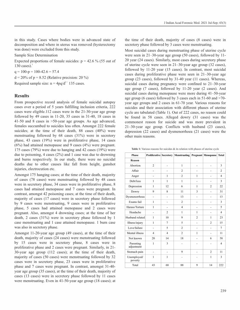
in this study. Cases where bodies were in advanced state of decomposition and where in uterus was removed (hysterectomy was done) were excluded from this study.
Sample Size Determination
Expected proportions of female suicides: p = 42.6 % (55 out of 1130 cases).
q = 100-p = 100-42.6 = 57.4
d = 20% of p = 8.52 (Relative precision: 20 %)2 =Required sample size: n = 4pq/d 135 cases.
Results
From prospective record analysis of female suicidal autopsy cases over a period of 5 years fulfilling inclusion criteria, 222 cases were eligible.112 cases were in the 21-30-year age group followed by 49 cases in 11-20, 35 cases in 31-40, 18 cases in 41-50 and 8 cases in >50-year age groups. As age advanced, females succumbed to suicides less often. Amongst 222 female suicides; at the time of their death, 88 cases (40%) were menstruating followed by 68 cases (31%) were in secretory phase, 43 cases (19%) were in proliferative phase, 14 cases (6%) had attained menopause and 9 cases (4%) were pregnant. 175 cases (79%) were due to hanging and 42 cases (19%) were due to poisoning, 4 cases (2%) and 1 case was due to drowning and burns respectively. In our study, there were no suicidal deaths due to other causes like fall from height, gunshot injuries, electrocution etc.
Amongst 175 hanging cases; at the time of their death, majority of cases (78 cases) were menstruating followed by 48 cases were in secretory phase, 34 cases were in proliferative phase, 8 cases had attained menopause and 7 cases were pregnant. In contrast, amongst 42 poisoning cases; at the time of their death, majority of cases (17 cases) were in secretory phase followed by 9 cases were menstruating, 9 cases were in proliferative phase, 5 cases had attained menopause and 2 cases were pregnant. Also, amongst 4 drowning cases; at the time of her death, 2 cases (31%) were in secretory phase followed by 1 case menstruating and 1 case attained menopause. 1 burn case was also in secretory phase.
Amongst 11-20-year age group (49 cases), at the time of their death, majority of cases (24 cases) were menstruating followed by 15 cases were in secretory phase, 8 cases were in proliferative phase and 2 cases were pregnant. Similarly, in 21-30-year age group (112 cases); at the time of their death, majority of cases (50 cases) were menstruating followed by 32 cases were in secretory phase, 23 cases were in proliferative phase and 7 cases were pregnant. In contrast, amongst 31-40-year age group (35 cases), at the time of their death, majority of cases (13 cases) were in secretory phase followed by 11 cases were menstruating. Even in 41-50-year age group (18 cases); at
the time of their death, majority of cases (8 cases) were in secretory phase followed by 3 cases were menstruating.
Most suicidal cases during menstruating phase of uterine cycle were seen in 21–30-year age group (50 cases), followed by 11-20 year (24 cases). Similarly, most cases during secretory phase of uterine cycle were seen in 21–30-year age group (32 cases), followed by 11-20 year (15 cases). In contrast, most suicidal cases during proliferative phase were seen in 21–30-year age group (23 cases), followed by 31-40 year (11 cases). Whereas, suicidal cases during pregnancy were confined to 21–30-year age group (7 cases), followed by 11-20 year (2 cases). And suicidal cases during menopause were more during 41–50-year age group (6 cases) followed by 3 cases each in 51-60 and >70-year age groups and 2 cases in 61-70 year. Various reasons for suicides and their association with different phases of uterine cycle are tabulated (Table 1). Out of 222 cases, no reason could be found in 58 cases. Alleged dowry (31 cases) was the commonest reason for suicide and was more prevalent in 21–30-year age group. Conflicts with husband (23 cases), depression (22 cases) and dysmenorrhoea (21 cases) were the other main reasons.
239
J Indian Acad Forensic Med. 2021 Jul-Sep; 43(3)
Phase Proliferative Secretory Menstruating Pregnant Menopause Total
Reason
Abortion 2 - 1 - - 3
Affair - - 2 - - 2
Anger - 1 2 1 - 4
Debt/loss 1 1 1 - - 3
Depression 1 12 6 1 2 22
Dowry 9 8 11 3 - 31
Dysmenorrhoea - - 13 - - 13
Exams fail 1 - 2 - - 3
Harass/Torture 1 - 4 - - 5
Headache - 2 1 1 - 4
Husband related 1 10 9 2 1 23
Illness/injury 1 7 5 - 2 15
Love/failure - 5 2 - - 7
Mental illness 4 4 2 1 - 11
Not known 20 14 18 - 6 58
Parenting adjustment
1 3 - - - 4
Stomach pain - - 9 - 2 11
Unemployed/poverty
1 1 - - 1 3
Total 43 68 88 9 14 222
Table 1: Various reasons for suicides & its relation with phases of uterine cycle
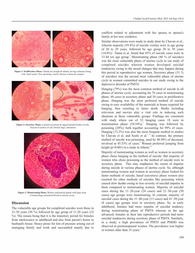
Discussion
The vulnerable age groups for completed suicides were those in 21-30 years (50 %) followed by age group of 11-20 years (22 %). The reason being that it is the transitory period for females from adolescence to adulthood and also from parent's house to husband's house. Hence prone for lots of pressure arising out of managing family and work and succumbed mainly due to
conflicts related to adjustment with her spouse or spouse's family at her new residence.
Similar observations were made in study done by Chavan et al., wherein majority (59.4%) of suicide victims were in age group of 20 to 29 years, followed by age group 30 to 39 years
1(14.8%). Tanna et al. found that 85% of suicide cases were in 615-44 yrs age group. Menstruating phase (40 % of suicides)
was the most vulnerable phase of uterine cycle in our study of completed suicides wherein women developed suicidal tendencies owing to the mood changes that may happen during this period in reproductive age women. Secretory phase (31 % of suicides) was the second most vulnerable phase of uterine cycle in women committed suicides in our study owing to the depressive disorder of PMTS.
Hanging (79%) was the most common method of suicide in all phases of uterine cycle; accounting for 78 cases in menstruating phase, 48 cases in secretory phase and 34 cases in proliferative phase. Hanging was the most preferred method of suicide owing to easy availability of the materials in house required for hanging, thus resorting to faster death. Media including television and movies play a vital role in inducing such ideations in these vulnerable groups. Findings are consistent with study where out of 33 hanging cases 18 were in
7menstruation phase (54.54%). Hanging was followed by poisoning (20%); both together accounting for 99% of cases. Hanging (72.2%) was also the most frequent method in studies
1, 8by Chavan et al. and Soole et al. In contrast, the primary method of suicide was poisoning, used by 40.50% of deceased,
9involved in 45.53% of cases. Women preferred jumping from 10height (p<0.001) in a study in Athens.
Majority of menstruating women as well as women in secretory phase chose hanging as the method of suicide. But majority of women who chose poisoning as the method of suicide were in secretory phase. This may emphasize the extent of impulse during suicide in various phases of uterine cycle. So, although menstruating women and women in secretory phase looked for faster methods of suicide; luteal (secretory) phase women also resorted for other methods of suicides like poisoning which caused slow deaths owing to less severity of suicidal impulse in them compared to menstruating women. Majority of suicidal cases during the 11–20-year (24 cases) and 21–30-year (50 cases) age groups were menstruating. In contrast, majority of suicidal cases during the 31–40-year (13 cases) and 41–50-year (8 cases) age groups were in secretory phase. So, in early adulthood, females had more impulse of suicidal tendency during menstruating phase of PMTS whereas as the age advanced, females in their late reproductive periods had more suicidal tendencies during secretory phase of PMTS. Similarly, in a study, a high prevalence of PMTS and PMDD was observed in premenopausal women. The prevalence was higher
5in women older than 35 years.
J Indian Acad Forensic Med. 2021 Jul-Sep; 43(3)
240
Figure 1: Proliferative Phase. Glands are straight & tubular, having columnar lining with basal nuclei. New sprouting vessels; Stroma is dense & compact
Figure 2: Secretory Phase. Luminal secretions & ragged luminal border of cells, Vacuole at surface of cell; Stroma large, edematous
Figure 3: Menstruating Phase. Broken endometrial glands with large areas of hemorrhage (menstrual blood) in uterine cavity
241
J Indian Acad Forensic Med. 2021 Jul-Sep; 43(3)
As most suicidal cases were in the 21–30-year age group (112 cases out of 222), the cases during any phase of menstrual cycle i.e., either proliferative, secretory or menstruating phase were more commonly seen in the 21–30-year age group. It is for the same reason that majority of suicidal cases during pregnancy were also found in the 21–30-year age group. Also, in another study, the prevalence of PMS (pre-menstrual syndrome) was
11higher in non-married women, in women aged 35-44 years.
In our study, deceased women were found exclusively in menstruating phase when dysmenorrhoea (13 cases) and stomach pain (9 cases) were the reasons for their suicide; probably owing to less tolerance of pain during PMTS. They were found more in secretory phase than menstruating phase when the reasons for their suicides were depression (12 cases) or other reasons like chronic illness (7 cases), love failure (5 cases), parenting adjustment (3 cases). And they were distributed almost equally both in secretory (10 cases) and menstrual (9 cases) phases in husband related conflicts. In a study, both PMS and PMDD were strongly associated with poor physical health and psychological distress. Socio-cultural factors seem to determine the prevalence, perception and
10handling of PMS. Whereas more serious reasons like dowry and cases of unknown reasons were distributed in all the phases of uterine cycle. In a study it was found that reasons for committing suicide were mainly due to marital discord
7(36.36%) followed by health issues (15.15%).
Conclusion
In our study, suicides cases were mostly seen in women menstruating and in secretory (luteal) phase indicating these phases of uterine (menstrual) cycle as the most vulnerable phase in reproductive women for mood alterations leading to suicides. This could be due to an entity called pre-menstrual syndrome, a period between 4 days prior to start of menses to the end of menstruation; wherein a woman is vulnerable for sadness and impulsive acts. This indicates the time of applying the various strategies to curb the unhealthy emotional state of a woman in the form of health education, counseling and treatment of PMTS. Other psycho-social variables associated/affecting the mental status of a woman can also be curbed during that phase. Efforts to increase social connection in depressed and isolated women of reproductive age group especially in the period of PMTS or PMDD are recommended. Vulnerability of women during this period for anger, irritability and mood swings to be known and understood by the family especially husbands so that they keep their calm and help in easing out the stress of daily activities of his life partner. Suicide prevention efforts should include treating mental disorders such as depression including this PMTS or PMDD
and also improving economic conditions. The media, which includes the Internet, may help prevent suicide by providing a social group of women for those who are in need during this period of pre-menstrual tension syndrome.
Ethical clearance: A prior approval was obtained from the Institutional Ethics Committee
Conflict of interest: None to declare
Source of funding: None to declare
References1. Chavan BS, Singh GP, Kaur J, Kochar R. Psychological autopsy of
101 suicide cases from northwest region of India. Indian J Psychiatry. 2008;50(1):34-38.
2. Wikipedia contributors. Suicide [Internet]. Wikipedia, The Free Encyclopedia. 2020 [cited 2020 July 11]. Available from: https://en.wikipedia.org/w/index.php?title=Suicide&oldid=1059540969
3. Chávez-Hernández A-M, Macías-García L-F. Understanding suicide in socially vulnerable contexts: Psychological autopsy in a small town in Mexico. Suicide Life Threat Behav [Internet]. 2016;46(1) :3–12. Avai lable f rom: h t tp : / /dx .doi .org/ 10.1111/sltb.12166
4. Pinkerton JV, Guico-Pabia CJ, Taylor HS. Menstrual cycle-related exacerbation of disease. Am J Obstet Gynecol, 2010 Mar;202(3):221-31.
5. Joshi JV, Pandey SN, Galvankar P, Gogate JA. Prevalence of premenstrual symptoms: Preliminary analysis and brief review of management strategies, J Midlife Health, 2010; 1(1): 30–34.
6. Tanna JA, Patel PN, Kalele SD. Psychological autopsy of suicide cases in bhavnagar region of gujarat, India. Med Leg Update. January 2013; 13(1):27-32.
7. Chetankumar R, Kokatanur C M. A study of relationship between menstrual cycle and hanging. Med Leg update. July- Dec 2016;16(2):23-25.
8. Soole R, Kõlves K, De Leo D. Suicide in children: A systematic review. Arch Suicide Res [Internet]. 2015;19(3):285–304. Available from: http://dx.doi.org/10.1080/13811118.2014.996694.
9. Khan FA, Anand B, Devi MG, Murthy KK. Psychological autopsy of suicide—a cross-sectional study. Indian J Psychiatry. 2005 Apr-Jun;47(2):73–78.
10. Paraschakis A, Michopoulos I, Christodoulou C, Koutsaftis F, Lykouras L, Douzenis A. A 2-year psychological autopsy study of completed suicides in the athens greater area, Greece. Psychiatry Investig [Internet]. 2015;12(2):212–7. Available from: http://dx.doi.org/10.4306/pi.2015.12.2.212
11. Tschudin S, Bertea PC, Zemp E. Prevalence and predictors of premenstrual syndrome and premenstrual dysphoric disorder in a population-based sample, Arch Womens Ment Health, 2010 Dec;13(6):485-94.
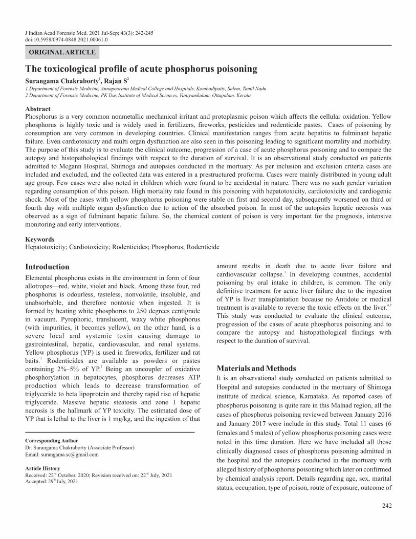
AbstractPhosphorus is a very common nonmetallic mechanical irritant and protoplasmic poison which affects the cellular oxidation. Yellow phosphorus is highly toxic and is widely used in fertilizers, fireworks, pesticides and rodenticide pastes. Cases of poisoning by consumption are very common in developing countries. Clinical manifestation ranges from acute hepatitis to fulminant hepatic failure. Even cardiotoxicity and multi organ dysfunction are also seen in this poisoning leading to significant mortality and morbidity. The purpose of this study is to evaluate the clinical outcome, progression of a case of acute phosphorus poisoning and to compare the autopsy and histopathological findings with respect to the duration of survival. It is an observational study conducted on patients admitted to Mcgann Hospital, Shimoga and autopsies conducted in the mortuary. As per inclusion and exclusion criteria cases are included and excluded, and the collected data was entered in a prestructured proforma. Cases were mainly distributed in young adult age group. Few cases were also noted in children which were found to be accidental in nature. There was no such gender variation regarding consumption of this poison. High mortality rate found in this poisoning with hepatotoxicity, cardiotoxicity and cardiogenic shock. Most of the cases with yellow phosphorus poisoning were stable on first and second day, subsequently worsened on third or fourth day with multiple organ dysfunction due to action of the absorbed poison. In most of the autopsies hepatic necrosis was observed as a sign of fulminant hepatic failure. So, the chemical content of poison is very important for the prognosis, intensive monitoring and early interventions.
KeywordsHepatotoxicity; Cardiotoxicity; Rodenticides; Phosphorus; Rodenticide
The toxicological profile of acute phosphorus poisoning
ORIGINAL ARTICLE
Introduction
Elemental phosphorus exists in the environment in form of four allotropes—red, white, violet and black. Among these four, red phosphorus is odourless, tasteless, nonvolatile, insoluble, and unabsorbable, and therefore nontoxic when ingested. It is formed by heating white phosphorus to 250 degrees centigrade in vacuum. Pyrophoric, translucent, waxy white phosphorus (with impurities, it becomes yellow), on the other hand, is a severe local and systemic toxin causing damage to gastrointestinal, hepatic, cardiovascular, and renal systems. Yellow phosphorus (YP) is used in fireworks, fertilizer and rat
1baits. Rodenticides are available as powders or pastes 2containing 2%–5% of YP. Being an uncoupler of oxidative
phosphorylation in hepatocytes, phosphorus decreases ATP production which leads to decrease transformation of triglyceride to beta lipoprotein and thereby rapid rise of hepatic triglyceride. Massive hepatic steatosis and zone 1 hepatic necrosis is the hallmark of YP toxicity. The estimated dose of YP that is lethal to the liver is 1 mg/kg, and the ingestion of that
amount results in death due to acute liver failure and 3cardiovascular collapse. In developing countries, accidental
poisoning by oral intake in children, is common. The only definitive treatment for acute liver failure due to the ingestion of YP is liver transplantation because no Antidote or medical
4,7 treatment is available to reverse the toxic effects on the liver.This study was conducted to evaluate the clinical outcome, progression of the cases of acute phosphorus poisoning and to compare the autopsy and histopathological findings with respect to the duration of survival.
Materials and MethodsIt is an observational study conducted on patients admitted to
Hospital and autopsies conducted in the mortuary of Shimoga
institute of medical science, Karnataka. As reported cases of
phosphorus poisoning is quite rare in this Malnad region, all the
cases of phosphorus poisoning reviewed between January 2016
and January 2017 were include in this study. Total 11 cases (6
females and 5 males) of yellow phosphorus poisoning cases were
noted in this time duration. Here we have included all those
clinically diagnosed cases of phosphorus poisoning admitted in
the hospital and the autopsies conducted in the mortuary with
alleged history of phosphorus poisoning which later on confirmed
by chemical analysis report. Details regarding age, sex, marital
status, occupation, type of poison, route of exposure, outcome of
242
Corresponding Author
Dr. Surangama Chakraborty (Associate Professor)
Email: [email protected]
Article Historynd ndReceived: 22 October, 2020; Revision received on: 22 July, 2021thAccepted: 29 July, 2021
1 2Surangama Chakraborty , Rajan S1 Department of Forensic Medicine, Annapoorana Medical College and Hospitals, Kombadipatty, Salem, Tamil Nadu
2 Department of Forensic Medicine, PK Das Institute of Medical Sciences, Vaniyamkulam, Ottapalam, Kerala
J Indian Acad Forensic Med. 2021 Jul-Sep; 43(3): 242-245doi:10.5958/0974-0848.2021.00061.0
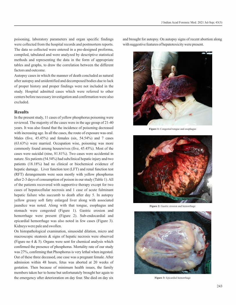
poisoning, laboratory parameters and organ specific findings
were collected from the hospital records and postmortem reports.
The data so collected were entered in a pre-designed proforma,
compiled, tabulated and were analyzed by descriptive statistical
methods and representing the data in the form of appropriate
tables and graphs, to draw the correlation between the different
factors and outcome.
Autopsy cases in which the manner of death concluded as natural
after autopsy and unidentified and decomposed bodies due to lack
of proper history and proper findings were not included in the
study. Hospital admitted cases which were referred to other
centers before necessary investigation and confirmation were also
excluded.
ResultsIn the present study, 11 cases of yellow phosphorus poisoning were
reviewed. The majority of the cases were in the age group of 21-40
years. It was also found that the incidence of poisoning decreased
with increasing age. In all the cases, the route of exposure was oral.
Males (five, 45.45%) and females (six, 54.54%) and 7 cases
(63.63%) were married. Occupation wise, poisoning was more
commonly found among housewives (five, 45.45%). Most of the
cases were suicidal (nine, 81.81%). Two cases were accidental in
nature. Six patients (54.54%) had subclinical hepatic injury and two
patients (18.18%) had no clinical or biochemical evidence of
hepatic damage. Liver function test (LFT) and renal function test
(RFT) derangements were seen mostly with yellow phosphorus
after 2-3 days of consumption of poison in our study (Table 1). All
of the patients recovered with supportive therapy except for two
cases of hepatocellular necrosis and 1 case of acute fulminant
hepatic failure who succumb to death after day 5. In autopsy
yellow greasy soft fatty enlarged liver along with associated
jaundice was noted. Along with that tongue, esophagus and
stomach were congested (Figure 1). Gastric erosion and
hemorrhage were present (Figure 2). Sub-endocardial and
epicardial hemorrhage was also noted in few cases (Figure 3).
Kidneys were pale and swollen.
On histopathological examination, sinusoidal dilation, micro and
macroscopic steatosis & signs of hepatic necrosis were observed
(Figure no 4 & 5). Organs were sent for chemical analysis which
confirmed the presence of phosphorus. Mortality rate of our study
was 27%, confirming that Phosphorus is very lethal when ingested.
Out of these three deceased, one case was a pregnant female. After
admission within 48 hours, fetus was aborted at 20 weeks of
gestation. Then because of minimum health issues, the family
members taken her to home but unfortunately brought her again to
the emergency after deterioration on day four. She died on day six
and brought for autopsy. On autopsy signs of recent abortion along
with suggestive features of hepatotoxicity were present.
243
J Indian Acad Forensic Med. 2021 Jul-Sep; 43(3)
Figure 1: Congested tongue and esophagus
Figure 2: Gastric erosion and hemorrhage
Figure 2: Gastric erosion and hemorrhage
Figure 3: Epicardial hemorrhage
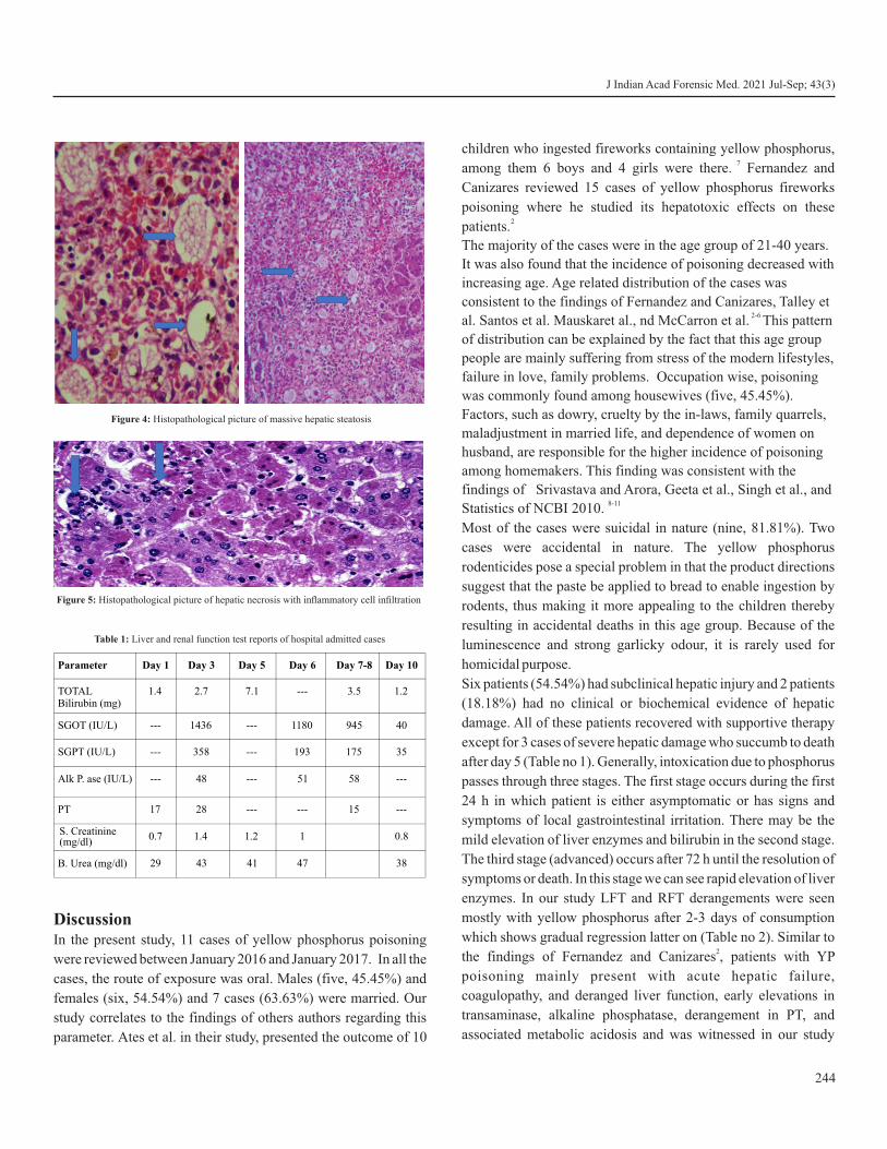
Discussion In the present study, 11 cases of yellow phosphorus poisoning
were reviewed between January 2016 and January 2017. In all the
cases, the route of exposure was oral. Males (five, 45.45%) and
females (six, 54.54%) and 7 cases (63.63%) were married. Our
study correlates to the findings of others authors regarding this
parameter. Ates et al. in their study, presented the outcome of 10
children who ingested fireworks containing yellow phosphorus, 7among them 6 boys and 4 girls were there. Fernandez and
Canizares reviewed 15 cases of yellow phosphorus fireworks
poisoning where he studied its hepatotoxic effects on these 2patients.
The majority of the cases were in the age group of 21-40 years.
It was also found that the incidence of poisoning decreased with
increasing age. Age related distribution of the cases was
consistent to the findings of Fernandez and Canizares, Talley et 2-6 al. Santos et al. Mauskaret al., nd McCarron et al. This pattern
of distribution can be explained by the fact that this age group
people are mainly suffering from stress of the modern lifestyles,
failure in love, family problems. Occupation wise, poisoning
was commonly found among housewives (five, 45.45%).
Factors, such as dowry, cruelty by the in-laws, family quarrels,
maladjustment in married life, and dependence of women on
husband, are responsible for the higher incidence of poisoning
among homemakers. This finding was consistent with the
findings of Srivastava and Arora, Geeta et al., Singh et al., and 8-11Statistics of NCBI 2010.
Most of the cases were suicidal in nature (nine, 81.81%). Two
cases were accidental in nature. The yellow phosphorus
rodenticides pose a special problem in that the product directions
suggest that the paste be applied to bread to enable ingestion by
rodents, thus making it more appealing to the children thereby
resulting in accidental deaths in this age group. Because of the
luminescence and strong garlicky odour, it is rarely used for
homicidal purpose.
Six patients (54.54%) had subclinical hepatic injury and 2 patients
(18.18%) had no clinical or biochemical evidence of hepatic
damage. All of these patients recovered with supportive therapy
except for 3 cases of severe hepatic damage who succumb to death
after day 5 (Table no 1). Generally, intoxication due to phosphorus
passes through three stages. The first stage occurs during the first
24 h in which patient is either asymptomatic or has signs and
symptoms of local gastrointestinal irritation. There may be the
mild elevation of liver enzymes and bilirubin in the second stage.
The third stage (advanced) occurs after 72 h until the resolution of
symptoms or death. In this stage we can see rapid elevation of liver
enzymes. In our study LFT and RFT derangements were seen
mostly with yellow phosphorus after 2-3 days of consumption
which shows gradual regression latter on (Table no 2). Similar to 2the findings of Fernandez and Canizares , patients with YP
poisoning mainly present with acute hepatic failure,
coagulopathy, and deranged liver function, early elevations in
transaminase, alkaline phosphatase, derangement in PT, and
associated metabolic acidosis and was witnessed in our study
J Indian Acad Forensic Med. 2021 Jul-Sep; 43(3)
244
Figure 4: Histopathological picture of massive hepatic steatosis
Figure 5: Histopathological picture of hepatic necrosis with inflammatory cell infiltration
Parameter Day 1 Day 3 Day 5 Day 6 Day 10
TOTAL Bilirubin (mg)
1.4 2.7 7.1 --- 3.5 1.2
SGOT (IU/L) --- 1436 --- 1180 945 40
SGPT (IU/L) --- 358 --- 193 175 35
Alk P. ase (IU/L) --- 48 --- 51 58 ---
PT 17 28 --- --- 15 ---
S. Creatinine(mg/dl)
0.7 1.4 1.2 1 0.8
B. Urea (mg/dl) 29 43 41 47 38
Day 7-8
Table 1: Liver and renal function test reports of hospital admitted cases
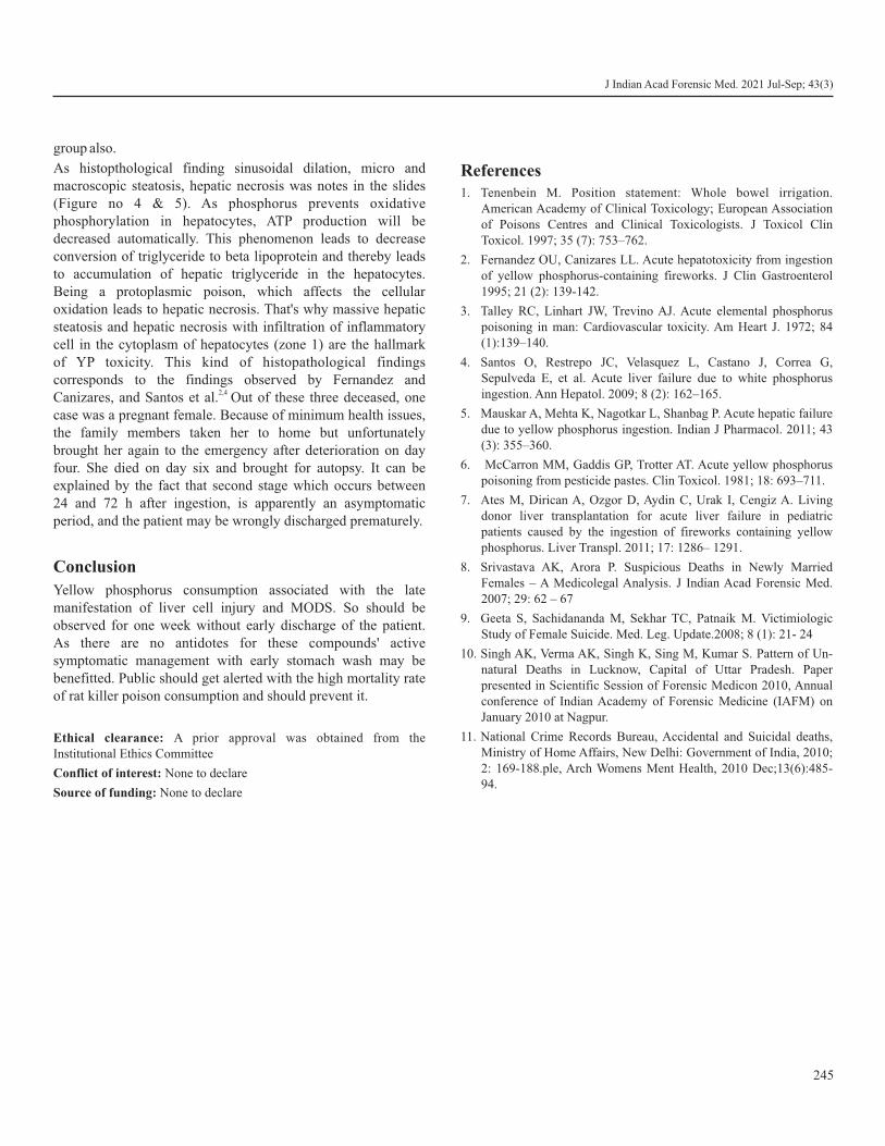
245
J Indian Acad Forensic Med. 2021 Jul-Sep; 43(3)
group also.
As histopthological finding sinusoidal dilation, micro and macroscopic steatosis, hepatic necrosis was notes in the slides (Figure no 4 & 5). As phosphorus prevents oxidative phosphorylation in hepatocytes, ATP production will be decreased automatically. This phenomenon leads to decrease conversion of triglyceride to beta lipoprotein and thereby leads to accumulation of hepatic triglyceride in the hepatocytes. Being a protoplasmic poison, which affects the cellular oxidation leads to hepatic necrosis. That's why massive hepatic steatosis and hepatic necrosis with infiltration of inflammatory cell in the cytoplasm of hepatocytes (zone 1) are the hallmark of YP toxicity. This kind of histopathological findings corresponds to the findings observed by Fernandez and
2,4 Canizares, and Santos et al. Out of these three deceased, one case was a pregnant female. Because of minimum health issues, the family members taken her to home but unfortunately brought her again to the emergency after deterioration on day four. She died on day six and brought for autopsy. It can be explained by the fact that second stage which occurs between 24 and 72 h after ingestion, is apparently an asymptomatic period, and the patient may be wrongly discharged prematurely.
Conclusion
Yellow phosphorus consumption associated with the late manifestation of liver cell injury and MODS. So should be observed for one week without early discharge of the patient. As there are no antidotes for these compounds' active symptomatic management with early stomach wash may be benefitted. Public should get alerted with the high mortality rate of rat killer poison consumption and should prevent it.
Ethical clearance: A prior approval was obtained from the Institutional Ethics Committee
Conflict of interest: None to declare
Source of funding: None to declare
References1. Tenenbein M. Position statement: Whole bowel irrigation.
American Academy of Clinical Toxicology; European Association of Poisons Centres and Clinical Toxicologists. J Toxicol Clin Toxicol. 1997; 35 (7): 753–762.
2. Fernandez OU, Canizares LL. Acute hepatotoxicity from ingestion of yellow phosphorus-containing fireworks. J Clin Gastroenterol 1995; 21 (2): 139-142.
3. Talley RC, Linhart JW, Trevino AJ. Acute elemental phosphorus poisoning in man: Cardiovascular toxicity. Am Heart J. 1972; 84 (1):139–140.
4. Santos O, Restrepo JC, Velasquez L, Castano J, Correa G, Sepulveda E, et al. Acute liver failure due to white phosphorus ingestion. Ann Hepatol. 2009; 8 (2): 162–165.
5. Mauskar A, Mehta K, Nagotkar L, Shanbag P. Acute hepatic failure due to yellow phosphorus ingestion. Indian J Pharmacol. 2011; 43 (3): 355–360.
6. McCarron MM, Gaddis GP, Trotter AT. Acute yellow phosphorus poisoning from pesticide pastes. Clin Toxicol. 1981; 18: 693–711.
7. Ates M, Dirican A, Ozgor D, Aydin C, Urak I, Cengiz A. Living donor liver transplantation for acute liver failure in pediatric patients caused by the ingestion of fireworks containing yellow phosphorus. Liver Transpl. 2011; 17: 1286– 1291.
8. Srivastava AK, Arora P. Suspicious Deaths in Newly Married Females – A Medicolegal Analysis. J Indian Acad Forensic Med. 2007; 29: 62 – 67
9. Geeta S, Sachidananda M, Sekhar TC, Patnaik M. Victimiologic Study of Female Suicide. Med. Leg. Update.2008; 8 (1): 21- 24
10. Singh AK, Verma AK, Singh K, Sing M, Kumar S. Pattern of Un-natural Deaths in Lucknow, Capital of Uttar Pradesh. Paper presented in Scientific Session of Forensic Medicon 2010, Annual conference of Indian Academy of Forensic Medicine (IAFM) on January 2010 at Nagpur.
11. National Crime Records Bureau, Accidental and Suicidal deaths, Ministry of Home Affairs, New Delhi: Government of India, 2010; 2: 169-188.ple, Arch Womens Ment Health, 2010 Dec;13(6):485-94.
AbstractThe commonest cause of poisoning in India and other developing countries are insecticide and pesticide, which are agriculturally based economics, poverty and easy availability. This study collected gastric lavage from all suspected and confirmed insecticide poisoning cases brought to the hospital. They are being analysed by the thin-layer chromatography method for the detection of the type of insecticide. This is a cross-sectional study, and statistical analysis is done by correlation and regression analysis method. Total 182 cases of insecticide poisoning were brought to the emergency services of a Government Hospital at Agartala, Tripura, during the period January 2013 to June 2014, out of which 79.1% were within the age group of 21 years to 40 years, 78.6% of the victims are male, 50% of the victims were farmers, 98% of the times the mode of poisoning were suicidal, 74% of the cases the insecticide involved was organophosphorus group of poison (OP), while 19% of the cases were Organochlorine group of poisons (OC), and only 7% of the cases were Carbamate group of poison (CAR). Due to undue pressure for performance and good lifestyle and failure to comply, the young productive age group tends to take such irreversible steps of committing suicide using easily available material. The need of the hour is a very restricted supply of insecticides with proper documentation and proper counselling sessions for survivors of such incidents.
KeywordsInsecticide poisoning; Organophosphorus poisoning; Suicide
A comprehensive study on insecticide poisoning patients brought to a tertiary government hospital in north eastern region of India
ORIGINAL ARTICLE
Introduction
The commonest cause of poisoning in India and other developing countries are insecticide and pesticide, which are agriculturally based economics, poverty and easy availability. Since 1985, aluminium phosphide (a solid fumigant pesticide, insecticide and rodenticide) poisoning has been reported as the commonest cause of intentional poisoning in northern India viz; Haryana, Punjab, Rajasthan and Delhi. The exact incidence of poisoning in India is uncertain due to the lack of data at the central level. Most cases are not reported, and mortality data are poor indicators of incidence of poisoning. It has been estimated that about 5-6 persons per lakh die due to poisoning every year. Among the adults, females predominate in all age groups with an evident preponderance in the second and third decades of
1life.
Acute poisoning in children is almost entirely accidental, while in adults, it is mainly suicidal. Mortality varies from country to country depending on the nature of poison and the availability of facilities and treatment by qualified persons. Several
hospitals based retrospective studies in India have shown an increasing incidence of pesticide poisoning during the last decade. Organophosphates, aluminium phosphide and
2rodenticide are most commonly involved in such poisonings . Organophosphorous compounds are one of the gradually increasing causes of accidental and suicidal poisoning with high morbidity and mortality rates, especially in developing countries and are estimated to kill around 200,000 people each
2,3year, largely in the Asia Pacific region . Clinical and toxicological diagnostic and treatment facilities are often
4,5inadequate due to a lack of trained personnel . Thus there is a requirement of strengthening the diagnostic capabilities nationwide for better prevention and treatment of insecticide poisoning cases.
Further, the lack of information on the ingredients of various products makes it difficult to plan and develop poison policies and control programmes to help provide reliable information on time to doctors and other medical personnel and first responders
4,5for appropriate treatment . This study collected gastric lavage from all suspected and confirmed insecticide poisoning cases brought to the hospital. They are being analysed by the thin-layer chromatography method to detect the type of insecticides present in the poisoning sample. The study was conducted to detect the type of insecticides in poisoning cases admitted at AGMC & GBPH, Agartala and to study the demographic profile of the subjects admitted with insecticide poisoning.
246
Corresponding Author
Dr Antara Debbarma (Assistant Professor) Email: [email protected] Mobile: +91-9205623401
Article Historyth thReceived: 12 October, 2020; Revision received on: 12 August, 2021thAccepted: 18 August, 2021
Antara Debbarma1, Juthika Debbarma2
1 Assistant Professor, Department of Forensic Medicine, at Santosh Medical College & Hospital, Ghaziabad, UP
2 Associate Professor, Department of Forensic Medicine, at Agartala Government Medical College, Agartala, Tripura
J Indian Acad Forensic Med. 2021 Jul-Sep; 43(3): 246-248doi:10.5958/0974-0848.2021.00062.2
Material and Methods
From January 2013 to June 2014, 182 cases of insecticide poisoning, which were brought to the emergency services of a Government Hospital at Agartala, Tripura, were included in the study. After due consent, history taking and gastric lavage sample collection were done and same taken to State Forensic Science laboratory, Narsingarh, for chemical analysis by thin-layer chromatography method. This is a cross-sectional study, and statistical analysis is done by correlation and regression analysis method. Inclusion criteria: All the cases of confirmed and suspected insecticide poisoning were taken. Exclusion criteria: Poisonings other than insecticide were not taken. This is a cross-sectional study, and statistical analysis is done by correlation and regression analysis method.
Results
Out of 182 cases, the maximum number of cases were within 21 to 40 years, constituting 79.1% of the total cases. The maximum number of victims are male, constituting 78.6% of all cases.50% of the victims were farmers, while 13.2% were housewives, followed by labourers (12.1%), and the least being 1.1% of victims, had their own business. 98% of the time, the mode of poisoning was suicidal while the rest 2% was because of accidental poisoning. It was observed that 92.3% of the cases were from the low socioeconomic background, whilst the rest of the minimal percentage were from middle-class families. In contrast, no cases were being seen from upper-class families. In 74% of the cases, the insecticide involved was the Organophosphorous group of poison (OP). In comparison, in 19% of the cases, the insecticide involved was the Organochlorine group of poisons (OC). Only 7% of the cases showed the presence of the Carbamate group of poison (CAR). 92.9% of the victims had no significant history of any previous poisoning attempts. 78.6 % of the victims died due to poisoning even after being admitted to the hospital, while 21.4% of the victims recovered after treatment. 21.4 % of the cases who survived were asked for a psychiatric referral to fight the ordeal the victims have gone through.
Discussion
In this present study, it was found that a maximum number of the victims belongs to the age group of 21 to 40 years with a total number of 144 cases (79.1%). The findings of this study
. 6are consistent with the studies conducted by Guntheti et al , 7 . 8 9Khade et al. , Kavya et al , Ashwini et al. In this present
study; it has been reflected that 78.60% of the victim were male, thereby constituting a maximum number of cases while 21.40% of the victim were female. The findings are consistent
6 7 with the studies conducted by Guntheti et al. , Bashir et al. ,
8 . 9 10 11Kavya al. and Ashwini et al , Sandhu et al. , Patel et al. , and 12Kumar et al. . From the data analysis of this study, it was found
that the majority of the victims were farmers with a total number of 91 cases, thereby constituting 50% of all the cases. Similar findings are recorded in the studies conducted by Kavya
. 8 9 13al Ashwini et al and Joshi et al . This present study depicts that 98% of the victims had a history of suicidal ingestion of insecticide poison while 2% had a history of accidental ingestion. Similar findings were reported in the studies
. 16conducted by Xinqi et al. 14, L Verhulst et al. 15, Kumar et al . 9. and Ashwin et al The socioeconomic status of the victims of
insecticide poisoning cases was studied. The majority of the victims (92.3%) were of low socioeconomic status, while 7.7% were of middle socioeconomic status. There were no reported cases of insecticide poisoning from upper socioeconomic status.
17,This finding is consistent with the findings of Agarwal SB. 18 6 19 Gupta et al. , Guntheti et al. and Mrinal et al. After the
laboratory analysis of the samples collected from the victims, it has been seen that 74.2% of the cases were positive for Organophosphorous group of poison, 18.7% of the cases were positive for Organochlorous group of poison while only 7.1% of the cases were positive for Carbamate group of poison.
13 5Similar results were seen in Subhash et al. , Tejash et al. , and 9Ashwini Kumar Sahoo . However, in the study conducted by
. 20Garg et al , aluminium phosphide was the commonest poison involved in the poisoning, followed by the Organophosphorous
16group of poison and in the study conducted by Kumar et al. The carbamate group of poison was commonest, followed by the Organophosphorous group of poison.
Out of all the victims of insecticide poisoning, only 21.4% of the victims recovered after treatment, while 78.6% of the patients died due to poisoning. Those patients who recovered were given a psychiatric referral to assess their mental status and further management, similar to the study conducted by
. 21 . 9, Indiana et al . In the study conducted by Ashwini et al it has been shown that 64% of the victims recovered, which is not consistent with the present study.
Conclusion
Insecticide poisoning is one of the emerging problems worldwide, especially in developing countries, with millions and thousands of deaths occurring every year, particularly in
22young people . In present times, the unrestricted easy availability of the poison makes it an easy tool for self-ingestion. In 1985, the UN Food and Agriculture Organization (FAO) produced a voluntary code of conduct for the pesticide industry to limit the harmful effects of pesticides. Unfortunately, a lack of adequate government resources in the developing world makes this code ineffective, and thousands of
23deaths continue to occur today . The majority of the victims in
247
J Indian Acad Forensic Med. 2021 Jul-Sep; 43(3)
the study were young males from an agricultural background. In today's world of fierce competition, everyone wants to fair a good lifestyle and thus, in turn, adding extra pressure over the person, especially the young productive age group and failure to comply that leads to such irreversible steps of committing suicide using easily available material. The need of the hour is a very restricted supply of insecticides with proper documentation and proper counselling sessions for survivors of such incidents.
Ethical clearance: A prior approval was obtained from the Institutional Ethics Committee
Conflict of interest: None to declare
Source of funding: None to declare
References1. Reddy, N. K. (2017). Essentials of Forensic Medicine and
Toxicology. India: Jaypee Brothers Medical Publishers Pvt. Limited. 33rd ed. Hyderabad ;2014: 500-2.
2. Strengthening poison control centres in the region towards sustainable development through sound management of chemicals; regional committee. Provisional agenda item 12 fifty-second session SEA/RC52/8 .25June 1999. Available from http://repository.searo.who.int/handle/123456789/16653 [cited on 29th November 2014 ]
3. Kora SA, Doddamani GB, Halagali GR, Vijayamahantesh SN, Boke U. Socio-Demographic Profile of the Organophosphorous Poisoning Cases in Southern India. J Clin Diagn Res 2011;5(5):953-6.
4. Srivastava A, Peshin SS, Kaleekal T, Gupta SK. An epidemiological study of poisoning cases reported to the national poisons information centre, All India Institute of Medical Sciences, New Delhi; Hum Exp Toxicol.2005; 24(6):279-85.
5. Prajapati T, Prajapati K, Tandon R N, Merchant S. A study of acute poisoning cases excluding animal bites at civil hospital, Ahmedabad. J Indian Acad Forensic Med. 2013 ;35(2):120-2.
6. Gunther BK, Singh UP. The pattern of poisoning in Khammam. J Indian Acad Forensic Med.2011; 33(4):296-300.
7. Bashir MSM, Khade A, Bhagat S, Irfanuddin M. Gender differences in the pattern of Organophosphorous poisoning in a tribal district of Andhra Pradesh. Indian J. Forensic Med. Toxicol. [Internet].2011[Cited 2020 Oct 8]; 5(1):54-7.
8. Kavya ST, Srinivas V, Chandana, Madhumati, R (2012). Clinical profile of patient with Organophosphorous poisoning in an intensive care unit in a tertiary hospital. Int. J. Clin. Cases Invest. 4(3): 24-31
9. Sahoo A, Sastry A, Rauta S, A M M P, Mahapatra S. Study of
organophosphorous poisoning cases at maharajah institute of medical sciences, A.P. J. evol. med. dent. sci. (Online). 2014;3(35):9201-9206.
10. Sandhu SS, Garg A, Gorea RK. Poisoning Trend in Faridkot Region: A retrospective study. J Punjab Acad Forensic Med Toxicol. 2010; 10(1):20-3.
11. Patel DJ, Tekade PR. Profile of Organophosphorous poisoning at Maharani Hospital, Jagdalpur, Chhattisgarh: A Three Years Study. J Indian Acad Forensic Med. 2011; 33(2):102-5.
12. Kumar S, Pathak A, Mangal HM. Trends of Fatal Poisoning in Saurashtra Region of Gujrat (A prospective study). J Indian Acad Forensic Med.2011; 33(3):197-9.
13. Joshi SC, Prakash C, Joshi A, Joshi G. Profile of Organophosphorus Poisoning at Tertiary Care Hospital in Uttarakhand. J Indian Acad Forensic Med.2013Oct-Dec; 35(4): 346-8.
14. Dong X, Simon M. The Epidemiology of Organophosphate Poisoning in Urban Zimbabwe from 1995 to 2000. Int J Occup Med Environ Health. 2001;7(4):333-338.
15. Verhulst L, Waggie Z, Hatherill M, Reynolds L, Argent A. Presentation and outcome of severe anticholinesterase insecticide poisoning. Arch Dis Child. 2002; 86: 352-55.
16. Kumar S, Verma AK. A study of elderly unnatural deaths in medico-legal autopsies at Lucknow locality. Med Sci Law. 2014;54(3):127-31. doi: 10.1177/0025802413502783. PMID: 24166690.
17. Agarwal SB. A clinical, biochemical, neurobehavioral and socio-psychological study of 190 patients admitted to hospital as a result of acute Organophosphorous poisoning. Environ. Res.1993; 62(1):63-70.]
18. Gupta BD, Vaghela PC. Profile of fatal poisoning in and around Jamnagar. J. Ind Acad Forensic Med.2005; 27 (3): 145-8
19. Haloi M, Haloi M D, Patowary A. Death due to poisoning in District of Kamrup, Assam A Medico-legal Study. J Indian Acad Forensic Med. 2013; 35(1):17-20.]
20. Garg V, Verma SK. Trends of poisoning in Rural area of Southwest, Punjab, J. Indian Acad Forensic Med. 2010;32(3):189-93.
21. Indiana J, Herrero R, Albertazzi C. Comparative study of cases of poisoning by Organophosphorous insecticides in various regions of Costa Rica. Revista de Biologia Tropical.1983; 31(1):139-44.
22. Bertolote JM, Fleischmann A, Eddleston M, Gunnell D. Deaths from pesticide poisoning: a global response. Br J Psychiatry. 2006 Sep; 189:201-3.
23. Turabi A, Danyal A, Hasan S, Durrani A, Ahmed M. Organophosphate Poisoning in The Urban Population; Study Conducted at National Population Centre, Karachi. Biomedica. 2008; 24:124-9.
J Indian Acad Forensic Med. 2021 Jul-Sep; 43(3)
248
AbstractBrain is one of the most vital organs in the body, despite it being protected by bony skull. Craniocerebral injury or head injury is defined by National Advisory Neurological Disease and Stroke Council as a “morbid state resulting from gross or subtle structural changes in the scalp, skull and or the contents of the skull produced by mechanical forces”.1 A prospective study was conducted in the department of forensic medicine at Indira Gandhi Government Medical College & Hospital Nagpur, a tertiary care centre for a period of two years. During the period of study, a total of 2048 cases of medicolegal autopsy were evaluated. Out of the total medicolegal autopsy cases a total of 202 cases of death due to craniocerebral injuries were included in the present study. Consumption of alcohol is a common causation for road traffic accidents and subsequent craniocerebral injuries. Study of various aspects of head injury helps in better understanding of such cases.
KeywordsForensic medicine; Forensic pathology; Regional injuries; Craniocerebral injuries; Head injury.
Autopsy analysis of craniocerebral injuries at a tertiary healthcare centre
ORIGINAL ARTICLE
Introduction
Brain is one of the most vital organ of the body. Brain regulates vital functions, initiates and coordinates motor activities, receives sensory impulses and is the seat of emotions and intellectual activities. Each of these functions has its specific location within brain. Brain is protected against mechanical stresses by bony skull. Brain is one of the most vulnerable organs in the body in spite of it being protected by bony skull. Injuries to brain has increased due to mechanisation in industries, high velocity transport vehicles, increased use of alcohol in modern life and many more reasons. The total incidence rate of traumatic brain injuries was 235 cases/100 000 people with an average mortality of 15/100 000 people per year and thus accounted for the majority of trauma deaths in
1Europe. Craniocerebral injury or head injury is defined by National Advisory Neurological Disease and Stroke Council as a “morbid state resulting from gross or subtle structural changes in the scalp, skull and or the contents of the skull produced by
2mechanical forces”. By the same definition concept of mechanical forces is restricted to those forces applied externally to the head, thus excluding surgical ablations and internally acting forces such as increased intracranial pressure resulting from oedema, hydrocephalus or a mass occupying lesion without antecedent head trauma.
Materials and Methods
A descriptive cross-sectional study was conducted in the department of forensic medicine at Indira Gandhi Government Medical College & Hospital Nagpur, a tertiary care centre for a period of two years. During the period of study, a total of 2048 cases of medicolegal autopsy were evaluated. Out of the total medicolegal autopsy cases a total of 202 cases of death due to craniocerebral injuries were studied in the current study. History of the case, the details of the cases, circumstances of the death and other relevant information related to the study was obtained from the police personals investigating the cases. Cases with inadequate history, doubtful findings and bodies which were brought in decomposed state were excluded. Age, gender, place of death, period of survival, circumstances of death, internal injuries to head and various structures of head were studied in details.
All the details of the cases were studied in detail in relation to the various age groups, various external injuries, various internal injuries and cases with alcohol consumptions. Data thus obtained were compiled and evaluated in relation to the various aspects of craniocerebral injuries accordingly.
Results
Out of the total 202 cases of craniocerebral injury, 139 cases had isolated craniocerebral injury while 63 cases had craniocerebral injury associated with other injuries. Out of the total 202 cases, 175 (86.6 %) were males while remaining 27 (13.4 %) cases were females. Out of the total 202 cases, 128 (63.4 %) cases died on the spot while 74 (36.6 %) cases died after getting admitted in hospital. Out of the total 175 male cases, 108 cases died on the spot while 67 cases died after getting admitted in hospital. Out of the total 27 female cases; 20
249
Corresponding Author
Dr Shibanand Nepal Karmakar (Associate Professor)
Email: [email protected]
Mobile: +91-9766159176
Article Historyth thReceived: 5 November, 2020; Revision received on: 5 July, 2021
thAccepted: 15 July, 2021
1 2 1Bandu Waman Ramteke , Shibanand Nepal Karmakar , Nilesh Keshav Tumram1 Department of Forensic Medicine, Government Medical College, Chandrapur, Maharashtra
2 Department of Forensic Medicine, Terna Medical College, Nerul, Navi Mumbai, Maharashtra
J Indian Acad Forensic Med. 2021 Jul-Sep; 43(3): 249-253doi:10.5958/0974-0848.2021.00063.4
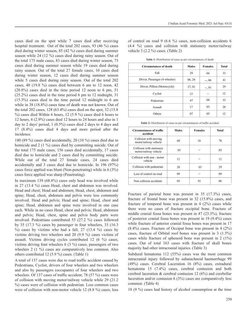
cases died on the spot while 7 cases died after receiving hospital treatment. Out of the total 202 cases, 93 (46 %) cases died during winter season, 85 (42 %) cases died during summer season while 24 (12 %) cases died during rainy season. Out of the total 175 male cases, 83 cases died during winter season, 73 cases died during summer season while 19 cases died during rainy season. Out of the total 27 female cases, 10 cases died during winter season, 12 cases died during summer season while 5 cases died during rainy season. Out of the total 202 cases, 40 (19.8 %) cases died between 6 am to 12 noon, 42 (20.8%) cases died in the time period 12 noon to 6 pm, 51 (25.2%) cases died in the time period 6 pm to 12 midnight, 31 (15.3%) cases died in the time period 12 midnight to 6 am while in 38 (18.8%) cases time of death was not known. Out of the total 202 cases, 128 (63.4%) cases died on the spot, 32 (15.8 %) cases died Within 6 hours, 12 (5.9 %) cases died 6 hours to 12 hours, 6 (2.9%) cases died 12 hours to 24 hours and also in 1 day to 2 days' period, 1 (0.5%) cases died 2 days to 4 days and 17 (8.4%) cases died 4 days and more period after the incidence.
180 (89 %) cases died accidentally, 20 (10 %) cases died due to homicide and 2 (1 %) cases died by committing suicide. Out of the total 175 male cases, 156 cases died accidentally, 17 cases died due to homicide and 2 cases died by committing suicide. While out of the total 27 female cases, 24 cases died accidentally and 3 cases died due to homicide. In 196 (97%) cases force applied was blunt (Non-penetrating) while in 6 (3%) cases force applied was sharp (Penetrating).
In maximum 139 (68.8%) cases only head was involved while in 27 (13.4 %) cases Head, chest and abdomen was involved. Head and chest; Head and abdomen; Head, chest, abdomen and spine; Head, chest, abdomen and pelvis were less commonly involved. Head and pelvis; Head and spine; Head, chest and spine; Head, abdomen and spine were involved in one case each. While in no cases Head, chest and pelvis; Head, abdomen and pelvis; Head, chest, spine and pelvis body parts were involved. Pedestrians contributed 55 (27.2 %) cases followed by 35 (17.3 %) cases by passenger in four wheelers, 33 (16.3 %) cases by victims who had a fall, 27 (13.4 %) cases by victims driving two wheelers and 20 (9.9 %) cases victims of assault. Victims driving cycles contributed 12 (6 %) cases; victims driving four wheelers 6 (3 %) cases, passengers of two wheelers 2 (1 %) cases are comparatively less common. Also others contributed 12 (5.9 %) cases. (Table 1)
A total of 137 cases were due to road traffic accident caused by Pedestrians, Cyclist, drivers of four wheelers and two wheelers and also by passengers (occupants) of four wheelers and two wheelers. Of 137 cases of traffic accident; 78 (57 %) cases were of collision with moving motor/railway vehicle while 29 (21.2 %) cases were of collision with pedestrian. Less common cases were of collision with non-motor vehicle 12 (8.8 %) cases, loss
of control on road 9 (6.6 %) cases, non-collision accidents 6 (4.4 %) cases and collision with stationery motor/railway vehicle 3 (2.2 %) cases. (Table 2)
Fracture of parietal bone was present in 35 (17.3%) cases, fracture of frontal bone was present in 32 (15.8%) cases, and fracture of temporal bone was present in 4 (2%) cases while there were no cases of fracture occipital bone. Fracture of middle cranial fossa bones was present in 47 (23.3%), fracture of posterior cranial fossa bones was present in 19 (9.4%) cases while fracture of anterior cranial fossa bones was present in 17 (8.4%) cases. Fracture of Occiput bone was present in 4 (2%) cases, fracture of Orbital roof bones was present in 3 (1.5%) cases while fracture of sphenoid bone was present in 2 (1%) cases. Out of total 163 cases with fracture of skull bones majority had other intracranial injuries. (Table 3)
Subdural hematoma 112 (55%) cases was the most common intracranial injury followed by subarachnoid haemorrhage 99 (49%) cases. Cerebral Laceration 16 (8%) cases, extradural hematoma 15 (7.4%) cases, cerebral contusion and both cerebral laceration & cerebral contusion 12 (6%) and cerebellar laceration and/or contusion 6 (3%) cases are comparatively less common. (Table 4)
18 (9 %) cases had history of alcohol consumption at the time
250
J Indian Acad Forensic Med. 2021 Jul-Sep; 43(3)
Circumstances of death Males Females Total
Fall 29 04 33
Driver, Passenger (4-wheeler) 06, 29 --, 06 41
Driver, Pillion (Motorcycle) 27, 01 29
Cyclist 12 -- 12
Pedestrian 47 08
03
05
55
Assault 17 20
Others 07 12
--, 01
Table 1: Distribution of cases as per circumstances of death
Circumstances of trafficaccident
Males Females Total
Collision with moving motor/railway vehicle 68 10 78
Collision with stationery motor/railway vehicle 03 -- 03
Collision with non - motorvehicle
12 -- 12
Collision with pedestrian 26 03 29
Loss of control on road 09 -- 09
Non collision accidents 05 01 06
Table 2: Distribution of cases as per circumstances of traffic accident
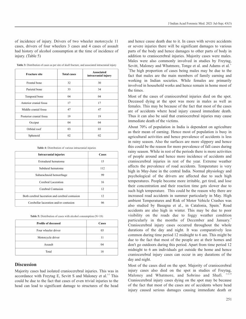
of incidence of injury. Drivers of two wheeler motorcycle 11 cases, drivers of four wheelers 3 cases and 4 cases of assault had history of alcohol consumption at the time of incidence of injury. (Table 5)
Discussion
Majority cases had isolated craniocerebral injuries. This was in 3-5accordance with Freytag E, Sevitt S and Maloney et al. This
could be due to the fact that cases of even trivial injuries to the head can lead to significant damage to structures of the head
and hence cause death due to it. In cases with severe accidents or severe injuries there will be significant damages to various parts of the body and hence damages to other parts of body in addition to craniocerebral injuries. Majority cases were males. Males were also commonly involved in studies by Freytag,
3-Sevitt, Maloney and Whatmore, Tonge et al. and Adams et al.7The high proportion of cases being males may be due to the fact that males are the main members of family earning and working in Indian societies. While females are primarily involved in household works and hence remain in home most of the times.
Most of the cases of craniocerebral injuries died on the spot. Deceased dying at the spot was more in males as well as females. This may be because of the fact that most of the cases are of accidents where head injury caused immediate death. Thus it can also be said that craniocerebral injuries may cause immediate death of the victims.
About 70% of population in India is dependent on agriculture as their mean of earning. Hence most of population is busy in agricultural activities and hence prevalence of accidents is less in rainy season. Also the surfaces are more slippery and hence this could be the reason for more prevalence of fall cases during rainy season. While in rest of the periods there is more activities of people around and hence more incidence of accidents and craniocerebral injuries in rest of the year. Extreme weather affects the prevalence of road accidents. Temperature is very high in May-June in the central India. Normal physiology and psychological of the drivers are affected due to such high temperatures. People become more irritable, get tired, and lose their concentration and their reaction time gets slower due to such high temperature. This could be the reason why there are increased road accidents in summer particularly in May. High ambient Temperatures and Risk of Motor Vehicle Crashes was
8also studied by Basagna et al., in Catalonia, Spain. Road accidents are also high in winter. This may be due to poor visibility on the roads due to foggy weather condition
9particularly in the months of December and January. Craniocerebral injury cases occurred throughout the whole durations of the day and night. It was comparatively less common during time period 12 midnight to 6 am. This might be due to the fact that most of the people are at their homes and don't go outdoors during this period. Apart from time period 12 midnight to 6 am individuals get outside the home and hence craniocerebral injury cases can occur in any durations of the day and night.
Most of the cases died on the spot. Majority of craniocerebral injury cases also died on the spot in studies of Freytag,
3,4,10 Maloney and Whatmore, and Sobrino and Shafi. Craniocerebral injury cases dying on the spot may be because of the fact that most of the cases are of accidents where head injury caused serious damages causing immediate death or
J Indian Acad Forensic Med. 2021 Jul-Sep; 43(3)
251
Fracture site Total casesAssociated
intracranial injury
Frontal bone 32 30
Parietal bone 35 34
Temporal bone 04 04
Anterior cranial fossa 17
17
Middle cranial fossa 47 47
Posterior cranial fossa 19 19
Occiput 04 04
Orbital roof 03 03
Sphenoid 02 02
Table 3: Distribution of cases as per site of skull fracture, and associated intracranial injury
Intracranial injuries Cases
Extradural hematoma 15
Subdural hematoma 112
Subarachnoid hemorrhage 99
Cerebral Laceration 16
Cerebral Contusion 12
Both cerebral laceration and cerebral contusion 12
Cerebellar laceration and/or contusion 06
Table 4: Distribution of various intracranial injuries
Profile of deceased Cases
Four wheeler driver 03
Motorcycle driver 11
Assault 04
Total 18
Table 5: Distribution of cases with alcohol consumption (N=18)
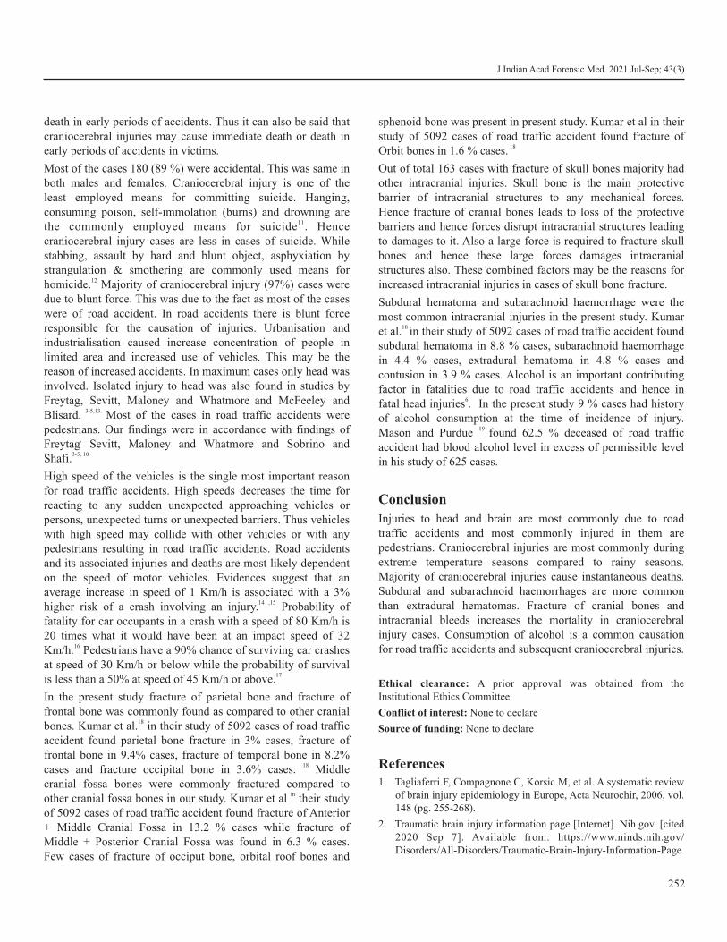
death in early periods of accidents. Thus it can also be said that craniocerebral injuries may cause immediate death or death in early periods of accidents in victims.
Most of the cases 180 (89 %) were accidental. This was same in both males and females. Craniocerebral injury is one of the least employed means for committing suicide. Hanging, consuming poison, self-immolation (burns) and drowning are
11the commonly employed means for suicide . Hence craniocerebral injury cases are less in cases of suicide. While stabbing, assault by hard and blunt object, asphyxiation by strangulation & smothering are commonly used means for
12homicide. Majority of craniocerebral injury (97%) cases were due to blunt force. This was due to the fact as most of the cases were of road accident. In road accidents there is blunt force responsible for the causation of injuries. Urbanisation and industrialisation caused increase concentration of people in limited area and increased use of vehicles. This may be the reason of increased accidents. In maximum cases only head was involved. Isolated injury to head was also found in studies by
Freytag, Sevitt, Maloney and Whatmore and McFeeley and 3-5,13. Blisard. Most of the cases in road traffic accidents were
pedestrians. Our findings were in accordance with findings of ,Freytag Sevitt, Maloney and Whatmore and Sobrino and
3-5, 10Shafi.
High speed of the vehicles is the single most important reason for road traffic accidents. High speeds decreases the time for reacting to any sudden unexpected approaching vehicles or persons, unexpected turns or unexpected barriers. Thus vehicles with high speed may collide with other vehicles or with any pedestrians resulting in road traffic accidents. Road accidents and its associated injuries and deaths are most likely dependent on the speed of motor vehicles. Evidences suggest that an average increase in speed of 1 Km/h is associated with a 3%
14 ,15higher risk of a crash involving an injury. Probability of fatality for car occupants in a crash with a speed of 80 Km/h is 20 times what it would have been at an impact speed of 32
16Km/h. Pedestrians have a 90% chance of surviving car crashes at speed of 30 Km/h or below while the probability of survival
17is less than a 50% at speed of 45 Km/h or above.
In the present study fracture of parietal bone and fracture of frontal bone was commonly found as compared to other cranial
18bones. Kumar et al. in their study of 5092 cases of road traffic accident found parietal bone fracture in 3% cases, fracture of frontal bone in 9.4% cases, fracture of temporal bone in 8.2%
18cases and fracture occipital bone in 3.6% cases. Middle cranial fossa bones were commonly fractured compared to
inother cranial fossa bones in our study. Kumar et al their study of 5092 cases of road traffic accident found fracture of Anterior + Middle Cranial Fossa in 13.2 % cases while fracture of
Middle + Posterior Cranial Fossa was found in 6.3 % cases.Few cases of fracture of occiput bone, orbital roof bones and
sphenoid bone was present in present study. Kumar et al in their study of 5092 cases of road traffic accident found fracture of
18Orbit bones in 1.6 % cases.
Out of total 163 cases with fracture of skull bones majority had other intracranial injuries. Skull bone is the main protective barrier of intracranial structures to any mechanical forces. Hence fracture of cranial bones leads to loss of the protective barriers and hence forces disrupt intracranial structures leading to damages to it. Also a large force is required to fracture skull bones and hence these large forces damages intracranial structures also. These combined factors may be the reasons for increased intracranial injuries in cases of skull bone fracture.
Subdural hematoma and subarachnoid haemorrhage were the most common intracranial injuries in the present study. Kumar
18 et al. in their study of 5092 cases of road traffic accident found subdural hematoma in 8.8 % cases, subarachnoid haemorrhage in 4.4 % cases, extradural hematoma in 4.8 % cases and contusion in 3.9 % cases. Alcohol is an important contributing factor in fatalities due to road traffic accidents and hence in
6fatal head injuries . In the present study 9 % cases had history of alcohol consumption at the time of incidence of injury.
19 Mason and Purdue found 62.5 % deceased of road traffic accident had blood alcohol level in excess of permissible level in his study of 625 cases.
Conclusion
Injuries to head and brain are most commonly due to road traffic accidents and most commonly injured in them are pedestrians. Craniocerebral injuries are most commonly during extreme temperature seasons compared to rainy seasons. Majority of craniocerebral injuries cause instantaneous deaths. Subdural and subarachnoid haemorrhages are more common than extradural hematomas. Fracture of cranial bones and intracranial bleeds increases the mortality in craniocerebral injury cases. Consumption of alcohol is a common causation for road traffic accidents and subsequent craniocerebral injuries.
Ethical clearance: A prior approval was obtained from the Institutional Ethics Committee
Conflict of interest: None to declare
Source of funding: None to declare
References1. Tagliaferri F, Compagnone C, Korsic M, et al. A systematic review
of brain injury epidemiology in Europe, Acta Neurochir, 2006, vol. 148 (pg. 255-268).
2. Traumatic brain injury information page [Internet]. Nih.gov. [cited 2020 Sep 7]. Available from: https://www.ninds.nih.gov/ Disorders/All-Disorders/Traumatic-Brain-Injury-Information-Page
J Indian Acad Forensic Med. 2021 Jul-Sep; 43(3)
252
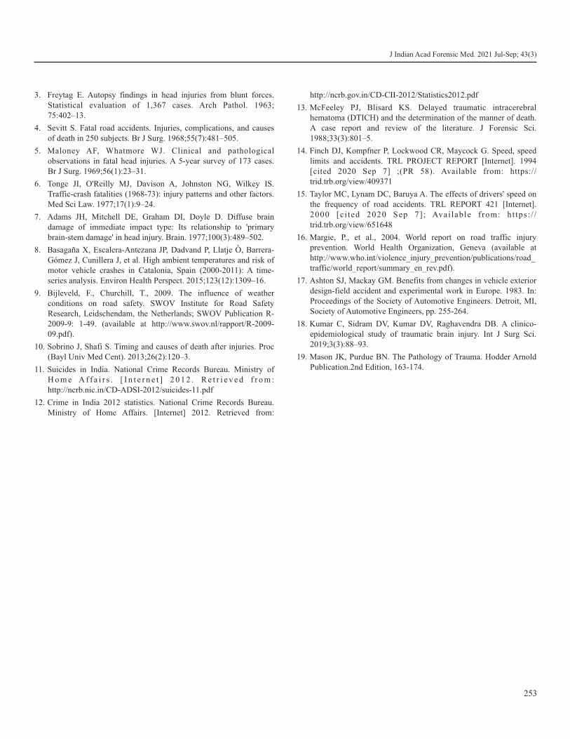
J Indian Acad Forensic Med. 2021 Jul-Sep; 43(3)
253
3. Freytag E. Autopsy findings in head injuries from blunt forces. Statistical evaluation of 1,367 cases. Arch Pathol. 1963; 75:402–13.
4. Sevitt S. Fatal road accidents. Injuries, complications, and causes of death in 250 subjects. Br J Surg. 1968;55(7):481–505.
5. Maloney AF, Whatmore WJ. Clinical and pathological observations in fatal head injuries. A 5-year survey of 173 cases. Br J Surg. 1969;56(1):23–31.
6. Tonge JI, O'Reilly MJ, Davison A, Johnston NG, Wilkey IS. Traffic-crash fatalities (1968-73): injury patterns and other factors. Med Sci Law. 1977;17(1):9–24.
7. Adams JH, Mitchell DE, Graham DI, Doyle D. Diffuse brain damage of immediate impact type: Its relationship to 'primary brain-stem damage' in head injury. Brain. 1977;100(3):489–502.
8. Basagaña X, Escalera-Antezana JP, Dadvand P, Llatje Ò, Barrera-Gómez J, Cunillera J, et al. High ambient temperatures and risk of motor vehicle crashes in Catalonia, Spain (2000-2011): A time-series analysis. Environ Health Perspect. 2015;123(12):1309–16.
9. Bijleveld, F., Churchill, T., 2009. The influence of weather conditions on road safety. SWOV Institute for Road Safety Research, Leidschendam, the Netherlands; SWOV Publication R-2009-9: 1-49. (available at http://www.swov.nl/rapport/R-2009-09.pdf).
10. Sobrino J, Shafi S. Timing and causes of death after injuries. Proc (Bayl Univ Med Cent). 2013;26(2):120–3.
11. Suicides in India. National Crime Records Bureau. Ministry of H o m e A f f a i r s . [ I n t e r n e t ] 2 0 1 2 . R e t r i e v e d f r o m : http://ncrb.nic.in/CD-ADSI-2012/suicides-11.pdf
12. Crime in India 2012 statistics. National Crime Records Bureau. Ministry of Home Affairs. [Internet] 2012. Retrieved from:
http://ncrb.gov.in/CD-CII-2012/Statistics2012.pdf
13. McFeeley PJ, Blisard KS. Delayed traumatic intracerebral hematoma (DTICH) and the determination of the manner of death. A case report and review of the literature. J Forensic Sci. 1988;33(3):801–5.
14. Finch DJ, Kompfner P, Lockwood CR, Maycock G. Speed, speed limits and accidents. TRL PROJECT REPORT [Internet]. 1994 [cited 2020 Sep 7] ;(PR 58). Available from: https:// trid.trb.org/view/409371
15. Taylor MC, Lynam DC, Baruya A. The effects of drivers' speed on the frequency of road accidents. TRL REPORT 421 [Internet]. 2000 [ci ted 2020 Sep 7] ; Avai lable f rom: ht tps: / / trid.trb.org/view/651648
16. Margie, P., et al., 2004. World report on road traffic injury prevention. World Health Organization, Geneva (available at http://www.who.int/violence_injury_prevention/publications/road_traffic/world_report/summary_en_rev.pdf).
17. Ashton SJ, Mackay GM. Benefits from changes in vehicle exterior design-field accident and experimental work in Europe. 1983. In: Proceedings of the Society of Automotive Engineers. Detroit, MI, Society of Automotive Engineers, pp. 255-264.
18. Kumar C, Sidram DV, Kumar DV, Raghavendra DB. A clinico-epidemiological study of traumatic brain injury. Int J Surg Sci. 2019;3(3):88–93.
19. Mason JK, Purdue BN. The Pathology of Trauma. Hodder Arnold Publication.2nd Edition, 163-174.
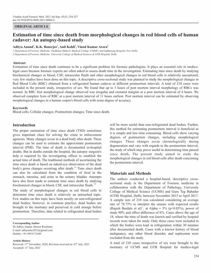
AbstractEstimation of time since death continues to be a significant problem for forensic pathologists. It plays an essential role in medico-legal cases because forensic experts are often asked to assess death time in the investigation. Estimating time since death by studying biochemical changes in blood, CSF, intraocular fluids and other morphological changes in red blood cells is relatively unexplored; very few studies have been done on this topic. A descriptive cross-sectional study was planned to study the morphological changes in Red Blood Cells (RBC) obtained from a refrigerated human cadaver at different postmortem intervals. A total of 210 cases were included in the present study, irrespective of sex. We found that up to 3 hours of post mortem interval morphology of RBCs was normal. In RBC first morphological change observed was irregular and crenated margins at a post mortem interval of 4 hours. We observed complete lysis of RBC at a post mortem interval of 11 hours earliest. Post mortem interval can be estimated by observing morphological changes in a human corpse's blood cells with some degree of accuracy.
KeywordsBlood cells; Cellular changes; Postmortem changes; Time since death.
Estimation of time since death from morphological changes in red blood cells of humancadaver: An autopsy-based study
ORIGINAL ARTICLE
Introduction
The proper estimation of time since death (TSD) sometimes gives important clues for solving the crime to enforcement agencies. Many changes occur in a dead body after death. These changes can be used to estimate the approximate postmortem interval (PMI). The time of death is documented in-hospital deaths. But in deaths outside the hospital, the autopsy surgeon's help is required by the investigating officer to establish the actual time of death. The traditional methods of ascertaining the time since death is based on naked-eye observation of the dead
1-4body's gross changes occurring after death. Time since death can also be calculated from the condition of food in the stomach, intestine, and urine in the urinary bladder. Attempts have also been made to estimate time since death by studying
5-9biochemical changes in blood, CSF, and intraocular fluids.
The study of morphological changes in red blood cells to determine time since death is relatively an untouched topic. Few studies on this topic have been mostly on non-refrigerated dead bodies; however, in common practice, dead bodies are brought to the mortuary and preserved in cold storage before postmortem. Therefore, data related to refrigerated dead bodies
will be more useful than non-refrigerated dead bodies. Further, this method for estimating postmortem interval is beneficial as it is simple and less time-consuming. Blood cells show varying degrees of postmortem changes, including morphological changes. These changes occur chronologically during degeneration and vary with regards to the postmortem interval, the study of which may prove useful in determining time passed since death. The present study aimed to study the morphological changes in red blood cells after death concerning the postmortem interval.
Materials and Methods
The authors conducted a hospital-based, descriptive cross-sectional study in the Department of Forensic medicine in collaboration with the Department of Pathology, University College of Medical Science (UCMS) and Guru Teg Bahadur (GTB) Hospital, Delhi, between November 2015 to April 2017. A sample size of 210 was calculated considering an average rate of 78.75% to interpret the smears with expected results
11(Rajesh Bardale et al) , at Alpha = 5% (p<0.05%), power of study 80% and effect difference of 6%. Cases above the age of 18, where the time of death was known and verified by hospital records were taken for study. Only those cases were included in which the bodies were kept in refrigeration within 30 minutes after documented death. Cases with a known history of blood malignancy, any other blood disorder, and septicemia were excluded from the study.
A total of 210 cases irrespective of sex were brought to the mortuary of UCMS and GTB Hospital for medico-legal
254
Corresponding Author
Dr Aditya Anand, (Senior Resident)
E-mail: [email protected]
Mobile: +91-9953171449
Article Historyth thReceived: 17 November, 2020; Revision received on: 07 July, 2021 thAccepted: 11 July, 2021
1 2 2 2Aditya Anand , K.K. Banerjee , Anil Kohli , Vinod Kumar Arora1 Department of Forensic Medicine, Vardhman Mahavir Medical College (VMMC) and Safdarjung Hospital, New Delhi
2 Department of Forensic Medicine, University College of Medical Sciences & GTB Hospital, Delhi
J Indian Acad Forensic Med. 2021 Jul-Sep; 43(3): 254-257doi:10.5958/0974-0848.2021.00064.6
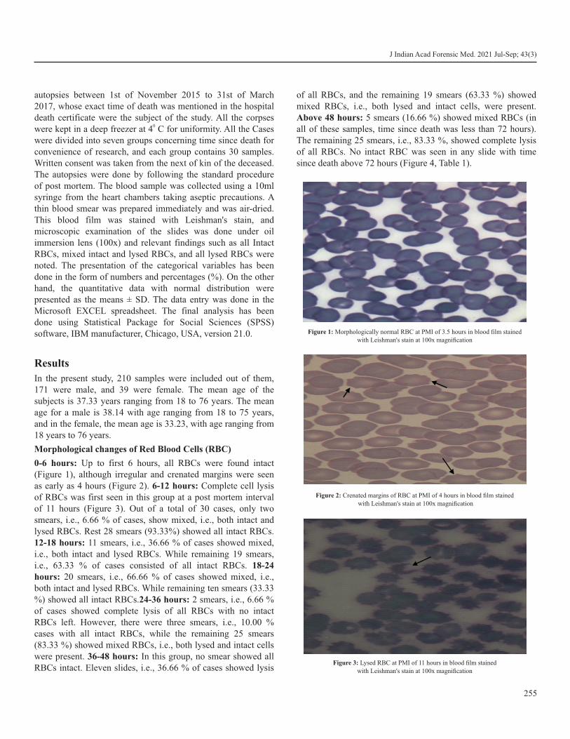
autopsies between 1st of November 2015 to 31st of March 2017, whose exact time of death was mentioned in the hospital death certificate were the subject of the study. All the corpses
0were kept in a deep freezer at 4 C for uniformity. All the Cases were divided into seven groups concerning time since death for convenience of research, and each group contains 30 samples. Written consent was taken from the next of kin of the deceased. The autopsies were done by following the standard procedure of post mortem. The blood sample was collected using a 10ml syringe from the heart chambers taking aseptic precautions. A thin blood smear was prepared immediately and was air-dried. This blood film was stained with Leishman's stain, and microscopic examination of the slides was done under oil immersion lens (100x) and relevant findings such as all Intact RBCs, mixed intact and lysed RBCs, and all lysed RBCs were noted. The presentation of the categorical variables has been done in the form of numbers and percentages (%). On the other hand, the quantitative data with normal distribution were presented as the means ± SD. The data entry was done in the Microsoft EXCEL spreadsheet. The final analysis has been done using Statistical Package for Social Sciences (SPSS) software, IBM manufacturer, Chicago, USA, version 21.0.
Results
In the present study, 210 samples were included out of them, 171 were male, and 39 were female. The mean age of the subjects is 37.33 years ranging from 18 to 76 years. The mean age for a male is 38.14 with age ranging from 18 to 75 years, and in the female, the mean age is 33.23, with age ranging from 18 years to 76 years.
Morphological changes of Red Blood Cells (RBC)
0-6 hours: Up to first 6 hours, all RBCs were found intact (Figure 1), although irregular and crenated margins were seen as early as 4 hours (Figure 2). 6-12 hours: Complete cell lysis of RBCs was first seen in this group at a post mortem interval of 11 hours (Figure 3). Out of a total of 30 cases, only two smears, i.e., 6.66 % of cases, show mixed, i.e., both intact and lysed RBCs. Rest 28 smears (93.33%) showed all intact RBCs. 12-18 hours: 11 smears, i.e., 36.66 % of cases showed mixed, i.e., both intact and lysed RBCs. While remaining 19 smears, i.e., 63.33 % of cases consisted of all intact RBCs. 18-24 hours: 20 smears, i.e., 66.66 % of cases showed mixed, i.e., both intact and lysed RBCs. While remaining ten smears (33.33 %) showed all intact RBCs.24-36 hours: 2 smears, i.e., 6.66 % of cases showed complete lysis of all RBCs with no intact RBCs left. However, there were three smears, i.e., 10.00 % cases with all intact RBCs, while the remaining 25 smears (83.33 %) showed mixed RBCs, i.e., both lysed and intact cells were present. 36-48 hours: In this group, no smear showed all RBCs intact. Eleven slides, i.e., 36.66 % of cases showed lysis
of all RBCs, and the remaining 19 smears (63.33 %) showed mixed RBCs, i.e., both lysed and intact cells, were present. Above 48 hours: 5 smears (16.66 %) showed mixed RBCs (in all of these samples, time since death was less than 72 hours). The remaining 25 smears, i.e., 83.33 %, showed complete lysis of all RBCs. No intact RBC was seen in any slide with time since death above 72 hours (Figure 4, Table 1).
255
J Indian Acad Forensic Med. 2021 Jul-Sep; 43(3)
Figure 1: Morphologically normal RBC at PMI of 3.5 hours in blood film stained
with Leishman's stain at 100x magnification
Figure 2: Crenated margins of RBC at PMI of 4 hours in blood film stained
with Leishman's stain at 100x magnification
Figure 3: Lysed RBC at PMI of 11 hours in blood film stained
with Leishman's stain at 100x magnification
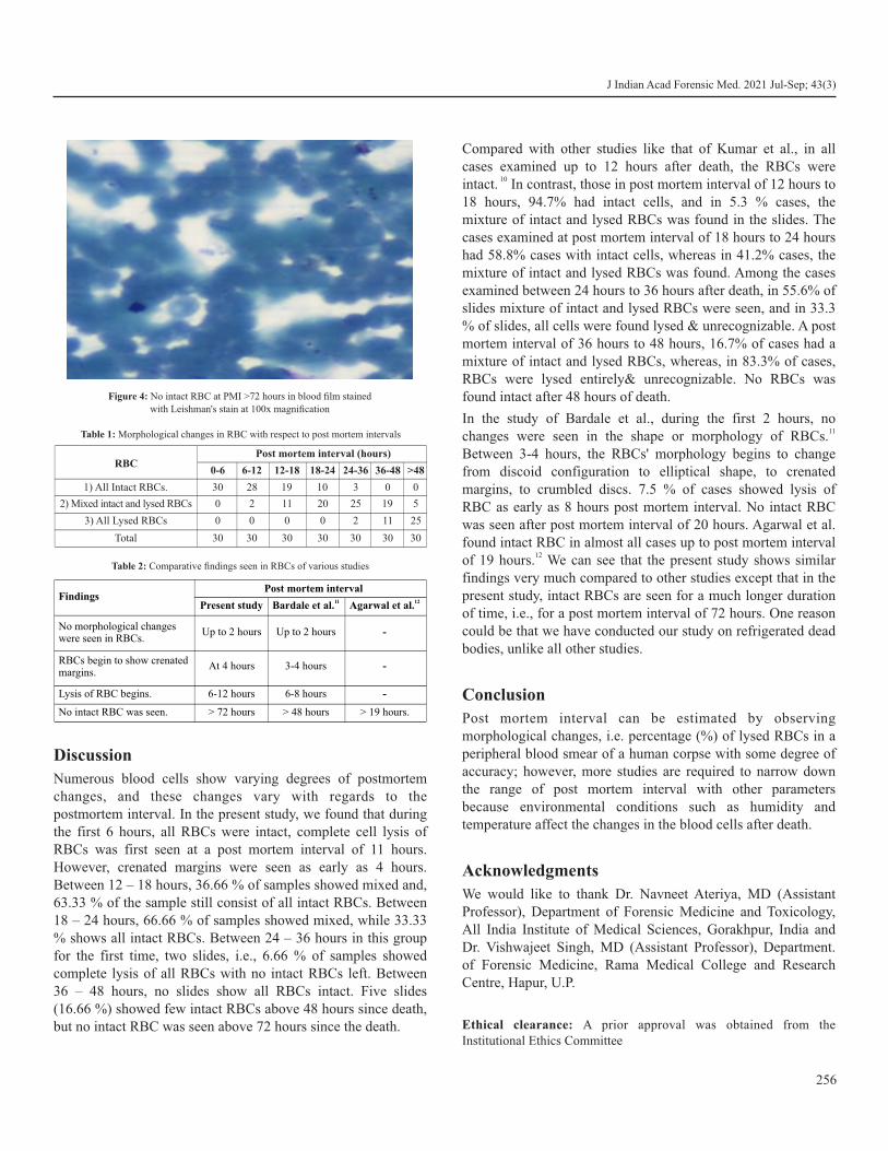
Discussion
Numerous blood cells show varying degrees of postmortem changes, and these changes vary with regards to the postmortem interval. In the present study, we found that during the first 6 hours, all RBCs were intact, complete cell lysis of RBCs was first seen at a post mortem interval of 11 hours. However, crenated margins were seen as early as 4 hours. Between 12 – 18 hours, 36.66 % of samples showed mixed and, 63.33 % of the sample still consist of all intact RBCs. Between 18 – 24 hours, 66.66 % of samples showed mixed, while 33.33 % shows all intact RBCs. Between 24 – 36 hours in this group for the first time, two slides, i.e., 6.66 % of samples showed complete lysis of all RBCs with no intact RBCs left. Between 36 – 48 hours, no slides show all RBCs intact. Five slides (16.66 %) showed few intact RBCs above 48 hours since death, but no intact RBC was seen above 72 hours since the death.
Compared with other studies like that of Kumar et al., in all cases examined up to 12 hours after death, the RBCs were
10intact. In contrast, those in post mortem interval of 12 hours to 18 hours, 94.7% had intact cells, and in 5.3 % cases, the mixture of intact and lysed RBCs was found in the slides. The cases examined at post mortem interval of 18 hours to 24 hours had 58.8% cases with intact cells, whereas in 41.2% cases, the mixture of intact and lysed RBCs was found. Among the cases examined between 24 hours to 36 hours after death, in 55.6% of slides mixture of intact and lysed RBCs were seen, and in 33.3 % of slides, all cells were found lysed & unrecognizable. A post mortem interval of 36 hours to 48 hours, 16.7% of cases had a mixture of intact and lysed RBCs, whereas, in 83.3% of cases, RBCs were lysed entirely& unrecognizable. No RBCs was found intact after 48 hours of death.
In the study of Bardale et al., during the first 2 hours, no 11changes were seen in the shape or morphology of RBCs.
Between 3-4 hours, the RBCs' morphology begins to change from discoid configuration to elliptical shape, to crenated margins, to crumbled discs. 7.5 % of cases showed lysis of RBC as early as 8 hours post mortem interval. No intact RBC was seen after post mortem interval of 20 hours. Agarwal et al. found intact RBC in almost all cases up to post mortem interval
12of 19 hours. We can see that the present study shows similar findings very much compared to other studies except that in the present study, intact RBCs are seen for a much longer duration of time, i.e., for a post mortem interval of 72 hours. One reason could be that we have conducted our study on refrigerated dead bodies, unlike all other studies.
Conclusion
Post mortem interval can be estimated by observing morphological changes, i.e. percentage (%) of lysed RBCs in a peripheral blood smear of a human corpse with some degree of accuracy; however, more studies are required to narrow down the range of post mortem interval with other parameters because environmental conditions such as humidity and temperature affect the changes in the blood cells after death.
Acknowledgments
We would like to thank Dr. Navneet Ateriya, MD (Assistant Professor), Department of Forensic Medicine and Toxicology, All India Institute of Medical Sciences, Gorakhpur, India and Dr. Vishwajeet Singh, MD (Assistant Professor), Department. of Forensic Medicine, Rama Medical College and Research Centre, Hapur, U.P.
Ethical clearance: A prior approval was obtained from the Institutional Ethics Committee
J Indian Acad Forensic Med. 2021 Jul-Sep; 43(3)
256
Figure 4: No intact RBC at PMI >72 hours in blood film stained
with Leishman's stain at 100x magnification
RBCPost mortem interval (hours)
0-6 6-12 12-18 18-24 24-36 36-48 >48
1) All Intact RBCs. 30 28 19 10 3 0 0
2) Mixed intact and lysed RBCs 0
2
11
20 25 19 5
3) All Lysed RBCs 0 0 0 0 2 11 25
Total 30 30 30 30 30 30 30
Table 1: Morphological changes in RBC with respect to post mortem intervals
FindingsPost mortem interval
Present study 11Bardale et al. 12Agarwal et al.
No morphological changeswere seen in RBCs.
Up to 2 hours Up to 2 hours -
RBCs begin to show crenatedmargins.
At 4 hours
3-4 hours -
Lysis of RBC begins. 6-12 hours 6-8 hours -
No intact RBC was seen. > 72 hours > 48 hours > 19 hours.
Table 2: Comparative findings seen in RBCs of various studies
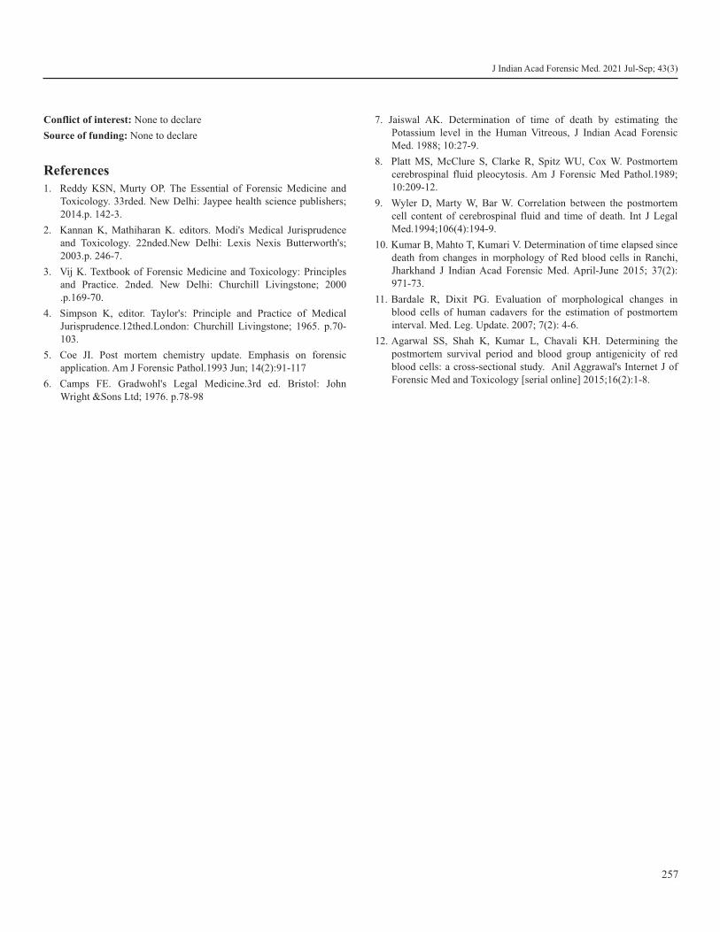
Conflict of interest: None to declare
Source of funding: None to declare
References1. Reddy KSN, Murty OP. The Essential of Forensic Medicine and
Toxicology. 33rded. New Delhi: Jaypee health science publishers; 2014.p. 142-3.
2. Kannan K, Mathiharan K. editors. Modi's Medical Jurisprudence and Toxicology. 22nded.New Delhi: Lexis Nexis Butterworth's; 2003.p. 246-7.
3. Vij K. Textbook of Forensic Medicine and Toxicology: Principles and Practice. 2nded. New Delhi: Churchill Livingstone; 2000 .p.169-70.
4. Simpson K, editor. Taylor's: Principle and Practice of Medical Jurisprudence.12thed.London: Churchill Livingstone; 1965. p.70-103.
5. Coe JI. Post mortem chemistry update. Emphasis on forensic application. Am J Forensic Pathol.1993 Jun; 14(2):91-117
6. Camps FE. Gradwohl's Legal Medicine.3rd ed. Bristol: John Wright &Sons Ltd; 1976. p.78-98
7. Jaiswal AK. Determination of time of death by estimating the Potassium level in the Human Vitreous, J Indian Acad Forensic Med. 1988; 10:27-9.
8. Platt MS, McClure S, Clarke R, Spitz WU, Cox W. Postmortem cerebrospinal fluid pleocytosis. Am J Forensic Med Pathol.1989; 10:209-12.
9. Wyler D, Marty W, Bar W. Correlation between the postmortem cell content of cerebrospinal fluid and time of death. Int J Legal Med.1994;106(4):194-9.
10. Kumar B, Mahto T, Kumari V. Determination of time elapsed since death from changes in morphology of Red blood cells in Ranchi, Jharkhand J Indian Acad Forensic Med. April-June 2015; 37(2): 971-73.
11. Bardale R, Dixit PG. Evaluation of morphological changes in blood cells of human cadavers for the estimation of postmortem interval. Med. Leg. Update. 2007; 7(2): 4-6.
12. Agarwal SS, Shah K, Kumar L, Chavali KH. Determining the postmortem survival period and blood group antigenicity of red blood cells: a cross-sectional study. Anil Aggrawal's Internet J of Forensic Med and Toxicology [serial online] 2015;16(2):1-8.
J Indian Acad Forensic Med. 2021 Jul-Sep; 43(3)
257
AbstractSexing accuracy of several metric dimensions of second and seventh cervical vertebras from CT scan images of a series of Iranian adult people were investigated. In this cross-sectional study, sexual dimorphism of 15 indexes of second cervical vertebra and 10 indexes of the seventh cervical vertebra were investigated. Indexes were obtained from CT scan images of 70 patients (35 males and 35 females) who underwent imaging for indications other than present study. Among measured dimension of second vertebra, 9 were associated with sex. Of these indexes, Maximum width of the superior Facet and Maximum height of axis were independent predictors of sex with correct sex classification of 81.4% when used in combination. Of 10 investigated indexes of the seventh vertebra, 4 were associated with sex: Length of superior facets, Length of the inferior surface of the vertebral body, Width of the inferior surface of the vertebral body and Length of spinous process. Length of the inferior surface of the vertebral body and Length of spinous process were independent sex predictors and together provided sex classification accuracy of 78.6%. We found considerable accuracy in sex classification by using metric dimensions of cervical vertebras in adult Iranian population.
KeywordsCervical vertebrae; Computer tomography; Dimension; Metric; Sex
Investigation of sexing accuracy of second and seventh cervical vertebras in adult Iranian population by using CT scan images
ORIGINAL ARTICLE
Introduction
Reliable estimation of sex from human skeletal remains is one of the most important factors in biological profiling and forensic anthropology casework especially in cases of mass
1,7disaster and high intensity explosions. Therefore, finding novel trustworthy methods for skeletal identification is
4considered as an essential component of medico-legal surveys. Forensic anthropology is applied with the goal to identify unknown human skeletal remains through the standard scientific techniques and to make a biological profile, an
4osteological biography. The biological profile indices including age at death, stature, and ancestry are sex dependent which are
1,4,8affected by sex estimation as a primary significance. Some traditionally morphological and anthropometric methods have been employed by forensic scientists and physical
9-11anthropologists for the sex estimation of bone samples. Several sex determination studies have been conducted by metric analysis of talus, femur, patella, humerus, calcaneus, metatarsals, tarsals, metacarpals, phalanges, scapula, clavicle
12-20and sternum. Different studies had applied the first and the second cervical vertebrae to determine sex correctly with nearly
the same level of accuracy compared to the other traditionally 3,21used bones. Other studies applied CT imaging and real bone
subjects with different sample size and level of accuracy for sex 3.21-26discrimination from the vertebra. Wescott et al. showed that
sex determination accuracy between 76-86% in the sagittal 22length. They showed that accuracy levels for estimating sex
22from the axis varied correctly between 77-90% correct. Voisin et al. showed that the useful sexually dimorphic bones for forensic sex estimation are the seven cervical vertebrae (C1-
27C7). Forensic medicine has been revolutionized using modern cross-sectional imaging techniques including 3D imaging techniques such as computer tomography (CT) and magnetic
7resonance imaging (MRI) over the past decade. These methods could be useful in visualizing almost every anatomical and
28pathological structure with high resolution and quality.
In a review of methods involved in sex estimation from human skeletal remains in South Africa within the forensic context showed that sex considered as one of the key factors in
29biological profiling of the individuals. The examined methods of sex determination included morphological, metrical,
29 geometric morphometries and molecular approaches. The aim of current study was focus on the level of sexual accuracy in the second and seventh cervical vertebrae to establish an accurate sex estimation method using measurements data obtained from the CT scan imaging of Iranian skeletal populations.
Material and Methods
This cross-sectional study was carried out on sexuality accuracy of several metric dimensions of second and seventh cervical
258
Corresponding Author
Dr. Arezoo Amiri
Email: [email protected]
Article Historyth thReceived: 11 November, 2020; Revision received on: 05 July, 2021thAccepted: 09 July, 2021
1 1 2 1Reza Saadat Mostafavi , Azadeh Memarian , Arezoo Amiri , Omid Motamedi1 Department of Radiology, Iran University of Medical Sciences, Tehran, Iran
2 Department of Forensic Medicine, Iran University of Medical Sciences, Tehran, Iran
J Indian Acad Forensic Med. 2021 Jul-Sep; 43(3): 258-264doi:10.5958/0974-0848.2021.00065.8
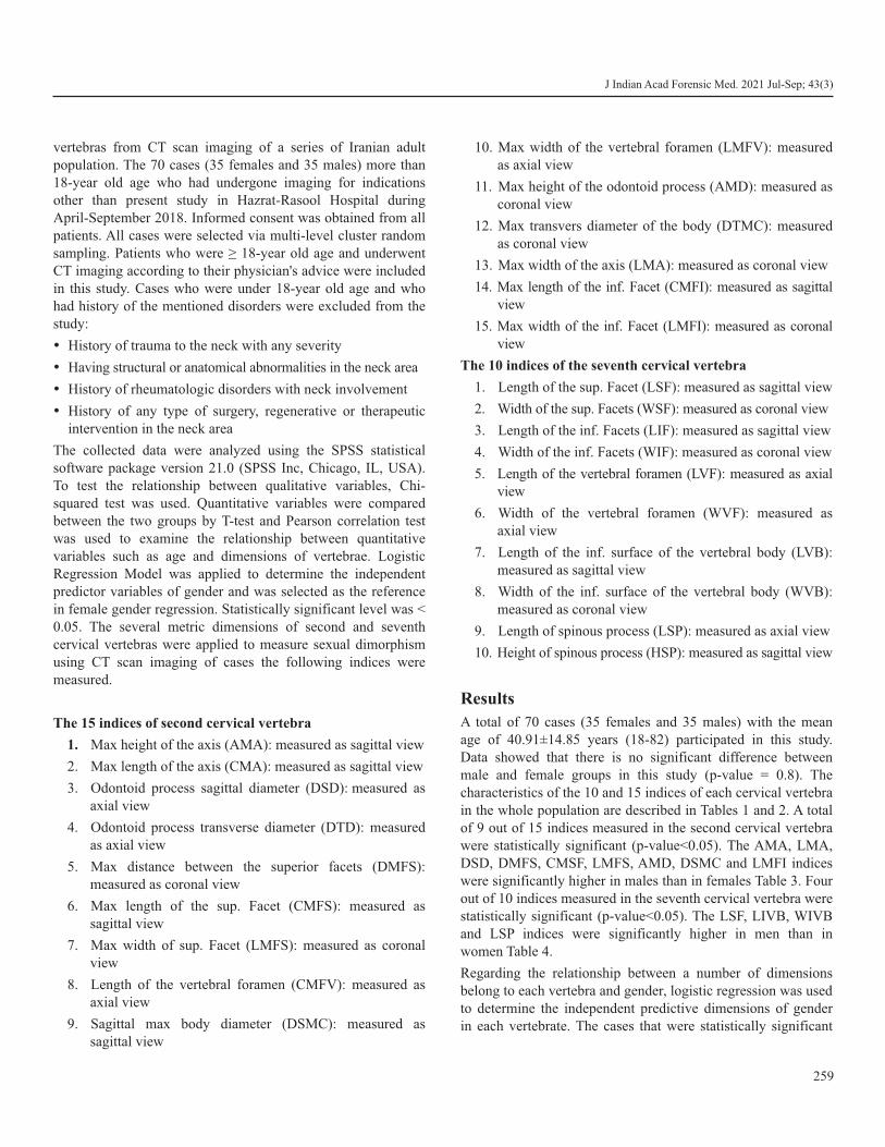
vertebras from CT scan imaging of a series of Iranian adult population. The 70 cases (35 females and 35 males) more than 18-year old age who had undergone imaging for indications other than present study in Hazrat-Rasool Hospital during April-September 2018. Informed consent was obtained from all patients. All cases were selected via multi-level cluster random sampling. Patients who were ≥ 18-year old age and underwent CT imaging according to their physician's advice were included in this study. Cases who were under 18-year old age and who had history of the mentioned disorders were excluded from the study:
Ÿ History of trauma to the neck with any severity
Ÿ Having structural or anatomical abnormalities in the neck area
Ÿ History of rheumatologic disorders with neck involvement
Ÿ History of any type of surgery, regenerative or therapeutic intervention in the neck area
The collected data were analyzed using the SPSS statistical software package version 21.0 (SPSS Inc, Chicago, IL, USA). To test the relationship between qualitative variables, Chi-squared test was used. Quantitative variables were compared between the two groups by T-test and Pearson correlation test was used to examine the relationship between quantitative variables such as age and dimensions of vertebrae. Logistic Regression Model was applied to determine the independent predictor variables of gender and was selected as the reference in female gender regression. Statistically significant level was < 0.05. The several metric dimensions of second and seventh cervical vertebras were applied to measure sexual dimorphism using CT scan imaging of cases the following indices were measured.
The 15 indices of second cervical vertebra
1. Max height of the axis (AMA): measured as sagittal view
2. Max length of the axis (CMA): measured as sagittal view
3. Odontoid process sagittal diameter (DSD): measured as axial view
4. Odontoid process transverse diameter (DTD): measured as axial view
5. Max distance between the superior facets (DMFS): measured as coronal view
6. Max length of the sup. Facet (CMFS): measured as sagittal view
7. Max width of sup. Facet (LMFS): measured as coronal view
8. Length of the vertebral foramen (CMFV): measured as axial view
9. Sagittal max body diameter (DSMC): measured as sagittal view
10. Max width of the vertebral foramen (LMFV): measured as axial view
11. Max height of the odontoid process (AMD): measured as coronal view
12. Max transvers diameter of the body (DTMC): measured as coronal view
13. Max width of the axis (LMA): measured as coronal view
14. Max length of the inf. Facet (CMFI): measured as sagittal view
15. Max width of the inf. Facet (LMFI): measured as coronal view
The 10 indices of the seventh cervical vertebra
1. Length of the sup. Facet (LSF): measured as sagittal view
2. Width of the sup. Facets (WSF): measured as coronal view
3. Length of the inf. Facets (LIF): measured as sagittal view
4. Width of the inf. Facets (WIF): measured as coronal view
5. Length of the vertebral foramen (LVF): measured as axial view
6. Width of the vertebral foramen (WVF): measured as axial view
7. Length of the inf. surface of the vertebral body (LVB): measured as sagittal view
8. Width of the inf. surface of the vertebral body (WVB): measured as coronal view
9. Length of spinous process (LSP): measured as axial view
10. Height of spinous process (HSP): measured as sagittal view
Results
A total of 70 cases (35 females and 35 males) with the mean age of 40.91±14.85 years (18-82) participated in this study. Data showed that there is no significant difference between male and female groups in this study (p-value = 0.8). The characteristics of the 10 and 15 indices of each cervical vertebra in the whole population are described in Tables 1 and 2. A total of 9 out of 15 indices measured in the second cervical vertebra were statistically significant (p-value<0.05). The AMA, LMA, DSD, DMFS, CMSF, LMFS, AMD, DSMC and LMFI indices were significantly higher in males than in females Table 3. Four out of 10 indices measured in the seventh cervical vertebra were statistically significant (p-value<0.05). The LSF, LIVB, WIVB and LSP indices were significantly higher in men than in women Table 4.
Regarding the relationship between a number of dimensions belong to each vertebra and gender, logistic regression was used to determine the independent predictive dimensions of gender in each vertebrate. The cases that were statistically significant
259
J Indian Acad Forensic Med. 2021 Jul-Sep; 43(3)
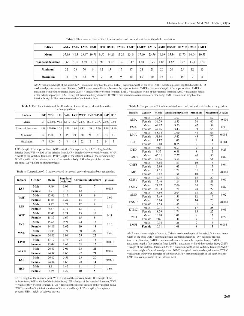
J Indian Acad Forensic Med. 2021 Jul-Sep; 43(3)
Indices AMA CMA LMA DSD DTD DMFS CMFS LMFS CMFV LMFV AMD DSMC DTMC CMFI LMFI
Mean 37.93 48.5 53.47
10.79
9.50
44.29
13.26
13.84
17.69
23.76
16.19 15.34 18.70 10.04 10.53
Standard deviation 3.68 3.76 4.98
1.03
.90
3.07
1.62
1.47
1.40
1.93
1.86 1.62 1.77 1.23 1.24
Minimum 52 58 70 14 12 54 17 17 21 28 20 20 25 12 13
Maximum 30 39 43 9 7 36 9 10 15 20 12 11 15 7 8
Table 1: The characteristics of the 15 indices of second cervical vertebra in the whole population
AMA: maximum height of the axis; CMA = maximum length of the axis; LMA = maximum width of the axis; DSD = odontoid process sagittal diameter; DTD
= odontoid process transverse diameter; DMFS = maximum distance between the superior facets; CMFS = maximum length of the superior facet; LMFS =
maximum width of the superior facet; CMFV = length of the vertebral foramen; LMFV = maximum width of the vertebral foramen; AMD = maximum height
of the odontoid process; DSMC = sagittal maximum body diameter; DTMC = maximum transverse diameter of the body; CMFI = maximum length of the
inferior facet; LMFI = maximum width of the inferior facet.
Indices LSF WSF LIF WIF LVF WVF LIVB WIVB LSP HSP
Mean 91 12.1286 9.57 12.17 15.27 24.79 16.33 25.79 23.99 7.80
Standard deviation 1.18 1.21490 1.20 1.50 1.96 1.85 1.88 2.59 3.98 14.10
Minimum 12 15.00 13 15 24 30 21 33 33 11
Maximum 7 9.00 7 8 13 22 12 21 14 5
LSF = length of the superior facet; WSF = width of the superior facet; LIF = length of the
inferior facet; WIF = width of the inferior facet; LVF = length of the vertebral foramen; WVF
= width of the vertebral foramen; LIVB = length of the inferior surface of the vertebral body;
WIVB = width of the inferior surface of the vertebral body; LSP = length of the spinous
process; HSP = height of spinous process
Table 2: The characteristics of the 10 indices of seventh cervical vertebra in the whole population
Indices Gender Mean Standard deviation Minimum Maximum p-value
AMAMale 39.57 3.93 31 52
<0.001Female 36.29 2.53 30 40
CMAMale 49.97 3.53 42 58
0.30Female 47.06 3.43 39 56
LMAMale 55.14 3.90 46 63
0.004Female 51.80 5.42 43 70
DSDMale 11.17 1.07 9 14
0.001Female 10.40 0.85 9 12
DTDMale 9.63 0.91 7 11
0.23Female 9.37 0.88 8 12
DMFSMale 45.11 2.27 40 50
0.02Female 43.46 3.54 36 54
CMFSMale 13.66 1.53 10 16
0.04Female 12.86 1.63 9 17
LMFSMale 14.51 1.29 11 17
<0.001Female 13.17 1.34 10 16
CMFVMale 17.97 1.50 15 20
0.09Female 17.40 1.24 15 21
LMFVMale 24.17 2.06 20 28
0.07Female 23.34 1.71 20 27
AMDMale 16.69 2.04 13 20
0.02Female 15.69 1.51 12 19
DSMCMale 16.14 1.37 14 20
<0.001Female 14.54 1.46 11 19
DTMCMale 19.11 1.73 17 25
0.05Female 18.29 1.74 15 22
CMFIMale 10.20 1.02 8 12
0.29Female 9.89 1.41 7 12
LMFIMale 10.94 1.26 9 13
0.004Female 10.11 1.08 8 13
Table 3: Comparison of 15 indices related to second cervical vertebra between genders
AMA = maximum height of the axis; CMA = maximum length of the axis; LMA = maximum
width of the axis; DSD = odontoid process sagittal diameter; DTD = odontoid process
transverse diameter; DMFS = maximum distance between the superior facets; CMFS =
maximum length of the superior facet; LMFS = maximum width of the superior facet; CMFV
= length of the vertebral foramen; LMFV = maximum width of the vertebral foramen; AMD =
maximum height of the odontoid process; DSMC = sagittal maximum body diameter; DTMC
= maximum transverse diameter of the body; CMFI = maximum length of the inferior facet;
LMFI = maximum width of the inferior facet.
Indices Gender Mean Standarddeviation
Minimum Maximum p-value
LSFMale 9.49 1.09 12 7
0.005Female 8.71 1.15 12 7
WSFMale 12.40 1.17 15 9
0.06Female 11.86 1.22 14 9
LIFMale 9.77 1.21 12 8
0.16Female 9.37 1.17 13 7
WIFMale 12.46 1.24 15 10
0.11Female 11.89 1.69 15 8
LVFMale 15.66 2.21
24 130.10
Female 14.89 1.62
19 13
WVFMale 24.94 1.71 30 22
0.48Female 24.63 1.99 29 22
LIVBMale 17.17 1.76 21 13
<0.001Female 15.49 1.62 21 12
WIVBMale 26.63 3.06 33 21
0.006Female 24.94 1.66 27 21
LSPMale 26.03 3.51 33 20
<0.001Female 24.94 1.66 28 14
HSPMale 8.11 1.47 11 5
0.06Female 7.49 1.29 10 5
Table 4: Comparison of 10 indices related to seventh cervical vertebra between genders
LSF = length of the superior facet; WSF = width of the superior facet; LIF = length of the
inferior facet; WIF = width of the inferior facet; LVF = length of the vertebral foramen; WVF
= width of the vertebral foramen; LIVB = length of the inferior surface of the vertebral body;
WIVB = width of the inferior surface of the vertebral body; LSP = length of the spinous
process; HSP = height of spinous process
260
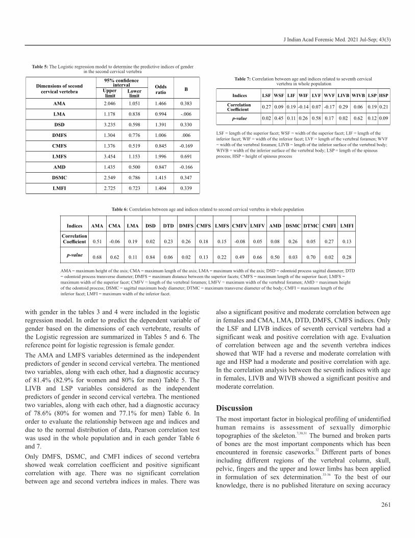
with gender in the tables 3 and 4 were included in the logistic regression model. In order to predict the dependent variable of gender based on the dimensions of each vertebrate, results of the Logistic regression are summarized in Tables 5 and 6. The reference point for logistic regression is female gender.
The AMA and LMFS variables determined as the independent predictors of gender in second cervical vertebra. The mentioned two variables, along with each other, had a diagnostic accuracy of 81.4% (82.9% for women and 80% for men) Table 5. The LIVB and LSP variables considered as the independent predictors of gender in second cervical vertebra. The mentioned two variables, along with each other, had a diagnostic accuracy of 78.6% (80% for women and 77.1% for men) Table 6. In order to evaluate the relationship between age and indices and due to the normal distribution of data, Pearson correlation test was used in the whole population and in each gender Table 6 and 7.
Only DMFS, DSMC, and CMFI indices of second vertebra showed weak correlation coefficient and positive significant correlation with age. There was no significant correlation between age and second vertebra indices in males. There was
also a significant positive and moderate correlation between age in females and CMA, LMA, DTD, DMFS, CMFS indices. Only the LSF and LIVB indices of seventh cervical vertebra had a significant weak and positive correlation with age. Evaluation of correlation between age and the seventh vertebra indices showed that WIF had a reverse and moderate correlation with age and HSP had a moderate and positive correlation with age. In the correlation analysis between the seventh indices with age in females, LIVB and WIVB showed a significant positive and moderate correlation.
Discussion
The most important factor in biological profiling of unidentified human remains is assessment of sexually dimorphic
7,30,31topographies of the skeleton. The burned and broken parts of bones are the most important components which has been
32encountered in forensic caseworks. Different parts of bones including different regions of the vertebral column, skull, pelvic, fingers and the upper and lower limbs has been applied
33-36in formulation of sex determination. To the best of our knowledge, there is no published literature on sexing accuracy
J Indian Acad Forensic Med. 2021 Jul-Sep; 43(3)
261
Dimensions of second cervical vertebra
Odds ratio
BUpper limit
Lower limit
AMA 2.046 1.051 1.466 0.383
LMA 1.178 0.838 0.994
DSD 3.235 0.598 1.391 0.330
DMFS 1.304 0.776 1.006 .006
CMFS 1.376 0.519 0.845 -0.169
LMFS 3.454 1.153 1.996 0.691
AMD 1.435 0.500 0.847 -0.166
DSMC 2.549 0.786 1.415 0.347
LMFI 2.725 0.723 1.404 0.339
95% confidence interval
-.006
Table 5: The Logistic regression model to determine the predictive indices of gender in the second cervical vertebra
Indices AMA CMA LMA DSD DTD DMFS CMFS LMFS CMFV LMFV AMD DSMC DTMC CMFI LMFI
CorrelationCoefficient 0.51 -0.06 0.19 0.02 0.23
0.26
0.18
0.15
-0.08
0.05 0.08 0.26 0.05 0.27 0.13
p-value 0.68 0.62 0.11 0.84 0.06 0.02 0.13 0.22 0.49 0.66 0.50 0.03 0.70 0.02 0.28
Table 6: Correlation between age and indices related to second cervical vertebra in whole population
AMA = maximum height of the axis; CMA = maximum length of the axis; LMA = maximum width of the axis; DSD = odontoid process sagittal diameter; DTD
= odontoid process transverse diameter; DMFS = maximum distance between the superior facets; CMFS = maximum length of the superior facet; LMFS =
maximum width of the superior facet; CMFV = length of the vertebral foramen; LMFV = maximum width of the vertebral foramen; AMD = maximum height
of the odontoid process; DSMC = sagittal maximum body diameter; DTMC = maximum transverse diameter of the body; CMFI = maximum length of the
inferior facet; LMFI = maximum width of the inferior facet.
Indices LSF WSF LIF WIF LVF WVF LIVB WIVB LSP HSP
Correlation Coefficient
0.27 0.09 0.19 -0.14 0.07 -0.17 0.29 0.06 0.19 0.21
p-value 0.02 0.45 0.11 0.26 0.58 0.17 0.02 0.62 0.12 0.09
Table 7: Correlation between age and indices related to seventh cervical vertebra in whole population
LSF = length of the superior facet; WSF = width of the superior facet; LIF = length of the
inferior facet; WIF = width of the inferior facet; LVF = length of the vertebral foramen; WVF
= width of the vertebral foramen; LIVB = length of the inferior surface of the vertebral body;
WIVB = width of the inferior surface of the vertebral body; LSP = length of the spinous
process; HSP = height of spinous process
from anthropometric measurements of second and seventh cervical vertebras among Iranian adult people using CT scan imaging. In the present study, for the first time, the role of the metric dimensions of second and seventh vertebra in sex determination of some adult population of Iran was investigated according to their cervical CT scan. Findings of the second cervical vertebra indices in males and females showed that in all cases, the dimensions of collected indices from the vertebrae of males are greater than that of females, but in 9 out of 15 indices, this difference was statistically significant. These 9 indices were including AMA, LMA, DSD, DMFS, CMFS, LMFS, AMD, DSMC, and LMFI. Among the 9 variables which had significant correlation with sex, 2 variables of LMSF and AMA considered as the independent predictive of sex with odds ratio of 2 and 1.47, respectively and they had diagnostic accuracy of 81.4%. This indicates the presence of sexual dimorphism in the measurements of Iranian vertebrae. The most significant mean difference was found in the linear dimensions of the second vertebra of LMA (3.34 mm) and AMA (3.28 mm) between the two sexes. A review of various studies on sex determination based on the dimensions of the second cervical vertebra and the comparison of their results with each other showed that there were some limitations among the studies including firstly, there were few studies, and, secondly, the dimensions examined were not the same between the studies.
In a study on cervical vertebra of skeletal specimens, the 15 linear indices of cervical vertebra were examined, which were largely in accordance with the metric dimensions measured in
1our study. The size of the examined dimensions was slightly 1different from the dimensions of our study. In all cases,
dimensions were higher in males, but the LMA and DSMC 1indices had the most difference between the two sexes. In their
study, four indices (CMA, LMA, LMFSD, and DSMC) were independent predictors of gender with predictive accuracy of
187% between sexes. The highest risk ratio was related to 1DSMC (2.66) and LMA (1.97), respectively. In other study
conducted by Marlow et al., 9 indices of cervical vertebra dimensions were measured, some of these dimensions are similar to those measured in our study. All of these dimensions
3in males were significantly larger than females. Of these dimensions, XSL, SFS, SFT, LVF, and XDH were gender-independent predictors that provided a total of 77% gender
3diagnostic accuracy. Independent gender variables in our study are somewhat similar to those of the four independent
3variables. In the analysis of the 5 dimensions of the second cervical vertebra between different populations, the diagnostic
22,26accuracy was obtained over 80%. These five dimensions were including the greatest length of the sagittal (from the anterior part of the vertebra to the posterior part of the genital area), the sagittal of the upper fast diameter, the cross-sectional diameter of the upper fast (the largest diameter of the upper
surface of the fast that is perpendicular to the sagittal surface), The length of the vertebral hole (the length of the inner hole measured at its lower end on the median plate), the largest length of the vertebra (the distance between the lower to the
22,26upper part of the vertebra in the anterior plane).
In comparison of our study with the above-mentioned studies on second cervical vertebral indices, a few points were noted: First, in all of the mentioned cases, as well as our study, the upper fast was considered as an independent predictor of gender; Second, the diagnostic accuracy of sex determination in different dimensions was significant in all cases; Third, although all dimensions were larger in males than females, but the mean difference was low even in significant indices suggesting it is essential to be more focused on practical points of these low rates of mean difference.
In terms of seventh cervical vertebra of current study, the 10 linear indices were investigated. All dimensions in this vertebra were higher in males than in females, and statistically significant correlations with gender was observed in 4 indices including the LSF, LIVB, WIVB and LSP. Of these four indices, only two variables LIVB (with odds ratio of 1.55) and LSP (odds ratio of 1.31) were independent gender predictors. The two variables had a diagnostic accuracy of 78.6%. Studies on the role of the role of seventh cervical vertebra dimensions in the sex determination were scarce and there are differences in measured metric dimensions and study methods among them. In confirmation of the findings of current study, assessment of the seventh cervical vertebra dimensions amongst the three groups of whites, blacks and South Africa tribes showed that the anterior posterior's length, width and height of the vertebra
37were greater in males than females. In a study in Spain, the length of the lower part of vertebra and the length and width of the intervertebral hole were recognized as strong sex determinants of the seventh cervical vertebra with a diagnostic
38accuracy of 81% for men and 79% for women. Another study on the European white population showed that the maximum height of the seventh cervical vertebra and the transverse diameter of the intervertebral hole in this group were independent gender predictors with a diagnostic accuracy of
4over 80%. Results of the mentioned studies showed the accuracy of different dimensions of seventh cervical vertebra in sex determination.
Studies have shown that the dimensions of the cervical vertebrae are correlated with skeletal dimensions and the larger dimensions of the skeletal vertebrae in males are attributed to
27, 39 the larger body size of them. However, in this study, both the dimensions of the vertebral bodies and their posterior pectorals were statistically significant with gender. However, more studies are needed to analyze the cause of gender differences in the vertebra. In the present study, relationships between age and
J Indian Acad Forensic Med. 2021 Jul-Sep; 43(3)
262
dimensions of the vertebrae were observed. Correlation between age and cervical vertebral dimensions was mostly positive and weak. Considering the age range of population was under the 69 year, the possibility of degenerative changes in the vertebra should be considered, which affects the dimensions of the vertebra. Other studies have shown the relationship between
4,40 age and dimensions of cervical vertebra. Even cervical vertebrae are known as a precise standard for bone age
41determination. Further studies are required to evaluate the effects of age on cervical vertebra dimensions.
Conclusion
In conclusion, all dimensions measured for the second and seventh cervical vertebra were higher in men than women in this study. The height of the second vertebra and the transverse diameter of the upper fast were independent gender predictors, with an accuracy of 81.4%. In the seventh vertebra, the anterior posterior of the vertebral body and the posterior length of the vertebra were independent predictors of gender, which had an accuracy of 78.6% in sex determination. The results of this study showed a high accuracy of cervical vertebral dimensions in sex determination of skeletal remains in Iranian population.
Ethical clearance: A prior approval was obtained from the Institutional Ethics Committee
Conflict of interest: None to declare
Source of funding: None to declare
References1. Gama I, Navega D, Cunha E. Sex estimation using the second
cervical vertebra: a morphometric analysis in a documented Portuguese skeletal sample. Int. J. Legal Med. 2015;129(2):365-72.
2. Krishan K, Chatterjee PM, Kanchan T, Kaur S, Baryah N, Singh RK. A review of sex estimation techniques during examination of skeletal remains in forensic anthropology casework. Forensic Sci Int. 2016;261:165.e1-8.
3. Marlow EJ, Pastor RF. Sex determination using the second cervical vertebra—a test of the method. J. Forensic Sci. 2011;56(1):165-9.
4. Rozendaal AS. Estimating sex from the seven cervical vertebrae : an analysis of White European skeletal populations. 2016 [cited 2 0 2 0 J u l y 7 ] ; Av a i l a b l e f r o m : h t t p s : / / l i b r a r y 2 . smu.ca/handle/01/26493
5. Noorian Zavareh F, Ameri M, Kordrostami R, Dadashzade N. The role of gender dimorphism on the relative length of fingers and the determination of the gender of the Iranian population: brief report. Tehran Univ. Med. J. TUMS Publications. 2017;75(8):616-20.
6. Soltani S, Ameri M, Aghakhani K, Ghorbani S. Evaluation of Greater Sciatic Notch Parameters in Sex Determination of Hip Bone by Three-Dimensional CT Images. J. Clin. Diagnostic Res. 2018;12(9).
7. El Dine FMB, El Shafei MM. Sex determination using anthropometric measurements from multi-slice computed tomography of the 12th thoracic and the first lumbar vertebrae among adult Egyptians. Egypt. J. Forensic Sci. 2015;5(3):82-9.
8. Ubelaker DH, DeGaglia CM. Population variation in skeletal sexual dimorphism. Forensic Sci Int. 2017;278:407.e1-407.e7.
9. Krogman W, Iscan MY. The human skeleton in forensic medicine. 2nd ed. Springfield, MO: Charles C Thomas Publisher; 1986.
10. Iscan MY, Steyn M. The human skeleton in forensic medicine: Charles C Thomas Publisher; 2013.
11. Rogers TL. Determining the sex of human remains through cranial morphology. J. Forensic Sci. 2005;50(3):1-8.
12. Mostafa EM, El-Elemi AH, El-Beblawy MA, Dawood AE-WA. Adult sex identification using digital radiographs of the proximal epiphysis of the femur at Suez Canal University Hospital in Ismailia, Egypt. Egypt. J. Forensic Sci. 2012;2(3):81-8.
13. Holman DJ, Bennett KA. Determination of sex from arm bone measurements. Am. J. Phys. 1991;84(4):421-6.
14. Moneim WMA, Hady RHA, Maaboud RMA, Fathy HM, Hamed AM. Identification of sex depending on radiological examination of foot and patella. Am J Forensic Med Pathol. 2008;29(2):136-40.
15. Abd-elaleem SA-e, Abd-elhameed M, Ewis AA-e. Talus measurements as a diagnostic tool for sexual dimorphism in Egyptian population. J Forensic Leg Med. 2012;19(2):70-6.
16. Zakaria MS, Mohammed AH, Habib SR, Hanna MM, Fahiem AL. Calcaneus radiograph as a diagnostic tool for sexual dimorphism in Egyptians. J Forensic Leg Med. 2010;17(7):378-82.
17. Eshak GA, Ahmed HM, Gawad EAA. Gender determination from hand bones length and volume using multidetector computed tomography: a study in Egyptian people. J Forensic Leg Med. 2011;18(6):246-52.
18. Papaioannou VA, Kranioti EF, Joveneaux P, Nathena D, Michalodimitrakis M. Sexual dimorphism of the scapula and the clavicle in a contemporary Greek population: applications in forensic identification. Forensic Sci. Int. 2012;217(1-3):231. e1-. e7.
19. Dabbs GR, Moore‐Jansen PH. A method for estimating sex using metric analysis of the scapula. J. Forensic Sci. 2010;55(1):149-52.
20. Singh J, Pathak R, Singh D. Morphometric sex determination from various sternal widths of Northwest Indian sternums collected from autopsy cadavers: a comparison of sexing methods. Egypt. J. Forensic Sci. 2012;2(1):18-28.
21. Marino EA. Sex estimation using the first cervical vertebra. Am. J. Phys. 1995;97(2):127-33.
22. Wescott DJ. Sex variation in the second cervical vertebra. J. Forensic Sci.. 2000;45(2):462-6.
23. Yu SB, Lee UY, Kwak DS, Ahn YW, Jin CZ, Zhao J, et al. Determination of sex for the 12th thoracic vertebra by morphometry of three‐dimensional reconstructed vertebral models. J. Forensic Sci. 2008;53(3):620-5.
24. Zheng WX, Cheng FB, Cheng KL, Tian Y, Lai Y, Zhang WS, et al. Sex assessment using measurements of the first lumbar vertebra. Forensic Sci. Int. 2012;219(1-3):285. e1-. e5.
25. Hou WB, Cheng KL, Tian SY, Lu YQ, Han YY, Lai Y, et al. Metric
J Indian Acad Forensic Med. 2021 Jul-Sep; 43(3)
263
method for sex determination based on the 12th thoracic vertebra in contemporary north-easterners in China. J Forensic Leg Med. 2012;19(3):137-43.
26. Bethard JD, Seet BL. Sex determination from the second cervical vertebra: a test of Wescott's method on a modern American sample. J. Forensic Sci. 2013;58(1):101-3.
27. Voisin MD. Sexual dimorphism in the 12th thoracic vertebra and its potential for sex estimation of human skeletal remains: Wichita State University; 2011.
28. Benazzi S, Bertelli P, Lippi B, Bedini E, Caudana R, Gruppioni G, et al. Virtual anthropology and forensic arts: the facial reconstruction of Ferrante Gonzaga. J. Archaeol. Sci. 2010;37(7):1572-8.
29. Bidmos MA, Gibbon VE, Štrkalj G. Recent advances in sex identification of human skeletal remains in South Africa. S. Afr. J. Sci. 2010;106(11-12):1-6.
30. Holland TD. Use of the cranial base in the identification of fire victims. J. Forensic Sci. 1989;34(2):458-60.
31. Bruzek J, Murail P. Methodology and reliability of sex determination from the skeleton. Forensic anthropology and medicine: Springer; 2006. p. 225-42.
32. Ogawa Y, Imaizumi K, Miyasaka S, Yoshino M. Discriminant functions for sex estimation of modern Japanese skulls. J Forensic Leg Med. 2013;20(4):234-8.
33. Mall G, Graw M, Gehring K-D, Hubig M. Determination of sex from femora. Forensic Sci. Int. 2000;113(1-3):315-21.
34. Rogers TL. A visual method of determining the sex of skeletal remains using the distal humerus. J. Forensic Sci. 1999;44(1):57-60.
35. Case DT, Ross AH. Sex determination from hand and foot bone lengths. J. Forensic Sci. 2007;52(2):264-70.
36. Bruzek J. A method for visual determination of sex, using the human hip bone. Am J Phys Anthropol. 2002;117(2):157–68.
37. Kibii JM, Pan R, Tobias PV. Morphometric variations of the 7th cervical vertebrae of Zulu, White, and Colored South Africans. Clin Anat. 2010;23(4):399–406.
38. Amores A, Botella MC, Alemán I. Sexual dimorphism in the 7th cervical and 12th thoracic vertebrae from a Mediterranean population. J. Forensic Sci. 2014;59(2):301-5.
39. Torimitsu S, Makino Y, Saitoh H, Sakuma A, Ishii N, Hayakawa M, et al. Stature estimation in Japanese cadavers based on the second cervical vertebra measured using multidetector computed tomography. Leg Med (Tokyo). 2015;17(3):145–9.
40. Parenteau CS, Wang NC, Zhang P, Caird MS, Wang SC. Quantification of pediatric and adult cervical vertebra—anatomical characteristics by age and gender for automotive application. Traffic Inj. Prev. 2014;15(6):572-82.
41. Mito T, Sato K, Mitani H. Cervical vertebral bone age in girls. Am J Orthod Dentofacial Orthop. 2002;122(4):380-5.
J Indian Acad Forensic Med. 2021 Jul-Sep; 43(3)
264
AbstractVarious laws have been implemented and amended for safeguarding the individuals suffering from mental ailments. The latest addition is the Mental Healthcare Act 2017 which tries to answer several lacunas in previous editions. The act tries to give legal solicitude with much needed precedence to rights of such patients. This review article is an attempt to go through the MHCA 2017 and its predecessor act, the Mental Health Act 1987 to discuss different modifications incorporated. This article tries to simply discuss the practical aspects of this act and the medicolegal perspective which is very much important for a practicing physician.
KeywordsMHCA 2017; Mental illness; Advance directive; Nominated representative; Forensic
Mental Healthcare Act (MHCA 2017)–A review from Forensic perspective
REVIEW ARTICLE
Introduction
The field of medicine is very much related to the legal system. A doctor is required to know the legal system and laws of land followed by his society while practicing medicine. Mental illness is a disease where the patient might not be fit enough for decision making. Thus, the patients with mental illness become the perfect victim for any exploitation. The exploitations might vary from negligence during medical treatment to voluntarily causing harm to the person. This could be done by anyone from family members to a total stranger for any reason ranging from having fun to hate crimes. In such patients with mental illness, the doctor is required to follow the guidelines based on the central legislation regarding mental healthcare, so that there won't be any negligence.
In pre-independent India, in 1858 the Indian Lunatic Asylum Act was introduced. Later on, in 1912, this was repealed by
1Indian Lunacy Act. In 1987, after Independence came an act 2with lot of changes, the Mental Health Act. Still there were
much more lacunas and gaps in the Mental Health Act which led to the constitution of The Mental Healthcare act 2017 which came into implementation on 29th May 2018 after presidential assent. The provisions in the act guide in catering of mental healthcare services to persons with mental illness (PMI/ PWMI). This act also helps in protecting the rights of such
3persons during delivery of mental healthcare services. In India, the burden of mental illness is huge owing to the socioeconomic status, illiteracy and unemployment. There are
large number of such persons not able to avail the mental health care services. The number of mentally ill patients getting treatment was much less proportionate to the statistics of total number of such patients for various reasons like social stigma, availability of a smaller number of trained mental healthcare professionals, a smaller number of institutions for such patients, inability to afford quality treatment etc. Indian government and legislature are trying to address these problems through implementation of different legislatures.
United Nations had started an international human rights treaty, the CRPD (Convention on the Rights of Persons with Disabilities) to protect the rights and dignity of people with
4mental illness. In this, mental illness is also added as a disability. So CRPD is applicable for mental illness just like any other physical disability. This meant the mental illnesses pounding on the personal and professional life of majority got a voice of compassion and empathy. This convention and treaty demanded changes in existing legislatures all around the world. The treaty
thwas put forward on 13 of December in year 2006 and India stsigned the same on 1 April 2007. Now since the CRPD was
signed, there raised a need for a much stronger and up to date legislature to address the legalities of care in a mentally ill and to circumvent the three-decade old The Mental Health Act, 1987. The MHA 1987 was not found fully compliant with the UNCRPD resolution. To comply with it, the 2017 edition of mental health act was passed by parliament on April 2017. In 1987 Mental Health Act, here we are comparing the Mental Health Act 1987 with the latest Mental Healthcare Act 2017.
1987 Mental Health Act
The act was passed in year 1987 and came into force by April 1993. The definition of act says "An act to consolidate and amend the law relating to the treatment and care of mentally ill persons, to make better provision with respect to their property and affairs and for matters connected therewith or incidental
265
Corresponding Author
Dr Atul S. Keche (Associate Professor)
Email : [email protected]
Mobile: +91-9890621600
Article Historyth thReceived: 10 October, 2020; Revision received on: 10 August, 2021 thAccepted: 15 August, 2021
Sravan J S, Atul S. Keche, Vivek Kumar Chouksey, Poovaragavan VDepartment of Forensic Medicine & Toxicology, AIIMS Bhopal
J Indian Acad Forensic Med. 2021 Jul-Sep; 43(3): 265-268doi:10.5958/0974-0848.2021.00066.X
thereto." The act has 10 Chapters and 98 sections. One of the important things the act did was cancelling the use of some terms which were offensive but used officially and provided some new terms. Like the term 'lunatic' was changed to 'mentally ill person'. 'Asylum' was changed to 'Psychiatric Hospital' and it introduced terminologies such as Reception order. The act established authorities to give license after stringent legal formalities and quality control. The act also made provision for treatment of minor ailment on outpatient basis, thus reducing detentions. Simple and easy guidelines were to be followed for both admission and discharge. Research on subjects with mental illness was prohibited without proper consent from the guardian.
Even though this act was a stepping stone at the time period when this was implemented, there were lots of shortcomings to this act. Most of them came out during practice and later were used as the lacunas in legal system. Even though the mandatory licensing came into existence, the licensing authorities lacked presence of a doctor who would have been in a better position to evaluate the amenities and services provided by these centers. In a developing country like India, concept of establishing new hospitals appear to be a costly affair and new establishments will require more motivation. This might negatively affect the population of our country already suffering due to lower number of mental healthcare establishments. The timely licensing and frequent inspection of such establishments by licensing authority will put extra burden on health budget of both Central and State government. No mention is made to incorporate general hospitals and centers as mental health care facilities in this act. The vast majority of population with lesser degree of mental illness comes to general clinics and hospitals for other ailments and there if they are diagnosed with any minor mental illness then proper care could be given there only. The hospitals which are already established in public healthcare, if taken along for treating minor mental ailment also could have been much beneficial for providing better mental health care. As no provisions are made for home treatment, much stress is laid on hospital admission and treatment which subsequently makes the health care costlier. This act does not provide much for the rehabilitation of patients or caretaker or about care of patients after discharge. It is not mentioned anywhere in the act about the protocol or guidelines to be followed if there are no caretakers available or if available, whether they are willing to take care of the patient after discharge. If discharged who will take care of the patient since some of the patients won't be able to take care of themselves. And if the patient still needs to be treated, there are no guidelines as to who will take care of the expenses incurred and for how long. Though the act prohibits any research on mentally ill patients without consent from guardian; there might be cases where the guardian may not act in the best interest of
the patient. This might lead to human rights violation of the mentally ill. The act doesn't mention about the penalty for anyone who forcefully detains someone out of vested interests in home or in mental health establishments. The patient who is on treatment cannot say anything about his treatment in this act. Either the doctor or the legal guardian is given full power to take any decision. This could lead to conflict of interests which can affect the wellbeing of the patient. The act doesn't address the social stigma towards a mentally ill patient other than theoretically changing some terms which were previously used. There could have been provisions for reducing social stigma
5such as educating the public by various means.
Mental Healthcare Act 2017
Even though this act was put forward for complying with CRPD, attention has been given to update the lacunas present in Mental Health Act 1987. This act is much more elaborate with 16 chapters and 126 individual sections. One of the main changes in chapter one itself is redefinition of mental health professional (MHP) where post graduate doctors in AYUSH are also included. This improves choices of patient and increases the much essential work force which is required to tackle large number of patients. Mental health establishments (MHE) are much more clearly defined by this act and includes AYUSH establishments and rehabilitation centers. This gives more focus on the social and vocational rehabilitation of patients. Another major inclusion in this chapter is that of personality disorder and addiction problems inside the definition of 'mental
6illness'. But it does not include mental retardation.
Admission and discharge: Voluntary admission is changed as independent admission. This refers to admission of PMI who requires very minimum support in taking decision and PMI who has severe mental illness that requires admission and is likely to understand the need and nature of such admission or a PMI who capable of taking decisions on his treatment choices and mental health care. Informed consent is the essential part. In cases when a PMI is not taking the treatment due to his inability to understand the nature and purpose of treatment and also not able to take care of himself or if the PMI is violent, then the PMI should be admitted as Supported admission. (Section 89 of MHC act 2017) This is done after the application given by NR (Nominated representative). Police officer can take a wandering PMI or the one who is not able to take his own care under its protection. Such persons may be produced before public health
7establishment for admission after informing NR.
Advance Directive (AD): Chapter three deals with AD. Any person can make an advance directive in writing provided he is not a minor, It is his right which empowers the patient to choose his treatment and to nominate a representative (NR) to make treatment related decisions on the patient's behalf when he/she
266
J Indian Acad Forensic Med. 2021 Jul-Sep; 43(3)
8is rendered as lacking capacity. For minors, care giver/ legal guardian will act as representative. The Advance directive has to be followed by healthcare personnel unless in case of Emergency treatment. If caregiver/ Mental Healthcare professional (MHP) are unsatisfied with Advance Directive, they can put up an application at Review Board to alter or cancel it. It is essential to document care at each step, as the courts presume that care was not provided if there is lack of
9documentation.
Nominated Representative (NR): Guidelines in choosing Nominated Representative is given in Chapter 4. NR can give valid consent, seek information about diagnosis, admission, treatments and can help in decisions related to discharge planning, rehabilitation etc. Any individual who is not a minor, shall have a right to appoint a nominated representative. If NR is not appointed, then individual appointed in Advance Directive, a relative, care giver, person appointed by board or its designated representative shall be deemed to be a NR.
Human rights: Mental HealthCare Act 2017 provides for PMI to have a dignified life and protection from cruel treatment. The act also has sections for the right to self-hygiene, privacy, appropriate clothing, salary for work, community living, enough food, no compulsion to wear uniforms. The act also describes right for emergency and ambulance services in time of need, mobile/ e-mail facilities for PMI when admitted and free legal services when required. It also includes health insurance for mental disorders and treating the PMI according to international guidelines. There are provisions in the act for a woman and child below 3 years of age to be together. If the woman is suspected to harm the child and if separation for more than 30 days is essential, competent authority should approve it. The other measures included in the act are to conduct programs
4for preventing suicide and stigma against PMI. The significant and serious issue of unemployment in persons with mental
7,8disabilities is not given importance in this act.
Decriminalization of suicide: The MHC act 2017 effectively decriminalizes any attempted Suicide. Section 115 MHCA 2017 accepts that any person who is attempting suicide will be presumed to be under severe stress. Since the decision of Suicide has been taken under immense stress, the MHC act suggests not to punish the survivor under Section 309 of the Indian Penal Code. But this does not mean that Section 309 IPC is null and void. Now if a person has to be tried under Section 309 IPC, burden of proof that the person was not under any immense mental stress is up to prosecution and not on the defendant. The act safeguards from the legalities by presuming severe stress and need for seeking treatment by the psychiatrist instead of putting unwarranted legal burden on already ailing
8,9patient.
Central mental health authority and state mental health authority: Chapter XI of the MHCA 2017 outlines the MHRBs and its functions. MHRB (Mental health review board) will now hear disputes which earlier directly went to the consumer courts, Human Rights Commission, and civil courts etc. Mostly in every district mental health review board will be set up and it will be for a term of 5 years.
The rights of PWMI construe into the moral aspects of psychiatric care. The ethics of psychiatric care are mainly related to the 4 main ethical principles i.e., autonomy (respect for patient's right to self-determination), beneficence (duty to do good), non-maleficence (duty to not do bad), and justice (treat all people equally and equitably). It also includes confidentiality (and disclosure), boundary violations, informed consent including involuntary treatment etc.
All doctors including a psychiatrist have the basic responsibility of keeping sensitive information regarding their patients confidential. On request from PWMI who wants the information regarding diagnosis to be disclosed, the treating doctor can disclose the same to the caregivers. The doctor should essentially obtain written informed consent from the spouse, including permission as to how much can be disclosed. This should be documented in the patient's notes. Such written document from the patient could protect the psychiatrist from any future litigation in the Court of Law. When a patient is admitted to a tertiary care hospital through intervention of Honorable courts, it's difficult to obtain adequate history since the relatives/caregivers are either unwilling to discuss or untraceable.
Psychiatrist appearing in the Court: Many doctors get their training in General Hospitals with Psychiatry Units without any direct exposure to forensic aspects which is usually dealt with at specialized forensic psychiatry units. They may be undertrained in legal aspects of patient care. In such cases the consultants should involve the resident doctors who are posted in such facilities to actively indulge in the legal aspect of patient care. This involves exposure to medical boards, discharge committee meetings, certification, etc. Outcome of insanity pleas: The treating psychiatrist of an accused may be summoned to court of law, if accused has a proof that he was under his psychiatry treatment prior to the crime committed. PWMI who committed crime if has documentary evidence of mental illness, then chance of acquittal on the grounds of mental illness is high. Absconding behavior in patients with mental illness: It's found mostly in males, especially diagnosed with schizophrenia or mood disorder with history of substance abuse. Absconding behavior in PMI admitted involuntarily without care givers poses more “responsibility” on the hospital
10authorities.
J Indian Acad Forensic Med. 2021 Jul-Sep; 43(3)
267
Recommendations
The treating psychiatrist should be competent with court 10evidence and legal procedures to avoid unwanted hiccups. The
Mental Healthcare Act is giving much needed balance to the patient and caregivers which could increase the number of law suits against treating doctors and mental health institutions. Most of the forensic textbooks still mention the outdated Mental Health Act 1987. We would suggest the forensic medical fraternity to include the new MHCA2017 act since it's the most updated and useful information for any medical practitioner.
Ethical clearance: Not required
Conflict of interest: None to declare
Source of funding: None to declare
References1. Indian_lunacy_act_1912.pdf [Internet]. [cited 2020 May 6].
Available from: http://www.rfhha.org/images/pdf/Hospital_ Laws/Indian_lunacy_act_1912.pdf
2. THEMENTALHEALTHACT1987.pdf [Internet]. [cited 2020 May 6 ] . Av a i l a b l e f r o m : h t t p : / / n c w a p p s . n i c . i n / a c t s / THEMENTALHEALTHACT1987.pdf.
3. Mental Healthcare Act, 2017.pdf [Internet]. [cited 2020 May 6]. Available from: https://www.prsindia.org/uploads/media/ Mental%20Health/Mental%20Healthcare%20Act,%202017.pdf..
4. Convention on the Rights of Persons with Disabilities. In: Wikipedia [Internet]. 2020 [cited 2020 May 6]. Available from: https://en.wikipedia.org/w/index.php?title=Convention_on_the_Rights_of_Persons_with_Disabilities&oldid=948690792.
5. Rastogi P. Mental health Act, 1987 - An analysis. J Indian Acad Forensic Med. 2005;27(3):176-9.
6. Singh OP. Mental Healthcare Act (MHCA 2017)- Is a Relook Necessary for Effective Implementation? Indian J Psychiatry. 2019 ;61(Suppl 4): S636.
7. Neredumilli PK, Padma V, Radharani S. Mental health care act 2017: Review and upcoming issues. Arch Ment Health. 2018 1;19(1):9-14.
8. Harbishettar V, Enara A, Gowda M. Making the most of Mental Healthcare Act 2017: Practitioners' perspective. Indian J Psychiatry. 2019 ;61(Suppl 4): S645–9.
9. Jagadish A, Ali F, Gowda MR. Mental Healthcare Act 2017 – The way ahead: Opportunities and Challenges. Indian J Psychol Med. 2019;41(2):113–8.
10. Bipeta R. Legal and Ethical Aspects of Mental Health Care. Indian J Psychol Med. 2019;41(2):108–12.
J Indian Acad Forensic Med. 2021 Jul-Sep; 43(3)
268
AbstractIn communicable diseases pandemic, death certificates are vital for public health surveillance and deaths. A uniform protocol is expected for the same as inconsistent death certification protocol may lead to misleading death-related statistics. Deaths due to COVID-19 may occur in varying circumstances and even in patients without prodromal symptoms. Similarly, deaths in suspected COVID-19 patients may be misdiagnosed or certified. An accurate certification of deaths due to COVID–19 infections is vital as it is critical for ongoing public health surveillance and response. Similarly, e-Mortality (e-Mor) software, hosted by The National Centre for Disease Informatics and Research (NCDIR) should be used as it records details of the death of all institution and non-institution-based deaths with a guide to prevent errors in the cause of death. The article is intended to guide health professionals on proper cause-of-death certification for cases where confirmed or suspected COVID–19 infection resulted in death.
KeywordsCOVID-19; The underlying cause of death (UCOD); Pandemic; MCCD.
Medical Certification of Cause of Death (MCCD) with special reference to deaths due to Coronavirus Disease 2019 (COVID–19)
REVIEW ARTICLE
Introduction
Registration of Births and Deaths Act, Sec. 2 (b) defines death as the permanent disappearance of all evidence of life at any
1,2time after live birth has taken place. Mortality statistics form an integral part of the vital statistics system. They are one of the essential components of population growth. Further, the cause-specific mortality rates are key indicators of the health trends in the population. They are provided on a scientific basis by the Medical Certification of Cause of Death (MCCD).
The data on the cause of death contained in the certificate serve many purposes; they help assess the effectiveness of public health programs and provide feedback for future policy and implementation. They are essential for better health planning, management and deciding priorities of health and medical research programs. A notable example of the utility of death certificates for public health surveillance is the ongoing monitoring of deaths due to COVID-19. This article is intended to guide Health professionals on proper cause-of-death certification for cases where confirmed or suspected COVID–19 infection resulted in death.
Medical Certification of Cause of Death under the Civil Registration System has statutory backing under sections 10(2) and 10(3) of the Registration of Births & Deaths Act, 1969. As
per Section 17 (1) (b) of the Registration of Birth and Death act, any person can obtain an extract from the register relating to
2any death. However, the information on the cause of death will 2not be disclosed unless it is in the interest of the public. MCCD
is also essential for claiming family allowance, hospital reimbursement, life insurance claims, obtaining a succession certificate, settling inheritance/property claims, releasing gratuity and provident fund claims, deleting the deceased name for the Ration Card, etc. Apart from these, MCCD have various administrative and statistical importance while implementing
3safety norms, public health program, research purposes, etc.
General guidelines for issuing of MCCD
The medical practitioner has to diagnose the occurrence of death, decide the cause of death, and record the same in
4MCCD. It is common to refer to form 4 / 4A (Medical Certificate of the cause of death) issued by doctors as a Death Certificate. However, as per the act, the term Certificate of Death or death certificate refers to Form 6 issued by the Office
5of Registrar and not by Doctor. A medical practitioner should issue MCCD immediately after the person's death, provided
5that the Doctor is certain about the cause of death.
· The death report (Form 2) and MCCD (Form 4/4A) 6should reach the Registrar within a prescribed time limit.
· He should not withhold issuance of a medical certificate of 5cause of death even if the relatives have not cleared his dues.
· In case it is an Unnatural death, the body should be handed over to the police.5 However, the Doctor is responsible for informing the Registrar about the
6occurrence of death.
269
Corresponding Author
Dr Mandar Ramchandra Sane (Associate Professor)
Email: [email protected]
Mobile: +91-9589896664
Article Historyth thReceived: 12 November, 2020; Revision received on: 16 July, 2021thAccepted: 19 July, 2021
1 2 3Toshal Wankhade , Manish Shrigiriwar , Mandar Ramchandra Sane
Department of Forensic Medicine & Toxicology, AIIMS Nagpur
J Indian Acad Forensic Med. 2021 Jul-Sep; 43(3): 269-272doi:10.5958/0974-0848.2021.00067.1
How to fill MCCD
The Medical Certificate of Cause of Death (MCCD) is of two types;
1. Form No. 4 - For deaths occurring in the hospitals.
2. Form No. 4A - For non-institutional deaths.
The certificate should be filled in 2 parts. Part I mentions the events which led to death, and Part II mentions the conditions which contributed to the death.
Part I: Cause of death in part-I can be written in sequential lines (a, b, and c or more).
Line (a): Immediate cause
The direct or immediate cause of death is reported in line (a). This is the disease, injury, or complication that directly preceded death. It can be the sole entry in the statement if only one condition was present at death. There must always be an entry in line (a). The mode of dying (e.g., heart failure, respiratory failure) should not be stated at all since it is no more than a symptom of the fact that death occurred and provides no useful information.
Line (b): Antecedent cause
If the condition on line (a) was the consequence of another condition, record that in line (b).
This condition must be antecedent to the immediate cause of death. An antecedent condition might have just prepared the way for the immediate cause of death, by damage to tissues or impairment of function, even after a long interval.
Line (c): The condition, if any, which gave rise to the antecedent condition on line (b) is to be reported here. Usually, the condition or circumstance on the lowest line used in part I will be taken as the basis for underlying cause statistics, though the classification of it may be modified to take account of complications or other conditions entered by special provisions
6of the ICD.
However, if the sequence of events comprises more than three stages, an extra line (and entries) may be made in part I as (d).
Part II: Other significant Conditions contributing to the death but not related to the disease or condition causing Part-I
All diseases or conditions, which were not directly related to the disease-causing the death, though, might have unfavorably
6influenced the morbid process, should be entered here. This includes conditions like obesity, diabetes, occupational hazards, exposure history, surgical history, etc.
· The interval between Onset and Death: The exact period from the onset of the morbid condition and the date of
death is to be mentioned.
· Accident, Suicide, Homicide: Explain the circumstances or cause of the accident briefly. If the Doctor suspects that death is due to unnatural cause and when the manner of unnatural death is not ascertained, it must be mentioned as Pending Investigations.
· Female death: If women are of the childbearing age group (15-49 yrs.), information on pregnancy and delivery is to be given
· Name of the Practitioner: The name with the registration number of the medical practitioner should be mentioned.
MCCD in Deaths Due to Coronavirus Disease 2019 (COVID–19)
COVID-19 is the infectious disease caused by the most recently discovered coronavirus (SARS- CoV- 2) from Wuhan, China, in December 2019. The COVID-19 disease outbreak was declared a Public Health Emergency of International Concern (PHEIC) on 30 January 2020 by the World Health Organization, and later
7,8on 11 March 2020 as a Global Pandemic. During such situations, mortality surveillance becomes a vital public health tool to assess the impact of viral infection.
The underlying cause of death in the case of COVID-19 disease
COVID-19 is reported to cause pneumonia/acute respiratory distress syndrome (ARDS) / cardiac injury/disseminated intravascular coagulation, and so on. These may lead to death and maybe recorded inline 'a' or 'b' of Part-I. COVID-19 is likely the underlying cause of death (UCOD) that leads to ARDS or Pneumonia in most deaths due to COVID-19 (test positive and symptoms positive). In these cases, COVID-19 must be captured in the last line / lowest line of Part 1 of MCCD form 4/4 A. Acute respiratory failure is a mode of dying, and it is prudent not to record it in line a/b/c.
Patients may present with other pre-existing comorbid conditions such as chronic obstructive pulmonary disease (COPD) or asthma, chronic bronchitis, ischemic heart disease, cancer, and diabetes mellitus. These conditions increase the risk of developing respiratory infections and lead to complications and severe disease in a COVID-19 positive individual. These conditions are not considered as UCOD as they have directly not caused death due to COVID-19.
Points to be avoided in writing cause of death (COD):
· Avoid mode of dying as the cause of death – Mode of dying merely tells you that death has occurred and is not specifically related to the disease process.
270
J Indian Acad Forensic Med. 2021 Jul-Sep; 43(3)
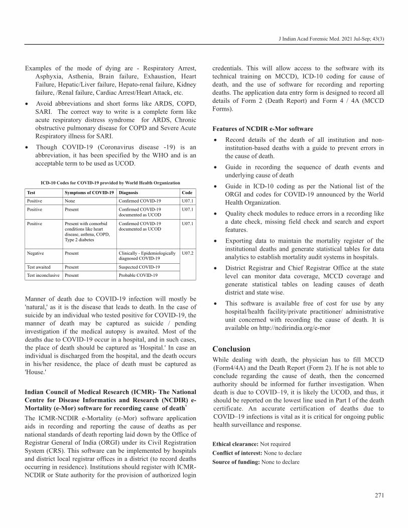
Examples of the mode of dying are - Respiratory Arrest, Asphyxia, Asthenia, Brain failure, Exhaustion, Heart Failure, Hepatic/Liver failure, Hepato-renal failure, Kidney failure, /Renal failure, Cardiac Arrest/Heart Attack, etc.
· Avoid abbreviations and short forms like ARDS, COPD, SARI. The correct way to write is a complete form like acute respiratory distress syndrome for ARDS, Chronic obstructive pulmonary disease for COPD and Severe Acute Respiratory illness for SARI.
· Though COVID-19 (Coronavirus disease -19) is an abbreviation, it has been specified by the WHO and is an acceptable term to be used as UCOD.
Manner of death due to COVID-19 infection will mostly be 'natural,' as it is the disease that leads to death. In the case of suicide by an individual who tested positive for COVID-19, the manner of death may be captured as suicide / pending investigation if the medical autopsy is awaited. Most of the deaths due to COVID-19 occur in a hospital, and in such cases, the place of death should be captured as 'Hospital.' In case an individual is discharged from the hospital, and the death occurs in his/her residence, the place of death must be captured as 'House.'
Indian Council of Medical Research (ICMR)- The National Centre for Disease Informatics and Research (NCDIR) e-
9Mortality (e-Mor) software for recording cause of death
The ICMR-NCDIR e-Mortality (e-Mor) software application aids in recording and reporting the cause of deaths as per national standards of death reporting laid down by the Office of Registrar General of India (ORGI) under its Civil Registration System (CRS). This software can be implemented by hospitals and district local registrar offices in a district (to record deaths occurring in residence). Institutions should register with ICMR-NCDIR or State authority for the provision of authorized login
credentials. This will allow access to the software with its technical training on MCCD), ICD-10 coding for cause of death, and the use of software for recording and reporting deaths. The application data entry form is designed to record all details of Form 2 (Death Report) and Form 4 / 4A (MCCD Forms).
Features of NCDIR e-Mor software
· Record details of the death of all institution and non-institution-based deaths with a guide to prevent errors in the cause of death.
· Guide in recording the sequence of death events and underlying cause of death
· Guide in ICD-10 coding as per the National list of the ORGI and codes for COVID-19 announced by the World Health Organization.
· Quality check modules to reduce errors in a recording like a date check, missing field check and search and export features.
· Exporting data to maintain the mortality register of the institutional deaths and generate statistical tables for data analytics to establish mortality audit systems in hospitals.
· District Registrar and Chief Registrar Office at the state level can monitor data coverage, MCCD coverage and generate statistical tables on leading causes of death district and state wise.
· This software is available free of cost for use by any hospital/health facility/private practitioner/ administrative unit concerned with recording the cause of death. It is available on http://ncdirindia.org/e-mor
Conclusion
While dealing with death, the physician has to fill MCCD (Form4/4A) and the Death Report (Form 2). If he is not able to conclude regarding the cause of death, then the concerned authority should be informed for further investigation. When death is due to COVID–19, it is likely the UCOD, and thus, it should be reported on the lowest line used in Part I of the death certificate. An accurate certification of deaths due to COVID–19 infections is vital as it is critical for ongoing public health surveillance and response.
Ethical clearance: Not required
Conflict of interest: None to declare
Source of funding: None to declare
J Indian Acad Forensic Med. 2021 Jul-Sep; 43(3)
271
Test Symptoms of COVID-19 Diagnosis Code
Positive None Confirmed COVID-19 U07.1
Positive Present Confirmed COVID-19 documented as UCOD
U07.1
Positive Present with comorbid conditions like heart disease, asthma, COPD, Type 2 diabetes
Confirmed COVID-19documented as UCOD
U07.1
Negative Present Clinically - Epidemiologically diagnosed COVID-19
U07.2
Test awaited Present Suspected COVID-19
Test inconclusive Present Probable COVID-19
ICD-10 Codes for COVID-19 provided by World Health Organization
References1. Reddy KSN, Murthy OP. The Essentials of Forensic Medicine and
Toxicology. 34th ed. New Delhi: Jaypee Brothers Medical Publishers; 2017.
2. Ministry of Home Affairs Govt. of India. Registration of Births and Deaths Act. India; 1969: p5-9.
3. Medical certification of cause of death, [Internet]. [cited 2020 11 November] Avai lable f rom ht tp: / /www.mlam.in/pdf / medicalcertificates/mccd.pdf
4. Kotabagi RB, Chaturvedi RK, Banerjee A. Medical certification of cause of death. MJAFI. 2004; 60:261–72.
5. Gupta BD, Trangadia MM, Mehta RA, Vadgama DK. Death certificate: Ignorance and facts. J Indian Acad Forensic Med. 2013;35(3):259–61.
6. The Registrar General. Physicians' manual on medical certification of cause of death [Internet]. [cited 2020 6 June]. Available from: 'https://www.censusindia.gov.in/2011-Documents/mccd_Report1/ Physicians_Manual_MCCD.pdf
7. World Health Organization. Novel coronavirus—China. Geneva, Switzerland. 2020. [Internet] [cited 2020 6 June]. Available from: COVID-19 Public Health Emergency of International Concern (PHEIC) Global research and innovation forum (who.int)
8. World Health Organization. WHO Director-General's opening remarks at the media briefing on COVID–19—11 March 2020. Geneva, Switzerland. 2020 [Internet]. [cited 2020 6 June]. Available from: https://www.who.int/dg/speeches/detail/who-director-general-s-opening-remarks-at-the-media-briefing-on-covid-19---11-march-2020
9. World Health Organization. COVID-19 coding in ICD-10 [Internet]. [cited 2020 6 June]. Available from: https:// w w w. w h o . i n t / c l a s s i f i c a t i o n s / i c d / C O V I D - 1 9 - c o d i n g -icd10.pdf?ua=1
10. National Centre for Disease Informatics and Research. Guidance for appropriate recording of COVID-19 related deaths in India [Internet]. [cited 2020 6 June]. Available from: https:// www.ncdirindia.org/Downloads/CoD_COVID-19_Guidance.pdf
J Indian Acad Forensic Med. 2021 Jul-Sep; 43(3)
272
AbstractLadder-rung tears have been described in the literature as multiple superficial parallel intimal tears in the aorta. They are mostly associated with a road traffic accident. The isthmus part of the thoracic aorta is more vulnerable to this injury as it is tethered. This injury is also more common with old age due to weakness of the vascular walls with the advancement of age. Multiple mechanisms have been proposed in the literature based on different principles of biomechanics. We report an autopsy case of road traffic accident with ladder-rung tears in abdominal aorta in young motorcyclist, which is a rare occurrence, along with a review of possible mechanisms of aortic injuries in road traffic accidents.
KeywordsLadder-rung tear; Aorta; Road traffic accident; Autopsy; Acceleration-deceleration injury; Motorcyclist
Ladder-rung tears of aorta - An unusual presentation of medico-legal significance
CASE REPORT
Introduction
Road traffic accidents often present with a varied spectrum of injuries. Injuries to the great vessels, particularly aorta, are also not uncommon. Aortic injuries can be seen car occupants, pedestrians, motorcyclists or cyclists in varying frequencies. As per recent statistics, aortic rupture is slightly more frequent for pedestrians and motorcyclists in comparison to car occupants or
1cyclists. Aortic injuries can be confined to intima or may extend to fatal aortic rupture. Kraus et al. in their study on motor cycle accidents found that non-fatal aortic injuries amount to 0.2 % cases whereas fatal aortic injuries to 15.4% of
2cases. Intimal tears in the aorta can be multiple and parallel to each other like a step ladder. Hence, they are described as ladder-
3rung tears. Old aged persons are more vulnerable due to the 4-7weakening of the vascular wall with the advancement of age.
The most common site of ladder-rung tear is descending thoracic aorta, particularly the isthmus of the aorta. Sudden deceleration injuries, shearing forces, osseous pinch, or a sudden increase in the intrathoracic pressure have been
[4,8-10]proposed as possible mechanism for its causation . Ladder-4,8,9,11,12rung tears are a rarity in the abdominal aorta. We report a
case of a young adult who succumbed to head injury, two days after a road traffic accident. Superficial intimal tears in a step ladder pattern were observed in the abdominal aorta at autopsy. The age of the deceased and the site of the intimal tears are being reported as rare atypical findings.
Case report
A 26 year old, male was brought to an emergency with a history of a road traffic accident. The victim was riding a two-wheeler bike and was allegedly hit by a truck. CT scan revealed extradural, subdural, and subarachnoid haemorrhages along with skull fractures. The patient underwent decompression craniotomy for extradural hemorrhage, and was managed conservatively for the rest of the injuries. He succumbed to his injuries two days later. At autopsy, sutured wounds were noted on the scalp, forehead, and right cheek. Besides, there were multiple abrasions, contusions, and lacerations on the head, face, chest, right upper limb, and lower limbs. Internal examination revealed diffuse subgaleal haemorrhage on the scalp and fissured fracture of the middle and posterior cranial fossae. Bilateral subdural haemorrhage and subarachnoid haemorrhage were present. Frontal lobes were contused. Vertebrae were intact. Abdominal visceral organs were intact. Examination of the abdominal aorta showed multiple superficial tears on its intimal surface. They were located about 4 cm below the level of aortic hiatus and present across the length of 11 cm. The tears were multiple, transverse and parallel to each other with size ranging from 0.5cm to 2.5cm and limited only to intima. In the lower most tear, intimal layer is stripped over an area of about 1.5 x 1cm; however the tear it is confined to intima (Figure 1). The external surface of the aorta was unremarkable. These tears are described as ladder-rung tears in the literature. Cause of death was attributed to craniocerebral injuries sustained in a road traffic accident.
Discussion
Road traffic accident cases are routinely encountered in medicolegal practice. The pattern of injuries depends upon multiple factors like the type of collision, speed of the vehicle, type of vehicle, relative position, safety measures, etc. Vascular
273
Corresponding Author
Dr Vikas Meshram (Associate Professor)
Email:[email protected].
Mobile: +91-7300345821
Article Historyth thReceived: 27 May, 2020 Revision received on: 13 July, 2021 stAccepted: 21 July, 2021
1 2 1 1 1Vikas Meshram , Ashish Saraf , Vaibhav Gupta , Tanuj Kanchan , Raghvendra Singh Shekhawat1 Department of Forensic Medicine and Toxicology, All India Institute of Medical Sciences, Jodhpur, India
2 Department of Forensic Medicine and Toxicology, All India Institute of Medical Sciences, Gorakhpur, India
J Indian Acad Forensic Med. 2021 Jul-Sep; 43(3): 273-275doi:10.5958/0974-0848.2021.00068.3
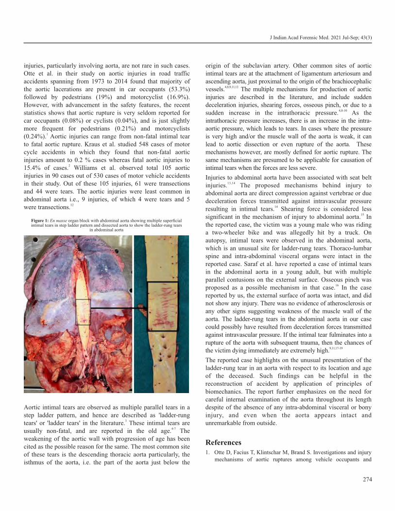
injuries, particularly involving aorta, are not rare in such cases. Otte et al. in their study on aortic injuries in road traffic accidents spanning from 1973 to 2014 found that majority of the aortic lacerations are present in car occupants (53.3%) followed by pedestrians (19%) and motorcyclist (16.9%). However, with advancement in the safety features, the recent statistics shows that aortic rupture is very seldom reported for car occupants (0.08%) or cyclists (0.04%), and is just slightly more frequent for pedestrians (0.21%) and motorcyclists
1(0.24%). Aortic injuries can range from non-fatal intimal tear to fatal aortic rupture. Kraus et al. studied 548 cases of motor cycle accidents in which they found that non-fatal aortic injuries amount to 0.2 % cases whereas fatal aortic injuries to
215.4% of cases. Williams et al. observed total 105 aortic injuries in 90 cases out of 530 cases of motor vehicle accidents in their study. Out of these 105 injuries, 61 were transections and 44 were tears. The aortic injuries were least common in abdominal aorta i.e., 9 injuries, of which 4 were tears and 5
12were transections.
Aortic intimal tears are observed as multiple parallel tears in a step ladder pattern, and hence are described as 'ladder-rung
3tears' or 'ladder tears' in the literature. These intimal tears are 4-7usually non-fatal, and are reported in the old age. The
weakening of the aortic wall with progression of age has been cited as the possible reason for the same. The most common site of these tears is the descending thoracic aorta particularly, the isthmus of the aorta, i.e. the part of the aorta just below the
origin of the subclavian artery. Other common sites of aortic intimal tears are at the attachment of ligamentum arteriosum and ascending aorta, just proximal to the origin of the brachiocephalic
4,8,9,11,12vessels. The multiple mechanisms for production of aortic injuries are described in the literature, and include sudden deceleration injuries, shearing forces, osseous pinch, or due to a
4,8-10sudden increase in the intrathoracic pressure. As the intrathoracic pressure increases, there is an increase in the intra-aortic pressure, which leads to tears. In cases where the pressure is very high and/or the muscle wall of the aorta is weak, it can lead to aortic dissection or even rupture of the aorta. These mechanisms however, are mostly defined for aortic rupture. The same mechanisms are presumed to be applicable for causation of intimal tears when the forces are less severe.
Injuries to abdominal aorta have been associated with seat belt 13,14injuries. The proposed mechanisms behind injury to
abdominal aorta are direct compression against vertebrae or due deceleration forces transmitted against intravascular pressure
14resulting in intimal tears. Shearing force is considered less 15significant in the mechanism of injury to abdominal aorta. In
the reported case, the victim was a young male who was riding a two-wheeler bike and was allegedly hit by a truck. On autopsy, intimal tears were observed in the abdominal aorta, which is an unusual site for ladder-rung tears. Thoraco-lumbar spine and intra-abdominal visceral organs were intact in the reported case. Saraf et al. have reported a case of intimal tears in the abdominal aorta in a young adult, but with multiple parallel contusions on the external surface. Osseous pinch was
16proposed as a possible mechanism in that case. In the case reported by us, the external surface of aorta was intact, and did not show any injury. There was no evidence of atherosclerosis or any other signs suggesting weakness of the muscle wall of the aorta. The ladder-rung tears in the abdominal aorta in our case could possibly have resulted from deceleration forces transmitted against intravascular pressure. If the intimal tear fulminates into a rupture of the aorta with subsequent trauma, then the chances of
9,11,17-19the victim dying immediately are extremely high.
The reported case highlights on the unusual presentation of the ladder-rung tear in an aorta with respect to its location and age of the deceased. Such findings can be helpful in the reconstruction of accident by application of principles of biomechanics. The report further emphasizes on the need for careful internal examination of the aorta throughout its length despite of the absence of any intra-abdominal visceral or bony injury, and even when the aorta appears intact and unremarkable from outside.
References1. Otte D, Facius T, Klintschar M, Brand S. Investigations and injury
mechanisms of aortic ruptures among vehicle occupants and
Figure 1: En masse organ block with abdominal aorta showing multiple superficial intimal tears in step ladder pattern and dissected aorta to show the ladder-rung tears
in abdominal aorta
274
J Indian Acad Forensic Med. 2021 Jul-Sep; 43(3)
vulnerable road users over time. Int Res Counc Biomech Inj. 2016: 870–887.
2. Kraus J F, Peek-Asa C, Cryer H G. Incidence, severity, and patterns of intrathoracic and intra-abdominal injuries in motorcycle crashes. J. Trauma.2002; 52 (3): 548–553.
3. Sauko P, Knight B. Knight's forensic pathology. 4th ed. New York: CRC press; 2016. p222.
4. Shkrum M J, McClafferty K J, Green R N, Nowak E S, Young J G. Mechanisms of aortic injury in fatalities occurring in motor vehicle collisions. J Forensic Sci.1999;44(1):44-56.
5. McGwin G Jr, Reiff DA, Moran SG, Rue LW 3rd. Incidence and Characteristics of Motor Vehicle Collision-Related Blunt Thoracic Aortic Injury According to Age. J Trauma. 2002;52(5):859-65.
6. Eddy AC, Rusch VW, Fligner CL, Reay DT, Rice CL. The Epidemiology of Traumatic Rupture of the Thoracic Aorta in Children: A 13-year Review. J Trauma. 1990;30(8):989-91.
7. Strassmann G. Traumatic Rupture of the Aorta. Am Heart J. 1947;33(4):508-15.
8. Katyal D, McLellan BA, Brenneman FD, Boulanger BR, Sharkey PW, Waddell JP. Lateral impact motor vehicle collisions: significant cause of blunt traumatic rupture of the thoracic aorta. J Trauma. 1997;42(5):769-72.
9. Siegel JH, Smith JA, Siddiqi SQ. Change in Velocity and Energy Dissipation on Impact in Motor Vehicle Crashes as a Function of the Direction of Crash: Key Factors in the Production of Thoracic Aortic Injuries, Their Pattern of Associated Injuries and Patient Survival. A Crash Injury Research Engineering Network (CIREN) Study. J Trauma. 2004;57(4):760-77.
10. Richens D, Field M, Hashim S, Neale M, Oakley C. A Finite Element Model of Blunt Traumatic Aortic Rupture. Eur J Cardiothorac Surg. 2004;25(6):1039-47.
11. Burkhart HM, Gomez GA, Jacobson, LE, Pless JE, Broadie TA. Fatal blunt aortic injuries: a review of 242 autopsy cases. J. Trauma. 2001; 50(1):113–15.
12. Williams J, Graff J, Uku J, Steinig J. Aortic injury in vehicular trauma. Ann Thorac Surg. 1994;57(3):726-730.
13. Roth SM, Wheeler JR, Gregory RT, Gayle RG. Blunt injury of the abdominal aorta: a review. J Trauma. 1997;42(4):748-55.
14. Asbun HJ, Irani H, Roe EJ, Bloch JH. Intra-abdominal seat belt injury. J Trauma. 1990;30(2):189-93.
15. Nucifora G, Hysko F, Vasciaveo A. Blunt traumatic abdominal aortic rupture: CT imaging. Emerg Radiol. 2008;15(3)1: 211-13.
16. Steenburg SD, Ravenel JG. Multi-detector computed tomography findings of atypical blunt traumatic aortic injuries: a pictorial review. Emerg Radiol. 2007;14(3):143-50
17. Saraf A, Ateriya N, Shekhawat RS. Atypical Presentation of Intimal Tear of Aorta in a Young Deceased Boy Secondary to Road Traffic Collision. Natl Med J India. 2019;32(1):52
18. Richens D, Kotidis K, Neale M, Oakley C, Fails A. Rupture of the aorta following road traffic accidents in the United Kingdom 1992-1999. The results of the co-operative crash injury study. Eur J Cardiothorac Surg. 2003;23(2):143-48.
19. Fitzharris M, Franklyn M, Frampton R, Yang K, Morris A, Fildes B. Thoracic Aortic Injury in Motor Vehicle Crashes: The Effect of Impact Direction, Side of Body Struck, and Seat Belt Use. J Trauma. 2004;57(3):582-90
J Indian Acad Forensic Med. 2021 Jul-Sep; 43(3)
275
AbstractCompressed air is a concentrated stream of air at high pressure and it is released through a narrow pipe with high speed that can cause fatal injuries if it is not handled appropriately. Rectal perforation by foreign bodies and iatrogenic injuries during colonoscopy is known, however barotrauma by compressed air is less often encountered. The case herein reported is that of a victim who presented with the history of insertion of a compressed air tube into the anus by his friends. The patient had severe abdominal pain and on examination, there was a distension involving the lower abdomen with the diffuse tenderness.
KeywordsBarotrauma; Transanal; Compressed air; Colonic injury
Fatal colorectal injury by compressed air through anal insufflation
CASE REPORT
Introduction
Compressed air is the air which is kept under a pressure that is greater than the atmospheric pressure and is widely used for industrial as well as domestic purposes. As it stores a large amount of gas at high pressure, its injudicious use may lead to fatal incidents in the workplace. Transanal insufflation of compressed air may cause perforation, especially in the rectum and sigmoid colon or any part of the colon. It is being suggested that it takes only 1-2 seconds to deliver enough compressed air
1to cause major damage. Injuries may vary from cat scratch 2 colon to colorectal perforation or blowout. A misdirected jet of
compressed air to the head can cause serious eye injuries or rupture of the eardrum. If it is applied in the mouth, it can damage the lungs as well as esophagus. Even its careless use to blow away dirt or dust from the body could allow the air to enter the body damaging the internal organs. The air jets that are being utilized in the industries usually have 50-100 lbs pressure or even more which enters the anus more promptly as
3 compared to a proctoscope or a finger leading to barotrauma.Here we are reporting a case of 14 years old male with the history of insertion of a compressed air tube into the anus by his friends, causing barotraumas to the intestines.
Case Report
A male aged 14 years was a worker in an industry. He was approached from behind by his colleagues and the compressed air was insufflated through the anal route using the compressed
air cleaner. The victim started complaining of severe abdominal pain and distension and hence was taken to a hospital. On examination the patient was restless and had unstable vitals i.e. pulse rate- 105 per minute, blood pressure- 94/58 mm Hg and respiratory rate- 24 per minute. Per abdominal examination showed abdominal distension with a diffuse tenderness and reduced bowel sounds. The erect Chest X-ray showed presence of air under the diaphragm which suggested gastrointestinal perforation. His condition worsened with time and subsequently succumbed to the injuries. An autopsy was done in the mortuary of District Hospital, Aligarh, Uttar Pradesh. In postmortem examination, externally there was an abraded wound at the anal margins. A huge gush of air was noted on opening the abdomen. The entire peritoneal cavity was soiled with faecal matter and blood, and the peritoneum showed the features of ulceration and peritonitis. Multiple seromuscular tears were present in the rectum and colon. The cause of death was opined as shock and haemorrhage as a result of colorectal injuries suffered due to transanal insufflation of the compressed air.
Discussion
Rectal perforation by foreign bodies along with iatrogenic injuries during colonoscopy is known, however barotrauma by compressed air is less often encountered. Few cases of transanal barotraumas due to compressed air leading to colonic injury have been reported from time to time. It was first reported in
4the literature in 1904. Similar case has been reported by Pahwa 5et al. , in which high-pressure compressed air jet was playfully
insufflated through the anal orifice of the victim resulting in sigmoid colon perforation. Colorectal injury can occur when the nozzle is merely placed near the anus, even when the clothes
6,7are worn.
Clothes do not protect the force of compressed air as the funnel shaped anatomy of the buttock facilitates the air to concentrate around the anus and allows the easy delivery of air through the anus. The most susceptible site for iatrogenic barotrauma is the
276
Corresponding Author
Dr. Kashif Ali (Senior Resident)
Email: [email protected]
Mobile: +91-9986468326
Article Historyth ndReceived: 28 September, 2020; Revision received on: 22 July, 2021 thAccepted: 24 July, 2021
Mohd Kaleem Khan, Kashif AliDepartment of Forensic Medicine, Jawaharlal Nehru Medical College, AMU, Aligarh (UP)
J Indian Acad Forensic Med. 2021 Jul-Sep; 43(3): 276-277doi:10.5958/0974-0848.2021.00069.5
caecum which have the largest diameter in colon. It is explained by the law of Laplace where the tension in the wall is
8 proportional to the radius of the lumen. The various experimental studies have shown that the human colon bursts with only about 120–200 mmHg of pressure while
9seromuscular rupture occurs at lower levels. The cause of death can be acute air embolism, acute fat embolism, acute respiratory insufficiency due to increased intra-abdominal pressure, acute heart failure due to insufficient preload and
10peritoneal shock.
A study done by Ali et al. showed 10 construction workers with 11an isolated perforation particularly in the left half of the colon.
Barotraumas can also be caused by a pneumatic pressure cleaner or during water sports when a crash on the water surface can lead to bowel perforation or rectal injury as a result
12,13 of the water enema. The normal intestine of a dog required a 2pressure of 0.49-0.88 kg/cm to get ruptured when Andrews
14 used compressed air is used to distend the intestine. The average pressure needed to cause a full thickness tear in human
2 gastrointestinal tracts is 0.29 kg/cm which is shown by the 15 study done by Burt. The highly compressed air which is used
widely in industrial fields exerts a pressure higher than 10 2kg/cm and which is far greater than the pressure needed for
intestinal perforation.
Conclusion
Barotrauma related colorectal injuries by compressed air are rare conditions which are encountered at the emergency departments. Trans-anal insufflations of compressed air often result in serious colonic injury. The decision to operate and its management must be taken in short span of time as its morbidity and mortality rates are high. Though negligent or intention injuries attract punishment under various laws of the country but it fails to address the social embarrassment and psychological traumas. It is a high time to implement strict safety guidelines at the workplace to discourage these types of incidents.
Ethical clearance: None Required
Conflict of interest: None to declare
Source of funding: None to declare
References1. Kim SJ, Ahn SI, Hong KC, Kim JS, Shin SH, Woo ZH. Pneumatic
colonic rupture accompanied by tension pneumoperitoneum. Yonsei Med J 2000; 41(4):533-5.
2. McDonnell WM, Loura F, Pointon MJ, Greenson JK. Cat scratch colon. Endoscopy. 2007; 39(5):459-61.
3. Suraj L, Shaikh T. Prank gone wrong, transverse colon perforation due to air compressor pipe placed in anus. Int J Res Med Sci 2017;5:1119-22.
4. Stone GW. Rupture of the bowel caused by compressed air. Lancet 1904; 2:216.
5. Pahwa HS, Kumar A, Srivastava R, Rai A. Trans-anal barotrauma by compressed air leading to sigmoid perforation due to a dangerous practical joke. BMJ Case Rep. 2012; 1:2012.
6. Ergin M, Ozer MR, Kocat S, Karakus N, Babagil B, Cander B. A rare case of colorectal injury with compressed air. J Med Cases 2013; 4:159-60.
7. Suh HH, Kim YJ, Kim SK. Colorectal injury by compressed air- a report of 2 cases. J Korean Med Sci 1996; 11:179-82.
8. Kozarek RA, Earnest DL, Silverstein ME, Smith RG. Air-pressure-induced colon injury during diagnostic colonoscopy. Gastroenterology 1980; 78:7-14.
9. Kozarek RA, Sanowski RA. Use of pressure release valve to prevent colonic injury during colonoscopy. GastrointestEndosc. 1980; 26(4):139–42.
10. Weber M, Kolbus F, Dressler J and Lessig R. Survived ileocecal blowout from compressed air. International Journal of Legal Medicine 2011;125(2):283-7.
11. Ali M, Hammad R, Hasan F. Compressed air injury to the colon/rectum and its sequels. Pak J Med Health Sci. 2010; 4:409-412.
12. Gill RS. Hydrostatic rectosigmoid perforation: A rare personal watercraft injury. J Pediatr Surg. 2011; 46:402-404.
13. Sy ED. Pneumatic colon injury following high pressure blow gun dust cleaner spray to the perineum. Int J Surg Case Rep.2015;6.
14. Andrews EW. Pneumatic rupture of the intestine or new type of industrial accident. SurgGynecolObstet 1911; 12:63-4.
15. Burt CA. Pneumatic ruptures of the intestinal canal. Arch Surg 1931; 22:875-902.
277
J Indian Acad Forensic Med. 2021 Jul-Sep; 43(3)
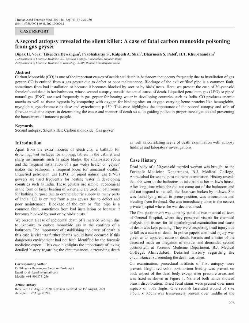
AbstractCarbon Monoxide (CO) is one of the important causes of accidental death in bathroom that occurs frequently due to installation of gas geyser. CO is emitted from a gas geyser due to defect or poor maintenance. Blockage of the exit or 'flue' pipe is a common fault, sometimes from bad installation or because it becomes blocked by soot or by birds' nests. Here, we present the case of 30-year-old female found dead in her bathroom, whose second autopsy unveils the actual cause of death. Liquefied petroleum gas (LPG) or piped natural gas (PNG) are used frequently in gas geyser for heating water in developing countries such as India. CO produces anemic anoxia as well as tissue hypoxia by competing with oxygen for binding sites on oxygen carrying heme proteins like hemoglobin, myoglobin, cytochrome-c oxidase and cytochrome p-450. This case highlights the importance of the second autopsy and role of forensic medicine expert in determining the cause and manner of death so as to guiding police in proper investigation and preventing the harassment of innocent people.
KeywordsSecond autopsy; Silent killer; Carbon monoxide; Gas geyser
A second autopsy revealed the silent killer: A case of fatal carbon monoxide poisoning from gas geyser
CASE REPORT
Introduction
Apart from the extra hazards of electricity, a bathtub for drowning, wet surfaces for slipping, tablets in the cabinet and sharp instruments such as razor blades, the small-sized room and the frequent installation of a gas water heater or 'geyser'
1 makes the bathroom a frequent locus for unnatural deaths.Liquefied petroleum gas (LPG) or piped natural gas (PNG) geysers are used frequently for heating water in developing countries such as India. These geysers are simple, economical in the form of faster heating of water and are used in bathrooms for bathing purpose due to erratic electric supply in many parts
2 of India. CO is emitted from a gas geyser due to defect and poor maintenance. Blockage of the exit or 'flue' pipe is a common fault, sometimes from bad installation or because it
1,3becomes blocked by soot or by birds' nests.
We present a case of accidental death of a married woman due to exposure to carbon monoxide gas in the confines of a bathroom. The importance of establishing the cause of death in this case is clear as further deaths would have occurred if this dangerous environment had not been identified by the forensic
3medicine expert. This case highlights the importance of taking detailed history regarding the circumstances surrounding death
as well as correlating scene of death examination with autopsy findings and laboratory investigations.
Case History
Dead body of a 30-year-old married woman was brought to the Forensic Medicine Department, B.J. Medical College, Ahmedabad for second post-mortem examination. History reveals that she went to the bathroom to take bath at her in-law's house. After long time when she did not come out of the bathroom and did not respond to the call, the door was broken by in laws. She was found lying naked in prone position, was unconscious and bleeding from forehead. She was immediately taken to the nearest private hospital where she was declared dead.
The first postmortem was done by panel of two medical officers of General Hospital, where they preserved viscera for chemical analysis and tissues for histopathological examination and cause of death was kept pending. They were suspecting head injury due to fall as a cause of death. In police papers also head injury was given as an apparent cause of death. Parents and a sister of the deceased made an allegation of murder and demanded second postmortem at Forensic Medicine Department, B.J. Medical College, Ahmedabad. Detailed history regarding the circumstances surrounding the death was taken.
On examination, procedural artifacts of first autopsy were present. Bright red color postmortem lividity was present on back aspect of the dead body except over pressure areas and was fixed as shown in Figure 1. Nails of both hands showed bluish discoloration. Dried fecal stains were present over inner aspects of both thighs. One reddish lacerated wound of size 3.5cm x 0.5cm was transversely present over middle of the
278
Corresponding Author
Dr Tikendra Dewangan (Assistant Professor)
Email id- [email protected]
Mobile: +91-9098751256
Article Historyth thReceived: 17 August, 2020; Revision received on: 15 August, 2021 thAccepted: 19 August, 2021
1 2 1 1 1 1Dipak H. Vora , Tikendra Dewangan , Prabhakaran S , Kalpesh A. Shah , Dharmesh S. Patel , H.T. Khubchandani1 Department of Forensic Medicine, B.J. Medical College, Ahmedabad, Gujarat, India
2 Department of Forensic Medicine & Toxicology, RIMS, Raipur, Chhattisgarh, India
J Indian Acad Forensic Med. 2021 Jul-Sep; 43(3): 278-280doi:10.5958/0974-0848.2021.00070.1
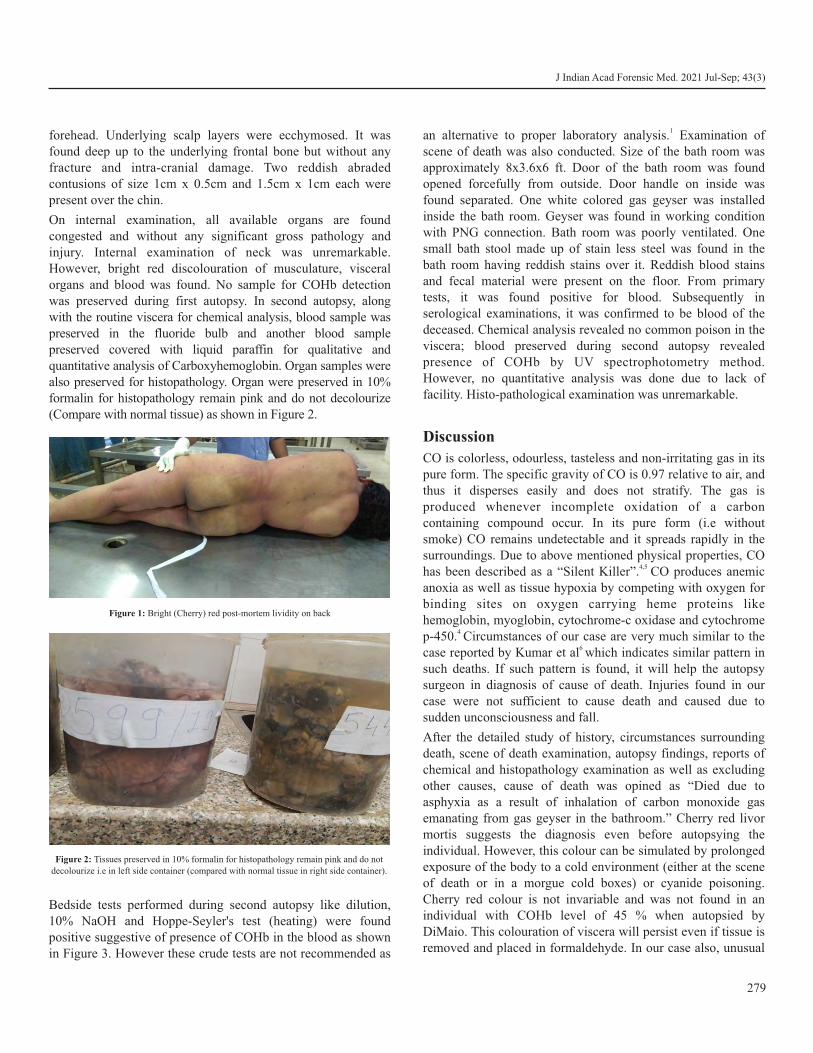
forehead. Underlying scalp layers were ecchymosed. It was found deep up to the underlying frontal bone but without any fracture and intra-cranial damage. Two reddish abraded contusions of size 1cm x 0.5cm and 1.5cm x 1cm each were present over the chin.
On internal examination, all available organs are found congested and without any significant gross pathology and injury. Internal examination of neck was unremarkable. However, bright red discolouration of musculature, visceral organs and blood was found. No sample for COHb detection was preserved during first autopsy. In second autopsy, along with the routine viscera for chemical analysis, blood sample was preserved in the fluoride bulb and another blood sample preserved covered with liquid paraffin for qualitative and quantitative analysis of Carboxyhemoglobin. Organ samples were also preserved for histopathology. Organ were preserved in 10% formalin for histopathology remain pink and do not decolourize (Compare with normal tissue) as shown in Figure 2.
Bedside tests performed during second autopsy like dilution, 10% NaOH and Hoppe-Seyler's test (heating) were found positive suggestive of presence of COHb in the blood as shown in Figure 3. However these crude tests are not recommended as
1an alternative to proper laboratory analysis. Examination of scene of death was also conducted. Size of the bath room was approximately 8x3.6x6 ft. Door of the bath room was found opened forcefully from outside. Door handle on inside was found separated. One white colored gas geyser was installed inside the bath room. Geyser was found in working condition with PNG connection. Bath room was poorly ventilated. One small bath stool made up of stain less steel was found in the bath room having reddish stains over it. Reddish blood stains and fecal material were present on the floor. From primary tests, it was found positive for blood. Subsequently in serological examinations, it was confirmed to be blood of the deceased. Chemical analysis revealed no common poison in the viscera; blood preserved during second autopsy revealed presence of COHb by UV spectrophotometry method. However, no quantitative analysis was done due to lack of facility. Histo-pathological examination was unremarkable.
Discussion
CO is colorless, odourless, tasteless and non-irritating gas in its pure form. The specific gravity of CO is 0.97 relative to air, and thus it disperses easily and does not stratify. The gas is produced whenever incomplete oxidation of a carbon containing compound occur. In its pure form (i.e without smoke) CO remains undetectable and it spreads rapidly in the surroundings. Due to above mentioned physical properties, CO
4,5 has been described as a “Silent Killer”. CO produces anemic anoxia as well as tissue hypoxia by competing with oxygen for binding sites on oxygen carrying heme proteins like hemoglobin, myoglobin, cytochrome-c oxidase and cytochrome
4 p-450. Circumstances of our case are very much similar to the 6 case reported by Kumar et al which indicates similar pattern in
such deaths. If such pattern is found, it will help the autopsy surgeon in diagnosis of cause of death. Injuries found in our case were not sufficient to cause death and caused due to sudden unconsciousness and fall.
After the detailed study of history, circumstances surrounding death, scene of death examination, autopsy findings, reports of chemical and histopathology examination as well as excluding other causes, cause of death was opined as “Died due to asphyxia as a result of inhalation of carbon monoxide gas emanating from gas geyser in the bathroom.” Cherry red livor mortis suggests the diagnosis even before autopsying the individual. However, this colour can be simulated by prolonged exposure of the body to a cold environment (either at the scene of death or in a morgue cold boxes) or cyanide poisoning. Cherry red colour is not invariable and was not found in an individual with COHb level of 45 % when autopsied by DiMaio. This colouration of viscera will persist even if tissue is removed and placed in formaldehyde. In our case also, unusual
279
J Indian Acad Forensic Med. 2021 Jul-Sep; 43(3)
Figure 1: Bright (Cherry) red post-mortem lividity on back
Figure 2: Tissues preserved in 10% formalin for histopathology remain pink and do not decolourize i.e in left side container (compared with normal tissue in right side container).
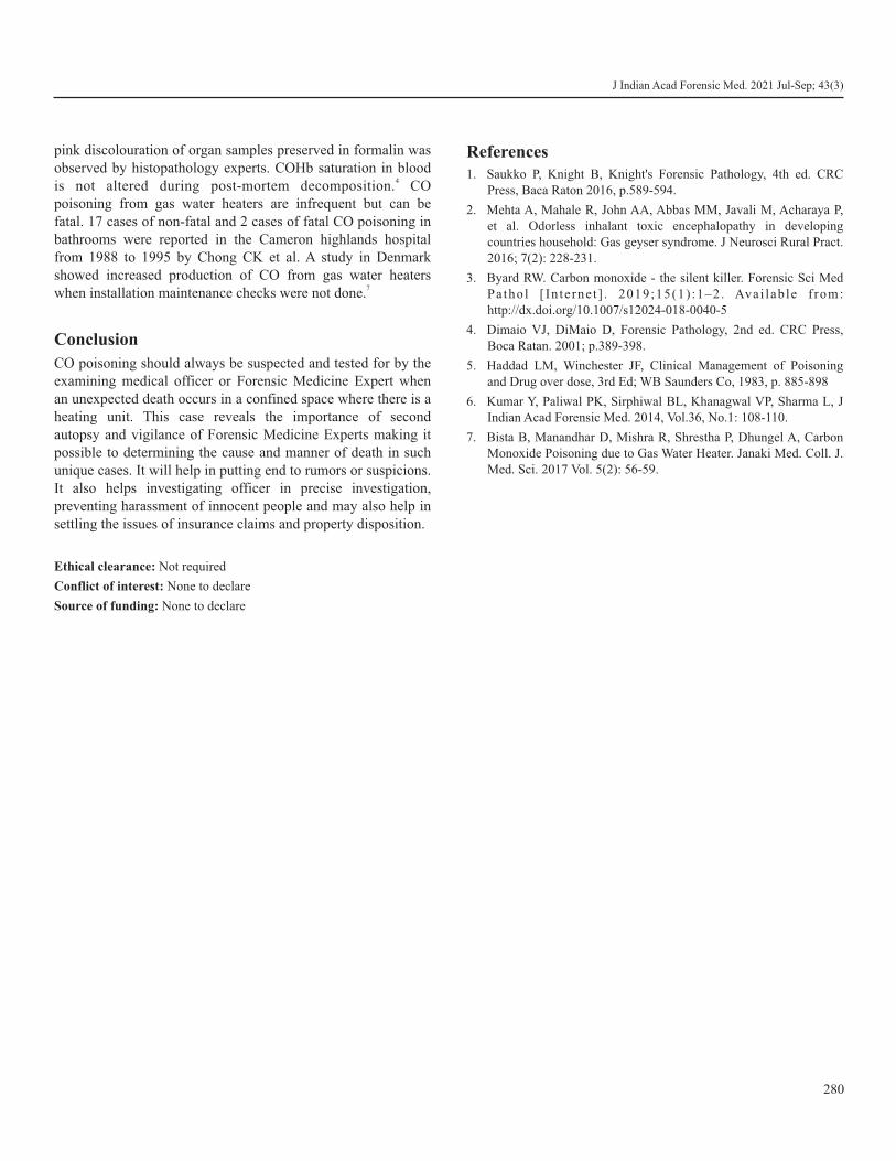
pink discolouration of organ samples preserved in formalin was observed by histopathology experts. COHb saturation in blood
4is not altered during post-mortem decomposition. CO poisoning from gas water heaters are infrequent but can be fatal. 17 cases of non-fatal and 2 cases of fatal CO poisoning in bathrooms were reported in the Cameron highlands hospital from 1988 to 1995 by Chong CK et al. A study in Denmark showed increased production of CO from gas water heaters
7when installation maintenance checks were not done.
Conclusion
CO poisoning should always be suspected and tested for by the examining medical officer or Forensic Medicine Expert when an unexpected death occurs in a confined space where there is a heating unit. This case reveals the importance of second autopsy and vigilance of Forensic Medicine Experts making it possible to determining the cause and manner of death in such unique cases. It will help in putting end to rumors or suspicions. It also helps investigating officer in precise investigation, preventing harassment of innocent people and may also help in settling the issues of insurance claims and property disposition.
Ethical clearance: Not required
Conflict of interest: None to declare
Source of funding: None to declare
References1. Saukko P, Knight B, Knight's Forensic Pathology, 4th ed. CRC
Press, Baca Raton 2016, p.589-594.
2. Mehta A, Mahale R, John AA, Abbas MM, Javali M, Acharaya P, et al. Odorless inhalant toxic encephalopathy in developing countries household: Gas geyser syndrome. J Neurosci Rural Pract. 2016; 7(2): 228-231.
3. Byard RW. Carbon monoxide - the silent killer. Forensic Sci Med Pathol [ In te rne t ] . 2019;15(1) :1–2 . Avai lab le f rom: http://dx.doi.org/10.1007/s12024-018-0040-5
4. Dimaio VJ, DiMaio D, Forensic Pathology, 2nd ed. CRC Press, Boca Ratan. 2001; p.389-398.
5. Haddad LM, Winchester JF, Clinical Management of Poisoning and Drug over dose, 3rd Ed; WB Saunders Co, 1983, p. 885-898
6. Kumar Y, Paliwal PK, Sirphiwal BL, Khanagwal VP, Sharma L, J Indian Acad Forensic Med. 2014, Vol.36, No.1: 108-110.
7. Bista B, Manandhar D, Mishra R, Shrestha P, Dhungel A, Carbon Monoxide Poisoning due to Gas Water Heater. Janaki Med. Coll. J. Med. Sci. 2017 Vol. 5(2): 56-59.
J Indian Acad Forensic Med. 2021 Jul-Sep; 43(3)
280
AbstractIn general, suicide by stab wound is rare especially among females and often challenges the medico-legal acumen of an autopsy surgeon while deciding the manner of death. In this report, we present a case of a 46 year old woman found dead in her home with a single abdominal stab wound. No other injury including hesitation wounds was found during autopsy. History given by the relatives revealed that she was mentally unstable and was suffering from a chronic illness. The death was labeled a suicide based on the findings observed during autopsy along with those found at the crime scene and the personal history of the victim.
KeywordsSuicide; Stab wound; Abdomen
A fatal case of self-inflicted abdominal stab wound
CASE REPORT
Introduction
In comparison to other means of suicide, death due to self-inflicted sharp force injury is uncommon constituting only 0.6 % of all suicides in India. A gender disparity is also observed in
1such deaths with male accounting for most of the cases. The sharp force injury (SFI) frequently encountered in such deaths is the incised wound; stab wound, on the other hand, is usually
2 seen in homicides. Therefore, a suicide by stabbing is often view with suspicion of foul play and presents an autopsy surgeon with a difficult task of differentiating a suicide from a
3 homicide. In this report, we present a case of a female suicide by stab wound to the abdomen.
Case report
The dead body of a 46 year old female, who was found dead in her house, was brought to the mortuary for Medico-legal autopsy. Following the examination of the police inquest, the history given by the relatives and the police statements, it is learned that the woman was staying alone in a single room house, having a single entry and exit door, within the same campus along with her other relatives. As per history, she had history of seizures and mentally unstable for which no medical diagnosis and treatment was taken. There was also history of self-harm in the past. On the morning of that fateful day, she had an attack of seizure and later in the evening she was found dead inside her house which was locked from inside because of which the door had to be forcibly opened. On examination of
the crime scene, (Fig 1) the dead body was lying in a supine position on the bed with blood present underneath and side of the body, blouse pulled upwards and the blood stained knife lying in the vicinity.
Autopsy findings
All the clothing was blood stained and bore neither cuts nor tears. Dried blood stains were observed over the chest, abdomen, upper thighs, forearms and hands. The eyes were congested and the body appeared pale. Extrusion of the intestines along with mesenteries through an abdominal wound was also observed. (Fig 2) Rigor mortis was fully developed all over the body and the post - mortem staining was present on the back and fixed.
On external examination, one vertically placed stab wound was observed over the upper abdomen, through which the intestines extruded out, measuring 4.5 cms x 2 cms, abdominal cavity deep with red clean cut margins situated on the midline, 5 cms above the umbilicus and 81 cms above heel. The beveling was present on the right side with undermining of the margins on the left and the track of the wound involved the skin, subcutaneous tissues and rectus muscle up to the abdominal cavity. No other injury was present externally.
Internally, the jejunum including the adjacent mesenteries was cut measuring 2.5 cms x 2 cms and situated 391 cms away from the ileo-caecal junction. (Fig 3) The peritoneal cavity contained 500 ml of blood and all the other organs appeared pale. The stomach contained 250 gms of semi digested food matter, yellowish in colour, with no specific smell.
Discussion
The present case, based on the circumstances, crime scene examination and autopsy findings, was labeled as a suicide by the concerned investigating agency and the factors facilitating
281
Corresponding Author
Daunipaia Slong (Assistant Professor)
E-mail: [email protected]
Mobile: +919863957224
Article Historyth thReceived: 9 November, 2020; Revision received on: 20 August, 2021
thAccepted: 29 August, 2021
1 2 2Mitul M Sangma , AJ Patowary , Daunipaia Slong1 District Medical & Health Officer, East Khasi Hills, Meghalaya
2 Department of Forensic Medicine, North Eastern Indira Gandhi Regional Institute of Health and Medical Sciences (NEIGRIHMS), Mawdiangdiang, Shillong, Meghalaya
J Indian Acad Forensic Med. 2021 Jul-Sep; 43(3): 281-283doi:10.5958/0974-0848.2021.00071.3
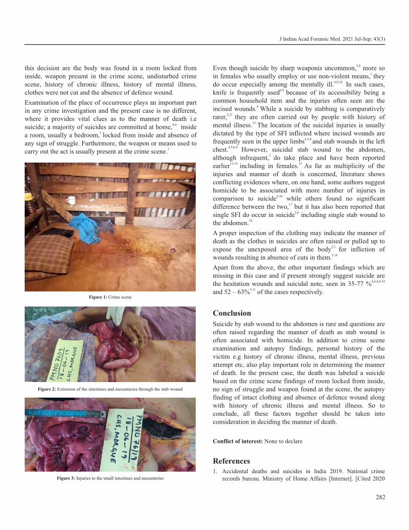
this decision are the body was found in a room locked from inside, weapon present in the crime scene, undisturbed crime scene, history of chronic illness, history of mental illness, clothes were not cut and the absence of defence wound.
Examination of the place of occurrence plays an important part in any crime investigation and the present case is no different, where it provides vital clues as to the manner of death i.e
4-6suicide; a majority of suicides are committed at home, inside 5 a room, usually a bedroom, locked from inside and absence of
any sign of struggle. Furthermore, the weapon or means used to 7carry out the act is usually present at the crime scene.
2,8Even though suicide by sharp weaponis uncommon, more so 1 in females who usually employ or use non-violent means, they
6,9,10 do occur especially among the mentally ill. In such cases, 6,9 knife is frequently used because of its accessibility being a
common household item and the injuries often seen are the 8 incised wounds. While a suicide by stabbing is comparatively
2,11 rarer, they are often carried out by people with history of 12 mental illness. The location of the suicidal injuries is usually
dictated by the type of SFI inflicted where incised wounds are 4,5,8 frequently seen in the upper limbs and stab wounds in the left
4,5,8,9 chest. However, suicidal stab wound to the abdomen, 3 although infrequent, do take place and have been reported
13,14 15 earlier including in females. As far as multiplicity of the injuries and manner of death is concerned, literature shows conflicting evidences where, on one hand, some authors suggest homicide to be associated with more number of injuries in
8,16comparison to suicide while others found no significant 17difference between the two, but it has also been reported that
5,6single SFI do occur in suicide including single stab wound to 18the abdomen.
A proper inspection of the clothing may indicate the manner of death as the clothes in suicides are often raised or pulled up to
2,3 expose the unexposed area of the body for infliction of 5,16wounds resulting in absence of cuts in them.
Apart from the above, the other important findings which are missing in this case and if present strongly suggest suicide are
4,6,8,9,19 the hesitation wounds and suicidal note, seen in 35-77 %5,11and 52 – 63% of the cases respectively.
Conclusion
Suicide by stab wound to the abdomen is rare and questions are often raised regarding the manner of death as stab wound is often associated with homicide. In addition to crime scene examination and autopsy findings, personal history of the victim e.g history of chronic illness, mental illness, previous attempt etc, also play important role in determining the manner of death. In the present case, the death was labeled a suicide based on the crime scene findings of room locked from inside, no sign of struggle and weapon found at the scene, the autopsy finding of intact clothing and absence of defence wound along with history of chronic illness and mental illness. So to conclude, all these factors together should be taken into consideration in deciding the manner of death.
Conflict of interest: None to declare
References1. Accidental deaths and suicides in India 2019. National crime
records bureau. Ministry of Home Affairs [Internet]. [Cited 2020
282
J Indian Acad Forensic Med. 2021 Jul-Sep; 43(3)
Figure 1: Crime scene
Figure 2: Extrusion of the intestines and mesenteries through the stab wound
Figure 3: Injuries to the small intestines and mesenteries
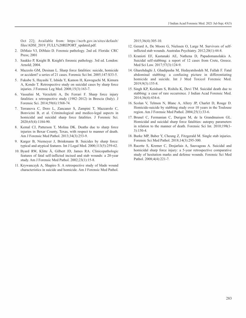
Oct 22]; Available from: https://ncrb.gov.in/sites/default/ files/ADSI_2019_FULL%20REPORT_updated.pdf.
2. DiMaio VJ, DiMaio D. Forensic pathology. 2nd ed. Florida: CRC Press; 2001
3. Saukko P, Knight B. Knight's forensic pathology. 3rd ed. London: Arnold; 2004.
4. Mazzolo GM, Desinan L. Sharp force fatalities: suicide, homicide or accident? a series of 21 cases. Forensic Sci Int. 2005;147:S33-5.
5. Fukube S, Hayashi T, Ishida Y, Kamon H, Kawaguchi M, Kimura A, Kondo T. Retrospective study on suicidal cases by sharp force injuries. J Forensic Leg Med. 2008;15(3):163-7.
6. Vassalini M, Verzeletti A, De Ferrari F. Sharp force injury fatalities: a retrospective study (1982–2012) in Brescia (Italy). J Forensic Sci. 2014;59(6):1568-74.
7. Terranova C, Doro L, Zancaner S, Zampini T, Mazzarolo C, Bonvicini B, et al. Criminological and medico‐legal aspects in homicidal and suicidal sharp force fatalities. J Forensic Sci. 2020;65(4):1184-90.
8. Kemal CJ, Patterson T, Molina DK. Deaths due to sharp force injuries in Bexar County, Texas, with respect to manner of death. Am J Forensic Med Pathol. 2013;34(3):253-9.
9. Karger B, Niemeyer J, Brinkmann B. Suicides by sharp force: typical and atypical features. Int J Legal Med. 2000;113(5):259-62.
10. Byard RW, Klitte Å, Gilbert JD, James RA. Clinicopathologic features of fatal self-inflicted incised and stab wounds: a 20-year study. Am J Forensic Med Pathol. 2002;23(1):15-8.
11. Krywanczyk A, Shapiro S. A retrospective study of blade wound characteristics in suicide and homicide. Am J Forensic Med Pathol.
2015;36(4):305-10.
12. Gerard A, De Moore G, Nielssen O, Large M. Survivors of self-inflicted stab wounds. Australas Psychiatry. 2012;20(1):44-8.
13. Kranioti EF, Kastanaki AE, Nathena D, Papadomanolakis A. Suicidal self-stabbing: a report of 12 cases from Crete, Greece. Med Sci Law. 2017;57(3):124-9.
14. Gharehdaghi J, Ghadipasha M, Hedayatshodeh M, Fallah F. Fatal abdominal stabbing: a confusing picture in differentiating homicide and suicide. Int J Med Toxicol Forensic Med. 2019;9(3):155-8.
15. Singh KP, Keisham S, Rishilu K, Devi TM. Suicidal death due to stabbing: a case of rare occurrence. J Indian Acad Forensic Med. 2014;36(4):434-6.
16. Scolan V, Telmon N, Blanc A, Allery JP, Charlet D, Rouge D. Homicide-suicide by stabbing study over 10 years in the Toulouse region. Am J Forensic Med Pathol. 2004;25(1):33-6.
17. Brunel C, Fermanian C, Durigon M, de la Grandmaison GL. Homicidal and suicidal sharp force fatalities: autopsy parameters in relation to the manner of death. Forensic Sci Int. 2010;198(1-3):150-4.
18. Burke MP, Baber Y, Cheung Z, Fitzgerald M. Single stab injuries. Forensic Sci Med Pathol. 2018;14(3):295-300.
19. Racette S, Kremer C, Desjarlais A, Sauvageau A. Suicidal and homicidal sharp force injury: a 5-year retrospective comparative study of hesitation marks and defense wounds. Forensic Sci Med Pathol. 2008;4(4):221-7.
J Indian Acad Forensic Med. 2021 Jul-Sep; 43(3)
283
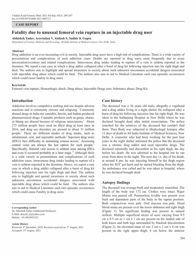
AbstractDrug addiction is an ever-increasing evil in society. Injectable drug users have a high risk of complications. There is a wide variety of presentations and complications of such addiction cases. Deaths are reported in drug users most frequently due to acute intoxication/overdose and related complications. Intravenous drug intake leading to rupture of a vein is seldom reported in the literature. We report a rare case in which a drug addict collapsed after a burst of drug kit following injection into his right thigh and died. The authors aim to highlight and spread awareness in society about such unknown uncommon accidental dangers associated with injectable drug abuse which could be fatal. The authors also aim to aid to Medical Literature such rare episodic occurrences which could cause fatality in drug users.
KeywordsFemoral vein rupture; Hemorrhagic shock; Drug abuse; Injectable Drugs user; Substance abuse; Drug Kit.
Fatality due to unusual femoral vein rupture in an injectable drug user
CASE REPORT
Introduction
Addiction involves compulsive seeking and use despite adverse 1reactions and is commonly chronic and relapsing. Commonly
abused drugs in India are cannabis, heroin, and Indian-produced pharmaceutical drugs. Cannabis products such as ganja, charas,
2or bhang are abused because of religious associations. About 275 million people have used an illicit drug at least once in 2016, and drug use disorders are present in about 31 million
3people. There are different modes of drug intake, such as 4inhalational, oral, and injectable methods. Injection drug users
5(IDUs) face difficulty in sustaining venous access. Accessing 6central veins are always the last option for such people.
Specifically, femoral vein access is seldom seen among IDUs 7,8and even if occurred probably at a later stage. Although there
is a wide variety in presentations and complications of such addiction cases, intravenous drug intake leading to rupture of a vein is seldom reported in the literature. Hence, we report a rare case in which a drug addict collapsed after a burst of drug kit following injection into his right thigh and died. The authors aim to highlight and spread awareness in society about such unknown uncommon accidental dangers associated with injectable drug abuse which could be fatal. The authors also aim to aid to Medical Literature such rare episodic occurrences which could cause Fatality in drug users.
Case history
The deceased was a 36 years old male, allegedly a vagabond and a drug addict living in a night shelter He collapsed after a burst of drug kit following injection into his right thigh. He was taken to the Safdarjung Hospital in New Delhi where he was declared brought dead after initial resuscitation. The police searched for the relatives of the deceased but couldn't find them. Then Body was subjected to Medicolegal Autopsy after 12 days of death in All India Institute of Medical Sciences, New Delhi. A counselor who had been working for the past four years in the night shelter informed the police that the deceased was a chronic drug addict and used injectable drugs. The deceased reportedly had discomfort in his right thigh, the day before his death. He was admitted to the hospital but he ran away from there in the night. The next day i.e. day of his death, at around 6 pm, he was injecting himself in the thigh region when his 'KIT' got burst and he started bleeding from the thigh. An ambulance was called and he was taken to hospital, where he was declared brought dead.
Autopsy findings
The deceased was average-built and moderately nourished. The length of the body was 172 cm. Clothes were intact. Rigor Mortis was passed off. Postmortem lividity was present over back and dependent parts of the body in the supine position. Both conjunctivae were pale. Oral mucosa was pale. Dried blood stains are present over the lower abdomen and right thigh (Figure 1). No significant finding was present in natural orifices. Multiple superficial ulcers of sizes varying from 0.5 cm x 0.5 cm to 1 cm x 1 cm are present on the medial side of both knees and both legs surrounded by blackish discoloration (Figure 2). An ulcerated sinus of size 2 cm x 2 cm x 4 cm was present in the right upper thigh, 6 cm below the anterior
284
Corresponding Author
Dr. Abhishek Yadav (Additional Professor)
E-Mail: [email protected]
Mobile: +91-9818052523
Article Historyth thReceived: 9 December, 2020; Revision received on: 5 August, 2021
thAccepted: 15 August, 2021
Abhishek Yadav, Aravindan V, Abilash S, Sudhir K Gupta Department of Forensic Medicine and Toxicology, All India Institute of Medical sciences, New Delhi, India
J Indian Acad Forensic Med. 2021 Jul-Sep; 43(3): 284-287doi:10.5958/0974-0848.2021.00072.5
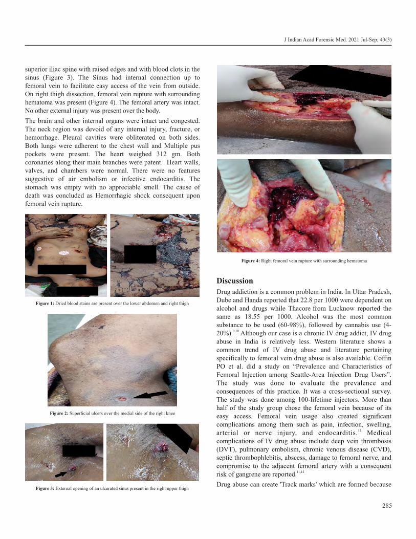
superior iliac spine with raised edges and with blood clots in the sinus (Figure 3). The Sinus had internal connection up to femoral vein to facilitate easy access of the vein from outside. On right thigh dissection, femoral vein rupture with surrounding hematoma was present (Figure 4). The femoral artery was intact. No other external injury was present over the body.
The brain and other internal organs were intact and congested. The neck region was devoid of any internal injury, fracture, or hemorrhage. Pleural cavities were obliterated on both sides. Both lungs were adherent to the chest wall and Multiple pus pockets were present. The heart weighed 312 gm. Both coronaries along their main branches were patent. Heart walls, valves, and chambers were normal. There were no features suggestive of air embolism or infective endocarditis. The stomach was empty with no appreciable smell. The cause of death was concluded as Hemorrhagic shock consequent upon femoral vein rupture.
Discussion
Drug addiction is a common problem in India. In Uttar Pradesh, Dube and Handa reported that 22.8 per 1000 were dependent on
alcohol and drugs while Thacore from Lucknow reported the same as 18.55 per 1000. Alcohol was the most common substance to be used (60-98%), followed by cannabis use (4-
9,1020%). Although our case is a chronic IV drug addict, IV drug abuse in India is relatively less. Western literature shows a common trend of IV drug abuse and literature pertaining specifically to femoral vein drug abuse is also available. Coffin PO et al. did a study on “Prevalence and Characteristics of Femoral Injection among Seattle-Area Injection Drug Users”. The study was done to evaluate the prevalence and consequences of this practice. It was a cross-sectional survey. The study was done among 100-lifetime injectors. More than half of the study group chose the femoral vein because of its easy access. Femoral vein usage also created significant complications among them such as pain, infection, swelling,
11arterial or nerve injury, and endocarditis. Medical complications of IV drug abuse include deep vein thrombosis (DVT), pulmonary embolism, chronic venous disease (CVD), septic thrombophlebitis, abscess, damage to femoral nerve, and compromise to the adjacent femoral artery with a consequent
11,12risk of gangrene are reported.
Drug abuse can create 'Track marks' which are formed because
285
J Indian Acad Forensic Med. 2021 Jul-Sep; 43(3)
Figure 1: Dried blood stains are present over the lower abdomen and right thigh
Figure 2: Superficial ulcers over the medial side of the right knee
Figure 3: External opening of an ulcerated sinus present in the right upper thigh
Figure 4: Right femoral vein rupture with surrounding hematoma
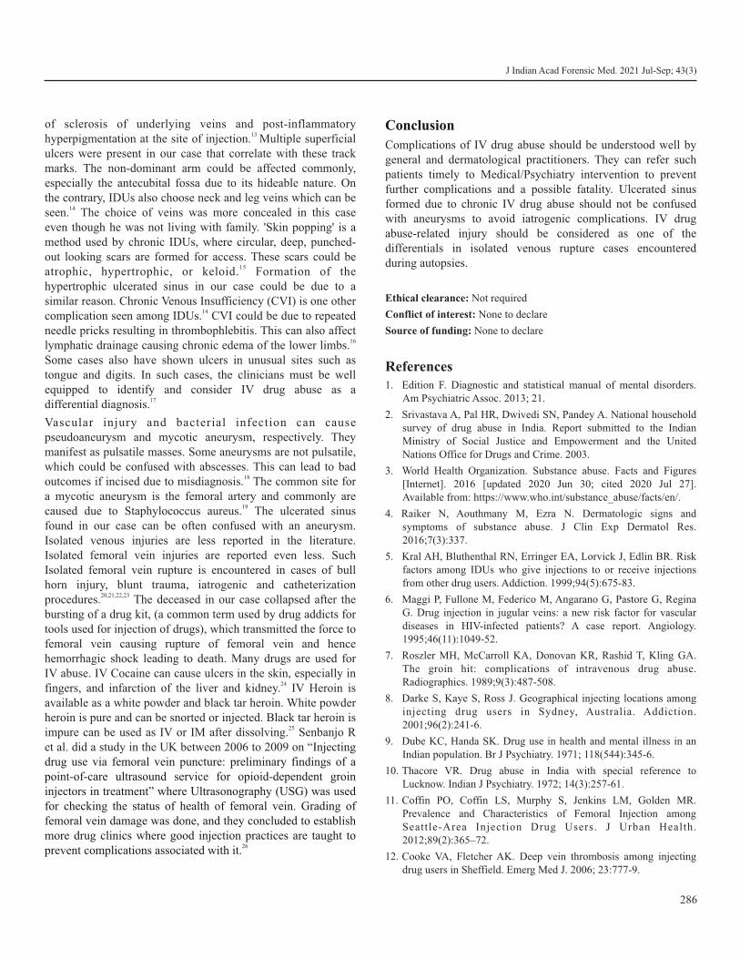
of sclerosis of underlying veins and post-inflammatory 13 hyperpigmentation at the site of injection. Multiple superficial
ulcers were present in our case that correlate with these track marks. The non-dominant arm could be affected commonly, especially the antecubital fossa due to its hideable nature. On the contrary, IDUs also choose neck and leg veins which can be
14seen. The choice of veins was more concealed in this case even though he was not living with family. 'Skin popping' is a method used by chronic IDUs, where circular, deep, punched-out looking scars are formed for access. These scars could be
15atrophic, hypertrophic, or keloid. Formation of the hypertrophic ulcerated sinus in our case could be due to a similar reason. Chronic Venous Insufficiency (CVI) is one other
14complication seen among IDUs. CVI could be due to repeated needle pricks resulting in thrombophlebitis. This can also affect
16lymphatic drainage causing chronic edema of the lower limbs. Some cases also have shown ulcers in unusual sites such as tongue and digits. In such cases, the clinicians must be well equipped to identify and consider IV drug abuse as a
17differential diagnosis.
Vascular injury and bacterial infection can cause pseudoaneurysm and mycotic aneurysm, respectively. They manifest as pulsatile masses. Some aneurysms are not pulsatile, which could be confused with abscesses. This can lead to bad
18outcomes if incised due to misdiagnosis. The common site for a mycotic aneurysm is the femoral artery and commonly are
19caused due to Staphylococcus aureus. The ulcerated sinus found in our case can be often confused with an aneurysm. Isolated venous injuries are less reported in the literature. Isolated femoral vein injuries are reported even less. Such Isolated femoral vein rupture is encountered in cases of bull horn injury, blunt trauma, iatrogenic and catheterization
20,21,22,23procedures. The deceased in our case collapsed after the bursting of a drug kit, (a common term used by drug addicts for tools used for injection of drugs), which transmitted the force to femoral vein causing rupture of femoral vein and hence hemorrhagic shock leading to death. Many drugs are used for IV abuse. IV Cocaine can cause ulcers in the skin, especially in
24fingers, and infarction of the liver and kidney. IV Heroin is available as a white powder and black tar heroin. White powder heroin is pure and can be snorted or injected. Black tar heroin is
25impure can be used as IV or IM after dissolving. Senbanjo R et al. did a study in the UK between 2006 to 2009 on “Injecting drug use via femoral vein puncture: preliminary findings of a point-of-care ultrasound service for opioid-dependent groin injectors in treatment” where Ultrasonography (USG) was used for checking the status of health of femoral vein. Grading of femoral vein damage was done, and they concluded to establish more drug clinics where good injection practices are taught to
26prevent complications associated with it.
Conclusion
Complications of IV drug abuse should be understood well by general and dermatological practitioners. They can refer such patients timely to Medical/Psychiatry intervention to prevent further complications and a possible fatality. Ulcerated sinus formed due to chronic IV drug abuse should not be confused with aneurysms to avoid iatrogenic complications. IV drug abuse-related injury should be considered as one of the differentials in isolated venous rupture cases encountered during autopsies.
Ethical clearance: Not required
Conflict of interest: None to declare
Source of funding: None to declare
References1. Edition F. Diagnostic and statistical manual of mental disorders.
Am Psychiatric Assoc. 2013; 21.
2. Srivastava A, Pal HR, Dwivedi SN, Pandey A. National household survey of drug abuse in India. Report submitted to the Indian Ministry of Social Justice and Empowerment and the United Nations Office for Drugs and Crime. 2003.
3. World Health Organization. Substance abuse. Facts and Figures [Internet]. 2016 [updated 2020 Jun 30; cited 2020 Jul 27]. Available from: https://www.who.int/substance_abuse/facts/en/.
4. Raiker N, Aouthmany M, Ezra N. Dermatologic signs and symptoms of substance abuse. J Clin Exp Dermatol Res. 2016;7(3):337.
5. Kral AH, Bluthenthal RN, Erringer EA, Lorvick J, Edlin BR. Risk factors among IDUs who give injections to or receive injections from other drug users. Addiction. 1999;94(5):675-83.
6. Maggi P, Fullone M, Federico M, Angarano G, Pastore G, Regina G. Drug injection in jugular veins: a new risk factor for vascular diseases in HIV-infected patients? A case report. Angiology. 1995;46(11):1049-52.
7. Roszler MH, McCarroll KA, Donovan KR, Rashid T, Kling GA. The groin hit: complications of intravenous drug abuse. Radiographics. 1989;9(3):487-508.
8. Darke S, Kaye S, Ross J. Geographical injecting locations among injecting drug users in Sydney, Australia. Addiction. 2001;96(2):241-6.
9. Dube KC, Handa SK. Drug use in health and mental illness in an Indian population. Br J Psychiatry. 1971; 118(544):345-6.
10. Thacore VR. Drug abuse in India with special reference to Lucknow. Indian J Psychiatry. 1972; 14(3):257-61.
11. Coffin PO, Coffin LS, Murphy S, Jenkins LM, Golden MR. Prevalence and Characteristics of Femoral Injection among Seattle-Area Injection Drug Users. J Urban Health. 2012;89(2):365–72.
12. Cooke VA, Fletcher AK. Deep vein thrombosis among injecting drug users in Sheffield. Emerg Med J. 2006; 23:777-9.
J Indian Acad Forensic Med. 2021 Jul-Sep; 43(3)
286
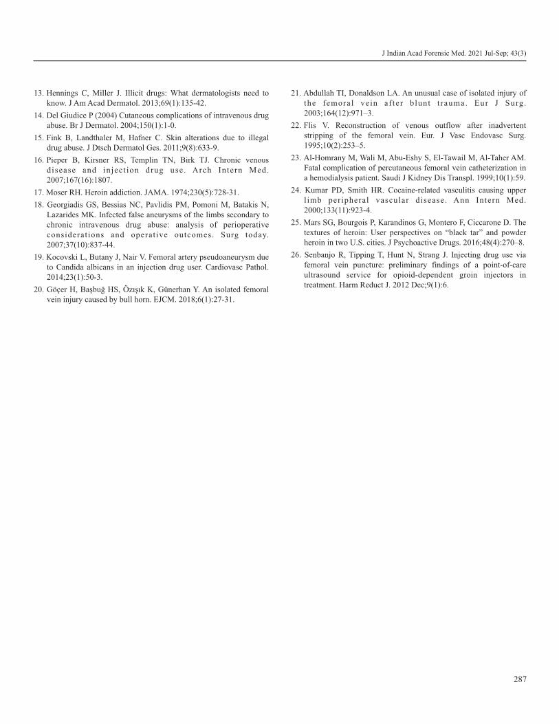
J Indian Acad Forensic Med. 2021 Jul-Sep; 43(3)
13. Hennings C, Miller J. Illicit drugs: What dermatologists need to know. J Am Acad Dermatol. 2013;69(1):135-42.
14. Del Giudice P (2004) Cutaneous complications of intravenous drug abuse. Br J Dermatol. 2004;150(1):1-0.
15. Fink B, Landthaler M, Hafner C. Skin alterations due to illegal drug abuse. J Dtsch Dermatol Ges. 2011;9(8):633-9.
16. Pieper B, Kirsner RS, Templin TN, Birk TJ. Chronic venous disease and inject ion drug use . Arch Intern Med. 2007;167(16):1807.
17. Moser RH. Heroin addiction. JAMA. 1974;230(5):728-31.
18. Georgiadis GS, Bessias NC, Pavlidis PM, Pomoni M, Batakis N, Lazarides MK. Infected false aneurysms of the limbs secondary to chronic intravenous drug abuse: analysis of perioperative considerations and operative outcomes. Surg today. 2007;37(10):837-44.
19. Kocovski L, Butany J, Nair V. Femoral artery pseudoaneurysm due to Candida albicans in an injection drug user. Cardiovasc Pathol. 2014;23(1):50-3.
20. Göçer H, Başbuğ HS, Özışık K, Günerhan Y. An isolated femoral vein injury caused by bull horn. EJCM. 2018;6(1):27-31.
21. Abdullah TI, Donaldson LA. An unusual case of isolated injury of the femoral vein af ter b lunt t rauma. Eur J Surg. 2003;164(12):971–3.
22. Flis V. Reconstruction of venous outflow after inadvertent stripping of the femoral vein. Eur. J Vasc Endovasc Surg. 1995;10(2):253–5.
23. Al-Homrany M, Wali M, Abu-Eshy S, El-Tawail M, Al-Taher AM. Fatal complication of percutaneous femoral vein catheterization in a hemodialysis patient. Saudi J Kidney Dis Transpl. 1999;10(1):59.
24. Kumar PD, Smith HR. Cocaine-related vasculitis causing upper l imb peripheral vascular disease. Ann Intern Med. 2000;133(11):923-4.
25. Mars SG, Bourgois P, Karandinos G, Montero F, Ciccarone D. The textures of heroin: User perspectives on “black tar” and powder heroin in two U.S. cities. J Psychoactive Drugs. 2016;48(4):270–8.
26. Senbanjo R, Tipping T, Hunt N, Strang J. Injecting drug use via femoral vein puncture: preliminary findings of a point-of-care ultrasound service for opioid-dependent groin injectors in treatment. Harm Reduct J. 2012 Dec;9(1):6.
287
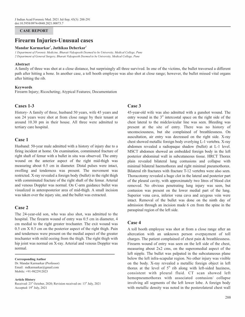
AbstractA family of three was shot at a close distance, but surprisingly all three survived. In one of the victims, the bullet traversed a different path after hitting a bone. In another case, a toll booth employee was also shot at close range; however, the bullet missed vital organs after hitting the rib.
KeywordsFirearm Injury; Ricocheting; Atypical Features; Documentation
Firearm Injuries-Unusual cases
CASE REPORT
Cases 1-3
History- A family of three, husband 50 years, wife 45 years and son 24 years were shot at from close range by their tenant at around 10.30 pm in their house. All three were admitted to tertiary care hospital.
Case 1
Husband: 50-year male admitted with a history of injury due to a firing incident at home. On examination, comminuted fracture of right shaft of femur with a bullet in situ was observed. The entry wound on the anterior aspect of the right mid-thigh was measuring about 0.5 cm in diameter. Distal pulses were intact, swelling and tenderness was present. The movement was restricted. X-ray revealed a foreign body (bullet) in the right thigh with comminuted fracture of the right shaft of the femur. Arterial and venous Doppler was normal. On C-arm guidance bullet was visualized in anteroposterior area of mid-thigh. A small incision was taken over the injury site, and the bullet was extracted.
Case 2
The 24-year-old son, who was also shot, was admitted to the hospital. The firearm wound of entry was 0.5 cm in diameter, 4 cm medial to the right greater trochanter. The exit wound was 0.5 cm X 0.5 cm on the posterior aspect of the right thigh. Pain and tenderness were present on the medial aspect of the greater trochanter with mild oozing from the thigh. The right thigh with hip joint was normal on X-ray. Arterial and venous Doppler was normal.
Case 3
45-year-old wife was also admitted with a gunshot wound. The rdentry wound in the 3 intercostal space on the right side of the
chest lateral to the midclavicular line was seen. Bleeding was present at the site of entry. There was no history of unconsciousness, but she complained of breathlessness. On auscultation, air entry was decreased on the right side. X-ray chest showed metallic foreign body overlying L-1 vertebra. X-ray abdomen revealed a radiopaque shadow (bullet) at L-1 level. MSCT abdomen showed an embedded foreign body in the left posterior abdominal wall in subcutaneous tissue. HRCT Thorax plain revealed bilateral lung contusions and collapse with minimal bilateral haemothorax and right minimal pneumothorax. Bilateral rib fractures with fracture T-12 vertebra were also seen. Thoracotomy revealed a huge clot in the lateral and posterior part of the pleural cavity, with approximately two litres of blood clot removed. No obvious penetrating lung injury was seen, but contusion was present on the lower medial part of the lung. Superior vena cava, inferior vena cava and azygous vein were intact. Removal of the bullet was done on the ninth day of admission through an incision made 6 cm from the spine in the paraspinal region of the left side.
Case 4
A toll booth employee was shot at from a close range after an altercation with an unknown person overpayment of toll charges. The patient complained of chest pain & breathlessness. Firearm wound of entry was seen on the left side of the chest, measuring about 2x2 cms, on the superomedial aspect of the left nipple. The bullet was palpated in the subcutaneous plane below the left infra-scapular region. No other injury was visible on the body. X-ray revealed a metallic foreign object in left
ththorax at the level of 5 rib along with left-sided haziness, consistent with pleural fluid. CT scan showed left hemopneumothorax with associated contusion/ collapse involving all segments of the left lower lobe. A foreign body with metallic density was noted in the posterolateral chest wall
288
Corresponding Author
Dr. Mandar Karmarkar (Professor)
Email: [email protected]
Mobile: +91-9822912023
Article Historyrd thReceived: 23 October, 2020; Revision received on: 13 July, 2021thAccepted: 19 July, 2021
1 2Mandar Karmarkar , Juthikaa Deherkar 1 Department of Forensic Medicine, Bharati Vidyapeeth Deemed to be University, Medical College, Pune
2 Department of General Surgery, Bharati Vidyapeeth Deemed to be University, Medical College, Pune
J Indian Acad Forensic Med. 2021 Jul-Sep; 43(3): 288-291doi:10.5958/0974-0848.2021.00073.7
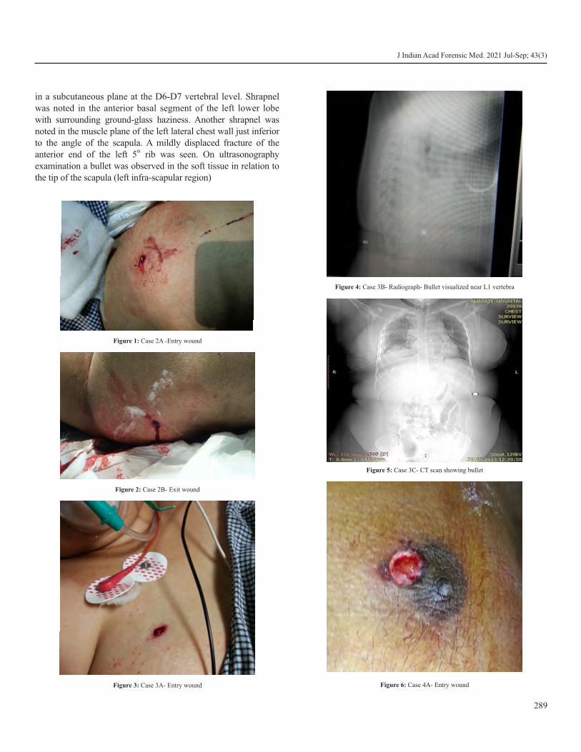
in a subcutaneous plane at the D6-D7 vertebral level. Shrapnel was noted in the anterior basal segment of the left lower lobe with surrounding ground-glass haziness. Another shrapnel was noted in the muscle plane of the left lateral chest wall just inferior to the angle of the scapula. A mildly displaced fracture of the
thanterior end of the left 5 rib was seen. On ultrasonography examination a bullet was observed in the soft tissue in relation to the tip of the scapula (left infra-scapular region)
289
J Indian Acad Forensic Med. 2021 Jul-Sep; 43(3)
Figure 1: Case 2A -Entry wound
Figure 2: Case 2B- Exit wound
Figure 3: Case 3A- Entry wound
Figure 4: Case 3B- Radiograph- Bullet visualized near L1 vertebra
Figure 5: Case 3C- CT scan showing bullet
Figure 6: Case 4A- Entry wound
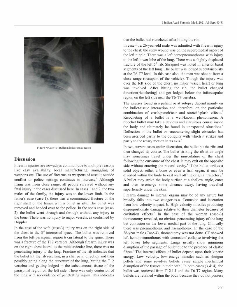
Discussion
Firearm injuries are nowadays common due to multiple reasons like easy availability, local manufacturing, smuggling of weapons etc. The use of firearms as weapons of assault outside
1conflict or police settings continues to increase. Although firing was from close range, all people survived without any fatal injury in the cases discussed here. In cases 1 and 2, the two males of the family, the injury was to the lower limb. In the father's case (case-1), there was a comminuted fracture of the right shaft of the femur with a bullet in situ. The bullet was removed and handed over to the police. In the son's case (case-2), the bullet went through and through without any injury to the bone. There was no injury to major vessels, as confirmed by Doppler.
In the case of the wife (case-3) injury was on the right side of rdthe chest in the 3 intercostal space. The bullet was removed
from the left paraspinal region 6 cm lateral to the spine. There was a fracture of the T12 vertebra. Although firearm injury was on the right chest lateral to the midclavicular line, there was no penetrating injury to the lung. Fracture of the rib indicates that the bullet hit the rib resulting in a change in direction and then possibly going along the curvature of the lung, hitting the T12 vertebra and getting lodged in the subcutaneous tissue of the paraspinal region on the left side. There was only contusion of the lung with no evidence of penetrating injury. This indicates
that the bullet had ricocheted after hitting the rib.
In case-4, a 26-year-old male was admitted with firearm injury to the chest; the entry wound was on the superomedial aspect of the left nipple. There was a left hemopneumothorax with injury to the left lower lobe of the lung. There was a slightly displaced fracture of the left 5th rib. Shrapnel was noted in anterior basal segments of the left lung. The bullet was lodged subcutaneously at the T6-T7 level. In this case also, the man was shot at from a close range (occupant of the vehicle). Though the injury was over the left side of the chest, no major vessel, heart or lung was involved. After hitting the rib, the bullet changed direction(ricocheting) and got lodged below the infrascapular region on the left side near the T6-T7 vertebra.
The injuries found in a patient or at autopsy depend mainly on the bullet-tissue interaction and, therefore, on the particular
2combination of crush/punch/tear and stretch/splash effects. Ricocheting of a bullet is a well-known phenomenon. A ricochet bullet may take a devious and circuitous course inside
3the body and ultimately be found in unexpected situations. Deflection of the bullet on encountering slight obstacles has been ascribed partly to the obliquity with which it strikes and
4partly to the rotary motion in its axis.
In two current cases under discussion, the bullet hit the ribs and then changed its course. The bullet striking the rib at an angle may sometimes travel under the musculature of the chest following the curvature of the chest. It may exit on the opposite
4side without entering the pleural cavity. If the bullet strikes a solid object, either a bone or even a firm organ, it may be
5diverted within the body to exit well off the original trajectory. A bullet may strike the body surface at a shallow angle, enter and then re-emerge some distance away, having travelled
5superficially under the skin.
Firearm damage to internal organs may be of any nature but broadly falls into two categories-a. Contusion and laceration from low-velocity impact. b. High-velocity missiles producing disproportionate damage relative to their diameter because of
5cavitation effects. In the case of the woman (case-3) thoracotomy revealed, no obvious penetrating injury of the lung but contusion on the lower medial part of the lung. Clinically there was pneumothorax and haemothorax. In the case of the 26-year male (Case-4), thoracotomy was not done. CT showed left hemopneumothorax with contusion/ collapse involving the left lower lobe segments. Lungs usually show minimum disruption of the passage of bullet due to the presence of elastic
3fibres. The internal effects of bullet depend upon their kinetic energy. Low velocity, low energy missiles such as shotgun pellets and some revolver bullets cause simple mechanical
1disruption of the tissues in their path. In both cases (3 & 4), the bullet was retrieved from T12-L1 and the T6-T7 region. Many bullets are retained within the body because they do not possess
J Indian Acad Forensic Med. 2021 Jul-Sep; 43(3)
290
Figure 7: Case 4B- Bullet in infrascapular region
J Indian Acad Forensic Med. 2021 Jul-Sep; 43(3)
enough energy to complete the passage through it, or the energy is dissipated on contact with other structures (e.g., bone). Wound severity is related to the bullet construction and its
1trajectory and the properties of the body tissues traversed.
For both solid surfaces and water there is a critical angle of impact(incidence) below which a bullet striking the surface will ricochet rather than penetrate. The critical angle is determined by the nature of the surface, the construction of the bullet and the velocity of the bullet. Thus round-nosed bullets are more likely to ricochet than flat-nosed, full metal-jacketed than lead and low velocity more than high velocity. Bullets ricocheting off solid surfaces usually ricochet off at smaller angles than the impact.6 Mechanism of projectile wound production depends on many factors like shape, size, the velocity of the missile, character of motion in the flight, the density of the tissues, hydrostatic forces, rate of energy transmission from the missile to the tissue.4
A radiological examination is an important tool for diagnosing and investigating firearm cases. The usefulness of x-ray examination of gunshot victim is undeniable. X-ray is important for locating missiles/pellets, fragments or jackets, determining the track of the wound, defects in bones in the areas not easily
4approachable on direct examination. In all cases, not only x-ray examination but CT, USG and Doppler were also done. CT & x-ray both helped in locating the bullet and its retrieval. In fact, in case-4, the operation could be avoided, and the bullet was retrieved by taking a subcutaneous incision. Another important aspect of firearm cases is proper documentation and collection of evidentiary material. In the cases discussed here, Forensic Medicine & Toxicology department and the General Surgery department worked together for accurate documentation of injuries, photographs and collection of necessary evidence.
Conclusion
Firearm injury is not a rarity nowadays; Forensic Medicine faculty should be able to handle clinical medico-legal work. In the family (husband, son and wife, i.e., case-1, 2, 3), all three
were shot at but survived. The wife had a chest injury, but because of the bullet hitting the rib and resultant ricocheting,
thmajor organs/vessels were spared. In the 4 case, the injury was on the left side of the chest, but again, there was no injury to the heart/great vessels because of the intervening rib. Thus, although firing was from close range (less than 1 meter), all the victims survived and two because of the ricochet effect. In the present cases, the authors could not examine clothes as police took them immediately for an examination. In the case of the family, there was a dispute with the tenant, who shot them in
ththeir house from close range. In the 4 case, the person was working at a toll booth, and there was an altercation overpayment of toll, so one of the occupants fired at the worker from close range. It is rare to see victim/s survive when fired from close range.
Ethical clearance: Not required
Conflict of interest: None to declare
Source of funding: None to declare
References1. Payne-James J, Jones RM, editors. Simpson's forensic medicine,
14th edition. 14th ed. New York, NY: Productivity Press; 2019. Pg. 133-139.
2. Mason JK, editor. The pathology of trauma, 3rd Edition. London,England: Hodder Arnold; 2000. Pg. 58
3. Mukherjee, J. B. Forensic Medicine and Toxicology. SecondEdition. India, Academic Publishers, 1994. Pg. 349
4. Vij, K. Textbook of Forensic Medicine & Toxicology: Principles &Practice. 6th Edition. India, Elsevier India, 2014. Pg. 231-250
5. Saukko P, Knight B. Knight's forensic pathology, 3Ed. CRC Press;2004. Pgs. 266-70
6. DiMaio VJM. Gunshot wounds: Practical aspects of firearms,ballistics, and forensic techniques, third edition. 2nd ed. New York, NY: Productivity Press; 2015. Pg. 109
291
AbstractDomestic violence is prevalent globally. Of which one of the entities is Child abuse which occurs in many forms. The most difficult to identify and examine is violence aimed at children since they are the most vulnerable part of the family. The consequence of child abuse are many ranging from various physical injuries to psychological and mental trauma which affects the overall development of child as well. Modern forensic medical examination systems recognized the need for special training for an expert to deal with such cases. There is a dire need to place various protocols and guidelines in this context by various countries in their own societal perspective. This will surely aid in in-depth research into this societal problem as well as help in proper dispensation of justice. In India although POCSO has been placed since 2012 which primarily addresses one of the forms of child abuse ie sexual only. The paper discusses various aspects of forensic medical examination in such a situation which will go a long way in placing the better documentation in a humane manner.
KeywordsChild abuse; Documentation; Forensic interview; POCSO
Pediatric Forensic Examination in Domestic Violence cases- Problems and solutions thereof in global perspective
PERSPECTIVE
Introduction
Child abuse occurs in many forms. Some may be visible which are highlighted by media while others, not less frequent, are less obvious or even invisible, occurring in remote places and closed doors. Since 1980 onwards, problem of domestic violence has become more and more highlighted and hence more curiosity among clinical forensic medicine researchers. They deal with issues of preventing and detecting facts of
1, 2violence and examination aspects. The most difficult to identify and examine is violence aimed at children since they
3are the most vulnerable part of the family. Nearly 80% of all abusers in such kind of violence are the parents.
Domestic violence can manifest in physical, psychological & sexual forms. There are variety of obstacles in the identification of child abuse victims (like religious and societal “norms”)
4, 5 which have been created in many nations and countries. Not every society in our contemporary world is ready to acknowledge the presence as well as the magnitude of this
6apparent problem. India and Ukraine being no exception to it. Although in India having very close knitted societal structure these are perceived in a different manner. Yet slowly it is being recognized by all concerned.
Due to the complexity of timely evidence of domestic violence, statistics varies widely among various countries. Eg. In a national study in Romania in the same year ; according to parents' reports in questionnaires, the prevalence of physical domestic abuse was 18.4% while according to children's reports it was 24.4%. According to official statistics only about 2% of children needed public services protection. Research by Save the Children found 75% of children had been subjected to
7corporal punishment. A UNICEF survey which included 35 countries of Europe and Central Asia found that from 24 to 59% of children had experienced violent or aggressive behavior
8within their families. Among contemporary young parents 77% 9reported that they had physically punished a child. In India with
enactment of Protection of Children against Sexual Offences (POCSO) in 2012; gradually this problem of society is being recognized and awareness is being created at various levels.
Effects/Consequences of Child Abuse:
Domestic violence has massive effect at our day-to-day life including direct and delayed subsequences. These can be recognized as under:
Ÿ A fatal consequence- Homicide which involves weapons is among the top three causes of death in adolescents. The
10,11boys are over 80% of victims and perpetrators.
Ÿ Severe injuries-There are hundreds of male victims of youth violence who sustain injuries because of physical
10fighting and assault.
Ÿ Violence at an early age can lead to decreasing of brain development and cognitive skills resulting in educational
292
Corresponding Author
Dr Adarsh Kumar (Professor)
Department of Forensic Medicine & Toxicology, AIIMS , New Delhi , India
Email Id: ([email protected]
Mobile: +91-9868438856)
Article HistoryReceived: 22nd May, 2021; Accepted: 25th August, 2021
1 1 1 2 3Bondarchuk Hanna , Gunas Valery , Perebetyuk Anatoliy , Fomina Lyudmila , Adarsh Kumar 1 Department of Pathological Anatomy, Forensic Medicine and Law, National Pirogov Memorial Medical University, Vinnytsya, Ukraine.2 Department of Human anatomy, National Pirogov Memorial Medical University, Vinnytsya, Ukraine3 Department of Forensic Medicine & Toxicology, AIIMS , New Delhi, India
J Indian Acad Forensic Med. 2021 Jul-Sep; 43(3): 292-295doi:10.5958/0974-0848.2021.00074.9
12, 13under-achievement, etc.
Ÿ Family abused children are more likely to have drug abuse, nicotine dependence, alcohol abuse and incorrect
13sexual behavior.
Ÿ Such environment leads to unwanted pregnancies, abortions and sexually transmitted infections, including
10 HIV.
Ÿ When people become older risk of cardiovascular disease, cancer, diabetes and other health problems arise due to the
10,13negative behavior coping.
Ÿ It is more usual to them to drop out of study, have difficulty finding and keeping a job, and to be at risk for later victimization, suicidal attempts, create perverted
10,13parent-child relationships.
Modern forensic medical examination systems recognized the 14,15need for special training for an expert in such cases. Studies
include the psychological characteristics of the subjects, methods and approaches for the most effective and objective collection of verbal and physical data with minimal additional
16,17trauma to the subjects. These recommendations are 18,19,20constantly updated and approaches are improved. Most
protocols recommend taking into account the capabilities of the child, depending on the maturity of the psyche and the ability to understand questions and adequately formulate answers to
21,22,23them. Every trained specialist must be proficient in ability to cooperate with children as well as an ability to support
24them.
Recommended Protocols and Guidelines:
ü It is imperative that the interviewer is aware with the child prior to the main forensic examination. Information about the age of the child, its family, level of development, the possible health problems and traumas, school performance, cultural background, possible malfunctions with regard to basic skills such as the use of
25language should be collected before first meeting. A list of the main issues that we should make clear during interview has to be created. Another modern researcher may have an opposite mind: as knowledge about examined child might have impact to the interviewer, he is advised not to know anything about the case or the
26facts before the first meeting.
ü Examiner must make clear the rules to the child, explaining every point thoroughly. He should ask the child to avoid any assumptions telling exclusively what has happened; listen to the questions attentively not being in hurry; not to hesitate to ask if any question has not been understood. He should get acquainted the child with the right to interrupt and correct the interviewer, to say "I
don't know", but impossibility of examiner to be aware of the details. It's important to encourage the abused to describe everything he can recall even things that seems
26unimportant. The examiner should explain to the child that discussed questions will not be discussed with anyone more, but some justice/social service workers can
27 be notified for child's own safety.
ü He should use phrases that demonstrate his empathy to the difficult and painful position of the child and avoid comments underestimating the situation the child is
26experiencing. The friendly examiner's posture, eye contact and smiling demeanor encourages the abused
28child to disclose more securely during interview. Studies point out the child reacts better in familiar places and child-friendly setting. Significant effect on cognitive functions and distracting the attention of children can be created with visual and audio stimulations, disturbance from third parties or interruption of the interview for any
26reason.
ü There is a need to adapt to the developmental level of the child in every case because examining children like adults inevitably leads to many misunderstandings and causes
29,30the perverted information to be captured. If the linguistic developmental level of the child is not taken into account, difficulties will arise with the
31comprehension of the legal terminology. E,g. the pre-school age children have limited memory and linguistic skills, the suggestibility, the bright imagination and
32increased trust to adults.
ü The interviewer should give the opportunity to tell what happened in child`s own words. If the interview is strictly structured , the possibilities of collecting significant information decreases and great part of it might be
33missed.
ü The interviewer should collect information by using specific type of questions, frequency the questions are posed with, the time between them, and the duration of
34,35the interview. Repeated questions can make the child feel that its previous answers are not "right" or "desired", annulling its primary grounds. It is recommended to begin the interview with open-ended questions, which give the child an opportunity to report the incident in the greatest
36,37possible detail and to be in charge of the narration. Closed questions can be used for declining the risk of suggestibility and increasing the possibility of extracting more specific details. However, leading questions must be
38avoided as children are too vulnerable to them.
He should use this kind of questions only when all other alternatives have been exhausted.
One of the most important problems of the forensic examiner`s
293
J Indian Acad Forensic Med. 2021 Jul-Sep; 43(3)
work is the establishment of the fact of a single or earlier repeated facts of violence, the frequency and time period of
39their recurrence. Our attentiveness when establishing the limitation of causing damage and traces of previously caused damage costs a fortune for opening the new volume of violence. The issue that has been loudly heard lately is the occurrence of psychosomatic diseases that lead to prolongation of the disease
40duration with changing the severity of caused damage.
Adults with a history of child abuse are more likely to experience physical health problems. A particular goal of forensic examination is searching for relationships between the injuries caused and health disorders that arose a long time after them, which were based on the psychosomatic component and acquired the form of the disease. Children who have been exposed to family violence suffer symptoms of bed-wetting or nightmares, and are at great risk of having allergies; asthma,
41gastrointestinal problems, headaches and flu. Adolescents and young adults with a history of child sexual abuse were five
42times more likely to have an eating disorder.
Swedish researchers found a strong association between reported physical abuse and three or more psychosomatic symptoms and the relationship between abuse severity and somatic symptoms. The girls have significantly more
43psychosomatic symptoms. A US meta-analysis found that child maltreatment was related to an increased risk of neurological, musculoskeletal, respiratory, cardiovascular and gastrointestinal problems but contrary to some other studies, not
44gynecological problems.
Conclusion
There is a dire need to understand these complicated issues of clinical forensic medicine practice which invariably are kept under the wraps. The Forensic Medicine Expert must always try to hone his skills so that he can contribute for the betterment of society. In order to increase the accuracy and volume of information obtained during a forensic interview with a child, the expert must be specially trained in the features of communication with children of all ages and strictly adhere to the rules forensic examination in cases of child domestic abuse. The disclosure of fundamentally different volumes of crimes depends on the expert's readiness to recognize traces of previously received injuries. A distinctive feature of forensic research is the search for causal relationships between the injuries caused and the diseases that arise long time after them, which were based on the psychosomatic component and acquired the form of the disease. One of the most important aspect in the chain of domestic abuse elimination is the correct establishment of the facts of child domestic abuse with the real severity of damage.
References1. Herman S, Freitas TR. Error rates in forensic child sexual abuse
evaluations Psychological injury and law. 2010 Jun 1;3(2):133-47.
2. Rumble L, Febrianto RF, Larasati MN, Hamilton C, Mathews B, Dunne MP. Childhood sexual violence in Indonesia: a systematic review. Trauma, violence, & abuse. 2020 Apr;21(2):284-99.
3. dos Santos Silva W, de Oliveira Barroso-Júnior U. Characteristics of children under 12 years subjected to forensic examination on suspicion of child sexual abuse in Salvador Brazil. Spanish Journal of Legal Medicine. 2016 Apr 1;42(2):55-61.
4. Antoniadou E, Dardavesis T, Pavlou E, Zaggelidou E. Child physical abuse in northern Greece: A retrospective study based on forensic protocols. J Forensic Biomed. 2017;8(135):2.
5. Shako K, Kalsi M. Forensic observations and recommendations on sexual and gender based violence in Kenya. Forensic Science International: Synergy. 2019 Jan 1;1:185-203.
6. Guma M, Henda N. The socio-cultural context of child abuse: A betrayal of trust.
7. Rada C. Violence against women by male partners and against children within the family: prevalence, associated factors, and intergenerational transmission in Romania, a cross-sectional study. BMC Public health. 2014 Dec;14(1):129.
8. UNICEF. Toward a world free from violence: Global survey on violence against children. New York: UNICEF, Office of the Special Representative of the Secretary-General on Violence against Children. 2013.
9. Smith AB. The state of research on the effects of physical punishment. Social Policy Journal of New Zealand. 2006 Mar 27;27:114.
10. Lamont A. Effects of child abuse and neglect for children and adolescents. Australian Institute of Family Studies; 2010.
11. Raykova KA, Efimov AA, Kurzin LM, Savenkova EN, Alekseev YD, Ivakhina SA. The role of projective psycho diagnostic methods in identifying domestic violence cases over children. Reports Tambov State University. Series: Natural and technical sciences. 2016;21(6).
12. Australian Institute of Health and Welfare. Family, domestic and sexual violence in Australia 2018.
13. Hunter C. Effects of child abuse and neglect for children and adolescents. Melbourne: Child Family Community Australia information exchange, Australian Institute of Family Studies. Retrieved from www3. aifs. gov. au/cfca/publications. 2014.
14. La Rooy D, Brubacher SP, Aromäki-Stratos A, Cyr M, Hershkowitz I, Korkman J, Myklebust T, Naka M, Peixoto CE, Roberts KP, tewart H. The NICHD Protocol: A review of an internationally-used evidence-based tool for training child forensic interviewers. Journal of Criminological Research, Policy and Practice. 2015 Jun 8.
15. Lamb ME, Sternberg KJ, Orbach Y, Hershkowitz I, Horowitz D, Esplin PW. The effects of intensive training and ongoing supervision on the quality of investigative interviews with alleged sex abuse victims. Applied Developmental Science. 2002;6(3):114-25.
16. Cyr M, Lamb ME. Assessing the effectiveness of the NICHD
J Indian Acad Forensic Med. 2021 Jul-Sep; 43(3)
294
J Indian Acad Forensic Med. 2021 Jul-Sep; 43(3)
investigative interview protocol when interviewing French-speaking alleged victims of child sexual abuse in Quebec. Child Abuse & Neglect. 2009;33(5):257-68.
17. Pichler AS, Sharman SJ, Powell M, Westera N, Goodman-Delahunty J. Association between interview quality and child sexual abuse trial outcome. Journal of Family Violence. 2019 Mar 25:1-9
18. Benia LR, Hauck-Filho N, Dillenburg M, Stein LM. The NICHD investigative interview protocol: A meta-analytic review. Journal of child sexual abuse. 2015;24(3):259-79.
19. Hershkowitz I, Lamb ME, Katz C. Allegation rates in forensic child abuse investigations: Comparing the revised and standard NICHD protocols. Psychology, Public Policy, and Law. 2014;20(3):336.
20. Rapholo SF, Makhubele JC. Forensic Interviewing Techniques in Child Sexual Abuse Allegations: Implications for the South African Context. Global Journal of Health Science. 2019;11(6): 53-63.
21. Lamb ME, Orbach Y, Hershkowitz I, Esplin PW, Horowitz D. A structured forensic interview protocol improves the quality and informativeness of investigative interviews with children: A review of research using the NICHD Investigative Interview Protocol. Child abuse & neglect. 2007;31(11-12):1201-31.
22. Lamb ME, Orbach Y, Sternberg KJ, Aldridge JA, Pearson S, Stewart HL, Esplin PW, Bowler L. Use of a structured investigative protocol enhances the quality of investigative interviews with alleged victims of child sexual abuse in Britain. Applied Cognitive Psychology: The Official Journal of the Society for Applied Research in Memory and Cognition. 2009;23(4):449-67.
23. Magalhães T, Ribeiro CS, Jardim P, Vieira DN. Forensic procedures for interview physical exam and evidence collection in children and young people victims of physical and/or sexual abuse. Acta medica portuguesa. 2011;24(2):339-48.
24. Saywitz KJ, Larson RP, Hobbs SD, Wells CR. Developing rapport with children in forensic interviews: Systematic review of experimental research. Behavioral Sciences & the Law. 2015;33(4):372-89.
25. Otgaar H, Chan JC, Calado B, La Rooy D. Immediate interviewing increases children's suggestibility in the short term, but not in the long term. Legal and Criminological Psychology. 2019;24(1):24-40.
26. Newlin C, Steele LC, Chamberlin A, Anderson J, Kenniston J, Russell A, Stewart H, Vaughan-Eden V. Child forensic interviewing: Best practices. US Department of Justice, Office of Justice Programs, Office of Juvenile Justice and Delinquency Prevention; 2015 Sep.
27. Ahern EC, Lamb ME. Children's reports of disclosure recipient reactions in forensic interviews: Comparing the NICHD and MoGP protocols. Journal of Police and Criminal Psychology. 2017;32(2):85-93.
28. Ahern EC, Hershkowitz I, Lamb ME, Blasbalg U, Winstanley A. Support and reluctance in the pre‐substantive phase of alleged child abuse victim investigative interviews: Revised versus Standard NICHD protocols. Behavioral sciences & the law. 2014;32(6):762-74.
29. Lamb ME, Sternberg KJ, Orbach Y, Esplin PW, Stewart H, Mitchell S. Age differences in young children's responses to open-ended invitations in the course of forensic interviews. Journal of consulting and clinical psychology. 2003;71(5):926.
30. Sakelliadis EI, Spiliopoulou CA, Papadodima SA. Forensic investigation of child victim with sexual abuse. Indian pediatrics. 2009;46(2): 144-51.
31. Sparta SN, Koocher GP, editors. Forensic mental health assessment of children and adolescents. Oxford University Press; 2006 Mar 16.
32. Bruck M, Ceci SJ. The suggestibility of children's memory. Annual review of psychology. 1999;50(1):419-39.
33. Korkman J, Santtila P, Westeråker M, Sandnabba NK. Interviewing techniques and follow-up questions in child sexual abuse interviews. European Journal of Developmental Psychology. 2008;5(1):108-28.
34. Gagnon K, Cyr M. Sexual abuse and preschoolers: Forensic details in regard of question types. Child abuse & neglect. 2017;67:109-18.
35. Sumampouw NE, Otgaar H, La Rooy D, de Ruiter C. The Quality of Forensic Child Interviewing in Child Sexual Abuse Cases in Indonesia. Journal of Police and Criminal Psychology. 2019:1-2.
36. Berg R, Munthe-Kaas HM, Baiju N, Muller AE, Brurberg KG. The accuracy of using open‐ended questions in structured conversations with children: a systematic review.
37. Powell MB, Guadagno B, Benson M. Improving child investigative interviewer performance through computer-based learning activities. Policing and Society. 2016;26(4):365-74.
38. Korkman J, Santtila P, Sandnabba NK. Dynamics of verbal interaction between interviewer and child in interviews with alleged victims of child sexual abuse. Scandinavian journal of psychology. 2006;47(2):109-19.
39. Azad A, Christianson SÅ, Selenius H. Children's reporting patterns after witnessing homicidal violence–the effect of repeated experience and repeated interviews. Psychology, Crime & Law. 2014;20(5):407-29.
40. Wegman HL, Stetler C. A meta-analytic review of the effects of childhood abuse on medical outcomes in adulthood. Psychosomatic medicine. 2009;71(8):805-12.
41. Ross CA. Childhood sexual abuse and psychosomatic symptoms in irritable bowel syndrome. Journal of child sexual abuse. 2005;14(1):27-38.
42. Cuijpers P, Smit F, Unger F, Stikkelbroek Y, ten Have M, de Graaf R. The disease burden of childhood adversities in adults: a population-based study. Child abuse & neglect. 2011;35(11):937-45.
43. Jernbro C, Svensson B, Tindberg Y, Janson S. Multiple psychosomatic symptoms can indicate child physical abuse–results from a study of Swedish schoolchildren. Acta paediatrica. 2012;101(3):324-9.
44. Pacella ML, Hruska B, Delahanty DL. The physical health consequences of PTSD and PTSD symptoms: a meta-analytic review. Journal of anxiety disorders. 2013;27(1):33-46.
295
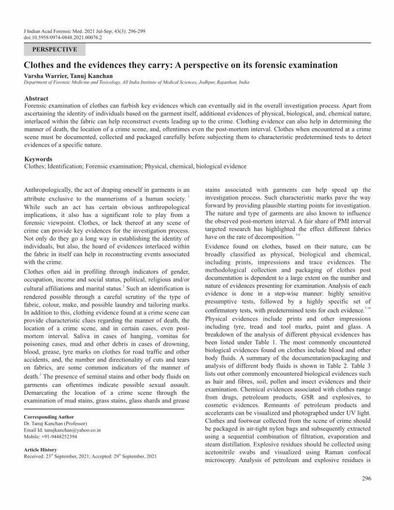
AbstractForensic examination of clothes can furbish key evidences which can eventually aid in the overall investigation process. Apart from ascertaining the identity of individuals based on the garment itself, additional evidences of physical, biological, and, chemical nature, interlaced within the fabric can help reconstruct events leading up to the crime. Clothing evidence can also help in determining the manner of death, the location of a crime scene, and, oftentimes even the post-mortem interval. Clothes when encountered at a crime scene must be documented, collected and packaged carefully before subjecting them to characteristic predetermined tests to detect evidences of a specific nature.
KeywordsClothes; Identification; Forensic examination; Physical, chemical, biological evidence
Clothes and the evidences they carry: A perspective on its forensic examination
PERSPECTIVE
Anthropologically, the act of draping oneself in garments is an 1attribute exclusive to the mannerisms of a human society.
While such an act has certain obvious anthropological implications, it also has a significant role to play from a forensic viewpoint. Clothes, or lack thereof at any scene of crime can provide key evidences for the investigation process. Not only do they go a long way in establishing the identity of individuals, but also, the hoard of evidences interlaced within the fabric in itself can help in reconstructing events associated with the crime.
Clothes often aid in profiling through indicators of gender, occupation, income and social status, political, religious and/or
2cultural affiliations and marital status. Such an identification is
rendered possible through a careful scrutiny of the type of fabric, colour, make, and possible laundry and tailoring marks. In addition to this, clothing evidence found at a crime scene can provide characteristic clues regarding the manner of death, the location of a crime scene, and in certain cases, even post-mortem interval. Saliva in cases of hanging, vomitus for poisoning cases, mud and other debris in cases of drowning, blood, grease, tyre marks on clothes for road traffic and other accidents, and, the number and directionality of cuts and tears on fabrics, are some common indicators of the manner of
2death. The presence of seminal stains and other body fluids on
garments can oftentimes indicate possible sexual assault. Demarcating the location of a crime scene through the examination of mud stains, grass stains, glass shards and grease
stains associated with garments can help speed up the investigation process. Such characteristic marks pave the way forward by providing plausible starting points for investigation. The nature and type of garments are also known to influence the observed post-mortem interval. A fair share of PMI interval targeted research has highlighted the effect different fabrics
3-6have on the rate of decomposition.
Evidence found on clothes, based on their nature, can be broadly classified as physical, biological and chemical, including prints, impressions and trace evidences. The methodological collection and packaging of clothes post documentation is dependent to a large extent on the number and nature of evidences presenting for examination. Analysis of each evidence is done in a step-wise manner: highly sensitive presumptive tests, followed by a highly specific set of
7-14confirmatory tests, with predetermined tests for each evidence.
Physical evidences include prints and other impressions including tyre, tread and tool marks, paint and glass. A breakdown of the analysis of different physical evidences has been listed under Table 1. The most commonly encountered biological evidences found on clothes include blood and other body fluids. A summary of the documentation/packaging and analysis of different body fluids is shown in Table 2. Table 3 lists out other commonly encountered biological evidences such as hair and fibres, soil, pollen and insect evidences and their examination. Chemical evidences associated with clothes range from drugs, petroleum products, GSR and explosives, to cosmetic evidences. Remnants of petroleum products and accelerants can be visualized and photographed under UV light. Clothes and footwear collected from the scene of crime should be packaged in air-tight nylon bags and subsequently extracted using a sequential combination of filtration, evaporation and steam distillation. Explosive residues should be collected using acetonitrile swabs and visualized using Raman confocal microscopy. Analysis of petroleum and explosive residues is
Corresponding Author
Dr. Tanuj Kanchan (Professor)
Email Id: [email protected]
Mobile: +91-9448252394
Article Historyrd thReceived: 23 September, 2021; Accepted: 29 September, 2021
Varsha Warrier, Tanuj KanchanDepartment of Forensic Medicine and Toxicology, All India Institute of Medical Sciences, Jodhpur, Rajasthan, India
J Indian Acad Forensic Med. 2021 Jul-Sep; 43(3): 296-299doi:10.5958/0974-0848.2021.00076.2
296
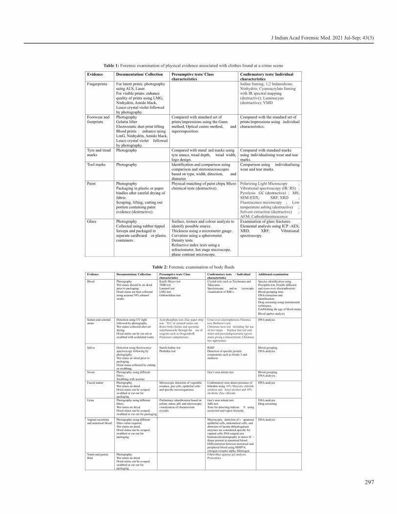
Evidence Documentation/ Collection Presumptive tests/ Class characteristics
Confirmatory tests/ Individual characteristics
Fingerprints For latent prints: photography using ALS, Laser.For visible prints: enhance quality of prints using LMG, Ninhydrin, Amido black, Leuco crystal violet followed by photography.
Iodine fuming; 1,2 Indanedione; Ninhydrin; Cyanoacrylate fuming with IR spectral mapping (destructive); Luminocyan (destructive); VMD
Footwear and footprints
PhotographyGelatin lifterElectrostatic dust print liftingBlood prints : enhance using LmG, Ninhydrin, Amido black, Leuco crystal violet followed by photography.
Compared with standard set of prints/impressions using the Gunn method, Optical centre method, and superimposition.
Compared with the standard set of prints/impressions using individual characteristics.
Tyre and tread marks
Photography Compared with stand ard marks using tyre stance, tread depth, tread width, logo design.
Compared with standard marks using individualising wear and tear marks.
Tool marks Photography Identification and comparison using comparison and stereomicroscopes based on type, width, direction, and diameter.
Comparison using individualising wear and tear marks.
Paint PhotographyPackaging in plastic or paper bindles after careful drying of fabric.Scraping, lifting, cutting out portion containing paint evidence (destructive).
Physical matching of paint chips; Micro chemical tests (destructive).
Polarizing Light Microscopy ; Vibrational spectroscopy (IR/ RS) ; Pyrolysis GC (destructive) ; MS; SEM-EDX; XRF, XRD ; Fluorescence microscopy ; Low temperature ashing (destructive) ; Solvent extraction (destructive) ; AEM; Cathodoluminescence
Glass PhotographyCollected using rubber tipped forceps and packaged in separate cardboard or plastic containers.
Surface, texture and colour analysis to identify possible source.Thickness using a micrometer gauge.Curvature using a spherometer.Density testsRefractive index tests using a refractometer, hot stage microscope, phase contrast microscope.
Examination of glass fractures.Elemental analysis using ICP -AES;XRD; XRF; Vibrational spectroscopy.
Table 1: Forensic examination of physical evidence associated with clothes found at a crime scene
Evidence Documentation/ Collection Presumptive tests/ Class characteristics
Confirmatory tests / Individual characteristics
Additional examination
Blood PhotographyWet stains should be air dried prior to packaging.Dried stains are best collected using acetone/70% ethanol swabs.
Kastle Meyer test TMB testLuminol testLMG testOrthotolidine test
Crystal tests such as Teichmann and Takayama.Spectroscopy and m icroscopic visualisation of RBCs.
Species identification using Precipitin test, Double diffusion and cross-over electrophoresis.Blood grouping tests.DNA extraction and identification.Drug screening using instrumental techniques.Establishing the age of blood stains.
Blood spatter analysis.
Semen and seminal stains
Detection using UV lightfollowed by photography.Wet stains collected after air drying.Dried stains can be cut out or swabbed with acidulated water.
Acid phosphate test; Zinc paper strip test; TLC of seminal stains can detect both choline and spermine simultaneously through the use of reagents such as Dragendroff, Potassium iodoplatinato.
Cross-over electrophoresis Florence test; Barberio’s test.Christmas tress test including the use of two stains - Nuclear fast red (red stain) and picroindigocarmine (green stain) giving a characteristic Christmas tree appearance.
DNA analysis
Saliva Detection using fluorescence spectroscopy following by photography.Wet stains air dried prior to packaging.Dried stains collected by cutting or swabbing.
Starch-Iodine testPhabedas test
RSIDDetection of specific protein components such as histan-3 and statherin
Blood grouping.DNA analysis.
Sweat Photography using different filters.Swabbing with acetone
Gee’s urea nitrate test Blood grouping.DNA analysis.
Faecal matter Photography.Wet stains air dried .Dried stains can be scraped, swabbed or cut out for packaging.
Microscopic detection of vegetable residues, pus cells, epithelial cells and specific microorganisms.
Confirmatory tests detect presence of bilirubin using 10% Mercuric chloride solution and Amyl alcohol and 10% alcoholic Zinc chloride.
DNA analysis
Urine Photography using different filters.Wet stains air dried.Dried stains can be scraped, swabbed or cut out for packaging
Preliminary identification based on colour, odour, pH, and microscopic visualization of characteristic crystals.
Gee’s urea nitrate test.Jaffe test.Tests for detecting indican E using resorcinol and cupric bromide.
DNA analysis.Drug screening.
Vaginal secretions and menstrual blood
Photography using different filters when required.Wet stains air dried .Dried stains can be scraped, swabbed or cut out for packaging.
Microscopic detection of s quamous epithelial cells, endometrial cells, and detection of lactate dehydrogenase enzymes are considered specific for vaginal cells; PAS reagent test.Immunochromatography to detect D -dimer present in menstrual blood.Differentiation between menstrual and peripheral blood using MMP14, estrogen receptor alpha, fibrinogen.
DNA analysis
Vomit and gastric fluid
Photography.Wet stains air dried .Dried stains can be scraped, swabbed or cut out for packaging
Fibrin-blue agarose gel analysis.Proteomics.
Table 2: Forensic examination of body fluids
J Indian Acad Forensic Med. 2021 Jul-Sep; 43(3)
297
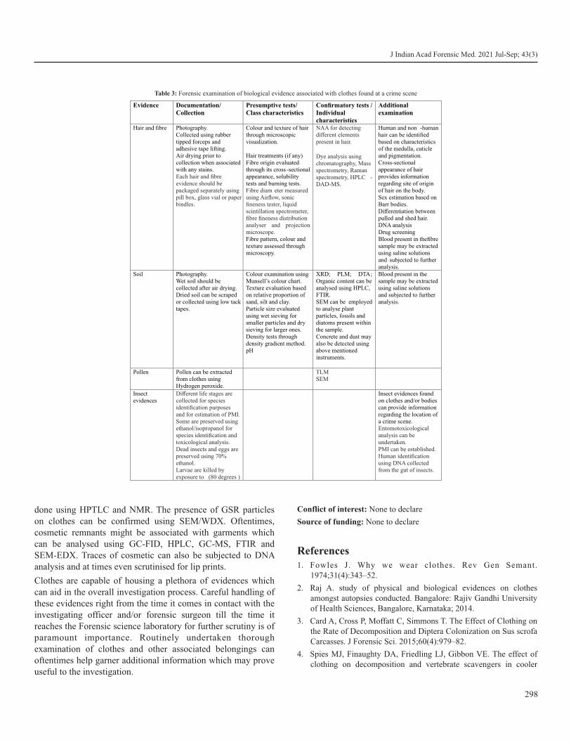
done using HPTLC and NMR. The presence of GSR particles on clothes can be confirmed using SEM/WDX. Oftentimes, cosmetic remnants might be associated with garments which can be analysed using GC-FID, HPLC, GC-MS, FTIR and SEM-EDX. Traces of cosmetic can also be subjected to DNA analysis and at times even scrutinised for lip prints.
Clothes are capable of housing a plethora of evidences which can aid in the overall investigation process. Careful handling of these evidences right from the time it comes in contact with the investigating officer and/or forensic surgeon till the time it reaches the Forensic science laboratory for further scrutiny is of paramount importance. Routinely undertaken thorough examination of clothes and other associated belongings can oftentimes help garner additional information which may prove useful to the investigation.
Conflict of interest: None to declare
Source of funding: None to declare
References1. Fowles J. Why we wear clothes. Rev Gen Semant.
1974;31(4):343–52.
2. Raj A. study of physical and biological evidences on clothes amongst autopsies conducted. Bangalore: Rajiv Gandhi University of Health Sciences, Bangalore, Karnataka; 2014.
3. Card A, Cross P, Moffatt C, Simmons T. The Effect of Clothing on the Rate of Decomposition and Diptera Colonization on Sus scrofa Carcasses. J Forensic Sci. 2015;60(4):979–82.
4. Spies MJ, Finaughty DA, Friedling LJ, Gibbon VE. The effect of clothing on decomposition and vertebrate scavengers in cooler
J Indian Acad Forensic Med. 2021 Jul-Sep; 43(3)
Evidence Documentation/ Collection
Presumptive tests/ Class characteristics
Confirmatory tests / Individual characteristics
Additional examination
Hair and fibre Photography.Collected using rubber tipped forceps and adhesive tape lifting.Air drying prior to collection when associated with any stains.Each hair and fibre evidence should be packaged separately using pill box, glass vial or paper bindles.
Colour and texture of hair through microscopic visualization.
Hair treatments (if any)Fibre origin evaluated through its cross -sectional appearance, solubility tests and burning tests. Fibre diam eter measured using Airflow, sonic fineness tester, liquid scintillation spectrometer, fibre fineness distribution analyser and projection microscope.Fibre pattern, colour and texture assessed through microscopy.
NAA for detecting different elements present in hair.
Dye analysis using chromatography, Mass spectrometry, Raman spectrometry, HPLC -DAD-MS.
Human and non -human hair can be identified based on characteristics of the medulla, cuticle and pigmentation.Cross-sectional appearance of hair provides information regarding site of origin of hair on the body.Sex estimation based on Barr bodies.Differentiation between pulled and shed hair.DNA analysisDrug screeningBlood present in the fibre sample may be extracted using saline solutions and subjected to further analysis.
Soil Photography.Wet soil should be collected after air drying.Dried soil can be scraped or collected using low tack tapes.
Colour examination using Munsell’s colour chart.Texture evaluation based on relative proportion of sand, silt and clay.
Particle size evaluated using wet sieving for smaller particles and dry sieving for larger ones. Density tests through density gradient method. pH
XRD; PLM; DTA; Organic content can be analysed using HPLC, FTIR.SEM can be employed to analyse plant particles, fossils and diatoms present within the sample.Concrete and dust may also be detected using above mentioned instruments.
Blood present in the sample may be extracted using saline solutions and subjected to further analysis.
Pollen Pollen can be extracted from clothes using Hydrogen peroxide.
TLMSEM
Insect evidences
Different life stages are collected for species identification purposes and for estimation of PMI.Some are preserved using ethanol/isopropanol for species identification and toxicological analysis.Dead insects and eggs are preserved using 70% ethanol.Larvae are killed by exposure to (80 degrees )
Insect evidences found on clothes and/or bodies can provide information regarding the location of a crime scene.Entomotoxicological analysis can be undertaken.PMI can be established.Human identification using DNA collected from the gut of insects.
Table 3: Forensic examination of biological evidence associated with clothes found at a crime scene
298
months of the temperate southwestern Cape, South Africa. Forensic Sci Int. 2020; 309: 110197.
5. Miller RA. The Effects of Clothing on Human Decomposition: Implications for Estimating Time Since Death. Knoxville: University of Tennessee; 2002.
6. Mashaly AM, Mahmoud A, Ebaid H. Influence of Clothing on Decomposition and Presence of Insects on Rabbit Carcasses. J Med Entomol. 2019;56(4):921–7.
7. https://www.asteetrace.org/static/images/pdf/01%20Forensic% 20Paint%20Analysis%20and%20Comparison%20Guidelines.pdf
8. Andrews RN, Hawker H, Crosbie SF. Evaluation of five methods for measuring mean fibre diameter of fleece samples from New Zealand sheep. N Z J Exp Agric. 1987;15(1):23–31.
9. Forensic Examination of Soil Evidence [Internet]. [cited 2022 Jan 21]. Available from: https://www.jstage.jst.go.jp/ article/ jasti/7/2/7_2_95/_article
10. Kulstein G, Wiegand P. DNA/RNA co-analysis of seminal fluid-stained fabrics after water immersion for up to seven days. Forensic Sci Int Genet Suppl Ser. 2017;6:e27–8.
11. Yamada S, Hirata K, Tsugawa N, Bunai Y, Ohya I. Vomit identification by a pepsin assay using a fibrin blue-agarose gel plate . Forensic Sci Int. 1992; 52 (2): 215-21.
12. Coulson S, Morgan-Smith R, Mitchell S, McBriar T. An investigation into the presence of petrol on the clothing and shoes of members of the public. Forensic Sci Int . 2008 Feb;175(1):44–54.
13. Schotman TG, Xu X, Rodewijk N, van der Weerd J. Application of dye analysis in forensic fibre and textile examination: Case examples. Forensic Sci Int. 2017;278:338–50.
14. Alotaibi SS, Sayed SM, Alosaimi M, Alharthi R, Banjar A, Abdulqader N, et al. Pollen molecular biology: Applications in the forensic palynology and future prospects: A review. Saudi J Biol Sci. 2020;27(5):1185–90.
J Indian Acad Forensic Med. 2021 Jul-Sep; 43(3)
299
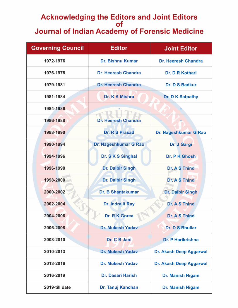
Acknowledging the Editors and Joint Editors of
Journal of Indian Academy of Forensic Medicine
1972-1976
1976-1978
1979-1981
1981-1984
1984-1986
1986-1988
1988-1990
1990-1994
1994-1996
1996-1998
1998-2000
2000-2002
2002-2004
2004-2006
2006-2008
2008-2010
2010-2013
2013-2016
2016-2019
2019-till date
Dr. Bishnu Kumar
Dr. Heeresh Chandra
Dr. Heeresh Chandra
Dr. K K Mishra
-
Dr. Heeresh Chandra
Dr. R S Prasad
Dr. Nageshkumar G Rao
Dr. S K S Singhal
Dr. Dalbir Singh
Dr. Dalbir Singh
Dr. B Shantakumar
Dr. Indrajit Ray
Dr. R K Gorea
Dr. Mukesh Yadav
Dr. C B Jani
Dr. Mukesh Yadav
Dr. Mukesh Yadav
Dr. Dasari Harish
Dr. Tanuj Kanchan
Dr. Heeresh Chandra
D R KothariDr.
D S BadkurDr.
D K SatpathyDr.
-
-
Nageshkumar G RaoDr.
J GargiDr.
P K GhoshDr.
A S ThindDr.
A S ThindDr.
Dalbir SinghDr.
A S ThindDr.
A S ThindDr.
D S BhullarDr.
P HarikrishnaDr.
Akash Deep AggarwalDr.
Akash Deep AggarwalDr.
Manish NigamDr.
Manish NigamDr.
Governing Council Editor Joint Editor

Related Documents