J Indian Acad Forensic Med, 32(4) ISSN 0971-0973 i Indian Academy of Forensic Medicine (IAFM) (Registration No.349, 12th May, 1972, Panji, Goa) Governing Council 2010-2012 President Dr. D.S.Badkur General Secretary Dr.Adarsh Kumar Treasurer Dr. A.S. Thind Vice President Dr. Dalbir Singh (NZ) Dr. P.Sampath Kumar (SZ) Dr. Tulsi Mahto (EZ) Dr. H.T. Katade (WZ) Dr. R.K. Singh (CZ) Joint Secretary Dr. Dasari Harish (NZ) Dr. Cyriac Job (SZ) Dr. Shoban Das (EZ) Dr. Hasumati Patel (WZ) Dr. P.S.Thakur (CZ) Editor Dr. Mukesh Yadav Joint Editor Dr. Akash Deep Aggarwal Executive Member Dr.B.P. Dubey (Ex. President) Dr.A.K.Sharma Dr.Sarvesh Tandon Dr.C.P.Bhaisora Dr.Pankaj Gupta Dr.Luv Sharma Dr.Sanjoy Das (Ex. Secretary) Dr.Amandeep Singh Dr.Mukesh K.Goyal Dr.C.B. Jani Dr.Jaynti Yadav Dr.P.K.Tiwari

Welcome message from author
This document is posted to help you gain knowledge. Please leave a comment to let me know what you think about it! Share it to your friends and learn new things together.
Transcript
J Indian Acad Forensic Med, 32(4) ISSN 0971-0973
i
Indian Academy of Forensic Medicine
(IAFM) (Registration No.349, 12th May, 1972, Panji, Goa)
Governing Council 2010-2012
President
Dr. D.S.Badkur
General Secretary Dr.Adarsh Kumar
Treasurer
Dr. A.S. Thind
Vice President Dr. Dalbir Singh (NZ)
Dr. P.Sampath Kumar (SZ)
Dr. Tulsi Mahto (EZ)
Dr. H.T. Katade (WZ)
Dr. R.K. Singh (CZ)
Joint Secretary Dr. Dasari Harish (NZ)
Dr. Cyriac Job (SZ)
Dr. Shoban Das (EZ)
Dr. Hasumati Patel (WZ)
Dr. P.S.Thakur (CZ)
Editor Dr. Mukesh Yadav
Joint Editor
Dr. Akash Deep Aggarwal
Executive Member
Dr.B.P. Dubey (Ex. President)
Dr.A.K.Sharma
Dr.Sarvesh Tandon
Dr.C.P.Bhaisora
Dr.Pankaj Gupta
Dr.Luv Sharma
Dr.Sanjoy Das (Ex. Secretary)
Dr.Amandeep Singh
Dr.Mukesh K.Goyal
Dr.C.B. Jani
Dr.Jaynti Yadav
Dr.P.K.Tiwari
J Indian Acad Forensic Med, 32(4) ISSN 0971-0973
ii
Journal of Indian Academy of Forensic Medicine
(JIAFM) Editor, Dr. Mukesh Yadav
Professor & HOD
Forensic Medicine & Toxicology,
School of Medical Sciences & Research
Sharda University, Greater Noida, U.P. PIN:
201306 Residence
G-216, Parsvanath Edens,
Alfa-II, Greater Noida, G.B. Nagar,
U.P.-2010308
Ph. No. 0120-2326060,
Mobile No. 09411480753
Email: [email protected]
Joint Editor Dr. Akash Deep Aggarwal
Assistant Professor,
Department of Forensic Medicine,
Pt. B.D. Sharma PHGIMS, Rohtak,
Haryana-124001 Residence
H.No. 14, Desi Mehmandari,
Patiala, Punjab
PIN: 147001
Mobile No. 9815652621
Email:[email protected]
Peer Review Group
Dr.A.K. Srivstava Prof. & HOD,
Forensic Medicine &Toxicology Subharti Medical College,
Meerut, U.P. Dr.V.V.Pillay Prof. & HOD,
Analytical Toxicology,
Chief of Poison Control Centre
AIMS & R, Cochin-Kerala
Dr.R.K. Gorea
Prof. & HOD,
Forensic Medicine &Toxicology
Gyan Sagar Medical College,
Banur, Patiala, Punjab
Forensic Medicine,
Dr.C.B.Jani Prof. & HOD,
Forensic Medicine &
Toxicology P.S MedicalCollege,
Karamsad, Distt. Anand,
Gujarat, PIN: 388325
Dr.T.K Bose
Prof. & HOD,
Forensic and State Medicine
Govt. Medical College
Kolkata, West Bengal
Dr.G. Pradeepkumar Prof. & HOD,
Forensic Medicine &Toxicology
Kasturba Medical College,
Manipal, Karnatka
Advisory Board
Sharma G.K., (New Delhi) Verma S.K., (New Delhi)
Kaur Balbir, (Srinagar)
Bansal Y., (Chandigarh)
Kumar Shantha B., (Tamilnadu)
Gupta B.D., (Gujrat)
Manju Nath K.H, (Karnatka)
Das Sanjoy, (U.K.)
Bhaisora C.P, (U.K.)
Meel B.L. (South Africa)
Mahtoo Tulsi, (Jharkhand)
Ravindran K, (Pondicherry)
Sabri Imran, (U.P.)
Rastogi Prateek (Karnatka)
Potwary AJ (Assam)
Singh R.K. (Chhatisgarh)
Dongre A.P. (Nagpur)
Rastogi Pooja (U.P.)
Sharma Aditya (H.P.)
Khanagwal V. (Haryana)
Gupta Pankaj (Punjab)
Pounder Derrick, (UK)
Khaja Shaikh (A.P.)
Basu R (W.B.)
Naik R.S. (Maharastra)
Godhikirakar Madhu (Goa)
Job Cyriac (Kerala)
Vinita K. (U.P.)
Yadav B.N. (Nepal)
Printed and published by Dr. Mukesh Yadav, Editor, JIAFM and Dr. A. D. Aggarwal, Joint Editor, JIAFM on
behalf of Indian Academy of Forensic Medicine at name of the press [SHIVANI PRINTERS, NOIDA, U.P.]

J Indian Acad Forensic Med, 32(4) ISSN 0971-0973
278
Journal of Indian Academy of Forensic Medicine
Contents
From the Editor‟s Desk 280
Editorial
Discovery Rule and Medical Negligence 281
Original Research Papers Access of High Technology Based Medical Diagnostic Tool to Convicted
Prisoners Lodged in a Typically Large Indian Jail – CT Scan as Case Study 283 Munawwar Husain, Usama B. Ghaffar, Jawed Ahmad Usmani, Shameem Jahan Rivzi
Fatal Road Traffic Accidents among Young Children 286 Harnam Singh, A. D. Aggarwal
Application of Victims‟ Fingernails in Forensic DNA Analysis 289 Kamoun Arwa, Mahfoudh Nadia, Ayadi Adnene, Hammemi Zouheir, Maatoug Samir, Makni Hafedh
A Two-year Burns Fatality Study 292 Rahul Chawla, Ashok Chanana, Hukumat Rai, A. D. Aggarwal, Harnam Singh, Gaurav Sharma
Computed Tomographic Studies on Ossification Status of Medial Epiphysis
of Clavicle: Effect of Slice Thickness and Dose Distribution 298 Kaur Gurdeep, Khandelwal N., Jasuja O.P.
The Profile of Age in cases of Victims of Sexual Offence 303
Chandresh Tailor, Ganesh Govekar, Gaurang Patel, Dharmesh Silajiya
Ultrasonographical Age Estimation from Fetal Biparietal Diameter 308 Garg A, Pathak N, Gorea RK, Mohan P
Analysis of Railway Fatalities in Central India 311 Ramesh Nanaji Wasnik
Custodial Deaths - An Overview of the Prevailing Healthcare Scenario 315 Y. S. Bansal, Murali G., Dalbir Singh
A Study on Appraisal of Effectiveness of the MCCD Scheme 318 Swapnil S Agarwal, Vijay Kumar A G, Lavlesh Kumar, Binay K Bastia, Krishnadutt H Chavali
Determination of Sex from Adult Sternum by Discriminant Function Analysis
on Autopsy Sample of Indian Bengali Population: A New Approach 321 Partha Pratim Mukhopadhyay
Estimation of Stature by Percutaneous Measurements of Distal Half of
Upper Limb (Forearm & Hand) 325 Kumar Amit, Srivastava A. K., Verma A.K.
Estimation of Stature from Measurements of Long Bones, Hand and Foot
Dimensions 329 Chikhalkar B.G., Mangaonkar A.A., Nanandkar S.D., Peddawad R.G.
Volume 32 • Number 4 • Oct--December 2010
J Indian Acad Forensic Med, 32(4) ISSN 0971-0973
279
A Study of Serum Cholinesterase Levels in Organo phosphorous Poisoning
Cases 332
Bharath Kumar Guntheti, Shaik Khaja, S.S. Panda
ABO Blood Grouping from Tooth Material 336 Mahabalesh Shetty, Premalatha K
Case Reports Fatal Traumatic Rupture of Ascending Aortic Aneurysm Having Idiopathic
Cystic Medial Necrosis: An Autopsy Case 339 Pannag S. Kumar, Silvano Dias Sapeco, R.G. Wiseman Pinto, Francisco Couto
Death Due to Swine Influenza -A Forensic Autopsy Report 343 Manpreet Kaul, Jagdish Gargi, Ashok Chanana, Rajeev Kumar Chaudhary
Suicidal Acid Injury: A Case Report 347 Vijayanath.V., Raju.G.M., K.Nagaraj Rao, Anitha. M. R.,
Review Papers Taser Technology: Medical, Legal, Ethical & Social Implications of Introduction
of Taser Gun in India 349 Richa Choudhary, Imran Sabri
Aluminium Phosphide Poisoning: Management and Prevention 352 S.Ranjan Bajpai
Serum Enzymes Changes after Death & Its CorrelationwithTime since Death 355 S. P. Garg, Vidya Garg
A Review of Medico-legal Consequences of Gossypiboma 358 Monika Garg, Akash Deep Aggarwal
Broder Scope of COPRA,1986 & Medical Profession 362 Mukesh Yadav, Pooja Rastogi
Medical Audit and Death Audit 369 Somnath Das, Surendra Kumar Pandey, Prabir Chakraborty
Supplement IAFM Membership Subscription Form 371
Copy Right: No part of this publication may be reprinted or publish without the prior permission of the
Editor, JIAFM. Submission of all paper to the journal is understood to imply that it is not being considered for
publication elsewhere. Submission of multi authored papers implies that the consent of each author has been
obtained. In this journal, every effort has been made NOT to publish inaccurate or misleading
information. However, the Editor, Joint Editor, Peer Review Group and Advisory Board accept NO
liability in consequences of such statements. EDITOR ((JIAFM)
Address request for reprint or further information relating to any article may please be made with author and in
case of multi authored article, please communicate to Corresponding Author or the First Author
J Indian Acad Forensic Med, 32(4) ISSN 0971-0973
280
From Editor‟s Desk
JIAFM A Quarterly Publication
Volume 32, Number 4, Oct-December, 2010
I feel immense pleasure to present before you the third issue of 2010. I assure you about the
quality of research papers and quality of printing in future issues. Your valuable suggestions are
always encouraging me and I heartily welcome for future suggestions. On behalf of Executive
Committee of IAFM for the years 2010-2011, I took resolution to further improve the quality and
status of our Journal. We always learn from mistakes and try to improve upon these. I am
thankful to the advertisers who have provided additional financial resources for improving the
quality of this issue.
Dr. Mukesh Yadav
Editor
Editor
Subscription Information Members of IAFM will receive the free of cost.
Non Members and Institutions (Annual Subscription rates)
Personal: In India, Rs. 1000/ (Rest of the world: US$ 200/ or equivalent)
Institutions: In India, Rs. 3000/ (Rest of the world: US$ 400/ or equivalent)
Subscription orders and payments should be made in favour of “Editor, JIAFM, payable at
Greater Noida”
We Accept: Bank Cheque / Demand Drafts (Add Rs. 50/- for outstation Cheques)
The Scope of the Journal covers all aspects of Forensic Medicine and allied fields, research
and applied.
Claims for missing issue: A copy will be sent free to the member / subscriber provided the claim is made within 2 months of
publication of the issue & self addressed envelop of the size 9” x 12” is sent to the Editor. (Those
who want the journals to be dispatched by Registered Post must affix Rs. 50/ worth postage stamps).
The journal is indexed with IndMed and made available online by Diwan Enterprise (New Delhi) at:
1. www.indianjournals.com
2. http://www.medind.nic.in
3. www.jiafm.com
4. www.iafm1972.org

J Indian Acad Forensic Med, 32(4) ISSN 0971-0973
281
Editorial
Discovery Rule and Medical Negligence
As a General Rule consumer court can reject the complaint of medical negligence if filed after lapse of two
years from the date of cause of action under section 24A. The “Discovery Rule” evolved by the Courts in United
States has been recently applied by the Hon‟ble Supreme Court of India in a case decided on 20th
October 2010. The
patient was employed as a Nurse in Government Hospital, Goa, she had no reason to suspect that gauze might
have been left in her abdomen at the time of surgery performed in November, 1993 and the Gujarat State
Commission was not at all justified in non suiting her on the premise that the cause of action had accrued in
the year 1993 and complaint filed in the year 25.10.2002. [Para 12]
The term cause of action is not defined in the Act of 1986 the same has to be interpreted keeping in view
the context in which it has been used in Section 24A (1) and object of the legislation. The question of limitation
is a mixed question of law and fact.
Section 24A, Limitation period: (1) The District Forum, the State Commission or the National Commission shall not admit a complaint
unless it is filed within two years from the date on which the cause of action has arisen.
(2) Notwithstanding anything contained in sub-section (1), a complaint may be entertained after the period
specified in sub-section (1), if the complainant satisfies the District Forum, the State Commission or the
National Commission, as the case may be, that he had sufficient cause for not filing the complaint
within such period:
Provided that no such complaint shall be entertained unless the National Commission, the State Commission or
the District Forum, as the case may be, records its reasons for condoning such delay." [Para 13]
Whether effect of negligence is „patent‟ or „latent‟? In cases of medical negligence, no straitjacket formula can be applied for determining as to when the
cause of action has accrued to the consumer.
Each case is to be decided on its own facts. If the effect of negligence on the doctor's part or any
person associated with him is patent, the cause of action will be deemed to have arisen on the date
when the act of negligence was done.
If, on the other hand, the effect of negligence is latent, then the cause of action will arise on the date
when the patient or his representative- complainant discovers the harm/injury caused due to such act or the date when the patient or his representative-complainant could have, by exercise of reasonable
diligence discovered the act constituting negligence. [Para 18]
What is the Discovery Rule? Where a foreign object is negligently left in a patient's body by a surgeon and the patient is in
ignorance of the fact, and consequently of his right of action for malpractice, the cause of action does
not accrue until the patient learns of, or in the exercise of reasonable care and diligence should have
learned of the presence of such foreign object in his body.
Background of the Discovery Rule: The Discovery Rule to which reference has been made was evolved by the Courts in United States
because it was found that the claim lodged by the complainants in cases involving acts of medical
negligence were getting defeated by strict adherence to the statutes of limitation. [Para 19]
Global Scenario on applicability of the Discovery Rule: In Pennsylvania, the Discovery Rule was adopted in Ayers v. Morgan case.
In that case a surgeon had left a sponge in the patient's body when he performed an operation. It was held
that the statute of limitation did not begin to run until years later when the presence of the sponge in the
patient's body was discovered.
In West Virginia, the Discovery Rule was applied in Morgan v. Grace Hospital Inc. case.
In that case a piece of sponge had been left in the wound during a surgical operation but its presence in
the body did not come to light until 10 years later. The Court rejected the objection of limitation and
observed:
"It simply places an undue strain upon common sense, reality, logic and simple justice to say that a
cause of action had „accrue‟ to the plaintiff until the X-ray examination disclosed a foreign object within
her abdomen and until she had reasonable basis for believing or reasonable means of ascertaining that the
foreign object was within her abdomen as a consequence of the negligent performance of the
hysterectomy.”
J Indian Acad Forensic Med, 32(4) ISSN 0971-0973
282
Again, the Supreme Court observed:
“We believe that the „discovery rule‟ as stated and applied in cases cited above represents a distinct and
marked trend in recent decisions of appellate courts throughout the nation."
Recent application of the Discovery Rule: In Idaho, the Discovery Rule was invoked in Billings v. Sisters of Mercy of Idaho case.
The facts of that case were that the plaintiff underwent a surgical operation in 1946. A sponge was left in
the wound when the incision was closed. The same was discovered in the patient's body in 1961.
During the intervening period the patient sustained considerable suffering, during which she
consulted various physicians.
Limitations of „General Rule‟: After reviewing numerous authorities at great length, the Court cast aside the earlier doctrine, adopted the
Discovery Rule and observed:
“In reality, the „general rule‟ has little to recommend it. It is neither the position of a majority of the
jurisdictions nor is it firmly based on considerations of reason or justice. We will, therefore, adhere to the
following rule: where a foreign object is negligently left in a patient's body by a surgeon and the patient is
in ignorance of the fact, and consequently of his right of action for malpractice, the cause of action does
not accrue until the patient learns of, or in the exercise of reasonable care and diligence should have
learned of the presence of such foreign object in his body.”
The facts in Quinton v. United States case were that the wife of the plaintiff was given blood transfusion in
a Government hospital in 1956. In June, 1959, the plaintiff and his wife during the latter's pregnancy
discovered that wrong type of blood was given to her in 1956 and as a result she gave birth to a stillborn
child.
The Government sought dismissal of the action for damages on the ground of limitation. The Court of
Appeals opined that when a claim accrues under the Federal Tort Claims Act, it is governed by Federal law
and not by local State law.
The Court then held that the period of limitation does not begin to run until the claimant discovers, or in
the exercise of reasonable diligence should have discovered the act constituting the alleged negligence.
In Josephine Flanagan v. Mount Eden General Hospital LEXSEE case, the application of the rule of
Discovery was considered in the background of fact that during the course of operation done on 14.7.1958,
surgical clamps were inserted in the plaintiff's body. In 1966, the plaintiff consulted a doctor because she
experienced severe pain in the region of her abdomen. The doctor told her that surgical clamps were
discovered by X-ray analysis. Thereafter, another operation was performed to remove the clamps.
The defendants sought dismissal of the complaint on the ground that the same was barred by time. The Court
referred to the Discovery Rule and observed:
"The so-called discovery rule employed in foreign object medical malpractice case is in compatible
harmony with the purpose for which Statutes of Limitation were enacted and strikes a fair balance in the field
of medical malpractice. The unsoundness of the traditional rule, as applied in the case where an object is
discovered in the plaintiff's body, is patent. "It simply places an undue strain upon common sense, reality,
logic and simple justice to say that a cause of action had `accrued' to the plaintiff until the X-ray
examination disclosed a foreign object within her abdomen and until she had reasonable basis for
believing or reasonable means of ascertaining that the foreign object was within her abdomen as a
consequence of the negligent performance of the operation." In the case before SC, the danger of belated, false or frivolous claims is eliminated. In addition,
plaintiff's claim does not raise questions as to credibility nor does it rest on professional diagnostic judgment
or discretion. It rests solely on the presence of a foreign object within her abdomen.
The policy of insulating defendants from the burden of defending stale claims brought by a party who, with
reasonable diligence, could have instituted the action more expeditiously is not a convincing justification for
the harsh consequences resulting from applying the same concept of accrual in foreign object cases as is
applied in medical treatment cases.
A clamp, though immersed within the patient's body and undiscovered for a long period of time, retains its
identity so that a defendant's ability to defend a “stale” claim is not unduly impaired. Therefore, where a foreign
object has negligently been left in the patient's body, the Statute of Limitations will not begin to run until the
patient could have reasonably discovered the malpractice." (Emphasis added)
Doctor should make aware of these new developments in the field of medical negligence to protect from
potential law suit as well as take reasonable precautions in the interest of patient to avoid his sufferings for which he
approached the concerned doctor.

J Indian Acad Forensic Med, 32(4) ISSN 0971-0973
283
Original research paper
Access of High Technology Based Medical Diagnostic Tool to
Convicted Prisoners Lodged in a Typically Large Indian Jail –
CT Scan as Case Study
*Dr Munawwar Husain, **Dr Usama B. Ghaffar, ***Dr Jawed Ahmad Usmani, ****Dr Shameem Jahan Rivzi
Abstract Computed tomography scan of whole body or part of the body is an excellent tool that has elbowed out
other radiological procedures demoting their diagnostic importance. However, it is costly to install and operate
successfully following the prescribed procedure of equipment maintenance, overhaul and replacement of depleted
parts. Keeping this in view, this exercise was contemplated to find out if convicted prisoners lodged in jail are being
discriminated at any time by denying CT scan against medical advice. A typically large Indian jail was selected
because it would be reflective of similar conditions prevailing in other jails. It was found that although the
prescription rate of CT scan was less in prison inmates as compared with the general population, no element of
discriminatory nature or prejudice could be detected. Search on internet and other related journals yielded no result
on this topic. Hence it was felt that a beginning should be made. In future large sample studies could be taken up for
an authoritative work. Such a work would serve the requirement of the government for enacting policies as well as
create awareness among the penitentiary officers.
Key Words: CT scan, Convicted prisoners, High technology, Awareness
Introduction: In India, prisons are literally bursting at their
seams due to overcrowding thus exceeding the intake
capacity of prisoners. An example is of Tihar Jail
Complex [1] at New Delhi, the biggest prison in
India. It is illustrative of demoralizing state of
penitentiary affairs. Similar conditions, by and large,
prevail in district central jails. The root cause is large
number of under trials lodged in prisons and the sheer
number of pending cases in courts of justice
decelerating the process of final dispensation. Since
the annual budget of prisons is determined by the
allotted capacity of convicted prisoners, the heavy
number of under trials stretches the recurring
financial allocation to gossameric thinness.
Corresponding Author: *Dr. Munawwar Husain
Reader & fmr Medical Superintendent
Deptt. of Forensic Medicine
J N Medical College
Aligarh Muslim University
Aligarh 202 002. India
E-mail: [email protected]
Ph.: +91 (0571) 2720038 (Off) /
H/P: +919837314652 / +919997497939
**Assist Professor,
Era Medical College, Lucknow
*** Professor, Abha Medical College,
Kingdom of Saudi Arabia
****Professor and Chairman of the department
The present research work was undertaken
to find out whether the prisoners are getting a fair
deal when it come to medical treatment. Computed
Tomography (CT) scanning was selected as the
indicator of medical attention paid to the prisoners
because of the following reasons:
I. It is a specific diagnostic tool that utilizes high
technology, and hence involves considerable cost
to the patient.
II. While considering cost-benefit analysis (CBA), CT
comes midway to contrast X-ray and ultra
sonogram (USG) which are cheaper, and magnetic
resonance imaging (MRI) that is three times
costlier than CT scan.
III. CT scan needs referral by qualified and specialized
doctors, and hence it indirectly reflect on the
attention paid to the incarcerated prisoner. It is a
highly focused investigative procedure.
IV. CT units are costly to install and operate, and
hence no prison hospital can afford to have one as
huge investment is required to establish the
ancillary infrastructure. The patients needing CT
scan have to be referred to outside hospital
anyway.
A typical large Indian jail on which the study was
conducted
Description of Jail: The prison is spread on an area of 23 acres
of land in the heart of the city. Its inner boundary
wall is 16 feet high and run for 3979 feet. It has
J Indian Acad Forensic Med, 32(4) ISSN 0971-0973
284
separate barracks; 29 for male and 1 for female
prisoners effectively segregating the sexes. Each
barrack has a capacity to accommodate 30-60 people.
In addition it has 16 cells initially meant for solitary
confinement but now serving as quarantine area. The
prison‟s total capacity is to lodge 1050 prisoners;
1030 males and 20 females. At the time of writing
this paper the number of prisoners lodged was 2300,
more than half of them under trials.
Hospital Figures:
The hospital staff consists of Resident
Medical Officers (02), pharmacists (02), laboratory
technician (1), laboratory attendant (1); doctors-on-
call includes female gynecologist, medicine and
surgery specialists. Their services are generally
borrowed from the main central government hospital
of the district as and when required.
The hospital is small in terms of admission
capacity: 16 beds for male patients and 2 for female
patients. 8 beds for males and 2 for females are
exclusively earmarked for communicable diseases.
Diagnostic facilities include provision of
simple X-ray, laboratory for testing sputum (AFB),
blood (haemogram, malaria parasite), urine (routine
and microscopic) and stool (for ova, cyst and occult
blood). In addition it has an electrocardiographic
(ECG) machine. However, its reading and
interpretation is done by the medicine doctor-on-call.
Bed occupancy rate hovers around 80% of
the total bed capacity throughout the year. Most
ailments treated are typhoid, diarrhea, and simple
cases of food poisoning, fever and skin eruptions.
Serious cases requiring specialized care are referred
to higher centers / hospital. Emergency section
provides first aid to the patient.
Budgetary Provision for Medical Care to
the Inmates: Few years back the prison was paid @ 0.72
paisa (F/N)
per patient per day by the State
Government. For 1050 capacity this would be Rs 2,
75, 940/- annually. However, the financial position is
slightly eased now taking into consideration the
spiraling inflation index – including on
pharmaceuticals – and presently the prison is paid Rs
15,00,000/- annually which would come to Rs 3.91
per day per inmate. Nevertheless, this increase is 5.43
times higher than the previous one.
Observations and Discussion: The present study has been conducted in a
typical Indian jail which is reflective of more or less
similar happenings, malaise and improvisation as in
other jails in India. Though access to medical records
of the patients was denied much information was
obtained from the doctor who was in charge of the
management of the hospital. A total of 5 CT scans
were recommended during the period from January
2008 to December 2008. Those recommended for CT
belonged to medical domains of neurosurgery,
neurology, carcinoma (suspected metastasis) and
neglected injuries, though rare in the last case. CT
scans were recommended by the specialists who had
observed the patient for quite longer period. If this
figure of 5 in a year is compared by the CT scans
done in a local 1050-bedded medical college hospital
the disclosure would be alarming. Total CT scans in
the medical college hospital stood at 7512 during the
same period. Though it caters to specialties and super
specialties the comparison would not be comparable
in the wildest of imagination. Nevertheless, for
feasibility of study the bed-wise recommendation of
CT in medical college hospital would be 7.15 per bed
per year (discounting the disease profile of the
patient). In that respect on 18 beds of the prison the
CT recommendation should be more or less 128
during the same duration.
The population of prison inmates is drawn
from the same district; hence it would be expected
that they were predisposed to similar ailments and
illnesses age-wise as those attending the medical
college hospital. But it must not be lost sight of that
medical college hospital draws patients from distant
places too. Hence the demographic profile changes.
Therefore, the population profile in both cases
changes drastically and becomes incomparable due to
nth
variants.
Being a referral tertiary hospital the medical
college hospital gets the referred patients. Mostly
they have exhausted other avenues of treatment
locally to where they belonged. Hence most of them
are far advanced in disease process. Therefore, CT
option becomes primary. Comparably, the prison
population is mostly under 40s and has lesser
predisposition to fall prey to conditions demanding
immediate CT scan as a matter of rule. Priorities
changes because the first line of radiological
diagnostic exposure would be limited to X-ray and
USG.
Conclusion: From the study it was deduced that the
prison has not discriminated nor denied the option of
CT scanning to prisoners if medically required.
Those who really required CT scanning were sent
outside the prison for medical referral and
investigation keeping the interest of the patient intact.
However, the claim by the prison authorities that all
expenses were borne by the prison may be taken with
a pinch of salt. In few cases this may have been borne
by the relatives of the prisoner discreetly. However,
whatever be the case this was unambiguously
established that referral was quick and prompt and
CT scan if advised was followed. This was on the
plus side. However, the possibility cannot be gainsaid
that doctors may aver from prescribing CT scan
because they know that the prison would not support
J Indian Acad Forensic Med, 32(4) ISSN 0971-0973
285
such medical expenditure. So either the needy most
or those who could sponsor the test by their own
resources may be taken up for the same.
Suggestions: 1. Pooling of resources must be done.
2. Since health is human right extended by the State
irrevocably, transfer of serious patients to the
hospital equipped with necessary equipment
must be done. For this purpose and in
accordance with the above legal instrument
establishment of a large well-equipped hospital
may be considered as an extension of
penitentiary complex. It may accommodate
patients from other prisons. It should arguably be
a state-of-the-art affair. Not only CT scan, other
diagnostic and treatment facilities too, may be
centralized making it a composite facility center
for the jail inmates. This hospital would cater to
prison population of local area as well as other
prisons located within a perimeter of 100
kilometers or more. In the meantime the medical
allocation of budget should be increased. Since
investigation costs are high separate head of
accounts may be created within the broader
medical expenditure exclusively meant for the
purpose of investigation.
3. A large study is needed with wider terms of
reference that could pin-point other deficiencies
in order to formulate a policy conducive with the
prison environment and beneficial to the inmates.
4. There is dearth of such type of focused studies
which if properly pursued would definitely lead
to amelioration of pathetic conditions in prisons.
Therefore, such academic ventures must be
encouraged.
References:
1. http://tiharprisons.nic.in/html/profile.htm
Acknowledgement: Thanks to all the jail staff who co-operated
in the study.
J Indian Acad Forensic Med, 32(4) ISSN 0971-0973
286
Original research paper
Fatal Road Traffic Accidents among Young Children
*Harnam Singh, **A. D. Aggarwal
Abstract Fatal road traffic accidents in childhood constitute a significant public health problem. Young children are
extremely vulnerable to such injuries which are vastly preventable. 59 cases of fatal road traffic accidents in children
aged below 16 years, autopsied during 1 year period were studied. Males accounted for 83.1% cases with male-
female ratio of 4.9. The most common age group involved was 13-16 years. The most frequent victims of road
traffic accidents were pedestrians (61%) followed by cyclists (13.6%). More than half of the cases occurred in
winter season and majority occurred at 12-4 PM. Children themselves were at fault in majority of cases. Head injury
alone was fatal in 72.9% cases. None of the victim received any treatment or first-aid at the site of accident. 72.9%
of victims died with in 6 hrs of accident. The study highlights the pattern of fatalities due to road accidents in
children and suggests suitable preventive measures to reduce burden of childhood mortality due to road accidents.
Key Words: Road Traffic Accidents, Children, Injury, Fatal.
Introduction: In many Countries around the world,
injuries are the leading cause of death.
Approximately 20% of all unintentional deaths
worldwide occur in children under 15 years old and
are among 10 leading causes of death. Road accidents
account for 21% of all death in this age group. [1] 0-
14 year children constitute 30.4% of total population
in our country. Accidental death of children accounts
for 6.7% of total such death out of which 36.3% are
due to road accidents. [2] Road Traffic injuries are a
leading cause of death in children. Pedestrian are 30
times more in involved in accidents as compare to
cyclists and car occupants [3].
Road accidents accounted for 55% of all
accidental death in children and in almost all of these,
the unsafe behavior of child was considered to be at
fault. [4] These road accident deaths occur in healthy
children who might have been expected to have had
productive lives and cause immeasurable distress and
guilt to the parents and other parties involved. So the
prevention of accidents in children is being
increasingly recognized as an important public health
issue.
Corresponding Author:
Harnam Singh
*Associate Professor,
Forensic Medicine,
Muzaffarnagar Medical College,
Muzaffarnagar, U.P.
Email: [email protected]
**Assistant Professor, Forensic Medicine,
PGIMS, Rohtak
Material & Methods: All the children under 16 years of age were
included in study, which died due to road accidents
over one year period. During 1 year 450 cases of road accidents
were brought for postmortem examination. Out of
these 59 cases were below 16 years of age. These
cases were thoroughly studied for age and sex
distribution, place, time and cause of accident,
pattern and distribution of injuries, fatal injuries and
cause of accident. The history was taken from
relatives, friends, and police inquest report and
hospital records. The data thus obtained was analyzed
statistically.
Observations: In one year study period 59 children aged
less than 16 years died due to road accidents out of
450 cases (13.1%) out of which 83.1% were males
and 16.9% were females. The commonest age group
involved was 13-16 years (30.5%) followed by 9-12
years (27.1%) and 6-8 years (20.4%) respectively.
(Table - 1)
There were no fatal accidents before one
year of age and after that the incidence increased as
the age group increased. The national and state
highways accounted for 55.9% of all cases followed
by village roads (23.8%). (Table-2)
Pedestrians (61%) were the commonest
group of road users killed followed by cyclists
(13.6%) (Table - 3) 54.2% of fatal accidents occurred
in winter season (Table - 4). The majority of
accidents occurred between 12-2 PM (27.1%)
followed by 2-4 PM (18.6%) and 8-10 AM (15.3%).
No accident occurred between 10 PM to 6 AM.
(Table - 5) Trucks and buses were responsible for
40% of fatal accident followed by cars and jeeps
J Indian Acad Forensic Med, 32(4) ISSN 0971-0973
287
(30.5%) (Table - 6) Hit & Run type of accidents
occurred 59.3% cases followed by run over accidents
in 18.6% cases (Table-7). Children were themselves
at fault in majority of cases like negligent road
crossing (22%), playing on road, (16.9%) and cycling
without helmet (13.6%).(Table-8)
None of the victim received any treatment or
first-aid at the site of accident. 16.9% cases died on
spot and only 1.7% cases reached hospital with in 15
min of accident where as majority reached with in
15-30 min (18.6%) followed by 30-45 min (15.3%).
(Table-9)
43 (72.9%) patients died within six hrs of
accidents out of which 10 (16.9%) died on spot, six
(10.2%) with in 0.5-1 hrs and 23 (40%) with in 1-6
hrs. Only two (3.4%) patients survived for more than
14 days. The longest survival period was 26 days 20
hrs in a pedestrian who died due to subdural effusion
and compression of brain. (Table-10)
The commonest site injured was had and
face (84.7%) followed by lower limbs (76.3%) and
upper limbs (72.9%). Multiple injuries are a rule in
road traffic accidents. In total there were 189 major
injuries in 59 cases i.e. injury per case was 3.2.
(Table-11)
Head injury was fatal in 72.9% cases
followed by abdominal (30.5%) and chest injuries
(28.7%). There were 87 fatal injuries in 59 cases that
is fatal injury/case was 1.47 (Table-12)
Hemorrhage and shock was leading cause of
death accounting for 37.3% deaths followed by
laceration of brain and intracranial bleed in 22%
cases each. (Table-13)
Discussion: Road traffic accidents are a major cause of
childhood motility. After one year of age as the age
group advances, the incidence of fatal accidents
increases. Males outnumbered females in ratio of 5:1.
[4]
Pedestrians and cyclist are the common
group injured. [3, 4, 5, 6, 7] Majority of fatal
accidents occurred during winter season. Children
were at fault in majority of cases. They were either
playing on the road or crossing the roads,
unsupervised by adults. The cyclists were not
wearing any protection helmets.[4,5,8] None of the
injured received any treatment or first aid at the site
of accident.16.9% cases died on the spot and only
1.7% reached hospital with in 15 minutes of accident.
3/4th
of these death occurred with in first 6 hour. [9]
Multiple injuries are a rule in road accidents.
Major injury per case was 3.2 and fatal injury per
case was 1.47. Head injuries alone were cause of
fatalities in majority of cases (72.9%) [4, 8, 10]
Road accidents are most common cause of
death in children over one year of age. So the
prevention of injury to children remains high priority
for society. So the preventive measures should be
directed towards improving the road safety for
children, increased supervision of children by adults
and the provision of safe play areas away from the
traffic. [11]
Conclusion: Fatal road accidents are a major cause of
childhood mortality up to 16 years of age involving
mainly males. Children are themselves at fault in
majority of cases. To prevent these early childhood
deaths, children should be educated about traffic
rules. They should be separated from high-speed
highways and safe playgrounds should be developed
for their recreation. The cyclists should have proper
training and should be encouraged to obey traffic
rules.
Wearing of safety helmets should be made
compulsory even for the cyclists. Smaller children
should not be left unattended by parents near the
roads. Special restraining devices should be installed
in cars and buses. Walking should be encouraged in
children rather than cycling for good health and safe
journey.
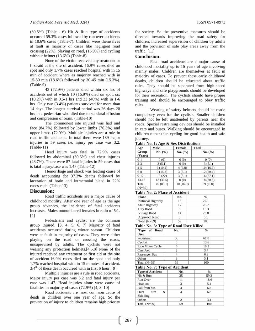
Table No. 1: Age & Sex Distribution Age
Group
(Years)
Male Female Total
No. (%) No. (%) No. (%)
0-1 0 (0) 0 (0) 0 (0)
2-3 3 (5.1) 0 (0) 3 (5.1)
4-5 6 (10.2) 4 (6.8) 10 (16.9)
6-8 9 (15.3) 3 (5.1) 12 (20.4)
9-12 13 (22) 3 (5.1) 16 (27.1)
13-16 18 (30.5) 0 (0) 18 (30.5)
Total (N=59)
49 (83.1) 10 (16.9) 59 (100)
Table No. 2: Place of Accident
Table No. 3: Type of Road User Killed Type of Road
User
No. %
Pedestrian 36 61.0
Cyclist 8 13.6
Ride Motor Cycle 6 10.2
Cars Jeep 2 3.4
Passenger Bus 4 6.8
Others 3 5.1
Total (N=59) 59 100
Table No. 7: Type of Accident Type of Accident No. %
Hit & Run 35 59.3
Run Over 11 18.6
Head on 3 5.1
Fall from bus 4 6.8
Over turn & Skidding
4 6.8
Others 2 3.4
Total (N=59) 59 100
Place No. %
National Highway 16 27.1
State Highway 17 28.7
City Road 9 15.3
Village Road 14 23.8
Approach Road 3 5.1
Total (N=59) 59 100

J Indian Acad Forensic Med, 32(4) ISSN 0971-0973
288
Table No. 4: Seasonal Variation Seasons No. %
Winter Seasons 32 54.2
Summer Seasons 19 32.2
Raining Seasons 8 13.6
Total (N=59) 59 100
Table No. 5: Time of Accident Time of Accident No. %
6-8 A.M 3 5.1
8-10 A.M 9 15.3
10-12 A.M 7 11.9
12-2 P.M 16 27.1
2-4 P.M 11 18.6
4-6 P.M 6 10.2
6-8 P.M 6 10.2
8-10 P.M 1 1.7
10P.M. - 6 A.M 0 0
Total (N=59) 59 100
Table No. 6: Vehicles Responsible for Accident Responsible
Vehicles
No. %
Trucks & Buses 23 40.0
Cars & Jeeps 18 30.5
Tractor 7 11.9
Two wheelers 6 10.2
Others 5 8.5
Total (N=59) 59 100
Table No. 8: Cause of Accident Cause of Accident No. %
Negligent Road
Crossing
13 22.0
Playing on Road 10 16.9
Negligent Cycling 8 13.6
Negligent Driving 8 13.6
Over Speeding 4 6.8
Poor Vision/Fog 3 5.1
Standing on Doors/Scoters
7 11.9
Others 6 10.2
Total (N=59) 59 100
Table No. 9: Hospital Survival Period Time to reach Hospital No. %
<15 Min 1 1.7
15-30 Min 11 18.6
30-45 Min 9 15.3
45-60 Min 6 10.2
1-1.5 hrs 9 15.3
1.5-2 hrs 2 3.4
2-2.5 hrs 7 11.9
>3 hrs 4 6.8
Spot Death 10 16.9
Total (N=59) 59 100
Table No. 10: Survival Period Survival Period No. %
0-0.5 hrs 14 23.8
0.5-1 hrs 6 10.2
1-6 hrs 23 40.0
6-12 hrs 3 5.1
12-24 hrs 1 1.7
24-48 hrs 4 6.8
48-72 hrs 1 1.7
3-5 days 3 5.1
5-7 days 0 0
7-14 days 2 3.4
>14 days 2 3.4
Total (N=59) 59 100
Table No. 11: Site of Injury Site of Injury No. %
Head & Face 50 84.7
Neck 3 5.1
Thorax 26 44.1
Abdomen & Pelvis 22 37.3
Upper limb 43 72.9
Lower limb 45 76.3
Total (N=59) 189 Injury /Case =3.2
Table No. 12: Fatal Injuries Site of Fatal injury No. %
Head 43 72.9
Cervical Spine 4 6.8
Chest 17 28.7
Abdomen 18 30.5
Pelvis 3 5.1
Lower limb 2 3.4
Total (N=59) 87 Injury /Case =1.47
Table No. 13: Cause of Death Cause of Death No. %
Hemorrhage & Shock
22 37.3
Laceration of Brain 13 22.0
Intracranial Bleed 13 22.0
Compression of Brain
6 10.2
Respiratory Failure 4 6.8
Rupture of Heart 1 1.7
Total (N=59) 59 100
References: 1. Wilson CG. Accidents and Poisoning in children. In:
Parthsarthy A, Nair MKG, Menon PSN. (Editors). IAP
Textbook of Paediatrics. 3rd Edn. New Delhi: Jaypee
Brothers. 2006:p.971-982 2. Crime in India. National Crime Records Bureau, Ministry of
Home Affairs, Govt. of India. 2007
3. Sonkin B, Edwards P, Roberts I, Green J. Walking, cycling and transport safety: an analysis of child road deaths.
J R Soc Med 2006;99(8);402-405
4. Bannon MJ, Carter YH, Mason KT. Causes of fatal childhood accidents in North Staffordshire 1980-1989. Arch
Emerg Med 1992;9:357-366
5. Sharples PM, Storey A, Aynsley-Green A, Eyre JA. Causes of fatal childhood accidents involving head injury in
northern region 1979-86. BMJ 1990;301(6762):1193-7
6. Durkins MS, Laraque D, Lubman I, Barlow B. Epidemiology and prevention of traffic injuries to urban
children and adolescents. Pediatrics 1999;103(6):74
7. Coupland C, Hippisley-Cox J, Kendrick D, Savelyich B. Severe traffic injuries to children Trent 1992-7: time trend
analysis. BMJ 2003;327(7415):593-594
8. Soreide K, Kruger AJ, Ellingsen CL, Jjosevik KE. Pediatreic trauma deaths are predominated by severe head
injuries during spring and summer. Scand J Trauma Resusc Emerg Med 2009;17:3
9. Suominen P, Kinioja A, Ohman J, Korpela R, Rintala R,
Oikkola KT. Severe and fatal childhood trauma. Injury 1998;29(6):425-30
10. Kanchan T, Menezes RG, Monteiro FN. Fatal
unintentional injuries among young children - A hospital based retrospective analysis. Forensic Leg Med
2009;16(6);307-11
11. Durbin DR. Preventing motor vehicle injuries. Curr Opin Pediatr 1999; 11(6);583-7.
J Indian Acad Forensic Med, 32(4) ISSN 0971-0973
289
Original research paper
Application of Victims‟ Fingernails in Forensic DNA Analysis *Kamoun Arwa, *Mahfoudh Nadia, **Ayadi Adnene, **Hammemi Zouheir, **Maatoug Samir,
**Makni Hafedh
Abstract DNA extracted from the victims‟ fingernails may assist in the identification of the aggressor. Fingernails
were collected from 8 victims, and were subjected to DNA extraction using the Kit « Tissue and Hair Extraction Kit
(Promega) ».
All samples were typed for 15 autosomal short tandem repeats and for amelogenin using the Kit
« Powerplex TM16 system (Promega) » and the ABI Prism 310 DNA sequencer. The profiles obtained were
compared with those achieved by similar typing of victims‟ and suspects‟ blood.
In two Forensic investigations, mixed genotypes were detected in DNA extracted from the nails: Alleles originating
from the victim were coamplified with other alleles that matched the suspect‟s genotypic profile. This indicated that
victims‟ fingernails contained biological material (blood, epithelial cells) originating from the suspect.
Our results confirmed the usefulness of the nails as a specimen for forensic identification of the aggressor.
Keywords: Fingernails, DNA, Victim, STR, Promega, Genotypic Profile, Fingernails, Hair, Tissue
Introduction:
DNA analysis has proven to be a valuable
technique for human identification and for the
resolution of criminal disputes.Human nail material
has been identified as a potential source of biological
material for Forensic DNA testing [1, 2, 3].
Fingernail clippings collected from victims
in assault cases, principally sexual cases, are
occasionally sent to Forensic laboratories as a
possible source of DNA originating from the
perpetrator [4].
The aim of the present study was to optimize
the extraction conditions using the Kit DNA IQTM
system (Promega) in order to identify a foreign
profile in victims‟ fingernails possibly originating
from the perpetrator.
Materials and Methods:
Extraction of DNA Within 8 forensic caseworks, we received
the fingernail clipping of 10 victims of murder (7
males and 3 females).
Corresponding Author: *Dr Kamoun Arwa
Hôpital Hédi Chaker, Route El Ain Km 0.5, 3029
Sfax, Tunisia
E-mail : [email protected]
Tel: 216 98652824, Fax: 216 74248622
** Professor
Service de médecine légale,
EPS Habib Bourguiba,
3029, Sfax, Tunisia
Blood collected from the 10 victims and
from 13 suspects, and bloodstains sampled from the
crime scenes. Samples were stored at -20°C until
DNA extraction.
DNA extraction from blood was carried out
by salting out technique.
Bloodstains and fingernails were extracted with DNA
IQTM
system according to the protocol described by
Promega [5]:
For bloodstains, 150µl of lysis buffer was
added and incubated for 30 mn at 70°C. Lysis buffer
and sample were then transferred to a DNA IQ Spin
Basket seated in a 1.5ml microcentrifuge tube and
Centrifuged at room temperature for 2 minutes at
maximum speed.
DNA was purified by adding 7µl resin. After washing
and drying of the resin, DNA was eluted in 50µl of
elution buffer. The nail clippings received no
treatment before extraction.
DNA extraction from nail clippings was
carried out by a modification of the protocol
described above. In order to minimize the quantity of
endogenous DNA recovered, digestion was done at
room temperature and not at 70°C for a short period
of time ranging from 1 minute to 10 minutes, mixing
was done by a gentle pipetting and not by vortexing.
DNA purification and elution steps were not
modified.
Short Tandem Repeat Amplification And
Typing: The DNA samples (1ng) were amplified
using the powerplex 16 Kit following the
manufacturer‟s recommendations. The amplified
J Indian Acad Forensic Med, 32(4) ISSN 0971-0973
290
products were analyzed using an ABI Prism 310
Genetic Analyzer according to the manufacturer‟s
recommended protocol.
Initial fragment sizing was performed by the
GeneScan Software (Applied Biosystems). Allele
calling was performed by Promega‟s PowerTyperTM
16 macro operating within the genotyper® software
program (Applied Biosystems).
Genetic profiles obtained from the nail clippings
were compared to those typed from blood samples.
Results: In the 1
st Forensic casework, the victim was
a man. The nail clippings digestion was carried out at
room temperature for 10 mn. Genetic profile analysis
revealed evidence for the presence of DNA from the
offender in fingernail clippings of the victim‟s hands,
with allele signal intensities 3 times lower than those
of the victim [figure 1].
In the second investigation, the victim was a
woman. Victim‟s nails were soaked in the lysis buffer
for only 2 mn at room temperature. Amplification
with the powerplex 16TM
system showed a foreign
male genetic profile which could be assigned to the
perpetrator [figure 2].
In the other caseworks, fingernails collected
from 8 victims were digested for only 1mn at room
temperature. Genetic profiles typed were identical to
those obtained from the blood nail donors.
In one investigation, genetic profiles retrieved from 2
bloodstains matched the profile of one suspect,
providing hence evidence for his culpability [figure
3].
Discussion: DNA Identification is often useful in
forensic investigation, since it could originate from
the perpetrator, particularly when the sample is taken
from the victim cadaver. Indeed, sexual assaults or
homicides are often associated with multiple actions
of aggression and defence which may lead to transfer
of DNA containing material: skin epithelial cells,
blood. Therefore, in relevant cases, the analysis is
focused on fingernail clippings or on debris scraped
from underneath nails. [4] Several experimental
studies aimed to develop techniques for foreign DNA
extraction from nails of volunteers having scratched
other subjects.
Oz et al used phenol/chloroform for the
extraction of DNA from the entire fingernail
clippings. [6] Amplification of 4 autosomal STRs
(short tandem repeats) produced a genetic profile
identical to that typed from the buccal swabs of the
same volunteers. They concluded that the routine
fingernail clippings would not contribute essential
information in forensic casework. In fact, when
digesting the entire nails, endogenous DNA would be
relatively abundant and thus preferentially amplified.
When studying the resolving power of the
powerplex 16TM
system, Krenke at al found that only
17% of minor alleles could be detected at a ratio of
1:19. [7]
Gangitano et al optimized a non organic DNA
extraction procedure for fingernail clippings after
scratching. [8]
DNA samples were typed for an STR locus
residing on the Y chromosome: DYS19. The success
rate of typing of the scratched person was 64%. This
strategy based on the identification of haplotypic
markers could be useful only for the exclusion of
male suspects.
Wiegand et al reported that a foreign profile
could be obtained from debris scraped from
underneath nails if removal of particles was carried
out with sufficient care. [9]
Cline et al developed a technique for
isolating and purifying foreign DNA in fingernail
clippings [10]: human test nails were heavily coated
with mouse liver and allowed to dry several days. A
one hour H2O/EDTA (ethylenediaminetetraacetic
acid) soak of contaminated nail clippings released
only exogenous DNA. The presence or absence of
each species DNA was confirmed through
mitochondrial DNA amplification using PCR sites
conserved in all mammals.
To date, genetic identification of foreign
DNA in fingernail clippings was successful in 2
cases. In the 1st report, debris from the fingernails of
the suspect were scraped out with a plastic spatula
and extracted with Chelex 100. Amplification with
the pentaplex kit genRES MPX revealed alleles at all
loci which could be assigned to the victim. [11]
In the 2nd
case, a 2 years old micro-
bloodstain under the fingernails of a victim was
scraped. A mixed DNA sample from both the victim
and the scraped person was recovered. [9]
In our report, we extracted DNA from the
entire fingernail clippings of 10 victims by DNA
IQTM
system. We used mild digestion conditions
(shorter incubation time, room temperature, gentle
mixing) in order to minimize victim‟s epithelial cells‟
lysis. We succeeded to identify a foreign DNA
pattern from fingernail clippings in 2 cases. In the
other caseworks, failure to identify additional alleles
could be attributed to the absence of foreign
biological material in the victims‟ fingernails.
Conclusion: Our study underlines the value of the genetic
analysis of fingernails in forensic investigations. In
fact, victims‟ fingernails may contain biological
material which could possibly originate from the
perpetrator: body fluids scratched epithelial cells.
Digestion conditions must be optimized to minimize
extraction of victim‟s DNA.
J Indian Acad Forensic Med, 32(4) ISSN 0971-0973
291
References: 1. Tahir MA, Watson N. Typing of DNA HLA-DQ alpha
alleles extracted from human nail material using polymerase chain reaction. J Forensic Sci. 1995;
40(4):634-6.
2. Anderson TD, Ross JP, Roby RK, Lee DA, Holland
MM. A validation study for the extraction and analysis
of DNA from human nail material and its application to
forensic casework. J Forensic Sci. 1999; 44(5):1053-6.
3. Tahir MA, Balraj E, Luke L, Gilbert T, Hamby JE,
Amjad M. DNA typing of samples for polymarker, DQA1, and nine STR loci from a human body exhumed
after 27 years. J Forensic Sci. 2000; 45(4):902-7.
4. Keating SM, Allard JE. What's in a name?--Medical samples and scientific evidence in sexual assaults. Med
Sci Law. 1994; 34(3):187-201.
5. DNA IQ™ System- Small Sample Casework Prototcol. Madison (WI): Promega Corporation; 2002.
6. Oz C, Zamir A. An evaluation of the relevance of
routine DNA typing of fingernail clippings for forensic casework. J Forensic Sci. 2000; 45(1):158-60.
7. Krenke BE, Tereba A, Anderson SJ, Buel E,
Culhane S, Finis CJ, et al. Validation of a 16-locus
fluorescent multiplex system. J Forensic Sci. 2002;
47(4):773-85.
8. Gangitano DA, Garófalo MG, Juvenal GJ, Budowle
B, Padula RA. Typing of the locus DYS19 from DNA
derived from fingernail clippings using PCR Concert
rapid purification system. J Forensic Sci. 2002; 47(1):175-7.
9. Wiegand P, Bajanowski T, Brinkmann B. DNA typing of debris from fingernails. Int J Leg Med. 1993;
106:81-83.
10. Cline RE, Laurent NM, Foran DR. The fingernails of Mary Sullivan: developing reliable methods for
selectively isolating endogenous and exogenous DNA
from evidence. J Forensic Sci. 2003; 48(2):328-33. 11. Lederer T, Betz P, Seidl S. DNA analysis of fingernail
debris using different multiplex systems: a case report.
Int J Legal Med. 2001; 114(4-5):263-6.

J Indian Acad Forensic Med, 32(4) ISSN 0971-0973
292
Original research paper
A Two-year Burns Fatality Study
*Rahul Chawla, **Ashok Chanana, **Hukumat Rai, ***A. D. Aggarwal, ****Harnam Singh,
*****Gaurav Sharma
Abstract A severe burn injury is the most devastating injury a person can sustain and yet hope to survive. It is a
common catastrophe today as burn injury cases are one of the common emergencies admitted to any hospital. There
are several social, economic, cultural and psychological factors interplaying which influence the reporting,
treatment, management and if the patient dies the further investigations.As the etiological factors of burn injuries
vary considerably in different communities, careful analysis of the epidemiological features in every community is
needed before a sound prevention programme can be planned and implemented. When stratified by age, more
females were found in most age groups. Most burns were domestic, with cooking being the most prevalent activity.
The maximum incidence of burn injuries in males were noted in the age group of 21-30 years. 56% cases who
suffered burns were housewives.26% females had 91-100% burns. Smell of kerosene was present in 4% cases.
Maximum burns were of 3rd
degree with 28% males and 54% females. Head & neck were involved in 94% cases
Extremities were involved in all cases.
Key Words: Burns, Fatal, Fire, Dowry, Death
Introduction: Fire has been known to mankind for about
400,000 years. Although the use of fire was known to
ancient man, it is probably the potential fury of an
unharnessed fire that made man bow before it. India
has an ancient culture where fire was worshiped since
the civilization started. Along with water (jal), air
(vayu), earth (prithvi), fire (agni) is perceived as one
of the four basic components of universe. [1]
Burns constitute a major cause of death and
morbidity whatever reason may be, in the world and
in this country too. Burns always have posed a threat
to the sensitive human body. An accurate estimate of
incidence of burns is going to be difficult to obtain
for the huge and diversely composed population of
this country.
Corresponding Author: *Assistant Professor,
Forensic Medicine, MM Institute of Medical
Sciences & Research, Mullana
E-mail: [email protected]
**Additional Professor
Govt. Medical College, Amritsar
***Assistant Professor
Pt.B.D.Sharma PGIMS, Amritsar
****Associate Professor
Medical College, Muzaffarnagar
*****Associate Professor
Katuri Medical College, Guntur
The loads of overpopulation, illiteracy, poor
standards of safety at home and in the industry
further add to overwhelming rise in the burn
incidents. As everywhere else, the modes of
sustaining burn injuries in India are the same i.e.
flames, scalds, electrical and thermal. The most
common cause of flame burns is accident. [1]
Undoubtedly a severe burn is the most
devastating injury a person can sustain and yet hope
to survive. In the United States, there are
approximately 2 million thermal injuries every year
and 130,000 of them necessitate hospital admission.
Approximately 10,000 to 12,000 of these individuals
die as a result of thermal injury annually. [2]
Material and Methods: The study consisted of 50 cases alleged to
have died of burns and brought to mortuary attached
to the Department of Forensic Medicine and
Toxicology, Government Medical College, Amritsar
from May 2004 to July 2005.
All the 50 cases were first thoroughly
examined for noting demographic details and other
relevant observations. The information was collected
from accompanying relatives, hospital records, and
police papers to ascertain the incidence, manner and
circumstances of burns. The external and internal
findings of burns on autopsy were noted along with
the examination of clothes.
Observations: The present medico-legal study of burns in
50 cases was conducted on dead bodies brought in
the Department of Forensic Medicine & Toxicology,

J Indian Acad Forensic Med, 32(4) ISSN 0971-0973
293
Govt. Medical College, Amritsar with effect from
May 2004 to July 2005.
Age and sex wise distribution of burns is
depicted in table no. 1 and figures I & II. Out of 50
study cases, 36% cases belonged to males which
included one case of eunuch who was a castrated
male. For all practical purposes, this case was
discussed as a male in the study. 64% cases belonged
to females.
The maximum incidence of burn injuries in
males were noted in the age group of 21-30 years i.e.
12% and minimum cases were reported in the age
group of 1-10 years i.e. 2% and no case was observed
in the category of 0-1 years. The maximum incidence
of burn injury in females was noted in the age group
of 21-30 years i.e. 52% and minimum cases were
reported in age group of above 60 years and no case
was reported in the age group 0-1 years. The
minimum age to suffer burns was 2 years and
maximum age was 64 years.
56% cases who suffered burns were
housewives and 6% females were labourer. In males,
16% were labourer, 8% were businessmen, 6% were
doing private jobs and 2% were students. In females,
4% were students. (As shown in table no. 3 and
figure no. IV).
Maximum percentage of burns was seen in
females as compared to males in 26% cases. In
males, maximum, 10% cases, suffered burns to the
extent of 0-50%, followed by 8% cases suffering
burns to the extent of 81-90%. In females, maximum,
26% cases fell in the category of 91-100%. Equal
numbers of cases, 6% each, were charred. (As shown
in table no. 7 and figure no. V).
72% cases were non-smokers and non
alcoholics. 4% females were only bidi smokers. 2%
males were only alcoholic. 22% males were both
smokers and alcoholics. (Table no. 4).
In 78% cases, bodies were devoid of clothes
and in 22% cases, burnt clothes were intact. Smell of
kerosene was present in 4% cases. (Table no. 5).
1st degree burns were suffered by 6% cases
in male and 8% cases in females. 2nd
degree burns
were seen only in females in 4% cases. Maximum
burns were of 3rd
degree in which 28% males and
54% females sustained burns. (Table no. 6)
Head & neck were involved in 94% cases
and spared in 6% cases. Chest & abdomen were
involved in 92% cases and spared in 8% cases.
Extremities were involved in 100% cases. Genitalia
were involved in 50% cases. (As per table no. 8)
Table 2: Area Wise Incidence and Distribution of
Burns Area No. %
Rural 14 28%
Urban 36 72%
Total 50 100%
Table 1: Age and Sex Wise Distribution of Burns Age (Years) Males Females Total
No. (%) No. (%) No. (%)
0-1 0 (0) 0 (0) 0 (0)
1-10 1 (2) 2 (4) 3 (6)
11-20 2 (4) 4 (8) 6 (12)
21-30 6 (12) 20 (40) 26 (52)
31-40 3 (6) 3 (6) 6 (12)
41-50 4 (8) 1 (2) 5 (10)
51-60 2 (4) 1 (2) 3 (6)
> 60 0 (0) 1 (2) 1(2)
Total 17 (36) 32 (64) 50 (100)
Figure I: Sex Wise Distribution of Burns
Figure II: Age and Sex Wise Distribution of Burns
Figure III: Area wise Distribution of Burns
Table 3: Incidence and Distribution of Occupation
of Burn Cases Occupation Male Female
No. (%) No. (%)
House wife 0 (0) 28 (56)
Labourer 9 (18) 3 (6)
Business 4 (8) 0 (0)
Student 1 (2) 2 (4)
Private job 3 (6) 0 (0)
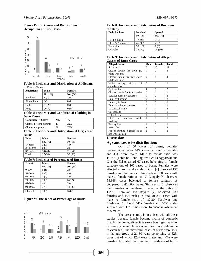
J Indian Acad Forensic Med, 32(4) ISSN 0971-0973
294
Figure IV: Incidence and Distribution of
Occupation of Burn Cases
Table 4: Incidence and Distribution of Addictions
in Burn Cases. Addictions Male Female
No. (%) No. (%)
Smoking 0 (0) 2 (4)
Alcoholism 1(2) 0 (0)
Both 11(22) 0 (0)
None 36(72) 0 (0)
Table 5: Incidence and Condition of Clothing in
Burn Cases Condition Of Cloths No. %
Clothes present & burnt 11 22%
Clothes not present 39 78%
Table 6: Incidence and Distribution of Degrees of
Burns Type Male Female
No. (%) No. (%)
1st degree 3 (6) 4 (8)
2nd degree 0 (0) 2 (4)
3rd degree 14 (28) 27 (54)
Total 17 (34) 33 (66)
Table 7: Incidence of Percentage of Burns Extent Male Female
No. (%) No. (%)
0-50% 5 (10) 2 (4)
51-60% 0 (0) 4 (8)
61-70% 2 (4) 2 (4)
71-80% 1 (2) 6 (12)
81-90% 4(8) 2 (4)
91-100% 3(6) 13 (26)
Charred 3 (6) 3 (6 )
Figure V: Incidence of Percentage of Burns
Table 8: Incidence and Distribution of Burns on
the Body Body Regions Involved Spared
No. (%) No. (%)
Head & Neck 47 (94) 3 (6)
Chest & Abdomen 46 (92) 4 (8)
Extremities 50 (100) 0 (0)
Genitalia 25 (50) 25 (50)
Table 9: Incidence and Distribution of Alleged
Causes of Burn Cases Alleged Causes Male Female Total
Stove burst 6 13 19
Clothes caught fire from gas
while working
0 2 2
Clothes caught fire from stove while working
0 4 4
While saving victims of
cylinder blast
0 1 1
Cylinder blast 4 1 5
Clothes caught fire from candle 0 1 1
Suicidal burns by kerosene 0 2 2
Burnt by husband 0 2 2
Burnt by in laws 0 1 1
Burnt by a known person 0 1 1
To conceal crime 1 1 2
Gas leakage 0 1 1
Fall into fire 1 0 1
Blast of machine while
working
1 0 1
Factory fire 1 0 1
House fire 2 2 4
Fall of burning cigarette in to
bed while asleep
2 0 2
Discussion:
Age and sex wise distribution: Out of 50 cases of burns, females
predominates males. 64% cases belonged to females
and 36% were males. Male to female ratio was
1:1.77. (Table no.1 and Figures I & II) Aggarwal and
Chandra [3] observed 67 cases belonging to female
category out of 100 cases of burns. Females were
affected more than the males. Doshi [4] observed 157
females and 143 males in his study of 300 cases with
male to female ratio of 1:1.17. Ganguly [5] observed
58.34% cases belonged to female category as
compared to 41.66% males. Sinha et al [6] observed
that females outnumbered males in the ratio of
1.25:1. Haralkar and Rayate [7] observed 239
females and 104 males in total of 343 cases with
male to female ratio of 1:2.30. Naralwar and
Meshram [8] found 64% females and 36% males
suffered with 1.76 times more frequent involvement
of females.
The present study is in unison with all these
studies, because female become victim of domestic
fire. In the home, either it is stove burst, gas leakage,
or wearing loose clothes which are more vulnerable
to catch fire. The maximum cases of burns were seen
in the age group of 21-30 years comprising of 52%
cases out of which 12% were males and 40% were
females. In males, the maximum incidence of burns
J Indian Acad Forensic Med, 32(4) ISSN 0971-0973
295
was seen in age group of 21-30 years comprising of
12% cases followed by 41-50 years comprising of
8% cases. In the age group of 31-40 years, 6% cases
were reported. In the category of 11-20 years and 51-
60 years, 4% cases were reported in each
respectively. 2% cases were observed in the category
of 1-10 years. No case was reported in the category
of 0-10 years and above 60 years. In females, the
maximum incidence of 40% cases was observed in
the age group of 31-40 years followed by 8% cases in
the age group of 11-20 years. 6% and 4% cases each
were observed in the age group of 31-40 years and 1-
10 years respectively. 2% cases each were observed
in the age group of 41-50 years, 51-60 years and
above 60 years respectively. No case was reported in
the age group of 0-10 years.
Doshi [4] found male to female ratio of
1:0.88 in age group of below 15 years and 1:1.17 in
age group of 15-25 years. Sinha et al [6] found the
boys were affected more than the girls, while in the
next group; subsequently females dominated males
probably due to their engagement in cooking in
kitchen. In 3rd decade, there was not much difference
in sex incidence. Sharma et al [9] observed that out of
110 cases, 33 belonged to 0-10 years, 28 in 21-30
years, 12 in 31-40 years, 3 in 41-50 years, 4 in 51-60
years and 3 in 61-70 years. Aggarwal and Chandra
[3] observed 31 deaths in age group of 11-30 years
and 13 deaths in 31 to 40 years. Majority of them
belonged to 2nd and 3rd decade i.e. 67 cases.
Females were double the number of males and were
in their 2nd and 3rd decade. Haralkar and Rayate [7]
found the maximum number of burn cases i.e. 156
(45.48%) belonged to age group between 15 and 25
years. Minimum number of patients i.e. 61 (17.28%)
were in the age group between 35-45 years.
The present study is in consistence with the
studies of above mentioned authors in respect to
preponderance of female sex and age groups due to
involvement of females in kitchen work, even in
younger age and early marriages in society, clothing
pattern, few suicidal and dowry deaths are also
reported in this age.
Area-wise distribution: In present study, urban habitat comprising of
72% cases predominately the rural habitat in 28%
cases. (Table 2 and Figure III) However, Sinha et al
[6] observed high incidence in rural habitat. Haralkar
and Rayate [7] observed the rural preponderance
probably due to style of living and low socio-
economic status. Use of shegadi, chulah, stove for
cooking was seen more in rural than in urban areas.
Punjab is a developed state and has lot of industry.
There is great rush of migratory population in the
urban areas who still use stoves in the kitchen and
majority of cases reported belong to poor
socioeconomic strata females catching fire.
Occupation:
In the present study, housewives
predominated comprising of 56% cases other
occupations in females, 6% cases of laborers and 4%
cases were students. In males, the category of
laborers comprising of 18% cases predominated
followed by 6% cases of private jobs, 8% case of
businessmen. (Table 3 and Figure IV) Aggarwal and
Chandra [3] observed that all the females of 3rd
decade and some of 2nd decade were housewives.
Haralkar and Rayate [7] observed in their study of
343 burn cases admitted at General Hospital, Solapur,
that 49.85% were housewives, 6.2% agri-labourers,
10.2% non agri-labourers, 3.5% own business and
unemployed 11.08% and doing no work were
18.06%. The present study was in consistence with
studies of above mentioned authors due to
involvement of females in the kitchen work.
Addiction: In the present study 4% females were bidi
smokers. In males 22% were both smokers and
alcoholics and 2% were alcoholics (Table 4). Despite
high rates of addiction in this part of country, only
22% males were both smokers and alcoholics and 2%
were alcoholics. 4% females were bidi smokers. The
alleged cause of burns as a result of fall of burning
cigarette into bed while asleep was only 2 cases out
of 50 study cases. None of the case showed the
presence of alcohol on autopsy. In the study of Leth
et al [10], 51% of house fire deaths were due to
tobacco smoking, often in combination with alcohol
intoxication or handicap. Merley and Baker [11]
observed that more than half of the deaths resulted
from cigarette ignited fires though 39% of people
who died in such fires were not cigarette smokers
themselves. Gormsen et al [12] in 169 autopsy cases
found that more than half of fire victims had alcohol
exceeding 0.05%. The present study was not in
consistence to the studies in western world. In the
study of Parks et al [13] falling asleep while smoking
was one of the major etiologic factor and substance
abuse were seen in 25% cases. In the current study,
though domestic fire dominated in female victims,
but fire due to alcohol or smoking cigarettes was
negligible. This was due to Punjabi culture, where
addiction to these agents is negligible.
Clothing and accelerant: In the present study, body was devoid of
clothings in 78% cases and in 22% cases burnt
clothes were intact. Smell of kerosene was present in
4% cases. (Table 5) Sukhai et al [14] observed the
use of accelerant in 76.8% cases and paraffin was
preferred. In the study of Parks et al [13], gasoline
was the commonest solvent involved in burn
fatalities. Betz et al [15] observed in 18 out of 21
cases, use of gasoline as accelerant. Current study
also points out use of kerosene and its detection only

J Indian Acad Forensic Med, 32(4) ISSN 0971-0973
296
in 4% cases as majority had been treated in the
hospital and wounds were cleaned and in other cases,
fire was due to domestic gas or factory fire and there
were hardly any clothes for evidence of combustible
material.
Degree of burns:
In the present study, maximum burns were
of 3rd degree (Wilson) in which 28% males and 54%
females sustained burns. (Table 6) Betz et al [15] the
predominance of 3rd and 4th degree burns in his
21cases study. Stefan [16] in his study observed that
the depth of burns has no relation with the fatality,
rather burns of 2nd and 3rd degrees of 57.3% body
surface survived more than 16 days .
Percentage of burns: In the present study, maximum percentage
of burns, 32% cases were in the category of 91-
100%. Only 14% cases had sustained less than 50%
burns. (Table7 and Figure V). In the study of
Aggarwal and Chandra [3], percentage of burns was
up to 25% in 3 cases, between 25-50% in 32 cases,
between 50-75% in 23 cases and was more than 75%
of surface area in 42 cases. Maximum deaths due to
burns were because of surface area involved in the
burn injury. Sukhai et al [14] observed the mean age
burn surface area of 63.3% leading to death
irrespective of depth. Betz et al [15] in his study of
suicidal cases, the extent of burns ranged from 50%
to 100% of body surface. Similar observations were
seen in the current study as observed by Sukhai et al
[14], Betz et al [15]. It is also concluded that it is the
percentage of body surface area which decides death
due to burns. Minimum percentage of area leading to
burn death in the present study was 50%.
Distribution of burns: In the present study, extremities were
involved in 100% cases, followed by head and neck
in 94% cases, chest and abdomen in 92% cases,
genitalia in 50% cases. (Table 8) Cases in which
percentage of burns was above 90% involving head,
neck, trunk or extremities died within 24 hours. In the
study conducted by Datey et al [17], it was observed
that maximum cases had burns involving limbs and
trunk, next in order was involvement of head, face &
neck and genitalia. Mcindoe [18] found that burns of
the trunk and head were more serious to life than
burns of extremities and burns of the flexures and
external genitalia carried a bad prognosis, if not to
life then to health. Similar observations were
observed in the current study.
Alleged causes of Burns: In the present study, alleged cause of death
was stove burst in 6% males and 13% females.
(Table 9) Suicidal burns by kerosene were seen only
in 2% cases all of which were females. In the study
of 100 cases of Aggarwal and Chandra [3],
commonest mode of committing suicide was by
sprinkling kerosene oil over the body and setting
them to fire. In 11 cases who committed suicide, 8
cases were of females, out of which in 2 cases, illness
and domestic quarrel were responsible for this act
and 3 cases were males. In rest 6 cases the motive
was not known. In the present study, 4 cases resulted
from house fire.
Aggarwal and Chandra noted 2 out of 100
cases where jhuggi caught fire accidentally. In the
present study 1 case of a female where the body was
burnt after killing by poisoning and I cases of a male
where the body was burnt after killing by ligature
strangulation. Suarez-Penarando [19] studied two
cases of homicidal ligature strangulation with
extensive burning of bodies. Haralkar and Rayate [7],
in his study observed that burns were more common
in housewives than other occupation because
housewives were more exposed to injury prone
environment while cooking. Cooking at floor level,
use of kerosene pressure stove, wearing of loose
clothes such as sarees, dupattas made them more
prone for burn injuries. Narlawar and Meshram [8]
observed that kerosene stove flames was major cause
of burn due to which females burned 2.04 times more
frequently than males. Domestic fire was the main
reason of burn injuries.
Conclusions: Despite the modernization, the domestic fire
is the major cause of burns with maximum
involvement of females and the stove burst, being the
main cause. Dowry deaths, curse to our so-called
modern society, are still prevalent, in spite of
stringent laws and amendments in the acts. As this
problem of thermal deaths persists in our country, the
government along with various working groups and
bodies need to come together with more sincere
efforts so as to minimize burn mortality and also to
prevent and reduce their incidence.
References: 1. Sawhney CP, Ahuja RB, Goel A. Burns in India:
Epidemiology and Problems in Management. Ind J Burns 1993; 1(1): 1-4.
2. Smith JW, Aston SJ. Thermal and Electrical injuries. Grabb
and Smith‟s Plastic Surgery. 4th Ed. Boston: Little Brown. 1991: p. 675.
3. Aggarwal BBL, Chandra J. A Study of Fatal Cases of
Burns in South Zone Delhi. Punjab Med J 1970; 20(12): 451. 4. Doshi AJ. Aetiology of Burns. Curr Med Pract 1976; 20:
316-320.
5. Ganguli AC. Burns. J Ind Med Assoc 1976; 67: 150-152. 6. Sinha JK, Khanna NN, Tripathi K. Etiology and
Prevention of Burns. Ind J Surg 1976; 38: 82.
7. Haralkar SJ, Rayate M. Socio-demographic profile of burn cases in the reproductive age group (15-45 years) admitted in
SCSM General Hospital, Solapur. Solapur Med J 2005; 2(2):
3-9. 8. Narlawar UW, Meshram FA. Epidermiological
determinants of burns and its outcome in Nagpur. Milestone J
DMER 2002; 2(1):19. 9. Sharma BK, Seth KK, Dharkia RS. Burn injuries and their
prevention. J Ind Med Assoc 1978; 71: 202-205.

J Indian Acad Forensic Med, 32(4) ISSN 0971-0973
297
10. Leth PM, Gregersen M, Saroc S. Fathal accidents in house
fires. The most significant causes such as smoking and
alcohol abuse, multiplied by four the incidence during the last 40 years. Ugeskalarger 1998; 160(23): 3403-3408.
11. Mierley MC, Baker SP. Fatal house fire in an urban
population. JAMA 1983; 249(11): 1466-1488. 12. Gormsen H, Jeppesen N, Lund A. The cause of death in
fire victims. Forensic Sci Int 1984; 24(2): 107-111.
13. Parks JG, Noguchi TT, Klatt EC. The epidermis of fatal burn injuries. J Forensic Sci 1989; 34(2): 399-406.
14. Sukhai A, Harris C, Moorad RG, Dada MA. Suicide by
self immolation in Durban South Africa; A fine year retrospective review. Am J Forensic Med Pathol 2002; 23(3):
295-298.
15. Betz P, Roider G, Meyer LV, Drasch G, Eisenmenger W. Carboxyhemoglobin Blood Concentrations in Suicides by
Fire. J British Acad Forensic Sci 1996; 36: 313-316. 16. Stefan J. Burn Injuries-contemporary and previous findings.
Sound Lek 2004; 49(4): 57-62.
17. Datey S, Muathy WS, Taskar AD. A study of burn injury cases from three hospitals. Ind J Public Health 1981; 25: 117-
124.
18. McIndone. The General effects of Injury and Hemorrhage. Proc R Soc Med 1940; 34: 56.
19. Suárez-Peñaranda JM, Muñoz JI, López DAB, Vieira
DN, Rico R, Alvarez T, Concheiro L. Concealed homicidal strangulation by burning. Am J Forensic Med Pathol 1999;
20(2): 141-144.

J Indian Acad Forensic Med, 32(4) ISSN 0971-0973
298
Original research paper
Computed Tomographic Studies on Ossification Status of Medial
Epiphysis of Clavicle: Effect of Slice Thickness and Dose
Distribution
*Kaur Gurdeep, *Khandelwal N., **Jasuja O.P.
Abstract The accuracy of technique adopted for Forensic age diagnostics of young adults and adolescents especially
in case of livings lies in the standardization of the technical parameters used. The emerging radiological techniques,
when used in standardized way may minimize the possibilities of misinterpretation, as it has been practically shown
in present study. CT scans of 100 live subjects were performed on 16-slice (Siemen‟s Sensation 16) CT scan
machine and the volumetric data acquired was reconstructed into five separate sets of slice thickness for each one of
the subjects included in the study and the ossification status for each set of slice thickness was determined for all the
subjects separately. The results are almost identical while evaluating ossification stages from 1 and 2 mm thick slice
data but the differences are found in the ossification stages when evaluated using 3 mm,5mm and 7mm slice
thickness as compared those found in 1 and 2mm slice thickness. It was concluded that by increasing slice thickness
the rate of error-nous interpretation are also increasing.
Thus, the minimum reliable thickness to produce high resolution scans in order to get maximum accuracy is
2 mm for staging medial clavicular ossification from CT scan and the reconstruction should be done using kernel
(filter) B60F at window setting osteo (1500/450HU).
Key Words: Forensic, ossification, Clavicle, Computed Tomography, Slice Thickness, Dose Distribution
Introduction: The demand of estimating age of
adolescents and young adults has increased in the
recent years due to the increasing cross border
migration [1]. Age estimation in cadavers, human
remains and living individuals is generally needed to
solve the issues with significant legal and social
ramifications for individual as well as for the
community. The accuracy of the technique to be used
for age estimation especially in case of living
individuals is of utmost importance in both the
situations criminal as well as civil. The newer
radiological techniques proved invaluable inventions
of modern era for diagnostic purpose, may also be
used for age diagnostics in forensic context.
Corresponding Author: **Professor of Forensic Science,
Department of Forensic Science,
Punjabi University, Patiala147002, India
Email: [email protected]
*Department of Radiodiognosis & Imaging,
PGIMER, Chandigarh, India
*Research student
The proper way of using these techniques
has reduced the possibilities of misinterpretation to
the minimum and raised the accuracy level of the
results to the maximum.
The current state of forensic age estimation
of living subjects is mainly considered for the
purpose of criminal prosecution [2]. Forensic age
estimation in living adolescents and young adults
undergoing criminal proceedings is generally
performed to determine whether the defendant of
questionable age has reached the age of criminal
responsibility and whether general criminal law of
adults can be applied[3].
In most countries the age threshold of legal
relevance ranges between 14 to 21 years of age [4].
When it is necessary to prove that the subject has
attained the age of 21 years, an additional x ray
examination or CT scan of clavicle is recommended
along with the recommendation of the “Study Group
On Forensic Age Diagnostics”, which includes
physical examination of the suspect, an x ray of left
hand and dental examination and orthopantogram to
know about the dental status.
Clavicular Epiphyseal ossification is must in
case it is necessary to prove the proband has reached
the criminal liability threshold of 21 years , as the
other system on which the development analysis is
based are fully matured by this time[5]. If bone

J Indian Acad Forensic Med, 32(4) ISSN 0971-0973
299
development of hand has been completed, an
additional radiological examination of the clavicles
should be realized. [6]
Kreitner et al. stated that the ossification
status of medial extremity of clavicle can easily be
assessed by computed tomography as conventional
radiographs which were the basis of analysis in some
comprehensive studies are sub optimal because of the
over lapping of the ribs, vertebrae and mediastinal
shadows. Interpretation of the stages of medial
epiphyseal development is even prevented in some
cases. Conventional tomography is time consuming
and lack clarity .Computed tomography eliminates all
these problems. [7]
Schulze et al [8] has reliably determined the
ossification status of medial epiphysis of clavicle in
556 cases, aged between 15 to 30 years of age using
the classification of stages used by Schmeling et al.
[9].He discussed that partial volume effect in CT
using thicker slice was possible explanation of early
visualization of stage 5(21 years) in his study as
compared to 26 years in the study of Schmeling et al.
and established the effect of slice thickness on
staging the ossification status. Mulher et al [10] has
determined the ossification stages for different slice
thickness 1mm, 3mm, 5mm and 7mm separately in
each one of 40 subjects included in the study to prove
that inadequate choice of slice thickness can lead to
misinterpretation of ossification status.
Schulze et al [11] suggested the use of
reconstruction kernel (filter) suitable for osseous
structure and to view the scan in bone window to
study the status of ossification of medial epiphysis of
clavicle for better visualization.
The method of clavicle examination also
appeared to be significant. For the age interval in
which mature clavicles have been observed, the
predicted probability of being mature clavicles is
greater when X-rays or CT scans are used instead of
dry bone specimens. Inappropriate slice thickness of
CT scan can affect age diagnosis, as an almost
completely fused clavicle may be diagnosed as being
mature when details are lost due to greater thickness.
[12]
Present study is aimed to study the chances
of interpretation errors while defining the ossification
stages of medial epiphysis of clavicle due to technical
parameters selected for scanning and how does the
resolution affect the results. The slice thickness is an
important parameter of CT scanning which depends
on the choice of pitch ratio in helical CT, which in
turn influences z-axis resolutions. The choice of
higher pitch ratio value therefore reduces spatial
resolution performance [13]. The radiation dose (as
expressed in CTDI vol) is inversely proportional to
pitch.
The trade off in increasing pitch is an
increase in effective slice thickness, which in turn
results in increased volume averaging and reduced
image signal (contrast between object to be detected
and background)[14]. Thus the slice thickness
depending upon the choice of pitch ratio value in turn
influences both image quality as well as dose
distribution to the patient and suboptimal quality
scans may lead to interpretation problem while doing
staging to define ossification status, but the dose
distribution to the patient is correspondingly
increased to produce scans of optimal quality at less
slice thickness to increase contrast to noise ratio in
the resultant CT image. [13, 14]
Materials and Methods: CT scans of 100 live subjects (55 males and
45 females) falling in the age group of 12years to 30
years, originally performed for their diagnostic
purpose as contrast enhanced CT chest, neck
angiography, pulmonary angiography and bronchial
artery angiography on 16 slice (Siemen‟s Sensation
16) CT Scan machine, retrospectively analyzed to
find out the ossification status of bilateral medial
clavicular epiphyses at different slice thickness. The CT examination of all the patients were
performed acquiring volumetric data using technical
parameters:
KVp - 120; MA - 140/150; Rotation time -
0.5sec; Pitch – 1.15; slice collimation- 16 x 0.75;
FOV 294 mm; Matrix 512 x 512. The volumetric data
acquired from spiral CT scans performed using above
mentioned parameters reconstructed into axial scans
of slice thickness - 1mm, 2mm, 3mm, 5mm and 7
mm using kernel B-60 (filter suitable for osseous
structures) at window width / window level 1500 /
450 H.U. (osteo).
The respective ossification stage was determined
corresponding to each slice thickness separately for
both side in case of all the subjects involved, using
following classification criteria. [8]
Stage I: Ossification centre not ossified.
Stage II: Ossification centre ossified but
epiphyseal cartilage not ossified.
Stage III : Epiphyseal cartilage partially
ossified. (Fused)
Stage IV : Epiphyseal cartilage completely
ossified, but epiphyseal scar is still
visualized.
Stage V: Epiphyseal scar is no longer
visible.
The radiation dose distributed to the patient
as calculated by the dosimeter inhibited in the
scanner itself while performing the examination was
CTDIvol = 11.54 mGy.
Results: All CT scan images obtained after
reconstruction at different slice thickness permit the
evaluation of ossification status. Following results
show the difference in the ossification stages of the
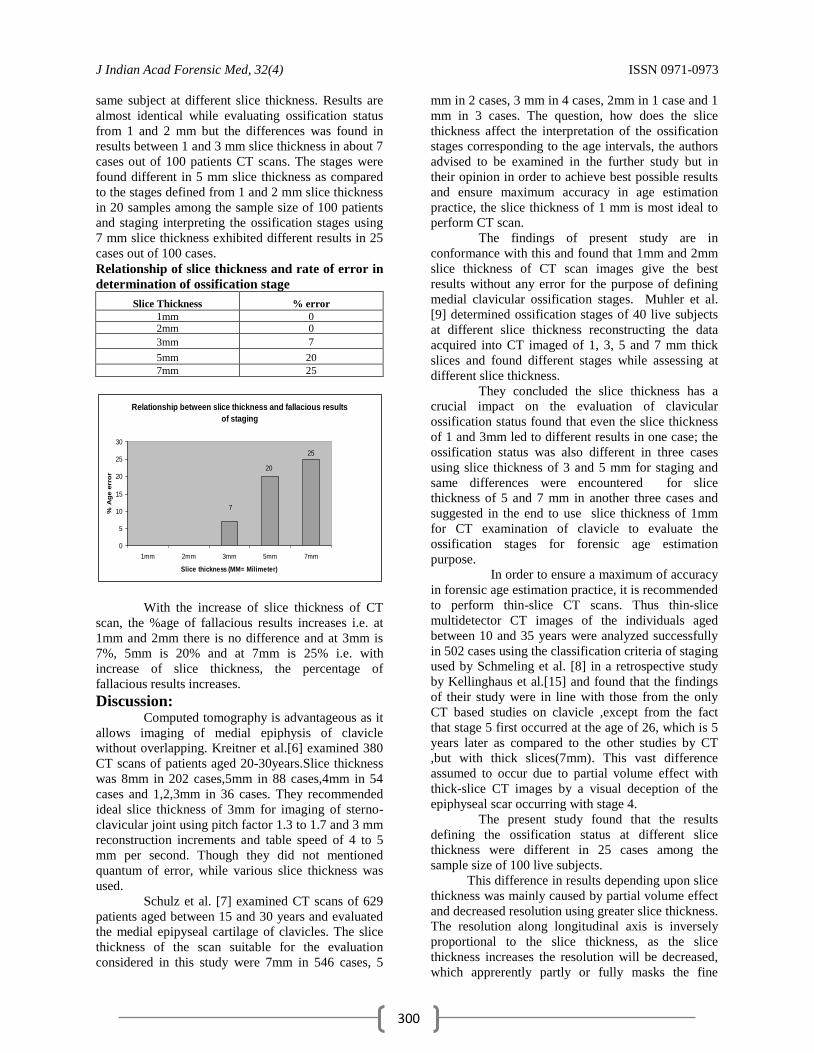
J Indian Acad Forensic Med, 32(4) ISSN 0971-0973
300
same subject at different slice thickness. Results are
almost identical while evaluating ossification status
from 1 and 2 mm but the differences was found in
results between 1 and 3 mm slice thickness in about 7
cases out of 100 patients CT scans. The stages were
found different in 5 mm slice thickness as compared
to the stages defined from 1 and 2 mm slice thickness
in 20 samples among the sample size of 100 patients
and staging interpreting the ossification stages using
7 mm slice thickness exhibited different results in 25
cases out of 100 cases.
Relationship of slice thickness and rate of error in
determination of ossification stage
Slice Thickness % error
1mm 0 2mm 0
3mm 7
5mm 20
7mm 25
Relationship between slice thickness and fallacious results
of staging
0
5
10
15
20
25
30
1mm 2mm 3mm 5mm 7mm
Slice thickness (MM= Milimeter)
% A
ge e
rro
r
7
20
25
With the increase of slice thickness of CT
scan, the %age of fallacious results increases i.e. at
1mm and 2mm there is no difference and at 3mm is
7%, 5mm is 20% and at 7mm is 25% i.e. with
increase of slice thickness, the percentage of
fallacious results increases.
Discussion: Computed tomography is advantageous as it
allows imaging of medial epiphysis of clavicle
without overlapping. Kreitner et al.[6] examined 380
CT scans of patients aged 20-30years.Slice thickness
was 8mm in 202 cases,5mm in 88 cases,4mm in 54
cases and 1,2,3mm in 36 cases. They recommended
ideal slice thickness of 3mm for imaging of sterno-
clavicular joint using pitch factor 1.3 to 1.7 and 3 mm
reconstruction increments and table speed of 4 to 5
mm per second. Though they did not mentioned
quantum of error, while various slice thickness was
used.
Schulz et al. [7] examined CT scans of 629
patients aged between 15 and 30 years and evaluated
the medial epipyseal cartilage of clavicles. The slice
thickness of the scan suitable for the evaluation
considered in this study were 7mm in 546 cases, 5
mm in 2 cases, 3 mm in 4 cases, 2mm in 1 case and 1
mm in 3 cases. The question, how does the slice
thickness affect the interpretation of the ossification
stages corresponding to the age intervals, the authors
advised to be examined in the further study but in
their opinion in order to achieve best possible results
and ensure maximum accuracy in age estimation
practice, the slice thickness of 1 mm is most ideal to
perform CT scan.
The findings of present study are in
conformance with this and found that 1mm and 2mm
slice thickness of CT scan images give the best
results without any error for the purpose of defining
medial clavicular ossification stages. Muhler et al.
[9] determined ossification stages of 40 live subjects
at different slice thickness reconstructing the data
acquired into CT imaged of 1, 3, 5 and 7 mm thick
slices and found different stages while assessing at
different slice thickness.
They concluded the slice thickness has a
crucial impact on the evaluation of clavicular
ossification status found that even the slice thickness
of 1 and 3mm led to different results in one case; the
ossification status was also different in three cases
using slice thickness of 3 and 5 mm for staging and
same differences were encountered for slice
thickness of 5 and 7 mm in another three cases and
suggested in the end to use slice thickness of 1mm
for CT examination of clavicle to evaluate the
ossification stages for forensic age estimation
purpose.
In order to ensure a maximum of accuracy
in forensic age estimation practice, it is recommended
to perform thin-slice CT scans. Thus thin-slice
multidetector CT images of the individuals aged
between 10 and 35 years were analyzed successfully
in 502 cases using the classification criteria of staging
used by Schmeling et al. [8] in a retrospective study
by Kellinghaus et al.[15] and found that the findings
of their study were in line with those from the only
CT based studies on clavicle ,except from the fact
that stage 5 first occurred at the age of 26, which is 5
years later as compared to the other studies by CT
,but with thick slices(7mm). This vast difference
assumed to occur due to partial volume effect with
thick-slice CT images by a visual deception of the
epiphyseal scar occurring with stage 4.
The present study found that the results
defining the ossification status at different slice
thickness were different in 25 cases among the
sample size of 100 live subjects.
This difference in results depending upon slice
thickness was mainly caused by partial volume effect
and decreased resolution using greater slice thickness.
The resolution along longitudinal axis is inversely
proportional to the slice thickness, as the slice
thickness increases the resolution will be decreased,
which apprerently partly or fully masks the fine
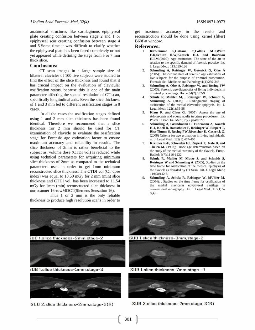
J Indian Acad Forensic Med, 32(4) ISSN 0971-0973
301
anatomical structures like cartilaginous epiphyseal
plate creating confusion between stage 2 and 1 or
epiphyseal scar creating confusion between stage 4
and 5.Some time it was difficult to clarify whether
the epiphyseal plate has been fused completely or not
yet appeared while defining the stage from 5 or 7 mm
thick slice.
Conclusions: CT scan images in a large sample size of
bilateral clavicles of 100 live subjects were studied to
find the effect of the slice thickness and found that it
has crucial impact on the evaluation of clavicular
ossification status, because this is one of the main
parameter affecting the special resolution of CT scan,
specifically longitudinal axis. Even the slice thickness
of 1 and 3 mm led to different ossification stages in 8
cases.
In all the cases the ossification stages defined
using 1 and 2 mm slice thickness has been found
identical. Therefore we recommend that a slice
thickness 1or 2 mm should be used for CT
examination of clavicle to evaluate the ossification
stage for Forensic age estimation factor to ensure
maximum accuracy and reliability in results. The
slice thickness of 2mm is rather beneficial to the
subject as, volume dose (CTDI vol) is reduced while
using technical parameters for acquiring minimum
slice thickness of 2mm as compared to the technical
parameters used in order to get 1mm minimum
reconstructed slice thickness. The CTDI vol (CT dose
index) was equal to 10.50 mGy for 2 mm (min) slice
thickness and CTDI vol has been increased to 11.54
mGy for 1mm (min) reconstructed slice thickness in
our scanner 16-rowMDCT(Siemens Sensation 16).
Thus 1 or 2 mm is the only reliable
thickness to produce high resolution scans in order to
get maximum accuracy in the results and
reconstruction should be done using kernel (filter)
B60f at window.
References: 1. Ritz-Timme S,Cattane C,Collins M.J,Waite
E.R,Schutz H.W,Kaatsch H.J. and Borrman
H.I.M.(2000); Age estimation: The state of the art in relation to the specific demand of forensic practice. Int.
J. Legal Med.; 113:129-136
2. Schmeling A, Reisinger W, Geserick G, Olze A (2005); The current state of forensic age estimation of
live subjects for the purpose of criminal prosecution.
Forensic Sci. Medicine and Pathology;1(4):239-246
3. Schmeling A, Olze A, Reisinger W, and Rosing FW (2003); Forensic age diagnostics of living individuals in
criminal proceedings. Homo 54(2):162-9
4. Schulz R, Muhler M, , Reisinger W, Schmidt S,
Schmeling A. (2008); . Radiographic staging of
ossification of the medial clavicular epiphysis. Int. J. Legal Med.; 122(1):55-58
5. Klaus R. and Claus G. (2005); Assess the age of
Adolescents and young adults in crime procedures. Int. Poster J Dent Oral Med ; 7(2): poster 275
6. Schmeling A, Grundmann C, Fuhrmann A, Kaatch
H-J, Knell B, Ramsthaler F, Reisinger W, Riepert T,
Ritz-Timme S, Rosing FW,R0tzscher K, Geserick G.
(2008) Criteria for age estimation in living individuals.
nt. J. Legal Med.; 122(1):457-460
7. Kreitner K-F, Schweden FJ, Riepert T, Nafe B, and
Thelen M. (1998); Bone age determination based on
the study of the medial extremity of the clavicle. Europ. Radiol; 8(7):1116-1222.
8. Schulz R, Muhler M, Mutze S, and Schmidt S,
Reisinger W and Schmeling A. (2005); Studies on the time frame for ossification of the medical epiphysis of
the clavicle as revealed by CT Scan. Int. J. Legal Med.; 119(3):142-5.
9. Schmeling A, Schulz R, Reisinger W, MUhler M. (2004); . Studies on the time frame for ossification of
the medial clavicular epyphyseal cartilage in
conventional radiography. Int. J. Legal Med.; 118(1):5-
8(4).
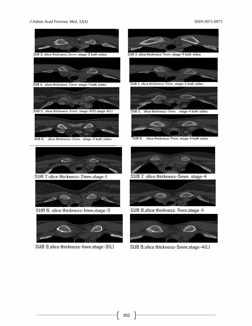
J Indian Acad Forensic Med, 32(4) ISSN 0971-0973
302
J Indian Acad Forensic Med, 32(4) ISSN 0971-0973
303
Original research paper
The Profile of Age in cases of Victims of Sexual Offence
*Dr. Chandresh Tailor, **Dr. Ganesh Govekar, ***Dr. Gaurang Patel, ****Dr. Dharmesh Silajiya
Abstract Sexual assault on female subjects is a global health & human right issue. The problem has legal as well
health related bearing. Female victims, often young girls are the worst sufferers of crime like rape, kidnapping and
trafficking etc... To prove this type of crime, age estimation is most important. The ages of relevance to criminal
liability ranges between 14 and 18 years in most countries. In the present study 141 cases were studied for age
estimation in cases of victims of sexual assault cases at Dept. of Forensic Medicine & Toxicology, Govt. Medical
College, Surat. In accordance with the updated recommendations for age estimation, a physical examination with
determination of anthropometric measures, inspection of signs of sexual maturation, dental examination and X- ray
examination were carried out. Surprisingly we found that appearance and fusion of ossification centers and 3rd
molar
eruption were occurred in few cases at earlier age as contrary to that mentioned in standard literatures.
Key Words: Sexual Assault, Victims, Age Estimation, Rape
Introduction: Estimation of human age is a procedure
adopted by anthropologists, archaeologists and
forensic scientists. Age is one of the essential factors
in establishing the identity of an individual and also
in law, the crime and punishment is entirely based on
the age of a person. In the modern society, the crimes
against the children are increasing. According to
Aggrwal MI & Pathak IC (1957) [1], epiphysis of
bones unites during particular age periods which are
remarkably constant for a particular epiphysis. This is
possible due to complex but dependable system by
which the osseous framework of the body develops,
grows & matures. Epiphysis of the bones unites at a
particular age and this is helpful in age determination.
Determination of age is helpful in both civil and
criminal cases. In the living, age determination is the
most important issue to the court and to the common
citizen as well.
Corresponding Author: *Assistant Professor
Dept. of Forensic Medicine & Toxicology,
Govt. Medical College & New Civil Hospital
Surat- 395001, Gujarat
E-mail Address: [email protected]
Mobile. No.09909914952
**Professor and Head
***Tutor
****Associate Professor
Dept. of Forensic Medicine & Toxicology,
B.J.Medical College, Ahmadabad, Gujarat
Age determination is also important while
taking consent or in cases relating to juvenile
offenders, rape, kidnapping, employment in
Govternment establishments, competency as a
witness, attainment of majority, marriage, fixation of
criminal responsibility, etc. Extensive work on the
determination of age of epiphyseal union has been
carried out in different states of India as well as
abroad and from the finding of various workers, it is
evident that there is not only difference in the age of
epiphyseal union in India and abroad, but also in the
different states of India. These differences may be on
account of varying genetic and epigenetic factors like
climatic, economic and dietetic conditions. Among
many factors used for age estimation, none has
withstood the test of time which necessiciates the
continuous work on this vital issue by the medico
legalists. The present study was undertaken
retrospectively for determination of age in cases of
victims of sexual offence coming particularly to the
Forensic Medicine & Toxicology Dept. for age
determination.
Material & Method: The cases of victims of sexual assaults
registered under Sec. 376 IPC, Sec.361 IPC, and Sec.
362 IPC, Sec. 366 IPC, Sec. 377 IPC were brought
for estimation of age to the Of Forensic Medicine &
Toxicology department, Govt. Medical College &
New Civil Hospital, Surat. Total 141 cases of victims
of sexual offences were studied which were of period
from January 2006 to June 2009. The age estimation
was done first by general emblem. In the general
emblem, general information of victim was taken i.e.
J Indian Acad Forensic Med, 32(4) ISSN 0971-0973
304
name, age, sex, residence, education, occupation,
requested by, brought by, date, place & time of
examination. The age was asked from the victim of
sexual offence which was verified either by parents
who come along with the victim or from birth proof.
Proper written consent for age estimation was taken
from the victim & signature was taken. Patients were
examined in the presence of female witness, &
signature of witness was also taken. Two
Identification marks of victim were noted & history
of case was also recorded.
The determination of age of victim was done
on the basis of:
1. Physical examination: in which general
examination was done by measuring height,
weight, signs of puberty, like development
of pubic hair, Axillary hair, development of
breast, onset of menarche.
2. Dental examination in which total no of
teeth, type of teeth, presence of 3rd
molar
and space for 3rd
molar was noted down.
3. Radiological Examination in which
appearance & fusion of various bony centers
was noted. The X-ray performed was simple
machine X-ray and it was read by us. The
opinion of radiologist was not taken into
consideration.
The data thus collected was analysed using Epi-6
software. The result obtained was matched with the
data of the standard textbooks and recent scientific
literatures.
Observations: It was observed that the maximum
percentages of the victims are in the age group of 14-
17 yrs (i.e. 71.6%), the mean age being 16 yr. Among
these 80.9% were from urban population and almost
all (99%) were educated. Almost half (48.9%) were
engaged in household work and about one quarter
(23.4%) were students in different grades. The history
of runaway with the friend, colleague or known
person, mostly consented or on abetment was
common in almost all the cases were brought for age
estimation in the department.
Out of a total of 141 cases, 57 (40%) spent
some hours, but did not stayed for whole day, 58
cases (42%) stayed together for several days, while
26 (18%) lived together for several months after
which either they came back by themselves or were
brought by police, after complaint lodged by their
parents.
The fact of having sexual intercourse was
furnished by 68.7% while they stay together. Sexual
activity with the partners was mostly in the age group
of 14-17 years. The indulgence in sexual activity was
even observed in the age of 9 years ranging upwards
up to the age of 23 years. In majority of the cases
83.7% sexual activity took place with mutual
consent.
Secondary sexual characteristics: 1. Pubic hair: stage 1-5: starting at 11 yrs and
completed at 15 yrs.
2. Axillary hair: Appearing at the age of 14-15
yrs.
3. Breast development: starting at 11 yrs and
completed at 15 yrs.
No discrepancy was observed in above
mentioned criterion on matching these data with the
standard literature [3, 6-13].
Dental examination: Eruption of third molar is very irregular. The
usual age mentioned in literature for its eruption is
17-25 years.
This holds true in the present study also, but
we do depend a lot on its eruption or non eruption to
fix the age. In the present study comprising the age
ranging between 09 to 23 years; 52 (36.8%) cases
were between 17-23 years of age, which reflects that
out of 141 cases examined, 125 (88.6%) did not have
their third molar erupted. It was also observed that
the third molar was seen erupted as early as in 16 yrs
and it was even seen absent in the age of 23 years.
Radiological examination - Ossification
centers‟: Acromian process of Scapula: Age of fusion is 17 –
18 years.
The fusion of tip of acromian process of
scapula was observed as early as at 14 yrs of age in
more than a quarter (26.6%) of cases. It was seen
united in 61% at 15 yrs, in 82% at 16 yrs & in 86% at
17 yrs of age. It was also observed that it was not
fused in 13.88% of cases even at the age of 17 yrs.
Head of Humerus: head, greater & lesser tubercle
fuses with shaft at 19 yrs.
The fusion of head of Humerus with the
shaft was observed as early as, at the age of 14 yrs
and also as not fused, as late at 20 yrs of age. It was
observed that it was fused at the age of 14 yrs in 13%
of cases, 11.4% at 15 yrs, 20% at 16 yrs, 36% at 17
yrs, 43% at 18 yrs, 80% at 19 yrs and 33% at 20 yrs
of age.
Medial Epicondyle of Humerus: Age of fusion 14-
16 years.
The fusion of medial Epicondyle was observed in
nearly half (45.45%) of the cases at age of 13 years.
In 86% cases at 14 yrs, 97% at 15 yrs and in all cases
of 16 & 17 years.
Upper end of radius and ulna: Age of fusion 16-17
years.
The fusion of upper end of radius and ulna at
elbow joint was observed in greater percentage at
quite early age and it differed with what was quoted
in contemporary literature. It was observed to be

J Indian Acad Forensic Med, 32(4) ISSN 0971-0973
305
fused in 54% at the age of 13 years, 80% at age 14
yrs and 93% at the age of 15 years.
Lower end of radius and ulna: Age of fusion 18-19
years.
Similar findings were observed in the fusion
of lower end of radius and ulna. Fusion of lower end
was reported even at the early age of 14 years in 13%
cases. It increased to 35% at the age of 15 years, 55%
at the age of 16 years, 58% at the age of 17 years and
surprisingly low percentage (57%) of fusion was
observed at the stated age, i.e. 18 years. On an
average in 46% the fusion was observed in the cases
examined in the age group of 14 to 18 years.
Iliac crest: Age of Appearance 14 years. Fusion
occurs at age of 18-20 years.
Below 13 yrs of age, iliac crest was not
appeared seen in 100% cases. Between 14-18 yrs age
in 95% cases iliac crest appeared but not fused. After
19 yrs of age iliac crest fuses in all cases.
Tri-radiate cartilage: Age of fusion 14-15 years.
Tri-radiate cartilage was seen to be fused in
45% of the cases in the age of 13 years, whereas it
was found fused in 73% subjects of age 14 years,
which is the age at which it fuses as per the available
literatures. [3, 6-13] In rest i.e. in 27% cases of that
age group it was observed un-fused. Similarly in the
age of 15 years, that is the upper limit of age of
fusion, it was observed fused only in 87% of cases.
Ischial tuberosity: Age of Appearance 15-16 years
and of fusion 20-22 years.
It was said to be more appropriate to
consider the appearance of ossification than fusion
while estimating age by radiological findings. Here
appearance of centre for Ischial tuberosity was
observed in half of the case at early age i.e. 12 years,
27% at age 13 yrs and 45% at the age of 14 years.
More importantly it was found that the centre has not
appeared in one quarter of the cases at the age of 15
years which is the prescribed age of appearance
mentioned in standard literature. [3, 6-13]
Head of Femur: Age of fusion 17-18 years.
Following the trend of observation of fusion
of ossification centers at early age, contrary to what is
mentioned in literatures [3, 6-13] pertaining to Indian
population, head of femur was found fused in two-
third of the cases at very early age of 14 years. At 15
years it was found in 58% of cases and at 16 years in
65.5% of cases. In the prescribed age of fusion, i.e. at
17-18 years it was observed to be fused in 86% of
cases and not in hundred percent.
Few (9.2%) of the subjects (alleged victims
of sexual assault) furnished proof of age, in the form
of birth certificates issued by registering authorities
or school leaving certificates. The documentary
proof failed to match with the age estimated on the
basis of considering physical and secondary sexual
growth, dental examination and Radiological
Examination In 25% Of Cases, Which Is Alarming.
Discussion: Third molar erupts in the age group of 17-25
yrs, as Per the standard literature [3, 6-13] In our
study third molar was seen erupted even at the age of
16 yrs in 3% of case and at the age of 15 yrs in 10 %
of cases. Modi6 claimed that third molar erupted even
in 14 yrs & 15 yrs of age. Sahay[6] found third molar
erupted between 15-16 yrs. Lall and Townsand[5]
found third molar erupted even at the age of 15-16
yrs. Therefore stage of eruption should be noted
during examination. In some rare cases third molar
may not appear until the advanced adult age. It may
appear even after 50 yrs of age.
According to Modi [6], owing to variation in
climatic, dietetic, hereditary and other factors
affecting the people of the different states of India, it
cannot be reasonably expected to formulate a uniform
standard for the determination of the age of the union
of epiphysis for the whole of India.
In our study acromian process of scapula is
fused in 80% of cases at the age of 17 yrs. Some
cases show fusion even at the age of 15 and 16 yr.
According to Galsraun study [6] acromian process
fused in female at the age group of 13-16 yrs.
According to Pillai study [6] and Flecker study [6] it
shows fusion at the age of 17-18 yrs.
In our study we have observed that head of
Humerus fuses with shaft at the age of 19 yrs in 80%
of cases. While at the age of 20 yrs it fuses only in
33% cases, remaining 67% of case show partial
fusion. We have also observed that it shows fusion
even at the age of 14 yrs and 15 yrs in few cases.
According to Galstraun study [6] and Pillai study [6]
it shows fusion at the age of 14-16 yrs. According to
Basu & Basu [6] study it shows fusion at the age of
16-17 yrs. According to Hepworth study it shows
fusion at the age of 17-18 yrs. According to Davies &
Parson [6] it fuses at the age of 19-21 yrs.
In our study we have observed that Medial
Epicondyle of Humerus shows fusion in all cases at
the age of 16-17 yrs. It was observed similar to
Galstraun study [6], Basu & Basu study [6],
Hepworth Study [2], lall & Nut Study, Pillai Study
[6], Flecker study [6] and Franklin study [6].
According to Davies & Parson study [6]it shows
fusion at the age of 20 yrs.
In our study we observed that upper end of
Radius & Ulna fuses at the earlier age contrary to
standard literature. According to Galstraun [6] it
shows fusion at the age of 14-15 yrs., according to
Basu & Basu study [6] and Hepworth study [2] it
shows fusion at the age of 13-14 yrs. Similarly it was
also observed with Flecker study [6], Davies &
Parson study [6] & Franklin study. [6]
Similarly lower end of Radius & Ulna shows
fusion in early age i.e. 14-15 yrs in few cases. After
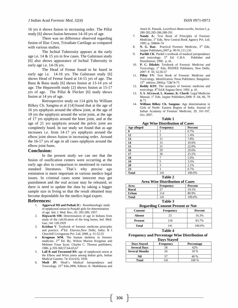
J Indian Acad Forensic Med, 32(4) ISSN 0971-0973
306
16 yrs it shows fusion in increasing order. The Pillai
study [6] shows fusion between 14-18 yrs of age.
There was no difference observed regarding
fusion of Iliac Crest, Triradiate Cartilage as compared
with various studies.
The Ischial Tuberosity appears at the early
age i.e. 14 & 15 yrs in few cases. The Galstraun study
[6] also shows appearance of Ischial Tuberosity in
early age i.e. 14-16 yrs.
The Head of Femur found to be fused in
early age i.e. 14-16 yrs. The Galstraun study [6]
shows Head of Femur fused at 14-15 yrs of age. The
Basu & Basu study [6] shows fusion at 13-14 yrs of
age. The Hepaworth study [2] shows fusion at 15-17
yrs of age. The Pillai & Flecker [6] study shows
fusion at 14 yrs of age.
Retrospective study on 114 girls by William
Bilkey Ch. Sangma et al [14] found that at the age of
16 yrs epiphysis around the elbow joint, at the age of
18 yrs the epiphysis around the wrist joint, at the age
of 17 yrs epiphysis around the knee joint, and at the
age of 21 yrs epiphysis around the pelvic joint are
completely fused. In our study we found that as age
increases i.e. from 14-17 yrs epiphysis around the
elbow joint shows fusion in increasing order. Around
the 16-17 yrs of age in all cases epiphysis around the
elbow joint fuses.
Conclusion: In the present study we can see that the
fusion of ossification centers were occurring at the
early age also in comparison to mentioned in various
standard literatures. That‟s why precise age
estimation is more important in various medico legal
issues. In criminal cases some innocent may get
punishment and the real accuse may be released, so
there is need to update the data by taking a bigger
sample size in living so that the result obtained may
become dependable for the medico legal expert.
References: 1. Aggarwal MI and Pathak IC: Roenthenologic study
of epiphyseal union in Punjabi girls for determination
of age. Ind. J. Med. Res.; 45: 283-289, 1957. 2. Hepworth SM: Determination of age in Indians from
study of the calcification of the long bones, Ind. Med.
Gaz.; 64: 128,1929 3. Krishan V. Textbook of forensic medicine principles
and practice. 4thEd. Elsevier,New Delhi, India: B I
Churchill Livingstone Pvt. Ltd; 2008. p. 51-52,55 4. Krogman WM. The human skeleton in forensic
medicine. 2nd Ed. By, Wilton Marion Krogman and
Mehmet Yasar Iscan. Charles C. Thomas publishers; 1986. p.359-360,57,64-65,67
5. Lall R and Townsend RS: age of epiphyseal union at
the Elbow and Wrist joints among Indian girls, Indian Medical Gazette; 74: 614-616, 1939.
6. Modi JP: Modi‟s Medical Jurisprudence and
Toxicology. 23rd Edn.2006. Editors: K. Mathiharan and
Amrit K. Patnaik, LexisNexis Butterworths, Section I, p.
280-282,285-286,288-291
7. Nandy A.: Text Book of Principles of Forensic
Medicine, 1st Edn, New Central Book Agency Pvt. Ltd.
1995: p. 59&66-74 8. N. G. Rao: Practical Forensic Medicine, 3rd Edn,
Jaypee Publishers,2007,p. 90-91,111,116
9. Parikh CK. Parikh‟s textbook of medical jurisprudence and toxicology. 5th Ed. C.B.S. Publisher and
Distribution; 1990. p. 41. 10. P. C. Dikshit: Textbook of Forensic Medicine and
Toxicology, 1st Edn, PEEPEE Publishers, New Delhi,
2007: P. 50, 52,56-57 11. Pillay VV: Text Book of Forensic Medicine and
Toxicology. Identification. Paras Publishers. Bangalore.
15th edition; 2004:p. 72&74-75 12. Reddy KSN. The synopsis of forensic medicine and
toxicology. 8th Ed.K Saguna Devi; 1992. p. 41.
13. S. S. AGrawal, L. Kumar, K. Chavli: Legal Medicine Manual, 1st Edn, Jaypee Publishers,2008: P. 64, 66, 70
& 71
14. William Bilkey Ch. Sangma: Age determination in Girls of North- Eastern Region of India. Journal of
Indian Acadamy of Forensic Medicine; 29, 101-107,
Oct. 2007.
Table 1
Age Wise Distribution of Cases Age alleged Frequency Percent
9 1 0.7%
12 2 1.4%
13 11 7.8%
14 15 10.6%
15 31 22.0%
16 29 20.6%
17 36 25.5%
18 7 5.0%
19 5 3.5%
20 3 2.1%
23 1 0.7%
Total 141 100.0%
Table 2
Area Wise Distribution of Cases Area Frequency Percent
Rural 27 19.1%
Urban 114 80.9%
Total 141 100.0%
Table 3
Regarding Consent Present or Not
Consent Frequency Percent
Absent 23 16.3%
Present 118 83.7%
Total 141 100.0%
Table 4
Frequency and Percentage Wise Distribution of
Days Stayed Days Stayed Frequency Percentage
Several Days 58 42%
Several Months 26 18 %
Nil 57 40 %
Total 141 100 %

J Indian Acad Forensic Med, 32(4) ISSN 0971-0973
307
Table 5
Comparison of Age corresponding with Proof with Proof of Age
Age Corresponding With Proof
Proof of Age nil No Yes TOTAL
Absent
128
0
0
128
Present
0
3
10
13
TOTAL
128
3
10
141
Table 6
Comparative study of age of appearance of ossification centre
Sr.
No.
Examination
Dental/ Radiological
Eruption/Ossification
As Per the
Standard
Literature &
Scientific
Material
(Age in Yrs)
In Our Study
Also Found at the Early Age
(In Yrs. with percentage)
Age as Per
Literature
(In Yrs. with
Percentage)
1. Dental Examination Age of Eruption 17-25 14(7%), 15(10%) 16(3%)
17(3%), 18(14%)
2. Acromian Process of
Scapula
Age of Fusion 17-18 14(26.6%), 15(61%),
16(82%)
17(86%)
3. Head of Humerus Age of Fusion 19 14(13%), 15(11.4%)
16(20%), 17(36%), 18(43%)
19(80%)
4. Medial Epicondyle of
Humerus
Age of Fusion 14-16 13(45.45%), 14(86%),
15(97%)
16 & 17(100%)
5. Upper end of Radius &
Ulna
Age of Fusion 16-17 13(54%), 14(80%), 15(93%) 16&17(100%)
6. Lower end of Radius &
Ulna
Age of Fusion 18-19 14(13%), 15(35%), 16(55%),
17(58%)
18 & 19(100%)
7. Iliac Crest Age of Appearance 14 - 14(100%)
8. Iliac Crest Age of Fusion 18-20 - 18-20(100%)
9. Tri Radiate Cartilage Age of Fusion 14-15 13(45%), 14(73%) 15(87%)
10. Ischial Tuberosity Age of Appearance 15-16 12(50%), 13(27%), 14(45%) 15(75%)
11. Ischial Tuberosity Age of Fusion 20-22 - 20-22(100%)
12. Head of Femur Age of Fusion 17-18 14(66%), 15(58%), 16(66%) 17-18(86%)
J Indian Acad Forensic Med, 32(4) ISSN 0971-0973
308
Original research paper
Ultrasonographical Age Estimation from Fetal Biparietal Diameter
*Garg A, **Pathak N, ***Gorea RK, ****Mohan P
Abstract In a wide variety of circumstances of feticide, fetal age determination is important for identification. This is
an important identification feature in postmortem cases. There are many different parameters to determine the age
from fetus. But actual problem arises, when the body of fetus is either mutilated or decomposed. The data of femur
length and other variables at various gestational ages can be used for age estimation of fetus at autopsy. But at time
of autopsy each bone may or may not be present for autopsy. So, this study of ultrasonic fetal biparietal diameter
was done to collect data, which can be utilized to ascertain the age of fetus in autopsy cases particularly in the
Punjab region. This regional data can also be used to determine gestation age, if exact last menstrual period date is
not known in antenatal cases of this region or to develop charts for ultrasound dating of pregnancy based on bi-
parietal diameter and, second, to derive reference curves for normal fetal growth based on bi-parietal diameter.
Key Words: Age Determination, Bi-Parietal Diameter, Ultrasonography, Gestational Age, Pregnancy, Ante Natal
Introduction: Measurement of various fetal body parts is
known is fetal biometry. Ultrasonography is done in
every ante natal case for measurement of various fetal
parts. If the exact date of LMP is known then it can
be useful in correlating the bi-parietal diameter with
gestational age.
During fetal autopsy, all the parameters for
identification, may or may not be available
particularly when body is decomposed or mutilated
and sometimes only few bones are available. Then
there is no other option except to find out the age
from bones. Measurement of length of bone and
diameters at autopsy is very easy, less time
consuming and a cheaper way of finding out the age
of the fetus. In many cases we may not have all the
bones. If skull is present then we can measure bi-
parietal diameter and by doing this study, we are able
to provide data in form of charts to solve such cases.
Review of literature: No single parameter is sufficient in giving
accurate fetal age ultrasonographically. Few useful
measurements in the fetus are femur length, length of
kidney [1], abdominal circumference and head
circumference. [2]
Corresponding Author: *Assistant Professor,
Department of Forensic Medicine
E mail: [email protected]
Mobile: +919872402904
** Associate Professor, Obstetric & Gyanecology
*** Professor & Head, Forensic Medicine
**** Professor & Head, Obstetric & Gyanecology
Gian Sagar Medical College, Patiala,
Punjab, India, 140601
If more than one parameter is taken in
determining the age of fetus, it is considered better as
reliance is not kept on one parameter. [2]
Measurement of kidney length is useful
between 24th
to 38th
weeks. [1] Femur length and
BPD have more value prior to 36 weeks but after 36
weeks head circumference and femur length has more
value [2]. Length of femur is also better parameter as
compared to BPD for determination of age of fetus in
the third trimester. [3] Bi-parietal diameter
measurement is in less common use after 20 weeks of
gestation. Measurements of bi-parietal diameter also
help in determination of age of fetus.
The bi-parietal diameter and femur length
correlated equally well with gestational age.
However, the bi-parietal diameter was more than
twice as sensitive as the femur length to variation in
fetal growth. Femur length had a larger error
associated with its measurement. [4]
It is well established that ultrasound
measurement of femur length and bi-parietal diameter
are comparably accurate estimators of gestational age
when obtained in the first half of pregnancy. Both
estimators, however, become less accurate later in
pregnancy. [5]
It is well known that ethnicity has a significant
influence on fetal biometry. [6, 7]
The correlation coefficient of gestational age
versus fetal femur length is statistically greater than
that of the gestational age versus fetal biparietal
diameter. [8]
Measurement of the fetal femur appears to be a
reliable method for assessing gestational age, which
can compensate for the limitations of the BPD
method. [9]

J Indian Acad Forensic Med, 32(4) ISSN 0971-0973
309
Material and Method: The antenatal cases coming to Gian Sagar
Medical College for routine Ultrasonography during
pregnancy will be studied for bi-parietal diameter.
This is a random case study. A total of 271 cases
were taken. Only those cases had been taken where
there was no mistake for last menstrual period
(LMP). Consent of every volunteer was taken. Cases
with fetal congenital and developmental
abnormalities were excluded. During
Ultrasonography, fetal bi-parietal diameter was noted.
LMP of these cases was recorded. We had prepared a
graph from the available data. Then this graph will be
utilized to solve the cases of age determination in
medico legal cases.
Result: With the help of ultrasonic examination of
these volunteer cases, the measured bi-parietal
diameter is presented in tabulated form.
Chart No. 1 shows location based
distribution of cases taken in this study depicted that
most of the cases belong to Punjab state.
Chart No. 2 shows age wise distribution of
cases taken in this study.
Bi-parietal Diameter: Table No. 1 shows the tabulated form of bi-
parietal diameter at different lunar months of
gestational months while Chart No. 3 is showing its
graphical representation. According to present study,
average bi-parietal diameter of skull at 4th
lunar
month of pregnancy is 30.63 mm and maximum at
10th
lunar month is 87.43 mm.
Table No. 2 shows the tabulated form of
biparietal diameter at different weeks of gestational
age while chart 4 is showing its graphical
representation. According to present study, minimum
biparietal diameter of skull at 14 weeks of pregnancy
is 28.30 mm and maximum at 39 week is 93.08 mm.
Discussion: We had prepared the charts or graphs from
the available ultrasonic data of biparietal diameter
especially for Punjab region. Trend line was also
drawn. These trend line and graphs of biparietal
diameter obtained ultrasonically can be used to
determine the age of fetus brought for autopsy. The
expected date of delivery can also be calculated by
using these charts or graphs by getting gestation age
from biparietal diameter obtained from ultrasound in
antenatal cases where last menstrual period is not
known. These charts are more useful in Punjab region
population as these are directly produced from local
population. On the whole, if one variable is known,
we can calculate the other variable from these graphs
and trend line.
Good curves in the graphs were obtained
when horizontal axis becomes lunar month
gestational age [10].
Conclusion: Our new set of reference chart and table for
fetal biometric measurements in reference to
biparietal diameter is ready for Punjabi population
group.
References: 1. Konje JC, Abrams KR, Bell SC, Taylor DJ. Determination
of gestational age after the 24th week of gestation from fetal
kidney length measurements. Ultrasound Obstet Gynecol.
2002 Jun; 19(6): 592-7. PMID: 12047540
2. Hohler CW. Ultrasound estimation of gestational age. Clin
Obstet Gynecol. 1984 Jun; 27(2):314-26. PMID: 6378468
3. Tse CH, Lee KW. A comparison of the fetal femur length
and biparietal diameter in predicting gestational age in the
third trimester. Aust N Z J Obstet Gynaecol. 1984 Aug;
24(3): 186-8. PMID: 6596082
4. Wolfson RN, Peisner DB, Chik LL and Sokol RJ.
Comparison of biparietal diameter and femur length in the
third trimester: effects of gestational age and variation in fetal
growth. J Ultrasound Med. 1986 Mar; 5(3):145-9. PMID:
3517362
5. Egley CC, Seeds JW, Cefalo RC. Femur length versus
biparietal diameter for estimating gestational age in the third
trimester. Am J Perinatol. 1986 Apr; 3(2):77-9. PMID:
3516170.
6. Yeo GS, Chan WB, Lun KC, Lai FM. Racial differences in
fetal morphometry in Singapore. Ann Acad Med Singapore
1994; 23: 371–376. PMID: 7944253
7. Jacquemyn Y, Sys SU, Verdonk P. Fetal biometry in
different ethnic groups. Early Hum Dev 2000; 57: 1–13.
PMID: 7944253
8. Yeh MN, Bracero L, Reilly KB, Murtha L, Aboulafia M,
Barron BA. Ultrasonic measurement of the femur length as
an index of fetal gestational age. Am J Obstet Gynecol. 1982
Nov 1; 144(5):519-22. PMID: 7137237
9. Shalev E, Feldman E, Weiner E, Zuckerman H. Assessment
of gestational age by ultrasonic measurement of the femur
length. Acta Obstet Gynecol Scand. 1985; 64(1): 71-4.
PMID: 3883692
10. Gorea RK, Mohan P, Garg A and Pathak N. Fetal age
determination from length of femur and humerus by
ultrasonography. Medico-legal Update 2009 Jan-June; 9(1):
23-6.
Chart -1
Location Wise Distribution of Cases
4 8
258
10
50
100
150
200
250
300
Bihar Haryana Punjab UP
State of origin

J Indian Acad Forensic Med, 32(4) ISSN 0971-0973
310
Chart No. 2 Showing Age Wise Distribution of Case
Table-1
Showing Average Biparietal Diameter in Relation
to Lunar Months of Pregnancy Gestational Age ( lunar
months)
Average
length(mm)
4 30.63
5 41.76
6 53.93
7 63.65
8 76.50
9 83.21
10 87.43
Chart-3
Showing Average Bi-parietal Diameter in Relation
to Lunar Months of Pregnancy
Table- 2
Showing Average Bi-parietal Diameter in Relation
to Weeks of Pregnancy Gestational
Age (in wks)
Average
BPD (In
mm)
Gestational
Age (in wks)
Average
BPD (In
mm)
14 28.30 27 71.73
15 31.10 28 71.44
16 34.68 29 73.63
17 36.98 30 76.65
18 43.73 31 79.88
19 48.03 32 80.91
20 49.20 33 82.52
21 51.68 34 83.31
22 53.65 35 85.77
23 56.88 36 84.12
24 58.24 37 86.94
25 59.47 38 89.69
26 66.12 39 93.08
Chart-4
Showing Average Bi-parietal Diameter in Relation
to Weeks of Pregnancy

J Indian Acad Forensic Med, 32(4) ISSN 0971-0973
311
Original research paper
Analysis of Railway Fatalities in Central India
* Ramesh NanajiWasnik
Abstract A two year retrospective study of railway related fatal cases has been carried out in the Department of
Forensic Medicine & Toxicology, Indira Gandhi Government Medical College, Nagpur. Railway fatality cases were
stand for 5.99% (Total 173) of all autopsies (n=2888) conducted during the period of January 2001 to December
2002.The fatalities were predominantly seen in the males. Male to female ratio was 8.62:1.The maximum numbers
of victims were in the age group of 20-49 years (n= 120 cases, 69.34%). Majority of victims died on the spot
(96.53%). Maximum number of railway fatalities were accidental (91.32 %) followed by the suicidal (8.68 %) in
nature, whereas none of the homicidal railway fatalities noted. Crush injuries were found mostly over lower limbs
followed by the upper limb then over the face and head. Maximum victims (84.38 %) died due to hemorrhage and
shock following injuries to the vital organs.
Key Words: Train Accident, Railway Fatalities, Safety, Fatal train Injuries, India
Introduction: The injuries and deaths due to accidents are
inescapable in the modern way of living. The
accidental deaths are mostly due to the road traffic
accidents but the deaths due to railway fatalities are
also not negligible, especially in the areas where
railway traffic is higher. A train accident is defined as
a "collision, derailment, or any other event involving
the operation of on-track equipments." Train accidents
can cause devastating damages and personal injuries
including the death of the person. Trains are
frequently involved in accidents that critically injured
passengers and innocent bystanders. These accidents
are indeed disastrous and catastrophic due to the speed
that trains travel. Indeed, a train accident can
definitely result in loss of one‟s life or his or her
property as well.
Nagpur is an important city as well as an
industrial town and the second capital of State of
Maharashtra. It is situated in the central part of India.
As per the sensex 2001, the population of Nagpur city
was 20,51,320 and that of Nagpur district was
40,51,444, of which the peoples residing in urban
area were 64.36 and in rural area were 35.64 %
respectively, these areas are connected very well by
the railway network. Nagpur has rapid growth in
educational establishments, roads, buildings,
business, energy infrastructure, industrialization and
of course population during last 25 years.
Corresponding Author:
*Assistant Professor
Department of Forensic Medicine
Chennai Medical College & Research Centre
Irungalur, Trichy-621105Tamilnadu
E-mail Id: [email protected]
There is also increase in the Gross National
Product (GNP) and improvement in the standard of
living.Being the heart of India, many national railway
routes pass through the Nagpur railway station,
joining the various states around it. Nagpur station is
having the heavy burden of transportation of the
passengers and goods as well; therefore the
incidences of railway fatalities and mishaps are also
higher at this junction. Most of the reported cases of
railway deaths were either directly hit by the train or
recovered in the vicinity of the railway tracks.
Fatal railway injury is characterized by
extensive disruption of more than one body region.
Hence, to properly understand the epidemiology of
railway deaths and the pattern of injuries produced, a
retrospective analytical study has been undertaken.
Material and Methods: The present retrospective study of 173 fatal
cases of train accident has been carried out in the
Department of Forensic Medicine and Toxicology,
Indira Gandhi Medical College, Nagpur for a period
of two years from Jan 2001 to Dec 2002. The data
was obtained from police papers (Requisition and
inquest Panchnama) regarding information on age,
sex, and supposed manner of death, pertinent history
and autopsy report.
It was verified from the friends and relatives of
deceased. The age, sex, types of injuries, pattern and
distribution of fatal injuries were noted. All the data
thus collected were analyzed and findings are
presented in this paper.
Observation: Out of 2888 medico-legal autopsies
conducted during the period of two years from
January 2001 to December 2002, 173 cases (5.99%)
were due to train accidents.

J Indian Acad Forensic Med, 32(4) ISSN 0971-0973
312
Figure1: Month Wise Distribution of Railway
Fatality.
Fatalities by rail occur throughout the year
but uniformity was not observed in the victims of
railway accidents. However, the fatalities were
equivalently seen in all the seasons i.e. in summer
season 60 cases (34.68 %), rainy season 54 cases
(31.21 %) and in winter season 59 cases (34.10 %).
Figure 2: Age and sex wise distribution of
Railway fatality.
The above table shows that the fatalities by
the railways were predominantly seen in the males
155 cases (89.59%) as compared to females i.e. 18
cases (10.40%). Male to female ratio was 8.62:1. The
maximum numbers of victims were found to be in the
age group of 20-49 age groups i.e. 120 cases
(69.34%). Figure 3: Area- wise distributions of Railway fatality
There were 122 cases (70.52%) cases from
urban area while 51 cases (29.48%) cases were from
rural area.
Table 1: Cases brought from
Victims No. of cases %
Spot 167 96.53
Ward/ Casualty 6 3.46
Total 173 100
From the above table was clearly evident that
majority of victims died on the spot 96.53% because
of sustaining severe type of injuries, insufficient
ambulance services.
Figure 4: Gender and manner of death in Railway
fatality
Accidental railway fatalities were the
commonest accounting to the 158 (91.32 %) cases,
while the suicide seen in only 15 (8.68 %) cases
whereas none of the homicidal cases were recorded. Injuries sustained in railway fatality shows
that the crush injuries were found mostly over lower
limbs (n=105), followed by the upper limb (n=102),
face and head (n=72) and least in the perineum and
posterior aspect of thorax. Crush injury to the neck in
the form of decapitation was observed in 27 cases.
The crush separation of the trunk from the body was
seen in 31 cases.
Table 3: Thoracic and abdominal organ
involvement in fatal railway accident Organ Type of injury THORAX Contusion Crush/
Laceration %
Lungs 46 79 11.35
Heart 7 9 88.65
ABDOMEN
Liver 10 71 41.75
Spleen 5 49 27.83
Kidney 15 44 30.41
Stomach
Perforation
18
Intestines 37
Bladder 23
Crush laceration and the contusion to the
lung were seen in most of the cases compare to the
heart. Injury to lungs was seen in the 88.65 % cases
whereas injury to the heart accounts only in 11.35 %

J Indian Acad Forensic Med, 32(4) ISSN 0971-0973
313
cases. Liver injury was observed in 41.75 % followed
by the kidneys (27.83 %) and the spleen (30.41 %).
Table 4: Cause of death in railway fatalities Cause of Death Cases %
Injuries to the Vital
organs 112 64.73
Head injury 26 15.02
Shock and Haemorrhage 34 19.65
Septicaemia 1 0.05
Total 173 100
Maximum numbers of fatalities were due to
injuries to vital organs i.e.112 (64.73 %). Shock and
hemorrhage accounts for 34(19.65 %) alone and head
injury in 26(15.02 %) cases.
Discussion: Out of 2888 medico-legal autopsies, 173
cases (5.99%) were due to railway fatality. The
incidence of railway deaths is far lower in this area
compare to the others [1, 2]. It was observed that the
fatal train accidents occurred throughout the year
without any uniformity and seasonal variation.
Railway fatalities were 60 (34.68 %) in summer
season, 54 (31.21 %) in rainy season and in winter
season 59 (34.10 %).
Fatalities by the railway were predominantly
seen in the male in 155 cases (89.59%) as compared
to female 18 cases (10.40%). Male to female ratio
was 8.62:1. The male predominance over female was
due to the fact that most of the outstation activities
are usually carried out by the males. Males
outnumbering females in railway fatalities have also
been observed by other. [1, 3-8] The maximum
numbers of victims were found to be in the age group
of 20-49 age groups i.e. 120 cases (69.34%); similar
findings were observed by the others. [1, 3-5, 7-10]
The passengers on the train, specially males of these
age group takes the risk to get boarding in the
running train, hanging on to the doors, windows or
side bars and travelling on the roof getting injured by
low over-bridges, tunnels, also leaning out of the
windows of a running train when the trains are
overcrowded. Males are doing most of the laborers
work along the rail tracks (Gagman), try to cross the
rail lines while the train was nearby , takes risk to
walk along the railway track, etc are the reasons to
get succumb to the train fatalities specially by the
male .
There were 122 cases (70.52%) from urban
areas while 51 cases (29.48%) were from rural area.
Railway fatalities were predominantly seen in the
urban area as train transportation is the cheapest
mode; urban people adopt it to make to and fro to their
outstation working places. The factors which are
responsible for higher incidences of railway fatalities
are a) in the urban area peoples residing in slum area
usually goes nearby the railway tracks to attend their
natural calls b) walks along the railway track as the
short cut to reach the destination early disregarding
the railway safety rules, c) encroachment of the
platforms by the people for business and other
purposes. The reasons behind the lower incidence of
railway fatalities in rural areas are that most of the
villages are either far away or not connected with the
railway stations and tracks. The people only
occasionally avail the transport facility because in
such villages the major way of transport is by bullock
cart or by bus etc, hence reflecting the lower incidence
of railway fatalities in rural areas. Study clearly shows that majority of victims
died on the spot because of sustaining severe type of
injuries, insufficient ambulance services, which is in
agreement with others study. [1, 6, 8, 9, 11]
Accidental railway fatality was the
commonest manner than others accounting to 91.32 %
cases while the suicide seen in only 8.68 % cases
whereas none of the homicidal cases recorded in the
railway fatalities, these are consistent with the finding
of various studies. [1, 2, 4, 5, 7-10] The accidental
railway fatalities are due to the fall from the running
train, while boarding a running train, while going
hanging on the doors etc mostly done by the males;
dashed by a passing train while walking or crossing
along the railway tracks, shunting accidents, collisions
or derailment etc.
Injuries sustained by the train showed that
crush injuries were found mostly over lower limbs
followed by the upper limb, face and head, neck
regions and then the thorax and abdomen. Crush
injury to the neck was observed in the 27 cases. The
fractures to the lower and upper limbs with the
disarticulation were the commonest one followed by
head and face. The crush separation of the trunk from
the body was seen in 31 cases whereas the multiple
fractures to the ribs were commonly seen due to
effect of shearing and grinding force from the
rotating train wheels. Most of the injuries sustained
were abrasions and contused abrasions followed by
the laceration and the fracture of limbs and then the
decapitation, similar findings were observed by some
observant [4, 9]. The study clearly revealed that head
was injured in most of the cases followed by upper
limb, neck, lower limb & trunk in descending order.
[1, 8, 9] The other injuries were due to the primary
and secondary impact. The primary impact injuries
are related to the head and arms, chest, trunk as it is
usually get struck from the side, which are usually
multiple and extensive and the secondary injuries are
due to been thrown down and run over resulting in
the crushing and deep injuries. [9]
Concerning the thoraco-abdominal organ
involvement, the crush laceration and the contusion to
the lung was seen in most of the cases compared to the
heart. Reason is that lung is the superficial organ than
the heart and occupies maximum area of the thoracic
cavity / rib cage. Most of the parts of the heart are
retro-sternal therefore lungs are more vulnerable to
injury as compare to heart. The majority of serious

J Indian Acad Forensic Med, 32(4) ISSN 0971-0973
314
injuries to the chest occurred from forced compression
by the crush effect of the wheels of the trains.
Abdominal injuries are sustained following the
primary and secondary impact resulting in grave
injuries to the abdominal viscera.
Maximum numbers of fatalities were due to
injuries to vital organs 64.73 %, Shock and
hemorrhage alone in 19.65 %, head injury in 26 (15.02
%) cases and in only one case septicemia was the
cause of death. Maximum fatalities (84.38 %) were
due to hemorrhage and shock following injuries by the
train to the different vital parts of the body and the
crush amputation of the limbs, decapitation causing
hemorrhage and shock. Similar finding was observed
by. [2, 4-6]
Conclusion: Most of the railway fatalities were
accidental in nature and in the bread earning age
group particularly among the males. The increasing
number of population, overcrowding in the trains,
reckless and careless behaviour of the passengers,
pedestrians and the train drivers towards safety norms
are the constant causes of railway fatalities. The high
levels of the railway fatalities make a strong case for
the necessary accident control interventions. Public as
well as the railway authorities must take some
measures to bring down these fatalities. People must
follow some easy set of laws like do not travel on
footboard, do not enter or get down from running
trains, do not try to cross the level crossing gate when
it is closed, be alert and reduce your speed while
approaching railway unmanned level crossing, never
guess the speed of the train and adhere to the set
norms of railway safety to curb this menace. The
railway authority must take some steps to prevent the
accidents by acknowledging the safety engineering,
training and awareness among staff, attentive
surveillance, high quality maintenance and strict law
enforcement.
References: 1. Sabale PR, Mohite SC. Railway Fatalities in South West
Mumbai. Medico-Legal Update - An International Journal,
2010; Volume 10, Issue 1, Print ISSN: 0971-720X. 2. Sheikh MI, Shah JV, Patel R. Study of Deaths due to
Railway Accident. Journal of Indian Academy of Forensic Medicine, 2008; Volume 30, Issue 3, Print ISSN: 0971-0973.
3. Gharpure PV, Gharpure MA. The role of accidents in
mortality. Indian Journal of Medical Sciences, March 1959; Vol. 13, No.3: 227-231.
4. Ammamullah S. Railway Death in Jammu & Kashmir.
Medical News Medicine & Law, 1983; 101-105.
5. PathakA, Barai P, Mahajan AK, Rathod B, Desai KP,
Basu S. Risking Limbs and Life – Railway fatalities in
Vadodara: (A Retrospective Study). Journal of Forensic Medicine and Toxicology, 2009, Volume 26, Issue 1, Print
ISSN: 0971-1929.
6. Bloch-Bogusławska E, Engelgardt P, Wolska E,
Paradowska A. Analysis of deaths caused by rail-vehicles in
the materials collected by the Department of Forensic
Medicine in Bydgoszcz in the years 1992-2002.Arch Med SadowejKryminol, Jul-Sep 2006; 56(3): 181-186.
7. Lerer LB, Matzopoulos, Richard GB. Fatal Railway
Injuries in Cape Town, South Africa. American Journal of Forensic Medicine & Pathology, June 1997; Volume 18,
Issue 2: 144-147.
8. Mohanty MK, Panigrahi MK, Mohanty S, Patnaik KK. Death due to traumatic railway injury. Med Sci Law, 2007;
47: 156-160.
9. Hu YP, Cao Y, Ma KJ. Analysis of the death cases in the urban rail traffic accident in Shanghai. Fa Yi XueZaZhi, 2009
Jun; 25(3): 198-199.
10. Davis GG, Alexander CB, Brissie RM. A 15-Year Review of Railway-Related Deaths in Jefferson County,
Alabama.American Journal of Forensic Medicine &
Pathology, December 1997; Volume 18, Issue 4: 363-368. 11. Rautji R, Dogra TD. Rail traffic accidents: a retrospective
study. Med Sci Law, Jan 2004; 44(1):67-70.
Table 2
Types of injuries in train victims Anatomical Site Abrasion Contused
abrasion
Lacerated
Wound
Contusion #/Crush/
amputation/decapitation
Head &face 4 35 85 10 72
Neck 2 4 6 3 27
Thorax anterior 2 36 9 6 40
Thorax posterior 3 13 4 3 17
Abdomen anterior 4 16 3 6 31
Abdomen posterior 6 11 9 1 22
Upper limb 12 37 21 4 102
Lower limb 16 37 39 3 105
Perineum region 8 1 5 0 17

J Indian Acad Forensic Med, 32(4) ISSN 0971-0973
315
Original research paper
Custodial Deaths - An Overview of the Prevailing Healthcare
Scenario
*Y S Bansal, **Murali G, ***Dalbir Singh
Abstract Preventing torture in custody and ensuring strict compliance of its guidelines in custodial deaths is one of
the important agenda on the NHRC‟s list. Sudden and unexpected death in custody is commonly associated with
allegations of torture against law enforcement agencies. Delay in providing basic medical care is one of the
commonest allegations by the relatives against jail authorities. In this study we analysed the pattern of custodial
deaths that had been brought to the mortuary at PGIMER, Chandigarh for medico legal autopsy and have suggested
few preventive measures to reduce the morbidity as well as mortality among prisoners. Ninety custodial deaths that
occurred while undergoing treatment in PGIMER, Chandigarh in the last decade were analysed, based on various
factors such as age, sex, treatment protocol, etc. Of these, 95% were males and 5% were females; Sixty three
percent cases were reported from the Punjab zone, 27% from Haryana, 7% from Chandigarh, 2% from Uttar Pradesh
and 1% from Himachal Pradesh. Eighty nine percent deaths were attributed to natural causes, while 11% cases were
due to unnatural causes, mostly suicides.
Key Words- Custodial death, Human Rights, Natural disease, Jail, prisoners
Background: The motto of the National Human Rights
Commission is “Sarve Bhavantu Sukhinah”.
Happiness and health for all is sought to be achieved
through a rights-based regime where respect for
human beings and their dignity is cardinal.
President‟s assent to the Protection of Human Rights
Act was a major breakthrough in this direction.
Section 3 of the Act provides for the setting up of the
National Human Rights Commission (NHRC) and
Section 21 provides for the setting up of various
States Commissions (SHRC). [1]
As per the Oxford dictionary custody means
“protective care or guardianship of someone or
something”. In the legal parlance Custody is defined
as any point in time when a person‟s freedom of
movement has been denied by law enforcement
agencies, such as during transport prior to booking, or
during arrest, prosecution, sentencing, and
correctional confinement. [2] All over the world,
especially in developing countries like ours Custodial
death is one of the key sensitive issues with respect to
human rights violation.
Corresponding Author: *Associate Professor
Department of Forensic Medicine,
PGIMER, Chandigarh 160012
Ph-0172-2755209
E-mail: [email protected]
**Junior Resident
***Addl Prof & Head
Sudden and unexpected death in custody is
commonly associated with allegations of police
misconduct, media speculation, rumours, and intense
community concern. It is also recognized that law
enforcement misdemeanours do occur, hence
thorough and objective investigation by the Forensic
Pathologist is crucial to provide indisputable facts
regarding the cause of death.[3] As per the NHRC
guidelines, all custodial deaths are to be reported
within 24 hrs and post-mortem examination is to be
conducted by a panel of doctors & videography has
been made mandatory. NHRC Report from 2001-02
to 2006-7 showed an increase in custodial deaths all
over India.[4] Though majority of the cases in
custody die due to natural causes, but issues such as
negligence in medical aid or improper healthcare
facilities cannot be ruled out. This study is an attempt
to see the pattern of custodial deaths in North West
Chandigarh zone of India so as to bring awareness
among law enforcement agencies for better care of
prisoners.
Materials and Methods: This is a retrospective analysis of 90 cases
of custodial deaths that have occurred in the last
decade during their treatment in PGIMER
Chandigarh, who had previously received treatment
either in the respective jail hospitals or peripheral
health centres. The post-mortem examination of these
cases was conducted in the mortuary of the institute
as per the guidelines laid out by National Human
Rights Commission. Relevant information was
gathered from post-mortem reports and medical
record files. Causes of death were categorized under

J Indian Acad Forensic Med, 32(4) ISSN 0971-0973
316
natural (disease process) and unnatural
(suicides/accidents/homicides). Factors such as sex,
age, place of occurrence, treatment protocols, past
medical history, allegations of foul play/negligence,
etc were taken into account while analysing the
sequence of the events leading to death in these cases.
Results: A total of 90 cases of custodial death were
analyzed. Out of these, 85 were males (95%) and 5
were females (5%), the eldest prisoner was 85 years
old & the youngest was of 16 years.( Fig-1) Sixty
three percent cases were reported from the Punjab
zone,27% from Haryana,7% from Chandigarh,2%
from Uttar Pradesh and 1% from Himachal Pradesh.(
Fig-2) Natural causes accounted for 80 (89%) deaths,
whereas 10 (11%) cases were of unnatural
causes.[Fig-3] Among the natural causes, 51%cases
had single organ system involvement, majority being
pulmonary; while 49% cases had multiple system
involvement. HIV was diagnosed in 9 cases (11%),
out of which 6 cases were reported from Punjab jails
and 3 from Haryana jails. All the HIV cases were in
the age group of 22-35yrs. Two cases were diagnosed
to be Hepatitis B positive. Among the unnatural
deaths, suicides constituted 60% cases, mainly fall
from height (3 cases), followed by poisoning (2
cases) and one due to suicidal hanging. Majority of
homicidal cases were due to trauma (3 cases).
Homicidal burn in judicial custody was reported in
one case.
Discussion: Premature death of persons in custody is
always tragic. The legal authorities are bound by the
law to provide adequate necessary amenities to
ensure the health and safety of persons in their
custody, including timely medical assistance, and
treating the inmates in a humane manner. Majority of
the cases studied died due to natural causes, which is
in accordance with global scenario.[5][6] A study in
Ontario showed 41% natural deaths whereas a study
in California showed that natural causes constituted
62% of the custodial deaths.[2][5] Suicidal cases
were more in the west as compared to our country.
They were mostly due to poisoning and hanging In
the present study, natural causes constituted89% of
the custodial deaths which is similar to the
observations made by the NHRC that some 80 per
cent of the deaths that occurred in custody were
attributable to causes such as illness and old age. The
remaining 20% occurred for a variety of reasons
including, in certain cases, illness aggravated by
medical negligence, violence between prisoners, or
suicide. It is these latter cases that have exercised the
Commission over the years, requiring it to issue
specific directions in respect of individual cases of
such deaths.[7]Eleven percent of the natural deaths
were HIV positive; however history regarding their
disease status was not available. Majority of the
deaths were due to pulmonary involvement, mostly
Tuberculosis. Two cases were HBV positive. Proper
history was not available in the suicides/ homicides.
However, it has come to light that of the suicides, 3
were due to fall, two were of poisoning and one was
of hanging. All these cases of suicide, point to
negligence on the part of person who was in charge
of their custody at that relevant point of time.
Aluminium phosphide with ethyl alcohol was
detected in both the cases of poisoning. How the
inmates were able to procure poison and alcohol
inside the jail premises was not known. It is these
kind of cases which raises suspicion of foul play by
the jail authorities.
The law commission has observed: [8]
“Even if the police record the arrest and
custody of a victim, a death in the police station is
made to look like a suicide or accident and the body
is disposed off quickly, with the connivance of a
doctor. Records are manipulated to shield the police
personals responsible. The local politicians and
warlords join the conspiracy. The relatives & friends
of the victim are unable to seek justice because of
fear, poverty and ignorance. Police atrocities and
custodial violence have become so much part of our
lives that films and novels have recently made them
staple themes.”
It was observed that proper records
pertaining to their medical illnesses, medical
treatment, history of any addiction, etc were not
available. Many cases had a history of opium and
alcohol addiction. Some may have contracted illness
during confinement while many cases had pre-
existing illnesses. In a few of the cases, allegations of
negligence were made against jail authorities,
including non deliverance of timely medical
assistance.
In one of the cases put before the NHRC where there
was an allegation of death due to delay in the
provision of timely medical aid, the Commission held
that the person appeared to have died because of the
denial of timely medical aid, which showed gross
negligence on the part of the jail prison
superintendent.[9]
The Article 21, which is one of the luminary
provisions in the Constitution of India, also lays
emphasis on the fact that no person shall be
deprived of his life and personal liberty except
according to the procedure established by law.
Even the convicts, under-trials, detainees,
and other prisoners in custody cannot be denied this
precious right.
Our study was limited by the lack of
standard data available in the records, it was found
that though inquest proceedings in all custodial
deaths were supposed to be conducted under 176
CrPC, [10] but the investigating officer was heavily

J Indian Acad Forensic Med, 32(4) ISSN 0971-0973
317
dependent on jail authorities for the relevant
information.
Suggestions & Conclusion: When the state takes away a person‟s
liberty, it assumes full responsibility for protecting
their human rights. The most fundamental of these is
the right to life. Each year, however, many people die
in custody. Though majority of these deaths are due
to natural causes, but improper medical facilities
could be an important aggravating factor. Providing
healthcare facilities, equivalent to that available in the
community is one of the most important remedial
measures. The provision of adequate treatment for
HIV, communicable diseases, drug and alcohol
addiction in detention is essential in order to protect
the rights to life. Proper awareness among jail
authorities and prisoners in such cases can prevent
further spread of infection among the inmates. Developing good practice standards on
training; reviewing recommendations from NHRC,
and monitoring progress in their implementation are
some of the steps in a positive direction. Systemic
measures to improve prison conditions, collecting and
sharing information on deaths in custody; and
commissioning research and implementation of the
recommendations by the Government, should be
undertaken. The views of the Commission for better
maintenance and running of prisons, better trained
and more dedicated staff, including medical staff, and
de-crowding of prisons are few of the important
suggestions to be followed.
Factors such as timely medical diagnosis
and treatment, facilities for quarantine in
communicable diseases are few of the important
issues relating to the healthcare of the individuals in
custody. Strictly following guidelines & slight
modification in the already laid down procedures, as
well as compliance among jail authorities will go a
long way reducing the morbidity and mortality
among prisoners.
15-25 26-35 36-45 46-55 56-65 66-75 76-85
13
17 1719
11
6
210 0 0
2 20
Gender-age wise distribution
Males Females
Figure 1
Fig -2
PUNJAB57
HARYANA24
UP2
HIMACHAL1
CHANDIGARH6
Zone wise distribution of custodial cases
Figure 2
References: 1. The Protection of Human Rights Act, 1993. Act 10 of 1994,
with Amendment Act, 2006. Springborn RR. Outlook: Death in custody. Department of Justice Criminal Justice Statistics
Centre, California. May 2005.
2. Gill J, Koelmeyer TD. Death in Custody and Undiagnosed Central Neurocytoma. Am J Forensic Med Pathol 2009;30:
289–291
3. Torture in India 2010. Asian Centre for Human Rights, April 2010.p-1
4. Wobeser W, Datema J, Bechard B, et al. Causes of death
among people in custody in Ontario, 1990–1999. Can Med Assoc J. 2002; 167: 1109 –1113.
5. Grant J, Southall P, Fowler D, et al. Death in custody: a
historical analysis. J Forensic Sci. 2007; 52: 1177–1181. 6. National Human Rights Commission Annual report 2002-
2003.
7. The Law Commission report(1995) as cited in M.J.Anthony(1997).Landmark Judgements on Illegal
Custody and police torture New Delhi, Indian Social
Institute,p1 8. Death of Shibu - delay in the provision of timely medical
aid: Kerala, Case No. 136/11/2000-2001-ACD, NHRC
Annual Report 2002-03.p181. 9. The Criminal Procedure Code, 1973. Act 2 of 1974. With
Amendment Act 2005.
80
10
MANNER OF DEATH
UNNATURAL
NATURAL
SUICIDE6
HOMICIDE
4
HIV-9
HBV-2
PUNJAB57
HARYANA24
UP2
HIMACHAL1
CHANDIGARH6
Zone wise distribution of custodial cases
J Indian Acad Forensic Med, 32(4) ISSN 0971-0973
318
Original research paper
A Study on Appraisal of Effectiveness of the MCCD Scheme
*Dr. Swapnil S Agarwal, **Dr. Vijay Kumar A G, ***Dr. Lavlesh Kumar, ****Dr. Binay K Bastia,
*****Dr. Krishnadutt H Chavali
Abstract Medical Certification of Cause of Death [MCCD] scheme was proposed by WHO as an imperative tool to
obtain scientific and reliable information in terms of causes of mortality. It was accepted by the Government of India
with suitable incorporations made in The Registration of Births and Deaths Act, 1969[1]. A cross sectional study
was done with an objective to appraise the completeness and accuracy in filling up of these certificates as per the
prescribed guidelines and subsequently assess the success of the MCCD training scheme. Information was collected
on the various components of the cause of death certificate in all cases brought for autopsy over a period of two year
and statistical analysis done. Results of the study reflected that there was a conflict of opinion and understanding as
to the meaning of the terms „causes of death‟, „modes of death‟, and „manners of death‟ among the doctors. Extra
effort needs to be put forth to educate and generate awareness regarding complete and accurate filling of the forms
and to make them understand the very purpose of MCCD scheme, else it won‟t serve the very purpose for which it
was introduced. Also the lacunae in the scheme that exist need to be addressed.
Key Words: Medical Certification of Cause of Death, WHO
Introduction: Mortality statistics are quite essential for the
welfare of the community, health planning,
management of health programs, for control measures
in preventing spread of epidemic, to build up
scientific database for medical research, to know the
impact of health services, to evaluate health
indicators like infant mortality rate [IMR], maternal
mortality rate [MMR] etc. and to find out magnitude
of emerging and re-emerging diseases.
Therefore, every physician has become
obligated to issue a cause of death certificate in the
unfortunate death of his patient. Incomplete or
inaccurate entry in these certificates poses difficulty
in obtaining reliable information pertaining to causes
of mortality. To overcome this, Medical certification
of cause of death [MCCD] scheme was introduced;
which is basically a part of International Statistical
Classification of Diseases [ICD] and health related
problems formulated by WHO.
Corresponding Author: *Associate Professor & I/C HOD,
Department of Forensic Medicine & Toxicology,
Pramukhswami Medical College, Karamsad
Anand, Gujarat, INDIA 388325
E-mail: [email protected]
**KLE Academy of Higher Education,
J N Medical College, Belgaum
***, **** SBKS Medical Institute & RC,
Piparia, Vadodara
***** GMC, Chandigarh
The purpose is to permit systematic
recording, analysis, interpretation and comparison of
morbidity and mortality data collected in different
countries or areas at different times. [2]
Therefore, the Government of India has
made a provision in the Registration of Births and
Deaths Act, 1969 [India] for certification by the
registered medical practitioner who has attended the
deceased during his last illness. [1]
To ensure correct and proper filling up of
these forms, the Government is currently covering
this scheme in phased manner to include medical
practitioners at primary, secondary and tertiary levels.
It is also being included in the MBBS curriculum so
that budding medical practitioners will be well versed
on this issue.
There is no provision for the concerned
officials to appraise the efficacy of the training
program. We conducted a literature search through
PUBMED, Medline and various Indian public health
journals to know the impact of the awareness scheme.
Surprisingly, literature pertaining to the effectiveness
of this awareness program is scant and virtually non-
existent. Hence, we conducted a survey to find out
the effectiveness of the program by studying the
various components of the cause of death certificate,
certified by the doctors who have already been
trained under the scheme.
Material & Methods: We carried out the study at a tertiary
hospital attached to a medical college. Being a
teaching hospital, all the concerned doctors were
J Indian Acad Forensic Med, 32(4) ISSN 0971-0973
319
covered by the government launched awareness
program.
Cause of death certificate issued in all
medico legal cases (N=296) that were subjected to
autopsy during the year 2008 and 2009 were
included. These cases had been treated at the same
hospital before autopsy. Cause of death certificates
issued by treating physician, along with the history
and treatment records were studied and analyzed to
evaluate the accuracy and entirety in filling up of the
forms as per the prescribed guidelines. We compared
the cause of death from records with the autopsy
findings.
Results: We studied a total of 296 certificates, out of
which 207 were male deaths and 89 were female
deaths. The analytical outcome of the study revealed
that preliminary components of the certificate viz.
full name, age, sex, address were correctly entered in
all the cases. Immediate cause of death was
mentioned in all the cases but 291[98%] cases had a
medical terminology. In rest 5 cases, 4 had written
brought dead while in 1 case, it was written as „sent
for postmortem examination‟. Terms used to describe
modes of death like cardiac arrest, cardiac shock,
sudden cardiac failure, respiratory failure, respiratory
paralysis, respiratory arrest etc. that should have been
avoided, were mentioned in 254 [86%] cases. The
cause of death includes any disease or injury
responsible to initiate a chain of events incompatible
with life resulting in death of a person. [3] It was
mentioned in 14% of the cases only. Part II was filled
in all the cases. Interval between onset and terminal
event of various conditions mentioned was written in
80 [37%] cases. Manner of death was indicated in
166 cases [56%]. Out of 89 female cases, particulars
were filled in 54 [60%] cases only.
The doctor certifying death is required to put
his signature, mention his/her full name &
designation along with date and preferably should use
his/her seal bearing registration number, at the
bottom of the certificate. All the certificates bear
signature but only 10 (3%) certificates had the seal
with registration number of the physician. The last
part of the certificate was detached and handed over
to the relatives in all the cases.
Discussion: In the present study we found that the
doctors are finding it difficult to correctly fill the
immediate cause of death. Only 14% could correctly
fill this column. Astonishingly, 86% of the doctors
are confused between the terms „cause of death‟ and
„modes of death‟. The differences are explicitly
mentioned in textbooks and literature and are covered
extensively in the 2nd
MBBS curricula. Although the
MCCD guideline specifically mentions that, „the
cause of death should not be confused with the modes
of death‟; the dilemma still persists.
One of the reasons may be that the treating
physicians refer the textbooks on medicine, surgery
and the allied subjects that do not mention the
difference. The text books that mention these
differences are mostly forensic medicine textbook,
that are taught in the 2nd
phase of MBBS curricula.
Another reason may be the indifferent
attitude towards filling up of these forms, which the
treating physicians feel that, this is unnecessary. This
may also be the reason why the doctors ignore or
omit to put their seal with registration number in the
death certificate. Guidelines of the MCCD as well as
„Indian Medical Council (Professional conduct,
etiquette, and ethics) Regulation 2002.[4], insist that
every medical certificate including the cause of death
certificate should bear the seal of the doctor which
should bear the registration number. It will be foolish
to argue that the doctors in a teaching hospital are
ignorant of these basic guidelines. On the contrary, it
may be concluded that there is a general indifferences
among the physicians towards certain guidelines that
are imposed upon them against their wishes.
Another area of concern is failure to mention
the interval between onset and terminal event of
death. The MCCD guidelines do not mention what is
exactly meant by onset of the illness. Furthermore, as
the tertiary health centers mostly deal with the cases
which are either referred from some primary health
centers or treated at other hospitals, the „onset of
terminal illnesses, sometimes becomes blurred. Many
times, the patients are admitted in their terminal stage
of the disease. Therefore it is very difficult to
pinpoint this interval. Hence the guidelines should
explicitly define this interval, and for different
hospitals as well as different disease conditions.
Other minor areas that need to be addressed are the
manners of death, which again is sometimes confused
with modes of death or causes of death. In case of
female deaths, if the lady is not in the child bearing
age, the importance loses its ground. However as a
part of the routine the doctors should meticulously fill
all the columns as per the guidelines.
Conclusion: The MCCD scheme is an important step in
regularizing and maintaining uniformity of issuing
the cause of death certificate by medical practitioners.
However our study revealed that there is a conflict of
opinion as to the meaning of the terms „causes of
death‟, „modes of death‟, and „manners of death‟.
The section on Interval between onset and terminal
events needs to be redefined for different settings to
ensure a proper and genuine filling up of these
certificates. Extra effort needs to be put forth towards
awareness regarding complete and accurate filling of
the forms. If it is not done, it won‟t serve the purpose

J Indian Acad Forensic Med, 32(4) ISSN 0971-0973
320
of being an important tool to obtain scientific and
reliable information in terms of causes of mortality.
Also the lacunae in the scheme that exist need to be
addressed.
The findings of the study are as follows: Total number of cases studied (n= 296) Correctly filled Incorrectly filled
Immediate cause of death mentioned 291[98%] had medical terminologies 5[2%] had something else
Modes of death mentioned as immediate cause of death 42 [14%] had real cause of death 254 [86%] had modes of death
Interval between onset and terminal event 80 [37%] 216 [63%]
Manner of death 166 [56%] 130 [44%]
Data with regards to female deaths [n= 89] 54 [60%] 35 [40%]
Seal of the doctor containing registration no. 10 [3%] 286 [97%]
References: 1. The Registration of Birth and Deaths Act, 1969 [Act
No. 18 of 1969].
2. Physicians‟ Manual on Medical Certification of Cause of Death, Vital Statistics Division, Office of the
Registrar General, India, Ministry of Home Affairs,
New Delhi, 4th Edition.
3. Mathiharan K, Patnaik A K: Modi‟s Medical
Jurisprudence and Toxicology, 23rd Edition, Lexis
Nexis, New Delhi, 2005: 357. 4. Indian Medical Council (Professional conduct,
etiquette, and ethics) Regulation 2002. Available at
World Wide Web http://mciindia.org/know/rules/ethics.htm
J Indian Acad Forensic Med, 32(4) ISSN 0971-0973
321
Original research paper
Determination of Sex from Adult Sternum by Discriminant
Function Analysis on Autopsy Sample of Indian Bengali Population:
A New Approach
*Dr. Partha Pratim Mukhopadhyay
Abstract Sex determination from skeletal remains is well studied and extensively documented subject .The sternum
has drawn considerable attention in Forensic and anthropological studies. Morphological variants of sternum are
well documented The present study was designed to examine the sexual dimorphism of adult sternum in a
population specific autopsy sample using discriminant function analysis on 70 adult bones (35 male and 35 female).
It was observed that the sternum exhibited sexual dimorphism in the study population (Indian Bengali). The
following discriminant function was obtained: DF = .86*Ws +1.04 * WM4 +. 77 *PCL 25.03. Overall 100 % of the
cases could be correctly classified in to the two sexes from the three predictors [width of supra sternal notch (Ws),
Width of sternum at level of 4th
rib (WM4) and posterior curved length of sternum (PCL)] in the model. Cross-
validated results showed correct classification in 100% cases. The results of this preliminary study show that
sternum exhibit considerable sexual dimorphism and these variables contribute to discrimination between the two
sexes in the study population. This investigation also reiterates that discriminant functions are population specific.
Sexing of adult human sternum is thus possible with reasonable accuracy using the discriminant function on a
sample obtained from the said population by this approach using a linear combination of new parameters.
Key Words: Forensic Anthropology, Human Identification, Sternum, Sex Determination, Discriminant Function
Analysis
Introduction: Sex determination from skeletal remains is
well studied and extensively documented subject of
research. The sternum is an elongated, flattened bone,
forming the middle portion of the anterior chest. Its
upper end supports the clavicles, and its margins
articulate with the cartilages of the first seven pairs of
ribs. The human sternum consists of three parts,
named from above downward, the manubrium, the
body, and the xiphoid process.
Like several skeletal remains the sternum
also present population- specific morphometric
features. Racial differences are prominent as shown by
studies as early as Hyrtl‟s and Ashleys [1]. The
sternum has drawn considerable attention in studies
related to sexual dimorphism [2, 3, 4, 5] and
application in estimation of stature. [6]
Corresponding Author: *Associate Professor.
Department of Forensic and State Medicine
Burdwan Medical College, West Bengal
India, Pin -713104
BMC Doctors‟ Quarters
Flat No 6 Block 1, Baburbag, Burdwan, West
Bengal.India, Pin -713104
E-mail: [email protected]
This further warranted population-based
studies on sternum. Another study [7] focused on
sex-based morphometry of the sternum in
Maharashtrian population of India.
The present study was designed to identify
these morphological features (predictors) and
examine the sexual dimorphism of adult sternum in a
population specific sample applying linear
discriminant function analysis. The purpose was to
examine how a combination of these newer variables
discriminated between the two sexes in a population
specific sample (Indian Bengali). The variables used
in the present series were new and taken from direct
measurement at autopsy in wet bone sample.
Method: A study on autopsy cases was conducted at
the mortuary of the department of Forensic
Medicine, Burdwan Medical College, Burdwan,
West Bengal, India. Thirty-five consecutive cases of
unnatural deaths in adult male subjects (above 18
years of age) were examined (at autopsy) by the
author. The sternum of those subjects (fresh bodies)
were dissected and removed from the thoracic cage.
Cases with observable deformity, injuries over chest
wall and fractured sternum were excluded. All the
cases were examined (autopsy) within 24 hour of
death. Subsequently healthy sternum of 35
consecutive female subjects were included from the

J Indian Acad Forensic Med, 32(4) ISSN 0971-0973
322
routine forensic autopsy cases using the same
method and criteria .The sample thus obtained
comprised of 70 sterna (35 male and 35 female).
At complete Forensic autopsy the sternum in
all the cases were carefully dissected using a standard
linear midline incision. The clavicles were
disarticulated from the sternum. The sternal margins
that articulate with the cartilages of the first seven
pairs of ribs were carefully cut so that the body could
be measured with precision. Direct measurements
were taken by technical quality divider, metallic
(steel) graduated scale and tailors tape with readings
up to one mm. Three readings were taken and
average of the results was recorded. All
measurements were taken keeping the bone on flat
surface in anatomical positions.The following
measurements were taken.
(1) Width of supra sternal notch (Ws).
(2) Width of sternum at level of 4th
rib (WM4).
(3) Posterior curved length of sternum (PCL)
Metric data was summarized as mean and
standard deviations. Discriminant function analysis
was done to examine the dimorphism in sternum and
how the variables could correctly assign the bones to
the proper sex. Statistical analysis was done using
SPSS software version 10.0 for windows. Two-tailed
P value of less than 0.05 was considered significant.
Results: The present study sample comprised of
equal number of male and female intact adult sterna.
The sample size was 70.The summary of univariate
and multivariate analysis seen from Table 1. It was
observed that the sternum was larger in males
regarding all the three variables namely width of
supra sternal notch (Ws), Width of sternum at level of
4th
rib (WM4) and posterior curved length of sternum
(PCL). The results showed that the mean posterior
curved length of male and female sternum was
22.4±1.01cm and 17.92-±0.98 cm, respectively. The
mean width of the male and female mesosternum at
the level of 4th
rib was 5.3 ± .31cm and 4.4± .33 cm
respectively. The width of the suprasternal notch of
manubrium for male and females was 5.71 ± .39cm
and 4.79 ± .31 cm, respectively.
Table 2 shows the results of the statistical
test of normality of the distribution of the variables.
We performed One-Sample Kolmogorov-Smirnov
Test to examine the normality. All the three variables
were normally distributed thus fulfilling the
assumptions and requirement of discriminant function
analysis
A direct discriminant function analysis was
performed using three variables as predictors of sex
of sternum. All the variables were entered together.
The Predictors were width of supra sternal notch
(Ws), width of sternum at level of 4th
rib (WM4) and
posterior curved length of sternum (PCL). The
classification groups were male and female. One
discriminant function was calculated with Wilks‟
Lambda equal to .125 chi square (χ2) equal to 138.41,
degree of freedom 3 and P value of .000. Because P
value was less than .05, we could say that the model
was a good fit for the data. The following
Discriminant Function (DF) was obtained:
DF = .86*Ws +1.04 * WM4 +. 77 *PCL – 25.03 The standardized canonical coefficients and
the structure weights (pooled within-groups
correlations between discriminating variables and
standardized canonical discriminant functions) reveal
that all of the three variables contributed to the
multivariate effect (Table 4). The best predictor for
distinguishing between male and female sternum was
the posterior curved length of sternum (PCL).
The Cut Score was 0 [Calculated from group
centroid (Table 5) by obtaining the arithmetic mean
of the values]. The group centriods indicate the
unstandardized canonical discriminant functions
evaluated at group means. Cases where the D F score
was less than 0 the sternum female. For values of
discriminant score above 0, the sterna were male.
Overall as well as at individual level 100 % of the
sample was correctly classified into their group by
this model. (Table 6) In cross validation each case is
classified by the functions derived from all cases
other than that case. Cross-validated results also
showed 100 % of the cases correctly classified by this
tri variable model. (Fig1)
Discussion: Earlier works have shown that metric
analysis of the sternum is a helpful technique in the
sex determination of a skeleton. [1,2]
A study [3] on
400 North Indian sterna showed that the combined
length of the manubrium and mesosternum was more
than 140 mm. the sternum was male, and if less than
131 mm. it was female. The same study also
concluded that length of the manubrium, manubrium-
corpus index, width of the first or third sternebra or
their index, were not found to be useful in sexing a
given sternum while using multivariate analysis the
probability of correctly identifying the sex of a
sternum was over 85%.
The anthropometric measurements of the
sterna in the present series (Indian, Bengali) are
comparable with those of studies [3, 4, 5,] on sterna
from North Indian and South Indian population.
Sexual dimorphism in the sternum has also been
investigated in Nigerian population using the
morphometric variables. [8]
The present work consisted of direct
measurement of the sternum in autopsy cases.
Discriminant function analysis is used to determine
which continuous variables discriminate between two
or more naturally occurring groups. In this study the
method of discriminant function analysis was used to
evaluate how those three variables [width of supra

J Indian Acad Forensic Med, 32(4) ISSN 0971-0973
323
sternal notch (Ws), width of sternum at level of 4th
rib
(WM4) and posterior curved length of sternum
(PCL)] can discriminate between male and female
sternum. The present sample was homogenous as to
sex. In its natural position the inclination of the
sternum is oblique from above, downward and
forward. It is slightly convex in front and concave
behind. So we considered the posterior curved length
of sternum as one of the variables in the present
investigation.
The analysis showed that this model could
correctly classify overall 100% of original grouped
cases. The posterior curved length of sternum was the
best predictor of sex. This result in this population
specific (Indian Bengali) sample is better than those
of other Indian studies [3, 4] where 70 to 85 percent
correct classification was possible using the
manubrium length, length of body and width of body
as predictors.The present results are also more
accurate than studies from South India [5, 7] where
sternal index was examined for classification of sex.
This further asserts our contention that
morphometry and sexual dimorphism in adult human
sternum is population and race specific. Discriminant
functions too are population specific as found in the
present investigation.
Mutilated or grossly decomposed human
remains can be examined by applying this method of
sex determination from morphometry. We believe
that direct measurement in autopsy cases is more
accurate and reliable than other works of
morphometry with radiography and digital
measurement. No correction factors for measurement
(distance of x-ray film and orientation) were needed.
The present work was conducted with
direct measurement of only three variables. Further
research with a larger study design and more number
of measurable variables should be done to
discriminate between male and female sternum in
Indian Bengali population. Morphological variations
related to race and population can be worked out
using population-based studies.
This is a humble attempt at studying the
sexual dimorphism in adult sternum in Indian
Bengali population and the application of linear
discriminant function analysis to estimate sex using
three newer variables. The Present approach in
Indian Bengali population is new as we have used
(a) direct measurement at autopsy in wet bones (b)
the linear combination of posterior curved length,
width of supra sternal notch and width of sternum at
level of 4th
rib that have not been used as predictors
in earlier works (c) the measurement techniques are
simple, easy to perform and repeatable.
Only one shortcoming of the present work
is the presence of soft tissue in the strernal sample
that might have influenced the morphometry.
This however was uniform and minimal
specially in measuring the curved length of the
posterior aspect of sternum. The results of this
preliminary study show that sternum exhibit
considerable sexual dimorphism and these variables
contribute to discrimination between the two sexes
in the study population. In maybe concluded that
sexing of adult human sternum is thus possible with
high accuracy using the discriminant function on a
sample obtained from the said population taking
these three variables.
The accuracy of discriminant function was
100% in both groups, proving that this can be used
to distinguish males from females in a statistically
significant manner thus overcoming the difficulty of
application of sternal index and other methods. The
two variables namely width of supra sternal notch
(Ws) and width of sternum at level of 4th
rib (WM4)
can also be successfully used in fragmentary remains
of sterna as in many cases of forensic interest the
entire bone might not be available. This technical
aspect of sexing of human sternum can be of
immense help in human identification.
References: 1. Dahiphale V.P., Baheete B.H., Kamkhedkar S.G. Sexing
the Human Sternum In Marathwada Region. J Anat Soc.India 2002; 51(2): 162-167
2. Gautam RS, Shah GV, Jadar HR, Gohn BJ. The human
sternum as an index of age and sex. Journal of Anatomy 2003; 52(1): 20-23.
3. Jitindar, Jhingan V, Kulkarni M. Sexing the human
sternum.Am J Physl Anthrpol 2005;53(2):217-24. 4. Atal D.K Gender differentiation from sternal width. Journal
of Indian Academy of Forensic Medicine 2008; 30(4): 32-37.
5. Hunnargi SA, Menezes RG, Kanchan T, Lobo SW, Uysal
S, Herekar NG, Krishan K, Garg RK. Sternal index: Is it a
reliable indicator of sex in the Maharashtrian population of
India .J Forensic Leg Med 2009, 16(2): 56-8. 6. Meneges R G, Kanchan T, Kumar GP et al. Stature
estimation from the length of the sternum in South Indian
males: a preliminary study. J Forensic Leg Med 2009 16(8): 441-3.
7. Hunnargi S, R. Menezes, T. Kanchan, S. Lobo, V. Binu, S.
Uysal, H. Kumar, P. Baral, N. Herekar, R. Garg Sexual dimorphism of the human sternum in a Maharashtrian
population of India: A morphometric analysis. Legal
Medicine 2009; 10(1): 6-10.
8. Osunwoke E.A, Gwunireama I.U, Orish C.N, Ordu K.S,
Ebowe I. A study of sexual dimorphism of the human sternum in the southern Nigerian population .J Appl Biosci
2010; 26:1636-39.
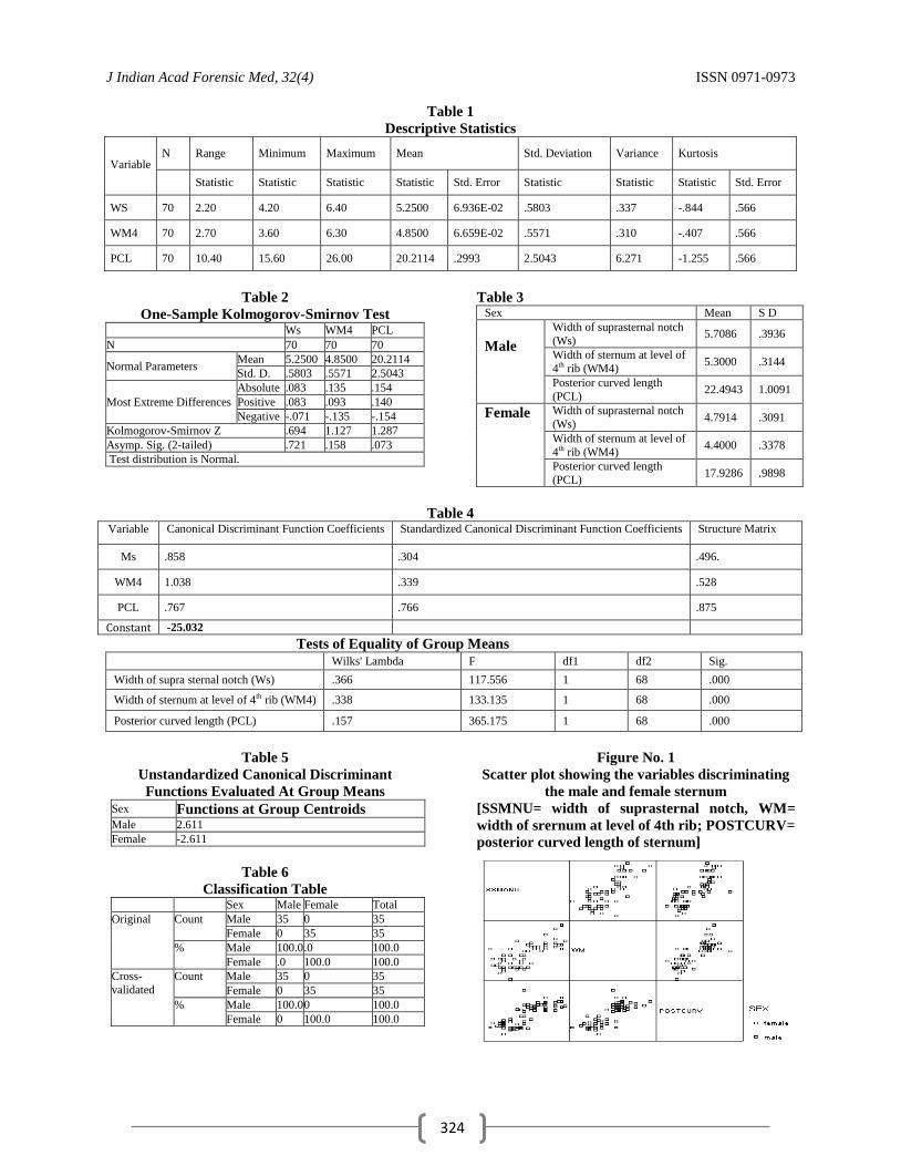
J Indian Acad Forensic Med, 32(4) ISSN 0971-0973
324
Table 1
Descriptive Statistics
Variable N Range Minimum Maximum Mean Std. Deviation Variance Kurtosis
Statistic Statistic Statistic Statistic Std. Error Statistic Statistic Statistic Std. Error
WS 70 2.20 4.20 6.40 5.2500 6.936E-02 .5803 .337 -.844 .566
WM4 70 2.70 3.60 6.30 4.8500 6.659E-02 .5571 .310 -.407 .566
PCL 70 10.40 15.60 26.00 20.2114 .2993 2.5043 6.271 -1.255 .566
Table 2
One-Sample Kolmogorov-Smirnov Test Ws WM4 PCL
N 70 70 70
Normal Parameters Mean 5.2500 4.8500 20.2114
Std. D. .5803 .5571 2.5043
Most Extreme Differences
Absolute .083 .135 .154
Positive .083 .093 .140
Negative -.071 -.135 -.154
Kolmogorov-Smirnov Z .694 1.127 1.287
Asymp. Sig. (2-tailed) .721 .158 .073
Test distribution is Normal.
Table 3 Sex Mean S D
Male
Width of suprasternal notch
(Ws) 5.7086 .3936
Width of sternum at level of
4th rib (WM4) 5.3000 .3144
Posterior curved length
(PCL) 22.4943 1.0091
Female Width of suprasternal notch
(Ws) 4.7914 .3091
Width of sternum at level of
4th rib (WM4) 4.4000 .3378
Posterior curved length
(PCL) 17.9286 .9898
Table 4 Variable Canonical Discriminant Function Coefficients Standardized Canonical Discriminant Function Coefficients Structure Matrix
Ms .858 .304 .496.
WM4 1.038 .339 .528
PCL .767 .766 .875
Constant -25.032
Tests of Equality of Group Means
Wilks' Lambda F df1 df2 Sig.
Width of supra sternal notch (Ws) .366 117.556 1 68 .000
Width of sternum at level of 4th rib (WM4) .338 133.135 1 68 .000
Posterior curved length (PCL) .157 365.175 1 68 .000
Table 5
Unstandardized Canonical Discriminant
Functions Evaluated At Group Means
Sex Functions at Group Centroids
Male 2.611
Female -2.611
Table 6
Classification Table Sex Male Female Total
Original Count Male 35 0 35
Female 0 35 35
% Male 100.0 .0 100.0
Female .0 100.0 100.0
Cross-
validated
Count Male 35 0 35
Female 0 35 35
% Male 100.0 0 100.0
Female 0 100.0 100.0
Figure No. 1
Scatter plot showing the variables discriminating
the male and female sternum
[SSMNU= width of suprasternal notch, WM=
width of srernum at level of 4th rib; POSTCURV=
posterior curved length of sternum]

J Indian Acad Forensic Med, 32(4) ISSN 0971-0973
325
Original research paper
Estimation of Stature by Percutaneous Measurements
of Distal Half of Upper Limb (Forearm & Hand)
*Kumar Amit, ** Srivastava A. K.,*** Verma A.K.
Abstract Determination of stature from skeletal / dismembered remains is not new for Forensic Experts. In this study
efforts are made to reconstruct stature from distal half of upper limb (forearm & hand), for which standing height
and distance between tips of olecranon process and middle finger of right and left arms are measured in 100 adult
male and female students of 19 -25 years of age. These measurements are analyzed statistically to establish relation
between stature and forearm and hand in form of regression equation and multiplication factor.
Regression equations are derived separately for both the sexes by using
formula and calculated as = 2.42 + 56.64 for males and 2.29 +
60.3 for females, stands for standing height and for combined length of forearm & hand. By putting the
value of x in different situations statures are calculated and compared with the corresponding real standing height
and these were close (+/- 3 cm) in most of the cases.
Multiplication factors are also calculated in the study and these are 3.67 for male and 3.73 for female but
their results are less compatible with regression equations.
Key Words: Stature, Dismembered Remain, Regression Equation, Multiplication Factor
Introduction: Stature is one of the important criteria for
establishing identification of unknown person/dead
body. It is usually measured as standing height of the
individual but evaluation of stature is difficult when
dead bodies are mutilated, burnt or skeletonized.
Reconstruction of stature from skeletal /
dismembered remains is not new for Anthropologists
/ Forensic experts. A number of multiplication factors
and regression equations have been developed to
reconstruct stature from long boner throughout the
world (1-6). But estimation of stature in mutilated
bodies especially from their bones is a tedious and
time consuming process which involves cleaning and
preparing of bones.
Corresponding Author: *Assistant professor,
Department of Forensic Medicine,
Subharti Medical College,
Delhi-Hardwar bye-pass road,
Meerut, U.P.-250002
Mob. No. 09456612562
E-mail: [email protected]
**Professor & Head, *** Associate Professor,
CSM Medical University, Lucknow, U.P.
Even then the result may be quite erroneous
because of considerable statistical differences
between the lengths of fresh and dry bones (7). Per-
cutaneous measurements of different parts of body
especially of upper and lower extremities and their
relation with stature may be the correct solution for
mutilated remains. Rough estimation of stature from
parts of upper & lower limbs is used since years as
stature is supposed to be equal to the length between
the tips of middle fingers of both arms when they are
fully extended (8). Stature can also be calculated
from one arm by multiplying the whole length with
two and add 34 cm for chest or from distal half of
upper limb (from tip of olecranon process to tip of
middle finger) by multiplying with 19/5 (9). Recently
Forensic experts/ anthropologists are trying to
develop appropriate formulae/ regression equations
for stature from per-cutaneous measurements of
different parts of upper & lower limbs (10-13).
The study “estimation of stature by per-
cutaneous measurements of distal half of the upper
limb (forearms & hands)” is an effort to establish
correlation between distal half of the upper limb and
stature. This enables doctors/ autopsy surgeons to
establish stature in mutilated bodies especially when
forearm & hand is intact and most of the other parts
are damaged.
Materials and Methods: 200 healthy students (100 males and 100
females) of Subharti Medical College Meerut
between 19-25 years of age were selected for this
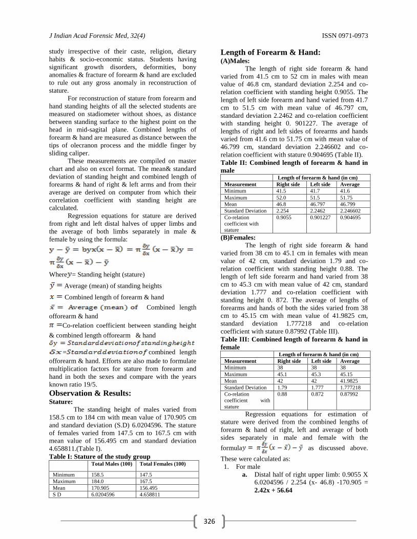
J Indian Acad Forensic Med, 32(4) ISSN 0971-0973
326
study irrespective of their caste, religion, dietary
habits & socio-economic status. Students having
significant growth disorders, deformities, bony
anomalies & fracture of forearm & hand are excluded
to rule out any gross anomaly in reconstruction of
stature. For reconstruction of stature from forearm and
hand standing heights of all the selected students are
measured on stadiometer without shoes, as distance
between standing surface to the highest point on the
head in mid-sagital plane. Combined lengths of
forearm & hand are measured as distance between the
tips of olecranon process and the middle finger by
sliding caliper. These measurements are compiled on master
chart and also on excel format. The mean& standard
deviation of standing height and combined length of
forearms & hand of right & left arms and from their
average are derived on computer from which their
correlation coefficient with standing height are
calculated. Regression equations for stature are derived
from right and left distal halves of upper limbs and
the average of both limbs separately in male &
female by using the formula:
Where = Standing height (stature)
Average (mean) of standing heights
Combined length of forearm & hand
Combined length
offorearm & hand
Co-relation coefficient between standing height
& combined length offorearm & hand
= combined length
offorearm & hand. Efforts are also made to formulate
multiplication factors for stature from forearm and
hand in both the sexes and compare with the years
known ratio 19/5.
Observation & Results: Stature:
The standing height of males varied from
158.5 cm to 184 cm with mean value of 170.905 cm
and standard deviation (S.D) 6.0204596. The stature
of females varied from 147.5 cm to 167.5 cm with
mean value of 156.495 cm and standard deviation
4.658811.(Table I).
Table I: Stature of the study group Total Males (100) Total Females (100)
Minimum 158.5 147.5
Maximum 184.0 167.5
Mean 170.905 156.495
S D 6.0204596 4.658811
Length of Forearm & Hand: (A)Males:
The length of right side forearm & hand
varied from 41.5 cm to 52 cm in males with mean
value of 46.8 cm, standard deviation 2.254 and co-
relation coefficient with standing height 0.9055. The
length of left side forearm and hand varied from 41.7
cm to 51.5 cm with mean value of 46.797 cm,
standard deviation 2.2462 and co-relation coefficient
with standing height 0. 901227. The average of
lengths of right and left sides of forearms and hands
varied from 41.6 cm to 51.75 cm with mean value of
46.799 cm, standard deviation 2.246602 and co-
relation coefficient with stature 0.904695 (Table II).
Table II: Combined length of forearm & hand in
male Length of forearm & hand (in cm)
Measurement Right side Left side Average
Minimum 41.5 41.7 41.6
Maximum 52.0 51.5 51.75
Mean 46.8 46.797 46.799
Standard Deviation 2.254 2.2462 2.246602
Co-relation
coefficient with stature
0.9055 0.901227 0.904695
(B)Females:
The length of right side forearm & hand
varied from 38 cm to 45.1 cm in females with mean
value of 42 cm, standard deviation 1.79 and co-
relation coefficient with standing height 0.88. The
length of left side forearm and hand varied from 38
cm to 45.3 cm with mean value of 42 cm, standard
deviation 1.777 and co-relation coefficient with
standing height 0. 872. The average of lengths of
forearms and hands of both the sides varied from 38
cm to 45.15 cm with mean value of 41.9825 cm,
standard deviation 1.777218 and co-relation
coefficient with stature 0.87992 (Table III).
Table III: Combined length of forearm & hand in
female Length of forearm & hand (in cm)
Measurement Right side Left side Average
Minimum 38 38 38
Maximum 45.1 45.3 45.15
Mean 42 42 41.9825
Standard Deviation 1.79 1.777 1.777218
Co-relation
coefficient with
stature
0.88 0.872 0.87992
Regression equations for estimation of
stature were derived from the combined lengths of
forearm & hand of right, left and average of both
sides separately in male and female with the
formula as discussed above.
These were calculated as:
1. For male
a. Distal half of right upper limb: 0.9055 X
6.0204596 / 2.254 (x- 46.8) -170.905 =
2.42x + 56.64

J Indian Acad Forensic Med, 32(4) ISSN 0971-0973
327
b. Distal half of left upper limb: 0.901227
X 6.0204596 / 2.2464 (x- 46.797) –
170.905 = 2.42x + 56.64
c. Distal half of average of right & left
upper limbs: 0.904695 X 6.0204596 /
2.246602 (x- 46.799) – 170.905 = 2.42x
+ 56.64
2. For female
a. Distal half of right upper limb: 0.88 X
4.658811 / 1.79 (x- 42) -156.5 = 2.29x
+ 60.3 b. Distal half of left upper limb: 0.872 X
4.658811 / 1.777 (x- 42) -156.5 =2.29x
+ 60.3 c. Distal half of average of right & left
upper limbs: 0.877992 X 4.658811 /
1.777218 (x- 41.9825) -156.5 =2.31x +
59. 3. The regression equations for stature from right,
left and average of both sides of forearm & hand
in male are calculated as 2.42x + 56.64 in all the
three situations. Here x is the length between
tips of olecranon to middle finger. By putting
the value of x in different situations the statures
are calculated and compared with the
corresponding real standing height and these
were close (+/- 3) in most of the cases. As in a
male of 158.5 cm the length of right forearm &
hand was 41.5 cm and the stature calculated by
the regression equation 2.42x + 56.64 was 157.1
cm which was 1.4 cm less than the real standing
height.
4. In females the regression equations for stature
from right and left side of forearm & hand are
calculated as 2.29x + 60.3 and from average of
both side of forearm & hand as 2.31x + 59.5. By
putting the value of x in different situations the
statures are calculated and compared with the
corresponding real standing heights and these
were also close (+/- 4) in most of the cases. As
in a female of 147.5 cm, the length of right
forearm & hand was 38 cm and the stature
calculated by the regression equation 2.29x +
60.3 was 147.3 cm which was very close, only
0.2 cm, less than the real standing height. Such
inferences were drawn in most of the cases of
males and females
Multiplication Factor: To establish multiplication factor, ratio of
standing height and average of combined length of
right and left forearm & hand are calculated
separately in males and females which ranged from
3.51 to 3.83 with average of 3.67 in male and 3.54 to
3.92 with average of 3.73 in female (table V). By these ratios errors in reconstruction of
stature are more than +/- 5 cm in 19% of males and
13% of females which ranges from -7.14 to +7.92 cm
and -8.08 to + 7.21 respectively.
Discussion: Estimation of stature is a crucial requirement
in post mortem examination of dead bodies especially
when they are un-identified and badly decomposed,
mutilated or skeletonised. In this study of
reconstruction of stature by distal half of upper limb,
a direct relationship was observed between combined
length of forearm & hand and standing height and
regression equations and multiplication factors are
derived separately for males and females. The
regression equations and multiplication factors for
right and left arms were found same. So the role of
right and left side measurements in determination of
stature is statistically insignificant.
The regression equations for stature from
forearm and hand are 2.42x + 56.64 for male and
2.29x + 60.3 for female and when we calculate
statures by these formula the results are very close to
actual height, less than +/-3 cm in most of the cases.
A variation of more than +/- 5 cm was observed only
in 5% of males and 3% of females.
The multiplication factor between stature
and forearm & hand is 3.67 in male and 3.73 in
female. These are close to the observation of Vij (14).
By these factors when statures are calculated the
errors vary from -8.08 to +7.92, of which the
difference of more than +/-5 cm in 19% of males and
13% of females table VI.
When statures are calculated from
multiplication factor of 19/5 (3.8) there is a mark
difference in actual and reconstructed statures. Here
variation in calculated statures ranges from -14.71 to
+4.51 and they are more than the real statures in 95%
of males and 75% of females. So multiplication
factors are statistically inferior and less reliable than
regression equations.
Conclusion: Though standing height and combined length
of forearm and hand were significantly higher
in male, a direct relationship was observed
between these parts in both the sexes.
The difference in right and left side
measurements are little and statistically
insignificant in derivation of regression
equation and multiplication factor.
The regression equations for reconstruction of
stature from distal half of upper limbs are
2.42X+56.64 for male and 2.29X + 60.3 for
female, where X is the length between tips of
olecranon process to middle finger of the same
side.
Calculated statures from these equations are
close to the actual height, only +/- 3 cm in
most of the cases.

J Indian Acad Forensic Med, 32(4) ISSN 0971-0973
328
The multiplication factors between stature and
forearm & hand are 3.67 in male and 3.73 in
female which are much compatible than the
ratio of 19/5.
Regression equations are statistically superior
and more reliable than multiplication factors.
References: 1. Pearson K. Mathematical contributions to the theory of
evolution. V. On the reconstruction of the stature of prehistoric races. Phil Trans Roy Soc London 1899;
192: 169-244.
2. Trotter M & Gleser G C. Estimation of stature form long bones of American Whites and Negroes. Am J
Phys Anthropol 1952; 10: 463-514.
3. Trotter M & Gleser G C. A re-evaluation of stature based on measurements taken during life and of long
bones after death. Am J Phys Anthropol 1958; 16: 79-
123. 4. Pan N. Lengths of long bones and their proportions to
body height in Hindus. J Anat 1924; 58: 374-378.
5. Nath B S. Estimation of stature from long bones in Indians of the United Provinces: a medico-legal inquiry
in anthropometry. Indian J Med Res 1931; 18: 1245-
1263.
6. Siddiqui M A H & Shah M A. Estimation of stature
from long bones of Punjabis. Indian J Med Res 1944;
31: 105-108.
7. Brues A M. Identification of skeletal remains. J Crim
Law Crimon & Pol Sci 1958; 48: 551-563 8. Subrahmanyam B V. Modi‟s Medical Jurisprudence &
Toxicology. Butterworths India, New Delhi, 1999; 120.
9. Dikshit P C; Forensic Medicine & Toxicology. PEEPEE publishers & distributors New Delhi, 2007, 70
10. Bhatnagar D P, Thapar S P & Batish M K. Identification of personal height from the somatometry
of the hand in Punjabi males.Forensic Sci Int. 1984;
24:137-141. 11. Nath S & Kaur S. Reconstruction of stature through
percutaneous measurements of upper and lower limb
bone lenghts among Rajputs of district Sirmour, Himachal Pradesh. South Asian Anthropologist 1998;
19(2):57-62.
12. Krishan K & Sharma A. Estimation of stature from dimensions of hands and feet in a North Indian
population. J Forensic Legal Med. 2007; 14:327-332.
13. Rastogi P & Yoganarasimha K. Stature estimation using palm length I Indian population. Int J Med Tox &
Leg Med. 2008; 11:37-41.
14. Vij K; Forensic Medicine & Toxicology. Reed Elsevier
India private Ltd. 2005: 70
Table IV
Regression equations and variations in calculated stature Subject Side Regression equation Length of Forearm & hand Standing height Calculated
stature
Variation
in cm
Male Right 2.42 X + 56.64 Min. 41.5 cm 158.5 157.1 -1.4
Max. 52 cm 182 182.5 0.5
Left 2.42 X + 56.64 Min. 41.7 cm 158.5 157.6 - 0.9
Max. 51.5 cm 182 181.6 - 0.4
Average of both
2.42 X + 56.64 Min. 41.6 cm 158.5 157.3 - 1.2
Max. 51.75 cm 182 181.8 - 0.2
Female Right 2.29 X + 60.3 Min. 38 cm 147.5 147.3 - 0.2
Max. 45.1 cm 167.5 163.6 - 3.9
Left 2.29 X + 60.3 Min. 38 cm 147.5 147.3 - 0.2
Max. 45.3 cm 165 164 - 1
Average of
both
2.31 X + 59.5 Min. 38 cm 147.5 147.3 - 0.2
Max. 45.15 cm 165 163.8 -1.2
Table V
Multiplication factor & variation in reconstructed stature Ratio of standing height and average of distal half of right & left upper limbs Male Female
Minimum 3.51 3.54
Maximum 3.83 3.92
Average (Multiplication factor) 3.67 3.73
Variation in reconstructed stature with multiplication factor -7.14 - +7.92 > +/- 5 in
19%
-8.08 - +7.21> +/- 5 in
13%
Table VI
Variation in reconstructed stature from Regression equation & Multiplication factors Variation with regression equations -6.08 - + 4.32> +/- 5 in 5 % -6.14 - + 4.76> +/- 5 in 3 %
Variation with new Multiplication factor (3.67 / 3.73) -7.14 - +7.92> +/- 5 in 19% -8.08 - +7.21> +/- 5 in 13%
Variations with Multiplication factor 19/5 (3.8) -14.71 - +1.48> +/- 5 in 69% -11.09 - +4,51> +/- 5 in 30%

J Indian Acad Forensic Med, 32(4) ISSN 0971-0973
329
Original research paper
Estimation of Stature from Measurements of Long Bones, Hand and
Foot Dimensions
*Chikhalkar B.G.,**Mangaonkar A.A.,***Nanandkar S.D.,****Peddawad R.G.
Abstract Estimation of stature holds a special place in the field of Forensic anthropometry. The present study is an
attempt to evaluate a possible correlation between stature of an individual & six parameters; hand-length, hand-
width, foot-length, foot-width, forearm length & knee-to-ankle length individually in a local population of Mumbai.
A sample of 300 medical students; 147 male & 153 female studying in Grant Medical College & Sir JJ Group of
Hospitals was considered & measurements were taken for each of the parameters. It was found that all the six
parameters showed a correlation with stature but at different degrees (significance calculated through the paired t-
test). Forearm-length showed the highest degree of correlation (r = 0.6558) followed by foot-length (r = 0.6102).
Knee-to-ankle length showed the lowest degree of correlation (r = 0.2086). Mathematical formulae for estimating
stature were developed for each of these parameters through basic linear regression. It can be concluded that the
present study has provided regression equations for six different parameters that can be used for stature estimation in
the population of Mumbai. These equations should not be used for other Indian population groups.
Key Words: Height, Stature, Hand-Length, Hand-Width, Foot-Length, Foot-Width, Forearm-Length, Knee-Ankle
Length
Introduction: Estimation of stature has a significant
importance in the field of forensic anthropometry.
Establishing the identity of an individual from
mutilated, decomposed, & amputed body fragments
has become an important necessity in recent times
due to natural disasters like earthquakes, tsunamis,
cyclones, floods and man-made disasters like terror
attacks, bomb blasts, mass accidents, wars, plane
crashes etc. It is important both for legal &
humanitarian reasons. „Stature‟ is one of the most
important elements in the identification of an
individual.
Many different body parts can be used in the
estimation of stature. Certain long bones &
appendages can be aptly used in the calculation of
height of a person. Many studies have shown the
correlation of stature with body appendages [1-9] &
with long bones [10-19]. But there are inter-racial &
inter-geographical differences in measurements &
their correlation with stature. What may be true for
one race or one region may not be true for the other.
Corresponding Author: Assoc. Professor, Dept. of Forensic Medicine &
Toxicology, Grant Medical College & Sir JJ Group
of Hospitals, Byculla, Mumbai -400008.
E-mail: [email protected],
Ph No.: 09969037650 **Student, MBBS (III/II) *** Professor & Head, **** Resident Doctor,
Even within our vast homeland of India
there are many different ethnic populations & they
having their own variations. [1, 2, 6-13, 18, 19] The
lack of anthropometric data concerning the local
population of Mumbai was felt as the city is prone to
disasters like the terror attacks on 26th
November
2008.Hence the present study was aimed at &
concentrated on the Indian population of Mumbai of
known stature of which anthropometric
measurements of upper limb & lower limb were
calculated & correlated with stature to find
multiplication factors & regression formulae. Study
was carried out at Department of Forensic Medicine
& Toxicology, Grant Medical College & Sir JJ Group
of Hospitals, Mumbai.
Materials & Methods: The study was a cross-sectional one with 300
subjects in the age group of 19 to 23 years, with 147
males & 153 females. The procedure, aims &
objectives of the study were informed & explained in
a group. A written valid informed consent was taken
from each of the participants. A small group of ten
students were taken for measurements each day at a
fixed time to avoid diurnal variations. The students
were measured for the following parameters: Weight: Using an electronic weighing
machine. Height: Standard anthropometric measuring
instruments. Forearm Length: From tip of olecranon
process to mid-point joining radial & ulnar

J Indian Acad Forensic Med, 32(4) ISSN 0971-0973
330
tuberosity using a standard measuring tape
(Position: Arm-Flexed). Hand-Width: From base of 5
th to 2
nd
metacarpal using a standard vernier calliper
(Position: Prone). Hand-Length: From mid-point below radial
& ulnar tuberosity to tip of middle finger
using a standard measuring tape (Position:
Supine).
Foot Length: From tip of toe to heel on the
medial side using a standard measuring tape
(Position: Standing).
Foot Width: From base of 1st to 5
th
metatarsal using standard vernier calliper
(Position: Standing)
Knee to Ankle: From mid-point below
radial & ulnar tuberosity to tip of middle
finger using a standard measuring tape
(Position: Sitting, knee flexed).
Statistical Analysis: The primary outcome was the regression
equation for each parameter. Correlation coefficient
(-1 to +1) was calculated for each parameter as were
range, mean and standard deviation. We analysed our
data using SPSS (version 16.0.2) & calculated
significance via the paired t-test.
Results: The results are given below in the table after
the statistical analysis. (Table-1) Regression Equations:
The regression equations derived for each of
the parameters are as follows:
Estimation of stature from hand-length:
Y = 116.892872 + 2.665389 * X (Y: Stature
of individual, X: Hand-length)
Estimation of stature from hand-width:
Y = 113.561732 + 7.139216 * X (Y: Stature
of individual, X: Hand-width)
Estimation of stature from Foot-length:
Y = 79.72379 + 3.650632 * X (Y: Stature of
individual, X: Foot-length)
Estimation of stature from Foot-width:
Y = 114.828119 + 5.906901 * X (Y: Stature
of individual, X: Foot-width)
Estimation of stature from Forearm- Length:
Y = 86.772654 + 2.997967 * X (Y: Stature
of individual, X: Forearm Length)
Estimation of stature from Knee-to-ankle length:
Y = 156.543454 + 0.296018 * X (Y: Stature
of individual, X: Knee-to-ankle length)
Discussion: In a study done by Bhavna & Surinder Nath
concerning estimation of stature based on lower limb
measurements on 503 Shia Muslims in the age group
of 20-40 years of New Delhi, the Tibial Length was
found to be the best estimate of stature [13]. In the
present study, among the lower extremity
measurements foot-length(r=0.6102) was found to be
the best estimate of stature. Also, knee-to-ankle
length correlated poorly with stature(r=0.2086)
contrary to the findings in the above mentioned
study.
A study based on the measurements of foot
length and body height of total 502 students between
17 to 22 years of age was done by Patel Shah et al
[2]. They had found that foot-length showed a high
degree of correlation with height of the individual.
The present study also highlights a strong correlation
between height & foot-length (r = 0.6102).
A study was done to examine the
relationship between stature and dimensions of hands
and feet among Rajputs of Himachal Pradesh - a
North Indian endogamous group done by Krishnan et
al [6]. Hand length, hand breadth, foot length and
foot breadth of 246 subjects were considered & it was
found that foot-length was the best measure of
estimating stature of an individual. In our study,
among the hand & feet measurements, hand-width (r
= 0.6004) was found to be the best estimate of
stature. Studies by Agnihotri A,Purwar B[9] ; Sen J,
Ghosh S[7] ; Kanchan T, Menezes RG[20] et al. had
similar conclusions to the present study.
A study by Athawle et al. on one hundred
Maharashtrian male adults of ages between 25 to 30
years, Showed that height could be reliably estimated
from forearm length. The present study also showed
similar results.
The present study highlights that hand-
length, hand-width, forearm length foot-length &
foot-width can be reliably used for estimation of
stature in the region of Mumbai. Of all the
parameters, forearm length showed the highest
degree of correlation (r = 0.6558) followed by foot-
length (r = 0.6001). Knee-to-ankle length showed the
least correlation(r = 0.2086). It would not be wise to
apply the same data on other Indian population
groups.
Limitations:
1. In the present study, age range of only 19 to
23 years is considered.
2. Measurements of only healthy individuals
are considered. Hence the data may not be
applicable to individuals who are
malnourished &/or suffering from
congenital structural malformations.
3. Sex variation is not taken into consideration.
4. Applicability of anthropometric
measurements in living & deceased
individuals may practically differ.
5. The present study is a preliminary one &
would be followed up by other studies to
address the above limitations.

J Indian Acad Forensic Med, 32(4) ISSN 0971-0973
331
Table-1
Range, Standard Deviation (SD), Correlation Coefficient(r) & Regression coefficient(b) values of the
Anthropometric Measurements Parameters
Assessed Range (in cm) Mean + SD R B
Height 138-186 167.265 + 8.494
Hand-Length 11.50-27 18.938 + 1.88 0.5902 2.6654
Hand-Width 5.80-8.90 7.537 + 0.714 0.6004 7.1392
Foot-Length 20.85-28 24.008 + 1.420 0.6102 3.6506
Foot-Width 7.10-10.05 8.895 + 0.703 0.4886 5.9069
Forearm-length 23-31.20 26.884 + 1.866 0.6588 2.9980
Knee-to-ankle length 20.50-85.05 36.574 + 5.984 0.2086 0.2960
Conclusions: The present study has established
definite correlation between stature & six
parameters individually, namely, hand-width,
hand-length, foot-length, foot-width, knee-to
ankle length & forearm length. Out of the six
parameters studied, forearm length showed the
highest degree of correlation(r = 0.6558) and
knee-to-ankle length showed lowest degree of
correlation(r = 0.2086); also regression equations
have been established for each of them. This is a study of the first kind in the
Mumbai region. It will help in medico-legal
cases in establishing identity of an individual
when only some remains of the body are found
as in mass disasters, bomb explosions, accidents
etc. It will also help in establishing identity in
certain civil cases. There are lot of variations in estimating
stature from limb measurements among people
of different region & race. Hence there is a need
to conduct more studies among people of
different regions & ethnicity so that stature
estimation becomes more reliable & identity of
an individual is easily established.
Acknowledgement: Indian Council of Medical Research,
New Delhi supported the study financially
through a grant under the Short-term research
studentship (STS) programme-2009.
References: 1. Chavan K.D, Datir SB et al. Correlation of foot length
with height amongst Maharashtrian population of India. Journal of Indian Academy of Forensic Medicine.2009;
31(4):334-337.
2. Patel S M et al. Estimation of height from
measurements of foot length in Gujarat region. J Anat
Soc India. 2007; 56(1): 25-27. 3. Sanli SG, Kizilkanat ED et al. Stature estimation
based on hand length and foot length.Journal of Clinical
Anatomy.2005 Nov; 18(8):589-96. 4. Rastogi P, Nagesh KR et al. Estimation of stature from
hand dimensions of north and South Indians. Leg Med
(Tokyo). 2008 July; 10(4):185-9.
5. Ozden H, Balci Y et al. Stature and sex estimate using foot and shoe dimensions. Forensic Science International. 2005;
147:181-4.
6. Krishnan K, Sharma A et al. Estimation of stature from dimensions of hands and feet in a North Indian population.
Journal of Forensic and Legal Medicine.2007; 14(6):327-
332 7. Sen J, Ghosh S et al. Estimation of stature from foot length
and foot breadth among the Rajbanshi: An indigenous
population of North Bengal. Forensic Science
International. 2008; 181(1): 55.e1-55.e6.
8. Jasuja OP et al. Estimation of Stature from Hand &
Phalange Length. Journal of Indian Academy of Forensic Medicine. 2004; 26(3): 100-106.
9. Agnihotri AK, Purwar B et al. Estimation of Stature by
foot-length. Journal of Forensic and Legal Medicine. 2007; 14(5): 279-283.
10. Shroff AG et al. Determination of height from length of
superior extremity & its segments. Journal of Anatomical Society of India. 1979; 28: 53.
11. Bhavna Nath et al. Estimation of stature from
measurements of lower limbs. Anthropologist Special Volume. 2007; 3: 219-222.
12. Hauser R et al. The Estimation of Stature on the basis of
measurements of the femur. Forensic Science International.2005; 147(2):185-190.
13. Bhavna & S. Nath.Use of Lower Limb Measurements in
Reconstructing Stature among Shia Muslims. Internet Journal of Biological Anthropology.2009; 2(2):86-97.
14. Trotter M, Gleser G et al. Estimation of stature from long
bones of American Whites and Negroes. American Journal of Physical Anthropology.1952; 10: 463-514.
15. T. Sjovold et al. Estimation of stature from long bones
utilizing the line of Organic Correlation. Journal Human Evolution. 1990; 5(5):431-447.
16. Musgrave JH et al.The estimation of adult stature from
metacarpal bone length. American Journal of Physical Anthropology.2005; 48(1):113 – 119.
17. Abrahamyan DO et al. Estimation of stature and length of
limb segments in children and adolescents from whole-body dual-energy X-ray absorptiometry scans. Journal of
Paediatric Radiology. 2008; 38(3): 311-315.
18. Singh, Sohel et al. Estimation of stature from clavicle in Punjabis: a preliminary report. Indian Journal of Medical
Research. 1951; 40:67-71.
19. Athwale. Anthropological study of height from length of forearm bones. A study of one hundred Maharashtrian male
adults of ages between 25 to 30 years. American Journal of Physical Anthropology. 1963; 21:105-112.
20. Kanchan T et al. Stature estimation from foot dimensions.
Forensic Science International. 2008; 179(2-3):241.e1-5
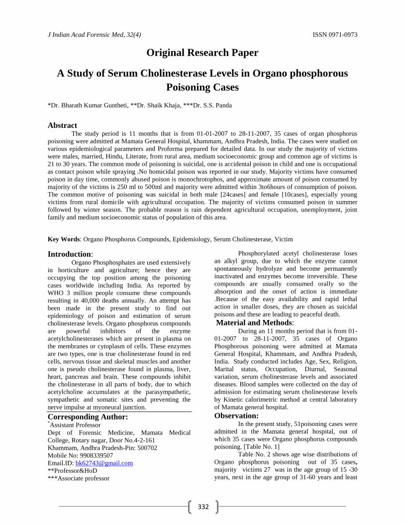
J Indian Acad Forensic Med, 32(4) ISSN 0971-0973
332
Original Research Paper
A Study of Serum Cholinesterase Levels in Organo phosphorous
Poisoning Cases
*Dr. Bharath Kumar Guntheti, **Dr. Shaik Khaja, ***Dr. S.S. Panda
Abstract The study period is 11 months that is from 01-01-2007 to 28-11-2007, 35 cases of organ phosphorus
poisoning were admitted at Mamata General Hospital, khammam, Andhra Pradesh, India. The cases were studied on
various epidemiological parameters and Proforma prepared for detailed data. In our study the majority of victims
were males, married, Hindu, Literate, from rural area, medium socioeconomic group and common age of victims is
21 to 30 years. The common mode of poisoning is suicidal, one is accidental poison in child and one is occupational
as contact poison while spraying .No homicidal poison was reported in our study. Majority victims have consumed
poison in day time, commonly abused poison is monochrotophos, and approximate amount of poison consumed by
majority of the victims is 250 ml to 500ml and majority were admitted within 3to6hours of consumption of poison.
The common motive of poisoning was suicidal in both male [24cases] and female [10cases], especially young
victims from rural domicile with agricultural occupation. The majority of victims consumed poison in summer
followed by winter season. The probable reason is rain dependent agricultural occupation, unemployment, joint
family and medium socioeconomic status of population of this area.
Key Words: Organo Phosphorus Compounds, Epidemiology, Serum Cholinesterase, Victim
Introduction: Organo Phosphosphates are used extensively
in horticulture and agriculture; hence they are
occupying the top position among the poisoning
cases worldwide including India. As reported by
WHO 3 million people consume these compounds
resulting in 40,000 deaths annually. An attempt has
been made in the present study to find out
epidemiology of poison and estimation of serum
cholinesterase levels. Organo phosphorus compounds
are powerful inhibitors of the enzyme
acetylcholinesterases which are present in plasma on
the membranes or cytoplasm of cells. These enzymes
are two types, one is true cholinesterase found in red
cells, nervous tissue and skeletal muscles and another
one is pseudo cholinesterase found in plasma, liver,
heart, pancreas and brain. These compounds inhibit
the cholinesterase in all parts of body, due to which
acetylcholine accumulates at the parasympathetic,
sympathetic and somatic sites and preventing the
nerve impulse at myoneural junction.
Corresponding Author: *Assistant Professor
Dept of Forensic Medicine, Mamata Medical
College, Rotary nagar, Door No.4-2-161
Khammam, Andhra Pradesh-Pin: 500702
Mobile No: 9908339507
Email.ID: [email protected]
**Professor&HoD
***Associate professor
Phosphorylated acetyl cholinesterase loses
an alkyl group, due to which the enzyme cannot
spontaneously hydrolyze and become permanently
inactivated and enzymes become irreversible. These
compounds are usually consumed orally so the
absorption and the onset of action is immediate
.Because of the easy availability and rapid lethal
action in smaller doses, they are chosen as suicidal
poisons and these are leading to peaceful death.
Material and Methods: During an 11 months period that is from 01-
01-2007 to 28-11-2007, 35 cases of Organo
Phosphorous poisoning were admitted at Mamata
General Hospital, Khammam, and Andhra Pradesh,
India. Study conducted includes Age, Sex, Religion,
Marital status, Occupation, Diurnal, Seasonal
variation, serum cholinesterase levels and associated
diseases. Blood samples were collected on the day of
admission for estimating serum cholinesterase levels
by Kinetic calorimetric method at central laboratory
of Mamata general hospital.
Observation: In the present study, 51poisoning cases were
admitted in the Mamata general hospital, out of
which 35 cases were Organo phosphorus compounds
poisoning. [Table No. 1]
Table No. 2 shows age wise distributions of
Organo phosphorus poisoning out of 35 cases,
majority victims 27 was in the age group of 15 -30
years, next in the age group of 31-60 years and least

J Indian Acad Forensic Med, 32(4) ISSN 0971-0973
333
one in the age group of 1-5years. Table No. 3 shows
sex wise distribution, in which 25 were male victims
including male child and 10 were female victims.
Table No. 4 shows marital status of victims, out of
35, 25 males were admitted, majority 25 males were
married and 4 were unmarried. Out of 35, 10females
were admitted, 5females were married 5 were
unmarried. Table No. 5 shows area wise distribution,
majority 28 victims belong to rural area and 7 victims
belong to urban area.
Table No. 6 giving information regarding
literacy status of victims, out of 35,25 males were
admitted, 22males were literates and remaining are
illiterates.10 females were admitted, 8 were literates
and remaining are illiterates. Table No. 7 shows the
socioeconomic status of victims, out of 35, 27 victims
belong to medium socioeconomic class, next is 6
victims were low socioeconomic class and 2 victims
belong to high socioeconomic class.
Table No. 8 giving information about place
of incidence, majority 27 consumed poison at home
followed by 7 victims at work place and 01 victim at
out side of home have consumed poison.
Table No. 9 showing occupation wise
distribution of poisoning, majority 17 victims belong
to farmers followed by7 were unemployed, next 5
were house wife and last 2 were student victims
Table No. 10 showing the incidence of day
and night time of poisoning, majority victims 32 were
consumed poison during day [6am to 6pm] time and
remaining were consumed poison during Nighttime
[6pm to 6am]. Table No. 11showing religion wise
distribution of victims, majority 26 victims were
Hindus next is 05 Muslims victims and 04christian
community. In India, majority population is Hindu
community. Table No. 12 showing seasonal variation,
majority victims 26 has consumed poison during
summer followed by 8 during winter and one is in
during monsoon season
Table No.13 giving information about mode
of poisoning, common mode is suicidal 33; followed
by accidental 01and 01 is in occupational in nature
.No homicidal poisoning is not reported in our study
Table No. 14 showing the period of survival, majority
33 victims admitted in Mamata general hospital with
in3to 6 hours, 01 victim after 8hours and 01victim
admitted after 24 hours after consumed poison. Table
No.15 shows majority 33victims have consumed
approximate amount of poison is 250ml to500ml
followed by 10 ml by 01 victim and 01 victim
become a contact poison during [occupational
poisoning] spraying insecticide Table No. 16 giving information about
levels of serum cholinesterase in Organo phosphorus
poisoning death cases, death with fatal doses
eighty[8] out of thirty-five [35] are not compactable
with life, show low levels of serum cholinesterase[70
to 878 u/L].Serial no.8 inspire of 878 u/L of serum
cholinestrase, victim already suffering with
pulmonary tuberculosis. Table No.17 giving
information regarding survived male victims serum
cholinesterase levels 814 u/ L to9056 u/L [17 cases]
Table No. 18 giving information regarding survived
female victims serum cholinesterase levels 1078 u/ L
to 9240u/L [08cases]. Table No. 19 showing the
pattern of poisoning, commonest is organ phosphorus
insecticide35, followed by aluminum phosphid11 and
5were snake bite poisoning. Table No. 20 showing
trade names of different Organo phosphorus
compounds and no. of cases, commonly consumed
insecticide is Monochrotophos [18], followed by
Malathion [10] next Chlorpyriphos [4] and
Quinalphos [2] least is Dimethoate. [1]
Discussion and Conclusion: In the present study, total no of poisoning
cases 51 admitted, out of 35 was organophosphorus
poison cases were studied in detailed of epidemiology
and serum cholinesterase levels. Males [71.57%]
were more prone to suicide by poisoning compare to
females [28.57% ] and they were in age group of 15-
30years [77.14%] next age group is 31-60years
[20%] and 8 deaths was[ 22.85%] occur in age group
of 21-40year ,similar results are reported by other
studies. [1-3, 7] This age group is more active stage
of life and more vulnerable for suicidal poisoning.
This study reveals male preponderance [71.14%]
compare to female [28.57%]. These are consistent
with other studies. [1-3, 6, 8]
The majority of victims are married
[74.28%]and remaining were unmarried[25.71%],
this report suggest that ,more victims among married
compared to the unmarried population ,because
married population exposure to more problems of
social, financial ,occupational and as well as
domestic worries. Similar results are observed by
other studies. [1-6] The majority of victims were
from rural [80 %] and urban [20%] domicile variation
is reported. These are consistent with other studies.
[8, 4] The higher incidence was observed in literate
[85.71%] compared to illiterate [14.28%]. These are
consistent with other studies [4, 8] because
they can
easily get poison by trade name.
Most of victims were belong to [77.14%]
medium socioeconomic class. This might be due to
modern life style, stress, tension, family and social
problems more common in this class. These similar
finding is made by other studies. [2, 8, 14] The
majority of victims are consumed poison at home
[77.14%], [14] this might be due to a firm decision
was taken in the mind of victims to die that is why
there are only stay at home.
The high incidence was observed in farmers
[48.57%] compare to that in people of other
occupation. [2, 8] More victims are committed
suicide during day time [91.42%] [14] compare to
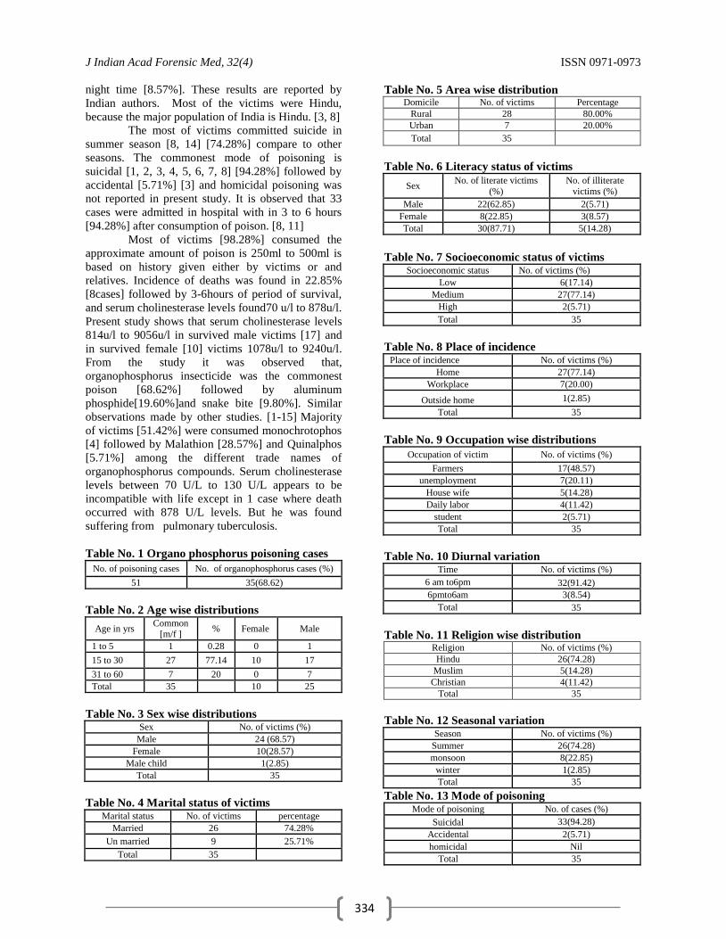
J Indian Acad Forensic Med, 32(4) ISSN 0971-0973
334
night time [8.57%]. These results are reported by
Indian authors. Most of the victims were Hindu,
because the major population of India is Hindu. [3, 8]
The most of victims committed suicide in
summer season [8, 14] [74.28%] compare to other
seasons. The commonest mode of poisoning is
suicidal [1, 2, 3, 4, 5, 6, 7, 8] [94.28%] followed by
accidental [5.71%] [3] and homicidal poisoning was
not reported in present study. It is observed that 33
cases were admitted in hospital with in 3 to 6 hours
[94.28%] after consumption of poison. [8, 11]
Most of victims [98.28%] consumed the
approximate amount of poison is 250ml to 500ml is
based on history given either by victims or and
relatives. Incidence of deaths was found in 22.85%
[8cases] followed by 3-6hours of period of survival,
and serum cholinesterase levels found70 u/l to 878u/l.
Present study shows that serum cholinesterase levels
814u/l to 9056u/l in survived male victims [17] and
in survived female [10] victims 1078u/l to 9240u/l.
From the study it was observed that,
organophosphorus insecticide was the commonest
poison [68.62%] followed by aluminum
phosphide[19.60%]and snake bite [9.80%]. Similar
observations made by other studies. [1-15] Majority
of victims [51.42%] were consumed monochrotophos
[4] followed by Malathion [28.57%] and Quinalphos
[5.71%] among the different trade names of
organophosphorus compounds. Serum cholinesterase
levels between 70 U/L to 130 U/L appears to be
incompatible with life except in 1 case where death
occurred with 878 U/L levels. But he was found
suffering from pulmonary tuberculosis.
Table No. 1 Organo phosphorus poisoning cases
No. of poisoning cases No. of organophosphorus cases (%)
51 35(68.62)
Table No. 2 Age wise distributions
Age in yrs Common
[m/f ] % Female Male
1 to 5 1 0.28 0 1
15 to 30 27 77.14 10 17
31 to 60 7 20 0 7
Total 35 10 25
Table No. 3 Sex wise distributions Sex No. of victims (%)
Male 24 (68.57)
Female 10(28.57)
Male child 1(2.85)
Total 35
Table No. 4 Marital status of victims Marital status No. of victims percentage
Married 26 74.28%
Un married 9 25.71%
Total 35
Table No. 5 Area wise distribution Domicile No. of victims Percentage
Rural 28 80.00%
Urban 7 20.00%
Total 35
Table No. 6 Literacy status of victims
Sex No. of literate victims
(%)
No. of illiterate
victims (%)
Male 22(62.85) 2(5.71)
Female 8(22.85) 3(8.57)
Total 30(87.71) 5(14.28)
Table No. 7 Socioeconomic status of victims Socioeconomic status No. of victims (%)
Low 6(17.14)
Medium 27(77.14)
High 2(5.71)
Total 35
Table No. 8 Place of incidence Place of incidence No. of victims (%)
Home 27(77.14)
Workplace 7(20.00)
Outside home 1(2.85)
Total 35
Table No. 9 Occupation wise distributions
Occupation of victim No. of victims (%)
Farmers 17(48.57)
unemployment 7(20.11)
House wife 5(14.28)
Daily labor 4(11.42)
student 2(5.71)
Total 35
Table No. 10 Diurnal variation Time No. of victims (%)
6 am to6pm 32(91.42)
6pmto6am 3(8.54)
Total 35
Table No. 11 Religion wise distribution Religion No. of victims (%)
Hindu 26(74.28)
Muslim 5(14.28)
Christian 4(11.42)
Total 35
Table No. 12 Seasonal variation Season No. of victims (%)
Summer 26(74.28)
monsoon 8(22.85)
winter 1(2.85)
Total 35
Table No. 13 Mode of poisoning Mode of poisoning No. of cases (%)
Suicidal 33(94.28)
Accidental 2(5.71)
homicidal Nil
Total 35
J Indian Acad Forensic Med, 32(4) ISSN 0971-0973
335
Table No. 14 Period of survived Period of survival No. of victims (%)
Within 3to6 hrs 33(94.28)
More than 8hrs 1(2.85)
Less than 24hrs 1(2.85)
Total 35
Table No. 15 Amount of poison consumed
Mode of
poison
Approximate amount
of poison consumed No. of victims (%)
suicidal 250ml to 500ml 33(94.28)
Accidental 10ml 1(2.85)
occupational Contact poison 1(2.85)
homicidal Nil Nil
Total
35
Table No. 16 Serum cholinesterase levels in death
8 cases
Sl. No
Age/sex Approximate amount
of poison consumed
Serum
cholinesterase
levels
1 60/M 500ml 70u/l
2 15 / M 450ml 110 u/l
3 22/M 500ml 80u/l
4 43 /M 500ml 70u/l
5 23/M 500ml 130u/l
6 22/M 500ml 134u/l
7 25/M 500ml 70u/l
8 50/M 500ml 878u/l
Table No. 17 Serum cholinesterase levels in
survived male Sl. No
Serum cholinesterase levels
Mode of poisoning
prognosis
1 4850u/l suicidal Recovered
2 5400u/l Suicidal Recovered
3 1981u/l Suicidal Recovered
4 814u/l Suicidal Recovered
5 1434u/l Suicidal Recovered
6 3501u/l Suicidal Recovered
7 4014u/l Suicidal Recovered
8 1356u/l Suicidal Recovered
9 990u/l Suicidal Recovered
10 2616u/l Accidental Recovered
11 1155u/l Contact poison Recovered
12 2976u/l Suicidal Recovered
13 1050u/l Suicidal Recovered
14 9056u/l Suicidal Recovered
15 1030u/l Suicidal Recovered
16 5600u/l Suicidal Recovered
17 2010u/l Suicidal Recovered
Table No. 19 Pattern of poisoning Sl. No.
Poison Male
victims Female victims
Total (%)
1 Insecticide
organophosphus 25 10 35(68.62)
2 Aluminum
phosphide 2 8 10(19.60)
3 Snake bite 4 1 5(9.80)
4 Ukknown poison 1 Nil 1(1.96)
Table No. 20 Trade names of O. P. Compounds Sl.
No.
Trade name of different
organophosphorus compounds
No of victims
consumed poison (%)
1 monochrotophos 18(51.42)
2 malathion 10(28.57)
3 Chlorpyriphos 4(11.42)
4 Quinalphos 2(5.71)
5 Dimethoate 1(2.85)
Total 35
Table No. 18 Serum cholinesterase levels in
survived female Sl.
No. Serum cholinesterase levels
Mode of
poisoning
Prognosis
1 1166u/l Suicidal Recovered
2 1113u/l Suicidal Recovered
3 6136u/l Suicidal Recovered
4 3486u/l Suicidal Recovered
5 2180u/l Suicidal Recovered
6 9240u/l Suicidal Recovered
7 1156u/l Suicidal Recovered
8 5833u/l Suicidal Recovered
9 7210u/l Suicidal Recovered
10 1078u/l Suicidal Recovered
References: 1. Aggarwal NK and Aggarwal BBL. Trends of poisoning in
Delhi, JIAFM.1998; 20[2]; 32-36. 2. B D Gupta. Profile of fatal poisoning in and around
Jamnagar.2005; JIAFM; 27[3]: 145-148. 3. Dalal J.S. Poisoning trends –A post –mortem study; JIAFM;
1998; 20[2]: 27-31. 4. Dhattarwal SK and Harnam Singh. Profile of deaths due to
poisoning in Rothak, Haryana. Journal of forensic medicine
and toxicology, 2001; 18[2]:28-29. 5. Eddleston M M, Philips MR. Self poisoning with
pesticides; BMJ; 2004; 328: 4244, [3january], doi:
10.1136/bmj. 328. 7430. 42. 6. Gargi J, Rai H, Chanana A, Raj G, Sharma G, and Bagga
IJS. Current trends of poisoning. A hospital profile, Journal
of Punjab Academy of Forensic medicine and Toxicology, 2003; 3: 41-45.
7. Kapila P, Sekhon HS and Mishra VK. Study of poisoning
deaths in and Shimla, Internet Indian journal of Forensic Medicine Toxicology 2003; 1[2]
8. Kar amjit Singh. Poisoning trends in the Malwa region of
Punjab.PAFMAT; 2003; Vol; 3, 26-28. 9. Mohonty MK, Kumar V, Bastia BK and Arun M. An
analysis of poisoning deaths in Manipal, India .Vet Hum
Toxicology.2004; 46: 208-209. 10. Murari A and Sharma GK. A comparative study of
poisoning cases autopsied in LHMC New Delhi and JIPMER,Pondichery,2000;19 [1]: 18-20.
11. Nigam M, Jain AK, Dubey BP and Sharma VK. Trends of
Organophosphorus poisoning in Bhopal region. An autopsy based study, JIAFM, 2004: 26[2]: 62-65.
12. Sharma BR, Dasari, Sharma V and Vij K. The
epidemiology of poisoning .An Indian view point, Journal of Forensic Medicine and Toxicology, 2000; 19[2]: 5-11.
13. Shingh VP, SharmaBR, Dasari H and Krishan V. A ten
year study of poisoning cases in a tertiary care hospital, Internet Indian journal of Forensic Medicine and Toxicology.
2004; 1[2]. 14. Shreemanta Kumar Dash. Sociodemographic profile of
poisoning cases; JIAFM.2005; 27[3]133-137. 15. Zine KU and Mohant AC. Pattern of acute poisoning at
Indira Gandhi Medical College and Hospital, Nagpur. JIAFM, 1998; 20[2]; 37-39.

J Indian Acad Forensic Med, 32(4) ISSN 0971-0973
336
Original research paper
ABO Blood Grouping from Tooth Material
*Dr. Mahabalesh Shetty, **Dr. Premalatha K
Abstract
Human identification is a mainstay of civilization and identification of unknown individuals has always
been of paramount importance to society. Establishing individuality is an imperative aspect in any investigating
procedure. The use of biological evidence for identification of an individual is a relatively recent development, but it
is expensive and time consuming.
The Forensic importance of dental tissue has been well recognized because of the fact that tooth is hardest
of all human tissues and they can be preserved intact for a long period of time after death. Blood group has been one
of the corner stones for identification of biological materials in Forensic investigations, and ABO blood grouping is
widely used in Forensic laboratories. In this study an attempt is made to determine ABO blood group from the
dental pulp and the dentin extracted from dead body by absorption –elution method.
Key Words: Human identification, ABO blood grouping, Pulp, Dentin
Introduction: Forensic identification over a period of time
has evolved into an art of science and involves
various specialties. A doctor‟s role as forensic
pathologist, a Forensic Odontologist goes hand in
hand with the police officer in establishing the
„IDENTITY‟ of individual in mass disasters. Teeth
are the most durable organs in the body and can be
heated to temperatures of 1,600 C without
appreciable loss of microstructure. [1] Teeth can
survive long after soft and skeletal tissues have been
destroyed [1]. Biological evidence generally means
the comparison of genetic material like DNA. But
DNA analysis can be expensive and time consuming.
[2] The use of blood group substance in medico legal
examination is based on the fact that once a group is
established in an individual it remains unchanged
throughout his life.[3] Blood group substances in the
hard dental tissues thus remain unaffected even in
adverse environmental conditions.
Pulp tissue is one of the most protected of
the oral tissues being surrounded from all sides by
dental hard tissues; and also tooth pulp contains lot of
blood vessels, blood group antigens are most
certainly bound to be present in tooth pulp. [4] It is
presumed that blood group substances in dentine
were located in dentin tubules. [4]
Corresponding Author: *Associate Prof. & HOD
Forensic Medicine & Toxicology
K.S. Hegde Medical Academy, Manglore, Karnatka
E-mail: [email protected]
**Prof. & HOD, Deptt. of Maxillo- Facial Surgery
Manipal college of Dental Sciences,
Manipal University, Mangalore
Thus it was decided to use dental pulp and
dentine for detection of ABO group substances.In
this study an attempt is made to determine ABO
blood group from the dental pulp and the dentin by
absorption –elution method. [5]
Material & Methods: The present study was conducted in K S
Hegde Medical Academy, Mangalore. 60 extracted
teeth were collected, and these teeth were extracted
from dead bodies brought to our Mortuary for medico
legal autopsies. The age ranging from 14-60yrs. 31
males and 29 females were selected for the study.
Exclusion Criteria:
Putrefied bodies, Deciduous tooth, Injured
tooth, Root canal treated and Carious tooth because
of the possibility of showing false positive or false
negative results, were excluded. After routine
extraction procedure, blood was taken from the
freshly extracted socket for blood group
determination by agglutination method and acted as
control group.
The extracted teeth were washed under
running water and debris were removed with the
probe, and wiped with gauze and kept in numbered
bottles: the teeth were dried and stored for a span of
6months at the room temperature without any
preservative. The blood grouping was performed by
absorption elution test using dentin and dental pulp,
which was later, compared with the recorded blood
group from the extracted socket.
Laboratory Procedure:
Modelling wax was folded and made into
block. The tooth was than embedded on the
modelling wax block. The tooth was split vertically
with carborundum disc and then the dental pulp was
scooped with sterile spoon excavator which was

J Indian Acad Forensic Med, 32(4) ISSN 0971-0973
337
placed in a test tube containing sterile thread and a
drop of saline.
The test tubes were placed in the incubator
at 56°C for 30 minutes for drying where the blood
group antigens of dental pulp were absorbed on
sterile cotton thread.
Only anti –A & anti –B antisera were used,
assuming blood group O when no agglutination
occurred.
Blood stained threads of 2 mm length were
cut and placed in a drop of anti-A serum in a slide
cavity. Similar pieces were placed in anti-B serum.
The slides were then kept in moist chamber at 4°C
for 2 hours for complete absorption. After absorption,
the antiserum was pipetted off from the thread by
capillary pipettes and then the thread was thoroughly
washed 3 to 4 times in ice cold saline, for the
complete removal of unreacted antibodies from it.
Slides were again placed in moist chamber
and kept in an incubator at 56°C for 30 minutes to
break the antibody- antigen bond (Elution).One drop
of a 0.5% suspension of known RBC blood group
was added and the samples were again placed in the
humidified recipient and were incubated at 56° C for
15 minutes to enhance agglutination.
The slides were then removed from the
incubator to be kept at room temperature for 45
minutes to 1 hour and were observed under
microscope at magnification of 100x for
agglutination i.e. ABO blood groups.
The remaining tooth consisted of dentin,
which was pulverized, with straight fissure bur. The
pulverized tooth powder was put in two test tubes, to
each of this test tubes 3 drops of antiserum A, B was
added and confirming the test samples being
sufficiently soaked with antiserum for two and half
hours and left standing at room temperature.
After removing antiserum, each sample was
washed three times with cold saline solution (it was
centrifuged and the supernatant was sucked with
pipette).
Then two drops fresh saline was added to
the sample and the test tube were heated in a water
bath (50-55oc) for 10 minutes to elude the antibodies.
A drop of 0.5% A or B group red cell
suspension was immediately put into each respective
test tube of known blood and the samples were again
put in humified recipient.
They were incubated for 370c for 30min to
enhance agglutination, and after this procedure it was
centrifuged at 1,500-2000rpm for min. By gentle
shaking of the test tube the presence or absence of
red cell agglutination was ascertained with
microscope at magnification of 100X.
It was further cross-confirmed with control
group.
Tooth Sectioning:
Results:
The blood grouping was attempted to
establish from dentin and pulp and it was further
confirmed with the control. 60 permanent teeth were
collected from 60 subjects, 29 were females, age
ranging from 15 yrs to 56 yrs. There were 31 males,
age ranging from 16 yrs to 55 yrs. (shown in table 1).
Table - 1: Age Distribution with Sex Age(yrs) Male Female Combined
No. (%) No. (%) No. ( %)
< 20 11( 18.33) 12 (20) 23( 38.3)
21-40 12 (20) 11 (18.33) 23 (38.3)
41-60 8 (13.33) 6 (10.0) 14 (23.4)
Total 31 (51.67) 29 (48.33) 60 (100.0)
Mean +SD 27.74 +11.35 25.86+12.35 26.92+12.74
Blood group belonging to „O‟ were 26 in
numbers (43.4%).Blood group A were 20 (33.3%) ,
Blood group B were 11 (18.3%),Blood group AB
were 3 (5%) of the control as shown in table 2.
Blood grouping on Dentin was done after 180 days of
extraction. None of the teeth showed ABO antigen in
Dentin after 180 days of extraction (table 2).
Table- 2: Blood Grouping on Dentin after 180
Days of Extraction Blood
groups
Dentin
Control group Study group
Positive Negative
A 20 (33.3%) 0(0%) 20(33.3%)
B 11(18.3%) 0(0%) 11(18.3%)
AB 3(5%) 0(0%) 3(5%)
O 26(43.4%) 0(0%) 26(43.4%)
Total 60(100%) (0%) 60(100%)
Inference ABO antigens were not found in Dentin
Blood grouping on pulp was done after 180
days of extraction. 58 teeth showed positive results
for ABO Blood group in pulp. One tooth showed
negative results for ABO blood group and mistyping
in another tooth. Sensitivity of pulp in relation to
control is 96.7% as shown (table- 3).
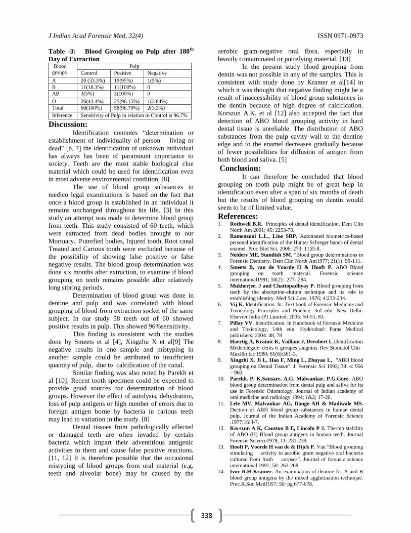
J Indian Acad Forensic Med, 32(4) ISSN 0971-0973
338
Table -3: Blood Grouping on Pulp after 180th
Day of Extraction Blood
groups
Pulp
Control Positive Negative
A 20 (33.3%) 19(95%) 1(5%)
B 11(18.3%) 11(100%) 0
AB 3(5%) 3(100%) 0
O 26(43.4%) 25(96.15%) 1(3.84%)
Total 60(100%) 58(96.70%) 2(3.3%)
Inference Sensitivity of Pulp in relation to Control is 96.7%
Discussion: Identification connotes “determination or
establishment of individuality of person – living or
dead” [6, 7] the identification of unknown individual
has always has been of paramount importance to
society. Teeth are the most stable biological clue
material which could be used for identification even
in most adverse environmental condition. [8]
The use of blood group substances in
medico legal examinations is based on the fact that
once a blood group is established in an individual it
remains unchanged throughout his life. [3] In this
study an attempt was made to determine blood group
from teeth. This study consisted of 60 teeth, which
were extracted from dead bodies brought to our
Mortuary. Putrefied bodies, Injured tooth, Root canal
Treated and Carious tooth were excluded because of
the possibility of showing false positive or false
negative results. The blood group determination was
done six months after extraction, to examine if blood
grouping on teeth remains possible after relatively
long storing periods.
Determination of blood group was done in
dentine and pulp and was correlated with blood
grouping of blood from extraction socket of the same
subject. In our study 58 teeth out of 60 showed
positive results in pulp. This showed 96%sensitivity.
This finding is consistent with the studies
done by Smeets et al [4], Xingzhu X et al[9] The
negative results in one sample and mistyping in
another sample could be attributed to insufficient
quantity of pulp, due to calcification of the canal.
Similar finding was also noted by Parekh et
al [10]. Recent tooth specimen could be expected to
provide good sources for determination of blood
groups. However the effect of autolysis, dehydration,
loss of pulp antigens or high number of errors due to
foreign antigen borne by bacteria in carious teeth
may lead to variation in the study. [8]
Dental tissues from pathologically affected
or damaged teeth are often invaded by certain
bacteria which impart their adventitious antigenic
activities to them and cause false positive reactions.
[11, 12] It is therefore possible that the occasional
mistyping of blood groups from oral material (e.g.
teeth and alveolar bone) may be caused by the
aerobic gram-negative oral flora, especially in
heavily contaminated or putrefying material. [13]
In the present study blood grouping from
dentin was not possible in any of the samples. This is
consistent with study done by Kramer et al[14] in
which it was thought that negative finding might be a
result of inaccessibility of blood group substances in
the dentin because of high degree of calcification.
Korszun A.K. et al [12] also accepted the fact that
detection of ABO blood grouping activity in hard
dental tissue is unreliable. The distribution of ABO
substances from the pulp cavity wall to the dentine
edge and to the enamel decreases gradually because
of fewer possibilities for diffusion of antigen from
both blood and saliva. [5]
Conclusion: It can therefore be concluded that blood
grouping on tooth pulp might be of great help in
identification even after a span of six months of death
but the results of blood grouping on dentin would
seem to be of limited value.
References: 1. Rothwell B.R. Principles of dental identification. Dent Clin
North Am 2001; 45: 2253-70. 2. Ramenzoni L.L., Line SRP. Automated biometrics-based
personal identification of the Hunter Schreger bands of dental
enamel. Proc Biol Sci. 2006; 273: 1155-8. 3. Neiders ME, Standish SM. “Blood group determinations in
Forensic Dentistry. Dent Clin North Am1977; 21(1): 99-111.
4. Smeets B, van de Voorde H & Hooft P. ABO Blood grouping on tooth material. Forensic science
international1991; 50(2): 277- 284.
5. Mukherjee. J and Chattopadhyay P. Blood grouping from teeth by the absorption-elution technique and its role in
establishing identity. Med Sci .Law, 1976; 4:232-234.
6. Vij K. Identification. In: Text book of Forensic Medicine and Toxicology Principles and Practice. 3rd edn. New Delhi:
Elsevier India (P) Limited; 2005: 50-51, 83.
7. Pillay VV. Identification. In Handbook of Forensic Medicine and Toxicology, 14th edn. Hyderabad: Paras Medical
publishers; 2004: 48, 78
8. Haertig A, Krainic K, Vaillant J, Derobert L.Identification
Medicolegale: dents et groupes sanguins. Rev.Stomatol Chir
Maxillo fac 1980; 81(6):361-3. 9. Xingzhi X, Ji L, Hao F, Ming L, Zhuyao L. "ABO blood
grouping on Dental Tissue", J. Forensic Sci 1993; 38: 4: 956
– 960. 10. Parekh. P, K.Sansare, A.G. Malwankar, P.G.Gore. ABO
blood group determination from dental pulp and saliva for its
use in Forensic Odontology. Journal of Indian academy of oral medicine and radiology 1994; 1&2; 17-20.
11. Lele MV, Malvankar AG, Dange AH & Madiwale MS.
Dection of ABH blood group substances in human dental pulp, Journal of the Indian Academy of Forensic Science
.1977;16:3-7.
12. Korszun A K, Causton B E, Lincoln P J. Thermo stability of ABO (H) Blood group antigens in human teeth. Journal
Forensic Science1978; 11: 231-239.
13. Hooft P, Voorde H van de & Dijck P. Van “Blood grouping simulating activity in aerobic gram negative oral bacteria
cultured from fresh corpses”. Journal of forensic science
international 1991; 50: 263-268. 14. Ivor R.H Kramer. An examination of dentine for A and B
blood group antigens by the mixed agglutination technique.
Proc.R.Soc.Med1957; 50: pg 677-678.
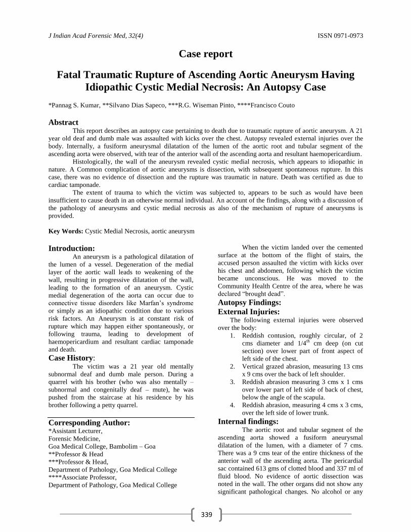
J Indian Acad Forensic Med, 32(4) ISSN 0971-0973
339
Case report
Fatal Traumatic Rupture of Ascending Aortic Aneurysm Having
Idiopathic Cystic Medial Necrosis: An Autopsy Case *Pannag S. Kumar, **Silvano Dias Sapeco, ***R.G. Wiseman Pinto, ****Francisco Couto
Abstract This report describes an autopsy case pertaining to death due to traumatic rupture of aortic aneurysm. A 21
year old deaf and dumb male was assaulted with kicks over the chest. Autopsy revealed external injuries over the
body. Internally, a fusiform aneurysmal dilatation of the lumen of the aortic root and tubular segment of the
ascending aorta were observed, with tear of the anterior wall of the ascending aorta and resultant haemopericardium.
Histologically, the wall of the aneurysm revealed cystic medial necrosis, which appears to idiopathic in
nature. A Common complication of aortic aneurysms is dissection, with subsequent spontaneous rupture. In this
case, there was no evidence of dissection and the rupture was traumatic in nature. Death was certified as due to
cardiac tamponade.
The extent of trauma to which the victim was subjected to, appears to be such as would have been
insufficient to cause death in an otherwise normal individual. An account of the findings, along with a discussion of
the pathology of aneurysms and cystic medial necrosis as also of the mechanism of rupture of aneurysms is
provided.
Key Words: Cystic Medial Necrosis, aortic aneurysm
Introduction: An aneurysm is a pathological dilatation of
the lumen of a vessel. Degeneration of the medial
layer of the aortic wall leads to weakening of the
wall, resulting in progressive dilatation of the wall,
leading to the formation of an aneurysm. Cystic
medial degeneration of the aorta can occur due to
connective tissue disorders like Marfan‟s syndrome
or simply as an idiopathic condition due to various
risk factors. An Aneurysm is at constant risk of
rupture which may happen either spontaneously, or
following trauma, leading to development of
haemopericardium and resultant cardiac tamponade
and death.
Case History: The victim was a 21 year old mentally
subnormal deaf and dumb male person. During a
quarrel with his brother (who was also mentally –
subnormal and congenitally deaf – mute), he was
pushed from the staircase at his residence by his
brother following a petty quarrel.
Corresponding Author: *Assistant Lecturer,
Forensic Medicine,
Goa Medical College, Bambolim – Goa
**Professor & Head
***Professor & Head,
Department of Pathology, Goa Medical College
****Associate Professor,
Department of Pathology, Goa Medical College
When the victim landed over the cemented
surface at the bottom of the flight of stairs, the
accused person assaulted the victim with kicks over
his chest and abdomen, following which the victim
became unconscious. He was moved to the
Community Health Centre of the area, where he was
declared “brought dead”.
Autopsy Findings:
External Injuries: The following external injuries were observed
over the body:
1. Reddish contusion, roughly circular, of 2
cms diameter and 1/4th
cm deep (on cut
section) over lower part of front aspect of
left side of the chest.
2. Vertical grazed abrasion, measuring 13 cms
x 9 cms over the back of left shoulder.
3. Reddish abrasion measuring 3 cms x 1 cms
over lower part of left side of back of chest,
below the angle of the scapula.
4. Reddish abrasion, measuring 4 cms x 3 cms,
over the left side of lower trunk.
Internal findings: The aortic root and tubular segment of the
ascending aorta showed a fusiform aneurysmal
dilatation of the lumen, with a diameter of 7 cms.
There was a 9 cms tear of the entire thickness of the
anterior wall of the ascending aorta. The pericardial
sac contained 613 gms of clotted blood and 337 ml of
fluid blood. No evidence of aortic dissection was
noted in the wall. The other organs did not show any
significant pathological changes. No alcohol or any

J Indian Acad Forensic Med, 32(4) ISSN 0971-0973
340
other substance was detected at chemical analysis of
viscera. No fractures of the ribs or the sternum were
found.
Histopathological Examination Findings: Sections from the wall of the ascending
aorta showed myxoid degeneration in the media.
There was elastic tissue fragmentation and separation
of the fibromuscular and elastic elements of the
media by numerous cystic cleft – like spaces in the
media containing basophilic amorphous extracellular
matrix / ground substance. There was increased
fibrosis of the media. These findings are consistent
with Cystic Medial Necrosis.
Cause of Death: Death was certified as “due to cardiac
tamponade consequent to rupture of the dilated
ascending aorta having cystic medial necrosis as a
result of cumulative effect of the external injuries on
the body, which was likely to cause death”.
Discussion: The aorta is composed of three layers: the
thin inner layer or intima, a thick middle layer or
media and a rather thin outer layer called the
adventitia. The strength of the aorta lies in its media,
which is composed of laminated but intertwining
sheets of elastic tissue arranged in a spiral manner
that affords maximum tensile strength. This
tremendous accretion of elastic tissue gives the aorta
not only tensile strength but also distensibility and
elasticity.
The term aortic aneurysm refers to a
pathological dilatation of the normal lumen, being
defined as a permanent localised dilatation of the
aorta having a diameter at least 1.5 times of the
expected normal diameter of that given aortic
segment. [1] The normal diameter of the ascending
aorta is about 3 cms and length is about 5 cms. [2] In
the instant case, the ascending aorta was dilated to a
clearly aneurysmal width of 7 cms. An area of
expanding investigation is the role of cellular
mechanisms in the pathogenesis of aortic aneurysms.
Destruction of the media and its elastic tissue is the
striking histological feature of aortic aneurysms.
Experimental evidence indicates excessive
activity of proteolytic enzymes in the aortas of
affected patients, which may lead to deterioration of
structural matrix proteins such as elastin and collagen
in the aortic media and thereby promote or perpetuate
the formation of aneurysms. Aneurysmal aortas
contain elastolytic activity with an active elastase not
present in the normal aorta, and other active
proteolytic enzymes as well. The risk of rupture
increases with aneurysm size. Smaller than 4 cms
Aneurysm have 0-2 % risk of rupture, whereas those
larger than 5 cms have 22% risk. [1]
Aneurysms of the ascending aorta most
often result from the process of cystic medial
degeneration/ necrosis. [1] Histologically, as in the
instant case, cystic medial necrosis is characterised
by elastic tissue fragmentation and separation of the
fibromuscular and elastic elements of the tunica
media by small left – like spaces where the normal
elastic tissue is lost, and these areas are filled with the
amorphous extracellular matrix of connective tissue
and resemble, but are not true “cysts”. Ultimately,
there may be large scale loss of elastic laminae. Thus,
the terminology “cystic medial necrosis”, as medial
degeneration is often called, is inaccurate, because
neither true necrosis nor true cysts are present.
Inflammation is absent. [1]
Although these changes occur most
commonly in the ascending aorta, in some cases, the
entire aorta may be involved. The histological
changes lead to weakening of the aortic wall, which
in turn results in the formation of a fusiform
aneurysm. Cystic medial necrosis is found in
connective tissue disorders like Marfan‟s Syndrome
and Ehler Danlos Syndrome. However, in patients
without Marfan‟s syndrome, it is not possible to
recognise the histological diagnosis of cystic medial
necrosis prospectively, i.e. without surgery or
autopsy.
This fact has significantly limited
understanding of cystic medial degeneration and its
natural history by the scientific community, and it
remains unclear as to what extent cystic medial
degeneration may represent an independent disease
process versus a manifestation of another disease
state. It has long been suspected that patients who
have proven cystic medial degeneration without the
classic phenotypic manifestations of Marfan‟s
syndrome may in fact have a variation or „forme
fruste’ of Marfan‟s syndrome; though this theory
remains unproven. [1] In fact, Kubota J et al have
reported a case of two brothers who presented with
multiple visceral artery aneurysms and diffuse
connective tissue fragility, but did not have any
features of Marfan‟s syndrome. According to them,
these cases belong to the heterogenous group of
Marfan‟s syndrome in accordance to the above
mentioned theory. [4]
However, on the contrary, many patients
with ascending thoracic aortic aneurysms appear to
have nothing more than idiopathic cystic medial
degeneration, where it is unclear as to what
specifically predisposes to the development of medial
degeneration. It was first thought to be a degenerative
process associated with old age, but subsequent
reports have shown that it occurs not infrequently in
young people. Hypertension is a common risk factor.
Another risk factor is syphilis, though now a rare
cause, in which degeneration of the aortic media
occurs during the secondary phase of the disease
producing a weakening of the aortic wall. Other risk

J Indian Acad Forensic Med, 32(4) ISSN 0971-0973
341
actors are Toxemia, nicotine, hyperadrenalism,
infectious aortitis, great vessel arteritis. [1, 2, 5]
In the instant case, the since deceased was
mentally subnormal and congenitally deaf – mute.
His brother, the assailant, was also known to be
mentally subnormal and congenitally deaf – mute.
Otherwise, there were no definitive physical
characteristics to suggest that the victim was
suffering from either Ehler Danlos Syndrome or
Marfan‟s syndrome. Hence, it appears most likely
that the case is one of idiopathic cystic medial
necrosis, rather than one due to Marfan‟or Ehler
Danlos syndrome. A genetic study in the instant case
would have been helpful in arriving at a diagnosis, to
prove or disprove the above mentioned theory that
the victim may have been suffering from a variant of
Marfan‟s syndrome.
A common complication of aortic
aneurysms is “dissection”, which begins with a tear
in the intima that exposes a diseased medial layer to
the systemic pressure of intraluminal blood. The
blood penetrates into the media, cleaving it into two
layers longitudinally and producing a blood filled
false lumen within the aortic wall. This false lumen
propagates distally progressively for a variable
distance. Dissection is common in connective tissue
disorders like Marfan‟and Ehler Danlos syndrome.
[2] Such a dissecting aneurysm frequently ruptures
spontaneously or following trivial trauma or
following a bout of hypertension, resulting in
haemopericardium. In the instant case, there was no
evidence of dissection. The rupture of the aneurysm
was rather, secondary to trauma to which the since
deceased was subjected to by the assailant.
Cardiac tamponade is a clinical syndrome
caused by the accumulation of fluid in the pericardial
space, resulting in reduced ventricular filling and
subsequent hemodynamic compromise. In cardiac
tamponade, blood accumulates in the pericardial sac
faster than it can escape, either because the bleeding
rate exceeds the drainage. When there is no laceration
of the pericardium, there is no escape route for the
blood from the pericardial sac. When sufficient blood
accumulates, the pressure in the pericardial sac
increases and begins to prevent the passive filling of
the atria during diastole. The cardiac output falls, as
does the systemic blood pressure and the venous
pressure rises. If unrelieved, death follows, though
the time that this takes is variable and difficult to
calculate retrospectively on pathological findings.
About 400-500 ml of blood is sufficient to cause
death, though this seems to be a greater volume than
is seen in tamponades. [6] In the instant case, the
pericardial sac contained 613 gms of clotted blood
and 337 ml of fluid blood.
Medico legally, culpability of the accused
person would be determined by the interaction of
trauma and disease. The injury sustained by the since
deceased would be categorized as one that is “likely
to cause death”, because although it was an injury
that posed a threat to the life of the victim and death
as a consequence was not surprising owing to the
structures involved, yet it was one where the death
was caused due to superimposed trauma in an already
diseased person. The same degree of external trauma
to which the since deceased was subjected would not
have resulted in death in a normal and healthy
individual. Hence, the accused would be held guilty
vide S. 299 IPC and punishable vide S. 304 PC for
culpable homicide not amounting to murder. [9]
The ascending aorta is one of the four
common sites where the aorta may rupture following
trauma, the others being the aortic isthmus distal to
the ligamentum arteriosum, the lower part of the
aorta above the diaphragm, and at the junction of the
innominate artery with the aortic arch. [8]
Rupture is more likely at sites of aneurysms,
because of the reduced thickness and hence greater
weakness of the wall, and because of the “La Place‟s
law”, according to which tension acting on the wall
of a vessel is greater where the luminal diameter is
greater. Hence, a much lesser degree of trauma would
be required to rupture an aneurysmal aorta as
compared to a normal one, as in the instant case.
According to the osseous pinch mechanism /
the aorta is crushed or pinched between the vertebral
column and the inner surface of the manubrium, first
rib and the clavicles during antero–posterior thoracic
compressive deformation. [9]
Sudden deceleration also contributes to
trauma to the aorta. During sudden deceleration,
traction and torsion forces are placed on the aorta at
points of anatomic fixation, i.e. isthmus and
supravalvular ascending aorta. This is called the
mechanism of “differential deceleration”. During
falls, the primary stress is longitudinal traction, with
the weight of the heart producing greatest traction on
the aortic root. Also, a sudden increase in
intraluminal aortic pressure occurs with the force of
impact, which is called the “water hammer effect.”
[10]
In the instant case, all of the above
mechanisms may have acted together to produce
laceration of the wall of the aorta. There was history
of direct trauma to the chest by kicks from the
assailant. The victim was seen to have been lying on
the floor at the time he was being kicked, indicating
that the chest would have been fixed, the back being
supported by the floor. Presence of a bruise over the
precordium as mentioned above makes it highly
likely that kicks may have been perpendicularly
directed with respect to the chest wall in a stamping –
like action. This would cause antero posterior
compression of the chest, traumatizing the ascending
aortic aneurysm in accordance with the osseous pinch
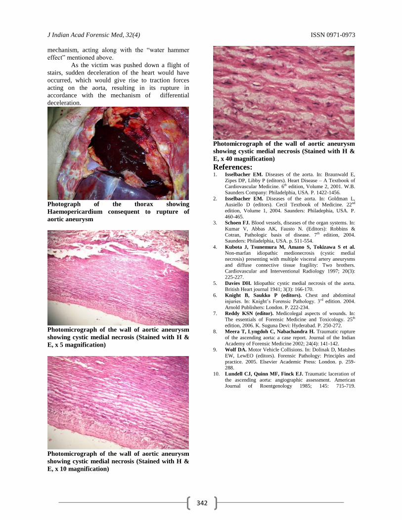
J Indian Acad Forensic Med, 32(4) ISSN 0971-0973
342
mechanism, acting along with the “water hammer
effect” mentioned above.
As the victim was pushed down a flight of
stairs, sudden deceleration of the heart would have
occurred, which would give rise to traction forces
acting on the aorta, resulting in its rupture in
accordance with the mechanism of differential
deceleration.
Photograph of the thorax showing
Haemopericardium consequent to rupture of
aortic aneurysm
Photomicrograph of the wall of aortic aneurysm
showing cystic medial necrosis (Stained with H &
E, x 5 magnification)
Photomicrograph of the wall of aortic aneurysm
showing cystic medial necrosis (Stained with H &
E, x 10 magnification)
Photomicrograph of the wall of aortic aneurysm
showing cystic medial necrosis (Stained with H &
E, x 40 magnification)
References: 1. Isselbacher EM. Diseases of the aorta. In: Braunwald E,
Zipes DP, Libby P (editors). Heart Disease – A Textbook of Cardiovascular Medicine. 6th edition, Volume 2, 2001. W.B.
Saunders Company: Philadelphia, USA. P. 1422-1456.
2. Isselbacher EM. Diseases of the aorta. In: Goldman L, Ausiello D (editors). Cecil Textbook of Medicine. 22nd
edition, Volume 1, 2004. Saunders: Philadephia, USA. P.
460-465. 3. Schoen FJ. Blood vessels, diseases of the organ systems. In:
Kumar V, Abbas AK, Fausto N. (Editors): Robbins &
Cotran, Pathologic basis of disease. 7th edition, 2004.
Saunders: Philadelphia, USA. p. 511-554.
4. Kubota J, Tsunemura M, Amano S, Tokizawa S et al. Non-marfan idiopathic medionecrosis (cystic medial necrosis) presenting with multiple visceral artery aneurysms
and diffuse connective tissue fragility: Two brothers.
Cardiovascular and Interventional Radiology 1997; 20(3): 225-227.
5. Davies DH. Idiopathic cystic medial necrosis of the aorta.
British Heart journal 1941; 3(3): 166-170. 6. Knight B, Saukko P (editors). Chest and abdominal
injuries. In: Knight‟s Forensic Pathology. 3rd edition. 2004.
Arnold Publishers: London. P. 222-234. 7. Reddy KSN (editor). Medicolegal aspects of wounds. In:
The essentials of Forensic Medicine and Toxicology. 25th
edition, 2006. K. Suguna Devi: Hyderabad. P. 250-272. 8. Meera T, Lyngdoh C, Nabachandra H. Traumatic rupture
of the ascending aorta: a case report. Journal of the Indian
Academy of Forensic Medicine 2002; 24(4): 141-142. 9. Wolf DA. Motor Vehicle Collisions. In: Dolinak D, Matshes
EW, LewEO (editors). Forensic Pathology: Principles and
practice. 2005. Elsevier Academic Press: London. p. 259-288.
10. Lundell CJ, Quinn MF, Finck EJ. Traumatic laceration of
the ascending aorta: angiographic assessment. American Journal of Roentgenology 1985; 145: 715-719.
J Indian Acad Forensic Med, 32(4) ISSN 0971-0973
343
Case report
Death Due to Swine Influenza -A Forensic Autopsy Report
*Dr. Manpreet Kaul, **Dr. Jagdish Gargi, ***Dr. Ashok Chanana, ****Dr. Rajeev Kumar Chaudhary
Abstract A 29 year male who was working in army (soldier) at Pune, came to his house at Tarntaran (Punjab) on
vacations to meet his family where he developed high grade fever, cough, dyspnoea, and was admitted in a private
hospital. He was clinically suspected to be suffering from bilateral viral pneumonitis but the diagnosis of viral
pneumonitis was never confirmed by laboratory tests in spite of admission in a hospital. Later on he died & the
police was informed about his death. The police completed the inquest U/S 174CrPC & the dead body was shifted to
the mortuary of Govt. Medical College, Amritsar for post mortem examination. A team of autopsy surgeons
conducted the post mortem examination. The dead body was examined; nasal & pharyngeal swabs along with blood
sample were taken from deceased and were sent to virology Deptt. , Post Graduate Institute, Chandigarh. The
laboratory report mentioned the findings in favor of H1N1 virus & cause of death was declared as asphyxia as a
result of viral Pneumonia (Species Swine Flu).
Key Words:-Soldier, Migration, Viral Pneumonia, Autopsy, H1N1
Introduction: Swine influenza is also called Pig influenza,
swine flu, hog flu and pig flu. Swine Influenza (swine
flu) is a respiratory disease of pigs caused by type -A
influenza viruses that causes regular outbreaks in
pigs. Basically this is a disease of pigs, but human
infections can and do happen. Swine flu viruses have
been reported to spread from person-to-person. This
transmission is limited in late March and early April
2009. Cases of human infection with swine influenza
A (H1N1) viruses were first reported in Southern
California and near San Antonio, Texas. [1]
Perhaps
to the best of our knowledge this is the first & only
reported case till date from India in which in-spite of
suffering from viral pneumonitis no attempt was
made to confirm the diagnosis of H1N1 in the patient
even if it was well known fact that Pune was a
pandemic diseased area at that time & this was
confirmed only after his death at autopsy examination
by Forensic experts.
The disease is due to viruses from the type A
of the Orthomyxoviridae family, (there are three
types of Orthomyxoviridae, A, B and C). [1]
Corresponding Author: *Deptt. of Forensic Medicine & Toxicology
Govt. Medical College, Amritsar
Email- kaulmanpreet@ gmail.com
Ph. No.:- 9878014129
** Prof & Head
***Associate Professor
****Junior Resident
Deptt. of Forensic Medicine & Toxicology,
Govt. Medical College, Amritsar
Etiology of Swine Influenza is complex
according to the high genetic variation of the
causative viruses, mainly on two glycoprotein:
heamagglutin (H) and neuramidase (N). The known
Swine Influenza virus strains include influenza C and
the subtypes of influenza A known as H1N1, H1N2,
H3N1, H3N2, and H2N3. [2]
Swine influenza virus is common throughout
pig populations worldwide. Transmission of the virus
from pigs to humans is not common. People with
regular exposure to infected pigs are at increased risk
of swine flu infection. The meat of an infected animal
poses no risk of infection when properly cooked Pigs
are the main host. However, strains of swine
influenza virus can also be directly transmissible to
humans, and reciprocally. Swine influenza was
responsible for the human outbreak in 1918-20 that
killed more than 20 million people over the world
(Spanish flu). [3]
Swine influenza was first proposed to be a
disease related to human influenza during the 1918
flu pandemic, when pigs became sick at the same
time as humans(4). The first identification of an
influenza virus as a disease of pigs was established
about ten years later, in 1930. [5] For the following
60 years, swine influenza strains were almost
exclusively considered H1N1. Then, between 1997
and 2002, new strains of three different subtypes and
five different genotypes emerged as causes of
influenza among pigs in North America.
People who work with poultry and swine,
especially people with intense exposures, are at
increased risk of zoonotic infection with influenza
virus endemic in these animals, and constitute a
population of human hosts in which zoonosis and
reassortment can co-occur. [6] Vaccination of these

J Indian Acad Forensic Med, 32(4) ISSN 0971-0973
344
workers against influenza and surveillance for new
influenza strains among this population may therefore
be an important public health measure.
[7]
Transmission of influenza from swine to humans who
work with swine was documented in a small
surveillance study performed in 2004 at the
University of Iowa. [8] The transmission from swine
to human is believed to occur mainly in swine farms
where farmers are in close contact with live pigs.
Although strains of swine influenza are usually not
able to infect humans this may occasionally happen,
so farmers and veterinarians are encouraged to use a
face mask when dealing with infected animals. The use of vaccines on swine to prevent
their infection is a major method of limiting swine to
human transmission. Influenza spreads between
humans through coughing or sneezing and people
touching something with the virus on it and then
touching their own nose or mouth. Swine flu cannot
be spread by pork products, since the virus is not
transmitted through food. The swine flu in humans is most contagious
during the first five days of the illness although some
people, most commonly children, can remain
contagious for up to ten days. Diagnosis can be made
by laboratory examination of the virus infected
specimen or mucus or blood, collected from the
patient during the first five days. [9]
When in such infected cases death is
reported and inquest is held then autopsy surgeon
should conduct postmortem examination as per
universal work precaution for minimizing risk of
exposure to blood & body fluids as mentioned below
Autopsy Procedures In general, Standard Precautions should be
used and safety procedures for human remains
infected with novel influenza virus should be
consistent with those used for any autopsy procedure.
However, additional respiratory protection is needed
during an autopsy procedure that generates aerosols
(e.g., use of oscillating saws). It is prudent to
minimize the number of personnel participating in
post mortem examinations. [10]
Figure 1- Showing the Personal Protection
Equipment Kit Supplied by Government
Figure 2- Showing Contents of Personal
Protection Equipment Kit [Plastic Cover (1)
containing Gown & Hood (2), one pair of Shoe
Covers (3), One Face Mask (4), One Pair of Gloves
(5), One Protective eye wear (6)]
Figure 3- N-95 Face Mask (Not Supplied with the
PPE Kit)
Figure 4- Autopsy Surgeon wearing Personal
Protective Suit
Figure 5- Vial containing Nasopharyngeal swabs
J Indian Acad Forensic Med, 32(4) ISSN 0971-0973
345
Personal protective equipment (PPE): Wear standard autopsy PPE, including a scrub
suit worn under an impervious gown or apron,
eye protection (e.g. goggles, face shield), double
surgical gloves with an interposed layer of cut-
proof synthetic mesh gloves, surgical mask or
respirator, and shoe covers.
Add respiratory protection if aerosols might be
generated. This includes N-95 (Figure 3) disposable particulate respirators or powered air
purifying respirator (PAPR). Autopsy personnel
who cannot wear a disposable particulate
respirator because of facial hair or other fit
limitations should wear a loose-fitting (e.g.
helmeted or hooded) PAPR.
Remove PPE before leaving the autopsy suite
and dispose in accordance with facility policies
and procedures.
Engineering Controls: Whenever possible, perform autopsies on human
remains infected with novel influenza A (H1N1)
in autopsy settings that have adequate air-
handling system. This includes a minimum of six
(old construction) to twelve (new construction)
air changes per hour (ACH), negative pressure
relative to adjacent areas as per
recommendations for airborne infection isolation
rooms (AIIRs) and direct exhaust of air to the
outside or passed through a HEPA filter if air is
re-circulated. Exhaust systems around the
autopsy table should direct air (and aerosols)
away from health care workers performing the
procedure (e.g., exhaust downward). For
autopsies, local airflow control (e.g., laminar
flow systems) can be used to direct aerosols
away from personnel; however, this safety
feature does not eliminate the need for
appropriate PPE.
Use containment devices whenever possible. Use
bio-safety cabinets for the handling and
examination of smaller specimens. When
available, use vacuum shrouds for oscillating
saws to contain aerosols and reduce the volume
released into the ambient air environment.
Protective outer garments should be removed
when leaving the immediate autopsy area and
discarded in appropriate laundry or waste
receptacles, either in an antechamber to the
autopsy suite or immediately inside the entrance
if an antechamber is unavailable. Hand hygiene
is recommended immediately after PPE removal.
Prevention of Percutaneous Injuries: Follow standard safety procedures for
preventing percutaneous injuries during
autopsy.
Case History: The present case report relates to a 29 years
old male military employ posted at Pune who came to
his home at Tarntaran, Punjab on vacations to meet
his family members. He suffered from high grade
fever, cough, dyspnoea for 4-5 days & was admitted
to private hospital where he was clinically diagnosed
as case of viral pneumonitis, in-spite of prevalence of
Swine flu in his area & symptoms indicating toward
viral disease, he was never tested for swine flu the
reason best known to treating physician & after 5 day
of treatment patient died.
The dead body was brought for postmortem
examination, U/S 174 CrPC to the mortuary of
Forensic Medicine & Toxicology department, Govt.
Medical College, Amritsar, in December 2009. After
going through the history of case and hospital record
Postmortem examination was conducted after
adhering to the universal precaution (fig.1-5).
Post mortem Examination:
External Finding: It was a dead body of a male
5‟8” in length moderately built & nourished wearing
apparels. Rigor Mortis was present throughout body.
Post mortem staining was present on the back sparing
areas of contact pressure, fixed & purplish in colour.
Eyes and mouth were closed. No injury was found. Internal Examination: Bilaterally pleurae were
congested & adherent at places. On gross
examination of both lungs patchy areas of reddish
grey consolidation affecting lobes bilaterally were
found (more involvement seen in lower lobes). On
cut section patchy lesion of granules, firm, red grey
raised above surface were present, on squeezing
yellowish pink fluid exuding out. Generalize visceral
congestion was present. After completion of examination a sealed jar
containing Heart, piece of each lung, liver, kidney
(half each) in 10% formalin-saline solution were
preserve for histopathological examination.
Nasopharyngeal swab were taken in a vial containing
viral transport media & blood samples were placed in
a triple polythene cover & were covered in a plastic
jar bearing a seal .This was kept in a vaccine carrier
containing ice packs for maintaining cold chain. It
was sent to department of virology Postgraduate
institutes Chandigarh.
Histopathological Examination: showed
following findings in lungs-Dilated alveoli filled with
proteinaceous material and lined by thick hyaline
membrane .In the interstitium, mononuclear infiltrate
is seen.
Impression:-features are those of viral
pneumonia.
Virology PGI Report: Nasopharyngeal swab
was positive for H1N1 test.
J Indian Acad Forensic Med, 32(4) ISSN 0971-0973
346
Opinion: After going through post mortem finding,
virology report from Postgraduate institutes
Chandigarh & pathology report cause of death
declared in this case was asphyxia as a result of viral
pneumonia (species swine flu) which was sufficient
in ordinary course of nature to cause death.
Discussion:
Patient who had migrated from Pune to
Punjab & experienced features of viral pneumonia
remained under treatment in private hospital for 4-5
days but the clinical diagnosis was never confirmed
by laboratory method and in-spite of free laboratory
testing facilities provided by state Govt. for detection
of suspected swine influenza case.
In this case treating physician never
suspected swine influenza infection in the said patient
who was having undoubted history of migration from
swine pandemic area & symptom pointing towards
swine influenza infection, like high grade fever,
cough & respiratory distress. Treating physician
never bothered to collect & get blood &
nasopharyngeal swabs examined for detection of
swine influenza infection.The job of attending
physician was ultimately accomplished by Forensic
experts at the time of autopsy examination which lead
to confirmation of swine flu (H1N1) viral infection
which was too late.
If diagnosis of H1N1 viral infection had been
confirmed by treating physician the patient would
have been saved by giving specific treatment & there
would have been no need of Medico-legal Autopsy.
To the best of our knowledge this is first Autopsy
report case of confirmation of death by swine flu
(H1N1) viral infection in-spite of the patient being
admitted in the hospital where clinical diagnosis was
never confirmed. So the author feel it need
publication giving the guidelines to autopsy
surgeon‟s of the due precautions to be taken at the
time of post mortem examination & other related
issues
References: 1. Ananthanarayan R, Paniker CKJ. Ananthanarayan
and Paniker‟s Textbook of Microbiology. 7th ed. Hyderabad (AP): Orient Longman Private limited;
2005. P. 508-9.
2. Cynthia M, Kahn, MA. The Merck Veterinary
Manual: [Online]. 2008; Available from:
URL:http://www.merckvetmanual.com/mvm/index.jsp.
3. Taubenberger JK, Morens DM. "1918 Influenza: the mother of all pandemics". Emerg Infect Dis. [Online].
(2006); Available from: URL: http://www.cdc.gov/ncidod/eid/vol12no01/05-0979.htm
4. Knobler S, Mack A, Mahmoud A, Lemon S. The
Story of Influenza". The Threat of Pandemic Influenza: Are We Ready? [Online]. Washington, D.C.: The
National Academies Press. p. 75. Available from: URL:
http://books.nap.edu/openbook.php?isbn=0309095042&page=75.
5. Olsen CW. "The emergence of novel swine influenza
viruses in North America". Virus Research. [Online]. (May 2002). Available from: URL:
http://linkinghub.elsevier.com/retrieve/pii/S0168170202
000278. 6. "Influenza Factsheet". Center for Food Security and
Public Health, Iowa State University. [Online].
Available from: URL: http://www.cfsph.iastate.edu/Factsheets/pdfs/influenza.
pdf.
7. Gilchrist MJ, Greko C, Wallinga DB, Beran GW,
Riley DG, Thorne PS. "The potential role of
concentrated animal feeding operations in infectious
disease epidemics and antibiotic resistance". Environmental Health Perspectives. 2007; 115 (2): 313–
6.
8. Saenz RA, Hethcote HW, Gray GC. "Confined animal feeding operations as amplifiers of influenza".
Vector Borne and Zoonotic Diseases. 2006; 6 (4): 338–
46.
9. Gramer MR, Lee JH, Choi YK, Goyal SM, Joo HS.
"Serologic and genetic characterization of North
American H3N2 swine influenza A viruses". Canadian Journal of Veterinary Research = Revue Canadienne De
Recherche Vétérinaire. 2007; 71 (3): 201–6.
10. Centers for disease control & prevention, your online source for credible healthy information H1N1 >
Guidelines .postmortem Noval N1H1 influenza.
[Online] 2009; [cited May 2009]. Available from: URL: http://www.cdc.gov/n1n1/post_mortem .htm.
on,

J Indian Acad Forensic Med, 32(4) ISSN 0971-0973
347
Case report
Suicidal Acid Injury: A Case Report
*Vijayanath.V, ** Raju.G.M, *** K.Nagaraj Rao, **** Anitha. M. R.
Abstract Deaths from exposure to caustic substances are infrequently reported in the Forensic Medicine literature. A
case of death due to Sulphuric acid ingestion in a patient of Major Depressive Disorder is being reported. This
patient was planned for Electro-convulsive therapy, prior to the procedure he had been to bathroom. And when
planned to prepare for the procedure on the ECT table, his mouth has smell of pungent odour. This made the
psychiatrist to withhold the procedure and subjected him for observation. Later he complained of severe chest pain
and restlessness, for which he has been shifted to emergency ward with possible precautions taken before shifting.
He was provisionally diagnosed of acid ingestion but there was no history either from patient or from his
relatives, as patient has consumed in the bathroom just before the Electro convulsive therapy. Diagnosis was
confirmed only after autopsy report. The legal issues concerning suicide in mentally ill patient, autopsy findings,
forensic issues and Patho-physiology concerning death by acid ingestion have been discussed.
Key Words: Suicidal Acid injury; Major Depressive Disorder
Case Report: An 18 year old, unmarried male youth gold
smith by occupation reported to psychiatry OPD of a
general hospital with complaints of severe suicidal
ideas and suicidal gestures through hanging, sadness,
loss of interest in work and daily activities, easy
fatigability, lack of concentration, sleeplessness and
lack of appetite of three weeks duration. He had
approached a private psychiatric nursing home a day
to prior to it. He was referred to the hospital due to
his strong suicidal ideas. He was diagnosed to have
severe Major depressive Disorder.
Physical examination revealed no
abnormality. Patient was on empty stomach.
Considering his strong suicidal ideas, ECT was
planned. While making arrangements for
administration of ECT patient was made to wait in
the reception along with attendants. After a lapse of
about half an hour he was taken on to ECT table.
There the doctor smelt pungent odour, when the
observation was "- continued patient became restless
and was clutching his upper abdomen.
Corresponding Author: *Assistant Professor
Department of Forensic Medicine & Toxicology
S.S.Institute of Medical Sciences & Research Centre
Davangere- 577005, Karnataka (India)
E mail: [email protected]
Contact No.; 91-99866-16961
**Department of Forensic Medicine & Toxicology
J.J.M. Medical College, Davangere-577004
***Prof. & HOD, Department of Psychiarty
****Assist. Prof. Department of Anatomy
On enquiry he complained of burning in
chest and severe pain abdomen in gastric region. His
blood pressure was 130/ 70 mm of Hg, Pulse was 120
beats per minute, pupils were of midsize were non reactive. A tentative diagnosis of Acid Poisoning was
done. He was given 2 egg albumin and 3 bananas.
Patient was shifted to emergency ward. As the patient
developed difficulty in breathing and became
semiconscious he was put on assisted breathing
through ventilator. The patient's condition gradually
deteriorated and he died after 2 hours. An autopsy
was performed 6 hours after the death. Autopsy revealed extensive demarcated
cutaneous bums on the inner aspect of the lips. The
stomach was perforated, discolored and was
disfigured. The liver and spleen were fixed to thoracic cage, hardened on their outer surface and
were covered with approximately 2 cms of the firm
gray tissue on the surface. Bilaterally, the hemi
diaphragm, intercostals and psoas- muscles showed
marked liquefaction. The pleural cavities were filled
with a tar-like fluid along with black particles.
Attempt of eviscerate the hollow organs resulted in
their immediate dissolution. The kidney, pancreas
and portion of the bowel had no features of the post
mortem autolysis and their morphology was well
retained. The gall bladder filled with bile, testes,
prostate large arteries, brain, and spinal cord were not
grossly affected. The blood in the heart and vessels
was not clotted and still was in liquid state. Black
liquid material in the stomach, blood, pleural fluid
and viscera‟s were collected, sealed and sent to FSL.
Report from Forensic laboratory: This mixture
resulted from dissolution of the muscles lining the

J Indian Acad Forensic Med, 32(4) ISSN 0971-0973
348
inner aspect of the body cavities and presence of
Sulphuric acid and pH of 2.5 in the plural fluid. The majority of these gross changes were
the result of post-mortem contact with the acid. The
interval between the times of discovery of the victim
and time of death was approximately about 5 hours.
An additional 9 hours lapsed before the autopsy done.
This allowed for continuous contact with the tissues
for more than 14 hours. The formation of 3500 ml of
highly acidic pleural fluid was most likely because of
leaching of the acid from the digestive tract into the
thoracic cavity where it dissolved muscular and
connective support structures. Rapid cellular death,
destruction of stomach and esophagus prevent histological examination to confirm the damage. The
most noteworthy findings were a chemical test result
which indicated the presence of Sulphuric acid and
pH of 2.5 in the plural fluid.
Discussion: Gimmon et al [1] recognize children,
psychotics, and persons attempting suicide as being individuals most commonly involved in acid
ingestion. Within the adult population, suicidal intentions are recognized as the commonest cause of
sulfuric acid ingestion [2] as was in this case. The
noteworthy point in this case was that the individual
was suffering from severe major depressive disorder
with strong suicidal ideas and despite constant
attendance of attendants with the patient the patient
managed to consume the acid. The ease of
availability of acid with the goldsmiths contributed to
selection of this mode of attempting suicide. The
interval of time before death, known as lethal time,
after acute ingestion of sulphuric acid is a LT min
(minimum time after ingestion of fatal dose of poison
that will cause death) of 30 Minutes and an LT50
(time in which 50% of persons will die after ingesting
a fatal dose of a Poison) of 5 hours. [3]
The time before death in the present case
was about 5 hours. When perforation does occur in
those rare and severe cases, it may occur in a fasting
state, and in those cases, is usually seen within 24
hours of ingestion. At autopsy, tissues may be gray-
black in colour, hard and dry, and well demarcated. If
enough heat had been generated, there may be
subsequent charring. [4] When a strong acid (pH<2)
comes into contact with internal tissue, it causes
coagulation necrosis, tissue disintegration and or
ulceration of tissue. Coagulation necrosis produces
rapid tissue changes which include consolidation of normally loose connective tissue, thrombosis of
intramural vessels, ulceration, fibrosis, and haemolysis of erythrocytes. Sulphuric acid ingestion
will lead to death by rapid cardiovascular collapse or
shock Secondary to gastrointestinal tract rupture
related chemical peritonitis. [5]
Acids can be highly irritative & unpleasant
taste, which can lead to choking and gagging after
ingestion. Chocking and gagging leads to the acid
coming into contact with glottic structures and
chemical epiglottitic with airway compromise can
result.[6] Individuals who attempt suicides by
ingesting acid are typically conscious and lucid
during the process, and as a result, concomitant
aspiration pneumonia rarely occurs. When it occurs,
it greatly increases the likelihood of death. [7]Unlike alkalis which cause severe injuries to esophagus
without significant trauma to the stomach, acids conversely leave the esophagus relatively spared, but
cause significant trauma to the stomach. [8] The
findings of a tar-like fluid along with black particles
in the pleural and peritoneal cavities call for
explanation. One useful classification scheme groups
chemicals according to how they damage protein;
corrosives (sulfuric acid, hydrochloric acid etc,),
reducing agents (ferrousion, sulfite compounds, etc,)
desiccants (calcium sulfate, silica gel etc,), vesicants
(sulfur mustard, etc) and protoplasmic (hydrazoic
acid, etc) poisons. [9]
Conclusion: Suicidal ingestion of sulfuric acid and other
corrosive agents is uncommon but not rare. Exposure
to human being produces characteristic set of injuries
in those who survive long enough to develop a
significant systemic response. Investigating officers
& Physicians must keep these points in their mind
regarding acid ingestion.
1. Acid ingestion kills a person very rapidly.
2. Those that die late, subsequent to acid exposure,
a spectrum of predictable injuries are seen.
3. In those who died rapidly after ingestion of an
acid, many of the autopsy findings may represent
post-mortem artifact
References: 1. Schaffer SB, Hebert AF. Caustic ingestion. J La state Med
Soc. 2000; 152: 590-596.
2. Tseng YL, Wu MH, Lin MY, et al. Outcome of acid
ingestion related aspiration pneumonia Eur J Cardiothoracic Surg. 2000; 21:638-643.
3. Harchelroad F, Rottinghaus D. Chemical burns. In: JE T,
GD K, JS S, eds. Emergency medicine: A comprehensive study guide. New York, NY: McGraw-Hill; 2003.
4. Dolinak D, Matshses E, Lew E. Environmental injuries. In:
Do1inakD, Matshses E, Lew E, eds. Forensic Pathology: Principles & Practice. San Diego,CA: Elsevier: 2005.
5. Flammiger A, Maibach H. Sulfuric acid burns (corrosion
and acute irritation) Evidence based overview to management. Cutan Ocul Toxicol. 2006; 25: 55-61.
6. Munnoch DA, Darcy CM, Whallett EJ, et al. Work related bums in south Wales 1995-96. Burns.2000; 565-570.
7. Reilly DA, Gamer WL. Management of chemical injuries to
the upper extremity. Hand din.2000; 16; 215-224. 8. Cartotto RC, Peters WJ, Neligan PC, et al. Chemical
burns. Can J Surg.1996; 39; 205-211.
9. Smith ML. Pediatric bums: management of thermal, electrical, and chemical burns and Burn like dermatologic
conditions. Pediatr Ann.2000; 29:367-378.
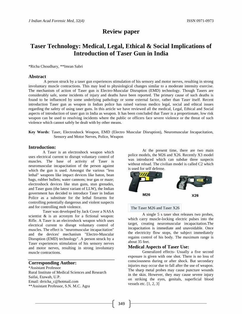
J Indian Acad Forensic Med, 32(4) ISSN 0971-0973
349
Review paper
Taser Technology: Medical, Legal, Ethical & Social Implications of
Introduction of Taser Gun in India
*Richa Choudhary, **Imran Sabri
Abstract A person struck by a taser gun experiences stimulation of his sensory and motor nerves, resulting in strong
involuntary muscle contractions. This may lead to physiological changes similar to a moderate intensity exercise.
The mechanism of action of Taser gun is Electro-Muscular Disruption (EMD) technology. Though Tasers are
considerably safe, some incidents of injury and deaths have been reported. The primary cause of such deaths is
found to be influenced by some underlying pathology or some external factor, rather than Taser itself. Recent
introduction Taser gun as weapon in Indian police has raised various medico legal, social and ethical issues
regarding the safety of using taser guns. In this article we have reviewed all the medical, Legal, Ethical and Social
aspects of introduction of taser gun in India as weapon. It has been concluded that Taser is a proportionate, low risk
weapon can be used to resolving incidents where the public or officers face severe violence or the threat of such
violence which cannot safely be dealt with by other means.
Key Words: Taser, Electroshock Weapon, EMD (Electro Muscular Disruption), Neuromuscular Incapacitation,
Sensory and Motor Nerves, Police, Weapon
Introduction:
A Taser is an electroshock weapon which
uses electrical current to disrupt voluntary control of
muscles. The base of activity of Taser is
neuromuscular incapacitation of the person against
which the gun is used. Amongst the various "less
lethal" weapons like impact devices like baton, bean
bags, rubber bullets; water cannons; tear gas or maze;
electroshock devices like stun guns, stun grenades,
and Taser guns (the latest variant of LLW), the Indian
government has decided to introduce Taser in Indian
Police as a substitute for the lethal firearms for
controlling potentially dangerous and violent suspects
and for controlling mob violence.
Taser was developed by Jack Cover a NASA
scientist & is an acronym for a fictional weapon:
Rifle. A Taser is an electroshock weapon which uses
electrical current to disrupt voluntary control of
muscles. The effect is "neuromuscular incapacitation"
and the devices' mechanism "Electro-Muscular
Disruption (EMD) technology". A person struck by a
Taser experiences stimulation of his sensory nerves
and motor nerves, resulting in strong involuntary
muscle contractions.
Corresponding Author: *Assistant Professor
Rural Institute of Medical Sciences and Research
Saifai, Etawah, U.P.
Email: [email protected]
**Assistant Professor, S.N. M.C. Agra
At the present time, there are two main
police models, the M26 and X26. Recently X3 model
was introduced which can subdue three suspects
without reload. The civilian model is called C2 which
is used for self defense.
The Taser M26 and Taser X26
A single 5 s taser shot releases two probes,
which carry muscle-locking electric pulses into the
target, creating neuromuscular incapacitation.The
incapacitation is immediate and unavoidable. Once
the electricity flow stops, the subject immediately
regains control of his body. The maximum range is
about 35 feet.
Medical Aspects of Taser Use:
Generalized effects- Usually a five second
exposure is given with one shot. There is no loss of
consciousness during or after shock. But secondary
injuries may occur due to fall after the use of weapon.
The sharp metal probes may cause puncture wounds
in the skin. However, they may cause severe injury
on striking the eyes, genitals, superficial blood
vessels etc. [1, 2, 3]
J Indian Acad Forensic Med, 32(4) ISSN 0971-0973
350
Cardiac Effects: The Taser produces a current of 0.36 - 1.76
joules per pulse. This is generally not expected to
affect cardiac conduction. No dysrhythmias have
been reported in small studies of healthy volunteers,
though cardiac dysfunction could occur in cases of
people on pacemakers, mentally ill, drug abusers,
epilepsy patients etc. [4, 5, 6]
Physiological Effects: The Systolic blood pressure decreased while
Diastolic blood pressures, heart rate, calcium,
sodium, potassium, bicarbonate and lactate levels and
blood pH changed slightly after a 5 second Taser
exposure.[6]
The VE, RR, and TV were mildly elevated for a brief
period; there was no metabolic acidosis induced
hyperventilation.[7] The heart rhythm before, during
& after taser exposure showed no adverse cardiac
effects or rhythm changes.[8, 9]
Legal Issues: Tasers are illegal or subject to legal
restrictions on their availability and use in many
jurisdictions. In India only licensed police officials,
CRPF men and National Security Guards can use the
Taser guns.
It is legal to use Taser or any force by the police if it
is found reasonable as measured from the perspective
of a reasonable officer at the scene.
The government has approved their use
against individuals fighting against the police rather
than on those who merely fail to comply with police
commands. The proposed TASER use policy for the
Indian Police Department would allow for the use of
the TASER against an individual only if that person
is actively resisting arrest or in circumstances where
deadly force is authorized. [10]
Though use of tasers for personal security or self
defense is legal in many countries, in India it is
illegal to carry a taser for personal use.
Indian Scenario: Indian Police and Central Reserve Police
Force personnel in occupied Kashmir have been
regularly using bullets and tear-gas shells to kill or
injure peaceful pro-freedom protesters. The firearms
have also killed a number of civilians during the anti-
India demonstrations.
Indian state police forces and central
security forces, which are conducting joint anti-
Maoist operations, have signed contracts for Taser
weapons with the Taser International. The Jammu
and Kashmir is the first state in India to order and use
Taser guns. Punjab has issued its police 80 of the
weapons. Madhya Pradesh and the anti-hijack teams
of National Security Guards are also likely to order
the weapon.
Ethical Aspects: The police officers or NSGs should focus on
the use of negotiation techniques and should not over
rely on Taser guns. They should use the Taser in
those situations where use of lethal force is also
permitted.
Taser should never be used on children, pregnant
females, elderly, and heart patients having
pacemaker, drug abusers, and mentally ill people.
This is to avoid likelihood of injury and death. [11]
The officer using a taser gun should be
properly trained in ethical use of force based on the
scenario.
The purpose of Taser is to subdue violent and
dangerous individuals. It should never be used solely
for the purpose of inflicting punishment or pain.
Taser should only be used on dangerous
individuals and never on those who are passively
resisting arrest.[12] Various philanthropic societies
like Amnesty International consider Taser as a form
of torture, causing extreme pain and sometimes
severe injury or even death of the person struck by
Taser. [13]
Social Aspects: Taser are guns are being used or overused in
various developed nations throughout the world. Its
recent introduction in Indian Law enforcement
system may have a dual impact on the society. From
the perspective of the police, Taser is a welcome
thing, providing them extra security while dealing
with hard core criminals or to disperse or sub due a
violent mob. As far as Indian society is concerned,
they may not like the idea of being tasered while
doing protests against the government. If some
incident of injury or death occurs due to Taser use,
this may lead to further protests by the Human Rights
Activists, against the use of taser. Besides, the law
authorities should also be prepared for dealing with
the compensation claims resulting from any
accidental injury by the use this controversial gun.
Discussion: Introduction of newer weapons in India is a
matter of discussion. Taser gun is just an example of
that. Many questions arise while using newer
techniques in India. Acceptability of newer weapons
depends on its outcome as well as social acceptance.
The main aim of the police is to control the
miscreants without or with minimum loss of life and
injuries. Use of taser can be a better alternative to
achieve this goal. A new weapon to be effective is to
be well accepted socially also. The political
intervention in use of newer techniques is also to be
seen. An ideal weapon for the police is that achieves
the target with minimum loss of life. Taser gun is
medically good technique as is causes temporary
incapacitation of the culprits with minimum effects
on the body. Legally the Taser gun cannot be given in
J Indian Acad Forensic Med, 32(4) ISSN 0971-0973
351
public hands as they can misuse it. However police
can be authorized to use it. Ethically taser can be
used with some exceptions. Socially it may be not
acceptable by the general public as is the case with
other weapons also. Keeping all this in view the
Taser gun is very close to an ideal weapon for the
police is day to day mob control exercises.
Conclusion: Thus we can say that introduction of Taser
gun has got minimum medical dangers, least legal
problems, ethically acceptable and socially sound. By
using a Taser a dangerous assailant or violent mob
could be controlled, thus preventing any injury or
harm to law enforcement officers, innocent citizens,
or themselves. This reinforces the value of Taser as a
useful tool to make the public and officers safer and
to resolve potentially violent situations effectively
and rapidly. To conclude we‟ll say Taser is a
proportionate, low risk means of resolving incidents
where the public or officers face severe violence or
the threat of such violence which cannot safely be
dealt with by other means.
References: 1. Mark W. Kroll, Hugh Calkins.Taser safety, CMAJ.
2007; Sept. 28(3):195-201.
2. Chen, S. L., Richard, C. K., Murthy, R.
C. Perforating ocular injury by Taser. Clin.& Exp. Opht., 2006;34: 378 –380.
3. Bozeman W.P., Winslow JE. Medical aspects of less
lethal weapons. Int. Jour. of Resc. & Dis. Med. (2005). 4. Jason Payne- James, Bob Sheridan, Graham Smith.
Medical implications of Taser, BMJ (2010); 340.
5. Strote J, HR Hutson. Taser use in restraint-related deaths. Prehosp Emerg Care 10:4, 447-450, 2006.
6. Bleetman, a., Steyn, R. & lee C. (2004) Introduction
of the Taser into British policing. Implications for UK
emergency departments: an overview of electronic
weaponry. Emergency Medicine Journal, 21, 136 –140
7. Lee. B.; Vittinghoff, E; Whiteman, D; Park, M; Lau,
LL; Tseng, ZH. "Relation of Taser (Electrical Stun
Gun) Deployment to Increase in In-Custody Sudden
Deaths". AMJCARD[2008] Pg 103 8. Vilke, Gary M. et al. “Physiological Effects of a
Conducted Electrical Weapon on Human Subjects” Annals of Emergency Medicine (2007):
1-7.H.O, Jeffrey D. et al. Cardiovascular and
Physiologic Effects of Conducted Electrical Weapon Discharge in Resting Adults. Acad. of Emer. Med.
(2006):1-7.
9. Jenkinson E., Neeson, C. & Bleetman, A. (2006). The relative risk of police use-of-force options: evaluating
the potential for deployment of
electroniweaponry. Journal of Clinical Forensic Medicine, 13, 229 –241.
10. Mcbride, DK., Tedder NB. (2006) Efficacy and Safety
of Electrical Stun Devices. 11. John Kleining. „Ethical constraints on Taser use by
police, Policing and Use of force (2007), 472-484.
12. Amnesty International USA. Amnesty International‟s Continuing Concerns about Taser Use. (2006).
13. Smith, J. and I. Greaves. The use of chemical
incapacitant sprays: a review. J Trauma, 2002. 52(3): p. 595-600.
14. Wilkinson, D. PSDB Further Evaluation of Taser
Devices. 2005, Hertfordshire, United Kingdom: Police Scientific Development Branch.
15. Suyama, J. et al. Injury patterns related to use of less-
lethal weapons during a period of civil unrest. J Emerg Med, 2003. 25(2): p. 219-27.
16. Levine S.D., S.C., Chan T, Vilke G, Dunford J.
Cardiac Monitoring of Subjects Exposed to the Taser. Acad Emerg Med, 2005. 12(5 Suppl 1): p. 71.
17. TASER Task Force Medical Findings. 2004, Orange
County Sheriff's Office Florida: Orlando, FL.

J Indian Acad Forensic Med, 32(4) ISSN 0971-0973
352
Review paper
Aluminium Phosphide Poisoning: Management and Prevention
*Dr.S.Ranjan Bajpai
Abstract Death by Aluminium phosphide (AlP) and organo-phosphide poisoning are the commonest forms of
poisoning in India. Aluminium phosphide is used as rodenticide and pesticide in grain storage facilities. It produces
phosphine gas, which results in mitochondrial poisoning. There is no known antidote for AIP poisoning. The effects
of AlP poisoning, mediated by phosphine and mechanism of action have not been established.The information
related to this poisoning is available in the literature and substantiated by practical experience. Liberated phosphine
cannot be detoxified but Magnesium sulphate has been reported to be effective. Another remedy can be rapid
absorption by coconut oil which might be helpful.
Treatment consists of gastric lavage with potassium permanganate solution, oral administration of charcoal
and sorbitol suspension, intravenous administration of sodium bicarbonate, magnesium sulphate and calcium
gluconate, and oral administration of sodium bicarbonate and coconut oil. Use of coconut oil for treatment in this
poisoning is suggested, although clinical trials and scientific approval is still awaited. Every suggestion, news
regarding new modality of treatment for this poisoning needs to be shared in the interest of saving precious human
lives. It is with this aim the present paper is written.
Key Words: Aluminium Phosphide, Phosphine, Rodenticide, Pesticide
Introduction: Agrochemical poisoning is a major public
health problem in developing countries particularly in
setting of low education and poor regulatory
frameworks. Aluminium phosphide is a solid
fumigant pesticide. It is marketed in India as tablets
of celphos and quickphos. Aluminium phosphide has
currently aroused interest with increasing number of
cases in the past three decades due to increased use in
agricultural and non-agricultural purpose; hence easy
availability has increased its misuse to commit
suicide.
Management: Patients with Aluminium phosphide
poisoning are managed conservatively as no specific
antidote to the poison is known. The objective of the
treatment is to provide symptomatic and supportive
aid to the patients till phosphine is excreted through
the lungs and kidneys.
A. Reducing absorption of phosphine:
i. Gastric lavage with KMnO4 (1:10000) or
with magnesium sulphate (MgSO4) to
oxidise the unabsorbed poison.
Corresponding Author: *Assistant Professor
Department of Forensic Medicine
A. J. Institute of Medical Sciences
Mangalore (Karnataka)
Email: [email protected]
Mobile No.: +91 - 9425301920
ii. Slurry of activated charcoal orally or
through nasogastric tube to adsorb
phosphine from the gastro intestinal tract.
iii. Antacids, to reduce symptoms pertaining to
the stomach and to reduce the absorption of
phosphine through the stomach, H2
antagonists can also be used.
iv. Medicated liquid paraffin or MgSO4 to
accelerate the excretion of Aluminium
phosphide and phosphine from the gastro
intestinal tract.
B. Reducing Cellular Toxicity of phosphine:
Phosphine produces atmosphere of hypoxia
and free radical stress by binding with cytochrome
oxidase and by inhibiting catalase. Magnesium ions
help in scavenging free radicals through GSH
recovery hence is effective as parenteral antioxidant
in this poisoning. Magnesium is anti-hypoxic and
anti-arrhythmic agent, hence due to all its beneficial
effects; it is effective in reducing the mortality in this
poisoning. The dose schedule observed to be
effective was 1 gm of MgSO4 i. v. stat followed by 1
gm after every 4 – 6 hours, with magnesium levels
remaining within safe limits (3 – 3.6 mEq.). Lower or
higher dosages have not been observed to be much
effective.
Liberated phosphine cannot be detoxified as
there is no specific antidote available. Magnesium
sulphate has been reported to be effective in reducing
mortality but some workers had found no significant
difference in dose related mortality rates in patients
treated with and without MgSO4. In a study
J Indian Acad Forensic Med, 32(4) ISSN 0971-0973
353
conducted by U.K.Singh et al. (1997), survival was
better in patients who presented within 4 hours and
i.v. MgSO4 was started and the authors believe that
MgSO4 therapy should be instituted early in all
patient(s) with Aluminium phosphide poisoning,
based on serum magnesium levels, considering the
high mortality of Aluminium phosphide and lack of
specific antidote.
Magnesium sulphate has been tried with
limited success for its general membrane stabilising
effect in cardiac cells.
Sushil Kumar et al. (1990) report that shock
and ECG changes in Aluminium phosphide poisoning
cases could be reverted in large percentage of cases
with larger doses of i.v. MgSO4, which raised the
serum Mg levels to about 5.2 mEq/lt.
Chugh et al. (1994) in a study on 155
patients of Aluminium phosphide poisoning studied
the efficacy of magnesium sulphate in relation to
dose of Aluminium phosphide ingested and
concluded that the effect on is mortality related to the
dose of pesticide consumed. They also observed that
MgSO4 reduced the mortality more or less equally,
irrespective of the dose of pesticide consumed.
Siwach et al. (1994) have also observed that
dose related mortality rates in patients treated with
and without MgSO4 are not significantly different.
They estimated serum Magnesium at six points
within first 24 hours and found no evidence of
hypomagnesemia at any stage, they also observed
that the chemical method of Mg++
estimation
(especially tital yellow) may give low values in
patients receiving calcium gluconate simultaneously
and tissue Mg++
reflects more accurately total Mg++
status of the body in comparison to serum level alone.
Magnesium estimation was done by them using
atomic absorption spectrophotometer which is the
more reliable method for Mg++
estimation.
Magnesium content of various organs in non
survivors and RBC Mg++
content along with serum
level were estimated. Hypomagnesemia was not
observed in any case. Magnesium content in the
organs was observed to be significantly higher than
control.
The authors also state that magnesium is
weak antiarrythmic agent and may be useful in
controlling few supra ventricular arrhythmia and
recommend use of more potent antiarrythmic agent
like Amiodarone and continued cardiac monitoring
for timely control ventricular arrythmias observed in
Aluminium phosphide poisoning.
Siwach also report (1997) that correction of
acidosis by slow infusion of sodium bicarbonate is
very helpful. If blood gas analysis is possible, total
base deficit may be calculated using the following
formula.
Base deficit = 0.4 x body wt. x desired HCO3-
measured HCO3- (mEq).
(Desired HCO3-
= 25 mEq/L) half of the calculated
dose should be infused immediately and repeated
doses calculated in every 3 hours. If blood pH
improves with use sodium bicarbonate, it is
associated with good prognosis. C. Steps to increase phosphine excretion:
Phosphine is stable and is excreted through
breath and urine. Adequate hydration, renal perfusion
with IV fluid and low dose dopamine (4 to 6 micro
gm/kg/min) enhances the excretion through urine.
Diuretic in presence of BP 70 – 80 mm Hg may be
useful.
Dialysis is indicated in cases with acute
renal failure if the patient is haemodynamically
stable. Supported measures in the treatment of
Aluminium phosphide poisoning include treatment of
hypoxia by oxygen inhalation or endotracheal
intubation or assisted ventilation along with
monitoring of blood gases. Treatment of shock with 3
to 4 liters of 0.9 % saline in first 8 to 12 hours, low
dose dopamine with dobutamine, hydrocortisone 200
to 400 mg I/V after 4 to 6 hours to reduce dose of
dopamine to check the capillary leakage in lung and
to potentiate the responsiveness of shock to
endogenous catacolamines. Hydrocortisone also
compensated for low levels of cortisone found in this
poisoning.
Prevention: Since death is rapid and survival after
significant poisoning is rare, prevention is the logical
option. The most effective way for prevention is to
either ban or impose strict regulation on the sale of
aluminium phosphide tablets.
Cageing of tablets in smaller plastic with
holes and spikes so that they can‟t be swallowed as
such, is likely to reduce the incidence of Aluminium
phosphide poisoning.
Preventive measures have been classified by Chug et
al. under following headings:
I. Prophylaxis during and after
fumigation at home and outside: a. Protect the copper containing parts before
fumigation.
b. Do not fumigate the grains when
temperature is below 50C.
c. Adequate washing facility should be
available at times during handling.
d. Eating, drinking, smoking should be
prohibited during and before washing after
handling.
e. During fumigation, operator must be given
efficient respiratory protection.
f. Complete the distribution of tablets within
limited time.
g. In warehouses, person should not sleep in
the room fumigated.

J Indian Acad Forensic Med, 32(4) ISSN 0971-0973
354
h. Wear synthetic rubber gloves, boots, light
weight impervious clothes, apron, and
suitable eye protectives during fumigation
and at the time of opening the container.
i. Always open the container in air.
j. Do not inhale dust or fumes of fumigated
grains.
k. Use of phosphine detectors strips before
entering a fumigated area.
The optimum temperature for fumigation is
150C to 20
0C and humidity 63.7 %. When fumigated
grains is exposed to air for half an hour, the whole
phosphine gets dissipated rapidly and the cereals
become fit for consumption. Phosphine in humid air
is oxidised and becomes non-toxic.
II. Education: Farmers handling the fumigant must be
made aware of its lethal aspects. They should demand
the required amount of tablets. They should be
advised to bury the tablets in open fields after use.
They should keep their tablets away from the reach of
children and other family members.
III. Advice to Government Agencies: State agencies should restrict the open sales
of this pesticide. The tablets should not be given to
young persons and children without proper
verification and confirmation. Dealers not following
the Government instructions should be punished. The
manufacturers should be advised to make small packs
of 2 – 3 tablets with suitable container. If possible the
pesticide should be banned.
References: 1. Clinical Toxicology, Nov.2009,Vol.47,No.9, Pages
908-909, DOI- 10.3109/15563650903285657
2. Anger, Francois; Paysant, Francois and et al . Journal of
Analytical Toxicology,Vol.24,No.2,March 2000, Pages 90-92(3)
3. Human and Experimental Toxicology, Vol.24,No.4,
pages 215-218 (2005) DOI- 10.1191/0960327105ht5130a
4. Shadnia Shahin, Rahini Mojgan, Pajournand
Abdokarim, Rasouli Hasein Mohd. Poison Center,
lohgman Hakim Hospital,Shaheed-Behesthti University
of Medical Sciences Research Centre, Tehran,Iran –Successful treatment of acute aluminium phosphide
poisoning: possible benefit of coconut oil.
5. Chugh SN, Jaggal KL, Malhotra KC. 1990. Steps to reduce the mortality in Aluminium phosphide
poisoning. JAPI, 38 – 32.
6. Chugh SN. 1992. Aluminium phosphide poisoning – Present status and management. JAPI. 40; 401 – 405.
7. Chugh SN, Mittal S, Seth S, Chugh K. 1995. Lipid
peroxidation in acute aluminium phosphide poisoning. JAPI. 43; 265 – 66.
8. Chugh SN, Prem Kumar, Aggrawal HK et al. 1994.
Efficacy of magnesium sulphate in Aluminium
phosphide poisoning – Comparison of two different
dose schedules. JAPI. 42; 373 – 75.
9. Siwach SB, Pratap Singh, Sushil Ahlawat, Abha
Dua, Deepak Sharma. 1994. Serum and tissue
magnesium content in patients of acute Aluminium
phosphide poisoning and critical evaluation of high dose magnesium sulphate therapy in reducing mortality.
JAPI. 42 (2); 107 – 10. 10. Avasthi R, Sharma R. 1994. Aluminium phosphide
poisoning and magnesium sulphate therapy. JAPI. 42;
670. 11. Singh UK, Chakraborty B, Prasad R. 1997.
Aluminium phosphide poisoning: A growing concern in
pediatric population. Indian Pediatrics. 34; 650 – 51. 12. Sepaha GC, Bharani AK, Jain SM et al. 1985. Acute
Aluminium phosphide poisoning. JIMA. 83; 378.
13. Iseri LT, Chugh P, Tobis J. 1983. Magnesium therapy for intractable ventricular tachyarrhythmias in normal
magnesemic patients. West J. Med. 138; 823 – 826.
14. Sushil Kumar, Elhence GP, Mital HS, Mehrotra VS,
Khanna PN. 1990. Intravenous magnesium sulphate
therapy in aluminium phosphide poisoning. JAPI. 38
(1); 32. 15. Siwach SB. 1997. Recent trends in the management of
acute aluminium phosphide poisoning. Post Graduate
Medicine. 11; 411 – 413. 16. Sharma A. 1995. Oral Aluminium phosphide
poisoning. Indian Pediatrics. 32; 339 – 342.
17. Koley TP. 1998. Aluminium phosphide poisoning – Part I. Indian Journal of Clinical Practice. 9; 5.

J Indian Acad Forensic Med, 32(4) ISSN 0971-0973
355
Review Paper
Serum Enzymes Changes after Death & Its Correlation with
Time since Death *Dr. S. P. Garg, **Dr. Vidya Garg
Abstract Estimation of time since death is one of the primary objectives of an autopsy. Forensic Scientists and
researchers have been persevering hard to find out methods of accurate determination of postmortem interval since
long. However, the concept of “Postmortem Clocking” so far seems to be a distant dream only. The favorite
biological fluids, to study postmortem biochemical changes, have been those which withstand putrefactive changes
for longer duration, like vitreous humor, cerebrospinal fluid, pericardial fluid etc. In blood, markers like electrolytes,
urea, creatinine, glucose etc have been more commonly studied. Enthusiastic studies have been undertaken by
various researchers to find out reasonably reliable methods of estimating postmortem interval by studying serial
quantitative changes in serum levels of various enzymes and to extrapolate the data obtained therefore in terms of
duration of death. However, the accuracy of such an opinion remains big area of concern even today, as the range of
duration is mostly too wide to be practically useful.
Key Words: Time since death, Serum enzymes, Amino Transferases, Acid Phosphatase
Introduction: The estimation of time since death is most
important yet most inaccurate & controversial topics
in Forensic Medicine.[12]When calculated
accurately, it has the potential to unravel many
unfolded medico-legal mysteries. The estimation of
time since death by whatsoever means or methods
relies on changes that occur in the body after death
[10]. Autopsy surgeons continue to rely on age old
subjective methods of observing the degree and
chronological staging of external as well as visceral
postmortem somatic changes like cooling of the
body, rigor mortis, changes in the eyes, hypostasis,
signs of decomposition, mummification, adipocere
formation, maggot infestation etc corroborated with
circumstantial evidences for the same.
Review of Literature: Little has changed from the ancient days,
except that the data acquisition equipment then was
merely the back of the hand to test the coolness of the
corpse‟s skin and their eyes & nose to evaluate
decomposition, we now have multichannel
thermometry with thermocouples sensitive to a
fractions of a degree, enzyme methods, vitreous
chemistry, muscular reactivity and several other
avenues for collecting data.
Corresponding Author: * Associate Professor,
Department of Forensic Medicine
E-14, Doctor‟s Colony, Rewa (MP) 486001
E–mail:[email protected]
Mobile: 094254-70383
**Assistant Professor, Department of Physiology
Bundelkhand Medical College
Regrettably, the accuracy of estimating the
postmortem interval has by no means kept pace with
the enormous strides made in technological
sophistication [13]. Hence the estimation of time
since death continues to be a difficult task for the
Forensic Pathologist [19] and there is a continuous
need for the development of an accurate method by
which the time of death can be determined to within a
few minutes.
The first thing to recognize is that an
estimate based merely on the changes in the body
after death is liable to serious error [9], highly
inaccurate [4] and only an approximation can be
achieved if proper observations are made from the
beginning [19], [16]. K S N Reddy [22] goes to the
extent of saying that that exact time of death cannot
be fixed by any method. However Jung wrote of
Columbus that “using subjective assumptions, a false
hypothesis and a route abandoned by modern
navigation nevertheless (he) discovered America [9].
Several studies have been undertaken to find
out objective methods such as biochemical,
histological, serological assays etc involving variety
of biological fluids & tissues. In the past enthusiastic
studies have been undertaken by various researchers
to find out some such methods by studying serial
quantitative changes in serum levels of various
enzymes and to extrapolate the data obtained
therefore in terms of duration of death. However, the
accuracy of such an opinion is a big area of concern,
as the range of duration is mostly too wide to be
practically useful.
The favorite biological fluids, to study
postmortem biochemical changes, have been those
which withstand putrefactive changes for longer

J Indian Acad Forensic Med, 32(4) ISSN 0971-0973
356
duration, like vitreous humor, cerebrospinal fluid,
pericardial fluid etc than blood. The available
literature on biochemical (enzymal) changes in
postmortem blood (serum) and its relation with time
since death is largely contributed by forensic
scientists from the temperate countries. Nandy [17]
cautions that the values of such studies may not be
usefully applicable in our country over a vast area as
the decomposition is much advanced by 3rd
/4th
day
after death. Pillay[21] is of opinion that though a lot
of research work has been undertaken to estimate the
time since death based on biochemical changes in
various body fluids after death, they are mostly only
of academic interest. They are of limited practical
value.
Transaminases/ Aminotransferases: Several authors and scientists have reported
a definite & progressive rise in the levels of
transaminases in serum of the dead body as the time
since death inceases but the most elaborate study has
been done by Hall [11], Enticknap [6] and Coe [3].
Parikh C. K. [19], Modi JP, Subramanium B. V. [16],
K S N Reddy [22], Simpson [23] andBiswas[2] have
reported definite rise in these enzymal levels after
death but do not offer any details regarding
estimation of time since death from such results.
However, Mukherjee [15] & Nandy [17] report the
rising trend of serum aminotransferase levels up to
second to third day of death and opine that such
calculations are too erratic and asymmetrical to be of
any help in anything like “Postmortem Clocking”.
Evans [7] establishes the initial rise of
transaminase activity of three to four hours after
death and there was a fairly steady climb to high
levels until about sixty hours postmortem (also
reported by B Knight [13]), when diminution activity
commenced.
Hall [11] reported a large, rapid postmortem
increase in SGOT in intracardiac blood but found
little elevation of the level in femoral blood from
clinically well patients who died abruptly.
Enticknap [6] showed a striking progressive
increase in SGOT levels in blood from arm vessels.
The level rose from nearly nil to about 10 µm per ml
per hour after 4 hours and then the curve became less
steep so that a about 18 hours after death the level
was about 15 µm per ml per hour. The graph then
became nearly horizontal up to 30 hours after death
when again the graph line rose steeply to register the
peek at about 55 hours after death at the level of
about 28 10 µm per ml per hour. From this point the
level kept on falling till about 66 hours. He observed
that such a trend of change can be used to estimate
time since death.
Acid Phosphatase: Parikh C. K. [19], Modi JP, Subramanium
B. V. [16], K S N Reddy [22], Simpson [23] and
Biswas [2] have reported definite rise in these
enzymal levels after death. Garg et al [16] while
reporting rise in serum levels with increasing
postmortem interval, did not find any statistically
significant correlation. Nandy [17]& Dixit [5] reported peak level
in between 36 to 48 hours after death, while
Mukherjee [15] noted the rising titre in the first two
to three days after death. Reddy [22] reports 20 times
increase in Acid Phosphatase level in 48 hours after
death.
Enticknap[6] reported rise of serum Acid
Phosphatase level from just more than nil just after
death to 2.5 King Armstrong unit at 18 hour
postmortem then there was transient fall up to 30
hours after death (15 KA Units). Again the enzymal
levels registered rise so that at 40 hours the peak was
attained at 40 kA unit level and finally the levels fell
till 50 hours time at 25 kA units and remained at the
same level for well beyond 60 hour time after death.
Alkaline Phosphatase: Naumann [18] pointed out that Alkaline
Phosphatase level reached average concentration of
5.3 Bodansky units in 14 cases 101/2
hours after death
(normal antemortem range 1.5 to 4 Bodansky unit).
Enticknap [6] used King Armstrong unit and showed
that the concentration rose from 8 kA units just after
death to 40 kA units after 30 hours and then rose
steeply up to 40 hours when it recorded a peak of 70
kA units and afterwards fell downwards.Coe [3]
opined that the concentration almost doubled and
tripled 8 & 18 hours after death respectively.
Amylase: Enticknap [6] demonstrated that amylase
levels after death showed double peaks or a biphasic
rise. It rose steeply from 100 Somoghiunit just after
death to 350 units 6 hours after death, and then it
declined to the level of 150 units after 30 hours and
again registered a peak of more than 350 units 40
hours after death and finally dropped to 200 unit level
after 50 hours of death. Reddy [22] reports 3 to 4
times increase in amylase level on second day after
death.
Lactate Dehydrogenase: Enticknap [6] studied changes in Lactate
Dehydrogenase levels in serum after death and found
that there was almost linear increase in concentration
of this enzyme up to well over 60 hours and he
suggested that peak may not be attained until fourth
day and because of linear rise it may be useful in
calculating time since death. Lythgoe [14] did not
find a linear relationship in rise in enzyme activity.
He suggested that the rise was in three phases. First
being a rapid rise immediately after death, then a
slower increase up to 30 hours postmortem and then
final rise probably reflecting the onset of vascular
hemolysis.

J Indian Acad Forensic Med, 32(4) ISSN 0971-0973
357
Esterase including Cholinesterase: Petty et al [20] found no significant
difference in true blood Cholinesterase level between
refrigerated and non refrigerated samples and no
significant decrease in activity in samples
periodically analyzed up to three weeks after death.
Arnason and Biarnason [1] studied total
serum esterase by starch gel electrophoresis on
postmortem sera obtained from ¼ hour to 720 hours
after death. Out of 5 fractions found in living
individuals, several fractions disappear in
postmortem serum, one fraction becomes stronger,
continuing to increase in strength for at least 1000
hours after death, and at least one new fraction
develops which is not found in serum from living
patents.
Phosphoglutamutase: Dixit PC et al [5] studied the relationship of
Phosphoglutamutase in postmortem blood with time
and cause of death. They concluded that the
estimation of time since death depends upon many
factors, including environmental conditions.
Stability of Enzymes after Death: Generally speaking it is best to do enzyme
assays within four hours from time of blood
collection. All enzymes can be stored overnight at 0-
40C, but Acid Phosphatase may not keep
satisfactorily at room temperature. Aldolase and
Alanine Aminotransferase are unstable when frozen
at-250 C but keeps all right at 4
0C.
Effects of Anticoagulants: Anticoagulants inhibit some enzymes so
serum is preferred to plasma. Citrate inhibits
amylase; fluoride inhibits many enzymes while
Ethylene DiamineTetraacetic Acid inhibits
phosphatase. Heparin has been recommended if
plasma is to be used but has been said to inhibit
creatine kinase and Lactate Dehydrogenase. Oxalates
also inhibit several enzymes.
Effect of Hemolysis: As Red Blood Corpuscles (RBCs) are much
richer than the plasma in several enzymes, it is
necessary to avoid hemolysis. Also on storing some
leakage of enzymes may occur from red cells,
leukocytes and platelets. The early separation of
serum should be done. However exceptions do exist
like Glucose 6 Phosphate Dehydrogenase and Acid
Phosphatase.
Garg et al [8] observed that increasing
hemolysis of postmortem blood with greater
postmortem interval seems to be the single most
important confounding factor giving erroneously high
values with photoelectric colorimetry. Definite and
marked rise in serum enzymal levels after death was
noted by them from 2 hours after death onwards.
They also noted that in many cases with
increasing time since death enzyme levels register
increasing values but interspersed cases show such
abnormality and non regular high or low values that
deciphering the graphical pattern thus drawn
involving two variables in terms of time since death
seems unwisely. The refrigerated bodies and samples
gave abnormally low values. The cases dying of
multiple injuries involving trauma to liver showed
markedly high levels. An interesting finding was that
in burn cases the graph was relatively more linear.
They summarized that the conventional and routinely
used subjective parameters like rigor mortis,
hypostasis, cooling of the body, putrefactive changes
etc. combined with the experience, acumen and "third
eye" of the medicolegal expert and circumstantial
evidence remain to be the best available tools for
estimation of time since death.
References: 1. Arnason A., Bjarnason O. (1972). Postmortem changes of
human serum esterases Acta Patho lMicrobiol Scand (A) 80;
841-846.
2. Biswas G. (2010). Review of Forensic Medicine & Toxicology, Jaypee Publication, 1st edition; 109.
3. Coe J.I. (1974). Postmortem chemistry, practical
considerations and a review of literature J. Forensic Sci; 19:13-32.
4. Cox H.M.V., Sinha U.S., Knight B. (1995). Medical
Jurisprudence & Toxicology 6th edition The law book company (P) ltd. Allahabad: 206.
5. Dixit P.C. (2007). Text Book of Forensic Medicine &
Toxicology, Peepe Publishers 1st edition: 110. 6. Enticknap J.B. (1960). Biochemical changes in cadaver sera
J. Forensic Med; 7:135-146.
1. Evans WED (1963). The Chemistry of Death Charles C. Thomas Springfield; 100.
2. Garg S.P., Arora A, Dubey BP (2005).A study of Serum
Enzymal changes after Death & its correlation with Time since Death. JIAFM: 27 (1). ISSN 0971-0973
3. Glaister (1973). Medical Jurisprudence & Toxicology 13th
edition Churchil Livingston: 118. 4. Gradohl. Legal medicine (1976). 3rd edition (Indian) K M
Verghese Company Bombay: 95-97.
5. Hall W.E.B. (1958). The medicolegal application of the serum transaminase test J. Forensic Sci; 3:117-122.
6. Knight B. Lawyer‟s guide to Forensic Medicine William
Heinmann Medicine Books Limited, London 1992.
7. Knight B, Henssge C, Kranpecher T, Madea b, Nokes L. The estimation of time since death in the early postmortem period Arnold Publishers 1995.
8. Lythgoe A.S. (1980). The activity of Lactate Dehydrogenase
in cadaver sera: a comparison of different sampling sites. Med. Sci. Law Vol. 20, No. 1: 48-53.
9. Mukherjee J.B. (1994). Forensic Medicine & Toxicology
2nd edition Arnold Associates Vol. I ; 253-254. 10. Modi JP and Subramanium B. V. (1998). Medical
Jurisprudence & Toxicology. 22nd edition, Butterworths
Publication: 246. 11. Nandy A. (2010). Principles of Forensic Medicine including
Toxicology 3rd edition: 280.
12. Naumann H.N. Postmortem (1956). Liver Function Tests. Am. J. Clin. Pathol. 26: 495-505.
13. Parikh C. K. Textbook of Medical Jurisprudence &
Toxicology (1992). 5th edition Medico-legal Center, Bombay 173
14. Petty C.S., Lovel M.P., Moore F. (1958).Organophosphorus
insecticides and postmortem blood cholinesterase levels J. Forensic Sci 3 226-237.
J Indian Acad Forensic Med, 32(4) ISSN 0971-0973
358
Review Paper
A Review of Medicolegal Consequences of Gossypiboma
*Monika Garg, **Akash Deep Aggarwal
Abstract Foreign bodies forgotten in the abdomen include towels, artery forceps, pieces of broken instruments or
irrigation sets and rubber tubes. The most common surgically retained foreign body is the laparotomy sponge. Such
materials (textilomas or gossypibomas) cause foreign body reaction in the surrounding tissue. The complications
caused by these foreign bodies are well known, but cases are rarely published because of medico legal implications.
The diagnosis of gossypiboma and the second surgical operation needed for removal of medical problem can lead to
start of legal problem between the patient and the surgeon at fault. The medico legal consequences of gossypiboma
are significant. Patients may be inadvertently informed that masses might be malignant and may undergo
unnecessarily invasive investigations, procedures or operations. Gossypiboma may lead to disappointing and
undesired consequences for a surgeon; moreover, it is one of the significant medico-legal problems needs to be
solved by specialists of forensic medicine.
Key Words: Gossypiboma, Retained, Sponge, Foreign Body, Legal
Introduction Forgotten or missed foreign bodies, such as
cotton sponges, gauze or instruments, after any
surgical procedures are considered a misadventure
and is associated with several legal problems. The
term “gossypiboma” denotes a mass of cotton
retained in the body after any intervention. [1] This
term is derived from the Latin gossypium for
“cotton” and the Swahili word boma for “a place of
concealment.” Other terms used for gossypiboma
include “textiloma”, “cottonoid”, “cottonballoma”
“muslinomas” or “gauzeoma”. Gossypiboma was
rarely reported in literature and the reports of this
technical oversight are the tip of an iceberg because
the symptoms of gossypiboma are usually
nonspecific and some patients remain asymptomatic
and are never discovered or documentation is not
enough in some diagnosed cases. Data concerning the
incidence of gossypiboma tend to fluctuate and the
incidence of gossypiboma is difficult to estimate
because of a low reporting rate lest medico-legal
implication. [2]
PubMed reveals about 300 reported cases of
gossypiboma worldwide, out of which about 15 from
India. Whilst the date of the arising out of first
malpractice suit about gossypibomas was reported to
be 1933 in medical literature, this date was reported
to be 1897 by some medical authors who researched
court records and located judgments. [2]
Corresponding Author: *Assistant Professor, Pathology
**Assistant Professor, Forensic Medicine
Post Graduate Institute of Medical Sciences, Rohtak,
E-mail: [email protected]
Some textilomas cause infection or abscess
formation in the early stage, whereas others remain
clinically silent for many years. Most cases of
textiloma in the literature have been connected with
abdominal or thoracic surgery; very few have been
linked with spinal surgery. Although precautions are
taken to avoid leaving such materials behind,
mistakes do happen and the resultant foreign bodies
can cause various clinical and radiological
manifestations. In the early period after surgery, these
forgotten materials can lead to infections and abscess
formation. However, some remain clinically
asymptomatic for many years, and then cause a
foreign body reaction in the surrounding tissue, with
new clinical signs indicating significant mass effect.
Foreign bodies that are left behind during operations
may organize and increase in size but such changes
are not correlated with time. To date, the case
reported with the longest period from surgery to
manifestation of symptoms is an intrapulmonary
foreign body 43 years after thoracotomy. Civil
lawsuits brought against surgeons for surgical
complications are becoming more frequent, and this
is prompting surgical teams to be even more careful.
It is possible to overlook cotton and gauze pads in the
surgical field. [3]
The medico-legal consequences of
gossypiboma are significant. Patients may be
inadvertently informed that masses might be
malignant and may undergo unnecessarily invasive
investigations such as angiography and unnecessarily
radical extirpative surgery. [4]
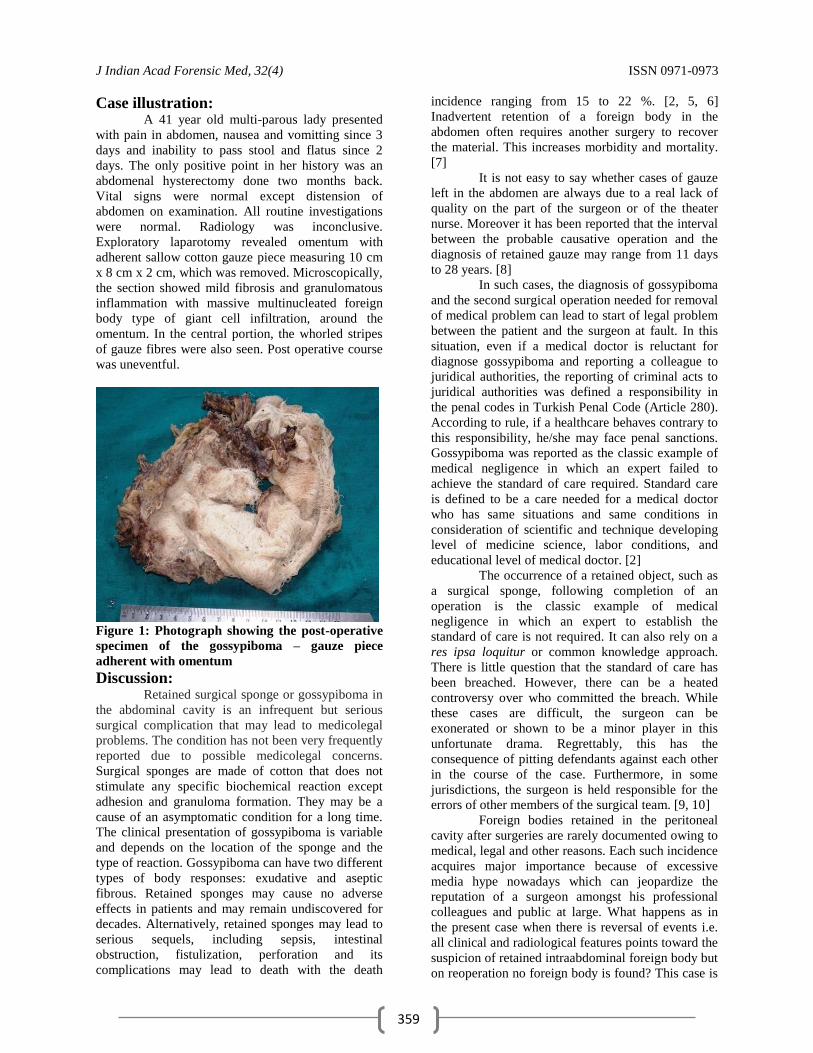
J Indian Acad Forensic Med, 32(4) ISSN 0971-0973
359
Case illustration: A 41 year old multi-parous lady presented
with pain in abdomen, nausea and vomitting since 3
days and inability to pass stool and flatus since 2
days. The only positive point in her history was an
abdomenal hysterectomy done two months back.
Vital signs were normal except distension of
abdomen on examination. All routine investigations
were normal. Radiology was inconclusive.
Exploratory laparotomy revealed omentum with
adherent sallow cotton gauze piece measuring 10 cm
x 8 cm x 2 cm, which was removed. Microscopically,
the section showed mild fibrosis and granulomatous
inflammation with massive multinucleated foreign
body type of giant cell infiltration, around the
omentum. In the central portion, the whorled stripes
of gauze fibres were also seen. Post operative course
was uneventful.
Figure 1: Photograph showing the post-operative
specimen of the gossypiboma – gauze piece
adherent with omentum
Discussion: Retained surgical sponge or gossypiboma in
the abdominal cavity is an infrequent but serious
surgical complication that may lead to medicolegal
problems. The condition has not been very frequently
reported due to possible medicolegal concerns.
Surgical sponges are made of cotton that does not
stimulate any specific biochemical reaction except
adhesion and granuloma formation. They may be a
cause of an asymptomatic condition for a long time.
The clinical presentation of gossypiboma is variable
and depends on the location of the sponge and the
type of reaction. Gossypiboma can have two different
types of body responses: exudative and aseptic
fibrous. Retained sponges may cause no adverse
effects in patients and may remain undiscovered for
decades. Alternatively, retained sponges may lead to
serious sequels, including sepsis, intestinal
obstruction, fistulization, perforation and its
complications may lead to death with the death
incidence ranging from 15 to 22 %. [2, 5, 6]
Inadvertent retention of a foreign body in the
abdomen often requires another surgery to recover
the material. This increases morbidity and mortality.
[7]
It is not easy to say whether cases of gauze
left in the abdomen are always due to a real lack of
quality on the part of the surgeon or of the theater
nurse. Moreover it has been reported that the interval
between the probable causative operation and the
diagnosis of retained gauze may range from 11 days
to 28 years. [8]
In such cases, the diagnosis of gossypiboma
and the second surgical operation needed for removal
of medical problem can lead to start of legal problem
between the patient and the surgeon at fault. In this
situation, even if a medical doctor is reluctant for
diagnose gossypiboma and reporting a colleague to
juridical authorities, the reporting of criminal acts to
juridical authorities was defined a responsibility in
the penal codes in Turkish Penal Code (Article 280).
According to rule, if a healthcare behaves contrary to
this responsibility, he/she may face penal sanctions.
Gossypiboma was reported as the classic example of
medical negligence in which an expert failed to
achieve the standard of care required. Standard care
is defined to be a care needed for a medical doctor
who has same situations and same conditions in
consideration of scientific and technique developing
level of medicine science, labor conditions, and
educational level of medical doctor. [2]
The occurrence of a retained object, such as
a surgical sponge, following completion of an
operation is the classic example of medical
negligence in which an expert to establish the
standard of care is not required. It can also rely on a
res ipsa loquitur or common knowledge approach.
There is little question that the standard of care has
been breached. However, there can be a heated
controversy over who committed the breach. While
these cases are difficult, the surgeon can be
exonerated or shown to be a minor player in this
unfortunate drama. Regrettably, this has the
consequence of pitting defendants against each other
in the course of the case. Furthermore, in some
jurisdictions, the surgeon is held responsible for the
errors of other members of the surgical team. [9, 10]
Foreign bodies retained in the peritoneal
cavity after surgeries are rarely documented owing to
medical, legal and other reasons. Each such incidence
acquires major importance because of excessive
media hype nowadays which can jeopardize the
reputation of a surgeon amongst his professional
colleagues and public at large. What happens as in
the present case when there is reversal of events i.e.
all clinical and radiological features points toward the
suspicion of retained intraabdominal foreign body but
on reoperation no foreign body is found? This case is
J Indian Acad Forensic Med, 32(4) ISSN 0971-0973
360
being reported to emphasize the fact that even when
there is high index of suspicion for a retained
intraabdominal foreign body, the reoperation may be
carried out by explaining the indication of resurgery
different rather than retained foreign body, as
incisional hernia in the present case, to avoid
unnecessary embarrassment. [11]
Because these cases are avoidable and
frequently injurious, many lead to malpractice
claims; given the high likelihood of litigation after
such cases, most liability insurers also encourage
clinicians and hospitals to report them. Therefore, we
used malpractice-insurance files from several
institutions to identify cases. Malpractice claims and
reports are an imperfect representation of the true
incidence and nature of any complication. Some
cases of retained foreign bodies undoubtedly did not
result in either a claim by a patient or a report by the
physician to the insurer. The factors involved in such
cases may differ from those in the cases we studied.
However, we know of no reason why they would
differ in terms of the mechanism of causation. In
addition, these mishaps appear to have a high
likelihood of leading to litigation, given how
injurious and potentially avoidable they are. [12]
It is clear in most recent publications that the
rate described is grossly underestimated; reasons for
this are related to the possible medico-legal
implications, the fear of litigation which could end up
in heavy expenses for compensations and adverse
publicity for institutions and surgeons; in fact, it is
clear that the responsibility of the surgeon and
members of the team in the Operation Theatre could
be called in case of litigation. Concerning the
medico-legal aspect, the local laws define
responsibilities and compensation mechanism
following what is described as medical negligence;
but, despite the fact that all our patients and/or their
relatives were informed of the findings, none of our
cases resulted in malpractice claim! No rate of
retained foreign body can be considered “acceptable”
whatever the environment and conditions of work;
their consequences in terms of morbidity and
mortality can still be too heavy and costly. Their
management will still rely for a long time on
prevention because in almost all cases, it could be
related to human errors; this type of errors will
probably never be completely abolished, but the
incidence of retained surgical sponge can be reduced
to a “minimum” by strict adherence to regulations,
especially systematic and rigorous sponge count; this
is particularly important during emergency
procedures. [13]
In medical literature, there are few articles
about the medico-legal evaluation of gossypiboma.
One such case from Turkey reports that, the court
inquired about possibility of life threatening situation,
organ weakness, and fertility. The experts concluded
that the retained sponge was related to procedure and
was a serious complication and amounted to medical
malpractice and it threatened the life of victim, but
did not cause to organ weakness or infertility. [2]
According to the theory of loss-of-chance,
the damage of plaintiff is the loss of the chance of
survival or recovery; and there would be
compensation for this loss. The preexisting condition
and the effect of the doctor‟s tortuous conduct attach
within a relatively short time, the burden of providing
the extent to which the preexisting condition
influenced the health of the patient should be shifted
to the doctor. [14]
The claims about medical negligence can be
usually subject to trials in both of penal judgment and
compensation trial. Gawande et al reported that, in 47
cases from 1985 to 2001 in USA, claims resulted in
an average of $52,581 in costs for compensation and
legal-defense expenses. Kaiser et al [15]
demonstrating a prevalence of 40 cases from 1988 to
1994 in USA, with an expenditure of $572,079 for
defense costs and $2,072,319 in indemnity payments,
indicate that the issue about retained surgical sponge
may reflect a more widespread and significant
problem than generally is expected.
A falsely correct gauze count happens in
76% of the re-operated cases. Nevertheless, since this
figure derives mainly from forensic literature or from
the insurance companies, it may well be that it does
not reflect the real incidence of the phenomenon. If
all such cases were openly reported, the incidence
would most certainly be higher and could be listed
among the other possible surgical complications,
which though impossible to eliminate completely,
and that this could lead to a considerable change in
medico-forensic attitudes towards the problem.
In spite of continual improvement in
surgical procedures and the technical evolution aimed
at protecting patients in the operating theatre,
published data report that the problem of residual
foreign bodies after surgery is still unresolved and,
furthermore, the scarcity of reports regarding this
event, probably due to the inevitable medico-forensic
implications, means that its incidence is still
underestimated. It is therefore to be hoped that cases
of retained surgical gauze in the abdomen will be
constantly reported in the medical literature in future,
in order to make a real estimate of the incidence of
this event, to standardize recommended procedures
for avoiding it, but above all, in order to modify the
medico-forensic implications of the phenomenon.
[16]
In some countries, medical negligence cases
are often commenced as criminal proceedings, as
cases of manslaughter or personal injury. To avoid
gossypiboma-related troubles, the operating room
team must pay thorough attention to detail, surgical
sponges should always be counted at least twice, one

J Indian Acad Forensic Med, 32(4) ISSN 0971-0973
361
by one (once preoperatively and once
postoperatively), radiopaque filaments should be
used, the surgeon should completely explore the
abdominal cavity before closing the peritoneum, and,
if there is doubt about the count of sponges,
intraoperative abdominal x-ray must be performed.
[2]
Conclusion: Retained surgical sponge can lead to
significant medical and legal problems between the
patient and the doctor. It may be incorrectly
diagnosed preoperatively, which can lead to
unnecessary invasive diagnostic procedures and
operations. [17] Possible excuses given for sponge
retention are emergency surgery, unexpected change
in the surgical procedure, disorganization, hurried
sponge counts, long operations, unstable patient
condition, inexperienced staff, inadequate staff
numbers, and patient with high body mass index; but
these cannot be allowed to prevail. [2] Patient-
clinician and clinician-radiologist interactions and
compliance enhance the possibility of accurate
diagnosis. [18] In spite of the diagnostic and
therapeutic difficulties, the presence of a foreign
body inside the patient can be easily proved and the
patient may litigate the responsible surgeon because
this is an avoidable problem [19] and the surgeon will
face charges of negligence. [20]
References: 1. Sharma D, Pratap A, Tandon A, Shukla RC, Shukla VK.
Unconsidered cause of bowel obstruction – gossypiboma.
Can J Surg 2008; 51(2):e34-e35.
2. Tarik U, Gokhan DM, Sunay YM, Mahmut A. The medico-legal importance of gossypiboma. 4th Mediterranean
Academy of Forensic Science Meeting, 14-18 October 2008,
Antalya-Turkey. Abstract CD of Poster Presentations, 2009: 82-3.
3. Okten AI, Adam M, Gezercan Y. Textiloma: a case of
foreign body mimicking a spinal mass. Eur Spine J 2006; 15(5):S626–S629.
4. Zbar AP, Agrawal A, Saeedi IT, Utidjian MRA.
Gossypiboma revisited: a case report and review of the literature. J R Coll Surg Edinb 1998; 43:417-418.
5. Kim CK, Park BK, Ha H. Gossypiboma in Abdomen and
Pelvis: MRI Findings in Four Patients. AJR 2007; 189:814–817.
6. Prasad S, Krishnan A, Limdi J, Patankar T. Imaging
features of gossypiboma: report of two cases. J Postgrad Med
1999; 45:18-9.
7. Dakubo J, Clegg-Lamptey JN, Hodasi WM, Obaka HE,
Toboh H, Asempa W. An Intra-Abdominal Gossypiboma.
Ghana Med J 2009; 43(1):43-45.
8. Grassi N, Cipolla C, Torcivia A, Bottino A, Fiorentino E,
Ficano L, Pantuso G. Trans-visceral migration of retained
surgical gauze as a cause of intestinal obstruction: a case report. J Med Case Reports 2008; 2:17.
9. Jackson JZ. A primer on the unhappy defense of the surgeon
in a retained sponge case. Med Law Update 2003; 3(3):10-13.
10. Tumer AR, Yasti AC. Medical and legal evaluations of the
retained foreign bodies in Turkey. Leg Med (Tokyo). 2005; 7(5):311-3.
11. Singla SL, Kadian YS, Godara R, Kajal P. Foxing
Retained Intraabdominal Foreign Body After Surgery: A Nightmare For The Surgeon. Internet J Gastroenterol 2008;
7(1):1-3.
12. Gawande AA, Studdert DM, Orav EJ, Brennan TA,
Zinner MJ. Risk factors for retained instruments and
sponges after surgery. N Engl J Med 2003; 348:229-35.
13. Mefire AC, Tchounzou R, Guifo ML, Fokou M, Pagbe JJ,
Essomba A, Malonga EE. Retained sponge after abdominal
surgery: experience from a third world country. Pan African
Med J 2009; 2:10.
14. Shyung LR, Chang WH, Lin SC, Shih SC, Kao CR, Chou
SY. Report of gossypiboma from the standpoint in medicine
and law. World J Gastroenterol 2005; 11(8):1248-1249.
15. Kaiser CW, Friedman S, Spurling KP, Slowick T, Kaiser
HA. The retained surgical sponge. Ann Surg 1996;
224(1):79-84.
16. Grassi N, Cipolla C, Torcivia A, Bottino A, Fiorentino E,
Ficano L, Pantuso G. Trans-visceral migration of retained
surgical gauze as a cause of intestinal obstruction: a case report. J Med Case Reports 2008; 2:17.
17. Yildirim S, Tarim A, Nursal TZ, Yildirim T, Caliskan K,
Torer N, Karagulle E, Noyan T, Moray G, Haberal M.
Retained surgical sponge (gossypiboma) after intraabdominal
or retroperitoneal surgery: 14 cases treated at a single center.
Langenbecks Arch Surg. 2006; 391(4):390-5. 18. Moslemi MK, Abedinzadeh A. Retained Intraabdominal
Gossypiboma, Five Years after Bilateral Orchiopexy. Case
Report Med 2010; 2010:420357. 19. Gencosmanoglu R, Inceoglu R. An unusual cause of small
bowel obstruction: Gossypiboma – case report. BMC Surgery
2003; 3:6.
20. Kadian YS, Singla SL, Godara R, Duhan N, Agarwal S,
Kajal P, Goyal R. Gossypbioma: A Differential Diagnosis of a Lump in the Abdomen. Internet J Gastroenterol 2008;
7(1):4-8.

J Indian Acad Forensic Med, 32(4) ISSN 0971-0973
362
Review paper
Broder Scope of COPRA, 1986 & Medical Profession
*Dr. Mukesh Yadav, **Dr.Pooja Rastogi
Abstract Negligence is the breach of a duty caused by the omission to do something which a reasonable man, guided
by those considerations which ordinarily regulate the conduct of human affairs would do, or doing something which
a prudent and reasonable man would not do. The definition involves three constituents of negligence: (1) A legal
duty to exercise due care on the part of the party complained of towards the party complaining the former's conduct
within the scope of the duty; (2) breach of the said duty; and (3) consequential damage. Cause of action for
negligence arises only when damage occurs; for, damage is a necessary ingredient of this tort.
Hon‟ble Supreme Court Bench comprising B.N. Agrawal, P.P. Naolekar & Dalveer Bhandari, pronounced
a Judgment on May 8, 2007 on this issue. Other relevant decisions of various Consumer Forums including National
Consumer Disputes Redressal Commission are discussed. This paper will help in enlightening medical fraternity on
new dimensions of scope of COPRA 1986, meaning and interpretations of term „service‟, „consumer‟ and
„jurisdiction‟.
Key Words: Service, Consumer, Negligence, Legal Duty, Breach of Duty, Damage
Introduction: In the current scenario, consumer‟s rights as
related to health services need to be addressed and
defined precisely. The issues like disregarding for
medical ethics, medical negligence, modalities of
medico-legal cases, responsibility and accountability
of health care professionals and institutions in case of
grievances.
Former Judge of Supreme Court Hon‟ble
Justice V.R.Krishna Ayer has suggested (Indian
express, 26-4-1993) that “Justice to medical
profession is equally important. Every grievance
should not lead to prosecution or suit for damages.
The Court has to take care to insist on a preliminary
screening by other experts before considering the
guilt or otherwise of the doctor in question. It is right
to say that a board of high-placed medical persons
should be consulted before proceeding to punish
doctors who are charged with dereliction of duties.”
From time to time Supreme Court of India
including various High Courts and Consumer Courts
broaden the scope of Consumer Courts by redefining
the meaning of terms „service‟, „consumer‟, etc.
Corresponding Author: *Prof. & HOD,
Forensic Medicine & Toxicology
School of Medical Sciences & Research, Sharda
University, Greater Noida, G.B. Nagar, U.P., PIN:
201308
Mob. No. 9411480753, 0120-2326060
Email: [email protected]
**Associate Professor
Recently in the month of July 2010 State
Consumer Dispute Redressal Commission, New
Delhi added new dimension to the definition of
„Consumer‟ which needs debate for the benefit of
medical fraternity and public in large to save the
deteriorating patient-doctor relationship. Recent
decisions of various courts and consumer forum are
dealt with in detail in this study.
Free treatment by Private Doctors
brought under COPRA, 1986: In a case before SCDRC, New Delhi “It was
argued by the counsel for Doctor Manocha that since
he was a private doctor and there was no payment or
promise to pay there was no relationship of consumer
and service provider between the two, and as such the
Consumer Protection Act 1986 will not be
applicable”. However in view of the finding of the
Hon‟ble Supreme Court as noted above this objection
is unsustainable because the Supreme Court has
mentioned that the obligation of every doctor whether
a Govt. or otherwise for protecting life is a
paramount obligation and is total and absolute.
Article 141 of the Constitution of India says
that the laws laid down by the Supreme Court will be
binding on all. The decision of the Supreme Court
casts an absolute liability even on a private doctor to
save life and in that manner he becomes a service
provider to a dying and injured person who is in need
of urgent medical help and the injured person
becomes a consumer within the meaning of the
Consumer Protection Act 1986. Besides, the remedy
under the Consumer Protection Act 1986, it is an

J Indian Acad Forensic Med, 32(4) ISSN 0971-0973
363
additional remedy and compensation in such matters
can therefore be claimed there under. [Para 19]
Every patient has a right to get treatment
in case of emergency: Recently Delhi SCDR Court on 12
th July
2010 made following observations:
The Universal Declaration of Human Rights
has recognised the inherent dignity and the equal and
inalienable rights of all members of the human
family. The rights of the patients have developed on
the concept of fundamental dignity and equality of all
human beings. The World Medical Association
declaration on the right of the patient (Amended by
the 47th
World Medical Association General
Assembly Bali Indonesia Sept. 1995) represents the
principle rights of patients that should be recognised
and respected by physicians and health care
institutions. They are as follows:
“(i)……….
Every patient has a right to get treatment in case of
emergency.
(x)………..” [Para 20]
Consumer Court emphasized that Rule
No.III of the aforesaid declaration which mentions
that every patient has a right to get treatment in case
of emergency.
The case of the injured person in hand was a
case of emergency.
He had a right of treatment.
A doctor was there at hand, who could
render first aid and save life, but he turned
his face the other side and refused to render
first aid.
Court further emphasized that “There was as
such violation of this specific condition and in this
manner also liability is cast on Doctor Manocha”.
[Para 22]
India is a signatory to this declaration and
this declaration is therefore binding on India. These
conditions have therefore the Force of Law and are
enforceable. It has been clarified by the Supreme
Court in the case of PUCL Vs Union of India 1997-1-
SCC 301 that Rules of International Law which are
not contrary to Municipal Law shall be applicable in
India. [Para 21]
On the issue of Compensation: As regards the quantum of compensation
awarded by District Forum against Doctor Manocha,
Court observed “we are not inclined to accept the
plea of his counsel that the compensation is excessive
and oppressive”.
Court further added “No amount of compensation can
be considered as adequate where life is lost, because
fatalities are irreversible. Looking into the enormity
of callousness, displayed by the doctor the
compensation can in no way be deemed excessive”.
Court concluded on this issue and the appeal
of Orchid Hospital was allowed and the award made
against it by the District Forum is set aside while the
award made by the District Forum against Doctor
Manocha is maintained and his appeal stands
dismissed.
Facts of ESI Case: The appellant was insured with the
respondent-Employees' State Insurance
Corporation (ESIC) and his contribution towards the
insurance scheme under the Employees' State
Insurance Act, 1948 ("the ESI Act") was being
deducted regularly from his salary and deposited by
his employer with the Corporation. In 1993, the
appellant's wife was admitted in the ESI dispensary
at Sonepat, Haryana for her treatment for diabetes.
However, the condition of his wife continued to
deteriorate. Para 1] [1]
As alleged by the appellant, there were
instances when the doctors were not available even
during emergencies. Later, the appellant got his wife
medically examined in a private hospital.
The tests done revealed that his wife had
been diagnosed incorrectly in the ESI dispensary; and
that the deterioration in the condition of the
appellant's wife was a direct result of the wrong
diagnosis. The appellant filed a complaint under the
Consumer Protection Act, 1986 ("the CP Act") before
the District Consumer Disputes Redressal Forum
seeking:
1. Compensation towards mental agony,
harassment, physical torture, pains, sufferings
and monetary loss for the negligence of the
authorities;
2. Direction for removal of, and improvement in,
the deficiencies; and
3. Direction for payment of interest on the
amount of reimbursement bills. [Para 1] [1]
Objections raised by The ESI
Corporation: The Corporation raised certain preliminary
objections, namely,
(i) That the complaint filed is not
maintainable in the District Consumer
Forum and is liable to be dismissed as the
wife of the complainant was treated in the
ESI dispensary, Sonepat, which is a
government dispensary and the
complainant cannot be treated as a
consumer; and
(ii) That the complainant is not a consumer
within the definition of „consumer' in the
CP Act and he is not entitled to file a
complaint against the ESI dispensary.
It was also contended that the facility of
medical treatment in government hospital cannot
be regarded as a „service‟ hired for consideration,
J Indian Acad Forensic Med, 32(4) ISSN 0971-0973
364
apart from the other defenses raised in the written
statement. [Para 1] [1]
The District Consumer Forum relied on the
ratio of a case [3] [Birbal Singh v. ESI
Corporation, 1993 II CPJ 1028], wherein on a
complaint filed for compensation for being aggrieved
by poor medical attention received by the late wife of
the complainant at an ESI hospital, the Haryana
State Commission had held that the complainants
did not come within the ambit of the definition of
„consumer‟ under the CP Act because of the
gratuitous nature of the medical services
provided. On this basis, the District Forum held
that the services rendered by the ESI dispensary
are gratuitous in nature and, therefore, out of the
purview of the CP Act. [Para 2] [1]
Appeal was preferred to the Haryana State
Consumer Disputes Redressal Commission and it
was urged by the appellant that ESI is a scheme of
insurance and hence the service rendered by the
Corporation was not gratuitous. The State
Commission relying on the judgment in a case [3]
[Birbal Singh (supra)] and [4] [Indian Medical
Association v. V.P. Shantha and Others, (1995) 6
SCC 651] held that free medical services were not
covered by the CP Act and upheld the judgment of
the District Forum.
Appellant preferred a revision before the
National Consumer Disputes Redressal
Commission, but the same was also dismissed in
limine. Hence, appeal by special leaves preferred
before the Hon‟ble Supreme Court. [Para 2] [1]
Questions for consideration by the
Supreme Court: By second counter affidavit filed in
August, 2000, the respondent-Corporation has also
raised the question of the jurisdiction of a
consumer forum. The respondent contended that by
virtue of Section 75 of the ESI Act [2], the dispute
raised by the appellant is covered and is to be decided
by the Employees' Insurance Court established under
Section 74 of the ESI Act [2] and it being a special
Act the jurisdiction of the consumer forum is ousted.
[Para 3] [1] From the decisions rendered by the District
Forum, the State Commission and the National
Commission, and the questions raised by the
appellant and the respondent, the question that falls
for our consideration is two-fold: 1. Whether the service rendered by an
ESI hospital is gratuitous or not, and consequently
whether it falls within the ambit of „service‟ as
defined in the CP Act, [14]?
2. Whether Section 74 read with Section 75 of the
ESI Act, [2] ousts the jurisdiction of the consumer
forum as regards the issues involved for
consideration? [Para 4] [1]
It is contended by the learned counsel for the
appellant, that in the case of IMA [4] although it was
held that the free medical service was not covered
under the CP Act, includes any medical service
given under the scheme of insurance within the scope
of the CP Act and, therefore, the claim made by the
appellant squarely falls within the jurisdiction of the
consumer forum, the appellant being a consumer and
the respondent's dispensary having rendered a service
to him for consideration. [Conclusion No. (11) in
Para 55] [4] [Para 5] [1]
Meanings and interpretations of term
„consumer‟: The definition of „consumer‟ in the CP Act
[14] is apparently wide enough and encompasses
within its fold not only the goods but also the
services, bought or hired, for consideration. Such
consideration may be paid or promised completely or
partly under any system of deferred payment and
includes any beneficiary of such person other than
the person who hires the service for consideration.
The Act being a beneficial legislation, aims
to protect the interests of a consumer as understood in
the business parlance. The important characteristics
of goods and services under the Act are that they are
supplied at a price to cover the costs and generate
profit or income for the seller of goods or provider of
services. The comprehensive definition aims at
covering every man who pays money as the price
or cost of goods and services. However, by virtue
of the definition, the person who obtains goods for
resale or for any commercial purpose is excluded,
but the services hired for consideration even for
commercial purposes are not excluded. [Para 7]
[1]
Meanings and interpretations of term
‟service': The term „service' unambiguously indicates
in the definition that the definition is not restrictive
and includes within its ambit such services as well
which are specified therein. However, a service
hired or availed, which does not cost anything or
can be said free of charge, or under a contract of
personal service, is not included within the meaning
of „service‟ for the purposes of the CP Act [14].
A three Judges Bench of Apex Court in case of IMA
[4] has extensively considered the provisions of the
CP Act and particularly what shall be a „service‟
within the meaning of Section 2(1) (o) of the said
Act. The Court was considering whether the service
rendered by the doctors would fall within the purview
of the CP Act, it being a service rendered for the
charges; and whether the patients, who are treated by
the doctors, are „consumers‟ as defined in Section
2(1)(d) of the CP Act. The Court said that the
definition of „service‟ in Section 2(1) (o) can be split
into three parts: the main part, the inclusionary part
J Indian Acad Forensic Med, 32(4) ISSN 0971-0973
365
and the exclusionary part. The main part is
explanatory in nature and defines service to mean
service of any description which is made available to
the potential users. The inclusionary part expressly
includes the provision of facilities in connection with
banking, financing, insurance, transport, processing,
supply of electrical or other energy, board or lodging
or both, housing construction, entertainment,
amusement or the purveying of news or other
information, whereas the exclusionary part excludes
rendering of any service free of charge or under a
contract of personal service. The exclusionary part
in Section 2(1) (o) excludes from the main part
service rendered (i) free of charge; or (ii) under a
contract of personal service. The expression
'contract of personal service' in the exclusionary
part of Section 2(1) (o) must be construed as
excluding the services rendered by an employee to
his employer under the contract of personal service
from the ambit of the expression „service‟. [Para 8]
[1]
Meanings and interpretations of terms „contract of service‟ and a „contract for
service‟: There is a distinction between a „contract of
service‟ and a „contract for service‟. A „contract
for service‟ implies a contract whereby one party
undertakes to render service e.g. Professional or
technical service, to or for another in the performance
of which he is not subject to detailed direction and
control and exercises professional or technical skill
and uses his own knowledge and discretion, whereas
a „contract of service‟ implies relationship of
master and servant and involves an obligation to
obey orders in the work to be performed and as to its
mode and manner of performance. A contract of
service is excluded for consideration from the
ambit of definition of „service‟ in the CP Act,
whereas a contract for service is included. As
regards service rendered free of charge under Section
2(1) (o), the Court held that the medical
practitioners, government hospitals /nursing
homes and private hospitals / nursing homes, who
render service without any charge whatsoever to
every person availing of the service would not fall
within the ambit of „service‟ under Section 2(1)(o)
of the Act. The payment of a token amount for
registration purposes only would, however, not
alter the position in respect of such doctors and
hospitals, but the service rendered for which
charges are required to be paid by everybody
availing the service would fall within the purview
of the expression `service' as defined in Section
2(1)(o) of the Act. [Para 8] [1]
On the issue of Doctor-Patient
Relationship:
The Court held that the relationship
between a medical practitioner and a patient
carries within it a certain degree of mutual
confidence and trust and, therefore, the service
rendered by the medical practitioners can be
regarded as a service of personal nature, but since
there is no relationship of master and servant between
the doctor and the patient the contract between the
medical practitioner and his patient cannot be treated
as a contract of personal service and it is a contract
for service and the service rendered by the medical
practitioner to his patient under such contract is
not covered by the exclusionary part of the
definition of „service‟ contained in Section 2(1) (o)
of the CP Act. [Para 8] [1]
In paragraph 55 of the judgment, the Court
summarized its conclusions. We are really concerned
in this case with conclusions Nos. (9), (10), (11) and
(12). Conclusion No. (9) Is in regard to the service
rendered at a government Hospital / health center /
dispensary where no charges whatsoever are made
from any person and they are given free service,
which would not be a service under Section 2(1)(o)
of the CP Act. [Para 8] [1] Conclusion No. (10) Lays down that where the
service is rendered at a government hospital
/health center / dispensary on payment of charges and also rendered free of charge, then it would fall
within the ambit of the expression „service‟. [Para 8]
[1] Conclusion No. (11) says that if a patient or
his relation availed of the service of a medical
practitioner or hospital / nursing home where the
charges for consultation, diagnosis and medical
treatment are borne by the insurance company,
then such service would fall within the ambit of
service. [Para 8] [1]
Similarly, under conclusion No. (12), where
as a part of the conditions of service the employer
bears the expenses of medical treatment of an
employee and his family members dependent on
him, then the service rendered by a medical
practitioner or a hospital / nursing home would
not be treated to be free of charge and would
constitute „service‟ under Section 2(1) (o). [Para 8]
[1]
In the case of Laxman Thamappa Kotgiri
[5] [ v. G.M. Central Railway & Ors., 2005 (1)
Scale 600], where an employee of the railways had
filed a complaint on the ground that his wife had
been negligently treated at a hospital of the Central
Railway as a result of which she had died, the State
Commission concluded that since the hospital had
been set up to treat railway employees predominantly
and the service provided was free of charge it did not
come within the definition of „service‟ under the CP
Act and hence the complaint was not maintainable.
On appeal to the National Commission, the judgment
J Indian Acad Forensic Med, 32(4) ISSN 0971-0973
366
of the State Commission was upheld and the appeal
filed by the employee was rejected. Thereafter,
appeal was preferred to this Court. [Para 9] [1]
Allowing the appeal, Supreme Court has held as
under:
"There is no dispute that the Hospital in
question has been set up for the purpose of granting
medical treatment to the Railway employees and their
dependents. Apart from the nominal charges
which are taken from such an employee, this
facility is part of the service conditions of the
Railway employees. V.P. Shantha's case [4] has
made a distinction between non-Governmental
hospital / nursing home where no charge
whatsoever was made from any person availing of
the service and all patients are given free service
(vide Para 55(6) at page 681) and services rendered
at Government Hospital / Health Centre / Dispensary
where no charge whatsoever is made from any person
availing of the services and all patients are given free
service (vide Para 55(9) on the other hand and
service rendered to an employee and his family
members by a medical practitioner or a hospital /
nursing home which are given as part of the
conditions of service to the employee and where
the employer bears expenses of the medical
treatment of the employee and his family
members, (paragraph 55(12) on the other. [Para 6]
[5]
In the first two circumstances, it would
not be free service within the definition of the Sec.
2(1) (o) of the Act. In the third circumstance it
would be. [Para 6] [5]
Since it is not in dispute that the medical
treatment in the said Hospital is given to employees
like the appellant and his family members is part of
the conditions of service of the appellant and that the
Hospital is run and subsidized by the appellants
employer, namely, the Union of India, the appellant's
case would fall within the parameters laid down in
paragraph 55(12) of the judgment in V.P.
Shantha's case [4]and not within the parameters of
either Para 55(6) or Para 55(9) of the said case".
[Para 7] [5] [Para 9] [1]
Further, the appellant has brought to our
notice a judgment of this Court in the case of
Regional Provident Fund Commissioner [6] [v.
Shiv Kumar Joshi, (2000) 1 SCC 98], wherein the
Employees' Provident Fund Scheme, 1952 [7],
framed under Section 5 of the Employees'
Provident Fund Act came for consideration of the
Court and the Court held [Para 10] [1]
" A perusal of the Scheme unambiguously shows that
it is for consideration which is applicable to all those
factories and establishments covered under the Act
and the Scheme who are required to become a
member of the fund under the Scheme.” The
contribution of the employee has to be equal to the
contribution payable by the employer in respect of
such employee. [Para 11] [6]
The words "in respect of" are significant as
they indicate the liability of the employer to pay his
part of the contribution in consideration of the
employee working with him. But for the
employment of the employee there is no obligation
upon the employer to pay his part of the contribution
to the Scheme. The administrative charges, as
required to be paid under Para 30 of the Scheme are
also paid for consideration of the employee being the
member of the Scheme and for the services rendered
under the Scheme. It is immaterial as to whether
such charges are deducted actually from the
wages of the employee or paid by his employer in
respect of the member-employee of the Scheme
working for such employer. It cannot be held that
even though the employee is a member of the
Scheme, yet the employer would only be deemed to
be a consumer for having made payments of the
administrative charges. .."[Para 11] [6] [Para 10] [1]
Meanings and interpretations of
„Jurisdiction‟ of COPRA: The Hon‟ble Court observed that on a plain
reading of the provisions of the ESI Act, [2] it is
apparent that the Corporation is required to
maintain and establish the hospitals and
dispensaries and to provide medical and surgical
services. Service rendered in the hospital to the
insured person or his family member for medical
treatment is not free, in the sense that the expense
incurred for the service rendered in the hospital
would be borne from the contributions made to the
insurance scheme by the employer and the employee
and, therefore, the principle enunciated in conclusion
No. (11) in Para 55 in the case of Indian Medical
Association (supra) will squarely apply to the facts of
the present case, where the appellant has availed the
services under the insurance policy which is
compulsory under the statute. Wherever the charges
for medical treatment are borne under the insurance
policy, it would be a service rendered within the
ambit of Section 2(1) (o) of the CP Act. It cannot
be said to be a free service rendered by the ESI
hospital / dispensary. [Para 13] [1]
The service rendered by the medical
practitioners of Hospitals / nursing homes run by the
ESI Corporation cannot be regarded as a service
rendered free of charge. The person availing of
such service under an insurance scheme of
medical care, where under the charges for
consultation, diagnosis and medical treatment are
borne by the insurer, such service would fall within
the ambit of „service‟ as defined in Section 2(1) (o)
of the CP Act. We are of the opinion that the service
provided by the ESI hospital/dispensary falls within
the ambit of „service‟ as defined in Section 2(1) (o)
J Indian Acad Forensic Med, 32(4) ISSN 0971-0973
367
of the CP Act. ESI scheme is an insurance scheme
and it contributes for the service rendered by the ESI
hospitals/dispensaries, of medical care in its
Hospitals / dispensaries, and as such service cannot
be treated as gratuitous. [Para 14] [1]
Hon‟ble Court further observed that “We
shall now proceed to consider the second question
raised by Shri Vijay K. Mehta, the learned counsel
for the respondent that by virtue of Section 74 read
with Section 75, and particularly Section 75(e), of the
ESI Act, the claim made by the appellant would
exclusively fall for decision within the jurisdiction of
the Employees' Insurance Court and that being the
position the consumer forum has no jurisdiction to
adjudicate upon the issue. [Para 15] [1] It has been held in numerous cases of this
Court that the jurisdiction of a consumer forum has to
be construed liberally so as to bring many cases
under it for their speedy disposal. In the case of M/s.
Spring Meadows Hospital [8] [and Another v.
Harjol Ahluwalia and Another, AIR 1998 SC
1801], it was held that the CP Act creates a
framework for speedy disposal of consumer
disputes and an attempt has been made to remove
the existing evils of the ordinary court system. The Act being a beneficial legislation should receive
a liberal construction. In another case [9] [State of
Karnataka v. Vishwabarathi House Building Co-
op. Society and Others, AIR 2003 SC 1043], the
Court speaking on the jurisdiction of the consumer
fora held that the provisions of the said Act are
required to be interpreted as broadly as possible.
These judgments have been cited with approval in
another case [Para 16, 17] [10] [of the judgment in
Secretary, Thirumurugan Cooperative
Agricultural Credit Society v. M. Lalitha and
Others, (2004) 1 SCC 305]. The trend of the decisions of this Court is
that the jurisdiction of the consumer forum should
not and would not be curtailed unless there is an
express provision prohibiting the consumer forum to
take up the matter which falls within the jurisdiction
of civil court or any other forum as established under
some enactment. [Para 17] [1]
The Court had gone to the extent of saying
that if two different fora have jurisdiction to entertain
the dispute in regard to the same subject, the
jurisdiction of the consumer forum would not be
barred and the power of the consumer forum to
adjudicate upon the dispute could not be negated.
[Para 17] [1]
The submission of the learned counsel for
the respondent is that the claim made by the appellant
before the consumer forum raises a dispute in regard
to damages for negligence of doctors in the ESI
hospital / dispensary and would tantamount to
claiming benefit and the amount under the ESI Act
provisions and would fall within clause (e) of Section
75(1) and, therefore, it is the Employees' Insurance
Court alone which has the jurisdiction to decide it.
We are afraid that we cannot agree with the
submission made by the learned counsel. Section 75
provides for the subjects on which the jurisdiction
shall be exercised by the Employees' Insurance
Court. Clause (e) of Section 75(1) gives power to the
Employees' Insurance Court to adjudicate upon the
dispute of the right of any person to any benefit and
as to the amount and duration thereof. The benefit
which has been referred to has a reference to the
benefits under the Act, i.e., the ESI Act.
The "ESI Rules" [11] have been framed in
exercise of the powers under Section 95 of the ESI
Act [2]. Rule 56 [11] provides for maternity
benefits, Rule 57 [11] for disablement benefits,
Rule 58 for dependents' benefits, Rule 60 [11] for
medical benefits to insured person who ceases to be
in an insurable employment on account of permanent
disablement and Rule 61 for medical benefits to
retired insured persons. Thus, these are the benefits
which are provided under the Rules to the
employees and the ex-employees for which claim
can be made in the Employees' Insurance Court.
The appellant's claim has no relation to any of the
benefits which are provided in the Rules for which
the claim can be made in the Employees' Insurance
Court. The appellant's claim is for damages for
the negligence on the part of the ESI hospital /
dispensary and the doctors working therein. [Para
18] [1] A bare perusal of the provisions of clauses
(a) to (g) of Section 75(1) [2] clearly shows that it
does not include claim for damages for medical
negligence, like the present case which we are
dealing with. Although the question does not
directly arise before us, we shall consider what in the
ordinary course shall constitute negligence. [Para 19]
[1]
Summary and Conclusions: Therefore, the claimant has to satisfy three
ingredients of negligence before he can claim
damages for medical negligence of the doctors and
that could not be a question which could be
adjudicated upon by the Employees' Insurance Courts
which have been given specific powers of the issues,
which they can adjudicate and decide.
Claim for damages for negligence of the
doctors or the ESI hospital / dispensary is clearly
beyond the jurisdictional power of the Employees'
Insurance Court. An Employees' Insurance Court
has jurisdiction to decide certain claims which fall
under sub-section (2) of Section 75 of the ESI Act.
A bare reading of Section 75(2) also does
not indicate, in any manner, that the claim for
damages for negligence would fall within the
purview of the decisions being made by the
J Indian Acad Forensic Med, 32(4) ISSN 0971-0973
368
Employees' Insurance Court. Further, it can be seen
that any claim arising out of and within the purview
of the Employees' Insurance Court is expressly barred
by virtue of sub-section (3) to be adjudicated upon by
a civil court, but there is no such express bar for the
consumer forum to exercise the jurisdiction even if
the subject matter of the claim or dispute falls within
clauses (a) to (g) of sub-section (1) of Section 75 or
where the jurisdiction to adjudicate upon the claim is
vested with the Employees' Insurance Court under
clauses (a) to (f) of sub-section (2) of Section 75 if it
is a consumer's dispute falling under the CP Act.
[Para 20] [1]
Having considered all these aspects, Court
was of the view that the appellant is a consumer
within the ambit of Section 2(1) (d) of the CP Act,
1986 [14] and the medical service rendered in the ESI
hospital / dispensary by the respondent Corporation
falls within the ambit of Section 2(1) (o) of the
Consumer Protection Act and, therefore, the
consumer forum has jurisdiction to adjudicate upon
the case of the appellant. Court further hold that the
jurisdiction of the consumer forum is not ousted by
virtue of sub-section (1) or (2) or (3) of Section 75 of
the Employees' State Insurance Act, 1948. [Para 21]
[1]
SCDRC made it clear that doctor has to
attend the emergency irrespective to fees paid or not
by the patient or relative and render at least first aid,
stablise the patient and the person reaches the proper
expert as early as possible, failing which he may be
sued under the Consumer Court for deficiency in
service to the patient.
Court further added “No amount of
compensation can be considered as adequate where
life is lost, because fatalities are irreversible. Looking
into the enormity of callousness, displayed by the
doctor the compensation can in no way be deemed
excessive”.
Court concluded on this issue and the appeal
of Orchid Hospital was allowed and the award made
against it by the District Forum is set aside while the
award made by the District Forum against Doctor
Manocha is maintained and his appeal was dismissed.
Copy of this judgment was sent to the Medical
Council of India for such action against Doctor
Manocha if any, as may be considered appropriate.
References: 1. Kishore Lal v. Chairman, Employees State Insurance
Corporation, Date of Judgment: 08/05/2007, Case No.:
Appeal (civil) 4965 of 2000.
2. The Employees' State Insurance Act, 1948. 3. Birbal Singh v. ESI Corporation, 1993 II CPJ 1028.
4. Indian Medical Association v. V.P. Shantha and Others, (1995) 6 SCC 651.
5. Laxman Thamappa Kotgiri v. G.M. Central Railway &
Ors., 2005 (1) Scale 600.
6. Regional Provident Fund Commissioner v. Shiv Kumar Joshi, (2000) 1 SCC 98.
7. The Employees' Provident Fund Scheme, 1952.
8. M/s. Spring Meadows Hospital and Another v. Harjol Ahluwalia and Another, AIR 1998 SC 1801.
9. State of Karnataka v. Vishwabarathi House Building Co-op.
Society and Others, AIR 2003 SC 1043. 10. Secretary, Thirumurugan Cooperative Agricultural Credit
Society v. M. Lalitha and Others, (2004) 1 SCC 305.
11. The Employees' State Insurance (Central) Rules, 1950. 12. Jacob Mathew v. State of Punjab and Another, (2005) 6
SCC 1.
13. The Law of Torts, Ratanlal & Dhirajlal (24th Ed. 2002, edited by Justice G.P. Singh): at pp. 441-442.
14. The Consumer Protection Act, 1986.
15. Doctor A.K. Manocha in Appeal No. A-2008/751 &
Principal Officer M/s. Orchid Hospital & Heart Centre,
New Delhi in Appeal No. A-2008/752 (Arising from the
order dated 16.06.2008 passed by District Forum (West) Janak Puri, New Delhi, in Complaint Case No.52/2006) vs.
Mrs. Savita Gulyani, SCDR Commission Delhi order
Dated: 12.07. 2010.

J Indian Acad Forensic Med, 32(4) ISSN 0971-0973
369
Review paper
Medical Audit and Death Audit
*Somnath Das, **Surendra Kumar Pandey, ***Prabir Chakraborty
Abstract A good death is not a single event; a good death is a series of events, relationships and preparation that
takes place over time (Evans and Walsh, 2002). [1]There is no gold standard for what constitutes a good death, the
definition varies between individuals and therefore quality care must be negotiated to incorporate the individual
patient values and preferences (Steinhauser et al, 2000).[2] Death audit meetings are infrequent in Government
hospitals in India to analyse the circumstances which led to death of patients and what are the possible steps if taken
might have prevented the death(Times of India 2004).[3]Medical audit determines the quality of medical care
provided to patients from analyzing the clinical records and hospital services. In the present article we have given
Idea about history, purpose, and maintenance record and analysis process. Government is planning to make infant
and maternal death audit by clinical team compulsory as a part of efforts to ensure that no women and child will died
in state (Andhra Pradesh) in want of medical attention (The Hindu 2010).[4]
Key Words: Medical Audit, Death Audit, Maternal Death, Infant Death, Clinical Records
Introduction: It was Mac Eachern who stated, “That
financial deficiencies can eventually be met but
medical deficiencies may cost lives & loss of health
which can never be retrieved. The aspect of
„dealings‟ in medical care, along with examination &
verification in a hospital is termed as medical audit”.
The main objective of evaluation of medical care in
retrospect through qualitative analysis of clinical
records, including analysis of hospital services is a
simpler way to look into the meaning of Medical
Audit. In relation to this another term “Death Audit”
came in to force, which means a technique or process
of quantitative death record analysis & compiling the
information pertaining to the professional activities
of the hospital, as well as the qualitative analysis &
evaluation of the data so collected.
Corresponding Author: **Assistant Professor
Department of Forensic Medicine,
Institute of Medical Sciences,
Banaras Hindu University
Varanasi -221005 Uttar Pradesh
[email protected] *Assistant Professor
Department of Forensic & State Medicine,
Midnapore Medical College, Paschim Medinipur
West Bengal
***Assistant Professor
Department of Forensic & State Medicine
Bankura Sammilani Medical College
Bankura, West Bengal
History of Medical Audit and Death
Audit: The history of medical audit dates back to
18th
century in England where the system came in to
force for the first time. In India however, the process
is slow and apart from some specified area of
maternal mortality or infant mortality medical or
death audit is truly lacking.
Purpose:
The primary purpose of such an audit is to
elevate the quality & efficiency of medical care, &
for so doing, to seek the cause for poor results.
Pre Requisites: Formation of a Committee:
When a committee is chosen to audit the
records, major departments should be represented &
there should be rotation of members to give various
persons an opportunity to contribute to the
programmer. Of course, the members must be
experienced physicians who have good judgment &
are frank, fearless, & without prejudices. Essentially
among the members of the committee there must be
one Forensic Expert, a Pathologist and the Doctor
who was in charge of the patient during his or her
treatment.
Medical Record Librarian: A trained medical record librarian with a good
background of medical knowledge is essential to
carry out medical accounting or quantitative case
record analysis, which is the first step of death audit.
In the absence of such a person it is considered that
an intern or a house surgeon should be able to
supervise & guide the staff to carry out the analysis.
J Indian Acad Forensic Med, 32(4) ISSN 0971-0973
370
Medical Record: Last, but the most important item is the source of
information for medical audit, i.e. the medical record
kept by the hospital. These must be complete &
accurate, for obviously, analysis or an audit can be no
better than the medical records from which it is
compiled. However, sooner the audit is established,
realizing that their records are being scrutinized,
better will be the physicians‟ records.
Mechanics of the Audit:
1. Preparatory Phase: See the completeness, accuracy, & adequacy
of components of the record.
Agreement or lack of agreement between
provisional & final diagnosis and that cause
of death identified by the Post Mortem
Examination, i.e. the history & physical
findings & the end results;
Whether the final & pathological diagnosis
and the cause of death agree;
Whether a consultation was requested or
not, and if so, recorded or not.
Whether P.M. Examination was done or not
and what was the result.
2. Analysis of Recorded Data: The other phase of the death audit is the
actual analysis of the recorded data in
the clinical records, the field reports
pertaining to the professional work of
the hospital & other related information.
These are of two kinds:-
(i) External (ii) Internal
3. Duties of the Committee:
i) To detect possible errors in diagnosis,
treatment, judgment or technique.
ii) To check the statement of prognosis &
results (discharge or death). If he agrees
with the statement of the physician he
will approve the record for indexing; if
disagrees, the committee will:
a) Confer with the attending physician
& arrive at a decision.
b) Return the records to the physician
for elaboration & correction, or
c) If the results are entirely out of line
(confirmed by P.M. Examination),
make necessary suggestions &
recommendations so that the error
is not repeated.
iii) To indicate if a case is of educational
value for inclusion in the staff meetings.
iv) After the auditor or the audit committee
has finished with the record, it is sent to
the medical record librarian for filing.
Before filing, the observation of audit is
transferred to the physician‟s index
card, & also indexes for diseases &
death are prepared.
Methodology of Medical and Death
Audit: 1) Criteria Development: Criteria development for
the audit depends on the indications for
admission, hospital services recommended for
optimal care, range of length of stay &
indications for discharge, & complications or
cause of death.
2) Selection of Cases with Diagnosis
3) Post Mortem report statement regarding the
cause of death
4) Worksheet preparation
5) Case evaluation
6) Tabulation of evaluation
7) Presentation of reports
Criticism: Poor result may be due to:
Incompetent administration of the hospital
Inadequately equipped physical plant
Lack of essential supporting services
Lack of competent personnel
Poor technical support after P.M.
examination
Conclusion: It should be remembered that patient care
includes elements that may be examined objectively
or subjectively or both. The objective elements can
be measured by statistical documentation & analysis
to serve as a point of departure from which
qualitative judgment can be made, where as the
subjective elements require qualitative judgment
through clinical evaluation. Continuous evaluation
provides stimulation for improvement of clinical
services, professional education, hospital
administration & better patient care. Medical and
Death audit, when practiced together can go long way
in improving the quality of patient care in our
hospitals, which at present is far below the
expectation of the community.
References: 1. Evans N, Walsh H. The organization death and dying
in todays society: Nursing standard.2002; Vol.16: 33-38
2. Steinhauser K.E, Christakis N.A, Clip E.C et al. Factors considered Important at the end of life by
patients family, physician and other care
providers.JAMA.2000; 284:2476-2482 3. Abantika G. Death audit meetings rare in Government
hospitals. July 26, 2004. Times of India New Delhi.
4. Special correspondent. Maternal and infant death audit made compulsory. July 12, 2010. The Hindu
Hyderabad
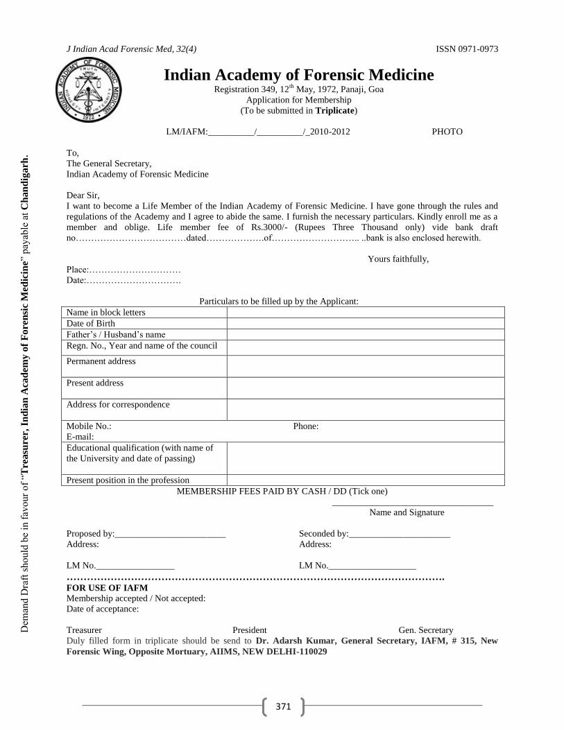
J Indian Acad Forensic Med, 32(4) ISSN 0971-0973
371
Indian Academy of Forensic Medicine Registration 349, 12
th May, 1972, Panaji, Goa
Application for Membership
(To be submitted in Triplicate)
LM/IAFM:__________/__________/_2010-2012 PHOTO
To,
The General Secretary,
Indian Academy of Forensic Medicine
Dear Sir,
I want to become a Life Member of the Indian Academy of Forensic Medicine. I have gone through the rules and
regulations of the Academy and I agree to abide the same. I furnish the necessary particulars. Kindly enroll me as a
member and oblige. Life member fee of Rs.3000/- (Rupees Three Thousand only) vide bank draft
no………………………………dated……………….of……………………….. ..bank is also enclosed herewith.
Yours faithfully,
Place:…………………………
Date:………………………….
Particulars to be filled up by the Applicant:
Name in block letters
Date of Birth
Father‟s / Husband‟s name
Regn. No., Year and name of the council
Permanent address
Present address
Address for correspondence
Mobile No.: Phone:
E-mail:
Educational qualification (with name of
the University and date of passing)
Present position in the profession
MEMBERSHIP FEES PAID BY CASH / DD (Tick one)
___________________________________
Name and Signature
Proposed by:________________________ Seconded by:______________________
Address: Address:
LM No._________________ LM No.___________________
…………………………………………………………………………………………………. FOR USE OF IAFM
Membership accepted / Not accepted:
Date of acceptance:
Treasurer President Gen. Secretary
Duly filled form in triplicate should be send to Dr. Adarsh Kumar, General Secretary, IAFM, # 315, New
Forensic Wing, Opposite Mortuary, AIIMS, NEW DELHI-110029
Dem
and D
raft
sh
ould
be
in f
avo
ur
of
“T
reasu
rer,
In
dia
n A
cad
emy o
f F
ore
nsi
c M
edic
ine”
pay
able
at
Ch
an
dig
arh
.
Related Documents


