Neuroscience and Biobehavioral Reviews 35 (2011) 1644–1653 Contents lists available at ScienceDirect Neuroscience and Biobehavioral Reviews journa l h o me pa g e: www.elsevier.com/locate/neubiorev Review A computational neuroscience approach to schizophrenia and its onset Edmund T. Rolls a,∗ , Gustavo Deco b,c a Oxford Centre for Computational Neuroscience, Oxford, UK b Computational Neuroscience, Universitat Pompeu Fabra, Barcelona, Spain c Institució Catalana de Recerca i Estudis Avanc ¸ ats, Barcelona, Spain a r t i c l e i n f o Article history: Received 8 April 2010 Received in revised form 12 August 2010 Accepted 2 September 2010 Keywords: Schizophrenia Adolescence Computational neuroscience Synaptic pruning Grey matter volume NMDA receptors Dopamine GABA inhibition Attractor networks Neural networks Stochastic neurodynamics Noise in the brain Neuronal spiking a b s t r a c t Computational neuroscience integrate-and-fire attractor network models can be used to understand the factors that alter the stability of cortical networks in the face of noise caused for example by neuronal spiking times. A reduction of the firing rates of cortical neurons caused for example by reduced NMDA receptor function (present in schizophrenia) can lead to instability of the high firing rate attractor states that normally implement short-term memory and attention, contributing to the cognitive and negative symptoms of schizophrenia. Reduced cortical inhibition caused by a reduction of GABA neurotransmis- sion (present in schizophrenia) can lead to instability of the spontaneous firing states of cortical networks, leading to a noise-induced jump to a high firing rate attractor state even in the absence of external inputs, contributing to the positive symptoms of schizophrenia. We consider how effects occurring at the time of late adolescence including synaptic pruning, decreases in grey matter volume, and changes in GABA- mediated inhibition and dopamine may contribute to the onset in some individuals of schizophrenia at this time. © 2010 Elsevier Ltd. All rights reserved. Contents 1. Introduction . . . . . . . . . . . . . . . . . . . . . . . . . . . . . . . . . . . . . . . . . . . . . . . . . . . . . . . . . . . . . . . . . . . . . . . . . . . . . . . . . . . . . . . . . . . . . . . . . . . . . . . . . . . . . . . . . . . . . . . . . . . . . . . . . . . . . . . . 1645 2. A top-down computational neuroscience approach to schizophrenia . . . . . . . . . . . . . . . . . . . . . . . . . . . . . . . . . . . . . . . . . . . . . . . . . . . . . . . . . . . . . . . . . . . . . . . . . . . . 1645 3. A neurodynamical hypothesis of schizophrenia . . . . . . . . . . . . . . . . . . . . . . . . . . . . . . . . . . . . . . . . . . . . . . . . . . . . . . . . . . . . . . . . . . . . . . . . . . . . . . . . . . . . . . . . . . . . . . . . . . 1646 3.1. Cognitive symptoms . . . . . . . . . . . . . . . . . . . . . . . . . . . . . . . . . . . . . . . . . . . . . . . . . . . . . . . . . . . . . . . . . . . . . . . . . . . . . . . . . . . . . . . . . . . . . . . . . . . . . . . . . . . . . . . . . . . . . . . 1646 3.2. Negative symptoms . . . . . . . . . . . . . . . . . . . . . . . . . . . . . . . . . . . . . . . . . . . . . . . . . . . . . . . . . . . . . . . . . . . . . . . . . . . . . . . . . . . . . . . . . . . . . . . . . . . . . . . . . . . . . . . . . . . . . . . . 1647 3.3. Positive symptoms . . . . . . . . . . . . . . . . . . . . . . . . . . . . . . . . . . . . . . . . . . . . . . . . . . . . . . . . . . . . . . . . . . . . . . . . . . . . . . . . . . . . . . . . . . . . . . . . . . . . . . . . . . . . . . . . . . . . . . . . . 1647 4. Schizophrenia and noise . . . . . . . . . . . . . . . . . . . . . . . . . . . . . . . . . . . . . . . . . . . . . . . . . . . . . . . . . . . . . . . . . . . . . . . . . . . . . . . . . . . . . . . . . . . . . . . . . . . . . . . . . . . . . . . . . . . . . . . . . . . 1648 5. The role of prefrontal cortex dopamine . . . . . . . . . . . . . . . . . . . . . . . . . . . . . . . . . . . . . . . . . . . . . . . . . . . . . . . . . . . . . . . . . . . . . . . . . . . . . . . . . . . . . . . . . . . . . . . . . . . . . . . . . . . 1648 6. Brain changes during adolescence that could influence these processes . . . . . . . . . . . . . . . . . . . . . . . . . . . . . . . . . . . . . . . . . . . . . . . . . . . . . . . . . . . . . . . . . . . . . . . . . 1648 6.1. Grey matter and synaptic changes associated with adolescence . . . . . . . . . . . . . . . . . . . . . . . . . . . . . . . . . . . . . . . . . . . . . . . . . . . . . . . . . . . . . . . . . . . . . . . . . . 1648 6.2. GABA changes at adolescence . . . . . . . . . . . . . . . . . . . . . . . . . . . . . . . . . . . . . . . . . . . . . . . . . . . . . . . . . . . . . . . . . . . . . . . . . . . . . . . . . . . . . . . . . . . . . . . . . . . . . . . . . . . . . . 1649 6.3. Dopamine changes at adolescence . . . . . . . . . . . . . . . . . . . . . . . . . . . . . . . . . . . . . . . . . . . . . . . . . . . . . . . . . . . . . . . . . . . . . . . . . . . . . . . . . . . . . . . . . . . . . . . . . . . . . . . . . 1649 7. Conclusions . . . . . . . . . . . . . . . . . . . . . . . . . . . . . . . . . . . . . . . . . . . . . . . . . . . . . . . . . . . . . . . . . . . . . . . . . . . . . . . . . . . . . . . . . . . . . . . . . . . . . . . . . . . . . . . . . . . . . . . . . . . . . . . . . . . . . . . . 1650 8. Stochastic neurodynamics: appendix . . . . . . . . . . . . . . . . . . . . . . . . . . . . . . . . . . . . . . . . . . . . . . . . . . . . . . . . . . . . . . . . . . . . . . . . . . . . . . . . . . . . . . . . . . . . . . . . . . . . . . . . . . . . . 1651 8.1. The attractor framework . . . . . . . . . . . . . . . . . . . . . . . . . . . . . . . . . . . . . . . . . . . . . . . . . . . . . . . . . . . . . . . . . . . . . . . . . . . . . . . . . . . . . . . . . . . . . . . . . . . . . . . . . . . . . . . . . . . 1651 8.2. Energy landscape and stochasticity . . . . . . . . . . . . . . . . . . . . . . . . . . . . . . . . . . . . . . . . . . . . . . . . . . . . . . . . . . . . . . . . . . . . . . . . . . . . . . . . . . . . . . . . . . . . . . . . . . . . . . . . 1651 Acknowledgments . . . . . . . . . . . . . . . . . . . . . . . . . . . . . . . . . . . . . . . . . . . . . . . . . . . . . . . . . . . . . . . . . . . . . . . . . . . . . . . . . . . . . . . . . . . . . . . . . . . . . . . . . . . . . . . . . . . . . . . . . . . . . . . . . 1652 References . . . . . . . . . . . . . . . . . . . . . . . . . . . . . . . . . . . . . . . . . . . . . . . . . . . . . . . . . . . . . . . . . . . . . . . . . . . . . . . . . . . . . . . . . . . . . . . . . . . . . . . . . . . . . . . . . . . . . . . . . . . . . . . . . . . . . . . . . 1652 ∗ Corresponding author. E-mail address: [email protected] (E.T. Rolls). URL: http://www.oxcns.org (E.T. Rolls). 0149-7634/$ – see front matter © 2010 Elsevier Ltd. All rights reserved. doi:10.1016/j.neubiorev.2010.09.001

Welcome message from author
This document is posted to help you gain knowledge. Please leave a comment to let me know what you think about it! Share it to your friends and learn new things together.
Transcript

Journal Identification = NBR Article Identification = 1348 Date: July 6, 2011 Time: 6:41 pm
R
A
Ea
b
c
a
ARRA
KSACSGNDGANSNN
C
0d
Neuroscience and Biobehavioral Reviews 35 (2011) 1644–1653
Contents lists available at ScienceDirect
Neuroscience and Biobehavioral Reviews
journa l h o me pa g e: www.elsev ier .com/ locate /neubiorev
eview
computational neuroscience approach to schizophrenia and its onset
dmund T. Rollsa,∗, Gustavo Decob,c
Oxford Centre for Computational Neuroscience, Oxford, UKComputational Neuroscience, Universitat Pompeu Fabra, Barcelona, SpainInstitució Catalana de Recerca i Estudis Avanc ats, Barcelona, Spain
r t i c l e i n f o
rticle history:eceived 8 April 2010eceived in revised form 12 August 2010ccepted 2 September 2010
eywords:chizophreniadolescenceomputational neuroscienceynaptic pruningrey matter volumeMDA receptors
a b s t r a c t
Computational neuroscience integrate-and-fire attractor network models can be used to understand thefactors that alter the stability of cortical networks in the face of noise caused for example by neuronalspiking times. A reduction of the firing rates of cortical neurons caused for example by reduced NMDAreceptor function (present in schizophrenia) can lead to instability of the high firing rate attractor statesthat normally implement short-term memory and attention, contributing to the cognitive and negativesymptoms of schizophrenia. Reduced cortical inhibition caused by a reduction of GABA neurotransmis-sion (present in schizophrenia) can lead to instability of the spontaneous firing states of cortical networks,leading to a noise-induced jump to a high firing rate attractor state even in the absence of external inputs,contributing to the positive symptoms of schizophrenia. We consider how effects occurring at the timeof late adolescence including synaptic pruning, decreases in grey matter volume, and changes in GABA-mediated inhibition and dopamine may contribute to the onset in some individuals of schizophrenia at
opamineABA inhibitionttractor networkseural networks
this time.© 2010 Elsevier Ltd. All rights reserved.
tochastic neurodynamicsoise in the braineuronal spiking
ontents
1. Introduction . . . . . . . . . . . . . . . . . . . . . . . . . . . . . . . . . . . . . . . . . . . . . . . . . . . . . . . . . . . . . . . . . . . . . . . . . . . . . . . . . . . . . . . . . . . . . . . . . . . . . . . . . . . . . . . . . . . . . . . . . . . . . . . . . . . . . . . . 16452. A top-down computational neuroscience approach to schizophrenia. . . . . . . . . . . . . . . . . . . . . . . . . . . . . . . . . . . . . . . . . . . . . . . . . . . . . . . . . . . . . . . . . . . . . . . . . . . . 16453. A neurodynamical hypothesis of schizophrenia . . . . . . . . . . . . . . . . . . . . . . . . . . . . . . . . . . . . . . . . . . . . . . . . . . . . . . . . . . . . . . . . . . . . . . . . . . . . . . . . . . . . . . . . . . . . . . . . . . 1646
3.1. Cognitive symptoms . . . . . . . . . . . . . . . . . . . . . . . . . . . . . . . . . . . . . . . . . . . . . . . . . . . . . . . . . . . . . . . . . . . . . . . . . . . . . . . . . . . . . . . . . . . . . . . . . . . . . . . . . . . . . . . . . . . . . . . 16463.2. Negative symptoms . . . . . . . . . . . . . . . . . . . . . . . . . . . . . . . . . . . . . . . . . . . . . . . . . . . . . . . . . . . . . . . . . . . . . . . . . . . . . . . . . . . . . . . . . . . . . . . . . . . . . . . . . . . . . . . . . . . . . . . . 16473.3. Positive symptoms . . . . . . . . . . . . . . . . . . . . . . . . . . . . . . . . . . . . . . . . . . . . . . . . . . . . . . . . . . . . . . . . . . . . . . . . . . . . . . . . . . . . . . . . . . . . . . . . . . . . . . . . . . . . . . . . . . . . . . . . . 1647
4. Schizophrenia and noise . . . . . . . . . . . . . . . . . . . . . . . . . . . . . . . . . . . . . . . . . . . . . . . . . . . . . . . . . . . . . . . . . . . . . . . . . . . . . . . . . . . . . . . . . . . . . . . . . . . . . . . . . . . . . . . . . . . . . . . . . . . 16485. The role of prefrontal cortex dopamine . . . . . . . . . . . . . . . . . . . . . . . . . . . . . . . . . . . . . . . . . . . . . . . . . . . . . . . . . . . . . . . . . . . . . . . . . . . . . . . . . . . . . . . . . . . . . . . . . . . . . . . . . . . 16486. Brain changes during adolescence that could influence these processes . . . . . . . . . . . . . . . . . . . . . . . . . . . . . . . . . . . . . . . . . . . . . . . . . . . . . . . . . . . . . . . . . . . . . . . . . 1648
6.1. Grey matter and synaptic changes associated with adolescence. . . . . . . . . . . . . . . . . . . . . . . . . . . . . . . . . . . . . . . . . . . . . . . . . . . . . . . . . . . . . . . . . . . . . . . . . . 16486.2. GABA changes at adolescence . . . . . . . . . . . . . . . . . . . . . . . . . . . . . . . . . . . . . . . . . . . . . . . . . . . . . . . . . . . . . . . . . . . . . . . . . . . . . . . . . . . . . . . . . . . . . . . . . . . . . . . . . . . . . . 16496.3. Dopamine changes at adolescence . . . . . . . . . . . . . . . . . . . . . . . . . . . . . . . . . . . . . . . . . . . . . . . . . . . . . . . . . . . . . . . . . . . . . . . . . . . . . . . . . . . . . . . . . . . . . . . . . . . . . . . . . 1649
7. Conclusions . . . . . . . . . . . . . . . . . . . . . . . . . . . . . . . . . . . . . . . . . . . . . . . . . . . . . . . . . . . . . . . . . . . . . . . . . . . . . . . . . . . . . . . . . . . . . . . . . . . . . . . . . . . . . . . . . . . . . . . . . . . . . . . . . . . . . . . . 16508. Stochastic neurodynamics: appendix . . . . . . . . . . . . . . . . . . . . . . . . . . . . . . . . . . . . . . . . . . . . . . . . . . . . . . . . . . . . . . . . . . . . . . . . . . . . . . . . . . . . . . . . . . . . . . . . . . . . . . . . . . . . . 1651
8.1. The attractor framework . . . . . . . . . . . . . . . . . . . . . . . . . . . . . . . . . . . . . . . . . . . . . . . . . . . . . . . . . . . . . . . . . . . . . . . . . . . . . . . . . . . . . . . . . . . . . . . . . . . . . . . . . . . . . . . . . . . 1651
8.2. Energy landscape and stochasticity . . . . . . . . . . . . . . . . . . . . . . . . . . . . . . . .Acknowledgments . . . . . . . . . . . . . . . . . . . . . . . . . . . . . . . . . . . . . . . . . . . . . . . . . . . . . . . . .
References . . . . . . . . . . . . . . . . . . . . . . . . . . . . . . . . . . . . . . . . . . . . . . . . . . . . . . . . . . . . . . . . . .
∗ Corresponding author.E-mail address: [email protected] (E.T. Rolls).URL: http://www.oxcns.org (E.T. Rolls).
149-7634/$ – see front matter © 2010 Elsevier Ltd. All rights reserved.oi:10.1016/j.neubiorev.2010.09.001
. . . . . . . . . . . . . . . . . . . . . . . . . . . . . . . . . . . . . . . . . . . . . . . . . . . . . . . . . . . . . . . . . . . . . . . . 1651
. . . . . . . . . . . . . . . . . . . . . . . . . . . . . . . . . . . . . . . . . . . . . . . . . . . . . . . . . . . . . . . . . . . . . . . . 1652 . . . . . . . . . . . . . . . . . . . . . . . . . . . . . . . . . . . . . . . . . . . . . . . . . . . . . . . . . . . . . . . . . . . . . . . 1652

Journal Identification = NBR Article Identification = 1348 Date: July 6, 2011 Time: 6:41 pm
iobeh
1
ats
mwsceennadmsab(2wtt
2s
bcnn22
wtsRctmtawfedtdfrai“sccattt
E.T. Rolls, G. Deco / Neuroscience and B
. Introduction
In this paper, first we describe a computational neurosciencepproach to schizophrenia. Then we consider how brain changeshat occur in late adolescence may contribute to the onset ofchizophrenia.
The computational neuroscience approach we take involvesodeling cortical systems at the level of integrate-and-fire neuronsith synaptically activated ion channels in attractor or autoas-
ociation networks implemented with the recurrent collateralonnections between pyramidal cells. This enables us to link fromffects expressed at synapses and ion channels, through theirffects on the spiking neuronal activity in the network and theoise that this introduces into the system, to global effects of theetwork such as the stability of short-term memory, attentional,nd decision-making systems, and thus to cognitive function,ysfunction, and behavior. This provides a unifying approach toany aspects of cortical function, which helps in the under-
tanding of short-term memory, long-term memory, top-downttention, decision-making, executive function, and the relationetween the emotional and the reasoning systems in the brainDeco and Rolls, 2003, 2005a, 2006; Deco et al., 2009; Rolls, 2008,010a,b; Rolls and Deco, 2002, 2010). This paper is the first inhich we have linked this approach to how brain changes at
he time of adolescence might influence some of these func-ions.
. A top-down computational neuroscience approach tochizophrenia
Some computational neuroscience approaches to schizophreniauild upon single-neuron biophysics, physiology, and pharma-ology in schizophrenia, and analyze their effects in neuraletworks, which are then linked to the symptoms of schizophre-ia (Durstewitz et al., 1999, 2000; Durstewitz and Seamans,008; Seamans and Yang, 2004; Winterer and Weinberger,004).
We have adopted a top-down approach which considershether generic alterations in the operation and stability of cor-
ical circuits in different cortical areas might lead to the differentymptoms of schizophrenia (Loh et al., 2007a,b; Rolls, 2005, 2008;olls et al., 2008b). Bottom-up approaches start with putativehanges at the neural level such as alterations in dopamine, andry to understand the implications for function, which are of course
ultiple, of these changes. The top-down approach complementshe bottom-up approaches, as it starts from the set of symptomsnd maps them onto a dynamical systems computational frame-ork. The dynamical systems computational approach considers
actors that affect the stability of networks in the brain, and theffects of noise in those networks on the stability. Because theynamical systems we consider can be, and are, implemented athe level of integrate-and-fire neurons with neuronal and synapticynamics that are biophysically realistic, and incorporate dif-erent classes of ion channel activated by different transmittereceptors, effects of changes at these different levels, includinglterations in ion channels and transmitters, can be investigatedn and predicted from the model. We call this class of modelmechanistic”, in that it describes the underlying neuronal andubneuronal mechanisms involved in the dynamics in a biologi-ally plausible way, so that predictions can be made about howhanges in any one part of the mechanism will affect the over-
ll, “global”, operation of the system, measured for example byhe stability of short-term memory and attentional states. Thushe top-down approach emphasizes how the computations inhe system perform particular functions, and then considers howavioral Reviews 35 (2011) 1644–1653 1645
possibly combinations of several neural changes can influencethe operation of the system, and how alterations of a num-ber of possible different neural factors may be able to restorethe computational functions being performed by the neural sys-tem.
We contrast this with phenomenological models, which attemptto capture the behavior of the system, but without regard towhether the system could be implemented in the brain, and with-out any neurally plausible mechanism being modelled. An exampleis the accumulator model of decision-making, in which an accu-mulator (linear adder) adds up each of the two (or more) inputs itreceives, adds some noise, and then considers that a decision hasbeen taken when some arbitrary threshold is reached in which thesum of one of the inputs is by some amount greater than the other(Ratcliff et al., 1999; Smith and Ratcliff, 2004; Vickers, 1979). Sucha phenomenological model does not allow any direct predictionsabout how changes in ion channels, neurotransmitters, etc. alterthe operation of the system, and indeed, to make such predictions,the phenomenological model, which may not represent realisticallythe type of processing that occurs in the brain, would itself haveto be modelled in a neural system. Similar issues arise with con-nectionist models (Cohen and Servan-Schreiber, 1992) describedelsewhere (Rolls et al., 2008b) which also do not incorporate detailsof the neurophysiological and biophysical mechanisms that con-tribute to the neural dynamics, and may therefore be less able tomodel how changes in a number of neurobiological factors influ-ence the operation of the system. The mechanistic approach weadopt instead often is able to provide accounts for how importantfunctional properties of the system arise as emergent properties ofthe system. One example is Weber’s law in decision-making (Decoand Rolls, 2006). Another example is confidence in a decision thathas been made, which is encoded in the firing rates of the neu-rons in the winning attractor (Rolls and Deco, 2010; Rolls et al.,2010a,b).
The stochastic dynamical systems approach that we utilize(Rolls and Deco, 2010) is introduced in Section 8. The full imple-mentation of the equations for the neuron and synaptic dynamicsis described elsewhere (Loh et al., 2007a; Rolls et al., 2008b), andincludes currents passing through voltage-dependent and hencenon-linear ion channels activated by NMDA receptors, and cur-rents through ion channels activated by AMPA and GABA receptors.The positive feedback in the recurrent collateral connections in thenetwork, the NMDA receptor non-linearity, and the non-linearityintroduced by the threshold for firing of the neurons in the system,provide the system with non-linearities that enable it to have theproperties of an attractor network (Deco and Rolls, 2005b; Rollsand Deco, 2010).
A feature that we have adopted from Brunel and Wang (2001)of the approach we use is a mean-field equivalent analysis of thenetwork using techniques from theoretical physics. This allowsmeasurement of the fixed points of the system, the flow in thesystem, and the operating areas in the parameter spaces that willproduce for example a stable spontaneous firing rate and also stablehigh firing rates for each of the memory attractor states (dependingon the starting conditions) in a noiseless system, equivalent to a sys-tem of infinite size (Brunel and Wang, 2001; Deco and Rolls, 2006;Loh et al., 2007a; Rolls and Deco, 2010). This enables suitable valuesof for example the synaptic connection weights in the system to bechosen. If these parameters are then used in the integrate-and-fireversion of the model, which has noise due to the approximatelyPoisson spiking times of the neurons, the effects of the noise onthe operation of the system, and of alterations for example of the
different synaptic currents produced through different transmit-ter receptors in the system, can be investigated (Brunel and Wang,2001; Deco and Rolls, 2006; Loh et al., 2007a; Rolls and Deco, 2010;Rolls et al., 2008a,b).
Journal Identification = NBR Article Identification = 1348 Date: July 6, 2011 Time: 6:41 pm
1 iobehavioral Reviews 35 (2011) 1644–1653
3
3
iLtwip
ata1vbmmswtocseoinlhsrawR
asmaRomatbrms1bbbtL
fspi1SRt
0 0.5 1 1.5 2 2.5 30
10
20
30
40
50
60
Firi
ng r
ate
[Hz]
Spontaneous stablePersistent stab le
0 0.5 1 1.5 2 2.5 30
10
20
30
40
50
60
time [s]
Firi
ng r
ate
[Hz]
Spontaneous unstablePersistent uns tab le
Fig. 1. Example trials of integrate-and-fire attractor network simulations of short-term memory. The average firing rate of all the neurons in the short-term memorypopulation S1 of neurons is shown. (Top: normal operation) On a trial in which arecall stimulus was applied to S1 at 0–500 ms, firing continued normally until theend of the trial in the ‘persistent’ simulation condition. On a trial on which no recallstimulus was applied to S1, spontaneous firing (i.e. at a low rate) continued until theend of the trial in the ‘spontaneous’ simulation condition. (Bottom: unstable oper-ation). On this persistent condition trial, the firing decreased during the trial as thenetwork fell out of the attractor because of the statistical fluctuations caused by thespiking dynamics. This type of instability is more likely if NMDA receptor activatedion channel currents become decreased, or by other factors that decrease neuronalexcitability. This provides a model of impaired cognitive function in for exampleschizophrenia. On the spontaneous condition trial, the firing increased during thetrial because of the statistical fluctuations. This type of instability is more likely ifGABA receptor activated ion channel currents become decreased, or by other fac-tors that decrease cortical inhibition. This type of instability in which a network
646 E.T. Rolls, G. Deco / Neuroscience and B
. A neurodynamical hypothesis of schizophrenia
.1. Cognitive symptoms
The cognitive symptoms of schizophrenia include distractibil-ty, poor attention, and the dysexecutive syndrome (Green, 1996;iddle, 1987; Mueser and McGurk, 2004). It has been suggestedhat at the core of the cognitive symptoms of schizophrenia is aorking-memory deficit characterized by a difficulty in maintain-
ng items in short-term memory implemented in the dorsolateralrefrontal cortex (Goldman-Rakic, 1994, 1999).
Short-term memory is implemented in the prefrontal cortexs follows. Pyramidal neurons in the cerebral cortex have a rela-ively high density of excitatory connections to each other within
local area of 1–3 mm (Abeles, 1991; Braitenberg and Schütz,991). These local recurrent collateral excitatory connections pro-ide a positive-feedback mechanism (which is kept under controly GABA inhibitory interneurons) that enables a set of neurons toaintain their activity for many seconds to implement a short-termemory (Goldman-Rakic, 1995). Each memory is formed by the
et of the neurons in the local cortical network that were coactivehen the memory was formed, resulting in strengthened excita-
ory connections between that set of neurons through the processf long-term potentiation, which is a property of these recurrentollateral connections. When a subset of these neurons is sub-equently activated, positive feedback through the strengthenedxcitatory connections between the neurons results in activationf the whole set of neurons, and so produces the completion of anncomplete memory. Thus, in an attractor network, the state of theetwork is “attracted” towards the state in which the memory was
earned; this is called an “attractor state”. An attractor network canave many different attractor states, each consisting of a differentubset of the neurons being active; any one subset of neurons canepresent a short-term memory. The operation and properties ofttractor networks are described in Section 8 and more fully else-here (Amit, 1989; Hertz et al., 1991; Hopfield, 1982; Rolls, 2008;olls and Deco, 2002, 2010).
Attractor networks appear to operate in the prefrontal cortex,n area that is important in attention and short-term memory, ashown for example by firing in the delay period of a short-termemory task (Funahashi et al., 1989; Fuster, 1995, 2000; Fuster
nd Alexander, 1971; Goldman-Rakic, 1996; Kubota and Niki, 1971;olls, 2008). Short-term memory is the ability to hold informationn-line during a short time period (Fuster, 1995, 2000) and is funda-ental to top-down attention in the sense that whatever requires
ttention (e.g. a spatial location) has to be maintained in a short-erm memory. The short-term memory then biases competitionetween the multiple bottom-up items in the stimulus input; theesult is an advantage in the neuronal competition between theultiple inputs for the item that receives top-down bias from the
hort-term memory (Deco and Rolls, 2005a; Desimone and Duncan,995; Rolls and Deco, 2002). The impairments of attention inducedy prefrontal cortex damage may be accounted for in large party an impairment in the ability to hold the object of attention sta-ly and without distraction in the short-term memory systems inhe prefrontal cortex (Goldman-Rakic, 1996; Goldman-Rakic andeung, 2002; Rolls, 2008).
Specific simulations of impairments in the operation of pre-rontal attractor networks can help to explain how the cognitiveymptoms of schizophrenia, including poor short-term memory,oor ability to allocate and maintain attention, and distractibil-
ty, occur (Frith and Dolan, 1997; Loh et al., 2007a; Seidman et al.,
994; Weinberger and Berman, 1996). Indeed, building on work byeamans and Yang (2004), Rolls, Loh and Deco (Loh et al., 2007a;olls, 2005) have proposed that the working-memory and atten-ional deficits might be related to instabilities of the high-firingjumps because of noise into a high firing rate state that is not triggered by an exter-nal input to the network contributes it is suggested to the positive symptoms ofschizophrenia. (After Rolls et al. (2008a).)
states in attractor networks in the prefrontal cortex (Fig. 1). Specif-ically, NMDA receptor hypofunction, which has been associatedwith schizophrenia (Coyle, 2006; Coyle et al., 2003), results inreduced currents running through NMDA receptor-activated ionchannels; this causes neurons to fire less fast, leading to shallowerbasins of attraction (see Section 8) of the high firing-rate attrac-tor states of the network (Loh et al., 2007a). The shallower basinsof attraction arise firstly because with the neurons firing less fast,there is less positive feedback in the recurrent collateral connec-tions between the neurons in the attractor, and this makes thesystem more vulnerable to noise. The noise could be external tothe network, but an important source of noise that can destabi-lize the high firing rate attractor state is the random spiking timesof neurons for a given mean firing rate, which produce statisticalfluctuations by which there might due to a random set of events beless (or more) firing in a set of neurons than average, which couldmake the system fall out of a high firing rate attractor state (Rollsand Deco, 2010). (The spike times of individual neurons are closeto being Poisson distributed.)
A second way in which reduced NMDA receptor function (orother factors such as synaptic pruning) could decrease the depth ofthe basins of attraction is by making the strengths of the synapticconnections between the neurons in the attractor weaker, whichagain reduces the positive feedback between the neurons in theattractor, and makes the attractor state more vulnerable to noise.These concepts are made quantitative in Section 8, Eq. (1), and in The
Noisy Brain (Rolls and Deco, 2010). Thus, the stability of the attractorstate is reduced. The result is difficulty in maintaining short-termmemory and thus attention (see Fig. 1 and also Durstewitz, 2007;Durstewitz and Seamans, 2002). The shallower basins of attrac-
Journal Identification = NBR Article Identification = 1348 Date: July 6, 2011 Time: 6:41 pm
iobehavioral Reviews 35 (2011) 1644–1653 1647
trtt
3
idwMhpetrbctkit2W
tpatttebsdi
3
oattRNfi2totmwtef
itensTtt
0 10 20 30 40 50 600
5
10
15
20
25
30
time [s]
Firi
ng r
ate
[Hz]
Pool S1Pool S2
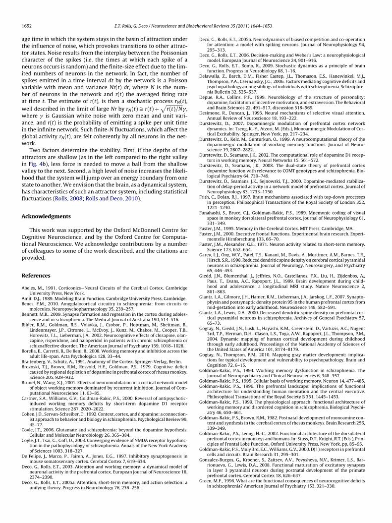
Fig. 2. Wandering between attractor states. An integrate-and-fire simulation of anattractor network with two memories stored in it, S1 and S2. With the normal synap-tic efficacies, and no initial cue in the simulation, the network would stay stablyin the spontaneous state with very little activity in the neurons in the S1 and S2neuronal populations or pools. However, on trials of the type shown in which theNMDA conductances were reduced by 5% and GABA by 10%, the activity moves nois-ily between the attractor for the spontaneous state and the two persistent states S1
E.T. Rolls, G. Deco / Neuroscience and B
ion and the reduced time constant of the system caused by NMDAeceptor (NMDAR) hypofunction (Wang, 2006), in the presence ofhe stochastic firing-related noise in the networks, result in dis-ractibility, poor attention and working-memory difficulties.
.2. Negative symptoms
The negative symptoms represent a complex of symptomsncluding apathy, poor rapport, lack of spontaneity, motor retar-ation, disturbance of volition, blunted affect, and emotionalithdrawal and passive behavior (Liddle, 1987; Mueser andcGurk, 2004). The negative symptoms and cognitive deficits are
ighly correlated in patients with schizophrenia and their non-sychotic relatives (Bilder et al., 2002; Delawalla et al., 2006; Jacobst al., 2007). Rolls, Loh and Deco propose that the negative symp-oms are also related to the decreased firing rates caused by aeduction in currents through NMDAR-activated channels, but inrain regions that may include the orbitofrontal cortex and anterioringulate cortex (Loh et al., 2007a; Rolls, 2005, 2008) rather thanhe prefrontal cortex. Indeed, lesions in these brain areas are wellnown to produce symptoms that resemble the negative symptomsn schizophrenia, and neuronal firing rates and BOLD activations inhese regions are correlated with reward value and pleasure (Paus,001; Rolls, 1999, 2005, 2006, 2008; Rolls and Grabenhorst, 2008;interer et al., 2002).This is a unifying approach to the cognitive and negative symp-
oms: the same reduction in NMDAR-activated channel currentsroduces on the one hand, instability in high-firing-rate states inttractor networks in the dorsolateral prefrontal cortex and therebyhe cognitive symptoms, and on the other hand, a reduction inhe firing rate of neurons in the orbitofrontal and cingulate cor-ex, leading to the negative symptoms. In addition to the reducedmotion caused by the reduced firing rates, attractor networks maye present in the orbitofrontal cortex that help to maintain moodtate (Rolls, 2008), and a decrease in their stability by the reducedepth in the basins of attraction could make emotions more labile
n schizophrenia/schizoaffective disorder.
.3. Positive symptoms
The positive symptoms of schizophrenia include bizarre trainsf thoughts, hallucinations, and delusions (Liddle, 1987; Muesernd McGurk, 2004). In contrast to the cognitive and negative symp-oms, the positive symptoms generally occur intermittently duringhe course of the illness, and this clinical state is called “psychosis”.olls, Loh and Deco propose that owing to reduced currents throughMDAR-activated channels, the basins of attraction of the high-ring-rate attractor states are shallow (Durstewitz, 2007; Loh et al.,007a; Rolls, 2005) in the temporal lobe, which includes the seman-ic memory networks and the auditory association cortex. Becausef the resulting statistical fluctuations in the states of the attrac-or networks, internal representations of thoughts and perceptions
ove too freely around in the energy landscape, from thought toeakly associated thought, leading to bizarre thoughts and associa-
ions, and to hallucinations (see Figs. 1 and 2). Such thoughts mightventually be associated together in semantic memory, leading toalse beliefs and delusions (Rolls, 2005, 2008).
In addition, Loh et al. (2007a) propose that a reduction in GABAnterneuron efficacy in schizophrenic patients may also contributeo the generation of positive symptoms: lower GABA-interneuronfficacy reduces the depth of the basin of attraction of the sponta-eous state, making it more likely that a high firing-rate attractor
tate will emerge out of the spontaneous firing of the neurons.his is illustrated in Fig. 1 (bottom). On the spontaneous condi-ion trial, the firing, which should have remained low throughouthe trial as no cue was provided to start up the short-term mem-and S2 by virtue of statistical fluctuations caused by the randomness of the spikingactivity. The two curves show the activity of the two selective pools S1 and S2 overtime smoothed with a 1 s sliding averaging window. (After Loh et al. (2007a).)
ory, increased during the trial because of the statistical fluctuations,that is the spiking-related randomness in the network. This type ofinstability is more likely if GABA receptor activated ion channelcurrents become decreased, or by other factors that decrease cor-tical inhibition. This type of instability in which a network jumpsbecause of noise into a high firing rate state that is not triggeredby an external input to the network contributes it is suggestedto the positive symptoms of schizophrenia, including for examplehallucinations, delusions, and feelings of lack of control or beingcontrolled by others (Loh et al., 2007a; Rolls et al., 2008b). Empiricalevidence supports this computational proposal: markers indicatingdecreased inhibition by the GABA system are found in neocorticalareas (Lewis et al., 2005) and in parts of the hippocampus (Benes,2010). On the basis of this model, we have proposed (Loh et al.,2007a; Rolls et al., 2008b) that treating schizophrenia patients withD2 antagonists could increase the GABA currents (Seamans et al.,2001; Seamans and Yang, 2004) in the networks, which would alle-viate the positive symptoms by reducing the spontaneous firingrates, which would deepen the spontaneous attractor state (seeFigs. 1 and 3). This effect of D2 antagonists leaves the persistentattractors shallow because the high firing rates are reduced, whichmay explain why the D2 antagonists do not have a major effecton the negative and cognitive symptoms. To target negative symp-toms, we have suggested that D1 agonists may help to deepen thebasin of attraction of the high-firing-rate attractor state (Loh et al.,2007a; Rolls et al., 2008b). This two-dimensional approach allowsus to address the specific characteristics of the psychotic (positive)symptoms which appear in episodes, in contrast to the negativeand cognitive symptoms which typically persist over time.
When both NMDA and GABA are reduced one might thinkthat these two counterbalancing effects (excitatory and inhibitory)would cancel each other out. However, this is not the case:modeling these conditions showed that the stability of both the
spontaneous and the high-firing-rate states is reduced (Loh et al.,2007a) (see also Brunel and Wang, 2001; Durstewitz and Seamans,2002). Indeed, under these conditions, the network wanderedfreely between the two short-term memory (high firing-rate) states
Journal Identification = NBR Article Identification = 1348 Date: July 6, 2011 Time: 6:41 pm
1648 E.T. Rolls, G. Deco / Neuroscience and Biobeh
= dendritic
= output firing
activationhi
yi
output
ie
external input
ijwyj
F
itbaas
4
dasdmsb(
rcdFfioiottsGni2
wefwa2
tial decrease in cortical grey matter thickness (Giedd et al., 1999;
ig. 3. The architecture of an autoassociation or attractor network (see Section 8.1).
n the network and the spontaneous state (Fig. 2). We relate this pat-ern to the positive symptoms of schizophrenia, in which both theasins of attraction of the spontaneous and high-firing-rate statesre shallow, and the system jumps, helped by the statistical fluctu-tions, between the different attractor states and the spontaneoustate (Fig. 2) (Loh et al., 2007a).
. Schizophrenia and noise
The changes in the integrate-and fire model we have justescribed produced by alterations in the activation of synapticallyctivated ion channels can be interpreted in terms of a reducedignal-to-noise ratio. In the computational models, the stability isefined as the proportion of trials on which the correct short termemory (or signal) is maintained until the end of the trial, and the
ignal-to-noise ratio can be measured by the mean squared dividedy the variance of the synaptic currents over the whole trial periodLoh et al., 2007a,b).
Three possible mechanisms for a decreased signal-to-noiseatio as reflected in reduced stability are highlighted by theomputational models as follows, and may be relevant to theecreased signal-to-noise ratio described below in schizophrenia.irst, reduced NMDAR-activated synaptic currents will reduce thering rates of neurons, and this will decrease the depth of the basinsf attraction of cortical attractor states, making them less stablen the face of spiking-related and other noise in the brain, andf distracting stimuli in the world. Second, the reduced contribu-ion of NMDAR-activated current will reduce the time constant ofhe whole attractor network, also making the attractor states lesstable in the face of noise and distracting stimuli. Third, reducedABAR-mediated currents may reduce the stability of the sponta-eous state, and the resulting noise will cause the system to jump
nto a high-firing-rate attractor state, as described above (Loh et al.,007a,b).
A way to link the signal-to-noise ratio measure from modelsith experimental data is to use the trial-by-trial variability with
xperimental measures. With this approach, there is some evidenceor decreased signal-to-noise ratio in schizophrenia, in studies in
hich the variability of EEG and functional neuroimaging data inttentional tasks is measured (Rolls et al., 2008b; Winterer et al.,004, 2000).
avioral Reviews 35 (2011) 1644–1653
5. The role of prefrontal cortex dopamine
With the computational approach described, links can be madeto help understand the role of dopamine in schizophrenia (Rolls etal., 2008b). Dopamine, acting through D1 receptors, is importantin maintaining high firing activity in prefrontal cortical networksinvolved in short-term memory and attention (Brozoski et al., 1979;Sawaguchi and Goldman-Rakic, 1991, 1994; Sawaguchi et al., 1988,1990), with an optimal level being important (Goldman-Rakic etal., 2000; Vijayraghavan et al., 2007; Williams and Goldman-Rakic,1995). Impairments in this system may contribute to the cognitivedeficits in schizophrenia.
While D1 receptor activation increases NMDA and GABAreceptor-activated ion channel conductances (a D1-receptor-dominated state), D2-receptor stimulation tends to have theopposite effect (a D2-receptor-dominated state) (Seamans et al.,2001; Seamans and Yang, 2004). On the basis of these find-ings, Seamans and Yang (2004) and others (Castner et al., 2000;Durstewitz, 2007; Durstewitz et al., 2000; Seamans and Yang,2004) predict that D1 agonists and D2 antagonists should help totreat schizophrenic symptoms by increasing NMDAR- and GABA-R-activated synaptic conductances. Indeed, an important effect ofD2 antagonists relates to the increase in GABA-R activated currents(Seamans et al., 2001; Seamans and Yang, 2004), which would makethe spontaneous firing state more stable and may help to controlthe positive symptoms of schizophrenia (Rolls et al., 2008b).
6. Brain changes during adolescence that could influencethese processes
There are marked changes in emotion, cognition, and behaviorat the time of adolescence (Steinberg, 2005).
In relation to the cognitive symptoms of schizophrenia, therehas always been the fact that schizophrenia is a condition that oftenhas its onset in the late teens or twenties (Lewis and Levitt, 2002),and it has been suggested that there could be a link in this respectto changes in NMDA and related receptor functions that are relatedto aging (Rolls, 2008; Rolls and Deco, 2010). In particular, workingmemory shows a steady decline with age from the 20 s to the 70 s(Borella et al., 2008; Johnson et al., 2010; Swanson, 1999), and itmay be the case that by the late teens or early twenties NMDAand related receptor systems (including dopamine) may be lessefficacious than when younger, so that the cognitive symptoms ofschizophrenia are more likely to occur at this age than earlier.
In this section, we examine some of the changes that occur in thebrain during (late) adolescence, and address whether there is evi-dence that the changes that occur about this time, if interacting withother causal factors, could contribute to the onset of schizophreniawhen it occurs in late adolescence and the early twenties. The com-putational points made next are clear, but the extent to which thechanges in the brain lead them to apply is an area where furtherinvestigation is needed.
6.1. Grey matter and synaptic changes associated withadolescence
In the primate dorsolateral prefrontal cortex (DLPFC), thedensity of excitatory synapses decreases by 40–50% during adoles-cence, and this occurs without a major change in synaptic strength(Gonzalez-Burgos et al., 2008). Structural neuroimaging studies inhumans demonstrate that adolescence is associated with a substan-
Gogtay and Thompson, 2010), a decrease usually interpreted toresult from the massive and increased synaptic pruning occurringduring adolescence and early adulthood (Gogtay et al., 2004; Rakic,

Journal Identification = NBR Article Identification = 1348 Date: July 6, 2011 Time: 6:41 pm
iobeh
1iwp
ooLciedFc(
sonhtwcvctrotasitcanr2ttctisl2
etcmRnaaoptoe112tmt
E.T. Rolls, G. Deco / Neuroscience and B
996; Rakic et al., 1994). Indeed, the total volume of grey matterncreases across the cortex prior to puberty, reaching a peak some-
here in the early to-mid pubertal period after which there is aost-pubertal decline.
It is noted that a decreased density of dendritic spines (a markerf excitatory synaptic inputs to pyramidal neurons) in the DLPFCf individuals with schizophrenia (Garey et al., 1998; Glantz andewis, 2000), is consistent with reduced excitatory drive in DLPFCircuits in schizophrenia. In addition, convergent lines of evidencendicate that schizophrenia might be associated with reducedxcitatory synaptic neurotransmission through NMDA (N-methyl--aspartate) glutamate receptors (Coyle, 2006; Coyle et al., 2003).urther, the loss of excitatory synapses is even greater at adoles-ence in those diagnosed with schizophrenia, amounting to 60%Bennett, 2009; Glantz et al., 2007; Glantz and Lewis, 2000).
What are the functional and computational implications of thisynaptic pruning and decrease in grey matter volume at the timef adolescence? A first possible computational implication is thateurons in the prefrontal cortex become less excitable, as theyave fewer excitatory synapses. A decrease in the firing rates ofhese neurons could decrease the stability of cortical attractor net-orks that implement short-term memory, and attention. This
ould result, if added to other effects, in a tendency of some indi-iduals at about the time of adolescence to show the cognitivehanges of schizophrenia. Similarly, if the same pruning of exci-atory synapses occurred in the orbitofrontal cortex and the firingates of the neurons were lower as a result, this could result (intherwise predisposed individuals) in some of the negative symp-oms of schizophrenia (decreased emotionality and motivation)ppearing at about this time. Whether changes in GABA compen-ate for the decreased excitatory drive to cortical pyramidal cellsn adolescence is considered in Section 6.2. The argument here ishat the massive and increased synaptic pruning in late adoles-ence/the early twenties (Gogtay et al., 2004; Rakic, 1996; Rakic etl., 1994) may be sufficiently large to reduce the stability of attractoretworks involved in working memory, especially with the largereduction in excitatory synapses found in schizophrenics (Bennett,009; Glantz et al., 2007; Glantz and Lewis, 2000). (Although synap-ic pruning starts earlier in development, we do not know whetherhe synapses being pruned before and during early adolescence areomputationally efficacious in these attractor networks (i.e. are onhe right neurons to support attractors, and with the associativelyncreased synaptic strength required to support discrete attractortates (Rolls, 2008); and the synaptic pruning rate does becomearger in late adolescence and the early twenties (Gogtay et al.,004; Rakic, 1996; Rakic et al., 1994).)
A second computational implication of a reduced number ofxcitatory connections onto each neuron arises from the fact thathe number of different memories that can be stored in an asso-iative network is proportional to the number of associativelyodifiable excitatory connections onto each neuron (Rolls, 2008;
olls and Treves, 1998). This applies to both pattern associationetworks used to associate one stimulus with another, for example
visual stimulus with the sight of food (Rolls and Treves, 1990);nd to autoassociation or attractor networks used to store a mem-ry as a set of events, and later recall the whole memory from anyart (Treves and Rolls, 1991). The number of different memorieshat can be stored in both types of associative network is of therder of the number of associatively modifiable connections ontoach neuron if sparse representations are used (Rolls and Treves,990; Treves and Rolls, 1991). This number will be in the order of0,000 for a network in a small, local, region of the neocortex (Rolls,
008). The implication of a reduction in the number of associa-ively modifiable synapses onto each neuron will thus be a reducedemory capacity, and, particularly important in the present con-ext, more interference between different memories (Rolls, 2008).
avioral Reviews 35 (2011) 1644–1653 1649
This increased interference will tend to impair the distinctivenessand stability of any one memory by altering the shape of the energylandscape, and making the system more sensitive to the effects ofnoise generated by the Poisson nature of the spiking of neurons(Rolls, 2008; Rolls and Deco, 2010). This will for example tend toimpair short-term memory and attention, and increase distractibil-ity. A reduced number of synapses onto each neuron could, by thetime of late adolescence, be sufficient to have therefore an impacton for example working memory, and this in individuals predis-posed to schizophrenia could contribute to an account of why thecognitive symptoms of schizophrenia may become apparent in lateadolescence or the early twenties.
6.2. GABA changes at adolescence
Any change during adolescence in the excitatory input to cor-tical pyramidal cells could be compensated by changes in theGABA inhibitory interneurons that receive from the pyramidal cellsand provide negative feedback to them. No significant changes inthe density of inhibitory synapses are observed in the neocortexduring adolescence (De Felipe et al., 1997; Rakic et al., 1986). How-ever, the efficacy of GABAergic transmission may change duringadolescence because during this developmental period substantialchanges occur in the levels of expression of GABAA receptors, GABAtransporters, and parvalbumin, an interneuron-specific calciumbinding protein (Lewis et al., 2004). Whether or not GABA-mediatedtransmission can regulate excitatory synaptic pruning during ado-lescence remains to be investigated.
In schizophrenia, a deficiency in signalling through theTrkB neurotrophin receptor leads to reduced GABA (gamma-aminobutyric acid) synthesis in the parvalbumin-containingsubpopulation of inhibitory GABA neurons in the dorsolateral pre-frontal cortex of individuals with schizophrenia. Despite both pre-and post-synaptic compensatory responses, there is a resultingdecrease in the perisomatic inhibition of pyramidal neurons (Lewiset al., 2005). Moreover, markers indicating decreased inhibition bythe GABA system were found in other neocortical areas (Lewis etal., 2005) and in parts of the hippocampus (Benes, 2010), of indi-viduals with schizophrenia. As described in Section 3.3, a decreasein inhibition could decrease the stability of the spontaneous firingstate of cortical attractor networks, and might least to spurious highfiring rate states, which could contribute to the positive symptomsof schizophrenia.
6.3. Dopamine changes at adolescence
In primates, cortical and subcortical tissue concentrations ofdopamine (DA) are increased during adolescence compared tochildhood and adulthood (Goldman-Rakic and Brown, 1982; Irwinet al., 1994). In addition, DA innervation of the frontal cortex peaksduring adolescence relative to childhood and adulthood, specifi-cally in cortical layer III, which contains pyramidal cells responsiblefor cortico-cortical information processing (Rosenberg and Lewis,1995). D1 and D2 receptor densities appear to decrease from ado-lescence to adulthood in both nonhuman primate and humancortex and subcortical regions, though peaks in receptor densityoccur in childhood (Jucaite et al., 2010; Lidow and Rakic, 1992;Seeman et al., 1987). Thus, cortical and subcortical regions undergospecific increases in DA concentrations and innervation duringadolescence, with receptor levels decreasing from peaks achievedduring childhood (Wahlstrom et al., 2010). It has been noted else-where that the ratio of D1 to D2 receptor mediated effects may
be a relevant factor, with D1 receptor activation increasing NMDAand GABA conductances, and D2-receptor stimulation tending tohave the opposite effect (Rolls et al., 2008b; Seamans et al., 2001;Seamans and Yang, 2004).
Journal Identification = NBR Article Identification = 1348 Date: July 6, 2011 Time: 6:41 pm
1 iobeh
lDumsavcp2
cai2tNnacaopbobD
stm
7
s
ts
sddr
wi
ric
paf
djsrthsn
650 E.T. Rolls, G. Deco / Neuroscience and B
It has been suggested that increases in risk-taking and emotionalability at the time of adolescence could be related to heightenedA efficacy (Wahlstrom et al., 2010). According to this approach, DAnderlies a behavioral activation system that modulates incentive-otivated approach behavior (Depue and Collins, 1999). This
ystem promotes reward-seeking through activity in limbic, stri-tal, and frontal networks. It is suggested that the increase occursia a tonic increase in DA availability which impacts both sub-ortical (limbic and striatal) and cortical (prefrontal) circuits, androduces over-activation of incentive motivation (Wahlstrom et al.,010).
If there are impairments in the DA system that contribute to theognitive deficits in schizophrenia (Goldman-Rakic, 1999; Rolls etl., 2008b), then it is possible that the decrease in D1 receptor bind-ng from adolescence to adulthood in normal subjects (Jucaite et al.,010) may have the effect of reducing the stability of cortical attrac-ors (by reducing the firing rate of the neurons in part by reducingMDA receptor mediated synaptic conductances) involved in cog-itive functions in the prefrontal cortex such as short-term memorynd attention. This decrease could be a factor that contributes toognitive symptoms of schizophrenia tending to appear in latedolescence or early adulthood, by contributing to the instabilityf a system at that time that may already be less stable in thoseredisposed to have schizophrenia. Decreased affect, which coulde related to decreased effects of rewards and punishers in therbitofrontal cortex and related systems (Rolls, 2005), which maye modulated by dopamine, may appear at about the same time, if1 receptors become reduced.
As noted in Sections 3.3 and 6.2, the use of D2 blockers inchizophrenia may not relate to these cognitive and emotional sys-ems, but instead to increasing the activity of GABA systems which
ay help to control the positive symptoms (Rolls et al., 2008b).
. Conclusions
We have reviewed a stochastic neurodynamics approach tochizophrenia, which suggests that:
A reduced depth in the basins of attraction of cortical attrac-or states destabilizes the activity at the network level due to thetatistical fluctuations caused by the stochastic spiking of neurons.
A decrease in the NMDA receptor conductances, present inchizophrenia, which reduces the depth of the attractor basins,ecreases the stability of short-term memory states and increasesistractibility. The effects produced decrease the signal-to-noiseatio of the networks.
The cognitive symptoms of schizophrenia such as distractibility,orking memory deficits or poor attention could be caused by this
nstability of attractor states in prefrontal cortical networks.A reduction of dopamine in the prefrontal cortex, producing
educed dopamine D1 receptor activation in schizophrenia, act-ng at least in part by reducing NMDA receptor-activated synapticurrents, can produce similar effects.
A reduction of NMDA receptor-activated synaptic conductances,resent in schizophrenia, produces lower firing rates in neurons,nd in the orbitofrontal and anterior cingulate cortex could accountor the negative symptoms including a reduction of emotions.
Decreasing the GABA as well as the NMDA conductances pro-uces not only switches between the attractor states, but also
umps from spontaneous activity into one of the attractors. Thepontaneous state of firing is less stable when GABA efficacy iseduced because there is less inhibition. We relate this to the posi-
ive symptoms of schizophrenia including delusions, paranoia, andallucinations, which may arise because the basins of attraction arehallow and there is instability in temporal lobe semantic memoryetworks, leading thoughts to move too freely round the attrac-avioral Reviews 35 (2011) 1644–1653
tor energy landscape. The instability that accounts for the positivesymptoms we argue is due to less stability of the spontaneous(unstimulated) state of firing due to reduced GABA inhibition whichleads to entry into a high firing state in the absence of a relevantstimulus; and is due also to less stability of the high firing ratestates due to reduced NMDA receptor mediated neuronal activa-tion so that a high firing rate state moves too freely into otherhigh firing rate attractor states, or back to the spontaneous firingstate.
This approach shows how any factors that reduce corticalexcitability and any factors that reduce cortical inhibition can beinterpreted in terms of the stochastic neurodynamics of corticalsystems. This opens a way to interpret effects, possibly not yetdiscovered, that reduce cortical excitation, and/or reduce corticalinhibition, on the stability of cortical networks involved in cogni-tive functions. This approach also opens up new ways to explorecombinations of treatments that by influencing these two sourcesof instability in cortical networks, altered excitation and alteredinhibition, might ameliorate the instabilities, and improve func-tioning.
The approach described thus enables links to be made fromfactors that modulate currents in synapses, or the excitability ofcortical neurons, or alterations in cortical inhibition, to the effectsthat these will have on the global function performed by a network,to implement for example cognitive processes such as short-termmemory and attention.
In terms of some of the brain changes that are prominent at thetime of adolescence, the following points can be made.
First, and this includes changes yet to be discovered or fullyunderstood, one way in which the changes can be interpreted isin terms of their effects on the stochastic neurodynamics of thecerebral cortex. As we have seen, factors that decrease corticalexcitability may produce cognitive changes such as alterations ofshort-term memory, attention, or executive function because ofinstability of the high firing rate attractor states. Further, factorsthat decrease cortical inhibition may produce cognitive changesbecause of instability of the low firing rate attractor states, whichmight be provoked by noise caused for example by the stochasticneuronal firing to lead to spurious states not produced by externalinputs or requirements.
Second, in relation to the evidence that the synaptic prun-ing, and the reduction in grey matter volume, that normally takeplace during adolescence are especially marked in those whodevelop schizophrenia, this could lead to reduced cortical excitabil-ity, which would lead to cognitive and negative symptoms. Thismay provide an understanding of why schizophrenia has a ten-dency to become evident in late adolescence or the early twenties,as this is a time when cortical pruning is taking place especiallyrapidly, and even more rapidly in those diagnosed with schizophre-nia.
Third, in so far as reduced NMDAR function is related toschizophrenia (Coyle, 2006), we might predict that this reductionwould become especially evident in late adolescence especially inthose who develop schizophrenia.
Fourth, given that the efficacy of GABAergic transmission maydecrease during adolescence because of changes in the levels ofexpression of GABAA receptors, GABA transporters, and parvalbu-min (an interneuron-specific calcium binding protein) (Lewis et al.,2004), and that reduced GABAergic inhibition of pyramidal neu-rons is associated with schizophrenia (Lewis et al., 2005), we mightexpect the stability of the spontaneous firing states to be reduced,and the positive symptoms to become evident at about this time.
Fifth, given that dopamine levels are high in adolescence com-pared to adulthood (Section 6.3), and that reduced dopamine partlyby reducing NMDA currents may decrease cortical excitability andcontribute to the cognitive and negative symptoms of schizophre-

Journal Identification = NBR Article Identification = 1348 Date: July 6, 2011 Time: 6:41 pm
iobehavioral Reviews 35 (2011) 1644–1653 1651
ndl
8
8
do2ccntrfra2rsad2iasaw
atrwawpwanntrbTmow
E
wfiatl
ppfiatc
E.T. Rolls, G. Deco / Neuroscience and B
ia (Section 3), it would be interesting to investigate whetheropamine levels or efficacy in those who become schizophrenic are
ow relative to the average especially at the time of adolescence.
. Stochastic neurodynamics: appendix
.1. The attractor framework
The attractor framework is important in many aspects of neuralynamics, including the systems that implement short-term mem-ry, long-term memory, attention, and decision-making (Rolls,008; Rolls and Deco, 2002). In an attractor network of inter-onnected neurons, a memory pattern (or set of active neurons)an be stored by synaptic modification and activated by exter-al inputs. When a retrieval cue is applied, the system movesowards one of the stored patterns, thus implementing memoryetrieval. The system can implement memory retrieval in this wayrom an incomplete retrieval cue, implementing “completion”. Theetrieved pattern can be stably maintained by the system evenfter the input ceases (Hertz et al., 1991; Hopfield, 1982; Rolls,008). These patterns could correspond to memories, perceptualepresentations or thoughts. If several input cues are presentedimultaneously, the non-linear dynamics results in one of thettractor states winning, and this implements a realistic model ofecision-making in the brain (Deco and Rolls, 2006; Rolls and Deco,010; Wang, 2002, 2008). In all these dynamical processes, noise,
n part due to the random spiking times of individual neurons (for given mean rate), can make the processes in a finite-sized systemtochastic, making the stability of the system, memory retrieval,nd decision-making probabilistic, which, as we have shown else-here, can be advantageous (Rolls and Deco, 2010).
The architecture of an attractor or autoassociation network iss follows (see Fig. 3): external inputs ei activate the neurons inhe network, and produce firing yi, where i refers to the i’th neu-on. The neurons are connected by recurrent collateral synapses wij,here j refers to the j’th synapse on a neuron. By these synapses
n input pattern on ei is associated with itself, and thus the net-ork is referred to as an autoassociation network. Because there isositive feedback via the recurrent collateral connections, the net-ork can sustain persistent firing. These synaptic connections are
ssumed to build up by an associative (Hebbian) learning mecha-ism (Hebb, 1949; Rolls, 2008) (according to which the more twoeurons are simultaneously active the stronger the neural connec-ion becomes). The inhibitory interneurons are not shown. Theyeceive inputs from the pyramidal cells, and make negative feed-ack connections onto the pyramidal cells to control their activity.he recall state (which could be used to implement short-termemory, or memory recall) in an attractor network can be thought
f as the local minimum in an energy landscape (Hopfield, 1982),here the energy would be defined as
= −12
∑
i,j
wij(yi− < y >)(yj− < y >) (1)
here yi is the firing of neuron i, and <y> indicates the averagering rate. The intuition here is that if both yi and yj are above theirverage rates, and are exciting each other through a strong synapse,hen the firing will tend to be stable and maintained, resulting in aow energy state that is stable.
Autoassociation attractor systems have two types of stable fixedoint: a spontaneous state with a low firing rate, and one or moreersistent states with high firing rates in which the neurons keep
ring. Each one of the high firing rate attractor states can implementdifferent memory. When the system is moved to a position inhe space by an external retrieval cue stimulus, it will move to thelosest stable attractor state. The area in the space within which
Fig. 4. (a) The neuronal components of an integrate-and-fire attractor network sim-ulation (see Section 8.2). (b) The energy landscape of an integrate-and-fire attractornetwork simulation (see Section 8.2).
the system will move to a stable attractor state is called its basin ofattraction.
8.2. Energy landscape and stochasticity
Realistic attractor network architectures of the cerebral cor-tex are typically implemented by integrate-and-fire neurons andrealistic synaptic dynamics (see Fig. 4a; Brunel and Wang, 2001;Deco and Rolls, 2006; Rolls and Deco, 2010). The integrate-and-firemodel describes the subthreshold membrane potential, which isinfluenced by synaptic currents:
CmdV(t)
dt= −gm(V(t) − VL) − Isyn(t)
Isyn(t) = gs(V(t) − VE)N∑
j=1
wijsj(t)
dsj(t)dt
= −dsj(t)�
+∑
k
ı(t − tkj )
where Cm is the membrane capacitance, gm the leak conductance,V(t) the membrane potential of the neuron, VL the resting potential,and Isyn(t) the incoming synaptic currents. Here we just write onesynaptic current Isyn(t), where gs is the synaptic conductance, VEthe current source, wij the synaptic weights, and sj(t) the fractionsof open synaptic channels. The synaptic variable sj(t) is describedby an exponential decay with time constant � and influenced bythe incoming spikes k. When the threshold membrane potentialVthr is reached, the neuron is set to the reset potential Vreset atwhich it is kept for a refractory period tref and the action potentialis propagated to the other neurons. These networks can maintaina stable pattern of firing of a subset of neurons, which are stronglyinterconnected.
The attractor dynamics can be pictured by effective energy land-scapes, which indicate the basin of attraction by valleys, and theattractor states or fixed points by the bottom of the valleys (seeFig. 4b). The stability of an attractor is characterized by the aver-
Journal Identification = NBR Article Identification = 1348 Date: July 6, 2011 Time: 6:41 pm
1 iobeh
attcnisvbawwaigw
aivhshfl
A
Ctop
R
A
AB
B
B
B
BB
B
C
C
C
C
D
D
D
652 E.T. Rolls, G. Deco / Neuroscience and B
ge time in which the system stays in the basin of attraction underhe influence of noise, which provokes transitions to other attrac-or states. Noise results from the interplay between the Poissonianharacter of the spikes (i.e. the times at which each spike of aeurons occurs is random) and the finite-size effect due to the lim-
ted numbers of neurons in the network. In fact, the number ofpikes emitted in a time interval dt by the network is a Poissonariable with mean and variance Nr(t) dt, where N is the num-er of neurons in the network and r(t) the averaged firing ratet time t. The estimate of r(t), is then a stochastic process rN(t),ell described in the limit of large Nr by rN(t) ∼= r(t) +
√r(t)/N� ,
here � is Gaussian white noise with zero mean and unit vari-nce, and r(t) is the probability of emitting a spike per unit timen the infinite network. Such finite-N fluctuations, which affect thelobal activity rN(t), are felt coherently by all neurons in the net-ork.
Two factors determine the stability. First, if the depths of thettractors are shallow (as in the left compared to the right valleyn Fig. 4b), less force is needed to move a ball from the shallowalley to the next. Second, a high level of noise increases the likeli-ood that the system will jump over an energy boundary from onetate to another. We envision that the brain, as a dynamical system,as characteristics of such an attractor system, including statisticaluctuations (Rolls, 2008; Rolls and Deco, 2010).
cknowledgments
This work was supported by the Oxford McDonnell Centre forognitive Neuroscience, and by the Oxford Centre for Computa-ional Neuroscience. We acknowledge contributions by a numberf colleagues to some of the work described, and the citations arerovided.
eferences
beles, M., 1991. Corticonics—Neural Circuits of the Cerebral Cortex. CambridgeUniversity Press, New York.
mit, D.J., 1989. Modeling Brain Function. Cambridge University Press, Cambridge.enes, F.M., 2010. Amygdalocortical circuitry in schizophrenia: from circuits to
molecules. Neuropsychopharmacology 35, 239–257.ennett, M.R., 2009. Synapse formation and regression in the cortex during adoles-
cence and in schizophrenia. The Medical Journal of Australia 190, S14–S16.ilder, R.M., Goldman, R.S., Volavka, J., Czobor, P., Hoptman, M., Sheitman, B.,
Lindenmayer, J.P., Citrome, L., McEvoy, J., Kunz, M., Chakos, M., Cooper, T.B.,Horowitz, T.L., Lieberman, J.A., 2002. Neurocognitive effects of clozapine, olan-zapine, risperidone, and haloperidol in patients with chronic schizophrenia orschizoaffective disorder. The American Journal of Psychiatry 159, 1018–1028.
orella, E., Carretti, B., De Beni, R., 2008. Working memory and inhibition across theadult life-span. Acta Psychologica 128, 33–44.
raitenberg, V., Schütz, A., 1991. Anatomy of the Cortex. Springer-Verlag, Berlin.rozoski, T.J., Brown, R.M., Rosvold, H.E., Goldman, P.S., 1979. Cognitive deficit
caused by regional depletion of dopamine in prefrontal cortex of rhesus monkey.Science 205, 929–932.
runel, N., Wang, X.J., 2001. Effects of neuromodulation in a cortical network modelof object working memory dominated by recurrent inhibition. Journal of Com-putational Neuroscience 11, 63–85.
astner, S.A., Williams, G.V., Goldman-Rakic, P.S., 2000. Reversal of antipsychotic-induced working memory deficits by short-term dopamine D1 receptorstimulation. Science 287, 2020–2022.
ohen, J.D., Servan-Schreiber, D., 1992. Context, cortex, and dopamine: a connection-ist approach to behavior and biology in schizophrenia. Psychological Review 99,45–77.
oyle, J.T., 2006. Glutamate and schizophrenia: beyond the dopamine hypothesis.Cellular and Molecular Neurobiology 26, 365–384.
oyle, J.T., Tsai, G., Goff, D., 2003. Converging evidence of NMDA receptor hypofunc-tion in the pathophysiology of schizophrenia. Annals of the New York Academyof Sciences 1003, 318–327.
e Felipe, J., Marco, P., Fairen, A., Jones, E.G., 1997. Inhibitory synaptogenesis inmouse somatosensory cortex. Cerebral Cortex 7, 619–634.
eco, G., Rolls, E.T., 2003. Attention and working memory: a dynamical model ofneuronal activity in the prefrontal cortex. European Journal of Neuroscience 18,2374–2390.
eco, G., Rolls, E.T., 2005a. Attention, short-term memory, and action selection: aunifying theory. Progress in Neurobiology 76, 236–256.
avioral Reviews 35 (2011) 1644–1653
Deco, G., Rolls, E.T., 2005b. Neurodynamics of biased competition and co-operationfor attention: a model with spiking neurons. Journal of Neurophysiology 94,295–313.
Deco, G., Rolls, E.T., 2006. Decision-making and Weber’s Law: a neurophysiologicalmodel. European Journal of Neuroscience 24, 901–916.
Deco, G., Rolls, E.T., Romo, R., 2009. Stochastic dynamics as a principle of brainfunction. Progress in Neurobiology 88, 1–16.
Delawalla, Z., Barch, D.M., Fisher Eastep, J.L., Thomason, E.S., Hanewinkel, M.J.,Thompson, P.A., Csernansky, J.G., 2006. Factors mediating cognitive deficits andpsychopathology among siblings of individuals with schizophrenia. Schizophre-nia Bulletin 32, 525–537.
Depue, R.A., Collins, P.F., 1999. Neurobiology of the structure of personality:dopamine, facilitation of incentive motivation, and extraversion. The Behavioraland Brain Sciences 22, 491–517, discussion 518–569.
Desimone, R., Duncan, J., 1995. Neural mechanisms of selective visual attention.Annual Review of Neuroscience 18, 193–222.
Durstewitz, D., 2007. Dopaminergic modulation of prefrontal cortex networkdynamics. In: Tseng, K.-Y., Atzori, M. (Eds.), Monoaminergic Modulation of Cor-tical Excitability. Springer, New York, pp. 217–234.
Durstewitz, D., Kelc, M., Gunturkun, O., 1999. A neurocomputational theory of thedopaminergic modulation of working memory functions. Journal of Neuro-science 19, 2807–2822.
Durstewitz, D., Seamans, J.K., 2002. The computational role of dopamine D1 recep-tors in working memory. Neural Networks 15, 561–572.
Durstewitz, D., Seamans, J.K., 2008. The dual-state theory of prefrontal cortexdopamine function with relevance to COMT genotypes and schizophrenia. Bio-logical Psychiatry 64, 739–749.
Durstewitz, D., Seamans, J.K., Sejnowski, T.J., 2000. Dopamine-mediated stabiliza-tion of delay-period activity in a network model of prefrontal cortex. Journal ofNeurophysiology 83, 1733–1750.
Frith, C., Dolan, R.J., 1997. Brain mechanisms associated with top-down processesin perception. Philosophical Transactions of the Royal Society of London 352,1221–1230.
Funahashi, S., Bruce, C.J., Goldman-Rakic, P.S., 1989. Mnemonic coding of visualspace in monkey dorsolateral prefrontal cortex. Journal of Neurophysiology 61,331–349.
Fuster, J.M., 1995. Memory in the Cerebral Cortex. MIT Press, Cambridge, MA.Fuster, J.M., 2000. Executive frontal functions. Experimental brain research. Experi-
mentelle Hirnforschung 133, 66–70.Fuster, J.M., Alexander, G.E., 1971. Neuron activity related to short-term memory.
Science 173, 652–654.Garey, L.J., Ong, W.Y., Patel, T.S., Kanani, M., Davis, A., Mortimer, A.M., Barnes, T.R.,
Hirsch, S.R., 1998. Reduced dendritic spine density on cerebral cortical pyramidalneurons in schizophrenia. Journal of Neurology, Neurosurgery, and Psychiatry65, 446–453.
Giedd, J.N., Blumenthal, J., Jeffries, N.O., Castellanos, F.X., Liu, H., Zijdenbos, A.,Paus, T., Evans, A.C., Rapoport, J.L., 1999. Brain development during child-hood and adolescence: a longitudinal MRI study. Nature Neuroscience 2,861–863.
Glantz, L.A., Gilmore, J.H., Hamer, R.M., Lieberman, J.A., Jarskog, L.F., 2007. Synapto-physin and postsynaptic density protein 95 in the human prefrontal cortex frommid-gestation into early adulthood. Neuroscience 149, 582–591.
Glantz, L.A., Lewis, D.A., 2000. Decreased dendritic spine density on prefrontal cor-tical pyramidal neurons in schizophrenia. Archives of General Psychiatry 57,65–73.
Gogtay, N., Giedd, J.N., Lusk, L., Hayashi, K.M., Greenstein, D., Vaituzis, A.C., Nugent3rd, T.F., Herman, D.H., Clasen, L.S., Toga, A.W., Rapoport, J.L., Thompson, P.M.,2004. Dynamic mapping of human cortical development during childhoodthrough early adulthood. Proceedings of the National Academy of Sciences ofthe United States of America 101, 8174–8179.
Gogtay, N., Thompson, P.M., 2010. Mapping gray matter development: implica-tions for typical development and vulnerability to psychopathology. Brain andCognition 72, 6–15.
Goldman-Rakic, P.S., 1994. Working memory dysfunction in schizophrenia. TheJournal of Neuropsychiatry and Clinical Neurosciences 6, 348–357.
Goldman-Rakic, P.S., 1995. Cellular basis of working memory. Neuron 14, 477–485.Goldman-Rakic, P.S., 1996. The prefrontal landscape: implications of functional
architecture for understanding human mentation and the central executive.Philosophical Transactions of the Royal Society B 351, 1445–1453.
Goldman-Rakic, P.S., 1999. The physiological approach: functional architecture ofworking memory and disordered cognition in schizophrenia. Biological Psychi-atry 46, 650–661.
Goldman-Rakic, P.S., Brown, R.M., 1982. Postnatal development of monoamine con-tent and synthesis in the cerebral cortex of rhesus monkeys. Brain Research 256,339–349.
Goldman-Rakic, P.S., Leung, H.-C., 2002. Functional architecture of the dorsolateralprefrontal cortex in monkeys and humans. In: Stuss, D.T., Knight, R.T. (Eds.), Prin-ciples of Frontal Lobe Function. Oxford University Press, New York, pp. 85–95.
Goldman-Rakic, P.S., Muly 3rd, E.C., Williams, G.V., 2000. D(1) receptors in prefrontalcells and circuits. Brain Research 31, 295–301.
Gonzalez-Burgos, G., Kroener, S., Zaitsev, A.V., Povysheva, N.V., Krimer, L.S., Bar-
rionuevo, G., Lewis, D.A., 2008. Functional maturation of excitatory synapsesin layer 3 pyramidal neurons during postnatal development of the primateprefrontal cortex. Cerebral Cortex 18, 626–637.Green, M.F., 1996. What are the functional consequences of neurocognitive deficitsin schizophrenia? American Journal of Psychiatry 153, 321–330.

Journal Identification = NBR Article Identification = 1348 Date: July 6, 2011 Time: 6:41 pm
iobeh
H
H
H
I
J
J
J
K
L
L
L
L
L
L
L
MP
R
R
R
R
RRR
R
R
R
R
R
R
R
R
R
E.T. Rolls, G. Deco / Neuroscience and B
ebb, D.O., 1949. The Organization of Behavior: A Neuropsychological Theory. Wiley,New York.
ertz, J., Krogh, A., Palmer, R.G., 1991. An Introduction to the Theory of NeuralComputation. Addison-Wesley, Wokingham.
opfield, J.J., 1982. Neural networks and physical systems with emergent collectivecomputational abilities. Proceedings of the National Academy of Science of theUnited States of America 79, 2554–2558.
rwin, I., DeLanney, L.E., McNeill, T., Chan, P., Forno, L.S., Murphy Jr., G.M., Di Monte,D.A., Sandy, M.S., Langston, J.W., 1994. Aging and the nigrostriatal dopaminesystem: a non-human primate study. Neurodegeneration 3, 251–265.
acobs, J., Kahana, M.J., Ekstrom, A.D., Fried, I., 2007. Brain oscillations control timingof single-neuron activity in humans. Journal of Neuroscience 27, 3839–3844.
ohnson, W., Logie, R.H., Brockmole, J.R., 2010. Working memory tasks differ in factorstructure across age cohorts: Implications for dedifferentiation. Intelligence 38,513–528.
ucaite, A., Forssberg, H., Karlsson, P., Halldin, C., Farde, L., 2010. Age-related reduc-tion in dopamine D1 receptors in the human brain: from late childhood toadulthood, a positron emission tomography study. Neuroscience 167, 104–110.
ubota, K., Niki, H., 1971. Prefrontal cortical unit activity and delayed alternationperformance in monkeys. Journal of Neurophysiology 34, 337–347.
ewis, D.A., Cruz, D., Eggan, S., Erickson, S., 2004. Postnatal development of pre-frontal inhibitory circuits and the pathophysiology of cognitive dysfunction inschizophrenia. Annals of the New York Academy of Sciences 1021, 64–76.
ewis, D.A., Hashimoto, T., Volk, D.W., 2005. Cortical inhibitory neurons andschizophrenia. Nature Reviews 6, 312–324.
ewis, D.A., Levitt, P., 2002. Schizophrenia as a disorder of neurodevelopment.Annual Review of Neuroscience 25, 409–432.
iddle, P.F., 1987. The symptoms of chronic schizophrenia. A re-examination of thepositive-negative dichotomy. The British Journal of Psychiatry 151, 145–151.
idow, M.S., Rakic, P., 1992. Scheduling of monoaminergic neurotransmitter receptorexpression in the primate neocortex during postnatal development. CerebralCortex 2, 401–416.
oh, M., Rolls, E.T., Deco, G., 2007a. A dynamical systems hypothesis of schizophrenia.PLoS Computational Biology 3, e228, 210.1371/journal.pcbi.0030228.
oh, M., Rolls, E.T., Deco, G., 2007b. Statistical fluctuations in attractor networksrelated to schizophrenia. Pharmacopsychiatry 40, S78–S84.
ueser, K.T., McGurk, S.R., 2004. Schizophrenia. Lancet 363, 2063–2072.aus, T., 2001. Primate anterior cingulate cortex: where motor control, drive and
cognition interface. Nature Reviews Neuroscience 2, 417–424.akic, P., 1996. Development of the cerebral cortex in human and nonhuman pri-
mates. In: Lewis, M.D. (Ed.), Child and Adolescent Psychiatry. Williams andWilkins, Baltimore, MD, pp. 9–30.
akic, P., Bourgeois, J.P., Eckenhoff, M.F., Zecevic, N., Goldman-Rakic, P.S., 1986. Con-current overproduction of synapses in diverse regions of the primate cerebralcortex. Science 232, 232–235.
akic, P., Bourgeois, J.P., Goldman-Rakic, P.S., 1994. Synaptic development of thecerebral cortex: implications for learning, memory, and mental illness. Progressin Brain Research 102, 227–243.
atcliff, R., Van Zandt, T., McKoon, G., 1999. Connectionist and diffusion models ofreaction time. Psychological Reviews 106, 261–300.
olls, E.T., 1999. The Brain and Emotion. Oxford University Press, Oxford.olls, E.T., 2005. Emotion Explained. Oxford University Press, Oxford.olls, E.T., 2006. The neurophysiology and functions of the orbitofrontal cortex. In:
Zald, D.H., Rauch, S.L. (Eds.), The Orbitofrontal Cortex. Oxford University Press,Oxford, pp. 95–124.
olls, E.T., 2008. Memory, Attention, and Decision-Making: A Unifying Computa-tional Neuroscience Approach. Oxford University Press, Oxford.
olls, E.T., 2010a. A computational theory of episodic memory formation in thehippocampus. Behavioural Brain Research 205, 180–196.
olls, E.T., 2010b. Noise in the brain, decision-making, determinism, free will, andconsciousness. In: Perry, E., Collerton, D., Ashton, H., Lebeau, F. (Eds.), New Hori-zons in the Neuroscience of Consciousness. John Benjamin, Amsterdam.
olls, E.T., Deco, G., 2002. Computational Neuroscience of Vision. Oxford UniversityPress, Oxford.
olls, E.T., Deco, G., 2010. The Noisy Brain: Stochastic Dynamics as a Principle ofBrain Function. Oxford University Press, Oxford.
olls, E.T., Grabenhorst, F., 2008. The orbitofrontal cortex and beyond: from affectto decision-making. Progress in Neurobiology 86, 216–244.
olls, E.T., Grabenhorst, F., Deco, G., 2010a. Choice, difficulty, and confidence in the
brain. NeuroImage 53, 694–706.olls, E.T., Grabenhorst, F., Deco, G., 2010b. Decision-making, errors, and confidencein the brain. Journal of Neurophysiology. Electronic publication 1 September.
olls, E.T., Loh, M., Deco, G., 2008a. An attractor hypothesis of obsessive-compulsivedisorder. European Journal of Neuroscience 28, 782–793.
avioral Reviews 35 (2011) 1644–1653 1653
Rolls, E.T., Loh, M., Deco, G., Winterer, G., 2008b. Computational models ofschizophrenia and dopamine modulation in the prefrontal cortex. NatureReviews Neuroscience 9, 696–709.
Rolls, E.T., Treves, A., 1990. The relative advantages of sparse versus distributedencoding for associative neuronal networks in the brain. Network (Bristol, Eng-land) 1, 407–421.
Rolls, E.T., Treves, A., 1998. Neural Networks and Brain Function. Oxford UniversityPress, Oxford.
Rosenberg, D.R., Lewis, D.A., 1995. Postnatal maturation of the dopaminergicinnervation of monkey prefrontal and motor cortices: a tyrosine hydroxylaseimmunohistochemical analysis. The Journal of Comparative Neurology 358,383–400.
Sawaguchi, T., Goldman-Rakic, P.S., 1991. D1 dopamine receptors in prefrontal cor-tex: involvement in working memory. Science 251, 947–950.
Sawaguchi, T., Goldman-Rakic, P.S., 1994. The role of D1-dopamine receptor in work-ing memory: local injections of dopamine antagonists into the prefrontal cortexof rhesus monkeys performing an oculomotor delayed-response task. Journal ofNeurophysiology 71, 515–528.
Sawaguchi, T., Matsumura, M., Kubota, K., 1988. Dopamine enhances the neuronalactivity of spatial short-term memory task in the primate prefrontal cortex.Neuroscience Research 5, 465–473.
Sawaguchi, T., Matsumura, M., Kubota, K., 1990. Catecholaminergic effects on neu-ronal activity related to a delayed response task in monkey prefrontal cortex.Journal of Neurophysiology 63, 1385–1400.
Seamans, J.K., Durstewitz, D., Christie, B.R., Stevens, C.F., Sejnowski, T.J., 2001.Dopamine D1/D5 receptor modulation of excitatory synaptic inputs to layer Vprefrontal cortex neurons. Proceedings of the National Academy of Sciences ofthe United States of America 98, 301–306.
Seamans, J.K., Yang, C.R., 2004. The principal features and mechanisms of dopaminemodulation in the prefrontal cortex. Progress in Neurobiology 74, 1–58.
Seeman, P., Bzowej, N.H., Guan, H.C., Bergeron, C., Becker, L.E., Reynolds, G.P., Bird,E.D., Riederer, P., Jellinger, K., Watanabe, S., et al., 1987. Human brain dopaminereceptors in children and aging adults. Synapse (New York, NY) 1, 399–404.
Seidman, L.J., Yurgelun-Todd, D., Kremen, W.S., Woods, B.T., Goldstein, J.M., Faraone,S.V., Tsuang, M.T., 1994. Relationship of prefrontal and temporal lobe MRI mea-sures to neuropsychological performance in chronic schizophrenia. BiologicalPsychiatry 35, 235–246.
Smith, P.L., Ratcliff, R., 2004. Psychology and neurobiology of simple decisions.Trends in Neurosciences 27, 161–168.
Steinberg, L., 2005. Cognitive and affective development in adolescence. Trends inCognitive Sciences 9, 69–74.
Swanson, H.L., 1999. What develops in working memory? A life span perspective.Developmental Psychology 35, 986–1000.
Treves, A., Rolls, E.T., 1991. What determines the capacity of autoassociative mem-ories in the brain? Network (Bristol, England) 2, 371–397.
Vickers, D., 1979. Decision Processes in Visual Perception. Academic Press, New York.Vijayraghavan, S., Wang, M., Birnbaum, S.G., Williams, G.V., Arnsten, A.F., 2007.
Inverted-U dopamine D1 receptor actions on prefrontal neurons engaged inworking memory. Nature Neuroscience 10, 376–384.
Wahlstrom, D., White, T., Luciana, M., 2010. Neurobehavioral evidence for changes indopamine system activity during adolescence. Neuroscience and BiobehavioralReviews 34, 631–648.
Wang, X.J., 2002. Probabilistic decision making by slow reverberation in corticalcircuits. Neuron 36, 955–968.
Wang, X.J., 2006. Toward a prefrontal microcircuit model for cognitive deficits inschizophrenia. Pharmacopsychiatry 39 (Suppl. 1), S80–S87.
Wang, X.J., 2008. Decision making in recurrent neuronal circuits. Neuron 60,215–234.
Weinberger, D.R., Berman, K.F., 1996. Prefrontal function in schizophrenia: con-founds and controversies. Philosophical Transactions of the Royal Society ofLondon 351, 1495–1503.
Williams, G.V., Goldman-Rakic, P.S., 1995. Modulation of memory fields bydopamine D1 receptors in prefrontal cortex. Nature 376, 572–575.
Winterer, G., Adams, C.M., Jones, D.W., Knutson, B., 2002. Volition to action—anevent-related fMRI study. NeuroImage 17, 851–858.
Winterer, G., Coppola, R., Goldberg, T.E., Egan, M.F., Jones, D.W., Sanchez, C.E., Wein-berger, D.R., 2004. Prefrontal broadband noise, working memory, and geneticrisk for schizophrenia. The American Journal of Psychiatry 161, 490–500.
Winterer, G., Weinberger, D.R., 2004. Genes, dopamine and cortical signal-to-noise
ratio in schizophrenia. Trends in Neurosciences 27, 683–690.Winterer, G., Ziller, M., Dorn, H., Frick, K., Mulert, C., Wuebben, Y., Herrmann, W.M.,Coppola, R., 2000. Schizophrenia: reduced signal-to-noise ratio and impairedphase-locking during information processing. Clinical Neurophysiology 111,837–849.
Related Documents