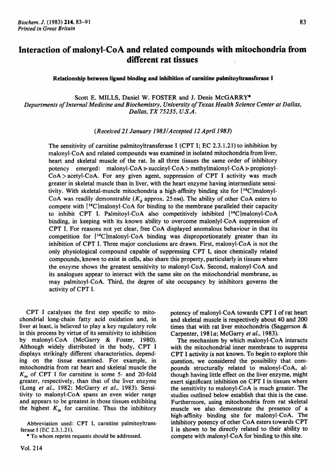
Biochem. J. (1983) 214, 83-91 Printed in Great Britain 83 Interaction of malonyl-CoA and related compounds with mitochondria from different rat tissues Relationship between ligand binding and inhibition of carnitine palmitoyltransferase I Scott E. MILLS, Daniel W. FOSTER and J. Denis McGARRY* Departments ofInternal Medicine and Biochemistry, University of Texas Health Science Center at Dallas, Dallas, TX 75235, U.S.A. (Received 21 January 1983/Accepted 12 April 1983) The sensitivity of carnitine palmitoyltransferase I (CPT I; EC 2.3.1.21) to inhibition by malonyl-CoA and related compounds was examined in isolated mitochondria from liver, heart and skeletal muscle of the rat. In all three tissues the same order of inhibitory potency emerged: malonyl-CoA > succinyl-CoA > methylmalonyl-CoA > propionyl- CoA> acetyl-CoA. For any given agent, suppression of CPT I activity was much greater in skeletal muscle than in liver, with the heart enzyme having intermediate sensi- tivity. With skeletal-muscle mitochondria a high-affinity binding site for [14C]malonyl- CoA was readily demonstrable (Kd approx. 25 nM). The ability of other CoA esters to compete with ['4C]malonyl-CoA for binding to the membrane paralleled their capacity to inhibit CPT I. Palmitoyl-CoA also competitively inhibited ['4C]malonyl-CoA binding, in keeping with its known ability to overcome malonlyl-CoA suppression of CPT I. For reasons not yet clear, free CoA displayed anomalous behaviour in that its competition for ['4C]malonyl-CoA binding was disproportionately greater than its inhibition of CPT I. Three major conclusions are drawn. First, malonyl-CoA is not the only physiological compound capable of suppressing CPT I, since chemically related compounds, known to exist in cells, also share this property, particularly in tissues where the enzyme shows the greatest sensitivity to malonyl-CoA. Second, malonyl-CoA and its analogues appear to interact with the same site on the mitochondrial membrane, as may palmitoyl-CoA. Third, the degree of site occupancy by inhibitors governs the activity of CPT I. CPT I catalyses the first step specific to mito- chondrial long-chain fatty acid oxidation and, in liver at least, is believed to play a key regulatory role in this process by virtue of its sensitivity to inhibition by malonyl-CoA (McGarry & Foster, 1980). Although widely distributed in the body, CPT I displays strikingly different characteristics, depend- ing on the tissue examined. For example, in mitochondria from rat heart and skeletal muscle the Km of CPT I for carnitine is some 5- and 20-fold greater, respectively, than that of the liver enzyme (Long et al., 1982; McGarry et al., 1983). Sensi- tivity to malonyl-CoA spans an even wider range and appears to be greatest in those tissues exhibiting the highest Km for carnitine. Thus the inhibitory Abbreviation used: CPT I, carnitine palmitoyltrans- ferase I (EC 2.3.1.2 1). * To whom reprint requests should be addressed. Vol. 214 potency of malonyl-CoA towards CPT I of rat heart and skeletal muscle is respectively about 40 and 200 times that with rat liver mitochondria (Saggerson & Carpenter, 198 1a; McGarry etal., 1983). The mechanism by which malonyl-CoA interacts with the mitochondrial inner membrane to suppress CPT I activity is not known. To begin to explore this question, we considered the possibility that com- pounds structurally related to malonyl-CoA, al- though having little effect on the liver enzyme, might exert significant inhibition on CPT I in tissues where the sensitivity to malonyl-CoA is much greater. The studies outlined below establish that this is the case. Furthermore, using mitochondria from rat skeletal muscle we also demonstrate the presence of a high-affinity binding site for malonyl-CoA. The inhibitory potency of other CoA esters towards CPT I is shown to be directly related to their ability to compete with malonyl-CoA for binding to this site.

Welcome message from author
This document is posted to help you gain knowledge. Please leave a comment to let me know what you think about it! Share it to your friends and learn new things together.
Transcript
Biochem. J. (1983) 214, 83-91Printed in Great Britain
83
Interaction of malonyl-CoA and related compounds with mitochondria fromdifferent rat tissues
Relationship between ligand binding and inhibition of carnitine palmitoyltransferase I
Scott E. MILLS, Daniel W. FOSTER and J. Denis McGARRY*Departments ofInternal Medicine and Biochemistry, University of Texas Health Science Center at Dallas,
Dallas, TX 75235, U.S.A.
(Received 21 January 1983/Accepted 12 April 1983)
The sensitivity of carnitine palmitoyltransferase I (CPT I; EC 2.3.1.21) to inhibition bymalonyl-CoA and related compounds was examined in isolated mitochondria from liver,heart and skeletal muscle of the rat. In all three tissues the same order of inhibitorypotency emerged: malonyl-CoA > succinyl-CoA > methylmalonyl-CoA > propionyl-CoA> acetyl-CoA. For any given agent, suppression of CPT I activity was muchgreater in skeletal muscle than in liver, with the heart enzyme having intermediate sensi-tivity. With skeletal-muscle mitochondria a high-affinity binding site for [14C]malonyl-CoA was readily demonstrable (Kd approx. 25 nM). The ability of other CoA esters to
compete with ['4C]malonyl-CoA for binding to the membrane paralleled their capacityto inhibit CPT I. Palmitoyl-CoA also competitively inhibited ['4C]malonyl-CoAbinding, in keeping with its known ability to overcome malonlyl-CoA suppression ofCPT I. For reasons not yet clear, free CoA displayed anomalous behaviour in that itscompetition for ['4C]malonyl-CoA binding was disproportionately greater than itsinhibition of CPT I. Three major conclusions are drawn. First, malonyl-CoA is not theonly physiological compound capable of suppressing CPT I, since chemically relatedcompounds, known to exist in cells, also share this property, particularly in tissues wherethe enzyme shows the greatest sensitivity to malonyl-CoA. Second, malonyl-CoA andits analogues appear to interact with the same site on the mitochondrial membrane, as
may palmitoyl-CoA. Third, the degree of site occupancy by inhibitors governs theactivity of CPT I.
CPT I catalyses the first step specific to mito-chondrial long-chain fatty acid oxidation and, inliver at least, is believed to play a key regulatory rolein this process by virtue of its sensitivity to inhibitionby malonyl-CoA (McGarry & Foster, 1980).Although widely distributed in the body, CPT Idisplays strikingly different characteristics, depend-ing on the tissue examined. For example, inmitochondria from rat heart and skeletal muscle theKm of CPT I for carnitine is some 5- and 20-foldgreater, respectively, than that of the liver enzyme(Long et al., 1982; McGarry et al., 1983). Sensi-tivity to malonyl-CoA spans an even wider rangeand appears to be greatest in those tissues exhibitingthe highest Km for carnitine. Thus the inhibitory
Abbreviation used: CPT I, carnitine palmitoyltrans-ferase I (EC 2.3.1.2 1).
* To whom reprint requests should be addressed.
Vol. 214
potency of malonyl-CoA towards CPT I of rat heartand skeletal muscle is respectively about 40 and 200times that with rat liver mitochondria (Saggerson &Carpenter, 198 1a; McGarry etal., 1983).
The mechanism by which malonyl-CoA interactswith the mitochondrial inner membrane to suppressCPT I activity is not known. To begin to explore thisquestion, we considered the possibility that com-pounds structurally related to malonyl-CoA, al-though having little effect on the liver enzyme, mightexert significant inhibition on CPT I in tissues wherethe sensitivity to malonyl-CoA is much greater. Thestudies outlined below establish that this is the case.Furthermore, using mitochondria from rat skeletalmuscle we also demonstrate the presence of ahigh-affinity binding site for malonyl-CoA. Theinhibitory potency of other CoA esters towards CPTI is shown to be directly related to their ability tocompete with malonyl-CoA for binding to this site.
S. E. Mills, D. W. Foster and J. D. McGarry
Experimental
AnimalsMale Sprague-Dawley rats (body wts. 130-
160 g) were maintained on a high-sucrose low-fatdiet as previously described (McGarry et al., 1978b)and were used for experiments in the fed state.
Preparation ofmitochondriaMitochondria were isolated from liver, heart and
skeletal muscle by the procedures described byMcGarry et al. (1983) (methods A and C were usedwith similar results), and were finally suspended in0.15 M-KCI. Protein was measured by the methodof Lowry etal. (1951).
Assay ofcarnitinepalmitoyltransferaseCPT I was measured in intact mitochondria in the
direction palmitoyl-CoA + [ 14Clcarnitine -.palmi-toyl[14C]carnitine + CoA, as described by McGarryet al. (1983). Final concentrations of palmitoyl-CoA,L-carnitine and fatty acid-free bovine serum albuminwere 50pM, 200#M and 1%, respectively. With0.2-0.4mg of mitochondrial protein/ml of assaymixture at 300C, reaction rates were linear with timeover the time periods studied (generally 5-8min).The concentrations of inhibitors are given in thelegends to Figures and Tables. Some mitochondrialpreparations, particularly those from heart andskeletal muscle, displayed small and variable quanti-ties of CPT activity that was insensitive to malonyl-CoA. As discussed by McGarry et al. (1983), thisprobably stemmed from exposure of some CPT II asa result of damage to the mitochondria during theirpreparation. The extent of this contamination wascalculated from a plot of (percentage CPT sup-pression by malonyl-CoA)-l versus [malonyl-CoAI-' and was used to obtain a more accurateestimate of the malonyl-CoA-sensitivity of CPT I(McGarry et al., 1983). Uncorrected data are shownin the Figures, and Table 1 gives both uncorrectedand corrected values.
Malonyl-CoA binding to skeletal-muscle mitochon-dria
Assays were performed in glass centrifuge tubes(l0mmx75mm) in a final volume of lml con-taining: Tris/HCl (pH 7.4), 90mM; KCI, 60mM;reduced glutathione, 0.25mM; MgCl2, 4mM; fattyacid-free albumin, 1%, w/v; 0.01-0.4puM-[1,3-14C1-malonyl-CoA (50,uCi/,umol) in the absence orpresence of 100puM unlabelled malonyl-CoA (toobtain non-specific binding or trapping of 14C); andabout 3mg of mitochondrial protein. Incubationswere started by addition of the mitochondria andwere usually continued for 20min at 0°C withmixing at 3-4min intervals. The tubes were thencentrifuged at 15000g for 10min at 40C, the
supernatants were aspirated and 0.25 ml of 1 M-KOH was immediately added to the pellets. Solu-bilization of the mitochondria was facilitated byheating at 500C for 30min, after which time thecontents of the tube, together with a 0.8ml waterwash, were transferred to counting vials and assayedfor radioactivity after addition of lOml of AquasolII (New England Nuclear). Non-specific bindingseldom exceeded 20% of the total. The differencebetween total and non-specific binding was used tocalculate the specific-binding data. Competitionstudies were performed in a similar manner, exceptthat the concentration of labelled malonyl-CoA wasfixed at 0.25#M and unlabelled ligands were presentfrom zero time at the indicated concentrations.
MaterialsCoA and all CoA esters were from P-L Bio-
chemicals (Milwaukee, WI, U.S.A.). Highly purifiedmalonyl-CoA decarboxylase was kindly provided byDr. P. E. Kolattukudy (Washington State University,Seattle, WA, U.S.A.). The sources of other materialshave been given previously (McGarry et al., 1983).
Results
Effects of malonyl-CoA and related compounds onmitochondrial CPTI activity
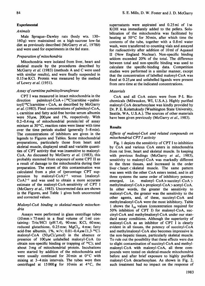
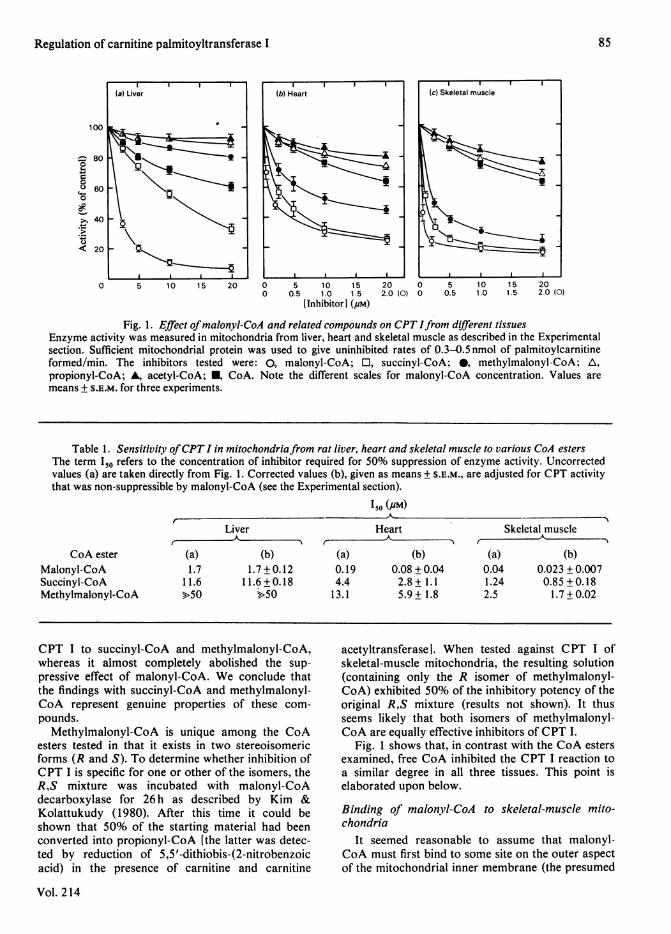
Fig. 1 depicts the sensitivity of CPT I to inhibitionby CoA and various CoA esters in mitochondriafrom rat liver, heart and skeletal muscle. In keepingwith previous findings (McGarry et al., 1983),sensitivity to malonyl-CoA was markedly differentin the three tissues, and increased in the orderliver < heart < skeletal muscle. The same patternwas seen with the other CoA esters tested, and in allthree systems the same order of inhibitory potencyemerged, i.e. malonyl - CoA > succinyl -CoA >methylmalonyl-CoA> propionyl-CoA > acetyl-CoA.In other words, the greater the sensitivity tomalonyl-CoA, the greater was the sensitivity to theother agents, and, of these, succinyl-CoA andmethylmalonyl-CoA were the most inhibitory. Table1 shows the I50 values (concentration required for50% inhibition of CPT I) for malonyl-CoA, suc-cinyl-CoA and methylmalonyl-CoA under our stan-dard assay conditions. Although the superiority ofmalonyl-CoA as an inhibitor of CPT I is clearlyevident in all tissues, the potency of succinyl-CoAand methylmalonyl-CoA also becomes impressive inthe non-hepatic tissues, particularly skeletal muscle.To rule out the possibility that these effects were dueto slight contamination of succinyl-CoA and methyl-malonyl-CoA with malonyl-CoA, all three com-pounds were tested on skeletal-muscle mitochondriabefore and after brief exposure to highly purifiedmalonyl-CoA decarboxylase. As shown in Fig. 2,such treatment had no impact on the response of
1983
84
Regulation of carnitine palmitoyltransferase I
100
0
fi0
C..
.g.
80
60
40
20~
0 5 10 15 20 0 5 10 15 20 0 5 10 15 200 0.5 1.0 1.5 2.0 (0) 0 0.5 1.0 1.5 2.0 (0)
[Inhibitor] (pM)
Fig. 1. Effect ofmalonyl-CoA and related compounds on CPT Ifrom different tissuesEnzyme activity was measured in mitochondria from liver, heart and skeletal muscle as described in the Experimentalsection. Sufficient mitochondrial protein was used to give uninhibited rates of 0.3-0.5 nmol of palmitoylcarnitineformed/min. The inhibitors tested were: 0, malonyl-CoA; O, succinyl-CoA; 0, methylmalonyl-CoA; A,propionyl-CoA; A, acetyl-CoA; U, CoA. Note the different scales for malonyl-CoA concentration. Values are
means + S.E.M. for three experiments.
Table 1. Sensitivity ofCPT I in mitochondriafrom rat liver, heart and skeletal muscle to various CoA estersThe term I5o refers to the concentration of inhibitor required for 50% suppression of enzyme activity. Uncorrectedvalues (a) are taken directly from Fig. 1. Corrected values (b), given as means + S.E.M., are adjusted for CPT activitythat was non-suppressible by malonyl-CoA (see the Experimental section).
so (UM)
Liverr
A 5%CoA ester
Malonyl-CoASuccinyl-CoAMethylmalonyl-CoA
(a)1.7
11.6>50
HeartAI --"
(b)1.7 + 0.12
11.6 + 0.18>50
(a)0.194.413.1
Skeletal muscle
(b)0.08 + 0.042.8 + 1.15.9 + 1.8
(a)0.041.242.5
(b)0.023 + 0.0070.85 + 0.181.7 + 0.02
CPT I to succinyl-CoA and methylmalonyl-CoA,whereas it almost completely abolished the sup-
pressive effect of malonyl-CoA. We conclude thatthe findings with succinyl-CoA and methylmalonyl-CoA represent genuine properties of these com-
pounds.Methylmalonyl-CoA is unique among the CoA
esters tested in that it exists in two stereoisomericforms (R and S). To determine whether inhibition ofCPT I is specific for one or other of the isomers, theR,S mixture was incubated with malonyl-CoAdecarboxylase for 26 h as described by Kim &Kolattukudy (1980). After this time it could beshown that 50% of the starting material had beenconverted into propionyl-CoA [the latter was detec-ted by reduction of 5,5'-dithiobis-(2-nitrobenzoicacid) in the presence of carnitine and carnitine
Vol. 214
acetyltransferasel. When tested against CPT I ofskeletal-muscle mitochondria, the resulting solution(containing only the R isomer of methylmalonyl-CoA) exhibited 50% of the inhibitory potency of theoriginal R,S mixture (results not shown). It thusseems likely that both isomers of methylmalonyl-CoA are equally effective inhibitors of CPT I.
Fig. 1 shows that, in contrast with the CoA estersexamined, free CoA inhibited the CPT I reaction toa similar degree in all three tissues. This point iselaborated upon below.
Binding of malonyl-CoA to skeletal-muscle mito-chondrna
It seemed reasonable to assume that malonyl-CoA must first bind to some site on the outer aspectof the mitochondrial inner membrane (the presumed
Il(a) Liver
~5
I IYL -
85
S. E. Mills, D. W. Foster and J. D. McGarry
100
4
20
0 5 10 15 200 0.5 1.0 1.5 2.0 (O.@)
[Inhibitor] (#M)
Fig. 2. Response of skeletal-muscle CPT I to CoA estersin the absence or presence of malonyl-CoA decarb-
oxylaseAssays were performed as described for Fig. 1 in theabsence (open symbols) or presence (closed sym-bols) of malonyl-CoA decarboxylase (8,g), whichwas added to the incubation mixture 20min beforeaddition of the mitochondria. The inhibitors testedwere: 0, 0, malonyl-CoA; El, I, succinyl-CoA; andA, A, methylmalonyl-CoA. The results of oneexperiment are shown. Note different scale formalonyl-CoA concentration.
300 10 20Time (min)
Fig. 3. Time course of ['4CImalonyl-CoA binding toskeletal-muscle mitochondria
Mitochondria (approx. 3 mg of protein) were incu-bated at 0°C or room temperature with 0.25#M-[ 1,3-'4CImalonyl-CoA in the presence or absence of100puM unlabelled malonyl-CoA as described in theExperimental section. Specific binding is shown for arepresentative example of several closely agreeingexperiments.
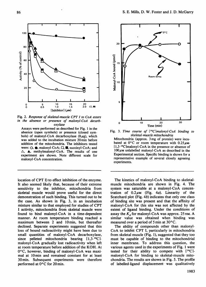
location of CPT I) to effect inhibition of the enzyme.It also seemed likely that, because of their extremesensitivity to the inhibitor, mitochondria fromskeletal muscle would prove useful for the directdemonstration of such binding. This turned out to bethe case. As shown in Fig. 3, in an incubationmixture similar to that employed for studies of CPTI activity, mitochondria from skeletal muscle werefound to bind malonyl-CoA in a time-dependentmanner. At room temperature binding reached amaximum between 2 and 10min, but thereafterdeclined. Separate experiments suggested that thisloss of bound radioactivity might have been due tosmall quantities of malonyl-CoA decarboxylase,since pelleted mitochondria bearing [1,3-14C1-malonyl-CoA gradually lost radioactivity when leftat room temperature before addition of the KOH. At0°C, however, binding of malonyl-CoA was maxi-mal at 10min and remained constant for at least30 min. Subsequent experiments were thereforeperformed at 00C for 20min.
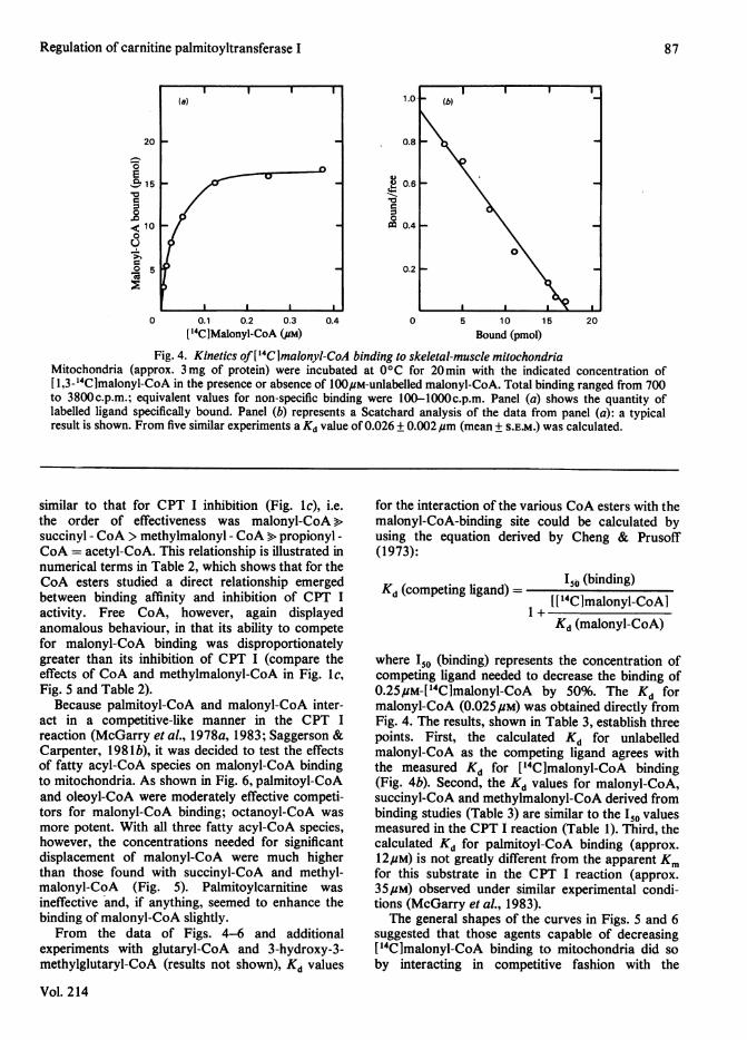
The kinetics of malonyl-CoA binding to skeletal-muscle mitochondria are shown in Fig. 4. Thesystem was saturable at a malonyl-CoA concen-tration of 0.2#M (Fig. 4a). Linearity of theScatchard plot (Fig. 4b) indicates that only one classof binding site was present and that the affinity ofmalonyl-CoA for this site was not affected by theextent of ligand binding. Under the conditions ofassay the Kd for malonyl-CoA was approx. 25 nm. Asimilar value was obtained when binding wasmeasured over a period of 5 min at 300 C.The ability of compounds other than malonyl-
CoA to inhibit CPT I, particularly in mitochondriafrom skeletal muscle (Fig. 1), suggested that they toomust be capable of binding to the mitochondrialinner membrane. To address this question, thevarious agents used in the experiments of Fig. 1 weretested for their ability to compete with [14C1-malonyl-CoA for binding to skeletal-muscle mito-chondria. The results are shown in Fig. 5. The profileof labelled-ligand displacement-was qualitatively
1983
86
Regulation of carnitine palmitoyltransferase I
20 -
1-1
0E15 _
< 10 K0
u
0 5
I I I I
0 0.1 0.2 0.3['4C]Malonyl-CoA (.aM)
0.4
1.0-
0.8
Q 0.6
-0r.
0
0.4
0.21
205 10 15Bound (pmol)
Fig. 4. Kinetics of[14Clmalonyl-CoA binding to skeletal-muscle mitochondriaMitochondria (approx. 3mg of protein) were incubated at 0°C for 20min with the indicated concentration of[ 1,3-_4Clmalonyl-CoA in the presence or absence of 100p,M-unlabelled malonyl-CoA. Total binding ranged from 700to 3800c.p.m.; equivalent values for non-specific binding were 100-lOOOc.p.m. Panel (a) shows the quantity oflabelled ligand specifically bound. Panel (b) represents a Scatchard analysis of the data from panel (a): a typicalresult is shown. From five similar experiments a Kd value of 0.026 + 0.002,um (mean + S.E.M.) was calculated.
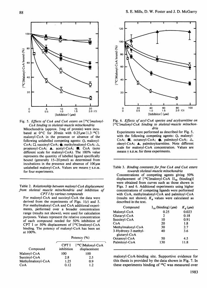
similar to that for CPT I inhibition (Fig. lc), i.e.the order of effectiveness was malonyl-CoA>succinyl - CoA > methylmalonyl - CoA > propionyl -
CoA = acetyl-CoA. This relationship is illustrated innumerical terms in Table 2, which shows that for theCoA esters studied a direct relationship emergedbetween binding affinity and inhibition of CPT Iactivity. Free CoA, however, again displayedanomalous behaviour, in that its ability to competefor malonyl-CoA binding was disproportionatelygreater than its inhibition of CPT I (compare theeffects of CoA and methylmalonyl-CoA in Fig. ic,Fig. 5 and Table 2).
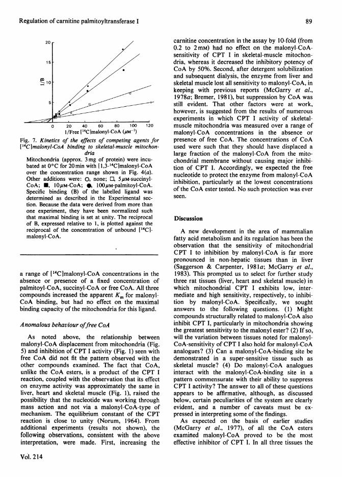
Because palmitoyl-CoA and malonyl-CoA inter-act in a competitive-like manner in the CPT Ireaction (McGarry et al., 1978a, 1983; Saggerson &Carpenter, 1981b), it was decided to test the effectsof fatty acyl-CoA species on malonyl-CoA bindingto mitochondria. As shown in Fig. 6, palmitoyl-CoAand oleoyl-CoA were moderately effective competi-tors for malonyl-CoA binding; octanoyl-CoA wasmore potent. With all three fatty acyl-CoA species,however, the concentrations needed for significantdisplacement of malonyl-CoA were much higherthan those found with succinyl-CoA and methyl-malonyl-CoA (Fig. 5). Palmitoylcarnitine wasineffective and, if anything, seemed to enhance thebinding of malonyl-CoA slightly.
From the data of Figs. 4-6 and additionalexperiments with glutaryl-CoA and 3-hydroxy-3-methylglutaryl-CoA (results not shown), Kd values
Vol. 214
for the interaction of the various CoA esters with themalonyl-CoA-binding site could be calculated byusing the equation derived by Cheng & Prusoff(1973):
Kd (competing ligand) = I5 (binding)
1 +['14Clmalonyl-CoAIKd (malonyl-CoA)
where Io (binding) represents the concentration ofcompeting ligand needed to decrease the binding of0.25pM-['4C]malonyl-CoA by 50%. The Kd formalonyl-CoA (0.025pM) was obtained directly fromFig. 4. The results, shown in Table 3, establish threepoints. First, the calculated Kd for unlabelledmalonyl-CoA as the competing ligand agrees withthe measured Kd for ['4C]malonyl-CoA binding(Fig. 4b). Second, the Kd values for malonyl-CoA,succinyl-CoA and methylmalonyl-CoA derived frombinding studies (Table 3) are similar to the 1 valuesmeasured in the CPT I reaction (Table 1). Third, thecalculated Kd for palmitoyl-CoA binding (approx.12,M) is not greatly different from the apparent Kmfor this substrate in the CPT I reaction (approx.35,UM) observed under similar experimental condi-tions (McGarry et al., 1983).
The general shapes of the curves in Figs. 5 and 6suggested that those agents capable of decreasing[14C]malonyl-CoA binding to mitochondria did soby interacting in competitive fashion with the
(b)
0
87
-1- I
S. E. Mills, D. W. Foster and J. D. McGarry
80
.0<60j
0
0
*~40
20
0 5 10 15 200 0.5 1.0 1.5 2.0 (0)
[Inhibitor] (pM)
Fig. 5. Effects of CoA and CoA esters on 14Clmalonyl-CoA binding to skeletal-muscle mitochondria
Mitochondria (approx. 3 mg of protein) were incu-bated at 0°C for 20min with 0.25pM-[1,3-14C1-malonyl-CoA in the presence or absence of thefollowing unlabelled competing agents: 0, malonyl-CoA; O, succinyl-CoA; 0, methylmalonyl-CoA; A,
propionyl-CoA; A, acetyl-CoA; *, CoA (notedifferent scale for malonyl-CoA). The 100% valuerepresents the quantity of labelled ligand specificallybound (generally 15-20pmol) as determined fromincubations in the presence and absence of 100,Munlabelled malonyl-CoA. Values are means + S.E.M.for four experiments.
Table 2. Relationship between malonyl-CoA displacementfrom skeletal muscle mitochondria and inhibition of
CPTI by various compoundsFor malonyl-CoA and succinyl-CoA the data were
derived from the experiments of Figs. l(c) and 5.For methylmalonyl-CoA and CoA additional experi-ments, performed over a broader concentrationrange (results not shown), were used for calculationpurposes. Values represent the relative concentrationof each compound needed for 50% inhibition ofCPT I or 50% displacement of [14Clmalonyl-CoAbinding. The potency of malonyl-CoA has been setat 100%.
Potency (%)
CompoundMalonyl-CoASuccinyl-CoAMethylmalonyl-CoACoA
CPT Iinhibition100
2.81.250.12
[14CIMalonyl-CoAdisplacement
1002.50.91.2
120
1004
-o
0
c0
0
0
Cd
I
80
60
40
20
0 20 40 600 0.5 1.0 1.5
[Inhibitor] (#M)
802.0 (0)
100
Fig. 6. Effects of acyl-CoA species and acylcarnitine on
['4Clmalonyl-CoA binding to skeletal-muscle mitochon-dria
Experiments were performed as described for Fig. 5,with the following competing agents: 0, malonyl-CoA; *, octanoyl-CoA; 0, palmitoyl-CoA; A,
oleoyl-CoA; A, palmitoylcarnitine. Note differentscale for malonyl-CoA concentration. Values aremeans + S.E.M. for three experiments.
Table 3. Binding constants forfree CoA and CoA esterstowards skeletal-muscle mitochondria
Concentrations of competing agents giving 50%displacement of [14C]malonyl-CoA 1150 (binding)]were obtained from curves such as those shown inFigs. 5 and 6. Additional experiments using higherconcentrations of competing ligands were performedwith CoA, methylmalonyl-CoA and palmitoyl-CoA(results not shown).described in the text.
CompoundMalonyl-CoAGlutaryl-CoASuccinyl-CoACoAMethylmalonyl-CoA3-Hydroxy-3-methyl-
glutaryl-CoAOctanoyl-CoAPalmitoyl-CoA
Kd values were calculated as
I50 (binding) (#M)0.25210203040
50130
Kd (PM)
0.0230.180.911.82.73.6
4.511.8
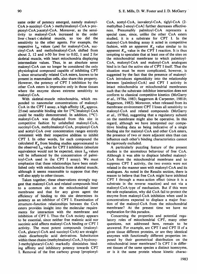
malonyl-CoA-binding site. Supportive evidence forthis thesis is provided by the data shown in Fig. 7. Inthese experiments binding of 14C was measured over
1983
I I I I I
_~~~~~~~_
I
I I I I I\
88
Regulation of carnitine palmitoyltransferase I
0 20 40 60 80 100 1201/Free ['4Clmalonyl-CoA (,am-p)
Fig. 7. Kinetics of the effects of competing agents for['4CImalonyl-CoA binding to skeletal-muscle mitochon-
driaMitochondria (approx. 3 mg of protein) were incu-bated at 00C for 20min with [1,3-14CImalonyl-CoAover the concentration range shown in Fig. 4(a).Other additions were: 0, none; 0, 5puM-succinyl-CoA; *, l0OuM-CoA; 0, 100#M-palmitoyl-CoA.Specific binding (B) of the labelled ligand wasdetermined as described in the Experimental sec-
tion. Because the data were derived from more thanone experiment, they have been normalized suchthat maximal binding is set at unity. The reciprocalof B, expressed relative to 1, is plotted against thereciprocal of the concentration of unbound [14CI-malonyl-CoA.
a range of [14C]malonyl-CoA concentrations in theabsence or presence of a fixed concentration ofpalmitoyl-CoA, succinyl-CoA or free CoA. All threecompounds increased the apparent Km for malonyl-CoA binding, but had no effect on the maximalbinding capacity of the mitochondria for this ligand.
Anomalous behaviour offree CoA
As noted above, the relationship betweenmalonyl-CoA displacement from mitochondria (Fig.5) and inhibition of CPT I activity (Fig. 1) seen withfree CoA did not fit the pattern observed with theother compounds examined. The fact that CoA,unlike the CoA esters, is a product of the CPT Ireaction, coupled with the observation that its effecton enzyme activity was approximately the same inliver, heart and skeletal muscle (Fig. 1), raised thepossibility that the nucleotide was working throughmass action and not via a malonyl-CoA-type ofmechanism. The equilibrium constant of the CPTreaction is close to unity (Norum, 1964). Fromadditional experiments (results not shown), thefollowing observations, consistent with the aboveinterpretation, were made. First, increasing the
Vol. 214
carnitine concentration in the assay by 10-fold (from0.2 to 2 mM) had no effect on the malonyl-CoA-sensitivity of CPT I in skeletal-muscle mitochon-dria, whereas it decreased the inhibitory potency ofCoA by 50%. Second, after detergent solubilizationand subsequent dialysis, the enzyme from liver andskeletal muscle lost all sensitivity to malonyl-CoA, inkeeping with previous reports (McGarry et al.,1978a; Bremer, 1981), but suppression by CoA was-still evident. That other factors were at work,however, is suggested from the results of numerousexperiments in which CPT I activity of skeletal-muscle mitochondria was measured over a range ofmalonyl-CoA concentrations in the absence orpresence of free CoA. The concentrations of CoAused were such that they should have displaced alarge fraction of the malonyl-CoA from the mito-chondrial membrane without causing major inhibi-tion of CPT I. Accordingly, we expected the freenucleotide to protect the enzyme from malonyl-CoAinhibition, particularly at the lowest concentrationsof the CoA ester tested. No such protection was everseen.
Discussion
A new development in the area of mammalianfatty acid metabolism and its regulation has been theobservation that the sensitivity of mitochondrialCPT I to inhibition by malonyl-CoA is far morepronounced in non-hepatic tissues than in liver(Saggerson & Carpenter, 1981a; McGarry et al.,1983). This prompted us to select for further studythree rat tissues (liver, heart and skeletal muscle) inwhich mitochondrial CPT I exhibits low, inter-mediate and high sensitivity, respectively, to inhibi-tion by malonyl-CoA. Specifically, we soughtanswers to the following questions. (1) Mightcompounds structurally related to malonyl-CoA alsoinhibit CPT I, particularly in mitochondria showingthe greatest sensitivity to the malonyl ester? (2) If so,will the variation between tissues noted for malonyl-CoA-sensitivity of CPT I also hold for malonyl-CoAanalogues? (3) Can a malonyl-CoA-binding site bedemonstrated in a super-sensitive tissue such asskeletal muscle? (4) Do malonyl-CoA analoguesinteract with the malonyl-CoA-binding site in apattern commensurate with their ability to suppressCPT I activity? The answer to all of these questionsappears to be affirmative, although, as discussedbelow, certain peculiarities of the system are clearlyevident, and a number of caveats must be ex-pressed in interpreting some of the findings.As expected on the basis of earlier studies
(McGarry et al., 1977), of all the CoA estersexamined malonyl-CoA proved to be the mosteffective inhibitor of CPT I. In all three tissues the
89
S. E. Mills, D. W. Foster and J. D. McGarry
same order of potency emerged, namely malonyl-CoA > succinyl-CoA > methylmalonyl-CoA > pro-pionyl-CoA>acetyl-CoA. Moreover, as the sensi-tivity to malonyl-CoA increased in the orderliver <heart < skeletal muscle, so too did thesensitivity to the other agents. For example, therespective ISo values (pM) for malonyl-CoA, suc-cinyl-CoA and methylmalonyl-CoA shifted fromabout 2, 12 and >50 for liver to 0.02, 1 and 2 forskeletal muscle, with heart mitochondria displayingintermediate values. Thus, in an absolute sensemalonyl-CoA can no longer be considered the onlyphysiological compound capable of inhibiting CPTI, since structurally related CoA esters, known to bepresent in mammalian cells, also share this property.However, the potency of CPT I inhibition by theother CoA esters is impressive only in those tissueswhere the enzyme shows extreme sensitivity tomalonyl-CoA.
With skeletal-muscle mitochondria, which res-ponded to nanomolar concentrations of malonyl-CoA in the CPT I assay, a high-affinity (Kd approx.25nM) saturable binding site for ['4C]malonyl-CoAcould be readily demonstrated. In addition, ['4C1-malonyl-CoA was displaced from this site incompetitive fashion by unlabelled malonyl-CoA,succinyl-CoA, methylmalonyl-CoA, propionyl-CoAand acetyl-CoA over concentration ranges entirelyconsistent with their respective abilities to inhibitCPT I. In other words, for each compound thecalculated Kd from binding studies approximated tothe observed 1 value for CPT I inhibition (absoluteequivalence would not be expected, since I5 valueswill be influenced by the concentration of palmi-toyl-CoA used in the CPT I assay). We mustemphasize that these relationships, have been estab-lished only with mitochondria from skeletal muscle,although it seems reasonable to suppose that theywill also apply to other tissues.Taken together, these observations strongly sug-
gest that malonyl-CoA and related compounds bindto a common site on the mitochondrial innermembrane and that for any given agent theefficiency of binding to this site determines itspotency as an inhibitor of CPT I. Examination ofstructure-function relationships between the CoAesters provides insight into the molecular require-ments for interaction with the membrane andinhibition of CPT I. Thus the CoA moiety appearsto be essential, since neither free malonic acid norsuccinic acid affects malonyl-CoA binding or CPT Iactivity. The most potent compounds (malonyl-CoA, glutaryl-CoA and succinyl-CoA) are straight-chain dicarboxylic acid derivatives. Substitutionwithin these chains (methylmalonyl-CoA, 3-hydroxy-3-methylglutaryl-CoA) markedly diminishes bind-ing affinity and inhibitory potency towards CPTI. Removal of the free carboxy group [propionyl-
CoA, acetyl-CoA, isovaleryl-CoA, tiglyl-CoA (2-methylbut-2-enoyl-CoA] further decreases effective-ness. Presumably palmitoyl-CoA represents aspecial case, since, unlike the other CoA estersstudied, it is a substrate for CPT I. In themalonyl-CoA-binding assay it acted in competitivefashion, with an apparent Kd value similar to itsapparent Km value in the CPT I reaction. It is thustempting to speculate that at least one of the sites onthe mitochondrial membrane to which palmitoyl-CoA, malonyl-CoA and malonyl-CoA analoguesbind is in fact the active site of CPT I itself. That thesituation must be more complicated, however, issuggested by the fact that the presence of malonyl-CoA introduces sigmoidicity into the relationshipbetween [palmitoyl-CoA] and CPT I activity inintact mitochondria or mitochondrial membranessuch that the substrate-inhibitor interaction does notconform to classical competitive kinetics (McGarryet al., 1978a, 1983; Saggerson & Carpenter, 198 lb;Saggerson, 1982). Moreover, when released from itsmembrane environment CPT I loses all sensitivity tomalonyl-CoA and related compounds (McGarryet al., 1978a), suggesting that a regulatory subuniton the membrane might also be operative. In thisregard, although we have interpreted our compe-titive binding data as being indicative of a singlebinding site for malonyl-CoA and other CoA esters,the presence of two or more adjacent sites than caninfluence each other's binding characteristics cannotbe rigorously excluded.A particularly puzzling feature of the present
studies is the anomalous behaviour of free CoA.Although it was able to displace labelled malonyl-CoA from the mitochondrial membrane and tosuppress CPT I activity, the two events were notrelated in the manner seen with malonyl-CoA and itsanalogues. As noted in the Results section, there isreason to believe that free CoA might have inhibitedCPT I through a mass-action effect (since it is asubstrate in the reverse reaction) and not via amalonyl-CoA-type of mechanism. But if this werethe sole explanation, why did CoA fail to protect theenzyme from malonyl-CoA inhibition when used inconcentrations expected to displace a major frac-tion of the malonyl-CoA from the mitochondrialmembrane? At the present time we have noexplanation for this paradox.
Concerning the properties and potential regu-latory roles of mitochondrial CPT, many otherquestions, not addressed here, remain to beanswered. For example, are CPT I and CPT II of agiven tissue different proteins, or are they identicalenzymes with functionally different properties im-posed by their geometrical distribution on themitochondrial inner membrane? Is CPT I in differ-ent tissues of the same species a distinct isoenzyme,or is it the same protein whose kinetic charac-
1983
90
Regulation of carnitine palmitoyltransferase I 91
teristics are grossly altered by variations in mem-brane environment? Extension of the studies de-scribed here to mitochondrial preparations fromwhich CPT I has been selectively removed, coupledwith reconstitution experiments involving mitochon-drial membranes and solubilized enzyme, couldprovide useful insight into these questions. From aphysiological standpoint, the notion that malonyl-CoA or related intermediates might under certaincircumstances exert control over CPT I in non-hepatic tissues (McGarry et al., 1983) remains anintriguing prospect. However, progress on this frontmust await the technology needed for accuratedetermination of the intracellular location andconcentrations of such compounds.
The expert technical assistance of Karen Herrmann-Thatcher and Murphy Daniels is gratefully ack-nowledged. We are also indebted to Dr. P. E. Kolat-tukudy, Washington State University, Seattle, for pro-viding us with malonyl-CoA decarboxylase. This workwas supported by grants from the U.S. Public HealthService (AM 18573 and AM 07307).
References
Bremer, J. (1981) Biochim. Biophys. Acta 665, 628-631Cheng, Y. C. & Prusoff, W. H. (1973) Biochem. Pharma-
col. 22, 3099-3108Kim, Y. S. & Kolattukudy, P. E. (1980) J. Biol. Chem.
255, 686-689Long, C. S., Haller, R. G., Foster, D. W. & McGarry,
J. D. (1982) Neurology 32, 663-666Lowry, 0. H., Rosebrough, N. J., Farr, A. L. & Randall,
R. J. (195 1)J. Biol. Chem. 193, 265-275McGarry, J. D. & Foster, D. W. (1980) Annu. Rev.
Biochem. 49, 395-420McGarry, J. D., Mannaerts, G. P. & Foster, D. W.
(1977) J. Clin. Invest. 60, 265-270McGarry, J. D., Leatherman, G. F. & Foster, D. W.
(1978a)J. Biol. Chem. 253, 4128-4136McGarry, J. D., Stark, M. J. & Foster, D. W. (1978b) J.
Biol. Chem. 253, 8291-8293McGarry, J. D., Mills, S. E. & Foster, D. W. (1983)
Biochem. J. 214, 21-28Norum, K. R. (1964) Biochim. Biophys. Acta 89, 95-108Saggerson, E. D. (1982) Biochem. J. 202, 397-405Saggerson, E. D. & Carpenter, C. A. (198 la) FEBS Lett.
129, 229-232Saggerson, E. D. & Carpenter, C. A. (198 lb) FEBS Lett.
132, 166-168
Vol. 214
Related Documents