12 Innate Immune Receptors in Atherosclerosis Jennifer E. Cole, Anusha N. Seneviratne and Claudia Monaco Kennedy Institute of Rheumatology, Imperial College London United Kingdom 1. Introduction The inflammatory response is an important process, aiming to restore tissue homeostasis following tissue injury or infection. Acute inflammation is a tightly controlled process. If an inflammatory stimulus persists or if normal immune function is perturbed, inflammation may become chronic. Atherosclerosis is a chronic inflammatory disorder involving components of both the innate and adaptive immune systems (Ross, 1999). The innate immune system provides the first line of defence against invading pathogens. Innate immune detection of pathogens relies on a set of pattern recognition receptors (PRRs) that recognise and respond to conserved pathogen-associated molecular patterns (PAMPs). Growing evidence supports roles for PRRs in the initiation and progression of atherosclerosis. In this chapter, the agonists, signalling pathways, expression and functions of PRRs, in particular in reference to atherosclerosis, will be discussed. The potential therapeutic benefit of targeting PRRs for treatment of atherosclerosis will also be explored. 2. Pattern recognition receptors PAMPs are recognised by an expanding number of PRRs, which currently includes at least 50 members. PRRs can be categorised into one of three families: Toll-like receptors (TLRs), Retinoic acid inducible gene I (RIG-I)-like receptors (RLRs), and Nucleotide-binding oligomerization domain (NOD)-like receptors (NLRs). Both extracellular and intracellular compartments are patrolled by PRRs with each family of receptors specialising in surveying a given location. TLRs are transmembrane PRRs either located in the cellular membrane (interacting with the extracellular space) or within intracellular vesicles such as endosomes or lysosomes. Cytosolic PRRs include RLRs and NLRs that detect intracellular PAMPs. While cytosolic PRRs are universally expressed in the majority of cells in the body (Takeuchi & Akira, 2010), TLR expression is more restricted. TLR, NLR and RLR ligation by an agonist stimulates downstream signalling cascades activating 2 major types of transcription factor: the nuclear factor kB (NFκB), and interferon response factors (IRFs). 2.1 Toll-like receptors (TLRs) The TLR family contains at least 13 different members in mammals. Following ligand binding, TLRs dimerise, with most receptors (with the exception of TLR2 and TLR4) forming homodimers. Components of the bacterial cell wall including bacterial lipoproteins, endotoxin and flagellin are sensed by TLR2, TLR4 and TLR5 respectively. TLR3, TLR7, TLR8 www.intechopen.com

Welcome message from author
This document is posted to help you gain knowledge. Please leave a comment to let me know what you think about it! Share it to your friends and learn new things together.
Transcript

12
Innate Immune Receptors in Atherosclerosis
Jennifer E. Cole, Anusha N. Seneviratne and Claudia Monaco Kennedy Institute of Rheumatology, Imperial College London
United Kingdom
1. Introduction
The inflammatory response is an important process, aiming to restore tissue homeostasis
following tissue injury or infection. Acute inflammation is a tightly controlled process. If an
inflammatory stimulus persists or if normal immune function is perturbed, inflammation
may become chronic. Atherosclerosis is a chronic inflammatory disorder involving
components of both the innate and adaptive immune systems (Ross, 1999). The innate
immune system provides the first line of defence against invading pathogens. Innate
immune detection of pathogens relies on a set of pattern recognition receptors (PRRs) that
recognise and respond to conserved pathogen-associated molecular patterns (PAMPs).
Growing evidence supports roles for PRRs in the initiation and progression of
atherosclerosis. In this chapter, the agonists, signalling pathways, expression and functions
of PRRs, in particular in reference to atherosclerosis, will be discussed. The potential
therapeutic benefit of targeting PRRs for treatment of atherosclerosis will also be explored.
2. Pattern recognition receptors
PAMPs are recognised by an expanding number of PRRs, which currently includes at least
50 members. PRRs can be categorised into one of three families: Toll-like receptors (TLRs),
Retinoic acid inducible gene I (RIG-I)-like receptors (RLRs), and Nucleotide-binding
oligomerization domain (NOD)-like receptors (NLRs). Both extracellular and intracellular
compartments are patrolled by PRRs with each family of receptors specialising in surveying
a given location. TLRs are transmembrane PRRs either located in the cellular membrane
(interacting with the extracellular space) or within intracellular vesicles such as endosomes
or lysosomes. Cytosolic PRRs include RLRs and NLRs that detect intracellular PAMPs.
While cytosolic PRRs are universally expressed in the majority of cells in the body (Takeuchi
& Akira, 2010), TLR expression is more restricted. TLR, NLR and RLR ligation by an agonist
stimulates downstream signalling cascades activating 2 major types of transcription factor:
the nuclear factor kB (NFκB), and interferon response factors (IRFs).
2.1 Toll-like receptors (TLRs) The TLR family contains at least 13 different members in mammals. Following ligand binding, TLRs dimerise, with most receptors (with the exception of TLR2 and TLR4) forming homodimers. Components of the bacterial cell wall including bacterial lipoproteins, endotoxin and flagellin are sensed by TLR2, TLR4 and TLR5 respectively. TLR3, TLR7, TLR8
www.intechopen.com
Atherogenesis
256
and TLR9 are not located on the cell surface and are instead located on the membranes of endoplasmic reticulum (ER), endosomes and lysosomes, where they detect nucleic acids derived from bacteria or viruses. The TLR family share their cytoplasmic Toll/Interleukin-1 Receptor (TIR) domain – essential for signal transduction - with their larger parent family which includes interleukin-1-receptor (IL-1Rs). The extracellular regions of TLRs contain tandemly arranged leucine rich repeats (LRR) creating a horseshoe-shaped solenoid structure (Liu et al., 2008a). TLRs are connected to their downstream signalling cascades via five TLR adaptor molecules that are recruited to and homophilically interact with the TIR domain: myeloid differentiation protein 88 (MyD88), Toll–interleukin-1 receptor domain-containing adaptor inducing interferon-┚ (TRIF), TIR domain-containing adaptor protein (TIRAP)/MyD88-adaptor-like (MAL), TRIF related adaptor molecule (TRAM) and sterile alpha and HEAT/Armadillo motif (SARM).
2.2 RIG-I-like receptors (RLRs) Double stranded RNA (dsRNA) in the cytoplasm can be sensed in both immune and non-
immune cells via RLRs. The RLR family includes retinoic acid inducible gene I (RIG-I),
melanoma differentiation associated gene 5 (MDA5) and laboratory of genetics and
physiology 2 (LGP2). RLRs possess a central RNA helicase domain with the ATPase
binding motif DExD/H. The C-terminal regulatory domain is responsible for binding to
dsRNAs. RIG-I and MDA5 have two N-terminal caspase activation and recruitment
domains (CARDs), which allows homophilic interactions between activated RIG-I or
MDA5 and the adaptor protein mitochondrial antiviral signaling (MAVS, also known as
IPS-1, VISA, and Cardif), which is found in the outer mitochondrial membrane (Takeuchi
& Akira, 2010).
2.3 NOD-like receptors (NLRs) NLRs belong to a large family of soluble proteins that are present in the cytoplasm and
detect intracellular ligands. There are 23 NLR genes in humans and 34 in mice. Three
distinct subfamilies of NLRs exist: NODs, NLRPs (or NALPs) and IL-1┚-converting enzyme
(ICE)-protease activating factor (IPAF). NLRs are composed of the following domains: a C-
terminal ligand-sensing leucine-rich repeat (LRR) domain, a central nucleotide-binding and
oligomerization (NACHT) domain (responsible for oligomerization), and an N-terminal
effector pyrin domain (PYD), caspase recruitment domain family (CARD) or baculoviral
IAP repeat (BIR) mediating homophilic interactions in downstream signalling. The
physiological function of most NLRs is still not understood.
3. PRR agonists in atherosclerosis
A vast and diverse array of ligands including viruses, lipids and extracellular matrix components are collectively recognised by PRRs (Lundberg & Hansson, 2010). Each individual PRR exhibits specificity in the repertoire of ligands that it recognises and responds to. In a process known as ‘sterile inflammation’, activation of PRRs can occur in the absence of exogenous stimuli (Rifkin et al., 2005). The PRR agonists in this context are generated as a result of tissue damage and inflammation and are known as ‘damage-associated molecular patterns’ (DAMPs). Thus, PRR ligands encompass both exogenous PAMPs and endogenous DAMPs. Increasing evidence suggests that different co-receptors
www.intechopen.com
Innate Immune Receptors in Atherosclerosis
257
and accessory molecules and thus different mechanisms of action are used by TLRs in response to ligation by PAMPs and DAMPs (reviewed in (Piccinini & Midwood, 2010)). Exogenous PRR ligands, such as viruses and bacteria, and endogenous PRR ligands, including extracellular matrix components, modified lipids and heat shock proteins are PRR ligands that may be relevant in the context of atherosclerosis.
3.1 Exogenous PRR agonists Exogenous agonists are the best defined PRR ligands and include components of bacteria
and viruses. TLR2 recognises a diverse array of PAMPs using heterodimerisation with
TLR1 or TLR6. TLR2 is key in the recognition of Gram-positive bacteria (Underhill et al.,
1999a; Underhill et al., 1999b). Lipoteichoic acid, is a ligand of TLR2/TLR6 heterodimers
as are peptidoglycan and zymosan (Gantner et al., 2003; Ozinsky et al., 2000; Schroder et
al., 2003). Using CD36 as a co-receptor, TLR2/TLR6 heterodimers also recognise
mycoplasma diacylated lipoproteins peptide (Brightbill et al., 1999; Hoebe et al., 2005;
Takeuchi et al., 2001). Triacylated lipoproteins are ligands for TLR1/TLR2 heterodimers
(Jin et al., 2007; Takeuchi et al., 2002). Endotoxin (lipopolysaccharide), a component of the
outer membrane of Gram-negative bacteria, is an agonist for the TLR4 signalling complex
(Shimazu et al., 1999; Wright et al., 1990). Compared to TLR2 and TLR4, other TLRs have
a relatively limited repertoire of TLR ligands. TLR3 senses viral double-stranded RNA
and some small interfering RNAs. A synthetic dsRNA analogue Poly(I:C) is commonly
used as a TLR3 activator (Takeuchi & Akira, 2010). Bacterial flagellin is recognised by
TLR5 and TLR9 detects unmethylated CpG DNA, typically of bacterial origin (O'Neill &
Bowie, 2007). TLR7 is the main sensor of ssRNA derived from RNA viruses including
human immunodeficiency virus and influenza A. More recently, TLR7 on myeloid
dendritic cells has also been shown to be capable of sensing bacterial RNA (Mancuso et
al., 2009).
Genomic RNA of dsRNA viruses and dsRNA generated as the replication intermediate of
ssRNA viruses in the cytosol are ligands for RLRs. Short dsRNAs with 5’ triphosphate ends
are sensed by RIG-I whereas MDA5 recognises longer dsRNAs (Kato et al., 2006). NOD1
and NOD2 sense peptidoglycan. NOD1 recognises a peptidoglycan motif: dipeptide ┛-d-
glutamyl-meso-dia-minopimelic acid (iE-DAP) and NOD2 recognises muramyl dipeptide
(Chamaillard et al., 2003; Girardin et al., 2003a; Girardin et al., 2003b; Inohara et al., 2003).
Whole pathogens including bacteria with pore-forming toxins and viruses including
influenza virus are activators of the NLRP3 inflammasome (reviewed in (Schroder &
Tschopp, 2010)).
Numerous exogenous PAMPS may be ligands for PRRs in atherosclerosis. Chlamydia
pneumonia, porphyromonas gingivalis and cytomegalovirus are exogenous PRR ligands found
in atherosclerotic plaques (Chiu et al., 1997; Kuo et al., 1993). These bacterial and viral
infectious agents have been associated with an increased risk of atherosclerosis
development (Kalayoglu et al., 2002; Kiechl et al., 2001; Scannapieco et al., 2003) and are
recognised by TLR2 and TLR4 (Burns et al., 2006; Compton et al., 2003; Naiki et al., 2008).
The failure to detect active viral replication within atherosclerotic plaques (Kol et al., 1995;
Zhou et al., 1999) suggests PRR activation by infectious agents and not viral replication itself
is the link between infectious disease and cardiovascular risk. Exogenous heat shock
proteins (HSPs), nucleic acids (Lehtiniemi et al., 2005; Ott et al., 2006) and peptidoglycan
www.intechopen.com

Atherogenesis
258
(Laman et al., 2002) are also present in atherosclerotic lesions and thus may activate PRRs in
atherogenesis.
3.2 Endogenous PRR agonists Many endogenous PRR ligands are present in atherosclerotic lesions and thus PRR
activation in atherosclerosis could result from a combination of exogenous and
endogenous ligand sensing. Indeed, work by Curtiss and colleagues supports a role for
endogenous TLR ligands in atherogenesis (Mullick et al., 2005). Extracellular matrix
(ECM) is degraded during tissue injury and remodelling leading to the generation of ECM
components, which can function as PRR ligands. Fibrinogen can activate TLR4 signalling
as can the fibronectin alternatively spliced exon encoding type III extra domain A (EDA)
and tenascin C (Midwood & Orend, 2009; Okamura et al., 2001; Smiley et al., 2001).
Hyaluronan, a large glycosaminoglycan component of the ECM, and biglycan activate
TLR2 and TLR4 signalling (Schaefer et al., 2005; Scheibner et al., 2006; Taylor et al., 2004).
Hyaluronan can induce IL-1 release by macrophages in a NLRP3-dependent manner
(Yamasaki et al., 2009) while biglycan also activates the NLRP3 inflammasome (Babelova
et al., 2009). The large ECM proteoglycan versican is a TLR2/6 ligand associated with
cytokine production in tumor-infiltrating macrophages (Kim et al., 2009). These ECM
components may be generated during injury and remodeling of the vessel wall and thus
may activate PRRs in atherosclerosis.
Lipids, key components of atherosclerotic plaques, are TLR ligands. Minimally modified
low-density lipoproteins induce cytokine and reactive oxygen species generation via TLR4
signalling complexes (Miller et al., 2003). In association with CD36, TLR4/TLR6 hetero-
dimers sense oxidized LDL leading to increased chemokine expression (Stewart et al., 2010).
Saturated fatty acids elicit TLR4 activation whereas polyunsaturated fatty acids inhibit TLR4
activation (Lee et al., 2003). However, the ability of saturated fatty acids to directly induce
TLR signaling has been questioned (Erridge & Samani, 2009). TLR2 can also sense ApoCIII,
a component of very-low-density lipoprotein (VLDL) (Kawakami et al., 2008).
HSPs are present in murine atherosclerotic lesions and are ligands for TLR2 and TLR4
(Asea et al., 2002; Kanwar et al., 2001). However, some studies using low-endotoxin
preparations have disputed the role of HSPs as ligands for TLRs (Bausinger et al., 2002).
The nuclear protein high-mobility group box-1 (HMGB-1) is expressed in human
atherosclerotic smooth muscle cells (Porto et al., 2006). HMGB1 binds DNA and is a
ligand for TLR2, TLR4, TLR9 and other nucleic acid sensors (Park et al., 2004; Yanai et al.,
2009). mRNA from necrotic cells, which may be present in atherosclerotic plaques, is a
TLR3 agonist (Kariko et al., 2004).
The NLRP3 inflammasome can be activated by many factors including extracellular ATP,
potassium efflux and reactive oxygen species. In addition, intracellular crystals such as
monosodium urate crystals and cholesterol crystals can activate the NLRP3 inflammasome
(Duewell et al., 2010; Martinon et al., 2006).
4. Signalling of pattern recognition receptors
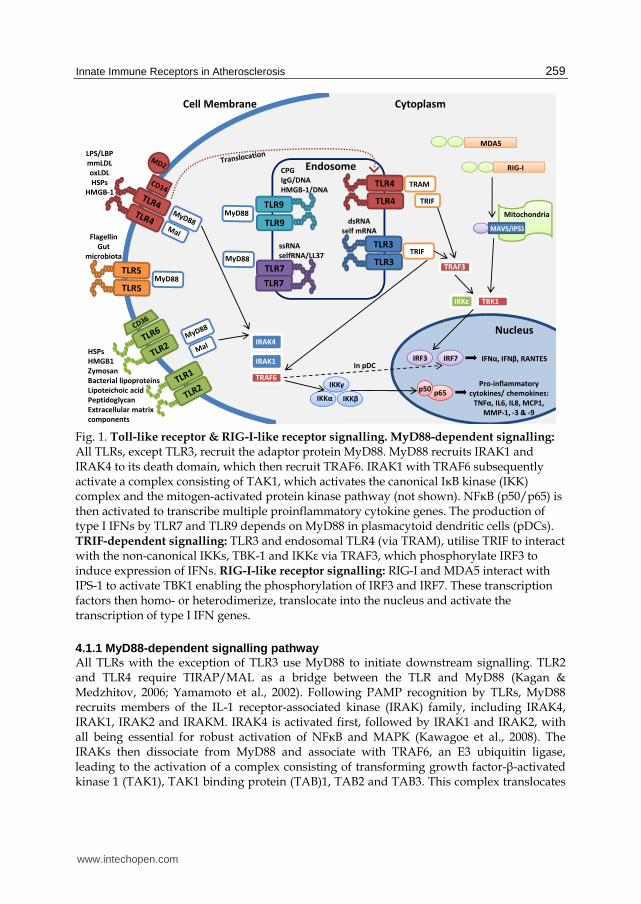
4.1 Toll-like receptor signalling TLR signalling is composed of two distinct signalling pathways depending on whether the adaptor molecule MyD88 is used following ligation and activation (Figure 1).
www.intechopen.com
Innate Immune Receptors in Atherosclerosis
259
Cell Membrane
Nucleus
Cytoplasm
LPS/LBP
mmLDL
oxLDL
HSPs
HMGB-1
Flagellin
Gut
microbiota
HSPs
HMGB1
Zymosan
Bacterial lipoproteins
Lipoteichoic acid
Peptidoglycan
Extracellular matrix
components
Endosome
TLR7
TLR7
TLR9
TLR9
IRAK4
IRAK1
TRAF6
ssRNA
selfRNA/LL37
CPG
IgG/DNA
HMGB-1/DNATRAM
TRIF
dsRNA
self mRNA
TRIF
IKKγ
IKKα IKKβp65
p50Pro-inflammatory
cytokines/ chemokines:
TNFα, IL6, IL8, MCP1,
MMP-1, -3 & -9
TRAF3
TBK1IKKε
IRF3 IRF7In pDC
IFNα, IFNβ, RANTES
Mitochondria
MAVS/IPS1
MDA5
RIG-I
Fig. 1. Toll-like receptor & RIG-I-like receptor signalling. MyD88-dependent signalling: All TLRs, except TLR3, recruit the adaptor protein MyD88. MyD88 recruits IRAK1 and IRAK4 to its death domain, which then recruit TRAF6. IRAK1 with TRAF6 subsequently activate a complex consisting of TAK1, which activates the canonical IκB kinase (IKK) complex and the mitogen-activated protein kinase pathway (not shown). NFκB (p50/p65) is then activated to transcribe multiple proinflammatory cytokine genes. The production of type I IFNs by TLR7 and TLR9 depends on MyD88 in plasmacytoid dendritic cells (pDCs). TRIF-dependent signalling: TLR3 and endosomal TLR4 (via TRAM), utilise TRIF to interact with the non-canonical IKKs, TBK-1 and IKKε via TRAF3, which phosphorylate IRF3 to induce expression of IFNs. RIG-I-like receptor signalling: RIG-I and MDA5 interact with IPS-1 to activate TBK1 enabling the phosphorylation of IRF3 and IRF7. These transcription factors then homo- or heterodimerize, translocate into the nucleus and activate the transcription of type I IFN genes.
4.1.1 MyD88-dependent signalling pathway All TLRs with the exception of TLR3 use MyD88 to initiate downstream signalling. TLR2 and TLR4 require TIRAP/MAL as a bridge between the TLR and MyD88 (Kagan & Medzhitov, 2006; Yamamoto et al., 2002). Following PAMP recognition by TLRs, MyD88 recruits members of the IL-1 receptor-associated kinase (IRAK) family, including IRAK4, IRAK1, IRAK2 and IRAKM. IRAK4 is activated first, followed by IRAK1 and IRAK2, with all being essential for robust activation of NFκB and MAPK (Kawagoe et al., 2008). The IRAKs then dissociate from MyD88 and associate with TRAF6, an E3 ubiquitin ligase, leading to the activation of a complex consisting of transforming growth factor-┚-activated kinase 1 (TAK1), TAK1 binding protein (TAB)1, TAB2 and TAB3. This complex translocates
www.intechopen.com
Atherogenesis
260
into the cytosol where TAK1 phosphorylates IKK┚. Subsequently, the IKK complex, consisting of IKK┙, IKK┚ and NFκB essential modulator (NEMO), phosphorylates IκB┙, an NFκB inhibitory protein. Phosphorylated IκB┙ is degraded by the ubiquitin proteosome system, freeing NFκB to translocate to the nucleus and mediate transcription of inflammatory genes. TAK1 also phosphorylates MAPK6 activating the MAP kinases Erk1, Erk2, p38 and Jnk. Activation of the MAPK pathway triggers the formation of activated protein (AP)-1, a transcription factor complex controlling genes encoding many cytokines (Johnson & Lapadat, 2002). TLR7 and TLR9 induce the production of type 1 IFNs and NFκB-dependent cytokines via
the MyD88 dependent pathway. Plasmacytoid dendritic cells (pDCs) constitutively express
Interferon Regulatory Factor (IRF)7 which binds to MyD88 forming a complex with IRAK1,
IRAK4, TRAF3, TRAF6 and IKK┙ (Kawai & Akira, 2008). Phosphorylated IRF7 then
translocates to the nucleus and facilitates the production of type 1 IFNs (Kawai & Akira,
2010). In contrast, conventional dendritic cells (cDCs) mediate the activation of IRF1
resulting in IFN-┚ gene expression (Negishi et al., 2006; Schmitz et al., 2007).
MyD88 is critical for the downstream inflammatory effects following ligation of many TLRs.
MyD88 knockout (MyD88-/-) mice do not respond to peptidoglycan and lipoprotein
stimulation of TLR2 (Takeuchi et al., 2002; Takeuchi et al., 2000), Imidazoquinoline
stimulation of TLR7 (Hemmi et al., 2002) or stimulation of TLR9 by CpG DNA motifs
(Häcker et al., 2000; Schnare et al., 2000). Similarly, MyD88-/- mice have an abolished
response to TLR4 stimulation by LPS (Kawai et al., 1999) or TLR5 stimulation by bacterial
flagellin (Hayashi et al., 2001).
4.1.2 TRIF-dependent signalling pathway TLR3 utilises signalling via TRIF to elicit responses (Alexopoulou et al., 2001). TRIF
associates with TRAF3 and TRAF6 via its N-terminal TRAF binding-motifs (Takeuchi &
Akira, 2010). TRAF3 activates 2 noncanonical IKK-related kinases, TBK1 and IKKε which
phosphorylate IRF3 enabling its nuclear translocation (Häcker & Karin, 2006; Oganesyan et
al., 2006). IRF3 mediates the production of proinflammatory cytokines, type 1 IFNs and
increased expression of IFN-induced genes including Adar1, Ifit3 and IRF7 (Tenoever et al.,
2007). TRIF also interacts with RIP1 and RIP3 (Takeuchi & Akira, 2010). The TNFR-
associated death domain protein (TRADD) is involved in TRIF dependent signalling
(Ermolaeva et al., 2008). A complex is formed consisting of TRADD, FADD and RIP1.
TRADD triggers the ubiquitination of RIP1 activating NF-κB. Following stimulation by
Poly(I:C), a synthetic dsRNA analogue, FADD activates caspase-8 and caspase-10
(Takahashi et al., 2006). These cleaved caspases activate NFκB (Takahashi et al., 2006). In
addition, TRIF associates with TRAF6 to activate TAK1. This is thought to occur in an
ubiquitination-dependent mechanism similar to the MyD88-dependent pathway resulting in
phosphorylation of the inhibitory molecule IκB┙ by IKK┙ and IKK┚ (Alexopoulou et al.,
2001). SARM is a an inhibitor of TRIF-mediated signalling in humans (Carty et al., 2006).
TLR4 is unique in that it can utilise both the MyD88 and TRIF dependent pathways with the sequential activation of 4 adaptor molecules. It appears that the receptor’s cellular localisation determines which pathway is triggered (Kagan et al., 2008; Tanimura et al., 2008). Upon ligand binding, membrane bound TLR4 recruits MyD88 which binds to MAL to activate NFκB and MAPK (Kagan & Medzhitov, 2006). Secondly, TLR4 translocates to the endosome via dynamin-dependent endocytosis. There TLR4 associates with TRAM to
www.intechopen.com

Innate Immune Receptors in Atherosclerosis
261
trigger the TRIF-dependent pathway resulting in IRF3 activation and late phase activation of NFκB and MAPK (Kagan et al., 2008; Rowe et al., 2006; Tanimura et al., 2008).
4.2 RIG-I-like receptor (RLR) signalling RLR signalling activates NFκB, MAPK, and IRFs to induce type I IFNs. LGP2 may regulate the functions of RIG-I and MDA5 as LGP2-deficient mice have elevated levels of type I IFNs. Overexpression of IPS-1 (also called MAVS) activates the promoters of NFκB and type I IFNs inhibiting viral replication. The induction of IFN┚ by IPS-1 requires TBK1 and IKKi (Kawai et al., 2005). IPS-1 has a C-terminal transmembrane domain required for mitochondrial targeting (Seth et al., 2005), and deleting this region of IPS-1 prevents IRF3 and NF-κB activation. TRAF3 directly binds both IPS-1 and TBK1/IKKi enabling type I IFN induction in response to ssRNA viral infection. TBK1 is broadly expressed in many tissues while IKKi expression is stimulated upon pro-inflammatory signals such as TNF-┙ and IFN-┛. It has been suggested that TBK1 aids the initiation of signalling following viral infection while IKKi regulates the immune response in the later stages of viral infection (Kawai & Akira, 2007). IKKi can phosphorylate STAT1 and IRF3 to regulate antiviral gene expression. IPS-1 also interacts with RIP-1 and FADD (Kawai et al., 2005) forming a complex with caspase-10 and caspase-8. The detection of poly I:C triggers the cleavage of these caspases (Takahashi et al., 2006) activating their death effector domain to activate NF-κB.
4.3 NOD-like receptor (NLR) signalling NODs activate MAPKs and NFκB via the serine–threonine kinase RICK and consequently activate TAK1 kinase. NLRs activate the release of the IL-1 family of inflammatory cytokines through the formation of large cytoplasmic complexes known as ‘inflammasomes’, which include caspase-1. Inflammasomes are characterised into three main complexes —the NLRP3/NALP3 inflammasome, the NLRP1/NALP1 inflammasome and the IPAF/NLRC4 inflammasome. The NLRP3 inflammasome is currently the most studied and consists of the NLRP3 scaffold, the apoptosis-associated speck-like protein-containing CARD (ASC) adaptor, and caspase-1. ASC links the NLR and caspase; normally caspase 1 and 11 (Wang et al., 1998). Upon activation, caspase-1 cleaves the precursor cytokines into their bioactive form, most notably activating IL-1┚ and IL-18.
4.4 Integration of pattern recognition signalling The pattern recognition system involves numerous interactions between components of
different pathways. NOD stimulation, TLR activation and proinflammatory cytokine
stimulation can act as priming signals leading to NFκB activation, pro-IL1┚ synthesis, and
the activation of inflammasomal components such as caspase-11 and NLRP3 (Mariathasan
& Monack, 2007). A second signal then activates caspase-1 in the inflammasome complex.
Such second signals include activation by ATP of the P2X7 purinergic receptor with
potassium efflux, PAMPs and DAMPs such as oxidative stress, large particles and
ultraviolet light (Wang et al., 1998).
The interaction of IPS-1 with NLR proteins can modulate the activation of NFκB and IRF3
signalling. NLRX1/NOD5 may interact with IPS-1 and inhibit its binding to RIG-I and the
production of type I IFNs and pro-inflammatory cytokines. RIG-I can also directly activate the
inflammasome. Finally, NOD2 can translocate into mitochondria, and signal via IPS-1, inducing
type I IFN secretion via IRF3 during viral infection (reviewed in (Ting et al.,2010)).
www.intechopen.com
Atherogenesis
262
5. Expression of PRRs in health and atherosclerotic disease
TLRs are expressed by both leukocyte subsets and resident tissue cells (reviewed in (Cole et al., 2010)). In contrast to veins, which are relatively atherosclerosis-resistant, the arterial system is more predisposed to atherosclerotic lesion formation. This is mirrored by the sensitivity of venous and arterial cells to TLR agonists with arterial cells responding to a broader range of TLR agonists than venous cells (Erridge et al., 2008). Different arterial beds exhibit heterogeneity in their TLR mRNA expression. Carotid arteries and the aorta share a similar pattern of TLR expression with high expression of TLRs 1 through 6 and minimal to no expression of TLRs 7, 8 and 9. Iliac arteries display the broadest expression of TLRs expressing all but TLR3 whereas mesenteric and subclavian arteries express a narrower range of TLRs. TLR2 and TLR4 are the only TLR described to be ubiquitously expressed in normal human arteries (Pryshchep et al., 2008). During human atherogenesis, TLR expression (in particular expression of TLR1, TLR2 and TLR4) is increased in diseased vessels compared to healthy vessels (Edfeldt et al., 2002). Increased expression of TLR2 and TLR4 are found both in macrophages and in resident cells including adventitial fibroblasts, endothelial cells and smooth muscle cells from human atherosclerotic vessels (Edfeldt et al., 2002; Otsui et al., 2007; Vink et al., 2002; Xu et al., 2001). Similar to human atherosclerotic tissue, expression of TLR2 and TLR4 is increased in murine models of the disease (Mullick et al., 2008; Xu et al., 2001). In early atherosclerotic lesions, endothelial cells are the first cells to display TLR expression.
In LDLR-/- mice endothelial cells at atherosclerosis-prone regions of the vasculature, such
as the inner curve of the aortic arch, display increased TLR2 expression, which is also
associated with areas of monocyte recruitment (Mullick et al., 2008). Whether endothelial
TLR2 expression is a cause or effect of monocyte recruitment is unknown. Smooth muscle
cells (SMC) also respond to PAMPs and express TLRs. TLR-1, -3, -4 and -6 are constitutively
expressed at the mRNA level by cultured human vascular smooth muscle cells and TLR2,
TLR3 and TLR4 stimulation induces SMC production of cytokines and chemokines such as
IL6 and MCP1 (Stoll et al., 2004; Yang et al., 2005a; Yang et al., 2005b; Yang et al., 2006).
Recently, atheroma-derived SMC have been shown to exhibit a specific increase in TLR3
expression and TLR3-dependent functional responses compared to control aortic SMC (Cole
et al., 2011).
Although all leukocyte populations express TLRs, TLR expression on monocytes/ macrophages and dendritic cells is the best characterised. Monocytes, which constitute 5-10% of circulating blood leukocytes in both mouse and man, are key players at all stages of atherogenesis. Constant recruitment of monocytes into atherosclerotic plaques occurs and their recruitment is proportional to plaque size (Swirski et al., 2006). Human blood monocytes highly express TLR2 and TLR4 mRNA and respond to stimulation with their respective TLR ligands by secreting pro-inflammatory cytokines including TNF┙ and IL6 (Kadowaki et al., 2001; Visintin et al., 2001). TLR4 expression on peripheral blood monocytes appears to correlate with disease activity with monocytes from patients with acute coronary syndromes expressing more TLR4 than monocytes from patients with stable angina (Methe et al., 2005; Shiraki et al., 2006). Similarly, TLR2 expression is also increased on circulating blood monocytes from patients with atherosclerotic disease (Kuwahata et al., 2009; Mizoguchi et al., 2007). Circulating monocytes in ApoE-/- mice with advanced atherosclerosis also exhibit increased expression of TLR2 and TLR4 (Schoneveld et al., 2008). Subsets of monocytes and macrophages with differing characteristics have been described.
www.intechopen.com

Innate Immune Receptors in Atherosclerosis
263
The balance of these subsets in disease may determine the outcome for the patient. In both humans and mice, two major subsets of monocytes; ‘inflammatory’ and ‘resident’, have been described (Gordon & Taylor, 2005), which can be distinguished on the basis of size, granularity and expression pattern of adhesion molecules and chemokine receptors. Macrophages can also be divided into subsets and can be broadly defined as M1 ‘classically activated’ or M2 ‘alternatively activated’ (Gordon & Taylor, 2005). In terms of TLR responses, differing levels of LPS responsiveness has been described in two subsets of CD14+ peripheral blood monocytes (Moreno-Altamirano et al., 2007), type I interferon production following TLR2 stimulation has been shown to occur specifically in murine inflammatory monocytes from bone marrow and spleen (Barbalat et al., 2009) and M2 macrophages have been shown to exhibit 12-fold higher expression of TLR5 than M1 macrophages (Martinez et al., 2006). Despite these few studies, as yet, the differential expression of TLRs on monocyte and macrophage subsets has not been examined in detail. The role of dendritic cells (DC) in atherosclerosis is unknown however in normal arteries dendritic cells form networks in the intima, which is described as being part of a ‘vascular-associated lymphoid tissue’ (Bobryshev & Lord, 1995; Millonig et al., 2001; Wick et al., 1997). Their location in the healthy vessel wall, particularly in regions prone to atherosclerotic lesion development such as branch-points, suggests a role in atherosclerosis development (Lord & Bobryshev, 1999; Millonig et al., 2001). DCs can be broadly classified as either myeloid (mDC) or plasmacytoid (pDC) with both subsets being present in atherosclerotic plaques (Erbel et al., 2007; Niessner et al., 2006). mDCs express TLRs 2-8 at the mRNA level and secrete cytokines and upregulate costimulatory molecule expression in response to TLR-2, -3 and -4 activation (Jarrossay et al., 2001; Matsumoto et al., 2003). On the other hand, pDCs strongly express TLR7 and TLR9 mRNA and are activated, mature and secrete cytokines following exposure to the TLR9 ligand CpG (Hornung et al., 2002; Jarrossay et al., 2001; Kadowaki et al., 2001; Matsumoto et al., 2003). Both mDCs and pDCs express and respond to TLR7 ligation with R848 albeit with different functional outcomes: mDCs express IL12 while pDCs express IFN┙ (Ito et al., 2002). Although less is known regarding the expression of RLR and NLRs in atherosclerosis, increasing evidence supports a similar trend to that seen for TLRs. Intimal macrophages in aortic atherosclerotic lesions highly express RIG-I (Imaizumi et al., 2007). In healthy human coronary artery ring cultures, IFN┛ treatment augmented the expression of the RNA sensors TLR3, MDA5 and RIG-I (Ahmad et al., 2010).
6. Functional consequences of PRR activation in atherosclerosis
6.1 The role of the IL1/TLR superfamily in atherosclerotic lesion development The use of mice deficient in IL1/TLR superfamily molecules has revealed key roles for these signalling pathways in atherosclerotic lesion development. Deletion of MyD88 in ApoE-/- mice inhibits atherosclerotic lesion formation by 60% and also results in a 75% reduction in macrophage recruitment (Bjorkbacka et al., 2004; Michelsen et al., 2004). In addition, following carotid ligation, an 89% reduction in lesion formation is observed in ApoE-/- mice bred with an IRAK4 kinase-inactive knock-in mouse (Rekhter et al., 2008). MyD88 and IRAK4 are part of both the TLR and interleukin receptor (IL1R and IL18R) signalling pathways. ApoE-/-IL18-/- double knockout mice exhibit smaller lesions with a more stable phenotype compared to ApoE-/- (Elhage et al., 2003). Similarly, IL1┚ deficiency in ApoE-/- mice leads to a 30% reduction in lesion size and a reduction in pro-inflammatory mediators
www.intechopen.com
Atherogenesis
264
such as VCAM-1 and MCP-1 (Kirii et al., 2003). Overexpression of the endogenous IL1 inhibitor, IL1 receptor antagonist (IL1RA), attenuates lesion production (Merhi-Soussi et al., 2005) whereas IL1RA deletion in ApoE-/- augments lesion development at early timepoints (Isoda et al., 2004). TLR2 and TLR4 have been the most extensively studied in animal models of atherosclerosis.
A missense mutation in the TLR4 gene causing resistance to endotoxin has been identified in
C3H/HeJ mice (Poltorak et al., 1998; Qureshi et al., 1999). These mice are resistant to diet-
induced atherosclerosis (Nishina et al., 1993). However, no effect on lesion development was
observed when bone marrow from C3H/HeJ mice was transplanted into ApoE-/- mice (Shi
et al., 2000) suggesting resident vascular cell TLR4 signalling may be more important than
TLR4 on hematopoietic cells. Interestingly, a similar observation has been made for TLR2,
with TLR2-/- bone marrow transfer into LDLR-/- mice having no effect on lesion
development. However, a role for hematopoietic cells in recognition of exogenous TLR2
ligands was revealed when LDLR-/- mice were transplanted with TLR2-/- bone marrow
prior to stimulation with a synthetic TLR2 ligand as this led to reduced lesion development
(Mullick et al., 2005). In vascular injury models, deficiency of TLR2 or TLR4 leads to reduced
neointima formation and activation of TLR2 and TLR4 with agonists augments neointima
formation (Schoneveld et al., 2005; Vink et al., 2002). Furthermore, genetic deletion of either
TLR2 or TLR4 in atherosclerosis-prone mice confers marked protection from atherosclerotic
lesion development attenuating plaque formation by 30-69% (TLR2-deletion) and 55%
(TLR4-deletion) (Liu et al., 2008b; Michelsen et al., 2004; Mullick et al., 2005). Lesional
macrophage content is also significantly reduced in these TLR deficient animals (Liu et al.,
2008b; Michelsen et al., 2004). Administration of a TLR2 agonist to LDLR-/- mice promotes
lesion development (Mullick et al., 2005). A rabbit hypercholesterolemia model has revealed
that the expression of TLR2 and TLR4 may have a synergistic effect on lesion development
(Shinohara et al., 2007).
A protective role for TLR3 in arterial injury and early atherosclerosis has been described,
challenging the prevailing view that TLRs are purely detrimental in atherogenesis. TLR3
activation using the synthetic ligand Poly(I:C) led to attenuated neointima formation in
C57BL/6 but not TLR3-/- mice following carotid injury (Cole et al., 2011). Furthermore, TLR3
was shown to mediate protection against medial damage even in the absence of exogenous
TLR3 stimulation suggesting that following injury an endogenous TLR3 ligand is released
which maintains the integrity of the vessel wall. In addition, ApoE-/-TLR3-/- mice exhibited
increased lesion formation compared to ApoE-/- at an early but not later timepoint (Cole et al.,
2011). The mechanisms of the protective effects of TLR3 remain to be explored as does the
identification of endogenous TLR3 ligands in atherosclerosis. mRNA from necrotic cells and
stathmin, a microtubule regulatory protein have both been identified as potential endogenous
TLR3 ligands (Bsibsi et al., 2010; Kariko et al., 2004). A recent study showed that intravenous
administration of poly(I:C) induces endothelial dysfunction and increased atherosclerotic
lesion development (Zimmer et al., 2011). Together the studies of Cole et al., and Zimmer and
colleagues suggest a complex role for dsRNA sensing in atherosclerosis.
Evidence from human polymorphism and atheroma-cell culture studies also support roles for TLRs in atherosclerosis. Asp299Gly and Thr399Ile are two single-nucleotide TLR4 polymorphisms that are associated with reduced responses to inhaled LPS (Arbour et al., 2000). Despite individuals who carry these polymorphisms having lower circulating levels of proinflammatory cytokines and adhesion molecules (Cook et al., 2004), no definitive effect of
www.intechopen.com
Innate Immune Receptors in Atherosclerosis
265
these polymorphisms on cardiovascular disease has been identified (reviewed in (Frantz et al., 2007)). The TLR2 polymorphism Arg753Gln has been found, in a relatively small study, to be associated with restenosis and an increased risk of developing mycobacterial disease (Hamann et al., 2005). In human atherosclerosis, TLR2 and MyD88 have been shown to play a predominant role in NFκB activation, the production of proinflammatory cytokines including MCP-1 and IL6 and the generation of the matrix degrading enzymes MMP-1, -2, -3 and -9 (Monaco et al., 2009). This finding suggests that TLR2 signalling may promote plaque vulnerability and rupture. The same study found that TLR4 and its adaptor protein TRAM were not rate-limiting for cytokine production in human atherosclerosis but may have a role in MMP-1 and -3 production (Monaco et al., 2009).
6.2 Involvement of TLRs in lipid-associated signalling Foam cells are a hallmark feature of atherosclerotic lesions. TLR2, TLR4 and TLR9 ligation on
macrophages promotes lipid uptake and foam cell formation (Funk et al., 1993; Kim et al.,
2009; Lee et al., 2008; Oiknine & Aviram, 1992). Whilst TLR4-dependent fluid phase uptake
(macropinocytosis) of lipids occurs in differentiated macrophages (Choi et al., 2009), TLRs also
promote macrophage lipid uptake indirectly. In response to TLR3, TLR4 and TLR9 activation,
macrophage expression of the scavenger receptors SRA, macrophage receptor with
collagenous structure (MARCO) and lectin-like oxidised low-density lipoprotein receptor-1
(LOX-1) is increased (Doyle et al., 2004; Lee et al., 2008). Similarly TLR2, TLR3 and TLR4
ligation induces expression of fatty acid binding proteins such as aP2 and Mal1 in murine but
not human macrophages (Feingold et al., 2010; Kazemi et al., 2005). Lipid-X receptors (LXRs)
regulate expression of genes including ATP-binding cassette transporter A1 (ABCA1) and G1
(ABCG1), which are involved in cholesterol efflux. Activation of TLR3 and TLR4, via
signalling pathways involving IRF3, leads to attenuated expression of ABCA1 and ABCG1
through inhibition of LXR transcriptional activity (Castrillo et al., 2003). Furthermore, in a
recent study, low-grade endotoxemia in vivo inhibited reverse cholesterol transport in mice
and also impaired cholesterol efflux in ex vivo cultured human macrophages (McGillicuddy et
al., 2009). Thus TLR signalling can both promote lipid uptake and disrupt cholesterol efflux
therefore promoting foam cell formation and atherosclerotic lesion development.
6.3 The role of NLR and RIG-I in atherosclerosis The role of NLR in the development of atherosclerosis is emerging. The NLRP3 inflammasome, is the best characterised NLR thus far in atherosclerosis. Neointima formation is reduced in ASC-/- mice compared to control mice following wire-injury of the femoral artery (Yajima et al., 2008). In addition, neointima in ASC-/- mice exhibited attenuated IL1┚ and IL18 expression. BMT experiments revealed hematopoietic cell ASC expression is important for neointima formation in this model (Yajima et al., 2008). Cholesterol crystals, previously thought to be present only in advanced atherosclerotic lesions have been shown to be present in lesions of ApoE-/- mice as soon as 2 weeks after the initiation of high-fat feeding (Duewell et al., 2010). Cholesterol crystals can activate the NLRP3 inflammasome in human and murine macrophages leading to caspase-1 cleavage and IL1┚ release suggesting that these crystals may be endogenous danger signals in atherosclerosis (Duewell et al., 2010; Rajamaki et al., 2010). Bone marrow transfer of hematopoietic cells from mice lacking NLRP3, ASC or IL1┙/┚ into LDLR-/- mice leads to attenuated lesion formation and reduced serum IL18 levels compared to mice receiving
www.intechopen.com
Atherogenesis
266
bone marrow cells from wild-type mice (Duewell et al., 2010). However more recently, Menu et al have crossed ApoE-/- mice with NLRP3-/-, ASC-/- and caspase1-/- mice to create double knockout mice (Menu et al., 2011). Suprisingly, deletion of these 3 key components of the NLRP3 inflammasome in ApoE-/- mice did not greatly affect atherosclerotic lesion development, macrophage recruitment nor lesion stability suggesting that these molecules do not affect atherosclerosis (Menu et al., 2011). The use of different murine models may explain the differences between these findings and those of Duewell and colleagues. The role of other NLRs and RIG-I in atherosclerosis remains to be examined.
7. The therapeutic potential of PRRs in treating atherosclerosis
Atherosclerosis, the leading cause of coronary artery and cerebrovascular disease, which together comprise the leading causes of death worldwide (Lopez et al., 2006), are a significant social and economic burden. Thus, there is a pressing need to identify new molecular targets and develop novel therapeutics for the treatment of atherosclerosis. Since PRRs are key players at all stages of atherosclerotic lesion development, targeting PRRs is an exciting prospect for the treatment of cardiovascular disease. TLR2 and TLR4 are the best-characterised PRRs in atherosclerosis. Both receptors have been ascribed pro-atherogenic roles and thus inhibition of these receptors is currently the most appealing prospect for generation of PRR therapeutics. Reduction of protein expression of TLR2 and TLR4 in murine and human cells has been achieved using angiotensin II blockade, statin and insulin treatment (Ahn et al., 2007; Foldes et al., 2008; Ghanim et al., 2008). Whether such reductions translate to inhibition of functional responses and whether this inhibition is achievable in patients with cardiovascular disease remains to be determined. TLR2 blockade can inhibit cytokine, chemokine and MMP production in human atherosclerosis, while disruption of TLR4 signalling had little effect on the same outcomes (Monaco et al., 2009). Blockade of TLR2 is also beneficial in a murine model of myocardial ischemia/reperfusion injury (Arslan et al., 2010). TLR4 antagonists such as Eritoran are in development for immune disorders (reviewed in (Hennessy et al., 2010)) and may be beneficial in the treatment of cardiovascular disease. However, caution is needed when extrapolating murine data into human targets and therapeutics. Deletion of IL-1, TLR-2 and TLR-4 is equally effective in murine models of atherosclerosis, however only TLR-2 has a predominant role in human disease (Monaco et al., 2009). In addition to developing antagonists of pro-atherogenic PRRs, it is important to consider generating PRR agonists to target athero-protective PRRs such as TLR3. Before the most effective PRR therapeutics can be generated, it will be important to discern the precise pattern of PRR expression and the consequence of PRR signalling on all cell types in the vessel wall in both health and disease. As lesions develop, the composition of atherosclerotic plaques change and thus different PRRs and cell types may need to be targeted at different time-points during disease progression.
8. Concluding remarks
Evidence supporting a key role for PRRs in the initiation and development of atherosclerosis is growing and yet the full contribution of PRR activation and signalling to atherogenesis is only just emerging. Prominent pro-atherogenic roles have been assigned to TLR2 and TLR4 as these receptors have been shown to promote foam cell formation, macrophage
www.intechopen.com
Innate Immune Receptors in Atherosclerosis
267
recruitment and cytokine/MMP production – all key components of atherosclerotic plaque development. However, recent data has shown TLR3 to be atheroprotective. Furthermore, the roles and functions of the other PRRs including NODs and RIG-I remain to be explored. Given the complex consequences of TLR activation, further studies are required to fully elucidate the expression patterns, ligands (both endogenous and exogenous), signalling pathways and functions of PRRs in both health and at all stages of disease development. With increased knowledge, it may then be possible to design novel therapeutics targeting PRRs for the treatment of cardiovascular disease.
9. References
Ahmad, U., Ali, R., Lebastchi, A.H., Qin, L., Lo, S.F., Yakimov, A.O., Khan, S.F., Choy, J.C., Geirsson, A., Pober, J.S., & Tellides, G. (2010). IFN-gamma primes intact human coronary arteries and cultured coronary smooth muscle cells to double-stranded RNA- and self-RNA-induced inflammatory responses by upregulating TLR3 and melanoma differentiation-associated gene 5. J Immunol, Vol. 185, No. 2, pp. 1283-1294
Ahn, K.O., Lim, S.W., Li, C., Yang, H.J., Ghee, J.Y., Kim, J.Y., Kim, S.H., Kim, J., & Yang, C.W. (2007). Influence of angiotensin II on expression of toll-like receptor 2 and maturation of dendritic cells in chronic cyclosporine nephropathy. Transplantation, Vol. 83, No. 7, pp. 938-947
Alexopoulou, L., Holt, A., Medzhitov, R., & Flavell, R. (2001). Recognition of double-stranded RNA and activation of NF-κB by Toll-like receptor 3. Nature, Vol. 413, pp. 732-738
Arbour, N.C., Lorenz, E., Schutte, B.C., Zabner, J., Kline, J.N., Jones, M., Frees, K., Watt, J.L., & Schwartz, D.A. (2000). TLR4 mutations are associated with endotoxin hyporesponsiveness in humans. Nat Genet, Vol. 25, No. 2, pp. 187-191
Arslan, F., Smeets, M., O'Neill, L., Keogh, B., McGuirk, P., Timmers, L., Tersteeg, C., Hoefer, I., Doevendans, P., Pasterkamp, G., & de Kleijn, D. (2010). Myocardial ischemia/reperfusion injury is mediated by leukocytic toll-like receptor-2 and reduced by systemic administration of a novel anti-toll-like receptor-2 antibody. Circulation, Vol. 121, No. 1, pp. 80-90
Asea, A., Rehli, M., Kabingu, E., Boch, J.A., Bare, O., Auron, P.E., Stevenson, M.A., & Calderwood, S.K. (2002). Novel signal transduction pathway utilized by extracellular HSP70: role of toll-like receptor (TLR) 2 and TLR4. J Biol Chem, Vol. 277, No. 17, pp. 15028-15034
Babelova, A., Moreth, K., Tsalastra-Greul, W., Zeng-Brouwers, J., Eickelberg, O., Young, M.F., Bruckner, P., Pfeilschifter, J., Schaefer, R.M., Grone, H.J., & Schaefer, L. (2009). Biglycan, a danger signal that activates the NLRP3 inflammasome via toll-like and P2X receptors. J Biol Chem, Vol. 284, No. 36, pp. 24035-24048
Barbalat, R., Lau, L., Locksley, R., & Barton, G. (2009). Toll-like receptor 2 on inflammatory monocytes induces type I interferon in response to viral but not bacterial ligands. Nat Immunol, Vol. 10, No. 11, pp. 1200-1207
Bausinger, H., Lipsker, D., Ziylan, U., Manie, S., Briand, J.P., Cazenave, J.P., Muller, S., Haeuw, J.F., Ravanat, C., de la Salle, H., & Hanau, D. (2002). Endotoxin-free heat-
www.intechopen.com
Atherogenesis
268
shock protein 70 fails to induce APC activation. Eur J Immunol, Vol. 32, No. 12, pp. 3708-3713
Bjorkbacka, H., Kunjathoor, V.V., Moore, K.J., Koehn, S., Ordija, C.M., Lee, M.A., Means, T., Halmen, K., Luster, A.D., Golenbock, D.T., & Freeman, M.W. (2004). Reduced atherosclerosis in MyD88-null mice links elevated serum cholesterol levels to activation of innate immunity signaling pathways. Nat Med, Vol. 10, No. 4, pp. 416-421
Bobryshev, Y.V., & Lord, R.S. (1995). Ultrastructural recognition of cells with dendritic cell morphology in human aortic intima. Contacting interactions of Vascular Dendritic Cells in athero-resistant and athero-prone areas of the normal aorta. Arch Histol Cytol, Vol. 58, No. 3, pp. 307-322
Brightbill, H.D., Libraty, D.H., Krutzik, S.R., Yang, R.B., Belisle, J.T., Bleharski, J.R., Maitland, M., Norgard, M.V., Plevy, S.E., Smale, S.T., Brennan, P.J., Bloom, B.R., Godowski, P.J., & Modlin, R.L. (1999). Host defense mechanisms triggered by microbial lipoproteins through toll-like receptors. Science, Vol. 285, No. 5428, pp. 732-736
Bsibsi, M., Bajramovic, J.J., Vogt, M.H., van Duijvenvoorden, E., Baghat, A., Persoon-Deen, C., Tielen, F., Verbeek, R., Huitinga, I., Ryffel, B., Kros, A., Gerritsen, W.H., Amor, S., & van Noort, J.M. (2010). The microtubule regulator stathmin is an endogenous protein agonist for TLR3. J Immunol, Vol. 184, No. 12, pp. 6929-6937
Burns, E., Bachrach, G., Shapira, L., & Nussbaum, G. (2006). Cutting Edge: TLR2 is required for the innate response to Porphyromonas gingivalis: activation leads to bacterial persistence and TLR2 deficiency attenuates induced alveolar bone resorption. J Immunol, Vol. 177, No. 12, pp. 8296-8300
Carty, M., Goodbody, R., Schroder, M., Stack, J., Moynagh, P.N., & Bowie, A.G. (2006). The human adaptor SARM negatively regulates adaptor protein TRIF-dependent Toll-like receptor signaling. Nat Immunol, Vol. 7, pp. 1074-1081
Castrillo, A., Joseph, S., Vaidya, S., Haberland, M., Fogelman, A., Cheng, G., & Tontonoz, P. (2003). Crosstalk between LXR and toll-like receptor signaling mediates bacterial and viral antagonism of cholesterol metabolism. Mol Cell, Vol. 12, No. 4, pp. 805-816
Chamaillard, M., Hashimoto, M., Horie, Y., Masumoto, J., Qiu, S., Saab, L., Ogura, Y., Kawasaki, A., Fukase, K., Kusumoto, S., Valvano, M.A., Foster, S.J., Mak, T.W., Nunez, G., & Inohara, N. (2003). An essential role for NOD1 in host recognition of bacterial peptidoglycan containing diaminopimelic acid. Nat Immunol, Vol. 4, No. 7, pp. 702-707
Chiu, B., Viira, E., Tucker, W., & Fong, I.W. (1997). Chlamydia pneumoniae, cytomegalovirus, and herpes simplex virus in atherosclerosis of the carotid artery. Circulation, Vol. 96, No. 7, pp. 2144-2148
Choi, S., Harkewicz, R., Lee, J., Boullier, A., Almazan, F., Li, A., Witztum, J., Bae, Y., & Miller, Y. (2009). Lipoprotein accumulation in macrophages via toll-like receptor-4-dependent fluid phase uptake. Circ Res, Vol. 104, No. 12, pp. 1355-1363
Cole, J.E., Georgiou, E., & Monaco, C. (2010). The expression and functions of toll-like receptors in atherosclerosis. Mediators Inflamm, Vol. 2010, p. 393946
Cole, J.E., Navin, T.J., Cross, A.J., Goddard, M.E., Alexopoulou, L., Mitra, A.T., Davies, A.H., Flavell, R.A., Feldmann, M., & Monaco, C. (2011). From the Cover: Unexpected
www.intechopen.com
Innate Immune Receptors in Atherosclerosis
269
protective role for Toll-like receptor 3 in the arterial wall. Proc Natl Acad Sci U S A, Vol. 108, No. 6, pp. 2372-2377
Compton, T., Kurt-Jones, E.A., Boehme, K.W., Belko, J., Latz, E., Golenbock, D.T., & Finberg, R.W. (2003). Human cytomegalovirus activates inflammatory cytokine responses via CD14 and Toll-like receptor 2. J Virol, Vol. 77, No. 8, pp. 4588-4596
Conforti, R., Ma, Y., Morel, Y., Paturel, C., Terme, M., Viaud, S., Ryffel, B., Ferrantini, M., Uppaluri, R., Schreiber, R., Combadière, C., Chaput, N., André, F., Kroemer, G., & Zitvogel, L. (2010). Opposing effects of toll-like receptor (TLR3) signaling in tumors can be therapeutically uncoupled to optimize the anticancer efficacy of TLR3 ligands. Cancer Res, Vol. 70, No. 2, pp. 490-500
Cook, D.N., Pisetsky, D.S., & Schwartz, D.A. (2004). Toll-like receptors in the pathogenesis of human disease. Nat Immunol, Vol. 5, No. 10, pp. 975-979
Doyle, S., O'Connell, R., Miranda, G., Vaidya, S., Chow, E., Liu, P., Suzuki, S., Suzuki, N., Modlin, R., Yeh, W., Lane, T., & Cheng, G. (2004). Toll-like receptors induce a phagocytic gene program through p38. J Exp Med, Vol. 199, No. 1, pp. 81-90
Duewell, P., Kono, H., Rayner, K.J., Sirois, C.M., Vladimer, G., Bauernfeind, F.G., Abela, G.S., Franchi, L., Nuñez, G., Schnurr, M., Espevik, T., Lien, E., Fitzgerald, K.A., Rock, K.L., Moore, K.J., Wright, S.D., Hornung, V., & Latz, E. (2010). NLRP3 inflammasomes are required for atherogenesis and activated by cholesterol crystals. Nature, Vol. 464, No. 7293, pp. 1357-1361
Edfeldt, K., Swedenborg, J., Hansson, G., & Yan, Z. (2002). Expression of toll-like receptors in human atherosclerotic lesions: a possible pathway for plaque activation. Circulation, Vol. 105, No. 10, pp. 1158-1161
Elhage, R., Jawien, J., Rudling, M., Ljunggren, H.G., Takeda, K., Akira, S., Bayard, F., & Hansson, G.K. (2003). Reduced atherosclerosis in interleukin-18 deficient apolipoprotein E-knockout mice. Cardiovasc Res, Vol. 59, No. 1, pp. 234-240
Erbel, C., Sato, K., Meyer, F., Kopecky, S., Frye, R., Goronzy, J., & Weyand, C. (2007). Functional profile of activated dendritic cells in unstable atherosclerotic plaque. Basic Res Cardiol, Vol. 102, No. 2, pp. 123-132
Ermolaeva, M.A., Michallet, M.-C., Papadopoulou, N., Utermöhlen, O., Kranidioti, K., Kollias, G., Tschopp, J., & Pasparakis, M. (2008). Function of TRADD in tumor necrosis factor receptor 1 signaling and in TRIF-dependent inflammatory responses. Nature immunology, Vol. 9, pp. 1037-1046
Erridge, C., Burdess, A., Jackson, A.J., Murray, C., Riggio, M., Lappin, D., Milligan, S., Spickett, C.M., & Webb, D.J. (2008). Vascular cell responsiveness to Toll-like receptor ligands in carotid atheroma. Eur J Clin Invest, Vol. 38, No. 10, pp. 713-720
Erridge, C., & Samani, N.J. (2009). Saturated fatty acids do not directly stimulate Toll-like receptor signaling. Arterioscler Thromb Vasc Biol, Vol. 29, No. 11, pp. 1944-1949
Feingold, K.R., Kazemi, M.R., Magra, A.L., McDonald, C.M., Chui, L.G., Shigenaga, J.K., Patzek, S.M., Chan, Z.W., Londos, C., & Grunfeld, C. (2010). ADRP/ADFP and Mal1 expression are increased in macrophages treated with TLR agonists. Atherosclerosis, Vol. 209, No. 1, pp. 81-88
Foldes, G., von Haehling, S., Okonko, D.O., Jankowska, E.A., Poole-Wilson, P.A., & Anker, S.D. (2008). Fluvastatin reduces increased blood monocyte Toll-like receptor 4 expression in whole blood from patients with chronic heart failure. Int J Cardiol, Vol. 124, No. 1, pp. 80-85
www.intechopen.com
Atherogenesis
270
Frantz, S., Ertl, G., & Bauersachs, J. (2007). Mechanisms of disease: Toll-like receptors in cardiovascular disease. Nat Clin Pract Cardiovasc Med, Vol. 4, No. 8, pp. 444-454
Funk, J., Feingold, K., Moser, A., & Grunfeld, C. (1993). Lipopolysaccharide stimulation of RAW 264.7 macrophages induces lipid accumulation and foam cell formation. Atherosclerosis, Vol. 98, No. 1, pp. 67-82
Gantner, B.N., Simmons, R.M., Canavera, S.J., Akira, S., & Underhill, D.M. (2003). Collaborative induction of inflammatory responses by dectin-1 and Toll-like receptor 2. J Exp Med, Vol. 197, No. 9, pp. 1107-1117
Ghanim, H., Mohanty, P., Deopurkar, R., Sia, C.L., Korzeniewski, K., Abuaysheh, S., Chaudhuri, A., & Dandona, P. (2008). Acute modulation of toll-like receptors by insulin. Diabetes Care, Vol. 31, No. 9, pp. 1827-1831
Girardin, S.E., Boneca, I.G., Carneiro, L.A., Antignac, A., Jehanno, M., Viala, J., Tedin, K., Taha, M.K., Labigne, A., Zahringer, U., Coyle, A.J., DiStefano, P.S., Bertin, J., Sansonetti, P.J., & Philpott, D.J. (2003a). Nod1 detects a unique muropeptide from gram-negative bacterial peptidoglycan. Science, Vol. 300, No. 5625, pp. 1584-1587
Girardin, S.E., Boneca, I.G., Viala, J., Chamaillard, M., Labigne, A., Thomas, G., Philpott, D.J., & Sansonetti, P.J. (2003b). Nod2 is a general sensor of peptidoglycan through muramyl dipeptide (MDP) detection. J Biol Chem, Vol. 278, No. 11, pp. 8869-8872
Gordon, S., & Taylor, P. (2005). Monocyte and macrophage heterogeneity. Nat Rev Immunol, Vol. 5, No. 12, pp. 953-964
Häcker, H., Vabulas, R.M., Takeuchi, O., Hoshino, K., Akira, S., & Wagner, H. (2000). Immune cell activation by bacterial CpG-DNA through myeloid differentiation marker 88 and tumor necrosis factor receptor-associated factor (TRAF)6. The Journal of experimental medicine, Vol. 192, pp. 595-600
Häcker, H., & Karin, M. (2006). Regulation and function of IKK and IKK-related kinases. Science's STKE : signal transduction knowledge environment, Vol. 2006, p. re13
Hamann, L., Gomma, A., Schroder, N.W., Stamme, C., Glaeser, C., Schulz, S., Gross, M., Anker, S.D., Fox, K., & Schumann, R.R. (2005). A frequent toll-like receptor (TLR)-2 polymorphism is a risk factor for coronary restenosis. J Mol Med, Vol. 83, No. 6, pp. 478-485
Hayashi, F., Smith, K.D., Ozinsky, A., Hawn, T.R., Yi, E.C., Goodlett, D.R., Eng, J.K., Akira, S., Underhill, D.M., & Aderem, A. (2001). The innate immune response to bacterial flagellin is mediated by Toll-like receptor 5. Nature, Vol. 410, pp. 1099-1103
Hemmi, H., Kaisho, T., Takeuchi, O., Sato, S., Sanjo, H., Hoshino, K., Horiuchi, T., Tomizawa, H., Takeda, K., & Akira, S. (2002). Small anti-viral compounds activate immune cells via the TLR7 MyD88-dependent signaling pathway. Nature immunology, Vol. 3, pp. 196-200
Hennessy, E.J., Parker, A.E., & O'Neill, L.A. (2010). Targeting Toll-like receptors: emerging therapeutics? Nat Rev Drug Discov, Vol. 9, No. 4, pp. 293-307
Hoebe, K., Georgel, P., Rutschmann, S., Du, X., Mudd, S., Crozat, K., Sovath, S., Shamel, L., Hartung, T., Zähringer, U., & Beutler, B. (2005). CD36 is a sensor of diacylglycerides. Nature, Vol. 433, pp. 523-527
Hornung, V., Rothenfusser, S., Britsch, S., Krug, A., Jahrsdorfer, B., Giese, T., Endres, S., & Hartmann, G. (2002). Quantitative expression of toll-like receptor 1-10 mRNA in cellular subsets of human peripheral blood mononuclear cells and sensitivity to CpG oligodeoxynucleotides. J Immunol, Vol. 168, No. 9, pp. 4531-4537
www.intechopen.com
Innate Immune Receptors in Atherosclerosis
271
Imaizumi, T., Yagihashi, N., Kubota, K., Yoshida, H., Sakaki, H., Yagihashi, S., Kimura, H., & Satoh, K. (2007). Expression of retinoic acid-inducible gene-I (RIG-I) in macrophages: possible involvement of RIG-I in atherosclerosis. J Atheroscler Thromb, Vol. 14, No. 2, pp. 51-55
Inohara, N., Ogura, Y., Fontalba, A., Gutierrez, O., Pons, F., Crespo, J., Fukase, K., Inamura, S., Kusumoto, S., Hashimoto, M., Foster, S.J., Moran, A.P., Fernandez-Luna, J.L., & Nunez, G. (2003). Host recognition of bacterial muramyl dipeptide mediated through NOD2. Implications for Crohn's disease. J Biol Chem, Vol. 278, No. 8, pp. 5509-5512
Isoda, K., Sawada, S., Ishigami, N., Matsuki, T., Miyazaki, K., Kusuhara, M., Iwakura, Y., & Ohsuzu, F. (2004). Lack of interleukin-1 receptor antagonist modulates plaque composition in apolipoprotein E-deficient mice. Arterioscler Thromb Vasc Biol, Vol. 24, No. 6, pp. 1068-1073
Ito, T., Amakawa, R., Kaisho, T., Hemmi, H., Tajima, K., Uehira, K., Ozaki, Y., Tomizawa, H., Akira, S., & Fukuhara, S. (2002). Interferon-alpha and interleukin-12 are induced differentially by Toll-like receptor 7 ligands in human blood dendritic cell subsets. J Exp Med, Vol. 195, No. 11, pp. 1507-1512
Jarrossay, D., Napolitani, G., Colonna, M., Sallusto, F., & Lanzavecchia, A. (2001). Specialization and complementarity in microbial molecule recognition by human myeloid and plasmacytoid dendritic cells. Eur J Immunol, Vol. 31, No. 11, pp. 3388-3393
Jin, M.S., Kim, S.E., Heo, J.Y., Lee, M.E., Kim, H.M., Paik, S.G., Lee, H., & Lee, J.O. (2007). Crystal structure of the TLR1-TLR2 heterodimer induced by binding of a tri-acylated lipopeptide. Cell, Vol. 130, No. 6, pp. 1071-1082
Johnson, G.L., & Lapadat, R. (2002). Mitogen-activated protein kinase pathways mediated by ERK, JNK, and p38 protein kinases. Science (New York, NY), Vol. 298, pp. 1911-1912
Kadowaki, N., Ho, S., Antonenko, S., Malefyt, R.W., Kastelein, R.A., Bazan, F., & Liu, Y.J. (2001). Subsets of human dendritic cell precursors express different toll-like receptors and respond to different microbial antigens. J Exp Med, Vol. 194, No. 6, pp. 863-869
Kagan, J.C., & Medzhitov, R. (2006). Phosphoinositide-mediated adaptor recruitment controls Toll-like receptor signaling. Cell, Vol. 125, pp. 943-955
Kagan, J.C., Su, T., Horng, T., Chow, A., Akira, S., & Medzhitov, R. (2008). TRAM couples endocytosis of Toll-like receptor 4 to the induction of interferon-beta. Nat Immunol, Vol. 9, pp. 361-368
Kalayoglu, M.V., Libby, P., & Byrne, G.I. (2002). Chlamydia pneumoniae as an emerging risk factor in cardiovascular disease. JAMA, Vol. 288, No. 21, pp. 2724-2731
Kanwar, R., Kanwar, J., Wang, D., Ormrod, D., & Krissansen, G. (2001). Temporal expression of heat shock proteins 60 and 70 at lesion-prone sites during atherogenesis in ApoE-deficient mice. Arterioscler Thromb Vasc Biol, Vol. 21, No. 12, pp. 1991-1997
Kariko, K., Ni, H., Capodici, J., Lamphier, M., & Weissman, D. (2004). mRNA is an endogenous ligand for Toll-like receptor 3. J Biol Chem, Vol. 279, No. 13, pp. 12542-12550
www.intechopen.com

Atherogenesis
272
Kato, H., Takeuchi, O., Sato, S., Yoneyama, M., Yamamoto, M., Matsui, K., Uematsu, S., Jung, A., Kawai, T., Ishii, K.J., Yamaguchi, O., Otsu, K., Tsujimura, T., Koh, C.S., Reis e Sousa, C., Matsuura, Y., Fujita, T., & Akira, S. (2006). Differential roles of MDA5 and RIG-I helicases in the recognition of RNA viruses. Nature, Vol. 441, No. 7089, pp. 101-105
Kawagoe, T., Sato, S., Matsushita, K., Kato, H., Matsui, K., Kumagai, Y., Saitoh, T., Kawai, T., Takeuchi, O., & Akira, S. (2008). Sequential control of Toll-like receptor-dependent responses by IRAK1 and IRAK2. Nat Immunol, Vol. 9, No. 6, pp. 684-691
Kawai, T., Adachi, O., Ogawa, T., Takeda, K., & Akira, S. (1999). Unresponsiveness of MyD88-deficient mice to endotoxin. Immunity, Vol. 11, pp. 115-122
Kawai, T., Takahashi, K., Sato, S., Coban, C., Kumar, H., Kato, H., Ishii, K.J., Takeuchi, O., & Akira, S. (2005). IPS-1, an adaptor triggering RIG-I- and Mda5-mediated type I interferon induction. Nature immunology, Vol. 6, pp. 981-988
Kawai, T., & Akira, S. (2007). Signaling to NF-kappaB by Toll-like receptors. Trends in molecular medicine, Vol. 13, pp. 460-469
Kawai, T., & Akira, S. (2008). Toll-like receptor and RIG-I-like receptor signaling. Ann N Y Acad Sci, Vol. 1143, pp. 1-20
Kawai, T., & Akira, S. (2010). The role of pattern-recognition receptors in innate immunity: update on Toll-like receptors. Nat Immunol, Vol. 11, pp. 373-384
Kawakami, A., Osaka, M., Aikawa, M., Uematsu, S., Akira, S., Libby, P., Shimokado, K., Sacks, F.M., & Yoshida, M. (2008). Toll-Like Receptor 2 Mediates Apolipoprotein CIII-Induced Monocyte Activation. Circulation Research, Vol. 103, No. 12, pp. 1402-1409
Kazemi, M., McDonald, C., Shigenaga, J., Grunfeld, C., & Feingold, K. (2005). Adipocyte fatty acid-binding protein expression and lipid accumulation are increased during activation of murine macrophages by toll-like receptor agonists. Arterioscler Thromb Vasc Biol, Vol. 25, No. 6, pp. 1220-1224
Kiechl, S., Egger, G., Mayr, M., Wiedermann, C.J., Bonora, E., Oberhollenzer, F., Muggeo, M., Xu, Q., Wick, G., Poewe, W., & Willeit, J. (2001). Chronic infections and the risk of carotid atherosclerosis: prospective results from a large population study. Circulation, Vol. 103, No. 8, pp. 1064-1070
Kim, S., Takahashi, H., Lin, W.W., Descargues, P., Grivennikov, S., Kim, Y., Luo, J.L., & Karin, M. (2009). Carcinoma-produced factors activate myeloid cells through TLR2 to stimulate metastasis. Nature, Vol. 457, No. 7225, pp. 102-106
Kirii, H., Niwa, T., Yamada, Y., Wada, H., Saito, K., Iwakura, Y., Asano, M., Moriwaki, H., & Seishima, M. (2003). Lack of interleukin-1beta decreases the severity of atherosclerosis in ApoE-deficient mice. Arterioscler Thromb Vasc Biol, Vol. 23, No. 4, pp. 656-660
Kol, A., Sperti, G., Shani, J., Schulhoff, N., van de Greef, W., Landini, M.P., La Placa, M., Maseri, A., & Crea, F. (1995). Cytomegalovirus replication is not a cause of instability in unstable angina. Circulation, Vol. 91, No. 7, pp. 1910-1913
Kuo, C., Gown, A., Benditt, E., & Grayston, J. (1993). Detection of Chlamydia pneumoniae in aortic lesions of atherosclerosis by immunocytochemical stain. Arterioscler Thromb, Vol. 13, No. 10, pp. 1501-1504
Kuwahata, S., Fujita, S., Orihara, K., Hamasaki, S., Oba, R., Hirai, H., Nagata, K., Ishida, S., Kataoka, T., Oketani, N., Ichiki, H., Iriki, Y., Saihara, K., Okui, H., Ninomiya, Y., &
www.intechopen.com
Innate Immune Receptors in Atherosclerosis
273
Tei, C. (2009). High expression level of Toll-like receptor 2 on monocytes is an important risk factor for arteriosclerotic disease. Atherosclerosis, Vol. 209, No. 1, pp. 248-254
Laman, J.D., Schoneveld, A.H., Moll, F.L., van Meurs, M., & Pasterkamp, G. (2002). Significance of peptidoglycan, a proinflammatory bacterial antigen in atherosclerotic arteries and its association with vulnerable plaques. Am J Cardiol, Vol. 90, No. 2, pp. 119-123
Lee, J., Ye, J., Gao, Z., Youn, H., Lee, W., Zhao, L., Sizemore, N., & Hwang, D. (2003). Reciprocal modulation of Toll-like receptor-4 signaling pathways involving MyD88 and phosphatidylinositol 3-kinase/AKT by saturated and polyunsaturated fatty acids. J Biol Chem, Vol. 278, No. 39, pp. 37041-37051
Lee, J., Lim, E., Park, D., Lee, S., Kim, J., & Baek, S. (2008). A combination of Lox-1 and Nox1 regulates TLR9-mediated foam cell formation. Cell Signal, Vol. 20, No. 12, pp. 2266-2275
Lehtiniemi, J., Karhunen, P.J., Goebeler, S., Nikkari, S., & Nikkari, S.T. (2005). Identification of different bacterial DNAs in human coronary arteries. Eur J Clin Invest, Vol. 35, No. 1, pp. 13-16
Liu, L., Botos, I., Wang, Y., Leonard, J.N., Shiloach, J., Segal, D.M., & Davies, D.R. (2008a). Structural basis of toll-like receptor 3 signaling with double-stranded RNA. Science, Vol. 320, pp. 379-381
Liu, X., Ukai, T., Yumoto, H., Davey, M., Goswami, S., Gibson, F.C., & Genco, C.A. (2008b). Toll-like receptor 2 plays a critical role in the progression of atherosclerosis that is independent of dietary lipids. Atherosclerosis, Vol. 196, No. 1, pp. 146-154
Lopez, A.D., Mathers, C.D., Ezzati, M., Jamison, D.T., & Murray, C.J. (2006). Global and regional burden of disease and risk factors, 2001: systematic analysis of population health data. Lancet, Vol. 367, No. 9524, pp. 1747-1757
Lord, R.S., & Bobryshev, Y.V. (1999). Clustering of dendritic cells in athero-prone areas of the aorta. Atherosclerosis, Vol. 146, No. 1, pp. 197-198
Lundberg, A.M., & Hansson, G.K. (2010). Innate immune signals in atherosclerosis. Clin Immunol, Vol. 134, No. 1, pp. 5-24
Mancuso, G., Gambuzza, M., Midiri, A., Biondo, C., Papasergi, S., Akira, S., Teti, G., & Beninati, C. (2009). Bacterial recognition by TLR7 in the lysosomes of conventional dendritic cells. Nat Immunol, Vol. 10, No. 6, pp. 587-594
Mariathasan, S., & Monack, D.M. (2007). Inflammasome adaptors and sensors: intracellular regulators of infection and inflammation. Nat Rev Immunol, Vol. 7, pp. 31-40
Martinez, F.O., Gordon, S., Locati, M., & Mantovani, A. (2006). Transcriptional profiling of the human monocyte-to-macrophage differentiation and polarization: new molecules and patterns of gene expression. J Immunol, Vol. 177, No. 10, pp. 7303-7311
Martinon, F., Pétrilli, V., Mayor, A., Tardivel, A., & Tschopp, J. (2006). Gout-associated uric acid crystals activate the NALP3 inflammasome. Nature, Vol. 440, No. 7081, pp. 237-241
Matsumoto, M., Funami, K., Tanabe, M., Oshiumi, H., Shingai, M., Seto, Y., Yamamoto, A., & Seya, T. (2003). Subcellular localization of Toll-like receptor 3 in human dendritic cells. J Immunol, Vol. 171, No. 6, pp. 3154-3162
www.intechopen.com
Atherogenesis
274
McGillicuddy, F.C., de la Llera Moya, M., Hinkle, C.C., Joshi, M.R., Chiquoine, E.H., Billheimer, J.T., Rothblat, G.H., & Reilly, M.P. (2009). Inflammation impairs reverse cholesterol transport in vivo. Circulation, Vol. 119, No. 8, pp. 1135-1145
Menu, P., Pellegrin, M., Aubert, J.F., Bouzourene, K., Tardivel, A., Mazzolai, L., & Tschopp, J. (2011). Atherosclerosis in ApoE-deficient mice progresses independently of the NLRP3 inflammasome. Cell Death Dis, Vol. 2, p. e137
Merhi-Soussi, F., Kwak, B.R., Magne, D., Chadjichristos, C., Berti, M., Pelli, G., James, R.W., Mach, F., & Gabay, C. (2005). Interleukin-1 plays a major role in vascular inflammation and atherosclerosis in male apolipoprotein E-knockout mice. Cardiovasc Res, Vol. 66, No. 3, pp. 583-593
Methe, H., Kim, J., Kofler, S., Weis, M., Nabauer, M., & Koglin, J. (2005). Expansion of circulating Toll-like receptor 4-positive monocytes in patients with acute coronary syndrome. Circulation, Vol. 111, No. 20, pp. 2654-2661
Michelsen, K.S., Wong, M.H., Shah, P.K., Zhang, W., Yano, J., Doherty, T.M., Akira, S., Rajavashisth, T.B., & Arditi, M. (2004). Lack of Toll-like receptor 4 or myeloid differentiation factor 88 reduces atherosclerosis and alters plaque phenotype in mice deficient in apolipoprotein E. Proc Natl Acad Sci USA, Vol. 101, No. 29, pp. 10679-10684
Midwood, K.S., & Orend, G. (2009). The role of tenascin-C in tissue injury and tumorigenesis. J Cell Commun Signal, Vol. 3, No. 3-4, pp. 287-310
Miller, Y.I., Viriyakosol, S., Binder, C.J., Feramisco, J.R., Kirkland, T.N., & Witztum, J.L. (2003). Minimally modified LDL binds to CD14, induces macrophage spreading via TLR4/MD-2, and inhibits phagocytosis of apoptotic cells. J Biol Chem, Vol. 278, No. 3, pp. 1561-1568
Millonig, G., Niederegger, H., Rabl, W., Hochleitner, B., Hoefer, D., Romani, N., & Wick, G. (2001). Network of vascular-associated dendritic cells in intima of healthy young individuals. Arterioscler Thromb Vasc Biol, Vol. 21, No. 4, pp. 503-508
Mizoguchi, E., Orihara, K., Hamasaki, S., Ishida, S., Kataoka, T., Ogawa, M., Saihara, K., Okui, H., Fukudome, T., Shinsato, T., Shirasawa, T., Ichiki, H., Kubozono, T., Ninomiya, Y., Otsuji, Y., & Tei, C. (2007). Association between Toll-like receptors and the extent and severity of coronary artery disease in patients with stable angina. Coron Artery Dis, Vol. 18, No. 1, pp. 31-38
Monaco, C., Gregan, S.M., Navin, T.J., Foxwell, B.M., Davies, A.H., & Feldmann, M. (2009). Toll-like receptor-2 mediates inflammation and matrix degradation in human atherosclerosis. Circulation, Vol. 120, No. 24, pp. 2462-2469
Moreno-Altamirano, M.M., Aguilar-Carmona, I., & Sanchez-Garcia, F.J. (2007). Expression of GM1, a marker of lipid rafts, defines two subsets of human monocytes with differential endocytic capacity and lipopolysaccharide responsiveness. Immunology, Vol. 120, No. 4, pp. 536-543
Mullick, A., Tobias, P., & Curtiss, L. (2005). Modulation of atherosclerosis in mice by Toll-like receptor 2. J Clin Invest, Vol. 115, No. 11, pp. 3149-3156
Mullick, A.E., Soldau, K., Kiosses, W.B., Bell, T.A., 3rd, Tobias, P.S., & Curtiss, L.K. (2008). Increased endothelial expression of Toll-like receptor 2 at sites of disturbed blood flow exacerbates early atherogenic events. J Exp Med, Vol. 205, No. 2, pp. 373-383
Naiki, Y., Sorrentino, R., Wong, M.H., Michelsen, K.S., Shimada, K., Chen, S., Yilmaz, A., Slepenkin, A., Schröder, N.W.J., Crother, T.R., Bulut, Y., Doherty, T.M., Bradley, M.,
www.intechopen.com
Innate Immune Receptors in Atherosclerosis
275
Shaposhnik, Z., Peterson, E.M., Tontonoz, P., Shah, P.K., & Arditi, M. (2008). TLR/MyD88 and liver X receptor alpha signaling pathways reciprocally control Chlamydia pneumoniae-induced acceleration of atherosclerosis. J Immunol, Vol. 181, No. 10, pp. 7176-7185
Negishi, H., Fujita, Y., Yanai, H., Sakaguchi, S., Ouyang, X., Shinohara, M., Takayanagi, H., Ohba, Y., Taniguchi, T., & Honda, K. (2006). Evidence for licensing of IFN-gamma-induced IFN regulatory factor 1 transcription factor by MyD88 in Toll-like receptor-dependent gene induction program. Proceedings of the National Academy of Sciences of the United States of America, Vol. 103, pp. 15136-15141
Niessner, A., Sato, K., Chaikof, E., Colmegna, I., Goronzy, J., & Weyand, C. (2006). Pathogen-sensing plasmacytoid dendritic cells stimulate cytotoxic T-cell function in the atherosclerotic plaque through interferon-alpha. Circulation, Vol. 114, No. 23, pp. 2482-2489
Nishina, P., Wang, J., Toyofuku, W., Kuypers, F., Ishida, B., & Paigen, B. (1993). Atherosclerosis and plasma and liver lipids in nine inbred strains of mice. Lipids, Vol. 28, No. 7, pp. 599-605
O'Neill, L.A., & Bowie, A.G. (2007). The family of five: TIR-domain-containing adaptors in Toll-like receptor signalling. Nat Rev Immunol, Vol. 7, pp. 353-364
Oganesyan, G., Saha, S.K., Guo, B., He, J.Q., Shahangian, A., Zarnegar, B., Perry, A., & Cheng, G. (2006). Critical role of TRAF3 in the Toll-like receptor-dependent and -independent antiviral response. Nature, Vol. 439, pp. 208-211
Oiknine, J., & Aviram, M. (1992). Increased susceptibility to activation and increased uptake of low density lipoprotein by cholesterol-loaded macrophages. Arterioscler Thromb, Vol. 12, No. 6, pp. 745-753
Okamura, Y., Watari, M., Jerud, E.S., Young, D.W., Ishizaka, S.T., Rose, J., Chow, J.C., & Strauss, J.F., 3rd (2001). The extra domain A of fibronectin activates Toll-like receptor 4. J Biol Chem, Vol. 276, No. 13, pp. 10229-10233
Otsui, K., Inoue, N., Kobayashi, S., Shiraki, R., Honjo, T., Takahashi, M., Hirata, K., Kawashima, S., & Yokoyama, M. (2007). Enhanced expression of TLR4 in smooth muscle cells in human atherosclerotic coronary arteries. Heart Vessels, Vol. 22, No. 6, pp. 416-422
Ott, S., El Mokhtari, N., Musfeldt, M., Hellmig, S., Freitag, S., Rehman, A., Kuhbacher, T., Nikolaus, S., Namsolleck, P., Blaut, M., Hampe, J., Sahly, H., Reinecke, A., Haake, N., Gunther, R., Kruger, D., Lins, M., Herrmann, G., Folsch, U., Simon, R., & Schreiber, S. (2006). Detection of diverse bacterial signatures in atherosclerotic lesions of patients with coronary heart disease. Circulation, Vol. 113, No. 7, pp. 929-937
Ozinsky, A., Underhill, D.M., Fontenot, J.D., Hajjar, A.M., Smith, K.D., Wilson, C.B., Schroeder, L., & Aderem, A. (2000). The repertoire for pattern recognition of pathogens by the innate immune system is defined by cooperation between toll-like receptors. Proc Natl Acad Sci U S A, Vol. 97, No. 25, pp. 13766-13771
Park, J., Svetkauskaite, D., He, Q., Kim, J., Strassheim, D., Ishizaka, A., & Abraham, E. (2004). Involvement of toll-like receptors 2 and 4 in cellular activation by high mobility group box 1 protein. J Biol Chem, Vol. 279, No. 9, pp. 7370-7377
Piccinini, A.M., & Midwood, K.S. (2010). DAMPening inflammation by modulating TLR signalling. Mediators Inflamm, Vol. In press,
www.intechopen.com

Atherogenesis
276
Poltorak, A., He, X., Smirnova, I., Liu, M., Van Huffel, C., Du, X., Birdwell, D., Alejos, E., Silva, M., Galanos, C., Freudenberg, M., Ricciardi-Castagnoli, P., Layton, B., & Beutler, B. (1998). Defective LPS signaling in C3H/HeJ and C57BL/10ScCr mice: mutations in Tlr4 gene. Science, Vol. 282, No. 5396, pp. 2085-2088
Porto, A., Palumbo, R., Pieroni, M., Aprigliano, G., Chiesa, R., Sanvito, F., Maseri, A., & Bianchi, M.E. (2006). Smooth muscle cells in human atherosclerotic plaques secrete and proliferate in response to high mobility group box 1 protein. FASEB J, Vol. 20, No. 14, pp. 2565-2566
Pryshchep, O., Ma-Krupa, W., Younge, B., Goronzy, J., & Weyand, C. (2008). Vessel-specific Toll-like receptor profiles in human medium and large arteries. Circulation, Vol. 118, No. 12, pp. 1276-1284
Qureshi, S.T., Lariviere, L., Leveque, G., Clermont, S., Moore, K.J., Gros, P., & Malo, D. (1999). Endotoxin-tolerant mice have mutations in Toll-like receptor 4 (Tlr4). J Exp Med, Vol. 189, No. 4, pp. 615-625
Rajamaki, K., Lappalainen, J., Oorni, K., Valimaki, E., Matikainen, S., Kovanen, P.T., & Eklund, K.K. (2010). Cholesterol crystals activate the NLRP3 inflammasome in human macrophages: a novel link between cholesterol metabolism and inflammation. PLoS ONE, Vol. 5, No. 7, p. e11765
Rekhter, M., Staschke, K., Estridge, T., Rutherford, P., Jackson, N., Gifford-Moore, D., Foxworthy, P., Reidy, C., Huang, X.D., Kalbfleisch, M., Hui, K., Kuo, M.S., Gilmour, R., & Vlahos, C.J. (2008). Genetic ablation of IRAK4 kinase activity inhibits vascular lesion formation. Biochem Biophys Res Commun, Vol. 367, No. 3, pp. 642-648
Rifkin, I., Leadbetter, E., Busconi, L., Viglianti, G., & Marshak-Rothstein, A. (2005). Toll-like receptors, endogenous ligands, and systemic autoimmune disease. Immunol Rev, Vol. 204, pp. 27-42
Ross, R. (1999). Atherosclerosis--an inflammatory disease. N Engl J Med, Vol. 340, No. 2, pp. 115-126
Rowe, D.C., McGettrick, A.F., Latz, E., Monks, B.G., Gay, N.J., Yamamoto, M., Akira, S., O'Neill, L.A., Fitzgerald, K.A., & Golenbock, D.T. (2006). The myristoylation of TRIF-related adaptor molecule is essential for Toll-like receptor 4 signal transduction. Proceedings of the National Academy of Sciences of the United States of America, Vol. 103, pp. 6299-6304
Scannapieco, F.A., Bush, R.B., & Paju, S. (2003). Associations between periodontal disease and risk for atherosclerosis, cardiovascular disease, and stroke. A systematic review. Ann Periodontol, Vol. 8, No. 1, pp. 38-53
Schaefer, L., Babelova, A., Kiss, E., Hausser, H.J., Baliova, M., Krzyzankova, M., Marsche, G., Young, M.F., Mihalik, D., Gotte, M., Malle, E., Schaefer, R.M., & Grone, H.J. (2005). The matrix component biglycan is proinflammatory and signals through Toll-like receptors 4 and 2 in macrophages. J Clin Invest, Vol. 115, No. 8, pp. 2223-2233
Scheibner, K.A., Lutz, M.A., Boodoo, S., Fenton, M.J., Powell, J.D., & Horton, M.R. (2006). Hyaluronan fragments act as an endogenous danger signal by engaging TLR2. J Immunol, Vol. 177, No. 2, pp. 1272-1281
Schmitz, F., Heit, A., Guggemoos, S., Krug, A., Mages, J., Schiemann, M., Adler, H., Drexler, I., Haas, T., Lang, R., & Wagner, H. (2007). Interferon-regulatory-factor 1 controls Toll-like receptor 9-mediated IFN-beta production in myeloid dendritic cells. European journal of immunology, Vol. 37, pp. 315-327
www.intechopen.com
Innate Immune Receptors in Atherosclerosis
277
Schnare, M., Holt, A.C., Takeda, K., Akira, S., & Medzhitov, R. (2000). Recognition of CpG DNA is mediated by signaling pathways dependent on the adaptor protein MyD88. Current biology : CB, Vol. 10, pp. 1139-1142
Schoneveld, A., Hoefer, I., Sluijter, J., Laman, J., de Kleijn, D., & Pasterkamp, G. (2008). Atherosclerotic lesion development and Toll like receptor 2 and 4 responsiveness. Atherosclerosis, Vol. 197, No. 1, pp. 95-104
Schoneveld, A.H., Oude Nijhuis, M.M., van Middelaar, B., Laman, J.D., de Kleijn, D.P., & Pasterkamp, G. (2005). Toll-like receptor 2 stimulation induces intimal hyperplasia and atherosclerotic lesion development. Cardiovasc Res, Vol. 66, No. 1, pp. 162-169
Schroder, K., & Tschopp, J. (2010). The inflammasomes. Cell, Vol. 140, No. 6, pp. 821-832 Schroder, N.W., Morath, S., Alexander, C., Hamann, L., Hartung, T., Zahringer, U., Gobel,
U.B., Weber, J.R., & Schumann, R.R. (2003). Lipoteichoic acid (LTA) of Streptococcus pneumoniae and Staphylococcus aureus activates immune cells via Toll-like receptor (TLR)-2, lipopolysaccharide-binding protein (LBP), and CD14, whereas TLR-4 and MD-2 are not involved. J Biol Chem, Vol. 278, No. 18, pp. 15587-15594
Seth, R.B., Sun, L., Ea, C.K., & Chen, Z.J. (2005). Identification and characterization of MAVS, a mitochondrial antiviral signaling protein that activates NF-kappaB and IRF 3. Cell, Vol. 122, pp. 669-682
Shi, W., Wang, N.J., Shih, D.M., Sun, V.Z., Wang, X., & Lusis, A.J. (2000). Determinants of atherosclerosis susceptibility in the C3H and C57BL/6 mouse model: evidence for involvement of endothelial cells but not blood cells or cholesterol metabolism. Circ Res, Vol. 86, No. 10, pp. 1078-1084
Shimazu, R., Akashi, S., Ogata, H., Nagai, Y., Fukudome, K., Miyake, K., & Kimoto, M. (1999). MD-2, a molecule that confers lipopolysaccharide responsiveness on Toll-like receptor 4. J Exp Med, Vol. 189, No. 11, pp. 1777-1782
Shinohara, M., Hirata, K., Yamashita, T., Takaya, T., Sasaki, N., Shiraki, R., Ueyama, T., Emoto, N., Inoue, N., Yokoyama, M., & Kawashima, S. (2007). Local overexpression of toll-like receptors at the vessel wall induces atherosclerotic lesion formation: synergism of TLR2 and TLR4. Arterioscler Thromb Vasc Biol, Vol. 27, No. 11, pp. 2384-2391
Shiraki, R., Inoue, N., Kobayashi, S., Ejiri, J., Otsui, K., Honjo, T., Takahashi, M., Hirata, K., Yokoyama, M., & Kawashima, S. (2006). Toll-like receptor 4 expressions on peripheral blood monocytes were enhanced in coronary artery disease even in patients with low C-reactive protein. Life Sci, Vol. 80, No. 1, pp. 59-66
Smiley, S.T., King, J.A., & Hancock, W.W. (2001). Fibrinogen stimulates macrophage chemokine secretion through toll-like receptor 4. J Immunol, Vol. 167, No. 5, pp. 2887-2894
Stewart, C.R., Stuart, L.M., Wilkinson, K., van Gils, J.M., Deng, J., Halle, A., Rayner, K.J., Boyer, L., Zhong, R., Frazier, W.A., Lacy-Hulbert, A., Khoury, J.E., Golenbock, D.T., & Moore, K.J. (2010). CD36 ligands promote sterile inflammation through assembly of a Toll-like receptor 4 and 6 heterodimer. Nat Immunol, Vol. 11, No. 2, pp. 155-161
Stoll, L., Denning, G., Li, W., Rice, J., Harrelson, A., Romig, S., Gunnlaugsson, S., Miller, F., & Weintraub, N. (2004). Regulation of endotoxin-induced proinflammatory activation in human coronary artery cells: expression of functional membrane-
www.intechopen.com
Atherogenesis
278
bound CD14 by human coronary artery smooth muscle cells. J Immunol, Vol. 173, No. 2, pp. 1336-1343
Swirski, F., Pittet, M., Kircher, M., Aikawa, E., Jaffer, F., Libby, P., & Weissleder, R. (2006). Monocyte accumulation in mouse atherogenesis is progressive and proportional to extent of disease. Proc Natl Acad Sci U S A, Vol. 103, No. 27, pp. 10340-10345
Takahashi, K., Kawai, T., Kumar, H., Sato, S., Yonehara, S., & Akira, S. (2006). Roles of caspase-8 and caspase-10 in innate immune responses to double-stranded RNA. Journal of immunology (Baltimore, Md : 1950), Vol. 176, pp. 4520-4524
Takeuchi, O., Takeda, K., Hoshino, K., Adachi, O., Ogawa, T., & Akira, S. (2000). Cellular responses to bacterial cell wall components are mediated through MyD88-dependent signaling cascades. International immunology, Vol. 12, pp. 113-117
Takeuchi, O., Kawai, T., Muhlradt, P., Morr, M., Radolf, J., Zychlinsky, A., Takeda, K., & Akira, S. (2001). Discrimination of bacterial lipoproteins by Toll-like receptor 6. Int Immunol, Vol. 13, No. 7, pp. 933-940
Takeuchi, O., Sato, S., Horiuchi, T., Hoshino, K., Takeda, K., Dong, Z., Modlin, R., & Akira, S. (2002). Cutting edge: role of Toll-like receptor 1 in mediating immune response to microbial lipoproteins. J Immunol, Vol. 169, No. 1, pp. 10-14
Takeuchi, O., & Akira, S. (2010). Pattern recognition receptors and inflammation. Cell, Vol. 140, pp. 805-820
Tanimura, N., Saitoh, S., Matsumoto, F., Akashi-Takamura, S., & Miyake, K. (2008). Roles for LPS-dependent interaction and relocation of TLR4 and TRAM in TRIF-signaling. Biochemical and biophysical research communications, Vol. 368, pp. 94-99
Taylor, K.R., Trowbridge, J.M., Rudisill, J.A., Termeer, C.C., Simon, J.C., & Gallo, R.L. (2004). Hyaluronan fragments stimulate endothelial recognition of injury through TLR4. J Biol Chem, Vol. 279, No. 17, pp. 17079-17084
Tenoever, B.R., Ng, S.-L., Chua, M.A., McWhirter, S.M., García-Sastre, A., & Maniatis, T. (2007). Multiple functions of the IKK-related kinase IKKepsilon in interferon-mediated antiviral immunity. Science (New York, NY), Vol. 315, pp. 1274-1278
Ting, J.P.Y., Duncan, J.A., & Lei, Y. (2010). How the noninflammasome NLRs function in the innate immune system. Science, Vol. 327, pp. 286-290
Underhill, D.M., Ozinsky, A., Hajjar, A.M., Stevens, A., Wilson, C.B., Bassetti, M., & Aderem, A. (1999a). The Toll-like receptor 2 is recruited to macrophage phagosomes and discriminates between pathogens. Nature, Vol. 401, pp. 811-815
Underhill, D.M., Ozinsky, A., Smith, K.D., & Aderem, A. (1999b). Toll-like receptor-2 mediates mycobacteria-induced proinflammatory signaling in macrophages. Proc Natl Acad Sci U S A, Vol. 96, No. 25, pp. 14459-14463
Vink, A., Schoneveld, A.H., van der Meer, J.J., van Middelaar, B.J., Sluijter, J.P., Smeets, M.B., Quax, P.H., Lim, S.K., Borst, C., Pasterkamp, G., & de Kleijn, D.P. (2002). In vivo evidence for a role of toll-like receptor 4 in the development of intimal lesions. Circulation, Vol. 106, No. 15, pp. 1985-1990
Visintin, A., Mazzoni, A., Spitzer, J., Wyllie, D., Dower, S., & Segal, D. (2001). Regulation of Toll-like receptors in human monocytes and dendritic cells. J Immunol, Vol. 166, No. 1, pp. 249-255
www.intechopen.com
Innate Immune Receptors in Atherosclerosis
279
Wang, S., Miura, M., Jung, Y.K., Zhu, H., Li, E., & Yuan, J. (1998). Murine caspase-11, an ICE-interacting protease, is essential for the activation of ICE. Cell, Vol. 92, pp. 501-509
Wick, G., Romen, M., Amberger, A., Metzler, B., Mayr, M., Falkensammer, G., & Xu, Q. (1997). Atherosclerosis, autoimmunity, and vascular-associated lymphoid tissue. FASEB J, Vol. 11, No. 13, pp. 1199-1207
Wright, S.D., Ramos, R.A., Tobias, P.S., Ulevitch, R.J., & Mathison, J.C. (1990). CD14, a receptor for complexes of lipopolysaccharide (LPS) and LPS binding protein. Science, Vol. 249, No. 4975, pp. 1431-1433
Xu, X.H., Shah, P.K., Faure, E., Equils, O., Thomas, L., Fishbein, M.C., Luthringer, D., Xu, X.P., Rajavashisth, T.B., Yano, J., Kaul, S., & Arditi, M. (2001). Toll-like receptor-4 is expressed by macrophages in murine and human lipid-rich atherosclerotic plaques and upregulated by oxidized LDL. Circulation, Vol. 104, No. 25, pp. 3103-3108
Yajima, N., Takahashi, M., Morimoto, H., Shiba, Y., Takahashi, Y., Masumoto, J., Ise, H., Sagara, J., Nakayama, J., Taniguchi, S.i., & Ikeda, U. (2008). Critical role of bone marrow apoptosis-associated speck-like protein, an inflammasome adaptor molecule, in neointimal formation after vascular injury in mice. Circulation, Vol. 117, No. 24, pp. 3079-3087
Yamamoto, M., Sato, S., Hemmi, H., Sanjo, H., Uematsu, S., Kaisho, T., Hoshino, K., Takeuchi, O., Kobayashi, M., Fujita, T., Takeda, K., & Akira, S. (2002). Essential role for TIRAP in activation of the signalling cascade shared by TLR2 and TLR4. Nature, Vol. 420, pp. 324-329
Yamasaki, K., Muto, J., Taylor, K.R., Cogen, A.L., Audish, D., Bertin, J., Grant, E.P., Coyle, A.J., Misaghi, A., Hoffman, H.M., & Gallo, R.L. (2009). NLRP3/cryopyrin is necessary for interleukin-1beta (IL-1beta) release in response to hyaluronan, an endogenous trigger of inflammation in response to injury. J Biol Chem, Vol. 284, No. 19, pp. 12762-12771
Yanai, H., Ban, T., Wang, Z., Choi, M.K., Kawamura, T., Negishi, H., Nakasato, M., Lu, Y., Hangai, S., Koshiba, R., Savitsky, D., Ronfani, L., Akira, S., Bianchi, M.E., Honda, K., Tamura, T., Kodama, T., & Taniguchi, T. (2009). HMGB proteins function as universal sentinels for nucleic-acid-mediated innate immune responses. Nature, Vol. 462, No. 7269, pp. 99-103
Yang, X., Coriolan, D., Murthy, V., Schultz, K., Golenbock, D.T., & Beasley, D. (2005a). Proinflammatory phenotype of vascular smooth muscle cells: role of efficient Toll-like receptor 4 signaling. Am J Physiol Heart Circ Physiol, Vol. 289, No. 3, pp. H1069-1076
Yang, X., Coriolan, D., Schultz, K., Golenbock, D., & Beasley, D. (2005b). Toll-like receptor 2 mediates persistent chemokine release by Chlamydia pneumoniae-infected vascular smooth muscle cells. Arterioscler Thromb Vasc Biol, Vol. 25, No. 11, pp. 2308-2314
Yang, X., Murthy, V., Schultz, K., Tatro, J., Fitzgerald, K., & Beasley, D. (2006). Toll-like receptor 3 signaling evokes a proinflammatory and proliferative phenotype in human vascular smooth muscle cells. Am J Physiol Heart Circ Physiol, Vol. 291, No. 5, pp. H2334-2343
www.intechopen.com

Atherogenesis
280
Zhou, Y.F., Shou, M., Guetta, E., Guzman, R., Unger, E.F., Yu, Z.X., Zhang, J., Finkel, T., & Epstein, S.E. (1999). Cytomegalovirus infection of rats increases the neointimal response to vascular injury without consistent evidence of direct infection of the vascular wall. Circulation, Vol. 100, No. 14, pp. 1569-1575
Zimmer, S., Steinmetz, M., Asdonk, T., Motz, I., Coch, C., Hartmann, E., Barchet, W., Wassmann, S., Hartmann, G., & Nickenig, G. (2011). Activation of endothelial toll-like receptor 3 impairs endothelial function. Circ Res, Vol. 108, No. 11, pp. 1358-1366
www.intechopen.com

AtherogenesisEdited by Prof. Sampath Parthasarathy
ISBN 978-953-307-992-9Hard cover, 570 pagesPublisher InTechPublished online 11, January, 2012Published in print edition January, 2012
InTech EuropeUniversity Campus STeP Ri Slavka Krautzeka 83/A 51000 Rijeka, Croatia Phone: +385 (51) 770 447 Fax: +385 (51) 686 166www.intechopen.com
InTech ChinaUnit 405, Office Block, Hotel Equatorial Shanghai No.65, Yan An Road (West), Shanghai, 200040, China
Phone: +86-21-62489820 Fax: +86-21-62489821
This monograph will bring out the state-of-the-art advances in the dynamics of cholesterol transport and willaddress several important issues that pertain to oxidative stress and inflammation. The book is divided intothree major sections. The book will offer insights into the roles of specific cytokines, inflammation, andoxidative stress in atherosclerosis and is intended for new researchers who are curious about atherosclerosisas well as for established senior researchers and clinicians who would be interested in novel findings that maylink various aspects of the disease.
How to referenceIn order to correctly reference this scholarly work, feel free to copy and paste the following:
Jennifer E. Cole, Anusha N. Seneviratne and Claudia Monaco (2012). Innate Immune Receptors inAtherosclerosis, Atherogenesis, Prof. Sampath Parthasarathy (Ed.), ISBN: 978-953-307-992-9, InTech,Available from: http://www.intechopen.com/books/atherogenesis/innate-immune-receptors-in-atherosclerosis

© 2012 The Author(s). Licensee IntechOpen. This is an open access articledistributed under the terms of the Creative Commons Attribution 3.0License, which permits unrestricted use, distribution, and reproduction inany medium, provided the original work is properly cited.
Related Documents