Behavioral/Systems/Cognitive Modulation of the Innate Immune Response by NMDA Receptors Has Neuropathological Consequences Isaias Glezer, 1,2 Hakima Zekki, 1 Cristoforo Scavone, 2 and Serge Rivest 1 1 Laboratory of Molecular Endocrinology, Centre Hospitalier Universite ´ Laval Research Center, and Department of Anatomy and Physiology, Laval University, Que ´bec, Canada G1V 4G2, and 2 Department of Pharmacology, Institute of Biomedical Science, University of Sa ˜o Paulo, 05508-900 Sa ˜o Paulo, Brazil The aim of this study was to determine whether glutamate receptors modulate the innate immune response in the brain of C3H/HeN and C3H/HeJ mice; the latter bear a loss of function in the toll-like receptor (TLR) 4 gene. Mice received an intrastriatal (IS) infusion of lipopolysaccharide (LPS), the exogenous ligand for TLR4, and were killed at several times thereafter. This treatment activated the transcription of a wide variety of genes involved in the control of the innate immune response. MK-801, an antagonist of NMDA glutamate receptor subtype, exacerbated the effects of the endotoxin in the brain of C3H/HeN mice but not in TLR4-deficient animals. The ipsilateral side of C3H/HeN mice exhibited stronger hybridization signals for the mRNA encoding TLR2, CD14, tumor necrosis factor-, and inhibitory factor-B at various times after the treatment combining MK-801 and LPS. This robust inflammatory response in the brain of C3H/HeN mice was not associated with any convincing signs of neurodegeneration or demyelination that was verified via numerous approaches and at time up to 2 weeks after injection. However, animals that received long-term IS infusion of LPS, together with MK-801, exhibited a significant increase in demyelination levels within the ipsilateral side. Our results demonstrate that binding of glutamate to its cognate NMDA receptor modulates LPS-induced innate immune reaction in a TLR4-dependent manner. This acute response may be crucial to eliminate bacterial cell wall components and minimizing tissue injury. However, sustained deregulation of proinflammatory signaling involving NMDA receptors leads to demyelination and is likely to be a mechanism participating in such pathological conditions. Key words: innate immune response; in situ hybridization histochemistry; inflammation; lipopolysaccharide; proinflammatory cyto- kines; microglia; macrophages; glutamate; demyelination Introduction Mammalian toll-like receptors (TLRs) are receptors involved in the recognition of pathogen-associated molecular patterns (PAMPs) and also molecules responsible for mounting appropri- ate responses against microorganisms (Anderson, 2000). Lipo- polysaccharide (LPS) is a well characterized inducer of innate immune response recognized by monocytes/macrophages through its binding to membrane CD14 receptors, which transfer LPS to TLR4 via myeloid differentiation protein 2 (Akira et al., 2001). A TLR4 missense mutation was identified in mouse strain C3H/HeJ (Poltorak et al., 1998), which is refractory to LPS, and deletion of TLR4 leads to a phenotype that lacks responses against LPS (Beutler, 2000). The stimulation of TLR4 triggers the activity of the nuclear factor B (NF-B) transduction pathway (Ander- son, 2000). Nuclear translocation of NF-B activates numerous proinflammatory genes that encode cytokines, chemokines, pro- teins of the complement system, enzymes, and other molecules essential for pathogen elimination by innate immune response (Ghosh et al., 1998). Despite the beneficial role of microglial activation in immune- mediated host defense, microglial-derived proinflammatory molecules have been associated with neurodegenerative disor- ders. Activated microglia and astrocytes are found in the brain of Alzheimer’s, Parkinson’s, and Huntington’s disease patients as well as individuals suffering of multiple sclerosis (MS) and amyo- trophic lateral sclerosis (Pasinetti, 1998; Gonzales-Scarano and Baltuch, 1999; Nguyen et al., 2002). The serum and CSF of these patients also show elevated levels of immune molecules, such as interleukin (IL)-6, IL-1, and tumor necrosis factor (TNF-) (Pasinetti, 1998; Gonzales-Scarano and Baltuch, 1999; Nguyen et al., 2002). IL-1 and TNF- are secreted by activated parenchy- mal microglia and can be potent inducers of cell death in models of neurodegeneration (Pasinetti, 1998; Gonzales-Scarano and Baltuch, 1999; Nguyen et al., 2002). Glutamate is the major excitatory neurotransmitter in the CNS, and the NMDA subtype of glutamate ionotropic receptors has been implicated in neurodegenerative diseases (Beal, 1995; Simonian and Coyle, 1996). NMDA antagonists attenuate the neuronal cell death induced by many brain insults, making Received Aug. 29, 2003; revised Oct. 1, 2003; accepted Oct. 7, 2003. This work was supported by the Canadian Institutes of Health Research [CIHR; the former Medical Research Council of Canada (MRCC)]. S.R. is an MRCC Scientist and holds a Canadian Research Chair in Neuroimmunology. I.G. is supported by grants from Conselho Nacional de Desenvolvimento Cientı ´fico e Tecnolo ´gico (CNPq) (200842/01-3), Fundac ¸a ˜o de Amparo a ` Pesquisa do Estado de Sa ˜o Paulo (00/10679-0), and CIHR. H.Z. is supported by a studentship from the K. M. Hunter/CIHR, and C.S. is a research fellow of CNPq. We thank Dr. A. Israel (Institut Pasteur, Paris, France) for the mouse IB cDNA, Dr. D. Radzioch (McGill University, Montre ´al, Canada) for the plasmid containing the mouse TNF- cDNA, and Dr. K. Pahan (University of Nebraska, Lincoln, NE) for the mouse IL-12p40 cDNA. Correspondence should be addressed to Dr. Serge Rivest, Laboratory of Molecular Endocrinology, Centre Hospi- talier Universite ´ Laval Research Center, and Department of Anatomy and Physiology, Laval University, 2705 Boule- vard Laurier, Que ´bec, Canada G1V 4G2. E-mail: [email protected]. Copyright © 2003 Society for Neuroscience 0270-6474/03/2311094-10$15.00/0 11094 • The Journal of Neuroscience, December 3, 2003 • 23(35):11094 –11103

Welcome message from author
This document is posted to help you gain knowledge. Please leave a comment to let me know what you think about it! Share it to your friends and learn new things together.
Transcript
Behavioral/Systems/Cognitive
Modulation of the Innate Immune Response by NMDAReceptors Has Neuropathological Consequences
Isaias Glezer,1,2 Hakima Zekki,1 Cristoforo Scavone,2 and Serge Rivest1
1Laboratory of Molecular Endocrinology, Centre Hospitalier Universite Laval Research Center, and Department of Anatomy and Physiology, Laval
University, Quebec, Canada G1V 4G2, and 2Department of Pharmacology, Institute of Biomedical Science, University of Sao Paulo, 05508-900 Sao Paulo,
Brazil
The aim of this study was to determine whether glutamate receptors modulate the innate immune response in the brain of C3H/HeN and
C3H/HeJ mice; the latter bear a loss of function in the toll-like receptor (TLR) 4 gene. Mice received an intrastriatal (IS) infusion of
lipopolysaccharide (LPS), the exogenous ligand for TLR4, and were killed at several times thereafter. This treatment activated the
transcription of a wide variety of genes involved in the control of the innate immune response. MK-801, an antagonist of NMDA glutamate
receptor subtype, exacerbated the effects of the endotoxin in the brain of C3H/HeN mice but not in TLR4-deficient animals. The ipsilateral
side of C3H/HeN mice exhibited stronger hybridization signals for the mRNA encoding TLR2, CD14, tumor necrosis factor-�, and
inhibitory factor-�B� at various times after the treatment combining MK-801 and LPS. This robust inflammatory response in the brain
of C3H/HeN mice was not associated with any convincing signs of neurodegeneration or demyelination that was verified via numerous
approaches and at time up to 2 weeks after injection. However, animals that received long-term IS infusion of LPS, together with MK-801,
exhibited a significant increase in demyelination levels within the ipsilateral side. Our results demonstrate that binding of glutamate to its
cognate NMDA receptor modulates LPS-induced innate immune reaction in a TLR4-dependent manner. This acute response may be
crucial to eliminate bacterial cell wall components and minimizing tissue injury. However, sustained deregulation of proinflammatory
signaling involving NMDA receptors leads to demyelination and is likely to be a mechanism participating in such pathological conditions.
Key words: innate immune response; in situ hybridization histochemistry; inflammation; lipopolysaccharide; proinflammatory cyto-
kines; microglia; macrophages; glutamate; demyelination
IntroductionMammalian toll-like receptors (TLRs) are receptors involved inthe recognition of pathogen-associated molecular patterns(PAMPs) and also molecules responsible for mounting appropri-ate responses against microorganisms (Anderson, 2000). Lipo-polysaccharide (LPS) is a well characterized inducer of innateimmune response recognized by monocytes/macrophagesthrough its binding to membrane CD14 receptors, which transferLPS to TLR4 via myeloid differentiation protein 2 (Akira et al.,2001). A TLR4 missense mutation was identified in mouse strainC3H/HeJ (Poltorak et al., 1998), which is refractory to LPS, anddeletion of TLR4 leads to a phenotype that lacks responses againstLPS (Beutler, 2000). The stimulation of TLR4 triggers the activityof the nuclear factor �B (NF-�B) transduction pathway (Ander-
son, 2000). Nuclear translocation of NF-�B activates numerousproinflammatory genes that encode cytokines, chemokines, pro-teins of the complement system, enzymes, and other moleculesessential for pathogen elimination by innate immune response(Ghosh et al., 1998).
Despite the beneficial role of microglial activation in immune-mediated host defense, microglial-derived proinflammatorymolecules have been associated with neurodegenerative disor-ders. Activated microglia and astrocytes are found in the brain ofAlzheimer’s, Parkinson’s, and Huntington’s disease patients aswell as individuals suffering of multiple sclerosis (MS) and amyo-trophic lateral sclerosis (Pasinetti, 1998; Gonzales-Scarano andBaltuch, 1999; Nguyen et al., 2002). The serum and CSF of thesepatients also show elevated levels of immune molecules, such asinterleukin (IL)-6, IL-1�, and tumor necrosis factor � (TNF-�)(Pasinetti, 1998; Gonzales-Scarano and Baltuch, 1999; Nguyen etal., 2002). IL-1� and TNF-� are secreted by activated parenchy-mal microglia and can be potent inducers of cell death in modelsof neurodegeneration (Pasinetti, 1998; Gonzales-Scarano andBaltuch, 1999; Nguyen et al., 2002).
Glutamate is the major excitatory neurotransmitter in theCNS, and the NMDA subtype of glutamate ionotropic receptorshas been implicated in neurodegenerative diseases (Beal, 1995;Simonian and Coyle, 1996). NMDA antagonists attenuate theneuronal cell death induced by many brain insults, making
Received Aug. 29, 2003; revised Oct. 1, 2003; accepted Oct. 7, 2003.
This work was supported by the Canadian Institutes of Health Research [CIHR; the former Medical Research
Council of Canada (MRCC)]. S.R. is an MRCC Scientist and holds a Canadian Research Chair in Neuroimmunology. I.G.
is supported by grants from Conselho Nacional de Desenvolvimento Cientıfico e Tecnologico (CNPq) (200842/01-3),
Fundacao de Amparo a Pesquisa do Estado de Sao Paulo (00/10679-0), and CIHR. H.Z. is supported by a studentship
from the K. M. Hunter/CIHR, and C.S. is a research fellow of CNPq. We thank Dr. A. Israel (Institut Pasteur, Paris,
France) for the mouse I�B� cDNA, Dr. D. Radzioch (McGill University, Montreal, Canada) for the plasmid containing
the mouse TNF-� cDNA, and Dr. K. Pahan (University of Nebraska, Lincoln, NE) for the mouse IL-12p40 cDNA.
Correspondence should be addressed to Dr. Serge Rivest, Laboratory of Molecular Endocrinology, Centre Hospi-
talier Universite Laval Research Center, and Department of Anatomy and Physiology, Laval University, 2705 Boule-
vard Laurier, Quebec, Canada G1V 4G2. E-mail: [email protected].
Copyright © 2003 Society for Neuroscience 0270-6474/03/2311094-10$15.00/0
11094 • The Journal of Neuroscience, December 3, 2003 • 23(35):11094 –11103
NMDA receptor (NMDAR) blockade an attractive approach toprevent neuronal cell death (Beal, 1995). An interplay betweenglutamatergic transmission and glial response seems to occur be-cause glutamate receptors are localized in glial cells, activatedmicroglia releases molecules that signalize through NMDARs,and glutamate stimulates microglia to release TNF-� (Piani et al.,1992; Noda et al., 1999, 2000; Bezzi and Volterra, 2001; Schipke etal., 2001). Thus, glutamate seems to be a good candidate formediating the cellular communication between neurons and mi-croglia in both physiological and pathological states. Glutamater-gic modulation of microglial cells has important consequences,because these cells are sensitive to minor changes in the micro-environment and they are the major effectors of the cerebralinnate immune response that may lead to production of bothneurotrophic and neurotoxic molecules (Heese et al., 1998; Stolland Jander, 1999; Herx et al., 2000; Nguyen et al., 2002).
The aim of this study was, therefore, to determine whether theNMDAR antagonist MK-801 has the ability to alter the inflam-matory response in the brain of C3H/HeN and C3H/HeJ mice inresponse to an intracerebral bolus of the endotoxin LPS. We alsoinvestigated the potential consequences of such response in thecerebral tissue via different approaches.
Materials and MethodsAnimalsAdult male C3H/HeN mice (body weight, 25–29 gm; Charles River Can-ada, St. Constant, Quebec, Canada), C3H/HeJ mice (Jax Mice; JacksonLaboratory, Bar Harbor, ME), or Sprague Dawley rats (body weight,�250 gm; Charles River Canada) were acclimated to standard laboratoryconditions (14/10 hr light/dark cycle; lights on at 6:00 A.M. and off at8:00 P.M.) with ad libitum access to rodent chow and water. Animalbreeding and experiments were conducted according to Canadian Coun-cil on Animal Care guidelines, as administered by the Laval UniversityAnimal Care Committee.
Experimental protocolsAcute intraparenchymal injections. Mice receiving intrastriatal (IS) injec-tions were anesthetized with an intraperitoneal injection of avertin (2,2,2tribromoethanol; Sigma-Aldrich, St. Louis, MO) and placed in a stereo-taxic apparatus (David Kopf Instruments, Tujunga, CA). The right cau-date putamen was reached using a small cannula (33 gauge) at the coor-dinates 0.0 mm anteroposterior, �2.0 mm lateral, and �3.0 mmdorsoventral according to a mouse brain atlas (Paxinos and Franklin,2001). The animals received an infusion of either sterile pyrogen-freesaline (1 �l), LPS (0.5 �g; from Eschericia coli; serotype O55:B5; SigmaL2880), a mixture of LPS (0.5 �g) and MK-801 maleate (1 �g; Sigma), orMK-801 (1 �g) over 2 min by means of a microinjection pump (modelA-99; Razel Scientific Instruments, Stanford, CT). Three or four micewere used per group for each time point, for a total number of 96 mice forthis experiment.
At different time points, after intraparenchymal injections (6, 24, and72 hr and 2 weeks), animals were deeply anesthetized via an intraperito-neal injection of a mixture of ketamine hydrochloride and xylazine andthen rapidly perfused transcardially with 0.9% saline, followed by 4%paraformaldehyde in 0.1 M borax buffer, pH 9.5, at 4°C. Brains wereremoved rapidly from the skulls, postfixed for 2– 4 d, and then placed ina solution containing 10% sucrose diluted in 4% paraformaldehyde–borax buffer overnight at 4°C. The frozen brains were mounted on amicrotome (Reichert-Jung; Cambridge Instruments Company, Deer-field, IL) and cut into 20 �m coronal sections from the olfactory bulb tothe end of the medulla. The slices were collected in cold cryoprotectantsolution (0.05 M sodium phosphate buffer, pH 7.3, 30% ethylene glycol,and 20% glycerol) and stored at �20°C.
Long-term intraparenchymal infusions. A chronic indwelling cannulawas implanted as described previously (Nadeau and Rivest, 2003). Adultmale rats were anesthetized with an intraperitoneal injection of a mixture(1 ml/kg body weight) of ketamine hydrochloride (91 mg/kg) and xyla-
zine (9 mg/kg), and the site of injection was reached stereotaxically(David Kopf Instruments). With the incisor bar placed at 3.3 mm belowthe intraneural line (horizontal zero), the coordinates from bregma forthe guide cannula (22 gauge; C313G; Plastic One, Roanoke, VA) were 0.0mm anteroposterior, �3.0 mm lateral, and �2.8 mm dorsoventral, ac-cording to brain atlas (Paxinos and Watson, 1998). The guide cannulawas secured with screws and cranioplastic cement [cranioplastic powder(Plastic One); Dentsply repair material (Dentsply International, York,PA)]. The rats were then housed individually for a 10 d recuperationperiod. During the first 3 d after the surgery, rats received once daily asubcutaneous injection of 8 ml of Ringer’s lactate (Abbott Laboratories,Saint-Laurent, Canada) and 50 �l of ketoprofen (Rhone Merieux Can-ada, Victoriaville, Canada). After the recovery period, a mini-osmoticpump (Alzet model 2004; Durect Corporation, Cupertino, CA), con-nected to an internal cannula (28 gauge; 14 mm long from the pedestal;C313I; Plastic One) with Intramedic polyethylene tubing (PE-50; innerdiameter, 0.58 mm; outer diameter, 0.965 mm; Dow Corning, Midland,MI), was implanted subcutaneously in the interscapular region. The in-ternal cannula was connected to the guide cannula, reaching the dorso-ventral coordinate at �5.0 mm. The pumps (lot 10047-02; pumping rate,0.29 �l/hr) were filled with vehicle solution (sterile saline), LPS (0.0718�g/�l; yielding 0.5 �g/d), a mixture of LPS and MK-801 (LPS, 0.0718 �g/�l;MK-801, 0.1437 �g/�l; yielding 1 �g/d), or MK-801 (0.1437 �g/�l) andincubated at 37°C in sterile saline solution 48 hr before the implantation in
vivo. The animals were killed 72 hr after implantation of the mini-osmoticpumps. Three or five rats per group were used for the intraparenchymalinfusion of saline (n�3), LPS (n�5), LPS plus MK-801 (n�5), or MK-801(n � 3). Brain preparation was performed as described above, except for thecoronal sections that were cut at 30 �m thickness.
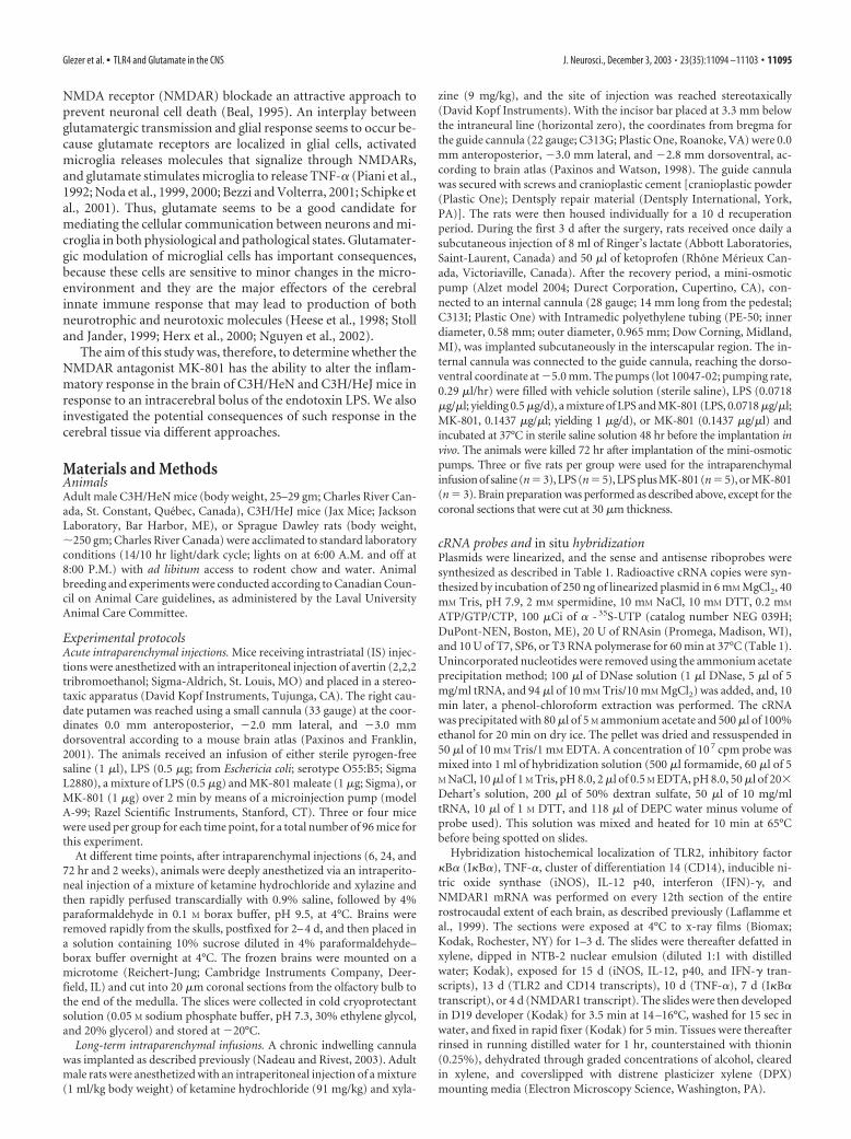
cRNA probes and in situ hybridizationPlasmids were linearized, and the sense and antisense riboprobes weresynthesized as described in Table 1. Radioactive cRNA copies were syn-thesized by incubation of 250 ng of linearized plasmid in 6 mM MgCl2, 40mM Tris, pH 7.9, 2 mM spermidine, 10 mM NaCl, 10 mM DTT, 0.2 mM
ATP/GTP/CTP, 100 �Ci of � - 35S-UTP (catalog number NEG 039H;DuPont-NEN, Boston, ME), 20 U of RNAsin (Promega, Madison, WI),and 10 U of T7, SP6, or T3 RNA polymerase for 60 min at 37°C (Table 1).Unincorporated nucleotides were removed using the ammonium acetateprecipitation method; 100 �l of DNase solution (1 �l DNase, 5 �l of 5mg/ml tRNA, and 94 �l of 10 mM Tris/10 mM MgCl2) was added, and, 10min later, a phenol-chloroform extraction was performed. The cRNAwas precipitated with 80 �l of 5 M ammonium acetate and 500 �l of 100%ethanol for 20 min on dry ice. The pellet was dried and ressuspended in50 �l of 10 mM Tris/1 mM EDTA. A concentration of 10 7 cpm probe wasmixed into 1 ml of hybridization solution (500 �l formamide, 60 �l of 5M NaCl, 10 �l of 1 M Tris, pH 8.0, 2 �l of 0.5 M EDTA, pH 8.0, 50 �l of 20�
Dehart’s solution, 200 �l of 50% dextran sulfate, 50 �l of 10 mg/mltRNA, 10 �l of 1 M DTT, and 118 �l of DEPC water minus volume ofprobe used). This solution was mixed and heated for 10 min at 65°Cbefore being spotted on slides.
Hybridization histochemical localization of TLR2, inhibitory factor�B� (I�B�), TNF-�, cluster of differentiation 14 (CD14), inducible ni-tric oxide synthase (iNOS), IL-12 p40, interferon (IFN)-�, andNMDAR1 mRNA was performed on every 12th section of the entirerostrocaudal extent of each brain, as described previously (Laflamme etal., 1999). The sections were exposed at 4°C to x-ray films (Biomax;Kodak, Rochester, NY) for 1–3 d. The slides were thereafter defatted inxylene, dipped in NTB-2 nuclear emulsion (diluted 1:1 with distilledwater; Kodak), exposed for 15 d (iNOS, IL-12, p40, and IFN-� tran-scripts), 13 d (TLR2 and CD14 transcripts), 10 d (TNF-�), 7 d (I�B�
transcript), or 4 d (NMDAR1 transcript). The slides were then developedin D19 developer (Kodak) for 3.5 min at 14 –16°C, washed for 15 sec inwater, and fixed in rapid fixer (Kodak) for 5 min. Tissues were thereafterrinsed in running distilled water for 1 hr, counterstained with thionin(0.25%), dehydrated through graded concentrations of alcohol, clearedin xylene, and coverslipped with distrene plasticizer xylene (DPX)mounting media (Electron Microscopy Science, Washington, PA).
Glezer et al. • TLR4 and Glutamate in the CNS J. Neurosci., December 3, 2003 • 23(35):11094 –11103 • 11095
Detection of neuronal cell death, demyelination, andhistological analysisThe presence of neuronal cell death was investigated with the Fluoro-Jade B (FJB) method. Briefly, every 12th section of the whole rostrocau-dal extent of each brain was mounted onto poly-L-lysine-coated slides,dried under vacuum for 2 hr, dehydrated through graded concentrationsof alcohol (50, 70, and 100%; 1 min), rehydrated through graded con-centrations of alcohol (100, 70, and 50%; 1 min), and rinsed for 1 min indistilled water. Then, the sections were dipped and shacked in potassiumpermanganate solution (0.06%) for 10 min and rinsed for 1 min indistilled water. Slides were next dipped and shacked into a solution con-taining a mixture of 0.0004% FJB (Histochem, Jefferson, AR) plus0.1% acetic acid (Sigma-Aldrich) plus 0.0002% 4�,6�-diamidino-2-phenylindole (Molecular Probes, Eugene, OR) for 20 min. The slideswere thereafter rinsed three times in distilled water (1 min each), dried,dipped in xylene three times (2 min each), and coverslipped with DPX.
Demyelination was determined via Luxol Fast Blue (LFB) staining.The brain sections mounted onto poly-L-lysine-coated slides were dehy-drated through graded concentrations of alcohol (50, 70, and 95%; 1 mineach) and incubated at 60°C for 6 hr in LFB solution [1% Solvent Blue 38(Sigma) in 95% ethanol and 0.5% acetic acid]. The sections were thenrinsed in 95% ethanol (1 min), 0.05% lithium carbonate (1–5 min;Sigma), and 70% alcohol (two dips). Thereafter, the slides were stained in1% eosine Y solution (EM Diagnostic System, Gibbstown, NJ) for 30 sec,rinsed in water, incubated in 0.25% cresyl violet (Sigma) for 30 sec,rinsed in water, dehydrated through graded concentrations of alcohol(50, 70, 95, and 100%; 1 min each), cleared in xylene for 1 min (twotimes), and coverslipped with DPX.
Nissl stain was used as a general index of cellular morphology that maybe altered in response to the different treatments.
Quantitative analysisQuantitative analyses were performed as described previously (Nadeauand Rivest, 2003). Hybridization signals were quantified on x-ray films(Biomax). Briefly, transmittance values (optical density) of the hybrid-ization signal were measured under a Northern Light Desktop Illumina-tor (Imaging Research) using a Sony Camera Video System attached to aMicro-Nikkor 55 mm-Vivitar extension tube set coupled with a com-puter and NIH Image software version 1.59/ppc [written by W. Rasband(National Institutes of Health, Bethesda, MD) and available from theinternet by anonymous ftp from http://rsb.info.nih.gov/nih-image/download.html]. OD values for each pixel were calculated using a knownstandard of intensity and distance measurements from a logarithmicspecter adapted from Bioimage Visage 110s (Millipore, Ann Arbor, MI).The entire hemisphere ipsilateral to the injection site was digitized andsubjected to densitometric analysis, yielding measurements of mean den-sity per area. The OD of each hemisphere was then corrected for theaverage background signal by subtracting the OD of area without positivesignal located in the contralateral side. Four different brain sections at thelevel of caudate putamen were used for each animal.
The dorsal basal ganglia and hippocampus were selected for the semi-quantification of NMDAR1 mRNA. These structures exhibited high con-stitutive NMDAR1 expression levels and strong transcriptional activa-
tion of immune-related genes in response to intracerebral LPS/MK-801administration. Measurements were performed in 10 C3H/HeN and 10C3H/HeJ mice, and data are reported as mean OD values (�SEM).
Demyelination was evaluated on digitalized LFB-stained sectionswithin the corpus callosum and caudoputamen as described previously(Nadeau and Rivest, 2003). The rationale to measure LFB staining (indexof myelinated fibers) in these regions is based on the pattern of theinflammatory wave that diffused across the dorsal basal ganglia and cor-pus collosum in response to cerebral infusions. Briefly, OD of corpuscallosum and caudato putamen was measured using the NIH Imagesoftware, and values of the contralateral side were subtracted from thoseof the ipsilateral side of four different rostrocaudal levels and expressed asabsolute values (mean � SEM).
The statistical analysis was performed by a one- or two-way ANOVA,followed by a Bonferroni/Dunn test procedure as post hoc comparisonsor Student’s t test for NMDAR1 subunit mRNA levels.
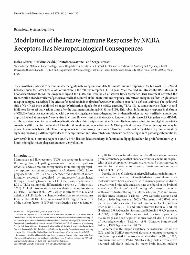
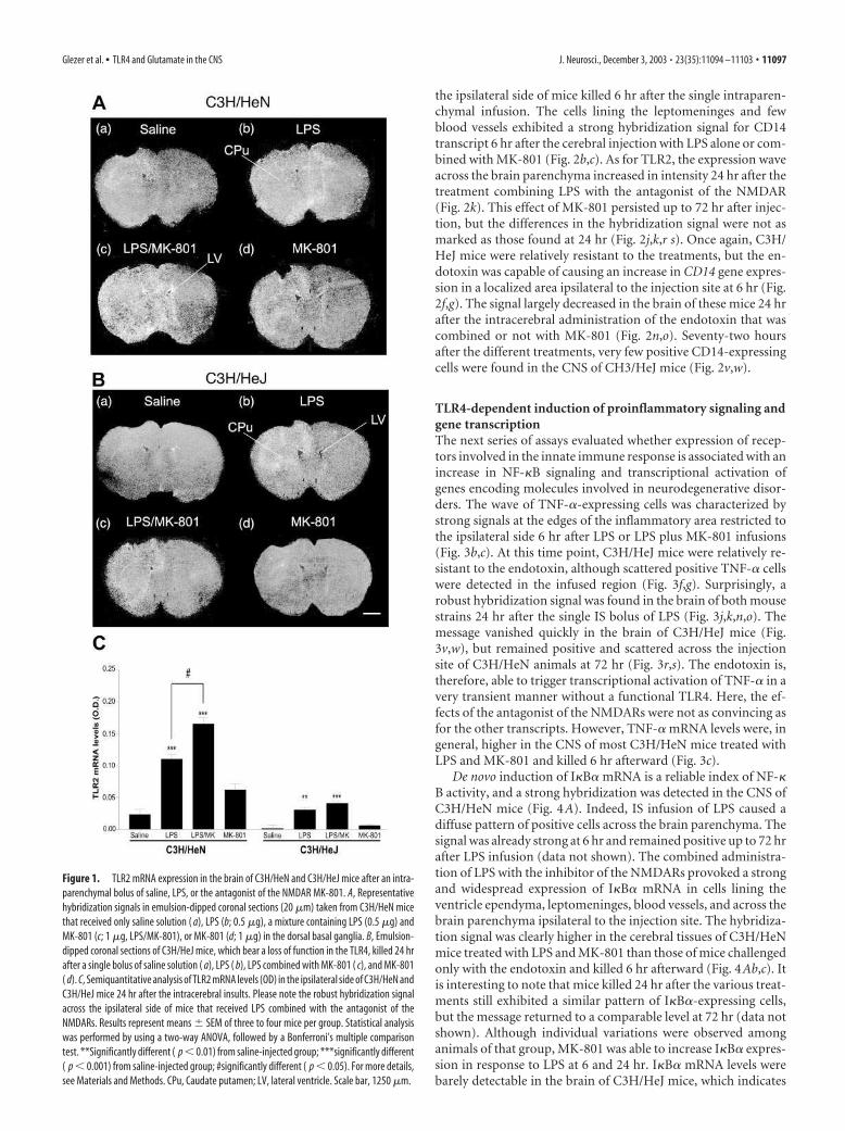
ResultsLPS-induced expression of TLR2 and CD14 is modulated byMK-801 in a TLR4-dependent mannerFigure 1 depicts representative hybridization signals in the brainof mice treated with saline, LPS, LPS/MK-801, or MK-801 alone.As expected, implantation of the cannula and the stress of infu-sion caused a low expression of TLR2 mRNA. Such a localizedpattern of positive TLR2-expressing cells was found in the brainof both saline- and MK-801-infused mice (Fig. 1). This low signalcontrasts with the intense and widespread transcriptional activa-tion of TLR2 ipsilateral to the side of LPS infusion (Fig. 1Ab,LPS). MK-801 exacerbated the effects of endotoxin injected di-rectly within the basal ganglia. At 24 hr, the signal was moreintense in the brain of mice treated with the NMDAR antagonistand endotoxin (Fig. 1Ac). Semiquantitative analyses performedon x-ray films revealed a significant difference between the LPS-and LPS/MK-801-treated groups (Fig. 1C).
This amplification by MK-801 was essentially abolished inC3H/HeJ mice, because these mice were much less sensitive toeither LPS alone or combined with the NMDAR antagonist (Fig.1B). It is interesting to note that despite the lack of functionalTLR4, a single intracerebral bolus of LPS was able to increaseTLR2 expression in regions ipsilateral to the injection site. How-ever, the signal was, to a great extent, lower in C3H/HeJ mice thanin their wild-type controls, and MK-801 remained without sig-nificant effect in TLR4-mutant mice (Fig. 1B,C). These data in-dicate that the ability of glutamate and NMDARs to modulate theinnate immune response in the brain is dependent on the priorbinding of LPS to its receptor TLR4.
The same phenomenon took place for the gene encodingCD14 (Fig. 2). The endotoxin caused a profound increase in thetranscriptional activation of the gene encoding LPS receptor in
Table 1. Plasmids and enzymes used for the synthesis of the cRNA probes
Plasmid Vector Length (bp)Enzymes used forthe sense probe
Enzymes used for theantisense probe Source
Mouse TLR2 PCR-blunt II-topo 2278 (almost full) Spe/T7 EcoRV/Sp6 Cloned by PCRa
Mouse I�B� Bluescript SK II� 1114 (full) HindIII/T3 BamHI/T7 Dr. A. Israel (Institut Pasteur, Paris, France)
Mouse TNF-� Bluescript SK II� 1300 (full � a 593 bp
non-coding part)
BamHI/T7 PstI/T3 Subcloned from a PUc19 plasmid provided by Dr. M. Oliver
(Laval University, Quebec, Canada)
Mouse CD14 pRc/CMV (5.4 kb) 1500 HindIII/SP6 ApaI/T7 Dr. Regine Landmann (University Hospital, Basel, Switzerland)
Mouse iNOS Bluescript SK II� 817 EcoRI/T7 KpnI/T3 Subcloned from a Puc19 plasmid provided by Dr. M. Oliver
Mouse NMDAR1 pCMV-sport 6 3300 HindII/Sp6 SalI/T7 American Type Culture Collection (Manassas, VA)
Mouse IL-12 p40 pCL-Neo (5.474 kb) 1050 NotI/T7 XhoI/T3 Dr. K. Pahan (Nebraska Medical Center University, Lincoln, NE)
Mouse IFN-� pGEMEX 550 EcoRI/SP6 HindIII/T3 Dr. I. Campbell (The Scripps Research Institute, La Jolla, CA)a The DNA fragment of 2.278 kb corresponding to the almost complete coding sequence (2.355 kb) of the reported mouse TLR2 mRNA (nucleotides 307–2661; Genbank accession number AF185284) was amplified by PCR from a cDNAmacrophage B10R cell line library using a pair of 23 bp oligonucleotide primers complementary to nucleotides 323–345 (5�-GGCTCTTCTGGATCTTGGTGGCC-3�) and 2579 –2601 (5�-GGGCCACTCCAGGTAGGTCTTGG-3�).
11096 • J. Neurosci., December 3, 2003 • 23(35):11094 –11103 Glezer et al. • TLR4 and Glutamate in the CNS
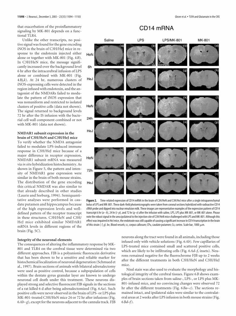
the ipsilateral side of mice killed 6 hr after the single intraparen-chymal infusion. The cells lining the leptomeninges and fewblood vessels exhibited a strong hybridization signal for CD14transcript 6 hr after the cerebral injection with LPS alone or com-bined with MK-801 (Fig. 2b,c). As for TLR2, the expression waveacross the brain parenchyma increased in intensity 24 hr after thetreatment combining LPS with the antagonist of the NMDAR(Fig. 2k). This effect of MK-801 persisted up to 72 hr after injec-tion, but the differences in the hybridization signal were not asmarked as those found at 24 hr (Fig. 2j,k,r s). Once again, C3H/HeJ mice were relatively resistant to the treatments, but the en-dotoxin was capable of causing an increase in CD14 gene expres-sion in a localized area ipsilateral to the injection site at 6 hr (Fig.2f,g). The signal largely decreased in the brain of these mice 24 hrafter the intracerebral administration of the endotoxin that wascombined or not with MK-801 (Fig. 2n,o). Seventy-two hoursafter the different treatments, very few positive CD14-expressingcells were found in the CNS of CH3/HeJ mice (Fig. 2v,w).
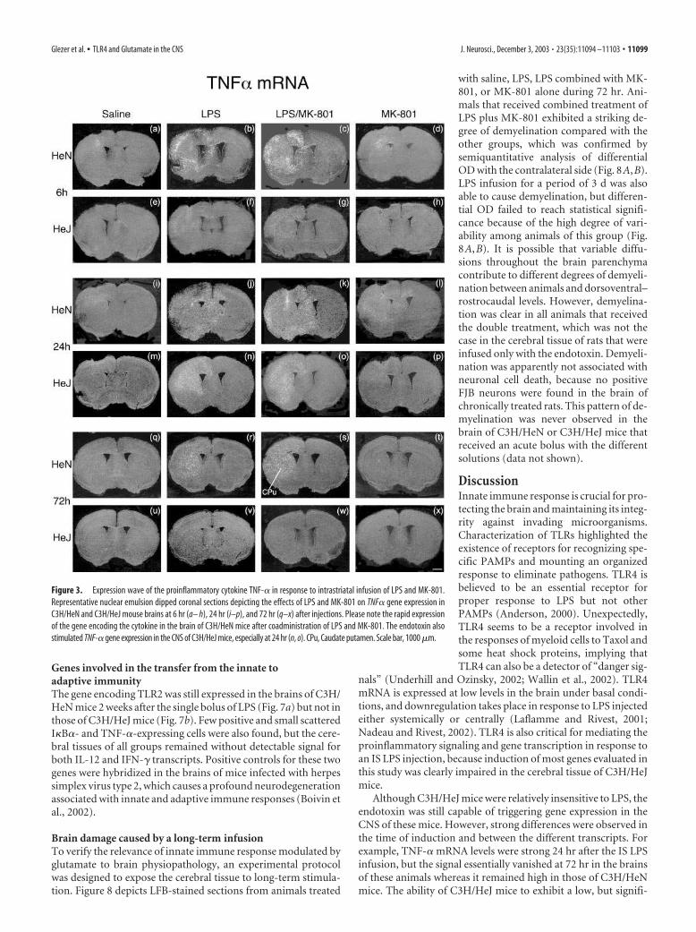
TLR4-dependent induction of proinflammatory signaling andgene transcriptionThe next series of assays evaluated whether expression of recep-tors involved in the innate immune response is associated with anincrease in NF-�B signaling and transcriptional activation ofgenes encoding molecules involved in neurodegenerative disor-ders. The wave of TNF-�-expressing cells was characterized bystrong signals at the edges of the inflammatory area restricted tothe ipsilateral side 6 hr after LPS or LPS plus MK-801 infusions(Fig. 3b,c). At this time point, C3H/HeJ mice were relatively re-sistant to the endotoxin, although scattered positive TNF-� cellswere detected in the infused region (Fig. 3f,g). Surprisingly, arobust hybridization signal was found in the brain of both mousestrains 24 hr after the single IS bolus of LPS (Fig. 3j,k,n,o). Themessage vanished quickly in the brain of C3H/HeJ mice (Fig.3v,w), but remained positive and scattered across the injectionsite of C3H/HeN animals at 72 hr (Fig. 3r,s). The endotoxin is,therefore, able to trigger transcriptional activation of TNF-� in avery transient manner without a functional TLR4. Here, the ef-fects of the antagonist of the NMDARs were not as convincing asfor the other transcripts. However, TNF-� mRNA levels were, ingeneral, higher in the CNS of most C3H/HeN mice treated withLPS and MK-801 and killed 6 hr afterward (Fig. 3c).
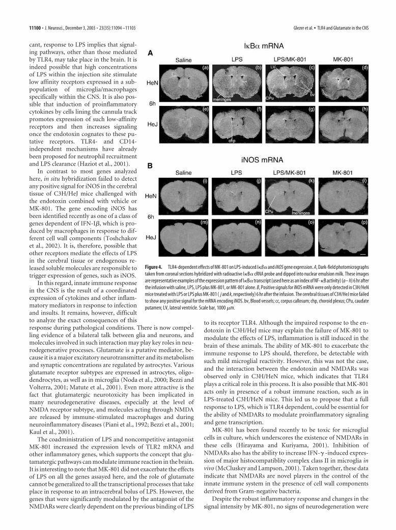
De novo induction of I�B� mRNA is a reliable index of NF-�B activity, and a strong hybridization was detected in the CNS ofC3H/HeN mice (Fig. 4A). Indeed, IS infusion of LPS caused adiffuse pattern of positive cells across the brain parenchyma. Thesignal was already strong at 6 hr and remained positive up to 72 hrafter LPS infusion (data not shown). The combined administra-tion of LPS with the inhibitor of the NMDARs provoked a strongand widespread expression of I�B� mRNA in cells lining theventricle ependyma, leptomeninges, blood vessels, and across thebrain parenchyma ipsilateral to the injection site. The hybridiza-tion signal was clearly higher in the cerebral tissues of C3H/HeNmice treated with LPS and MK-801 than those of mice challengedonly with the endotoxin and killed 6 hr afterward (Fig. 4Ab,c). Itis interesting to note that mice killed 24 hr after the various treat-ments still exhibited a similar pattern of I�B�-expressing cells,but the message returned to a comparable level at 72 hr (data notshown). Although individual variations were observed amonganimals of that group, MK-801 was able to increase I�B� expres-sion in response to LPS at 6 and 24 hr. I�B� mRNA levels werebarely detectable in the brain of C3H/HeJ mice, which indicates
Figure 1. TLR2 mRNA expression in the brain of C3H/HeN and C3H/HeJ mice after an intra-
parenchymal bolus of saline, LPS, or the antagonist of the NMDAR MK-801. A, Representative
hybridization signals in emulsion-dipped coronal sections (20 �m) taken from C3H/HeN mice
that received only saline solution ( a), LPS (b; 0.5 �g), a mixture containing LPS (0.5 �g) and
MK-801 (c; 1 �g, LPS/MK-801), or MK-801 (d; 1 �g) in the dorsal basal ganglia. B, Emulsion-
dipped coronal sections of C3H/HeJ mice, which bear a loss of function in the TLR4, killed 24 hr
after a single bolus of saline solution ( a), LPS ( b), LPS combined with MK-801 ( c), and MK-801
( d). C, Semiquantitative analysis of TLR2 mRNA levels (OD) in the ipsilateral side of C3H/HeN and
C3H/HeJ mice 24 hr after the intracerebral insults. Please note the robust hybridization signal
across the ipsilateral side of mice that received LPS combined with the antagonist of the
NMDARs. Results represent means � SEM of three to four mice per group. Statistical analysis
was performed by using a two-way ANOVA, followed by a Bonferroni’s multiple comparison
test. **Significantly different ( p � 0.01) from saline-injected group; ***significantly different
( p � 0.001) from saline-injected group; #significantly different ( p � 0.05). For more details,
see Materials and Methods. CPu, Caudate putamen; LV, lateral ventricle. Scale bar, 1250 �m.
Glezer et al. • TLR4 and Glutamate in the CNS J. Neurosci., December 3, 2003 • 23(35):11094 –11103 • 11097
that exacerbation of the proinflammatorysignaling by MK-801 depends on a func-tional TLR4.
Unlike the other transcripts, no posi-tive signal was found for the gene encodingiNOS in the brain of C3H/HeJ mice in re-sponse to the endotoxin injected eitheralone or together with MK-801 (Fig. 4B).In C3H/HeN mice, the message signifi-cantly increased over the background level6 hr after the intracerebral infusion of LPSalone or combined with MK-801 (Fig.4Bj,k). At 24 hr, numerous clusters ofiNOS-expressing cells were detected in theregion infused with endotoxin, and the an-tagonist of the NMDARs failed to modu-late the pattern of iNOS expression thatwas nonuniform and restricted to isolatedclusters of positive cells (data not shown).The signal returned to background levels72 hr after the IS infusion with the bacte-rial cell wall component combined or notwith MK-801 (data not shown).
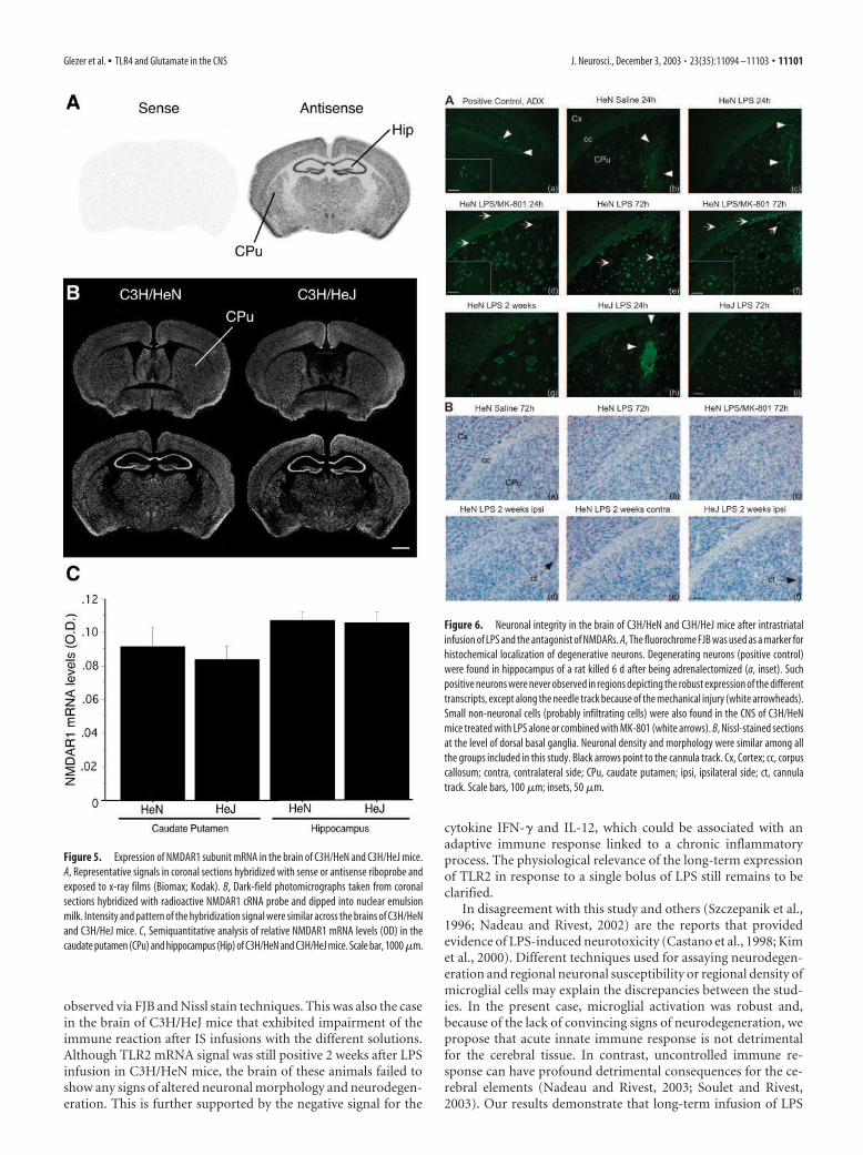
NMDAR1 subunit expression in thebrain of C3H/HeN and C3H/HeJ miceTo verify whether the NMDA antagonistfailed to modulate LPS-induced immuneresponse in C3H/HeJ mice because of amajor difference in receptor expression,NMDAR1 subunit mRNA was measuredvia in situ hybridization histochemistry. Asshown in Figure 5, the pattern and inten-sity of NMDAR1 gene expression weresimilar in the brain of both mouse strains.The distribution of the gene encodingthis critical NMDAR was also similar tothat already described in other studies(Laurie and Seeburg, 1994). Semiquanti-tative analyses were performed in cau-date putamen and hippocampus becauseof the high expression levels and well-defined pattern of the receptor transcriptin these structures. C3H/HeN and C3H/HeJ mice exhibited similar NMDAR1mRNA levels in different regions of thebrain (Fig. 5C).
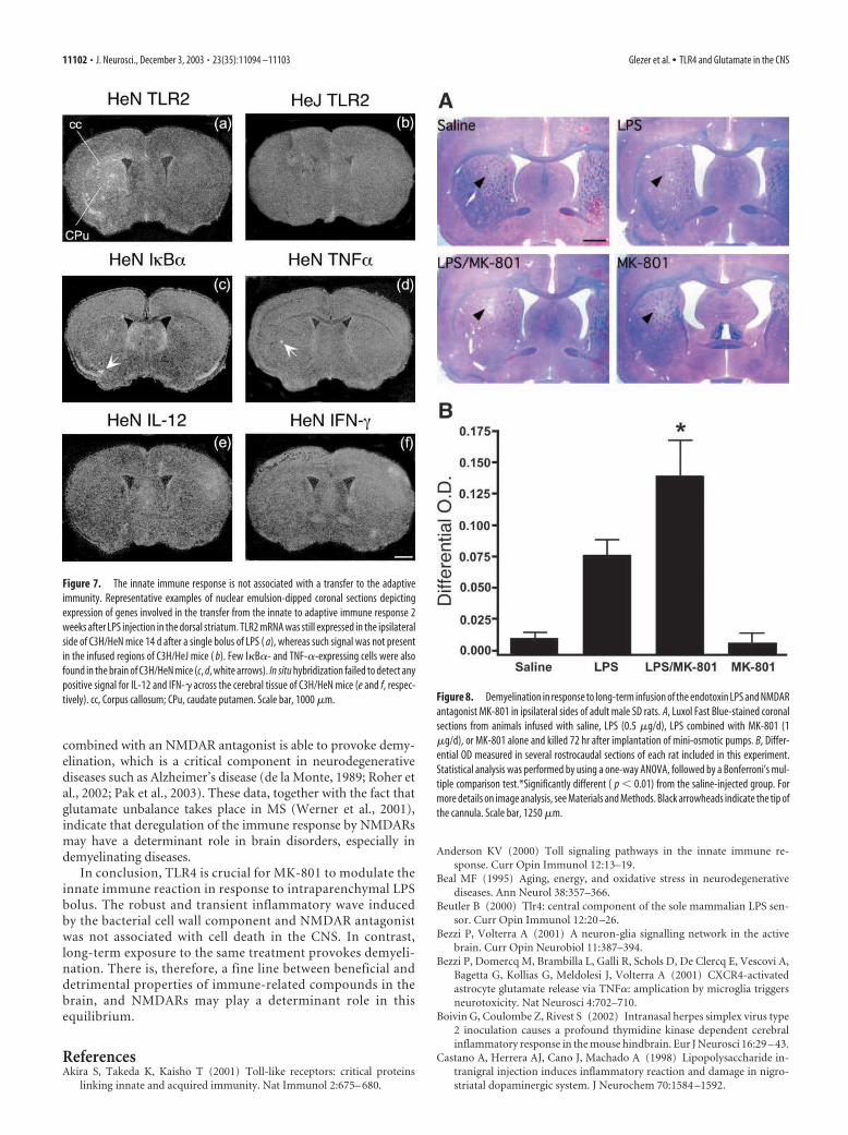
Integrity of the neuronal elementsThe consequences of altering the inflammatory response by MK-801 and TLR4 on the cerebral tissue were determined via twodifferent approaches. FJB is a polyanionic fluorescein derivativethat has been shown to be a sensitive and reliable marker forhistochemical localization of neuronal degeneration (Schmued etal., 1997). Brain sections of animals with bilateral adrenalectomywere used as positive control, because a subpopulation of cellswithin the dentate gyrus granular layer are known to undergoneuronal cell death under this treatment. These neurons dis-played strong and selective fluorescent FJB signals in the sectionsof a rat killed 6 d after being adrenalectomized (Fig. 6Aa). Suchpositive cells were never observed in the brain of LPS- or LPS plusMK-801-treated C3H/HeN mice 24 or 72 hr after infusions (Fig.6Ab– g), except for the neurons adjacent to the cannula track. FJB
neurons along the tract were found in all animals, including those
infused only with vehicle solutions (Fig. 6Ab). Few capillaries of
LPS-treated mice contained small and scattered positive cells,
which are likely to be infiltrating cells (Fig. 6Ad–f, insets). Neu-
rons remained negative for the fluorochrome FJB up to 2 weeks
after the different treatments in both C3H/HeN and C3H/HeJ
mice.
Nissl stain was also used to evaluate the morphology and his-
tological integrity of the cerebral tissues. Figure 6B shows exam-
ples of brain sections taken from saline-, LPS-, or LPS plus MK-
801-infused mice, and no convincing changes were observed 72
hr after the different treatments (Fig. 6Ba– c). The sections re-
mained intact, and ipsilateral sides were similar to the contralat-
eral areas at 2 weeks after LPS infusion in both mouse strains (Fig.
6Bd–f).
Figure 2. Time-related expression of CD14 mRNA in the brain of C3H/HeN and C3H/HeJ mice after a single intraparenchymal
bolus of LPS and MK-801. These dark-field photomicrographs were taken from coronal sections hybridized with radioactive CD14
cRNA probe and dipped into nuclear emulsion milk. These images are representative examples of the expression pattern of CD14
transcript 6 hr (a– h), 24 hr (i–p), and 72 hr (q–x) after the infusion with saline, LPS, LPS plus MK-801, or MK-801 alone. Please
note the robust signal in the area ipsilateral to the injection site of C3H/HeN mice challenged with LPS and MK-801. Although this
effect was impaired in HeJ mice, the endotoxin was still capable of causing a significant increase in CD14 transcription in the brain
of this strain ( f, g). bv, Blood vessels; cc, corpus callosum; CPu, caudate putamen; Cx, cortex. Scale bar, 1000 �m.
11098 • J. Neurosci., December 3, 2003 • 23(35):11094 –11103 Glezer et al. • TLR4 and Glutamate in the CNS
Genes involved in the transfer from the innate toadaptive immunityThe gene encoding TLR2 was still expressed in the brains of C3H/HeN mice 2 weeks after the single bolus of LPS (Fig. 7a) but not inthose of C3H/HeJ mice (Fig. 7b). Few positive and small scatteredI�B�- and TNF-�-expressing cells were also found, but the cere-bral tissues of all groups remained without detectable signal forboth IL-12 and IFN-� transcripts. Positive controls for these twogenes were hybridized in the brains of mice infected with herpessimplex virus type 2, which causes a profound neurodegenerationassociated with innate and adaptive immune responses (Boivin etal., 2002).
Brain damage caused by a long-term infusionTo verify the relevance of innate immune response modulated byglutamate to brain physiopathology, an experimental protocolwas designed to expose the cerebral tissue to long-term stimula-tion. Figure 8 depicts LFB-stained sections from animals treated
with saline, LPS, LPS combined with MK-801, or MK-801 alone during 72 hr. Ani-mals that received combined treatment ofLPS plus MK-801 exhibited a striking de-gree of demyelination compared with theother groups, which was confirmed bysemiquantitative analysis of differentialOD with the contralateral side (Fig. 8A,B).LPS infusion for a period of 3 d was alsoable to cause demyelination, but differen-tial OD failed to reach statistical signifi-cance because of the high degree of vari-ability among animals of this group (Fig.8A,B). It is possible that variable diffu-sions throughout the brain parenchymacontribute to different degrees of demyeli-nation between animals and dorsoventral–rostrocaudal levels. However, demyelina-tion was clear in all animals that receivedthe double treatment, which was not thecase in the cerebral tissue of rats that wereinfused only with the endotoxin. Demyeli-nation was apparently not associated withneuronal cell death, because no positiveFJB neurons were found in the brain ofchronically treated rats. This pattern of de-myelination was never observed in thebrain of C3H/HeN or C3H/HeJ mice thatreceived an acute bolus with the differentsolutions (data not shown).
DiscussionInnate immune response is crucial for pro-tecting the brain and maintaining its integ-rity against invading microorganisms.Characterization of TLRs highlighted theexistence of receptors for recognizing spe-cific PAMPs and mounting an organizedresponse to eliminate pathogens. TLR4 isbelieved to be an essential receptor forproper response to LPS but not otherPAMPs (Anderson, 2000). Unexpectedly,TLR4 seems to be a receptor involved inthe responses of myeloid cells to Taxol andsome heat shock proteins, implying thatTLR4 can also be a detector of “danger sig-
nals” (Underhill and Ozinsky, 2002; Wallin et al., 2002). TLR4mRNA is expressed at low levels in the brain under basal condi-tions, and downregulation takes place in response to LPS injectedeither systemically or centrally (Laflamme and Rivest, 2001;Nadeau and Rivest, 2002). TLR4 is also critical for mediating theproinflammatory signaling and gene transcription in response toan IS LPS injection, because induction of most genes evaluated inthis study was clearly impaired in the cerebral tissue of C3H/HeJmice.
Although C3H/HeJ mice were relatively insensitive to LPS, theendotoxin was still capable of triggering gene expression in theCNS of these mice. However, strong differences were observed inthe time of induction and between the different transcripts. Forexample, TNF-� mRNA levels were strong 24 hr after the IS LPSinfusion, but the signal essentially vanished at 72 hr in the brainsof these animals whereas it remained high in those of C3H/HeNmice. The ability of C3H/HeJ mice to exhibit a low, but signifi-
Figure 3. Expression wave of the proinflammatory cytokine TNF-� in response to intrastriatal infusion of LPS and MK-801.
Representative nuclear emulsion dipped coronal sections depicting the effects of LPS and MK-801 on TNF� gene expression in
C3H/HeN and C3H/HeJ mouse brains at 6 hr (a– h), 24 hr (i–p), and 72 hr (q–x) after injections. Please note the rapid expression
of the gene encoding the cytokine in the brain of C3H/HeN mice after coadministration of LPS and MK-801. The endotoxin also
stimulated TNF-� gene expression in the CNS of C3H/HeJ mice, especially at 24 hr (n, o). CPu, Caudate putamen. Scale bar, 1000 �m.
Glezer et al. • TLR4 and Glutamate in the CNS J. Neurosci., December 3, 2003 • 23(35):11094 –11103 • 11099
cant, response to LPS implies that signal-ing pathways, other than those mediatedby TLR4, may take place in the brain. It isindeed possible that high concentrationsof LPS within the injection site stimulatelow affinity receptors expressed in a sub-population of microglia/macrophagesspecifically within the CNS. It is also pos-sible that induction of proinflammatorycytokines by cells lining the cannula trackpromotes expression of such low-affinityreceptors and then increases signalingonce the endotoxin cognates to these pu-tative receptors. TLR4- and CD14-independent mechanisms have alreadybeen proposed for neutrophil recruitmentand LPS clearance (Haziot et al., 2001).
In contrast to most genes analyzedhere, in situ hybridization failed to detectany positive signal for iNOS in the cerebraltissue of C3H/HeJ mice challenged withthe endotoxin combined with vehicle orMK-801. The gene encoding iNOS hasbeen identified recently as one of a class ofgenes dependent of IFN-I�, which is pro-duced by macrophages in response to dif-ferent cell wall components (Toshchakovet al., 2002). It is, therefore, possible thatother receptors mediate the effects of LPSin the cerebral tissue or endogenous re-leased soluble molecules are responsible totrigger expression of genes, such as iNOS.
In this regard, innate immune responsein the CNS is the result of a coordinatedexpression of cytokines and other inflam-matory mediators in response to infectionand insults. It remains, however, difficultto analyze the exact consequences of thisresponse during pathological conditions. There is now compel-ling evidence of a bilateral talk between glia and neurons, andmolecules involved in such interaction may play key roles in neu-rodegenerative processes. Glutamate is a putative mediator, be-cause it is a major excitatory neurotransmitter and its metabolismand synaptic concentrations are regulated by astrocytes. Variousglutamate receptor subtypes are expressed in astrocytes, oligo-dendrocytes, as well as in microglia (Noda et al., 2000; Bezzi andVolterra, 2001; Matute et al., 2001). Even more attractive is thefact that glutamatergic neurotoxicity has been implicated inmany neurodegenerative diseases, especially at the level ofNMDA receptor subtype, and molecules acting through NMDAare released by immune-stimulated macrophages and duringneuroinflammatory diseases (Piani et al., 1992; Bezzi et al., 2001;Kaul et al., 2001).
The coadministration of LPS and noncompetitive antagonistMK-801 increased the expression levels of TLR2 mRNA andother inflammatory genes, which supports the concept that glu-tamatergic pathways can modulate immune reaction in the brain.It is interesting to note that MK-801 did not exacerbate the effectsof LPS on all the genes assayed here, and the role of glutamatecannot be generalized to all the transcriptional processes that takeplace in response to an intracerebral bolus of LPS. However, thegenes that were significantly modulated by the antagonist of theNMDARs were clearly dependent on the previous binding of LPS
to its receptor TLR4. Although the impaired response to the en-dotoxin in C3H/HeJ mice may explain the failure of MK-801 tomodulate the effects of LPS, inflammation is still induced in thebrain of these animals. The ability of MK-801 to exacerbate theimmune response to LPS should, therefore, be detectable withsuch mild microglial reactivity. However, this was not the case,and the interaction between the endotoxin and NMDARs wasobserved only in C3H/HeN mice, which indicates that TLR4plays a critical role in this process. It is also possible that MK-801acts only in presence of a robust immune reaction, such as inLPS-treated C3H/HeN mice. This led us to propose that a fullresponse to LPS, which is TLR4 dependent, could be essential forthe ability of NMDARs to modulate proinflammatory signalingand gene transcription.
MK-801 has been found recently to be toxic for microglialcells in culture, which underscores the existence of NMDARs inthese cells (Hirayama and Kuriyama, 2001). Inhibition ofNMDARs also has the ability to increase IFN-� -induced expres-sion of major histocompatiblity complex class II in microglia invivo (McCluskey and Lampson, 2001). Taken together, these dataindicate that NMDARs are novel players in the control of theinnate immune system in the presence of cell wall componentsderived from Gram-negative bacteria.
Despite the robust inflammatory response and changes in thesignal intensity by MK-801, no signs of neurodegeneration were
Figure 4. TLR4-dependent effects of MK-801 on LPS-induced I�B� and iNOS gene expression. A, Dark-field photomicrographs
taken from coronal sections hybridized with radioactive I�B� cRNA probe and dipped into nuclear emulsion milk. These images
are representative examples of the expression pattern of I�B� transcript (used here as an index of NF-�B activity) (a– h) 6 hr after
the infusion with saline, LPS, LPS plus MK-801, or MK-801 alone. B, Positive signals for iNOS mRNA were only detected in C3H/HeN
mice treated with LPS or LPS plus MK-801 ( j and k, respectively) 6 hr after the infusion. The cerebral tissues of C3H/HeJ mice failed
to show any positive signal for the mRNA encoding iNOS. bv, Blood vessels; cc, corpus callosum; chp, choroid plexus; CPu, caudate
putamen; LV, lateral ventricle. Scale bar, 1000 �m.
11100 • J. Neurosci., December 3, 2003 • 23(35):11094 –11103 Glezer et al. • TLR4 and Glutamate in the CNS
observed via FJB and Nissl stain techniques. This was also the casein the brain of C3H/HeJ mice that exhibited impairment of theimmune reaction after IS infusions with the different solutions.Although TLR2 mRNA signal was still positive 2 weeks after LPSinfusion in C3H/HeN mice, the brain of these animals failed toshow any signs of altered neuronal morphology and neurodegen-eration. This is further supported by the negative signal for the
cytokine IFN-� and IL-12, which could be associated with anadaptive immune response linked to a chronic inflammatoryprocess. The physiological relevance of the long-term expressionof TLR2 in response to a single bolus of LPS still remains to beclarified.
In disagreement with this study and others (Szczepanik et al.,1996; Nadeau and Rivest, 2002) are the reports that providedevidence of LPS-induced neurotoxicity (Castano et al., 1998; Kimet al., 2000). Different techniques used for assaying neurodegen-eration and regional neuronal susceptibility or regional density ofmicroglial cells may explain the discrepancies between the stud-ies. In the present case, microglial activation was robust and,because of the lack of convincing signs of neurodegeneration, wepropose that acute innate immune response is not detrimentalfor the cerebral tissue. In contrast, uncontrolled immune re-sponse can have profound detrimental consequences for the ce-rebral elements (Nadeau and Rivest, 2003; Soulet and Rivest,2003). Our results demonstrate that long-term infusion of LPS
Figure 5. Expression of NMDAR1 subunit mRNA in the brain of C3H/HeN and C3H/HeJ mice.
A, Representative signals in coronal sections hybridized with sense or antisense riboprobe and
exposed to x-ray films (Biomax; Kodak). B, Dark-field photomicrographs taken from coronal
sections hybridized with radioactive NMDAR1 cRNA probe and dipped into nuclear emulsion
milk. Intensity and pattern of the hybridization signal were similar across the brains of C3H/HeN
and C3H/HeJ mice. C, Semiquantitative analysis of relative NMDAR1 mRNA levels (OD) in the
caudate putamen (CPu) and hippocampus (Hip) of C3H/HeN and C3H/HeJ mice. Scale bar, 1000 �m.
Figure 6. Neuronal integrity in the brain of C3H/HeN and C3H/HeJ mice after intrastriatal
infusion of LPS and the antagonist of NMDARs. A, The fluorochrome FJB was used as a marker for
histochemical localization of degenerative neurons. Degenerating neurons (positive control)
were found in hippocampus of a rat killed 6 d after being adrenalectomized (a, inset). Such
positive neurons were never observed in regions depicting the robust expression of the different
transcripts, except along the needle track because of the mechanical injury (white arrowheads).
Small non-neuronal cells (probably infiltrating cells) were also found in the CNS of C3H/HeN
mice treated with LPS alone or combined with MK-801 (white arrows). B, Nissl-stained sections
at the level of dorsal basal ganglia. Neuronal density and morphology were similar among all
the groups included in this study. Black arrows point to the cannula track. Cx, Cortex; cc, corpus
callosum; contra, contralateral side; CPu, caudate putamen; ipsi, ipsilateral side; ct, cannula
track. Scale bars, 100 �m; insets, 50 �m.
Glezer et al. • TLR4 and Glutamate in the CNS J. Neurosci., December 3, 2003 • 23(35):11094 –11103 • 11101
combined with an NMDAR antagonist is able to provoke demy-elination, which is a critical component in neurodegenerativediseases such as Alzheimer’s disease (de la Monte, 1989; Roher etal., 2002; Pak et al., 2003). These data, together with the fact thatglutamate unbalance takes place in MS (Werner et al., 2001),indicate that deregulation of the immune response by NMDARsmay have a determinant role in brain disorders, especially indemyelinating diseases.
In conclusion, TLR4 is crucial for MK-801 to modulate theinnate immune reaction in response to intraparenchymal LPSbolus. The robust and transient inflammatory wave inducedby the bacterial cell wall component and NMDAR antagonistwas not associated with cell death in the CNS. In contrast,long-term exposure to the same treatment provokes demyeli-nation. There is, therefore, a fine line between beneficial anddetrimental properties of immune-related compounds in thebrain, and NMDARs may play a determinant role in thisequilibrium.
ReferencesAkira S, Takeda K, Kaisho T (2001) Toll-like receptors: critical proteins
linking innate and acquired immunity. Nat Immunol 2:675– 680.
Anderson KV (2000) Toll signaling pathways in the innate immune re-sponse. Curr Opin Immunol 12:13–19.
Beal MF (1995) Aging, energy, and oxidative stress in neurodegenerativediseases. Ann Neurol 38:357–366.
Beutler B (2000) Tlr4: central component of the sole mammalian LPS sen-sor. Curr Opin Immunol 12:20 –26.
Bezzi P, Volterra A (2001) A neuron-glia signalling network in the activebrain. Curr Opin Neurobiol 11:387–394.
Bezzi P, Domercq M, Brambilla L, Galli R, Schols D, De Clercq E, Vescovi A,Bagetta G, Kollias G, Meldolesi J, Volterra A (2001) CXCR4-activatedastrocyte glutamate release via TNF�: amplication by microglia triggersneurotoxicity. Nat Neurosci 4:702–710.
Boivin G, Coulombe Z, Rivest S (2002) Intranasal herpes simplex virus type2 inoculation causes a profound thymidine kinase dependent cerebralinflammatory response in the mouse hindbrain. Eur J Neurosci 16:29 – 43.
Castano A, Herrera AJ, Cano J, Machado A (1998) Lipopolysaccharide in-tranigral injection induces inflammatory reaction and damage in nigro-striatal dopaminergic system. J Neurochem 70:1584 –1592.
Figure 7. The innate immune response is not associated with a transfer to the adaptive
immunity. Representative examples of nuclear emulsion-dipped coronal sections depicting
expression of genes involved in the transfer from the innate to adaptive immune response 2
weeks after LPS injection in the dorsal striatum. TLR2 mRNA was still expressed in the ipsilateral
side of C3H/HeN mice 14 d after a single bolus of LPS ( a), whereas such signal was not present
in the infused regions of C3H/HeJ mice ( b). Few I�B�- and TNF-�-expressing cells were also
found in the brain of C3H/HeN mice (c, d, white arrows). In situ hybridization failed to detect any
positive signal for IL-12 and IFN-� across the cerebral tissue of C3H/HeN mice (e and f, respec-
tively). cc, Corpus callosum; CPu, caudate putamen. Scale bar, 1000 �m. Figure 8. Demyelination in response to long-term infusion of the endotoxin LPS and NMDAR
antagonist MK-801 in ipsilateral sides of adult male SD rats. A, Luxol Fast Blue-stained coronal
sections from animals infused with saline, LPS (0.5 �g/d), LPS combined with MK-801 (1
�g/d), or MK-801 alone and killed 72 hr after implantation of mini-osmotic pumps. B, Differ-
ential OD measured in several rostrocaudal sections of each rat included in this experiment.
Statistical analysis was performed by using a one-way ANOVA, followed by a Bonferroni’s mul-
tiple comparison test.*Significantly different ( p � 0.01) from the saline-injected group. For
more details on image analysis, see Materials and Methods. Black arrowheads indicate the tip of
the cannula. Scale bar, 1250 �m.
11102 • J. Neurosci., December 3, 2003 • 23(35):11094 –11103 Glezer et al. • TLR4 and Glutamate in the CNS
de la Monte SM (1989) Quantitation of cerebral atrophy in preclinical and
end-stage Alzheimer’s disease. Ann Neurol 25:450 – 459.
Ghosh S, May MJ, Kopp EB (1998) NF-�B and rel proteins: evolutionary
conserved mediators of immune responses. Annu Rev Immunol
16:225–260.
Gonzales-Scarano F, Baltuch G (1999) Microglia as mediators of inflamma-
tory and degenerative diseases. Annu Rev Neurosci 22:219 –240.
Haziot A, Hijiya N, Gangloff SC, Silver J, Goyert SM (2001) Induction of a
novel mechanism of accelerated bacterial clearance by lipopolysaccharide
in CD14-deficient and Toll-like receptor 4-deficient mice. J Immunol
166:1075–1078.
Heese K, Fiebich BL, Bauer J, Otten U (1998) NF-kappaB modulates
lipopolysaccharide-induced microglial nerve growth factor expression.
Glia 22:401– 407.
Herx LM, Rivest S, Yong VW (2000) Central nervous system-initiated in-
flammation and neurotrophism in trauma: IL-1 beta is required for the
production of ciliary neurotrophic factor. J Immunol 165:2232–2239.
Hirayama M, Kuriyama M (2001) MK-801 is cytotoxic to microglia in vitro
and its cytotoxicity is attenuated by glutamate, other excitotoxic agents
and atropine. Possible presence of glutamate receptor and muscarinic
receptor on microglia. Brain Res 897:204 –206.
Kaul M, Garden GA, Lipton SA (2001) Pathways to neuronal injury and
apoptosis in HIV-associated dementia. Nat Neurosci 410:988 –994.
Kim WG, Mohney RP, Wilson B, Jeohn GH, Liu B, Hong JS (2000) Regional
difference in susceptibility to lipopolysaccharide-induced neurotoxicity
in the rat brain: role of microglia. J Neurosci 20:6309 – 6316.
Laflamme N, Rivest S (2001) Toll-like receptor 4: the missing link of the
cerebral innate immune response triggered by circulating gram-negative
bacterial cell wall components. FASEB J 15:155–163.
Laflamme N, Lacroix S, Rivest S (1999) An essential role of interleukin-
1beta in mediating NF-kappaB activity and COX-2 transcription in cells
of the blood– brain barrier in response to a systemic and localized inflam-
mation but not during endotoxemia. J Neurosci 19:10923–10930.
Laurie DJ, Seeburg PH (1994) Regional and developmental heterogeneity in
splicing of the rat brain NMDAR1 mRNA. J Neurosci 14:3180 –3194.
Matute C, Alberdi E, Domercq M, Perez-Cerda F, Perez-Samartin A,
Sanchez-Gomez M (2001) The link between excitotoxic oligodendro-
glial death and demyelinating diseases. Trends Neurosci 24:224 –230.
McCluskey LP, Lampson LA (2001) Local immune regulation in the central
nervous system by substance P vs. glutamate. J Neuroimmunol
116:136 –146.
Nadeau S, Rivest S (2002) Endotoxemia prevents the cerebral inflammatory
wave induced by intraparenchymal lipopolysaccharide injection: role of
glucocorticoids and CD14. J Immunol 169:3370 –3381.
Nadeau S, Rivest S (2003) Glucocorticoids play a fundamental role in pro-
tecting the brain during innate immune response. J Neurosci
23:5536 –5544.
Nguyen MD, Julien JP, Rivest S (2002) Innate immunity: the missing link in
neuroprotection and neurodegeneration? Nat Rev Neurosci 3:216 –227.
Noda M, Nakanishi H, Akaike N (1999) Glutamate release from microglia
via glutamate transporter is enhanced by amyloid-beta peptide. Neuro-
science 92:1465–1474.
Noda M, Nakanishi H, Nabekura J, Akaike N (2000) AMPA-kainate sub-
types of glutamate receptor in rat cerebral microglia. J Neurosci20:251–258.
Pak K, Chan SL, Mattson MP (2003) Presenilin-1 mutation sensitizes oligo-dendrocytes to glutamate and amyloid toxicities, and exacerbates whitematter damage and memory impairment in mice. Neuromol Med3:53– 64.
Pasinetti G (1998) Cyclooxygenase and inflammation in Alzheimer’s dis-ease: experimental approaches and clinical interventions. J Neurosci Res54:1– 6.
Paxinos G, Franklin KBJ (2001) The mouse brain in stereotaxic coordinates,Ed 2. San Diego: Academic.
Paxinos G, Watson C (1998) The rat brain in stereotaxic coordinates, Ed 2.San Diego: Academic.
Piani D, Spranger M, Frei K, Schaggner A, Fontana A (1992) Macrophage-induced cytotoxicity of N-methyl-D-aspartate receptor positive neuronsinvolves excitatory amino acids rather than oxygen intermediates andcytokines. Eur J Immunol 22:2429 –2436.
Poltorak A, He X, Smirnova I, Liu MY, Van Huffel C, Du X, Birdwell D, AlejosE, Silva M, Galanos C, Freudenberg M, Ricciardi-Castagnoli P, Layton B,Beutler B (1998) Defective LPS signaling in C3H/HeJ and C57BL/10ScCr mice: mutations in Tlr4 gene. Science 282:2085–2088.
Roher AE, Weiss N, Kokjohn TA, Kuo YM, Kalback W, Anthony J, Watson D,Luehrs DC, Sue L, Walker D, Emmerling M, Goux W, Beach T (2002)Increased A beta peptides and reduced cholesterol and myelin proteinscharacterize white matter degeneration in Alzheimer’s disease. Biochem-istry 41:11080 –11090.
Schipke CG, Ohlemeyer C, Matyash M, Nolte C, Kettenmann H, Kirchhoff F(2001) Astrocytes of the mouse neocortex express functional N-methyl-D-aspartate receptors. FASEB J 15:1270 –1272.
Schmued LC, Albertson C, Slikker Jr W (1997) Fluoro-Jade: a novel fluoro-chrome for the sensitive and reliable histochemical localization of neuro-nal degeneration. Brain Res 751:37– 46.
Simonian NA, Coyle JT (1996) A oxidative stress in neurodegenerative dis-eases. Annu Rev Pharmacol Toxicol 36:83–106.
Soulet D, Rivest S (2003) Polyamines play a critical role in the control of theinnate immune response in the mouse central nervous system. J Cell Biol162:257–268.
Stoll G, Jander S (1999) The role of microglia and macrophages in thepathophysiology of the CNS. Prog Neurobiol 58:233–247.
Szczepanik AM, Fishkin RJ, Rush DK, Wilmot CA (1996) Effects of chronicintrahippocampal infusion of lipolysaccharide in the rat. Neuroscience70:57– 65.
Toshchakov V, Jones BW, Perera PY, Thomas K, Cody MJ, Zhang S, WilliamsBR, Major J, Hamilton TA, Fenton MJ, Vogel SN (2002) TLR4, but notTLR2, mediates IFN-beta-induced STAT1alpha/beta-dependent gene ex-pression in macrophages. Nat Immunol 3:392–398.
Underhill D, Ozinsky A (2002) Toll-like receptors: key mediators of mi-crobe detection. Curr Opin Immunol 14:103–110.
Wallin RP, Lundqvist A, More SH, Bonin A, Kiessling R, Ljunggren HG(2002) Heat-shock proteins as activators of the innate immune system.Trends Immunol 23:130 –135.
Werner P, Pitt D, Raine CS (2001) Multiple sclerosis: altered glutamate ho-meostasis in lesions correlates with oligodendrocyte and axonal damage.Ann Neurol 50:169 –180.
Glezer et al. • TLR4 and Glutamate in the CNS J. Neurosci., December 3, 2003 • 23(35):11094 –11103 • 11103
Related Documents