Inhibition of Insulin Sensitivity by Free Fatty Acids Requires Activation of Multiple Serine Kinases in 3T3-L1 Adipocytes ZHANGUO GAO, XIAOYING ZHANG, AAMIR ZUBERI, DANIEL HWANG, MICHAEL J. QUON, MICHAEL LEFEVRE, AND JIANPING YE Pennington Biomedical Research Center (Z.G., X.Z., A.Z., M.L., J.Y.), Louisiana State University, Baton Rouge, Louisiana 70808; Western Human Nutrition Research Center and Department of Nutrition (D.H.), University of California, Davis, California 95616; and Diabetes Unit (M.J.Q.), National Center for Complementary and Alternative Medicine, National Institutes of Health, Bethesda, Maryland 20892-1755 Insulin receptor substrate (IRS) has been sug- gested as a molecular target of free fatty acids (FFAs) for insulin resistance. However, the signal- ing pathways by which FFAs lead to the inhibition of IRS function remain to be established. In this study, we explored the FFA-signaling pathway that contributes to serine phosphorylation and degra- dation of IRS-1 in adipocytes and in dietary obese mice. Linoleic acid, an FFA used in this study, re- sulted in a reduction in insulin-induced glucose uptake in 3T3-L1 adipocytes. This mimics insulin resistance induced by high-fat diet in C57BL/6J mice. The reduction in glucose uptake is associ- ated with a decrease in IRS-1, but not IRS-2 or GLUT4 protein abundance. Decrease in IRS-1 pro- tein was proceeded by IRS-1 (serine 307) phos- phorylation that was catalyzed by serine kinases inhibitor B kinase (IKK) and c-JUN NH 2 -terminal kinase (JNK). IKK and JNK were activated by lino- leic acid and inhibition of the two kinases led to prevention of IRS-1 reduction. We demonstrate that protein kinase C (PKC) is expressed in adi- pocytes. In 3T3-L1 adipocytes and fat tissue, PKC was activated by fatty acids as indicated by its phosphorylation status, and by its protein level, respectively. Activation of PKC contributes to IKK and JNK activation as inhibition of PKC by cal- phostin C blocked activation of the latter kinases. Inhibition of either PKC or IKK plus JNK by chem- ical inhibitors resulted in protection of IRS-1 func- tion and insulin sensitivity in 3T3-L1 adipocytes. These data suggest that: 1) activation of PKC con- tributes to IKK and JNK activation by FFAs; 2) IKK and JNK mediate PKC signals for IRS-1 serine phosphorylation and degradation; and 3) this mo- lecular mechanism may be responsible for insulin resistance associated with hyperlipidemia. (Molec- ular Endocrinology 18: 2024–2034, 2004) I T IS ESTIMATED that there were 11 million diabetic patients (prevalence 4.0%) in the United States in the year 2000, and this number is going to increase to 29 million (prevalence 7.2%) by 2050 (1). Type 2 dia- betes accounts about 95% of the total diabetic cases. Although it is known that type 2 diabetes is closely associated with obesity, it remains to be investigated how obesity leads to type 2 diabetes. It is generally believed that insulin resistance is a major risk factor for type 2 diabetes. Three possible mechanisms have been suggested for the pathogenesis of insulin resis- tance. The first is that obesity leads to hyperlipidemia. A high level of free fatty acids (FFAs) in the plasma induces insulin resistance (2, 3). The second is that obesity results in overproduction of insulin-desensitiz- ing cytokines including TNF-, and TNF- contributes to insulin resistance (4). The third is that obesity in- creases activity of protein tyrosine phosphatases that interrupt insulin signaling by dephosphorylating the insulin receptor substrate (IRS) (5). It has been known for more than a decade that FFAs can induce insulin resistance (6). In human (6) or ani- mals (7), hyperlipidemia generated by iv infusion of lipid/heparin consistently induces acute insulin resis- tance in the body. The glucose tolerance returns to the normal range after hyperlipidemia is eliminated. At the molecular level, IRS protein has been suggested as a target of FFAs for insulin resistance (7, 8). Phosphor- ylation of serine 307 (Ser307) in IRS-1 protein has been linked to FFA-associated insulin resistance (7). In the normal rats, infusion of a lipid emulsion results in IRS-1 Ser307 phosphorylation, and this phosphorylation correlates to a reduced PI(3)K (phosphatidylinositol-3 kinase) activity in the skeletal muscle (7). However, it is not clear how FFAs lead to Ser307 phosphorylation in IRS-1. Abbreviations: 15dPGJ 2, 15-Deoxy- 12,14 -prostaglandin J 2 ; FFAs, free fatty acids; HA, hemagglutin; IKK, inhibitor B kinase; IP, immunoprecipitation; IRS-1, insulin receptor sub- strate 1; JNK, c-JUN NH 2 -terminal kinase; PI(3)K, phospha- tidylinositol-3 kinase; PKC, protein kinase C; PKD, protein kinase D; Ser307, serine 307. Molecular Endocrinology is published monthly by The Endocrine Society (http://www.endo-society.org), the foremost professional society serving the endocrine community. 0888-8809/04/$15.00/0 Molecular Endocrinology 18(8):2024–2034 Printed in U.S.A. Copyright © 2004 by The Endocrine Society doi: 10.1210/me.2003-0383 2024 by on December 13, 2007 mend.endojournals.org Downloaded from

Welcome message from author
This document is posted to help you gain knowledge. Please leave a comment to let me know what you think about it! Share it to your friends and learn new things together.
Transcript

Inhibition of Insulin Sensitivity by Free Fatty AcidsRequires Activation of Multiple Serine Kinases in3T3-L1 Adipocytes
ZHANGUO GAO, XIAOYING ZHANG, AAMIR ZUBERI, DANIEL HWANG, MICHAEL J. QUON,MICHAEL LEFEVRE, AND JIANPING YE
Pennington Biomedical Research Center (Z.G., X.Z., A.Z., M.L., J.Y.), Louisiana State University,Baton Rouge, Louisiana 70808; Western Human Nutrition Research Center and Department ofNutrition (D.H.), University of California, Davis, California 95616; and Diabetes Unit (M.J.Q.), NationalCenter for Complementary and Alternative Medicine, National Institutes of Health, Bethesda,Maryland 20892-1755
Insulin receptor substrate (IRS) has been sug-gested as a molecular target of free fatty acids(FFAs) for insulin resistance. However, the signal-ing pathways by which FFAs lead to the inhibitionof IRS function remain to be established. In thisstudy, we explored the FFA-signaling pathway thatcontributes to serine phosphorylation and degra-dation of IRS-1 in adipocytes and in dietary obesemice. Linoleic acid, an FFA used in this study, re-sulted in a reduction in insulin-induced glucoseuptake in 3T3-L1 adipocytes. This mimics insulinresistance induced by high-fat diet in C57BL/6Jmice. The reduction in glucose uptake is associ-ated with a decrease in IRS-1, but not IRS-2 orGLUT4 protein abundance. Decrease in IRS-1 pro-tein was proceeded by IRS-1 (serine 307) phos-phorylation that was catalyzed by serine kinasesinhibitor �B kinase (IKK) and c-JUN NH2-terminalkinase (JNK). IKK and JNK were activated by lino-
leic acid and inhibition of the two kinases led toprevention of IRS-1 reduction. We demonstratethat protein kinase C (PKC) � is expressed in adi-pocytes. In 3T3-L1 adipocytes and fat tissue, PKC�was activated by fatty acids as indicated by itsphosphorylation status, and by its protein level,respectively. Activation of PKC� contributes to IKKand JNK activation as inhibition of PKC� by cal-phostin C blocked activation of the latter kinases.Inhibition of either PKC� or IKK plus JNK by chem-ical inhibitors resulted in protection of IRS-1 func-tion and insulin sensitivity in 3T3-L1 adipocytes.These data suggest that: 1) activation of PKC� con-tributes to IKK and JNK activation by FFAs; 2) IKKand JNK mediate PKC� signals for IRS-1 serinephosphorylation and degradation; and 3) this mo-lecular mechanism may be responsible for insulinresistance associated with hyperlipidemia. (Molec-ular Endocrinology 18: 2024–2034, 2004)
IT IS ESTIMATED that there were 11 million diabeticpatients (prevalence 4.0%) in the United States in
the year 2000, and this number is going to increase to29 million (prevalence 7.2%) by 2050 (1). Type 2 dia-betes accounts about 95% of the total diabetic cases.Although it is known that type 2 diabetes is closelyassociated with obesity, it remains to be investigatedhow obesity leads to type 2 diabetes. It is generallybelieved that insulin resistance is a major risk factor fortype 2 diabetes. Three possible mechanisms havebeen suggested for the pathogenesis of insulin resis-tance. The first is that obesity leads to hyperlipidemia.A high level of free fatty acids (FFAs) in the plasma
induces insulin resistance (2, 3). The second is thatobesity results in overproduction of insulin-desensitiz-ing cytokines including TNF-�, and TNF-� contributesto insulin resistance (4). The third is that obesity in-creases activity of protein tyrosine phosphatases thatinterrupt insulin signaling by dephosphorylating theinsulin receptor substrate (IRS) (5).
It has been known for more than a decade that FFAscan induce insulin resistance (6). In human (6) or ani-mals (7), hyperlipidemia generated by iv infusion oflipid/heparin consistently induces acute insulin resis-tance in the body. The glucose tolerance returns to thenormal range after hyperlipidemia is eliminated. At themolecular level, IRS protein has been suggested as atarget of FFAs for insulin resistance (7, 8). Phosphor-ylation of serine 307 (Ser307) in IRS-1 protein has beenlinked to FFA-associated insulin resistance (7). In thenormal rats, infusion of a lipid emulsion results in IRS-1Ser307 phosphorylation, and this phosphorylationcorrelates to a reduced PI(3)K (phosphatidylinositol-3kinase) activity in the skeletal muscle (7). However, it isnot clear how FFAs lead to Ser307 phosphorylation inIRS-1.
Abbreviations: 15dPGJ2, 15-Deoxy-� 12,14-prostaglandinJ2; FFAs, free fatty acids; HA, hemagglutin; IKK, inhibitor �Bkinase; IP, immunoprecipitation; IRS-1, insulin receptor sub-strate 1; JNK, c-JUN NH2-terminal kinase; PI(3)K, phospha-tidylinositol-3 kinase; PKC, protein kinase C; PKD, proteinkinase D; Ser307, serine 307.
Molecular Endocrinology is published monthly by TheEndocrine Society (http://www.endo-society.org), theforemost professional society serving the endocrinecommunity.
0888-8809/04/$15.00/0 Molecular Endocrinology 18(8):2024–2034Printed in U.S.A. Copyright © 2004 by The Endocrine Society
doi: 10.1210/me.2003-0383
2024 by on December 13, 2007 mend.endojournals.orgDownloaded from

IRS-1 Ser307 phosphorylation is inducible and re-sponsible for the inhibition of IRS-1 function (9–12).White’s group first demonstrates that IRS-1 Ser307phosphorylation is induced by stimuli that lead to c-JUN NH2-terminal kinase (JNK) activation (9, 13).Ser307 phosphorylation may lead to the inhibition ofIRS-1 function through interrupting IRS/insulin recep-tor interaction (10) or promoting protein degradation ofIRS-1 (14). Our previous studies suggest that in addi-tion to JNK, inhibitor �B kinase (IKK) also phosphory-lates Ser307 (Ser312 in the human IRS-1) in IRS-1protein in response to TNF-� or serine phosphataseinhibitor calyculin A (11, 12).
In this study, we investigated the molecular eventsunderlying FFA-induced insulin resistance. We ob-served that linoleic acid induced insulin resistance in3T3-L1 adipocytes. This cellular model reflects insulinresistance induced by high-fat diet in C57BL/6J mice.The insulin resistance was associated with a Ser307phosphorylation followed by IRS-1 protein reduction.Activation of IKK and JNK was induced by FFA, andactivities of the two serine kinases were required forSer307 phosphorylation and degradation of IRS-1. Weshow that protein kinase C (PKC) � is expressed in fattissue and activation of PKC� by FFA leads to induc-tion of IKK and JNK activities.
RESULTS
Linoleic Acid Induces Insulin Resistance
We used 3T3-L1 adipocytes as a cellular model ana-lyzing FFA signaling pathway. 3T3-L1 adipocytes were
treated with linoleic acid (C18�9,12) to induce insulinresistance. Insulin-induced glucose uptake was mea-sured to determine insulin sensitivity. The result showsthat insulin-induced glucose uptake was inhibited byas much as 70% after a 16 h-treatment with linoleicacid (Fig. 1A). This is consistent with reports that FFAinduces insulin resistance in cell culture (8, 15). Tounderstand the role of IRS-1 in the mechanism ofFFA-induced insulin resistance, IRS-1 protein abun-dance was monitored in the FFA-treated cells in atime-course study (Fig. 1B). IRS-1 protein decreasedgradually. A 50% decrease was detected 6 h afteraddition of linoleic acid. At 24 h, 80% of IRS-1 proteinwas lost. Because IRS-1 protein abundance is mainlyregulated by protein degradation (16–18), this resultsuggests that the reduction in IRS-1 protein may be aresult of protein degradation in 3T3-L1 adipocytes.
IRS-2 and GLUT4 proteins were not reduced (Fig. 1,B and C). Interestingly, GLUT1 was induced by FFA(Fig. 1C). These results suggest that inhibition of insu-lin-induced glucose uptake by FFA is not a result ofreduction in IRS-2 or GLUT4. It is known that GLUT1does not involve in insulin-induced glucose uptake.
FFA Induces Serine Phosphorylation of IRS-1
Because serine phosphorylation precedes IRS-1 deg-radation (16–18), our results suggest that FFA mayinduce IRS-1 serine phosphorylation. To test the hy-pothesis, 3T3-L1 adipocytes were treated with linoleicacid and IRS-1 phosphorylation was determined withthe phospho-specific IRS-1 (Ser307) antibody by im-munoblotting. The phosphorylation was induced bylinoleic acid, and the induction was in a dose- and
Fig. 1. Insulin Resistance Induced by Fat AcidsFully differentiated 3T3-L1 adipocytes were treated with BSA-bound linoleic acid (300 �M) in serum-free medium. Glucose
uptake was determined 16 h later with different dose of insulin as indicated. A, Insulin-induced glucose uptake. Each barrepresents mean � SE of results from triplicates. The experiment was repeated three times with consistent results. B, IRS-1reduction in 3T3-L1 adipocytes. IRS-1 and IRS-2 proteins were determined in the whole cell lysate by immunoblotting. C, GLUT1and GLUT4 in 3T3-L1 adipocytes. GLUT1 and GLUT4 proteins were determined in the whole cell lysate by immunoblotting. Inpanels B and C, signals were quantified, and each bar represents mean � SE of results from three independent experiments. Therepresentative blot is shown.
Gao et al. • Inhibition of Insulin Sensitivity Mol Endocrinol, August 2004, 18(8):2024–2034 2025
by on December 13, 2007 mend.endojournals.orgDownloaded from

time-dependent manner. Linoleic acid was able to pro-mote the phosphorylation at 100 �M and the strongestactivity was observed at 300 �M (Fig. 2A). When FFAdose was further increased, the phosphorylation wasreduced at 400 �M. FFA BSA does not have this activity(data not shown). In the time-course study, a 300 �M
concentration of linoleic acid was used to treat the cellsfor different times (Fig. 2B). IRS-1 phosphorylation wasincreased at 1 h, and the signal was maintained up to 5 hbefore a drop at 8 h. Stearic (C18) and oleic (C18�9)acids were compared with linoleic acid for induction ofIRS-1 phosphorylation (Fig. 2C). There is no significantdifference in these FFAs (C18, C18�9, and C18�9,12) asindicated by IRS-1 Ser307 phosphorylation. This sug-gests that saturation status of FFAs may not play a rolein the serine phosphorylation of IRS-1.
IKK and JNK Mediate FFA-Induced IRS-1(Ser307) Phosphorylation
Ser307 of IRS-1 can be phosphorylated by either IKKor JNK (10, 12). Activation of these two serine kinaseswas examined after FFA treatment using the phospho-specific IKK or JNK antibodies. In mammalian cells,phosphorylation of Ser181 at the activation loop isessential for activation of the catalytic activity of IKK(19, 20). Similarly, phosphorylation of Thr183 andTyr185 are required for activation of JNK (21). For a 5-htreatment with linoleic acid, phosphorylation of IKK(Ser181) and JNK2 (Thr183/Tyr185) were both in-creased in 3T3-L1 adipocytes (Fig. 3A). However, ac-tivation of the two kinases exhibited a difference indose-dependence. IKK activation was observed at100 �M and JNK2 activation was detected at 300 �M
(Fig. 3A), suggesting that IKK is more sensitive to FFAs.It seems that JNK1 is constitutively activated in 3T3-L1adipocytes and JNK2 activity is induced by FFA. Toconfirm the role of IKK and JNK in Ser307 phosphoryla-tion of IRS-1, specific inhibitors 15-deoxy-�12,14-prosta-
glandin J2 (15d-PGJ2) and SP600125 were used to in-hibit IKK and JNK, respectively (22, 23). As expected,inhibition of IKK resulted in a reduction in Ser307 phos-phorylation (Fig. 3B). Similarly, inhibition of JNK bySP600125 also blocked Ser307 phosphorylation (Fig.3C). These data are consistent with those observed inHepG2 and 3T3-L1 preadipocytes that IKK and JNKmediate Ser307 phosphorylation (11, 12).
PKC Activation by Linoleic Acid
The molecular events underlying IKK and JNK acti-vation by FFAs is largely unknown. PKC is sug-gested as a kinase that mediates FFA-induced sig-nals for insulin resistance in the skeletal muscle (7,24–26). It is not clear whether PKC mediates FFAsignal in adipocytes. In this study, we analyzedphosphorylation status of different PKC isoforms inadipocytes to determine their activation. It is knownthat the catalytic activity of PKC is associated withphosphorylation of serine/threonine at the activationand autophosphorylation domains (27–29). Phos-phorylation of PKC was determined in immunoblotusing phospho-specific antibodies. The resultshows that phosphorylation of PKC� (Thr538) andPKC� (Thr410) was induced by linoleic acid (Fig. 4, Aand B). Phosphorylation of PKC� exhibited a peak at200 �M of the FFA. PKC� phosphorylation reachedthe peak at 400 �M. Functional consequence of PKCactivation is indicated by phosphorylation of thedownstream substrate protein kinase D (PKD) (alsoknown as PKC�) (30, 31). Phosphorylation of PKD atSer744/748 is dependent on PKC activity (31) and isincreased in a similar pattern to that of PKC� (Fig. 4,A and B). Phosphorylation of PKC�/� (Thr638/641),and PKC� (Ser643) was not changed by FFA in3T3-L1 adipocytes (data not shown). These resultssuggest that PKC� and PKC� are activated by FFA in3T3-L1 adipocytes.
Fig. 2. Induction of Ser307 Phosphorylation by FFAs3T3-L1 adipocytes were treated with BSA-bound FFAs at different doses or a fixed dose (300 �M) for different times as
indicated. IRS-1 phosphorylation was determined in the whole cell lysate by immunoblotting with the phospho-specific IRS-1(Ser307) antibody. All the experiments were repeated three times with consistent results. Each bar represents mean � SE of resultsfrom three experiments in the bar figure. A representative Western blot is shown in each panel. A, Dose response. The cells weretreated with linoleic acid for 5 h. B, Time course. The cells were treated with 300 �M of linoleic acid. C, Comparison of fatty acids.The cells were treated with stearic acid (C18), oleic acid (C18�9) and linoleic acid (C18�9,12) for 5 h.
2026 Mol Endocrinol, August 2004, 18(8):2024–2034 Gao et al. • Inhibition of Insulin Sensitivity
by on December 13, 2007 mend.endojournals.orgDownloaded from

Inhibition of PKC
A role of PKC in the induction of Ser307 phosphor-ylation has been reported recently (32). However, itis not clear how PKC leads to IRS-1 serine phos-phorylation because PKC has not been shown tophosphorylate Ser307 directly. It is likely that IKKand JNK mediate PKC activity because it is known
that PKC can activate IKK and JNK. To test thepossibility, we used a PKC-specific inhibitor cal-phostin C (33). Calphostin C inhibits conventionaland novel PKCs through competition with diacyl-glycerol at the regulatory domain of PKC (33). Cal-phostin C exhibited a strong inhibitory effect onIRS-1 Ser307 phosphorylation in cells treated withPKC activator (phorbol 12-myristate 13-acetate)
Fig. 3. Activation of IKK and JNK by FFAs3T3-L1 adipocytes were treated with linoleic acid at different doses. IKK and JNK activation was then determined in the whole
cell lysate by immunoblotting with phospho-specific antibodies. The experiments were repeated three times with consistentresults. Each bar represents mean � SE of results from three experiments in the bar figure. A representative Western blot is shownin each panel. A, Dose response of IKK and JNK activation in linoleic acid treatment for 5 h. B, Inhibition of IKK. 3T3-L1 adipocyteswere pretreated with IKK inhibitor 15dPGJ2 (15 �M, 30 min) to inhibit IKK activity. C, Inhibition of JNK. 3T3-L1 adipocytes werepretreated with JNK inhibitor SP600125 (30 �M, 30 min). In B and C, the cells were treated with 300 �M of linoleic acid for 3 hto induce Ser307 phosphorylation.
Fig. 4. Activation of PKC� by FFA3T3-L1 adipocytes were treated with linoleic acid at different doses for 5 h. Activation of PKC isoforms was then determined
with phospho-specific antibodies. Phospho-specific antibodies to PKC� (Thr538), PKC� (Thr410), and PKD (Ser744/748) wereobtained from Cell Signaling and used in the immunoblotting. The experiments were conducted by immunoprecipitating PKC withisoform specific antibodies followed by immunoblotting with phospho-specific antibodies. The experiment was repeated threetimes with consistent results. A, Phosphorylation of PKCs. B, Quantitation of signals in panel A. Each bar represents a mean �standard error of results from three measurements.
Gao et al. • Inhibition of Insulin Sensitivity Mol Endocrinol, August 2004, 18(8):2024–2034 2027
by on December 13, 2007 mend.endojournals.orgDownloaded from
(Fig. 5A). Calphostin C also inhibited FFA-inducedphosphorylation of IKK and JNK (Fig. 5B), suggest-ing that IKK and JNK activation is dependent onPKC activity. Consistently, IRS-1 (Ser307) phos-phorylation was also inhibited by calphostin C inFFA-treated adipocytes (Fig. 5C). These results sug-gest that IKK and JNK mediate PKC activity in IRS-1(Ser307) phosphorylation in adipocytes. BecausePKC� is not sensitive to Calphostin C, the resultdoes not support that PKC� is involved in IRS-1Ser307 phosphorylation induced by FFAs.
Impairment of IRS-1 Interaction with Up- andDownstream Signaling Component by Ser307Phosphorylation
The above observations suggest that Ser307 phos-phorylation is responsible for the impairment of IRS-1function. To test this possibility, insulin-induced asso-ciation of IRS-1 and insulin receptor was examinedusing the hemagglutin (HA)-tagged recombinant hu-man IRS-1. To determine the role of Ser307, pointmutation was introduced into IRS-1 protein to replaceSer307 with either alanine (A) or aspartate (D). Therecombinant IRS-1 was expressed in NIH3T3 cells thatexpress insulin receptor through stable transfection.The association of IRS-1 and insulin receptor wasexamined by immunoprecipitation (IP) with a mono-clonal antibody to the insulin receptor. The resultshows that the “A” mutant (IRS-1A) that represents anunphosphorylated Ser307 exhibited an affinity to insu-lin receptor (Fig. 6A). The “D” mutant (IRS-1D) thatmimics the phosphorylated Ser307 was unable to as-sociate with the insulin receptor. Expression of differ-ent forms of the recombinant IRS-1 is consistent in thetransfected cell (Fig. 6A). These data support thatphosphorylation of Ser307 leads to an impairment ofIRS-1/IR interaction. To confirm this effect, phosphor-ylation of IRS-1 at tyrosine 632 (Y632) was examinedby immunoblotting of IRS-1 with pY632-specific anti-body in the IP product. The phosphorylation was in-duced by insulin in 5 min. Exposure to linoleic acid ledto a reduction in Y632 phosphorylation (Fig. 6B). This
reduction was detected in 3 h of FFA treatment, sug-gesting that the inhibition of IRS-1 function is depen-dent on duration of FFA treatment. This time point isconsistent with that observed in vivo for lipid-inducedinsulin resistance (34).
IRS-1 function was also examined by determiningassociation of IRS-1 with p85 of PI(3)K and by Aktphosphorylation. 3T3-L1 adipocytes were treated withlinoleic acid for different times to induce Ser307 phos-phorylation, and the IRS-1/p85 association was in-duced by insulin. The association was determined in IPwith IRS-1 antibody. A reduction in p85 abundancewas observed after FFA treatment, and this becamedetectable at 3 h with FFA treatment (Fig. 6C). Con-sistently, activation of Akt was also inhibited as indi-cated by Thr308 phosphorylation (Fig. 6D). Theseresults further support that serine phosphorylationof IRS-1 leads to impairment of PI(3)K signaltransduction.
Restoration of Insulin Sensitivity by PKC Inhibitor
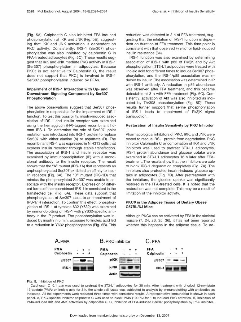
Pharmacological inhibitors of PKC, IKK, and JNK weretested to rescue IRS-1 protein from degradation. PKCinhibitor Calphostin C or combination of IKK and JNKinhibitors was used to pretreat 3T3-L1 adipocytes.IRS-1 protein abundance and glucose uptake wereexamined in 3T3-L1 adipocytes 16 h later after FFA-treatment. The results show that the inhibitors are ableto block IRS-1 degradation completely (Fig. 7A). Theinhibitors also protected insulin-induced glucose up-take in adipocytes (Fig. 7B). After pretreatment withthe inhibitors, the glucose uptake was significantlyrestored in the FFA-treated cells. It is noted that therestoration was not complete. This may be a result oflimitation of the inhibitor activity.
PKC� in the Adipose Tissue of Dietary ObeseC57BL/6J Mice
Although PKC� can be activated by FFA in the skeletalmuscle (7, 24, 26, 35, 36), it has not been reportedwhether this happens in the adipose tissue. To ad-
Fig. 5. Inhibition of PKCCalphostin C (0.1 �M) was used to pretreat the 3T3-L1 adipocytes for 30 min. After treatment with phorbol 12-myristate
13-acetate (PMA) or linoleic acid for 3 h, the whole cell lysate was subjected to analysis by immunoblotting with antibodies asindicated. All the experiments were repeated three times with consistent results. A representative immunoblot is shown in eachpanel. A, PKC-specific inhibitor calphostin C was used to block PMA (100 nM for 1 h) induced PKC activities. B, Inhibition ofPMA-induced IKK and JNK activation by calphostin C. C, Inhibition of FFA-induced Ser307 phosphorylation by PKC inhibitor.
2028 Mol Endocrinol, August 2004, 18(8):2024–2034 Gao et al. • Inhibition of Insulin Sensitivity
by on December 13, 2007 mend.endojournals.orgDownloaded from
dress this question, we examined PKC� in the fattissue of dietary obese mice. C57BL/6J mice were feda high-fat diet to induce dietary insulin resistance. Thisinsulin resistance is associated with body weight gain
in the mice (Fig. 8A). As markers of insulin resistance,hyperglycemia and hyperinsulinemia were detected at8–10 wk on high-fat diet (Fig. 8, B and C). The PKC�protein level was increased dramatically in the adipose
Fig. 6. Analysis of IRS-1 FunctionIRS-1 function was examined by determining inhibitory effect of FFA. IRS-1 was studied by analyzing association of IRS-1/IR,
IRS-1/p85, and by IRS-1 tyrosine phosphorylation as well as Akt phosphorylation. A, Reduction of IRS-1/IR interaction by Ser307phosphorylation. NIH-3T3 cells that express insulin receptor were used in the study to host mutated IRS-1. IP was conducted withinsulin receptor (IR) antibody. The IP product and recombinant IRS-1 in the whole cell lysate were blotted with IRS-1 antibody.B, Insulin-induced IRS-1 tyrosine phosphorylation. IRS-1 was immunoprecipitated from insulin-treated 3T3-L1 adipocytes. C,IRS-1 association with p85 of PI(3)K. 3T3-L1 adipocytes were used in IP with IRS-1 antibody. D, Akt activation by insulin. Aktphosphorylation at Thr308 was determined in 3T3-L1 adipocytes after 16-h treatment with linoleic acid. Insulin was used at 100nM for 30 min.
Fig. 7. Rescuing Glucose Uptake and IRS-1 ProteinFully differentiated 3T3-L1 adipocytes were pretreated with calphostin C (Cal) or combination of 15dPGJ2 (J2) and SP600125
(SP) for 30 min, then followed by treatment of BSA-bound linoleic acid (300 �M) for 16 h. IRS-1 protein abundance was examinedin Western blot and glucose uptake was measured by H3-2D-glucose intake. A, Blocking FFA-induced IRS-1 degradation in3T3-L1 adipocytes. Each bar represents means � SE of results from three experiments. A representative blot is shown. B,Insulin-induced glucose uptake was restored partially by the inhibitors. Each bar represents means � SE of results from triplicates.The experiment was repeated three times with consistent results.
Gao et al. • Inhibition of Insulin Sensitivity Mol Endocrinol, August 2004, 18(8):2024–2034 2029
by on December 13, 2007 mend.endojournals.orgDownloaded from

tissue when insulin resistance occurs in C57BL/6J miceon the high-fat diet (Fig. 8D), suggesting a chronic acti-vation of the PKC� serine kinase. Accordingly, the IRS-1protein abundance was reduced in the adipose tissue ofthe dietary insulin-resistant mice (Fig. 7D). This observa-tion is similar to PKC� increase in the red muscles fromfat-fed rats (26). These data suggest that hyperlipidemiaactivates PKC� in fat tissue.
DISCUSSION
Mice deficient in IKK or JNK are protected from insulinresistance induced by the high-fat diet (37, 38). Thisinformation suggests that IKK or JNK may be involvedin FFAs signal transduction that leads to insulin resis-tance. However, the activities of the two serine kinaseshave not been previously characterized in the FFAsignaling at the cellular and molecular levels. Thisstudy provides evidence that IKK and JNK may medi-ate PKC signals for insulin resistance induced by FFAsin adipocytes.
IKK and JNK may contribute to IRS-1 serine phos-phorylation in response to FFAs. In this study, weobserved that both IKK and JNK were activated bylinoleic acid, a FFA used in this study. The activation is
associated with the Ser307 phosphorylation in IRS-1.Inhibition of the two serine kinases led to protectionfrom Ser307 phosphorylation and degradation ofIRS-1 (Figs. 3 and 6). It is known that serine phosphor-ylation is associated with protein degradation of IRS-1(16, 39). Because there are about 50 serine/threonineresidues in IRS-1, it is hard to determine which serine/threonine is involved in the IRS-1 degradation. Re-cently, it has been shown that Ser307 phosphorylationcontributes to IRS-1 degradation in hepatocytes in theresponse to insulin (14). Our result suggests that thesame mechanism contributes to FFA-induced degra-dation of IRS-1 in adipocytes (Fig. 1). A decrease inIRS-1 abundance leads to insulin resistance as shownin IRS-1 knockout studies (40, 41). We observed thatIRS-1 protein was reduced in the fat tissue of mousemodel of dietary insulin resistance (Fig. 8B). This isconsistent with that IRS-1 protein is reduced in the fattissue of type 2 diabetes patient (42). We observedthat inhibition of IKK and JNK by pharmacologicalagents was able to protect 3T3-L1 adipocytes frominsulin resistance (Fig. 6). Taken together, our datasupport that IKK and JNK involve in FFA signalingpathway for insulin resistance.
IKK and JNK may mediate PKC signal for insulinresistance. It was reported that activities classical
Fig. 8. PKC� in the Adipose TissueC57BL/6J mice were fed a high-fat diet (HFD). Chow diet was used as a control. Body weight, fasting glucose, and insulin were
measured weekly or biweekly. In this experiment, ten mice were examined in each group. *, Significant difference (P � 0.05). A,Body weight gain. B, Fasting glucose. C, Fasting insulin. D, Immunoblotting of PKC� in the adipose tissue of normal andinsulin-resistant C57BL/6J mouse. PKC� protein level was determined in the whole cell lysate of fat tissue. The representative blotof two mice in each group is presented.
2030 Mol Endocrinol, August 2004, 18(8):2024–2034 Gao et al. • Inhibition of Insulin Sensitivity
by on December 13, 2007 mend.endojournals.orgDownloaded from

PKCs and novel PKCs are negatively associated withinsulin sensitivity (7, 24, 43, 44). Some reports suggestthat activation of these two classes of PKCs by phor-bol esters leads to activation of PI(3)K and glucosetransporters (45, 46). Serine phosphorylation of IRS-1represents a mechanism by which PKCs leads to theinhibition of insulin sensitivity (7, 35, 47, 48). However,it is not well defined how PKC promotes IRS-1 serinephosphorylation. PKC was shown to phosphorylateIRS-1 protein directly (49, 50); however, it remains tobe established how PKC leads to Ser307 phosphory-lation (32). In this study, we provide evidence that IKKand JNK may mediate PKC activity for Ser307 phos-phorylation. The evidence includes: 1) FFAs inducedactivation of PKC� as indicated by their phosphoryla-tion status (Fig. 4); 2) Inhibition of PKC activities byspecific inhibitor calphostin C resulted in suppressionof both IKK and JNK activities (Fig. 5). The inhibition isassociated with a reduction in IRS-1 serine phosphor-ylation (Ser307). Activation of IKK and JNK by PKC hasbeen well established in the signaling pathways of cellmembrane receptors. In B or T cells, PKC� and PKC�are responsible for IKK and JNK activation, respec-tively. These have been demonstrated in signalingpathway of B-cell receptor and T-cell receptor (51, 52).Because IKK and JNK may act as downstream signalmediators for PKC, our data suggest that IKK and JNKmediate PKC� signals in adipocytes.
PKC� may be involved in the FFAs signaling path-way in adipocytes. It has been suggested that PKC� isa major PKC isoenzyme in the skeletal muscle andactivation of PKC� by FFAs might be responsible forinsulin resistance in the skeletal muscle (7, 24). Al-though PKC� has drawn a lot of attention in the skel-etal muscle (7, 24), it is not clear whether PKC� playsa role in the adipose tissue. In this study, we evaluatedPKC� activity in adipocytes. Our result suggests thatPKC� is expressed in adipocytes and its phosphory-lation is induced by FFAs (Fig. 4). In addition, PKC�abundance is increased in the adipose tissue of di-etary obese mice, suggesting a chronic activation ofPKC�. In addition to PKC�, it was reported that PKC�(25), PKC� (25), and PKC� (26) could be activated byFFAs. However, these observations were made inmuscle. In this study, our data suggest that PKC�, butnot other PKC isoforms, is activated by FFA in adipo-cytes (Fig. 4). This result suggests a tissue-specificeffect of FFA activity.
We observed that phosphorylation of PKC can beinduced in adipocytes by FFAs. It is generally believedthat PKCs are constitutively phosphorylated at theactivation and autophosphorylation domains in cells.However, it is not clear what is responsible for theconstitutive phosphorylation. Our data suggest thatthe phosphorylation in some PKC isoforms is inducibleby insulin. In serum-starved 3T3-L1 adipocytes, phos-phorylation of PKC� (Thr538), PKC� (643), and PKC�/ (Thr410/403) are induced by insulin (data notshown). Thus, insulin may be responsible for the con-stitutive phosphorylation of certain PKCs in cultured
cells that are maintained in serum-containing medium.Our observation is consistent with that insulin inducesmembrane association of PKC in 3T3-L1 adipocytes inserum-free condition (53). Because activities of mostPKC isoforms are negatively associated with insulinsensitivity in cells and in animals as shown in PKC� or� knockout mice (44, 54), it is possible that activationof PKCs involves in the negative feedback of insulinsignaling. In this study, we observed that phosphory-lation of PKCs was induced by FFA in serum-freecondition. It is possible that FFAs contribute to insulinresistance through activation of this negative feedbackmechanism. An increase in intracellular diacylglycerolwas suggested to contribute to PKC� activation (7).
In summary, our data suggest a signaling pathwayof FFAs for insulin resistance in adipocytes (Fig. 9). Inthis pathway, FFAs activate PKC isoenzymes such asPKC� and leads to the activation of IKK and JNK.Activation of these two serine kinases leads to Ser307phosphorylation in IRS-1. The serine phosphorylationis responsible for a reduction in IRS-1 protein andinsulin resistance in adipocytes. This molecular path-way might operate in many cell types including adipo-cytes, myocytes, and hepatocytes.
MATERIALS AND METHODS
Reagents
Antibodies against phospho-IRS-1 (Ser307) (catalog no. 07-247) was obtained from Upstate Biotechnology (Lake Placid,
Fig. 9. Signal Transduction Pathway for Inhibition of IRS-1Function by FFA
FFA activates PKC� through diacylglycerol (DAG) or Cer-amide. Then, PKC� activates JNK and IKK for IRS-1 phos-phorylation at Ser307. A reduction of IRS-1 function andprotein abundance contributes to insulin resistance.
Gao et al. • Inhibition of Insulin Sensitivity Mol Endocrinol, August 2004, 18(8):2024–2034 2031
by on December 13, 2007 mend.endojournals.orgDownloaded from

NY). Antibodies against IRS-1 (sc-7200), IRS-2 (sc-8299),GLUT4 (sc-7938), PI (3)p85 (sc-423), insulin receptor � (sc-09), and phospho-JNK (sc-6254) were obtained from SantaCruz Biotechnology (Santa Cruz, CA). �-Actin (ab6276) andGLUT1 (ab1932–125) antibodies were obtained from Abcam(Cambridge, UK). Phospho-specific antibodies to PKC� /� II(catalog no. 9375), PKC� (catalog nos. 9374, 9376), PKC�(catalog no. 2064), PKC� (catalog no. 9377), PKC� (catalogno. 9378) and Akt (catalog no. 9275) were purchased fromCell Signaling Technology (Beverly, MA). Expression vectorsfor HA-IRS-1, HA-IRS-1A, and HA-IRS-1D were used in ourprevious study (11). JNK inhibitor SP600125 (catalog no.EI-305) was from Biomol (Plymouth Meeting, PA). 15dPGJ2(catalog no. 538927) was from Calbiochem (San Diego, CA).
Dietary Obese Mice
Male C57BL/6J mice at age of 4 wk were purchased from TheJackson Laboratory (Bar Harbor, ME) and housed in theanimal facility at the Pennington Biomedical Research Centerwith 12-h light, 12-h dark cycle and constant temperature (23C). The mice were free to access water and diet. After a 1-wkquarantine, the mice were divided into two groups, 12 miceper group. The experimental group was fed with high-fat diet(D12331, Research Diets, New Brunswick, NJ) in which fataccounts for 58 kcal%. The control group was fed with chowdiet. All procedures were performed in accordance with Na-tional Institute of Health guidelines for the care and use ofanimal and approved by the Institute Animal Care and UseCommittee at the Pennington Biomedical Research Center.
Fasting Plasma Glucose and Insulin
Fasting glucose and insulin were determined in the plasmaevery 2 wk. The blood (30 �l/mouse) was collected from thetail vein using heparinized micro-hematocrit capillary tubes(catalog no. 22–362-566; Fisher Scientific, Pittsburgh, PA)after overnight (16 h) starvation. The plasma was prepared bycentrifuging the blood at 4 C, 4000 rpm for 20 min. Theglucose level was determined with a FreeStyle blood glucosemonitoring system (TheraSense, Phoenix, AZ). The insulinlevel was determined with ELISA using the “Ultra SensitiveInsulin ELISA Kit” (catalog no. 90060, Crystal Chem, Chi-cago, IL).
3T3-L1 Adipocytes
The mouse fibroblast 3T3-L1 preadipocytes (CL-173) werepurchased from the American Type Culture Collection(ATCC, Manassas, VA) and maintained in DMEM culture me-dium supplemented with 10% fetal calf serum, and 4 mM
glutamine. For adipogenesis, 3T3-L1 preadipocytes weregrown into confluence in a six-well or 100-mm plate, and thenwere differentiated into adipocytes using a standard protocol.The 3T3-L1 cells were incubated in the adipogenic cocktail(5 �g/ml insulin, 0.5 mM isobutylmethylxanthine, and 10 �M
dexamethasone) for 2 d. This was followed by incubation ininsulin-supplemented medium for additional 4 d. The normalmedium was used at d 7 to maintain the adipocytes.
Fatty Acid Treatment
Stearic (S4751) was from Sigma (St. Louis, MO). Oleic(90260) and linoleic acids (90150) were purchased from Cay-man Chemical (Ann Arbor, MI). These FFAs were mixed withFFA-free BSA (152401, ICN Biomedicals, Irvine, CA) at aweight ratio of 1:1 to make BSA-bound FFA. The 3T3-L1adipocytes were serum-starved overnight in 0.1% BSADMEM and then treated with BSA-bound FFAs.
Glucose Uptake (55)
3T3-L1 preadipocytes (5 � 105/well) were differentiated intoadipocytes in a 12-well plate. After serum-starvation in 0.1%BSA DMEM for overnight, the cells were incubated in 1ml/well PBS containing 200 nM insulin for 30 min at 37 C.After washing in PBS, the cells were incubated in 1 ml PBScontaining 0.1 mM 2-deoxyglucose and 1 �Ci/ml 2-deoxy-D-[3H] glucose for 5 min. Then, the cells were washed threetimes in ice-cold PBS, and solubilized in 0.4 ml of 1% sodiumdodecyl sulfate. 3H-glucose uptake was detected in 4 ml ofscintillant using a Beckman LS6500 scintillation counter(Beckman Coulter, Inc., Fullerton, CA). Nonspecific deoxy-glucose uptake is measured in the presence of 20 �M cy-tochalasin B and is subtracted from the total uptake to getspecific glucose uptake.
Immunoblotting and IP (11)
The whole cell lysate was made by sonication in lysis buffer[1% Triton X-100, 50 mM KCl, 25 mM HEPES (pH 7.8), leu-peptin 10 �g/ml, aprotinin 20 �g/ml, 125 �M dithiothreitol, 1mM phenylmethylsulfonyl fluoride, 1 mM sodium orthovana-date]. IP was conducted with 200–400 �g protein and 2–4 �gantibodies. The IP product was then subjected to immuno-blotting analysis. The protein was resolved in SDS-PAGE,and transferred onto polyvinylidene difluoride membrane(162–0184, Bio-Rad, Hercules, CA). The membrane was pre-blotted in milk buffer for 20 min, and then immunoblotted witha primary antibody for 1–24 h followed by a secondary anti-body for 30 min. Horseradish peroxidase-conjugated sec-ondary antibodies (NA934V or NA931, Amersham Life Sci-ence, Piscataway, NJ) were used with chemiluminescencereagent for signal imaging (NEL-105, PerkinElmer, Boston,MA). To detect multiple signals from a single membrane, theblot membrane was treated with a stripping buffer (59 mM
trizma hydrochloride, 2% sodium dodecyl sulfate, 0.75%2-merthylethylenediamine) for 30 min at 42 C, washed ex-tensively in PBS for 2 h, and then used for reblotting with adifferent primary antibody. The intensity of Western blot sig-nal was quantified with an image analysis program PDQuest7.1 (Bio-Rad), and the signal was normalized against loadingcontrol.
Data Analysis
The data of glucose, insulin, glucose uptake and signals inimmunoblot are presented as mean � SE of triplicates in arepresentative experiments or results of three independentexperiments. Student’s t test was used with significance ofP � 0.05.
Acknowledgments
We thank Ms. Kathryn Redd for her excellent technicalassistance in this study.
Received October 1, 2003. Accepted May 7, 2004.Address all correspondence and requests for reprints to:
Jianping Ye, Pennington Biomedical Research Center, 6400Perkins Road, Baton Rouge, Louisiana 70808. E-mail:[email protected].
This work was partially supported by National Institutes ofHealth Grant (to J.Y.) (DK 68036) and the Pennington Bio-medical Research Foundation.
2032 Mol Endocrinol, August 2004, 18(8):2024–2034 Gao et al. • Inhibition of Insulin Sensitivity
by on December 13, 2007 mend.endojournals.orgDownloaded from

REFERENCES
1. Boyle JP, Honeycutt AA, Narayan KM, Hoerger TJ, GeissLS, Chen H, Thompson TJ 2001 Projection of diabetesburden through 2050: impact of changing demographyand disease prevalence in the U. S. Diabetes Care 24:1936–1940
2. Boden G 1997 Role of fatty acids in the pathogenesis ofinsulin resistance and NIDDM. Diabetes 46:3–10
3. Shulman GI 2000 Cellular mechanisms of insulin resis-tance. J Clin Invest 106:171–176
4. Peraldi P, Spiegelman B 1998 TNF-� and insulinresistance: summary and future prospects. Mol Cell Bio-chem 182:169–275
5. Saltiel AR, Kahn CR 2001 Insulin signalling and the reg-ulation of glucose and lipid metabolism. Nature 414:799–806
6. Boden G, Jadali F, White J, Liang Y, Mozzoli M, Chen X,Coleman E, Smith C 1991 Effects of fat on insulin-stimulated carbohydrate metabolism in normal men.J Clin Invest 88:960–966
7. Yu C, Chen Y, Cline GW, Zhang D, Zong H, Wang Y,Bergeron R, Kim JK, Cushman SW, Cooney GJ,Atcheson B, White MF, Kraegen EW, Shulman GI 2002Mechanism by which fatty acids inhibit insulin activationof insulin receptor substrate-1 (IRS-1)-associated phos-phatidylinositol 3-kinase activity in muscle. J Biol Chem277:50230–50236
8. Storz P, Doppler H, Wernig A, Pfizenmaier K, Muller G1999 Cross-talk mechanisms in the development of in-sulin resistance of skeletal muscle cells palmitate ratherthan tumour necrosis factor inhibits insulin-dependentprotein kinase B (PKB)/Akt stimulation and glucose up-take. Eur J Biochem 266:17–25
9. Aguirre V, Uchida T, Yenush L, Davis R, White MF 2000The c-Jun NH(2)-terminal kinase promotes insulin resis-tance during association with insulin receptor sub-strate-1 and phosphorylation of Ser(307). J Biol Chem275:9047–9054
10. Aguirre V, Werner ED, Giraud J, Lee YH, Shoelson SE,White MF 2002 Phosphorylation of ser307 in insulin re-ceptor substrate-1 blocks interactions with the insulinreceptor and inhibits insulin action. J Biol Chem 277:1531–1537
11. Gao Z, Hwang D, Bataille F, Lefevre M, York D, Quon MJ,Ye J 2002 Serine phosphorylation of insulin receptorsubstrate 1 (IRS-1) by inhibitor �B kinase (IKK) complex.J Biol Chem 277:48115–48121
12. Gao Z, Zuberi A, Quon M, Dong Z, Ye J 2003 Aspirininhibits TNF-induced serine phosphorylation of IRS-1through targeting multiple serine kinases. J Biol Chem278:24944–24950
13. Lee YH, Giraud J, Davis RJ, White MF 2003 cJUN N-terminal kinase (JNK) mediates feedback inhibition of theinsulin signaling cascade. J Biol Chem 278:2896–2902
14. Greene MW, Sakaue H, Wang L, Alessi DR, Roth RA2003 Modulation of insulin stimulated degradation ofhuman insulin receptor substrate-1 by serine 312 phos-phorylation. J Biol Chem 278:8199–8211
15. Van Epps-Fung M, Williford J, Wells A, Hardy RW 1997Fatty acid-induced insulin resistance in adipocytes. En-docrinology 138:4338–4345
16. Sun XJ, Goldberg JL, Qiao LY, Mitchell JJ 1999 Insulin-induced insulin receptor substrate-1 degradation is me-diated by the proteasome degradation pathway. Diabe-tes 48:1359–1364
17. Rui L, Fisher TL, Thomas J, White MF 2001 Regulation ofinsulin/insulin-like growth factor-1 signaling by protea-some-mediated degradation of insulin receptor sub-strate-2. J Biol Chem 276:40362–40367
18. Zhande R, Mitchell JJ, Wu J, Sun XJ 2002 Molecularmechanism of insulin-induced degradation of insulin re-ceptor substrate 1. Mol Cell Biol 22:1016–1026
19. Mercurio F, Zhu H, Murray BW, Shevchenko A, BennettBL, Li J, Young DB, Barbosa M, Mann M, Manning A,Rao A 1997 IKK-1 and IKK-2: cytokine-activated I�Bkinases essential for NF-�B activation. Science 278:860–866
20. Delhase M, Hayakawa M, Chen Y, Karin M 1999 Positiveand negative regulation of I�B kinase activity throughIKK� subunit phosphorylation. Science 284:309–313
21. Davis RJ 1999 Signal transduction by the c-Jun N-terminal kinase. Biochem Soc Symp 64:1–12
22. Rossi A, Kapahi P, Natoli G, Takahashi T, Chen Y, KarinM, Santoro MG 2000 Anti-inflammatory cyclopentenoneprostaglandins are direct inhibitors of I�B kinase. Nature403:103–108
23. Bennett BL, Sasaki DT, Murray BW, O’Leary EC, SakataST, Xu W, Leisten JC, Motiwala A, Pierce S, Satoh Y,Bhagwat SS, Manning AM, Anderson DW 2001SP600125, an anthrapyrazolone inhibitor of Jun N-termi-nal kinase. Proc Natl Acad Sci USA 98:13681–13686
24. Griffin ME, Marcucci MJ, Cline GW, Bell K, Barucci N,Lee D, Goodyear LJ, Kraegen EW, White MF, Shulman GI1999 Free fatty acid-induced insulin resistance is asso-ciated with activation of protein kinase C � and alter-ations in the insulin signaling cascade. Diabetes 48:1270–1274
25. Itani SI, Ruderman NB, Schmieder F, Boden G 2002Lipid-induced insulin resistance in human muscle is as-sociated with changes in diacylglycerol, protein kinase C,and I�B-�. Diabetes 51:2005–2011
26. Schmitz-Peiffer C, Browne CL, Oakes ND, Watkinson A,Chisholm DJ, Kraegen EW, Biden TJ 1997 Alterations inthe expression and cellular localization of protein kinaseC isozymes � and � are associated with insulin resistancein skeletal muscle of the high-fat-fed rat. Diabetes 46:169–178
27. Dutil EM, Keranen LM, DePaoli-Roach AA, Newton AC1994 In vivo regulation of protein kinase C by trans-phosphorylation followed by autophosphorylation. J BiolChem 269:29359–29362
28. Orr JW, Newton AC 1994 Requirement for negativecharge on “activation loop” of protein kinase C. J BiolChem 269:27715–27718
29. Keranen LM, Dutil EM, Newton AC 1995 Protein kinase Cis regulated in vivo by three functionally distinct phos-phorylations. Curr Biol 5:1394–1403
30. Valverde AM, Sinnett-Smith J, Van Lint J, Rozengurt E1994 Molecular cloning and characterization of proteinkinase D: a target for diacylglycerol and phorbol esterswith a distinctive catalytic domain. Proc Natl Acad SciUSA 91:8572–8576
31. Iglesias T, Waldron RT, Rozengurt E 1998 Identificationof in vivo phosphorylation sites required for protein ki-nase D activation. J Biol Chem 273:27662–27667
32. Jiang G, Dallas-Yang Q, Liu F, Moller DE, Zhang BB 2002Salicylic acid reverses PMA and TNF �-induced IRS1serine 307 phosphorylation and insulin resistance in HEK293 cells. J Biol Chem 278:180–186
33. Kobayashi E, Nakano H, Morimoto M, Tamaoki T 1989Calphostin C (UCN-1028C), a novel microbial com-pound, is a highly potent and specific inhibitor of proteinkinase C. Biochem Biophys Res Commun 159:548–553
34. Boden G, Chen X, Rosner J, Barton M 1995 Effects of a48-h fat infusion on insulin secretion and glucose utiliza-tion. Diabetes 44:1239–1242
35. Dresner A, Laurent D, Marcucci M, Griffin ME, Dufour S,Cline GW, Slezak LA, Andersen DK, Hundal RS, RothmanDL, Petersen KF, Shulman GI 1999 Effects of free fattyacids on glucose transport and IRS-1-associated phos-phatidylinositol 3-kinase activity. J Clin Invest 103:253–259
Gao et al. • Inhibition of Insulin Sensitivity Mol Endocrinol, August 2004, 18(8):2024–2034 2033
by on December 13, 2007 mend.endojournals.orgDownloaded from

36. Bell KS, Schmitz-Peiffer C, Lim-Fraser M, Biden TJ,Cooney GJ, Kraegen EW 2000 Acute reversal of lipid-induced muscle insulin resistance is associated withrapid alteration in PKC-� localization. Am J Physiol En-docrinol Metab 279:E1196–E1201
37. Yuan M, Konstantopoulos N, Lee J, Hansen L, Li ZW,Karin M, Shoelson SE 2001 Reversal of obesity- anddiet-induced insulin resistance with salicylates or tar-geted disruption of Ikk�. Science 293:1673–1677
38. Hirosumi J, Tuncman G, Chang L, Gorgun CZ, Uysal KT,Maeda K, Karin M, Hotamisligil GS 2002 A central role forJNK in obesity and insulin resistance. Nature 420:333–336
39. Stephens JM, Lee J, Pilch PF 1997 Tumor necrosisfactor-�-induced insulin resistance in 3T3-L1 adipocytesis accompanied by a loss of insulin receptor substrate-1and GLUT4 expression without a loss of insulin receptor-mediated signal transduction. J Biol Chem 272:971–976
40. Araki E, Lipes MA, Patti ME, Bruning JC, Haag 3rd B,Johnson RS, Kahn CR 1994 Alternative pathway of in-sulin signalling in mice with targeted disruption of theIRS-1 gene. Nature 372:186–190
41. Yamauchi T, Tobe K, Tamemoto H, Ueki K, Kaburagi Y,Yamamoto-Honda R, Takahashi Y, Yoshizawa F, AizawaS, Akanuma Y, Sonenberg N, Yazaki Y, Kadowaki T 1996Insulin signalling and insulin actions in the muscles andlivers of insulin-resistant, insulin receptor substrate 1-deficient mice. Mol Cell Biol 16:3074–3084
42. Rondinone CM, Wang LM, Lonnroth P, Wesslau C,Pierce JH, Smith U 1997 Insulin receptor substrate (IRS)1 is reduced and IRS-2 is the main docking protein forphosphatidylinositol 3-kinase in adipocytes from sub-jects with non-insulin-dependent diabetes mellitus. ProcNatl Acad Sci USA 94:4171–4175
43. Bandyopadhyay G, Standaert ML, Kikkawa U, Ono Y,Moscat J, Farese RV 1999 Effects of transiently ex-pressed atypical (�, ), conventional (�, �) and novel (�, �)protein kinase C isoforms on insulin-stimulated translo-cation of epitope-tagged GLUT4 glucose transporters inrat adipocytes: specific interchangeable effects of pro-tein kinases C-� and C-. Biochem J 337:461–470
44. Leitges M, Plomann M, Standaert ML, BandyopadhyayG, Sajan MP, Kanoh Y, Farese RV 2002 Knockout of PKC� enhances insulin signaling through PI3K. Mol Endocri-nol 16:847–858
45. Standaert ML, Bandyopadhyay G, Galloway L, Farese RV1996 Effects of phorbol esters on insulin-induced acti-vation of phosphatidylinositol 3-kinase, glucose trans-
port, and glycogen synthase in rat adipocytes. FEBS Lett388:26–28
46. Nave BT, Siddle K, Shepherd PR 1996 Phorbol estersstimulate phosphatidylinositol 3,4,5-trisphosphate pro-duction in 3T3-L1 adipocytes: implications for stimula-tion of glucose transport. Biochem J 318:203–205
47. Chin JE, Dickens M, Tavare JM, Roth RA 1993 Overex-pression of protein kinase C isoenzymes �, � I, , and �in cells overexpressing the insulin receptor. Effects onreceptor phosphorylation and signaling. J Biol Chem268:6338–6347
48. Danielsen AG, Liu F, Hosomi Y, Shii K, Roth RA 1995Activation of protein kinase C � inhibits signaling bymembers of the insulin receptor family. J Biol Chem270:21600–21605
49. Nakajima K, Yamauchi K, Shigematsu S, Ikeo S,Komatsu M, Aizawa T, Hashizume K 2000 Selectiveattenuation of metabolic branch of insulin receptordown-signaling by high glucose in a hepatoma cellline, HepG2 cells. J Biol Chem 275:20880–20886
50. Ravichandran LV, Esposito DL, Chen J, Quon MJ 2001Protein kinase C-� phosphorylates insulin receptor sub-strate-1 and impairs its ability to activate phosphatidyl-inositol 3-kinase in response to insulin. J Biol Chem276:3543–3549
51. Su TT, Guo B, Kawakami Y, Sommer K, Chae K,Humphries LA, Kato RM, Kang S, Patrone L, Wall R,Teitell M, Leitges M, Kawakami T, Rawlings DJ 2002PKC-� controls I�B kinase lipid raft recruitment and ac-tivation in response to BCR signaling. Nat Immunol3:780–786
52. Coudronniere N, Villalba M, Englund N, Altman A 2000NF-�B activation induced by T cell receptor/CD28 co-stimulation is mediated by protein kinase C-�. Proc NatlAcad Sci USA 97:3394–3399
53. Bandyopadhyay G, Standaert ML, Zhao L, Yu B, AvignonA, Galloway L, Karnam P, Moscat J, Farese RV 1997Activation of protein kinase C (�, �, and �) by insulin in3T3/L1 cells. Transfection studies suggest a role forPKC-� in glucose transport. J Biol Chem 272:2551–2558
54. Standaert ML, Bandyopadhyay G, Galloway L, Soto J,Ono Y, Kikkawa U, Farese RV, Leitges M 1999 Effects ofknockout of the protein kinase C � gene on glucosetransport and glucose homeostasis. Endocrinology 140:4470–4477
55. Kozma L, Baltensperger K, Klarlund J, Porras A, SantosE, Czech M 1993 The Ras signaling pathway mimicsinsulin action on glucose transporter translocation. ProcNatl Acad Sci USA 90:4460–4464
Molecular Endocrinology is published monthly by The Endocrine Society (http://www.endo-society.org), the foremostprofessional society serving the endocrine community.
2034 Mol Endocrinol, August 2004, 18(8):2024–2034 Gao et al. • Inhibition of Insulin Sensitivity
by on December 13, 2007 mend.endojournals.orgDownloaded from
Related Documents