Cyanidin 3-glucoside protects 3T3-L1 adipocytes against H 2 O 2 - or TNF-a-induced insulin resistance by inhibiting c-Jun NH 2 -terminal kinase activation Honghui Guo, Wenhua Ling *, Qing Wang, Chi Liu, Yan Hu, Min Xia Department of Nutrition, School of Public Health, Sun Yat-Sen University (Northern Campus), 74 Zhongshan Road 2, Guangzhou, Guangdong Province 510080, PR China 1. Introduction Insulin resistance, or an attenuated biological response to insulin, is a major pathological feature of diabetes and a central component in the so-called metabolic syndrome [1]. The exact mechanisms responsible for this abnormality are still not fully elucidated. Insulin physiologically initiates its biological function by activating the insulin receptor, resulting in tyrosine phosphorylation of insulin receptor substrate (IRS) proteins, such as IRS1 and IRS2. Tyrosine phosphorylation of IRS1 triggers downstream signaling pathways, and finally stimulates the translocation of glucose transporter 4 (GLUT4) biochemical pharmacology 75 (2008) 1393–1401 article info Article history: Received 19 October 2007 Accepted 28 November 2007 Keywords: Anthocyanin Cyanidin 3-glucoside c-Jun NH 2 -terminal kinase Insulin resistance 3T3-L1 adipocyte abstract Anthocyanins are naturally occurring plant pigments and exhibit an array of pharmaco- logical properties. Our previous study showed that black rice pigment extract rich in anthocyanin prevents and ameliorates high-fructose-induced insulin resistance in rats. In present study, cyanidin 3-glucoside (Cy-3-G), a typical anthocyanin most abundant in black rice was used to examine its protective effect on insulin sensitivity in 3T3-L1 adipocytes exposed to H 2 O 2 (generated by adding glucose oxidase to the medium) or tumor necrosis factor a (TNF-a). Twelve-hour exposure of 3T3-L1 adipocytes to H 2 O 2 or TNF-a resulted in the increase of c-Jun NH 2 -terminal kinase (JNK) activation and insulin receptor substrate 1 (IRS1) serine 307 phosphorylation, concomitantly with the decrease in insulin- stimulated IRS1 tyrosine phosphorylation and cellular glucose uptake. Blocking JNK expres- sion using RNA interference efficiently prevented the H 2 O 2 - or TNF-a-induced defects in insulin action. Pretreatment of cells with Cy-3-G reduced the intracellular production of reactive oxygen species, the activation of JNK, and attenuated H 2 O 2 - or TNF-a-induced insulin resistance in a dose-dependent manner. In parallel, N-acetyl-cysteine, an antiox- idant compound, did not exhibit an attenuation of TNF-a-induced insulin resistance. Taken together, these results indicated that Cy-3-G exerts a protective role against H 2 O 2 - or TNF-a- induced insulin resistance in 3T3-L1 adipocytes by inhibiting the JNK signal pathway. # 2007 Elsevier Inc. All rights reserved. * Corresponding author. Tel.: +86 20 87331597; fax: +86 20 87330446. E-mail address: [email protected] (W. Ling). Abbreviations: 2-DG, 2-deoxy-D-glucose; Cy-3-G, cyanidin 3-glucoside; DCF-DA, dihydrodichlorofluorescein diacetate; DMEM, Dulbec- co’s modified Eagle’s medium; FBS, fetal bovine serum; GLUT4, glucose transporter 4; IRS1, insulin receptor substrate 1; JNK, c-Jun NH 2 - terminal kinase; KRP, Krebs-Ringer’s phosphate; MTT, 3-(4,5-dimethylthiazol-2-yl)-2,5-diphenyl tetrazolium bromide; NAC, N-acetyl- cysteine; PM, plasma membrane; PY, phosphotyrosine; ROS, reactive oxygen species; siRNA, small interfering RNA; TNF-a, tumor necrosis factor a; TNFR1, TNF type-1 receptor; TRAF2, TNFR-associated factor 2. available at www.sciencedirect.com journal homepage: www.elsevier.com/locate/biochempharm 0006-2952/$ – see front matter # 2007 Elsevier Inc. All rights reserved. doi:10.1016/j.bcp.2007.11.016

Welcome message from author
This document is posted to help you gain knowledge. Please leave a comment to let me know what you think about it! Share it to your friends and learn new things together.
Transcript

Cyanidin 3-glucoside protects 3T3-L1 adipocytes againstH2O2- or TNF-a-induced insulin resistance by inhibitingc-Jun NH2-terminal kinase activation
Honghui Guo, Wenhua Ling *, Qing Wang, Chi Liu, Yan Hu, Min Xia
Department of Nutrition, School of Public Health, Sun Yat-Sen University (Northern Campus), 74 Zhongshan Road 2,
Guangzhou, Guangdong Province 510080, PR China
b i o c h e m i c a l p h a r m a c o l o g y 7 5 ( 2 0 0 8 ) 1 3 9 3 – 1 4 0 1
a r t i c l e i n f o
Article history:
Received 19 October 2007
Accepted 28 November 2007
Keywords:
Anthocyanin
Cyanidin 3-glucoside
c-Jun NH2-terminal kinase
Insulin resistance
3T3-L1 adipocyte
a b s t r a c t
Anthocyanins are naturally occurring plant pigments and exhibit an array of pharmaco-
logical properties. Our previous study showed that black rice pigment extract rich in
anthocyanin prevents and ameliorates high-fructose-induced insulin resistance in rats.
In present study, cyanidin 3-glucoside (Cy-3-G), a typical anthocyanin most abundant in
black rice was used to examine its protective effect on insulin sensitivity in 3T3-L1
adipocytes exposed to H2O2 (generated by adding glucose oxidase to the medium) or tumor
necrosis factor a (TNF-a). Twelve-hour exposure of 3T3-L1 adipocytes to H2O2 or TNF-a
resulted in the increase of c-Jun NH2-terminal kinase (JNK) activation and insulin receptor
substrate 1 (IRS1) serine 307 phosphorylation, concomitantly with the decrease in insulin-
stimulated IRS1 tyrosine phosphorylation and cellular glucose uptake. Blocking JNK expres-
sion using RNA interference efficiently prevented the H2O2- or TNF-a-induced defects in
insulin action. Pretreatment of cells with Cy-3-G reduced the intracellular production of
reactive oxygen species, the activation of JNK, and attenuated H2O2- or TNF-a-induced
insulin resistance in a dose-dependent manner. In parallel, N-acetyl-cysteine, an antiox-
idant compound, did not exhibit an attenuation of TNF-a-induced insulin resistance. Taken
together, these results indicated that Cy-3-G exerts a protective role against H2O2- or TNF-a-
induced insulin resistance in 3T3-L1 adipocytes by inhibiting the JNK signal pathway.
# 2007 Elsevier Inc. All rights reserved.
avai lab le at www.sc iencedi rec t .com
journal homepage: www.e lsev ier .com/ locate /b iochempharm
1. Introduction
Insulin resistance, or an attenuated biological response to
insulin, is a major pathological feature of diabetes and a
central component in the so-called metabolic syndrome [1].
The exact mechanisms responsible for this abnormality are
* Corresponding author. Tel.: +86 20 87331597; fax: +86 20 87330446.E-mail address: [email protected] (W. Ling).
Abbreviations: 2-DG, 2-deoxy-D-glucose; Cy-3-G, cyanidin 3-glucosidco’s modified Eagle’s medium; FBS, fetal bovine serum; GLUT4, glucosterminal kinase; KRP, Krebs-Ringer’s phosphate; MTT, 3-(4,5-dimethcysteine; PM, plasma membrane; PY, phosphotyrosine; ROS, reactive oxfactor a; TNFR1, TNF type-1 receptor; TRAF2, TNFR-associated factor0006-2952/$ – see front matter # 2007 Elsevier Inc. All rights reserveddoi:10.1016/j.bcp.2007.11.016
still not fully elucidated. Insulin physiologically initiates its
biological function by activating the insulin receptor, resulting
in tyrosine phosphorylation of insulin receptor substrate (IRS)
proteins, such as IRS1 and IRS2. Tyrosine phosphorylation of
IRS1 triggers downstream signaling pathways, and finally
stimulates the translocation of glucose transporter 4 (GLUT4)
e; DCF-DA, dihydrodichlorofluorescein diacetate; DMEM, Dulbec-e transporter 4; IRS1, insulin receptor substrate 1; JNK, c-Jun NH2-ylthiazol-2-yl)-2,5-diphenyl tetrazolium bromide; NAC, N-acetyl-ygen species; siRNA, small interfering RNA; TNF-a, tumor necrosis2..

b i o c h e m i c a l p h a r m a c o l o g y 7 5 ( 2 0 0 8 ) 1 3 9 3 – 1 4 0 11394
to facilitate the transport of glucose into cells [2,3]. Several
extracellular agents and stress stimuli, such as free fatty acids,
H2O2 and tumor necrosis factor a (TNF-a), all of which are
known contributors to insulin resistance, induce phosphor-
ylation of IRS1 at the inhibitory site serine 307 (Ser307) [4–6].
Ser307 is located next to the phosphotyrosine-binding (PTB)
domain in IRS1 and its phosphorylation inhibits the interac-
tion of the PTB domain with the phosphorylated NPEY motif in
the activated insulin receptor, thereby leading to inhibition of
insulin signaling [7]. Although a variety of serine/threonine
kinases have been described to be capable of mediating IRS1
phosphorylation [8], recent genetic evidence and emerging
pharmacological data indicate that c-Jun NH2-terminal kinase
(JNK; also named stress-activated protein kinase, SAPK) may
play a pivotal role in causing insulin resistance [9–11].
Recently, much attention has been paid to the physiological
effects of some food components that may be beneficial in
preventing insulin resistance and possibly reduce risks of
diabetes and metabolic syndrome. Anthocyanins are naturally
occurring polyphenolic compounds in the plant foods and
widely distributed in fruits, vegetables, and pigmented cereals
[12]. Many studies have shown that anthocyanin not only
imparts color to plants but also exhibits an array of pharma-
cological properties, such as antioxidative, anti-inflammatory
and antitumor activities [13]. Epidemiological investigations
have indicated that the moderate consumption of anthocya-
nins through the intake of red wine is associated with a lower
risk of coronary heart disease and metabolic syndrome [14,15].
In two recent reports, anthocyanins from purple corn and
cherry have been shown to ameliorate obesity and insulin
resistance in high-fat-fed mice [16,17]. Our previous study also
demonstrated that black rice pigment extract rich in antho-
cyaninpreventedand attenuatedthe insulinresistance induced
by a high-fructose diet in animal model [18]. However, the
molecular mechanism underlying this action remains
unknown and needs further investigation in cells.
Considering the key role of JNK activation in the progres-
sion of insulin resistance and the potential capability of
anthocyanin in preventing insulin resistance, we observed the
influence of cyanidin 3-glucoside (Cy-3-G), a typical antho-
cyanin in black rice and other higher plants [13,19], on insulin
resistance of 3T3-L1 adipocytes induced by exposure to H2O2
enzymatic-generating system or TNF-a. The signal pathway of
the action involved in JNK molecular activation was also
investigated.
2. Materials and methods
2.1. Cells and materials
Mouse embryo3T3-L1cellswereobtainedfromChinaCenter for
Type Culture Collection (Wuhan, China). Dulbecco’s modified
Eagle’s medium (DMEM), fetal bovine serum (FBS), and anti-
biotic mixture (penicillin-streptomycin) were purchased from
the Gibco BRL (Grand Island, NY, USA). Anti-GLUT4, anti-IRS1,
anti-p-IRS1Ser307, anti-phosphotyrosine, anti-b-actin, and anti-
JNK1 antibodies, and protein A/G plus agarose were obtained
from Santa Cruz Biotech. (Santa Cruz, CA, USA). Anti-p-JNK
(Thr183/Tyr185) antibody was obtained from Cell Signaling
Technology (Danvers, MA, USA). Recombinant human TNF-a
was purchased from CytoLab (Rehovot, Israel); short-acting
human insulin, fromNovoNordisk (Bagsvaerd,Denmark); HPLC
grade Cy-3-G, from Polyphenol AS (Sandnes, Norway); [1,2-3H]
2-deoxy-D-glucose (3H-2-DG), from GE Healthcare (Buckin-
ghamshire, UK); and all other chemicals, unless otherwise
specified, from Sigma (St. Louis, MO, USA).
2.2. Cell culture and treatments
3T3-L1 pre-adipocytes were grown to confluence in DMEM
containing 10% FBS and 100 U/mL penicillin-streptomycin at
37 8C in a humidified atmosphere of 5% CO2. Cells were
induced to differentiate to adipocytes 24 h after confluence by
changing the medium to DMEM supplemented with 10% FBS,
5 mg/mL insulin, 0.5 mM 3-isobutyl-methyl-xanthine and
0.25 mM dexamethasone sodium phosphate for 48 h. There-
after, the cells were maintained in the original propagation
DMEM, changing medium every 2 days until use. Unless
indicated otherwise, cells were used 9–10 days after differ-
entiation induction when exhibiting more than 90% adipocyte
phenotype. H2O2 was generated by adding 50 mU/mL glucose
oxidase to serum-free DMEM supplemented with 0.5% BSA.
The addition of 50 mU/mL glucose oxidase resulted in medium
H2O2 concentration that achieved a steady state of
11.3 � 1.7 mM after 30 min, as previously reported [20].
Differentiated adipocytes were treated with various concen-
trations of anthocyanin Cy-3-G, antioxidant N-acetyl-cysteine
(NAC), or JNK inhibitor SP600125 (Calbiochem, San Diego, CA,
USA) before they were exposed to the H2O2 generating system
or 1 nM TNF-a as indicated in each experiment.
2.3. Cytotoxicity tests
Cy-3-G and H2O2 induced cytotoxicity was measured using
MTT dye [3-(4,5-dimethylthiazol-2-yl)-2,5-diphenyl tetrazo-
lium bromide] as previously described [21]. The cells were
seeded and differentiated in 24-well plates. After 12 h
treatment with Cy-3-G (0, 10, 20, and 40 mM), or 50 mU/mL
glucose oxidase, 0.5 mg/mL MTT was added to each well and
incubated for 4 h to form formazan crystals. Then, the
medium was gently removed and the crystals were dissolved
in 1 mL of DMSO. Formazan crystals formed were quantified at
570 nm using an ELx800 reader (Bio-Tek Instruments Inc.,
Winooski, VT, USA). Cytotoxicity was expressed in terms of
cell viability, as a ratio of the treatments to the PBS buffer
control population.
2.4. Free radical scavenging activity assay
A fluorescent probe, dihydrodichlorofluorescein diacetate
(DCF-DA), was used to measure intracellular reactive oxygen
species (ROS) formation as described [22]. Briefly, cells were
incubated with 10, 20, or 40 mM of Cy-3-G or 1 mM NAC for 12 h,
before the 12-h exposure to H2O2 generating system or 1 nM
TNF-a. Then, cells were washed twice in Krebs-Ringer’s
phosphate (KRP) buffer (130 mM NaCl, 5 mM KCl, 1.3 mM
CaCl2, 1.3 mM MgSO4, 10 mM Na2HPO4, pH 7.4), incubated in
pre-warmed KRP containing 5 mM glucose and 5 mM DCF-DA,
and placed at 37 8C for 30 min. The reaction system was

b i o c h e m i c a l p h a r m a c o l o g y 7 5 ( 2 0 0 8 ) 1 3 9 3 – 1 4 0 1 1395
washed three times with KRP. After centrifugation at 1000 � g
for 5 min, the supernatants were removed and the pellets were
resolved with 1% Triton X-100. Fluorescence was measured at
an excitation wavelength of 480 nm and an emission
wavelength of 525 nm using a Cary Eclipse fluorescence
spectrophotometer (Varian, Palo Alto, CA, USA). Fluorescence
values were calculated after subtracting background fluores-
cence levels, measured under identical conditions but without
DCF-DA.
2.5. Glucose transport determination
The determination for glucose uptake was described previously
with some modifications [4]. Serum-starved adipocytes were
incubated with pre-warmed KRP containing 0.2% BSA and
10 nM insulin for 30 min. After this period, 3H-2-DG and
unlabeled 2-DG were dispensed into each well for a final
concentration of 1 mCi/mL and 0.1 mM, respectively. Cells were
incubated for anadditional10 min at37 8C,and the reaction was
terminated by three washes with ice-cold KRP. Cells were lysed
in 100 mL 0.1 M NaOH, and radioactivity was determined by
using a liquid scintillation counter (Canberra-Packard, Zellik,
Belgium). Datawereexpressedasmoles 3H-2-DG per minute per
milligram of lysate protein, which was determined using the
bicinchoninic acid method. Nonspecific uptake was assessed in
the presence of 10 mM cytochalasin B and subtracted from all of
the measured values. Nonspecific uptake and absorption was
always less than 8% of the total uptake.
2.6. JNK protein knockdown
To knockdown JNK1 protein expression, we performed
transfection of mouse JNK1 small interfering RNA (siRNA)
duplex with adipocytes on day 6 after differentiation. One
microliters of INTERFERin transfection reagent (Polyplus
transfection SA, Illkrich, France) was added to 200 mL of
DMEM serum-free medium containing 6 nM of each siRNA
oligo, incubated for 10 min, and added to the 12-well dishes
containing 1 mL of fresh medium. A nonrelated, scrambled
siRNA was used as a control. All siRNA oligos were designed
and synthesized by Santa Cruz. Twenty-four, 48, and 72 h
post-transfection, cells were lysed and subjected to SDS-PAGE
and Western blot analysis to evaluate intracellular JNK1
protein levels.
2.7. Immunoprecipitation and Western blotting
Crude plasma membrane (PM) was prepared as described by
Karlsson et al. with modifications [23]. Adipocytes were
homogenized in 0.32 M sucrose, 5 mM Tris–HCl, 120 mM
KCl, 1 mM EDTA, 0.5 mM EGTA, pH 7.5, with protease
inhibitors (10 mM leupeptin, 1 mM pepstatin, 1 mM aprotinin,
4 mM iodoacetate, and 50 mM phenylmethylsulfonyl fluoride)
using a Dounce glass homogenizer on slushy ice. A PM-
containing pellet, obtained by centrifugation at 25,000 � g for
1.5 h at 4 8C, was resuspended in 20 mM HEPES, 1 mM EDTA,
pH 7.4, with protease inhibitors, and was used for immuno-
blotting as described below.
To obtain total proteins, cells were lysised with a buffer
containing 20 mM Tris–Cl, 150 mM NaCl, 1% Nonidet P-40, 0.5%
sodium deoxycholate, 1 mM EDTA, 0.1% SDS, and protease
inhibitors, pH 7.5. The lysates were centrifuged at 12,000 � g
for 15 min, and the supernatants were used for immunopre-
cipitation and immunoblotting. For immunoprecipitation, the
supernatants were incubated with antibodies against IRS1
overnight, then shook gently with the addition of protein A/G
plus agarose for 2 h, centrifuged at 1000 � g for 5 min. Washed
the pellet four times with 1 mL PBS, each time repeating the
centrifugation step, collected and resuspended the immuno-
precipitates pellet in 50 mL of electrophoresis sample buffer.
Protein extractions were separated by using SDS-PAGE on 10%
(for GLUT4 and JNK1) or 8% (for IRS1) polyacrylamide gels, and
transferred to polyvinylidene difluoride (PVDF) membranes
(Millipore, Billerica, MA, USA). After blocking for 2 h with 5%
skimmed milk in TBS-T buffer (10 mM Tris, 150 mM NaCl, and
0.1% Tween-20), the membrane was incubated with primary
antibodies against GLUT4, IRS1, p-IRS1Ser307, phosphotyrosine
(PY), b-actin, JNK1 and p-JNK for 2 h. Specific antibody binding
was detected by horseradish peroxidase-conjugated second-
ary antibodies and visualized using enhanced chemilumines-
cence detection reagent (Santa Cruz). The band densities were
quantified using an image analyzer Quantity One System (Bio-
Rad, Richmond, CA, USA). All protein quantifications were
adjusted for the corresponding b-actin level, which was not
consistently changed by the different treatment conditions.
2.8. Statistical analysis
Statistical analyses were performed using the SPSS 11.0
package (SPSS Inc., Chicago, IL, USA). The results are presented
as the mean � S.E.M. of at least three-independent experi-
ments. Data were analyzed by one-way ANOVA and a post hoc
least significant difference (LSD)–t multiple comparisons test.
The level of significance was set at P < 0.05.
3. Results
3.1. Cytotoxicity and radical scavenging activity of Cy-3-G
The results of the MTT assay showed that there was no
significant change in cell viability after 12-h treatment with
40 mM Cy-3-G or 50 mU/mL glucose oxidase (data not shown).
Next, we measured ROS in adipocytes treated with 50 mU/
mL glucose oxidase or 1 nM TNF-a. The DCF-DA probe could be
oxidized to highly fluorescent compound dichlorofluorescein
(DCF) by intracellular ROS or low-molecular weight peroxides.
In adipocytes treated with 50 mU/mL glucose oxidase or 1 nM
TNF-a for 12 h, the mean DCF fluorescence was increased 4.7-
fold (P < 0.01) and 1.9-fold (P < 0.05), respectively, as compared
to control cells (Fig. 1). However, this effect was almost
abolished by pretreatment of 3T3-L1 adipocytes with 20 mM or
40 mM Cy-3-G, or 1 mM NAC for 12 h.
3.2. H2O2 and TNF-a inhibited the insulin-stimulatedglucose uptake in 3T3-L1 adipocytes
It has been reported that H2O2 or TNF-a induces a defect of
insulin signaling in 3T3-L1 adipocytes in a few hours to
several days at different concentrations [20,22,24]. Thus, we

Fig. 1 – Cy-3-G and NAC decreased intracellular ROS levels
in 3T3-L1 adipocytes treated with H2O2 or TNF-a. Fully
differentiated 3T3-L1 adipocytes were pretreated for 12 h
with 10, 20, or 40 mM of Cy-3-G, or 1 mM NAC, respectively,
and subsequently incubated with �12 mM H2O2 (generated
by adding 50 mU/mL glucose oxidase to the medium) or
1 nM TNF-a for 12 h. Cell lysates were prepared, and
intracellular ROS was measured using the fluorescent
probe, DCF-DA. Results are the mean W S.E.M. of triplicate
determinations of four experiments and are expressed as
fluorescence units detected in a fluorescence
spectrophotometer. *P < 0.05; **P < 0.01 compared with
untreated control.
Fig. 2 – H2O2 and TNF-a inhibited insulin-stimulated
glucose uptake in 3T3-L1 adipocytes. Serum-starved
adipocytes were placed into KRP buffer without glucose,
and incubated in the absence or presence of insulin
(10 nM) for 30 min. Next, 3H-2-DG was added for 10 min
and uptake measured. (A) Adipocytes were treated with
�12 mM H2O2 or 1 nM TNF-a for 0–48 h, and insulin-
stimulated glucose uptake was determined. (B) In washout
experiments, H2O2 or TNF-a was removed from the
medium after 12 h of incubation, and 3T3-L1 adipocytes
were fed with fresh medium. Glucose uptake was
assessed 12 h later in the presence of insulin. White bars,
basal; gray bars, H2O2 or TNF-a + insulin; black bars, H2O2
or TNF-a washout + insulin. The data are the
mean W S.E.M. of four-independent experiments. *P < 0.05;
**P < 0.01 compared with untreated control.
b i o c h e m i c a l p h a r m a c o l o g y 7 5 ( 2 0 0 8 ) 1 3 9 3 – 1 4 0 11396
examined the effect of relatively low dose of H2O2 (�12 mM) or
TNF-a (1 nM) on the insulin sensitivity in 3T3-L1 adipocytes
over a 48-h time course, by assessing their abilities to uptake
the glucose analogue 2-DG. As shown in the time–response
curve (Fig. 2A), 6 h exposure of H2O2 or TNF-a to adipocytes
caused a significant decrease (�30%) of insulin-stimulated
glucose uptake as compared to untreated cells (P < 0.05).
Furthermore, 12 h incubation with H2O2 or TNF-a led to 50–
55% reduction of the insulin-dependent glucose uptake,
which was similar to that seen in the clinical setting [22].
Thus, 12-h exposure was chosen as an appropriate time
period and used in the following experiments. Meantime, we
found that 12 h exposure of the cells to H2O2 or TNF-a did not
affect the basal glucose transport (Fig. 2B). The H2O2 and TNF-
a induced inhibition of insulin-stimulated glucose uptake
was also recovered to control values at 12 h after washout.
These observations indicate that the decreased insulin
response by H2O2 or TNF-a was not resulted from a
nonspecific toxic effect.
3.3. Cy-3-G increased the insulin-stimulated glucoseuptake in 3T3-L1 adipocytes treated with H2O2 or TNF-a
As shown in Fig. 3, the insulin-stimulated glucose uptake in
H2O2- or TNF-a-treated 3T3-L1 adipocytes was increased by
pretreatment with 10–40 mM Cy-3-G in a dose-dependent
manner. Meanwhile, pretreatment of the adipocytes with
1 mM NAC also attenuated the inhibition of insulin-stimulated
glucose transport induced by H2O2, but not by TNF-a.
3.4. Downregulation of JNK pathway and its protectiveeffects on glucose transport and insulin signal transduction
ROS and TNF-a are known to be potent activators of JNK whose
activation is associated with insulin resistance [25]. We further
examined whether JNK kinase activation mediates H2O2- and
TNF-a-induced cellular insulin resistance using JNK1 siRNA
and JNK specific inhibitor SP600125. As shown in Fig. 4A, JNK1
siRNA transfection significantly reduced the amount of
intracellular JNK1 protein without affecting the control

Fig. 3 – Cy-3-G improved glucose uptake capability in 3T3-L1 adipocytes treated with H2O2 or TNF-a. Cells were incubated for
12 h with Cy-3-G (10–40 mM) or NAC (1 mM) before exposed to �12 mM H2O2 or 1 nM TNF-a. Glucose uptake was assessed
12 h later in the presence of 10 nM insulin. The data are the mean W S.E.M. of four-independent experiments. *P < 0.05,
**P < 0.01 compared with H2O2-treated control; #P < 0.05, ##P < 0.01 compared with TNF-a-treated control.
b i o c h e m i c a l p h a r m a c o l o g y 7 5 ( 2 0 0 8 ) 1 3 9 3 – 1 4 0 1 1397
proteins expression in 3T3-L1 adipocytes, and this silencing
effect could last 48–72 h. Adipocytes transfected with JNK1
siRNA were treated with the glucose oxidase or TNF-a for 12 h,
and were then subjected to the glucose transport assay. JNK1
siRNA transfection effectively prevented the inhibition of
insulin-stimulated glucose uptake by treatment of H2O2 or
TNF-a in 3T3-L1 adipocytes, compared to cells transfected
with control scrambled siRNA (Fig. 4B). Moreover, 12 h
pretreatment of adipocytes with 20 mM SP600125 provided a
significant protection (P < 0.05), though not complete, against
the H2O2 or TNF-a induced impairment in insulin-stimulated
glucose transport activity (Fig. 4C).
Currently, proposed mechanism by which JNK activation
leads to insulin resistance is centered on the serine 307
phosphorylation of IRS1, which is a key regulatory protein in
insulin signaling [7,8]. Therefore, to identify a direct involve-
ment of JNK activation in 3T3-L1 adipocytes insulin resistance
induced by H2O2 or TNF-a, we measured the activation status
of JNK, insulin-stimulated tyrosine phosphorylation of IRS1,
and subsequent translocation of GLUT4 in SP600125 pre-
treated or JNK1 knockdown adipocytes. We found that JNK
activation, as indicated by an increase in phosphorylated JNK
band density, was stimulated by H2O2 and TNF-a in 3T3-L1
adipocytes after 12 h of incubation, to 2.5-fold and 2.3-fold,
respectively, of untreated control (Fig. 5A and B; P < 0.01 each).
There were no differences in the IRS1 protein expressions
among control, H2O2- and TNF-a-treated adipocytes. However,
treatment of cells with H2O2 or TNF-a for 12 h induced
increases (182.4 � 8.3% or 94.8 � 5.6%) in phosphorylation of
IRS1 at serine 307 as compared to control adipocytes (P < 0.01).
In parallel, the treatments resulted in decreases (64.0 � 2.3% or
49.5 � 6.2%) in IRS1 tyrosine phosphorylation, and decreases
(79.3 � 3.0% or 68.5 � 5.1%) in GLUT4 translocation, respec-
tively (all P < 0.01). Pretreatment of cells with 20 mM JNK
inhibitor SP600125 for 12 h resulted in a reduction of JNK
activation. More importantly, blocking JNK activation by
siRNA-transfection or SP600125 reversed these defects in
insulin signal transduction induced by H2O2 or TNF-a.
3.5. Effects of Cy-3-G on JNK activation and insulin signaltransduction
To elucidate the mechanisms mediating the protective effects
of Cy-3-G on insulin sensitivity in H2O2- or TNF-a-treated 3T3-
L1 adipocytes, we further investigated the effects of Cy-3-G on
JNK activation and insulin signal transduction. As shown in
Fig. 5A and B, pretreatment of Cy-3-G (over 20 mM) led to
significant inhibition of JNK activation and attenuation of
insulin transduction defects induced by either H2O2 or TNF-a
(all P < 0.05), without altering the expression of JNK1 and IRS1
in 3T3-L1 adipocytes. In contrast, pretreatment of another
antioxidant, NAC, also afforded significant protection of 3T3-
L1 adipocytes against elevated JNK activation and decreased
IRS1 tyrosine phosphorylation and GLUT4 translocation
induced only by 12-h H2O2, but not TNF-a exposure.
4. Discussion
Several lines of evidence have showed that anthocyanin is
effective in lowering glycemia in various experimental models
of diabetes [16,17,26]. However, there has been little evidence
that anthocyanins themselves are directly beneficial for the
improvement of insulin sensitivity. The present study has
demonstrated that Cy-3-G affords a significant protection
against H2O2- or TNF-a-induced insulin resistance in 3T3-L1
adipocytes by blocking JNK-mediated Ser307 phosphorylation
of IRS1. To our knowledge, this firstly demonstrated the
improvement of insulin sensitivity of anthocyanin with
respect to JNK signal pathway in 3T3-L1 adipocytes.
Both ROS and TNF-a are potent inducers of insulin
resistance [24,27], and have physiological relevance in vivo

Fig. 4 – Downregulation of JNK pathway and its protective effect on insulin-stimulated glucose uptake in 3T3-L1 adipocytes
treated with H2O2 or TNF-a. On day 6 postdifferentiation, 3T3-L1 adipocytes were transfected with JNK1 siRNA or scrambled
siRNA. Twenty-four, 48 and 72 h later, cells were lysed and subjected to SDS-PAGE and Western blot analysis using JNK1
specific antibodies (A). In parallel, on day 8 postdifferentiation, siRNA-transfected (B) or SP600125 pretreated (C) adipocytes
were exposed to �12 mM H2O2 or 1 nM TNF-a for 12 h, and assayed for glucose uptake as above. Each bar is a mean W S.E.M.
of three-independent experiments. *P < 0.05, **P < 0.01, ***P < 0.001 compared with basal control.
b i o c h e m i c a l p h a r m a c o l o g y 7 5 ( 2 0 0 8 ) 1 3 9 3 – 1 4 0 11398
[28]. Furthermore, elevated levels of ROS or TNF-a (or both)
have been shown to be associated with insulin-resistant state
such as steatohepatitis, obesity and metabolic syndrome
[29,30]. Rudich and his colleague demonstrated that prolonged
exposure of 3T3-L1 adipocytes to micromolar concentrations
of H2O2 induces an insulin-resistant state by impairing the
intracellular redox balance [20,27]. In the present study,
treatment of 3T3-L1 adipocytes with TNF-a also resulted in
intracellular ROS accumulation, indicating the impairments of
insulin sensitivity may be related to the increased intracellular
oxidative stress. A few antioxidants, including lipoic acid,
vitamin E, NAC and Mn(III) tetrakis (4-benzoic acid) porphyrin
(MnTBAP) have been shown to protect the cells from oxidative
stress-induced insulin resistance [20,22]. Using two different
cellular models of insulin resistance, we found both of Cy-3-G
and NAC were capable of reducing oxidative stress induced by
H2O2 or TNF-a. Cy-3-G ameliorated either H2O2 or TNF-a
induced inhibition in insulin-stimulated glucose uptake in a
dose-dependent manner. Differently, NAC only lowered H2O2,
but not TNF-a-induced inhibitory effect in insulin-stimulated
glucose uptake, even at higher concentration (1 mM). Thus,
the insulin sensitivity improved by Cy-3-G or NAC may differ
in part, and suggesting Cy-3-G induced the improvement of
insulin sensitivity may be through other pathways in addition
to its antioxidative action.
Despite the distinct properties between H2O2 and TNF-a, a
possible common pathway for the inhibitory effect of H2O2
and TNF-a on insulin action is proposed that they may trigger
a variety of serine/threonine kinase cascades [28]. However,
the kinases involved are still under investigation, some
molecules such as protein kinase C, inhibitor of nuclear factor
kB kinase b (IKKb), JNK, mTOR, and S6K1 are considered as
candidates [8]. One attractive possibility is that H2O2- and TNF-
a-induced insulin resistance is mediated by JNK. Recently, it
has been delineated that JNK1 isoform is responsible for JNK
overactivation in conditions associated with obesity and
insulin resistance [31], Ablation of the Jnk1 locus or treatment
with a cell-permeable peptide inhibitor of JNK improves the
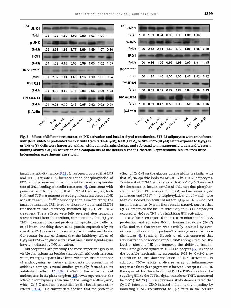
Fig. 5 – Effects of different treatments on JNK activation and insulin signal transduction. 3T3-L1 adipocytes were transfected
with JNK1 siRNA or pretreated for 12 h with Cy-3-G (10–40 mM), NAC (1 mM), or SP600125 (20 mM) before exposed to H2O2 (A)
or TNF-a (B). Cells were harvested with or without insulin stimulation, and subjected to immunoprecipitation and Western
blotting analysis of JNK activation and components of the insulin signaling cascade. Representative results from three-
independent experiments are shown.
b i o c h e m i c a l p h a r m a c o l o g y 7 5 ( 2 0 0 8 ) 1 3 9 3 – 1 4 0 1 1399
insulin sensitivity in mice [9,11]. It has been proposed that ROS
and TNF-a activate JNK, increase serine phosphorylation of
IRS1, and decrease insulin-stimulated tyrosine phosphoryla-
tion of IRS1, leading to insulin resistance [6]. Consistent with
previous reports, we found that in 3T3-L1 adipocytes, both
H2O2 and TNF-a treatment caused significant increases in JNK
activation and IRS1Ser307 phosphorylation. Concomitantly, the
insulin-stimulated IRS1 tyrosine-phosphorylation and GLUT4
translocation was markedly inhibited by H2O2 or TNF-a
treatment. These effects were fully reversed after removing
stress stimuli from the medium, demonstrating that H2O2 or
TNF-a treatment does not produce nonspecific, toxic effects.
In addition, knocking down JNK1 protein expression by its
specific siRNA prevented the occurrence of insulin resistance.
Our results further confirmed that the inhibitory effects of
H2O2 and TNF-a on glucose transport and insulin signaling are
largely mediated by JNK activation.
Anthocyanins are probably the most important group of
visible plant pigments besides chlorophyll. Although in recent
years, emerging reports have been evidenced the importance
of anthocyanins as dietary antioxidants for prevention of
oxidative damage, several studies gradually focused on its
antidiabetic effect [17,26,32]. Cy-3-G is the widest spread
anthocyanin in the plant kingdom [13]. It was reported that the
ortho-dihydroxyphenyl structure on the B-ring of anthocyanin,
which Cy-3-G also has, is essential for the health-promoting
effects [33,34]. Our current data showed that the protective
effect of Cy-3-G on the glucose uptake ability is similar with
that of JNK-specific inhibitor SP600125 in 3T3-L1 adipocytes.
Treatment of 3T3-L1 adipocytes with 40 mM Cy-3-G reversed
the decreases in insulin-simulated IRS1 tyrosine phosphor-
ylation and GLUT4 translocation to PM, and increases in JNK
activation and IRS1Ser307 phosphorylation, all of which have
been considered molecular bases for H2O2- or TNF-a-induced
insulin resistance. Overall, these results strongly suggest that
Cy-3-G improved the insulin sensitivity in 3T3-L1 adipocytes
exposed to H2O2 or TNF-a by inhibiting JNK activation.
TNF-a has been reported to increases mitochondrial ROS
production and activates JNK in human hepatoma (Huh7)
cells, and this observation was partially inhibited by over-
expression of uncoupling protein-1 or manganese superoxide
dismutase [6]. Similarly, Houstis et al. demonstrated that
administration of antioxidant MnTBAP strongly reduced the
level of phospho-JNK and improved the ability for insulin-
stimulated glucose uptake in 3T3-L1 adipocytes [22]. As one of
the possible mechanisms, scavenging ROS by Cy-3-G may
contribute to the downregulation of JNK activation. In
addition, TNF-a elicits a diverse array of inflammatory
responses through engagement of its type-1 receptor (TNFR1).
It is reported that the activation of JNK by TNF-a is initiated by
coupling JNK to the TNFR1 signal transducer TNFR-associated
factor-2 (TRAF2) [35]. Our previous study demonstrated that
Cy-3-G interrupts CD40-induced inflammatory signaling by
inhibiting TRAF2 recruitment to lipid rafts in the cellular

b i o c h e m i c a l p h a r m a c o l o g y 7 5 ( 2 0 0 8 ) 1 3 9 3 – 1 4 0 11400
membrane [36]. Thus, whether Cy-3-G down-regulates JNK
activation through inhibition of TRAF-2 recruitment needs
further elucidated.
In conclusion, we have shown that prolonged low-grade
H2O2 and TNF-a result in elevated JNK activation and impaired
insulin-stimulated glucose transport in 3T3-L1 adipocytes. Cy-
3-G effectively ameliorates H2O2- and TNF-a-induced insulin
resistance by inhibiting the JNK-IRS1 signal pathway. These
findings provide a novel insight into the therapeutic implica-
tions of anthocyanin in insulin resistance-related diseases.
Acknowledgments
This work was supported by the research grants from National
Basic Research Program (973 Program, 2006CB503902) of China,
and China Medical Board of New York Inc. (CMB 98-677).
r e f e r e n c e s
[1] Fletcher B, Lamendola C. Insulin resistance syndrome. JCardiovasc Nurs 2004;19:339–45.
[2] Saltiel AR, Kahn CR. Insulin signalling and the regulation ofglucose and lipid metabolism. Nature 2001;414:799–806.
[3] Johnston AM, Pirola L, Van Obberghen E. Molecularmechanisms of insulin receptor substrate protein-mediated modulation of insulin signalling. FEBS Lett2003;546:32–6.
[4] Gao Z, Zhang X, Zuberi A, Hwang D, Quon MJ, Lefevre M,et al. Inhibition of insulin sensitivity by free fatty acidsrequires activation of multiple serine kinases in 3T3-L1adipocytes. Mol Endocrinol 2004;18:2024–34.
[5] Bloch-Damti A, Potashnik R, Gual P, Le Marchand-Brustel Y,Tanti JF, Rudich A, et al. Differential effects of IRS1phosphorylated on Ser307 or Ser632 in the induction ofinsulin resistance by oxidative stress. Diabetologia2006;49:2463–73.
[6] Imoto K, Kukidome D, Nishikawa T, Matsuhisa T, Sonoda K,Fujisawa K, et al. Impact of mitochondrial reactive oxygenspecies and apoptosis signal-regulating kinase 1 on insulinsignaling. Diabetes 2006;55:1197–204.
[7] Aguirre V, Uchida T, Yenush L, Davis R, White MF. The c-Jun NH(2)-terminal kinase promotes insulin resistanceduring association with insulin receptor substrate-1 andphosphorylation of Ser(307). J Biol Chem 2000;275:9047–54.
[8] Gual P, Le Marchand-Brustel Y, Tanti JF. Positive andnegative regulation of insulin signaling through IRS-1phosphorylation. Biochimie 2005;87:99–109.
[9] Hirosumi J, Tuncman G, Chang L, Gorgun CZ, Uysal KT,Maeda K, et al. A central role for JNK in obesity and insulinresistance. Nature 2002;420:333–6.
[10] Nakatani Y, Kaneto H, Kawamori D, Hatazaki M, MiyatsukaT, Matsuoka TA, et al. Modulation of the JNK pathway inliver affects insulin resistance status. J Biol Chem2004;279:45803–9.
[11] Kaneto H, Nakatani Y, Miyatsuka T, Kawamori D, MatsuokaTA, Matsuhisa M, et al. Possible novel therapy for diabeteswith cell-permeable JNK-inhibitory peptide. Nat Med2004;10:1128–32.
[12] Clifford MN. Anthocyanins—nature, occurrence anddietary burden. J Sci Food Agric 2000;80:1063–72.
[13] Kong JM, Chia LS, Goh NK, Chia TF, Brouillard R. Analysisand biological activities of anthocyanins. Phytochemistry2003;64:923–33.
[14] Renaud S, de Lorgeril M. Wine, alcohol, platelets, and theFrench paradox for coronary heart disease. Lancet1992;339:1523–6.
[15] Alvarez Leon EE, Henriquez P, Serra-Majem L.Mediterranean diet and metabolic syndrome: a cross-sectional study in the Canary Islands. Public Health Nutr2006;9:1089–98.
[16] Tsuda T, Horio F, Uchida K, Aoki H, Osawa T. Dietarycyanidin 3-O-beta-D-glucoside-rich purple corn colorprevents obesity and ameliorates hyperglycemia in mice. JNutr 2003;133:2125–30.
[17] Jayaprakasam B, Olson LK, Schutzki RE, Tai MH, Nair MG.Amelioration of obesity and glucose intolerance in high-fat-fed C57BL/6 mice by anthocyanins and ursolic acid inCornelian cherry (Cornus mas). J Agric Food Chem2006;54:243–8.
[18] Guo H, Ling W, Wang Q, Liu C, Hu Y, Xia M, et al. Effect ofanthocyanin-rich extract from black rice (Oryza sativa L.indica) on hyperlipidemia and insulin resistance infructose-fed rats. Plant Foods Hum Nutr 2007;62:1–6.
[19] Hu C, Zawistowski J, Ling W, Kitts DD. Black rice (Oryzasativa L. indica) pigmented fraction suppresses bothreactive oxygen species and nitric oxide in chemical andbiological model systems. J Agric Food Chem 2003;51:5271–7.
[20] Rudich A, Tirosh A, Potashnik R, Khamaisi M, Bashan N.Lipoic acid protects against oxidative stress inducedimpairment in insulin stimulation of protein kinase B andglucose transport in 3T3-L1 adipocytes. Diabetologia1999;42:949–57.
[21] Roffey B, Atwal A, Kubow S. Cinnamon water extractsincrease glucose uptake but inhibit adiponectin secretionin 3T3-L1 adipose cells. Mol Nutr Food Res 2006;50:739–45.
[22] Houstis N, Rosen ED, Lander ES. Reactive oxygen specieshave a causal role in multiple forms of insulin resistance.Nature 2006;440:944–8.
[23] Karlsson M, Thorn H, Parpal S, Stralfors P, Gustavsson J.Insulin induces translocation of glucose transporter GLUT4to plasma membrane caveolae in adipocytes. FASEB J2002;16:249–51.
[24] Stephens JM, Lee J, Pilch PF. Tumor necrosis factor-alpha-induced insulin resistance in 3T3-L1 adipocytes isaccompanied by a loss of insulin receptor substrate-1 andGLUT4 expression without a loss of insulin receptor-mediated signal transduction. J Biol Chem 1997;272:971–6.
[25] Bennett BL, Satoh Y, Lewis AJ. JNK: a new therapeutic targetfor diabetes. Curr Opin Pharmacol 2003;3:420–5.
[26] Sasaki R, Nishimura N, Hoshino H, Isa Y, Kadowaki M, IchiT, et al. Cyanidin 3-glucoside ameliorates hyperglycemiaand insulin sensitivity due to downregulation of retinolbinding protein 4 expression in diabetic mice. BiochemPharmacol 2007;74:1619–27.
[27] Rudich A, Tirosh A, Potashnik R, Hemi R, Kanety H, BashanN. Prolonged oxidative stress impairs insulin-inducedGLUT4 translocation in 3T3-L1 adipocytes. Diabetes1998;47:1562–9.
[28] Wellen KE, Hotamisligil GS. Inflammation, stress, anddiabetes. J Clin Invest 2005;115:1111–9.
[29] Svegliati-Baroni G, Candelaresi C, Saccomanno S, Ferretti G,Bachetti T, Marzioni M, et al. A model of insulin resistanceand nonalcoholic steatohepatitis in rats: role of peroxisomeproliferator-activated receptor-alpha and n-3polyunsaturated fatty acid treatment on liver injury. Am JPathol 2006;169:846–60.
[30] Furukawa S, Fujita T, Shimabukuro M, Iwaki M, Yamada Y,Nakajima Y, et al. Increased oxidative stress in obesity andits impact on metabolic syndrome. J Clin Invest2004;114:1752–61.

b i o c h e m i c a l p h a r m a c o l o g y 7 5 ( 2 0 0 8 ) 1 3 9 3 – 1 4 0 1 1401
[31] Tuncman G, Hirosumi J, Solinas G, Chang L, Karin M,Hotamisligil GS. Functional in vivo interactions betweenJNK1 and JNK2 isoforms in obesity and insulin resistance.Proc Natl Acad Sci USA 2006;103:10741–6.
[32] Ghosh D, Konishi T. Anthocyanins and anthocyanin-richextracts: role in diabetes and eye function. Asia Pac J ClinNutr 2007;16:200–8.
[33] Hou DX, Kai K, Li JJ, Lin S, Terahara N, Wakamatsu M, et al.Anthocyanidins inhibit activator protein 1 activity and celltransformation: structure-activity relationship andmolecular mechanisms. Carcinogenesis 2004;25:29–36.
[34] Hou DX, Yanagita T, Uto T, Masuzaki S, Fujii M.Anthocyanidins inhibit cyclooxygenase-2 expression inLPS-evoked macrophages: structure-activity relationship
and molecular mechanisms involved. Biochem Pharmacol2005;70:417–25.
[35] Yuasa T, Ohno S, Kehrl JH, Kyriakis JM. Tumor necrosisfactor signaling to stress-activated protein kinase (SAPK)/Jun NH2-terminal kinase (JNK) and p38. Germinal centerkinase couples TRAF2 to mitogen-activated protein kinase/ERK kinase kinase 1 and SAPK while receptor interactingprotein associates with a mitogen-activated protein kinasekinase kinase upstream of MKK6 and p38. J Biol Chem1998;273:22681–92.
[36] Xia M, Ling W, Zhu H, Wang Q, Ma J, Hou M, et al.Anthocyanin prevents CD40-activated proinflammatorysignaling in endothelial cells by regulating cholesteroldistribution. Arterioscler Thromb Vasc Biol 2007;27:519–24.
Related Documents