BioMed Central Page 1 of 14 (page number not for citation purposes) BMC Developmental Biology Open Access Research article Inhibition of HMG CoA reductase reveals an unexpected role for cholesterol during PGC migration in the mouse Jiaxi Ding 1 , DeChen Jiang 2 , Michael Kurczy 3 , Jennifer Nalepka 1 , Brian Dudley 1 , Erin I Merkel 4 , Forbes D Porter 4 , Andrew G Ewing 3,5 , Nicholas Winograd 3 , James Burgess 2 and Kathleen Molyneaux* 1 Address: 1 Department of Genetics Case Western Reserve University, Cleveland, OH, USA, 2 Department of Chemistry, Case Western Reserve University, Cleveland, OH, USA, 3 Department of Chemistry, Penn State University, University Park, PA, USA, 4 Program on Developmental Endocrinology and Genetics, Eunice Kennedy Shriver National Institute of Child Health and Human Development, National Institutes of Health, Bethesda, MD, USA and 5 Department of Chemistry, Gothenburg University, Kemivägen 4, SE-41296 Gothenburg, Sweden Email: Jiaxi Ding - [email protected]; DeChen Jiang - [email protected]; Michael Kurczy - [email protected]; Jennifer Nalepka - [email protected]; Brian Dudley - [email protected]; Erin I Merkel - [email protected]; Forbes D Porter - [email protected]; Andrew G Ewing - [email protected]; Nicholas Winograd - [email protected]; James Burgess - [email protected]; Kathleen Molyneaux* - [email protected] * Corresponding author Abstract Background: Primordial germ cells (PGCs) are the embryonic precursors of the sperm and eggs. Environmental or genetic defects that alter PGC development can impair fertility or cause formation of germ cell tumors. Results: We demonstrate a novel role for cholesterol during germ cell migration in mice. Cholesterol was measured in living tissue dissected from mouse embryos and was found to accumulate within the developing gonads as germ cells migrate to colonize these structures. Cholesterol synthesis was blocked in culture by inhibiting the activity of HMG CoA reductase (HMGCR) resulting in germ cell survival and migration defects. These defects were rescued by co- addition of isoprenoids and cholesterol, but neither compound alone was sufficient. In contrast, loss of the last or penultimate enzyme in cholesterol biosynthesis did not alter PGC numbers or position in vivo. However embryos that lack these enzymes do not exhibit cholesterol defects at the stage at which PGCs are migrating. This demonstrates that during gestation, the cholesterol required for PGC migration can be supplied maternally. Conclusion: In the mouse, cholesterol is required for PGC survival and motility. It may act cell- autonomously by regulating clustering of growth factor receptors within PGCs or non cell- autonomously by controlling release of growth factors required for PGC guidance and survival. Background Primordial germ cells (PGCs) are the embryonic precur- sors of gametes. In most model systems, PGCs are migra- tory and navigate through or around diverse tissues in order to find the site of the developing gonads. PGC migration shares conserved features in many species indi- cating the process arose in a common ancestor. In verte- brates, however, the majority of factors implicated in PGC Published: 31 December 2008 BMC Developmental Biology 2008, 8:120 doi:10.1186/1471-213X-8-120 Received: 17 July 2008 Accepted: 31 December 2008 This article is available from: http://www.biomedcentral.com/1471-213X/8/120 © 2008 Ding et al; licensee BioMed Central Ltd. This is an Open Access article distributed under the terms of the Creative Commons Attribution License (http://creativecommons.org/licenses/by/2.0 ), which permits unrestricted use, distribution, and reproduction in any medium, provided the original work is properly cited.

Welcome message from author
This document is posted to help you gain knowledge. Please leave a comment to let me know what you think about it! Share it to your friends and learn new things together.
Transcript
BioMed CentralBMC Developmental Biology
ss
Open AcceResearch articleInhibition of HMG CoA reductase reveals an unexpected role for cholesterol during PGC migration in the mouseJiaxi Ding1, DeChen Jiang2, Michael Kurczy3, Jennifer Nalepka1, Brian Dudley1, Erin I Merkel4, Forbes D Porter4, Andrew G Ewing3,5, Nicholas Winograd3, James Burgess2 and Kathleen Molyneaux*1Address: 1Department of Genetics Case Western Reserve University, Cleveland, OH, USA, 2Department of Chemistry, Case Western Reserve University, Cleveland, OH, USA, 3Department of Chemistry, Penn State University, University Park, PA, USA, 4Program on Developmental Endocrinology and Genetics, Eunice Kennedy Shriver National Institute of Child Health and Human Development, National Institutes of Health, Bethesda, MD, USA and 5Department of Chemistry, Gothenburg University, Kemivägen 4, SE-41296 Gothenburg, Sweden
Email: Jiaxi Ding - [email protected]; DeChen Jiang - [email protected]; Michael Kurczy - [email protected]; Jennifer Nalepka - [email protected]; Brian Dudley - [email protected]; Erin I Merkel - [email protected]; Forbes D Porter - [email protected]; Andrew G Ewing - [email protected]; Nicholas Winograd - [email protected]; James Burgess - [email protected]; Kathleen Molyneaux* - [email protected]
* Corresponding author
AbstractBackground: Primordial germ cells (PGCs) are the embryonic precursors of the sperm and eggs.Environmental or genetic defects that alter PGC development can impair fertility or causeformation of germ cell tumors.
Results: We demonstrate a novel role for cholesterol during germ cell migration in mice.Cholesterol was measured in living tissue dissected from mouse embryos and was found toaccumulate within the developing gonads as germ cells migrate to colonize these structures.Cholesterol synthesis was blocked in culture by inhibiting the activity of HMG CoA reductase(HMGCR) resulting in germ cell survival and migration defects. These defects were rescued by co-addition of isoprenoids and cholesterol, but neither compound alone was sufficient. In contrast,loss of the last or penultimate enzyme in cholesterol biosynthesis did not alter PGC numbers orposition in vivo. However embryos that lack these enzymes do not exhibit cholesterol defects atthe stage at which PGCs are migrating. This demonstrates that during gestation, the cholesterolrequired for PGC migration can be supplied maternally.
Conclusion: In the mouse, cholesterol is required for PGC survival and motility. It may act cell-autonomously by regulating clustering of growth factor receptors within PGCs or non cell-autonomously by controlling release of growth factors required for PGC guidance and survival.
BackgroundPrimordial germ cells (PGCs) are the embryonic precur-sors of gametes. In most model systems, PGCs are migra-tory and navigate through or around diverse tissues in
order to find the site of the developing gonads. PGCmigration shares conserved features in many species indi-cating the process arose in a common ancestor. In verte-brates, however, the majority of factors implicated in PGC
Published: 31 December 2008
BMC Developmental Biology 2008, 8:120 doi:10.1186/1471-213X-8-120
Received: 17 July 2008Accepted: 31 December 2008
This article is available from: http://www.biomedcentral.com/1471-213X/8/120
© 2008 Ding et al; licensee BioMed Central Ltd. This is an Open Access article distributed under the terms of the Creative Commons Attribution License (http://creativecommons.org/licenses/by/2.0), which permits unrestricted use, distribution, and reproduction in any medium, provided the original work is properly cited.
Page 1 of 14(page number not for citation purposes)
BMC Developmental Biology 2008, 8:120 http://www.biomedcentral.com/1471-213X/8/120
guidance are either secreted or membrane bound proteingrowth factors (e.g. stromal derived factor 1 and Kit lig-and); whereas, evidence in Drosophila points to a lipid-based guidance system [1]. A recent study bridged the gapby demonstrating that zebrafish PGCs, like DrosophilaPGCs, require 3-hydroxy-3-methylglutaryl-coenzyme Areductase (HMGCR) for normal migration [2].
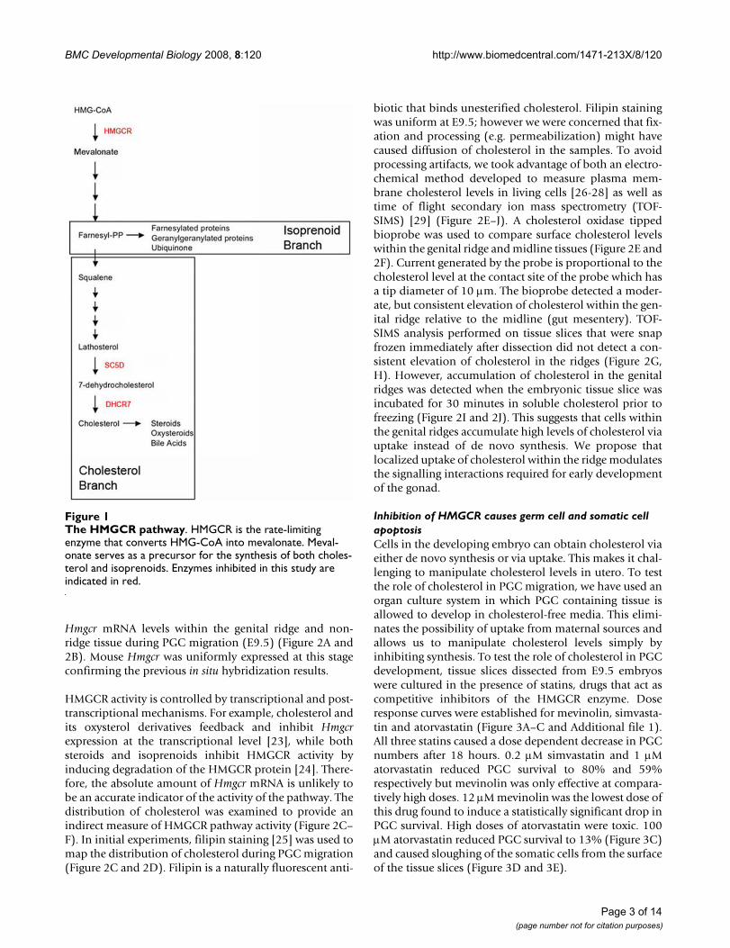
HMGCR is the rate limiting enzyme in isoprenoid andcholesterol biosynthesis. In flies, HMGCR was shown toact within the somatic gonadal precursors to controlrelease of a secreted PGC attractant [3]. Drosophila lackenzymes required downstream of HMGCR for cholesterolsynthesis indicating that isoprenoids are the relevantdownstream effectors [4]. In support of this, Santos andLehmann demonstrated that mutations in the geranylger-anyl transferase 1 β subunit cause PGC migration defects.It has been proposed that geranylgeranylation of smallGTPase in the Ras, Rac, or Rho families regulate secretionof hedgehog [5] or other putative Drosophila PGC attract-ants [4]. In zebrafish, evidence also points to a role forHMGCR and isoprenoids in PGC migration [2]. Inhibi-tion of HMGCR or geranylgeranyl transferase I cause PGCmigration defects. However, in this system it remainsunclear whether HMGCR is required in PGCs themselvesor within the soma. Additionally, zebrafish unlike flies arecapable of de novo cholesterol synthesis and this branchof the pathway was not carefully evaluated.
Cholesterol plays a vital role during vertebrate develop-ment. Mutations in genes required for cholesterol biosyn-thesis cause severe developmental defects. Loss of Hmgcr[6] or squalene synthase [7] result in early embryoniclethality in mouse models. Mutations in 3b-hydroxysterol-Δ7 reductase (Dhcr7) or lathosterol 5-desaturase (Sc5d) causeskeletal, neural, and in some cases urogenital defects inhumans [8] and in mice [9,10]. Additionally, mutationsin genes required for cholesterol transport are also associ-ated with embryonic lethality or patterning defects [11].
Several models have been evoked in order to explain therole of cholesterol during organogenesis. First, cholesterolis the precursor of steroid hormones, glucocorticoids, andoxysterols, all compounds known to mediate cell-cell sig-naling via activation of nuclear hormone receptors. Sec-ond, cholesterol directly regulates cell-cell signalling bycontrolling the diffusion [12] or reception [13] of mem-bers of the hedgehog growth factor family. Finally, choles-terol is a key structural component of the plasmamembrane. Cholesterol controls membrane fluidity andmodulates membrane protein interactions. Membranecholesterol has been shown to influence the activity ofgrowth factor receptors and cell-adhesion molecules byclustering these cell surface proteins into lipid rafts [14].Of particular note, lipid rafts have been shown to affectepidermal growth factor-induced chemotaxis [15] and
migration on fibronectin [16] in cell culture. This suggeststhat cholesterol levels might also alter cell migration invivo.
Considering the roles of cholesterol in cell-cell signallingand cell migration, we thought it imperative to testwhether this branch of the HMGCR pathway is requiredfor germ cell development in a vertebrate model. In mice,PGCs migrate from the gut to the genital ridges betweenembryonic day 9.5 (E9.5) and E10.5. Steroid hormonesand hedgehog growth factors are unlikely to play a role inthis process. Enzymes required to convert cholesterol intosteroid hormones are not expressed in the gonads untilE11.5 [17]. Likewise, the three vertebrate members of thehedgehog growth factor family are not expressed in theright place or time to play a role in PGC guidance in thissystem [18,19]. However, changes in cholesterol couldvery well impact the ability of PGCs to respond to pro-posed chemoattractants such as SDF1 [20] or KITL [21].
To examine the role of cholesterol in PGC migration, wefirst measured cholesterol in living tissue dissected fromE9.5 embryos and were surprised to find that cholesterolwas enriched in the genital ridges relative to the surround-ing tissue. This asymmetric distribution appears to bemaintained via selective uptake of cholesterol by cellswithin the genital ridges. We further demonstrate thatinhibition of HMGCR reduced total cholesterol andimpaired germ cell survival and migration in culture.These defects were rescued by co-addition of geranylgera-niol and cholesterol indicating that both compounds arerequired. To test if cholesterol biosynthesis is necessary forPGC migration in vivo, we examine the number and dis-tribution of PGCs in embryos lacking Dhcr7 or Sc5d (seeFigure 1). PGCs were normally distributed in both lines,but cholesterol levels are only modestly affected in theseembryos [9,10]. We conclude that cholesterol is requiredfor PGC migration but that this requirement can be metby uptake of cholesterol from maternal sources. We pro-pose that the asymmetric accumulation of cholesterolwithin the genital ridges controls signaling interactionsrequired for PGCs to colonize the gonads.
ResultsCholesterol is elevated in the genital ridgesDrosophila Hmgcr mRNA is elevated in somatic gonadalprecursors [3] supporting the model that HMGCR actsnon-cell autonomously to guide PGC migration. How-ever, zebrafish Hmgcr2 is uniformly expressed in cleavagestage and gastrulating embryos, raising the possibility thatHMGCR has both non-autonomous and autonomouseffects [2]. Like in zebrafish, mouse Hmgcr is ubiquitouslyexpressed in E10.5 embryos based on in situ hybridization[22]. However, in situs are not sensitive enough to revealmodest changes in gene expression. As a more sensitivetechnique, quantitative RT-PCR was used to compare
Page 2 of 14(page number not for citation purposes)
BMC Developmental Biology 2008, 8:120 http://www.biomedcentral.com/1471-213X/8/120
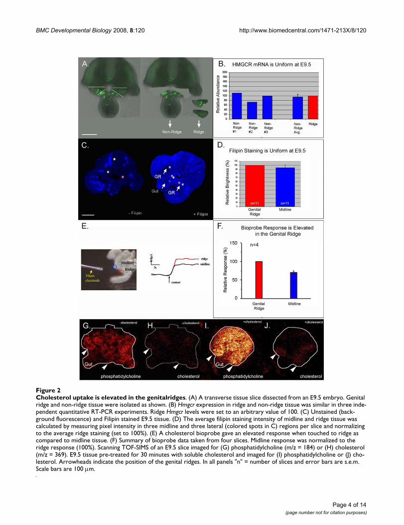
Hmgcr mRNA levels within the genital ridge and non-ridge tissue during PGC migration (E9.5) (Figure 2A and2B). Mouse Hmgcr was uniformly expressed at this stageconfirming the previous in situ hybridization results.
HMGCR activity is controlled by transcriptional and post-transcriptional mechanisms. For example, cholesterol andits oxysterol derivatives feedback and inhibit Hmgcrexpression at the transcriptional level [23], while bothsteroids and isoprenoids inhibit HMGCR activity byinducing degradation of the HMGCR protein [24]. There-fore, the absolute amount of Hmgcr mRNA is unlikely tobe an accurate indicator of the activity of the pathway. Thedistribution of cholesterol was examined to provide anindirect measure of HMGCR pathway activity (Figure 2C–F). In initial experiments, filipin staining [25] was used tomap the distribution of cholesterol during PGC migration(Figure 2C and 2D). Filipin is a naturally fluorescent anti-
biotic that binds unesterified cholesterol. Filipin stainingwas uniform at E9.5; however we were concerned that fix-ation and processing (e.g. permeabilization) might havecaused diffusion of cholesterol in the samples. To avoidprocessing artifacts, we took advantage of both an electro-chemical method developed to measure plasma mem-brane cholesterol levels in living cells [26-28] as well astime of flight secondary ion mass spectrometry (TOF-SIMS) [29] (Figure 2E–J). A cholesterol oxidase tippedbioprobe was used to compare surface cholesterol levelswithin the genital ridge and midline tissues (Figure 2E and2F). Current generated by the probe is proportional to thecholesterol level at the contact site of the probe which hasa tip diameter of 10 μm. The bioprobe detected a moder-ate, but consistent elevation of cholesterol within the gen-ital ridge relative to the midline (gut mesentery). TOF-SIMS analysis performed on tissue slices that were snapfrozen immediately after dissection did not detect a con-sistent elevation of cholesterol in the ridges (Figure 2G,H). However, accumulation of cholesterol in the genitalridges was detected when the embryonic tissue slice wasincubated for 30 minutes in soluble cholesterol prior tofreezing (Figure 2I and 2J). This suggests that cells withinthe genital ridges accumulate high levels of cholesterol viauptake instead of de novo synthesis. We propose thatlocalized uptake of cholesterol within the ridge modulatesthe signalling interactions required for early developmentof the gonad.
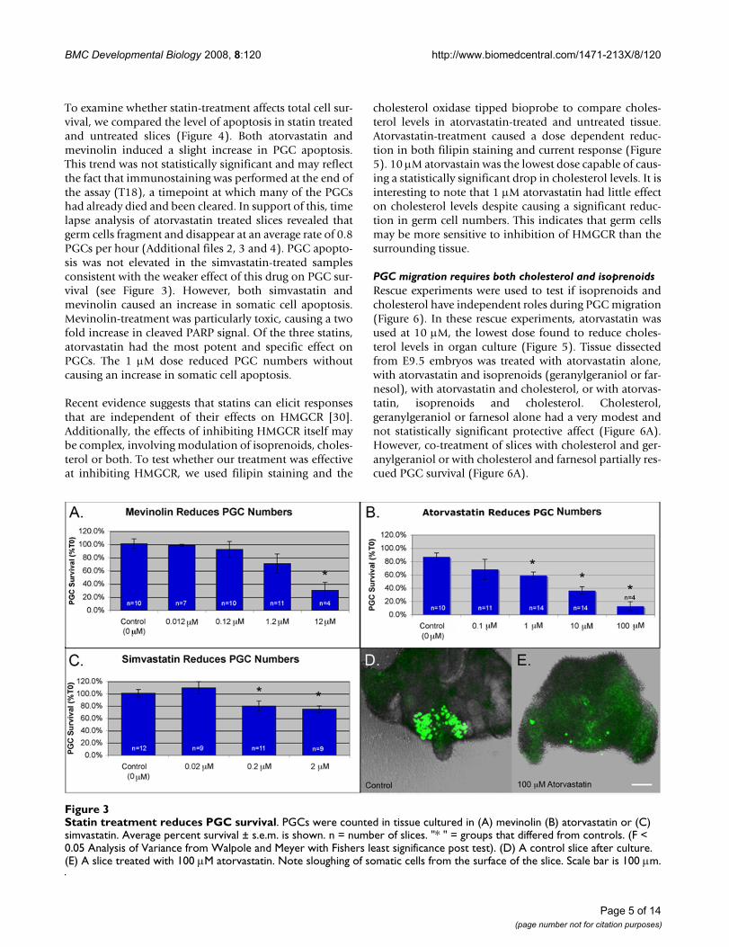
Inhibition of HMGCR causes germ cell and somatic cell apoptosisCells in the developing embryo can obtain cholesterol viaeither de novo synthesis or via uptake. This makes it chal-lenging to manipulate cholesterol levels in utero. To testthe role of cholesterol in PGC migration, we have used anorgan culture system in which PGC containing tissue isallowed to develop in cholesterol-free media. This elimi-nates the possibility of uptake from maternal sources andallows us to manipulate cholesterol levels simply byinhibiting synthesis. To test the role of cholesterol in PGCdevelopment, tissue slices dissected from E9.5 embryoswere cultured in the presence of statins, drugs that act ascompetitive inhibitors of the HMGCR enzyme. Doseresponse curves were established for mevinolin, simvasta-tin and atorvastatin (Figure 3A–C and Additional file 1).All three statins caused a dose dependent decrease in PGCnumbers after 18 hours. 0.2 μM simvastatin and 1 μMatorvastatin reduced PGC survival to 80% and 59%respectively but mevinolin was only effective at compara-tively high doses. 12 μM mevinolin was the lowest dose ofthis drug found to induce a statistically significant drop inPGC survival. High doses of atorvastatin were toxic. 100μM atorvastatin reduced PGC survival to 13% (Figure 3C)and caused sloughing of the somatic cells from the surfaceof the tissue slices (Figure 3D and 3E).
The HMGCR pathwayFigure 1The HMGCR pathway. HMGCR is the rate-limiting enzyme that converts HMG-CoA into mevalonate. Meval-onate serves as a precursor for the synthesis of both choles-terol and isoprenoids. Enzymes inhibited in this study are indicated in red.
Page 3 of 14(page number not for citation purposes)
BMC Developmental Biology 2008, 8:120 http://www.biomedcentral.com/1471-213X/8/120
Page 4 of 14(page number not for citation purposes)
Cholesterol uptake is elevated in the genitalridgesFigure 2Cholesterol uptake is elevated in the genitalridges. (A) A transverse tissue slice dissected from an E9.5 embryo. Genital ridge and non-ridge tissue were isolated as shown. (B) Hmgcr expression in ridge and non-ridge tissue was similar in three inde-pendent quantitative RT-PCR experiments. Ridge Hmgcr levels were set to an arbitrary value of 100. (C) Unstained (back-ground fluorescence) and Filipin stained E9.5 tissue. (D) The average filipin staining intensity of midline and ridge tissue was calculated by measuring pixel intensity in three midline and three lateral (colored spots in C) regions per slice and normalizing to the average ridge staining (set to 100%). (E) A cholesterol bioprobe gave an elevated response when touched to ridge as compared to midline tissue. (F) Summary of bioprobe data taken from four slices. Midline response was normalized to the ridge response (100%). Scanning TOF-SIMS of an E9.5 slice imaged for (G) phosphatidylcholine (m/z = 184) or (H) cholesterol (m/z = 369). E9.5 tissue pre-treated for 30 minutes with soluble cholesterol and imaged for (I) phosphatidylcholine or (J) cho-lesterol. Arrowheads indicate the position of the genital ridges. In all panels "n" = number of slices and error bars are s.e.m. Scale bars are 100 μm.
BMC Developmental Biology 2008, 8:120 http://www.biomedcentral.com/1471-213X/8/120
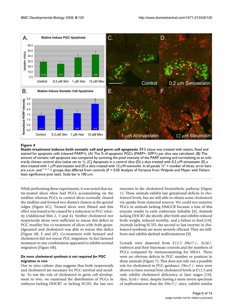
To examine whether statin-treatment affects total cell sur-vival, we compared the level of apoptosis in statin treatedand untreated slices (Figure 4). Both atorvastatin andmevinolin induced a slight increase in PGC apoptosis.This trend was not statistically significant and may reflectthe fact that immunostaining was performed at the end ofthe assay (T18), a timepoint at which many of the PGCshad already died and been cleared. In support of this, timelapse analysis of atorvastatin treated slices revealed thatgerm cells fragment and disappear at an average rate of 0.8PGCs per hour (Additional files 2, 3 and 4). PGC apopto-sis was not elevated in the simvastatin-treated samplesconsistent with the weaker effect of this drug on PGC sur-vival (see Figure 3). However, both simvastatin andmevinolin caused an increase in somatic cell apoptosis.Mevinolin-treatment was particularly toxic, causing a twofold increase in cleaved PARP signal. Of the three statins,atorvastatin had the most potent and specific effect onPGCs. The 1 μM dose reduced PGC numbers withoutcausing an increase in somatic cell apoptosis.
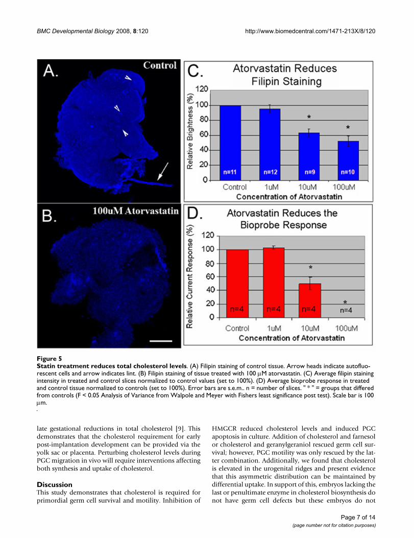
Recent evidence suggests that statins can elicit responsesthat are independent of their effects on HMGCR [30].Additionally, the effects of inhibiting HMGCR itself maybe complex, involving modulation of isoprenoids, choles-terol or both. To test whether our treatment was effectiveat inhibiting HMGCR, we used filipin staining and the
cholesterol oxidase tipped bioprobe to compare choles-terol levels in atorvastatin-treated and untreated tissue.Atorvastatin-treatment caused a dose dependent reduc-tion in both filipin staining and current response (Figure5). 10 μM atorvastain was the lowest dose capable of caus-ing a statistically significant drop in cholesterol levels. It isinteresting to note that 1 μM atorvastatin had little effecton cholesterol levels despite causing a significant reduc-tion in germ cell numbers. This indicates that germ cellsmay be more sensitive to inhibition of HMGCR than thesurrounding tissue.
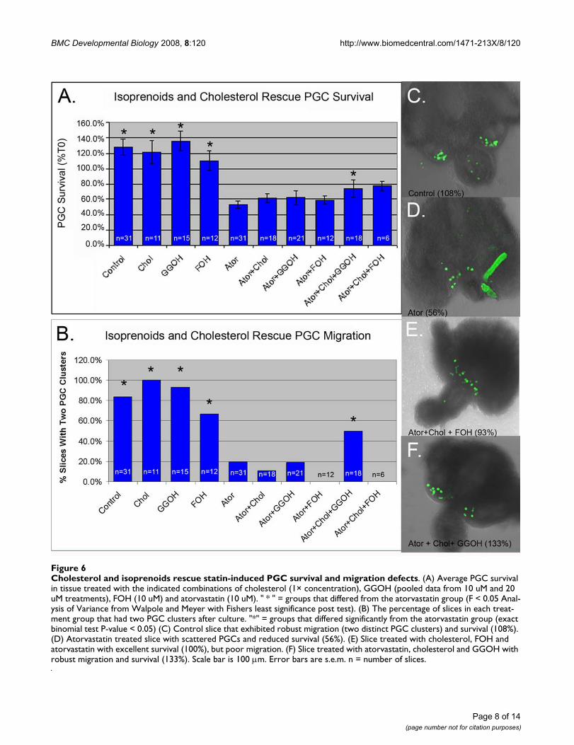
PGC migration requires both cholesterol and isoprenoidsRescue experiments were used to test if isoprenoids andcholesterol have independent roles during PGC migration(Figure 6). In these rescue experiments, atorvastatin wasused at 10 μM, the lowest dose found to reduce choles-terol levels in organ culture (Figure 5). Tissue dissectedfrom E9.5 embryos was treated with atorvastatin alone,with atorvastatin and isoprenoids (geranylgeraniol or far-nesol), with atorvastatin and cholesterol, or with atorvas-tatin, isoprenoids and cholesterol. Cholesterol,geranylgeraniol or farnesol alone had a very modest andnot statistically significant protective affect (Figure 6A).However, co-treatment of slices with cholesterol and ger-anylgeraniol or with cholesterol and farnesol partially res-cued PGC survival (Figure 6A).
Statin treatment reduces PGC survivalFigure 3Statin treatment reduces PGC survival. PGCs were counted in tissue cultured in (A) mevinolin (B) atorvastatin or (C) simvastatin. Average percent survival ± s.e.m. is shown. n = number of slices. "* " = groups that differed from controls. (F < 0.05 Analysis of Variance from Walpole and Meyer with Fishers least significance post test). (D) A control slice after culture. (E) A slice treated with 100 μM atorvastatin. Note sloughing of somatic cells from the surface of the slice. Scale bar is 100 μm.
Page 5 of 14(page number not for citation purposes)
BMC Developmental Biology 2008, 8:120 http://www.biomedcentral.com/1471-213X/8/120
While performing these experiments, it was noted that sta-tin-treated slices often had PGCs accumulating on themidline whereas PGCs in control slices normally clearedthe midline and formed two distinct clusters at the genitalridges (Figure 6C). Treated slices were filmed and thiseffect was found to be caused by a reduction in PGC veloc-ity (Additional files 2, 3 and 4). Neither cholesterol norisoprenoids alone were sufficient to rescue this defect inPGC motility but co-treatment of slices with both geran-ylgeraniol and cholesterol was able to rescue this defect(Figure 6B, E and 6F). Co-treatment with farnesol andcholesterol did not rescue PGC migration. In fact farnesoltreatment in any combination appeared to inhibit normalmigration (Figure 6B).
De novo cholesterol synthesis is not required for PGC migration in vivoOur in vitro culture data suggests that both isoprenoidsand cholesterol are necessary for PGC survival and motil-ity. To test the role of cholesterol in germ cell develop-ment in vivo, we examined the distribution of PGCs inembryos lacking DHCR7 or lacking SC5D, the last two
enzymes in the cholesterol biosynthetic pathway (Figure1). These animals exhibit late gestational deficits in cho-lesterol levels, but are still able to obtain some cholesterolvia uptake from maternal sources. We could not examinePGCs in animals lacking HMGCR because a loss of thisenzyme results in early embryonic lethality [6]. Animalslacking DHCR7 die shortly after birth and exhibit reducedbody weight, reduced motility, and a failure to feed [10].Animals lacking SC5D, the second to last enzyme in cho-lesterol synthesis are more severely effected. They are still-born and exhibit skeletal malformations [9].
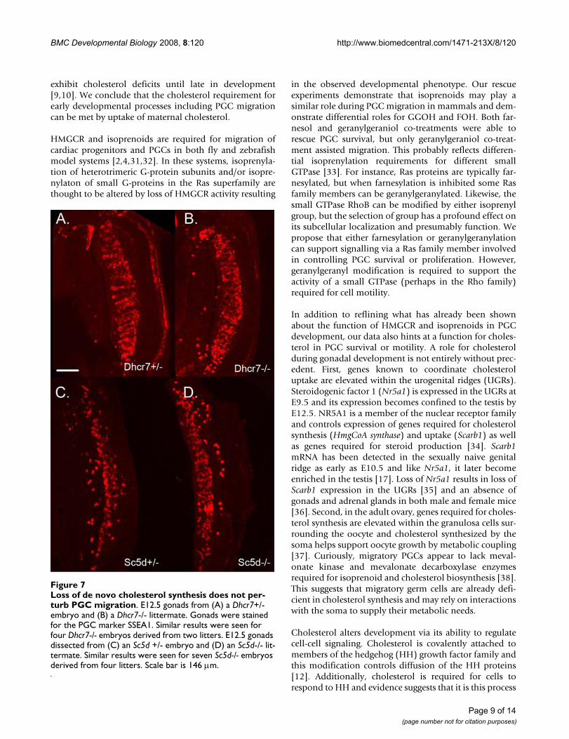
Gonads were dissected from E12.5 Dhcr7-/-, Sc5d-/-embryos and their littermate controls and the numbers ofPGCs compared by immunostaining for SSEA1. Therewere no obvious defects in PGC number or position inthese animals (Figure 7). This does not rule out a possiblerole for cholesterol in PGC guidance. Dhcr7-/- mice wereshown to have normal liver cholesterol levels at E12.5 andonly exhibit cholesterol deficiency at later stages [10].Also, Sc5d-/- mice, despite having a more severe spectrumof malformations than the Dhcr7-/- mice, exhibit similar
Statin treatment induces both somatic cell and germ cell apoptosisFigure 4Statin treatment induces both somatic cell and germ cell apoptosis. E9.5 tissue was treated with statins, fixed and stained for apoptotic cells (cleaved PARP+). (A) The % of apoptotic PGCs (PARP+. GFP+) per slice was calculated. (B) The amount of somatic cell apoptosis was compared by summing the pixel intensity of the PARP staining and normalizing to an arbi-trarily chosen control slice (value set to 1). (C) Apoptosis in a control slice (D) a slice treated with 0.2 μM simvastatin (E) a slice treated with 1 μM atorvastatin and (F) a slice treated with 12 μM mevinolin. In all panels "n" = number of slices, error bars are s.e.m. and " * " = groups that differed from controls (F < 0.05 Analysis of Variance from Walpole and Meyer with Fishers least significance post test). Scale bar is 100 μm.
Page 6 of 14(page number not for citation purposes)
BMC Developmental Biology 2008, 8:120 http://www.biomedcentral.com/1471-213X/8/120
late gestational reductions in total cholesterol [9]. Thisdemonstrates that the cholesterol requirement for earlypost-implantation development can be provided via theyolk sac or placenta. Perturbing cholesterol levels duringPGC migration in vivo will require interventions affectingboth synthesis and uptake of cholesterol.
DiscussionThis study demonstrates that cholesterol is required forprimordial germ cell survival and motility. Inhibition of
HMGCR reduced cholesterol levels and induced PGCapoptosis in culture. Addition of cholesterol and farnesolor cholesterol and geranylgeraniol rescued germ cell sur-vival; however, PGC motility was only rescued by the lat-ter combination. Additionally, we found that cholesterolis elevated in the urogenital ridges and present evidencethat this asymmetric distribution can be maintained bydifferential uptake. In support of this, embryos lacking thelast or penultimate enzyme in cholesterol biosynthesis donot have germ cell defects but these embryos do not
Statin treatment reduces total cholesterol levelsFigure 5Statin treatment reduces total cholesterol levels. (A) Filipin staining of control tissue. Arrow heads indicate autofluo-rescent cells and arrow indicates lint. (B) Filipin staining of tissue treated with 100 μM atorvastatin. (C) Average filipin staining intensity in treated and control slices normalized to control values (set to 100%). (D) Average bioprobe response in treated and control tissue normalized to controls (set to 100%). Error bars are s.e.m.. n = number of slices. " * " = groups that differed from controls (F < 0.05 Analysis of Variance from Walpole and Meyer with Fishers least significance post test). Scale bar is 100 μm.
Page 7 of 14(page number not for citation purposes)
BMC Developmental Biology 2008, 8:120 http://www.biomedcentral.com/1471-213X/8/120
Page 8 of 14(page number not for citation purposes)
Cholesterol and isoprenoids rescue statin-induced PGC survival and migration defectsFigure 6Cholesterol and isoprenoids rescue statin-induced PGC survival and migration defects. (A) Average PGC survival in tissue treated with the indicated combinations of cholesterol (1× concentration), GGOH (pooled data from 10 uM and 20 uM treatments), FOH (10 uM) and atorvastatin (10 uM). " * " = groups that differed from the atorvastatin group (F < 0.05 Anal-ysis of Variance from Walpole and Meyer with Fishers least significance post test). (B) The percentage of slices in each treat-ment group that had two PGC clusters after culture. "*" = groups that differed significantly from the atorvastatin group (exact binomial test P-value < 0.05) (C) Control slice that exhibited robust migration (two distinct PGC clusters) and survival (108%). (D) Atorvastatin treated slice with scattered PGCs and reduced survival (56%). (E) Slice treated with cholesterol, FOH and atorvastatin with excellent survival (100%), but poor migration. (F) Slice treated with atorvastatin, cholesterol and GGOH with robust migration and survival (133%). Scale bar is 100 μm. Error bars are s.e.m. n = number of slices.
BMC Developmental Biology 2008, 8:120 http://www.biomedcentral.com/1471-213X/8/120
exhibit cholesterol deficits until late in development[9,10]. We conclude that the cholesterol requirement forearly developmental processes including PGC migrationcan be met by uptake of maternal cholesterol.
HMGCR and isoprenoids are required for migration ofcardiac progenitors and PGCs in both fly and zebrafishmodel systems [2,4,31,32]. In these systems, isoprenyla-tion of heterotrimeric G-protein subunits and/or isopre-nylaton of small G-proteins in the Ras superfamily arethought to be altered by loss of HMGCR activity resulting
in the observed developmental phenotype. Our rescueexperiments demonstrate that isoprenoids may play asimilar role during PGC migration in mammals and dem-onstrate differential roles for GGOH and FOH. Both far-nesol and geranylgeraniol co-treatments were able torescue PGC survival, but only geranylgeraniol co-treat-ment assisted migration. This probably reflects differen-tial isoprenylation requirements for different smallGTPase [33]. For instance, Ras proteins are typically far-nesylated, but when farnesylation is inhibited some Rasfamily members can be geranylgeranylated. Likewise, thesmall GTPase RhoB can be modified by either isoprenylgroup, but the selection of group has a profound effect onits subcellular localization and presumably function. Wepropose that either farnesylation or geranylgeranylationcan support signalling via a Ras family member involvedin controlling PGC survival or proliferation. However,geranylgeranyl modification is required to support theactivity of a small GTPase (perhaps in the Rho family)required for cell motility.
In addition to reflining what has already been shownabout the function of HMGCR and isoprenoids in PGCdevelopment, our data also hints at a function for choles-terol in PGC survival or motility. A role for cholesterolduring gonadal development is not entirely without prec-edent. First, genes known to coordinate cholesteroluptake are elevated within the urogenital ridges (UGRs).Steroidogenic factor 1 (Nr5a1) is expressed in the UGRs atE9.5 and its expression becomes confined to the testis byE12.5. NR5A1 is a member of the nuclear receptor familyand controls expression of genes required for cholesterolsynthesis (HmgCoA synthase) and uptake (Scarb1) as wellas genes required for steroid production [34]. Scarb1mRNA has been detected in the sexually naive genitalridge as early as E10.5 and like Nr5a1, it later becomeenriched in the testis [17]. Loss of Nr5a1 results in loss ofScarb1 expression in the UGRs [35] and an absence ofgonads and adrenal glands in both male and female mice[36]. Second, in the adult ovary, genes required for choles-terol synthesis are elevated within the granulosa cells sur-rounding the oocyte and cholesterol synthesized by thesoma helps support oocyte growth by metabolic coupling[37]. Curiously, migratory PGCs appear to lack meval-onate kinase and mevalonate decarboxylase enzymesrequired for isoprenoid and cholesterol biosynthesis [38].This suggests that migratory germ cells are already defi-cient in cholesterol synthesis and may rely on interactionswith the soma to supply their metabolic needs.
Cholesterol alters development via its ability to regulatecell-cell signaling. Cholesterol is covalently attached tomembers of the hedgehog (HH) growth factor family andthis modification controls diffusion of the HH proteins[12]. Additionally, cholesterol is required for cells torespond to HH and evidence suggests that it is this process
Loss of de novo cholesterol synthesis does not perturb PGC migrationFigure 7Loss of de novo cholesterol synthesis does not per-turb PGC migration. E12.5 gonads from (A) a Dhcr7+/- embryo and (B) a Dhcr7-/- littermate. Gonads were stained for the PGC marker SSEA1. Similar results were seen for four Dhcr7-/- embryos derived from two litters. E12.5 gonads dissected from (C) an Sc5d +/- embryo and (D) an Sc5d-/- lit-termate. Similar results were seen for seven Sc5d-/- embryos derived from four litters. Scale bar is 146 μm.
Page 9 of 14(page number not for citation purposes)
BMC Developmental Biology 2008, 8:120 http://www.biomedcentral.com/1471-213X/8/120
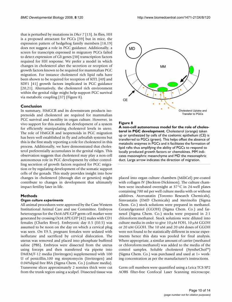
that is perturbed by mutations in Dhcr 7 [13]. In flies, HHis a proposed attractant for PGCs [39] but in mice, theexpression pattern of hedgehog family members [18,19]does not suggest a role in PGC guidance. Additionally, ascreen for transcripts expressed in migratory PGCs failedto detect expression of Gli genes [38] transcription factorsrequired for HH response. We prefer a model in whichchanges in cholesterol alter the secretion or reception ofgrowth factors known to be required for mammalian PGCmigration. For instance cholesterol rich lipid rafts havebeen shown to be required for reception of KITL [40] andSDF1 [41] growth factors implicated in PGC guidance[20,21]. Alternatively, the cholesterol rich environmentwithin the genital ridge might help support PGC survivalvia metabolic coupling [37] (Figure 8).
ConclusionIn summary, HMGCR and its downstream products iso-prenoids and cholesterol are required for mammalianPGC survival and motility in organ culture. However, invivo support for this awaits the development of a systemfor efficiently manipulating cholesterol levels in utero.The role of HMGCR and isoprenoids in PGC migrationhas been well established in fly and zebrafish systems butthis is the first study reporting a role for cholesterol in thisprocess. Additionally, we have demonstrated that choles-terol preferentially accumulates in the genital ridges. Thisobservation suggests that cholesterol may play a non-cellautonomous role in PGC development by either control-ling secretion of growth factors required for PGC migra-tion or by regulating development of the somatic supportcells of the gonads. This study provides insight into howchanges in cholesterol (through diet or genetics) mightcontribute to changes in development that ultimatelyimpact fertility later in life.
MethodsOrgan culture experimentsAll animal procedures were approved by the Case WesternInstitutional Animal Care and use Committee. Embryosheterozygous for the Oct4:ΔPE:GFP germ cell marker weregenerated by crossing Oct4:ΔPE:GFP [42] males with CD1females (Charles River). Embryonic day 0.5 (E0.5) wasassumed to be noon on the day on which a cervical plugwas seen. On E9.5, pregnant females were sedated withisoflurane and sacrificed by cervical dislocation. Theuterus was removed and placed into phosphate bufferedsaline (PBS). Embryos were dissected from the uterususing forceps and then transferred via pipette intoDMEM/F-12 media (Invitrogen) supplemented with 100U of penicillin,100 mg streptomycin (Invitrogen) and0.04%lipid free BSA (Sigma Chem. Co.) (culture media).Transverse slices approximately 2 somites thick were cutfrom the trunk region using a scalpel. Dissected tissue was
placed into organ culture chambers (MiliCel) pre-coatedwith collagen IV (Beckton-Dickinson). The culture cham-bers were incubated overnight at 37°C in 24-well platescontaining 700 ml per well culture media with or withoutadditives. Atorvastatin (Toronto Research Chemicals),Simvastatin (EMD Chemicals) and Mevinolin (SigmaChem. Co.) stock solutions were prepared in methanol.Geranylgeraniol (GGOH) (Sigma Chem. Co.) and far-nesol (Sigma Chem. Co.) stocks were prepared in 2:1chloroform:methanol. Stock solutions were diluted intoculture media in order to give 10 μM FOH, 10 μM GGOHor 20 uM GGOH. The 10 uM and 20 uM doses of GGOHwere not found to be statistically different in rescue exper-iments hence this data was pooled for final analysis.Where appropriate, a similar amount of carrier (methanolor chloroform:methanol) was added to the media of thecontrol samples. Soluble cholesterol (SyntheChol™)(Sigma Chem. Co.) was purchased and used at 1× work-ing concentration as per the manufacturer's instructions.
Germ cell numbers were quantified using a Leica TCS SP2AOBS filter-free Confocal Laser Scanning microscope.
A non-cell autonomous model for the role of cholesterol in PGC developmentFigure 8A non-cell autonomous model for the role of choles-terol in PGC development. Cholesterol (orange) taken up or synthesized by cells of the coelomic epithelium (CE) is transferred to PGCs (green). This helps offset the absence of metabolic enzymes in PGCs and it facilitates the formation of lipid rafts thus amplifying the ability of PGCs to respond to locally produced growth factors or chemokines. MM indi-cates mesonephric mesenchyme and MD the mesonephric duct. Large arrow indicates the direction of migration.
Page 10 of 14(page number not for citation purposes)
BMC Developmental Biology 2008, 8:120 http://www.biomedcentral.com/1471-213X/8/120
Each slice was optically sectioned (at 5 μm intervals) after0 hours (T0) and eighteen hours (T18) in culture. Germcell numbers were counted in the T0 and T18 picturesusing Velocity Image analysis software (Improvision). The% change in germ cell number for each slice equals T18/T10*100.
Time lapse analysisTissue was cultured at 37°C on the stage of the Leica TCSSP2 AOBS Confocal system. A single optical section wascaptured every 9 minutes for 15 hrs. (100 frames). Theautomated tracking feature of Velocity Image analysissoftware was used to track PGC movements. Trackingparameters were as follows. First, PGCs were identified bypercent intensity within the GFP channel (Lower:40Upper:100). Next, holes were filled in the identifiedobjects. Touching objects were separated based on size(using a 100 μm2 size guide). Then any small particles (<40 μm2) were excluded and large PGC clumps that hadfailed to be separated were also excluded (> 300 μm2).Tracking was performed on the identified objects usingthe shortest path algorithm with a maximum of a 10 μmdistance between nodes. Velocity is typically only able tofollow a given cell for part of a movie before it looses trackof the object due to changes in brightness, clumping,moving out of focus, death etc. To accommodate this, theresulting traces were sorted based on the length of timethat Velocity was able to follow the individual particles.Velocity measurements from the 20 temporally longesttraces were averaged to give an average cell velocity foreach film. Average trace times were 3.2 ± 0.17 s.e.m. hoursfor control slices (n = 80 traces in four films) and 2.9 ±0.11 s.e.m. for atorvastatin treated slices (n = 170 traces inseven films).
Cholesterol measurementsMouse tissue was fixed in 4% paraformaldehyde in PBSrocking overnight at 4°C. The tissue was then washed 10minutes at room temperature in 1.5 mg/mL glycine (inphosphate buffered saline with 0.1% triton × 100 (PBST))to block any active aldehyde groups which might other-wise contribute to autoflourescence. The tissue was rinsedthree times in PBST and rocked overnight at 4°C withPBST to permeabilize the tissue. Slices were incubatedwith 0.05 mg/mL filipin (Sigma Chem. Co.) at room tem-perature for 30 minutes rocking and were then washedthree times with PBST (15 minute per wash), incubated in1:1 glycerol:PBS (10 minutes) and mounted in. 4:1 glyc-erol:PBS (10 minutes). Filipin staining was visualized byconfocal microscopy using the UV laser at 360–370 nmexcitation and 425 nm emission. Filipin staining intensitywas quantified for each slice using Veloocity imaging soft-ware (Improvision). The background signal (the averagesignal of the -filipin slices) was subtracted and the raw sig-nal normalized to control values (set to 100%).
For bioprobe measurements, platinum microelectrodeswere fabricated in house (11.5 μm and 100 μm diameterwire, Goodfellow Corp.) as described [26]. Platinum wirewas inserted into glass capillaries (Kimax-51, Kimbleproducts) and placed inside a heated platinum coil. Theglass was pulled to create a thin insulating layer on theplatinum wire. The capillary microelectrodes were pol-ished using a beveling machine (WPI, Inc.) to produce adisk electrode. The microelectrodes were immediatelyimmersed in a 5 mM hexane solution of 11-mercap-toundecanoic acid (95%, Aldrich Chem. Co) for 2 hoursto form a carboxylic acid terminated monolayer on theelectrode surface. Then, the microelectrodes were treatedwith 2 mM 1-ethyl-3-(3-dimethylaminopropyl) carbodi-imide (Sigma Chem. Co.) in 100 mM PBS solution (pH7.4) for 30 minutes to activate the carboxyl groups to anacylisourea intermediate. The modified electrode wasimmersed in 1 mg/ml recombinant cholesterol oxidase(Oriental Yeast Co. Ltd., 42.0 units/mg) solution for 3hours allowing this intermediate to react with amineimmobilizing the enzyme on the electrode surface.
Amperometric measurements were conducted using atwo-electrode cell and a voltammeter-amperometer(Chem-Clamp, Dagan corp.). The three-pole bessel filterin voltammeter-amperometer was set to 100 Hz. The out-put was further processed using a noise-rejecting voltme-ter (model 7310 DSP, Signal Recovery Inc.) to digitallyfilter 60-Hz noise. An Ag/AgCl (1 molar KCl) referenceelectrode was used for all experiments, and the appliedpotential is 780 mV versus the normal hydrogen electrodefor all experiments. All experiments were performed in100 mM phosphate buffer (pH 7.4) at 36°C. Excised tis-sue was captured by a capillary prepared in house using anIM-6 microinjector (Narishige International USA, Inc.).The electrode was initially positioned about 50 μm fromthe tissue for acquisition of baseline data. The electrodewas repositioned for contacting the biological sample andacquisition of electrode response.
Secondary ion mass spectrometry images were acquiredusing a time of flight secondary ion mass spectrometer(TOF-SIMS) described elsewhere [43]. The instrument uti-lizes 40 keV C60
+ ion source (Ionoptika, LTD.). The pulsedprimary ion source was operated at an anode voltage of 40kV angled at 40° to the sample. The beam was focused toapproximately 1 μm in diameter, and delivered 10 pA ofcurrent in 50 ns pulses. Images were acquired by rasteringthe pulsed primary ion beam across the sample regionand collecting a mass spectrum for each pixel in theimage. Using imaging software written in-house, mole-cule-specific images were created by selecting a mass peakof interest from the summed total mass spectra and plot-ting the intensity of this mass at each pixel in the image.
Page 11 of 14(page number not for citation purposes)
BMC Developmental Biology 2008, 8:120 http://www.biomedcentral.com/1471-213X/8/120
SIMS imaging was carried out under ultra high vacuumrequiring that the samples be free of volatile water beforeanalysis. For this consideration samples were freeze-dried.Briefly the tissue slices they were rinsed in distilled water,frozen in liquid N2 and fixed to a copper sample stub,which was also cooled to liquid N2 temperature. The stuband the sample were entered into vacuum chamber (10-8
torr) and allowed to warm for several hours until all thewater had sublimed.
Whole-mount immunostainingE9.5 slices or E12.5 whole embryos were fixed in 8% para-formaldehyde in PBS rocking overnight at 4°C. The tissuewas rinsed three times in PBST and the embryonic gonadswere removed from the E12.5 samples. Tissue slices orwhole gonads were permeabilized overnight at 4°C inPBST. Tissue was then blocked overnight at 4°C in 2%goat serum/2% Ig-G free BSA (Jackson Immuno Research)in PBST. Samples were then incubated at 4°C overnight inprimary antibodies (1:100 anti-cleaved PARP) (Cell Sign-aling Technology), or 1:500 SSEA1 (Developmental Stud-ies Hybridoma Bank)) diluted in block followed by five 1hour room temperature washes with PBST. Samples wereincubated at 4°C overnight in 1:200 dilution of secondaryantibodies (goat anti-rabbit IgG Cy5 or goat anti-mouseIgM AlexaFluor 647) (Jackson Immuno Research) inblock followed by five 1 hour room temperature washeswith PBST. The tissue was then cleared by incubation in1:1 glycerol:PBS (10 minutes) followed by 4:1 glyc-erol:PBS (10 minutes) and mounted in Vectashieldmounting media. Samples were left overnight at 4°C toallow the Vectashield to completely penetrate and thenthe staining was visualized using confocal microscopy.For long-term storage of some whole embryo samples, thetissue was dehydrated through a methanol series andstored at -20°C prior to the antibody staining proceduredescribed above. Methanol treatment did not adverselyaffect either cleaved PARP staining or SSEA1 staining.
RT-PCR for HMGCR mRNAQuantitative RT-PCR was performed as described [44].Briefly, total mRNA was isolated from mouse tissue usingTRIzol (Invitrogen) and linear polyacrylamide (SigmaChem Co.) as a carrier. cDNA was prepared using theSuperscript III kit (Invitrogen). Primers against Hmgcr (F:CACCTCTCCGTGGG TTAAAA and R: GAAGAAGTAG-GCCCCCAATC), Tata-binding protein (F: CTTCGTGCAA-GAAATGCTGA and R:AGAACTTAGCTGGGAAGCCC)and β-Actin (F: AGAGGGAAATCGTGCGTGAC and R:CAATAGTGATGACCTGGCCGT) were designed usingPrimer 3 http://frodo.wi.mit.edu/. The genital ridge cDNAwas left undiluted or diluted 1:2 and 1:10 to generate a 3point standard curve corresponding to 100, 50 and 10
arbitrary expression units. The relative expression level ofHmgcr and loading controls (Tata-binding protein and β-actin) in the non genital ridge tissue was compared to thegenital ridge standard curve using real time PCR. Quanti-Tect SYBR Green Mix (Qiagen) was used as a source ofTaq, Buffer and dNTPs. The Chromo4 system (MJResearch) was used to perform the cycling, fluorescentmeasurements and melting curves. Cycling conditionswere 1) 95°C for 15 minutes, 2) 40× (denature (95°C 30sec), anneal (51°C 30 sec.), extend (72°C 30 seconds),plate read (74°C for 30 seconds followed by plate read))3) incubate at 72°C for 5 minutes, 4) melting curve from70°C to 95°C, read every 1°C. Raw expression units forHmgcr were normalized using the average of the loadingcontrols (tata-binding protein and β-actin).
AbbreviationsBSA: bovine serum albumin; DHCR7: 3b-hydroxysterol-Δ7 reductase; E9.5: embryonic day 9.5; GFP: green fluores-cent protein; HH: hedgehog; HMGCR: 3-hydroxy-3-meth-ylglutaryl-coenzyme A reductase; KITL: kit ligand; NR5A1:steroidogenic factor 1; PARP: poly(ADP-ribose) polymer-ase 1; PBS:phosphate buffered saline; PGC: primordialgerm cell; RT-PCR: real time polymerase chain reaction;SC5D: lathosterol 5-desaturase; SCARB1: scavenger recep-tor class b member 1; SDF1: stromal derived factor 1; TOF-SIMS: time of flight secondary ion mass spectrometry;UGR:urogenital ridge.
Authors' contributionsJD performed and quantified the statin treatments, filipinstaining and PARP staining. The initial draft of the resultssection was prepared as part of her undergraduate honorsthesis. DJ performed the bioprobe measurements. Thecurrent response data was analyzed by DJ and JB. MK, AEand NW performed TOF-SIMS analysis. JN checked plugsand maintained the Oct4ΔPE:GFP mouse colony. BD pre-pared the genital ridge and midline cDNA samples for realtime PCR. EM and FP isolated and genotyped the Dhcr7and Sc5d embryos originally generated by FP. KM per-formed the SSEA1 staining, rescue experiments and timelapse experiments and drafted the manuscript.
Additional material
Additional file 1Statin treatment reduces PGC survival. Average PGC numbers ± s.e.m in statin-treated and control tissue at the start and end of culture. This data is presented in Figure 3 as the % PGC survival.Click here for file[http://www.biomedcentral.com/content/supplementary/1471-213X-8-120-S1.jpeg]
Page 12 of 14(page number not for citation purposes)
BMC Developmental Biology 2008, 8:120 http://www.biomedcentral.com/1471-213X/8/120
AcknowledgementsWe acknowledge Patty Conrad for microscopy assistance. Funding support for the Leica AOBS confocal multi-user facility was supplied by a grant from NIH-NCRR (RR-017980-01). We thank Joe Nadeau, Jenny Liang and Helen Salz for critical reading of the manuscript. The MC-480/SSEA1 antibody developed by David Solter was obtained for the Developmental Studies Hybridoma Bank developed under the auspices of the NICHD and main-tained by the University of Iowa, Department of Biological Sciences, Iowa City, IA 52242. Financial support for this project was supplied by Case Western. This work was also funded in part by the intramural research of the Eunice Kennedy Shriver National Institute of Child Health and Human Development (NICHD).
References1. Kunwar PS, Siekhaus DE, Lehmann R: In vivo migration: a germ
cell perspective. Annu Rev Cell Dev Biol 2006, 22:237-65.2. Thorpe JL, Doitsidou M, Ho SY, Raz E, Farber SA: Germ cell migra-
tion in zebrafish is dependent on HMGCoA reductase activ-ity and prenylation. Dev Cell 2004, 6:295-302.
3. van Doren M, Broihier HT, Moore LA, Lehmann R: HMG-CoAreductase guides migrating primordial germ cells. Nature1998, 396:466-9.
4. Santos AC, Lehmann R: Isoprenoids control germ cell migra-tion downstream of HMGCoA reductase. Dev Cell 2004,6:283-93.
5. Deshpande G, Schedl P: HMGCoA reductase potentiateshedgehog signaling in Drosophila melanogaster. Dev Cell2005, 9:629-38.
6. Ohashi K, Osuga J, Tozawa R, Kitamine T, Yagyu H, Sekiya M, TomitaS, Okazaki H, Tamura Y, Yahagi N, et al.: Early embryonic lethalitycaused by targeted disruption of the 3-hydroxy-3-methylglu-taryl-CoA reductase gene. J Biol Chem 2003, 278:42936-41.
7. Tozawa R, Ishibashi S, Osuga J, Yagyu H, Oka T, Chen Z, Ohashi K,Perrey S, Shionoiri F, Yahagi N, et al.: Embryonic lethality and
defective neural tube closure in mice lacking squalene syn-thase. J Biol Chem 1999, 274:30843-8.
8. Moebius FF, Fitzky BU, Glossmann H: Genetic defects in post-squalene cholesterol biosynthesis. Trends Endocrinol Metab 2000,11:106-14.
9. Krakowiak PA, Wassif CA, Kratz L, Cozma D, Kovarova M, Harris G,Grinberg A, Yang Y, Hunter AG, Tsokos M, et al.: Lathosterolosis:an inborn error of human and murine cholesterol synthesisdue to lathosterol 5-desaturase deficiency. Hum Mol Genet2003, 12:1631-41.
10. Wassif CA, Zhu P, Kratz L, Krakowiak PA, Battaile KP, Weight FF,Grinberg A, Steiner RD, Nwokoro NA, Kelley RI, et al.: Biochemi-cal, phenotypic and neurophysiological characterization of agenetic mouse model of RSH/Smith – Lemli – Opitz syn-drome. Hum Mol Genet 2001, 10:555-64.
11. Farese RV Jr, Herz J: Cholesterol metabolism and embryogen-esis. Trends Genet 1998, 14:115-20.
12. Jeong J, McMahon AP: Cholesterol modification of Hedgehogfamily proteins. J Clin Invest 2002, 110:591-6.
13. Cooper MK, Wassif CA, Krakowiak PA, Taipale J, Gong R, Kelley RI,Porter FD, Beachy PA: A defective response to Hedgehog sign-aling in disorders of cholesterol biosynthesis. Nat Genet 2003,33:508-13.
14. Michel V, Bakovic M: Lipid rafts in health and disease. Biol Cell2007, 99:129-40.
15. Liu Y, Sun R, Wan W, Wang J, Oppenheim JJ, Chen L, Zhang N: Theinvolvement of lipid rafts in epidermal growth factor-induced chemotaxis of breast cancer cells. Mol Membr Biol2007, 24:91-101.
16. Ramprasad OG, Srinivas G, Rao KS, Joshi P, Thiery JP, Dufour S,Pande G: Changes in cholesterol levels in the plasma mem-brane modulate cell signaling and regulate cell adhesion andmigration on fibronectin. Cell Motil Cytoskeleton 2007,64:199-216.
17. Bouma GJ, Hart GT, Washburn LL, Recknagel AK, Eicher EM: Usingreal time RT-PCR analysis to determine multiple geneexpression patterns during XX and XY mouse fetal gonaddevelopment. Gene Expr Patterns 2004, 5:141-9.
18. Bitgood MJ, McMahon AP: Hedgehog and Bmp genes are coex-pressed at many diverse sites of cell-cell interaction in themouse embryo. Dev Biol 1995, 172:126-38.
19. Kanai-Azuma M, Kanai Y, Gad JM, Tajima Y, Taya C, Kurohmaru M,Sanai Y, Yonekawa H, Yazaki K, Tam PP, et al.: Depletion of defini-tive gut endoderm in Sox17-null mutant mice. Development2002, 129:2367-79.
20. Ara T, Nakamura Y, Egawa T, Sugiyama T, Abe K, Kishimoto T, MatsuiY, Nagasawa T: Impaired colonization of the gonads by pri-mordial germ cells in mice lacking a chemokine, stromalcell-derived factor-1 (SDF-1). Proc Natl Acad Sci USA 2003,100:5319-23.
21. Mahakali Zama A, Hudson FP 3rd, Bedell MA: Analysis of hypo-morphic KitlSl mutants suggests different requirements forKITL in proliferation and migration of mouse primordialgerm cells. Biol Reprod 2005, 73:639-47.
22. Marijanovic Z, Laubner D, Moller G, Gege C, Husen B, Adamski J,Breitling R: Closing the gap: identification of human 3-keto-steroid reductase, the last unknown enzyme of mammaliancholesterol biosynthesis. Mol Endocrinol 2003, 17:1715-25.
23. Edwards PA, Ericsson J: Sterols and isoprenoids: signaling mol-ecules derived from the cholesterol biosynthetic pathway.Annu Rev Biochem 1999, 68:157-85.
24. Sever N, Yang T, Brown MS, Goldstein JL, DeBose-Boyd RA: Accel-erated degradation of HMG CoA reductase mediated bybinding of insig-1 to its sterol-sensing domain. Mol Cell 2003,11:25-33.
25. Bornig H, Geyer G: Staining of cholesterol with the fluorescentantibiotic "filipin". Acta Histochem 1974, 50:110-5.
26. Devadoss A, Burgess JD: Steady-state detection of cholesterolcontained in the plasma membrane of a single cell using lipidbilayer-modified microelectrodes incorporating cholesteroloxidase. J Am Chem Soc 2004, 126:10214-5.
27. Jiang D, Devadoss A, Palencsar MS, Fang D, White NM, Kelley TJ,Smith JD, Burgess JD: Direct electrochemical evaluation ofplasma membrane cholesterol in live mammalian cells. J AmChem Soc 2007, 129:11352-3.
Additional file 2Time lapse analysis of PGC behaviour in atorvastatin treated tissue. Slices were cultured with (7 slices) or without (4 slices)10 uM atorvasta-tin and filmed for 15 hrs. (A) In atorvastatin-treated slices, PGCs were observed to fragment (arrow) and disappear. (B) The kinetics of PGC loss were determined by counting cells every 1.5 hrs. (C) Atorvastatin also slowed PGC migration. "n" = number of cells that were tracked. Error bars are s.e.m. "**" indicates a sample that differed from controls (Student's t-test p < 0.005).Click here for file[http://www.biomedcentral.com/content/supplementary/1471-213X-8-120-S2.jpeg]
Additional file 3PGC migration in control tissue. Time lapse of PGC behaviour in con-trol tissue cultured for fifteen hours.Click here for file[http://www.biomedcentral.com/content/supplementary/1471-213X-8-120-S3.mov]
Additional file 4PGC migration in atorvastatin treated tissue. Time lapse of PGC behaviour in tissue treated with 10 μM atorvastatin and cultured for fif-teen hours.Click here for file[http://www.biomedcentral.com/content/supplementary/1471-213X-8-120-S4.mov]
Page 13 of 14(page number not for citation purposes)
http://www.ncbi.nlm.nih.gov/entrez/query.fcgi?cmd=Retrieve&db=PubMed&dopt=Abstract&list_uids=9853754
http://www.ncbi.nlm.nih.gov/entrez/query.fcgi?cmd=Retrieve&db=PubMed&dopt=Abstract&list_uids=9853754
http://www.ncbi.nlm.nih.gov/entrez/query.fcgi?cmd=Retrieve&db=PubMed&dopt=Abstract&list_uids=9540409
http://www.ncbi.nlm.nih.gov/entrez/query.fcgi?cmd=Retrieve&db=PubMed&dopt=Abstract&list_uids=9540409
http://www.ncbi.nlm.nih.gov/entrez/query.fcgi?cmd=Retrieve&db=PubMed&dopt=Abstract&list_uids=7589793
http://www.ncbi.nlm.nih.gov/entrez/query.fcgi?cmd=Retrieve&db=PubMed&dopt=Abstract&list_uids=7589793
http://www.ncbi.nlm.nih.gov/entrez/query.fcgi?cmd=Retrieve&db=PubMed&dopt=Abstract&list_uids=7589793
http://www.ncbi.nlm.nih.gov/entrez/query.fcgi?cmd=Retrieve&db=PubMed&dopt=Abstract&list_uids=4140671
BMC Developmental Biology 2008, 8:120 http://www.biomedcentral.com/1471-213X/8/120
Publish with BioMed Central and every scientist can read your work free of charge
"BioMed Central will be the most significant development for disseminating the results of biomedical research in our lifetime."
Sir Paul Nurse, Cancer Research UK
Your research papers will be:
available free of charge to the entire biomedical community
peer reviewed and published immediately upon acceptance
cited in PubMed and archived on PubMed Central
yours — you keep the copyright
Submit your manuscript here:http://www.biomedcentral.com/info/publishing_adv.asp
BioMedcentral
28. Jiang D, Fang D, Kelley TJ, Burgess JD: Electrochemical Analysisof Cell Plasma Membrane Cholesterol at the Airway Surfaceof Mouse Trachea. Anal Chem 2008, 80(4):1235-1239.
29. Ostrowski SG, Kurczy ME, Roddy TP, Winograd N, Ewing AG: Sec-ondary ion MS imaging to relatively quantify cholesterol inthe membranes of individual cells from differentially treatedpopulations. Anal Chem 2007, 79:3554-60.
30. Demierre MF, Higgins PD, Gruber SB, Hawk E, Lippman SM: Statinsand cancer prevention. Nat Rev Cancer 2005, 5:930-42.
31. D'Amico L, Scott IC, Jungblut B, Stainier DY: A mutation inzebrafish hmgcr1b reveals a role for isoprenoids in verte-brate heart-tube formation. Curr Biol 2007, 17:252-9.
32. Yi P, Han Z, Li X, Olson EN: The mevalonate pathway controlsheart formation in Drosophila by isoprenylation ofGgamma1. Science 2006, 313:1301-3.
33. McTaggart SJ: Isoprenylated proteins. Cell Mol Life Sci 2006,63:255-67.
34. Mascaro C, Nadal A, Hegardt FG, Marrero PF, Haro D: Contribu-tion of steroidogenic factor 1 to the regulation of cholesterolsynthesis. Biochem J 2000, 350(Pt 3):785-90.
35. Cao G, Zhao L, Stangl H, Hasegawa T, Richardson JA, Parker KL,Hobbs HH: Developmental and hormonal regulation ofmurine scavenger receptor, class B, type 1. Mol Endocrinol1999, 13:1460-73.
36. Sadovsky Y, Crawford PA, Woodson KG, Polish JA, Clements MA,Tourtellotte LM, Simburger K, Milbrandt J: Mice deficient in theorphan receptor steroidogenic factor 1 lack adrenal glandsand gonads but express P450 side-chain-cleavage enzyme inthe placenta and have normal embryonic serum levels ofcorticosteroids. Proc Natl Acad Sci USA 1995, 92:10939-43.
37. Y-Q Su, Sugiura K, Wigglesworth K, J O'Brien M, Affourtit JP, PangasSA, Matzuk MM, Eppig JJ: Oocyte regulation of metabolic coop-erativity between mouse cumulus cells and oocytes: BMP15and GDF9 control cholesterol biosynthesis in cumulus cells.Development 2008, 135:111-121.
38. Molyneaux KA, Wang Y, Schaible K, Wylie C: Transcriptional pro-filing identifies genes differentially expressed during andafter migration in murine primordial germ cells. Gene ExprPatterns 2004, 4:167-81.
39. Deshpande G, Swanhart L, Chiang P, Schedl P: Hedgehog signalingin germ cell migration. Cell 2001, 106:759-69.
40. Jahn T, Leifheit E, Gooch S, Sindhu S, Weinberg K: Lipid rafts arerequired for Kit survival and proliferation signals. Blood 2007,110:1739-47.
41. Wang J, He L, Combs CA, Roderiquez G, Norcross MA: Dimeriza-tion of CXCR4 in living malignant cells: control of cell migra-tion by a synthetic peptide that reduces homologous CXCR4interactions. Mol Cancer Ther 2006, 5:2474-83.
42. Anderson R, Fassler R, Georges-Labouesse E, Hynes RO, Bader BL,Kreidberg JA, Schaible K, Heasman J, Wylie C: Mouse primordialgerm cells lacking beta1 integrins enter the germline but failto migrate normally to the gonads. Development 1999,126:1655-64.
43. Braun RM, Blenkinsopp P, Mullock SJ, Corlett C, Willey KF, Vicker-man JC, Winograd N: Performance characteristics of a chemi-cal imaging time-of-flight mass spectrometer. Rapid CommunMass Spectrom 1998, 12:1246-52.
44. Dudley BM, Runyan C, Takeuchi Y, Schaible K, Molyneaux K: BMPsignaling regulates PGC numbers and motility in organ cul-ture. Mech Dev 2007, 124:68-77.
Page 14 of 14(page number not for citation purposes)
http://www.ncbi.nlm.nih.gov/entrez/query.fcgi?cmd=Retrieve&db=PubMed&dopt=Abstract&list_uids=7479914
http://www.ncbi.nlm.nih.gov/entrez/query.fcgi?cmd=Retrieve&db=PubMed&dopt=Abstract&list_uids=7479914
http://www.ncbi.nlm.nih.gov/entrez/query.fcgi?cmd=Retrieve&db=PubMed&dopt=Abstract&list_uids=7479914
http://www.ncbi.nlm.nih.gov/entrez/query.fcgi?cmd=Retrieve&db=PubMed&dopt=Abstract&list_uids=9772767
Related Documents

![Simple, specific, accurate and precise UV–visible ...€¦ · (HMG-CoA) reductase inhibitor.[1,38] In- clinical trials, rosuvastatin achieved mark reduction in serum levels of LDL](https://static.cupdf.com/doc/110x72/5fbf752be1e6b70d543b923f/simple-specific-accurate-and-precise-uvavisible-hmg-coa-reductase-inhibitor138.jpg)


![HMG CoA reductase inhibitors [statins] for dialysis patients178940/UQ178940_OA.pdf · HMG CoA reductase inhibitors (statins) for dialysis patients Sankar D Navaneethan1, Rakesh Shrivastava2](https://static.cupdf.com/doc/110x72/5f0740f07e708231d41c12a3/hmg-coa-reductase-inhibitors-statins-for-dialysis-patients-178940uq178940oapdf.jpg)