Modulation of Dendritic Cell Immunobiology via Inhibition of 3-Hydroxy-3-Methylglutaryl-CoA (HMG- CoA) Reductase Tina Leuenberger 1,2. , Caspar F. Pfueller 3. , Felix Luessi 1 *, Ivo Bendix 4 , Magdalena Paterka 1,2 , Timour Prozorovski 5 , Denise Treue 6 , Sarah Luenstedt 2 , Josephine Herz 4 , Volker Siffrin 1,2 , Carmen Infante-Duarte 7 , Frauke Zipp 1,2" , Sonia Waiczies 8" 1 Department of Neurology, Focus Program Translational Neuroscience (FTN), Rhine Main Neuroscience Network (rmn 2 ), University Medical Center of the Johannes Gutenberg-University of Mainz, Mainz, Germany, 2 Max Delbrueck Center for Molecular Medicine Berlin-Buch, Berlin, Germany, 3 NeuroCure Clinical Research Center, Charite ´ University Medicine Berlin, Berlin, Germany, 4 Department of Pediatrics I/Neonatology, University Hospital Essen, Essen, Germany, 5 Department of Neurology, Heinrich-Heine-University, Duesseldorf, Germany, 6 Institute of Pathology, Charite ´ University Medicine Berlin, Berlin, Germany, 7 Institute for Medical Immunology, Charite ´ University Medicine Berlin, Berlin, Germany, 8 Berlin Ultrahigh Field Facility (B.U.F.F.), Max Delbru ¨ ck Center for Molecular Medicine, Berlin, Germany Abstract The maturation status of dendritic cells determines whether interacting T cells are activated or if they become tolerant. Previously we could induce T cell tolerance by applying a 3-hydroxy-3-methylglutaryl-CoA (HMG-CoA) reductase inhibitor (HMGCRI) atorvastatin, which also modulates MHC class II expression and has therapeutic potential in autoimmune disease. Here, we aimed at elucidating the impact of this therapeutic strategy on T cell differentiation as a consequence of alterations in dendritic cell function. We investigated the effect of HMGCRI during differentiation of peripheral human monocytes and murine bone marrow precursors to immature DC in vitro and assessed their phenotype. To examine the stimulatory and tolerogenic capacity of these modulated immature dendritic cells, we measured proliferation and suppressive function of CD4+ T cells after stimulation with the modulated immature dendritic cells. We found that an HMGCRI, atorvastatin, prevents dendrite formation during the generation of immature dendritic cells. The modulated immature dendritic cells had a diminished capacity to take up and present antigen as well as to induce an immune response. Of note, the consequence was an increased capacity to differentiate naı ¨ve T cells towards a suppressor phenotype that is less sensitive to proinflammatory stimuli and can effectively inhibit the proliferation of T effector cells in vitro. Thus, manipulation of antigen-presenting cells by HMGCRI contributes to an attenuated immune response as shown by promotion of T cells with suppressive capacities. Citation: Leuenberger T, Pfueller CF, Luessi F, Bendix I, Paterka M, et al. (2014) Modulation of Dendritic Cell Immunobiology via Inhibition of 3-Hydroxy-3- Methylglutaryl-CoA (HMG-CoA) Reductase. PLoS ONE 9(7): e100871. doi:10.1371/journal.pone.0100871 Editor: Heinz Wiendl, University of Mu ¨ nster, Germany Received February 11, 2014; Accepted May 31, 2014; Published July 11, 2014 Copyright: ß 2014 Leuenberger et al. This is an open-access article distributed under the terms of the Creative Commons Attribution License, which permits unrestricted use, distribution, and reproduction in any medium, provided the original author and source are credited. Funding: This work was supported by the Deutsche Forschungsgemeinschaft (DFG) to F. Z. (SFB-TRR 43 and SFB 650) and to T. L. and J. H. (GRK1258), and the Johannes Gutenberg-University Mainz (JGU) to F. L. (MAIFOR-grant and grant from the ‘‘Inneruniversita ¨ re Forschungsfo ¨ rderung (Stufe I)’’). The funders had no role in study design, data collection and analysis, decision to publish, or preparation of the manuscript. Competing Interests: C. I.-D. is a PLOS ONE Editorial Board member. This does not alter the authors’ adherence to PLOS ONE Editorial policies and criteria. * Email: [email protected] . These authors contributed equally to this work. " Introduction Bidirectional interactions between dendritic cells (DC) as professional antigen-presenting cells (APC) and T cells may result in either promotion or suppression of immune responses, depending on the environmental cues. In the peripheral circula- tion resting or immature DC (iDC) have a high capacity for taking up antigen but low capacity for binding and stimulating T cells [1]. In the presence of an inflammatory milieu, iDC transform into mature DC that exhibit a limited capacity for taking up antigen but exceptional capacity at stimulating T cells [2,3]. In the absence of maturation stimuli, DC remain inactivated at a steady state in peripheral tissues and within lymphoid tissues are able to present MHC-peptide complexes, also at a steady state, to naive T cells. The repetitive division of these T cells ultimately results in their demise since they undergo deletion, giving rise to a state of tolerance [4]. Monocyte-derived iDC were shown to induce a population of anergic T cells with suppressive functions in vitro [5] and in vivo in both mice [6,7] and healthy human individuals [8]. Indeed, one strategy introduced at the turn of this century was to expose autologous DC to antigen in the absence of a maturation signal and then transplant them back to induce regulatory T cells in vivo [8,9]. However, one evident problem in applying iDC as clinical therapy in allergy, autoimmunity or transplantation is that the inflammatory environment might lead to DC maturation, which would promote an immune reactivation rather than the desired down-modulation of the immune response. Thus, one clinical approach is to engineer tolerogenic DC ex vivo via pharmacological manipulation of these cells during or after their generation to iDC with stable tolerogenic properties. One group of PLOS ONE | www.plosone.org 1 July 2014 | Volume 9 | Issue 7 | e100871 FZ and SW are joint senior authors on this work.

Welcome message from author
This document is posted to help you gain knowledge. Please leave a comment to let me know what you think about it! Share it to your friends and learn new things together.
Transcript
Modulation of Dendritic Cell Immunobiology viaInhibition of 3-Hydroxy-3-Methylglutaryl-CoA (HMG-CoA) ReductaseTina Leuenberger1,2., Caspar F. Pfueller3., Felix Luessi1*, Ivo Bendix4, Magdalena Paterka1,2,
Timour Prozorovski5, Denise Treue6, Sarah Luenstedt2, Josephine Herz4, Volker Siffrin1,2,
Carmen Infante-Duarte7, Frauke Zipp1,2", Sonia Waiczies8"
1 Department of Neurology, Focus Program Translational Neuroscience (FTN), Rhine Main Neuroscience Network (rmn2), University Medical Center of the Johannes
Gutenberg-University of Mainz, Mainz, Germany, 2 Max Delbrueck Center for Molecular Medicine Berlin-Buch, Berlin, Germany, 3 NeuroCure Clinical Research Center,
Charite University Medicine Berlin, Berlin, Germany, 4 Department of Pediatrics I/Neonatology, University Hospital Essen, Essen, Germany, 5 Department of Neurology,
Heinrich-Heine-University, Duesseldorf, Germany, 6 Institute of Pathology, Charite University Medicine Berlin, Berlin, Germany, 7 Institute for Medical Immunology, Charite
University Medicine Berlin, Berlin, Germany, 8 Berlin Ultrahigh Field Facility (B.U.F.F.), Max Delbruck Center for Molecular Medicine, Berlin, Germany
Abstract
The maturation status of dendritic cells determines whether interacting T cells are activated or if they become tolerant.Previously we could induce T cell tolerance by applying a 3-hydroxy-3-methylglutaryl-CoA (HMG-CoA) reductase inhibitor(HMGCRI) atorvastatin, which also modulates MHC class II expression and has therapeutic potential in autoimmune disease.Here, we aimed at elucidating the impact of this therapeutic strategy on T cell differentiation as a consequence ofalterations in dendritic cell function. We investigated the effect of HMGCRI during differentiation of peripheral humanmonocytes and murine bone marrow precursors to immature DC in vitro and assessed their phenotype. To examine thestimulatory and tolerogenic capacity of these modulated immature dendritic cells, we measured proliferation andsuppressive function of CD4+ T cells after stimulation with the modulated immature dendritic cells. We found that anHMGCRI, atorvastatin, prevents dendrite formation during the generation of immature dendritic cells. The modulatedimmature dendritic cells had a diminished capacity to take up and present antigen as well as to induce an immuneresponse. Of note, the consequence was an increased capacity to differentiate naıve T cells towards a suppressor phenotypethat is less sensitive to proinflammatory stimuli and can effectively inhibit the proliferation of T effector cells in vitro. Thus,manipulation of antigen-presenting cells by HMGCRI contributes to an attenuated immune response as shown bypromotion of T cells with suppressive capacities.
Citation: Leuenberger T, Pfueller CF, Luessi F, Bendix I, Paterka M, et al. (2014) Modulation of Dendritic Cell Immunobiology via Inhibition of 3-Hydroxy-3-Methylglutaryl-CoA (HMG-CoA) Reductase. PLoS ONE 9(7): e100871. doi:10.1371/journal.pone.0100871
Editor: Heinz Wiendl, University of Munster, Germany
Received February 11, 2014; Accepted May 31, 2014; Published July 11, 2014
Copyright: � 2014 Leuenberger et al. This is an open-access article distributed under the terms of the Creative Commons Attribution License, which permitsunrestricted use, distribution, and reproduction in any medium, provided the original author and source are credited.
Funding: This work was supported by the Deutsche Forschungsgemeinschaft (DFG) to F. Z. (SFB-TRR 43 and SFB 650) and to T. L. and J. H. (GRK1258), and theJohannes Gutenberg-University Mainz (JGU) to F. L. (MAIFOR-grant and grant from the ‘‘Inneruniversitare Forschungsforderung (Stufe I)’’). The funders had no rolein study design, data collection and analysis, decision to publish, or preparation of the manuscript.
Competing Interests: C. I.-D. is a PLOS ONE Editorial Board member. This does not alter the authors’ adherence to PLOS ONE Editorial policies and criteria.
* Email: [email protected]
. These authors contributed equally to this work.
"
Introduction
Bidirectional interactions between dendritic cells (DC) as
professional antigen-presenting cells (APC) and T cells may result
in either promotion or suppression of immune responses,
depending on the environmental cues. In the peripheral circula-
tion resting or immature DC (iDC) have a high capacity for taking
up antigen but low capacity for binding and stimulating T cells [1].
In the presence of an inflammatory milieu, iDC transform into
mature DC that exhibit a limited capacity for taking up antigen
but exceptional capacity at stimulating T cells [2,3]. In the absence
of maturation stimuli, DC remain inactivated at a steady state in
peripheral tissues and within lymphoid tissues are able to present
MHC-peptide complexes, also at a steady state, to naive T cells.
The repetitive division of these T cells ultimately results in their
demise since they undergo deletion, giving rise to a state of
tolerance [4]. Monocyte-derived iDC were shown to induce a
population of anergic T cells with suppressive functions in vitro [5]
and in vivo in both mice [6,7] and healthy human individuals [8].
Indeed, one strategy introduced at the turn of this century was to
expose autologous DC to antigen in the absence of a maturation
signal and then transplant them back to induce regulatory T cells
in vivo [8,9]. However, one evident problem in applying iDC as
clinical therapy in allergy, autoimmunity or transplantation is that
the inflammatory environment might lead to DC maturation,
which would promote an immune reactivation rather than the
desired down-modulation of the immune response. Thus, one
clinical approach is to engineer tolerogenic DC ex vivo via
pharmacological manipulation of these cells during or after their
generation to iDC with stable tolerogenic properties. One group of
PLOS ONE | www.plosone.org 1 July 2014 | Volume 9 | Issue 7 | e100871
FZ and SW are joint senior authors on this work.
drugs that inhibits the maturation status of differentiated iDC are
the 3-hydroxy-3-methylglutaryl-CoA (HMG-CoA)-reductase in-
hibitors (HMGCRI), a family of cholesterol-lowering drugs also
known as statins [10,11]. We as well as other researchers have
previously shown that the HMGCRI atorvastatin is therapeutic in
EAE, the animal model of MS [12,13]. The first EAE study
reported on a reduction in Th1 differentiation in myelin-reactive
CD4+ T cells following atorvastatin treatment as well as a
regulation of the APC compartment, which subsequently influ-
enced the T cell response [13]. Our group reported on a direct
influence of atorvastatin on the anergic status [14] and cytoskeletal
reorganization [15] of T cells as possible mechanisms for the
salutary role of atorvastatin treatment.
Here we focused on the capacity of atorvastatin in modulating
the initiation of the immune response by exploring the influence of
this drug during the generation of iDC from peripheral human
monocytes or murine bone marrow precursors. We report that
atorvastatin can – via morphological alterations – interfere with
early differentiation processes, resulting in iDC (which we refer to
as aiDC) less capable of antigen uptake and as a result less
competent in stimulating allogeneic T cells. These observations
were accompanied by an inhibited surface expression of
costimulatory molecules and maturation markers. Furthermore,
aiDC showed a pronounced ability to transform naıve CD4+ T
cells into suppressor cells that have an increased inhibitory
capacity on activated T cells. Our present data demonstrate the
potential of atorvastatin to induce an anergic regulatory T cell
phenotype via alterations within the professional antigen-present-
ing cell compartment.
Materials and Methods
Reagents and antibodiesPure atorvastatin (provided by Pfizer) was dissolved in PBS.
Mevalonate – metabolite product of HMG-CoA reduction – was
prepared, as already described, by activating L-mevalonic acid
lactone (Sigma) [12]. Con A was purchased from Sigma. OVA323–
339 peptide was synthesized by P. Henklein’s group, Department of
Biochemistry, Charite University Medicine Berlin. Recombinant
human IL-2 (Hoffmann-La Roche), recombinant human IL-10
(Sigma), Interferon-a-2a (RoferonH,Roche), recombinant human
IL-15 (PeproTech EC), recombinant human GM-CSF (R&D).
Anti-CD3/OKT3 (kindly provided by Janssen-Cilag), anti-CD28
(R&D Systems).
Peripheral immune cellsPBMC were isolated by Ficoll Hypaque density gradient
centrifugation from buffy coats (German Red Cross, Berlin) of
healthy donors taken in accordance with the local ethics
committee (Ethikkommission der Charite, Universitatsmedizin
Berlin, Germany). The ethics committee waived the need for
consent due to fact that buffy coats of anonymous healthy blood
donors were provided by the German Red Cross donation center,
Berlin, Germany.
MiceC57Bl/6 and SJL/N mice were obtained from Charles River
Laboratories. Beta-actin EGFP-C57BL/6 (C57BL/6-Tg(ACTB-
EGFP)1Osb/J), beta-actin RFP-C57BL/6 (C57BL/6 Rosa26
tdRFP ‘‘DNeo-flip’’, obtained from H.J. Fehling, Ulm) and OT-II
(C57BL/6-Tg(TcraTcrb)425Cbn) mice were bred under specifi-
cally pathogen free (SPF) conditions at the central animal facility of
the Charite – Universitaetsmedizin Berlin (FEM). All animal
experiments were approved by the appropriate state committees
for animal welfare (Landesamt fur Gesundheit und Soziales
(LAGeSo), Berlin, Germany).
3H-thymidine incorporation assayTo measure the level of T cell response, cultures were cultured
for three days in 96-well round-bottom plates, followed by
incubation for 18 h with [3H] thymidine (Amersham) at a final
concentration of 100 mCi/ml. [3H] thymidine incorporation was
measured in a b-scintillation counter (Microbeta; Wallac). Results
(means of triplicate cultures) were expressed as counts per minute
(cpm) and T cell response calculated as an index of stimulation
following alloantigen or unspecific stimulus: cpmstimulated/cpmun-
stimulated.
Generation of human dendritic cellsMonocytes were sorted from PBMC from healthy donors using
human CD14 microbeads (Miltenyi Biotec). The purity of the
monocyte fraction was checked by flow cytometry using an APC-
labeled anti-human CD14 antibody (BD Pharmingen). The
resulting CD14 positive monocyte fraction was cultured at 4 *
106 cells/ml in the presence of recombinant human GM-CSF
(50 ng/ml) and recombinant human IL-4 (20 ng/ml) and different
concentrations of atorvastatin. On day 3 fresh medium, GM-CSF,
IL-4 were supplemented. On day 7 generated dendritic cells were
harvested.
Generation of mouse dendritic cellsFemurs of C57Bl/6 or beta-actin-EGFP-B6 mice were asepti-
cally removed. BM cells were isolated by flushing femurs with PBS
containing 0.5% BSA, the cells were grown in 100 mm Petri
dishes in a RPMI-1640 with and different concentrations of
atorvastatin, penicillin, streptomycin, glutamine, 2-mercaptoetha-
nol and 10% heat-inactivated FBS (Biochrom Germany) supple-
mented with GM-CSF containing supernatant from a transfected
293FT HEK cell line. GM-CSF concentration of the supernatant
was measured by ELISA, normalized and used in a final
concentration of 10 ng/ml. On days 3, 6 and 8 fresh medium
and GM-CSF supernatant were added; dendritic cells were
harvested on day 10.
Measurement of polymerized actin (f-actin) byimmunofluorescence staining and flow cytometry
For microscopic analysis, immature dendritic cells were plated
on poly-L-ornithine (Sigma) coated glass cover slips for 2 h at
37uC. Cells were then rinsed twice with PBS, fixed with 4% PFA
and stained with rhodamine-coupled phalloidin (1:50 dilution,
Molecular Probes). Nuclei were stained with 1 mg/ml Hoechst
33342 (Sigma). Preparations were visualized by using an inverse
fluorescence microscope (Leica, Heidelberg, Germany) for triple
immunofluorescence. Human iDC were also stained with a biotin-
coupled anti-CD11c as primary and FITC as secondary antibody
(BD Pharmingen). Five random fields from each section were
viewed under a 206 objective and a representative field was
depicted. Thresholds were set to eliminate background fluores-
cence if present. For further quantitative analysis we applied a
flow-cytometric protocol as previously described [15]. Briefly,
murine immature dendritic cells were washed with PBS and
thereafter fixed with 2% PFA for 15 min at room temperature in
the dark. After thoroughly washing with PBS, cells were
permeabilized with saponin buffer (0.5% saponin, 0.5% BSA)
for 10 min and then resuspended in staining solution containing
50 ng/ml FITC-Phalloidin (Sigma). After 45 min incubation at
room temperature in the dark, cells were thoroughly washed again
Modulation of DC Immunobiology via Inhibition of HMG-CoA Reductase
PLOS ONE | www.plosone.org 2 July 2014 | Volume 9 | Issue 7 | e100871
and mean fluorescence intensities were measured using a
FACSCalibur flow cytometer (BD Biosciences) and analyzed with
CELLQuest software.
Flow cytometryCell surface molecules were analysed by a FACSCaliburH flow
cytometer (Becton Dickinson). Cells were incubated for 10 min at
4uC with relevant or control antibodies in 100 ml total staining
volume in PBS-BSA. After staining, the cells were washed and
resuspended in 300 ml of PBS-BSA. The following antibodies were
used for the staining of human monocytes, T cells and dendritic
cells:, anti-CD1c-FITC [anti-BDCA-1] (Miltenyi), anti-CD11b-PE
[Mac-1] (BD Pharmingen), anti-CD14-APC (BD Pharmingen),
anti-CD40-FITC (BD Pharmingen), anti-CD45RA-APC (BD
Pharmingen), anti-CD86-PE (eBioscience), anti-HLA-DR-Cy-
Chrome (BD Pharmingen). The following antibodies were used
for the staining of murine dendritic cells and T cells: CD4-AF647,
va2-Bio, SA-PacificBlue, CD11c-FITC, CD86-PE, MHC II was
measured by a combination of biotin-labeled I-Ab/SA-APC (all
BD Pharmingen). To measure proliferation, T cells were labeled
with the fluorescent dye CFSE (Molecular Probes).
Phagocytosis measured by FITC-dextran incorporationTo measure the phagocytotic activity of iDCs, human iDCs
(16105) were resuspended in 100 ml PBS containing 1% human
AB serum and incubated with FITC-dextran (Sigma, 1 mg/ml) at
37uC and 0uC (negative control) for 30 min. The incubations were
stopped by adding 2 ml ice-cold PBS containing 1% human serum
and 0.02% sodium azide. The cells were washed three times with
cold PBS-azide and analyzed on a FACSCalibur flow cytometer.
Appropriate gates were set for the analysis of viable cells to exclude
debris and dead cells.
Mixed leukocyte reactionIn the human system immature dendritic cells were co-cultured
with varying ratios of allogeneic CD4+ T cells (iDC alone, 1:10,
1:20, 1:40, 1:80), isolated from healthy donors using the CD4 T
cell isolation kit (Miltenyi Biotec). In the mouse system C57Bl/6
derived iDC were co-cultured with varying ratios of spleen cells
from SJL/N mice (iDC alone, 1:10, 1:20, 1:40). Strength of MLR
was measured by [3H]-thymidine incorporation in both systems.
Generation of human regulatory T cellsNaıve CD4 T cells were sorted from PBMC from healthy
donors using a naıve CD4+ T cell isolation kit (Miltenyi) and were
co-cultured with previously generated allogeneic iDC at a ratio of
20:1 at a resulting cell concentration of 1 million cells per ml in
RPMI-1640 containing 50 IU/ml penicillin, 50 mg/ml strepto-
mycin, 2 mM glutamine and 10% FBS (Biochrom). On day 0 a
cytokine cocktail (concentrations given in parentheses) consisting
of recombinant IL-10 (100 U/ml), interferon-a-2a (675 U/ml),
IL-15 (20 U/ml) and IL-2 (16 U/ml) was added to the culture.
Every three days fresh medium, IL-2 and IL-15 were supple-
mented. On day 7 cells were restimulated with allogeneic dendritic
cells and cytokines as on day 0 and cultured for additional 7 days.
This protocol is adapted from Jonuleit et al. [5] and Bacchetta et
al. [16].
Ex vivo restimulation of lymph node T cellsImmature dendritic cells derived from bone marrow of beta-
actin RFP-transgenic C57BL/6 mice generated in the absence or
presence of atorvastatin were incubated for 3 h with OVA323-339
peptide (100 mg/ml) and then injected intracutaneously into Rag1-
ko mice. Naıve CD4 T cells were isolated from the spleen and
lymph node cells of OT-II animals by magnetic cell sorting, using
the Naıve T cell isolation kit (Miltenyi Biotec), and transferred to
the Rag1-ko mice one day after the iDC. After 5 days cells were
isolated from the draining lymph nodes, restimulated in vitro with
varying concentrations of OVA-peptide or control stimuli (anti-
CD3/CD28, concavalin A), and proliferation was measured in a
standard 3H-thymidine incorporation assay.
Priming of antigen-specific T cellsImmature dendritic cells (iDC and aiDC) were incubated with
OVA323–339-peptide for 30 min at 37uC. Cells were then
harvested, washed thoroughly, and cocultured with CFSE-labeled
OVA-specific naıve T cells isolated from OT-II transgenic mice by
magnetic cell sorting, using the Naıve T cell isolation kit (Miltenyi
Biotec). The ratio of iDC/aiDC to T cells was 1:10. After 72 hours
cells were harvested, T cell markers were stained (CD4-AF647,
va2-Bio, streptavidin-PacificBlue) and T cell proliferation was
measured by FACS as a decrease in CFSE-intensity.
Intracellular staining for IL-10 expressionAfter the differentiation period of 10 days murine iDC were
harvested, left unstimulated or stimulated with PMA and
ionomycin for 6 hours. After 4 hours brefeldin A was added to
the culture. The cells were then washed, surface stained with
CD11c, fixed with 2% PFA, and stained for intracellular IL-10
expression (anti-IL-10-APC, BD Pharmingen) and analysed by
flow cytometry on a FACSCanto II flow cytometer.
Measurement of cytokines from supernatants of DC-cultures
To measure secretion of cytokines by DC generated in the
absence or presence of atorvastatin, murine and human iDC/
aiDC were generated as described above. Immature DC were
harvested and replated in a defined concentration with LPS, but
no cytokines. After 24 hours, supernatants were collected and used
to determine cytokine concentrations using the mouse/human
FlowCytomix Multiplex kit from eBioscience, according to
manufacturers instructions.
Suppression assayFollowing 14 days differentiation with allogeneic iDC, human
regulatory T cells were harvested and co-cultured with anti-CD3/
anti-CD28 preactivated autologous CD4 T cells that were
prepared using CD4 microbeads (Miltenyi Biotec) in varying
ratios. Total cell numbers per well were kept constant at 26105.
Inhibition of proliferation of preactivated CD4 T cells was
detected by a standard 3H-thymidine incorporation assay.
In vivo migration assayImmature dendritic cells (iDC and aiDC) cells derived from
bone marrow of beta-actin RFP-transgenic C57BL/6 mice were
incubated with LPS for 12 h and OVA323-339 peptide for 3 h.
Thereafter, RFP-DC were harvested, washed thoroughly in
serum-free buffer and administered intracutaneously (86106) into
the hind limb of C57BL/6 mice. Following 18 h, mice were
sacrificed; lymph nodes extracted and transplanted RFP+CD11c+CD11b+ cells measured by FACS.
Data analysisRelative f-actin expression compared to untreated iDC in Fig 1
is presented as MEAN +/2 SD. Relative expression of surface
molecules in Fig 2 is presented as MEAN +/2 SD. To calculate
Modulation of DC Immunobiology via Inhibition of HMG-CoA Reductase
PLOS ONE | www.plosone.org 3 July 2014 | Volume 9 | Issue 7 | e100871
statistical significances between untreated iDC and aiDC, we used
the Mann-Whitney-U-test with respect to the numbers of
experiments performed; p,0.05 (*) was considered statistically
significant. Proliferation data in Fig 3 and 4 are presented as
MEAN +/2 SEM. Statistical analysis as shown in Fig 4 was
performed with the Kruskal-Wallis-Test and Dunnett’s Multiple
Comparison Post Test, p,0.05 was considered statistically
significant.
Results
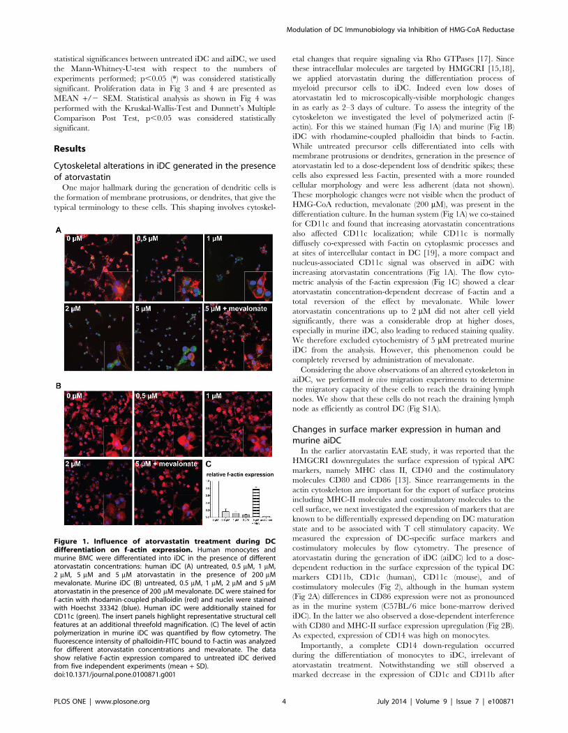
Cytoskeletal alterations in iDC generated in the presenceof atorvastatin
One major hallmark during the generation of dendritic cells is
the formation of membrane protrusions, or dendrites, that give the
typical terminology to these cells. This shaping involves cytoskel-
etal changes that require signaling via Rho GTPases [17]. Since
these intracellular molecules are targeted by HMGCRI [15,18],
we applied atorvastatin during the differentiation process of
myeloid precursor cells to iDC. Indeed even low doses of
atorvastatin led to microscopically-visible morphologic changes
in as early as 2–3 days of culture. To assess the integrity of the
cytoskeleton we investigated the level of polymerized actin (f-
actin). For this we stained human (Fig 1A) and murine (Fig 1B)
iDC with rhodamine-coupled phalloidin that binds to f-actin.
While untreated precursor cells differentiated into cells with
membrane protrusions or dendrites, generation in the presence of
atorvastatin led to a dose-dependent loss of dendritic spikes; these
cells also expressed less f-actin, presented with a more rounded
cellular morphology and were less adherent (data not shown).
These morphologic changes were not visible when the product of
HMG-CoA reduction, mevalonate (200 mM), was present in the
differentiation culture. In the human system (Fig 1A) we co-stained
for CD11c and found that increasing atorvastatin concentrations
also affected CD11c localization; while CD11c is normally
diffusely co-expressed with f-actin on cytoplasmic processes and
at sites of intercellular contact in DC [19], a more compact and
nucleus-associated CD11c signal was observed in aiDC with
increasing atorvastatin concentrations (Fig 1A). The flow cyto-
metric analysis of the f-actin expression (Fig 1C) showed a clear
atorvastatin concentration-dependent decrease of f-actin and a
total reversion of the effect by mevalonate. While lower
atorvastatin concentrations up to 2 mM did not alter cell yield
significantly, there was a considerable drop at higher doses,
especially in murine iDC, also leading to reduced staining quality.
We therefore excluded cytochemistry of 5 mM pretreated murine
iDC from the analysis. However, this phenomenon could be
completely reversed by administration of mevalonate.
Considering the above observations of an altered cytoskeleton in
aiDC, we performed in vivo migration experiments to determine
the migratory capacity of these cells to reach the draining lymph
nodes. We show that these cells do not reach the draining lymph
node as efficiently as control DC (Fig S1A).
Changes in surface marker expression in human andmurine aiDC
In the earlier atorvastatin EAE study, it was reported that the
HMGCRI downregulates the surface expression of typical APC
markers, namely MHC class II, CD40 and the costimulatory
molecules CD80 and CD86 [13]. Since rearrangements in the
actin cytoskeleton are important for the export of surface proteins
including MHC-II molecules and costimulatory molecules to the
cell surface, we next investigated the expression of markers that are
known to be differentially expressed depending on DC maturation
state and to be associated with T cell stimulatory capacity. We
measured the expression of DC-specific surface markers and
costimulatory molecules by flow cytometry. The presence of
atorvastatin during the generation of iDC (aiDC) led to a dose-
dependent reduction in the surface expression of the typical DC
markers CD11b, CD1c (human), CD11c (mouse), and of
costimulatory molecules (Fig 2), although in the human system
(Fig 2A) differences in CD86 expression were not as pronounced
as in the murine system (C57BL/6 mice bone-marrow derived
iDC). In the latter we also observed a dose-dependent interference
with CD80 and MHC-II surface expression upregulation (Fig 2B).
As expected, expression of CD14 was high on monocytes.
Importantly, a complete CD14 down-regulation occurred
during the differentiation of monocytes to iDC, irrelevant of
atorvastatin treatment. Notwithstanding we still observed a
marked decrease in the expression of CD1c and CD11b after
Figure 1. Influence of atorvastatin treatment during DCdifferentiation on f-actin expression. Human monocytes andmurine BMC were differentiated into iDC in the presence of differentatorvastatin concentrations: human iDC (A) untreated, 0.5 mM, 1 mM,2 mM, 5 mM and 5 mM atorvastatin in the presence of 200 mMmevalonate. Murine iDC (B) untreated, 0.5 mM, 1 mM, 2 mM and 5 mMatorvastatin in the presence of 200 mM mevalonate. DC were stained forf-actin with rhodamin-coupled phalloidin (red) and nuclei were stainedwith Hoechst 33342 (blue). Human iDC were additionally stained forCD11c (green). The insert panels highlight representative structural cellfeatures at an additional threefold magnification. (C) The level of actinpolymerization in murine iDC was quantified by flow cytometry. Thefluorescence intensity of phalloidin-FITC bound to f-actin was analyzedfor different atorvastatin concentrations and mevalonate. The datashow relative f-actin expression compared to untreated iDC derivedfrom five independent experiments (mean + SD).doi:10.1371/journal.pone.0100871.g001
Modulation of DC Immunobiology via Inhibition of HMG-CoA Reductase
PLOS ONE | www.plosone.org 4 July 2014 | Volume 9 | Issue 7 | e100871

atorvastatin incubation, even though these markers are typically
up-regulated in parallel to CD14 loss after the differentiation of
monocytes to iDC. Undifferentiated monocytes that were used as a
negative control, on the other hand, showed only a low expression
of CD40, CD86, CD1c and CD11b in comparison to all DC
groups (Fig 2A).
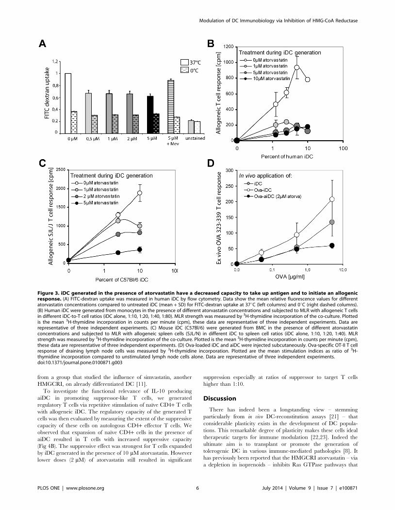
Decreased antigen uptake and presentation by aiDC invitro and in vivo
To investigate whether alterations in the iDC cytoskeleton
contribute to changes in the capacity of these cells to take up
antigen, we incubated iDC with FITC-dextran at 37uC and
calculated uptake by measuring fluorescence by flow-cytometric
analysis. We observed that increasing doses of atorvastatin led to a
decrease in FITC-dextran incorporation in human (Fig 3A) and
mouse cells (data not shown), indicating a decrease in antigen
uptake by these cells. Next we studied the impact of atorvastatin-
mediated interference with antigen uptake and expression of
surface molecules on the capacity of iDC to present antigen using
a MLR. We first generated iDC from monocytes in the presence
or absence of increasing doses of atorvastatin and then co-cultured
viable iDC in varying ratios (up to 1:10) with sorted CD4+ cells
from a second healthy donor. After 72 h allogeneic culture, the
strength of MLR was measured with a 3H-thymidine proliferation
assay. Maximal allogeneic T cell stimulation was achieved with
iDC to T cell ratios of 1:20 (Fig 3B). Priming of iDC with
atorvastatin during differentiation from monocytes resulted in a
marked dose-dependent decrease in T cell response, with a
concentration of 1 mM atorvastatin already leading to significant
MLR inhibition. A similar effect was also observed in the murine
system (Fig 3C). In this case, we co-cultured bone marrow-derived
iDC from C57Bl/6 mice, generated in the presence or absence of
atorvastatin with spleen cells of SJL/J mice.
Finally, we assessed the stimulatory capacity of aiDC to prime
antigen-specific T cells in vivo by measuring T cell proliferation
upon ex vivo antigen restimulation. We injected iDC and aiDC
loaded with OVA323-339 peptide intracutaneously into Rag1-ko
mice that lack endogenous T cells. The following day we injected
mice with naıve CD4 OT-II cells that express a T cell receptor
that recognizes the OVA323-339 epitope. After 5 days immune cells
were isolated from draining lymph nodes, restimulated in vitro with
OVA-peptide or control stimuli, and proliferation was measured
by 3[H] incorporation. We show that aiDC fail to promote the
typical pronounced T cell response upon antigen-specific restim-
ulation (Fig 3D). These results (Fig 3D) are in line with the in vitro
MLR experiments with allogenic DC generated in the presence of
atorvastatin (Fig 3B, 3C). However, when we performed in vitro
experiments to study the capacity of aiDC to present OVA-
peptide to antigen-specific T cells in culture, we show that aiDC
do not significantly differ from control iDC in presenting OVA323-
339 peptide to naıve CD4 OT-II T cells in vitro (Fig S1B).
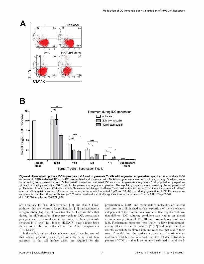
Suppressor activity of in vitro generated regulatory T cellsis enhanced by aiDC
We have previously reported that the induction of T cell anergy
by atorvastatin requires IL-10 signaling [14]. However it has not
been clear which immune cell population is the source of this
regulatory cytokine. Since IL-10 production by DC has been
shown to be critical for the induction of tolerance [20], we next
investigated IL-10 levels in iDC generated in the presence of
atorvastatin. Indeed aiDC produced significantly higher amounts
of IL-10 (up to 9-fold) as assessed by intracellular staining (Fig 4A).
We also observed an increased secretion of IL-4 in these cells as
well as a decrease in the pro-inflammatory cytokines TNF-alpha,
IL-6 and IL-1beta (Fig S2). This is in line with a previous report
Figure 2. Expression of MHCII, costimulatory and maturation markers is attenuated in aiDCs. (A) Expression of surface markers CD1c,CD11b, CD11c, CD14, CD40, CD80 and CD86 on human iDC cultured in the presence of atorvastatin was measured by flow cytometry after thedifferentiation period (day 7) and compared with surface marker expression of untreated iDC and undifferentiated monocytes from peripheral bloodtogether with isotype controls for untreated iDC and monocytes. The data show relative expression of surface molecules compared to untreated iDCderived from three independent experiments (mean 6 SD). (B) Expression of surface markers CD11b, CD11c, CD40, CD80, CD86 and MHC class II ofmurine iDC cultured in the presence of atorvastatin was measured by flow cytometry after the differentiation period (day 10) and compared withsurface marker expression of untreated iDC and isotype controls for untreated iDC. The data show relative expression of surface molecules comparedto untreated iDC derived from three independent experiments (mean 6 SD).doi:10.1371/journal.pone.0100871.g002
Modulation of DC Immunobiology via Inhibition of HMG-CoA Reductase
PLOS ONE | www.plosone.org 5 July 2014 | Volume 9 | Issue 7 | e100871
from a group that studied the influence of simvastatin, another
HMGCRI, on already differentiated DC [11].
To investigate the functional relevance of IL-10 producing
aiDC in promoting suppressor-like T cells, we generated
regulatory T cells via repetitive stimulation of naıve CD4+ T cells
with allogeneic iDC. The regulatory capacity of the generated T
cells was then evaluated by measuring the extent of the suppressive
capacity of these cells on autologous CD4+ effector T cells. We
observed that expansion of naıve CD4+ cells in the presence of
aiDC resulted in T cells with increased suppressive capacity
(Fig 4B). The suppressive effect was strongest for T cells expanded
by iDC generated in the presence of 10 mM atorvastatin. However
lower doses (2 mM) of atorvastatin still resulted in significant
suppression especially at ratios of suppressor to target T cells
higher than 1:10.
Discussion
There has indeed been a longstanding view – stemming
particularly from in vivo DC-reconstitution assays [21] – that
considerable plasticity exists in the development of DC popula-
tions. This remarkable degree of plasticity makes these cells ideal
therapeutic targets for immune modulation [22,23]. Indeed the
ultimate aim is to transplant or promote the generation of
tolerogenic DC in various immune-mediated pathologies [8]. It
has previously been reported that the HMGCRI atorvastatin – via
a depletion in isoprenoids – inhibits Ras GTPase pathways that
Figure 3. iDC generated in the presence of atorvastatin have a decreased capacity to take up antigen and to initiate an allogenicresponse. (A) FITC-dextran uptake was measured in human iDC by flow cytometry. Data show the mean relative fluorescence values for differentatorvastatin concentrations compared to untreated iDC (mean + SD) for FITC-dextran uptake at 37uC (left columns) and 0uC (right dashed columns).(B) Human iDC were generated from monocytes in the presence of different atorvastatin concentrations and subjected to MLR with allogeneic T cellsin different iDC-to-T cell ratios (iDC alone, 1:10, 1:20, 1:40, 1:80). MLR strength was measured by 3H-thymidine incorporation of the co-culture. Plottedis the mean 3H-thymidine incorporation in counts per minute (cpm), these data are representative of three independent experiments. Data arerepresentative of three independent experiments. (C) Mouse iDC (C57Bl/6) were generated from BMC in the presence of different atorvastatinconcentrations and subjected to MLR with allogeneic spleen cells (SJL/N) in different iDC to spleen cell ratios (iDC alone, 1:10, 1:20, 1:40). MLRstrength was measured by 3H-thymidine incorporation of the co-culture. Plotted is the mean 3H-thymidine incorporation in counts per minute (cpm),these data are representative of three independent experiments. (D) Ova-loaded iDC and aiDC were injected subcutaneously. Ova-specific OT-II T cellresponse of draining lymph node cells was measured by 3H-thymidine incorporation. Plotted are the mean stimulation indices as ratio of 3H-thymidine incorporation compared to unstimulated lymph node cells alone. Data are representative of three independent experiments.doi:10.1371/journal.pone.0100871.g003
Modulation of DC Immunobiology via Inhibition of HMG-CoA Reductase
PLOS ONE | www.plosone.org 6 July 2014 | Volume 9 | Issue 7 | e100871
are necessary for Th1 differentiation [18] and Rho GTPase
pathways that are necessary for proliferation [18] and actomyosin
reorganization [15] in myelin-reactive T cells. Here we show that
during the differentiation of precursor cells to iDC, atorvastatin
precipitates cell structural alterations, similar to those previously
reported in T cells [15]. Indeed HMGCRI have already been
shown to exhibit an influence on the APC compartment
[10,11,13,24].
As the actin-based cytoskeleton is rearranged, it can be assumed
that related processes, such as exosome formation and their
transport to the cell surface which are required for the
presentation of MHC and costimulatory molecules, are altered
and result in a diminished surface expression of these molecules
independent of their intracellular synthesis. Recently it was shown
that different iDC culturing conditions can lead to an altered
exosome composition of MHCII and costimulatory molecules
[25]. Furthermore exosomes were shown to have immunomod-
ulatory effects in specific contexts [26,27] and might therefore
directly contribute to altered immune responses that add to their
role of modulating the surface expression of costimulatory
molecules. Notably, we observed that the cellular distribution
pattern of CD11c – that is commonly distributed around the f-
Figure 4. Atorvastatin primes iDC to produce IL-10 and to generate T cells with a greater suppressive capacity. (A) Intracellular IL-10expression in C57Bl/6-derived iDC and aiDC, unstimulated and stimulated with PMA-ionomycin, was measured by flow cytometry. Quadrants wereset according to unstained controls. (B) Atorvastatin treated and untreated iDC were used to generate a regulatory T cell population by repetitivestimulation of allogeneic naıve CD4 T cells in the presence of regulatory cytokines. The regulatory capacity was assessed by the suppression ofproliferation of pre-activated CD4 effector cells. Shown are the changes of effector T cell proliferation (in percent) for different suppressor T cell to Teffector cell (targets) ratios and different atorvastatin concentrations (untreated, 2 mM and 10 mM) used during generation of iDC. Representativeexperiments of at least three are shown. p,0.05 was considered statistically significant, asterisks represent ** = p,0.01, *** = p,0.001.doi:10.1371/journal.pone.0100871.g004
Modulation of DC Immunobiology via Inhibition of HMG-CoA Reductase
PLOS ONE | www.plosone.org 7 July 2014 | Volume 9 | Issue 7 | e100871
actin core of podosomes [19] – was more compact and
intracellularly aggregated in aiDC. A failure of this integrin to
localize to dendritic processes might hinder an optimal and stable
contact with interacting T cells. In fact, we could demonstrate that
atorvastatin enables iDC to induce a suppressive T cell phenotype.
By applying atorvastatin-treated iDC (aiDC) loaded with OVA323–
339 peptide and OVA-specific T cells in Rag12/2 mice, the
capacity to induce less responsive T cells was conserved in vivo. The
in vitro–generated aiDC that induced less responsive T cells
expressed less CD40. Ligation of CD40 by CD40L (CD154) on T
cells is required for DC to undergo maturation since this signals
the induction of the costimulatory molecules CD80 and CD86 that
signal back to T cells to boost the immune response [28].
Furthermore, CD40 ligation releases iDC from a ‘‘default’’ control
mode by regulatory T cells [29]. On the other hand, iDC
incapable of delivering a complete activation signal can induce T
cell tolerance via the de novo generation of regulatory T cells [30]
or antigen-specific anergy following downregulation of CD154
[31]. Since the CD40/CD154 axis participates in the genesis of
inflammatory conditions including atherosclerosis and neuroin-
flammation [32,33], manipulation of this system by immunomod-
ulatory agents such as atorvastatin would provide a useful means
to shift the T cell response towards a regulatory phenotype. Using
a setup adapted from Jonuleit et al. [5] and Bacchetta et al. [16],
we showed that the impact of atorvastatin on iDC development
renders these cells (aiDC) even more potent at generating anergic
suppressor T cells. This setup may not match the cytokine
exposure in vivo, however we used it here merely as a tool for
maintaining and preserving the anergic T cell population. Notably
aiDC produced higher amounts of intracellular IL-10. It has
previously been reported that iDC can generate anergic IL-10–
producing suppressor T cells by repetitively stimulating allogeneic
naıve T cells [5]. Our data show that the regulatory T cells
generated via repetitive restimulation with IL-10 producing aiDC
have more pronounced suppressive properties compared to
untreated iDC. Our observation that iDC alone also cause a
tolerogenic modulation of effector T cells has already been
described in the literature [34]. One proposed mechanism for the
suppressor properties of regulatory T cells is the competition for
growth factors such as IL-2 between suppressor and effector target
cells [35]. From these experiments we cannot exclude competition
as one mechanism via which the suppressor cells suppress the
target cells. However we kept the total (target + suppressor) cell
number constant between the different groups to keep the
available growth factors per cell constant. Furthermore, neither
iDC nor suppressor T cells alone show any proliferation, thus the
competing effects also seem to be negligible.
Our present findings could contribute to the observations made
on a reduced severity in T cell mediated-diseases, such as EAE
[12,13], following atorvastatin treatment. In the last ten years
there have been several clinical studies in MS patients to
investigate the outcome of using HMGRI as stand-alone therapy
[36,37] or as add-on to other disease-modifying drugs [38–42].
Different observations were made in these studies. In our study we
reported that high-dose atorvastatin leads to a reduction in newly
emerging CNS lesions and is associated with increased IL-10
secretion [39]. One recent trial demonstrated the neuroprotective
properties of HMGCRI by showing a reduction in the rate of
brain atrophy during a 3 year follow-up period [36]. Furthermore,
some trials investigating HMGCRI in combination with interferon
beta-1a did not report on any beneficial effect [41,43,44], one
study with a small cohort of MS patients even reported on an
increase in disease activity [38].
In our study, we provide evidence that iDC generated in the
presence of atorvastatin (aiDC) support the expansion of
suppressor T cells. Recently, Weber et al. reported that
atorvastatin does not induce Th2 cells in vivo [45], in contrast
to the observations made from ex vivo–restimulated myelin-
specific T cells stemming from atorvastatin-treated EAE mice
[12,13]. In their study, Weber et al. also did not observe changes
in the frequency of CD4+CD25+Foxp3+ Tregs in EAE animals
following atorvastatin treatment but did observe an increase in IL-
10 secretion [45] possibly originating from the APC compartment.
From our T cell priming experiments with aiDC, we conclude
that atorvastatin promotes the differentation of iDC that do not
optimally prime T cells, both in vitro and in vivo. Patient-derived
DC modified to be tolerogenic could be considered as a future
therapeutic strategy in autoimmune disease. Interestingly, two
recent studies in experimental autoimmune myasthenia gravis [46]
and experimental autoimmune neuritis [47] revealed the thera-
peutic benefit of intraperitoneally applying DC that were pre-
treated with atorvastatin in culture. Both studies also report on the
generation of CD4+ Foxp3+ Tregs and donwregulation of Th1/
Th17 cytokines [46,47].
The observed differential influence of aiDC on alloantigen-
specific versus peptide-specific T cell responses could be explained
by the mechanistic diversity in antigen processing utilized by the
two different reactions; while alloantigen requires cytoskeleton-
dependent endolysosomal processing [48], peptides do not require
any intracellular processing prior to presentation. It is therefore
our understanding, that OVA peptide loaded aiDC are in a
position to bypass the influence of atorvastatin on antigen uptake
and processing. In line with this and our observations regarding
perturbations of the actin cytoskeleton in aiDC, we observed a
reduced propensity of aiDC to reach their target organ (draining
lymph nodes), which would partially explain the reduced capacity
of OVA peptide loaded aiDC to prime T cells in vivo.
In this study we observed that human iDC were more sensitive
to atorvastatin than murine iDC; lower doses of atorvastatin were
sufficient to inhibit actin polymerization and expression of surface
markers such as CD11c and CD11b in human iDC. We have
previously observed the same phenomenon in antigen-specific T
cells where atorvastatin appeared to influence human T cells at
lower doses than murine T cells [12]. One factor that could
explain this is the basal metabolic rate of both organisms, which is
about seven times higher in mice than in humans [49].
Taken together, we found that the HMGCRI atorvastatin –
that causes considerable alterations in cytoskeletal dynamics
during iDC development – consequently alters the surface
expression and localization of receptors and ligands that ultimately
determine the decision between T cell priming or tolerance. While
the relocalization of CD11c to the intracellular compartment
requires further in depth investigation, we believe that by targeting
CD40 expression, atorvastatin sets off a battery of downstream
events that are characteristic of tolerogenic DC. Indeed we
observed an increased production of IL-10, a decreased expression
of costimulatory molecules on aiDC, a reduced ability of these cells
to present antigen to T cells while still being able to migrate into T
cell areas of secondary lymphoid organs – notwithstanding
changes in the cytoskeleton – to induce T cell anergy and
generate CD4+ T cell populations with a regulatory phenotype.
Considering the pleiotropic nature of HMGCRI such as
atorvastatin, we cannot exclude the involvement of several
mechanisms that act together or independently to orchestrate
the observed changes in functions (DC migration and T cell
priming). For instance, we cannot completely attribute the
functional changes observed (eg. T cell priming) solely to changes
Modulation of DC Immunobiology via Inhibition of HMG-CoA Reductase
PLOS ONE | www.plosone.org 8 July 2014 | Volume 9 | Issue 7 | e100871
in surface expression of specific molecules. Indeed the cytoskeleton
of DC and T cells determines the DC-T cell contact duration
during T cell priming [50] and could be one target for atorvastatin
that alters the T cell response. We believe that atorvastatin alters
complex intracellular pathways – such as those related to the
cytoskeleton [15] – to finally precipitate functional changes such as
decreased migration or decreased priming capacity.
In summary, our study provides further evidence of atorvasta-
tin’s capacity to modulate the antigen-presenting cell compart-
ment, resulting in morphological as well as functional changes in
these cells that are connected with modulation of functional
properties of T cells mediated by the APC-T cell interaction. This
interaction plays a crucial role in different pathologies pertaining
to allergy, autoimmunity or transplantation and could therefore be
of therapeutic relevance in these clinical situations.
Supporting Information
Figure S1 Atorvastatin reduces in vivo migratory ca-pacity of iDC, but does not influence their capacity topresent peptide to antigen-specific T cells in vitro. (A)
Atorvastatin treated and untreated iDC generated from RFP-
fluorescent mice were injected into C57BL/6 mice. The
percentage of RFP-fluorescent iDC and aiDC that had migrated
to the draining lymph nodes 24 h after injection was determined
by FACS. Data from 5 mice are shown as mean percentage RFP+cells (6 SEM) of CD11c+CD11b+ cells within the harvested
lymph nodes. (B) Naıve T cells from OTII-transgenic mice were
labelled with CFSE and stimulated with OVA323-339 –loaded iDC
or aiDC generated in the absence or presence of different
concentrations of atrovastatin. CFSE-intensity was measured by
flow cytometry after 72 h and percentage of divided T cells was
analyzed. One representative experiment of three is shown.
(TIF)
Figure S2 Dendritic cells generated in the presence ofatorvastatin show an anti-inflammatory cytokinic pro-file. Soluble cytokines from supernatants of DC generated in the
absence or presence of atorvastatin were measured with
FlowCytomix Multiplex (eBioscience). The expression of each
cytokine is given as mean concentration of 2 experiments (pg/ml)
6SD.
(TIF)
Acknowledgments
We thank Bibiane Seeger, Janet Lips, Julia Skodowski, Heike Ehrengard
and Birgit Hohmann for technical assistance.
Author Contributions
Conceived and designed the experiments: TL CFP VS CID FZ SW.
Performed the experiments: FL IB MP TP DT SL JH. Analyzed the data:
TL CFP DT VS SW. Wrote the paper: TL CFP FZ SW.
References
1. Romani N, Koide S, Crowley M, Witmer-Pack M, Livingstone AM, et al. (1989)
Presentation of exogenous protein antigens by dendritic cells to T cell clones.Intact protein is presented best by immature, epidermal Langerhans cells. J Exp
Med 169: 1169–1178.
2. De Smedt T, Pajak B, Muraille E, Lespagnard L, Heinen E, et al. (1996)Regulation of dendritic cell numbers and maturation by lipopolysaccharide in
vivo. J Exp Med 184: 1413–1424.
3. Inaba K, Turley S, Iyoda T, Yamaide F, Shimoyama S, et al. (2000) The
formation of immunogenic major histocompatibility complex class II-peptideligands in lysosomal compartments of dendritic cells is regulated by
inflammatory stimuli. J Exp Med 191: 927–936.
4. Shortman K, Naik SH (2007) Steady-state and inflammatory dendritic-celldevelopment. Nat Rev Immunol 7: 19–30.
5. Jonuleit H, Schmitt E, Schuler G, Knop J, Enk AH (2000) Induction of
interleukin 10-producing, nonproliferating CD4(+) T cells with regulatoryproperties by repetitive stimulation with allogeneic immature human dendritic
cells. J Exp Med 192: 1213–1222.
6. Bonifaz L, Bonnyay D, Mahnke K, Rivera M, Nussenzweig MC, et al. (2002)
Efficient targeting of protein antigen to the dendritic cell receptor DEC-205 inthe steady state leads to antigen presentation on major histocompatibility
complex class I products and peripheral CD8+ T cell tolerance. J Exp Med 196:1627–1638.
7. Hawiger D, Inaba K, Dorsett Y, Guo M, Mahnke K, et al. (2001) Dendritic cells
induce peripheral T cell unresponsiveness under steady state conditions in vivo.
J Exp Med 194: 769–779.8. Dhodapkar MV, Steinman RM, Krasovsky J, Munz C, Bhardwaj N (2001)
Antigen-specific inhibition of effector T cell function in humans after injection of
immature dendritic cells. J Exp Med 193: 233–238.
9. Legge KL, Gregg RK, Maldonado-Lopez R, Li L, Caprio JC, et al. (2002) Onthe role of dendritic cells in peripheral T cell tolerance and modulation of
autoimmunity. J Exp Med 196: 217–227.
10. Yilmaz A, Reiss C, Tantawi O, Weng A, Stumpf C, et al. (2004) HMG-CoAreductase inhibitors suppress maturation of human dendritic cells: new
implications for atherosclerosis. Atherosclerosis 172: 85–93.
11. Yilmaz A, Reiss C, Weng A, Cicha I, Stumpf C, et al. (2006) Differential effectsof statins on relevant functions of human monocyte-derived dendritic cells.
J Leukoc Biol 79: 529–538.
12. Aktas O, Waiczies S, Smorodchenko A, Dorr J, Seeger B, et al. (2003)
Treatment of relapsing paralysis in experimental encephalomyelitis by targetingTh1 cells through atorvastatin. J Exp Med 197: 725–733.
13. Youssef S, Stuve O, Patarroyo JC, Ruiz PJ, Radosevich JL, et al. (2002) The
HMG-CoA reductase inhibitor, atorvastatin, promotes a Th2 bias and reversesparalysis in central nervous system autoimmune disease. Nature 420: 78–84.
14. Waiczies S, Prozorovski T, Infante-Duarte C, Hahner A, Aktas O, et al. (2005)
Atorvastatin Induces T Cell Anergy via Phosphorylation of ERK1. J Immunol174: 5630–5635.
15. Waiczies S, Bendix I, Prozorovski T, Ratner M, Nazarenko I, et al. (2007)
Geranylgeranylation but not GTP loading determines rho migratory function in
T cells. J Immunol 179: 6024–6032.
16. Bacchetta R, Sartirana C, Levings MK, Bordignon C, Narula S, et al. (2002)
Growth and expansion of human T regulatory type 1 cells are independent from
TCR activation but require exogenous cytokines. Eur J Immunol 32: 2237–
2245.
17. Benvenuti F, Hugues S, Walmsley M, Ruf S, Fetler L, et al. (2004) Requirement
of Rac1 and Rac2 expression by mature dendritic cells for T cell priming.
Science 305: 1150–1153.
18. Dunn SE, Youssef S, Goldstein MJ, Prod’homme T, Weber MS, et al. (2006)
Isoprenoids determine Th1/Th2 fate in pathogenic T cells, providing a
mechanism of modulation of autoimmunity by atorvastatin. J Exp Med 203:
401–412.
19. Burns S, Hardy SJ, Buddle J, Yong KL, Jones GE, et al. (2004) Maturation of
DC is associated with changes in motile characteristics and adherence. Cell
Motil Cytoskeleton 57: 118–132.
20. Akbari O, DeKruyff RH, Umetsu DT (2001) Pulmonary dendritic cells
producing IL-10 mediate tolerance induced by respiratory exposure to antigen.
Nat Immunol 2: 725–731.
21. D’Amico A, Wu L (2003) The early progenitors of mouse dendritic cells and
plasmacytoid predendritic cells are within the bone marrow hemopoietic
precursors expressing Flt3. J Exp Med 198: 293–303.
22. Zhang Y, Mukaida N, Wang J, Harada A, Akiyama M, et al. (1997) Induction of
dendritic cell differentiation by granulocyte-macrophage colony-stimulating
factor, stem cell factor, and tumor necrosis factor alpha in vitro from lineage
phenotypes-negative c-kit+ murine hematopoietic progenitor cells. Blood 90:
4842–4853.
23. Zhang Y, Zhang YY, Ogata M, Chen P, Harada A, et al. (1999) Transforming
growth factor-beta1 polarizes murine hematopoietic progenitor cells to generate
Langerhans cell-like dendritic cells through a monocyte/macrophage differen-
tiation pathway. Blood 93: 1208–1220.
24. Kwak B, Mulhaupt F, Myit S, Mach F (2000) Statins as a newly recognized type
of immunomodulator. Nat Med 6: 1399–1402.
25. Johansson SM, Admyre C, Scheynius A, Gabrielsson S (2008) Different types of
in vitro generated human monocyte-derived dendritic cells release exosomes
with distinct phenotypes. Immunology 123: 491–499.
26. Kim SH, Lechman ER, Bianco N, Menon R, Keravala A, et al. (2005)
Exosomes derived from IL-10-treated dendritic cells can suppress inflammation
and collagen-induced arthritis. J Immunol 174: 6440–6448.
27. Admyre C, Johansson SM, Qazi KR, Filen JJ, Lahesmaa R, et al. (2007)
Exosomes with immune modulatory features are present in human breast milk.
J Immunol 179: 1969–1978.
28. Banchereau J, Steinman RM (1998) Dendritic cells and the control of immunity.
Nature 392: 245–252.
Modulation of DC Immunobiology via Inhibition of HMG-CoA Reductase
PLOS ONE | www.plosone.org 9 July 2014 | Volume 9 | Issue 7 | e100871
29. Serra P, Amrani A, Yamanouchi J, Han B, Thiessen S, et al. (2003) CD40
ligation releases immature dendritic cells from the control of regulatory CD4+CD25+ T cells. Immunity 19: 877–889.
30. Yates SF, Paterson AM, Nolan KF, Cobbold SP, Saunders NJ, et al. (2007)
Induction of regulatory T cells and dominant tolerance by dendritic cellsincapable of full activation. J Immunol 179: 967–976.
31. Steinbrink K, Wolfl M, Jonuleit H, Knop J, Enk AH (1997) Induction oftolerance by IL-10-treated dendritic cells. J Immunol 159: 4772–4780.
32. Gerritse K, Laman JD, Noelle RJ, Aruffo A, Ledbetter JA, et al. (1996) CD40-
CD40 ligand interactions in experimental allergic encephalomyelitis andmultiple sclerosis. Proc Natl Acad Sci U S A 93: 2499–2504.
33. Laman JD, de Smet BJ, Schoneveld A, van Meurs M (1997) CD40-CD40Linteractions in atherosclerosis. Immunol Today 18: 272–277.
34. Cools N, Ponsaerts P, Van Tendeloo VF, Berneman ZN (2007) Balancingbetween immunity and tolerance: an interplay between dendritic cells,
regulatory T cells, and effector T cells. J Leukoc Biol 82: 1365–1374.
35. Scheffold A, Murphy KM, Hofer T (2007) Competition for cytokines: T(reg)cells take all. Nat Immunol 8: 1285–1287.
36. Chataway J, Schuerer N, Alsanousi A, Chan D, MacManus D, et al. (2014)Effect of high-dose simvastatin on brain atrophy and disability in secondary
progressive multiple sclerosis (MS-STAT): a randomised, placebo-controlled,
phase 2 trial. The Lancet37. Vollmer T, Key L, Durkalski V, Tyor W, Corboy J, et al. (2004) Oral
simvastatin treatment in relapsing-remitting multiple sclerosis. Lancet 363:1607–1608.
38. Birnbaum G, Cree B, Altafullah I, Zinser M, Reder AT (2008) Combining betainterferon and atorvastatin may increase disease activity in multiple sclerosis.
Neurology 71: 1390–1395.
39. Paul F, Waiczies S, Wuerfel J, Bellmann-Strobl J, Dorr J, et al. (2008) Oral high-dose atorvastatin treatment in relapsing-remitting multiple sclerosis. PLoS ONE
3: e1928.40. Lanzillo R, Orefice G, Quarantelli M, Rinaldi C, Prinster A, et al. (2010)
Atorvastatin Combined To Interferon to Verify the Efficacy (ACTIVE) in
relapsingCCoremitting active multiple sclerosis patients: a longitudinal con-trolled trial of combination therapy. Multiple Sclerosis 16: 450–454.
41. Sorensen PS, Lycke J, Er+nlinna JP, Edland A, Wu X, et al. (2011) Simvastatin
as add-on therapy to interferon beta-1a for relapsing-remitting multiple sclerosis
(SIMCOMBIN study): a placebo-controlled randomised phase 4 trial. The
Lancet Neurology 10: 691–701.
42. Togha M, Karvigh SA, Nabavi M, Moghadam NB, Harirchian MH, et al.
(2010) Simvastatin treatment in patients with relapsing-remitting multiple
sclerosis receiving interferon beta 1a: a double-blind randomized controlled trial.
Multiple Sclerosis 16: 848–854.
43. Kamm CP, El Koussy M, Humpert S, Findling O, Burren Y, et al. (2014)
Atorvastatin Added to Interferon Beta for Relapsing Multiple Sclerosis: 12-
Month Treatment Extension of the Randomized Multicenter SWABIMS Trial.
PLoS ONE 9: e86663.
44. Rudick RA, Pace A, Rani MR, Hyde R, Panzara M, et al. (2009) Effect of statins
on clinical and molecular responses to intramuscular interferon beta-1a.
Neurology 72: 1989–1993.
45. Weber M, Prod’homme T, Youssef S, Dunn S, Steinman L, et al. (2014) Neither
T-helper type 2 nor Foxp3+ regulatory T cells are necessary for therapeutic
benefit of atorvastatin in treatment of central nervous system autoimmunity.
Journal of Neuroinflammation 11: 29.
46. Li XL, Liu Y, Cao LL, Li H, Yue LT, et al. (2013) Atorvastatin-modified
dendritic cells in vitro ameliorate experimental autoimmune myasthenia gravis
by up-regulated Treg cells and shifted Th1/Th17 to Th2 cytokines. Mol Cell
Neurosci 56: 85–95.
47. Xu H, Li XL, Yue LT, Li H, Zhang M, et al. (2014) Therapeutic potential of
atorvastatin-modified dendritic cells in experimental autoimmune neuritis by
decreased Th1/Th17 cytokines and up-regulated T regulatory cells and NKR-
P1(+) cells. J Neuroimmunol 269: 28–37.
48. Bird PI, Trapani JA, Villadangos JA (2009) Endolysosomal proteases and their
inhibitors in immunity. Nat Rev Immunol 9: 871–882.
49. Ames BN, Shigenaga MK, Hagen TM (1993) Oxidants, antioxidants, and the
degenerative diseases of aging. Proc Natl Acad Sci U S A 90: 7915–7922.
50. Hugues S (2010) Dynamics of dendritic cell-T cell interactions: a role in T cell
outcome. Semin Immunopathol 32: 227–238.
Modulation of DC Immunobiology via Inhibition of HMG-CoA Reductase
PLOS ONE | www.plosone.org 10 July 2014 | Volume 9 | Issue 7 | e100871
Related Documents