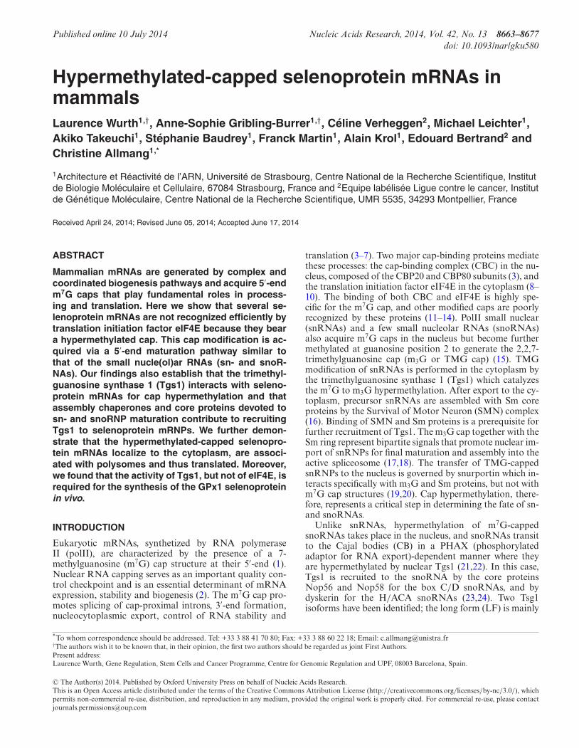
Published online 10 July 2014 Nucleic Acids Research, 2014, Vol. 42, No. 13 8663–8677 doi: 10.1093/nar/gku580 Hypermethylated-capped selenoprotein mRNAs in mammals Laurence Wurth 1,† , Anne-Sophie Gribling-Burrer 1,† ,C´ eline Verheggen 2 , Michael Leichter 1 , Akiko Takeuchi 1 , St ´ ephanie Baudrey 1 , Franck Martin 1 , Alain Krol 1 , Edouard Bertrand 2 and Christine Allmang 1,* 1 Architecture et R ´ eactivit ´ e de l’ARN, Universit ´ e de Strasbourg, Centre National de la Recherche Scientifique, Institut de Biologie Mol ´ eculaire et Cellulaire, 67084 Strasbourg, France and 2 Equipe lab ´ elis´ ee Ligue contre le cancer, Institut de G ´ en´ etique Mol ´ eculaire, Centre National de la Recherche Scientifique, UMR 5535, 34293 Montpellier, France Received April 24, 2014; Revised June 05, 2014; Accepted June 17, 2014 ABSTRACT Mammalian mRNAs are generated by complex and coordinated biogenesis pathways and acquire 5 -end m 7 G caps that play fundamental roles in process- ing and translation. Here we show that several se- lenoprotein mRNAs are not recognized efficiently by translation initiation factor eIF4E because they bear a hypermethylated cap. This cap modification is ac- quired via a 5 -end maturation pathway similar to that of the small nucle(ol)ar RNAs (sn- and snoR- NAs). Our findings also establish that the trimethyl- guanosine synthase 1 (Tgs1) interacts with seleno- protein mRNAs for cap hypermethylation and that assembly chaperones and core proteins devoted to sn- and snoRNP maturation contribute to recruiting Tgs1 to selenoprotein mRNPs. We further demon- strate that the hypermethylated-capped selenopro- tein mRNAs localize to the cytoplasm, are associ- ated with polysomes and thus translated. Moreover, we found that the activity of Tgs1, but not of eIF4E, is required for the synthesis of the GPx1 selenoprotein in vivo. INTRODUCTION Eukaryotic mRNAs, synthetized by RNA polymerase II (polII), are characterized by the presence of a 7- methylguanosine (m 7 G) cap structure at their 5 -end (1). Nuclear RNA capping serves as an important quality con- trol checkpoint and is an essential determinant of mRNA expression, stability and biogenesis (2). The m 7 G cap pro- motes splicing of cap-proximal introns, 3 -end formation, nucleocytoplasmic export, control of RNA stability and translation (3–7). Two major cap-binding proteins mediate these processes: the cap-binding complex (CBC) in the nu- cleus, composed of the CBP20 and CBP80 subunits (3), and the translation initiation factor eIF4E in the cytoplasm (8– 10). The binding of both CBC and eIF4E is highly spe- cific for the m 7 G cap, and other modified caps are poorly recognized by these proteins (11–14). PolII small nuclear (snRNAs) and a few small nucleolar RNAs (snoRNAs) also acquire m 7 G caps in the nucleus but become further methylated at guanosine position 2 to generate the 2,2,7- trimethylguanosine cap (m 3 G or TMG cap) (15). TMG modification of snRNAs is performed in the cytoplasm by the trimethylguanosine synthase 1 (Tgs1) which catalyzes the m 7 G to m 3 G hypermethylation. After export to the cy- toplasm, precursor snRNAs are assembled with Sm core proteins by the Survival of Motor Neuron (SMN) complex (16). Binding of SMN and Sm proteins is a prerequisite for further recruitment of Tgs1. The m 3 G cap together with the Sm ring represent bipartite signals that promote nuclear im- port of snRNPs for final maturation and assembly into the active spliceosome (17,18). The transfer of TMG-capped snRNPs to the nucleus is governed by snurportin which in- teracts specifically with m 3 G and Sm proteins, but not with m 7 G cap structures (19,20). Cap hypermethylation, there- fore, represents a critical step in determining the fate of sn- and snoRNAs. Unlike snRNAs, hypermethylation of m 7 G-capped snoRNAs takes place in the nucleus, and snoRNAs transit to the Cajal bodies (CB) in a PHAX (phosphorylated adaptor for RNA export)-dependent manner where they are hypermethylated by nuclear Tgs1 (21,22). In this case, Tgs1 is recruited to the snoRNA by the core proteins Nop56 and Nop58 for the box C/D snoRNAs, and by dyskerin for the H/ACA snoRNAs (23,24). Two Tsg1 isoforms have been identified; the long form (LF) is mainly * To whom correspondence should be addressed. Tel: +33 3 88 41 70 80; Fax: +33 3 88 60 22 18; Email: [email protected] † The authors wish it to be known that, in their opinion, the first two authors should be regarded as joint First Authors. Present address: Laurence Wurth, Gene Regulation, Stem Cells and Cancer Programme, Centre for Genomic Regulation and UPF, 08003 Barcelona, Spain. C The Author(s) 2014. Published by Oxford University Press on behalf of Nucleic Acids Research. This is an Open Access article distributed under the terms of the Creative Commons Attribution License (http://creativecommons.org/licenses/by-nc/3.0/), which permits non-commercial re-use, distribution, and reproduction in any medium, provided the original work is properly cited. For commercial re-use, please contact [email protected]

Welcome message from author
This document is posted to help you gain knowledge. Please leave a comment to let me know what you think about it! Share it to your friends and learn new things together.
Transcript
Published online 10 July 2014 Nucleic Acids Research, 2014, Vol. 42, No. 13 8663–8677doi: 10.1093/nar/gku580
Hypermethylated-capped selenoprotein mRNAs inmammalsLaurence Wurth1,†, Anne-Sophie Gribling-Burrer1,†, Celine Verheggen2, Michael Leichter1,Akiko Takeuchi1, Stephanie Baudrey1, Franck Martin1, Alain Krol1, Edouard Bertrand2 andChristine Allmang1,*
1Architecture et Reactivite de l’ARN, Universite de Strasbourg, Centre National de la Recherche Scientifique, Institutde Biologie Moleculaire et Cellulaire, 67084 Strasbourg, France and 2Equipe labelisee Ligue contre le cancer, Institutde Genetique Moleculaire, Centre National de la Recherche Scientifique, UMR 5535, 34293 Montpellier, France
Received April 24, 2014; Revised June 05, 2014; Accepted June 17, 2014
ABSTRACT
Mammalian mRNAs are generated by complex andcoordinated biogenesis pathways and acquire 5′-endm7G caps that play fundamental roles in process-ing and translation. Here we show that several se-lenoprotein mRNAs are not recognized efficiently bytranslation initiation factor eIF4E because they beara hypermethylated cap. This cap modification is ac-quired via a 5′-end maturation pathway similar tothat of the small nucle(ol)ar RNAs (sn- and snoR-NAs). Our findings also establish that the trimethyl-guanosine synthase 1 (Tgs1) interacts with seleno-protein mRNAs for cap hypermethylation and thatassembly chaperones and core proteins devoted tosn- and snoRNP maturation contribute to recruitingTgs1 to selenoprotein mRNPs. We further demon-strate that the hypermethylated-capped selenopro-tein mRNAs localize to the cytoplasm, are associ-ated with polysomes and thus translated. Moreover,we found that the activity of Tgs1, but not of eIF4E, isrequired for the synthesis of the GPx1 selenoproteinin vivo.
INTRODUCTION
Eukaryotic mRNAs, synthetized by RNA polymeraseII (polII), are characterized by the presence of a 7-methylguanosine (m7G) cap structure at their 5′-end (1).Nuclear RNA capping serves as an important quality con-trol checkpoint and is an essential determinant of mRNAexpression, stability and biogenesis (2). The m7G cap pro-motes splicing of cap-proximal introns, 3′-end formation,nucleocytoplasmic export, control of RNA stability and
translation (3–7). Two major cap-binding proteins mediatethese processes: the cap-binding complex (CBC) in the nu-cleus, composed of the CBP20 and CBP80 subunits (3), andthe translation initiation factor eIF4E in the cytoplasm (8–10). The binding of both CBC and eIF4E is highly spe-cific for the m7G cap, and other modified caps are poorlyrecognized by these proteins (11–14). PolII small nuclear(snRNAs) and a few small nucleolar RNAs (snoRNAs)also acquire m7G caps in the nucleus but become furthermethylated at guanosine position 2 to generate the 2,2,7-trimethylguanosine cap (m3G or TMG cap) (15). TMGmodification of snRNAs is performed in the cytoplasm bythe trimethylguanosine synthase 1 (Tgs1) which catalyzesthe m7G to m3G hypermethylation. After export to the cy-toplasm, precursor snRNAs are assembled with Sm coreproteins by the Survival of Motor Neuron (SMN) complex(16). Binding of SMN and Sm proteins is a prerequisite forfurther recruitment of Tgs1. The m3G cap together with theSm ring represent bipartite signals that promote nuclear im-port of snRNPs for final maturation and assembly into theactive spliceosome (17,18). The transfer of TMG-cappedsnRNPs to the nucleus is governed by snurportin which in-teracts specifically with m3G and Sm proteins, but not withm7G cap structures (19,20). Cap hypermethylation, there-fore, represents a critical step in determining the fate of sn-and snoRNAs.
Unlike snRNAs, hypermethylation of m7G-cappedsnoRNAs takes place in the nucleus, and snoRNAs transitto the Cajal bodies (CB) in a PHAX (phosphorylatedadaptor for RNA export)-dependent manner where theyare hypermethylated by nuclear Tgs1 (21,22). In this case,Tgs1 is recruited to the snoRNA by the core proteinsNop56 and Nop58 for the box C/D snoRNAs, and bydyskerin for the H/ACA snoRNAs (23,24). Two Tsg1isoforms have been identified; the long form (LF) is mainly
*To whom correspondence should be addressed. Tel: +33 3 88 41 70 80; Fax: +33 3 88 60 22 18; Email: [email protected]†The authors wish it to be known that, in their opinion, the first two authors should be regarded as joint First Authors.Present address:Laurence Wurth, Gene Regulation, Stem Cells and Cancer Programme, Centre for Genomic Regulation and UPF, 08003 Barcelona, Spain.
C© The Author(s) 2014. Published by Oxford University Press on behalf of Nucleic Acids Research.This is an Open Access article distributed under the terms of the Creative Commons Attribution License (http://creativecommons.org/licenses/by-nc/3.0/), whichpermits non-commercial re-use, distribution, and reproduction in any medium, provided the original work is properly cited. For commercial re-use, please [email protected]

8664 Nucleic Acids Research, 2014, Vol. 42, No. 13
involved in snRNA hypermethylation and is present inboth the nucleus and cytoplasm, whereas a short nuclearisoform (SF) is dedicated to snoRNA maturation (23). Inthe nucleoplasm, Tgs1 isoforms are present in a large mul-tiprotein complex that contains ribonucleoprotein (RNP)maturation, transport and assembly factors (15,22,25).
Selenoprotein mRNAs constitute an interesting class ofmRNAs. Indeed, because of the presence of an in-frameUGA codon, recoded as selenocysteine (Sec) and other-wise read as a stop codon, they are submitted to distinctivebiogenesis and translation pathways (26,27). Several cis-and trans-acting factors participate in the co-translationalrecoding of UGA Sec codons. Pivotal in this process arethe SElenoCysteine Insertion Sequence (SECIS), a stem-loop in the 3′ untranslated region (3′UTR) of selenopro-tein mRNAs, and the SECIS-binding protein 2 (SBP2) (28–31). We have previously shown that the assembly mecha-nism of selenoprotein mRNPs, in particular in the 3′UTRregion, is partly common with that of snRNP, snoRNP par-ticles and the telomerase (27). SBP2 which undergoes nu-cleocytoplasmic shuttling (32,33), binds to components ofthis conserved RNP assembly machinery linked to the pro-tein chaperone Hsp90 (27). Besides, SBP2 shares functionalRNA binding motifs with primary core proteins of sn- andsnoRNPs (27,34), suggesting that selenoprotein mRNPs,sn- and snoRNPs could also share a common 5′ maturationpathway.
In this work, we demonstrate that several mammalian se-lenoprotein mRNAs, in contrast to other cellular mRNAs,undergo a 5′-end maturation pathway similar to that of sn-and snoRNPs and bear hypermethylated caps.
MATERIALS AND METHODS
Cell culture
HEK293FT, HeLa and U2OS cells were cultured at 37◦Cin 5% CO2 in Dulbecco’s modified Eagle’s media (DMEM)containing 10% Fetal Calf Serum (FCS), 1% penicillin–streptomycin and 10 nM sodium selenite. One percent ge-neticine was added to HEK293FT cells. Cells were trans-fected using Turbofect (Fermentas) and siRNA inactivationwas done for 48 h using 30 or 100 nM of siRNA and Lipo-fectamine 2000 (Invitrogen) following the manufacturer’sconditions. siTGS1, siSMN, siSBP2 and sieIF4E RNAs andsicontrol were ON-TARGET plus smart pools of four dif-ferent siRNAs (Thermo Scientific Dharmacon), siNop58RNAs were from Santa Cruz. For rescue experiments,cells were transfected for 24 h after siRNA inactivationwith peGFP-Tgs1 (21,24), non-tagged plenti6/V5-SBP2 orpcDNA5-eIF4E expression plasmids and grown for an ad-ditional 24 h treatment. Cells were extracted with HNTGbuffer (20 mM HEPES–NaOH pH 7.9, 150 mM NaCl, 1%Triton, 10% glycerol, 1 mM MgCl2, 1 mM EGTA (Ethyleneglycol-bis(2-aminoethylether)-N,N,N’,N’-tetraacetic acid),1 mM PMSF (phenylmethylsulfonyl fluoride), anti-proteasecocktail from Roche). Subcellular fractionation was per-formed according to (35).
Generation of stable cell lines and induced protein expression
Flp-InTM T-RexTM 293 system (Invitrogen) was used ac-cording to the manufacturer’s instructions to generate sta-ble cell lines with regulated expression of 3xHA-GPx1(HA-GPx1) and 3xHA-GPx1mutCys (HA-Gpx1Cys). Sta-ble clones containing the required open reading frame un-der the control of the cytomegalovirus/tetO2 hybrid pro-moter were selected by culturing in selective medium con-taining 250 �g/ml of hygromycin (InvivoGen) and 15�g/ml of blasticidin (InvivoGen). Resistant colonies wereexpanded and tested for doxycycline-regulated protein ex-pression. Protein expression was induced by addition of0.5–1 �g/ml of doxycycline in culture medium and in thepresence of 10 nM of sodium selenite.
Stable isogenic HeLa cells expressing the Flag-taggedTgs1 proteins were obtained with HeLa Flp-In cells, by co-transfection of the parental cells with pcDNA5-3X Flag-Tgs1 (SF or LF) and a Flippase expression vector withLipofectamine and Plus reagent (Invitrogen). Stable cloneswere then selected with 50 mM hygromycin B (Calbiochem),picked individually, expanded and characterized by westernblots. Individual clones usually expressed similar levels ofthe tagged protein. DNA cloning was performed with theGateway system (Invitrogen).
Immunopurification, co-immunoprecipitation and westernblotting
Immunopurification of endogenous SBP2 com-plexes was performed as described in (27). For co-immunoprecipitation experiments, cell extracts (100–300�l) were incubated with 50 �l of protein A-sepharose(Sigma) coupled to the indicated antibody for 2 h at 4◦Cin NT2 buffer (50 mM Tris–HCl pH 7.5, 150 mM NaCl,1 mM MgCl2, 0.05% NP40, 1 mM DTT (Dithiothreitol),400 �M VRC (Vanadyl ribonucleoside complexes), 100 URNasin/ml, anti-protease cocktail) or with anti-GFP mag-netic beads (Miltenyi) in IPP150 (10 mM Tris–HCl pH 7.5,150 mM NaCl, 0.1% NP-40, 0.5 mM PMSF, 2 mM VRC).Beads alone were used as a control. Beads were washedwith NT2 or IPP150, respectively, resuspended in Laemmlibuffer, analyzed by sodium dodecyl sulfate-polyacrylamidegel electrophoresis (SDS-PAGE) followed by western blot.Antibodies used were: anti-His (H3) mouse monoclonal(Santa Cruz), anti-SBP2 rabbit polyclonal (27), anti-Tgs1mouse monoclonal (24), anti-Nop58 (C-20) polyclonalgoat antibodies (Santa Cruz), anti-SMN (2B1) mouse mon-oclonal (Santa Cruz), anti-rpS21 goat polyclonal (SantaCruz) and anti-AspRS rabbit polyclonal (36). For RNA IP,extracts were first clarified on protein A-sepharose to elim-inate unspecific RNA binders. For microarray experiment,co-immunoprecipitation was performed according to (37).
TMG RIP and qRT-PCR analysis
Trimethyl-capped RNAs were immunoprecipitated using astrictly specific rabbit polyclonal anti-m3G (TMG) serum(Synaptic Systems) also referred to as R1131 (38,39). Tenmicroliters of serum was coupled to 50 �l of protein A-Sepharose beads saturated with 20 �g of both purifiedbovine serum albumin (BSA) and total yeast tRNA in NT2
Nucleic Acids Research, 2014, Vol. 42, No. 13 8665
buffer for 18 h at 4◦C. The immobilized antibody was in-cubated with 200 �g of pre-cleared total RNAs preparedfrom HEK293FT cells (Tri-reagent, Euromedex) in a to-tal volume of 1 ml for 2 h at 4◦C. Beads were washed sixtimes in NT2 buffer, the bound RNA was extracted byphenol/chloroform and precipitated. After DNase treat-ment, RNAs were reversed transcribed using AMV-RT (Q-Biogen) and cDNAs were amplified by classical or quanti-tative RT-PCR (qRT-PCR). Level of mRNAs after siRNAtreatment was measured directly after RNA extraction us-ing the same qRT-PCR method. qRT-PCR was performedon MX3005P (Stratagene) using the Maxima SYBR GreenPCR kit (Fermentas). Oligonucleotides used for qRT-PCRare listed in Supplementary Table S1.
Anti-FLAG immunoprecipitation and microarray analysis
Anti-Flag Tgs1 LF and Tgs1 SF immunoprecipita-tions were done with M2 antibody-coated beads (Sigma-Aldrich). HeLa cells stably expressing Tgs1 LF, Tgs1 SF orparental cells as control, were extracted with lysis buffer (50mM Tris–HCl pH 7.5, 150 mM NaCl, 1 mM EDTA, 1% Tri-ton, antiprotease cocktail) for 30 min at 4◦C and centrifugedfor 10 min at 10 000 rpm at 4◦C. Extracts were incubatedwith M2 beads for 2 h at 4◦C. Beads were washed twice inHNTG and three times in phosphate buffered saline (PBS),RNAs from beads were homogenized in Trizol and purifiedwith RNeasy mini kit (Qiagen). cDNA were prepared andlabeled as recommended by the manufacturer (Affymetrix).RNAs were analyzed using human gene 1.0ST microarraycovering all annotated transcripts (Affymetrix). Files wereanalyzed and normalized with expression consol software(Affymetrix). The 3XFlag-Tgs1 IPs were performed in du-plicates, and the control IP (non-tagged parental cell line)was done in triplicate. Microarray data are accessible atGEO GSE57625.
Recombinant proteins and GST pull-down assays
Radiolabeled proteins were synthesized in the presence of35S-Met in rabbit reticulocyte lysate (TNT; Promega). Bind-ing was performed with 5 �g GST-tagged protein in 50mM Tris–HCl pH 7.5, 150 mM NaCl, 5 mM MgCl2, 0.1mM EDTA and 0.1% NP-40. Washing was performed withthe same buffer. GST- and His-tagged proteins were pro-duced by standard procedure and purified using glutathionesepharose (GE Healthcare) and Ni-NTA agarose (Qiagen),respectively. GST-4EK119A protein was expressed and puri-fied as described by (40). For GST pull-down experimentspurified GST-4EK119A or GST (200 �g) was bound to 200 �lof MagneGST beads (Promega) and incubated with 100 �gof HEK293 total RNA in RNP buffer (20 mM Tris–HClpH 7.5, 100 mM KCl, 0.1 mM EDTA, 1 mM DTT, 10%glycerol, 100 U RNasin/ml, anti-protease cocktail) for 2 hat 4◦C. Beads were washed five times with RNP buffer. TheRNAs present in the flow-through or on the beads were ex-tracted by phenol/chloroform and precipitated.
Yeast-two hybrid interaction tests
For yeast-two hybrid interaction (Y2H) assays, appropri-ate pGBKT7 (DB) or pGADT7 (AD) plasmids were co-
transformed into AH109 (Clontech Laboratories, Inc.) andplated on triple selective media (-Leu -Trp -Ade).
Polysome analysis
Polysome profiles were analyzed on sucrose gradients.HEK293 cells were cultivated up to 80% confluence. 2 ×10⁶ cells were collected, washed in 3 volumes PBS and in-cubated in the presence of 150 �g/ml cycloheximide for 20min on ice. Lysis of the cells was performed in polysomebuffer (PB) containing 10 mM HEPES–NaOH pH 7.5, 50mM KCl, 5 mM MgCl2, 1 mM DTT, 100 units RNasin/ml,400 �M VRC, protease inhibitors and 150 �g/ml cyclohex-imide or in polysome denaturing buffer (PDB) containing10 mM HEPES–NaOH pH 7.5, 10 mM potassium acetate,0.5 mM magnesium acetate, 5 mM DTT. Cytoplasmic ex-tracts were loaded onto a linear 7–47% sucrose gradient pre-pared in PB or PDB. Polysomes were separated by 2.5 hcentrifugation at 37 000 rpm using a Beckmann SW41 ro-tor. Gradients were monitored by following absorbance at254 nm. RNAs were extracted and analyzed by RT-qPCRas described previously.
RESULTS
A subclass of selenoprotein mRNAs harbor hypermethylatedcaps and show reduced affinity for eIF4E
Having previously established that selenoprotein mRNPsand sn-, snoRNPs share a common 3′ assembly pathway(27), we asked whether selenoprotein mRNAs could conse-quently undergo similar 5′ maturation events. Indeed, un-like mRNAs, PolII sn- and snoRNAs that are modifiedco-transcriptionally with a 5′ m7G cap, subsequently ac-quire a trimethylguanosine cap m2,2,7 (TMG) structure (15).We asked whether selenoprotein mRNAs could also bearsimilar cap modifications and could be recognized by thecanonical translational machinery. A prerequisite to cap-dependent translation is the recognition of the eukaryoticmRNA cap structure by the translation initiation complexeIF4F (composed of the three subunits eIF4E, eIF4A andeIF4G). eIF4E specifically recognizes the m7G moiety ofthe 5′ m7GpppN cap of eukaryotic mRNAs and was shownto poorly recognize TMG caps (41). Therefore, we first ex-amined the ability of selenoprotein mRNAs to be recog-nized by eIF4E.
We performed glutathione-S-transferase (GST) pull-down experiments using total RNAs from HEK293FT cellsand a high-affinity mutant of eIF4E, GST-eIF4EK119A (40).This mutant was developed for specific isolation of 5′ m7G-capped mRNAs and showed strict specificity but 10-foldhigher affinity for the m7G cap. The RNA content of eIF4Ebound and unbound fractions was determined by qRT-PCR analysis. For the detection of selenoprotein mRNAs,we used primers complementary to 12 out of the 25 seleno-protein mRNAs characterized in mammals (Figure 1A). �-actin, HPRT (hypoxanthine guanine phosphoribosyltrans-ferase) and LDHA (lactate dehydrogenase A) mRNAs wereused as the m7G-capped controls, U3 snoRNA and U2snRNA for TMG-capped controls. Coherently, an averageof 75% of the canonical �-actin, HPRT and LDHA mR-NAs were recovered in the eIF4E bound fraction, whereas

8666 Nucleic Acids Research, 2014, Vol. 42, No. 13
Figure 1. The cap of selenoprotein mRNAs is hypermethylated and poorlyrecognized by eIF4E. (A) Binding of recombinant GST-eIF4EK119A toHEK293 total RNA. GST-eIF4EK119A (or GST alone) was bound to glu-tathione beads and incubated with the extract. The percentage of mRNAspresent in the bound and unbound fractions were determined separatelyby qRT-PCR and normalized to 100%. Asymmetric error bars representthe minimum and maximum observations for three independent biologicalreplicates, reflecting intrinsic variability. SelR, GPx1, GPx4, SelM, SelW,SelT, SelO, TrxR1, Sel15, SelK, GPx3 and SelN are selenoprotein mRNAs.U3 snoRNA was used as a positive control, �-actin, HPRT and LDHA arehousekeeping mRNAs used as negative controls. (B and C) Total RNA ex-tracted from HEK293FT cells was immunoprecipitated with anti-TMGserum (�-m3G). Bound RNA was analyzed by (B) RT-PCR or (C) qRT-PCR. (−) Control without antibodies. In: input 10%. The graph representsthe percentage of mRNAs in IP compared with the input RNA. Error barsrepresent standard deviation of an average of three independent experi-ments. The horizontal line represents the level of housekeeping mRNAbinding (1–2% in average). See also Supplementary Figure S1 for specificitycontrols. (D) Heatmap representation of mRNA binding in GST-eIF4Epull-down and TMG IP experiments. The binding scale is represented tothe right, maximum binding values in each set of experiments are repre-sented in red and minimal binding values in green. Heat maps were gen-erated with the MeV software. The three classes of selenoprotein mRNAsare indicated.
74% of sn-, snoRNAs were found in the unbound fraction(Figure 1A). The results revealed that selenoprotein mR-NAs showed differential binding patterns to eIF4E. Theselenoprotein mRNAs of SelR, glutathione peroxidases 1and 4 (GPx1, GPx4), SelM and SelW showed a distribu-tion pattern similar to that of sn-, snoRNAs with only 20–35% of the mRNA recovered in the eIF4E bound fraction(Figure 1A). SelT, SelO, thioredoxin reductase 1 (TrxR1),Sel15, SelK selenoprotein mRNAs showed an intermediatepattern with over 50% of the mRNAs in the bound frac-tion (Figure 1A), whereas selenoprotein mRNAs compris-ing glutathione peroxidase 3 (GPx3) and selenoprotein N(SelN) were enriched up to 70% in the eIF4E bound fractionwith patterns similar to non-selenoprotein mRNAs (Figure1A).
To explain the differential binding to eIF4E, we askedwhether some of the selenoprotein mRNAs could bear 5′modifications similar to sn- and snoRNAs as a trimethyl-guanosine cap m2,2,7 (TMG) structure. To answer this ques-tion, RNAs extracted from HEK293FT cells were immuno-precipitated with the highly specific anti-TMG cap R1131serum that was demonstrated not to recognize monomethy-lated caps (38,39) (see also Supplementary Figure S1 forspecificity validation of the antibody). The immunoprecip-itated RNAs were extracted and analyzed by RT-PCR. Re-sults showed that anti-TMG antibodies recognized GPx1,GPx4 and TrxR1 selenoprotein mRNAs as well as U3snoRNA used as the control but not the m7G-capped �-actin mRNA, indicating that the cap of these endoge-nous selenoprotein mRNAs is hypermethylated (Figure1B). Quantitative RT-PCR analysis revealed that seven se-lenoprotein mRNAs, namely SelR, GPx1, GPx4, SelM,SelW, SelT and SelO, were specifically immunoprecipitatedwith anti-TMG antibody (Figure 1C) while TrxR1 was onlyweakly recognized. These results correlate with the GST-eIF4E pull-down experiments, as these TMG-capped se-lenoprotein mRNAs correspond to those poorly recognizedby eIF4E. TrxR1, SelT and SelO, that show an intermedi-ate binding pattern of eIF4E, are consistently recognizedin the TMG-IP (Figure 1A, C). Four selenoprotein mR-NAs were not pulled-down over the background (Sel15,GPx3, SelN and SelK) in TMG immunoprecipitations (Fig-ure 1C); they were consistently bound by eIF4E (Figure1A). As expected, the U3 snoRNA positive control was fullyretained but not the control HPRT or LDHA housekeep-ing mRNAs (Figure 1C). A significant fraction (5–15%) ofthe TMG-capped selenoprotein mRNAs was recovered inthe anti-TMG immunoprecipitation. This precipitation isweaker compared with U3 snoRNA (100%) but is neverthe-less consistent with previously reported levels for snRNAs(39).
Determinants such as the larger size, under-representation of selenoprotein mRNAs versus sno-,snRNAs as well as the shorter half-life of selenoproteinmRNAs compared to sn(o)RNAs may contribute to thelower recovery of selenoprotein mRNAs in the immuno-precipitation. It is also envisageable that different mRNAfolded structures in the 5′UTR could lead to differentialrecognition of individual mRNAs by the antibody.
TMG-IP experiments, in combination to the GST-eIF4Epull-down results, suggest that a substantial fraction of se-

Nucleic Acids Research, 2014, Vol. 42, No. 13 8667
lenoprotein mRNAs possesses a trimethylated guanosinecap. Altogether, our results reveal an inverse relationshipbetween eIF4E binding and TMG capping (Figure 1D) andsuggest the existence of three classes of selenoprotein mR-NAs. In the first class, <50% of the selenoprotein mRNAsare bound to eIF4E and the TMG-IP efficiency is >5%.These mRNAs include SelR, GPx1, GPx4, SelM and SelW(Figure 1D); they harbor a hypermethylated cap and arenot recognized efficiently by the translation factor eIF4E. Asecond class of selenoprotein mRNAs showed an interme-diate pattern with >50% of the mRNAs bound by eIF4Eand >5% TMG-IP efficiency; this class includes selenopro-tein mRNAs such as SelT, SelO and TrxR1 that can possi-bly bear both types of caps (Figure 1D). Finally, the thirdclass represents selenoprotein mRNAs––Sel15, SelK, GPx3and SelN––(Figure 1D) bound with <5% in the TMG-IP,that are m7G-capped and recognized by eIF4E. Selenopro-tein mRNAs thus appear to be subjected to differential 5′processing events.
Tgs1 hypermethylates the cap of selenoprotein mRNAs
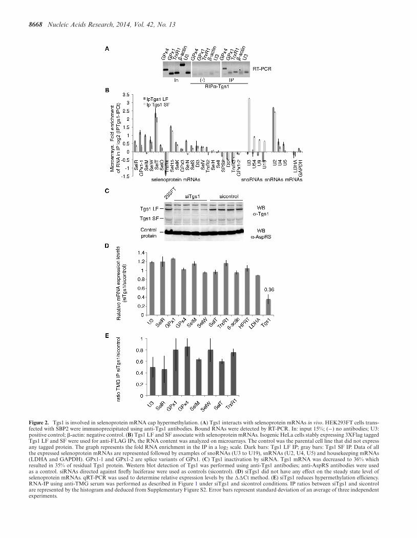
The trimethylguanosine synthase 1 (Tgs1) converts the m7Gcap of sn- and snoRNA precursors into the functionalTMG cap (21,24). This modification step plays a critical rolein targeting sn- and snoRNA precursors to their final mat-uration and functional site (15). Tgs1 is present in a largeU3 snoRNA processing complex that also contains com-ponents of the conserved RNP assembly complex linkedto Hsp90 (27). Because selenoprotein mRNAs are assem-bled into mRNPs by the same molecular machinery (27),we asked whether Tgs1 was linked to selenoprotein mRNPbiogenesis. Tgs1 exists as two molecular species: a full-length, mostly cytoplasmic (Tgs1 LF) and a shorter nuclearisoform (Tgs1 SF) (23). First, we examined if Tgs1 couldbe associated to selenoprotein mRNPs in vivo and per-formed immunoprecipitation of RNA–protein complexesfrom HEK293FT cells with an anti-Tgs1 antibody that rec-ognizes both isoforms (Figure 2A). RNAs associated withendogenous Tgs1 were detected by RT-PCR. Several se-lenoprotein mRNAs, such as GPx1 and 4 as well as TrxR1were specifically associated with Tgs1 in vivo, whereas thiswas not the case for the �-actin mRNA; U3 snoRNA wasthe positive interaction control that binds both Tgs1 iso-forms (Figure 2A). Whether the interaction of Tgs1 couldbe generalized to all selenoprotein mRNAs was investigatedby analyzing the RNA content of the Tgs1 immunoprecip-itation reaction (IP) by microarrays. Because we did nothave antibodies specific for either form of Tgs1, we used twoHeLa cell lines that stably express either Tgs1 LF or Tgs1SF with a 3XFlag tag, and performed the IPs using anti-Flag beads, using the parental cell line to perform controlIPs (37). On average, selenoprotein mRNAs were signifi-cantly enriched in the IPs versus non-selenoprotein mRNAsthat were not retained (Table 1). We found that both Tgs1LF and Tgs1 SF associated to selenoprotein mRNAs (Ta-ble 1 and Figure 2B); likewise and as expected from previ-ous studies (23), Tgs1 LF preferentially recognized snRNAswhereas Tgs1 SF bound more strongly to snoRNAs in ourexperiment (Table 1 and Figure 2B, (37)). To date, 25 seleno-protein genes have been identified in humans (42). Among
the 21 mRNAs expressed in our experiment, 14 were boundby Tgs1 LF and/or SF (Figure 2B), and 7 were not de-tected: TrxR1, SelH, SelI, SPS2, DI2, TrxR3 and GPx1-2. Binding efficiencies were variable, the strongest signalsbeing observed for SelT, Sel15, GPx1-1 and SelM seleno-protein mRNAs for which the enrichment was similar tothat seen for snRNAs and snoRNAs (Figure 2B). TrxR1results show differences with those obtained in the endoge-nous Tgs1 RNA IP experiments (Figure 2A) most likelybecause qRT-PCR is more sensitive and specific than mi-croarrays for the detection of the endogenous population ofTrxR1 mRNAs. Binding of Tgs1 does not appear to strictlycorrelate with TMG-capping, suggesting that recruitmentof the enzyme does not necessarily lead to cap modification.This is similar to the case of intronic snoRNAs which bindTgs1 without subsequent cap modification. Altogether ourresults show that selenoprotein mRNAs interact with thetwo isoforms of Tgs1. The affinity of Tgs1 LF for seleno-protein mRNAs appears only slightly higher than for Tgs1SF and resembles that of snRNAs.
Having shown the interaction of Tgs1 with selenoproteinmRNAs, we further investigated whether this enzyme wasindeed responsible for hypermethylation of the selenopro-tein mRNA cap. To this end, Tgs1 was knocked-down byRNA interference (RNAi) in HEK293FT cells (Figure 2C).Tgs1 mRNA was decreased to 36% (Figure 2D), which re-sulted in 35% of residual Tgs1 protein (Figure 2C). The dropof Tgs1 did not affect selenoprotein mRNA steady state lev-els (Figure 2D) but led to a global reduction of TMG-IPefficiency in correlation with the level of Tgs1 knock-down(Figure 2E and Supplementary Figure S2). Individual se-lenoprotein mRNAs responded differentially to Tgs1 inac-tivation. TMG-IP efficiency dropped down to 45% in thecase of SelR (Figure 2E), comparably to the U3 snoRNApositive control. Milder effects were observed in the case ofGPx1, GPx4, SelW and TrxR1 as the TMG-IP efficiencywas only reduced to 80%. This can be attributed to resid-ual Tgs1 and suggests that its depletion does not affectall selenoprotein mRNAs to the same extent. Hierarchyof selenoprotein expression and variations in selenoproteinmRNA stability have been reported in numerous studies,particularly with regard to the glutathione peroxidase fam-ily (43–46). Individual selenoprotein mRNAs respond in aunique fashion to various imbalances, including Tgs1 deple-tion. Altogether, these results indicate that Tgs1 is involvedin hypermethylation of the cap of the majority of seleno-protein mRNAs. Our data therefore conclude that the caphypermethylation activity of Tgs1 is thus not only restrictedto that of sn- and snoRNA substrates.
Tgs1 is recruited to selenoprotein mRNAs predominantly viathe assembly chaperone SMN but also by the core proteinNop58
The recruitment mode of Tgs1 is dependent on the natureof its RNA targets. In the case of sn- and snoRNAs, thebest characterized Tgs1 targets, two different strategies al-low Tgs1 isoforms to be recruited to the m7G cap. Recruit-ment of Tgs1 LF to snRNAs is dependent on both a ma-jor assembly factor, the Survival of Motor Neuron (SMN)complex, and the core protein SmB, whereas the core pro-
8668 Nucleic Acids Research, 2014, Vol. 42, No. 13
Figure 2. Tgs1 is involved in selenoprotein mRNA cap hypermethylation. (A) Tgs1 interacts with selenoprotein mRNAs in vivo. HEK293FT cells trans-fected with SBP2 were immunoprecipitated using anti-Tgs1 antibodies. Bound RNAs were detected by RT-PCR. In: input 15%; (−) no antibodies; U3:positive control; �-actin: negative control. (B) Tgs1 LF and SF associate with selenoprotein mRNAs. Isogenic HeLa cells stably expressing 3XFlag taggedTgs1 LF and SF were used for anti-FLAG IPs, the RNA content was analyzed on microarrays. The control was the parental cell line that did not expressany tagged protein. The graph represents the fold RNA enrichment in the IP in a log2 scale. Dark bars: Tgs1 LF IP; gray bars: Tgs1 SF IP. Data of allthe expressed selenoprotein mRNAs are represented followed by examples of snoRNAs (U3 to U19), snRNAs (U2, U4, U5) and housekeeping mRNAs(LDHA and GAPDH). GPx1-1 and GPx1-2 are splice variants of GPx1. (C) Tgs1 inactivation by siRNA. Tgs1 mRNA was decreased to 36% whichresulted in 35% of residual Tgs1 protein. Western blot detection of Tgs1 was performed using anti-Tgs1 antibodies; anti-AspRS antibodies were usedas a control. siRNAs directed against firefly luciferase were used as controls (sicontrol). (D) siTgs1 did not have any effect on the steady state level ofselenoprotein mRNAs. qRT-PCR was used to determine relative expression levels by the ��Ct method. (E) siTgs1 reduces hypermethylation efficiency.RNA-IP using anti-TMG serum was performed as described in Figure 1 under siTgs1 and sicontrol conditions. IP ratios between siTgs1 and sicontrolare represented by the histogram and deduced from Supplementary Figure S2. Error bars represent standard deviation of an average of three independentexperiments.

Nucleic Acids Research, 2014, Vol. 42, No. 13 8669
Table 1. Microarray analysis of RNA immunoprecipitations using Tgs1 SF and LF isoforms
log2 (IP Tgs1-IPCt) Tgs1-LF-Cont Tgs1-SF-Cont
Seleno mRNA (20 genes) 0.32 (0.0038) 0.16 (0.0001)snRNA (9 genes) 0.82 (0.0031) 0.25 (0.0031)C/D snoRNA (112 genes) −0.34 (0.0033) 1.2 (0.0019)H/ACA snoRNA (108 genes) −0.11 (0.0037) 0.61 (0.0001)mRNA (18685 genes) −0.022 (0.2612) −0.027 (0.1910)
Average of fold enrichment for each RNA family in IP (Tgs1 SF or LF) versus control measured in duplicate microarray analysis for Tgs1SF and Tgs1 LFRNA IPs. P-values were obtained from Mann–Whitney tests and are indicated in brackets. The average expression level of the entire set of selenoproteinmRNAs expressed in the HeLa cell line is indicated (20 expressed mRNAs among the 25 selenoprotein mRNA genes). The average expression level of theentire set of non-selenoprotein mRNAs is also indicated.
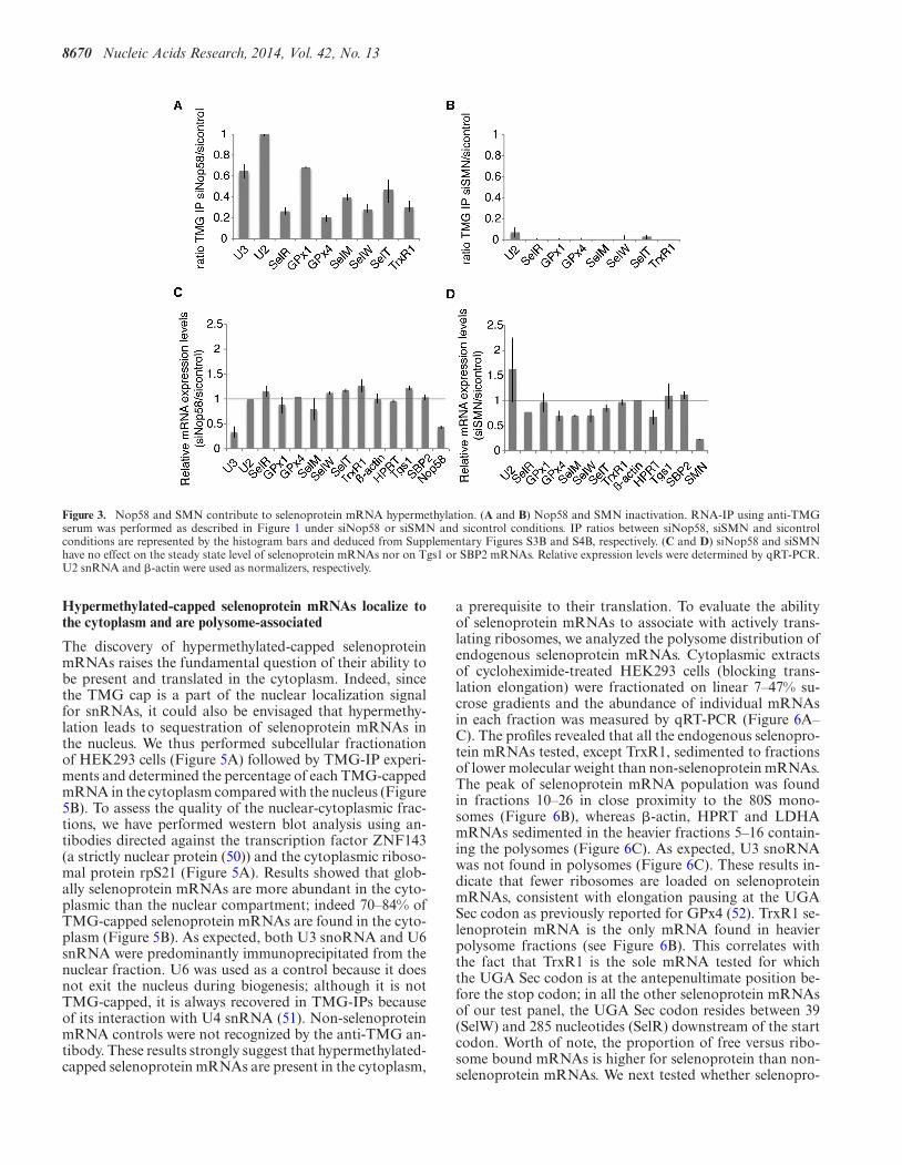
teins Nop56 and Nop58 recruit Tgs1 SF to box C/D snoR-NAs (23,47,48). To determine if Tgs1 could be recruited toselenoprotein mRNAs via one of these two pathways, weseparately inactivated expression of the Nop58 and SMNproteins by RNAi. This resulted in residual levels of 2 and8% of Nop58 and SMN proteins, respectively, but did notaffect expression of Tgs1 LF, SF and of the selenoproteinbiosynthesis factor SBP2 (Figure 3 and Supplementary Fig-ures S3A and S4A). Hypermethylation of the selenoproteinmRNA cap was affected in each case (Figure 3A and B).Cap hypermethylation was decreased in Nop58 depleted-cells by 60–80% (Figure 3A), but was totally abolished af-ter siSMN treatment (Figure 3B). The steady state levels ofselenoprotein mRNAs, Tgs1 and SBP2 mRNAs, as mea-sured by qRT-PCR were not significantly affected in bothcases (Figure 3C and D). As expected, Nop58 depletionstrongly affected the stability of U3 snoRNA (Figure 3C)while SMN knock-down deregulated expression of U2 (Fig-ure 3D). This is consistent with earlier reports showing thatSMN deficiency or knock-down altered the stoichiometryof snRNAs in mammalian cells and could even increasetheir level, particularly in the case of U2 snRNA (49). Weconclude that both Nop58 and SMN participate in hyper-methylation of the selenoprotein mRNA cap. The strong ef-fect of SMN inactivation suggests that recruitment of Tgs1LF likely constitutes the major pathway.
SBP2 interacts with Nop58 and SMN in vivo and recruitsTgs1
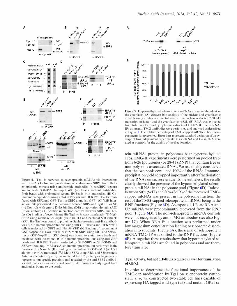
Next, we asked how Tgs1 isoforms, SMN and Nop58 couldbe recruited to selenoprotein mRNAs. As SBP2 plays cen-tral roles in selenoprotein biosynthesis by binding to the se-lenoprotein mRNA SECIS element (26), we first tested ifSBP2 could interact with Tgs1. Endogenous protein com-plexes associated with SBP2 were immunoprecipitated fromHeLa cell extracts using antibodies against the N-terminalregion of SBP2. Western blotting using anti-Tgs1 antibodyrevealed the association of SBP2 with endogenous Tgs1 LF,and little, if any, with Tgs1 SF (Figure 4A). No associa-tion was seen with the control protein Hsp70 (Figure 4A).To confirm this finding, we co-transfected SBP2 with eitherGFP-Tgs1 LF or GFP-Tgs1 SF and immunoprecipitatedthe total cell lysates with anti-GFP antibodies. As shownin Figure 4B, SBP2 associated with Tgs1 LF in vivo, whilebinding to Tgs1 SF was not detected. Y2H tests confirmedprotein–protein interactions between SBP2 and both Tgs1LF and Tgs1 SF (Figure 4C). Finally, in vitro binding assays
between 35S-labeled SBP2 proteins expressed in micrococ-cal nuclease treated rabbit reticulocyte lysate (RRL) and therecombinant His-Tgs1 LF protein produced in Escherichiacoli, showed quantitative binding of SBP2 to Tgs1 LF. How-ever, little interaction was observed between His-SBP2 and35S-Tgs1 SF (Figure 4D). Similar results were obtained us-ing 35S-labeled proteins translated in RNase-treated bac-terial S30 lysates (Figure 4D). Recombinant SBP2 proteinpreparations, produced from bacterial heterologous sys-tems, do not contain snRNAs, snoRNAs and correspond-ing RNP proteins, nor components dedicated to synthesis ofmammalian selenoprotein. This excludes the possibility thatthe protein–protein interactions shown are RNA dependentor mediated via protein components of the sn-, sno- andselenoprotein mRNP assembly machinery. Thus, Tgs1 LFand SBP2 associate in vivo and in vitro and this associa-tion is RNA independent. Because SMN and Nop58 inter-act with Tgs1 and appear to be required for selenoproteinmRNA cap-hypermethylation in vivo (Figure 3A and B),we next analyzed whether Nop58 and SMN also interactedwith SBP2. SBP2 was co-transfected with Nop58-YFP inHEK293FT cells and we immunoprecipitated the total celllysates with anti-GFP beads. As shown in Figure 4E, SBP2interacted with Nop58 in vivo. We also verified the interac-tion in vitro by GST pull-down experiments (Figure 4F) andfound that (35S-Met)-SBP2 produced in RRL or bacterialS30 extracts bound strongly to GST-Nop58 independentlyof RNA. In addition, RNA-IPs in Nop58-YFP transfectedcells revealed that GPx1 and GPx4 mRNAs were specifi-cally associated with Nop58 in vivo (Supplementary Fig-ure S5). To verify the link between SMN and selenopro-tein mRNPs, we co-transfected GFP-SBP2 or GFP-SMNand SBP2 in HEK293FT cells for co-IP analysis. We foundthat GFP-SBP2 was able to interact with endogenous SMNand conversely that GFP-SMN interacted with transfectedSBP2 in vivo (Figure 4G); these interactions were resistantto RNase A treatment and are therefore RNA independent(Figure 4G). GST pull-down experiments confirmed the in-teraction between (35S-Met)-SBP2 and GST-SMN in vitro(Figure 4H). We conclude that SBP2 plays a central role byinteracting with both SMN, Nop58 and Tgs1. The recruit-ment of Tgs1 is likely to be dependent on the formationof the ternary complexes between SBP2/SMN/Tgs1 LF onone side, and SBP2/Nop58/Tgs1 SF on the other.
8670 Nucleic Acids Research, 2014, Vol. 42, No. 13
Figure 3. Nop58 and SMN contribute to selenoprotein mRNA hypermethylation. (A and B) Nop58 and SMN inactivation. RNA-IP using anti-TMGserum was performed as described in Figure 1 under siNop58 or siSMN and sicontrol conditions. IP ratios between siNop58, siSMN and sicontrolconditions are represented by the histogram bars and deduced from Supplementary Figures S3B and S4B, respectively. (C and D) siNop58 and siSMNhave no effect on the steady state level of selenoprotein mRNAs nor on Tgs1 or SBP2 mRNAs. Relative expression levels were determined by qRT-PCR.U2 snRNA and �-actin were used as normalizers, respectively.
Hypermethylated-capped selenoprotein mRNAs localize tothe cytoplasm and are polysome-associated
The discovery of hypermethylated-capped selenoproteinmRNAs raises the fundamental question of their ability tobe present and translated in the cytoplasm. Indeed, sincethe TMG cap is a part of the nuclear localization signalfor snRNAs, it could also be envisaged that hypermethy-lation leads to sequestration of selenoprotein mRNAs inthe nucleus. We thus performed subcellular fractionationof HEK293 cells (Figure 5A) followed by TMG-IP experi-ments and determined the percentage of each TMG-cappedmRNA in the cytoplasm compared with the nucleus (Figure5B). To assess the quality of the nuclear-cytoplasmic frac-tions, we have performed western blot analysis using an-tibodies directed against the transcription factor ZNF143(a strictly nuclear protein (50)) and the cytoplasmic riboso-mal protein rpS21 (Figure 5A). Results showed that glob-ally selenoprotein mRNAs are more abundant in the cyto-plasmic than the nuclear compartment; indeed 70–84% ofTMG-capped selenoprotein mRNAs are found in the cyto-plasm (Figure 5B). As expected, both U3 snoRNA and U6snRNA were predominantly immunoprecipitated from thenuclear fraction. U6 was used as a control because it doesnot exit the nucleus during biogenesis; although it is notTMG-capped, it is always recovered in TMG-IPs becauseof its interaction with U4 snRNA (51). Non-selenoproteinmRNA controls were not recognized by the anti-TMG an-tibody. These results strongly suggest that hypermethylated-capped selenoprotein mRNAs are present in the cytoplasm,
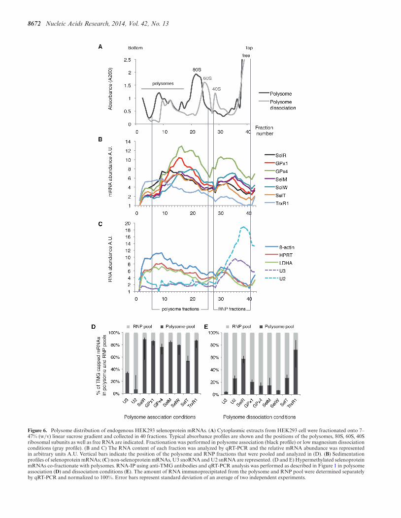
a prerequisite to their translation. To evaluate the abilityof selenoprotein mRNAs to associate with actively trans-lating ribosomes, we analyzed the polysome distribution ofendogenous selenoprotein mRNAs. Cytoplasmic extractsof cycloheximide-treated HEK293 cells (blocking trans-lation elongation) were fractionated on linear 7–47% su-crose gradients and the abundance of individual mRNAsin each fraction was measured by qRT-PCR (Figure 6A–C). The profiles revealed that all the endogenous selenopro-tein mRNAs tested, except TrxR1, sedimented to fractionsof lower molecular weight than non-selenoprotein mRNAs.The peak of selenoprotein mRNA population was foundin fractions 10–26 in close proximity to the 80S mono-somes (Figure 6B), whereas �-actin, HPRT and LDHAmRNAs sedimented in the heavier fractions 5–16 contain-ing the polysomes (Figure 6C). As expected, U3 snoRNAwas not found in polysomes (Figure 6C). These results in-dicate that fewer ribosomes are loaded on selenoproteinmRNAs, consistent with elongation pausing at the UGASec codon as previously reported for GPx4 (52). TrxR1 se-lenoprotein mRNA is the only mRNA found in heavierpolysome fractions (see Figure 6B). This correlates withthe fact that TrxR1 is the sole mRNA tested for whichthe UGA Sec codon is at the antepenultimate position be-fore the stop codon; in all the other selenoprotein mRNAsof our test panel, the UGA Sec codon resides between 39(SelW) and 285 nucleotides (SelR) downstream of the startcodon. Worth of note, the proportion of free versus ribo-some bound mRNAs is higher for selenoprotein than non-selenoprotein mRNAs. We next tested whether selenopro-
Nucleic Acids Research, 2014, Vol. 42, No. 13 8671
Figure 4. Tgs1 is recruited to selenoprotein mRNAs via interactionswith SBP2. (A) Immunopurification of endogenous SBP2 from HeLacytoplasmic extracts using antipeptide antibodies (�-pepSBP2) againstamino acids 380–852. In: input 4%; (−) beads without antibodies;PreI: beads with preimmune serum; IP: beads with antibodies. (B) Co-immunoprecipitations using anti-GFP beads and HEK293FT cells trans-fected with SBP2 and GFP-Tgs1 or SBP2 alone (no GFP). (C) Y2H inter-action tests performed in S. cerevisiae between SBP2 and Tgs1 LF or SF.(−) Controls with empty DNA binding (DB) or activation domain (AD)fusion vectors; (+) positive interaction control between SBP2 and Nu-fip. (D) Binding of recombinant His-Tgs1 to in vitro translated (35S-Met)-SBP2 using rabbit reticulocyte lysate (RRL) and bacterial S30 extracts(S30). His-Tgs1 was bound to protein A-Sepharose using anti-His antibod-ies. (E) Co-immunoprecipitations using anti-GFP beads and HEK293FTcells transfected by SBP2 and Nop58-YFP. (F) Binding of recombinantGST-Nop58 to in vitro translated (35S-Met)-SBP2 using RRL and S30 ex-tracts. GST-Nop58 (or GST alone) was bound to glutathione beads andincubated with the extract. (G) Co-immunoprecipitations using anti-GFPbeads and HEK293FT cells transfected by GFP-SBP2 or GFP-SMN andSBP2 without tag. (+ RNase A) co-immunoprecipitation performed in thepresence of RNase A. (H) Binding of recombinant GST-SMN (or GSTalone) to in vitro translated (35S-Met)-SBP2 using RRL and S30 extracts.Asterisks denote frequently encountered hSBP2 proteolysis fragments. urepresents non-specific protein signal revealed by the anti-SBP2 antibod-ies and that serves as an internal control. Ab: cross-reactivity signal fromantibodies bound to the beads.
Figure 5. Hypermethylated selenoprotein mRNAs are more abundant inthe cytoplasm. (A) Western blot analysis of the nuclear and cytoplasmicextracts using antibodies directed against the nuclear restricted ZNF143transcription factor and the cytoplasmic rpS21. (B) RNA was extractedfrom total, nuclear and cytoplasmic extracts of HEK293FT cells; RNA-IPs using anti-TMG antibodies were performed and analyzed as describedin Figure 1. The relative percentage of TMG-capped mRNA in both com-partments is represented. Error bars represent standard deviation of an av-erage of two independent experiments. U3 snoRNA and U6 snRNA wereused as controls for the quality of the fractionation.
tein mRNAs present in polysomes bear hypermethylatedcaps. TMG-IP experiments were performed on pooled frac-tions 6-26 (polysomes) or 28-41 (RNP) that contain free ornon-polysome associated RNAs. We reasonably consideredthat the two pools contained 100% of the RNAs. Immuno-precipitation yields dropped importantly after fractionationof the RNAs on sucrose gradients; nevertheless, the resultsclearly showed the presence of the hypermethylated seleno-protein mRNAs in the polysome pool (Figure 6D). Indeed,between 50% (SelT) and 80% (SelR) of the recovered TMG-capped mRNAs was present in the polysome fraction, therest of the TMG-capped selenoprotein mRNAs being in theRNP fractions (Figure 6D). As expected, U3 snoRNA andU2 snRNA were predominantly recovered from the RNPpool (Figure 6D). The non-selenoprotein mRNA controlswere not recognized by anti-TMG antibodies (see also Fig-ure 1C). When RNA fractionation was performed underlow magnesium concentration leading to ribosome dissoci-ation into subunits (Figure 6A), the signal of selenoproteinmRNA TMG-IP was shifted to the RNP fractions (Figure6E). Altogether these results show that hypermethylated se-lenoprotein mRNAs are found in polysomes and are there-fore translated.
Tgs1 activity, but not eIF4E, is required in vivo for translationof GPx1
In order to determine the functional importance of theTMG-cap modification by Tgs1 on selenoprotein synthe-sis in vivo, we constructed two stable cell lines capable ofexpressing HA tagged wild-type (wt) and mutant GPx1 se-
8672 Nucleic Acids Research, 2014, Vol. 42, No. 13
Figure 6. Polysome distribution of endogenous HEK293 selenoprotein mRNAs. (A) Cytoplasmic extracts from HEK293 cell were fractionated onto 7–47% (w/v) linear sucrose gradient and collected in 40 fractions. Typical absorbance profiles are shown and the positions of the polysomes, 80S, 60S, 40Sribosomal subunits as well as free RNA are indicated. Fractionation was performed in polysome association (black profile) or low magnesium dissociationconditions (gray profile). (B and C) The RNA content of each fraction was analyzed by qRT-PCR and the relative mRNA abundance was representedin arbitrary units A.U. Vertical bars indicate the position of the polysome and RNP fractions that were pooled and analyzed in (D). (B) Sedimentationprofiles of selenoprotein mRNAs; (C) non-selenoprotein mRNAs, U3 snoRNA and U2 snRNA are represented. (D and E) Hypermethylated selenoproteinmRNAs co-fractionate with polysomes. RNA-IP using anti-TMG antibodies and qRT-PCR analysis was performed as described in Figure 1 in polysomeassociation (D) and dissociation conditions (E). The amount of RNA immunoprecipitated from the polysome and RNP pool were determined separatelyby qRT-PCR and normalized to 100%. Error bars represent standard deviation of an average of two independent experiments.
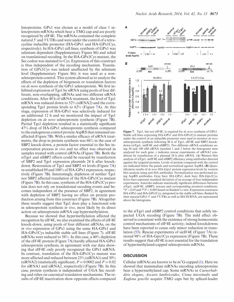
Nucleic Acids Research, 2014, Vol. 42, No. 13 8673
lenoproteins. GPx1 was chosen as a model of class 1 se-lenoprotein mRNAs which bear a TMG cap and are poorlyrecognized by eIF4E. The mRNAs contained the completenatural 5′ and 3′UTRs and were under the control of a tetra-cycline inducible promoter (HA-GPx1 and HA-GPx1Cys,respectively). In HA-GPx1 cell lines, synthesis of GPx1 wasselenium dependent (Supplementary Figure S6) and reliedon translational recoding. In the HA-GPx1Cys mutant, theSec codon was mutated to Cys. Expression of this constructis thus independent of the recoding mechanism. Transla-tion of GPx1Cys was indeed unaffected by the seleniumlevel (Supplementary Figure S6); it was used as a non-selenoprotein control. This system allowed us to analyze theeffects of the depletion of biogenesis or translation factorson de novo synthesis of the GPx1 selenoprotein. We first in-hibited expression of Tgs1 by siRNA using pools of four dif-ferent, non-overlapping, siRNAs and two different siRNAconditions. After 48 h of siRNA treatment, the level of Tgs1mRNA was reduced down to 52% (siRNA2) and the corre-sponding Tgs1 protein levels to 62% (Figure 7A). At thisstage, expression of HA-GPx1 was selectively induced foran additional 12 h and we monitored the impact of Tgs1depletion on de novo selenoprotein synthesis (Figure 7B).Partial Tgs1 depletion resulted in a statistically significant47% drop of HA-GPx1 selenoprotein synthesis comparedto the endogenous control protein AspRS that remained un-affected (Figure 7B). With a similar efficacy of siRNA treat-ments, the drop in protein synthesis was 66% in the case ofSBP2 knock-down, a protein factor essential to the Sec in-corporation process in vivo and no effect was observed insamples treated with control siRNAs (Figure 7B). Both thesiTgs1 and siSBP2 effects could be rescued by transfectionof SBP2 and Tgs1 expression plasmids 24 h after knock-down. Restoration of Tgs1 and SBP2 wt levels (Figure 7A)re-established 89 and 100% of HA-GPx1 expression, respec-tively (Figure 7B). Interestingly, depletion of neither Tgs1nor SBP2 affected translation of the HA-GPx1Cys mutant(Figure 7B). In this case, synthesis of the HA-GPx1Cys pro-tein does not rely on translational recoding events and be-comes independent of the presence of SBP2, in agreementwith depletion of SBP2 having no effect on protein pro-duction arising from this construct (Figure 7B). Altogetherthese results suggest that Tgs1 does play a functional rolein selenoprotein synthesis in vivo, most likely by its directaction on selenoprotein mRNA cap hypermethylation.
Because we showed that hypermethylation affected therecognition by eIF4E, we also examined the effects of eIF4Eknock-down, using pools of four different siRNAs, on thein vivo expression of GPx1 using the same HA-GPx1 andHA-GPx1Cys inducible stable cell lines (Figure 7). eIF4EmRNAs were reduced to 58%. In this case, a 40% depletionof the eIF4E protein (Figure 7A) hardly affected HA-GPx1selenoprotein synthesis, in agreement with our data show-ing that eIF4E only poorly recognized the GPx1 mRNA.In contrast, translation of the HA-GPx1Cys mutant wasmore affected and reduced between 23% (siRNA1) and 30%(siRNA2) (statistically significant, P = 0.0002 and P = 0.02for siRNA1 and siRNA2, respectively) (Figure 7B). In thiscase, protein synthesis is independent of UGA Sec recod-ing and relies on canonical translation mechanisms. The re-sults of eIF4E inactivation show opposite effects compared
Figure 7. Tgs1, but not eIF4E, is required for de novo synthesis of GPx1.Stable cell lines expressing HA-GPx1 and HA-GPx1Cys mutant proteinsunder the control of an inducible promoter were used to monitor de novoselenoprotein synthesis following 48 h of Tgs1, eIF4E and SBP2 knock-down (siTgs1, sieIF4E and siSBP2). Two different siRNA conditions us-ing 30 and 100 nM siRNA (marked 1 and 2 below the histogram) wereanalyzed for each gene. r indicates rescue experiments of siRNA2 con-ditions by transfection of a plasmid 24 h after siRNA. (A) Western blotanalysis of siTgs1, sieIF4E and siSBP2 efficiency using antibodies directedagainst the targeted proteins. Levels of protein compared with the controlare indicated below the panels and normalized against AspRS. (B) Quan-tification results of de novo HA-Gpx1 protein expression levels by westernblot analysis using anti-HA antibodies. Normalization was performed us-ing AspRS antibodies. Gray bars: HA-GPx1; dark bars: HA-Gpx1Cys.Error bars represent standard deviation of an average of four independentexperiments. Asterisks indicate statistically significant differences betweensiTgs1, sieIF4E, siSBP2, rescues and corresponding sicontrol conditions.*P < 0.05 and **P < 0.005 based on Student‘s t-test. Expression constructsHA-GPx1 and HA-GPx1Cys, integrated in the stable cell lines, flanked bytheir natural GPx1 5′ and 3′UTRs as well as SECIS RNA, are representedabove the histograms.
to the siTgs1 and siSBP2 control conditions that solely im-pacted UGA recoding (Figure 7B). The mild effect ob-served is consistent with the existence of strong homeostaticcontrol mechanisms of eIF4E activity; indeed knockdownshave been reported to cause only minor reduction in trans-lation (53). Rescue experiments of sieIF4E (Figure 7A) re-stored 98% of HA-Gpx1Cys expression (Figure 7B). Theseresults suggest that eIF4E is not essential for the translationof hypermethylated-capped selenoprotein mRNAs.
DISCUSSION
Cellular mRNAs are known to be m7G-capped (1). Here weshowed that mammalian mRNAs encoding selenoproteinsbear a hypermethylated cap. Some mRNAs in Caenorhab-ditis elegans, Ascaris lumbricoides, Ciona intestinalis andEuglena gracilis acquire TMG caps but by spliced-leader

8674 Nucleic Acids Research, 2014, Vol. 42, No. 13
Figure 8. Model for cap hypermethylation and translation of selenoprotein mRNPs. The caps of selenoprotein mRNAs are hypermethylated by Tgs1. Tgs1isoforms (Tgs1 SF and Tgs1 LF) interact with SBP2 and selenoprotein mRNAs and are recruited predominantly via the ternary complex SBP2/SMN/Tgs1LF (thick arrows) but also via SBP2/Nop58/Tgs1 SF (thin arrows). Hypermethylated-capped selenoprotein mRNAs are localized to the cytoplasm andare polysome-associated but show reduced affinity for eIF4E. Tgs1 activity is required for Sec incorporation or translation initiation.
(SL) trans-splicing, thus a radically different pathway thandescribed here (54–57). Some Rev/RRE-dependent HIV-1RNAs can also be TMG-capped by Tgs1, TMG cappingrepresenting in this case a regulation mechanism for selec-tive expression (58). To assess the impact of TMG cappingon selenoprotein mRNA export, stability and translationis challenging. Hypermethylation of selenoprotein mRNAcap may serve different purposes: dedicated assembly path-way for selenoprotein mRNPs and/or control of seleno-protein expression at the level of translation initiation orUGA Sec recoding. Our experiments reveal that seleno-protein mRNAs are subjected to differential 5′ processingand translational initiation events. Although some are sub-strates for Tgs1, harbor hypermethylated caps and are notrecognized efficiently by the translation factor eIF4E, oth-ers are m7G-capped and recognized by eIF4E. These dif-ferences between selenoprotein mRNAs are not surprisingas differential expression and stability regulation mecha-nisms have been reported for various subsets of selenopro-tein mRNAs under conditions as different as selenium lev-els, type of SBP2–SECIS interactions, and sensitivity to
NMD (nonsense mediated decay) (44,59–63). Control ofgene-specific selenoprotein expression and differential reg-ulation of UGA Sec recoding of a subset of selenoproteinmRNAs were recently shown to be selenium-dependent andmediated by the degree of tRNA[Ser]Sec Um34 methylation(43). Likewise, it is conceivable that cap hypermethylationcould also be part of a regulation process of selenoproteinexpression. As suggested by our results, hypermethylationdoes not necessarily occur equally on all selenoprotein mR-NAs, possibly regulating translation initiation in a way thatcould contribute to the mechanisms of hierarchy of seleno-protein expression. Mechanisms and factors influencing thefate of selenoprotein mRNAs are far from being understoodand so far no unified mechanism could be proposed forall selenoprotein mRNAs, at any stage of their mRNA lifecycle. Our findings point to the existence of an additionallevel of complexity in the mechanism of selenoprotein syn-thesis residing at the stage of mRNP assembly, processingand translation initiation (Figure 8). This process is gov-erned by the interactions between SBP2, Tgs1 and seleno-protein mRNAs. We established that the hypermethylase
Nucleic Acids Research, 2014, Vol. 42, No. 13 8675
Tgs1 is recruited to selenoprotein mRNAs predominantlyvia SMN but also Nop58, and therefore probably belongsto two possible ternary complexes: SBP2/Nop58/Tgs1 orSBP2/SMN/Tgs1 (Figure 8). Interestingly, oxidative stresswas identified as a novel regulation mechanism for selectiveselenoprotein synthesis (64). It was shown to induce nuclearshuttling of SBP2 and selenocysteine incorporation factors,and to promote assembly of selenoprotein mRNPs (32,33).It is conceivable that oxidative stress could also regulate hy-permethylation of selenoprotein mRNAs by stimulating re-cruitment of the nuclear isoform of Tgs1.
SBP2 is a member of the L7Ae family of RNA-bindingproteins (65) that include the archaeal L7Ae and eukary-otic 15.5 kDa core proteins of the box C/D sRNPs andsnoRNPs, respectively. We have previously shown that thestructural motifs of the SECIS and box C/D RNAs as wellas binding strategies governing the interaction of SBP2,L7Ae and 15.5 kDa to their cognate RNAs are similar andare controlled by a common conserved RNA chaperonecomplex (27,34,66). Interestingly, it is the L7Ae and 15.5kDa proteins that play a key role in box C/D RNP assemblyin Archaea and Eukarya, respectively, by specifically rec-ognizing their cognate RNA motif. Moreover, in the caseof U3 snoRNA, assembly and maturation was shown tobe dependent on a large multiprotein complex that con-tains, in addition to the core proteins, components of theRNA chaperone complex but also RNA processing factorsincluding Tgs1 (22,25). Likewise, our present data suggestthat SBP2 could trigger the assembly of the cap modifi-cation complex by recruiting Nop58 or SMN in a similarmanner, and this would ultimately lead to the binding ofTgs1 methylase for modification. Tgs1 SF is strictly nuclearbut Tgs1 LF is present both in the nucleus and the cyto-plasm. This suggests that m7G hypermethylation could oc-cur in either of the two cellular compartments via two al-ternative pathways and could obey a fast and potentiallyreversible regulation process (Figure 8). Altogether, our re-sults demonstrate that selenoprotein mRNAs indeed un-dergo a sophisticated assembly and 5’end maturation path-way similar to that of sn- and snoRNAs, and which involvecommon maturation factors and core proteins.
However, unlike TMG-capped sn- or snoRNAs, our re-sults also show that hypermethylated-capped selenoproteinmRNAs can be found in the cytoplasm and in activelytranslating ribosomes, excluding nuclear retention as a con-sequence of cap hypermethylation. Hypermethylation byTgs1 rather seems to be required for translational recod-ing events, as its knock-down reduced expression of GPx1.Our current data do not allow us to differentiate whetherTgs1 activity is required for Sec incorporation or transla-tion initiation (Figure 8). It is conceivable that both mech-anisms could be linked for optimal selenoprotein transla-tion. Specialized translation initiation factors or alterna-tive mechanisms are therefore likely to be involved in hy-permethylated mRNA recognition. One possibility wouldbe an IRES-dependent translation. Consistently, earlier re-ports showed that the cricket paralysis virus internal ribo-some entry site is able to support Sec incorporation in a lu-ciferase reporter construct, although the incorporation ef-ficiency was decreased (67). Our results also showed a re-duced affinity of the translation initiation factor eIF4E for
selenoprotein mRNA cap structures. However, eIF4E vari-ants that can accommodate the TMG cap were identifiedin the genome of C. elegans (68). Whether such a situationprevails for some selenoprotein mRNAs will be the aim offuture investigations.
ACCESSION NUMBER
NCBI Gene Expression Omnibus accession number:GSE57625.
SUPPLEMENTARY DATA
Supplementary Data are available at NAR Online.
ACKNOWLEDGMENTS
We thank R. Luhrmann for the gift of HeLa cell extractsand antibodies, C.H. Hagedorn for the pGST-eIF4E plas-mid, A. Schweigert for technical assistance, E. Westhof forinvaluable input and constant support, P. Carbon and G.Eriani for helpful discussions and critical reading of themanuscript and J. Steitz for useful comments and criticalreading of the manuscript.Authors contributions: L.W, A.S.G.B., C.V., A.K., E.B. andC.A. designed research. A.S.G.B., L.W., C.V., M.L., A.T.,S.B., F.M. and C.A. performed research. L.W., A.S.G.B.,C.V., F.M., A.K., E.B. and C.A. analyzed data. C.A. wrotethe paper.
FUNDING
Centre national de la recherche scientifique (CNRS);Agence Nationale de la Recherche [BLAN06–2 134682,ANR-2011-svse8 02501 to F.M.]. French Ministry of Re-search and the National Research Fund, Luxembourg [toL.W.]; Contract Doctoral of the University of Strasbourg[A.S.G.]. Funding for open access charge: CNRS.Conflict of interest statement. None declared.
REFERENCES1. Shatkin,A.J. (1976) Capping of eucaryotic mRNAs. Cell, 9, 645–653.2. Topisirovic,I., Svitkin,Y.V., Sonenberg,N. and Shatkin,A.J. (2011)
Cap and cap-binding proteins in the control of gene expression. WileyInterdiscip. Rev. RNA, 2, 277–298.
3. Izaurralde,E., Lewis,J., Gamberi,C., Jarmolowski,A., McGuigan,C.and Mattaj,I.W. (1995) A cap-binding protein complex mediating UsnRNA export. Nature, 376, 709–712.
4. Kohler,A. and Hurt,E. (2007) Exporting RNA from the nucleus tothe cytoplasm. Nat. Rev. Mol. Cell. Biol., 8, 761–773.
5. Mitchell,P. and Tollervey,D. (2001) mRNA turnover. Curr. Opin. CellBiol., 13, 320–325.
6. Wilusz,C.J., Wormington,M. and Peltz,S.W. (2001) The cap-to-tailguide to mRNA turnover. Nat. Rev. Mol. Cell. Biol., 2, 237–246.
7. Houseley,J. and Tollervey,D. (2009) The many pathways of RNAdegradation. Cell, 136, 763–776.
8. Fortes,P., Inada,T., Preiss,T., Hentze,M.W., Mattaj,I.W. andSachs,A.B. (2000) The yeast nuclear cap binding complex can interactwith translation factor eIF4G and mediate translation initiation.Mol. Cell, 6, 191–196.
9. McKendrick,L., Thompson,E., Ferreira,J., Morley,S.J. andLewis,J.D. (2001) Interaction of eukaryotic translation initiationfactor 4G with the nuclear cap-binding complex provides a linkbetween nuclear and cytoplasmic functions of the m(7) guanosinecap. Mol. Cell. Biol., 21, 3632–3641.

8676 Nucleic Acids Research, 2014, Vol. 42, No. 13
10. Sonenberg,N. (2008) eIF4E, the mRNA cap-binding protein: frombasic discovery to translational research. Biochem. Cell Biol., 86,178–183.
11. Calero,G., Wilson,K.F., Ly,T., Rios-Steiner,J.L., Clardy,J.C. andCerione,R.A. (2002) Structural basis of m7GpppG binding to thenuclear cap-binding protein complex. Nat. Struct. Biol., 9, 912–917.
12. Marcotrigiano,J., Gingras,A.C., Sonenberg,N. and Burley,S.K.(1997) Cocrystal structure of the messenger RNA 5’ cap-bindingprotein (eIF4E) bound to 7-methyl-GDP. Cell, 89, 951–961.
13. Izaurralde,E., Lewis,J., McGuigan,C., Jankowska,M.,Darzynkiewicz,E. and Mattaj,I.W. (1994) A nuclear cap bindingprotein complex involved in pre-mRNA splicing. Cell, 78, 657–668.
14. Darzynkiewicz,E., Stepinski,J., Ekiel,I., Jin,Y., Haber,D., Sijuwade,T.and Tahara,S.M. (1988) Beta-globin mRNAs capped with m7G,m2.7(2)G or m2.2.7(3)G differ in intrinsic translation efficiency.Nucleic Acids Res., 16, 8953–8962.
15. Matera,A.G., Terns,R.M. and Terns,M.P. (2007) Non-coding RNAs:lessons from the small nuclear and small nucleolar RNAs. Nat. Rev.Mol. Cell. Biol., 8, 209–220.
16. Chari,A., Paknia,E. and Fischer,U. (2009) The role of RNPbiogenesis in spinal muscular atrophy. Curr. Opin. Cell Biol., 21,387–393.
17. Fischer,U. and Luhrmann,R. (1990) An essential signaling role forthe m3G cap in the transport of U1 snRNP to the nucleus. Science,249, 786–790.
18. Fischer,U., Sumpter,V., Sekine,M., Satoh,T. and Luhrmann,R.(1993) Nucleo-cytoplasmic transport of U snRNPs: definition of anuclear location signal in the Sm core domain that binds a transportreceptor independently of the m3G cap. EMBO J., 12, 573–583.
19. Huber,J., Cronshagen,U., Kadokura,M., Marshallsay,C., Wada,T.,Sekine,M. and Luhrmann,R. (1998) Snurportin1, anm3G-cap-specific nuclear import receptor with a novel domainstructure. EMBO J., 17, 4114–4126.
20. Hamm,J., Darzynkiewicz,E., Tahara,S.M. and Mattaj,I.W. (1990)The trimethylguanosine cap structure of U1 snRNA is a componentof a bipartite nuclear targeting signal. Cell, 62, 569–577.
21. Verheggen,C., Lafontaine,D.L., Samarsky,D., Mouaikel,J.,Blanchard,J.M., Bordonne,R. and Bertrand,E. (2002) Mammalianand yeast U3 snoRNPs are matured in specific and related nuclearcompartments. EMBO J., 21, 2736–2745.
22. Boulon,S., Verheggen,C., Jady,B.E., Girard,C., Pescia,C., Paul,C.,Ospina,J.K., Kiss,T., Matera,A.G., Bordonne,R. et al. (2004) PHAXand CRM1 are required sequentially to transport U3 snoRNA tonucleoli. Mol. Cell, 16, 777–787.
23. Girard,C., Verheggen,C., Neel,H., Cammas,A., Vagner,S., Soret,J.,Bertrand,E. and Bordonne,R. (2008) Characterization of a shortisoform of human Tgs1 hypermethylase associating with smallnucleolar ribonucleoprotein core proteins and produced by limitedproteolytic processing. J. Biol. Chem., 283, 2060–2069.
24. Mouaikel,J., Verheggen,C., Bertrand,E., Tazi,J. and Bordonne,R.(2002) Hypermethylation of the cap structure of both yeast snRNAsand snoRNAs requires a conserved methyltransferase that is localizedto the nucleolus. Mol. Cell, 9, 891–901.
25. Watkins,N.J., Lemm,I., Ingelfinger,D., Schneider,C., Hobach,M.,Urlaub,H. and Luhrmann,R. (2004) Assembly and maturation of theU3 snoRNP in the nucleoplasm in a large dynamic multiproteincomplex. Mol. Cell, 16, 789–798.
26. Allmang,C., Wurth,L. and Krol,A. (2009) The selenium toselenoprotein pathway in eukaryotes: more molecular partners thananticipated. Biochim. Biophys. Acta, 1790, 1415–1423.
27. Boulon,S., Marmier-Gourrier,N., Pradet-Balade,B., Wurth,L.,Verheggen,C., Jady,B.E., Rothe,B., Pescia,C., Robert,M.C., Kiss,T.et al. (2008) The Hsp90 chaperone controls the biogenesis of L7AeRNPs through conserved machinery. J. Cell Biol., 180, 579–595.
28. Copeland,P.R., Fletcher,J.E., Carlson,B.A., Hatfield,D.L. andDriscoll,D.M. (2000) A novel RNA binding protein, SBP2, isrequired for the translation of mammalian selenoprotein mRNAs.EMBO J., 19, 306–314.
29. Lescure,A., Allmang,C., Yamada,K., Carbon,P. and Krol,A. (2002)cDNA cloning, expression pattern and RNA binding analysis ofhuman selenocysteine insertion sequence (SECIS) binding protein 2.Gene, 291, 279–285.
30. Takeuchi,A., Schmitt,D., Chapple,C., Babaylova,E., Karpova,G.,Guigo,R., Krol,A. and Allmang,C. (2009) A short motif in
Drosophila SECIS Binding Protein 2 provides differential bindingaffinity to SECIS RNA hairpins. Nucleic Acids Res., 37, 2126–2141.
31. Kinzy,S.A., Caban,K. and Copeland,P.R. (2005) Characterization ofthe SECIS binding protein 2 complex required for theco-translational insertion of selenocysteine in mammals. NucleicAcids Res., 33, 5172–5180.
32. Papp,L.V., Lu,J., Striebel,F., Kennedy,D., Holmgren,A. andKhanna,K.K. (2006) The redox state of SECIS binding protein 2controls its localization and selenocysteine incorporation function.Mol. Cell. Biol., 26, 4895–4910.
33. de Jesus,L.A., Hoffmann,P.R., Michaud,T., Forry,E.P.,Small-Howard,A., Stillwell,R.J., Morozova,N., Harney,J.W. andBerry,M.J. (2006) Nuclear assembly of UGA decoding complexes onselenoprotein mRNAs: a mechanism for eluding nonsense-mediateddecay? Mol. Cell. Biol., 26, 1795–1805.
34. Clery,A., Bourguignon-Igel,V., Allmang,C., Krol,A. and Branlant,C.(2007) An improved definition of the RNA-binding specificity ofSECIS-binding protein 2, an essential component of theselenocysteine incorporation machinery. Nucleic Acids Res., 35,1868–1884.
35. Dignam,J.D., Lebovitz,R.M. and Roeder,R.G. (1983) Accuratetranscription initiation by RNA polymerase II in a soluble extractfrom isolated mammalian nuclei. Nucleic Acids Res., 11, 1475–1489.
36. Frugier,M., Ryckelynck,M. and Giege,R. (2005) tRNA-balancedexpression of a eukaryal aminoacyl-tRNA synthetase by anmRNA-mediated pathway. EMBO Rep., 6, 860–865.
37. Pradet-Balade,B., Girard,C., Boulon,S., Paul,C., Azzag,K.,Bordonne,R., Bertrand,E. and Verheggen,C. (2011) CRM1 controlsthe composition of nucleoplasmic pre-snoRNA complexes to licencethem for nucleolar transport. EMBO J., 30, 2205–2218.
38. Luhrmann,R., Appel,B., Bringmann,P., Rinke,J., Reuter,R., Rothe,S.and Bald,R. (1982) Isolation and characterization of rabbit anti-m32,2,7G antibodies. Nucleic Acids Res., 10, 7103–7113.
39. Tycowski,K.T., Aab,A. and Steitz,J.A. (2004) Guide RNAs with 5’caps and novel box C/D snoRNA-like domains for modification ofsnRNAs in metazoa. Curr. Biol., 14, 1985–1995.
40. Choi,Y.H. and Hagedorn,C.H. (2003) Purifying mRNAs with ahigh-affinity eIF4E mutant identifies the short 3’ poly(A) endphenotype. Proc. Natl. Acad. Sci. U.S.A., 100, 7033–7038.
41. Marcotrigiano,J., Gingras,A.C., Sonenberg,N. and Burley,S.K.(1997) X-ray studies of the messenger RNA 5’ cap-binding protein(eIF4E) bound to 7-methyl-GDP. Nucleic Acids Symp. Ser., 8–11.
42. Kryukov,G.V., Castellano,S., Novoselov,S.V., Lobanov,A.V.,Zehtab,O., Guigo,R. and Gladyshev,V.N. (2003) Characterization ofmammalian selenoproteomes. Science, 300, 1439–1443.
43. Howard,M.T., Carlson,B.A., Anderson,C.B. and Hatfield,D.L. (2013)Translational redefinition of UGA codons is regulated by seleniumavailability. J. Biol. Chem., 288, 19401–19413.
44. Sun,X., Li,X., Moriarty,P.M., Henics,T., LaDuca,J.P. andMaquat,L.E. (2001) Nonsense-mediated decay of mRNA for theselenoprotein phospholipid hydroperoxide glutathione peroxidase isdetectable in cultured cells but masked or inhibited in rat tissues. Mol.Biol. Cell, 12, 1009–1017.
45. Weiss Sachdev,S. and Sunde,R.A. (2001) Selenium regulation oftranscript abundance and translational efficiency of glutathioneperoxidase-1 and -4 in rat liver. Biochem. J., 357, 851–858.
46. Moriarty,P.M., Reddy,C.C. and Maquat,L.E. (1998) Seleniumdeficiency reduces the abundance of mRNA for Se-dependentglutathione peroxidase 1 by a UGA-dependent mechanism likely tobe nonsense codon-mediated decay of cytoplasmic mRNA. Mol. Cell.Biol., 18, 2932–2939.
47. Paushkin,S., Gubitz,A.K., Massenet,S. and Dreyfuss,G. (2002) TheSMN complex, an assemblyosome of ribonucleoproteins. Curr. Opin.Cell Biol., 14, 305–312.
48. Mouaikel,J., Narayanan,U., Verheggen,C., Matera,A.G.,Bertrand,E., Tazi,J. and Bordonne,R. (2003) Interaction between thesmall-nuclear-RNA cap hypermethylase and the spinal muscularatrophy protein, survival of motor neuron. EMBO Rep., 4, 616–622.
49. Zhang,Z., Lotti,F., Dittmar,K., Younis,I., Wan,L., Kasim,M. andDreyfuss,G. (2008) SMN deficiency causes tissue-specificperturbations in the repertoire of snRNAs and widespread defects insplicing. Cell, 133, 585–600.

Nucleic Acids Research, 2014, Vol. 42, No. 13 8677
50. Myslinski,E., Krol,A. and Carbon,P. (1998) ZNF76 and ZNF143 aretwo human homologs of the transcriptional activator Staf. J. Biol.Chem., 273, 21998–22006.
51. Mottram,J., Perry,K.L., Lizardi,P.M., Luhrmann,R., Agabian,N. andNelson,R.G. (1989) Isolation and sequence of four small nuclear URNA genes of Trypanosoma brucei subsp. brucei: identification ofthe U2, U4, and U6 RNA analogs. Mol. Cell. Biol., 9, 1212–1223.
52. Fletcher,J.E., Copeland,P.R. and Driscoll,D.M. (2000) Polysomedistribution of phospholipid hydroperoxide glutathione peroxidasemRNA: evidence for a block in elongation at theUGA/selenocysteine codon. RNA, 6, 1573–1584.
53. Yanagiya,A., Suyama,E., Adachi,H., Svitkin,Y.V., Aza-Blanc,P.,Imataka,H., Mikami,S., Martineau,Y., Ronai,Z.A. and Sonenberg,N.(2012) Translational homeostasis via the mRNA cap-binding protein,eIF4E. Mol. Cell, 46, 847–858.
54. Liou,R.F. and Blumenthal,T. (1990) trans-spliced Caenorhabditiselegans mRNAs retain trimethylguanosine caps. Mol. Cell. Biol., 10,1764–1768.
55. Van Doren,K. and Hirsh,D. (1990) mRNAs that mature throughtrans-splicing in Caenorhabditis elegans have a trimethylguanosinecap at their 5’ termini. Mol. Cell. Biol., 10, 1769–1772.
56. Maroney,P.A., Denker,J.A., Darzynkiewicz,E., Laneve,R. andNilsen,T.W. (1995) Most mRNAs in the nematode Ascarislumbricoides are trans-spliced: a role for spliced leader addition intranslational efficiency. RNA, 1, 714–723.
57. Hastings,K.E. (2005) SL trans-splicing: easy come or easy go? TrendsGenet.: TIG, 21, 240–247.
58. Yedavalli,V.S. and Jeang,K.T. (2010) Trimethylguanosine cappingselectively promotes expression of Rev-dependent HIV-1 RNAs.Proc. Natl. Acad. Sci. U.S.A., 107, 14787–14792.
59. Lei,X.G., Evenson,J.K., Thompson,K.M. and Sunde,R.A. (1995)Glutathione peroxidase and phospholipid hydroperoxide glutathioneperoxidase are differentially regulated in rats by dietary selenium. J.Nutr., 125, 1438–1446.
60. Low,S.C., Grundner-Culemann,E., Harney,J.W. and Berry,M.J.(2000) SECIS-SBP2 interactions dictate selenocysteine incorporationefficiency and selenoprotein hierarchy. EMBO J., 19, 6882–6890.
61. Squires,J.E., Stoytchev,I., Forry,E.P. and Berry,M.J. (2007) SBP2binding affinity is a major determinant in differential selenoproteinmRNA translation and sensitivity to nonsense-mediated decay. Mol.Cell. Biol., 27, 7848–7855.
62. Budiman,M.E., Bubenik,J.L., Miniard,A.C., Middleton,L.M.,Gerber,C.A., Cash,A. and Driscoll,D.M. (2009) Eukaryotic initiationfactor 4a3 is a selenium-regulated RNA-binding protein thatselectively inhibits selenocysteine incorporation. Mol. Cell, 35,479–489.
63. Latreche,L., Duhieu,S., Touat-Hamici,Z., Jean-Jean,O. andChavatte,L. (2012) The differential expression of glutathioneperoxidase 1 and 4 depends on the nature of the SECIS element. RNABiol, 9, 681–690.
64. Touat-Hamici,Z., Legrain,Y., Bulteau,A.L. andChavatte,L. (2014) Selective up-regulation of human selenoproteinsin response to oxidative stress. J. Biol. Chem., 289, 14750–14761.
65. Koonin,E.V., Bork,P. and Sander,C. (1994) A novel RNA-bindingmotif in omnipotent suppressors of translation termination,ribosomal proteins and a ribosome modification enzyme? NucleicAcids Res., 22, 2166–2167.
66. Allmang,C., Carbon,P. and Krol,A. (2002) The SBP2 and 15.5kD/Snu13p proteins share the same RNA binding domain:identification of SBP2 amino acids important to SECIS RNAbinding. RNA, 8, 1308–1318.
67. Donovan,J. and Copeland,P.R. (2010) The efficiency ofselenocysteine incorporation is regulated by translation initiationfactors. J. Mol. Biol., 400, 659–664.
68. Keiper,B.D., Lamphear,B.J., Deshpande,A.M.,Jankowska-Anyszka,M., Aamodt,E.J., Blumenthal,T. andRhoads,R.E. (2000) Functional characterization of five eIF4Eisoforms in Caenorhabditis elegans. J. Biol. Chem., 275, 10590–10596.
Related Documents