A Novel Protein Kinase-Like Domain in a Selenoprotein, Widespread in the Tree of Life Malgorzata Dudkiewicz 3 , Teresa Szczepin ´ ska 1 , Marcin Grynberg 2 , Krzysztof Pawlowski 1,3 * 1 Nencki Institute of Experimental Biology, Polish Academy of Sciences, Warsaw, Poland, 2 Institute of Biochemistry and Biophysics, Polish Academy of Sciences, Warsaw, Poland, 3 Warsaw University of Life Sciences, Warsaw, Poland Abstract Selenoproteins serve important functions in many organisms, usually providing essential oxidoreductase enzymatic activity, often for defense against toxic xenobiotic substances. Most eukaryotic genomes possess a small number of these proteins, usually not more than 20. Selenoproteins belong to various structural classes, often related to oxidoreductase function, yet a few of them are completely uncharacterised. Here, the structural and functional prediction for the uncharacterised selenoprotein O (SELO) is presented. Using bioinformatics tools, we predict that SELO protein adopts a three-dimensional fold similar to protein kinases. Furthermore, we argue that despite the lack of conservation of the ‘‘classic’’ catalytic aspartate residue of the archetypical His-Arg-Asp motif, SELO kinases might have retained catalytic phosphotransferase activity, albeit with an atypical active site. Lastly, the role of the selenocysteine residue is considered and the possibility of an oxidoreductase-regulated kinase function for SELO is discussed. The novel kinase prediction is discussed in the context of functional data on SELO orthologues in model organisms, FMP40 a.k.a.YPL222W (yeast), and ydiU (bacteria). Expression data from bacteria and yeast suggest a role in oxidative stress response. Analysis of genomic neighbourhoods of SELO homologues in the three domains of life points toward a role in regulation of ABC transport, in oxidative stress response, or in basic metabolism regulation. Among bacteria possessing SELO homologues, there is a significant over-representation of aquatic organisms, also of aerobic ones. The selenocysteine residue in SELO proteins occurs only in few members of this protein family, including proteins from Metazoa, and few small eukaryotes (Ostreococcus, stramenopiles). It is also demonstrated that enterobacterial mchC proteins involved in maturation of bactericidal antibiotics, microcins, form a distant subfamily of the SELO proteins. The new protein structural domain, with a putative kinase function assigned, expands the known kinome and deserves experimental determination of its biological role within the cell-signaling network. Citation: Dudkiewicz M, Szczepin ´ ska T, Grynberg M, Pawlowski K (2012) A Novel Protein Kinase-Like Domain in a Selenoprotein, Widespread in the Tree of Life. PLoS ONE 7(2): e32138. doi:10.1371/journal.pone.0032138 Editor: Ahmed Moustafa, American University in Cairo, Egypt Received June 13, 2011; Accepted January 24, 2012; Published February 16, 2012 Copyright: ß 2012 Dudkiewicz et al. This is an open-access article distributed under the terms of the Creative Commons Attribution License, which permits unrestricted use, distribution, and reproduction in any medium, provided the original author and source are credited. Funding: MD, KP, TS and MG were supported by the Polish Ministry of Science and Higher Education grant N N301 3165 33. The funders had no role in study design, data collection and analysis, decision to publish, or preparation of the manuscript. Competing Interests: The authors have declared that no competing interests exist. * E-mail: [email protected] Introduction Selenoproteins are an intriguing evolutionary creation, char- acterised by the presence of an atypical aminoacid residue, selenocysteine. Maintaining the machinery for selenocysteine synthesis and incorporation just for the sake of a handful of proteins is costly [1], also the requirement for selenium acquisition from environment poses difficulty. However, advantages of selenocysteine (Sec, U) as compared to cysteine (Cys, C) may partly offset these problems. Among the unique properties of selenocysteine, instead of cysteine in enzymatic active sites, higher nucleophilicity of Sec versus Cys, higher oxidoreductase efficiency, and lower pK a , have been cited [1]. Human selenoproteins are encoded by 25 genes, and most of those with known functions are oxidoreductases with a selenocysteine being in the active site. A few human selenogenes are functionally uncharacterised [2]. In addition, we suppose that in the uncharacterised selenoproteins, a selenocysteine residue conserved in evolution is not very likely to be just a troublesome decoration. A catalytic or regulatory function for this residue, and the protein as a whole, seems more reasonable. Hence, we undertook a structural and functional prediction study for the human selenoprotein O (SELO), one of the very few uncharacterised selenoproteins in humans. The human SELO (NCBI gi: 172045770) has been predicted to be a selenoprotein by a bioinformatics approach and confirmed to be an expressed selenoprotein by Gladyshev and co-workers [2]. As outlined in the Results section, not all SELO family proteins identified by us are selenoproteins, yet we use the SELO name for the entire family for consistency. SELO selenoproteins have a single selenocysteine residue while those family members that are not selenoproteins, usually have a cysteine residue instead in the corresponding position. In most eukaryotes and many bacteria, SELO is present as a single-copy protein, while duplicate copies in many metazoans and a few bacteria exist. Incidentally, a few years ago Koonin and colleagues proposed SELO among the top ten most-wanted ’’unknown unknowns’’, when discussing the proteins of unknown structure and function posing exciting conceptual challenges for structure predictors, based on phyletic spread [3]. The same authors reiterated that list just recently, indicating ‘‘no news’’ for SELO [4]. PLoS ONE | www.plosone.org 1 February 2012 | Volume 7 | Issue 2 | e32138

Welcome message from author
This document is posted to help you gain knowledge. Please leave a comment to let me know what you think about it! Share it to your friends and learn new things together.
Transcript

A Novel Protein Kinase-Like Domain in a Selenoprotein,Widespread in the Tree of LifeMałgorzata Dudkiewicz3, Teresa Szczepinska1, Marcin Grynberg2, Krzysztof Pawłowski1,3*
1 Nencki Institute of Experimental Biology, Polish Academy of Sciences, Warsaw, Poland, 2 Institute of Biochemistry and Biophysics, Polish Academy of Sciences, Warsaw,
Poland, 3 Warsaw University of Life Sciences, Warsaw, Poland
Abstract
Selenoproteins serve important functions in many organisms, usually providing essential oxidoreductase enzymatic activity,often for defense against toxic xenobiotic substances. Most eukaryotic genomes possess a small number of these proteins,usually not more than 20. Selenoproteins belong to various structural classes, often related to oxidoreductase function, yet afew of them are completely uncharacterised. Here, the structural and functional prediction for the uncharacterisedselenoprotein O (SELO) is presented. Using bioinformatics tools, we predict that SELO protein adopts a three-dimensionalfold similar to protein kinases. Furthermore, we argue that despite the lack of conservation of the ‘‘classic’’ catalyticaspartate residue of the archetypical His-Arg-Asp motif, SELO kinases might have retained catalytic phosphotransferaseactivity, albeit with an atypical active site. Lastly, the role of the selenocysteine residue is considered and the possibility of anoxidoreductase-regulated kinase function for SELO is discussed. The novel kinase prediction is discussed in the context offunctional data on SELO orthologues in model organisms, FMP40 a.k.a.YPL222W (yeast), and ydiU (bacteria). Expression datafrom bacteria and yeast suggest a role in oxidative stress response. Analysis of genomic neighbourhoods of SELOhomologues in the three domains of life points toward a role in regulation of ABC transport, in oxidative stress response, orin basic metabolism regulation. Among bacteria possessing SELO homologues, there is a significant over-representation ofaquatic organisms, also of aerobic ones. The selenocysteine residue in SELO proteins occurs only in few members of thisprotein family, including proteins from Metazoa, and few small eukaryotes (Ostreococcus, stramenopiles). It is alsodemonstrated that enterobacterial mchC proteins involved in maturation of bactericidal antibiotics, microcins, form adistant subfamily of the SELO proteins. The new protein structural domain, with a putative kinase function assigned,expands the known kinome and deserves experimental determination of its biological role within the cell-signalingnetwork.
Citation: Dudkiewicz M, Szczepinska T, Grynberg M, Pawłowski K (2012) A Novel Protein Kinase-Like Domain in a Selenoprotein, Widespread in the Tree ofLife. PLoS ONE 7(2): e32138. doi:10.1371/journal.pone.0032138
Editor: Ahmed Moustafa, American University in Cairo, Egypt
Received June 13, 2011; Accepted January 24, 2012; Published February 16, 2012
Copyright: � 2012 Dudkiewicz et al. This is an open-access article distributed under the terms of the Creative Commons Attribution License, which permitsunrestricted use, distribution, and reproduction in any medium, provided the original author and source are credited.
Funding: MD, KP, TS and MG were supported by the Polish Ministry of Science and Higher Education grant N N301 3165 33. The funders had no role in studydesign, data collection and analysis, decision to publish, or preparation of the manuscript.
Competing Interests: The authors have declared that no competing interests exist.
* E-mail: [email protected]
Introduction
Selenoproteins are an intriguing evolutionary creation, char-
acterised by the presence of an atypical aminoacid residue,
selenocysteine. Maintaining the machinery for selenocysteine
synthesis and incorporation just for the sake of a handful of
proteins is costly [1], also the requirement for selenium acquisition
from environment poses difficulty. However, advantages of
selenocysteine (Sec, U) as compared to cysteine (Cys, C) may
partly offset these problems. Among the unique properties of
selenocysteine, instead of cysteine in enzymatic active sites, higher
nucleophilicity of Sec versus Cys, higher oxidoreductase efficiency,
and lower pKa, have been cited [1].
Human selenoproteins are encoded by 25 genes, and most of
those with known functions are oxidoreductases with a
selenocysteine being in the active site. A few human
selenogenes are functionally uncharacterised [2]. In addition,
we suppose that in the uncharacterised selenoproteins, a
selenocysteine residue conserved in evolution is not very likely
to be just a troublesome decoration. A catalytic or regulatory
function for this residue, and the protein as a whole, seems
more reasonable. Hence, we undertook a structural and
functional prediction study for the human selenoprotein O
(SELO), one of the very few uncharacterised selenoproteins in
humans. The human SELO (NCBI gi: 172045770) has been
predicted to be a selenoprotein by a bioinformatics approach
and confirmed to be an expressed selenoprotein by Gladyshev
and co-workers [2]. As outlined in the Results section, not all
SELO family proteins identified by us are selenoproteins, yet
we use the SELO name for the entire family for consistency.
SELO selenoproteins have a single selenocysteine residue while
those family members that are not selenoproteins, usually have
a cysteine residue instead in the corresponding position. In
most eukaryotes and many bacteria, SELO is present as a
single-copy protein, while duplicate copies in many metazoans
and a few bacteria exist.
Incidentally, a few years ago Koonin and colleagues proposed
SELO among the top ten most-wanted ’’unknown unknowns’’,
when discussing the proteins of unknown structure and function
posing exciting conceptual challenges for structure predictors,
based on phyletic spread [3]. The same authors reiterated that list
just recently, indicating ‘‘no news’’ for SELO [4].
PLoS ONE | www.plosone.org 1 February 2012 | Volume 7 | Issue 2 | e32138
Protein kinase-like (PKL) proteins are a huge clan of regulatory/
signalling and biosynthetic enzymes [5,6]. They regulate most
processes in a living cell, phosphorylating a wide spectrum of
substrates: proteins, lipids, carbohydrates, and smaller molecules.
Besides PKL kinases, other kinase families are known, functionally
related, but of dissimilar structures [7,8]. Most PKL proteins
feature a well-conserved structural scaffold, and a conserved active
site. These ‘‘classic’’ protein kinases number more than 500 in the
human genome, and due to their regulatory functions, they are
among the most popular drug targets [9,10,11]. The importance
of kinases in biology is reflected by several kinase-dedicated
databases, e.g. Protein kinase resource [12], Kinomer [13],
Kinbase [14], Kinase Knowledgebase [15], cataloguing structural,
chemical and functional information.
A large group of PKL proteins, lacking elements of the
archetypical catalytic site, have been termed pseudokinases and
believed to be inactive. However, these proteins are recently
‘‘being rethought’’ [16,17]. Examples appear of pseudokinases that
retain some residual phosphorylation catalytic capability despite
an ‘‘incapacitated’’ active site, like Erbb3, a disabled kinase with
residual activity lacking the ‘‘essential’’ HRD motif. In Erbb3, Asp
is replaced by Asn, and alternative catalytic mechanism is
proposed [18]. Zeqiraj and co-workers describe four types of
pseudokinases: Predicted pseudokinases, Pseudokinases, Low
activity kinases, Active pseudokinases [17]. Here, we demonstrate
the evidence that SELO definitely belongs to the first type
(predicted pseudokinase), and further present arguments that
SELO is likely to be type three or four (Low activity kinase or
Active pseudokinase).
Protein domains of unknown function (DUFs) constitute a
substantial part of the proteome of any organism [19]. Structure
determination efforts for DUFs inform us that ‘‘DUF families
likely represent very divergent branches of already known and
well-characterized families‘‘[20]. Thus, remote homology detec-
tion/structure prediction methods are now a routine approach for
proteome annotation [21]. However, in some cases, supervised
approach by expert users still offers added value to automated
annotation pipelines. Precedents include detection of a metallo-
protease domain in proteins claimed earlier to be ion channels
[22], identification of a peroxiredoxin-like domain in well-studied
tumor-implicated proteins [23], and functional annotation of eight
DUFs [24].
In this paper, we first present the kinase-like structural
prediction for the SELO family. Then, we discuss the relevance
of the structural predictions for the predicted molecular function.
Furthermore, we analyse the phylogenetic spread of the SELO
genes, and the characteristics of the bacterial and archaeal species
possessing the SELO domain proteins. We also summarise and
analyse the available functional data for SELO as well as its most
studied orthologues, namely those from yeast and Escherichia coli. In
the end, we present evidence for kinship between the SELO family
and mchC proteins present in a few enterobacterial strains and
involved in maturation of the bactericidal antibiotics, microcins.
Results
Identification and structure prediction of the SELOdomain
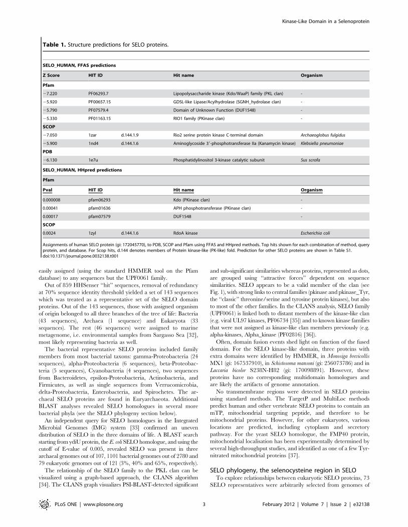
In order to predict structure for the SELO protein, the FFAS03
[25] method was used, yielding consistent, albeit of borderline
significance, predictions of kinase-like superfamily in the SCOP
database (http://scop.mrc-lmb.cam.ac.uk/scop/) and various
families of the kinase-like clan (CL0126) in the Pfam database
(see Table 1 and Table S1). Also, HHpred [26] ascribes the SELO
family to kinase-like proteins (see Table 1 and Table S1). More
specifically, the FFAS03 and HHpred structure prediction
methods consistently flagged the Kdo Lipopolysaccharide kinase
(Kdo/WaaP) family proteins (PF06293) [27], APH Phosphotrans-
ferase enzyme family (PF01636) [28] and various representatives
of the ‘‘classic’’ protein kinase family (PF00069). In the SCOP
database, the fold d.144, Protein kinase-like (PK-like, PKL)
dominated among the hits as the closest structural match for the
SELO domain. Similar results were obtained for the Escherichia coli
and yeast orthologues.
The kinase-like fold predictions were obtained for the central
region of human SELO protein, with alignments to the known
kinase-like structures spanning the stretch from approx residue 120
to 470. For the remaining SELO regions (residues 1–119 and 471–
669), no structural predictions were obtained nor homologous
sequences outside the SELO family were found. The SELO
alignments to different kinase domain hits did not always cover the
whole domain, yet together they did cover most of the domains.
For example, the FFAS alignment for SELO and PKA kinase
included the 47–283 region of PKA (67% of the kinase domain
sequence). Likewise, HHpred alignment for SELO and the E. coli
RdoA kinase covered 93% of the RdoA kinase domain as defined
in the SCOP database.
Structural predictions can be validated by predictions for distant
homologues using different methods. Indeed, using the sequence
of a SELO homologue, the ‘‘conserved hypothetical protein
[Marinomonas sp. MED121]’’, gi:86163056, one obtains in five PSI-
BLAST iterations significant similarity to a Ser/Thr protein kinase
of the PIM subfamily [Saccoglossus kowalevskii] (XP_002739315.1),
E-value 2E-3, with sequence identity 13%, in a 296 residue-long
alignment. The Saccoglossus protein is a close homologue of
human proto-oncogene Ser/Thr-protein kinase PIM1.
It has to be borne in mind that structure predictions are not
automatically extendable to functional predictions. Thus, func-
tional meaning of the structure assignment for SELO proteins will
be discussed further down.
Survey of SELO proteins, phylogenetic spread, therelationship of the SELO family to the rest of the proteinkinase-like clan
Standard BLAST searches for distant SELO protein homo-
logues did not bring up any sequences other than those
recognisable as SELO. The SELO protein family is recognised
in the Pfam database as one of the ‘‘Domains of unknown
function’’, UPF0061 (PF02696), annotated as ‘‘Uncharacterized
ACR, ydiU/UPF0061 family’’. In the Clusters of Orthologous
Groups (COG) database [29], bacterial SELO proteins are
combined in the group COG0397.
In order to survey SELO and similar proteins in the sequence
‘‘hyperspace’’, we used the conceptually similar Saturated BLAST
[30] and HHsenser [26] search tools with the central part of the
human SELO protein (NCBI gi: 1699163, region 190–470) as the
query, using the standard search parameters (see Methods section
for details). These procedures, which perform cascades of PSI-
BLAST or HHsearch searches, respectively, using representative
significant hits as queries in subsequent iterations, yielded several
hundred ‘‘hit’’ sequences from the nr and env_nr databases.
Since such a search can easily ‘‘drift out’’ of the original (SELO)
sequence family, the hit sequences were checked for the presence
of proteins that could be assigned to already known structural
domains using the well-established Pfam domain classification
system [31] that distinguishes protein domain families, sometimes
grouped in clans. In the total hit sequence population, none were
Kinase-Like Domain in a Selenoprotein
PLoS ONE | www.plosone.org 2 February 2012 | Volume 7 | Issue 2 | e32138
easily assigned (using the standard HMMER tool on the Pfam
database) to any sequences but the UPF0061 family.
Out of 859 HHSenser ‘‘hit’’ sequences, removal of redundancy
at 70% sequence identity threshold yielded a set of 143 sequences
which was treated as a representative set of the SELO domain
proteins. Out of the 143 sequences, those with assigned organism
of origin belonged to all three branches of the tree of life: Bacteria
(43 sequences), Archaea (1 sequence) and Eukaryota (33
sequences). The rest (46 sequences) were assigned to marine
metagenome, i.e. environmental samples from Sargasso Sea [32],
most likely representing bacteria as well.
The bacterial representative SELO proteins included family
members from most bacterial taxons: gamma-Proteobacteria (24
sequences), alpha-Proteobacteria (6 sequences), beta-Proteobac-
teria (5 sequences), Cyanobacteria (4 sequences), two sequences
from Bacteroidetes, epsilon-Proteobacteria, Actinobacteria, and
Firmicutes, as well as single sequences from Verrucomicrobia,
delta-Proteobacteria, Enterobacteria, and Spirochetes. The ar-
chaeal SELO proteins are found in Euryarchaeota. Additional
BLAST analyses revealed SELO homologues in several more
bacterial phyla (see the SELO phylogeny section below).
An independent query for SELO homologues in the Integrated
Microbial Genomes (IMG) system [33] confirmed an uneven
distribution of SELO in the three domains of life. A BLAST search
starting from ydiU protein, the E. coli SELO homologue, and using the
cutoff of E-value of 0.005, revealed SELO was present in three
archaeal genomes out of 107, 1101 bacterial genomes out of 2780 and
79 eukaryotic genomes out of 121 (3%, 40% and 65%, respectively).
The relationship of the SELO family to the PKL clan can be
visualized using a graph-based approach, the CLANS algorithm
[34]. The CLANS graph visualizes PSI-BLAST-detected significant
and sub-significant similarities whereas proteins, represented as dots,
are grouped using ‘‘attractive forces’’ dependent on sequence
similarities. SELO appears to be a valid member of the clan (see
Fig. 1), with strong links to central families (pkinase and pkinase_Tyr,
the ‘‘classic’’ threonine/serine and tyrosine protein kinases), but also
to most of the other families. In the CLANS analysis, SELO family
(UPF0061) is linked both to distant members of the kinase-like clan
(e.g. viral UL97 kinases, PF06734 [35]) and to known kinase families
that were not assigned as kinase-like clan members previously (e.g.
alpha-kinases, Alpha_kinase (PF02816) [36]).
Often, domain fusion events shed light on function of the fused
domain. For the SELO kinase-like domain, three proteins with
extra domains were identified by HMMER, in Monosiga brevicollis
MX1 (gi: 167537910), in Schistosoma mansoni (gi: 256073786) and in
Laccaria bicolor S238N-H82 (gi: 170098891). However, these
proteins have no corresponding multidomain homologues and
are likely the artifacts of genome annotation.
No transmembrane regions were detected in SELO proteins
using standard methods. The TargetP and MultiLoc methods
predict human and other vertebrate SELO proteins to contain an
mTP, mitochondrial targeting peptide, and therefore to be
mitochondrial proteins. However, for other eukaryotes, various
locations are predicted, including cytoplasm and secretory
pathway. For the yeast SELO homologue, the FMP40 protein,
mitochondrial localisation has been experimentally determined by
several high-throughput studies, and identified as one of a few Tyr-
nitrated mitochondrial proteins [37].
SELO phylogeny, the selenocysteine region in SELOTo explore relationships between eukaryotic SELO proteins, 73
SELO representatives were arbitrarily selected from genomes of
Table 1. Structure predictions for SELO proteins.
SELO_HUMAN, FFAS predictions
Z Score HIT ID Hit name Organism
Pfam
27.220 PF06293.7 Lipopolysaccharide kinase (Kdo/WaaP) family (PKL clan) -
25.920 PF00657.15 GDSL-like Lipase/Acylhydrolase (SGNH_hydrolase clan) -
25.790 PF07579.4 Domain of Unknown Function (DUF1548) -
25.330 PF01163.15 RIO1 family (PKinase clan) -
SCOP
27.050 1zar d.144.1.9 Rio2 serine protein kinase C-terminal domain Archaeoglobus fulgidus
25.900 1nd4 d.144.1.6 Aminoglycoside 39-phosphotransferase IIa (Kanamycin kinase) Klebsiella pneumoniae
PDB
26.130 1e7u Phosphatidylinositol 3-kinase catalytic subunit Sus scrofa
SELO_HUMAN, HHpred predictions
Pfam
Pval HIT ID Hit name Organism
0.000008 pfam06293 Kdo (PKinase clan) -
0.00041 pfam01636 APH phosphotransferase (PKinase clan) -
0.00017 pfam07579 DUF1548 -
SCOP
0.0024 1zyl d.144.1.6 RdoA kinase Escherichia coli
Assignments of human SELO protein (gi: 172045770), to PDB, SCOP and Pfam using FFAS and HHpred methods. Top hits shown for each combination of method, queryprotein, and database. For Scop hits, d.144 denotes members of Protein kinase-like (PK-like) fold. Prediction for other SELO proteins are shown in Table S1.doi:10.1371/journal.pone.0032138.t001
Kinase-Like Domain in a Selenoprotein
PLoS ONE | www.plosone.org 3 February 2012 | Volume 7 | Issue 2 | e32138

diverse organisms spanning all major eukaryotic taxons that
possess SELO family members. Also, two sequences from he
bacterium E. coli were included. The eukaryotic SELO phyloge-
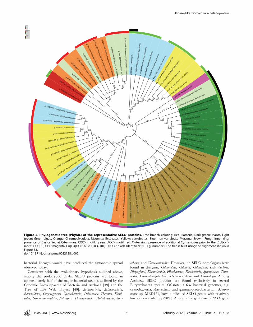
netic tree includes five main branches (Fig. 2). Firstly, a branch
with sequences from very diverse organisms that can be loosely
labeled together as marine picoeukaryotes. These include
sequences from green algae: Micromonas and Ostreococcus, strame-
nopiles: Aureococcus, the brown alga Ectocarpus and the diatoms
Thalassiosira and Phaeodactylum. Secondly, the tree includes a branch
with metazoan sequences, homologues of vertebrate SELO2 (see
below for definition) and sequence from other taxons: Cnidaria
(Nematostella), hemichordates (Saccoglossus), plocozoans (Trichoplax),
echinoderms (Strongylocentrotus), crustaceans (Daphnia). Branches 3,
4, and 5 are grouped together with strong bootstrap support of
0.91. The third branch contains plant and green algal proteins.
The fourth one is mostly composed of fungal sequences. The fifth
branch not only includes metazoan sequences, homologues of
SELO, but also sequences from green algae (Monosiga, Volvox,
Chlamydomonas), and alveolates (Perkinsus, Paramecium, Tetrahymena).
Separate in the super-branch (3,4,5) is the sequence from the
excavate Naegleria. Two bacterial sequences, ydiU and mchC from
E. coli, remain an outgroup. Notable is the absence of the SELO
gene in all nematodes including C. elegans and in most arthropods,
including Drosophila and all insects. An exception is the tick Ixodes
and crustaceans, e.g. Daphnia.
The similarities of metazoan SELO domains (see the phyloge-
netic tree in Fig. 2) suggest that (a) the common ancestor of
Metazoa had a duplicated SELO-like gene (most Metazoa
analysed, including the simple plocozoan Trichoplax, have two
paralogues of the SELO gene) and (b) in some lineages, e.g.
humans, the second gene, SELO2, was lost. The SELO2 gene is
present throughout Metazoa, including Gallus [Entrez Gene:
LOC420491], Canis [Entrez Gene: LOC607965], Equus [Entrez
Gene: LOC100065515], and Bos [Entrez Gene: LOC100297097];
however, it is missing from primates, rodents, and many other
mammals. The SELO and SELO2 genes are not close homologues,
e.g. in Bos the proteins share as little as 36% sequence identity over
550 residues. Here, we have introduced the unofficial gene symbol
SELO2 in order to denote the homologue of SELO present
throughout Metazoa, although SELO2 is not a selenoprotein.
SELO gene duplication is also observed in green algae, e.g.
Ostreococcus, Chlamydomonas and Volvox (see branches 3 and 5 in
Fig. 2) as well as stramenopiles, e.g. Aureococcus, Phaeodactylum (see
branch 1 in Fig. 2); however, these duplications seem to be
independent of the SELO/SELO2 duplication in Metazoa. The
duplicated genes are relatively distant, and the sequence identity
for the aligned paralogue pairs is 26%, 41%, 36%, 44% and 58%
in Ostreococcus, Chlamydomonas, Volvox, Aureococcus and Phaeodactylum,
respectively.
Among the five main eukaryotic phyla as delineated by Koonin
[38], SELO is absent from Rhizaria and present in representatives
of the other four phyla: Unikonts, except Amoebozoa, Excavates (but
only in the amoeboflagellate Naegleria gruberi), Plantae and
Chromoalveolates, except apicomplexans. Thus, SELO is probably
a pan-eukaryotic gene that has been lost from a number of
genomes.
A phylogenetic tree including representative sequences from all
domains of life as well as the marine metagenomic sequences,
unassigned to a taxon, shows a similar picture (see Fig. S1), albeit
highlighting the difficulties in elucidating the evolutionary
relationships between eukaryotic and prokaryotic SELO homo-
logues. Nevertheless, the most plausible evolutionary scenario
would involve SELO as an ancient gene in the last universal
ancestor of bacteria. Eukaryotes would have gained SELO from
the ancestor of the mitochondrial endosymbiont, while a few
Archaea would have acquired it via horizontal gene transfer.
Accordingly, SELO remains a mitochondrial protein in all the
studied eukaryotes. SELO gene loss in several eukaryotic and
Figure 1. CLANS graph visualizing PSI-BLAST-detected significant (dark grey) and sub-significant (light grey) similarities amongprotein kinase-like proteins. Red: UPF0061 (SELO) family, Light green: pkinase and pkinase_Tyr, Dark blue: APH phosphotransferase, Magenta:PIP5K, Grey: alpha_kinase. Pink: PI3_PI4, Light blue: Act_Frag_cataly, Yellow: PPDK_N, Dark green: Kdo, Orange: UL97.doi:10.1371/journal.pone.0032138.g001
Kinase-Like Domain in a Selenoprotein
PLoS ONE | www.plosone.org 4 February 2012 | Volume 7 | Issue 2 | e32138
bacterial lineages would have produced the taxonomic spread
observed today.
Consistent with the evolutionary hypothesis outlined above,
among the prokaryotic phyla, SELO proteins are found in
approximately half of the major bacterial taxons, as listed by the
Genomic Encyclopaedia of Bacteria and Archaea [39] and the
Tree of Life Web Project [40]: Acidobacteria, Actinobacteria,
Bacteroidetes, Chrysiogenetes, Cyanobacteria, Deinococcus-Thermus, Firmi-
cutes, Gemmatimonadetes, Nitrospira, Planctomycetes, Proteobacteria, Spir-
ochetes, and Verrucomicrobia. However, no SELO homologues were
found in Aquificae, Chlamydiae, Chlorobi, Chloroflexi, Deferribacteres,
Dictyoglomi, Elusimicrobia, Fibrobacteres, Fusobacteria, Synergistetes, Tener-
icutes, Thermodesulfobacteria, Thermomicrobium and Thermotogae. Among
Archaea, SELO proteins are found exclusively in several
Euryarchaeota species. Of note, a few bacterial genomes, e.g.
cyanobacteria, Acaryochloris and gamma-proteobacterium Marino-
monas sp. MED121, have duplicated SELO genes, with relatively
low sequence identity (28%). A more divergent case of SELO gene
Figure 2. Phylogenetic tree (PhyML) of the representative SELO proteins. Tree branch coloring: Red: Bacteria, Dark green: Plants, Lightgreen: Green algae, Orange: Chromoalveolata, Magenta: Excavates, Yellow: vertebrates, Blue: non-vertebrate Metazoa, Brown: Fungi. Inner ring:presence of Cys or Sec at C-terminus: CXX. motif: green; UXX. motif: red. Outer ring: presence of additional Cys residues prior to the [CU]XX.motif: CXX[CU]XX.: magenta, CX[CU]XX.: blue, CX(3–10)[CU]XX.: black. Identifiers: NCBI gi numbers. The tree is built using the alignment shown inFigure S3.doi:10.1371/journal.pone.0032138.g002
Kinase-Like Domain in a Selenoprotein
PLoS ONE | www.plosone.org 5 February 2012 | Volume 7 | Issue 2 | e32138

duplication produced the mchC gene in a small group of gamma-
proteobacteria (see the separate mchC section below).
The SELO proteins often possess a Cys-x-x-[Cys/Sec]-x-x-.
motif (e.g. CVTUSS. in humans) at the C-terminus, where the
‘‘.’’ character denotes the terminus of the polypeptide. Such
motifs are more common than expected by chance (according to
Prosite Scan results, http://prosite.expasy.org/, there are 585
observed CxxCxx. occurrences in the SwissProt database versus
281 expected considering average cysteine occurrence; probability
of the observed or higher number of CxxCxx. motifs is
practically 0). The CxxC motifs, in general, are reminiscent of
an oxidoreductase, e.g. thioredoxin, function [41] and are present
in majority of thioredoxin-fold proteins [42,43,44]. The CxxU
motif has been shown in another selenoprotein, the selenoprotein
H [45], to correspond with the thioredoxin active site. For
selenoprotein H, homologues with CxxC instead of CxxU occur in
insects and plants. Similarly, in a number of SELO protein
homologues, the CxxUxx. motif is replaced by CxxCxx. or by a
single cysteine residue. Out of SELO homologues, cysteine located
two positions prior to the C-terminus, Cxx., is more common
than selenocysteine, Uxx. (See Fig. 3 and Fig. S1).
Generally, the location of selenocysteine near the C-terminus of
a selenoprotein is a common feature among these proteins,
occurring either in CxxU pairs or as single U residues [46]. In the
selenoprotein K [47], an Uxx. motif is found, while in
selenoprotein S [48], there is an Ux. motif. Both these proteins
are involved in stress responses. Selenoprotein P (SEPP) contains
an UxUxxx. motif, and TXNRD1, TXNRD2 and TXNRD3
proteins have each U as the penultimate residue (Ux.). In the
latter three proteins, thioredoxin reductases [49], also in the lipid
hydroperoxidase SEPP [50], selenocysteine is involved in the
oxidoreductase function.
A rigorous assessment of the significance of the occurrence of
the CxxC/CxxU motif at SELO protein C-termini is not
straightforward because of uneven taxonomic sampling of SELO
homologues. However, in the representative set of 143 SELO
proteins, selected for even coverage of the sequence space (see
Methods section), there are 12 proteins with this motif. Although
this is a clear minority of SELO proteins, it still is an
overrepresentation as compared to a random situation. The
binomial test using SwissProt as reference population (585
occurrences in half a million sequences) allows one to estimate
the probability of Cxx[CU]xx. motif occurring by chance in the
SELO family at less than 10218. Thus, one can postulate that this
motif may have a functional role. Also, a generalised [CU]xx.
motif occurs in the representative SELO set 111 times, which is a
clear overrepresentation (binomial test probability less than
102189).
The CxxC and CxxU motifs in SELO proteins are not evenly
distributed on the phylogenetic tree. The Metazoan SELO branch
(branch 5 in Fig. 2) features mostly the Cxx[CU]xx. C-terminal
motif. The fungal branch (branch 4) is characterised by the
CxCxx. motif. The plant branch (branch 3) is characterised by
the C-x(3,10)-Cxx. motif. The Metazoan SELO2 branch (branch
2) has usually just the Cxx. motif, while the ‘‘marine
picoeukaryote’’ branch 1 has usually the Uxx motif.. The
bacterial proteins have usually a Cxx. motif, sometimes
augmented by an additional C residue to a CxxCxx. motif or a
C-x(3–10)-Cxx. one (see Fig. S1).
Structure models of the SELO domain, the relevance ofthe structure predictions for molecular function
Secondary structure predictions and sequence alignments to the
known kinase-like structures according to structure prediction
results suggest that the SELO domains are composed of an N-
terminal smaller lobe (ATP-binding), mostly composed of b-
strands, and a predominantly helical, larger, C-terminal lobe
(phosphotransfer). In terms of the classic kinase fold nomenclature
[51,52], SELO domain secondary structure order is as following:
b-2, b-3, a-C, b-4, b-5, a-D, a-E, b-6, b-7, b-8, b-9, a-EF, a-F, a-
G, see Fig. 3). These secondary structure elements form the core of
the two lobes of a typical kinase-like fold protein [52]. Three
alternative secondary structure prediction methods, Jpred, PsiPred
and SOPMA, produced very similar results when applied to
SELO proteins from humans, yeast and bacteria (see Fig. S2).
These predictions are in agreement with the kinase domain
secondary structure topology. Within the SELO alignment, there
are some conserved insertions, e.g. between SELO and SELO2
groups, and in the fungal sequences, as compared to the other
SELO proteins. These usually occur outside the predicted
secondary structure elements.
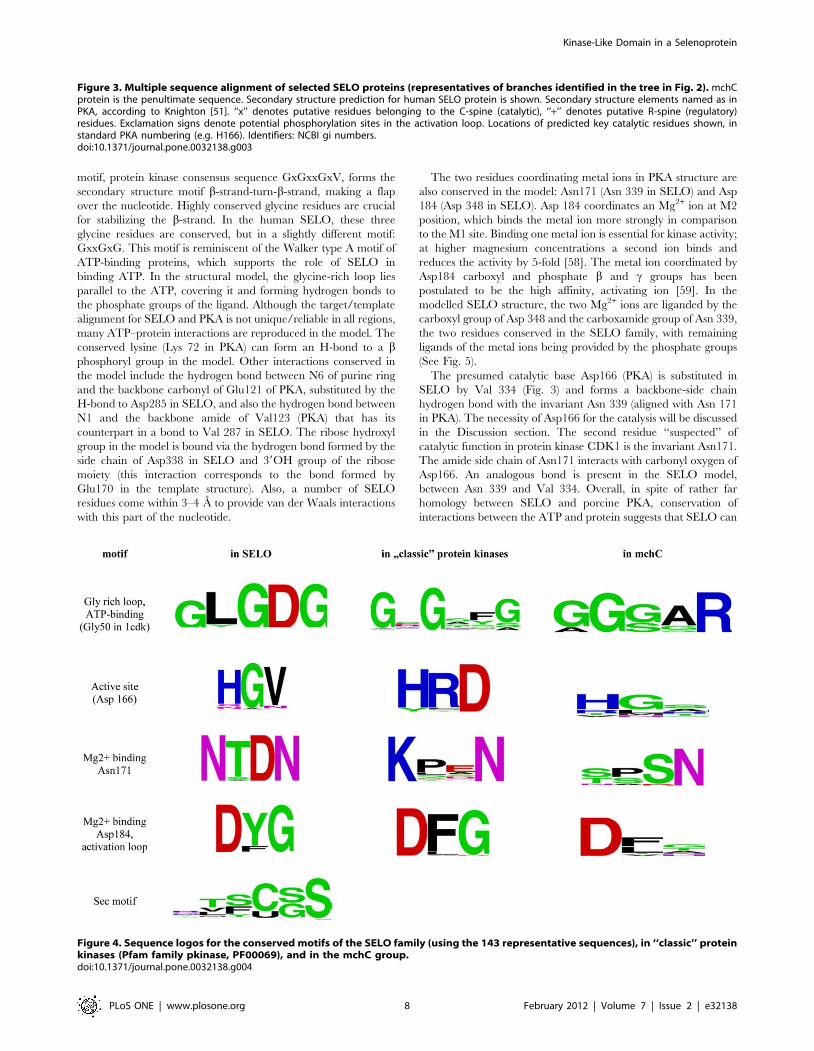
Out of the conserved regions I–XI (subdomains), as defined by
Hanks and co-workers for the first solved kinase structure, the
protein kinase A (PKA) [5], the subdomains I (ATP-binding G-
loop), II (lysine K72 in b-strand 3), III (glutamate 91 in a-helix C),
VIb (catalytic loop), VII (DFG, Mg2+ -binding site), and possibly
VIII (APE motif) are conserved in SELO proteins (See Fig. 3).
Among the functionally-critical conserved kinase residues, the
Lys72 (PKA numbering), involved in binding of the a and bphosphate groups of the ATP molecule [53,54,55] is almost
invariant in the SELO family. Also, the Glu91 (binding to Lys72
by a salt bridge and stabilising it) is invariant. The archetypic
HRD/YRD motif of protein kinases is not conserved in the SELO
family. It is usually substituted by an HGV motif, also by QGN or
HGS (see the logos in Fig. 4). Thus, the presumed kinase catalytic
base (Asp166 in PKA) is missing from SELO proteins. However, it
has been shown for other kinases, e.g. Erbb3, that the lack of
Asp166 does not necessarily mean absence of kinase activity (see
Discussion section) [18]. Moreover, the asparagine corresponding
to Asn171 of PKA is very well-conserved, and is responsible for
binding the second (‘‘inhibitory’’) Mg2+ ion. The ion-binding motif
D[Y/F]G is strictly conserved, binding the first (‘‘activating’’)
Mg2+ ion, and corresponding to Asp184 in PKA. The activation
loop region contains two partly conserved tyrosine residues that
may be the primary phosphorylation sites (see Fig. 3).
Finally, the network of hydrophobic residues that span the
kinase fold molecule and participate in regulation of the enzymatic
activity is conserved in the SELO family (see Fig. 3). This network
was recently defined by Taylor and co-workers and was termed as
regulatory and catalytic spines [56]. In SELO, precise identities of
residues participating in the two spines may be uncertain;
however, the candidate residues are conserved and can be aligned
to their counterparts in typical kinases. The kinase-like fold
proteins are known to vary in their structures around the typical
‘‘classic’’ arrangement [8]. Also, the SELO regions outside the
predicted kinase domain (approx. 120 residues at the N-terminus
and approx. 200 residues at the C-terminus) may fold over and
augment the kinase domain.
In cases of remote sequence similarity, building a structural
model is of an illustrative nature, yet it also serves as a feasibility
check for the predicted structure. Here, we discuss a structural
model of the SELO kinase domain (residues 150–485) built by
employing the archetypical kinase structure, PKA, as a template.
The ATP-binding pocket of the modelled structure of SELO
allows ligand binding in a manner considerably similar to that in
the classical protein kinases (PKL). The catalytic core of the
protein kinases contains a nucleotide binding motif unique among
proteins with nucleotide binding site [54,55,57]. This conserved
Kinase-Like Domain in a Selenoprotein
PLoS ONE | www.plosone.org 6 February 2012 | Volume 7 | Issue 2 | e32138

Kinase-Like Domain in a Selenoprotein
PLoS ONE | www.plosone.org 7 February 2012 | Volume 7 | Issue 2 | e32138
motif, protein kinase consensus sequence GxGxxGxV, forms the
secondary structure motif b-strand-turn-b-strand, making a flap
over the nucleotide. Highly conserved glycine residues are crucial
for stabilizing the b-strand. In the human SELO, these three
glycine residues are conserved, but in a slightly different motif:
GxxGxG. This motif is reminiscent of the Walker type A motif of
ATP-binding proteins, which supports the role of SELO in
binding ATP. In the structural model, the glycine-rich loop lies
parallel to the ATP, covering it and forming hydrogen bonds to
the phosphate groups of the ligand. Although the target/template
alignment for SELO and PKA is not unique/reliable in all regions,
many ATP–protein interactions are reproduced in the model. The
conserved lysine (Lys 72 in PKA) can form an H-bond to a bphosphoryl group in the model. Other interactions conserved in
the model include the hydrogen bond between N6 of purine ring
and the backbone carbonyl of Glu121 of PKA, substituted by the
H-bond to Asp285 in SELO, and also the hydrogen bond between
N1 and the backbone amide of Val123 (PKA) that has its
counterpart in a bond to Val 287 in SELO. The ribose hydroxyl
group in the model is bound via the hydrogen bond formed by the
side chain of Asp338 in SELO and 39OH group of the ribose
moiety (this interaction corresponds to the bond formed by
Glu170 in the template structure). Also, a number of SELO
residues come within 3–4 A to provide van der Waals interactions
with this part of the nucleotide.
The two residues coordinating metal ions in PKA structure are
also conserved in the model: Asn171 (Asn 339 in SELO) and Asp
184 (Asp 348 in SELO). Asp 184 coordinates an Mg2+ ion at M2
position, which binds the metal ion more strongly in comparison
to the M1 site. Binding one metal ion is essential for kinase activity;
at higher magnesium concentrations a second ion binds and
reduces the activity by 5-fold [58]. The metal ion coordinated by
Asp184 carboxyl and phosphate b and c groups has been
postulated to be the high affinity, activating ion [59]. In the
modelled SELO structure, the two Mg2+ ions are liganded by the
carboxyl group of Asp 348 and the carboxamide group of Asn 339,
the two residues conserved in the SELO family, with remaining
ligands of the metal ions being provided by the phosphate groups
(See Fig. 5).
The presumed catalytic base Asp166 (PKA) is substituted in
SELO by Val 334 (Fig. 3) and forms a backbone-side chain
hydrogen bond with the invariant Asn 339 (aligned with Asn 171
in PKA). The necessity of Asp166 for the catalysis will be discussed
in the Discussion section. The second residue ‘‘suspected’’ of
catalytic function in protein kinase CDK1 is the invariant Asn171.
The amide side chain of Asn171 interacts with carbonyl oxygen of
Asp166. An analogous bond is present in the SELO model,
between Asn 339 and Val 334. Overall, in spite of rather far
homology between SELO and porcine PKA, conservation of
interactions between the ATP and protein suggests that SELO can
Figure 4. Sequence logos for the conserved motifs of the SELO family (using the 143 representative sequences), in ‘‘classic’’ proteinkinases (Pfam family pkinase, PF00069), and in the mchC group.doi:10.1371/journal.pone.0032138.g004
Figure 3. Multiple sequence alignment of selected SELO proteins (representatives of branches identified in the tree in Fig. 2). mchCprotein is the penultimate sequence. Secondary structure prediction for human SELO protein is shown. Secondary structure elements named as inPKA, according to Knighton [51]. ‘‘x’’ denotes putative residues belonging to the C-spine (catalytic), ‘‘+’’ denotes putative R-spine (regulatory)residues. Exclamation signs denote potential phosphorylation sites in the activation loop. Locations of predicted key catalytic residues shown, instandard PKA numbering (e.g. H166). Identifiers: NCBI gi numbers.doi:10.1371/journal.pone.0032138.g003
Kinase-Like Domain in a Selenoprotein
PLoS ONE | www.plosone.org 8 February 2012 | Volume 7 | Issue 2 | e32138
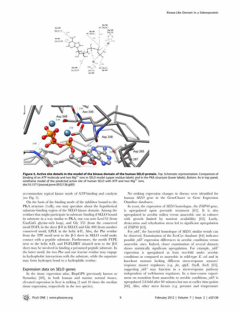
accommodate typical kinase mode of ATP-binding and catalysis
(see Fig. 5).
On the basis of the binding mode of the inhibitor bound to the
PKA structure (1cdk), one may speculate about the hypothetical
substrate-binding region of the SELO kinase domain. Among the
residues that might participate in substrate binding if SELO bound
its substrate in a way similar to PKA, one can note Leu152 (from
GxxGxG glycine-rich loop), and Gly 353 (from the conserved
motif FGFL in the sheet b-8 in SELO) and Glu 400 (from another
conserved motif, LPLE in the helix a-F). Also, the Phe residue
from the TPF motif next to the b-3 sheet in SELO could make
contact with a peptide substrate. Furthermore, the motifs FYPE
next to the helix a-D, and FGFLDRY situated next to the b-8
sheet may be involved in binding a presumed peptide substrate. In
the latter motif, the two Phe and one leucine residue may engage
in hydrophobic interactions with the substrate, while the aspartate
may form hydrogen bond to a hydrophilic residue.
Expression data on SELO genesIn the tissue expression atlas, BiopGPS (previously known as
Symatlas) [60], in both human and murine normal tissues,
elevated expression in liver is striking (2 and 10 times the median
tissue expression, respectively in the two species).
No striking expression changes in disease were identified for
human SELO gene in the GeneChaser or Gene Expression
Omnibus databases.
In yeast, the expression of SELO homologue, the FMP40 gene,
is upregulated upon peroxide treatment [61]. It is also
upregulated in aerobic milieu versus anaerobic one in cultures
with growth limited by nutrient availability [62]. Lastly,
desiccation and rehydration stress led to significant upregulation
of FMP40 [63].
For ydiU, the bacterial homologue of SELO, similar trends can
be observed. Examination of the EcoCyc database [64] indicates
possible ydiU expression differences in aerobic conditions versus
anaerobic ones. Indeed, closer examination of several datasets
shows statistically significant upregulation. For example, ydiU
expression is upregulated at least two-fold under aerobic
conditions as compared to anaerobic in wild-type E. coli and in
knockout mutants lacking different stress-response sensors/
response master regulators (e.g. fnr, appY, OxyR, SoxS) [65],
suggesting ydiU may function in a stress-response pathway
independent of well-known regulators. In a time-course experi-
ment on transition from anaerobic to aerobic conditions, ydiU is
upregulated 3.8-fold after 60 minutes but not at earlier time-points
[66]. Also, other stress factors (e.g. pressure and temperature
Figure 5. Active site details in the model of the kinase domain of the human SELO protein. Top. Schematic representation. Comparison ofbinding of an ATP molecule and two Mg2+ ions in SELO model (upper residue labels) and in the PKA structure (lower labels). Bottom. As in top panel,wireframe model of the predicted active site of human SELO with ATP and two Mg2+ ions.doi:10.1371/journal.pone.0032138.g005
Kinase-Like Domain in a Selenoprotein
PLoS ONE | www.plosone.org 9 February 2012 | Volume 7 | Issue 2 | e32138

changes) and modulation of stress response regulators (e.g. IscR,
oxyR) cause alteration in ydiU expression [67,68].
Genomic neighbourhoods of SELO proteins,characteristics of organisms possessing SELO genes
Lifestyle analysis of microbes harbouring SELO genes reveals a
significant overrepresentation of aquatic organisms (as compared
to a random sample of organisms). Binomial test probability of a
number of aquatic species equal-or-higher than the observed
number (47%) is 2.3E-11 with the expected (background)
frequency of aquatic species among the organisms with known
genomes being 16%. Also, there is a significant overrepresentation
of aerobic organisms (observed 65%, expected 32%, p-value 4.7E-
07). No significant deviations from the expected frequencies were
noted for organism motility nor preferred temperature range. The
enrichments for aquatic and aerobic lifestyles are independent, as
seen by Fischer’s exact test that estimates the p-value of the
observed aquatic and aerobic lifestyle contingency table at 0.14.
Statistical analysis of genomic neighbourhoods (see Table 2)
identified gene families, as defined by the COG classification
system [29], significantly overrepresented in a 22 kbp genomic
window centered around microbial SELO homologues in the
MicrobesOnline system. Most notable were the genes homologous
to btuD and btuC [69,70,71], and the ATPase and permease
components of the ABC-type cobalamin transport system (p-values
below 1E-10, see Methods section). Also, two COG groups related
to oxidative stress response stood out: msrB (methionine sulfoxide
reductase) and btuE (glutathione peroxidase) [72,73], p-valu-
es,1E-9. Other notable gene groups, possibly related to stress
response were BaeS (signal transduction histidine kinase, p-value
below 1E-10), and Fur (Fe2+/Zn2+ uptake regulation proteins
involved in ROS defense [74]).
Finally, for some oxidative stress response-related gene families,
only uncorrected p-values indicated overrepresentation, suggesting
at least a trend (correcting for multiple testing is discussed in the
Methods section). These were msrA (peptide methionine sulfoxide
reductase), and Gst (glutathione transferase). The msrA and msrB
genes, coding for two different types of methionine sulfoxide
reductases, often co-occurring with SELO homologues, have been
shown to have an important role in ROS defense [75,76,77]. In
addition, another group of genes encoding signalling proteins was
detected: Rtn, EAL domain-containing, involved in the turnover
of the second messenger, cyclic di-GMP and linking the sensing of
specific environmental cues to appropriate alterations in bacterial
physiology and/or gene expression [78,79].
It is noteworthy that this type of analysis might be biased by the
uneven sampling of microbial genomes sequenced to date.
However, in our genomic neighbourhood analysis, only nine
genera provided three or more genomes (up to seven), and these
genera belonged to various main bacterial phyla: Firmicutes,
Actinobacteria, Cyanobacteria, alpha- and gamma-proteobac-
teria, and Enterobacteria.
Together, striking among the gene groups overrepresented in
the SELO genomic neighbourhoods are oxidative stress response-
related genes and components of transport/efflux systems. Among
the latter, there were several COG groups coding for proteins
containing ABC transporter ATPase domains (Pfam family
ABC_tran, PF00005): btuD, salX, uup, ydiA, and COG groups
coding for Major Facilitator Superfamily transporters (Pfam clan
CL0015): araJ, emrA. Also, the COG family CirA, outer membrane
Table 2. Functional families (COGs) overrepresented in genomic neighbourhoods of microbial SELO homologues.
COG group Gene COG description
Probability offinding a COGin SELO/ydiUneighbourhood
Probability of finding aCOG in SELO/ydiUneighbourhood,corrected Structure type (Pfam clan)
COG4138 btuD ABC-type cobalamin transport system,ATPase component
,10210 ,10210 AAA ATPase, CL0023
COG4139 btuC ABC-type cobalamin transport system,permease component
,10210 ,10210 Membrane transporter, CL0142
COG1957 URH1 Inosine-uridine nucleoside N-ribohydrolase ,10210 ,10210
COG0642 BaeS Signal transduction histidine kinase ,10210 ,10210 His_Kinase_A, CL0025
COG0229 msrB Peptide methionine sulfoxide reductase ,10210 5.90E-10
COG0386 btuE Glutathione peroxidase ,10210 7.91E-10 Thioredoxin, CL0172
COG1806 ydiA Uncharacterized protein conserved inbacteria. Predicted AAA ATPase
,10210 2.44E-09 AAA ATPase, CL0023 (remotesimilarity, detected by FFAS)
COG0722 aroF aroG aroH 3-deoxy-D-arabino-heptulosonate 7-phosphate (DAHP) synthase
,10210 3.13E-09 DAHP synthetase I, TIM_barrel,CL0036
COG0271 BolA Stress-induced morphogen (activityunknown)
1.72E-10 8.39E-07
COG2917 yciB Intracellular septation protein A 1.49E-08 7.27E-05
COG0791 nlpC spr yafLydhO
Cell wall-associated hydrolases (invasion-associated proteins)
2.39E-08 0.00012 Peptidase_CA, CL0125
COG0574 ppsA Phosphoenolpyruvate synthase/pyruvatephosphate dikinase
1.21E-07 0.00059 Multidomain, contains Pyruvatekinase-like TIM barrel, CL0151
COG0735 Fur Fe2+/Zn2+ uptake regulation proteins 3.40E-07 0.0017 Helix-turn-helix clan, CL0123
COG4181 SalX ybbA Predicted ABC-type transportsystem, ATPase component
3.64E-07 0.0018 AAA ATPase, CL0023
Statistical analysis using 22-kbp wide genomic windows.doi:10.1371/journal.pone.0032138.t002
Kinase-Like Domain in a Selenoprotein
PLoS ONE | www.plosone.org 10 February 2012 | Volume 7 | Issue 2 | e32138

receptor proteins, involved mostly in Fe transport, was overrep-
resented. Although ATPase domains of the P-loop NTPase type,
including the distantly related ABC and AAA+ type domains, are
ubiquitous and occur in different proteins that carry out diverse
functions [80,81], the ATPase domains coded often in ydiU
neighbourhoods belong to families present in ABC transporters.
Many SELO-possessing species have their taxon-specific SELO
genomic neighbours. Escherichia coli, Shigella and Salmonella SELO
genes are surrounded both by ABC transporter component genes
and genes involved in various aspects of ‘‘basic metabolism’’
(phosphoenolpyruvate synthase (EC 2.7.9.2), 2-keto-3-deoxy-D-
arabino-heptulosonate-7-phosphate synthase I alpha (EC 2.5.1.54)
or long-chain-fatty-acid–CoA ligase (EC 6.2.1.3)). The SELO gene
in Francisella co-occurs with proton/sodium glutamate and sugar
symporters. Vibrio cholerae SELO co-occurs with genes involved in
glycerophospholipid metabolism (CDP-diacylglycerol–glycerol-3-
phosphate 3-phosphatidyltransferase (EC 2.7.8.5), phosphatidate
cytidylyltransferase (EC 2.7.7.41), 1-acyl-sn-glycerol-3-phosphate
acyltransferase (EC 2.3.1.51)) on one side and with the TRAP-type
C4-dicarboxylate transport system genes on the other side. The
SELO from Yersinia is a neighbour of polymyxin resistance genes
and the ‘‘basic metabolism’’ set of genes. The SELO in Bacilli
usually directly co-occurs with a proximal bifunctional protein
such as zinc-containing alcohol dehydrogenase; quinone oxidore-
ductase (NADPH:quinone reductase) (EC 1.1.1.-) involved in
glutathione-regulated potassium-efflux system which is preceded
by an ArsR transcription factor and on the distal side with a
response regulator. Not too far away there is a frequent gene
encoding the GlcD subunit of the glycolate oxidase which
catalyzes the first step in the utilisation of glycolate as the sole
source of carbon [82].
Furthermore, analysis of the promoter region of the ydiU gene of
E. coli K-12 substr. MG1655 revealed that it has a cis element that
binds to the IscR transcription factor [83]. IscR, the ‘‘Iron-sulphur
cluster Regulator’’, is negatively autoregulated, and contains an
iron-sulphur cluster that could act as a sensor of iron-sulphur
cluster assembly [84,85]. This protein regulates the expression of
the isc and suf operons that encode components of pathways of
iron-sulphur cluster assembly, iron-sulphur proteins, anaerobic
respiration enzymes, and biofilm formation proteins
[84,85,86,87,88,89]. This is yet another piece of evidence
supporting the hypothesis of role of ydiU in oxidative stress
response.
mchC, the microcin maturation proteins, as remotehomologues of the SELO proteins
Search for distant homologues of a SELO protein, starting from
a protein from Marinomonas sp. MED121, gi:86163056, brings up,
within the second PSI-BLAST iteration, significant similarity to a
group of mchC proteins present in several Escherichia coli strains,
Cellvibrio japonicus Ueda107, Klebsiella pneumoniae RYC492, Photo-
rhabdus asymbiotica, few Vibrio species, including two Vibrio cholerae
strains (MZO-2 and AM-19226), and a few Xanthomonas species
(for a list, see Table S2). The HHpred and FFAS searches on the
Pfam database confirm similarity of mchC proteins to the SELO
family and remote similarity to kinase families (see Table S1).
The group of about 20 mchC proteins exhibits large variability
(down to 26–35% sequence identity within the group, see Figs. S4
and S5), and large differences are observed even between several
mchC proteins from a single strain, Cellvibrio japonicus Ueda107
(down to 28% sequence identity). The characteristic kinase motifs,
identified for ‘‘typical’’ SELO proteins are noticeable in the mchC
subgroup, with the notable large variation in the motif
corresponding to the kinase HGD motif and HGV motif present
in most SELO proteins (see Fig. 4). The remote relation of the
mchC to other SELO proteins is exemplified by the low similarity
between E. coli ydiU and mchC proteins. The similarity is not
detectable by a BLAST comparison, yet it is obvious when profile-
profile methods are used, e.g. the FFAS Z-score is 288.0, and
sequence identity for the 529 residue-long FFAS alignment is only
12%.
In E. coli strains, the mchC gene is a part of the mch gene cluster
involved in production, maturation, and export of the bactericidal
antibiotic microcin H47 (the corresponding gene cluster in K.
pneumoniae is mce, and the appropriate microcin is termed as E492
[90,91]. The microcins themselves are coded by a gene in the mch
(or mce) cluster, and are posttranslationally modified. The mchC
protein is necessary for the modification involving the covalent
attachment of enterobactin, a siderophore moiety [91]. The K
pneumoniae orthologue of mchC, mceJ, functions in a protein
complex (mceI/mceJ) [92]. An analogous pair, mchC/mchD,
exists in the few E. coli strains and some other bacteria (see Table
S2). The mchD and mceI proteins are acyltransferases of
hemolysin C-like structure (RTX toxin acyltransferase family,
HlyC, PF02794 [93]). Since hlyC proteins are found only in
bacteria, an interaction similar to mceI/mceJ cannot be expected
to occur for eukaryotic SELO proteins.
Although no close homologue of mchD is annotated in the in V.
cholerae strains MZO-2 and AM-19226 that possess mchC
homologues, a tblastn search in Vibrio genomic sequences yields
MZO-2 and AM-19226 regions of significant similarity (after
translation) to the E. coli mchD sequence (E-value 1e-05, 30%
sequence identity), just next to the regions encoding the mchC gene,
suggesting the presence of an unannotated mchD homologue in the
two Vibrio strains. Similar putative mchD genes can also be found in
a few strains of Xanthomonas. This leads to a hypothesis that all
mchC proteins may act as accessory proteins/regulators for the
hlyC-like acyltransferases, mchD.
Interestingly, in two bacterial species that have many strains
with sequenced genomes, E. coli and V. cholerae, mchC gene is
present only in a number of strains out of dozens sequenced ones.
The E. coli strains harbouring mchC proteins are either
commensal, asymptomatic ones: bacteriuria strain 83972 [94],
commensal probiotic strain Nissle 1917 [95] or pathogenic ones:
strain CFT073, causing persistent diarrhea, and strain 042,
causing urinary tract infections [96]. In E. coli, presence of the
mchC gene coincides with presence of the whole microcin H47
gene cluster.
In V. cholerae, mchC homologues are found in just two strains,
MZO-2 and AM-19226. These are clinical strains isolated in
Bangladesh, yet different from the most deadly pandemic strains.
AM-19226 is a pathogenic strain that lacks the typical virulence
factors [97]. Interestingly, although as outlined above, a putative
region coding for mchD can be found in these two strains, together
with some other putative microcin export proteins (see below), a
region of homology to the mchB gene that codes for the microcin
H47 itself cannot be found in Vibrio. This does not rule out the
possibility that the mch homology region in Vibrio functions toward
the production, maturation and export of a yet-undiscovered H47-
like microcin. Indeed, similarity is not detectable by BLAST
between the well-studied and probably homologous microcins
H47 and E492 from E. coli and K. pneumoniae. However, their
similarity is detected by FFAS with a Zscore of borderline
significance, 26.15, and sequence identity of 31%.
Other mchC-possessing strains are usually pathogenic. Photo-
rhabdus asymbiotica [98] is an entomopathogenic bacterium, living in
a mutualistic association with entomopathogenic nematodes, yet it
causes wound infection in humans. It possesses a complete mch
Kinase-Like Domain in a Selenoprotein
PLoS ONE | www.plosone.org 11 February 2012 | Volume 7 | Issue 2 | e32138
gene cluster, significantly similar to that of E. coli, including the
mchC and microcin H47 genes. In K. pneumoniae, the mce gene
cluster, homologous to the mch cluster, is found only in the
clinically isolated strain RYC492 [99], but not in other K.
pneumoniae strains. The mchC protein is also found in a
saprotrophic soil bacterium Cellvibrio japonicus [100] (five distinct
genes) and several strains of various species of the plant pathogen
Xanthomonas [101,102].
In the strain E. coli Nissle 1917, the mch cluster is found in the
Genomic Island GEI I [95], which corresponds to the pathoge-
nicity island PAI-CFT073-SerX in the strain CFT073 [96,103].
The PAI-CFT073-serX island is also found in the strain ABU
83972 [104]. In V. cholerae strains MZO-2 and AM-19226, mchC is
encoded within the genomic island GI-52, together with five other
genes, including type I secretion membrane fusion protein, hlyD-
like, and ABC-type bacteriocin/lantibiotic exporter, HlyB-like
[105]. Interestingly, the island GI-52 has a cassette-like property,
i.e. different islands occupy the same region in different strains
[105]. It has to be noted that the islands containing the mch genes
need not be virulence/pathogenicity determinants per se, but they
may also confer general fitness advantage [104]. Yet, it has been
reported that particular microcins, including the mch cluster
encoded ones, corresponded to specific urovirulence gene profiles
[106]. Also, there is an overrepresentation of E. coli urinary tract
infection strains among those encoding microcin H47 [107].
Discussion
We present the discovery of a novel kinase-like family with a
member in humans and presence throughout the tree of life as a
small step towards filling in the blank spots in the complex
regulatory machinery of the living cell. Precise charting of the
human kinome is important for several reasons. The recent timely
essay ‘‘Too many roads not taken’’ [108] convincingly presents the
case for exploring the ‘‘unpopular‘‘ members of otherwise popular
protein families. The authors point out that even for the
established and attractive drug targets, kinases, majority of
scientific activity, including articles and patents, focuses on a
minority of proteins that have been historically popular. The
‘‘uncharacterised’’ proteins [19] are even less fortunate, often
getting little attention even if the experimental data point at their
involvement in interesting biological or disease processes [109].
Also, many kinase inhibitors targeted at well-known kinases may
have off-target effects onto the less-studied ones [110]. It has been
reported that some kinases affected by off-target effects of a
compound have less than 20% sequence identity in their active
sites as compared to the original target kinase [110]. Thus,
complete understanding of the kinome is important for proper
assessment of the potential off-target effects of kinase-modulating
compounds.
Although the kinomes are remarkably well-studied, it is
predicted that they may be not fully mapped yet, even in the
well-studied organisms. Novel protein kinases are expected in
bacteria, e.g. the only two annotated protein kinases in Mycoplasma
pneumoniae account for only five out of 63 identified protein
phosphorylation events [111].
The correlation of presence of SELO genes in bacteria with
aquatic and aerobic lifestyles aligns with the hypothesis that these
genes are involved in stress responses, possibly in oxidative stress
response. It has been shown that genes involved in cellular
responses to oxygen occur much more often among aerobes than
in anaerobes [112]. This is corroborated by our genomic
neighbourhood analyses. Although SELO genes in bacterial
genomes are located in clusters of diverse functions, a recurring
functional theme involves transport and oxidoreductase functions.
The transport and stress response roles are not excluding, since
export of redox-cycling antibiotics is one of the mechanisms of
defense against oxidative stress [113]. These findings are further
supported by the observation of the most prominent expression
differences measured for SELO genes that involve stress factors,
such as aerobic conditions versus anaerobic. Lastly, the predicted
kinase function, together with the frequent but not invariant
cysteine- or selenocysteine-containing Cxx[CU] motif, suggestive
of an oxidoreductase function, indicate a possibility of SELO
proteins’ involvement in kinase signalling coupled to redox
detection/signalling. Since no fold prediction has been possible
for the selenocysteine-containing C-terminal region, no ultimate
function assignment can be done. However, several lines of
circumstantial evidence, including genomic neighbourhoods, gene
expression data, microbial lifestyles and presence of Cys-Sec motif,
point toward the involvement of SELO in response to oxidative
stress. The C-terminal Cxx[CU] motif, although not always
present throughout the SELO family, occurs in all major lineage
representatives, which supports the hypothesis of its functional
relevance.
In the mchC branch of the SELO family, mchC functions in
maturation of molecules secreted via a hemolysin-like type 1
secretion system [114]. There is an obvious similarity between the
secretion system coded in the mch gene cluster, and the genomic
neighbourhoods of ydiU-like genes. For example, mchF protein
serves a similar role in the btuC/btuD pair [115] [69], coded by
genes frequent in ydiU neighbourhoods, with homologous ATPase
domains (btuD protein and C-terminal part of the mchF protein)
and structurally different membrane-spanning regions (btuC
protein and central part of mchF). This suggests that both ydiU-
like and mchC-like bacterial proteins as well as their eukaryotic
homologues may be involved in the regulation of secretion or
transport processes.
The predicted mitochondrial localisation of metazoan SELO
proteins and the experimentally detected presence of SELO
homologues in yeast mitochondria are consistent with the known
antioxidant activities of mitochondrial kinases [116,117].
Can the kinase function prediction for SELO proteins be
trusted, or is it only a reliable three-dimensional fold prediction?
According to previous studies on protein kinase catalysis,
phosphoryl transfer requires a basic residue to accept the proton
from the attacking hydroxyl group [118,119] and the main
candidate for this key residue absolutely required for kinase
activity, and with sufficient pKa in negatively charged environment
was Asp166 of PKA, which is not conserved in SELO proteins.
Asp166 is one of the most conserved amino acid residues found in
all PKL kinases [6]. However, for the human Erbb3 protein,
having Asn at a position corresponding to Asp166 and long
believed to be a pseudokinase, Lemmon and colleagues have
recently demonstrated some residual kinase activity [18]. They
proposed an atypical catalytic mechanism whereby the attacking
proton from the tyrosyl substrate moves to c- and then b-
phosphate group of the ATP molecule instead of the aspartate
corresponding to D166 in PKA. Similar results were previously
shown by mutating the residue corresponding to Asp166 in EGFR
(Asp to Ala change) [120] and showing retained biological
function. Another example of atypical active kinases are the
WNK (with no K) kinases, lacking the ultra-conserved Lys72 [121]
whereas a lysine located elsewhere in the sequence assumes the
role of Lys72. Also, recent work by Manning and co-workers [6]
provides evidence of rich diversity of kinase families in the marine
metagenome, with several families lacking one or more key
Kinase-Like Domain in a Selenoprotein
PLoS ONE | www.plosone.org 12 February 2012 | Volume 7 | Issue 2 | e32138
catalytic motifs previously identified in studies on eukaryotic PKL
kinases.
Therefore, as summarised recently by Taylor, ‘‘It is difficult to
say unambiguously given kinase will be inactive’’ [122]. Manning
and colleagues have performed structure comparison of an
inactive pseudokinase and a closely homologous active kinase
counterpart [123]. They have discussed various alterations in the
conserved kinase motifs and concluded that provided the ATP-
binding G-loop is functional, other conserved motifs may be at
times compensated for, if missing or altered.
For very distantly related homologues, the sequence alignment
details are known to be less reliable than the overall detection of
homology stemming from significant similarity [124]. Thus, some
of our definitions of SELO kinase active site short motifs, e.g. the
location of the classic Lys 72, may be erroneous. Also, even if the
kinase function predicted for SELO proteins is true, it is not
straightforward to predict the substrate. Among the top-ranking
hits for SELO, there were both lipopolysaccharide kinases [27]
and protein kinases. However, the lipopolysaccharide kinase
prediction seems less likely, because this family lacks the DFG/
DYG motif responsible for the magnesium ion binding in the
catalytic site. This motif is strictly conserved among protein
kinases, as well as among SELO homologues.
It has been previously demonstrated that many free-living
marine microbes possess homologues of virulence genes, hence
they propose a hypothesis that disease – related genes may be
sometimes originating from marine bacteria invading eukaryotic
hosts in the marine environment [125]. This adds to the appeal of
studying protein families with atypical phylogenetic distribution,
such as the SELO/ydiU family.
The challenge of turning a structure prediction into a useful
function prediction [126] involves specifying particular suggestions
for experimental validation. It is even more challenging to turn the
molecular function prediction into a biological process prediction,
the latter usually being not directly linked to protein structure.
Here, we strived to achieve this, complementing structural
predictions with the additional analyses of available functional
data, e.g. phylogeny, microbial habitat and lifestyle, and gene
expression data. The ultimate answers, nevertheless, will only
come from experiments, both biochemical and biological. Some of
the approaches aimed at validation of the SELO kinase function
may include standard assays for ATP binding and hydrolysis using
ATP derivatives [127] and comparing SELO proteins with
mutants having putative active site disrupted. Other approaches
may include proteomics analyses of overall protein phosphoryla-
tion changes in cell systems [128] upon disruption of the predicted
SELO kinase fingerprint. Furthermore, other cell phenotypic
features, such as resistance to oxidative stress may be monitored
upon modulation of SELO protein expression.
Methods
Identification of the SELO domain, structure prediction,sequence analysis
For remote homology identification, PSI-BLAST searches were
executed using the standard parameters on the nr and env_nr
databases at NCBI as of June 2010. Saturated BLAST [30]
searches used five iterations of PSI-BLAST on nr and env_nr
databases, BLAST expect value of 0.001 and redundancy
threshold for selection of representative sequences set to 60%
identity as criteria for seed selection. Also, HHsenser [26] was
used, querying the nr and env_nr sequence databases, with
standard parameters. For Pfam domain assignments, HMMER3
[129] on the Pfam [31] database as of January 2011 were used.
For survey of similarities within the thioredoxin-like clan, the
CLANS algorithm [34] was run on a set of sequences including (a)
all the Pfam ‘‘seeds’’ from the 17 families of the ‘‘protein kinase
domain’’ clan (CL0016), (b) the 143 representative SELO domains
(see Results) along with 10 representative mchC proteins, (c) other
proteins with expected similarity to the PKL clan, including all the
Pfam ‘‘seeds’’ from the Pfam families: Alpha_kinase (PF02816)
[36], PI3_PI4_kinase (PF00454) [130], Act-Frag_cataly kinase,
PF09192, [131], PPDK_N (PF01326) [132], PIP5K (PF01504)
[133]. For these families, structural similarity to the PKL kinases is
known [8]. CLANS was run with five iterations of PSI-BLAST,
using the BLOSUM45 substitution matrix and inclusion threshold
of 0.001. For the graph, similarity relations with significance of P-
value below 0.1 were considered.
Transmembrane region predictions were achieved by the
TMHMM and MEMSAT servers [134,135]. The Jpred, PsiPred
and SOPMA servers were used to predict the secondary structure
of the SELO domains [136,137,138]. Multiple alignments of the
SELO domain were constructed by using the PROMALS [139]
and MUSCLE programs [140]. The former includes predicted
secondary structures in the buildup of the alignment, and both
employ remote homologues of the aligned sequences. For the
multiple alignments, only regions that could be aligned to the
central part of the human SELO protein (region 150 to 450) were
used. Prediction of subcellular localisation was performed using
TargetP and MultiLoc [141,142].
For three-dimensional structure prediction, two methods were
used, namely FFAS03 [25] that uses sequence profile-to-profile
comparison and HHpred [26] that employs HMM-to-HMM
comparison. Both methods queried the Pfam, PDB and SCOP
[143] databases.
Phylogenetic analyses were performed using the phylogeny.fr
server [144], employing the maximum likelihood method PhyML,
with the Approximate Likelihood-Ratio Test (aLRT) for branch
support estimation. The tree was constructed using MUSCLE
multiple sequence alignment for the set of 143 representative
SELO domains and visualised using the iTOL tool [145]. The
sequence variability was displayed as sequence logos using the
WebLogo server [146].
Since selenocysteine is often misannotated as a stop codon, for
the representative set of 143 SELO proteins, we examined a
multiple alignment of the C termini of these proteins, and were
able to detect the likely misannotation of Sec residues at position
Uxx. in the following six proteins: NCBI gi:145355852
[Ostreococcus lucimarinus], 224011106 [Thalassiosira pseudonana],
260794380 [Branchiostoma floridae], 219126041 [Phaeodactylum tricor-
nutum], 195999240 [Trichoplax adhaerens], 116056105 [Ostreococcus
tauri].
Structure model of the SELO domainSequence alignment between the kinase-like region of SELO
and the PKA structure (PDB code:1cdk) was produced by the
FFAS03 structure prediction method and manually adjusted to
accommodate predicted secondary structures. Three-dimensional
structure models were constructed by the program MODELLER
[147]. The modelled region included the catalytic domain of
human selenoprotein O (NP._113642.1), residues 150–487, and
was constructed on the basis of the crystal structure of catalytic
subunit of the c-AMP–dependent protein kinase from porcine
heart (PDB:1cdk) which includes an ATP analogue, two Mn2+ ions
and a peptide derived from protein kinase inhibitor. ATP
analogue from the template was replaced by ATP molecule and
Mn2+ ions were replaced by Mg2+ ions in the final model. The
combined Modeller9v8 Python scripts (automodel mode) model-
Kinase-Like Domain in a Selenoprotein
PLoS ONE | www.plosone.org 13 February 2012 | Volume 7 | Issue 2 | e32138

multichain and model-ligand were used. Out of the models
presented by MODELLER, the one with most favourable molpdf
score was selected for further analysis. The MetaMQAP server
[148] was used to estimate the correctness of the 3D models using
a number of model quality assessment methods in a meta-analysis.
Analysis of microbial SELO domainsHabitat and lifestyle information for SELO protein-possessing
microbes was collected from the Microbial Genomes resource
within Entrez Genome Project database at NCBI. Analysis of
genome environments of the SELO bacterial species was
performed using SEED [149,150], MicrobesOnline [151] and
Integrated Microbial Genomes (IMG) [33]. Significance of
enrichment of the SELO neighbourhoods in gene classes was
performed as following. Genes annotated with COG database
identifiers [29] were identified in SELO homologue neighbour-
hoods in 265 genomes in the MicrobesOnline system, within
11 kbp in both directions. Probability of finding a gene from a
particular COG group in the SELO neighbourhood was estimated
using the binomial test. The background probability of finding a
particular COG group in any genomic region of the given size
(22 kbp) was estimated using the data on the COG occurrence in
66 representative microbial genomes analysed by the COG
authors [29]. The resultant probability was adjusted using the
Bonferroni correction, taking into account the total number of
1003 COGs tested (all that were observed in the SELO
neighbourhoods).
Gene coexpression was studied using the STRING tool [152].
Gene expression data was extracted for SELO genes using the
BioGPS system [60], and the Genechaser and GEO databases
[153,154].
Supporting Information
Figure S1 SELO phylogenetic tree (PhyML) includingrepresentative sequences from all domains of life as wellas the marine metagenomic sequences. Tree branch
coloring: Red: vertebrates, Yellow: non-vertebrate Metazoa,
Brown: Fungi, Green: other eukaryotes including green algae
and stramenopiles, Light blue: Bacteria, Dark blue: Archaea:
Inner and outer rings denote the presence of Cys or Sec at C-
terminus, as in Figure 2.
(PDF)
Figure S2 Secondary structure predictions for fourselected SELO proteins, in a MUSCLE Multiple se-quence alignment (PsiPred [155], Jpred [137], Sopma[138]). Secondary structure elements named as PKA, according
to Knighton [51]. ‘‘x’’ denotes putative residues belonging to the
C-spine (catalytic), ‘‘+’’ denotes putative R-spine (regulatory)
residues. Exclamation signs denote potential phosphorylation sites
in the activation loop. Locations of predicted key catalytic residues
shown in standard PKA numbering (e.g. H166).
(DOC)
Figure S3 Multiple sequence alignment (MUSCLE) ofselected eukaryotic SELO proteins (used for construc-tion of the tree shown in Fig. 2). Identifiers: NCBI gi
numbers.
(DOC)
Figure S4 Multiple sequence alignment (MUSCLE) ofmchC proteins, with human SELO and Escherichia coliydiU added. Identifiers: NCBI gi numbers.
(RTF)
Figure S5 Phylogenetic tree (PhyML) of mchC proteins,with human SELO and Escherichia coli ydiU added,constructed using the alignment from Figure S4. Identi-
fiers: NCBI gi numbers.
(PDF)
Table S1 Structure predictions for SELO proteins.Assignments of human SELO protein (gi: 172045770), yeast (S.
cerevisiae) FMP40 protein (gi: 3183490), E. coli ydiU protein (gi:
16129662) and E. coli mchC protein (gi: 47600579) to PDB, SCOP
and Pfam using FFAS and HHpred methods. Top hits shown for
each combination of method, query protein, and database. For
Scop hits, d.144 denotes members of Protein kinase-like (PK-like)
fold.
(DOC)
Table S2 Bacterial strains possessing mchC genes.
(DOC)
Author Contributions
Conceived and designed the experiments: KP. Performed the experiments:
KP MD MG TS. Analyzed the data: KP MD MG TS. Contributed
reagents/materials/analysis tools: KP. Wrote the paper: KP.
References
1. Arner ES (2010) Selenoproteins-What unique properties can arise with
selenocysteine in place of cysteine? Exp Cell Res 316: 1296–1303.
2. Kryukov GV, Castellano S, Novoselov SV, Lobanov AV, Zehtab O, et al.
(2003) Characterization of mammalian selenoproteomes. Science 300:
1439–1443.
3. Galperin MY, Koonin EV (2004) ‘Conserved hypothetical’ proteins: prio-
ritization of targets for experimental study. Nucleic Acids Res 32: 5452-
–5463.
4. Galperin MY, Koonin EV (2010) From complete genome sequence to
‘complete’ understanding? Trends Biotechnol 28: 398–406.
5. Hanks SK, Quinn AM, Hunter T (1988) The protein kinase family: conserved
features and deduced phylogeny of the catalytic domains. Science 241: 42–52.
6. Kannan N, Taylor SS, Zhai Y, Venter JC, Manning G (2007) Structural and
functional diversity of the microbial kinome. PLoS Biol 5: e17.
7. Cheek S, Zhang H, Grishin NV (2002) Sequence and structure classification of
kinases. J Mol Biol 320: 855–881.
8. Cheek S, Ginalski K, Zhang H, Grishin NV (2005) A comprehensive update of
the sequence and structure classification of kinases. BMC Struct Biol 5: 6.
9. Manning BD (2009) Challenges and opportunities in defining the essential
cancer kinome. Sci Signal 2: pe15.
10. Eglen RM, Reisine T (2009) The current status of drug discovery against the
human kinome. Assay Drug Dev Technol 7: 22–43.
11. Eglen R, Reisine T (2011) Drug discovery and the human kinome: Recent
trends. Pharmacol Ther.
12. Niedner RH, Buzko OV, Haste NM, Taylor A, Gribskov M, et al. (2006)
Protein kinase resource: an integrated environment for phosphorylation
research. Proteins 63: 78–86.
13. Martin DM, Miranda-Saavedra D, Barton GJ (2009) Kinomer v. 1.0: a
database of systematically classified eukaryotic protein kinases. Nucleic Acids
Res 37: D244–250.
14. KinBase. Available: http://kinase.com/kinbase/. The kinase database at
Sugen/Salk.
15. Brooijmans N, Chang YW, Mobilio D, Denny RA, Humblet C (2010) An
enriched structural kinase database to enable kinome-wide structure-based
analyses and drug discovery. Protein Sci 19: 763–774.
16. Kannan N, Taylor SS (2008) Rethinking pseudokinases. Cell 133: 204–205.
17. Zeqiraj E, van Aalten DM (2010) Pseudokinases-remnants of evolution or key
allosteric regulators? Curr Opin Struct Biol 20: 772–781.
18. Shi F, Telesco SE, Liu Y, Radhakrishnan R, Lemmon MA (2010) ErbB3/
HER3 intracellular domain is competent to bind ATP and catalyze
autophosphorylation. Proc Natl Acad Sci U S A 107: 7692–7697.
19. Bateman A, Coggill P, Finn RD (2010) DUFs: families in search of function.
Acta Crystallogr Sect F Struct Biol Cryst Commun 66: 1148–1152.
20. Jaroszewski L, Li Z, Krishna SS, Bakolitsa C, Wooley J, et al. (2009)
Exploration of uncharted regions of the protein universe. PLoS Biol 7:
e1000205.
21. Jaroszewski L (2009) Protein structure prediction based on sequence similarity.
Methods Mol Biol 569: 129–156.
Kinase-Like Domain in a Selenoprotein
PLoS ONE | www.plosone.org 14 February 2012 | Volume 7 | Issue 2 | e32138

22. Pawlowski K, Lepisto M, Meinander N, Sivars U, Varga M, et al. (2006) Novelconserved hydrolase domain in the CLCA family of alleged calcium-activated
chloride channels. Proteins 63: 424–439.
23. Pawlowski K, Muszewska A, Lenart A, Szczepinska T, Godzik A, et al. (2010)
A widespread peroxiredoxin-like domain present in tumor suppression- andprogression-implicated proteins. BMC Genomics 11: 590.
24. Goonesekere NC, Shipely K, O’Connor K (2010) The challenge of annotating
protein sequences: The tale of eight domains of unknown function in Pfam.Comput Biol Chem 34: 210–214.
25. Jaroszewski L, Rychlewski L, Li Z, Li W, Godzik A (2005) FFAS03: a server for
profile–profile sequence alignments. Nucleic Acids Res 33: W284–288.
26. Soding J, Biegert A, Lupas AN (2005) The HHpred interactive server for
protein homology detection and structure prediction. Nucleic Acids Res 33:
W244–248.
27. Krupa A, Srinivasan N (2002) Lipopolysaccharide phosphorylating enzymes
encoded in the genomes of Gram-negative bacteria are related to the
eukaryotic protein kinases. Protein Sci 11: 1580–1584.
28. Trower MK, Clark KG (1990) PCR cloning of a streptomycin phosphotrans-
ferase (aphE) gene from Streptomyces griseus ATCC 12475. Nucleic Acids Res
18: 4615.
29. Tatusov RL, Fedorova ND, Jackson JD, Jacobs AR, Kiryutin B, et al. (2003)
The COG database: an updated version includes eukaryotes. BMC Bioinfor-
matics 4: 41.
30. Li W, Pio F, Pawlowski K, Godzik A (2000) Saturated BLAST: an automated
multiple intermediate sequence search used to detect distant homology.
Bioinformatics 16: 1105–1110.
31. Finn RD, Mistry J, Tate J, Coggill P, Heger A, et al. (2009) The Pfam proteinfamilies database. Nucleic Acids Res.
32. Venter JC, Remington K, Heidelberg JF, Halpern AL, Rusch D, et al. (2004)
Environmental genome shotgun sequencing of the Sargasso Sea. Science 304:66–74.
33. Markowitz VM, Chen IM, Palaniappan K, Chu K, Szeto E, et al. (2010) The
integrated microbial genomes system: an expanding comparative analysisresource. Nucleic Acids Res 38: D382–390.
34. Frickey T, Lupas A (2004) CLANS: a Java application for visualizing protein
families based on pairwise similarity. Bioinformatics 20: 3702–3704.
35. Gershburg E, Pagano JS (2008) Conserved herpesvirus protein kinases.
Biochim Biophys Acta 1784: 203–212.
36. Ryazanov AG, Pavur KS, Dorovkov MV (1999) Alpha-kinases: a new class ofprotein kinases with a novel catalytic domain. Curr Biol 9: R43–45.
37. Bhattacharjee A, Majumdar U, Maity D, Sarkar TS, Goswami AM, et al.
(2009) In vivo protein tyrosine nitration in S. cerevisiae: identification oftyrosine-nitrated proteins in mitochondria. Biochem Biophys Res Commun
388: 612–617.
38. Koonin EV (2010) The origin and early evolution of eukaryotes in the light ofphylogenomics. Genome Biol 11: 209.
39. Wu D, Hugenholtz P, Mavromatis K, Pukall R, Dalin E, et al. (2009) A
phylogeny-driven genomic encyclopaedia of Bacteria and Archaea. Nature
462: 1056–1060.
40. Maddison DR, Schulz K-S, Maddison WP (2007) The Tree of Life Web
Project. Available: http://tolweb.org/.
41. Fomenko DE, Gladyshev VN (2003) Identity and functions of CxxC-derived
motifs. Biochemistry 42: 11214–11225.
42. Pan JL, Bardwell JC (2006) The origami of thioredoxin-like folds. Protein Sci
15: 2217–2227.
43. Quan S, Schneider I, Pan J, Von Hacht A, Bardwell JC (2007) The CXXC
motif is more than a redox rheostat. J Biol Chem 282: 28823–28833.
44. Atkinson HJ, Babbitt PC (2009) An atlas of the thioredoxin fold class reveals the
complexity of function-enabling adaptations. PLoS Comput Biol 5: e1000541.
45. Novoselov SV, Kryukov GV, Xu XM, Carlson BA, Hatfield DL, et al. (2007)
Selenoprotein H is a nucleolar thioredoxin-like protein with a uniqueexpression pattern. J Biol Chem 282: 11960–11968.
46. D’Ambrosio K, Limauro D, Pedone E, Galdi I, Pedone C, et al. (2009) Insights
into the catalytic mechanism of the Bcp family: functional and structuralanalysis of Bcp1 from Sulfolobus solfataricus. Proteins 76: 995–1006.
47. Du S, Zhou J, Jia Y, Huang K (2010) SelK is a novel ER stress-regulated
protein and protects HepG2 cells from ER stress agent-induced apoptosis. ArchBiochem Biophys 502: 137–143.
48. Du S, Liu H, Huang K (2010) Influence of SelS gene silence on beta-
Mercaptoethanol-mediated endoplasmic reticulum stress and cell apoptosis inHepG2 cells. Biochim Biophys Acta 1800: 511–517.
49. Arner ES (2009) Focus on mammalian thioredoxin reductases–important
selenoproteins with versatile functions. Biochim Biophys Acta 1790: 495–526.
50. Rock C, Moos PJ (2010) Selenoprotein P protects cells from lipid
hydroperoxides generated by 15-LOX-1. Prostaglandins Leukot Essent Fatty
Acids 83: 203–210.
51. Knighton DR, Zheng JH, Ten Eyck LF, Ashford VA, Xuong NH, et al. (1991)
Crystal structure of the catalytic subunit of cyclic adenosine monophosphate-
dependent protein kinase. Science 253: 407–414.
52. Scheeff ED, Bourne PE (2005) Structural evolution of the protein kinase-like
superfamily. PLoS Comput Biol 1: e49.
53. Schulz GE (1992) Binding of nucleotides by proteins. Curr Opin Struct Biol 2:
61–67.
54. Bossemeyer D, Engh RA, Kinzel V, Ponstingl H, Huber R (1993)Phosphotransferase and substrate binding mechanism of the cAMP-dependent
protein kinase catalytic subunit from porcine heart as deduced from the 2.0 A
structure of the complex with Mn2+ adenylyl imidodiphosphate and inhibitorpeptide PKI(5–24). Embo J 12: 849–859.
55. Bossemeyer D (1993) Loss of kinase activity. Nature 363: 590.
56. Kornev AP, Taylor SS (2010) Defining the conserved internal architecture of aprotein kinase. Biochim Biophys Acta 1804: 440–444.
57. Benner SA, Gerloff D (1991) Patterns of divergence in homologous proteins as
indicators of secondary and tertiary structure: a prediction of the structure ofthe catalytic domain of protein kinases. Adv Enzyme Regul 31: 121–181.
58. Armstrong RN, Kondo H, Granot J, Kaiser ET, Mildvan AS (1979) Magnetic
resonance and kinetic studies of the manganese(II) ion and substrate complexesof the catalytic subunit of adenosine 39,59-monophosphate dependent protein
kinase from bovine heart. Biochemistry 18: 1230–1238.
59. Granot J, Mildvan AS, Brown EM, Kondo H, Bramson HN, et al. (1979)Specificity of bovine heart protein kinase for the delta-stereoisomer of the
metal–ATP complex. FEBS Lett 103: 265–269.
60. Su AI, Wiltshire T, Batalov S, Lapp H, Ching KA, et al. (2004) A gene atlas ofthe mouse and human protein-encoding transcriptomes. Proc Natl Acad
Sci U S A 101: 6062–6067.
61. Causton HC, Ren B, Koh SS, Harbison CT, Kanin E, et al. (2001)
Remodeling of yeast genome expression in response to environmental changes.
Mol Biol Cell 12: 323–337.
62. Tai SL, Boer VM, Daran-Lapujade P, Walsh MC, de Winde JH, et al. (2005)
Two-dimensional transcriptome analysis in chemostat cultures. Combinatorial
effects of oxygen availability and macronutrient limitation in Saccharomycescerevisiae. J Biol Chem 280: 437–447.
63. Singh J, Kumar D, Ramakrishnan N, Singhal V, Jervis J, et al. (2005)
Transcriptional response of Saccharomyces cerevisiae to desiccation andrehydration. Appl Environ Microbiol 71: 8752–8763.
64. Keseler IM, Bonavides-Martinez C, Collado-Vides J, Gama-Castro S,Gunsalus RP, et al. (2009) EcoCyc: a comprehensive view of Escherichia coli
biology. Nucleic Acids Res 37: D464–470.
65. Covert MW, Knight EM, Reed JL, Herrgard MJ, Palsson BO (2004)Integrating high-throughput and computational data elucidates bacterial
networks. Nature 429: 92–96.
66. Partridge JD, Scott C, Tang Y, Poole RK, Green J (2006) Escherichia colitranscriptome dynamics during the transition from anaerobic to aerobic
conditions. J Biol Chem 281: 27806–27815.
67. Ishii A, Oshima T, Sato T, Nakasone K, Mori H, et al. (2005) Analysis ofhydrostatic pressure effects on transcription in Escherichia coli by DNA
microarray procedure. Extremophiles 9: 65–73.
68. Phadtare S, Inouye M (2004) Genome-wide transcriptional analysis of the coldshock response in wild-type and cold-sensitive, quadruple-csp-deletion strains of
Escherichia coli. J Bacteriol 186: 7007–7014.
69. Locher KP, Lee AT, Rees DC (2002) The E. coli BtuCD structure: aframework for ABC transporter architecture and mechanism. Science 296:
1091–1098.
70. Borths EL, Poolman B, Hvorup RN, Locher KP, Rees DC (2005) In vitrofunctional characterization of BtuCD-F, the Escherichia coli ABC transporter
for vitamin B12 uptake. Biochemistry 44: 16301–16309.
71. de Veaux LC, Clevenson DS, Bradbeer C, Kadner RJ (1986) Identification of
the btuCED polypeptides and evidence for their role in vitamin B12 transport
in Escherichia coli. J Bacteriol 167: 920–927.
72. Arenas FA, Diaz WA, Leal CA, Perez-Donoso JM, Imlay JA, et al. (2011) The
Escherichia coli btuE gene, encodes a glutathione peroxidase that is induced
under oxidative stress conditions. Biochem Biophys Res Commun 398:690–694.
73. Arenas FA, Covarrubias PC, Sandoval JM, Perez-Donoso JM, Imlay JA, et al.
(2010) The Escherichia coli BtuE protein functions as a resistance determinantagainst reactive oxygen species. PLoS One 6: e15979.
74. da Silva Neto JF, Braz VS, Italiani VC, Marques MV (2009) Fur controls ironhomeostasis and oxidative stress defense in the oligotrophic alpha-proteobac-
terium Caulobacter crescentus. Nucleic Acids Res 37: 4812–4825.
75. Kim MJ, Lee BC, Jeong J, Lee KJ, Hwang KY, et al. (2011) Tandem use ofselenocysteine: adaptation of a selenoprotein glutaredoxin for reduction of
selenoprotein methionine sulfoxide reductase. Mol Microbiol;doi: 10.1111/
j.1365-2958.2010.07500.x.
76. Lee BC, Dikiy A, Kim HY, Gladyshev VN (2009) Functions and evolution of
selenoprotein methionine sulfoxide reductases. Biochim Biophys Acta 1790:
1471–1477.
77. Kim HY, Gladyshev VN (2007) Methionine sulfoxide reductases: selenoprotein
forms and roles in antioxidant protein repair in mammals. Biochem J 407:
321–329.
78. Rao F, Qi Y, Chong HS, Kotaka M, Li B, et al. (2009) The functional role of a
conserved loop in EAL domain-based cyclic di-GMP-specific phosphodiester-ase. J Bacteriol 191: 4722–4731.
79. Ryan RP, Fouhy Y, Lucey JF, Dow JM (2006) Cyclic di-GMP signaling in
bacteria: recent advances and new puzzles. J Bacteriol 188: 8327–8334.
80. Iyer LM, Leipe DD, Koonin EV, Aravind L (2004) Evolutionary history andhigher order classification of AAA+ ATPases. J Struct Biol 146: 11–31.
81. Ogura T, Wilkinson AJ (2001) AAA+ superfamily ATPases: common
structure–diverse function. Genes Cells 6: 575–597.
Kinase-Like Domain in a Selenoprotein
PLoS ONE | www.plosone.org 15 February 2012 | Volume 7 | Issue 2 | e32138
82. Lord JM (1972) Glycolate oxidoreductase in Escherichia coli. Biochim BiophysActa 267: 227–237.
83. Nesbit AD, Giel JL, Rose JC, Kiley PJ (2009) Sequence-specific binding to a
subset of IscR-regulated promoters does not require IscR Fe-S cluster ligation.J Mol Biol 387: 28–41.
84. Schwartz CJ, Giel JL, Patschkowski T, Luther C, Ruzicka FJ, et al. (2001) IscR,an Fe-S cluster-containing transcription factor, represses expression of
Escherichia coli genes encoding Fe-S cluster assembly proteins. Proc Natl
Acad Sci U S A 98: 14895–14900.
85. Giel JL, Rodionov D, Liu M, Blattner FR, Kiley PJ (2006) IscR-dependent
gene expression links iron-sulphur cluster assembly to the control of O2-regulated genes in Escherichia coli. Mol Microbiol 60: 1058–1075.
86. Lee KC, Yeo WS, Roe JH (2008) Oxidant-responsive induction of the suf
operon, encoding a Fe-S assembly system, through Fur and IscR in Escherichiacoli. J Bacteriol 190: 8244–8247.
87. Barranco-Medina S, Lazaro JJ, Dietz KJ (2009) The oligomeric conformationof peroxiredoxins links redox state to function. FEBS Lett 583: 1809–1816.
88. Yeo WS, Lee JH, Lee KC, Roe JH (2006) IscR acts as an activator in response
to oxidative stress for the suf operon encoding Fe-S assembly proteins. MolMicrobiol 61: 206–218.
89. Tokumoto U, Takahashi Y (2001) Genetic analysis of the isc operon in
Escherichia coli involved in the biogenesis of cellular iron-sulfur proteins.J Biochem 130: 63–71.
90. Poey ME, Azpiroz MF, Lavina M (2006) Comparative analysis ofchromosome-encoded microcins. Antimicrob Agents Chemother 50:
1411–1418.
91. Vassiliadis G, Destoumieux-Garzon D, Lombard C, Rebuffat S, Peduzzi J(2010) Isolation and characterization of two members of the siderophore-
microcin family, microcins M and H47. Antimicrob Agents Chemother 54:288–297.
92. Nolan EM, Walsh CT (2008) Investigations of the MceIJ-catalyzed
posttranslational modification of the microcin E492 C-terminus: linkage ofribosomal and nonribosomal peptides to form ‘‘trojan horse’’ antibiotics.
Biochemistry 47: 9289–9299.
93. Trent MS, Worsham LM, Ernst-Fonberg ML (1999) HlyC, the internal protein
acyltransferase that activates hemolysin toxin: role of conserved histidine,
serine, and cysteine residues in enzymatic activity as probed by chemicalmodification and site-directed mutagenesis. Biochemistry 38: 3433–3439.
94. Klemm P, Hancock V, Schembri MA (2007) Mellowing out: adaptation tocommensalism by Escherichia coli asymptomatic bacteriuria strain 83972.
Infect Immun 75: 3688–3695.
95. Grozdanov L, Raasch C, Schulze J, Sonnenborn U, Gottschalk G, et al. (2004)Analysis of the genome structure of the nonpathogenic probiotic Escherichia
coli strain Nissle 1917. J Bacteriol 186: 5432–5441.
96. Lloyd AL, Rasko DA, Mobley HL (2007) Defining genomic islands anduropathogen-specific genes in uropathogenic Escherichia coli. J Bacteriol 189:
3532–3546.
97. Alam A, Miller KA, Chaand M, Butler JS, Dziejman M (2011) Identification of
Vibrio cholerae Type III Secretion System Effector Proteins. Infect Immun 79:1728–1740.
98. Costa SC, Chavez CV, Jubelin G, Givaudan A, Escoubas JM, et al. (2010)
Recent insight into the pathogenicity mechanisms of the emergent pathogenPhotorhabdus asymbiotica. Microbes Infect 12: 182–189.
99. Podschun R, Ullmann U (1998) Klebsiella spp. as nosocomial pathogens:
epidemiology, taxonomy, typing methods, and pathogenicity factors. ClinMicrobiol Rev 11: 589–603.
100. DeBoy RT, Mongodin EF, Fouts DE, Tailford LE, Khouri H, et al. (2008)Insights into plant cell wall degradation from the genome sequence of the soil
bacterium Cellvibrio japonicus. J Bacteriol 190: 5455–5463.
101. Buttner D, Bonas U (2010) Regulation and secretion of Xanthomonasvirulence factors. FEMS Microbiol Rev 34: 107–133.
102. Ryan RP, Vorholter FJ, Potnis N, Jones JB, Van Sluys MA, et al. (2011)Pathogenomics of Xanthomonas: understanding bacterium-plant interactions.
Nat Rev Microbiol 9: 344–355.
103. Luo C, Hu GQ, Zhu H (2009) Genome reannotation of Escherichia coliCFT073 with new insights into virulence. BMC Genomics 10: 552.
104. Vejborg RM, Friis C, Hancock V, Schembri MA, Klemm P (2010) A virulent
parent with probiotic progeny: comparative genomics of Escherichia coli strainsCFT073, Nissle 1917 and ABU 83972. Mol Genet Genomics 283: 469–484.
105. Chun J, Grim CJ, Hasan NA, Lee JH, Choi SY, et al. (2009) Comparativegenomics reveals mechanism for short-term and long-term clonal transitions in
pandemic Vibrio cholerae. Proc Natl Acad Sci U S A 106: 15442–15447.
106. Azpiroz MF, Poey ME, Lavina M (2009) Microcins and urovirulence inEscherichia coli. Microb Pathog 47: 274–280.
107. Smajs D, Micenkova L, Smarda J, Vrba M, Sevcikova A, et al. (2010)Bacteriocin synthesis in uropathogenic and commensal Escherichia coli: colicin
E1 is a potential virulence factor. BMC Microbiol 10: 288.
108. Edwards AM, Isserlin R, Bader GD, Frye SV, Willson TM, et al. (2011) Toomany roads not taken. Nature 470: 163–165.
109. Pawlowski K (2008) Uncharacterized/hypothetical proteins in biomedical‘omics’ experiments: is novelty being swept under the carpet? Brief Funct
Genomic Proteomic 7: 283–290.
110. Fedorov O, Muller S, Knapp S (2010) The (un)targeted cancer kinome. NatChem Biol 6: 166–169.
111. Schmidl SR, Gronau K, Pietack N, Hecker M, Becher D, et al. (2010) Thephosphoproteome of the minimal bacterium Mycoplasma pneumoniae:
analysis of the complete known Ser/Thr kinome suggests the existence of
novel kinases. Mol Cell Proteomics 9: 1228–1242.
112. Vieira-Silva S, Rocha EP (2008) An assessment of the impacts of molecularoxygen on the evolution of proteomes. Mol Biol Evol 25: 1931–1942.
113. Imlay JA (2008) Cellular defenses against superoxide and hydrogen peroxide.
Annu Rev Biochem 77: 755–776.
114. Holland IB, Schmitt L, Young J (2005) Type 1 protein secretion in bacteria, the
ABC-transporter dependent pathway (review). Mol Membr Biol 22: 29–39.
115. Azpiroz MF, Rodriguez E, Lavina M (2001) The structure, function, and originof the microcin H47 ATP-binding cassette exporter indicate its relatedness to
that of colicin V. Antimicrob Agents Chemother 45: 969–972.
116. Santiago AP, Chaves EA, Oliveira MF, Galina A (2008) Reactive oxygenspecies generation is modulated by mitochondrial kinases: correlation with
mitochondrial antioxidant peroxidases in rat tissues. Biochimie 90: 1566–1577.
117. Arciuch VG, Alippe Y, Carreras MC, Poderoso JJ (2009) Mitochondrialkinases in cell signaling: Facts and perspectives. Adv Drug Deliv Rev 61:
1234–1249.
118. Bramson HN, Kaiser ET, Mildvan AS (1984) Mechanistic studies of cAMP-dependent protein kinase action. CRC Crit Rev Biochem 15: 93–124.
119. Yoon MY, Cook PF (1987) Chemical mechanism of the adenosine cyclic 39,59-
monophosphate dependent protein kinase from pH studies. Biochemistry 26:4118–4125.
120. Coker KJ, Staros JV, Guyer CA (1994) A kinase-negative epidermal growth
factor receptor that retains the capacity to stimulate DNA synthesis. Proc Natl
Acad Sci U S A 91: 6967–6971.
121. McCormick JA, Ellison DH (2011) The WNKs: atypical protein kinases with
pleiotropic actions. Physiol Rev 91: 177–219.
122. Kornev AP, Taylor SS (2009) Pseudokinases: functional insights gleaned from
structure. Structure 17: 5–7.
123. Scheeff ED, Eswaran J, Bunkoczi G, Knapp S, Manning G (2009) Structure of
the pseudokinase VRK3 reveals a degraded catalytic site, a highly conservedkinase fold, and a putative regulatory binding site. Structure 17: 128–138.
124. Jaroszewski L, Rychlewski L, Godzik A (2000) Improving the quality of
twilight-zone alignments. Protein Sci 9: 1487–1496.
125. Persson OP, Pinhassi J, Riemann L, Marklund BI, Rhen M, et al. (2009) High
abundance of virulence gene homologues in marine bacteria. Environ
Microbiol 11: 1348–1357.
126. Zhang Y (2009) Protein structure prediction: when is it useful? Curr Opin
Struct Biol 19: 145–155.
127. Jameson DM, Eccleston JF (1997) Fluorescent nucleotide analogs: synthesis andapplications. Methods Enzymol 278: 363–390.
128. Bodenmiller B, Aebersold R (2010) Quantitative analysis of protein
phosphorylation on a system-wide scale by mass spectrometry-basedproteomics. Methods Enzymol 470: 317–334.
129. Eddy SR (2009) A new generation of homology search tools based on
probabilistic inference. Genome Inform 23: 205–211.
130. Djordjevic S, Driscoll PC (2002) Structural insight into substrate specificity and
regulatory mechanisms of phosphoinositide 3-kinases. Trends Biochem Sci 27:
426–432.
131. Steinbacher S, Hof P, Eichinger L, Schleicher M, Gettemans J, et al. (1999)
The crystal structure of the Physarum polycephalum actin-fragmin kinase: an
atypical protein kinase with a specialized substrate-binding domain. Embo J 18:
2923–2929.
132. Herzberg O, Chen CC, Kapadia G, McGuire M, Carroll LJ, et al. (1996)
Swiveling-domain mechanism for enzymatic phosphotransfer between remote
reaction sites. Proc Natl Acad Sci U S A 93: 2652–2657.
133. Ishihara H, Shibasaki Y, Kizuki N, Wada T, Yazaki Y, et al. (1998) Type I
phosphatidylinositol-4-phosphate 5-kinases. Cloning of the third isoform and
deletion/substitution analysis of members of this novel lipid kinase family. J BiolChem 273: 8741–8748.
134. Sonnhammer EL, von Heijne G, Krogh A (1998) A hidden Markov model for
predicting transmembrane helices in protein sequences. Proc Int Conf IntellSyst Mol Biol 6: 175–182.
135. Jones DT (2007) Improving the accuracy of transmembrane protein topology
prediction using evolutionary information. Bioinformatics 23: 538–544.
136. Ward JJ, McGuffin LJ, Buxton BF, Jones DT (2003) Secondary structure
prediction with support vector machines. Bioinformatics 19: 1650–1655.
137. Cole C, Barber JD, Barton GJ (2008) The Jpred 3 secondary structureprediction server. Nucleic Acids Res 36: W197–201.
138. Geourjon C, Deleage G (1995) SOPMA: significant improvements in protein
secondary structure prediction by consensus prediction from multiplealignments. Comput Appl Biosci 11: 681–684.
139. Pei J, Grishin N (2007) PROMALS: towards accurate multiple sequence
alignments of distantly related proteins. Bioinformatics.
140. Edgar RC (2004) MUSCLE: a multiple sequence alignment method with
reduced time and space complexity. BMC Bioinformatics 5: 113.
141. Emanuelsson O, Nielsen H, Brunak S, von Heijne G (2000) Predictingsubcellular localization of proteins based on their N-terminal amino acid
sequence. J Mol Biol 300: 1005–1016.
142. Hoglund A, Donnes P, Blum T, Adolph HW, Kohlbacher O (2006) MultiLoc:
prediction of protein subcellular localization using N-terminal targeting
Kinase-Like Domain in a Selenoprotein
PLoS ONE | www.plosone.org 16 February 2012 | Volume 7 | Issue 2 | e32138

sequences, sequence motifs and amino acid composition. Bioinformatics 22:
1158–1165.143. Andreeva A, Murzin AG (2010) Structural classification of proteins and
structural genomics: new insights into protein folding and evolution. Acta
Crystallogr Sect F Struct Biol Cryst Commun 66: 1190–1197.144. Dereeper A, Guignon V, Blanc G, Audic S, Buffet S, et al. (2008) Phylogeny.fr:
robust phylogenetic analysis for the non-specialist. Nucleic Acids Res 36:W465–469.
145. Letunic I, Bork P (2007) Interactive Tree Of Life (iTOL): an online tool for
phylogenetic tree display and annotation. Bioinformatics 23: 127–128.146. Crooks GE, Hon G, Chandonia JM, Brenner SE (2004) WebLogo: a sequence
logo generator. Genome Res 14: 1188–1190.147. Sali A, Blundell TL (1993) Comparative protein modelling by satisfaction of
spatial restraints. J Mol Biol 234: 779–815.148. Pawlowski M, Gajda MJ, Matlak R, Bujnicki JM (2008) MetaMQAP: a meta-
server for the quality assessment of protein models. BMC Bioinformatics 9:
403.149. Overbeek R, Begley T, Butler RM, Choudhuri JV, Chuang HY, et al. (2005)
The subsystems approach to genome annotation and its use in the project toannotate 1000 genomes. Nucleic Acids Res 33: 5691–5702.
150. DeJongh M, Formsma K, Boillot P, Gould J, Rycenga M, et al. (2007) Toward
the automated generation of genome-scale metabolic networks in the SEED.
BMC Bioinformatics 8: 139.
151. Dehal PS, Joachimiak MP, Price MN, Bates JT, Baumohl JK, et al. (2010)
MicrobesOnline: an integrated portal for comparative and functional
genomics. Nucleic Acids Res 38: D396–400.
152. von Mering C, Huynen M, Jaeggi D, Schmidt S, Bork P, et al. (2003)
STRING: a database of predicted functional associations between proteins.
Nucleic Acids Res 31: 258–261.
153. Barrett T, Troup DB, Wilhite SE, Ledoux P, Rudnev D, et al. (2009) NCBI
GEO: archive for high-throughput functional genomic data. Nucleic Acids Res
37: D885–890.
154. Chen R, Mallelwar R, Thosar A, Venkatasubrahmanyam S, Butte AJ (2008)
GeneChaser: identifying all biological and clinical conditions in which genes of
interest are differentially expressed. BMC Bioinformatics 9: 548.
155. Jones DT (1999) Protein secondary structure prediction based on position-
specific scoring matrices. J Mol Biol 292: 195–202.
Kinase-Like Domain in a Selenoprotein
PLoS ONE | www.plosone.org 17 February 2012 | Volume 7 | Issue 2 | e32138
Related Documents