Selenoprotein P protects cells from lipid hydroperoxides generated by 15-LOX-1 Colleen Rock and Philip J. Moos Department of Pharmacology and Toxicology, University of Utah, Salt Lake City, Utah 84112 Summary Reactive lipid hydroperoxides formed by lipoxygenases and cyclooxygenases can contribute to disease through cellular oxidative damage. Several selenoproteins have lipid hydroperoxidase activity including glutathione peroxidase 4, thioredoxin reductase, and selenoprotein P (SelP). SelP is an extracellular glycoprotein that functions both in selenium distribution and has antioxidant activity. The major objective of this study was to determine if SelP, at physiological concentrations and in selenium replete media, possessed hydroperoxidase activity directed at lipid hydroperoxides generated from the metabolism of arachidonic acid by 15-lipoxygenase-1 (15- LOX-1). SelP displayed in vitro lipid hydroperoxidase activity of 15-hydroperoxyeicosatetraenoic acid (15-HpETE), attenuated 15-HpETE oxidation in cellular assays, and in a transcellular assay when 15-LOX-1 is metabolically active. These results suggest that SelP can function as an antioxidant enzyme against reactive lipid intermediates formed during inflammation, but SelP has modest activity. Nevertheless, this effect may help protect cells against the oxidative damage induced by these lipid metabolites. Keywords 15-hydroperoxyeicosatetraenoic acid; 15-hydroxyeicosatetraenoic acid; selenoprotein P; lipid hydroperoxidase Introduction A relationship between chronic inflammation and carcinogenesis has been noted in numerous malignancies, including colon cancer and hepatocellular carcinoma [1,2]. It is believed that leukocytes and other phagocytic cells involved in the inflammatory process may lead to an induction of DNA damage in proliferating cells through the production of reactive oxygen and nitrogen species [3]. Additionally, enzymes expressed during the inflammatory process, including lipoxygenase (LOX) enzymes, have been shown to be upregulated in certain malignancies [4,5]. Specifically, 15-LOX-1 expression is directly proportional to severity of prostate cancer, as measured by Gleason staging [5,6]. Membrane lipids released and metabolized during inflammation, such as arachidonic acid (AA), have also been linked to various malignancies, including prostate cancer [7]. 15-LOX-1 can metabolize arachidonic acid to reactive hydroperoxy intermediates, such as 15- Address correspondence to: Philip J. Moos, Department of Pharmacology & Toxicology, University of Utah, L.S. Skaggs Pharmacy, Rm. 201, 30 S 2000 East, Salt Lake City, UT 84112, Tel. 801-585-5952, Fax. 801-585-5111, [email protected]. Publisher's Disclaimer: This is a PDF file of an unedited manuscript that has been accepted for publication. As a service to our customers we are providing this early version of the manuscript. The manuscript will undergo copyediting, typesetting, and review of the resulting proof before it is published in its final citable form. Please note that during the production process errors may be discovered which could affect the content, and all legal disclaimers that apply to the journal pertain. NIH Public Access Author Manuscript Prostaglandins Leukot Essent Fatty Acids. Author manuscript; available in PMC 2011 October 1. Published in final edited form as: Prostaglandins Leukot Essent Fatty Acids. 2010 ; 83(4-6): 203–210. doi:10.1016/j.plefa.2010.08.006. NIH-PA Author Manuscript NIH-PA Author Manuscript NIH-PA Author Manuscript

Welcome message from author
This document is posted to help you gain knowledge. Please leave a comment to let me know what you think about it! Share it to your friends and learn new things together.
Transcript

Selenoprotein P protects cells from lipid hydroperoxidesgenerated by 15-LOX-1
Colleen Rock and Philip J. MoosDepartment of Pharmacology and Toxicology, University of Utah, Salt Lake City, Utah 84112
SummaryReactive lipid hydroperoxides formed by lipoxygenases and cyclooxygenases can contribute todisease through cellular oxidative damage. Several selenoproteins have lipid hydroperoxidaseactivity including glutathione peroxidase 4, thioredoxin reductase, and selenoprotein P (SelP).SelP is an extracellular glycoprotein that functions both in selenium distribution and hasantioxidant activity. The major objective of this study was to determine if SelP, at physiologicalconcentrations and in selenium replete media, possessed hydroperoxidase activity directed at lipidhydroperoxides generated from the metabolism of arachidonic acid by 15-lipoxygenase-1 (15-LOX-1). SelP displayed in vitro lipid hydroperoxidase activity of 15-hydroperoxyeicosatetraenoicacid (15-HpETE), attenuated 15-HpETE oxidation in cellular assays, and in a transcellular assaywhen 15-LOX-1 is metabolically active. These results suggest that SelP can function as anantioxidant enzyme against reactive lipid intermediates formed during inflammation, but SelP hasmodest activity. Nevertheless, this effect may help protect cells against the oxidative damageinduced by these lipid metabolites.
Keywords15-hydroperoxyeicosatetraenoic acid; 15-hydroxyeicosatetraenoic acid; selenoprotein P; lipidhydroperoxidase
IntroductionA relationship between chronic inflammation and carcinogenesis has been noted innumerous malignancies, including colon cancer and hepatocellular carcinoma [1,2]. It isbelieved that leukocytes and other phagocytic cells involved in the inflammatory processmay lead to an induction of DNA damage in proliferating cells through the production ofreactive oxygen and nitrogen species [3]. Additionally, enzymes expressed during theinflammatory process, including lipoxygenase (LOX) enzymes, have been shown to beupregulated in certain malignancies [4,5]. Specifically, 15-LOX-1 expression is directlyproportional to severity of prostate cancer, as measured by Gleason staging [5,6]. Membranelipids released and metabolized during inflammation, such as arachidonic acid (AA), havealso been linked to various malignancies, including prostate cancer [7]. 15-LOX-1 canmetabolize arachidonic acid to reactive hydroperoxy intermediates, such as 15-
Address correspondence to: Philip J. Moos, Department of Pharmacology & Toxicology, University of Utah, L.S. Skaggs Pharmacy,Rm. 201, 30 S 2000 East, Salt Lake City, UT 84112, Tel. 801-585-5952, Fax. 801-585-5111, [email protected]'s Disclaimer: This is a PDF file of an unedited manuscript that has been accepted for publication. As a service to ourcustomers we are providing this early version of the manuscript. The manuscript will undergo copyediting, typesetting, and review ofthe resulting proof before it is published in its final citable form. Please note that during the production process errors may bediscovered which could affect the content, and all legal disclaimers that apply to the journal pertain.
NIH Public AccessAuthor ManuscriptProstaglandins Leukot Essent Fatty Acids. Author manuscript; available in PMC 2011 October1.
Published in final edited form as:Prostaglandins Leukot Essent Fatty Acids. 2010 ; 83(4-6): 203–210. doi:10.1016/j.plefa.2010.08.006.
NIH
-PA Author Manuscript
NIH
-PA Author Manuscript
NIH
-PA Author Manuscript
hydroperoxyeicosatetraenoic acid (15-HpETE), the oxidative precursor of 15-hydroxyeicosatetraenoic acid (15-HETE) [8,9]. In addition, end products of lipidperoxidation have been implicated as being mutagenic [10], further contributing to evidencethat inflammation may result in carcinogenesis through its ability to increase the oxidativetone of the cellular environment.
Cells possess several enzymes that can reduce lipid peroxides. Multiple selenoenzymes arespecifically involved in the reduction of oxidized lipids. Glutathione peroxidase 4 (GPx4,also called phospholipid hydroperoxide GPx, or PHGPX) is an essential selenoenzyme thatis associated with protection from lipid hydroperoxides [11]. Thioredoxin reductase (TR1)can also reduce some oxidized lipids [12], as well as indirectly modulate lipid peroxidesthrough the reduction of peroxiredoxins [13]. The role of selenoprotein P (SelP) as a lipidhydroperoxidase is still being elucidated. SelP is one of two selenoproteins primarily foundin the extracellular environment, with glutathione peroxidase 3 being the other [14,15], andone function is in selenium distribution [16,17]. As opposed to GPx4 and TR1, mice withthe SelP gene deleted are viable but they display altered selenium distribution [17,18].Besides the selenium distribution function, evidence from the literature also suggests anantioxidant function of SelP. This protein has been attributed to protecting rats againstdiquat-induced liver toxicity through a decrease in lipid peroxidation [19]. Depletion of SelPfrom plasma enhances plasma protein oxidation mediated by peroxynitrite-inducedoxidation and nitration [20]. In addition, SelP protects low-density lipoproteins fromperoxidation [21]. It is not clear if these in vivo antioxidant results are due to direct action ofSelP or if they are related to selenium redistribution and the antioxidant activity of otherselenoenzymes. In a cell-free in vitro system, SelP has been shown to reduce phospholipidhydroperoxide to a greater extent than other reactive oxygen species, including hydrogenperoxide and tert-butyl hydroperoxide (t-BHP) [22].
These data suggest that an enzymatic activity of SelP may be specific for lipid-derivedsubstrates, as opposed to other sources of reactive oxygen stress. The purpose of this studywas to determine if SelP displayed lipid hydroperoxidase activity directed at 15-LOX-1-generated metabolites under selenium replete conditions with physiological levels of SelP.Herein, we report on the ability of SelP to reduce 15-HpETE, and to protect humanembryonic kidney (HEK-293) cells from oxidation. Furthermore, SelP was capable ofprotecting a target cell population from oxidation produced by cells engineered withinducible 15-LOX-1 that were provided arachidonic acid substrate. The evidence presentedsuggests SelP may play a modest role in reducing lipid hydroperoxides following membranelipid metabolism, which could serve to protect the cells from the toxic effects of chronicinflammation.
Materials and MethodsMaterials
The HEK-293 cell line was purchased from American Type Culture Collection. AdvancedDMEM, CD-293, and zeocin were purchased from Invitrogen. Ponasterone A waspurchased from A.G. Scientific. Arachidonic acid was purchased from NuCheck Prep. E.coli thioredoxin, E. coli thioredoxin reductase, and tert-butyl hydroperoxide were purchasedfrom Sigma-Aldrich. Purified 15-HpETE and 15-HETE, were purchased from Cayman, aswas the 15-HETE enzyme immunoassay and anti-GPx4 antibody. Rat selenoprotein P andantibody were a gift from Drs. Kris Hill and Raymond Burk, Vanderbilt University.Monoclonal antibodies directed against thioredoxin reductase (B-2, sc-28321, lot# J1304);donkey polyclonal anti-mouse and anti-rabbit antibodies conjugated with horseradishperoxidase were purchased from Santa Cruz Biotechnology. Diphenylpyrenylphosphin(DPPP) was purchased from Dojindo Molecular Technologies.
Rock and Moos Page 2
Prostaglandins Leukot Essent Fatty Acids. Author manuscript; available in PMC 2011 October 1.
NIH
-PA Author Manuscript
NIH
-PA Author Manuscript
NIH
-PA Author Manuscript
Cell CultureUnless otherwise noted, the human embryonic kidney line HEK-293, as well as allsubsequently engineered cells, were cultured in Advanced DMEM medium containing 2%fetal bovine serum and 2mM L-glutamine. Cells were maintained at 37°C in a humidifiedincubator with 5% CO2. Advanced DMEM is supplemented with 5 μg/l sodium selenite(NaSeO3) and the serum contained 37 ng/ml selenium. Therefore, even with only 2% serum,the selenite content of this media results in selenium sufficient media as determined by theplateau of glutathione peroxidase 1 activity (a measure of selenoenzyme synthesis) [23–25].
Conditional Expression of 15-LOX-1pVgEcR (Invitrogen) encodes the fusion transcription factor used to generate ecdysone-inducible cells. HEK-293 were transfected with pVgEcR and selected for Zeocin resistanceto generate stable expression of the VgEcR gene product and are referred to as 293-EcR.293-EcR cells that conditionally express 15-LOX-1 were used as previously described[26,27].
Preparation and Purification of 1-Palmitoyl-2-(13-hydroperoxy-cis-9,trans-11-octdecadienoyl) Phosphatidylcholine (PLPC-OOH)
PLPC-OOH was prepared and quantified as previously described [22]. Briefly, PLPC wasoxidized with soybean lipoxidase and resulting PLPC-OOH was extracted with ethylacetate. The ethyl acetate extract was evaporated, dissolved in methanol, and PLPC-OOHwas purified by HPLC. PLPC-OOH was dissolved in methanol and stored at −20°C.
Biochemical Enzyme AssayA NADPH-coupled reaction was used to assess the ability of SelP to reduce various lipidsubstrates similar to assays previously published [28]. Lipid substrates tested in the assayincluded 10 μM 15-HETE, 10 μM 15-HpETE, 100 μM tert-butyl hydroperoxide, and 60 μMPLPC-OOH. The assay was run in a 384 well UV transparent clear bottom plate. Reactionmixtures contained 0.1M Tris-HCl, pH 7.4, 0.24 mM NADPH, 1mM EDTA, 0.025%Triton-X-100/0.3% sodium deoxycholate, ~0.1 Units E. coli thioredoxin reductase, 3.2 μgrat SelP, and appropriate lipid substrate aliquots. After a 10 min incubation at 25°C, thereaction was initiated by the addition of 6.66 μM E. coli thioredoxin to the sample wells. Incontrol experiments, reactions mixtures without rat SelP or E. coli thioredoxin were used toevaluate the spontaneous reaction rates. The oxidation of NADPH was measured bymonitoring the absorbance at 340 nm (A340) for ~500 sec.
Enrichment of Selenoprotein PIncreased transcription and translation of SelP has previously been observed in 293-EcRcells treated with the ecydsone analog ponasterone A (PonA) [29]. 293-EcR cells weremaintained in serum-free CD-293 cell culture medium supplemented with 2mM L-glutamine, 1 μM sodium selenite and 10 μM PonA. After 3 days, supernatant was collectedfrom the cells following centrifugation at 250 × g for 5 minutes. The supernatant wasconcentrated ~20 fold using a Centricon Plus-70 centrifugal filter (Millipore) with a 30-kDacutoff membrane. This concentrated media retains SelP, and was used in our experiments toevaluate antioxidant properties of SelP. As a control, supernatant was collected andconcentrated from vehicle (EtOH) treated 293-EcR cells.
Inductively coupled plasma (ICP) spectrometry (Perkin Elmer Optima 3100 XL) was usedto determine the Se content in the concentrated cell culture media. The instrument wascalibrated using SPEX CertiPrep Laboratory Performance Check Standard 1 (Metuchen,NJ). The results were collected in ppm using WinLab32 for ICP software (v. 3.4.0.0253) and
Rock and Moos Page 3
Prostaglandins Leukot Essent Fatty Acids. Author manuscript; available in PMC 2011 October 1.
NIH
-PA Author Manuscript
NIH
-PA Author Manuscript
NIH
-PA Author Manuscript

then converted to Se concentration. The difference in selenium content between the PonAand control supernatants was used to calculate SelP concentration based on the assumptionthat there are ten selenium atoms per molecule of SelP. Protein expression of SelP, GPx4,and TR1 was verified by immunoblotting. The supernatant collected from PonA-treatedcells was referred to as (+) SelP, while that collected from EtOH-treated cells was referredto as (−) SelP.
Measurement of Lipid HydroperoxidesLipid hydroperoxides were measured using diphenylpyrenylphosphin (DPPP), a molecularprobe that becomes fluorescent upon oxidation by lipid hydroperoxides [30]. HEK-293 cellswere plated on a 384-well tissue culture plate at a concentration of 18,000 cells/well. Cellswere labeled with 100 μM DPPP or DMSO control and were incubated overnight. Cellswere supplemented with (+) SelP supernatant at a concentration of ~ 47 nM (a 1:6 dilutionfrom the concentrated stock media) SelP. An equivalent amount of (−) SelP concentratedsupernatant was also tested, as was 100 nM sodium selenite, and selenium sufficient blankcontrol medium containing 5 μg/l selenium selenite. Immediately following the addition ofthese supplements, cells were treated with 0–100 μM 15-HpETE, 30 μM 15-HETE or EtOHcontrol. Because some variability in results were observed between batches of thehydroperoxy lipids purchased from Cayman, all reported results were tested from the samebatch number of 15-HpETE (13250-6) or 15-HETE (156030-19). Fluorescent intensitiesfollowing excitation at 351 nm were measured at the emission wavelength of 380 nm with aPerkin-Elmer Victor3 V plate reader.
Enzyme ImmunoassayEcR-15-LOX cells were seeded on a 24-well plate at a concentration of 1 ×10⁵ cells/well.After 24 hours culture medium was changed to serum-free CD293 supplemented with 2 mML-glutamine and 0.1% ovalbumin. Cells were treated with 10 μM PonA for 24 hours,followed by a 2 hour treatment with 60 μM arachidonic acid. Vehicle treatment with EtOHserved as controls for both treatment conditions. Culture medium was collected and 15(S)-HETE levels were measured by enzyme immunoassay according to the manufacturer’sinstructions.
ImmunoblottingFollowing collection of culture medium for enzyme immunoassay evaluation, EcR-15-LOXcells were resuspended in lysis buffer, sonicated at 4°C, centrifuged at 14,000 rpm for 10minutes, and supernatant was collected. For EcR-15-LOX samples, 5 μg of the supernatantprotein was separated by SDS-PAGE. For enriched supernatant samples collected from 293-EcR cells, 15 μl of the sample was separated by SDS-PAGE. Following separation, proteinswere transferred to a nitrocellulose membrane and membranes were probed for 15-LOX-1(Cayman Chemical, Ann Arbor, MI), SelP (gift from Kris Hill & Raymond Burk), GPx4(Cayman Chemical Company), or TR1 (Santa Cruz Biotechnology). A peroxidaseconjugated secondary antibody was used to detect chemiluminescence indicative of proteinexpression. Positive controls for GPx4 and TR1 expression included 30 μg mouse testicularlysate and 10 μg HEK-293 cell lysate, respectively.
Transcellular AssayEcR-15-LOX cells were plated in a 384-well tissue culture plate at 6,000 cells/well. Cellswere treated with 10 μM PonA and incubated overnight. HEK-293 cells were grown in25cm2 flasks in serum-free CD-293 cell culture medium supplemented with 2mM L-glutamine and 0.1% ovalbumin. These cells were labeled with 100 μM DPPP or DMSOcontrol. After 24 hours, DPPP-labeled HEK-293 cells, or unlabeled controls, were added
Rock and Moos Page 4
Prostaglandins Leukot Essent Fatty Acids. Author manuscript; available in PMC 2011 October 1.
NIH
-PA Author Manuscript
NIH
-PA Author Manuscript
NIH
-PA Author Manuscript

into the wells with the EcR-15-LOX cells at a concentration of 18,000 cells/well. Cells wereallowed to recover for 1 hour prior to the addition of (+) SelP at ~ 47 nM SelP or (−) SelPcontrol supernatant. Immediately following the addition of the concentrated supernatant,cells were treated with 60 μM arachidonic acid. Vehicle treatment with EtOH served ascontrols for both ponasterone A and arachidonic acid treatments. Thirty minutes afterarachidonic acid addition, fluorescent intensities were measured with a Perkin-Elmer Victor3
V plate reader as described above.
Statistical AnalysisGraphPad Instat, version 3.06, was used to evaluate the statistical significance of the results.Statistical significance was determined by one-way ANOVA with Bonferroni multiplecomparison post hoc tests, and differences were considered significant for p<0.05.
ResultsThe ability of SelP to reduce PLPC-OOH through a NADPH-coupled biochemical assay hasbeen described previously [22,28]. Similar methods were followed to test the ability of SelPto reduce 15-HpETE and 15-HETE. Because 15-HpETE was previously shown to inhibitthat activity of mammalian TR1 [27], E. coli, rather than mammalian, thioredoxin reductaseand thioredoxin were used in this assay. In experiments with the 15-LOX-1 metabolites, wewere unable to demonstrate TR-activity inhibition at 30 μM (data not shown). NADPHoxidation was used as an indirect measure of the hydroperoxidase activity of SelP.Nonenzymatic NADPH oxidation rates were observed when thioredoxin was not added tothe reaction mixtures and did not show substrate selectivity. NADPH oxidation when SelPwas not added to the reaction mixtures reflects activity by the E. coli thioredoxin system andwe did observe substrate preferences for t-BHP and 15-HpETE. However, with completereaction mixtures, the PLPC-OOH was the best substrate as measured by the most NADPHoxidation (Figure 1). 15-HpETE was the next best substrate, with ~70% of the activityobserved with the PLPC-OOH substrate. However, ~50% of that activity may be contributedby the thioredoxin system coupled in this reaction. Essentially no selective SelP activity wasobserved for 15-HETE and t-BHP.
Since we did observe SelP-mediated activity, we next worked to determine whether theactivity of SelP observed in the biochemical assay could be translated to a cell-based system.SelP was derived from the supernatant of ponasterone A treated 293-EcR cells.Transcription and translation of SelP has previously been shown to increase in these cellswhen the VgEcR gene expression system is activated by ponasterone A [29]. Microarrayresults do not support changes in the expression of other selenoproteins under theseconditions (data not shown); therefore, SelP is the only selenoprotein expected to beenriched in the concentrated supernatant collected from these cells.
Selenium content of concentrated supernatant was determined by ICP spectrometry.Increased selenium content was observed in the (+) SelP supernatant versus (−) SelP (0.285ppm vs. 0.066 ppm). The SelP content in these media were confirmed by immunoblotting,which showed considerable SelP expression in (+) SelP supernatant, but minimal SelP(generally <5% of the induced media) in the (−) SelP supernatant. Equal levels of TR1 wereobserved between the (+) SelP and (−) SelP samples and GPx4 expression was not observedin either sample (Figure 2). The (−) SelP concentrated media selenium content wascomparable to the background level of selenium measured in the CD-293 media when it wasnot concentrated, suggesting that the TR1 did not contribute significant selenium to theconcentrated media (data not shown). Using the difference in selenium content between thetwo supernatants, as well as the assumption of ten selenium atoms per molecule of SelP[31], the (+) SelP supernatant was calculated to have a concentration ~280nM or about 5–6
Rock and Moos Page 5
Prostaglandins Leukot Essent Fatty Acids. Author manuscript; available in PMC 2011 October 1.
NIH
-PA Author Manuscript
NIH
-PA Author Manuscript
NIH
-PA Author Manuscript

fold the normal SelP concentration of selenium replete plasma [32]. While twoimmunoreactive bands of SelP were identified (Figure 2), densitometric analysis of thesebands revealed that >93% of the SelP was found in the major band, and therefore, we did notadjust the calculation due to the minor band. The minor band is likely SelP terminated at aSec codon (UGA) and therefore does not contributed 10 Se atoms and the SelPconcentration in the concentrated media could be ~300 nM including truncated forms.
A fluorescent-based assay (DPPP) was used to detect lipid hydroperoxide levels followingexposure of HEK-293 cells to the reactive lipid metabolite 15-HpETE. With cells grown inselenium sufficient medium, the cellular oxidation was evaluated from 0-240 min. Under alltreatment conditions, cellular oxidation reaches its maximum after approximately 25–30minutes of 30 μM 15-HpETE exposure. SelP enriched media consistently demonstrated areduction of cellular oxidation over this time course (Figure 3A). Addition of 15-HpETEresulted in a dose-dependent increase in DPPP fluorescence and the cells with SelP enrichedmedia demonstrated significant protection from oxidation at between 10 to 100 μM 15-HpETE (Figure 3B). The addition of SelP reduced relative DPPP fluorescence compared toboth standard (blank) medium and (−) SelP controls (~12% and ~7% reduction,respectively). Since some studies, with viability as an outcome, have shown that both SelPand 100 nM sodium selenite can improve viability following oxidative stress [33, 34], theeffect of selenium supplementation with 100 nM sodium selenite was also tested in thisassay. However in this case, the addition of sodium selenite exerted an oxidative effect, asevidenced by an increase in relative DPPP fluorescence as compared to standard (identifiedas blank) control medium following 15-HpETE addition. In addition, no increase in DPPPfluorescence was observed following treatment with 30 μM of the less oxidative 15-HETElipid metabolite (Figure 3B).
To determine if SelP could protect cells from oxidation following 15-LOX-1 catalysis ofarachidonate, overexpression of 15-LOX-1 was achieved using an ecdysone-inducible geneexpression system (Figure 4A). While possible that upregulation of SelP expression by thisecdysone-inducible system could confound results of this experiment, this was likely aminor effect as the culture media of these cells remained non-concentrated. This hypothesisis supported by the results of the pharmacological DPPP assay, which showed that an ~20-fold concentration of (+) SelP supernatant was required in order to see modest reductions inlipid hydroperoxides levels. Following addition of arachidonic acid, the enzymatic activityof 15-LOX-1 was confirmed through detection of 15-HETE by enzyme immunoassay.Production of the metabolite increased significantly following treatment of cells with thecombination of ponasterone A and arachidonic acid, but was observed at only a minimallevel under control conditions (Figure 4B). DPPP was used to detect lipid hydroperoxidesfollowing arachidonic acid metabolism by this system. A transcellular assay in which DPPP-labeled HEK-293 cells were added onto EcR-15-LOX cells allowed for the measurement oflipid hydroperoxides in cells distant from those that were responsible for metabolizingarachidonic acid. Requiring lipid metabolites to move through the extracellular environmentprior to acting on DPPP-labeled cells, the ability of the predominantly extracellular SelP toreduce the reactivity of these metabolites was able to be evaluated. DPPP fluorescence of theHEK-293 cells increased following treatment of EcR-15-LOX cells with the combination ofponasterone A and arachidonic acid as compared to control conditions (Figure 4C). Theaddition of ~ 47 nM SelP attenuated this increase in fluorescence as compared to (−) SelPcontrol. While the antioxidant effect was modest, the change in cellular oxidation, based onfitting the 15-HpETE dose response in figure 3, indicates ~2-fold reduction of lipidhydroperoxides in the presence of the (+) SelP media compared to the (−) SelP media.
Rock and Moos Page 6
Prostaglandins Leukot Essent Fatty Acids. Author manuscript; available in PMC 2011 October 1.
NIH
-PA Author Manuscript
NIH
-PA Author Manuscript
NIH
-PA Author Manuscript

DiscussionThe health effects of selenium have been studied in multiple disease states, including cancer,cardiovascular disease, and inflammatory conditions [35–40]. Benefits of supplementalselenium intake are believed to be due to antioxidant activity of selenoenzymes [41–43];proteins capable of redox reactions through selenium atoms incorporated as the amino acidselenocysteine [44–46]. Many of the selenium-based health claims have been muted byrecent clinical data. Specifically, the anti-cancer benefit from selenium appears to exist onlyfor individuals with low serum selenium levels [47]. Earlier dietary supplementation studiesdemonstrated a decrease of cancer incidence that was most pronounced in individuals withlower serum selenium levels [36]. Additionally, serum selenium levels have been shown tobe inversely correlated to the incidence of certain cancers [37,38,48]. Recent results of theSelenium and Vitamin E Trial (SELECT) did not support the utility of supplementalselenomethionine in prostate cancer prevention in selenium sufficient individuals [49]. Still,the antioxidant activity of selenoenzymes are likely important in human health.
While protection against oxidative injury by the glutathione peroxidases have beenextensively characterized [50,51], the antioxidant activity of SelP is less well characterized.Biochemical data has supported a role for SelP as a phospholipid hydroperoxidase [22,28].However, the reducing capacity of SelP in this assay was measured to be two orders ofmagnitude lower than activity observed by GPx4 [52], suggesting that the contribution ofSelP as an antioxidant protein might be minimal as compared to other selenoproteins. Themodest activity we observed suggest that the lipid hydroperoxidase activity of SelP may noteven be enzymatic in vivo. The Sec amino acids are nucleophilic, and therefore, the modestlipid hydroperoxide protection documented could be non-enzymatic reactivity of theseamino acids. Our use of sodium selenite was intended to control for non-specific Se-basedantioxidant activity but the reduction of the selenite was oxidative in the timeframe of ourexperiments (Figure 3).
This study is the first, to our knowledge, to attempt to directly link SelP and the reduction oflipid hydroperoxides derived from 15-LOX-1 catalysis. This study extends work thatdemonstrated that selenium supplementation of endothelial cells produce significantlyhigher 15-HETE to 15-HpETE ratios, while selenium deficiency increased oxidation ofarachidonic acid to 15-HpETE [53]. The SelP lipid hydroperoxidase activity likely extendsto other lipoxygenase metabolites like 5-HpETE and 12-HpETE. The first assessment ofSelP lipid hydroperoxidase activity utilized PLPC-OOH (and used herein as a control); aproduct of soybean lipoxygenase activity on the phospholipid substrate. Therefore, we donot expect region-specificity in the lipoxygenase reaction to be a major determinant in theSelP-mediated hydroperoxidase activity. Since the region-specific LOX enzymes havedistinct roles during inflammation, e.g. 5-LOX metabolites are generally proinflammatorywhile 12/15-LOX metabolites are considered anti-inflammatory [54], the reductive activityof SelP may not have uniform activity during inflammation. However, the SelP activityappears to be distinct from, and considerably more modest when compared to the GPx4modulation of lipoxygenase pathways involved in cell death [55]. Additional evidencesupporting a lipid hydroperoxidase function of SelP in a cell-based system includes a studywhere lipid hydroperoxides are increased in myofibroblasts when SelP expression isknocked down [56]. Loss of SelP also led to apoptosis and decreased cell viability throughactivation of c-Jun N-terminal kinases in this model. In endothelial cells and astrocytes, SelPhas been shown to protect against t-BHP-induced cytotoxicity when cells were maintainedin selenium deficient medium [33,34]. SelP protected against cell death to the same extent asselenium supplementation with 100 nM sodium selenite and this effect was attributed toincreased expression and activity of cytosolic glutathione peroxidase. Both SelP and sodiumselenite increased this antioxidant protein, and the use of a glutathione peroxidase specific
Rock and Moos Page 7
Prostaglandins Leukot Essent Fatty Acids. Author manuscript; available in PMC 2011 October 1.
NIH
-PA Author Manuscript
NIH
-PA Author Manuscript
NIH
-PA Author Manuscript

inhibitor, counteracted SelP-mediated cytoprotection. While these results show antioxidantprotection of cells by SelP during selenium deficiency, the results may simply reflectselenium distribution or non-enzymatic reactivity of the lipid reactive species with Secmoieties contained within SelP. There is little published data that delineates whether thisfunction carries over to conditions where selenium is available at physiological sufficientlevels.
Here it is shown that when HEK-293 cells were maintained in selenium sufficient medium,~ 47 nM SelP reduced lipid hydroperoxide-dependent oxidation following exposure of thecells to 15-HpETE. This SelP concentration is considerably higher than that required toprotect endothelial cells (0.6 nM) or astrocytes (2 nM) from the oxidative damage of t-BHP[33,34]. The normal physiological concentration of SelP in selenium replete human serum isestimated at 50 nM [32]. A decrease to less than 5% of selenium-replete values has beenobserved in animals with severe selenium deficiency [57,58]. This suggests that thediscrepancy in SelP concentration required to exert antioxidant effects may be related towhether cells are maintained under selenium sufficient or deficient conditions.
The results presented here also show that reduction of lipid hydroperoxides in HEK-293cells was achieved when SelP and 15-HpETE were added concurrently. In addition, shortterm treatment with sodium selenite lead to increased oxidative tone in the cells, as reflectedby an increase in lipid hydroperoxides following simultaneous addition of sodium seleniteand 15-HpETE. Protection of endothelial cells and astrocytes against t-BHP-inducedcytotoxicity required pre-incubation with SelP or sodium selenite and no protection wasobserved in endothelial cells if SelP and t-BHP were added simultaneously [33,34]. Thisdelayed effect would account for the time required to synthesize cytosolic glutathioneperoxidase, the enzyme ultimately responsible for SelP-mediated protection in this model.The reduction of lipid hydroperoxides that was observed following short term treatment withSelP likely represents direct enzymatic activity of the protein, rather than a genomic effectrequiring the transcription and translation of secondary genes such as glutathioneperoxidase.
The cellular protection from oxidation by lipid hydroperoxides afforded by SelP observed inthis study, while significant, was modest (only a 7–12% reduction as compared to controlconditions). This level of antioxidant activity by SelP could be a consequence of theextracellular localization of this protein. Intracellular reduced glutathione protectsendothelial cells against 15-HpETE-induced cell injury and stimulates the conversion of 15-HpETE to 15-HETE [59]. Specifically, phospholipid hydroperoxide glutathione peroxidasehas been shown to reduce the hydroperoxy ester lipids formed by 15-LOX-1 metabolism[60] and is capable of inhibiting the activity of lipoxygenase enzymes [61]. If intracellularselenoproteins including glutathione peroxidases are the primary source of antioxidantdefense against the products of lipid metabolism, it is possible that the reactivity of thesemetabolites is minimized prior to reaching the extracellular environment, therefore reducingthe need for SelP to act as a detoxifying protein. As GPx4 expression was not observed inthe concentrated supernatants, this protein is not believed to be involved in the antioxidantprotection conferred by the SelP enriched culture medium. Additionally, because theenzymatic activity of mammalian TR1 is inhibited by 15-HpETE [27]and equal levels ofTR1 expression were observed between the (+) SelP and (−) SelP supernatants, it unlikelythat this protein is involved in the activity attributed to (+) SelP medium.
In conclusion, SelP has been shown to reduce lipid hydroperoxide-dependent oxidation inHEK-293 cells after exposure to 15-HpETE. This was observed following pharmacologicaltreatment with the metabolite, as well as endogenous production through ecdysone-inducibleexpression of 15-LOX-1. These results provide evidence that the lipid hydroperoxidase
Rock and Moos Page 8
Prostaglandins Leukot Essent Fatty Acids. Author manuscript; available in PMC 2011 October 1.
NIH
-PA Author Manuscript
NIH
-PA Author Manuscript
NIH
-PA Author Manuscript
activity of SelP initially observed in biochemical assays may also occur in a cell-basedmodel of 15-LOX-1 catalyzed arachidonic acid metabolism. By reducing lipidhydroperoxides following cell membrane metabolism, SelP may serve to decrease oxidativetone of tissues under inflammatory conditions. This could provide protection against thetoxic effects of lipid peroxidation, leading to a decrease in DNA damage and mutations andpotentially contributing to any anti-carcinogenic effects of selenium supplementation. Themodest antioxidant effects observed in our cellular and transcellular experiments suggestthat the selenium distribution function of SelP is more consistent with its primary functioncompared to lipid hydroperoxidase activity.
AcknowledgmentsThis project was supported by a USPHS Grants CA115616. We thank Drs. Hill and Burk (Vanderbilt University)for the rat SelP protein and antisera. We also thank Dr. Cassidy (University of Utah) for helping us with the coupledSelP activity assay. We also acknowledge the use of core facilities supported by P30 CA042014 awarded to theHuntsman Cancer Institute.
Abbreviations
15-HETE 15-hydroxyeicosatetraenoic acid
15-HpETE 15-hydroperoxyeicosatetraenoic
15-LOX-1 15-lipoxygenase-1
PLPC-OOH 1-palmitoyl-2-(13-hydroperoxy-cis-9,trans-11-octdecadienoyl)phosphatidylcholine
AA arachidonic acid
DMSO dimethyl sulfoxide
DPPP diphenylpyrenylphosphin
EtOH ethanol
GPx4 glutathione peroxidase 4
PonA ponasterone A
Sec selenocysteine
SelP selenoprotein P
NaSeO3 sodium selenite
t-BHP tert-butyl hydroperoxide
TR1 thioredoxin reductase 1
References1. Itzkowitz SH, Yio X. Inflammation and cancer IV. Colorectal cancer in inflammatory bowel
disease: the role of inflammation. Am J Physiol Gastrointest Liver Physiol 2004;287:G7–17.[PubMed: 15194558]
2. Macarthur M, Hold GL, El-Omar EM. Inflammation and Cancer II. Role of chronic inflammationand cytokine gene polymorphisms in the pathogenesis of gastrointestinal malignancy. Am J PhysiolGastrointest Liver Physiol 2004;286:G515–20. [PubMed: 15010360]
3. Coussens LM, Werb Z. Inflammation and cancer. Nature 2002;420:860–7. [PubMed: 12490959]4. Gupta S, Srivastava M, Ahmad N, Sakamoto K, Bostwick DG, Mukhtar H. Lipoxygenase-5 is
overexpressed in prostate adenocarcinoma. Cancer 2001;91:737–43. [PubMed: 11241241]
Rock and Moos Page 9
Prostaglandins Leukot Essent Fatty Acids. Author manuscript; available in PMC 2011 October 1.
NIH
-PA Author Manuscript
NIH
-PA Author Manuscript
NIH
-PA Author Manuscript

5. Kelavkar UP, Cohen C, Kamitani H, Eling TE, Badr KF. Concordant induction of 15-lipoxygenase-1 and mutant p53 expression in human prostate adenocarcinoma: correlation withGleason staging. Carcinogenesis 2000;21:1777–87. [PubMed: 11023533]
6. Kelavkar UP, Nixon JB, Cohen C, Dillehay D, Eling TE, Badr KF. Overexpression of 15-lipoxygenase-1 in PC-3 human prostate cancer cells increases tumorigenesis. Carcinogenesis2001;22:1765–73. [PubMed: 11698337]
7. Hursting SD, Thornquist M, Henderson MM. Types of dietary fat and the incidence of cancer at fivesites. Prev Med 1990;19:242–53. [PubMed: 2377587]
8. Natarajan R, Nadler JL. Lipid inflammatory mediators in diabetic vascular disease. ArteriosclerThromb Vasc Biol 2004;24:1542–8. [PubMed: 15166011]
9. Sordillo LM, Streicher KL, Mullarky IK, Gandy JC, Trigona W, Corl CM. Selenium inhibits 15-hydroperoxyoctadecadienoic acid-induced intracellular adhesion molecule expression in aorticendothelial cells. Free Radic Biol Med 2008;44:34–43. [PubMed: 18045545]
10. Ray G, Husain SA. Oxidants, antioxidants and carcinogenesis. Indian J Exp Biol 2002;40:1213–32. [PubMed: 13677623]
11. Yant LJ, Ran Q, Rao L, Van Remmen H, Shibatani T, Belter JG, Motta L, Richardson A, ProllaTA. The selenoprotein GPX4 is essential for mouse development and protects from radiation andoxidative damage insults. Free Radic Biol Med 2003;34:496–502. [PubMed: 12566075]
12. Bjornstedt M, Hamberg M, Kumar S, Xue J, Holmgren A. Human thioredoxin reductase directlyreduces lipid hydroperoxides by NADPH and selenocystine strongly stimulates the reaction viacatalytically generated selenols. J Biol Chem 1995;270:11761–4. [PubMed: 7744824]
13. Mitsumoto A, Takanezawa Y, Okawa K, Iwamatsu A, Nakagawa Y. Variants of peroxiredoxinsexpression in response to hydroperoxide stress. Free Radic Biol Med 2001;30:625–35. [PubMed:11295360]
14. Akesson B, Bellew T, Burk RF. Purification of selenoprotein P from human plasma. BiochimBiophys Acta 1994;1204:243–9. [PubMed: 8142465]
15. Takahashi K, Cohen HJ. Selenium-dependent glutathione peroxidase protein and activity:immunological investigations on cellular and plasma enzymes. Blood 1986;68:640–5. [PubMed:3742048]
16. Renko K, Werner M, Renner-Muller I, Cooper TG, Yeung CH, Hollenbach B, Scharpf M, KohrleJ, Schomburg L, Schweizer U. Hepatic selenoprotein P (SePP) expression restores seleniumtransport and prevents infertility and motor-incoordination in Sepp-knockout mice. Biochem J2008;409:741–9. [PubMed: 17961124]
17. Hill KE, Zhou J, McMahan WJ, Motley AK, Atkins JF, Gesteland RF, Burk RF. Deletion ofselenoprotein P alters distribution of selenium in the mouse. J Biol Chem 2003;278:13640–6.[PubMed: 12574155]
18. Burk RF, Hill KE, Motley AK, Austin LM, Norsworthy BK. Deletion of selenoprotein Pupregulates urinary selenium excretion and depresses whole-body selenium content. BiochimBiophys Acta 2006;1760:1789–93. [PubMed: 17014962]
19. Burk RF, Hill KE, Awad JA, Morrow JD, Kato T, Cockell KA, Lyons PR. Pathogenesis of diquat-induced liver necrosis in selenium-deficient rats: assessment of the roles of lipid peroxidation andselenoprotein P. Hepatology 1995;21:561–9. [PubMed: 7843731]
20. Arteel GE, Mostert V, Oubrahim H, Briviba K, Abel J, Sies H. Protection by selenoprotein P inhuman plasma against peroxynitrite-mediated oxidation and nitration. Biol Chem 1998;379:1201–5. [PubMed: 9792455]
21. Traulsen H, Steinbrenner H, Buchczyk DP, Klotz LO, Sies H. Selenoprotein P protects low-densitylipoprotein against oxidation. Free Radic Res 2004;38:123–8. [PubMed: 15104205]
22. Saito Y, Hayashi T, Tanaka A, Watanabe Y, Suzuki M, Saito E, Takahashi K. Selenoprotein P inhuman plasma as an extracellular phospholipid hydroperoxide glutathione peroxidase. Isolationand enzymatic characterization of human selenoprotein p. J Biol Chem 1999;274:2866–71.[PubMed: 9915822]
23. Leist M, Raab B, Maurer S, Rosick U, Brigelius-Flohe R. Conventional cell culture media do notadequately supply cells with antioxidants and thus facilitate peroxide-induced genotoxicity. FreeRadic Biol Med 1996;21:297–306. [PubMed: 8855440]
Rock and Moos Page 10
Prostaglandins Leukot Essent Fatty Acids. Author manuscript; available in PMC 2011 October 1.
NIH
-PA Author Manuscript
NIH
-PA Author Manuscript
NIH
-PA Author Manuscript
24. Baker RD Jr, Baker SS, Rao R. Selenium deficiency in tissue culture: implications for oxidativemetabolism. J Pediatr Gastroenterol Nutr 1998;27:387–92. [PubMed: 9779964]
25. Saito Y, Yoshida Y, Akazawa T, Takahashi K, Niki E. Cell death caused by selenium deficiencyand protective effect of antioxidants. J Biol Chem 2003;278:39428–34. [PubMed: 12888577]
26. Cordray P, Doyle K, Edes K, Moos PJ, Fitzpatrick FA. Oxidation of 2-Cys-peroxiredoxins byarachidonic acid peroxide metabolites of lipoxygenases and cyclooxygenase-2. J Biol Chem2007;282:32623–9. [PubMed: 17855346]
27. Yu MK, Moos PJ, Cassidy P, Wade M, Fitzpatrick FA. Conditional expression of 15-lipoxygenase-1 inhibits the selenoenzyme thioredoxin reductase: modulation of selenoproteins bylipoxygenase enzymes. J Biol Chem 2004;279:28028–35. [PubMed: 15123685]
28. Takebe G, Yarimizu J, Saito Y, Hayashi T, Nakamura H, Yodoi J, Nagasawa S, Takahashi K. Acomparative study on the hydroperoxide and thiol specificity of the glutathione peroxidase familyand selenoprotein P. J Biol Chem 2002;277:41254–8. [PubMed: 12185074]
29. Rock C, Moos PJ. Selenoprotein P regulation by the glucocorticoid receptor. Biometals2009;22:995–1009.
30. Takahashi M, Shibata M, Niki E. Estimation of lipid peroxidation of live cells using a fluorescentprobe, diphenyl-1-pyrenylphosphine. Free Radic Biol Med 2001;31:164–74. [PubMed: 11440828]
31. Burk RF, Hill KE. Selenoprotein P: an extracellular protein with unique physical characteristicsand a role in selenium homeostasis. Annu Rev Nutr 2005;25:215–35. [PubMed: 16011466]
32. Mostert V. Selenoprotein P: properties, functions, and regulation. Arch Biochem Biophys2000;376:433–8. [PubMed: 10775431]
33. Steinbrenner H, Alili L, Bilgic E, Sies H, Brenneisen P. Involvement of selenoprotein P inprotection of human astrocytes from oxidative damage. Free Radic Biol Med 2006;40:1513–23.[PubMed: 16632112]
34. Steinbrenner H, Bilgic E, Alili L, Sies H, Brenneisen P. Selenoprotein P protects endothelial cellsfrom oxidative damage by stimulation of glutathione peroxidase expression and activity. FreeRadic Res 2006;40:936–43. [PubMed: 17015273]
35. Angstwurm MW, Engelmann L, Zimmermann T, Lehmann C, Spes CH, Abel P, Strauss R, Meier-Hellmann A, Insel R, Radke J, Schuttler J, Gartner R. Selenium in Intensive Care (SIC): results ofa prospective randomized, placebo-controlled, multiple-center study in patients with severesystemic inflammatory response syndrome, sepsis, and septic shock. Crit Care Med 2007;35:118–26. [PubMed: 17095947]
36. Clark LC, Combs GF Jr, Turnbull BW, Slate EH, Chalker DK, Chow J, Davis LS, Glover RA,Graham GF, Gross EG, Krongrad A, Lesher JL Jr, Park HK, Sanders BB Jr, Smith CL, Taylor JR.Effects of selenium supplementation for cancer prevention in patients with carcinoma of the skin.A randomized controlled trial. Nutritional Prevention of Cancer Study Group. Jama1996;276:1957–63. [PubMed: 8971064]
37. Mark SD, Qiao YL, Dawsey SM, Wu YP, Katki H, Gunter EW, Fraumeni JF Jr, Blot WJ, DongZW, Taylor PR. Prospective study of serum selenium levels and incident esophageal and gastriccancers. J Natl Cancer Inst 2000;92:1753–63. [PubMed: 11058618]
38. Nomura AM, Lee J, Stemmermann GN, Combs GF Jr. Serum selenium and subsequent risk ofprostate cancer. Cancer Epidemiol Biomarkers Prev 2000;9:883–7. [PubMed: 11008904]
39. Brown KM, Arthur JR. Selenium, selenoproteins and human health: a review. Public Health Nutr2001;4:593–9. [PubMed: 11683552]
40. Angstwurm MW, Gaertner R. Practicalities of selenium supplementation in critically ill patients.Curr Opin Clin Nutr Metab Care 2006;9:233–8. [PubMed: 16607122]
41. Diwadkar-Navsariwala V, Diamond AM. The link between selenium and chemoprevention: a casefor selenoproteins. J Nutr 2004;134:2899–902. [PubMed: 15514248]
42. Irons R, Carlson BA, Hatfield DL, Davis CD. Both selenoproteins and low molecular weightselenocompounds reduce colon cancer risk in mice with genetically impaired selenoproteinexpression. J Nutr 2006;136:1311–7. [PubMed: 16614422]
43. Diwadkar-Navsariwala V, Prins GS, Swanson SM, Birch LA, Ray VH, Hedayat S, Lantvit DL,Diamond AM. Selenoprotein deficiency accelerates prostate carcinogenesis in a transgenic model.Proc Natl Acad Sci U S A 2006;103:8179–84. [PubMed: 16690748]
Rock and Moos Page 11
Prostaglandins Leukot Essent Fatty Acids. Author manuscript; available in PMC 2011 October 1.
NIH
-PA Author Manuscript
NIH
-PA Author Manuscript
NIH
-PA Author Manuscript

44. Tujebajeva RM, Copeland PR, Xu XM, Carlson BA, Harney JW, Driscoll DM, Hatfield DL, BerryMJ. Decoding apparatus for eukaryotic selenocysteine insertion. EMBO Rep 2000;1:158–63.[PubMed: 11265756]
45. Small-Howard A, Morozova N, Stoytcheva Z, Forry EP, Mansell JB, Harney JW, Carlson BA, XuXM, Hatfield DL, Berry MJ. Supramolecular complexes mediate selenocysteine incorporation invivo. Mol Cell Biol 2006;26:2337–46. [PubMed: 16508009]
46. Howard MT, Moyle MW, Aggarwal G, Carlson BA, Anderson CB. A recoding element thatstimulates decoding of UGA codons by Sec tRNA[Ser]Sec. Rna 2007;13:912–20. [PubMed:17456565]
47. Bleys J, Navas-Acien A, Guallar E. Serum selenium levels and all-cause, cancer, andcardiovascular mortality among US adults. Arch Intern Med 2008;168:404–10. [PubMed:18299496]
48. Clark LC, Hixson LJ, Combs GF Jr, Reid ME, Turnbull BW, Sampliner RE. Plasma seleniumconcentration predicts the prevalence of colorectal adenomatous polyps. Cancer EpidemiolBiomarkers Prev 1993;2:41–6. [PubMed: 8420611]
49. Lippman SM, Klein EA, Goodman PJ, Lucia MS, Thompson IM, Ford LG, Parnes HL, MinasianLM, Gaziano JM, Hartline JA, Parsons JK, Bearden JD 3rd, Crawford ED, Goodman GE, ClaudioJ, Winquist E, Cook ED, Karp DD, Walther P, Lieber MM, Kristal AR, Darke AK, Arnold KB,Ganz PA, Santella RM, Albanes D, Taylor PR, Probstfield JL, Jagpal TJ, Crowley JJ, MeyskensFL Jr, Baker LH, Coltman CA Jr. Effect of selenium and vitamin E on risk of prostate cancer andother cancers: the Selenium and Vitamin E Cancer Prevention Trial (SELECT). Jama2009;301:39–51. [PubMed: 19066370]
50. Arthur JR. The glutathione peroxidases. Cell Mol Life Sci 2000;57:1825–35. [PubMed: 11215509]51. Steinbrenner H, Sies H. Protection against reactive oxygen species by selenoproteins. Biochim
Biophys Acta 2009;1790:1478–85. [PubMed: 19268692]52. Ursini F, Maiorino M, Gregolin C. The selenoenzyme phospholipid hydroperoxide glutathione
peroxidase. Biochim Biophys Acta 1985;839:62–70. [PubMed: 3978121]53. Weaver JA, Maddox JF, Cao YZ, Mullarky IK, Sordillo LM. Increased 15-HPETE production
decreases prostacyclin synthase activity during oxidant stress in aortic endothelial cells. FreeRadic Biol Med 2001;30:299–308. [PubMed: 11165876]
54. Kuhn H, O’Donnell VB. Inflammation and immune regulation by 12/15-lipoxygenases. Prog LipidRes 2006;45:334–56. [PubMed: 16678271]
55. Seiler A, Schneider M, Forster H, Roth S, Wirth EK, Culmsee C, Plesnila N, Kremmer E, RadmarkO, Wurst W, Bornkamm GW, Schweizer U, Conrad M. Glutathione peroxidase 4 senses andtranslates oxidative stress into 12/15-lipoxygenase dependent- and AIF-mediated cell death. CellMetab 2008;8:237–48. [PubMed: 18762024]
56. Kabuyama Y, Oshima K, Kitamura T, Homma M, Yamaki J, Munakata M, Homma Y.Involvement of selenoprotein P in the regulation of redox balance and myofibroblast viability inidiopathic pulmonary fibrosis. Genes Cells 2007;12:1235–44. [PubMed: 17986007]
57. Nakayama A, Hill KE, Austin LM, Motley AK, Burk RF. All regions of mouse brain aredependent on selenoprotein P for maintenance of selenium. J Nutr 2007;137:690–3. [PubMed:17311961]
58. Yang JG, Hill KE, Burk RF. Dietary selenium intake controls rat plasma selenoprotein Pconcentration. J Nutr 1989;119:1010–2. [PubMed: 2754506]
59. Ochi H, Morita I, Murota S. Roles of glutathione and glutathione peroxidase in the protectionagainst endothelial cell injury induced by 15-hydroperoxyeicosatetraenoic acid. Arch BiochemBiophys 1992;294:407–11. [PubMed: 1314541]
60. Schnurr K, Belkner J, Ursini F, Schewe T, Kuhn H. The selenoenzyme phospholipidhydroperoxide glutathione peroxidase controls the activity of the 15-lipoxygenase with complexsubstrates and preserves the specificity of the oxygenation products. J Biol Chem 1996;271:4653–8. [PubMed: 8617728]
61. Huang HS, Chen CJ, Suzuki H, Yamamoto S, Chang WC. Inhibitory effect of phospholipidhydroperoxide glutathione peroxidase on the activity of lipoxygenases and cyclooxygenases.Prostaglandins Other Lipid Mediat 1999;58:65–75. [PubMed: 10560610]
Rock and Moos Page 12
Prostaglandins Leukot Essent Fatty Acids. Author manuscript; available in PMC 2011 October 1.
NIH
-PA Author Manuscript
NIH
-PA Author Manuscript
NIH
-PA Author Manuscript
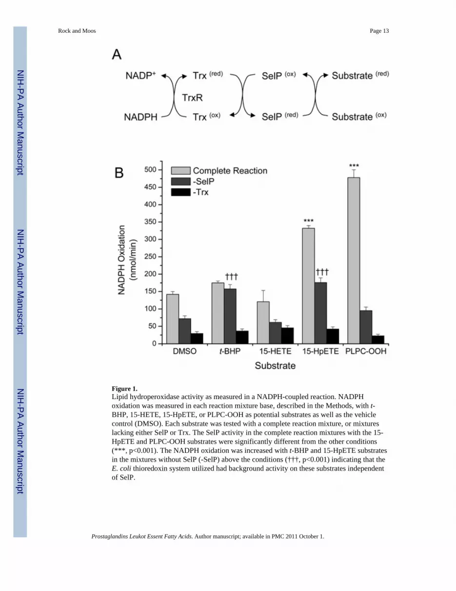
Figure 1.Lipid hydroperoxidase activity as measured in a NADPH-coupled reaction. NADPHoxidation was measured in each reaction mixture base, described in the Methods, with t-BHP, 15-HETE, 15-HpETE, or PLPC-OOH as potential substrates as well as the vehiclecontrol (DMSO). Each substrate was tested with a complete reaction mixture, or mixtureslacking either SelP or Trx. The SelP activity in the complete reaction mixtures with the 15-HpETE and PLPC-OOH substrates were significantly different from the other conditions(***, p<0.001). The NADPH oxidation was increased with t-BHP and 15-HpETE substratesin the mixtures without SelP (-SelP) above the conditions (†††, p<0.001) indicating that theE. coli thioredoxin system utilized had background activity on these substrates independentof SelP.
Rock and Moos Page 13
Prostaglandins Leukot Essent Fatty Acids. Author manuscript; available in PMC 2011 October 1.
NIH
-PA Author Manuscript
NIH
-PA Author Manuscript
NIH
-PA Author Manuscript

Figure 2.Assessment of selenium and SelP content in concentrated media. Left: ICP analysis ofselenium content in media samples; (+) SelP represents media from cells induced to expressSelP by ponasterone A while (−) SelP represents media from cells treated with EtOH(vehicle control). The selenium difference between these concentrated media is highlysignificant (***, p<0.001). Right: Immunoblot analysis for SelP, TR1 and GPx4 in thesemedia samples. The control for TR1 is 10 μg of HEK293 cell lysate, and the control forGPx4 is 30 μg of mouse testicular lysate.
Rock and Moos Page 14
Prostaglandins Leukot Essent Fatty Acids. Author manuscript; available in PMC 2011 October 1.
NIH
-PA Author Manuscript
NIH
-PA Author Manuscript
NIH
-PA Author Manuscript
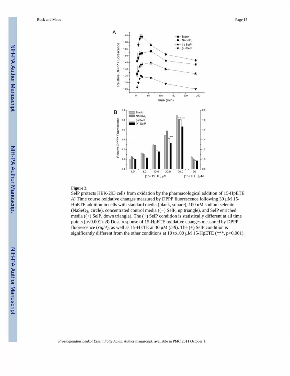
Figure 3.SelP protects HEK-293 cells from oxidation by the pharmacological addition of 15-HpETE.A) Time course oxidative changes measured by DPPP fluorescence following 30 μM 15-HpETE addition in cells with standard media (blank, square), 100 nM sodium selenite(NaSeO3, circle), concentrated control media ((−) SelP, up triangle), and SelP enrichedmedia ((+) SelP, down triangle). The (+) SelP condition is statistically different at all timepoints (p<0.001). B) Dose response of 15-HpETE oxidative changes measured by DPPPfluorescence (right), as well as 15-HETE at 30 μM (left). The (+) SelP condition issignificantly different from the other conditions at 10 to100 μM 15-HpETE (***, p<0.001).
Rock and Moos Page 15
Prostaglandins Leukot Essent Fatty Acids. Author manuscript; available in PMC 2011 October 1.
NIH
-PA Author Manuscript
NIH
-PA Author Manuscript
NIH
-PA Author Manuscript

Figure 4.SelP can protect target cells from the oxidation by effector cells following 15-LOX-1catalysis of arachidonate. A) Inducible expression of 15-LOX-1 in 293-EcR cells withintegrated 15-LOX-1. Four conditions are evaluated; vehicle control (EtOH), ponasterone A(PonA), arachidonic acid (AA), and the combination ponasterone A and arachidonate (PonA+AA). B) Production of the 15-LOX-1 metabolite 15-HETE under the four conditions justdescribed. C) Protection from oxidation, as measured by DPPP fluorescence, with theaddition of SelP enriched media, (+) SelP, compared to the control concentrated media, (−)SelP (***, p< 0.001).
Rock and Moos Page 16
Prostaglandins Leukot Essent Fatty Acids. Author manuscript; available in PMC 2011 October 1.
NIH
-PA Author Manuscript
NIH
-PA Author Manuscript
NIH
-PA Author Manuscript
Related Documents