1994 84: 1587-1593 J Plum, M De Smedt, MP Defresne, G Leclercq and B Vandekerckhove thymic microenvironment Human CD34+ fetal liver stem cells differentiate to T cells in a mouse http://bloodjournal.hematologylibrary.org/site/misc/rights.xhtml#repub_requests Information about reproducing this article in parts or in its entirety may be found online at: http://bloodjournal.hematologylibrary.org/site/misc/rights.xhtml#reprints Information about ordering reprints may be found online at: http://bloodjournal.hematologylibrary.org/site/subscriptions/index.xhtml Information about subscriptions and ASH membership may be found online at: reserved. Copyright 2011 by The American Society of Hematology; all rights 900, Washington DC 20036. weekly by the American Society of Hematology, 2021 L St, NW, Suite Blood (print ISSN 0006-4971, online ISSN 1528-0020), is published For personal use only. by guest on July 15, 2011. bloodjournal.hematologylibrary.org From

Welcome message from author
This document is posted to help you gain knowledge. Please leave a comment to let me know what you think about it! Share it to your friends and learn new things together.
Transcript
1994 84: 1587-1593
J Plum, M De Smedt, MP Defresne, G Leclercq and B Vandekerckhove thymic microenvironmentHuman CD34+ fetal liver stem cells differentiate to T cells in a mouse
http://bloodjournal.hematologylibrary.org/site/misc/rights.xhtml#repub_requestsInformation about reproducing this article in parts or in its entirety may be found online at:
http://bloodjournal.hematologylibrary.org/site/misc/rights.xhtml#reprintsInformation about ordering reprints may be found online at:
http://bloodjournal.hematologylibrary.org/site/subscriptions/index.xhtmlInformation about subscriptions and ASH membership may be found online at:
reserved.Copyright 2011 by The American Society of Hematology; all rights900, Washington DC 20036.weekly by the American Society of Hematology, 2021 L St, NW, Suite Blood (print ISSN 0006-4971, online ISSN 1528-0020), is published
For personal use only. by guest on July 15, 2011. bloodjournal.hematologylibrary.orgFrom

Human CD34+ Fetal Liver Stem Cells Differentiate to T Cells in a Mouse Thymic Microenvironment
By Jean Plum, Magda De Smedt, Marie-Paule Defresne, Georges Leclercq, and Bart Vandekerckhove
Hematopoietic stem cells differentiate in the thymus to T cells along precisely defined intermediates. This process is thymic epithelium dependent and involves cytokines and cell-cell interactions between thymic stroma and T-cell pre- cursors. Here we report that highly purified human CD34++ fetal liver stem cells differentiate to mature T cells, when seeded into isolated fetal thymic lobes of severe combined immunodeficient mice, and subsequently cultured in vitro. The human stem cells differentiate sequentially into CD4+CD8-CD3-, CD4+CD8+CD3-, CD4+CD8+CD3+, and fi-
EMATOPOIETIC stem cells are capable of self re- newal and differentiation into erythroid, myelomono-
cytic, and lymphoid lineages.’.’ Whereas in vitro stem cell assays are available for human myelopoiesis and B-lympho- cyte differentiati~n,~ this is not the case for T cells. For T-cell development, the three-dimensional structure of the thymic stroma seems to be indispensable? In the thymic stroma, the epithelium is essential for early T-cell differentia- tion of hematopoietic precursors as well as terminal differen- tiation to mature T cell^.^,^ The potential of hematopoietic precursor cells to differentiate along the T-cell lineage is therefore difficult to address, because no practical assays exist in which full T-cell differentiation occurs. Recently, severe combined immunodeficient (SCID)-hu mice, chime- ras obtained by transplantation of human hematolymphoid tissue into congenitally SCID host have been explored to provide an in vivo culture system for dissecting the human T-cell differentiation.”” CD34+ fetal precursor cells are in- jected with a microsyringe into HLA-mismatched human thymus fragments, partially depleted of hematopoietic cells by low temperature culture and allowed to develop on en- graftment into SCID m i ~ e . ~ . ~ Although this assay requires fetal tissue and is technically cumbersome, it represented a major breakthrough in the study of human hematopoiesis because it allows functional testing of precursor cells and dynamic T-cell differentiation experiment^.^.^ It has been reported recently that this allogeneic organotypic culture sys- tem can be adapted to an in vitro ~ y s t e m . ’ ~ “ ~ This human fetal thymic organ culture system was used to delineate the early events in human thymic de~elopment.’”’~
Here, we report in vitro human T-cell differentiation of CD34++ human fetal liver stem cells in a xenogeneic envi- ronment. The experiments are based on the fetal thymus organ culture as described by Kingston et al and Jenkinson et ai? As thymic environment for human T-cell development, fetal SCID thymus was used. As a result of a defective recombination of the genes coding for the antigen receptor, SCID mice (C.B.- 17 scidlscid) have an impaired thymocyte maturation.I6 The differentiation of their thymocytes is blocked before the CD4-CD8 double-positive stage,17 giving space for concurrent maturation of xenogeneic cells. This alternative assay method, developed in our laboratory, elimi- nates the need of human fetal thymus, the differences in HLA mismatch and in vivo testing.
H
MATERIALS AND METHODS
Preparation of human CD34++ fetal liver cells. Human fetal liver tissues were obtained after legal termination of pregnancy, and
Blood, Vol 84, No 5 (September l), 1994: pp 1587-1593
nally, CD4+CD8-CD3++ and CD4-CD8+CD3++ cells. Pheno- typic analysis for additional maturation markers showed that these CD4 and CD8 single-positive thymocytes are fully maturate cells. By immunochemistry, human HLA-DR’ cells with a dendritic morphology could be daected. This novel chimeric human-mouse fetal thymus organ culture offers a tool to study human T-cell ontogeny in vitro and is a rapid and reliable test method for T-cell precursor activity of cul- tured or transfected human stem cells. 0 1994 by The American Society of Hematology.
obtained and used following the guidelines of the Medical Ethical Commission of the University Hospital of Ghent. Human fetal liver cells were isolated by gentle disruption of the tissue in complete medium (Iscove’s modified Dulbecco’s medium/l0% fetal calf se- rum [FCS]; GIBCO, Paisley, Scotland), followed by density gradient centrifugation over Lymphoprep (Nyegaard, Oslo, Norway). Cells were washed and resuspended in 90% FCS/10% dimethyl sulfoxide and frozen in liquid NZ. After thawing, fetal liver cells were washed and immunofluorescently labelled with CD3 (Leu4 fluorescein iso- thiocyanate [FITC]), CD4 (Leu-3a FITC), CD8 (Leu-2a FITC), CD19 (Leu-l2 FITC), CD1 (CDla FlTC; Serotec, Oxford, United Kingdom), and with CD34 (HPCA-2 PE) (unless specified, all MoAbs were from Becton Dickinson Immunocytometry Systems, Mountain View, CA). CD34++ CD4-CD8-CD3-CD19-CDI- cells were sorted on a fluorescence-activated cell sorter (FACS) vantage (Becton Dickinson). The sorted cells were transferred to murine thymic lobes by the hanging drop method.I5
Mice. Fourteen to 15 day pregnant C.B.-17 SCID mice were obtained from our own specific pathogen-free breeding facility.
Fetal thymus organ culture (FTOC). Thymic lobes were pre- pared from fetal day 14 to 15 SCID mice. Hanging drops were prepared in Terasaki plates by adding in each well 25 pL of complete medium containing 10,000 or less sorted cells to one thymic lobe. The plates were immediately inverted to form hanging drops and incubated during 48 hours in a humidified incubator (7.5% CO, in air, 37°C). After incubation, the lobes were removed from the hang- ing drop, washed, and put on the surface of a nuclepore filter in organ culture in an incubator. At different time points, thymocytes were recovered by mechanical disruption of the thymic lobes with a small tissue grinder. Thymocytes were stained with eosin and
From the Department of Clinical Chemistry, Microbiology and Immunology, University of Ghent, University Hospital, Ghent, Bel- gium: and the Laboratory of Pathological Anatomy and Cytopathol- ogy, University of LiQe, LiPge, Belgium.
Submitted March 15, 1994; accepted May 9, 1994. Supported by grants from Flemish Action Program for Biotechnol-
ogy (VZAB), Fund for Medical Scientz$c Research (FGWO), and Fund for Concerned Research Program (OOA). G.L. is a research associate of the Belgian Fund for ScientiJic Research (NFWO).
Address reprint requests to Jean Plum, MD, PhD, Department of Clinical Chemistry, Microbiology and Immunology, University of Ghent, University Hospital, Blok A, De Pintelaan 185, B-9OOO Ghent. Belgium.
The publication costs of this article were defrayed in part by page charge payment. This article must therefore be hereby marked “advenisement” in accordance with 18 U.S.C. section 1734 solely to indicate this facr.
0 1994 by The American Society of Hematology. OOO6-4971/94/8405-0031$3.00/0
1587
For personal use only. by guest on July 15, 2011. bloodjournal.hematologylibrary.orgFrom
1588 PLUM ET AL
counted with a hematocytometer. At least 80% of the cells were viable.
Flow cy)?tometry. Before labeling, cells were suspended in phos- phate-buffered saline (PBS) 1% bovine serum albumin (BSA) -0.1% NaN, and the Fc receptor of the mouse thymocytes was blocked by preincubation for 15 minutes with anti-FcyRILnII MoAb (clone 2.4.G2)'* to avoid aspecific binding of antibodies by the mu- rine thymocytes. Subsequently, the cells were stained with a panel of MoAbs, as indicated. The MoAbs used were rat anti-mouse CD45 (CD45 R-PE, MI-9.3 HL; Gibco BRL: Immunoselect or CD45- Biotin, 30FI 1.1; Pharmingen, San Diego, CA), CDla (B-B5; Sero- tec, Oxford, United Kingdom) and the following mouse anti-human MoAbs from Becton Dickinson, CD3 (Leu-4 FITC, PE, or biotin), CD4 (Leu-3a FITC or PE), CD8 (Leu2a FITC or biotin). CD14 (Leu-M3 PE), CD19 (Leu-l2 PE), CD34 (HPCA-2 FITC or PE), CD45 (HLe-I FITC), anti-T-cell receptor (TCR)-a$ (TCR-I FITC), anti-TCR-yb (anti-TCR-ybll FITC). The biotinylated anti- bodies were revealed with second-step streptavidin cychrome (Phar- mingen). All MoAbs against human antigens were checked for nega- tive staining on SCID thymocytes after blocking with 2.462 MoAb. lsotype controls were also included in most staining series and were found to he negative. The cells were analyzed on a FACScan (Becton Dickinson) with an argon-ion laser tuned at 488 nm. Forward light scattering, orthogonal scattering, and three fluorescence signals were determined on 10,000 cells and stored in listmode data files. Data acquisition and analysis was performed with Lysis 2.0 software (Becton Dickinson). Dead cells were gated out by propidium iodide exclusion (PI). In most cases. viable human cells were gated by
A START AFTER SORT
exclusion of mouse CD45-PE or mouse CD45-biotin-streptavidin- cychrome-positive cells and PI-positive cells.
Immunocytochemisfry. After organ culture, thymic lobea were frozen in liquid nitrogen after embedding in OCT compound (Miles Scientific, Naperville, IL). Immunochemistry was performed on 5 to 6 pm-thick cryostat sections fixed in cold (4°C) acetone. Endogenous peroxidase was destroyed (PBS + 3% H202 , 10 minutes) and non- specific antibody binding was blocked throughout the procedure us- ing 3% BSA in PBS (Calbiochem, La Jolla, CA) and by preincuba- tion in 3% of normal rabbit Igs (Dako, Prosan, Gent, Belgium) for 30 minutes.
Sections were incubated sequentially, with intervening washes, in anti-human HLA-DR (Becton Dickinson) or in anti-human leuco- cyte-common antigen (L-CA, Dako), which recognizes the CD45 protein found on all cells of hematopoietic origin except erythro- cytes,'" followed by peroxidase-conjugated anti-mouse Igs (Dako). Sections were developed with 3,3'-diaminohenzidine as substrate (UCB, VEL Leuven. Belgium). Controls were incubation with the secondary antibody only, or incubation with unrelated mouse Igs instead of the specific antibodies.
RESULTS AND DISCUSSION
As shown in Fig IA, CD34"CD4-CD8-CDTCDla CD19~- hematopoietic stem cells were highly enriched from human fetal liver (gestational age ranged from 18 to 22 weeks) by flow cytometric sorting. These highly purified CD34" cells (>99.7%) were seeded into single thymic
I CDl, 3,4, 8. 19 - FITC b
B
- I 140 20
120 -
100
80
-
-
60 -
0 5 10 15 20 25 30 36
DAYS OF ORGAN CULTURE
z c I W m D
0 0 w e 0 m r- G; - 1 H
Fig 1. Growth kinetics of human and murine thy- mocytes in chimeric organ cultures. (A) Flow cytro- metric analysis of human fetal liver cells stained with CD34-PE and FITC-labelled CD1, CD4, CD8, CD3, and CD19 before and after sorting. The cells were purified by gating on CD34" and CD4-, CD8-. CD3-, CDI-, CD19-. (B) Growth kinetics of human and murine thymocytes. 10,OOO human CD34+' fetal liver cells were seeded in SCID murine fetal thymi by hanging drop. After 48 hours (day = 0 ) unseeded cells were washed away and the lobes were cultured on Nuclepore filters in FTOC. At dtferent time points, cells were counted and stained with human CD45 and mouse CD45 or with CD34 at the indicated times and assessed by flow cytometry (FACScan). The cell number for each subset was calculated by multi- plying the total cell number with the percentage of cells that stained for a given antigen divided by 100. Data are expressed as mean values (*l SDI of 3 to 4 independent experiments, each of which contained at least three lobes per data point. The scale on the left axis is for human CD45 and mouse CD45 cells, the smaller scale on the right axis is for CD34 cells. A gradual increase in human cells, a constant level of mouse cells and a constant level of CD34' until day 21 followed by a decline. Data are representative for three experiments.
For personal use only. by guest on July 15, 2011. bloodjournal.hematologylibrary.orgFrom
DIFFERENTIATION OF HUMAN T CELLS IN MOUSE THYMUS 1589
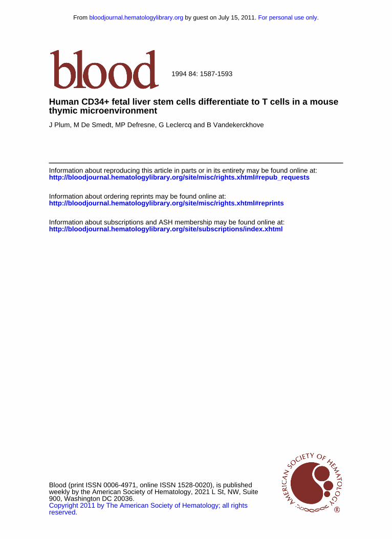
Fig 2. Kinetic analysis of the expression of CD4, CD8. Human- mouse chimeric cultures were set up as described in Fig l. Thymocytes were recovered, counted, and stained for coordi- nate expression of CD4 and CD8 a t the indicated times. Data are representative for two to four experiments (with at least 4 lobes each) per timepoint.
DAY 6 A
W
d
0
n n
DAY 14 DAY 21 r
2 23
DAY 27 DAY 35 64 1
CD8 FlTC b
lobes of fetal day 14 to 15 SCID mice. When 10,000 cells were placed in a hanging drop, together with one thymic lobe, approximately 2,000 cells entered the lobe during a time period of 48 hours (day = 0). As shown in Fig IB, the murine thymus supported the growth of human thymocytes as the number of human CD4S+ cells increased progres- sively. The number of endogenous SCID thymocytes re- mained at low levels. Starting from approximately 2,000 human cells in a lobe, about 120,000 human cells were ob- tained after S weeks of culture, which represents a 60-fold increase. The absolute number of CD34' cells remained con-
DAY 6 DAY 14
f DAY 27 B
Y
stant during the first 3 weeks of culture, and declined thereaf- ter (Fig lB), suggesting that the thymic microenvironment cannot maintain self-renewal of the hematopoietic stem cells. The pre-T-cell precursor frequency in the sorted CD34+'CD3-CD4-CD8- cell population was determined by limiting dilution using automatic cell deposition unit (Becton Dickinson). Groups of 20 lobes were used for transfer of 1 , 10, or 100 cellsflobe by hanging drop. The proportion of repopulated lobes containing human CD45'CD4+CD8' cells in each group was determined by FACS analysis. After 14 to 21 days, 0120 lobes were repopu-
DAY 21
" DAY 35
I CD3 FlTC
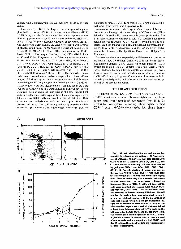
Fig 3. Kinetic analysis of the expression of CD3. Human- mouse chimeric cultures were set up as described in Fig 1. Thymocytes were recovered, counted, and stained for expres- sion of CD3 at the indicated times. Data are representative for two to four experiments lwith at least 4 lobes each) per timepoint. Data are presented as CD3 histograms at diffsrent time points.
For personal use only. by guest on July 15, 2011. bloodjournal.hematologylibrary.orgFrom
1590
lated in the group in the group of 1 cell/lobe, 1/20 lobes in the group of 10 cells/lobe and 15/20 in the group of 100 cells/lobe (data not shown). This shows that the chimeric human-mouse organ culture is an assay system for T-cell precursors at the clonal level. When human differentiation markers were studied, the successive appearance of different subsets was noted. Analysis of the cells that seeded the thy- mic lobe (day 0) showed that at least 80% of the cells were still CD34". After 6 days of R O C , a small population (-20%) expressed low levels of CD4 (Fig 2). These CD4'"" cells precede the emergence of CD4h'gh cells. After 14 days of culture, CD4'""CD3- and CD4h'ghCD3- cells were present. These cells were CDla+ and CD34- (data not shown). At that time, only a small number of cells expressed CD3 (Fig 3). In the human thymus, this CD1 +CD4+CDS-CD3- subset has been reported to contain the highest proportion of cycling cells'0 and it was suggested that this subset represents an early transitional stage between CD4-CD8- and CD4+CD8+
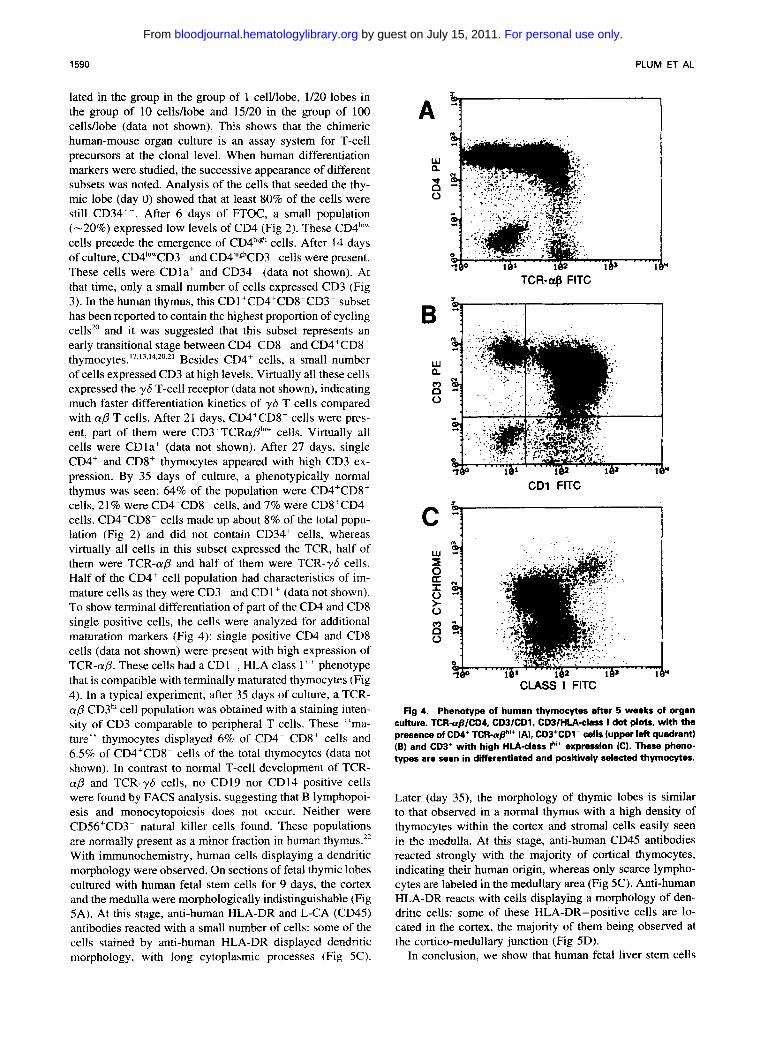
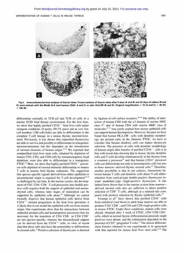
of cells expressed CD3 at high levels. Virtually all these cells expressed the yS T-cell receptor (data not shown), indicating much faster differentiation kinetics of y b T cells compared with ap T cells. After 21 days, CD4+CD8+ cells were pres- ent, part of them were CD3+TCRap1"" cells. Virtually all cells were CDla' (data not shown). After 27 days, single CD4+ and CD8+ thymocytes appeared with high CD3 ex- pression. By 35 days of culture, a phenotypically normal thymus was seen: 64% of the population were CD4+CD8+ cells, 21% were CD4+CD8- cells, and 7% were CD8+CD4" cells. CD4-CD8- cells made up about 8% of the total popu- lation (Fig 2 ) and did not contain CD34' cells, whereas virtually all cells in this subset expressed the TCR, half of them were TCR-a,f3 and half of them were TCR-?S cells. Half of the CD4+ cell population had characteristics of im- mature cells as they were CD3- and CD1+ (data not shown). To show terminal differentiation of part of the CD4 and CD8 single positive cells, the cells were analyzed for additional maturation markers (Fig 4): single positive CD4 and CD8 cells (data not shown) were present with high expression of TCR-(YD. These cells had a CDI-, HLA class I++ phenotype that is compatible with terminally maturated thymocytes (Fig 4). In a typical experiment, after 35 days of culture, a TCR- ap CD3h' cell population was obtained with a staining inten- sity of CD3 comparable to peripheral T cells. These "ma- ture" thymocytes displayed 6% of CD4- CD8' cells and 6.5% of CD4'CDK cells of the total thymocytes (data not shown). In contrast to normal T-cell development of TCR- ap and TCR-y6 cells, no CD19 nor CD14 positive cells were found by FACS analysis, suggesting that B lymphopoi- esis and monocytopoiesis does not occur. Neither were CD56+CD3- natural killer cells found. These populations are normally present as a minor fraction in human thymus." With immunochemistry, human cells displaying a dendritic morphology were observed. On sections of fetal thymic lobes cultured with human fetal stem cells for 9 days, the cortex and the medulla were morphologically indistinguishable (Fig 5A). At this stage, anti-human HLA-DR and L-CA (CD45) antibodies reacted with a small number of cells: some of the cells stained by anti-human HLA-DR displayed dendritic morphology, with long cytoplasmic processes (Fig 5C).
thymocytes.12.~"~4,20,21 Besides CD4+ cells, a small number
PLUM ET AL
A ' i
TCR-US FlTC
CD1 FlTC
CLASS 1 FlTC 1.I
Fig 4. Phenotype of human thymocytes after 5 weeks of organ culture. TCR-apICDI, CDBICDI. CDB/HLA-class I dot plots, with the presence of CD4+ TCR-np"* [A), CDS*CDl- cells [upper left quadrant) [B) and CD3' with high HlA-class lhi+ expression (C). These pheno- types are seen in differentiated and positively selected thymocytes.
Later (day 35), the morphology of thymic lobes is similar to that observed in a normal thymus with a high density of thymocytes within the cortex and stromal cells easily seen in the medulla. At this stage, anti-human CD45 antibodies reacted strongly with the majority of cortical thymocytes, indicating their human origin, whereas only scarce lympho- cytes are labeled in the medullary area (Fig 5C). Anti-human HLA-DR reacts with cells displaying a morphology of den- dritic cells: some of these HLA-DR-positive cells are lo- cated in the cortex, the majority of them being observed at the cortico-medullary junction (Fig 5D).
In conclusion, we show that human fetal liver stem cells
For personal use only. by guest on July 15, 2011. bloodjournal.hematologylibrary.orgFrom
1591
D) were stained with the MoAb CIA (anti-human CD45 A and Cl or anti-HLA-DR IB and D). Original magnification x 10 (A and Cl: x 40 ID); Fig 5. Immunohistochemical analysis of thymic lobes. Frozen sections of thymic lobes after 9 days IA and B1 and 35 days of cukure (B and
x 100 (B).
differentiate normally to TCR-aP and TCR-y6 cells in a murine SCID fetal thymic environment. For the first time, we show that highly purified CD34" fetal liver cells under stringent conditions of purity (99.7% pure) and at very low cell numbers ( 1 0 0 cells:lobe) are able to differentiate to the complete T-cell lineage in a mouse thymic microenviron- ment. Previously, it was shown that unpurified thymocytes are able to survive and possibly to differentiate in xenogeneic microenvironment, but this depended on the introduction of stromal elements of human origin.'""' We reported that semipurified fetal liver stem cells, obtained by depletion of mature CD3, CD4, and CD8 cells by immunomagnetic bead depletion, were also able to differentiate in a xenogeneic FTOC.2s Here, we show that highly purified CD34" precur- sor cells depleted of stromal elements differentiate to mature T cells in murine fetal thymic rudiments. The suggestion that species-specific signals derived from either epithelial or mesenchymal origin is required for T-cell de~elopment '~~'~ is challenged by our data. In the murine system, the develop- ment of CD3-CD4-CD8- T-cell precursors into double-pos- itive cells requires both the support of epithelial and mesen- chymal cells, whereas later stages of maturation can be supported by epithelial cells alone.'" Although we cannot formally disprove that human epithelial cells derive from CD34" stromal progenitors in the fetal liver specimen. it is likely that in our model the epithelial cells are from murine origin. Our experiments show that the interactions between epithelial stromal cells and hematopoietic precursors that are necessary for the transition of CD4TD8- to CD4'CD8' are not species-specific, whereas the mesenchymal support can be derived from the CD34" cells, in view of recent data that these cells also have the potentiality to differentiate in stromal cells.27 Positive selection of thymocytes is directed
by ligation of cell surface receptors.""" The ability of inter- action of human CD8 with the a3 domain of murine MHC class 13' and of human CD4 with murine MHC class I1 molecules2'~3' may partly explain how mouse epithelial cells can support human thymopoiesis. However, because we have found that human HLA-DR' cells with dendritic morphol- ogy are present early in the chimeric FTOC, we have to consider that human dendritic cells can induce thymocyte selection. The presence of cells with dendritic morphology of human origin after transfer of purified CD34" cells is in line with recent data showing that in mouse, thymic dendritic cells and T cells develop simultaneously in the thymus from a common a precursor" and that human CD34' precursor cells can differentiate not only to hematopoietic cells but also to bone marrow-derived thymic stromal cells.'7 Therefore, another possibility is that in our cultures, interactions be- tween human T cells and dendritic cells direct T-cell differ- entiation from cortical-type double-positive thymocytes to- ward medullary-type single-positive thymocytes. It has indeed been shown that in the murine system bone marrow- derived stromal cells also are sufficient to direct positive selection of CD8' T cells, although at a reduced rate com- pared with positive selection by thymic epithelial cells.'"
Yeoman et a127 have shown that purified CD34' cells from umbilical cord blood or adult bone marrow are able to produce CD4TD8'. and CD4 and CD8 single-positive cells in mouse R O C . Under these conditions, mature cells were already obtained after 1 week of FTOC, CD4'CD8'CD3- cells, which in normal thymic differentiation preceeds single positives were absent, and the colonization depended on the presence of CD7' progenitor T cells.'7 The slower differenti- ation kinetics obtained in our experiments is in agreement with that reported for mouse fetal liver stem The
For personal use only. by guest on July 15, 2011. bloodjournal.hematologylibrary.orgFrom
1592 PLUM ET AL
slower kinetics may also reflect that in the human-mouse FTOC the growth of human dendritic/stromal cells precedes and is required for the subsequent development of human T cells. In our cultures, we find CD4+CDgcCD3- cells and an emergence of populations of thymocytes in a time-ordered sequence in accordance with normal human thymic differen- tiation. Finally, the purified CD34++ cell population used in our experiments contained no CD7 bright cells, but about 20% of cells stained weakly for CD7. In a series of experi- ments, where the starting CD34++ population was supple- mentarily depleted for CD7, we observed a similar differenti- ation (data not shown). Our findings are in line with the observations of Barcena et al,I4 who found that CD7- fetal liver cells can differentiate to T cells. It requires further investigation to determine whether the difference in reconsti- tution kinetics, presence of CD3-CD4+CD8+ thymocytes and dependence of CD7' cells in the starting population are because of differences in the stem cell properties of embry- onic fetal liver versus the umbilical cord blood or adult bone marrow. Another factor that is of importance in our hands is the purity of the starting population. In qxperiments in which cell populations are contaminated with low amounts of mature T cells, these contaminants expand fast and to high numbers and interfere with the T-cell differentiation pattern (data not shown). Finally, we showed that fetal thy- mus organ culture is a reproducible method to support the full range of human T-cell maturation, as six independent sources of fetal liver cells and 99% of the cultures (600/608 lobes examined) were successful. This method is a unique tool to study early steps of T-cell differentiation, and to explore the role of cytokines and ligands in this process. In addition, because stem cells are targets for gene therapy, the stability of gene expression can be followed throughout the T-cell differentiation process. Because the thymus is suscep- tible for human immunodeficiency virus (HIV) infection, this method should prove useful to evaluate if gene therapy strategies are effective in the cure of HIV infection.
ACKNOWLEDGMENT
We thank Christian De Boever for art-work. We thank Marie- Christine Petit for excellent technical assistance.
REFERENCES
I. Sprangrude GJ, Heimfeld S, Weissman IL: Purification and characterization of mouse hematopoietic stem cells. Science 241 58, 1988
2. Keller G: In vitro analysis of murine B-cell development. Curr Opin Immunol 4: 133, 1992
3 . Toogood IRG, Dexter TM, Allen TD, Suda T, Lajtha LC: The development of a liquid culture system for the growth of human hone marrow cultures. Leuk Res 4:449, 1980
4. Kingston R, Jenkinson EJ, Owen JJT: A single stem cell can recolonize an embryonic thymus, producing phenotypically distinct T-cell populations. Nature 317:811, 1985
5. von Boehmer H: Developmental biology of T cells in T cell receptor transgenic mice. Annu Rev Immunol 8:531, 1990
6. von Boehmer H: Thymic selection: A matter of life and death. Immunol Today 13:454, 1992
7. McCune JM, Namikawa R, Weilbaecher KN, Kaneshima H, Schultz LD, Lieberman M, Weissman IL: The SCID-hu mouse: Murine model for the analysis of human hematolymphoid differenti- ation and function. Science 241:1632, 1988
8. Baum CM, Weissman IL, Tsukamoto AS, Buckle AM, Peaulc B: Isolation of a candidate human hematopoietic stem-cell popula- tion. Proc Natl Acad Sci USA 89:2804, 1992
9. PBault B, Weissman IL, Baum C, McCune JM, Tsukamoto A: Lymphoid reconstitution of the human fetal thymus in SCID mice with CD34" precursor cells. J Exp Med 174:1287, 1991
IO. Vandekerckhove BAE, Namikawa R, Bachetta R, Roncarolo MG: Human hematopoietic cells and thymic epithelial cells induce tolerance via different mechanisms in the SCID-hu mice. J Exp Med 175:1033, 1992
11. Vandekerckhove BAE, Bacal R, Jones D, Kono DH, Theoti- lopoulos N, Roncarollo MG: Thymic selection of the human T cell receptor Vp repertoire in SCID-hu mice. J Exp Med 176: I6 19, 1992
12. Kraft DL, Weismann IL, Waller EK: Differentiation of CD3-CD4-CD8- human fetal thymocytes in vivo: Characterization of a CD3-CD4+CD8- intermediate. J Exp Med 178:265, 1993
13. Galy A, Verma S, Barcena A, Spits H: Precursors of CD3+CD4+CD8+ cells in the human thymus are defined by expres- sion of CD34. Delineation of early events in human thymic develop- ment. J Exp Med 178:391, 1993
14. Barcena A, Muench MO, Galy AHM, Cupp J. Roncarolo MG, Philips JH, Spits H: Phenotypic and functional analysis of T cell precursors in the human fetal liver and thymus: CD7 expression in the early stages of T- and myeloid development. Blood 82:3401, I993
15. Jenkinson EJ, Franchi L, Kingston R, Owen JJT: Effect of deoxyguanosine on lymphopoiesis in the developing thymus rudi- ment in vitro: Application in the production of chimeric thymus rudiments. Eur J lmmunol 12583, 1982
16. Bosma MJ, Carrol AM: The SCID mouse mutant: Definition, characterization and potential uses. Annu Rev Immunol 9:323, I99 1
17. Hahu S, Norisha Y, Yagita H, Okumura K: Phenotype and differentiation stage of SCID mouse thymocytes. Curr Topics Micro- hi01 Immunol 152:27, 1989
18. Unkeless JC: Characterization of a monoclonal antibody against mouse macrophage and lymphocyte Fc receptors. J Exp Med 150:580, 1979
19. Thomas ML: The leucocyte common antigen family. Annu Rev Immunol 7:339, 1989
20. Alvarez-Vallina L, Gonzalez A, Gamhon F, Kreisler M, Diaz- Espada F: Delimitation of the proliferative stages in the human thymus indicates that cell expansion occurs before the expression of CD3 (T cell receptor). J Immunol 150:8, 1993
21. Hori T, Cupp J, Wrighton N. Lee F, Spits H: Identification of a novel human thymocyte subset with a phenotype of CD3 -CD4+CD8a'P-. Possible progeny of the CD3+CD4+CD8+ subset. J Immunol 146:4078, 1991
22. Lanier LL, Spits H, Phillips JH: The developmental relation- ship between NK cells and T cells. Immunol Today 13:392, 1992
23. Fisher AG, Larsson L, Goff LK, Herstall DE, Happerfield, Merckenschlager M: Human thymocyte development in mouse or- gan cultures. Int Immunol 2:571, 1990
24. Merkenschlager M, Fischer AG: Human postnatal thymo- cytes generate phenotypically immature CD3(dim), CD5(dim), CDla(bright) progeny in organ culture. J Immunol 148:1012, 1992
25. De Smedt M, Leclercq G, Vandekerckhove B, Plum J: Stem cell differentiation into mature lymphocytes using the SCID mouse thymus chimera model, in Wunder E, Sovalat H, Henon P, Serke S (eds): The hematopoietic stem cells. Mulhouse Manual, Dayton, OH, AlphaMed Press, 1994, p 303
26. Anderson G, Jenkinson EJ, Moore NC, Owen JJT: MHC class 11-positive epithelium and mesenchyme cells are both required for T-cell development in the thymus. Nature 362:70, 1993
27. Huang S, Terstappen LWMM: Formation of haematopietic microenvironment and haematopoietic stem cells from single human hone marrow stem cells. Nature 360:745, 1992
For personal use only. by guest on July 15, 2011. bloodjournal.hematologylibrary.orgFrom
DIFFERENTIATION OF HUMAN T CELLS IN MOUSE THYMUS 1593
28. MacDonald HR. Lees RK, Schneider R, Zinkernagel RM, Hengartner H: Positive selection of CD4' thymocytes controlled by MHC class I1 gene products. Nature 336:471, 1988
29. Marusic-Galesic S, Stephany DA, Longo DL, Kruisbeek AM: Development of CD4-CD8' cytotoxic T cells requires interactions with class I MHC determinants. Nature 333:180, 1988
30. Zuniga-Plucker JC, Jones LA, Longo DL, Kruisbeek AM: Both T C W H C accessory molecule/MHC interactions are required for positive and negative selection of mature T cell in the thymus. Cold Spring Harb Symp Quant Biol 54:153, 1989
31. Moots RJ, Samberg NL, Pazmany L, Frelinger JA, McMi- chael AJ, Stauss HJ: A cross-species functional interaction between the murine major histocompatibility complex class I a 3 domain of human CD8 revealed by peptide-specific cytotoxic T lymphocytes. Eur J Immunol 22: 1643, 1992
32. Bargaza-Gilbert E, Grass D, Lawrance SK, Peterson PA, Lacy E, Engelhard VH: Species specificity and augmentation of responses to class I1 major histocompatibility complex molecules in human CD4 transgenic mice. J Exp Med 175:1707, 1992
33. Vignalli DAA, Moreno J, Schiller D, Hammerling GJ: Spe-
cies-specific binding of CD4 to the p2 domain of the major histo- compatibility complex class I1 molecules. J Exp Med 175:925, 1992
34. Killeen N, Sawada S, Litmann DR: Regulated expression of human CD4 rescues helper T cell development in mice lacking expression of endogenous CD4. EMBO J 12:1547, 1993
35. Ardavin C, Wu L, Li C-L, Shortman K: Thymic dendritic cells and T cells develop simultaneously in the thymus from a common precursor population. Nature 362:761, 1993
36. Bix M, Raulet D: Inefficient positive selection of T cells directed by haematopoietic cells. Nature 359:330, 1992
37. Yeoman H, Gress RE, Bare CV, Leary AG, Boyse EA, Bard J, Shultz LD, Harris DT, DeLuca D: Human bone marrow and umbilical cord blood cells generate CD4+ and CD8' single-positive cells in murine fetal thymus organ culture. Proc Natl Acad Sci USA 90:10778, 1993
38. Petrie HT, Hugo P, Scollay R, Shortman K: Lineage relation- ship and developmental kinetics of immature thymocytes: CD3, CD4, and CD8 acquisition in vivo and in vitro. J Exp Med 172:1583, 1990
For personal use only. by guest on July 15, 2011. bloodjournal.hematologylibrary.orgFrom
Related Documents