Controversies in Experimental Dermatology Section Editor: Ralf Paus, Hamburg How best to fight that nasty itch – from new insights into the neuroimmunological, neuroendocrine, and neurophysiological bases of pruritus to novel therapeutic approaches T. Biro´, M. C. Ko, B. Bromm, E. T. Wei, P. Bigliardi, F. Siebenhaar, H. Hashizume, L. Misery, N.V. Bergasa, C. Kamei, J. Schouenborg, D. Roostermann, T. Szabo´, M. Maurer, M. Bigliardi-Qi, J. G. Meingassner, M. A. Hossen, M. Schmelz and M. Steinhoff Biro´ T, Ko MC, Bromm B, Wei ET, Bigliardi P, Siebenhaar F, Hashizume H, Misery L, Bergasa NV, Kamei C, Schouenborg J, Roostermann D, Szabo´ T, Maurer M, Bigliardi-Qi M, Meingassner JG, Hossen MA, Schmelz M, Steinhoff M. How best to fight that nasty itch – from new insights into the neuroimmunological, neuroendocrine, and neurophysiological bases of pruritus to novel therapeutic approaches. Exp Dermatol 2005: 14: 225–240. # Blackwell Munksgaard, 2005 While the enormous clinical and psychosocial importance of pruritus in many areas of medicine and the detrimental effects of chronic ‘itch’ on the quality of life of an affected individual are widely appreciated, the complexity of this sensation is still often grossly underestimated. The current Controversies feature highlights this complexity by portraying pruritus as a truly interdisciplinary problem at the crossroads of neurophysiology, neuroimmunology, neuropharmacology, protease research, internal medicine, and dermatology, which is combated most successfully if one keeps the multilayered nature of ‘itch’ in mind and adopts a holistic treatment approach – beyond the customary, frequently frustrane monotherapy with histamine receptor antagonists. In view of the often unsatisfactory, unidimensional, and altogether rather crude standard instruments for pruritus management that we still tend to use in clinical practice today, an interdisciplinary team of pruritus experts here critically examines recent progress in pruritus research that future itch management must take into consideration. Focusing on new insights into the neuroimmunological, neuroendocrine, and neurophysiological bases of pruritus, and discussing available neuropharmacological tools, specific research avenues are highlighted, whose pursuit promises to lead to novel, and hopefully more effective, forms of pruritus management. Viewpoint 1 Setting: Physiology seminar, Department of Physiology, University of Debrecen, Hungary Teacher: Good morning, Class! As you may all remember, during the previous seminars, we introduced the neurophysiological details of the mechanisms and interactions underlying one of the ‘ugliest’, yet most fascinating sensory phenomena – itch. Before reviewing your ideas that you collected as your ‘home-work’ (i.e. How would you best fight that nasty itch?), let me briefly sum- marize key features of our current understanding of itch. Itch (pruritus) is an unpleasant cutaneous sensation which provokes the desire to scratch (1). Neurophysiologically, itch sensation is initiated by pruritogenic substances that stimulate a subset of specialized skin C-fibers. The latter are distinct from the polymodal C-type neurons which transmit nociceptive (i.e. pain- ful) stimuli to the central nervous system (2). Many endogenous substances are regarded as ‘mediators of itch’ (3), e.g. amines Experimental Dermatology 2005: 14: 225–240 Copyright # Blackwell Munksgaard 2005 Blackwell Munksgaard . Printed in Denmark EXPERIMENTAL DERMATOLOGY ISSN 0906-6705 225

Welcome message from author
This document is posted to help you gain knowledge. Please leave a comment to let me know what you think about it! Share it to your friends and learn new things together.
Transcript
Controversies in Experimental Dermatology
Section Editor: Ralf Paus, Hamburg
How best to fight that nasty itch – fromnew insights into the neuroimmunological,neuroendocrine, and neurophysiologicalbases of pruritus to novel therapeuticapproaches
T. Biro, M. C. Ko, B. Bromm, E. T. Wei,
P. Bigliardi, F. Siebenhaar, H. Hashizume,
L. Misery, N.V. Bergasa, C. Kamei,
J. Schouenborg, D. Roostermann,
T. Szabo, M. Maurer, M. Bigliardi-Qi,
J. G. Meingassner, M. A. Hossen,
M. Schmelz and M. Steinhoff
Biro T, KoMC, BrommB,Wei ET, Bigliardi P, Siebenhaar F, Hashizume H,Misery L,Bergasa NV, Kamei C, Schouenborg J, Roostermann D, Szabo T, Maurer M,Bigliardi-Qi M, Meingassner JG, Hossen MA, Schmelz M, Steinhoff M. How best tofight that nasty itch – from new insights into the neuroimmunological, neuroendocrine,and neurophysiological bases of pruritus to novel therapeutic approaches.
Exp Dermatol 2005: 14: 225–240.# Blackwell Munksgaard, 2005
While the enormous clinical and psychosocial importance of pruritus in many areas ofmedicine and the detrimental effects of chronic ‘itch’ on the quality of life of an affectedindividual are widely appreciated, the complexity of this sensation is still often grosslyunderestimated. The current Controversies feature highlights this complexity byportraying pruritus as a truly interdisciplinary problem at the crossroads ofneurophysiology, neuroimmunology, neuropharmacology, protease research, internalmedicine, and dermatology, which is combated most successfully if one keeps themultilayered nature of ‘itch’ in mind and adopts a holistic treatment approach – beyondthe customary, frequently frustrane monotherapy with histamine receptor antagonists.In view of the often unsatisfactory, unidimensional, and altogether rather crudestandard instruments for pruritus management that we still tend to use in clinicalpractice today, an interdisciplinary team of pruritus experts here critically examinesrecent progress in pruritus research that future itch management must take intoconsideration. Focusing on new insights into the neuroimmunological, neuroendocrine,and neurophysiological bases of pruritus, and discussing availableneuropharmacological tools, specific research avenues are highlighted, whose pursuitpromises to lead to novel, and hopefully more effective, forms of pruritus management.
Viewpoint 1
Setting:
Physiology seminar, Department of Physiology, University ofDebrecen, Hungary
Teacher:
Good morning, Class! As you may all remember, during theprevious seminars, we introduced the neurophysiological detailsof the mechanisms and interactions underlying one of the
‘ugliest’, yet most fascinating sensory phenomena – itch. Beforereviewing your ideas that you collected as your ‘home-work’ (i.e.How would you best fight that nasty itch?), let me briefly sum-marize key features of our current understanding of itch.Itch (pruritus) is an unpleasant cutaneous sensation which
provokes the desire to scratch (1). Neurophysiologically, itchsensation is initiated by pruritogenic substances that stimulate asubset of specialized skin C-fibers. The latter are distinct from thepolymodal C-type neurons which transmit nociceptive (i.e. pain-ful) stimuli to the central nervous system (2). Many endogenoussubstances are regarded as ‘mediators of itch’ (3), e.g. amines
Experimental Dermatology 2005: 14: 225–240 Copyright # Blackwell Munksgaard 2005Blackwell Munksgaard . Printed in Denmark
EXPERIMENTAL DERMATOLOGY
ISSN 0906-6705
225
(such as histamine), proteases, opioids, lipid peroxidation metab-olites (such as leukotrienes, prostaglandins), neuropeptides (e.g.substance P), cytokines, growth factors (e.g. nerve growth factor),andmany others (reviewed in 3–5). These agents may either directlystimulate/sensitize the above itch-mediating sensory nerve endingsto fire action potentials and to locally release various neuropeptides(such as substance P) (2,6); or the agents primarily act on skin mastcells, which are in close anatomical contact with cutaneous sensoryafferents (7,8) and which release various substances (among whichhistamine functions as a key player) (9–11). These, in turn, initiateitch by stimulating the nerve fibers (3,4). Hence, the bi-directionalsensory neuron –mast cell interaction is the central core of processesthat give rise to the onset of pruritus.From a medical point of view, itch – a key symptom associated
with numerous skin disorders – may severely impair the quality oflife and therefore mandates effective therapy. So, let’s hear whatyour most clever suggestions for fighting pruritus are!
Juan (from Mexico, favorite leisure: cooking spicyfood):
Well, Sir, my champion is capsaicin, the pungent ingredient of hotchilipeppers(12).Thisnaturalproducttargetsthevanilloidreceptor-1(or as recently termed, transient receptor potential vanilloid-1receptor, VR1/TRPV1), a non-specific, calcium-permeable channellocated on C-type sensory neurons (13,14). The activation of thereceptor first excites these neurons (by initiating ionic fluxesand concomitant action potential firing and neuropeptide release)(14–16) and then, at higher doses and longer stimulation times,desensitizes the sensory afferents (16,17). This latter response pro-vides the basis for the therapeutic application of capsaicin to miti-gate pain and itch (16,18). That is, prolonged and/or repeatedcapsaicin application results in a depletion of neuropeptides suchas substance P in the C-type sensory neurons, hence interruptingthe unwelcome interplay between skin sensory neurons and mastcells (3,4,15–19). Indeed, topical capsaicin was shown to effectivelyprevent histamine-induced itch under experimental conditions(3,4,20). No wonder therefore that capsaicin is widely used in thetherapy of pruritus in numerous skin diseases such as prurigonodularis, notalgia paresthetica, pruritus ani, hemodialysis-relatedpruritus, uremic pruritus, etc. (3,4,18,19,21–25).We should, furthermore, note, Sir, that recent findings pro-
vided a new ‘hot’ twist to the field. Namely, functional VR1/TRPV1 channels were described on numerous non-neuronal celltypes (26–30) including, of greatest importance for our currentdebate, epidermal keratinocytes (31–35) and dermal mast cells(34,35)! In addition, it was also shown that the activation of thereceptor, among others, results in the release of various cytokinesand mediators from these cells that where shown to participate initch sensation (26,28,29,33). So, what if topically applied capsai-cin may not exclusively target sensory neurons, but also VR1/TRPV1-expressing mast cells and keratinocytes, and, hence, sig-nificantly alter the proposed neuronal – non-neuronal interactionnetwork to terminate itch (34,35)? Indeed, the importance ofkeratinocyte-specific VR1/TRPV1 in pruritogenic dermatoses issuggested by the finding that the expression of the receptor isdramatically increased in epidermal keratinocytes of prurigonodularis patients (35), a disease whose lead symptom (pruritus)and whose characteristic nodular skin lesions were very effectivelynormalized by topical capsaicin administration (19).Another interesting dilemma: why capsaicin is often ineffective
to alleviate the usually intolerable itch in atopic dermatitis (AD)patients (20,36)? Since both mast cell–sensory nerve contacts andneuropeptide contents are markedly increased in lesional andnon-lesional skin of AD patients (37,38), one would expect arather increased (but definitely not a decreased!) effectiveness ofcapsaicin to suppress itch. Thorough future investigations ofpossible alterations in the non-neuronal expression of VR1/TRPV1 in AD skin might help to clarify this conundrum.
Michel (from France, favorite leisure: thermalbaths, spa activities):
In addition of the exogenous vanilloid capsaicin mentioned byJuan, let me supplement his statements: Naturally, there are alsoendogenous substances that activate/sensitize the VR1/TRPV1.The receptor was first described to be activated by low-thresholdheat (43�C) and acidosis (13,14,16,17). Later, however, severalother endogenous agents (collectively referred to as ‘endovanil-loids’) (39) were shown to either directly activate the channeland/or, by initiating various intracellular signaling pathways,sensitize the VR1/TRPV1. These molecules are, for example,the eicosanoids (40,41), bradykinin (41,42), prostaglandins(40,43), and various neurotrophins (such as nerve growth factor,neurotrophin-3 and -4) (42,44), – exactly those pruritogenicagents, Sir, that you have listed in your introduction! In addition,it was also shown that the histamine-induced excitation ofsensory neurons does involve the activation/sensitization ofVR1/TRPV1 (45).Taken together, these findings provide strong further support
for the concept that VR1/TRPV1 is indeed a central integratorsystem in the itch pathway and therefore should be a key target ofanti-itch therapy. However, this idea also suggests that, alongwith using the VR1/TRPV1 agonist capsaicin, in future clinicalpractice, we should also consider applying VR1/TRPV1 antagon-ists (such as capsazepine or iodo-resiniferatoxin) (46,47) tosuppress itch.
Lars (from the Netherlands, favorite leisure:partying in Amsterdam):
And what about another, also very important endogenoussignaling pathway, the cannabinoid system, which has a veryintimate relationship with the VR1/TRPV1 signaling? Indeed,cannabinoid receptor-1 (CB1) agonists were shown to effectivelysuppress histamine-induced pruritus (48) suggesting the involve-ment of the CB1-related pathways in the initiation of itch. Itis worth to note, however, that the CB1 and the VR1/TRPV1show a marked colocalization pattern in the C-type sensoryneurons (18). In addition, the endogenous cannabinoid substanceanandamide, depending on its concentration and other localfactors, may also stimulate VR1/TRPV1, acting as an ‘endo-vanilloid’ (39,49,50).It is very likely therefore that CB1 ligands, at least in part, exert
their antipruritic actions via stimulation of the VR1/TRPV1pathway. Indeed, the effect of the CB1 agonist HU210 to attenu-ate histamine-induced itch was accompanied by a decreased neuro-peptide release from the sensory endings (48), very similarly to theaction of topically administered capsaicin (3,4,15–19). Finally,since cannabinoid receptors, very similarly to VR1/TRPV1, arealso expressed by non-neuronal cell types of human skin (e.g.epidermal keratinocytes) (51,52), one would also propose theirinvolvement in the neuronal–non-neuronal cellular network ofpruritus pathogenesis.
Teacher:
Congratulations, guys, very nice ideas! Indeed, a possible therap-eutic design ‘to fight that nasty itch’ would be the application ofagents targeting the VR1/TRPV1 and/or CB1 systems. One note-worthy idea is the coadministration of VR1/TRPV1 agonists(such as capsaicin) and CB1 agonists. This approach, on theone hand, would result in additive efforts to alleviate itch. SinceCB agonists (anandamide, HU210) were shown to prevent theexcitation induced by capsaicin (53,54), the coapplication of thetwo agonists, on the other hand, would prevent the acute burningsensation initiated by capsaicin (which rises due to the activationof the nociceptive, but not pruritogenic, C-fibers).
Biro et al.
226
Related to this latter phenomenon, let me conclude by adding afinal thought to your wonderful summaries. The most notoriousclinical limitation of capsaicin application is the capsaicin-induced burning sensation which is very often poorly toleratedby the patient (3,4,16,21). Therefore, another important goalwould be to find and/or synthetically design VR1/TRPV1 agon-ists that cause only minor receptor excitation but still possess asignificant desensitization power. A chief promising candidate tostart the experiments with would be resiniferatoxin, another nat-ural product of Euphorbia resinifera (a cactus-like plant)(16,17,55). Intriguingly, this VR1/TRPV1 agonist exerts a three-fold higher potency to induce desensitization (i.e. to treat painand itch) than excitation (i.e. to induce pain) (16,17).So, guys, this is it for today.And, please, quit that enervating scratching, if you can . . . !See you next week.
Tamas BıroDepartment of Physiology
University of Debrecen, Medical and Health Science CenterH-4012 Debrecen, PO Box 22
HungaryE-mail: [email protected]
Tamas SzaboDepartment of Pediatrics
University of Debrecen, Medical and Health Science CenterH-4012 Debrecen, PO Box 22
HungaryE-mail: [email protected]
References
1. Haffenreffer S. Physiol Rev 1660: 21: 357–381.2. Schmeltz M et al. J Neurosci 1997: 17: 8003–8008.3. Twycross R et al. QJM 2003: 96: 7–26.4. Yosipovitch G et al. Lancet 2003: 361: 690–694.5. Steinhoff M et al. J Neurosci 2003: 23: 6176–6180.6. Schmeltz M. Nat Neurosci 2001: 4: 9–10.7. Williams R M et al. Chem Immunol 1995: 61: 208–215.8. Botchkarev V A et al. J Comp Neurol 1997: 386: 379–395.9. Krumins S A, Broomfield C A. Neuropeptides 1992: 21:65–74.
10. Ansel J C et al. J Immunol 1993: 150: 4478–4488.11. Suzuki R et al. J Immunol 1999: 163: 2410–2415.12. Tresh L T. Pharm J 1846: 6: 941.13. Caterina M J et al. Nature 1997: 389: 816–824.14. CaterinaMJ, JuliusD.AnnuRevNeurosci 2001: 24: 487–517.15. Bevan S J et al., eds. In: Capsaicin in the Study of Pain
1993: 27–44.16. Szallasi A, Blumberg PM. Pharmacol Rev 1999: 51: 159–212.
17. Holzer P. Pharmacol Rev 1991: 43: 143–200.18. Szallasi A, Di Marzo V. Trends Neurosci 2000: 23:
491–497.19. Stander S et al. J Am Acad Dermatol 2001: 44: 471–478.20. Weisshaar E et al. Arch Dermatol Res 1998: 290: 306–311.21. Bıro T et al. J Invest Dermatol Symp Proc 1997: 2: 56–60.22. Raison-Peyron N et al. J Eur Acad Dermatol Venereol
1999: 12: 215–221.23. Lysy J et al. Gut 2003: 52: 1323–1326.24. Tarng D C et al. Nephron 1996: 72: 617–622.25. Cho Y L et al. J Am Acad Dermatol 1997: 36: 538–543.26. Bıro T et al. Blood 1998: 91: 1332–1340.27. Bıro T et al. Brain Res Mol Brain Res 1998: 56: 89–98.28. Veronesi B et al. Toxicol Appl Pharmacol 1999: 154:
106–115.29. Birder L A et al. Proc Natl Acad Sci USA 2001: 98:
13396–13401.30. Amantini C et al. Cell Death Differ 2004: 11: 1342–1356.31. Denda M et al. Biochem Biophys Res Commun 2001: 285:
1250–1252.32. Inoue K et al. Biochem Biophys Res Commun 2002: 291:
124–129.33. Southall M D et al. J Pharmacol Exp Ther 2003: 30:
217–222.34. Bodo E et al. J Invest Dermatol 2004: 123: 410–413.35. Stander S et al. Exp Dermatol 2004: 13: 129–139.36. Marsella R et al. Vet Dermatol 2002: 13: 131–139.37. Stander S, Steinhoff M. Exp Dermatol 2002: 11: 12–24.38. Jarvikallio A et al. Arch Dermatol Res 2003: 295: 2–7.39. Di Marzo V et al. Curr Opin Neurobiol 2002: 12: 372–379.40. Hwang S W et al. Proc Natl Acad Sci USA 2000: 97:
6155–6160.41. Shin J et al. Proc Natl Acad Sci USA 2002: 99:
10150–10155.42. Chuang H H et al. Nature 2001: 411: 957–962.43. Mohapatra D P, Nau C. J Biol Chem 2003: 278:
50080–50090.44. Lazar J et al. Life Sci 2004: 75: 153–163.45. Kim B M et al. Neurosci Lett 2004: 361: 159–162.46. Bevan S et al. Br J Pharmacol 1992: 107: 544–552.47. Wahl P et al. Mol Pharmacol 2001: 59: 9–15.48. Dvorak M et al. Inflamm Res 2003: 52: 238–254.49. Van Der Stelt M, Di Marzo V. Eur J Biochem 2004: 271:
1827–1834.50. De Petrocellis L et al. Br J Pharmacol 2004: 141: 765–774.51. Casanova M L et al. J Clin Invest 2003: 111: 43–50.52. Maccarrone M et al. J Biol Chem 2003: 278: 33896–33903.53. Richardson J D et al. Pain 1998: 75: 111–119.54. Rukwied R et al. Pain 2003: 102: 283–288.55. Hergenhahn M et al. Tetrahedron Lett 1975: 19:
1595–1599.
Viewpoint 2
Itch (pruritus) is a symptom of many clinical disorders; it afflicts alarge population of humans and is treated by a variety of phar-macological agents with variable success (1). Although little studyhas been devoted to the receptor/cellular mechanisms by whichitch is provoked, recent pharmacological and neurophysiologicalstudies have provided novel and exciting findings that shouldstimulate the skin-research community.Itch is an unpleasant skin sensation leading to a desire to
scratch. Researchers have expended a tremendous amount ofeffort to develop animal models of itch by measuring scratching
behavior, but rodent models have largely proven unsatis-factory. Rodents do not display significant scratching responseswhen they are injected with substances known to induce itchin humans. For example, histamine has been widely used inhumans to evoke itching sensation (2–4), but histamine does notevoke scratching in rodents (5,6). In addition, spinaladministration of morphine evokes intense long-lasting itchingsensation in humans (7,8), but spinal administration of morphinein rodents does not evoke profound long-lasting scratching(9,10).
Controversies
227
On the other hand, a number of substances can evoke scratch-ing in rodents (11), but some of these agents do not provokescratching in primates (12). Species differences in the in vivopharmacology of itch significantly contribute to different resultsand interpretations. The similarity and difference of receptorfunctions between rodents and primates should be carefully com-pared to develop hypotheses or make conclusions. Interestingly,serotonin (5-HT) provokes scratching in rodents (5,6), but 5-HTis weaker than histamine in provoking itch in humans (4,13,14).Furthermore, the fast onset and short duration of 5HT-induceditch in humans (4,14) is very different from the slow onset andlong duration of scratching evoked by a large dose of 5-HT in rats(5,6). 5-HT in the periphery may induce more pain than itch inhumans (14), and 5-HT receptors in the periphery also mediatehyperalgesia in rodents (15). Scratching in rodents may thereforenot consistently reflect only itching. Some scratching responses inrodents can be attenuated by morphine, indicating that scratchingsometimes represents a response to nociceptive stimulation in thisspecies (16,17).Non-human primates may be much better models for itching.
Humans and monkeys have similar thresholds for detecting sti-muli, and the neural systems responsible for these sensations inhumans and monkeys are fundamentally similar (18). Monkeyshave profound scratching responses when they receive eithermorphine or histamine, which are commonly known to induceitch in humans. Intrathecal administration of morphine inducesboth long-lasting (i.e. several hours) scratching and antinocicep-tion simultaneously in rhesus monkeys (19), and this observationparallels closely the behavioral effects of spinal morphine inhumans (7,8). Intrathecal morphine probably produces thelongest duration of substance-induced scratching observed innon-human primate models of itch (19,20). Intradermal adminis-tration of histamine into the hind limb of the monkey dose-dependently induces scratching (unpublished observations),and the duration (i.e. 15min) of histamine-induced scratching inmonkeys is similar to that reported in humans (2–4).Taken together, drug-induced scratching in monkeys may
represent a valuable model for the study of the function andbehavioral effects of specific receptors that may mediate itch inhumans. The monkey model has been providing answers to manyof the questions raised about itch and may be able to answermany more. It has been suggested that itch can be inhibited by anenhanced input of painful stimuli and inhibition of pain mayinduce itch (21).However, this theory cannot be applied to all opioid analgesics.
Mu opioid receptor (MOR) agonists induce antinociception andscratching, but kappa opioid receptor (KOR) agonists or deltaopioid receptor (DOR) agonists produce antinociception withoutprovoking scratching in monkeys (20). It has been demonstratedthat MOR in the central nervous system, but not KOR or DOR,is the primary mediator of itch associated with opioid analgesicsin primates (20). Although itch-selective spinal neurons have beenidentified based on histamine-evoked responses in cats (22), it isnot clear how these neurons respond to opioid analgesics.Neurophysiologists have tried to identify the existence of pruri-
ceptive neurons that respond specifically to pruritogenic asopposed to algogenic stimuli. Histamine-sensitive primary affer-ents in humans were recently found not to be specific for itch asthese neurons also responded to painful chemical agents such ascapsaicin (3,14). Similarly, nearly all spinothalamic tract neuronsrecorded in a monkey study exhibited vigorous and persistentresponses to capsaicin and were neither specifically nor selectivelyresponsive to histamine (23). Perhaps capsaicin cannot be usedsolely as a painful stimulus because capsaicin may also act as apruritic stimulus (24). It is interesting to note that topical cap-saicin can be used as both an analgesic and an antipruritic (24,25).Capsaicin acts as an agonist at the vanilloid receptor subtype 1(VR1/TRPV1) (26), which is widely distributed in the human skin(27). Sensory dysfunction following capsaicin application resultsfrom nearly complete degeneration of epidermal nerve fibers (28).
It will be valuable to investigate the role of VR1 in itch by usingselective VR1 agonists and antagonists in monkeys.What has the monkey model told us about the pharmacother-
apy of chronic itch? Treating refractory itch has been a challengein the clinics. The role of histamine may be minimal in chronicitch. Tachyphylaxis quickly develops to histamine-induced itch,and antihistamines are not effective in most dermatoses, systemicdisease, and spinal opioid-induced itch (1). The KOR may be aprominent potential therapeutic target because several studiessuggest that agonists at this receptor may be useful treatingrefractory itch. One potentially relevant finding was that scratch-ing was a prominent withdrawal sign in monkeys treated chronic-ally with and withdrawn from a selective KOR agonist (29).Many withdrawal symptoms from opioids appear to be oppositeto the acute effects of agonist administration (30). Excessivescratching activity observed in KOR withdrawal indicated thatacute administration of KOR agonists might have antipruriticfunction. Animal studies seem to support this notion, as systemicadministration of KOR agonists inhibited scratching evoked bypruritogenic agents without interfering with locomotor activity inrodents (31,32). A recent study also demonstrated that non-sedative doses of a KOR agonist can attenuate intrathecalmorphine-induced scratching without interfering with antinoci-ception in monkeys (12). More important, these animal studieshave led to a successful clinical trial of a novel KOR agonist,TRK-820, in hemodialysis patients suffering from uremic prur-itus (33). The pharmacological profile of TRK-820 is differentfrom that of prototypical KOR agonists such as U-50488Hbecause TRK-820 also has MOR antagonist actions (34). Never-theless, the findings of a good antipruritic effect with thiscompound encourage evaluation of other KOR agonists innon-human primates, and eventually in humans as well.It is worth noting that opioid receptor antagonists produce
parallel rightward shifts in dose–response curves of morphine-induced scratching (10,19). These observations indicate that anti-pruritic effects of naltrexone or nalmefene are derived at MOR bya competitive and reversible MOR antagonist action. In contrast,KOR agonists produce downward shifts in the dose–responsecurve of morphine-induced scratching, and the antipruritic actionsof KOR agonists can be reversed by a selective KOR antagonist(12). These observations indicate that KOR agonists do not pro-duce MOR antagonism, but inhibit MOR-mediated itch specific-ally through KOR activation. To date, the neurobiologicalmechanisms of the interaction between MOR and KOR in itch-selective neurons remain unclear. It is possible that activation ofKOR in specific sensory neurons produces the antipruritic effect.Therefore, it is pivotal to verify whether KOR agonists have abroader application as antipruritics in primates. There are differentpharmacological properties among a variety of KOR agonists (e.g.KOR subtypes, centrally vs. peripherally acting) (35,36). Futurestudies are required to establish different itch models in monkeysand to investigate the types of KOR agonists that have antipruriticeffects against itch evoked by other pruritogenic agents in prim-ates. These studies will provide a substantial contribution to the invivo pharmacology of itch and offer functional evidence of KORagonists as a new generation of antipruritics in humans.
M. C. KoDepartment of Pharmacology
University of Michigan Medical School1301 MSRB III
Ann Arbor, MI 48109USA
E-mail: [email protected]
References
1. Twycross R et al. QJM 2003: 96: 7–26.2. Simone D A et al. Somatosens Res 1987: 5: 81–92.
Biro et al.
228
3. Schmelz M et al. J Neurosci 1997: 17: 8003–8008.4. Thomsen J S et al. Br J Dermatol 2002: 146: 792–800.5. Thomsen J S et al. Acta Derm Venereol 2001: 81: 250–254.6. Jinks S L, Carstens E. J Neurophysiol 2002: 87:1280–1289.
7. Cousins M J, Mather L E. Anesthesiology 1984: 61:276–310.
8. Palmer C M et al. Anesthesiology 1999: 90: 437–444.9. Kuraishi Y et al. J Biomed Sci 2000: 7: 248–252.10. Lee H et al. Behav Pharmacol 2003: 14: 501–508.11. Kamei J, Nagase H. Eur J Pharmacol 2001: 418: 141–145.12. Ko M C et al. J Pharmacol Exp Ther 2003: 305: 173–179.13. Hagermark O. Skin Pharmacol 1992: 5: 1–8.14. Schmelz M et al. J Neurophysiol 2003: 89: 2441–2448.15. Togunaga A et al. Pain 1998: 76: 349–355.16. De Castro-Costa M et al. Pain 1987: 29: 123–131.17. Bossut D et al. Brain Res 1988: 455: 232–239.18. LaMotte R H, Campbell J N. J Neurophysiol 1978: 41:
509–528.19. Ko M C, Naughton N N. Anesthesiology 2000: 92:
795–805.20. Ko M C et al. J Pharmacol Exp Ther 2004: 310: 169–176.21. Ikoma A et al. Arch Dermatol 2003: 139: 1475–1478.
22. Andrew D, Craig A D. Nat Neurosci 2001: 4: 72–77.23. Simone D A et al. J Neurophysiol 2004: 91: 213–222.24. Green B G, Shaffer G S. Pain 1993: 53: 323–334.25. Lysy J et al. Gut 2003: 52: 1323–1326.26. Caterina M J, Julius D. Annu Rev Neurosci 2001: 24:
487–517.27. Stander S et al. Exp Dermatol 2004: 13: 129–139.28. Simone D A et al. J Neurosci 1998: 18: 8947–8959.29. Gmerek D E et al. J Pharmacol Exp Ther 1987: 242:
428–436.30. Heishman S J et al. J Pharmacol Exp Ther 1989: 248:
127–134.31. Cowan A, Gmerek D E. Trends Pharmacol Sci 1986: 7:
69–72.32. Togashi Y et al. Eur J Pharmacol 2002: 435: 259–264.33. Kumagai H et al. In: Yosipovitch G, Greaves M W,
Fleischer A B, McGlone F , eds. Itch – Basic Mechanismsand Therapy. New York: Marcel Dekker Inc., 2004:279–286.
34. Mizoguchi H et al. Eur J Pharmacol 2003: 461: 35–39.35. Ko M C et al. J Pharmacol Exp Ther 1998: 285: 518–526.36. Butelman E R et al. J Pharmacol Exp Ther 1998: 285:
595–601.
Viewpoint 3
Itch and pain use many body tools together: receptors of bothsensations are ramified endings of the thinnest (<1mm) unmyelin-ated nerve fibers which conduct onto the spinal cord withvelocities of less than 1m/s. In the spinal cord, already at thelevel of entrance, they synapse with other afferent and efferentsystems leading to scratch and flare reactions. Both pain and itchare projected toward the diencephalon through the so-calledlateral system of somatosensation (1) and are turned over to thecortex in unspecific thalamic areas, such as the intralaminar andmediodorsal nuclei.Cutaneous C-fiber inputs have not yet been shown to reach
primary somato-sensory fields, true for pain and itch messages,but do clearly activate the cingulate cortex. Here is, presumably inintimate connections with the parietal lobe, the final associativeprocessing in the cortex, which evaluates the ‘nasty’ emotionalcharacter of pain and itch (2). These brain structures are governedby the prefrontal lobe, which controls the threshold for toleratingand enduring the unpleasantness of pain and itch.Because of these similarities, the question arose whether itch
might be a submodality of pain. To challenge this idea, I suggestusing analogue definitions: itch vs. pain on the subjective level,pruritoception vs. nociception for the afferent system, pruritofen-sive vs. nocifensive reactions for the efferent system.Our knowledge in neuroanatomy, physiology, and psychology
of pain has enormously accumulated in the past three decades,including the development of pharmacological and physical ther-apy, matching today that of all other sensory systems, such asvision, hearing, or mechanosensation. In contrast, however, itchstill is a mystery. What actually causes itch, and for what reason?How can we measure itch? Are there similar aspects, such as thesensory-discriminative, emotional-affective, autonomous, andmotoric components, as has been carefully described by ques-tionnaires and evaluated by therapists in case of pain?Let us again start with the periphery: Could it be that pain and
itch use the same C-fibers as assumed by the majority of neuro-physiologists? Already von Frey suggested that discharge fre-quency of action potentials in same fibers may differentiatebetween itch and pain: low-frequency firing causes itch, high-
frequency discharges cause pain. This simplistic concept wasruled out by means of microstimulation in awake humans: thinneedle electrodes were fed into the nearest neighborhood of cuta-neous C fibers; stimulation with different frequencies alwayscaused pain of various strength, but never itch (3). Moreover,recordings in single C-fibers proved that there are ‘sleeping’ itchafferents, which can only be waked up by histamine, the bestexperimental itch stimulus. Nevertheless, personally, I am notcompletely convinced of the idea of itch-specific afferents,because from my own human physiological experiments overmany years with microelectroneurography, I am aware of thedifficulty of an unambiguous coordination of single-fiber dis-charges and over-all sensation (4).The mechanisms at entrance level in the spinal cord, too,
provide little help to distinguish between pruritoception andnociception. In both cases, we see similar autonomous skin reac-tions to C-fiber input: smooth muscle relaxation of local bloodvessels increases local blood flow, which causes reddening. Let usnever forget: all itching dermatoses are inflammatory! The size ofthe flares, by the way, seems to correspond to the skin patchesinnervated by the sum of the branches (receptive field) of thesingle C-fiber: if one branch is affected, the elicited action poten-tial depolarizes recurrently all other branches of the field, anddepolarization opens channels for intracellular prostaglandins.This idea is supported by the fact that receptive fields in theperiphery are much smaller than near, or at, the trunk; thesame is true for flare reactions (5). Skeletal muscle reflexes arenot initiated by spinal C-fiber input, neither in case of pain nor ofitch. In contrast to nociceptive A-delta-fibers, C-fibers do notsynapse with motor neurons at the level of spinal entrance. Infact, it is a hopeless endeavor to try escaping from torturing itchor pain by flight. All escape reactions to C-fiber input aresupraspinally coordinated behavioral reactions.In 1965, the gate control theory of pain was born, making its
parents the best-known pain researchers in the world: RonMelzack became president of the International Association forthe Study of Pain (IASP, which sports by now more than 20 000members). Pat Wall was elected livelong chief editor of the
Controversies
229
famous journal PAIN, thus influencing profoundly the course ofpain research. The gate control theory explained the well-knownfact that rubbing of hurting sites of the body relieves pain byinhibitory interactions between myelinated and unmyelinatedfibers. However, how important is this interaction really in caseof itch?We all know that the scratching that we engage in so as to relieve
itch is a strong pruritofensive reaction, which may even lead tosevere skin damage, along with bleeding and superinfection. Anequivalent of such strong nociceptive reactions in the field of painresearch is never or only very rarely described in the literature.Moreover, cold can clearly relieve itch, as has also been shownexperimentally: A-delta-fibers activated by cold stimuli inhibithistamine-induced C-fiber activity (6). This is, in principle, alsotrue for pain, but there cold helps only in special cases. No wonderthat the German word ‘jucken’ means both, itch (intransitively) andscratching (transitively). One really wonders why no dermatologisthas described this evident and crucial neurophysiological fact of itchbefore 1965, i.e. before the advent of the gate control theory of pain.And there is yet another intriguing mechanism open for itch
research: the descending noxious inhibitory control (DNIC),which starts in the midbrain and attenuates the pain messagealready at the level of entrance by descending fibers in the antero-lateral spinal tract (7). An analogue DPIC (with P standing forpruritus) has not yet been described for itch – as far I know, andone really wonders whether there is something like a DPIC thatcould be targeted and exploited therapeutically. The DNICsystem ensures that permanent noxious input will be reduced bysupraspinal mechanisms, which are controlled by the prefrontallobe. The DNIC system is also the essential site of analgesicaction for the gold standard in pain therapy, the narcoanalgesicmorphine or its endogenous counterparts, endorphins and enke-phalins. Morphine attenuates pain by activating the DNIC sys-tem. But it induces itch! Does this fact support the idea that itch isa prepain sensation? In the sense, that the torturing sensation-itchappears, when C-fiber-mediated pain is attenuated?Yes, there are more findings, which prove transitions between
itch and pain. Our institute developed the infrared laser heatpulse in order to selectively activate (A-delta- and) C-fibers inpain research (for review see, 8). To open new clinical fields forlaser applications, we applied laser stimuli to patients with atopicdermatitis who suffered from unbearable itch, some of themscratching the skin bloody and running the risk to commitsuicide. The idea was that subthreshold painful laser stimuliapplied to any itching skin site might enlarge itch-inducingC-fiber activity just above the pain threshold, thus down-modulating the unbearable itch to a tolerable, tiny pain sensationand this idea seems to work! Several patients came back after a
week or so asking for a further laser treatment. We have notpublished these observations so far, but I would be very pleased ifsomebody asks me to cooperate with him toward a furtherexploration of this concept.Of course, as everybody knows, on the supraspinal, in par-
ticular cortical and subjective levels there are overwhelmingdifferences between the feeling of itch and of pain. Most interest-ingly, the well-introduced pain questionnaires and diaries, whichare used in every pain practice or pain clinic, do not work well inthe case of itch! We developed the Eppendorf Itch Questionnaire(9) in analogy to Melzack’s McGill Pain Questionnaire: the resultof a 3-year monitoring with more than 100 patients was that byfar most patients were not interested at all in describing carefullythe kind, strength, or character of their itch, its time dependency,circadian rhythms, or its modulation by food or stress; they onlywanted to get rid of their torture, describing itch in very simplewords. Altogether we got the impression that itch is indeed a veryprotopathic sensation comparable to hunger or thirst and thatitch patients are much simpler in reflecting their illness than painpatients who stress the doctor with the description of their achingtorture in unending detail.
Burkhart BrommInstitute of Neurophysiology and Pathophysiology
University Hospital EppendorfD-20246 Hamburg
GermanyE-mail: [email protected]
References
1. Treede R D et al. Pain 2000: 87: 113–119.2. Bromm B et al. Progr Brain Res 2000: 129: 289–302.3. Schmelz M et al. J Neurosci 1997: 17: 8003–8008.4. Wall P D, McMahon S B. Pain 1985: 21: 209–229.5. Tur E et al. Acta Derm Venereol 1994: 74: 11–116.6. Bromm B et al. Neurosci Lett 1995: 187: 15–160.7. LeBar D et al. Opioids and diffuse noxious inhibitorycontrol (DNIC) in the rat. In: Bromm B, Desmedt J, eds.Pain and the Brain. From Nociception to Cognition.Amsterdam: Elsevier Science Press, 1995: 517–539.
8. Treede R D et al. Assessment of nociceptive pathways withlaser-evoked potentials in normal subjects and in patients.In: Bromm B, Desmedt J, eds. Pain and the Brain. FromNociception to Cognition. Amsterdam: Elsevier SciencePress, 1995: 377–392.
9. Darsow U et al. Hautarzt 1997: 48: 730–734.
Viewpoint 4
Histamine-sensitive, mechano-insensitive unmyelinated afferentnerve fibers have been identified that convey histamine-induceditch, and it has become clear that a specialized neuronal pathwayfor itch distinct from pain processing exists (1,2). However, anti-histamines do not relieve chronic itch in many patients, suggest-ing that histamine is not the main mediator. Moreover, in atopicdermatitis, one of most abundant pruritic diseases, itch can oftenbe induced mechanically (3), which contrasts the mechano-insensitivity of the histamine-sensitive C-fibers (1,2). Activationof mechano-insensitive fibers also has been shown to evoke awidespread axon reflex erythma (4), which is absent in itchinduced by papain (5) and also in some clinical itch conditions.Thus, although we have identified one itch pathway, it is
insufficient to explain all the clinical itch phenomena neither toserve as the only basis for an antipruritic therapy.
Sensitization to itch
Increased intradermal nerve fiber density has been found inpatients with chronic pruritus (6). In addition, increased epidermallevels of neurotrophin 4 (NT4) have been found in patients withatopic dermatitis (7), and massively increased serum levels of NGFandSubstancePhavebeen found to correlatewith the severity of thedisease in such patients (8). Increased fiber density and higher localNGF concentrations were also found in patients with contact der-matitis (9). It is known thatNGFandNT4 can sensitize nociceptors.These similarities between localized painful and pruritic lesions
suggest that on a peripheral level, similar mechanisms of nocicep-tor sprouting and sensitization exist. It has not yet been possibleto morphologically differentiate nociceptors from pruriceptors.Thus, there is no way at present to test for a specific sprouting of
Biro et al.
230
pruriceptors that would spare the nociceptors. Apart from thisobvious lack of knowledge, it is very unlikely that peripheralmechanisms alone account for the obvious differences betweenpatients with localized chronic itch and pain.
Central sensitization
There is a remarkable similarity between the phenomena asso-ciated with central sensitization to pain and itch. Activity inchemo-nociceptors leads not only to acute pain but, in addition,can sensitize second order neurons in the dorsal horn, therebyleading to increased sensitivity to pain (hyperalgesia).In itch processing, touch or brush-evoked pruritus around an
itching site has been termed ‘itchy skin’ (10,11). It requiresongoing activity in primary afferents and is most probably eli-cited by low threshold mechanoreceptors (A-b fibers) (11,12).Also, more intense prick-induced itch sensations in the surround-ings, ‘hyperknesis’, have been reported following histamine ion-tophoresis in healthy volunteers (13).The existence of central sensitization for itch can greatly
improve our understanding of clinical itch. Under the conditionsof central sensitization leading to punctuate hyperknesis, nor-mally painful stimuli are perceived as itching. This phenomenonhas already been described in patients suffering from atopicdermatitis, who perceive normally painful electrical stimuli asitching when applied inside their lesional skin (14). Furthermore,acetylcholine provokes itch instead of pain in patients with atopicdermatitis (15), indicating that pain-induced inhibition of itchmight be compromised in these patients. The exact mechanismsand roles of central sensitization for itch in specific, clinical con-ditions have still to be explored, whereas a major role of centralsensitization in patients with chronic pain is generally accepted.It should be noted that, in addition to the parallels between
experimentally induced secondary sensitization phenomena, there isalso emerging evidence for corresponding phenomena in patientswith chronic pain and chronic itch. In patients with neuropathicpain, it has recently been reported that histamine iontophoresisresulted in burning pain instead of pure itch which would be inducedby this procedure in healthy volunteers (16,17). This phenomenon isof special interest as it demonstrates spinal hypersensitivity to C-fiberinput. Conversely, normally painful electrical, chemical, mechanical,and thermal stimulations are perceived as itching when applied in orclose to lesional skin of atopic dermatitis patients (18).Histamine prick tests in non-lesional skin of atopic dermatitis
patients provoked less intense itching as compared to healthycontrols. However, when applied inside their lesions, itch ratingswere enhanced and lasted very long (19). Long-lasting activationof pruriceptors by histamine has been shown to experimentallyinduce central sensitization for itch in healthy volunteers (18).Ongoing activity of pruriceptors, which might underlie the devel-opment of central sensitization for itch, has already been con-firmed microneurographically in a patient with chronic pruritus(20). Thus, there is emerging evidence, for a role of centralsensitization for itch in chronic pruritus.As there are many mediators and mechanisms, which are
potentially algogenic in inflamed skin, many of them could
provoke itch in a sensitized patient. Thus, a therapeuticapproach, which targets only a single pruritic mediator, doesnot appear to be promising for patients with chronic pruriticskin diseases, e.g. atopic dermatitis. In contrast, the main ther-apeutic implication of this phenomenon is that a combination ofcentrally acting drugs counteracting the sensitization, and top-ically acting drugs counteracting the inflammation, should bemore promising in ameliorating pruritus in those cases.In summary, we are beginning to expand our knowledge from
experimental models to clinically relevant itch conditions.Although some progress has been made to date, we do not havea clear basis for mechanism-oriented treatment of itch. However,the close relation between central sensitization in chronic itch andchronic pain implicates that also similar therapeutic approachessuch as gabapentin (21) or clonidine (22) might be beneficial forthe treatment of neuropathic itch.
Martin SchmelzDept. Anesthesiology and Critical Care Medicine, Faculty of
Clinical Medicine MannheimUniversity of HeidelbergTheodor-Kutzer-Ufer 1-3
68167 MannheimGermany
E-mail: [email protected]
References
1. Andrew D, Craig A D. Nat Neurosci 2001: 4: 72–77.2. Schmelz M et al. J Neurosci 1997: 17: 8003–8008.3. Wahlgren C F et al. Acta Derm Venereol 1991: 71:488–494.
4. Schmelz M et al. Neuroreport 2000: 11: 645–648.5. Hagermark O. Acta Derm Venereol 1973: 53: 363–368.6. Urashima R, Mihara M. Virchows Arch 1998: 432:363–370.
7. Grewe M et al. J Invest Dermatol 2000: 114: 1108–1112.8. Toyoda M et al. Br J Dermatol 2002: 147: 71–79.9. Kinkelin I et al. Cell Tissue Res 2000: 302: 31–37.10. Bickford R G L. Clin Sci (Colch) 1938: 3: 377–386.11. Simone D A et al. Somatosens Mot Res 1991: 8: 271–279.12. Heyer G et al. Acta Derm Venereol (Stockh) 1995: 75:
348–352.13. Atanassoff P G et al. Somatosens Mot Res 1999: 16:
291–298.14. Nilsson H J, Schouenborg J. Pain 1999: 80: 103–112.15. Vogelsang M et al. Acta Derm Venereol 1995: 75:
434–436.16. Birklein F et al. Muscle Nerve 1997: 20: 1389–1395.17. Baron R et al. Neuroreport 2001: 12: 3475–3478.18. Ikoma A et al. Neurology 2004: 62: 212–217.19. Ikoma A et al. Arch Dermatol 2003: 139: 1455–1458.20. Schmelz M et al. Neurology 2003: 61: 564–566.21. Scheinfeld N et al. Int J Dermatol 2003: 42: 491–495.22. Elkersh M A et al. Reg Anesth Pain Med 2003: 28:
344–346.
Viewpoint 5
Enter the proteases!
During the last decade, scientists have learned that proteases aremore than just agents of destruction in the extracellular space.Here, we argue that proteases deserve much more careful scrutiny
in dealing with the problem of itch pathogenesis and improveditch management.Proteases have become appreciated as signaling molecules.
They signal by activating cell-surface receptors, thereby inducinga cascade of intracellular signals. Nature has developed several
Controversies
231
strategies to signal to cells via proteases, by (a) conventionalbinding and activation (plasmin-activator receptor), (b) cleavageof a protein which subsequently activates a receptor, or (c) nib-bling off a part of the extracellular domain of a receptor whichactivates the same (autoactivation) or the neighbored (transacti-vation) receptor (proteinase-activated receptor) (PAR) (1). Thefact that approximately up to 1200 genes (4.5% of the humangenome) encode proteases in the human gene and 475 proteasesare described in the human body reflects the impact of proteasesas signaling molecules in the body under physiological and patho-physiological conditions.We differentiate between serine proteases, cysteine proteases,
aspartic proteases, and metalloproteinases. They are generated bymany skin cells, including epithelium, endothelium, leukocytes(T cells, mast cells, eosinophils, and neutrophils), bacteria,fungi, and viruses, some of them with a potential contributionto nerval stimulation (2,3).
Proteases involved in pruritus
A role of proteolytic enzymes for the induction of pruritus inhumans was already suggested about 50years ago (4–6). In 1955,Arthur and Shelley demonstrated that cutaneous injection of onespicule with friction or pressure led to a burning itch lasting up to30min even when the spiculae were immediately removed fromthe skin by washing or by applying an adhesive tape (1,2). Mucu-nain, a protein with endopeptidase and dipeptidase activity, wasidentified as the active pruritic agent. The protein was extractableonly from the spicules using aqueous solutions and could beinactivated by autoclaving, changing the pH, or using a similardenaturing process that did not change the spicule structure.Further investigations confirmed the biochemical nature of thispruritic agent, identified as a thermolabile protein with a mol-ecular weight of 40000 (7). Depletion of mast cell histamine priorto the injection of trypsin or chymase markedly abrogated the itchresponse suggesting a role of trypsin and chymase as histamineliberators (4,6).Thereafter, a variety of proteases were identified to be prurito-
genic in humans in vivo including trypsin (8–10). Moreover,Hagermark and co-workers (11) postulated from their findingsthat serin proteases such as trypsin and mast cell chymase provokeitching and visible changes (edema, flare) when injected intracuta-neously. Interestingly, injection of the cystein protease papain alsoprovoked itch. Moreover, pretreatment with compound 40/80 didnot influence papain-elicited itch indicating that papain-induceditch is independent from the release of histamine (12).Thus, a direct role of proteases on primary afferent nerves
could not be excluded although the proteolytic effects weremerely interpreted as ‘toxic agents’ on ‘free’ sensory nerve end-ings. Later, from human studies with patients suffering fromatopic dermatitis (AD), we learned that protease inhibitors suchas epsilon-amino-caprinoic acid have beneficial effects for thetreatment of pruritus in AD patients, although more efficientwhen combined with antihistamines than alone (13). In supportof this idea, a specific mast cell chymase inhibitior, Y-40613 was
tested in a mouse pruritus model. Y-40613 dose-dependentlysuppressed the scratching response, strengthening the hypothesisthat chymase directly contributes to the development of pruritus(14). (Table 1).
Proteinase-activated receptors
On a receptor level, most evidence for a receptor-mediated effect ofserine proteases on pruritus comes from studying PARs. PARs areG protein-coupled receptors with seven transmembrane domains.These receptors are activated by a unique mechanism where pro-teinases hydrolyze, at a specific cleavage site, the extracellularamino terminus of the receptor. This cleavage exposes a newamino terminus that acts as a tethered ligand, which binds intra-molecularly to initiate cellular signals (15,16). Short syntheticpeptides based on the tethered ligand sequences of the differentPARs (PAR-activating peptides: PAR-APs) mimic the effects ofproteinases, activating selectively the different members of thePAR family. Four PARs have been cloned thus far: PAR1,PAR3, and PAR4 are targeted by thrombin and cathepsin G(PAR4) while PAR2 is activated by trypsin, mast cell tryptase,and other endogenous or exogenous serine proteases of differentorigin (3). To a lesser extent, other proteinases like factor Xa,cathepsin G, plasmin, and granzyme A are also capable of activat-ing PARs. The development of receptor-selective PAR-APs hasallowed accurately discerning the physiological consequences ofPAR activation in vivo, describing a role for PARs in plateletactivation, vascular functions, inflammatory or even nociceptiveresponses. PAR4 was the most recent member of the PAR familythat has been described.With exception of a clear role in thrombin-induced human platelet activation, very little is known about thephysiological and pathophysiological importance of PAR4.PARs are highly expressed in the nervous system. Both PAR1
and PAR2 have been described in neurons and the brain (17–21).Only recently, expression of PAR1 and PAR2 on neurons of theperipheral nervous system has been revealed. Both PAR1 andPAR2 were localized in guinea-pig enteric neurons (22).Activation of isolated myenteric neurons by thrombin, trypsin,tryptase, PAR1-AP and PAR2-AP resulted in calcium signal inmore than 50% of isolated neurons. More than 60% of guinea-pig enteric neurons expressed PAR1 and PAR2. A large portionof these neurons also expresses neuropeptides such as substance Por vasoactive intestinal peptide and neurotransmitters suchas nitric oxide and respond to adenosine triphosphate (22).Moreover, agonists of PAR1, PAR2, and PAR4 evoke depolariz-ing responses in Dogiel multipolar morphologic type II neuronswith AH-type electrophysiologic behavior and Dogiel uniaxonalmorphologic type I neurons with S-type electrophysiology (2).Importantly, functional PAR1 and PAR2 are present on primaryspinal afferents, where their activation causes rapid intracellularcalcium mobilization (21,23,24). In serial sections of human renalperipheral nerves, differential PAR expression was observedin healthy donors. Interestingly, PAR4 appears to be the mostabundant PAR present in this particular peripheral nerve (2).PAR expression patterns in human and murine skin have not
Table 1. Proteases identified in the stimulation of itch and inflammatory skin responses
Protease Specification Species Reference
Mucunain Itch Human 5, 6Trypsin Itch, erythema, wheal, flare Human 9, 11Mast cell chymase Itch Human 11Kallikrein Itch Mice Ny A Acta Derm Venereol. 2004;
Hagermark O. Acta Derm Venereol. 1974Papain Itch Human 11Tryptase/Trypsin(Synthetic PAR2 agonist, SLIGKV)
Itch, pain, erythema Human 27
Biro et al.
232
been fully explored yet, under normal and pathophysiologicalconditions.
PARs and itch
Recently, PAR2 has been localized on rat sensory neurons (25).Moreover, functional data strongly support the idea that theperipheral nervous system is directly regulated by PAR2 duringneurogenic inflammation, pain, and pruritus (21,26,27). In ratskin, PAR2 is localized on approximately 63% of primary affer-ent sensory neurons, 30–40% of them colocalizing with SP orcalcitonin-gene related peptide (CGRP). Later, PAR1 wasdescribed to contribute to neurogenic inflammation in murineskin (24). In rat skin, neuropeptides such as calcitonin gene-relatedpeptide and substance P from primary spinal afferent neurons areknown as important mediators of neurogenic inflammation andpain and probably contribute to pruritus.We speculated that serineproteinases including PAR2 agonists may activate PAR2 on sen-sory neurons to trigger itch in AD patients based on the followingfacts: there exists a close proximity of tryptase-containingmast cellsto spinal afferent fibers and agonists of PAR2 cause effects similarto those of tryptase in many tissues, comprising many of thecharacteristics of itching.Itching is one of the most frequent symptoms in dermatologic
diseases and accompanies inflammatory and immune responsesof many diseases such as AD, hypersensitivity reactions, or urti-caria, for example. Indeed, neuronal PAR2 appears to beinvolved in the induction of pruritus in human skin (28). Import-antly, the endogenous PAR2 agonist tryptase was increased up tofourfold in AD patients, and PAR2 expression was markedlyenhanced on primary afferent nerve fibers in lesional skin biopsiesof AD patients. In contrast, no significant differences in hist-amine concentrations were observed between AD patients andhealthy controls.On this background, one may speculate that tryptase may be
more important for the transmission of itch responses in AD thanhistamine. Intracutaneous injection of specific PAR2 agonists pro-voked enhanced and prolonged itch when applied intralesionally.These effects were not diminished when cetirizine, a histaminetype-1 blocker, was used indicating the specificity of these effects.Additionally, this observation may also explain why antisedativehistamines are poorly effective in AD patients (29). Thus, PAR2activation on cutaneous sensory nerves may be a novel pathwayfor the transmission of itch and inflammatory responses duringAD and probably other skin diseases. PAR2 antagonists may bepromising therapeutic targets for the treatment of pruritus (28).We postulate that different qualities of itch exist among various
diseases based on the inducing ‘itchy’ molecule (histamine, tryp-tase, cytokine, protons, etc.) and the corresponding receptor onprimary afferent neurons. In other words, in order to treat pruri-tus, future investigations have to focus on the characterization ofthe crucial molecules/receptors in each itchy disease.
Future aspects
After the unexpected discovery of proteases as mediators of Gprotein-coupled receptors and their role during inflammation,immunity, tumor growth, and now itch, our view about proteasesis more astonishing than ever. Besides neuropeptides, cytokines,amines, and kinins, serine proteases enter the stage as mediatorsof neuronal regulation. Using specific synthetic agonist peptides,PAR2 has been identified as a novel receptor for itch responses onprimary afferent sensory nerves of patients with AD.This observation expands our knowledge of potential classes of
itch mediators and supports the idea of a complex and multi-dimensional itch system. In particular, this implies that distinctitch mediators may be of differing relative importance duringdefined pruritic skin diseases (prurigo, AD, urticaria, renalpruritus, etc.). Thus, future research will have to focus on the
different molecules that regulate the itch responses in a particulardisease and will have to work out the underlying mechanisms thatare crucial for the transmission of itch on the molecular level.With regard to PARs, further studies are necessary to fullyexplain the underlying direct or indirect effects of PAR2-induceditch. For example, neuropeptides released from neurons uponPAR2 stimulation may activate the release of nociceptive mastcell mediators such as kinins or prostanoids (30).We conclude that, in certain pruritic skin diseases and under
defined circumstances, endogenous serine proteinases (includingtryptase) as well as exogenous proteases may activate PAR2 oncutaneous sensory nerves, thereby mediating itch. Beyond thisscenario, proteases may also induce itch responses by othermechanisms or as yet unknown ‘protease-receptors’ by activatingspecific intracellular signal transduction pathways (e.g. chymase).This will be a fascinating field within the ‘itch research commun-ity’ in the future.
Acknowledgements
Supported by IZKF Munster, SFB293, DFG, CERIES, Paris;Rosacea foundation & Novartis Research foundation (M.S).The contribution of Martin Schmelz is gratefully acknowl-edged.
Martin Steinhoff,Dirk Roosterman
Department of Dermatology, and Ludwig Boltzmann Institutefor Cell and Immunobiology of the SkinUniversity of Munster, 48149 Munster
GermanyE-mail: [email protected]
References
1. Ossovskaya V S, Bunnett NW. Physiol Rev 2004: 84: 579–621.2. Vergnolle N et al. Trends Neurosci 2003: 26: 496–500.3. Steinhoff M et al. Endocr Rev 2004, in press [Epub October12, 2004]
4. Arthur R P, Shelley W B. J Invest Dermatol 1955. 2004:25: 341–346.
5. Shelley W B, Arthur R P. AMA Arch Derm 1955: 72:399–406.
6. Shelley W B, Arthur R P. Science 1955: 122: 469–470.7. Denman S T, Wuepper K D. Clin Res 1982: 581A.8. Rajka G. Acta Derm Venereol 1967: 47: 154–157.9. Rajka G. Acta Derm Venereol 1969: 49: 401–403.10. Rajka G. Acta Derm Venereol 1970: 50: 270–272.11. Hagermark O et al. Acta Derm Venereol 1972: 52: 125–128.12. Hagermark D et al. Acta Derm Venereol 1972: 52: 125–128.13. Glukhen’kii B T, Grando S A. Sov Med 1985: 9: 115–117.14. Imada T et al. Jpn J Pharmacol 2002: 90: 214–217.15. Vu T K et al. Cell 1991: 64: 1057–1068.16. Bohm S K et al. J Biol Chem 1996: 271: 22003–22016.17. Nakanishi-Matsui M et al. Nature 2000: 404: 609–613.18. Niclou S P et al. Eur J Neurosci 1998: 10: 1590–1607.19. NiclouSetal.CellMolBiol (Noisy-le-grand) 1994: 40: 421–428.20. Wang H, Reiser G. Biol Chem 2003: 384: 193–202.21. Steinhoff M et al. Nat Med 2000: 6: 151–158.22. Pai K S et al. J Biol Chem 2001: 276: 32642–32647.23. Gao C et al. Gastroenterology 2002: 123: 1554–1564.24. de Garavilla L et al. Br J Pharmacol 2001: 133: 975–987.25. Zieger M et al. Biochem Biophys Res Commun 2001: 282:
952–957.26. Vergnolle N et al. Nat Med 2001: 7: 821–826.27. Steinhoff M et al. J Neurosci 2003: 23: 6176–6180.28. Storck J et al. Thromb Res 1996: 84: 463–473.29. Hanifin J M et al. J Am Acad Dermatol 2004: 50: 391–404.30. Jenkins A L et al. Dev Dyn 2000: 218: 465–471.
Controversies
233

Commentary 1
Itch, defined by Hafenreffer (1) as ‘an unpleasant sensation thatprovokes the desire to scratch’, is considered the hallmark of skindiseases such as atopic dermatitis, contact dermatitis, and urti-caria. It greatly affects the quality of life of patients sufferingfrom any of the abovementioned diseases, and although to someextent the cycle itch–scratch becomes enjoyable for those thatitchness can be controlled; an intense, persistent, and nasty itchcan cause considerable morbidity to the affected ones and to thepeople around them.Itch treatment is still a subject to be investigated more thor-
oughly. If we consider that both the etiology and pathophysiol-ogy of itch differ according to the type of the disease to which it isrelated (2) and that there is a variety of chemical mediators andreceptors involved in the pruritic responses (3–5), then it is notvery difficult to figure out why the alternatives to treat itch-associated diseases increase as the research in this field deepens.If we refer to the data obtained in our laboratory, it is possible
to say that H1 antagonists such as chlorpheniramine and diphen-hydramine showed significant inhibition of itch induced by com-pound 48/80 in BALB/c mice (6), but failed to inhibit itchinduced by the H3 antagonists iodophenpropit and clobenpropit(histamine and substance P releasers) in mast cell-deficient miceand their normal littermates (7). Loratadine, fexofenadine, andchlorpheniramine were also tested in antigen-antibody-inducedscratching behavior in ICR mice. All the drugs decreased theelicited pruritus, but only loratadine showed significant inhibitoryeffects.Going through a list of different receptors potentially involved
in itch, there is evidence that – even though chemical mediatorssuch as leukotrienes and thromboxanes have been suggested to beinvolved in pruritus-associated responses – zafirlukast as well asramatroban failed to inhibit scratching behavior induced byiodophenpropit and clobenpropit in mast cell-deficient mice. Onthe contrary, good results were obtained with the mast cell stabil-izer tranilast, which significantly inhibited compound 48/80-induced itch in ICR mice (6).Immunomodulatory therapy has also been established for the
treatment of allergic diseases. Regarding skin disorders, theimmunosupressants tacrolimus and pimecrolimus have beenreported to be effective as antipruritic agents (8,9).
The reports related to the treatment of itch by natural productsare increasing in a considerable fashion. Propolis, a substancemade by honeybees to protect their hives, inhibited compound48/80-induced but not histamine-induced scratching behavior inICR mice (10). Similar results were observed with the flowerextract of German chamomile in ddY mice (11).There are, indeed, many different ways to treat itch; but which
one is the best? Antihistamines, immunomodulatory agents, andeven traditional medicine show different perspectives dependingon the situation in which they are analyzed. There lies the answer:the best way to treat that nasty itch varies upon the influence ofmany factors and this is the reason why the interaction of patient-drug-physician as well as the creation of unique combined therap-ies for each individual are of great importance.
Chiaki Kamei,Maria A. Hossen
Department of PharmacologyFaculty of Pharmaceutical Sciences
Okayama University, Okayama 700-8530Japan
E-mail: [email protected]
References
1. Savin J A. J Am Acad Dermatol 1998: 38: 268–269.2. Yosipovich G et al. Am J Clin Dermatol 2003: 4 (9):617–622.
3. Kuraishi Y et al. Eur J Pharmacol 1995: 275: 229–233.4. Thomsen J S et al. Acta Derm Venereol 2001: 81: 250–254.5. Andoh T et al. J Pharmacol Exp Ther 1998: 286: 1140–1145.6. Sugimoto Y et al. Eur J Pharmacol 1998: 351: 1–5.7. Hossen M A et al. Br J Dermatol 2003: 149: 17–22.8. Katoh N et al. J Dermatol 2004: 31 (3): 194–199.9. Amrol D et al. Ann Allergy Asthma Immunol 2003: 6:563–566.
10. Shinmei Y et al. Int Immunopharmacol 2004: 4:1431–1436.
11. Kobayashi Y et al. Phytomedicine 2003: 10 (8): 657–664.
Commentary 2
If ion-channel proteins discretely encode temperature sensations(1,2), one would intuitively expect drug activation of cool andcold receptors (e.g. TRP (transient receptor potential)-M8, TRP-A1) to functionally suppress irritation, pain, and itch. Cold tem-peratures and menthol have some antipruritic activity, but fornasty itch, you want a drug that is more powerful.Icilin (Fig. 1) is qualitatively different from menthol in its phar-
macology; hence, direct comparison of potency between the two isspurious (3–6). However, on common bioassay endpoints such as‘wet-dog shake behavior’ (7) and calcium entry into cells expressingthe cool receptor TRP-M8, icilin is 400–800 times more active thanmenthol. Icilin administered into the oral cavity produces sensationsof cold inhumans, but such sensationswerenotobtainedwhena5%wt/vol solution in dimethylsulfoxidewas applied to the forearm skin(7). Surprisingly, 2% icilin in Aquaphor1 ointment was found tosuppress pruritus when applied on the legs of a womanwith xerosis,on the hands of a man with atopic dermatitis, on the anus of a manwith hemorrhoids, and on the lips of amanwith onset of cold sores.The icilin ointment had no odor or irritancy and its duration ofaction was 3–5h. Such results motivated studies on the activity oficilin in an animal model of itching.
N
NH
HO
O
NO2
Figure 1. Icilin. Synonyms: AG-3-5, 3,6-Dihydro-1-(2-hydroxyphenyl)-4-(3-nitrophenyl)-2(1H)-pyrimidinone, 1-[2-hydroxy]-4-[3-nitrophenyl]-1,2,3,6-tetrahydropyrimidine-2-one.
Biro et al.
234
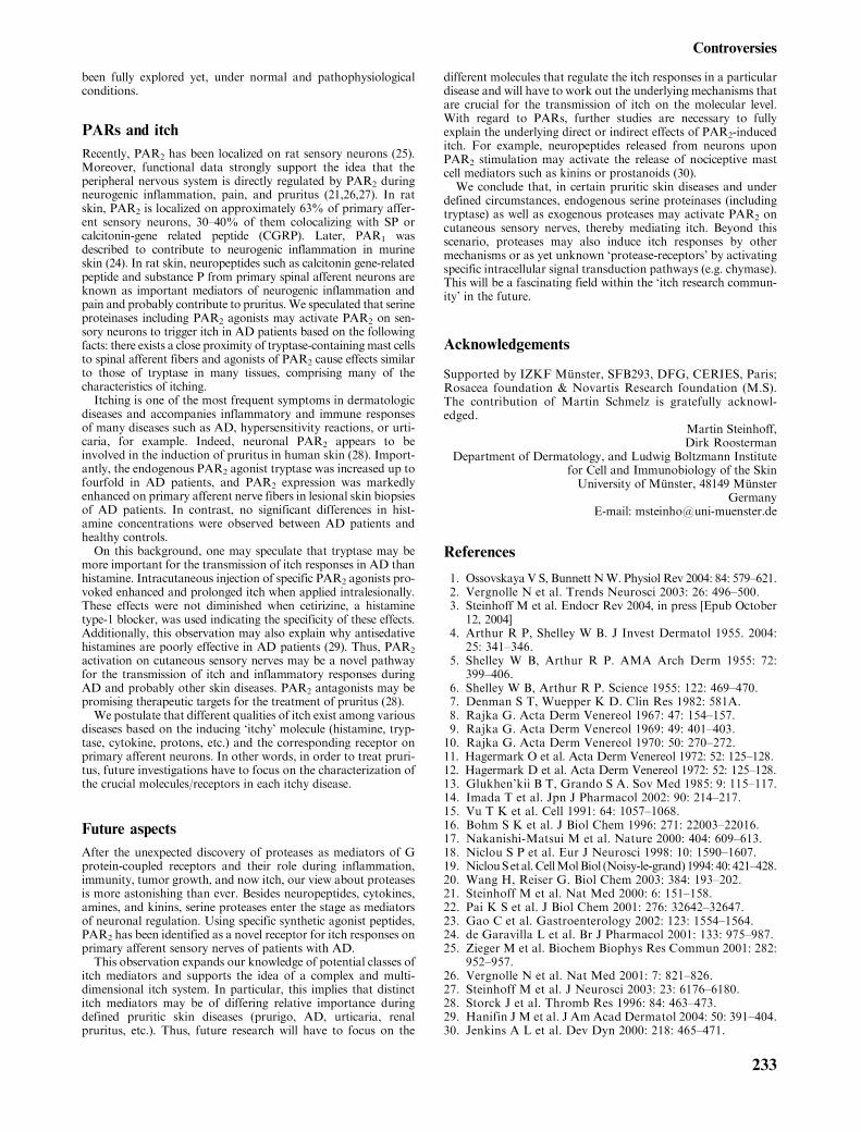
Weaned hairless rats maintained on a diet low in magnesiumdevelop a transient erythematous maculopapulous rash withsigns of generalized pruritus (8,9). The rats scratch and bitethemselves leading to skin lesions (Fig. 2, left panel). Icilin, as a2% powder suspended in Walgreens Advanced Care Lotion,applied once daily for 5 days significantly reduced bite andscratch marks in each animal (Fig. 2, right panel). Reduction indiseased animals was already observed after the first applicationand was overall 52% relative to vehicle-treated control animals(mean of 24 animals examined for 5 days in three independentstudies). Worth mentioning, erythema was not inhibited and signsof pruritus returned upon cessation of treatment. The animalsexhibited transiently ‘wet dog behavior’ after application of icilinat the tested concentration.
Preliminary studies showed that the single oral median lethal doseof icilin in male and female mice and rats was 5–7g/kg body weight,putting icilin into the category of a chemical with slight toxicpotential for short-term effects (10). Purified icilin was not muta-genic in the Ames test system in strains TA 97, 98, 100, 102, 1535,1537, 1538, with or without liver enzyme activation. Icilin is virtuallywater-insoluble; hence, administration of icilin onto the rectalmucosa of animals did not raise plasma levels above 1mg/ml.Now the stage is set for the entrance of new players – clinical
dermatologists – who must tell us if topical icilin or a relatedpyrimidine-2-one analog really manages to suppress that nasty itch.
Edward T. WeiUniversity of California
Berkeley, CA 94720USA
E-mail: [email protected] G. Meingassner
Novartis Institutes for Biomedical ResearchA-1235 Vienna
Austria
References
1. Patapoutian A et al. Nat Rev Neurosci 2003: 7: 529–539.2. Jordt S E et al. Curr Opin Neurobiol 2003: 4: 487–492.3. McKemy D D et al. Nature 2002: 416: 52–58.4. Story G M et al. Cell 2003: 112: 819–829.5. Behrendt H J et al. Br J Pharmacol 2004: 141: 737–745.6. Andersson D A et al. J Neurosci 2004: 24: 5364–5369.7. Wei E, Seid D A. J Pharm Pharmacol 1983: 35: 110–112.8. Bavandi A et al. Exp Dermatol 1992: 1: 199–205.9. Neckermann G et al. Br J Dermatol 2000: 142: 669–679.10. Gosselin R E, Smith R P, Hodge H C. Clinical Toxicology
of Commercial Products, 5th edn. Baltimore and London:Williams & Wilkins, 1984: 1–2.
Commentary 3
The sensation of intense ‘itch’ (pruritus) can reach a level ofsuffering that approaches that of pain or suffocation. Nonethe-less, there are numerous patients with pruritus whom we areunable to treat effectively. If histamine is the main mediator ofitch, antihistamines are effective. But this case is not as frequent asoften assumed. Indeed, the nasty sensation of itch and its controlare associated with many other pruritogenic substances, such asneuropeptides (mainly opioids), acetylcholine, serotonin, interfer-ons, interleukin-2, eicosanoids, and/or enzymes (e.g. tryptase).Recent findings have shown the importance of these other
substances and have explored the course of itch. A better under-standing of the pathophysiology of itch will also allow the dis-covery of new treatments. Hence, we know the activated areas inthe brain when itch occurs and the existence of specific histamin-ergic neurones in spinal cord and in the skin. The main unre-solved question is: Where in the skin is pruritus ‘born’? Is it in theepidermis, the dermis, or in the basal membrane? Also, thoughsignificant progress has been made in research on pruritus duringthe past decade, a major hindrance is the lack of in vitromodel forstudies on this topic. In therapeutic itch research, another majorbrake on progress is the confusing effect of placebo on pruritus,which could be (1) about 66%!Currently and in the future, creams containing local anesthetics (2),
glycocolle, oligosaccharides (3), calamine (4), capsaicin, doxepin (5),
strontium nitrate (6), or nedocromil sodium (7) may all be helpful inthe treatment of itch. But it is not possible to apply them on largesurfaces. After steroids, new treatments such as tacrolimus and pime-crolimus appear as effective treatments of pruritus (8). Itch is oftenwidespread, justifying the use of systemic treatments. Antihistaminesanti-H1are the reference treatment.But they are ineffective in numer-ous diseases. Nowadays, there is no real alternative. But I think thatnew therapeutic ways are hopeful. In my opinion, the most inter-esting is opiate antagonist. Naltrexone, which is available per os,could be a promising treatment (9,10). Gabapentin and its familyrepresent another interesting field of research. H3 agonists mightbe also useful (11), but antiserotonines (anti-SSTR3) are disap-pointing. Cyclosporine is effective not only in atopic dermatitisand pruritus, but also in non-inflammatory circumstances, likethe ‘rebel’ senile pruritus (12). Thalidomide might be helpful, butits prescription is excruciatingly regulated.The psyche is not only involved in psychogenic pruritus but in
all pruritus. Indeed, anxiety or depression is known to modulatethe burden of itching, and the thresholds of perception andtolerability are variable according to each patient. Psychologicalinterventions, like cognitive-behavioral methods, hypnosis, orbiofeedback could be probably helpful in some patients (13).Psychotropic drugs, like hydroxyzine, doxepin, fluoxetine, par-oxetine, or sertraline exert both antipruritic and psychic effects.
Figure 2. Hairless hypomagnesemic rats on day 5 of treatment.Left panel: vehicle-treated animal with severe bite and scratchmarks, taken as signs of generalized pruritus; right panel: animaltreated topically once daily with 2% icilin lotion.
Controversies
235
Laurent MiseryLaboratory of Cutaneous Neurobiology
Department of DermatologyUniversity Hospital
Brest, FranceE-mail: laurent.misery@chu-brest-fr
References
1. Epstein E, Pinsky J B. Arch Dermatol 1964: 89: 548–549.2. Shuttleworth D et al. Br J Dermatol 1988: 119: 535–540.3. Campech M et al. Brulures 2002: 11: 219–222.
4. Misery L et al. Nouv Dermatol 2004: 23: 7–9.5. Drake L A et al. J Am Acad Dermatol 1994: 31: 613–616.6. Zhai H et al. Dermatology 2000: 200: 244–246.7. Ahluwalia P et al. Br J Pharmacol 2001: 132: 613–616.8. Stander S, Luger T A. Hautarzt 2003: 54: 413–417.9. Legroux-Crespel E et al. Dermatology 2004: 208:326–330.
10. Metze D et al. Ann N Y Acad Sci 1999: 885: 430–432.11. Sugimoto Y et al. Clin Exp Allergy 2004: 34: 456–459.12. Teofoli P et al. Acta Derm Venereol 1998: 78: 232.13. Shenefelt P D. Dermatol Ther 2003: 16: 114–122.
Commentary 4
Itch is related to pain. Just like in pain research, it is important todistinguish different kinds of pruritus. The causes and consequenttherapeutic strategy for acute itchingwill notbe the sameas in chronicitching.Anacute itching inurticaria seems tobehistaminedependent.However, a chronic itching inatopic dermatitis orprurigowill involveseveral different inflammatory molecules and neuropeptides, such asopioids. We will look into some of the controversies on opioids.Opioid receptor antagonists have successfully been used to
treat several different pruritic conditions such as atopic dermatitis(1), cutaneous lymphoma, dry skin dermatitis, amyloidosis, psori-asis, prurigo nodularis (2), and hepatogenic pruritus (3). Someauthors suggest that the opiate receptor system is more importantin induction of chronic itch in atopic dermatitis than the hist-amine system (4).However, there are two major controversies involving opioids
and itching.
1. Can opioids induce itching independently from histamine? Somegroups suggested that the opioid-induced itching in the peripheryis only due to opioid-induced histamine release frommast cells inthe dermis. However, in a placebo-controlled, double-blind studyon histamine-induced focal itch and alloknesis with healthy sub-jects using the opioid receptor antagonist naltrexone and the H1-blocking agent cetirizine, naltrexone was found to reduce signifi-cantly both itching and alloknesis. Cetirizine reduced focal itchbut failed to influence the alloknesis phenomenon involved inchronic itch in atopic dermatitis (5). In a dry skin mouse modelfor chronic itching, there was no apparent difference of sponta-neous scratching between mast cell-deficient mice and normallittermates. Subcutaneously administered opioid antagonists sig-nificantly suppressed spontaneous scratching in the dry skinmodelmice. These results could explain why non-sedative, secondand third generation H1-antagonists have very limited effects onchronic itch and support the hypothesis that there is indeed animportant histamine-independent opioid-induced pruritus.
2. Do opioids induce itching only in the central nervous system(CNS) or are the nerve endings in skin involved as well?Although several authors describe an important role of opioidsin the induction of itch, most of them believe that this effect islimited to the CNS. However, methylnaltrexone significantlydecreases opioid-induced pruritus without affecting analgesia(6). Methylnaltrexone is a novel quaternary deriative of nal-trexone that does not cross the blood–brain barrier and acts asa selective peripheral opioid receptor antagonist. Our ownstudies strongly suggest that epidermal opioid receptors areinvolved in chronic itching in atopic dermatitis and prurigo.We discovered a functional active opioid receptor system inhuman skin, including peripheral nerve endings and keratino-cytes (7,8). Additionally, we observed an internalization of them-opiate receptors on keratinocytes in atopic dermatitis. Thefree opioid receptor ligands bind to the receptors on the thinand stretched peripheral nerve endings in hypertrophic epider-
mis in chronic dermatitis. This could lead to a strong itch signalto the CNS, where it will be further processed (8). We per-formed clinical studies using topically applied opioid receptorantagonists to treat chronic pruritus. The results show anincrease of m-opiate receptor expression in keratinocytes anda change of the nerve quality in the epidermis after localtreatment. These changes correlated with the clinical responseto the topically applied drug (unpublished data).
Just recently we postulated the ‘Layer Hypothesis’ as ourworking hypothesis. This hypothesis is a result of the above-described observations in chronic pruritus in patients and mice.The ‘Layer Hypothesis’ suggests that ‘itch’ is elicited in the epi-dermal unmyelinated nerve C-fibers and ‘pain’ in unmyelinatednerve fibers in the dermis (9). This theory combines elementsfrom the neurophysiological ‘Pattern’ and ‘Intensity’ hypothesisand is supported by the observation that the removal of epidermiseliminates itch but not pain (10). Maybe these epidermal nervefibers are the low electrical threshold and histamine-insensitiveC-fibers described recently by Ikoma et al. (11). The strongerstimulation which causes pain will activate mostly the dermalnerve endings. The ‘Layer Hypothesis’ can open a new discussionin itch research and will raise again some controversies in this field.The physiological mechanisms of itching remain to be elucidated
and tested in a reliable, comprehensive experimental model,which should cover different itch elicitor and different kindsof itching. Likewise, future treatments of itching should not beonly restricted to the use of antihistaminics. To understandthe involvement of different cytokines and neuropeptides in thedifferent forms of itching will not only solve the ‘itching’puzzle but also help to find new strategies to treat this verycommon, but sometimes devastating symptom. Our patientswill be grateful.
Paul BigliardiDepartment of Dermatology
University Hospital BaselCH-4031 Basel
SwitzerlandE-mail: [email protected]
Mei Bigliardi-QiDepartment of ResearchUniversity Hospital Basel
CH-4031 BaselSwitzerland
E-mail: [email protected]
References
1. Monroe E W. J Am Acad Dermatol 1989: 21 (1): 135–136.2. Metze D et al. J Am Acad Dermatol 1999: 41 (4): 533–539.3. Bergasa N V et al. Ann Intern Med 1995: 123 (3): 161–167.
Biro et al.
236
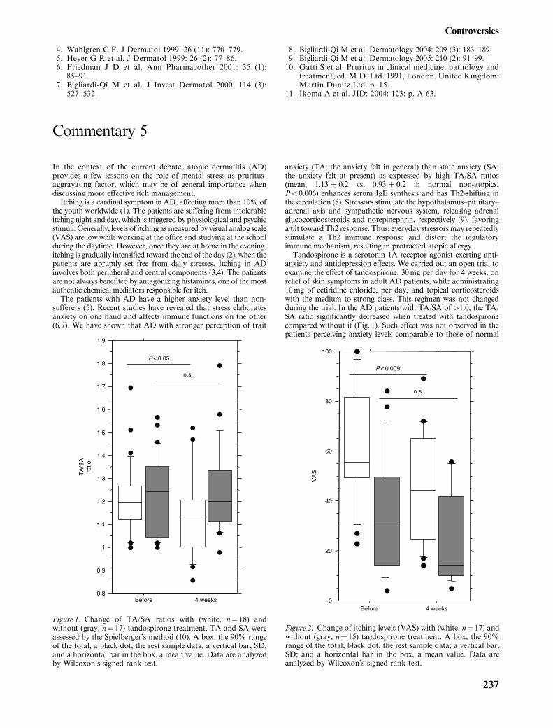
4. Wahlgren C F. J Dermatol 1999: 26 (11): 770–779.5. Heyer G R et al. J Dermatol 1999: 26 (2): 77–86.6. Friedman J D et al. Ann Pharmacother 2001: 35 (1):85–91.
7. Bigliardi-Qi M et al. J Invest Dermatol 2000: 114 (3):527–532.
8. Bigliardi-Qi M et al. Dermatology 2004: 209 (3): 183–189.9. Bigliardi-Qi M et al. Dermatology 2005: 210 (2): 91–99.10. Gatti S et al. Pruritus in clinical medicine: pathology and
treatment, ed. M.D. Ltd. 1991, London, United Kingdom:Martin Dunitz Ltd. p. 15.
11. Ikoma A et al. JID: 2004: 123: p. A 63.
Commentary 5
In the context of the current debate, atopic dermatitis (AD)provides a few lessons on the role of mental stress as pruritus-aggravating factor, which may be of general importance whendiscussing more effective itch management.Itching is a cardinal symptom in AD, affecting more than 10% of
the youth worldwide (1). The patients are suffering from intolerableitching night and day, which is triggered by physiological and psychicstimuli.Generally, levels of itching asmeasured by visual analog scale(VAS) are low while working at the office and studying at the schoolduring the daytime. However, once they are at home in the evening,itching is gradually intensified toward the endof the day (2), when thepatients are abruptly set free from daily stresses. Itching in ADinvolves both peripheral and central components (3,4). The patientsare not always benefited by antagonizing histamines, one of themostauthentic chemical mediators responsible for itch.The patients with AD have a higher anxiety level than non-
sufferers (5). Recent studies have revealed that stress elaboratesanxiety on one hand and affects immune functions on the other(6,7). We have shown that AD with stronger perception of trait
anxiety (TA; the anxiety felt in general) than state anxiety (SA;the anxiety felt at present) as expressed by high TA/SA ratios(mean, 1.13+ 0.2 vs. 0.93+ 0.2 in normal non-atopics,P< 0.006) enhances serum IgE synthesis and has Th2-shifting inthe circulation (8). Stressors stimulate the hypothalamus–pituitary–adrenal axis and sympathetic nervous system, releasing adrenalglucocorticosteroids and norepinephrin, respectively (9), favoringa tilt toward Th2 response. Thus, everyday stressorsmay repeatedlystimulate a Th2 immune response and distort the regulatoryimmune mechanism, resulting in protracted atopic allergy.Tandospirone is a serotonin 1A receptor agonist exerting anti-
anxiety and antidepression effects. We carried out an open trial toexamine the effect of tandospirone, 30mg per day for 4 weeks, onrelief of skin symptoms in adult AD patients, while administrating10mg of cetiridine chloride, per day, and topical corticosteroidswith the medium to strong class. This regimen was not changedduring the trial. In the AD patients with TA/SA of >1.0, the TA/SA ratio significantly decreased when treated with tandospironecompared without it (Fig. 1). Such effect was not observed in thepatients perceiving anxiety levels comparable to those of normal
0.8
0.9
1
1.1
1.2
1.3
1.4
1.5
1.6
1.7
1.8
1.9
Before 4 weeks
TA
/SA
ra
tio
P < 0.05
n.s.
Figure 1. Change of TA/SA ratios with (white, n¼ 18) andwithout (gray, n¼ 17) tandospirone treatment. TA and SA wereassessed by the Spielberger’s method (10). A box, the 90% rangeof the total; a black dot, the rest sample data; a vertical bar, SD;and a horizontal bar in the box, a mean value. Data are analyzedby Wilcoxon’s signed rank test.
Before 4 weeks
VA
S
P < 0.009
n.s.
0
20
40
60
80
100
Figure 2. Change of itching levels (VAS) with (white, n¼ 17) andwithout (gray, n¼ 15) tandospirone treatment. A box, the 90%range of the total; black dot, the rest sample data; a vertical bar,SD; and a horizontal bar in the box, a mean value. Data areanalyzed by Wilcoxon’s signed rank test.
Controversies
237
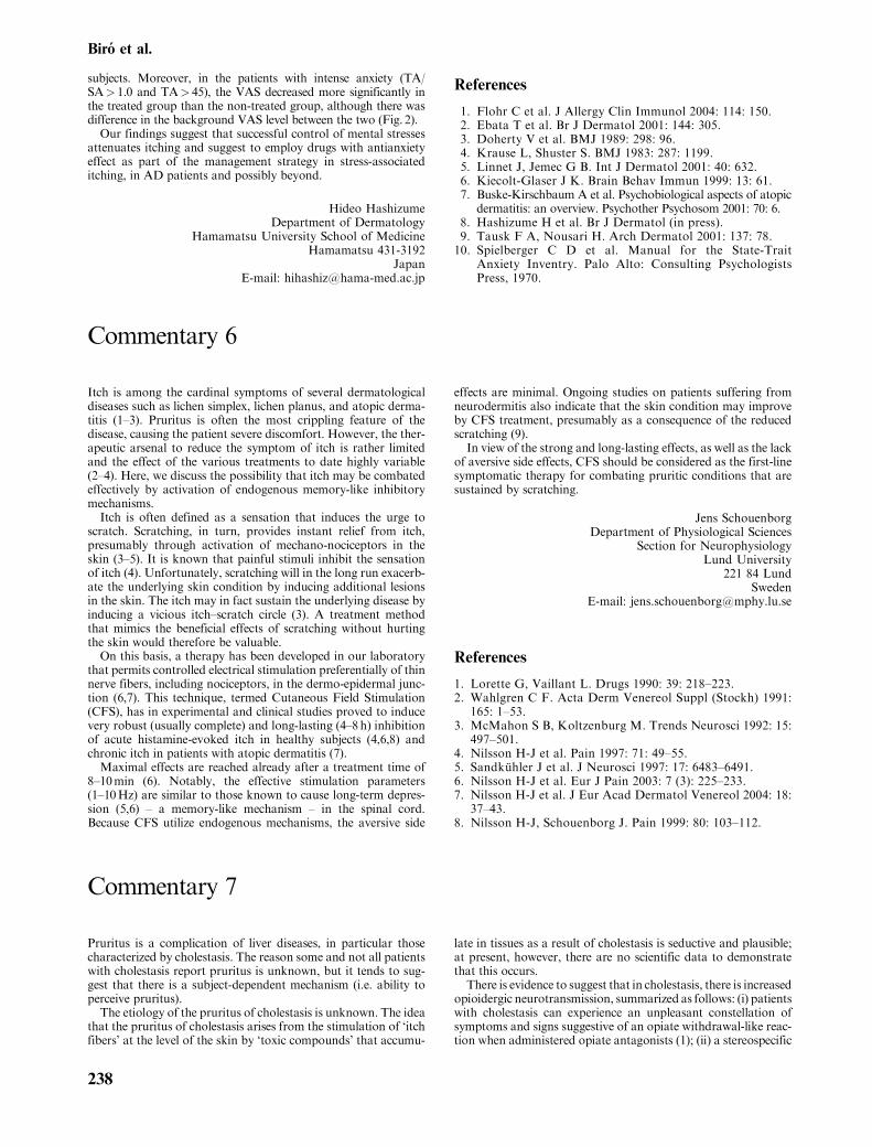
subjects. Moreover, in the patients with intense anxiety (TA/SA> 1.0 and TA> 45), the VAS decreased more significantly inthe treated group than the non-treated group, although there wasdifference in the background VAS level between the two (Fig. 2).Our findings suggest that successful control of mental stresses
attenuates itching and suggest to employ drugs with antianxietyeffect as part of the management strategy in stress-associateditching, in AD patients and possibly beyond.
Hideo HashizumeDepartment of Dermatology
Hamamatsu University School of MedicineHamamatsu 431-3192
JapanE-mail: [email protected]
References
1. Flohr C et al. J Allergy Clin Immunol 2004: 114: 150.2. Ebata T et al. Br J Dermatol 2001: 144: 305.3. Doherty V et al. BMJ 1989: 298: 96.4. Krause L, Shuster S. BMJ 1983: 287: 1199.5. Linnet J, Jemec G B. Int J Dermatol 2001: 40: 632.6. Kiecolt-Glaser J K. Brain Behav Immun 1999: 13: 61.7. Buske-Kirschbaum A et al. Psychobiological aspects of atopicdermatitis: an overview. Psychother Psychosom 2001: 70: 6.
8. Hashizume H et al. Br J Dermatol (in press).9. Tausk F A, Nousari H. Arch Dermatol 2001: 137: 78.10. Spielberger C D et al. Manual for the State-Trait
Anxiety Inventry. Palo Alto: Consulting PsychologistsPress, 1970.
Commentary 6
Itch is among the cardinal symptoms of several dermatologicaldiseases such as lichen simplex, lichen planus, and atopic derma-titis (1–3). Pruritus is often the most crippling feature of thedisease, causing the patient severe discomfort. However, the ther-apeutic arsenal to reduce the symptom of itch is rather limitedand the effect of the various treatments to date highly variable(2–4). Here, we discuss the possibility that itch may be combatedeffectively by activation of endogenous memory-like inhibitorymechanisms.Itch is often defined as a sensation that induces the urge to
scratch. Scratching, in turn, provides instant relief from itch,presumably through activation of mechano-nociceptors in theskin (3–5). It is known that painful stimuli inhibit the sensationof itch (4). Unfortunately, scratching will in the long run exacerb-ate the underlying skin condition by inducing additional lesionsin the skin. The itch may in fact sustain the underlying disease byinducing a vicious itch–scratch circle (3). A treatment methodthat mimics the beneficial effects of scratching without hurtingthe skin would therefore be valuable.On this basis, a therapy has been developed in our laboratory
that permits controlled electrical stimulation preferentially of thinnerve fibers, including nociceptors, in the dermo-epidermal junc-tion (6,7). This technique, termed Cutaneous Field Stimulation(CFS), has in experimental and clinical studies proved to inducevery robust (usually complete) and long-lasting (4–8h) inhibitionof acute histamine-evoked itch in healthy subjects (4,6,8) andchronic itch in patients with atopic dermatitis (7).Maximal effects are reached already after a treatment time of
8–10min (6). Notably, the effective stimulation parameters(1–10Hz) are similar to those known to cause long-term depres-sion (5,6) – a memory-like mechanism – in the spinal cord.Because CFS utilize endogenous mechanisms, the aversive side
effects are minimal. Ongoing studies on patients suffering fromneurodermitis also indicate that the skin condition may improveby CFS treatment, presumably as a consequence of the reducedscratching (9).In view of the strong and long-lasting effects, as well as the lack
of aversive side effects, CFS should be considered as the first-linesymptomatic therapy for combating pruritic conditions that aresustained by scratching.
Jens SchouenborgDepartment of Physiological Sciences
Section for NeurophysiologyLund University
221 84 LundSweden
E-mail: [email protected]
References
1. Lorette G, Vaillant L. Drugs 1990: 39: 218–223.2. Wahlgren C F. Acta Derm Venereol Suppl (Stockh) 1991:165: 1–53.
3. McMahon S B, Koltzenburg M. Trends Neurosci 1992: 15:497–501.
4. Nilsson H-J et al. Pain 1997: 71: 49–55.5. Sandkuhler J et al. J Neurosci 1997: 17: 6483–6491.6. Nilsson H-J et al. Eur J Pain 2003: 7 (3): 225–233.7. Nilsson H-J et al. J Eur Acad Dermatol Venereol 2004: 18:37–43.
8. Nilsson H-J, Schouenborg J. Pain 1999: 80: 103–112.
Commentary 7
Pruritus is a complication of liver diseases, in particular thosecharacterized by cholestasis. The reason some and not all patientswith cholestasis report pruritus is unknown, but it tends to sug-gest that there is a subject-dependent mechanism (i.e. ability toperceive pruritus).The etiology of the pruritus of cholestasis is unknown. The idea
that the pruritus of cholestasis arises from the stimulation of ‘itchfibers’ at the level of the skin by ‘toxic compounds’ that accumu-
late in tissues as a result of cholestasis is seductive and plausible;at present, however, there are no scientific data to demonstratethat this occurs.There is evidence to suggest that in cholestasis, there is increased
opioidergic neurotransmission, summarized as follows: (i) patientswith cholestasis can experience an unpleasant constellation ofsymptoms and signs suggestive of an opiate withdrawal-like reac-tion when administered opiate antagonists (1); (ii) a stereospecific
Biro et al.
238
naloxone reversible state of antinociception (analgesic) can bedisplayed by rats with cholestasis secondary to bile duct resection(2); and (iii) there is down-regulation ofmuopioid receptors in ratswith cholestasis secondary to bile duct resection (3).Analogous to the pruritus that results from the pharmacolog-
ical increase in opioidergic tone by central morphine (4,5), it ishypothesized that increased opioidergic tone in cholestasis maymediate the pruritus, at least in part, by a central mechanism (6).The reason for altered central neurotransmission in cholestasis isunknown; however, increased plasma levels of Met andLeu-enkephalin, two of the endogenous opioid peptides, havebeen reported in patients with liver disease, including those withPrimary biliary cirrhosis (PBC) (1,7).The source of peripheral endogenous opioids in cholestasis is
unknown; however, the liver may contribute to the increasedavailability of opioids in liver disease as suggested by the expres-sion of Met-enkephalin immunoreactivity in the cholestatic liver(8). It is not known whether opioids derived from the liver incholestasis mediate what has been interpreted as centrallyincreased opioidergic neurotransmission; however, there is evi-dence to suggest that opiate transport systems are found in theblood–brain barrier (9,10); furthermore, transport proteins foundin the basolateral domain of the hepatocyte are also found in thechoroid plexus and in the blood–brain barrier and can transportopiates in vitro and may potentially transport periphery-derivedopioids into the central nervous system (11–13).Pruritus is a subjective sensation, and it cannot be directly
quantitated. However, a system that records the behavior thatresults from pruritus, scratching, has been developed, providingthe possibility to obtain objective data (14). The use of opiateantagonists (e.g. naloxone and naltrexone) is supported by datafrom controlled clinical trials that used behavioral methodology(15–18). Experimental data in a primate model of morphine-induced scratching (19,20) suggested that activation of kappareceptors prevented opioid receptor-mediated pruritus. Butorpha-nol is an agonist at the kappa opioid receptor and an antagonist atthe mu opioid receptor with minimal or absent abuse potential.Unpublished experience with the use of butorphanol in patientswith cholestasis and pruritus supports this idea, as the pruritus hasbeen substantially relieved in a patient with intractable pruritus
from chronic hepatitis C. Accordingly, the use of butorphanolspray in selective patients may be a therapeutic alternative.
Nora V. BergasaDivision of Hepatology
State University of New YorkDownstate Medical Center
Kings County Hospital CenterBrooklyn
New York, USAE-mail: [email protected]
References
1. Thornton J R, Losowsky M S. BMJ 1988: 297 (6662):1501–1504.
2. Bergasa N V et al. J Hepatol 1994: 20: 85–90.3. Bergasa N V et al. J Hepatol 1992: 15 (1–2): 220–224.4. Ballantyne J C et al. Pain 1988: 33 (2): 149–160.5. Ballantyne J C et al. Anaesthesia 1989: 44 (10): 863.6. Jones E A, Bergasa N V. Hepatology 1990: 11 (5): 884–887.7. Thornton J R, Losowsky M S. BMJ 1988: 297: 1241–1242.8. Bergasa N. Liver 2002: 22 (2): 107–113.9. Banks W A, Kastin A J. Life Sci 1987: 41 (11): 1319–1338.10. Banks W A, Kastin A J. Am J Physiol 1990: 259 (1 Pt 1):
E1–E10.11. Gao B et al. J Histochem Cytochem 1999: 47 (10):
1255–1264.12. Gao B et al. Hepatology 1999: 30: 1220.13. Gao B et al. J Pharmacol Exp Ther 2000: 294: 73–79.14. Talbot T L et al. Biomed Instrum Technol 1991: 25 (5):
400–403.15. Bergasa N V et al. Gastroenterology 1992: 102 (2): 544–549.16. Bergasa N V et al. Ann Intern Med 1995: 123 (3): 161–167.17. Bergasa N V et al. Hepatology 1998: 27: 679–684.18. Bergasa N V et al. J Am Acad Dermatol 1999: 41:
431–434.19. Ko M C, Naughton N N. Anesthesiology 2000: 92 (3):
795–805.20. Ko M et al. J Pharmacol Exp Ther 2003: 305: 173–179.
Commentary 8
One is tempted to claim that the question posed by the currentdebate feature – How best to fight that nasty itch? – is ‘the wrongquestion at the right time’.We have to admit that, as of today, there is very limited know-
ledge as to what exactly causes itch. While this is especially truefor the so-called neuropathic and psychogenic forms of pruritus,in pruritoceptive itch, we at least know that sensory nerves (SNs)of the skin are required for the induction. This, however, does nothelp much in view of the large and ever-growing family of SN-activating factors, as we simply do not know, which of thesesignals are relevant in settings associated with pruritoceptiveitchiness.More effective treatment options, which are undoubtedly called
for, require us to first identify themechanisms and signals involvedin the induction of pruritus. Thus, if we do not want to put the cartin front of the horse and jump to conclusions, the right question toask is: How to induce that nasty itch? In other words, we can onlyattempt to identify relevant SN-activating mechanisms, once wewill havemanaged to develop experimental models of pruritus thatallow us to study its underlying mechanisms of induction. In ourview, none of the responses to the frequently used itch-inducingsubstances (1) such as benzoic acid, fumarates, dimethylsulfoxide,or capsaicin truly mimic any of the frequent skin conditions asso-ciated with itching.
Once these models have been established, the most promisingapproach for the identification of relevant skin-activating signals,at least in our view, involves ‘educated fishing’, i.e. we must firstlimit the huge number of contenders to a few highly likelycandidates, e.g. by asking: What skin conditions are frequentlyassociated with pruritus? Those factors that are known to beup-regulated in such skin conditions and which also exhibitSN-activating effects are most likely to be our therapeutic targets,which can then be tested (trial and error) in the models developed.For example, most chronic inflammatory skin conditions areinvariably associated with pruritus, e.g. atopic dermatitis, chronicurticaria, and prurigo nodularis. Virtually, all of these skin con-ditions involve the effects of activated skin mast cells (MCs) andtheir pro-inflammatory mediators including histamine, tryptase,prostaglandins, leukotrienes, and various cytokines, many ofwhich have been described as potent activators of skin SNs.Could it be then, thatMCs and SNs are ‘partners in crime’, i.e. in
the induction of pruritus, and could someMCmediators be targetsof novel and more effective therapeutic measurements? Severalindependent lines of evidence indicate that this could indeed be thecase: (i) activation of MCs and MC mediators are very likely tocontribute to the induction of itch (2), (ii) MC stabilizers, e.g.cromoglycate, ketotifen, and nedocromil, have been shown toreduce itching (3–5), and (iii) potent inhibitors and/or antagonists
Controversies
239
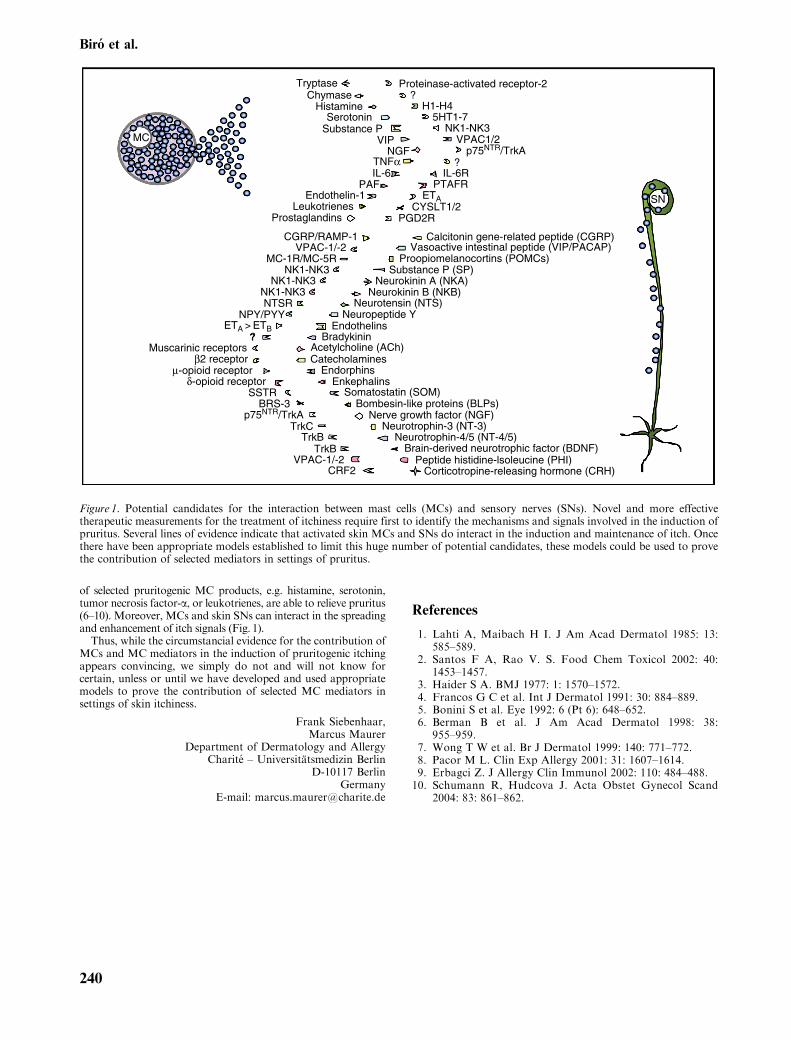
of selected pruritogenic MC products, e.g. histamine, serotonin,tumor necrosis factor-a, or leukotrienes, are able to relieve pruritus(6–10). Moreover, MCs and skin SNs can interact in the spreadingand enhancement of itch signals (Fig. 1).Thus, while the circumstancial evidence for the contribution of
MCs and MC mediators in the induction of pruritogenic itchingappears convincing, we simply do not and will not know forcertain, unless or until we have developed and used appropriatemodels to prove the contribution of selected MC mediators insettings of skin itchiness.
Frank Siebenhaar,Marcus Maurer
Department of Dermatology and AllergyCharite – Universitatsmedizin Berlin
D-10117 BerlinGermany
E-mail: [email protected]
References
1. Lahti A, Maibach H I. J Am Acad Dermatol 1985: 13:585–589.
2. Santos F A, Rao V. S. Food Chem Toxicol 2002: 40:1453–1457.
3. Haider S A. BMJ 1977: 1: 1570–1572.4. Francos G C et al. Int J Dermatol 1991: 30: 884–889.5. Bonini S et al. Eye 1992: 6 (Pt 6): 648–652.6. Berman B et al. J Am Acad Dermatol 1998: 38:955–959.
7. Wong T W et al. Br J Dermatol 1999: 140: 771–772.8. Pacor M L. Clin Exp Allergy 2001: 31: 1607–1614.9. Erbagci Z. J Allergy Clin Immunol 2002: 110: 484–488.10. Schumann R, Hudcova J. Acta Obstet Gynecol Scand
2004: 83: 861–862.
Tryptase
MC
SN
Proteinase-activated receptor-2?
H1-H45HT1-7
NK1-NK3
?IL-6R
PTAFRETA
CYSLT1/2PGD2R
Calcitonin gene-related peptide (CGRP)Vasoactive intestinal peptide (VIP/PACAP)
Proopiomelanocortins (POMCs)Substance P (SP)
Neurokinin A (NKA)Neurokinin B (NKB)
Neurotensin (NTS)Neuropeptide Y
EndothelinsBradykinin
Acetylcholine (ACh)Catecholamines
EndorphinsEnkephalins
Somatostatin (SOM)Bombesin-like proteins (BLPs)
Nerve growth factor (NGF)Neurotrophin-3 (NT-3)
Neurotrophin-4/5 (NT-4/5)Brain-derived neurotrophic factor (BDNF)
Peptide histidine-lsoleucine (PHI)Corticotropine-releasing hormone (CRH)
VPAC1/2p75NTR/TrkA
ChymaseHistamine
SerotoninSubstance P
VIPNGF
TNFαIL-6
PAFEndothelin-1
LeukotrienesProstaglandins
CGRP/RAMP-1VPAC-1/-2
MC-1R/MC-5RNK1-NK3
NK1-NK3NK1-NK3NTSR
NPY/PYYETA > ETB
?Muscarinic receptors
β2 receptorµ-opioid receptor
δ-opioid receptorSSTR
BRS-3p75NTR/TrkA
TrkCTrkB
TrkBVPAC-1/-2
CRF2
Figure 1. Potential candidates for the interaction between mast cells (MCs) and sensory nerves (SNs). Novel and more effectivetherapeutic measurements for the treatment of itchiness require first to identify the mechanisms and signals involved in the induction ofpruritus. Several lines of evidence indicate that activated skin MCs and SNs do interact in the induction and maintenance of itch. Oncethere have been appropriate models established to limit this huge number of potential candidates, these models could be used to provethe contribution of selected mediators in settings of pruritus.
Biro et al.
240
Related Documents