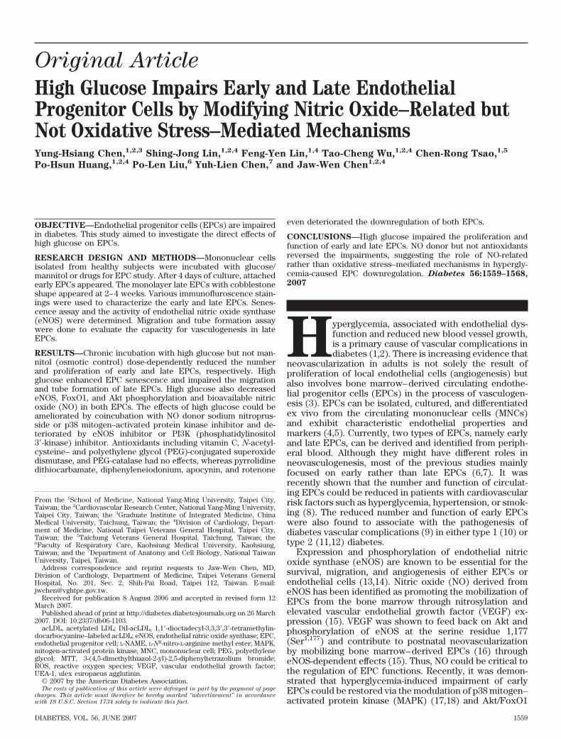
Original Article High Glucose Impairs Early and Late Endothelial Progenitor Cells by Modifying Nitric Oxide–Related but Not Oxidative Stress–Mediated Mechanisms Yung-Hsiang Chen, 1,2,3 Shing-Jong Lin, 1,2,4 Feng-Yen Lin, 1,4 Tao-Cheng Wu, 1,2,4 Chen-Rong Tsao, 1,5 Po-Hsun Huang, 1,2,4 Po-Len Liu, 6 Yuh-Lien Chen, 7 and Jaw-Wen Chen 1,2,4 OBJECTIVE—Endothelial progenitor cells (EPCs) are impaired in diabetes. This study aimed to investigate the direct effects of high glucose on EPCs. RESEARCH DESIGN AND METHODS—Mononuclear cells isolated from healthy subjects were incubated with glucose/ mannitol or drugs for EPC study. After 4 days of culture, attached early EPCs appeared. The monolayer late EPCs with cobblestone shape appeared at 2– 4 weeks. Various immunofluroscence stain- ings were used to characterize the early and late EPCs. Senes- cence assay and the activity of endothelial nitric oxide synthase (eNOS) were determined. Migration and tube formation assay were done to evaluate the capacity for vasculogenesis in late EPCs. RESULTS—Chronic incubation with high glucose but not man- nitol (osmotic control) dose-dependently reduced the number and proliferation of early and late EPCs, respectively. High glucose enhanced EPC senescence and impaired the migration and tube formation of late EPCs. High glucose also decreased eNOS, FoxO1, and Akt phosphorylation and bioavailable nitric oxide (NO) in both EPCs. The effects of high glucose could be ameliorated by coincubation with NO donor sodium nitroprus- side or p38 mitogen–activated protein kinase inhibitor and de- teriorated by eNOS inhibitor or PI3K (phosphatidylinositol 3-kinase) inhibitor. Antioxidants including vitamin C, N-acetyl- cysteine– and polyethylene glycol (PEG)-conjugated superoxide dismutase, and PEG-catalase had no effects, whereas pyrrolidine dithiocarbamate, diphenyleneiodonium, apocynin, and rotenone even deteriorated the downregulation of both EPCs. CONCLUSIONS—High glucose impaired the proliferation and function of early and late EPCs. NO donor but not antioxidants reversed the impairments, suggesting the role of NO-related rather than oxidative stress–mediated mechanisms in hypergly- cemia-caused EPC downregulation. Diabetes 56:1559–1568, 2007 H yperglycemia, associated with endothelial dys- function and reduced new blood vessel growth, is a primary cause of vascular complications in diabetes (1,2). There is increasing evidence that neovascularization in adults is not solely the result of proliferation of local endothelial cells (angiogenesis) but also involves bone marrow– derived circulating endothe- lial progenitor cells (EPCs) in the process of vasculogen- esis (3). EPCs can be isolated, cultured, and differentiated ex vivo from the circulating mononuclear cells (MNCs) and exhibit characteristic endothelial properties and markers (4,5). Currently, two types of EPCs, namely early and late EPCs, can be derived and identified from periph- eral blood. Although they might have different roles in neovasculogenesis, most of the previous studies mainly focused on early rather than late EPCs (6,7). It was recently shown that the number and function of circulat- ing EPCs could be reduced in patients with cardiovascular risk factors such as hyperglycemia, hypertension, or smok- ing (8). The reduced number and function of early EPCs were also found to associate with the pathogenesis of diabetes vascular complications (9) in either type 1 (10) or type 2 (11,12) diabetes. Expression and phosphorylation of endothelial nitric oxide synthase (eNOS) are known to be essential for the survival, migration, and angiogenesis of either EPCs or endothelial cells (13,14). Nitric oxide (NO) derived from eNOS has been identified as promoting the mobilization of EPCs from the bone marrow through nitrosylation and elevated vascular endothelial growth factor (VEGF) ex- pression (15). VEGF was shown to feed back on Akt and phosphorylation of eNOS at the serine residue 1,177 (Ser 1,177 ) and contribute to postnatal neovascularization by mobilizing bone marrow– derived EPCs (16) through eNOS-dependent effects (15). Thus, NO could be critical to the regulation of EPC functions. Recently, it was demon- strated that hyperglycemia-induced impairment of early EPCs could be restored via the modulation of p38 mitogen– activated protein kinase (MAPK) (17,18) and Akt/FoxO1 From the 1 School of Medicine, National Yang-Ming University, Taipei City, Taiwan; the 2 Cardiovascular Research Center, National Yang-Ming University, Taipei City, Taiwan; the 3 Graduate Institute of Integrated Medicine, China Medical University, Taichung, Taiwan; the 4 Division of Cardiology, Depart- ment of Medicine, National Taipei Veterans General Hospital, Taipei City, Taiwan; the 5 Taichung Veterans General Hospital, Taichung, Taiwan; the 6 Faculty of Respiratory Care, Kaohsiung Medical University, Kaohsiung, Taiwan; and the 7 Department of Anatomy and Cell Biology, National Taiwan University, Taipei, Taiwan. Address correspondence and reprint requests to Jaw-Wen Chen, MD, Division of Cardiology, Department of Medicine, Taipei Veterans General Hospital, No. 201, Sec. 2, Shih-Pai Road, Taipei 112, Taiwan. E-mail: [email protected]. Received for publication 8 August 2006 and accepted in revised form 12 March 2007. Published ahead of print at http://diabetes.diabetesjournals.org on 26 March 2007. DOI: 10.2337/db06-1103. acLDL, acetylated LDL; DiI-acLDL, 1,1-dioctadecyl-3,3,3,3-tetramethylin- docarbocyanine–labeled acLDL; eNOS, endothelial nitric oxide synthase; EPC, endothelial progenitor cell; L-NAME, L-N g -nitro-L-arginine methyl ester; MAPK, mitogen-activated protein kinase; MNC, mononuclear cell; PEG, polyethylene glycol; MTT, 3-(4,5-dimethylthiazol-2-yl)-2,5-diphenyltetrazolium bromide; ROS, reactive oxygen species; VEGF, vascular endothelial growth factor; UEA-1, ulex europaeus agglutinin. © 2007 by the American Diabetes Association. The costs of publication of this article were defrayed in part by the payment of page charges. This article must therefore be hereby marked “advertisement” in accordance with 18 U.S.C. Section 1734 solely to indicate this fact. DIABETES, VOL. 56, JUNE 2007 1559

Welcome message from author
This document is posted to help you gain knowledge. Please leave a comment to let me know what you think about it! Share it to your friends and learn new things together.
Transcript
Original Article
High Glucose Impairs Early and Late EndothelialProgenitor Cells by Modifying Nitric Oxide–Related butNot Oxidative Stress–Mediated MechanismsYung-Hsiang Chen,
1,2,3Shing-Jong Lin,
1,2,4Feng-Yen Lin,
1,4Tao-Cheng Wu,
1,2,4Chen-Rong Tsao,
1,5
Po-Hsun Huang,1,2,4
Po-Len Liu,6
Yuh-Lien Chen,7
and Jaw-Wen Chen1,2,4
OBJECTIVE—Endothelial progenitor cells (EPCs) are impairedin diabetes. This study aimed to investigate the direct effects ofhigh glucose on EPCs.
RESEARCH DESIGN AND METHODS—Mononuclear cellsisolated from healthy subjects were incubated with glucose/mannitol or drugs for EPC study. After 4 days of culture, attachedearly EPCs appeared. The monolayer late EPCs with cobblestoneshape appeared at 2–4 weeks. Various immunofluroscence stain-ings were used to characterize the early and late EPCs. Senes-cence assay and the activity of endothelial nitric oxide synthase(eNOS) were determined. Migration and tube formation assaywere done to evaluate the capacity for vasculogenesis in lateEPCs.
RESULTS—Chronic incubation with high glucose but not man-nitol (osmotic control) dose-dependently reduced the numberand proliferation of early and late EPCs, respectively. Highglucose enhanced EPC senescence and impaired the migrationand tube formation of late EPCs. High glucose also decreasedeNOS, FoxO1, and Akt phosphorylation and bioavailable nitricoxide (NO) in both EPCs. The effects of high glucose could beameliorated by coincubation with NO donor sodium nitroprus-side or p38 mitogen–activated protein kinase inhibitor and de-teriorated by eNOS inhibitor or PI3K (phosphatidylinositol3�-kinase) inhibitor. Antioxidants including vitamin C, N-acetyl-cysteine– and polyethylene glycol (PEG)-conjugated superoxidedismutase, and PEG-catalase had no effects, whereas pyrrolidinedithiocarbamate, diphenyleneiodonium, apocynin, and rotenone
even deteriorated the downregulation of both EPCs.
CONCLUSIONS—High glucose impaired the proliferation andfunction of early and late EPCs. NO donor but not antioxidantsreversed the impairments, suggesting the role of NO-relatedrather than oxidative stress–mediated mechanisms in hypergly-cemia-caused EPC downregulation. Diabetes 56:1559–1568,
2007
Hyperglycemia, associated with endothelial dys-function and reduced new blood vessel growth,is a primary cause of vascular complications indiabetes (1,2). There is increasing evidence that
neovascularization in adults is not solely the result ofproliferation of local endothelial cells (angiogenesis) butalso involves bone marrow–derived circulating endothe-lial progenitor cells (EPCs) in the process of vasculogen-esis (3). EPCs can be isolated, cultured, and differentiatedex vivo from the circulating mononuclear cells (MNCs)and exhibit characteristic endothelial properties andmarkers (4,5). Currently, two types of EPCs, namely earlyand late EPCs, can be derived and identified from periph-eral blood. Although they might have different roles inneovasculogenesis, most of the previous studies mainlyfocused on early rather than late EPCs (6,7). It wasrecently shown that the number and function of circulat-ing EPCs could be reduced in patients with cardiovascularrisk factors such as hyperglycemia, hypertension, or smok-ing (8). The reduced number and function of early EPCswere also found to associate with the pathogenesis ofdiabetes vascular complications (9) in either type 1 (10) ortype 2 (11,12) diabetes.
Expression and phosphorylation of endothelial nitricoxide synthase (eNOS) are known to be essential for thesurvival, migration, and angiogenesis of either EPCs orendothelial cells (13,14). Nitric oxide (NO) derived fromeNOS has been identified as promoting the mobilization ofEPCs from the bone marrow through nitrosylation andelevated vascular endothelial growth factor (VEGF) ex-pression (15). VEGF was shown to feed back on Akt andphosphorylation of eNOS at the serine residue 1,177(Ser1,177) and contribute to postnatal neovascularizationby mobilizing bone marrow–derived EPCs (16) througheNOS-dependent effects (15). Thus, NO could be critical tothe regulation of EPC functions. Recently, it was demon-strated that hyperglycemia-induced impairment of earlyEPCs could be restored via the modulation of p38 mitogen–activated protein kinase (MAPK) (17,18) and Akt/FoxO1
From the 1School of Medicine, National Yang-Ming University, Taipei City,Taiwan; the 2Cardiovascular Research Center, National Yang-Ming University,Taipei City, Taiwan; the 3Graduate Institute of Integrated Medicine, ChinaMedical University, Taichung, Taiwan; the 4Division of Cardiology, Depart-ment of Medicine, National Taipei Veterans General Hospital, Taipei City,Taiwan; the 5Taichung Veterans General Hospital, Taichung, Taiwan; the6Faculty of Respiratory Care, Kaohsiung Medical University, Kaohsiung,Taiwan; and the 7Department of Anatomy and Cell Biology, National TaiwanUniversity, Taipei, Taiwan.
Address correspondence and reprint requests to Jaw-Wen Chen, MD,Division of Cardiology, Department of Medicine, Taipei Veterans GeneralHospital, No. 201, Sec. 2, Shih-Pai Road, Taipei 112, Taiwan. E-mail:[email protected].
Received for publication 8 August 2006 and accepted in revised form 12March 2007.
Published ahead of print at http://diabetes.diabetesjournals.org on 26 March2007. DOI: 10.2337/db06-1103.
acLDL, acetylated LDL; DiI-acLDL, 1,1�-dioctadecyl-3,3,3�,3�-tetramethylin-docarbocyanine–labeled acLDL; eNOS, endothelial nitric oxide synthase; EPC,endothelial progenitor cell; L-NAME, L-Ng-nitro-L-arginine methyl ester; MAPK,mitogen-activated protein kinase; MNC, mononuclear cell; PEG, polyethyleneglycol; MTT, 3-(4,5-dimethylthiazol-2-yl)-2,5-diphenyltetrazolium bromide;ROS, reactive oxygen species; VEGF, vascular endothelial growth factor;UEA-1, ulex europaeus agglutinin.
© 2007 by the American Diabetes Association.The costs of publication of this article were defrayed in part by the payment of page
charges. This article must therefore be hereby marked “advertisement” in accordance
with 18 U.S.C. Section 1734 solely to indicate this fact.
DIABETES, VOL. 56, JUNE 2007 1559

signaling (19). However, the effects and mechanisms onlate EPCs are not known.
Excessive generation of reactive oxygen species (ROS)and reactive nitrogen species may contribute to endothe-lial dysfunction and play a critical role in the progressivedeterioration of vessel structure and function (20,21).While low levels of ROS are essential and participate inimportant intracellular signaling pathways (22), excessivegeneration of ROS may result in cytotoxic oxidative stress.In patients with diabetes, the production of ROS is in-creased, which could contribute to the onset and develop-ment of diabetes vascular complications (23). On the otherhand, hyperglycemia may impair endothelial NO produc-tion (24,25) and alter the intracellular reduction-oxidationstate in endothelial cells, which is believed to associatewith endothelial dysfunction and vascular complicationsin diabetes (26). Recently, hyperglycemia was shown toreduce the number and function of circulating blood–derived progenitor cells, mainly early EPCs, both in vivo(10–12) and in vitro (17,27) However, it is also suggestedthat human EPCs, with high intrinsic expression of anti-oxidant enzymes (28) such as manganese superoxidedismutase, could tolerate oxidative stress (29). Thus, theinfluence of hyperglycemia on intrinsic NO and redoxstatus in both early and late EPCs should be furtherclarified. This study was thus conducted to examine thedirect effects of high glucose on the number/proliferationand functional activities of both early and late EPCs and,more importantly, to investigate the individual roles of NO-or oxidative stress–related mechanisms in high glucose–induced impairments, if there are any.
RESEARCH DESIGN AND METHODS
EPC isolation and cultivation. Total MNCs were isolated from 40 mlperipheral blood of healthy young human volunteers by density gradientcentrifugation with Histopaq-1077 (density 1.077 g/ml; Sigma). MNCs (1 � 107)were plated in 2 ml endothelial growth medium (EGM-2 MV; Cambrex) (Fig.1A), with supplements (hydrocortisone, R3-insulin-like growth factor 1, humanendothelial growth factor, VEGF, human fibroblast growth factor, gentamicin,amphotericin B, vitamin C, and 20% fetal bovine serum) on fibronectin-coatedsix-well plates at 37°C in a 5% CO2 incubator. Under daily observation, after 4days of culturing, medium were changed and nonadherent cells were re-moved; attached early EPCs appeared, elongated with a spindle shape (Fig.1B). Thereafter, medium were replaced every 3 days, and each colony/clusterwas followed-up. A certain number of early EPCs can continue to grow intocolonies of late EPCs, which emerge 2–4 weeks after start of MNC culture.The late EPCs exhibited “cobblestone” morphology and monolayer growthpattern typical of mature endothelial cells at confluence (Fig. 1C) (6). Cellswere incubated on the day of isolation with glucose/mannitol or drugs withoutchanging the medium for early EPC study; cells under passage 3 were used forlate EPC study.EPC colony-forming assay. In another set of study, isolated MNCs wereresuspended in growth medium (EndoCult; StemCell Technologies, Vancou-ver, Canada), and in total 5 � 106 MNCs were preplated in fibronectin-coatedsix-well plates in duplicate. After 2 days, the nonadherent cells were collected,and 1 � 106 cells were replated onto a fibronectin-coated 24-well plate. On day5 of the assay, the number of colony-forming units per well was counted foreach sample. A colony of EPCs was defined as a central core of round cellswith elongated sprouting cells at the periphery. All colonies were countedmanually in a minimum of three wells by two independent investigators underblind condition. Cells were incubated on the day of isolation with glucose/mannitol.EPC characterization. The early EPCs were characterized as adherent cellsdouble positive for acetylated LDL (acLDL) uptake and lectin binding bydirect fluorescent staining (8). Briefly, the adherent cells were first incubatedwith 2.4 �g/ml 1,1�-dioctadecyl-3,3,3�,3�-tetramethylindocarbocyanine–labeledacLDL (DiI-acLDL; Molecule Probe) for 1 h and then fixed in 2% paraformal-dehyde and counterstained with 10 �g/ml fluorescein isothiocyanate–labeledlectin from ulex europaeus agglutinin (UEA-1) (Sigma). The late EPC-derivedoutgrowth endothelial cell population was also characterized by immunoflu-roscence staining for the expression of VE-cadherin, von Willebrand factor,
PECAM-1 (platelet/endothelial cell adhesion molecule-1) (CD31), CD34, ki-nase insert domain receptor (KDR)/VEGF receptor 2, and AC133 (CD133)(Santa Cruz). The fluorescent images were recorded under a laser scanningconfocal microscope.EPC number and proliferation assay. The number of early EPCs and theproliferation of late EPCs were determined by direct counting six randomhigh-power microscope fields (�100) and by 3-(4,5-dimethylthiazol-2-yl)-2,5-diphenyltetrazolium bromide (MTT) assay, respectively. Cells were culturedin plate with various concentrations of glucose or mannitol (as osmoticcontrol). After being cultured for 4 days, the amounts of early EPCs werecounted and late EPCs were supplemented with MTT (0.5 mg/ml, Sigma) andincubated for 4 h for proliferation assay. The blue formazen was dissolvedwith dimethyl sulfoxide and measured at 550/650 nm.EPC senescence assay. The cellular aging was determined with a SenescentCells Staining Kit (Sigma). Briefly, after washing with PBS, both early and lateEPCs were fixed for 6 min in 2% formaldehyde and 0.2% glutaraldehyde inPBS, and then incubated for 12 h at 37°C without CO2 with fresh _X-galstaining solution (1 mg/ml X-gal, 5 mmol/l potassium ferrocyanide, 5 mmol/lpotassium ferricyanide, and 2 mmol/l MgCl2; pH 6). After staining, blue-stainedcells and total cells were counted and the percentage of �-galactosidase–positive cells was calculated (30).EPC migration assay. The migratory function of late EPCs that is essentialfor vasculogenesis was evaluated by a modified Boyden chamber (Transwell,Coster) assay (31). In brief, isolated EPCs were detached as described abovewith trypsin/EDTA, and then 4 � 104 EPCs were placed in the upper chamberof 24-well Transwell plates with polycarbonate membrane (8-�m pores) withserum-free endothelial growth medium; VEGF (50 ng/ml) in medium wasplaced in the lower chamber. After incubation for 24 h, the membrane waswashed briefly with PBS and fixed with 4% paraformaldeyde. The upper sideof membrane was wiped gently with a cotton ball. The membrane was thenstained using hematoxylin solution and removed. The magnitude of migrationof late EPCs was evaluated by counting the migrated cells in six randomhigh-power (�100) microscope fields.EPC tube formation assay. Tube formation assay was performed on lateEPCs to assess the capacity for EPC vasculogenesis, which is believed to beimportant in new vessel formation. In vitro tube formation assay wasperformed with In Vitro Angiogenesis Assay Kit (Chemicon) (31). Theprotocol was according to the manufacturer’s instructions. In brief, ECMatrixgel solution was thawed at 4°C overnight, then mixed with ECMatrix diluentbuffer, and placed in a 96-well plate at 37°C for 1 h to allow the matrix solutionto solidify. EPCs were harvested as described above with trypsin/EDTA, then1 � 104 EPCs were placed on matrix solution with EGM-2 MV medium withglucose or mannitol, and incubated at 37°C for 16 h. Tubule formation wasinspected under an inverted light microscope (�100). Four representativefields were taken and the average of the total area of complete tubes formedby cells was compared by computer software, Image-Pro Plus.Western blot analysis. Protein extracts were prepared as previously de-scribed (32). Briefly, EPCs were lysed in a buffer (62.5 mmol/l Tris-HCl, 2%SDS, 10% glycerol, 0.5 mmol/l PMSF, 2 �g/ml aprotinin, pepstatin, andleupeptin). The protein lysates were subjected to SDS-PAGE, followed byelectroblotting onto PVDF membrane. Membranes were probed with mono-clonal antibodies that directed to phosphorylated eNOS (p-eNOS), eNOS,iNOS, �-tubulin (Chemicon), p-FoxO1, FoxO1, p-Akt, and Akt (Cell Signaling).Bands were visualized by chemiluminescence detection reagents (NEN).Densitometic analysis was conducted with ImageQuant (Promega) software.Measurement of nitrite and intracellular cGMP content. After incuba-tion of EPCs with glucose/mannitol for 4 h, the conditioned medium and celllysates were measured for nitrite level and cGMP content by Griess reagent[1% sulfanilamide and 0.1% N-(1-naphthyl)ethylenediamine in 2% phosphoricacid] and cGMP EIA Kit (Cayman), respectively.Statistical analysis. Data are presented as means � SE. Intergroup compar-isons were performed by Student’s t test or one-way ANOVA. Probabilityvalues of P � 0.05 were considered statistically significant.
RESULTS
Characterization of EPCs. EPCs were originated fromperipheral blood MNCs of healthy subjects as previouslydescribed (3). The peripheral blood MNCs that initiallyseeded on fibronectin-coated wells were round (Fig. 1A).After changing medium on day 4, attached early EPCsappeared and elongated with spindle shape (Fig. 1B). LateEPCs with cobblestone-like morphology similar to matureendothelial cells were grown to confluence (Fig. 1C). Acolony of EPCs was defined as a central core of round
NO, ROS, AND GLUCOSE-INDUCED EPC DYSFUNCTION
1560 DIABETES, VOL. 56, JUNE 2007

cells with elongated sprouting cells at the periphery (Fig.1D). EPC colony was further confirmed as cells doublepositive for acLDL uptake (Fig. 1E) and lectin (UEA-1)binding affinity (Fig. 1F). Late EPC characterization wasperformed by immunuhistochemical staining, the majorityof the cells expressed mature endothelial markers VE-cadherin (Fig. 1G), von Willebrand factor (Fig. 1H), andPECAM-1 (platelet/endothelial cell adhesion molecule-1)(CD31) (Fig. 1I). In addition, CD34 (Fig. 1J), KDR (Fig.1K), and AC133 (Fig. 1L) are considered critical markersof outgrowth endothelial cell–producing late EPCs, whichare different from hematopoietic progenitors or leuko-cytes (4,5). Because AC133 rapidly disappeared, CD34/KDR double positive may be an important marker of EPCsin vitro (7).
High glucose decreases EPC number, proliferation,and colony-forming capacity. After seeding MNCs onwells, cells were incubated with different concentrationsof glucose or mannitol (serving as osmotic control) for 4days. Incubation of cells with glucose decreased thenumber of differentiated, adherent, early EPCs in a dose-dependent manner. As compared with that in controlmedium (5 mmol/l glucose), the amount of early EPCsassessed by fluorescein isothiocyanate lectin and DiI-acLDL staining was significantly reduced, by 26.4 and33.6% in 20 and 25 mmol/l high-glucose medium, respec-tively (P � 0.05) (Fig. 2A). The effect of glucose on lateEPC proliferation was analyzed by MTT assay. Glucoseconcentration dependently inhibited EPC proliferation ac-tivity, which became apparent at 20 mmol/l and maximal
FIG. 1. Morphology and characterization of EPCs from peripheral blood. MNCs were isolated and plated on fibronectin-coated culture dish on thefirst day (A). Four days after plating, adherent early EPCs with spindle shape were shown (B). Twenty-one days after plating, late EPCs withcobblestone-like morphology were selected, reseeded, and grown to confluence (C). Phenotyping of endothelial characteristics of colony-formingunits of EPCs (D–F). A colony of EPCs was defined as a central core of round cells with elongated sprouting cells at the periphery (D). Most cellswere shown to simultaneously endocytose DiI-acLDL (red) (E) and bind fluorescein isothiocyanate UEA-1 (lectin) (green) (F). Immunofluo-rescence detection (green) of VE-cadherin (G), von Willebrand factor (H), PECAM-1 (platelet/endothelial cell adhesion molecule-1) (CD31) (I),CD34 (J), KDR (K), and AC133 (L) for late EPCs. Cells were counterstained with propidium iodide for nucleus (red). Scale bar: 50 �m.
Y.-H. CHEN AND ASSOCIATES
DIABETES, VOL. 56, JUNE 2007 1561

at 25 mmol/l (19.1 and 28.3% inhibition, respectively, P �0.05) (Fig. 2B). The capacity of MNCs to form coloniescapable of endothelial cell maturation and proliferationwas stimulatory impaired by high-glucose incubation (25mmol/l for 5 days, 38.6% inhibition, P � 0.05); by contrast,the osmotic control with mannitol did not change thecolony-forming units of EPCs (P � NS) (Fig. 2C). Thetime-dependent effects of high glucose (25 mmol/l) on thenumber and cell proliferation of EPCs were also studied.Long-term (�4 days) incubation with high glucose couldsignificantly decrease the number of adherent early EPCsand inhibit the proliferation activity of late EPCs (data notshown).High glucose increases senescence and impairs themigratory and tube formation function of EPCs. Todeterminate the onset of cellular aging, acidic �-galactosi-dase was detected as a biochemical marker for acidifica-tion typical of the EPC senescence (30). Compared withcontrol medium (5 mmol/l glucose), incubation of eitherearly or late EPCs with 25 mmol/l high glucose for 4 dayssignificantly enhanced the percentage of senescence-asso-ciated �-galactosidase–positive EPCs, whereas an osmoticcontrol with 20 mmol/l mannitol did not significantlyinfluence the early/late EPC senescence (Fig. 3A). Thedata indicated that EPC senescence could be enhanced byhigh-glucose but not mannitol cultivation.
The migratory function of EPCs in response to VEGFis believed to be important during neovascularization(16), and late EPCs have been shown to exhibit bettermigratory capacity than early EPCs in vitro (7). Theeffect of high glucose on late EPC migration was eval-uated using a modified Boyden chamber assay usingVEGF as a chemoattractic factor. After 4 days of cultur-ing, VEGF-induced augmentation of late EPC migrationwas significantly reduced, by 37.0%, in the high-glucose–treated group compared with the control group (P �0.05), whereas mannitol did not influence the late EPCmigration (P � NS) (Fig. 3B).
It has been shown that late EPCs but not early EPCsmake capillary network formation on Matrigel success-fully (6). An in vitro angiogenesis assay was performedwith late EPCs to investigate the effect of glucose on EPCneovascularization. After 4 days of culturing, the func-tional capacity for tube formation of late EPCs on ECMa-trix gel was significantly reduced in the high-glucose–treated group compared with the control group (62.2 � 7.1vs. 100 � 6.8%, P � 0.05), whereas the osmotic controlwith mannitol did not influence the late EPC tube forma-tion capacity (P � NS) (Fig. 3C). These data provided invitro evidence that high-glucose cultivation impairs themigration and vasculogenesis abilities of late EPCs.High glucose decreases the phosphorylation of eNOSand bioavailable NO in EPCs. It has been shown thathyperglycemia may inhibit eNOS activity by posttransla-tional modification at the Akt site in endothelial cells (33).We therefore investigated the effects of high glucose onthe NO system in EPCs. After 4 days of incubation, theeNOS phosphorylation at Ser1,177 shown by immunoblot-ting was significantly decreased both in early and lateEPCs that had been cultured with high-glucose mediumcompared with that in control conditions (Fig. 4A). Thisreduction in eNOS phosphorylation was associated with adecrease in EPC-derived NO production (nitrite levels;35.6 and 45.7% inhibition in early and late EPCs, respec-tively, P � 0.05) (Fig. 4B) and intracellular cGMP levels(38.1 and 40.8% inhibition in early and late EPCs, respec-
FIG. 2. High glucose decreases EPC number, proliferation, and colony-forming capacity. Cells were isolated and incubated with differentconcentrations of glucose or mannitol (osmotic control) for 4 days.Early EPCs were characterized as adherent cells that were dualpositive for lactin staining and DiI-acLDL uptake. The numbers ofEPCs were counted under microscope, and data are expressed as meannumbers of EPCs per high-power field (HPF) � SE (A). MTT assay wasalso performed for late EPC proliferation activity (B), normalized tocells incubated in control medium (5 mmol/l glucose). Colony-formingassay for control, mannitol- (20 mmol/l), and glucose- (25 mmol/l)incubated EPCs (C). Data are expressed as means � SE; n � 5, *P <0.05 vs. control.
NO, ROS, AND GLUCOSE-INDUCED EPC DYSFUNCTION
1562 DIABETES, VOL. 56, JUNE 2007

tively, P � 0.05) (Fig. 4C) without altering the total eNOSor inducible NOS (iNOS) expression, suggesting the selec-tive inhibition of eNOS activity by high glucose. Theosmotic control with mannitol did not influence either thephosphorylation or the expression of eNOS and iNOS inearly or late EPCs.Role of Akt/FoxO1, NO, p38 MAPK, and oxidativestress signalings in high-glucose–downregulated EPCs.We then investigated the role of Akt/FoxO1 activity inglucose toxicity effects on EPC differentiation (19). After4-day incubation, the Akt and FoxO1 phosphorylationshown by immunoblotting was significantly decreased inboth early and late EPCs cultured with high-glucosemedium (Fig. 5A). The potential roles of PI3K/Akt-, NO-,and p38 MAPK–related mechanisms were also examined.Coincubation with NO donor sodium nitroprusside (SNP)or p38 MAPK inhibitor SB230580 significantly amelioratedthe inhibitory effect of high glucose on EPC number andproliferation (for early and late EPCs, respectively). Incontrast, coincubation with NOS inhibitor L-Ng-nitro-L-arginine methyl ester (L-NAME) or PI3K inhibitorLY294002 significantly enhanced the inhibitory effect of
high glucose on EPC number/proliferation (Fig. 5B). Thesedata indicated that high glucose may downregulate EPCsby modulating PI3K/Akt-, NO-, and p38 MAPK-relatedmechanisms.
In addition, the potential role of oxidative stress in highglucose–induced impairment of EPCs was investigatedwith the optimal concentrations of diverse antioxidantsbase on the literatures and our pilot studies. As shown inFig. 5C, coincubation with various antioxidants, includingmembrane-permeable antioxidative enzymes (polyethyl-ene glycol/PEG-SOD and PEG-catalase), vitamin C, glu-tathione precursor (N-acetylcysteine/NAC), pyrrolidine di-thiocarbamate (PDTC), NADPH oxidase inhibitors (diphe-nyleneiodonium/DPI and apocynin), and mitochondrialcomplex I inhibitor (rotenone), failed to reverse the inhib-itory effect of high glucose on EPC number/proliferation.Interestingly, PDTC and DPI (two antioxidants that alsoinhibit NO production), apocynin, and rotenone evendramatically inhibited the proliferation of EPCs. It wasthen suggested that high glucose downregulated EPCs byimpairing NO- rather than by activating ROS-related mech-anisms.
FIG. 3. High glucose increases senescence and im-pairs the migratory and tube formation function ofEPCs. Cellular aging of control, mannitol- (20 mmol/l), or glucose- (25 mmol/l) incubated early and lateEPCs were analyzed by senescence-associated acidic�-galactosidase activity assay (A). A modified Boydenchamber assay was used with VEGF as chemoattrac-tive factor for late EPC migratory function. Repre-sentative photos are shown; the small dots are holesin the barrier membrane. The migrated cells werestained with hematoxylin and counted under micro-scope (B). An in vitro angiogenesis assay for lateEPCs was used with ECMatrix gel. Representativephotos for in vitro angiogenesis are shown. The cellswere stained with crystal violet, and the averages ofthe total area of complete tubes formed by cells werecompared by computer software (C). Data aremean � SE; n � 6, *P < 0.05 vs. control (5 mmol/lglucose).
Y.-H. CHEN AND ASSOCIATES
DIABETES, VOL. 56, JUNE 2007 1563

DISCUSSION
The major findings of this study included that 1) long-termexposure to high glucose inhibited EPC colony-formingability and reduced the number and proliferation activitybut enhanced the senescence of early and late EPCs; 2)long-term exposure to high glucose impaired the migrationand vasculogenesis activities of late EPCs; 3) in both early
and late EPCs, Akt, FoxO1, and eNOS phosphorylationand bioavailable NO were significantly reduced in thelong-term presence of high glucose; and 4) the inhibitoryeffects of high glucose on EPC could be reversed by NOdonor but not by various antioxidants. The proposedpossible schema of this regulatory event is shown in Fig. 6.The novel findings suggested that impaired NO mecha-
FIG. 4. High glucose decreases eNOS phosphorytion and bioavailable NO in EPCs. After incubation of EPCs with glucose or mannitol for 4 days,NOS protein levels, NO content, and cGMP formation were analyzed. A representative immunoblot shows the p-eNOS, eNOS, iNOS, and �-tubulinamounts of early and late EPCs in response to high glucose for 4 days. Each bar graph shows the summarized data from four separate experimentsby densitometry after normalization to �-tubulin (A). Nitrite production (as NO content) in early/late EPC culture medium was measured byGriess reagent (B). Intracellular cGMP (pmol/mg protein) in early/late EPCs was measured by using enzyme-linked immunosorbent assay kits(C). Data are means � SE; n � 5, *P < 0.05 vs. control (5 mmol/l glucose).
NO, ROS, AND GLUCOSE-INDUCED EPC DYSFUNCTION
1564 DIABETES, VOL. 56, JUNE 2007
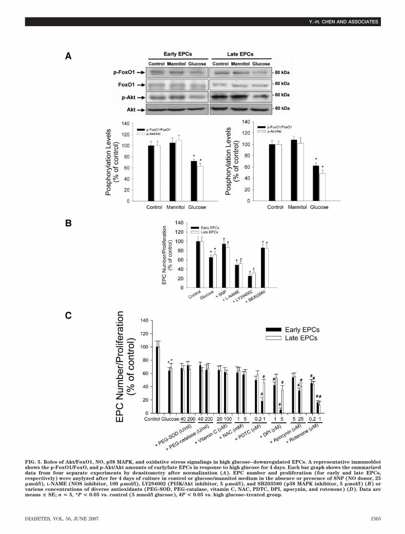
FIG. 5. Roles of Akt/FoxO1, NO, p38 MAPK, and oxidative stress signalings in high glucose–downregulated EPCs. A representative immunoblotshows the p-FoxO1/FoxO, and p-Akt/Akt amounts of early/late EPCs in response to high glucose for 4 days. Each bar graph shows the summarizeddata from four separate experiments by densitometry after normalization (A). EPC number and proliferation (for early and late EPCs,respectively) were anylyzed after for 4 days of culture in control or glucose/mannitol medium in the absence or presence of SNP (NO donor, 25�mol/l), L-NAME (NOS inhibitor, 100 �mol/l), LY294002 (PI3K/Akt inhibitor, 5 �mol/l), and SB203580 (p38 MAPK inhibitor, 5 �mol/l) (B) orvarious concentrations of diverse antioxidants (PEG-SOD, PEG-catalase, vitamin C, NAC, PDTC, DPI, apocynin, and rotenone) (D). Data aremeans � SE; n � 5, *P < 0.05 vs. control (5 mmol/l glucose), #P < 0.05 vs. high glucose–treated group.
Y.-H. CHEN AND ASSOCIATES
DIABETES, VOL. 56, JUNE 2007 1565

nisms rather than increased oxidative stress could con-tribute to the reduction of number/proliferation and func-tional activity of both early and late EPCs in the long-termpresence of high glucose.
It has been recognized that at least two different types ofEPCs, early and late EPCs, could be classified in ex vivoculture system according to their time-dependent appear-ance (6,7). They share some endothelial phenotype butshow different morphology, proliferation rate, survivalfeatures (6), and functions in neovascularization (7). Pre-vious studies have shown that the number was reducedand the migration as well as vasculogenesis capacitieswere impaired in EPCs from patients with type 1 (12) andtype 2 (10,11) diabetes. However, little data had beenprovided for the individual effect of high glucose ondifferent types of EPCs. In this ex vivo study, we for thefirst time demonstrated the detrimental effect of highglucose on the senescence, number/proliferation, and themigration/vasculogenesis activities of both early and lateEPCs, indicating the global harmful effect of high glucoseon EPCs. The impairments could not be attributed toosmotic stress because the effects were not observed inmannitol-treated EPCs. Our findings on late EPCs may beof particular clinical interest because they could presentthe vasculogenesis capacity and may serve as a potentialtherapeutic target for vascular regeneration.
Stem and progenitor cells are considered as highlypotent regenerating cells with high proliferation ability.However, their proliferation may be essentially dependenton aspects of the cellular environment such as cell-to-cellcommunication or growth factor support (30). It has beenshown that in ex vivo cultivation, the onset of EPCsenescence could be associated with a very low prolifer-ation capacity and profoundly impaired clonal expansion
potential (30). In the present study, long-term incubationwith high glucose not only decreased the number andproliferation of early and late EPCs, but also acceleratedthe onset of cellular senescence (18), which appear toinvolve the NO-mediated pathways because NO donorSNP reversed and eNOS inhibitor L-NAME enhanced theinhibitory effect of high glucose on both early and lateEPCs. Additionally, our findings were also compatible withrecent suggestions that p38 MAPK may play a pivotal rolein regulating the number of EPCs in the presence of highglucose or cytokines (17).
It has been suggested that high glucose may impaireNOS expression or its phosphorylation at the site Ser1,177,resulting in reduced NO production, which is associatedwith reduced proliferation and an increase in apoptosis ofendothelial cells and potentially contributes to the devel-opment of endothelial dysfunction and atherosclerosis indiabetes (33,34). In the present study, high glucose signif-icantly reduced eNOS phosphorylation and bioavailableNO without affecting total eNOS and iNOS expression,suggesting that the posttranslational mechanisms are im-paired by high glucose in both early and late EPCs. It ispartially similar to what was demonstrated in maturevascular endothelial cells. Recently, EPCs were shown toexpress lower levels of eNOS as compared with maturevascular endothelial cells (28,29), and transplantation ofautologous EPCs overexpressing eNOS in injured vesselsenhanced the vasculoprotective properties of reconsti-tuted endothelium (35). On the other hand, VEGF orerythropoietin could increase circulating progenitor cellsby activating eNOS predominantly through Akt-dependenteNOS phosphorylation (15,33,36). Supplement of statinscould also enhance endothelial NO production and reducethe senescence of EPCs (30). More interestingly, it was
FIG. 6. Schematic representation of hyperglycemia-induced early and late EPC downregulation. High glucose impaired the proliferation andfunction of early and late EPCs. NO donor but not antioxidants reversed the impairments, suggesting the role of NO-related rather thanROS-mediated mechanisms in hyperglycemia-caused EPC downregulation. DM, diabetic.
NO, ROS, AND GLUCOSE-INDUCED EPC DYSFUNCTION
1566 DIABETES, VOL. 56, JUNE 2007
shown that erythropoietin and benfotiamine could coun-teract glucose toxicity effects on EPC differentiation viaAkt/FoxO1 signaling (19). All of the above indicate thecritical role of NO mechanisms in maintaining EPC func-tions. Taking them together, one may speculate that thereduction of eNOS phosphorylation and NO production inthe presence of high glucose could contribute to theimpairments of EPCs and EPC-related vascular repair thatmight be observed in clinical diabetes (10,12).
It has been shown that ROS may play a key role in highglucose–induced apoptosis of mature vascular endothelialcells, which could be reversed by either eNOS activationor antioxidants (37,38). It was also suggested that hyper-glycemia alone, through the mitochondrial (39–41) and/orNAD(P)H oxidase-mediated (42) overproduction of ROS,can induce changes in gene expression and the behavior ofvascular endothelial cells in diabetes. Vitamin C and NAC,two water-soluble antioxidants, could enter mitochondriaand confer mitochondrial protection against oxidativeinjury (43) and prevent hyperglycemia-induced apoptosisin human aortic endothelial cells (38). Strategies aiming atreducing hyperglycemia-induced ROS have been sug-gested as an useful adjuvant to antihyperglycemic thera-pies in the restoration of vasculogenesis and theprevention of diabetes complications (44). Though hyper-glycemia-induced overproduction of ROS was used toexplain EPC impairments observed in clinical diabetes(44), there is, however, no direct experimental evidenceshowing that the impairments of EPC function could bereversed by decreasing intracellular ROS. In the presentstudy, the additional supplement with vitamin C (finalconcentration 100 �mol/l), NAC, or cell-permeable anti-oxidant enzymes all failed to ameliorate the adverseeffects of high glucose on both early and late EPCs,suggesting the response of EPCs to antioxidants could bedifferent from that of mature endothelial cells. In fact, ithas been shown that the expression of antioxidant enzymeis enhanced and the tolerability to exogenous oxidativestress increased in EPCs (28,29), which may contribute tothe survival of EPCs within the oxygen-poor environmentof the bone marrow and to their ability to engraft withinischemic tissue during vasculogenesis (44). On the otherhand, it was recently shown that endothelial cell apoptoticbodies may enhance the number and differentiation ofEPCs (45) and hydrogen peroxide produced by angio-poitein-1 mediates angiogenesis (46), further suggestingthat adequate oxidative stress might be required for thefunction of both early and late EPCs.
In the present study, chronic coincubation with PDTC(nuclear factor-B inhibitor and antioxidant), DPI andapocynin (two agents with antioxidative property by in-hibiting NADPH oxidase), or rotenone (mitochondrialcomplex I inhibitor) even significantly deteriorated highglucose–induced EPC impairment. Interestingly, PDTCand DPI are not only antioxidants but are also known asinhibitors of NOS mRNA translation (47) and flavoenzymeNOS (48), respectively. Compared with other antioxidants,they had more significant inhibitory effects on the numberof early EPCs, suggesting that both intracellular NO- andoxidative stress–related mechanisms are particularly re-quired for early EPCs in the long-term presence of highglucose. In fact, superoxide anion is a natural inhibitor ofFAS-mediated cell death (49), and appropriate levels ofROS are essential and participate in important intracellu-lar signaling (22). Our findings, different from the opinionsof others (44), did support the concept that adequate ROS
production may be helpful rather than harmful to EPCproliferation in the presence of high glucose. Accordingly,the main biochemical basis for high glucose–induced EPCdysfunction could be a complex other than excessiveoxidative stress formation. This may provide a potentialrationale for the dramatic failure of clinical trials withantioxidants for atherosclerosis in diabetic patients (50).
Similar with a previous study (17), a high-glucose(20–25 mmol/l) cell culture model was used to simulateclinical hyperglycemia for the in vitro evaluation of EPCfunction in the present study. The concentrations ofglucose were 80–89 mg/dl (4.5–5 mmol/l) in the mediumsas assayed by a clinical biochemical analyzer. The concen-trations of glucose were 100 mg/dl (5–5.5 mmol/l) indiabetes (low glucose) and 434 mg/dl (20–24 mmol/l) indiabetes (high glucose). Thus, the high glucose concentra-tion (20–25 mmol/l) used in the present study may corre-spond to 350–450 mg/dl of serum glucose levels, whichmay not present in well-controlled patients but might beseen after consuming a heavy meal in some poorly con-trolled diabetic patients. It could also happen in somestress conditions, such as those with inflammation orinfection and during hospitalization. Thus, the findings ofthe present study may relevant for the worse clinicalconditions that do obtain in some diabetic patients.
In conclusion, long-term presence of high glucose mayenhance cellular senescence and decrease cell number/proliferation and functional competences in both early andlate EPCs. Furthermore, impaired NO- rather than acti-vated oxidative stress–related mechanisms could be themain contributor to high glucose–induced EPC dysfunc-tion. These findings not only give further insights into thecomplex cellular mechanisms of EPCs for impaired vas-cular repair and abnormal neovasculogenesis in chronichyperglycemia, but also provide a rationale for the poten-tial therapeutic target for hyperglycemia-related vascularcomplications in diabetic patients.
ACKNOWLEDGMENTS
This work was supported by grants NSC 92-2314-B-010-053, NSC 93-2314-B-010-033, NSC 94-2314-B-010-060, andNSC 95-2314-B-010-024-MY3 to J.W.C. and NSC 93-2314-B-010-004 to S.J.L. from the National Science Council, Tai-wan, ROC, and grant V95C1-095 to J.W.C. from the TaipeiVeterans General Hospital, Taipei, Taiwan, ROC.
REFERENCES
1. Abaci A, Oguzhan A, Kahraman S, Eryol NK, Unal S, Arinc H, Ergin A:Effect of diabetes mellitus on formation of coronary collateral vessels.Circulation 99:2239–2242, 1999
2. Sheetz MJ, King GL: Molecular understanding of hyperglycemia’s adverseeffects for diabetic complications. JAMA 288:2579–2588, 2002
3. Asahara T, Murohara T, Sullivan A, Silver M, van der Zee R, Li T,Witzenbichler B, Schatteman G, Isner JM: Isolation of putative progenitorendothelial cells for angiogenesis. Science 275:964–967, 1997
4. Rehman J, Li J, Orschell CM, March KL: Peripheral blood “endothelialprogenitor cells” are derived from monocyte/macrophages and secreteangiogenic growth factors. Circulation 107:1164–1169, 2003
5. Rohde E, Malischnik C, Thaler D, Maierhofer T, Linkesch W, Lanzer G,Guelly C, Strunk D: Blood monocytes mimic endothelial progenitor cells.Stem Cells 24:357–367, 2006
6. Hur J, Yoon CH, Kim HS, Choi JH, Kang HJ, Hwang KK, Oh BH, Lee MM,Park YB: Characterization of two types of endothelial progenitor cells andtheir different contributions to neovasculogenesis. Arterioscler Thromb
Vasc Biol 24:288–293, 20047. Yoon CH, Hur J, Park KW, Kim JH, Lee CS, Oh IY, Kim TY, Cho HJ, Kang
HJ, Chae IH, Yang HK, Oh BH, Park YB, Kim HS: Synergistic neovascular-ization by mixed transplantation of early endothelial progenitor cells and
Y.-H. CHEN AND ASSOCIATES
DIABETES, VOL. 56, JUNE 2007 1567

late outgrowth endothelial cells: the role of angiogenic cytokines andmatrix metalloproteinases. Circulation 112:1618–1627, 2005
8. Vasa M, Fichtlscherer S, Aicher A, Adler K, Urbich C, Martin H, Zeiher AM,Dimmeler S: Number and migratory activity of circulating endothelialprogenitor cells inversely correlate with risk factors for coronary arterydisease. Circ Res 89:E1–E7, 2001
9. Fadini GP, Sartore S, Albiero M, Baesso I, Murphy E, Menegolo M, GregoF, Vigili de Kreutzenberg S, Tiengo A, Agostini C, Avogaro A: Number andfunction of endothelial progenitor cells as a marker of severity for diabeticvasculopathy. Arterioscler Thromb Vasc Biol 26:2140–2146, 2006
10. Loomans CJ, de Koning EJ, Staal FJ, Rookmaaker MB, Verseyden C, deBoer HC, Verhaar MC, Braam B, Rabelink TJ, van Zonneveld AJ: Endothe-lial progenitor cell dysfunction: a novel concept in the pathogenesis ofvascular complications of type 1 diabetes. Diabetes 53:195–199, 2004
11. Fadini GP, Miorin M, Facco M, Bonamico S, Baesso I, Grego F, MenegoloM, de Kreutzenberg SV, Tiengo A, Agostini C, Avogaro A: Circulatingendothelial progenitor cells are reduced in peripheral vascular complica-tions of type 2 diabetes mellitus. J Am Coll Cardiol 45:1449–1457, 2005
12. Tepper OM, Galiano RD, Capla JM, Kalka C, Gagne PJ, Jacobowitz GR,Levine JP, Gurtner GC: Human endothelial progenitor cells from type IIdiabetics exhibit impaired proliferation, adhesion, and incorporation intovascular structures. Circulation 106:2781–2786, 2002
13. Dimmeler S, Dernbach E, Zeiher AM: Phosphorylation of the endothelialnitric oxide synthase at ser-1177 is required for VEGF-induced endothelialcell migration. FEBS Lett 477:258–262, 2000
14. Urbich C, Reissner A, Chavakis E, Dernbach E, Haendeler J, Fleming I,Zeiher AM, Kaszkin M, Dimmeler S: Dephosphorylation of endothelialnitric oxide synthase contributes to the anti-angiogenic effects of endosta-tin. FASEB J 16:706–708, 2002
15. Aicher A, Heeschen C, Mildner-Rihm C, Urbich C, Ihling C, Technau-IhlingK, Zeiher AM, Dimmeler S: Essential role of endothelial nitric oxidesynthase for mobilization of stem and progenitor cells. Nat Med 9:1370–1376, 2003
16. Asahara T, Takahashi T, Masuda H, Kalka C, Chen D, Iwaguro H, Inai Y,Silver M, Isner JM: VEGF contributes to postnatal neovascularization bymobilizing bone marrow-derived endothelial progenitor cells. EMBO J
18:3964–3972, 199917. Seeger FH, Haendeler J, Walter DH, Rochwalsky U, Reinhold J, Urbich C,
Rossig L, Corbaz A, Chvatchko Y, Zeiher AM, Dimmeler S: p38 mitogen-activated protein kinase downregulates endothelial progenitor cells. Cir-
culation 111:1184–1191, 200518. Kuki S, Imanishi T, Kobayashi K, Matsuo Y, Obana M, Akasaka T:
Hyperglycemia accelerated endothelial progenitor cell senescence via theactivation of p38 mitogen-activated protein kinase. Circ J 70:1076–1081,2006
19. Marchetti V, Menghini R, Rizza S, Vivanti A, Feccia T, Lauro D, FukamizuA, Lauro R, Federici M: Benfotiamine Counteracts Glucose Toxicity Effectson Endothelial Progenitor Cell Differentiation via Akt/FoxO Signaling.Diabetes 55:2231–2237, 2006
20. Cai H, Harrison DG: Endothelial dysfunction in cardiovascular diseases:the role of oxidant stress. Circ Res 87:840–844, 2000
21. Suh YA, Arnold RS, Lassegue B, Shi J, Xu X, Sorescu D, Chung AB,Griendling KK, Lambeth JD: Cell transformation by the superoxide-generating oxidase Mox1. Nature 401:79–82, 1999
22. Sundaresan M, Yu ZX, Ferrans VJ, Irani K, Finkel T: Requirement forgeneration of H2O2 for platelet-derived growth factor signal transduction.Science 270:296–299, 1995
23. Baynes JW: Role of oxidative stress in development of complications indiabetes. Diabetes 40:405–412, 1991
24. Ding Y, Vaziri ND, Coulson R, Kamanna VS, Roh DD: Effects of simulatedhyperglycemia, insulin, and glucagon on endothelial nitric oxide synthaseexpression. Am J Physiol Endocrinol Metab 279:E11–E17, 2000
25. Guo X, Chen LW, Liu WL, Guo ZG: High glucose inhibits expression ofinducible and constitutive nitric oxide synthase in bovine aortic endothe-lial cells. Acta Pharmacol Sin 21:325–328, 2000
26. Srinivasan S, Hatley ME, Bolick DT, Palmer LA, Edelstein D, Brownlee M,Hedrick CC: Hyperglycaemia-induced superoxide production decreaseseNOS expression via AP-1 activation in aortic endothelial cells. Diabeto-
logia 47:1727–1734, 200427. Krankel N, Adams V, Linke A, Gielen S, Erbs S, Lenk K, Schuler G,
Hambrecht R: Hyperglycemia reduces survival and impairs function ofcirculating blood-derived progenitor cells. Arterioscler Thromb Vasc Biol
25:698–703, 200528. Dernbach E, Urbich C, Brandes RP, Hofmann WK, Zeiher AM, Dimmeler S:
Antioxidative stress-associated genes in circulating progenitor cells: evi-dence for enhanced resistance against oxidative stress. Blood 104:3591–3597, 2004
29. He T, Peterson TE, Holmuhamedov EL, Terzic A, Caplice NM, Oberley LW,Katusic ZS: Human endothelial progenitor cells tolerate oxidative stressdue to intrinsically high expression of manganese superoxide dismutase.Arterioscler Thromb Vasc Biol 24:2021–2027, 2004
30. Assmus B, Urbich C, Aicher A, Hofmann WK, Haendeler J, Rossig L,Spyridopoulos I, Zeiher AM, Dimmeler S: HMG-CoA reductase inhibitorsreduce senescence and increase proliferation of endothelial progenitorcells via regulation of cell cycle regulatory genes. Circ Res 92:1049–1055,2003
31. Chen JZ, Zhu JH, Wang XX, Zhu JH, Xie XD, Sun J, Shang YP, Guo XG, DaiHM, Hu SJ: Effects of homocysteine on number and activity of endothelialprogenitor cells from peripheral blood. J Mol Cell Cardiol 36:233–239, 2004
32. Chen JW, Chen YH, Lin FY, Chen YL, Lin SJ: Ginkgo biloba extract inhibitstumor necrosis factor-alpha-induced reactive oxygen species generation,transcription factor activation, and cell adhesion molecule expression inhuman aortic endothelial cells. Arterioscler Thromb Vasc Biol 23:1559–1566, 2003
33. Du XL, Edelstein D, Dimmeler S, Ju Q, Sui C, Brownlee M: Hyperglycemiainhibits endothelial nitric oxide synthase activity by posttranslationalmodification at the Akt site. J Clin Invest 108:1341–1348, 2001
34. Lopez-Farre A, Sanchez de Miguel L, Caramelo C, Gomez-Macias J, GarciaR, Mosquera JR, de Frutos T, Millas I, Rivas F, Echezarreta G, Casado S:Role of nitric oxide in autocrine control of growth and apoptosis ofendothelial cells. Am J Physiol 272:H760–H768, 1997
35. Urao N, Okigaki M, Yamada H, Aadachi Y, Matsuno K, Matsui A, MatsunagaS, Tateishi K, Nomura T, Takahashi T, Tatsumi T, Matsubara H: Erythro-poietin-mobilized endothelial progenitors enhance reendothelialization viaAkt-endothelial nitric oxide synthase activation and prevent neointimalhyperplasia. Circ Res 98:1405–1413, 2006
36. Kong D, Melo LG, Mangi AA, Zhang L, Lopez-Ilasaca M, Perrella MA, LiewCC, Pratt RE, Dzau VJ: Enhanced inhibition of neointimal hyperplasia bygenetically engineered endothelial progenitor cells. Circulation 109:1769–1775, 2004
37. Ho FM, Lin WW, Chen BC, Chao CM, Yang CR, Lin LY, Lai CC, Liu SH, LiauCS: High glucose-induced apoptosis in human vascular endothelial cells ismediated through NF-kappaB and c-Jun NH(2)-terminal kinase pathwayand prevented by PI3K/Akt/eNOS pathway. Cell Signal 18:391–399, 2006
38. Recchioni R, Marcheselli F, Moroni F, Pieri C: Apoptosis in human aorticendothelial cells induced by hyperglycemic condition involves mitochon-drial depolarization and is prevented by N-acetyl-L-cysteine. Metabolism
51:1384–1388, 200239. Nishikawa T, Edelstein D, Du XL, Yamagishi S, Matsumura T, Kaneda Y,
Yorek MA, Beebe D, Oates PJ, Hammes HP, Giardino I, Brownlee M:Normalizing mitochondrial superoxide production blocks three pathwaysof hyperglycaemic damage. Nature 404:787–790, 2000
40. Brownlee M: Biochemistry and molecular cell biology of diabetic compli-cations. Nature 414:813–820, 2001
41. Brodsky SV, Gao S, Li H, Goligorsky MS: Hyperglycemic switch frommitochondrial nitric oxide to superoxide production in endothelial cells.Am J Physiol Heart Circ Physiol 283:H2130–H2139, 2002
42. Cosentino F, Eto M, De Paolis P, van der Loo B, Bachschmid M, Ullrich V,Kouroedov A, Delli Gatti C, Joch H, Volpe M, Luscher TF: High glucosecauses upregulation of cyclooxygenase-2 and alters prostanoid profile inhuman endothelial cells: role of protein kinase C and reactive oxygenspecies. Circulation 107:1017–1023, 2003
43. Sagun KC, Carcamo JM, Golde DW: Vitamin C enters mitochondria viafacilitative glucose transporter 1 (Glut1) and confers mitochondrial pro-tection against oxidative injury. FASEB J 19:1657–1667, 2005
44. Callaghan MJ, Ceradini DJ, Gurtner GC: Hyperglycemia-induced reactiveoxygen species and impaired endothelial progenitor cell function. Anti-
oxid Redox Signal 7:1476–1482, 200545. Hristov M, Erl W, Linder S, Weber PC: Apoptotic bodies from endothelial
cells enhance the number and initiate the differentiation of humanendothelial progenitor cells in vitro. Blood 104:2761–2766, 2004
46. Kim YM, Kim KE, Koh GY, Ho YS, Lee KJ: Hydrogen peroxide produced byangiopoietin-1 mediates angiogenesis. Cancer Res 66:6167–6174, 2006
47. Sherman MP, Aeberhard EE, Wong VZ, Griscavage JM, Ignarro LJ: Pyrro-lidine dithiocarbamate inhibits induction of nitric oxide synthase activityin rat alveolar macrophages. Biochem Biophys Res Commun 191:1301–1308, 1993
48. Stuehr DJ, Fasehun OA, Kwon NS, Gross SS, Gonzalez JA, Levi R, NathanCF: Inhibition of macrophage and endothelial cell nitric oxide synthase bydiphenyleneiodonium and its analogs. FASEB J 5:98–103, 1991
49. Clement MV, Stamenkovic I: Superoxide anion is a natural inhibitor ofFAS-mediated cell death. EMBO J 15:216–225, 1996
50. Wiernsperger NF: Oxidative stress as a therapeutic target in diabetes:revisiting the controversy. Diabete Metab 29:579–585, 2003
NO, ROS, AND GLUCOSE-INDUCED EPC DYSFUNCTION
1568 DIABETES, VOL. 56, JUNE 2007
Related Documents