Hepatocyte Produced Matrix Metalloproteinases Are Regulated by CD147 in Liver Fibrogenesis Sarah R. Calabro 1,2." , Annette E. Maczurek 1,2." , Alison J. Morgan 1,2 , Thomas Tu 1,2 , Victoria W. Wen 1,2 , Christine Yee 1,2 , Auvro Mridha 2 , Maggie Lee 1,2 , William d’Avigdor 1,2 , Stephen A. Locarnini 3 , Geoffrey W. McCaughan 2,5,6 , Fiona J. Warner 1,2 , Susan V. McLennan 2,4 , Nicholas A. Shackel 1,2,5 * 1 Liver Cell Biology, Centenary Institute, Sydney, NSW, Australia, 2 Sydney Medical School, The University of Sydney, Sydney, NSW, Australia, 3 Victorian Infectious Disease Laboratory, Melbourne, VIC, Australia, 4 Department of Endocrinology, Royal Prince Alfred Hospital, Camperdown, Sydney, NSW, Australia, 5 A.W. Morrow Gastroenterology and Liver Centre, Royal Prince Alfred Hospital, Camperdown, Sydney, NSW, Australia, 6 Liver Injury and Cancer, Centenary Institute, Sydney, NSW, Australia Abstract Background: The classical paradigm of liver injury asserts that hepatic stellate cells (HSC) produce, remodel and turnover the abnormal extracellular matrix (ECM) of fibrosis via matrix metalloproteinases (MMPs). In extrahepatic tissues MMP production is regulated by a number of mechanisms including expression of the glycoprotein CD147. Previously, we have shown that CD147 is expressed on hepatocytes but not within the fibrotic septa in cirrhosis [1]. Therefore, we investigated if hepatocytes produce MMPs, regulated by CD147, which are capable of remodelling fibrotic ECM independent of the HSC. Methods: Non-diseased, fibrotic and cirrhotic livers were examined for MMP activity and markers of fibrosis in humans and mice. CD147 expression and MMP activity were co-localised by in-situ zymography. The role of CD147 was studied in-vitro with siRNA to CD147 in hepatocytes and in-vivo in mice with CCl 4 induced liver injury using a ˜ CD147 antibody intervention. Results: In liver fibrosis in both human and mouse tissue MMP expression and activity (MMP-2, -9, -13 and -14) increased with progressive injury and localised to hepatocytes. Additionally, as expected, MMPs were abundantly expressed by activated HSC. Further, with progressive fibrosis there was expression of CD147, which localised to hepatocytes but not to HSC. Functionally significant in-vitro regulation of hepatocyte MMP production by CD147 was demonstrated using siRNA to CD147 that decreased hepatocyte MMP-2 and -9 expression/activity. Further, in-vivo a-CD147 antibody intervention decreased liver MMP-2, -9, -13, -14, TGF-b and a-SMA expression in CCl 4 treated mice compared to controls. Conclusion: We have shown that hepatocytes produce active MMPs and that the glycoprotein CD147 regulates hepatocyte MMP expression. Targeting CD147 regulates hepatocyte MMP production both in-vitro and in-vivo, with the net result being reduced fibrotic matrix turnover in-vivo. Therefore, CD147 regulation of hepatocyte MMP is a novel pathway that could be targeted by future anti-fibrogenic agents. Citation: Calabro SR, Maczurek AE, Morgan AJ, Tu T, Wen VW, et al. (2014) Hepatocyte Produced Matrix Metalloproteinases Are Regulated by CD147 in Liver Fibrogenesis. PLoS ONE 9(7): e90571. doi:10.1371/journal.pone.0090571 Editor: Golo Ahlenstiel, University of Sydney, Australia Received October 28, 2013; Accepted February 2, 2014; Published July 30, 2014 Copyright: ß 2014 Calabro et al. This is an open-access article distributed under the terms of the Creative Commons Attribution License, which permits unrestricted use, distribution, and reproduction in any medium, provided the original author and source are credited. Funding: This research was funded by NHMRC grants 571408, 512283, 1063515 and 1009815. SC and AM were supported by University of Sydney Australian Postgraduate Awards and W. d’A by a scholarship from the Rebecca L Cooper Medical Foundation. The funders had no role in study design, data collection and analysis, decision to publish, or preparation of the manuscript. Competing Interests: The authors have declared that no competing interests exist. * Email: [email protected] . These authors contributed equally to this work. " SRC and AEM are first authors on this work. Introduction Regardless of the aetiology of chronic liver injury, a canonical pathway of fibrosis development results in progressively abnormal matrix deposition and eventual cirrhosis with the sequelae including liver failure and hepatocellular carcinoma (HCC) [2,3]. Chronic fibrotic liver injury is an active process char- acterised by abnormal extracellular matrix (ECM) deposition and remodelling [2,3]. Matrix metalloproteinases (MMPs) are proteo- lytic enzymes, which play an important role in all stages of progressive liver injury from fibrogenesis initiation through to resolution [4–9]. MMPs are abundantly produced by hepatic stellate cells (HSC) within the dense fibrotic bands, which surround nodules of hepatocytes. Further, MMPs are also secreted by other intrahepatic cell populations including inflammatory cells and hepatocytes [10–12]. However, the role of MMPs originating from non-HSC intrahepatic cell populations, such as hepatocytes [12], has not until recently been attributed a significant role in the ECM remodelling associated with progressive fibrosis. MMP-10 has been shown to be expressed by hepatocytes, cholangiocytes and macrophages and can clearly alter fibrogenesis in a non-HSC dependent manner [13]. However, functional studies of other PLOS ONE | www.plosone.org 1 July 2014 | Volume 9 | Issue 7 | e90571

Welcome message from author
This document is posted to help you gain knowledge. Please leave a comment to let me know what you think about it! Share it to your friends and learn new things together.
Transcript
Hepatocyte Produced Matrix Metalloproteinases AreRegulated by CD147 in Liver FibrogenesisSarah R. Calabro1,2.", Annette E. Maczurek1,2.", Alison J. Morgan1,2, Thomas Tu1,2, Victoria W. Wen1,2,
Christine Yee1,2, Auvro Mridha2, Maggie Lee1,2, William d’Avigdor1,2, Stephen A. Locarnini3,
Geoffrey W. McCaughan2,5,6, Fiona J. Warner1,2, Susan V. McLennan2,4, Nicholas A. Shackel1,2,5*
1 Liver Cell Biology, Centenary Institute, Sydney, NSW, Australia, 2 Sydney Medical School, The University of Sydney, Sydney, NSW, Australia, 3 Victorian Infectious Disease
Laboratory, Melbourne, VIC, Australia, 4 Department of Endocrinology, Royal Prince Alfred Hospital, Camperdown, Sydney, NSW, Australia, 5 A.W. Morrow
Gastroenterology and Liver Centre, Royal Prince Alfred Hospital, Camperdown, Sydney, NSW, Australia, 6 Liver Injury and Cancer, Centenary Institute, Sydney, NSW,
Australia
Abstract
Background: The classical paradigm of liver injury asserts that hepatic stellate cells (HSC) produce, remodel and turnoverthe abnormal extracellular matrix (ECM) of fibrosis via matrix metalloproteinases (MMPs). In extrahepatic tissues MMPproduction is regulated by a number of mechanisms including expression of the glycoprotein CD147. Previously, we haveshown that CD147 is expressed on hepatocytes but not within the fibrotic septa in cirrhosis [1]. Therefore, we investigated ifhepatocytes produce MMPs, regulated by CD147, which are capable of remodelling fibrotic ECM independent of the HSC.
Methods: Non-diseased, fibrotic and cirrhotic livers were examined for MMP activity and markers of fibrosis in humans andmice. CD147 expression and MMP activity were co-localised by in-situ zymography. The role of CD147 was studied in-vitrowith siRNA to CD147 in hepatocytes and in-vivo in mice with CCl4 induced liver injury using aCD147 antibody intervention.
Results: In liver fibrosis in both human and mouse tissue MMP expression and activity (MMP-2, -9, -13 and -14) increasedwith progressive injury and localised to hepatocytes. Additionally, as expected, MMPs were abundantly expressed byactivated HSC. Further, with progressive fibrosis there was expression of CD147, which localised to hepatocytes but not toHSC. Functionally significant in-vitro regulation of hepatocyte MMP production by CD147 was demonstrated using siRNA toCD147 that decreased hepatocyte MMP-2 and -9 expression/activity. Further, in-vivo a-CD147 antibody interventiondecreased liver MMP-2, -9, -13, -14, TGF-b and a-SMA expression in CCl4 treated mice compared to controls.
Conclusion: We have shown that hepatocytes produce active MMPs and that the glycoprotein CD147 regulates hepatocyteMMP expression. Targeting CD147 regulates hepatocyte MMP production both in-vitro and in-vivo, with the net result beingreduced fibrotic matrix turnover in-vivo. Therefore, CD147 regulation of hepatocyte MMP is a novel pathway that could betargeted by future anti-fibrogenic agents.
Citation: Calabro SR, Maczurek AE, Morgan AJ, Tu T, Wen VW, et al. (2014) Hepatocyte Produced Matrix Metalloproteinases Are Regulated by CD147 in LiverFibrogenesis. PLoS ONE 9(7): e90571. doi:10.1371/journal.pone.0090571
Editor: Golo Ahlenstiel, University of Sydney, Australia
Received October 28, 2013; Accepted February 2, 2014; Published July 30, 2014
Copyright: � 2014 Calabro et al. This is an open-access article distributed under the terms of the Creative Commons Attribution License, which permitsunrestricted use, distribution, and reproduction in any medium, provided the original author and source are credited.
Funding: This research was funded by NHMRC grants 571408, 512283, 1063515 and 1009815. SC and AM were supported by University of Sydney AustralianPostgraduate Awards and W. d’A by a scholarship from the Rebecca L Cooper Medical Foundation. The funders had no role in study design, data collection andanalysis, decision to publish, or preparation of the manuscript.
Competing Interests: The authors have declared that no competing interests exist.
* Email: [email protected]
. These authors contributed equally to this work.
" SRC and AEM are first authors on this work.
Introduction
Regardless of the aetiology of chronic liver injury, a canonical
pathway of fibrosis development results in progressively abnormal
matrix deposition and eventual cirrhosis with the sequelae
including liver failure and hepatocellular carcinoma (HCC)
[2,3]. Chronic fibrotic liver injury is an active process char-
acterised by abnormal extracellular matrix (ECM) deposition and
remodelling [2,3]. Matrix metalloproteinases (MMPs) are proteo-
lytic enzymes, which play an important role in all stages of
progressive liver injury from fibrogenesis initiation through to
resolution [4–9]. MMPs are abundantly produced by hepatic
stellate cells (HSC) within the dense fibrotic bands, which
surround nodules of hepatocytes. Further, MMPs are also secreted
by other intrahepatic cell populations including inflammatory cells
and hepatocytes [10–12]. However, the role of MMPs originating
from non-HSC intrahepatic cell populations, such as hepatocytes
[12], has not until recently been attributed a significant role in the
ECM remodelling associated with progressive fibrosis. MMP-10
has been shown to be expressed by hepatocytes, cholangiocytes
and macrophages and can clearly alter fibrogenesis in a non-HSC
dependent manner [13]. However, functional studies of other
PLOS ONE | www.plosone.org 1 July 2014 | Volume 9 | Issue 7 | e90571
MMPs in non-HSC cell populations are lacking. In particular,
studies of the role of the hepatocyte in intrahepatic fibrogenesis are
comparatively sparse and the hepatocyte, the main parenchymal
cell of the liver, is not considered to have a functionally significant
role in either ECM production or remodelling. Indeed it is widely
asserted that hepatocytes are ‘‘innocent bystanders’’ which release
products of cell apoptosis, necrosis, or secrete chemokines to
attract inflammatory cells and activate HSC that are responsible
for the production of the abnormal matrix, MMPs and the
remodelling of the ECM [2]. It is now apparent that the HSC
makes the abnormal matrix with progressive fibrosis but other
intrahepatic cell populations are, in addition to the HSC, capable
of remodelling the ECM [2,3,13].
We have reported that CD147, also known as Extracellular
Matrix Metalloproteinase Inducer (EMMPRIN) or basigin (Bsg), is
increased in cirrhotic liver and localised to the membrane of
hepatocytes [1,14]. Importantly, CD147 is not expressed within
the fibrotic septa where there is a predominance of activated HSC
[1,14]. CD147 is a widely-expressed multifunctional, highly
glycosylated, cell surface transmembrane protein which is
upregulated in many forms of tissue injury associated with
inflammation and matrix remodelling [15–17]. In multiple
extrahepatic organ systems and in inflammatory conditions such
as arthritis, CD147 has been shown to regulate MMP production
and determine the progression of fibrosis [14,18–24]. The
functional role of intrahepatic CD147 has been studied and it
has been variably co-localised with the HSC-marker a-SMA and
therefore has been implicated in HSC activation [24]. However,
this study used antibody HAb18G which is not available
commercially and our studies with established commercial
antibodies, including those available from widely adopted hybrid-
omas [25], show that CD147 is abundantly expressed on
hepatocytes but not HSC [14]. The discrepancies in these studies
are likely due to both isoforms and glycoforms of the protein that
have differing biological activities [14,24,26]. Therefore, based on
previous studies showing hepatocyte MMP production [12,13]
combined with our demonstrated changes in hepatocyte expres-
sion of CD147 with liver injury [1,14] we hypothesise that: Inresponse to injury hepatocytes produce MMPs regulated by CD147and thereby directly contribute to intrahepatic ECM remodelling,independent of the HSC. The data presented in this manuscript
supports this hypothesis and demonstrates that the hepatocyte
production of active MMPs is regulated by CD147. Importantly,
this is clearly functionally significant as in-vivo a-CD147
interventions alter fibrotic liver injury.
Experimental Procedures
Ethics StatementHuman tissues samples were obtained from Royal Prince Alfred
Hospital, Sydney with approval of Human Research Ethics
Committee (X10-0072). Human tissue used in this study was
previously utilized for research [1,27]. Informed written consent
was obtained from all participants. The ethics committee waived
the need for written consent for use of donor tissue. In Australia,
the ethics of human research is governed by the National
Statement on Ethical Conduct in Human Research (2007) issued
by the National Health and Medical Research Council
(NHMRC). Under these guidelines all research involving humans
requires ethical approval.
Animal experiments were performed in accordance with
Sydney University Animal Ethics Committee requirements
(K75/10-2008/3/4801). The Australian Code of Practice for the
Care and Use of Animals for Scientific Purposes was followed.
This includes a responsibility to protect and promote the welfare of
animals used.
We confirm that Sydney University Animal Ethics Committee
specifically approved the animal part of our study. The Code of
Practice embodies the principles of: Reduction of animal use,
Replacement of animal use and Refinement of animal use. These
are known as the "3 Rs". It is important to consider these
principles when designing and carrying out projects.
Human Tissue and Cell LinesNon-diseased donor and end-stage cirrhotic liver tissues were
collected from patients attending Prince Alfred Hospital, Sydney
during liver transplantation. pH5CH8 cells were kindly provided
by Prof. Li [28,29].
Mouse Studies and Primary Hepatocyte IsolationBalb/c and C57bl/6 mice were used for in-vivo studies [30,31].
We have elected to use the two mouse backgrounds, as they are
known to have differing fibrotic responses [32]. Liver injury was
induced with carbon tetrachloride (CCl4). For the CCl4 model
mice were injected twice weekly for upto four weeks with 100 ml of
12% v/v CCl4 in paraffin oil i.p, control mice only received
paraffin oil (Ajax Finechem). The role of CD147 was examined
using an a-CD147 blocking antibody (mAb clone RL73.2)
produced and purified as previously described [33]. The antibody
was administered (i.p 100 mg) twice weekly. Mice treated with
CCl4 and administered IgG2a (100 mg, HB-189, ATCC) were
used as controls. At termination animals were euthanized and
blood was obtained by cardiac puncture and used for measure-
ment of aspartate transaminase (AST). Livers were collected for
histological studies, measurement of MMP activity and expression
of genes of interest by quantitative PCR. Hepatocytes were
isolated using a two-step collagenase perfusion technique [34] and
gene expression levels were measured by quantitative PCR.
Viability was greater than 95% for isolated hepatocytes at
48 hours in culture. Contamination of the hepatocyte preparations
with Kupffer cells/macrophages was assessed by F4/80 staining
and morphology. Hepatocyte purity was consistently found to be
greater than 95%.
HistochemistryParaffin embedded human liver tissue from controls, non-
diseased donor samples, or subjects with end-stage cirrhosis caused
by primary biliary cirrhosis (PBC), primary sclerosing cholangitis
(PSC), alcoholic liver disease (ALD), autoimmune hepatitis (AIH)
and hepatitis C (HCV) taken at time of liver transplantation were
examined. The following antibodies were used: a-CD147 antibody
(clone MEM-M6/1, Abcam), a-MMP-1 (clone 41-1E5, Calbio-
chem), a-MMP-2 and a-MMP-9 (clone 8B4 and polyclonal
antibody H-129 Santa Cruz) or mouse IgG1 (MOPC 21, Abcam)
isotype control. Goat a-mouse-HRP and goat a-rabbit-HRP
(Dako) and NovaRED (Vector Labs) or DAB (Sigma) were used
for detection. Sections were counterstained with Haematoxylin
and Eosin (H&E).
Co-localisation studies were performed on 5 mm frozen and
fixed (acetone:methanol 1:1) sections using a-CD147-FITC (clone
MEM-M6/1, Abcam) in combination with a-cytokeratin 18
(CK18, clone DC10), a-CK19 (clone RCK108), a-CD45 (clone
2B11+PD7/26) or a-CD31 (clone JC70A) all from Dako
Cytomation and a-SMA-Cy3 (clone 1A4 from Sigma). AlexaFluor
goat a-mouse 594 was used as secondary antibody (Lifetechnol-
ogies). All sections were imaged by confocal microscopy.
H&E and Pico-Sirius Red (PSR) staining were used to
investigate tissue morphology and collagen content in paraffin-
Hepatocyte MMP Production and Regulation
PLOS ONE | www.plosone.org 2 July 2014 | Volume 9 | Issue 7 | e90571
embedded sections. Sections were cut and stained by the
University of Sydney Pathology Department.
Pico-Sirius Red QuantificationTotal liver sections stained with PSR were imaged using a Leica
DM6000B with LAS Power Mosaic (Leica, Germany) at a
magnification of 106. Image J 1.48J was used for quantification
of PSR staining. Briefly, five regions of interests (ROI, 1386 mm
61316 mm) per section were randomly selected, avoiding large
blood vessels and empty spaces. The images were converted into
Red-Green-Blue Stacks and the thresholds of the green channel
were equally adjusted for all images. The positive area fraction for
all ROIs was measured using Image J. Sections of at least four
animals (n = 428) were analysed for each treatment condition.
Determination and Localisation of MMP ActivityThe cellular localisation of MMP activity was examined on
frozen, unfixed liver sections using the following liver cell markers
CK18, a-SMA-Cy3 or CD147 as described above. AlexaFluor
goat a-mouse 594 was used to visualise CK18 and CD147, nuclei
were visualized using DAPI. MMP activity in human liver tissue
was subsequently studied by in-situ zymography. Antibody stained
sections were overlayed with agarose (1%w/v) containing
quenched-fluorescent (DQ) gelatin (1 mg/ml, Lifetechnologies).
The sections were then incubated for 2.5 hrs at 37uC before
imaging by confocal microscopy (Leica SP5). In a parallel series,
sections were overlayed with agarose containing DQ gelatin and
aminophenyl-mercuric acetate (7 mM, APMA) to activate all
MMPs [35,36]. Sections overlayed with agarose only, or DQ
agarose containing the MMP inhibitor 1,10-phenanthroline
(20 mM, Sigma) were studied as control. Gelatin zymography
was used to measure the pro- and active forms of MMP-2 and
MMP-9 in serum-free conditioned media using equal protein
concentrations, as determined by DC protein assay (Biorad) [37].
Results are expressed relative compared to control.
Knockdown of CD147 Expression using siRNAThe effect of knockdown CD147 protein expression was
examined in the human hepatocyte cell line pH 5CH8. Cells
were grown in DMEM and FCS (10%v/v) as previously described
[28,29]. At 70% confluence, 16105 cells were transfected with
either 100 pmol scrambled siRNA (59-GAAATCTGCCAACG-
CACTAAA-39) or siRNA targeting CD147 (siCD147, 59-
AAGTCGTCAGAACACATCAAC-39) using Lipofectamine
2000 (Lifetechnologies) according to the manufacturers’ protocol.
Cells in serum-free medium (0.1% BSA) were incubated for 48 hrs
before MMP expression was induced by addition of 10 ng/ ml
hTNF (Peprotech). Forty-eight hours later the conditioned media
and cell pellets were collected to measure total MMP activity, and
protein expression of MMP-2, MMP-9, MMP-14 and CD147.
Western BlotCells were homogenised in NP-40 sample buffer. After
incubation on ice for 20 mins, the lysates were centrifuged (3 mins
at 15,0006g) and the protein concentration was determined using
the DC protein assay (Biorad). Proteins were separated by
electrophoresis on 4–20% Bis-Tris NuPAGE gels (Lifetechnolo-
gies) and transferred to PVDF membranes prior to immunoblot-
ting with a-CD147 (clone ZMD.182, Zymed), a-MMP-14 (clone
EP1264Y, Abcam, US) or a-GAPDH (clone ZG003, Lifetechnol-
ogies) antibodies. Blots were incubated with a-mouse-HRP or a-
rabbit-HRP and visualised with Immobilon Western Chemilumi-
nescent HRP Substrate (Millipore). Relative protein expression
was determined by densitometry using Image J software program
and normalised to GAPDH.
Quantitative PCRTotal RNA from human and mouse livers or snap-frozen mouse
hepatocytes was isolated with TRIzol and cDNA was synthesised
using SuperScript III Reverse Transcriptase (all Lifetechnologies).
Transcripts were quantified using either specific Taqman probes
(Table S1, Lifetechnologies) according to the manufacturers’
instructions or SensiMix SYBR Low-ROX Kit (Bioline). Primer
sequences for SYBR assays are shown in Table S2. For SYBR
qPCR the reaction was activated by incubation at 95uC for
10 mins followed by 40 cycles of 15 secs at 95uC and 60 secs at
60uC. Relative mRNA expression was determined by normalisa-
tion to b-Actin, 18 S and 36B4.
Hydroxyproline AssayHydroxyproline content of mouse liver tissue was measured as a
marker of net ECM deposition as previously described [38].
Briefly, duplicate samples of liver tissue (60 mg) were hydrolysed in
1.5 ml of 6 M HCl at 110uC overnight. Cooled samples were
diluted to 6 ml in dH2O and adjusted to pH 7.4 before incubation
with activated charcoal (Ajax Finechem). After 30 mins the
samples were filtered (Whatman No. 3) and further diluted to
12 ml in dH2O. Two hundred ml sample were combined with
400 ml isopropanol and 200 ml chloramide T (308 mM) for
5 mins. Ehrlich’s solution (2.5 ml) was added and the samples
were incubated at 65uC for 25 mins before being cooled. Sample
aliquots of 200 ml were then transferred to a 96-well plate and the
absorbance was measured at 570 nm (POLARstar Omega; BMG
Labtech). Hydroxyproline concentration was calculated using a
hydroxyproline standard (Fluka Chemicals) and normalised for
starting tissue weight.
Data AnalysisExcept where otherwise indicated statistical analysis was
performed using Mann-Whitney U t-test. Significance was
accepted at p,0.05. All data is presented as mean 6 SEM and
expressed as fold change over control.
Results
We initially studied expression of MMP activity, protein and
mRNA in fibrotic liver injury and then studied expression of the
known MMP regulator CD147. Subsequently, we have described
the functional CD147 regulation of MMP activity in fibrotic liver
injury.
Matrix Metalloproteinases in Human Liver DiseaseTo investigate the expression of MMPs during liver injury we
stained non-diseased and end-stage cirrhotic human liver sections
with antibodies for MMP-1, MMP-2 and MMP-9 (Figure 1). We
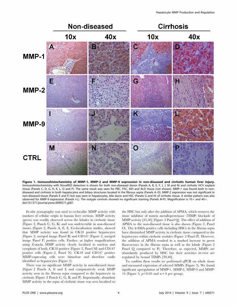
found expression of MMP-1 in non-diseased (Figure 1, Panels A,
B) and cirrhotic tissue (Figure 1, Panels C, D), while MMP-2 and
MMP-9 expression could only be detected in cirrhotic explant
tissue (Figure 1, Panels G, H, K and L) but not in non-diseased
tissue (Figure 1, Panels E, F, I and J). MMP expression was found
in hepatocytes as well as in the fibrous septa. No appreciable
staining was seen in the isotype antibody controls (Figure 1, Panels
M-P). As IHC cannot distinguish if non-septal HSC are positive
for CD147 or active MMPs, we proceeded to examine CD147
expression and MMP activity by confocal immunofluorescence in
combination with in-situ zymography.
Hepatocyte MMP Production and Regulation
PLOS ONE | www.plosone.org 3 July 2014 | Volume 9 | Issue 7 | e90571
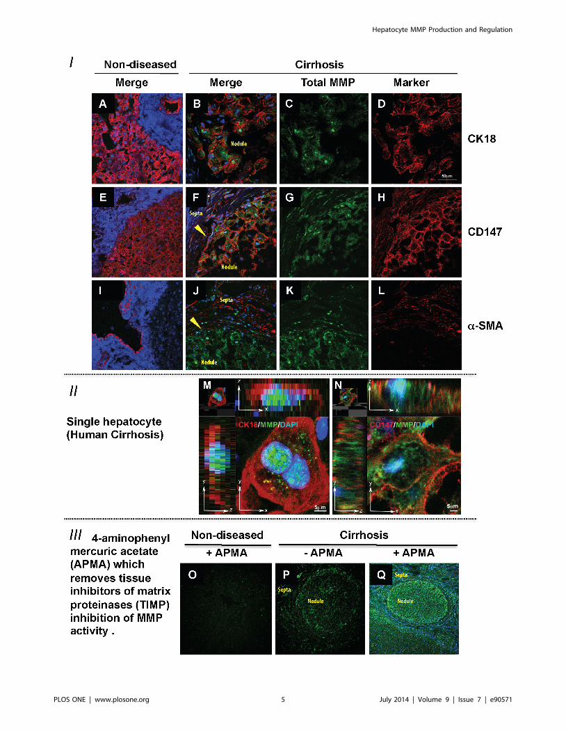
In-situ zymography was used to co-localise MMP activity with
markers of cellular origin in human liver sections. MMP activity
(green) was readily observed across the lobules in cirrhotic tissue
(Figure 2, Panels C, G, K) and was undetectable in non-diseased
tissues (Figure 2, Panels A, E, I). Co-localisation studies, showed
that MMP activity was found in CK18 positive hepatocytes
(Figure 2, merged image Panel B) and CD147 (Figure 2, merged
image Panel F) positive cells. Further, at higher magnifications
using Z-stacks MMP activity clearly localised to nucleus and
cytoplasm of both, CK18 positive (Figure 2, Panel M) and CD147
positive cells (Figure 2, Panel N). CK18 and CD147 positive
MMP-expressing cells were binuclear and therefore easily
identified as hepatocytes (Figure 2).
There was no significant MMP activity in non-diseased tissue
(Figure 2 Panels A, E and I) and comparatively weak MMP
activity seen in the fibrous septa compared to the hepatocyte in
cirrhosis (Figure 2 Panels C, G, K and P). Importantly, abundant
MMP activity in the septa of cirrhotic tissue was seen localised to
the HSC but only after the addition of APMA, which removes the
tissue inhibitor of matrix metalloproteinase (TIMP) blockade of
MMP activity [35,36] (Figure 2 Panel Q). The effect of addition of
APMA to the non-diseased tissue is also shown (Figure 2, Panel
O). The a-SMA positive cells including HSCs in the fibrous septa
have diminished MMP activity in cirrhotic tissue compared to the
hepatocytes within cirrhotic nodules (Figure 2 Panel P). However,
the addition of APMA resulted in a marked increase in green
fluorescence in the fibrous septa as well as the lobule (Figure 2
Panel Q compared to P). Therefore, as expected, MMPs are
abundantly produced by HSC but their activities in-vivo are
regulated by bound TIMPs [39,40].
To confirm these results we performed qPCR on whole tissue
and measured expression of selected MMPs (Figure 3). We found
significant upregulation of MMP-1, MMP-2, MMP-9 and MMP-
14 (Figure 3, p,0.05 and n = 4 per group).
Figure 1. Immunohistochemistry of MMP-1, MMP-2 and MMP-9 expression in non-diseased and cirrhotic human liver injury.Immunohistochemistry with NovaRED detection is shown for both non-diseased donor (Panels A, B, E, F, I, J, M and N) and cirrhotic HCV explanttissue (Panels C, D, G, H, K, L, O and P). The same result was seen for PBC, PSC, AIH and ALD tissue (not shown). MMP-1 was found both in non-diseased and cirrhosis in both hepatocytes and biliary structures located in the fibrous septa (Panels A–D). MMP-2 expression was not significant innon-diseased tissue (Panels E and F) but was seen in hepatocytes, bile ducts and HSC (Panels G and H) of cirrhotic tissue. A similar pattern was alsoobserved for MMP-9 expression (Panels I-L). The isotype controls showed no significant staining (Panels M-P). Magnification is 106 and 406.doi:10.1371/journal.pone.0090571.g001
Hepatocyte MMP Production and Regulation
PLOS ONE | www.plosone.org 4 July 2014 | Volume 9 | Issue 7 | e90571
Hepatocyte MMP Production and Regulation
PLOS ONE | www.plosone.org 5 July 2014 | Volume 9 | Issue 7 | e90571
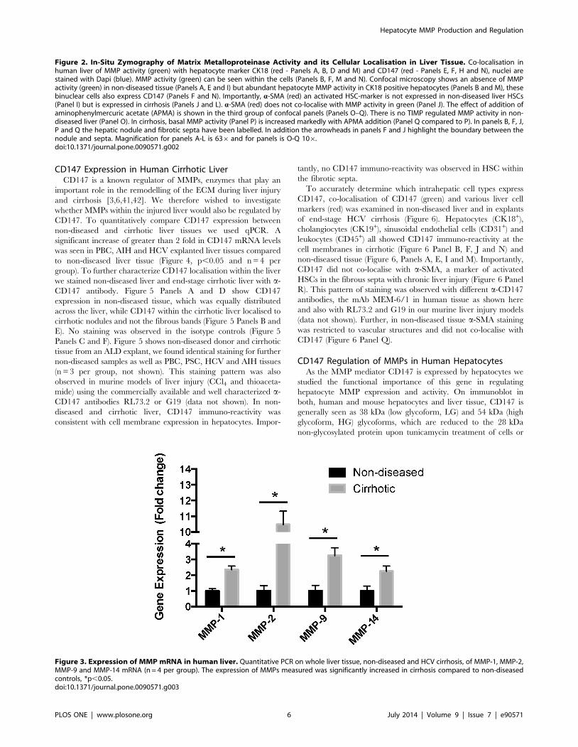
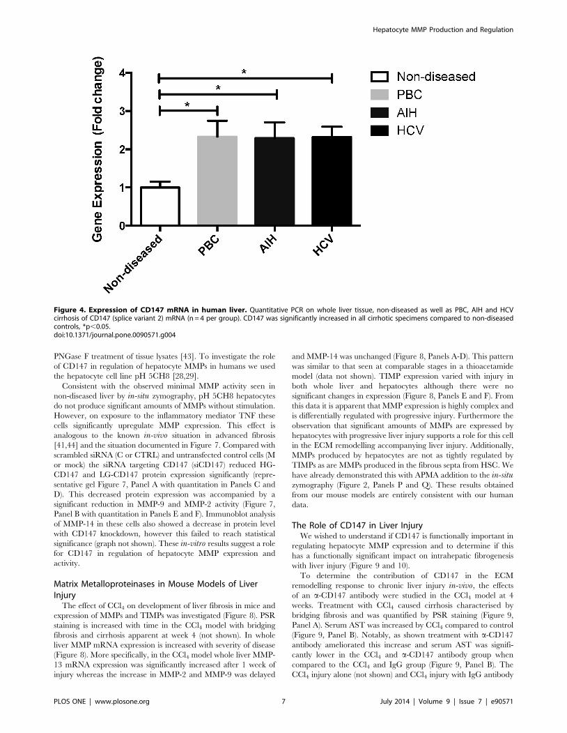
CD147 Expression in Human Cirrhotic LiverCD147 is a known regulator of MMPs, enzymes that play an
important role in the remodelling of the ECM during liver injury
and cirrhosis [3,6,41,42]. We therefore wished to investigate
whether MMPs within the injured liver would also be regulated by
CD147. To quantitatively compare CD147 expression between
non-diseased and cirrhotic liver tissues we used qPCR. A
significant increase of greater than 2 fold in CD147 mRNA levels
was seen in PBC, AIH and HCV explanted liver tissues compared
to non-diseased liver tissue (Figure 4, p,0.05 and n = 4 per
group). To further characterize CD147 localisation within the liver
we stained non-diseased liver and end-stage cirrhotic liver with a-
CD147 antibody. Figure 5 Panels A and D show CD147
expression in non-diseased tissue, which was equally distributed
across the liver, while CD147 within the cirrhotic liver localised to
cirrhotic nodules and not the fibrous bands (Figure 5 Panels B and
E). No staining was observed in the isotype controls (Figure 5
Panels C and F). Figure 5 shows non-diseased donor and cirrhotic
tissue from an ALD explant, we found identical staining for further
non-diseased samples as well as PBC, PSC, HCV and AIH tissues
(n = 3 per group, not shown). This staining pattern was also
observed in murine models of liver injury (CCl4 and thioaceta-
mide) using the commercially available and well characterized a-
CD147 antibodies RL73.2 or G19 (data not shown). In non-
diseased and cirrhotic liver, CD147 immuno-reactivity was
consistent with cell membrane expression in hepatocytes. Impor-
tantly, no CD147 immuno-reactivity was observed in HSC within
the fibrotic septa.
To accurately determine which intrahepatic cell types express
CD147, co-localisation of CD147 (green) and various liver cell
markers (red) was examined in non-diseased liver and in explants
of end-stage HCV cirrhosis (Figure 6). Hepatocytes (CK18+),
cholangiocytes (CK19+), sinusoidal endothelial cells (CD31+) and
leukocytes (CD45+) all showed CD147 immuno-reactivity at the
cell membranes in cirrhotic (Figure 6 Panel B, F, J and N) and
non-diseased tissue (Figure 6, Panels A, E, I and M). Importantly,
CD147 did not co-localise with a-SMA, a marker of activated
HSCs in the fibrous septa with chronic liver injury (Figure 6 Panel
R). This pattern of staining was observed with different a-CD147
antibodies, the mAb MEM-6/1 in human tissue as shown here
and also with RL73.2 and G19 in our murine liver injury models
(data not shown). Further, in non-diseased tissue a-SMA staining
was restricted to vascular structures and did not co-localise with
CD147 (Figure 6 Panel Q).
CD147 Regulation of MMPs in Human HepatocytesAs the MMP mediator CD147 is expressed by hepatocytes we
studied the functional importance of this gene in regulating
hepatocyte MMP expression and activity. On immunoblot in
both, human and mouse hepatocytes and liver tissue, CD147 is
generally seen as 38 kDa (low glycoform, LG) and 54 kDa (high
glycoform, HG) glycoforms, which are reduced to the 28 kDa
non-glycosylated protein upon tunicamycin treatment of cells or
Figure 2. In-Situ Zymography of Matrix Metalloproteinase Activity and its Cellular Localisation in Liver Tissue. Co-localisation inhuman liver of MMP activity (green) with hepatocyte marker CK18 (red - Panels A, B, D and M) and CD147 (red - Panels E, F, H and N), nuclei arestained with Dapi (blue). MMP activity (green) can be seen within the cells (Panels B, F, M and N). Confocal microscopy shows an absence of MMPactivity (green) in non-diseased tissue (Panels A, E and I) but abundant hepatocyte MMP activity in CK18 positive hepatocytes (Panels B and M), thesebinuclear cells also express CD147 (Panels F and N). Importantly, a-SMA (red) an activated HSC-marker is not expressed in non-diseased liver HSCs(Panel I) but is expressed in cirrhosis (Panels J and L). a-SMA (red) does not co-localise with MMP activity in green (Panel J). The effect of addition ofaminophenylmercuric acetate (APMA) is shown in the third group of confocal panels (Panels O–Q). There is no TIMP regulated MMP activity in non-diseased liver (Panel O). In cirrhosis, basal MMP activity (Panel P) is increased markedly with APMA addition (Panel Q compared to P). In panels B, F, J,P and Q the hepatic nodule and fibrotic septa have been labelled. In addition the arrowheads in panels F and J highlight the boundary between thenodule and septa. Magnification for panels A-L is 636 and for panels is O-Q 106.doi:10.1371/journal.pone.0090571.g002
Figure 3. Expression of MMP mRNA in human liver. Quantitative PCR on whole liver tissue, non-diseased and HCV cirrhosis, of MMP-1, MMP-2,MMP-9 and MMP-14 mRNA (n = 4 per group). The expression of MMPs measured was significantly increased in cirrhosis compared to non-diseasedcontrols, *p,0.05.doi:10.1371/journal.pone.0090571.g003
Hepatocyte MMP Production and Regulation
PLOS ONE | www.plosone.org 6 July 2014 | Volume 9 | Issue 7 | e90571
PNGase F treatment of tissue lysates [43]. To investigate the role
of CD147 in regulation of hepatocyte MMPs in humans we used
the hepatocyte cell line pH 5CH8 [28,29].
Consistent with the observed minimal MMP activity seen in
non-diseased liver by in-situ zymography, pH 5CH8 hepatocytes
do not produce significant amounts of MMPs without stimulation.
However, on exposure to the inflammatory mediator TNF these
cells significantly upregulate MMP expression. This effect is
analogous to the known in-vivo situation in advanced fibrosis
[41,44] and the situation documented in Figure 7. Compared with
scrambled siRNA (C or CTRL) and untransfected control cells (M
or mock) the siRNA targeting CD147 (siCD147) reduced HG-
CD147 and LG-CD147 protein expression significantly (repre-
sentative gel Figure 7, Panel A with quantitation in Panels C and
D). This decreased protein expression was accompanied by a
significant reduction in MMP-9 and MMP-2 activity (Figure 7,
Panel B with quantitation in Panels E and F). Immunoblot analysis
of MMP-14 in these cells also showed a decrease in protein level
with CD147 knockdown, however this failed to reach statistical
significance (graph not shown). These in-vitro results suggest a role
for CD147 in regulation of hepatocyte MMP expression and
activity.
Matrix Metalloproteinases in Mouse Models of LiverInjury
The effect of CCl4 on development of liver fibrosis in mice and
expression of MMPs and TIMPs was investigated (Figure 8). PSR
staining is increased with time in the CCl4 model with bridging
fibrosis and cirrhosis apparent at week 4 (not shown). In whole
liver MMP mRNA expression is increased with severity of disease
(Figure 8). More specifically, in the CCl4 model whole liver MMP-
13 mRNA expression was significantly increased after 1 week of
injury whereas the increase in MMP-2 and MMP-9 was delayed
and MMP-14 was unchanged (Figure 8, Panels A-D). This pattern
was similar to that seen at comparable stages in a thioacetamide
model (data not shown). TIMP expression varied with injury in
both whole liver and hepatocytes although there were no
significant changes in expression (Figure 8, Panels E and F). From
this data it is apparent that MMP expression is highly complex and
is differentially regulated with progressive injury. Furthermore the
observation that significant amounts of MMPs are expressed by
hepatocytes with progressive liver injury supports a role for this cell
in the ECM remodelling accompanying liver injury. Additionally,
MMPs produced by hepatocytes are not as tightly regulated by
TIMPs as are MMPs produced in the fibrous septa from HSC. We
have already demonstrated this with APMA addition to the in-situzymography (Figure 2, Panels P and Q). These results obtained
from our mouse models are entirely consistent with our human
data.
The Role of CD147 in Liver InjuryWe wished to understand if CD147 is functionally important in
regulating hepatocyte MMP expression and to determine if this
has a functionally significant impact on intrahepatic fibrogenesis
with liver injury (Figure 9 and 10).
To determine the contribution of CD147 in the ECM
remodelling response to chronic liver injury in-vivo, the effects
of an a-CD147 antibody were studied in the CCl4 model at 4
weeks. Treatment with CCl4 caused cirrhosis characterised by
bridging fibrosis and was quantified by PSR staining (Figure 9,
Panel A). Serum AST was increased by CCl4 compared to control
(Figure 9, Panel B). Notably, as shown treatment with a-CD147
antibody ameliorated this increase and serum AST was signifi-
cantly lower in the CCl4 and a-CD147 antibody group when
compared to the CCl4 and IgG group (Figure 9, Panel B). The
CCl4 injury alone (not shown) and CCl4 injury with IgG antibody
Figure 4. Expression of CD147 mRNA in human liver. Quantitative PCR on whole liver tissue, non-diseased as well as PBC, AIH and HCVcirrhosis of CD147 (splice variant 2) mRNA (n = 4 per group). CD147 was significantly increased in all cirrhotic specimens compared to non-diseasedcontrols, *p,0.05.doi:10.1371/journal.pone.0090571.g004
Hepatocyte MMP Production and Regulation
PLOS ONE | www.plosone.org 7 July 2014 | Volume 9 | Issue 7 | e90571

were indistinguishable. To determine whether a-CD147 antibody
could attenuate expression of mediators involved in fibrogenesis
we examined the expression of a-SMA, collagen I and collagen IV
and TGF- b, which were all increased with injury and significantly
decreased by the a-CD147 antibody intervention (Figure 9, Panels
C–F). The effect of a-CD147 antibody intervention on MMP
expression and hydroxyproline concentration as a marker of
collagen cross-linking was also studied (Figure 10). The antibody
intervention inhibited the induction of MMP-2, MMP-9, MMP-13
and MMP-14 mRNA by CCl4 (Figure 10, Panels B–E). The
reduced MMP expression with a-CD147 antibody intervention
led to an increased accumulation of cross-linked collagen evident
by significantly increased hydroxyproline concentration (Fig-
ure 10, Panel A) and PSR staining (Figure 9, Panel A). Together
these results are consistent with MMP expression being regulated
by CD147.
Discussion
CD147 has been found to regulate MMPs in a variety of tissues,
including human peripheral blood monocytes, human pulmonary
fibroblasts and in rheumatoid arthritis [15,21,23]. Additionally,
over decades CD147 has been associated with a diverse range of
Figure 5. Immunohistochemistry of CD147 in Human Liver Tissue. The a-CD147 antibody (MEM-6/1) was used to stain non-diseased (PanelsA and D) and cirrhotic ALD tissue (Panels B and E). CD147 is expressed by hepatocytes and bile ducts. No significant staining of HSC is seen in thefibrous septa, the only structures within the septa that are CD147 positive are bile ducts (see arrows Panel B and E). Isotype controls are shown inPanels C and F.doi:10.1371/journal.pone.0090571.g005
Hepatocyte MMP Production and Regulation
PLOS ONE | www.plosone.org 8 July 2014 | Volume 9 | Issue 7 | e90571
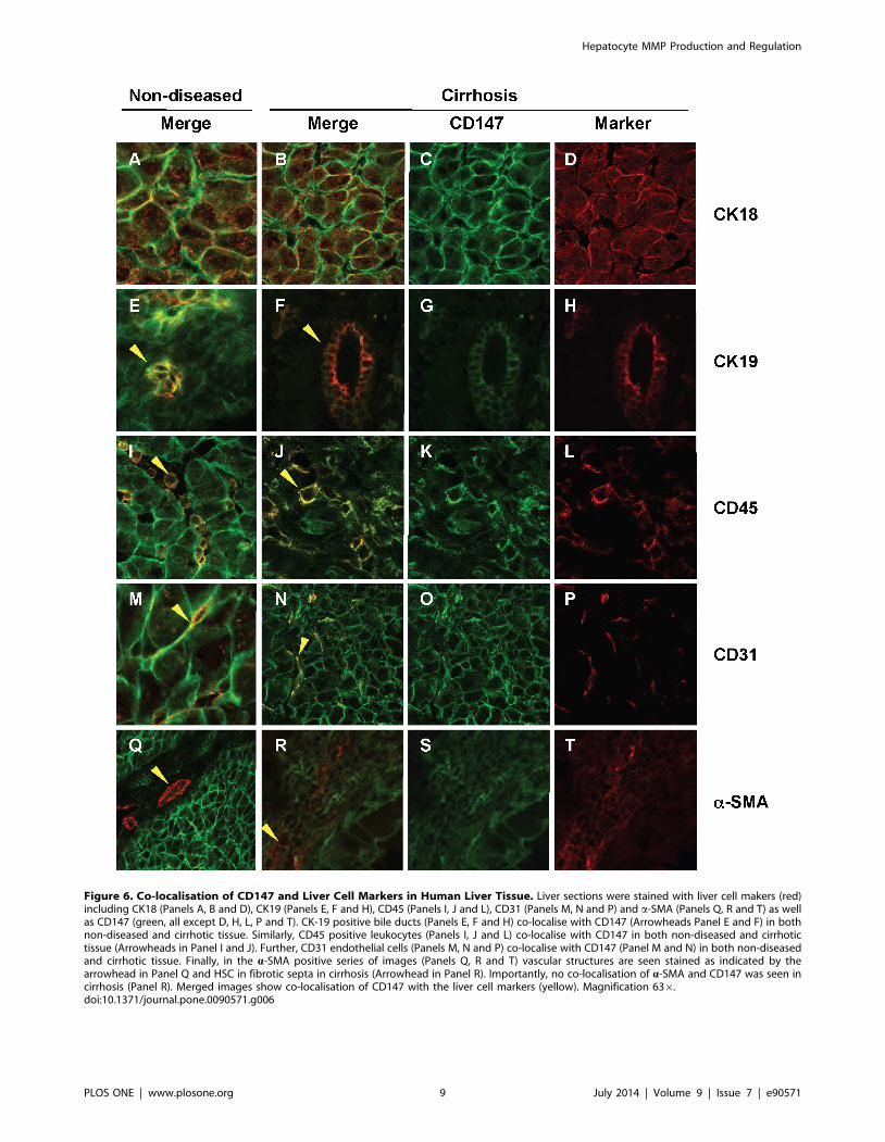
Figure 6. Co-localisation of CD147 and Liver Cell Markers in Human Liver Tissue. Liver sections were stained with liver cell makers (red)including CK18 (Panels A, B and D), CK19 (Panels E, F and H), CD45 (Panels I, J and L), CD31 (Panels M, N and P) and a-SMA (Panels Q, R and T) as wellas CD147 (green, all except D, H, L, P and T). CK-19 positive bile ducts (Panels E, F and H) co-localise with CD147 (Arrowheads Panel E and F) in bothnon-diseased and cirrhotic tissue. Similarly, CD45 positive leukocytes (Panels I, J and L) co-localise with CD147 in both non-diseased and cirrhotictissue (Arrowheads in Panel I and J). Further, CD31 endothelial cells (Panels M, N and P) co-localise with CD147 (Panel M and N) in both non-diseasedand cirrhotic tissue. Finally, in the a-SMA positive series of images (Panels Q, R and T) vascular structures are seen stained as indicated by thearrowhead in Panel Q and HSC in fibrotic septa in cirrhosis (Arrowhead in Panel R). Importantly, no co-localisation of a-SMA and CD147 was seen incirrhosis (Panel R). Merged images show co-localisation of CD147 with the liver cell markers (yellow). Magnification 636.doi:10.1371/journal.pone.0090571.g006
Hepatocyte MMP Production and Regulation
PLOS ONE | www.plosone.org 9 July 2014 | Volume 9 | Issue 7 | e90571
cancers such as melanoma, glioblastoma, breast or pancreatic
cancer and recently cholangiocarcinoma as well as hepatocellular
carcinoma (HCC), a liver cancer originating from hepatocytes
[16,17,45–49]. The functions of CD147 are varied and depend on
interacting proteins and cell types [50–58]. Many interacting
proteins are matrix components or inflammatory mediators that
are dramatically increased with injury (i.e. hyaluronan [50],
intercellular adhesion molecule (ICAM)-1 [51–53], lymphocyte
function-associated antigen (LFA)-1 [54–56] and CD43 [57,58]).
Previous studies examining MMP expression in non-HSC
populations, such as hepatocytes, have focused on regenerative
responses and resolution of injury [41,59]. Expression of MMPs by
hepatocytes has been described previously associated with a
regenerative response, and hepatocytes have been shown to
increase expression of MMPs-2, -9 and -14 in association with
injury resolution [12,60]. Further, MMP-10 expression by
hepatocytes as well cholangiocytes and macrophages is implicated
in the ECM remodelling of progressive fibrosis [13]. Previous work
and now the data in this manuscript demonstrate that hepatocytes
Figure 7. The Effect of Inhibition of CD147 on Matrix Metalloproteinase Expression and Activities in pH5CH8 Hepatocytes.Downregulation of MMPs with CD147 knockdown in a hepatocyte cell line in-vitro. pH5CH8 cells were transfected with siCtrl (C) or siCD147 (Si)oligonucleotides. Transfected cells and mock controls (M) were cultured in serum free conditions for 48 hrs. Shown in panel A are representativeimmunoblots of CD147 with the higher (HG CD147) and lower molecular weight (LG CD147) glycoforms, MMP-14 and GAPDH as loading control.Gelatin zymography of MMP-2 and -9 on the conditioned media from the same cells are shown in panel B. Densitometry was performed on theCD147 immunoblots and the results are shown as HG CD147 and LG CD147 forms normalised for GAPDH (n = 3, Panels C and D). Densitometricanalysis of gelatin zymography of MMP-9 and MMP-2 are shown (Panels E and F). *p,0.05 using Mann-Whitney U t-test, compared to Mock (n = 3 forall groups).doi:10.1371/journal.pone.0090571.g007
Hepatocyte MMP Production and Regulation
PLOS ONE | www.plosone.org 10 July 2014 | Volume 9 | Issue 7 | e90571

Figure 8. MMP and TIMP expression in whole liver and primary hepatocytes from CCl4 induced liver injury. Quantitative PCR of MMPand TIMP expression in whole liver and isolated primary hepatocytes. Cirrhosis was induced in a mouse model of liver injury (C57bl/6) with CCl4. RNAextracted from primary hepatocytes as well as whole liver were assessed at commencement of injury and weeks 1, 2 and 4. The expression of MMP-2(Panel A), MMP-9 (Panel B), MMP-13 (Panel C), MMP-14 (Panel D), TIMP-1 (Panel E) and TIMP-2 (Panel F) was assessed by quantitative PCR (n = 4 pergroup, data expressed as mean and SEM. *p,0.05 and **p,0.01 relative to untreated).doi:10.1371/journal.pone.0090571.g008
Hepatocyte MMP Production and Regulation
PLOS ONE | www.plosone.org 11 July 2014 | Volume 9 | Issue 7 | e90571

with progressive fibrotic injury do produce a number of active
MMPs capable of significant ECM remodelling.
In progressive fibrotic liver injury a number of mediators such
as TNF are increased which can induce MMP expression [41,44].
In primary hepatocytes, MMP-9 expression is increased with TNF
and epidermal growth factor, both key molecular mediators
increased in injury [41,44] and involved in hepatocyte regener-
ation [59]. In our study, the in-situ zymography technique
provided a powerful tool for confirming that hepatocytes produce
MMPs that are active. Further, hepatocytes isolated from injured
mouse livers expressed MMP-2, MMP-9, MMP-13 and MMP-14
mRNA at levels approximate to that seen in whole liver tissue.
Together our results are consistent with hepatocytes being a major
source of MMPs. Additionally, as expected, we also show that
HSC in the fibrous septa are associated with active MMPs.
Functionally this is likely to be important as hepatocytes, that are
surrounded by abundant pericellular fibrosis, have reduced
function [61]. In human cirrhotic liver samples in-situ zymogra-
phy in the absence of APMA showed MMP enzyme activity across
the nodule. Following the addition of APMA to remove TIMP
inhibition, increased MMP activity was observed in the hepato-
cytes within nodules and the fibrous septa. These results in which
APMA addition was used to determine the effect of TIMP
inhibition on MMP activity are comparative but intriguing. They
suggest that there is greater TIMP regulation of MMPs produced
by HSC in the fibrous septa compared to those MMPs produced
by hepatocytes. The significance of this result is unknown but we
propose that this may represent on-going active ECM turnover by
hepatocytes in the context of progressive injury. Further work will
need to determine the functional importance of this observation.
The temporal evolution of MMP expression in whole liver and
hepatocytes with injury is complex [8]. In both mouse models the
Figure 9. Injury in a mouse model of fibrosis with 4 weeks CCl4 and a-CD147 antibody intervention. Cirrhosis was induced in a mousemodel of liver injury (Balb/c) with CCl4. An antibody targeting CD147 (RL73.2) was given twice weekly for 4 weeks, mice given an equal amount of IgGwere used as control. There was no appreciable phenotype with either a-CD147 antibody or IgG control treatment in the absence of CCl4 (notshown). The injury groups are CCl4 (+/2 isotype control) and CCl4 with antibody targeting CD147. Quantitative data of PSR staining is shown in panelA. Injury as assessed by AST (Panel B), a-SMA (Panel C), Collagen I (Panel D), TGF-b (Panel E) and Collagen IV (Panel F) were all significantly reduced inCCl4 injury with a-CD147 antibody intervention compared to the IgG control treated with CCl4. All groups are n = 426 with data expressed as meanand SEM. *p,0.05.doi:10.1371/journal.pone.0090571.g009
Hepatocyte MMP Production and Regulation
PLOS ONE | www.plosone.org 12 July 2014 | Volume 9 | Issue 7 | e90571
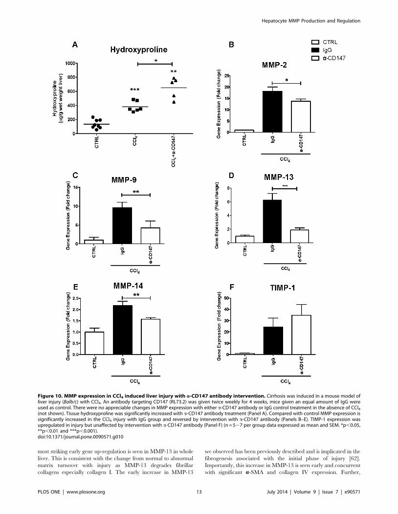
most striking early gene up-regulation is seen in MMP-13 in whole
liver. This is consistent with the change from normal to abnormal
matrix turnover with injury as MMP-13 degrades fibrillar
collagens especially collagen I. The early increase in MMP-13
we observed has been previously described and is implicated in the
fibrogenesis associated with the initial phase of injury [62].
Importantly, this increase in MMP-13 is seen early and concurrent
with significant a-SMA and collagen IV expression. Further,
Figure 10. MMP expression in CCl4 induced liver injury with a-CD147 antibody intervention. Cirrhosis was induced in a mouse model ofliver injury (Balb/c) with CCl4. An antibody targeting CD147 (RL73.2) was given twice weekly for 4 weeks, mice given an equal amount of IgG wereused as control. There were no appreciable changes in MMP expression with either a-CD147 antibody or IgG control treatment in the absence of CCl4(not shown). Tissue hydroxyproline was significantly increased with a-CD147 antibody treatment (Panel A). Compared with control MMP expression issignificantly increased in the CCl4 injury with IgG group and reversed by intervention with a-CD147 antibody (Panels B–E). TIMP-1 expression wasupregulated in injury but unaffected by intervention with a-CD147 antibody (Panel F) (n = 527 per group data expressed as mean and SEM. *p,0.05,**p,0.01 and ***p,0.001).doi:10.1371/journal.pone.0090571.g010
Hepatocyte MMP Production and Regulation
PLOS ONE | www.plosone.org 13 July 2014 | Volume 9 | Issue 7 | e90571
MMP-2 and MMP-9 were both found to be upregulated in
primary hepatocytes after one week of CCl4 treatment. MMP-9 is
primarily responsible for degradation of type IV and V collagens
and is produced by HSC, inflammatory cells [42,63] as well as
being expressed in hepatocytes with injury as demonstrated in this
study. In established end-stage injury with cirrhosis there is
increased expression of MMP-2, MMP-9, MMP-14 as well as
TIMP-1 and TIMP-2 [42,64]. The differential expression of
MMPs described in this study and by others [8] clearly implicates
individual MMPs, the cell of origin, and the stage of injury in
determining the final fibrotic phenotype. This impacts on the
functional significance of target gene interventions in progressive
fibrosis as this is dependent on the intrahepatic cell types
expressing the gene, the stage of injury, and which MMPs are
regulated by the target gene.
A key novel result from our work shows that MMPs produced
by hepatocytes can be regulated by the membrane glycoprotein
CD147. CD147 is a known mediator of both inflammation and
ECM remodelling [18,19,23] and was found expressed abundantly
on hepatocytes, with no significant staining evident through the
fibrous septa on HSC. In-vitro studies of the pH 5CH8 hepatocyte
cell line identified that MMP expression is partially regulated by
CD147. The in-vivo data further supports a CD147-dependent
role in mediating MMP dependent matrix remodelling. Following
a-CD147 antibody intervention in the CCl4 injury model we have
demonstrated a reduction in tissue injury characterised by
reduction in a-SMA mRNA and necroinflammatory activity
(AST). The reduction in inflammation may result from a reduction
in ECM degradation products that can perpetuate HSC activation
and the production of pro-inflammatory mediators such as TGF-b[2,65]. Additionally, the reduction in necroinflammatory activity
may reflect an additional anti-inflammatory effect of the a-CD147
antibody intervention given the near ubiquitous expression of this
glycoprotein on leukocytes. The net effect with a-CD147 antibody
intervention was a reduction in MMP expression but an increase
in hydroxyproline, indicating greater collagen crosslinking. This is
most likely due to the reduction in MMP-13 cleavage of
predominantly type I collagen seen in abnormal ECM combined
with the observed MMP-9 increase, which degrades predomi-
nantly normal type IV collagen. Further, MMP-14 is abundantly
expressed by hepatocytes and studies of CD147 regulation of
MMP-14 in tumour cells have found that a feedback loop exists,
whereby MMP-14 cleaves CD147 from the cell surface to produce
soluble CD147 ligand, which results in an autocrine regulation of
the expression of both MMP-14 and CD147 [66,67]. These
observations support the hypothesis that in hepatocytes there is
active production of MMPs and this is regulated by CD147 with
progressive injury.
These studies have shown that hepatocyte-derived MMPs are
capable of ECM turnover. Further, with reduced hepatocyte
activity of MMPs there is accumulation of cross-linked ECM.
Importantly, we have demonstrated that MMP expression can be
regulated by the glycoprotein CD147. The novel finding of
intrahepatic active hepatocyte MMP production, which is
regulated by CD147, presents a new pathway that could be
manipulated by possible future anti-fibrotic therapeutics.
Supporting Information
Table S1 Taqman probe sequences used for quantita-tive PCR.
(DOCX)
Table S2 Primer sequences used for quantitative PCRwith Sybr Green.
(DOCX)
Acknowledgments
We would also like to acknowledge the support of the Centenary Institute
Advanced Imaging Facility, Bosch Institute and the Pathology Depart-
ments at the University of Sydney. Prof Kui Li of the Department of
Microbiology, Immunology and Biochemistry The University of Tennessee
Health Science Center Tennessee USA provided the cells.
Author Contributions
Conceived and designed the experiments: SRC AEM WA FJW SVM
NAS. Performed the experiments: SRC AEM AJM TT VWW CY AM
ML WA FJW. Analyzed the data: SRC AEM FJW SL GWM SVM NAS.
Contributed reagents/materials/analysis tools: SL FJW GWM NAS.
Wrote the paper: AEM SVM FJW NAS. Designed and conducted
experiments: SRC AEM.
References
1. Shackel NA, McGuinness PH, Abbott CA, Gorrell MD, McCaughan GW(2002) Insights into the pathobiology of hepatitis C virus-associated cirrhosis:
analysis of intrahepatic differential gene expression. Am J Pathol 160: 641–654.
2. Bataller R, Brenner DA (2005) Liver fibrosis. J Clin Invest 115: 209–218.
3. Iredale JP (1997) Tissue inhibitors of metalloproteinases in liver fibrosis.International Journal of Biochemistry and Cell Biology 29: 43–54.
4. Arthur MJ (1990) Matrix degradation in the liver. Semin Liver Dis 10: 47–55.
5. Arthur MJ (1994) Degradation of matrix proteins in liver fibrosis. Pathol Res
Pract 190: 825–833.
6. Arthur MJ (1997) Matrix degradation in liver: a role in injury and repair.Hepatology 26: 1069–1071.
7. Arthur MJ (1998) Fibrosis and altered matrix degradation. Digestion 59: 376–
380.
8. Hemmann S, Graf J, Roderfeld M, Roeb E (2007) Expression of MMPs and
TIMPs in liver fibrosis - a systematic review with special emphasis on anti-fibrotic strategies. J Hepatol 46: 955–975.
9. Veidal SS, Karsdal MA, Vassiliadis E, Nawrocki A, Larsen MR, et al. (2011)
MMP mediated degradation of type VI collagen is highly associated with liverfibrosis–identification and validation of a novel biochemical marker assay. PLoS
One 6: e24753.
10. Thomas JA, Pope C, Wojtacha D, Robson AJ, Gordon-Walker TT, et al. (2011)Macrophage therapy for murine liver fibrosis recruits host effector cells
improving fibrosis, regeneration and function. Hepatology.
11. Fallowfield JA, Mizuno M, Kendall TJ, Constandinou CM, Benyon RC, et al.(2007) Scar-associated macrophages are a major source of hepatic matrix
metalloproteinase-13 and facilitate the resolution of murine hepatic fibrosis.
J Immunol 178: 5288–5295.
12. Garciade Leon Mdel C, Montfort I, Tello Montes E, Lopez Vancell R, Olivos
Garcia A, et al. (2006) Hepatocyte production of modulators of extracellular
liver matrix in normal and cirrhotic rat liver. Exp Mol Pathol 80: 97–108.
13. Garcia-Irigoyen O, Carotti S, Latasa MU, Uriarte I, Fernandez-Barrena MG, et
al. (2013) Matrix metalloproteinase-10 expression is induced during hepatic
injury and plays a fundamental role in liver tissue repair. Liver Int.
14. McLennan SV, Warner FJ, Shackel NA (2012) The role of CD147 in liver
injury: "The truth is in the details". J Hepatol.
15. Agrawal SM, Yong VW (2011) The many faces of EMMPRIN - roles in
neuroinflammation. Biochim Biophys Acta 1812: 213–219.
16. Toole BP (2003) Emmprin (CD147), a cell surface regulator of matrix
metalloproteinase production and function. Current topics in developmental
biology 54: 371–389.
17. Yan L, Zucker S, Toole BP (2005) Roles of the multifunctional glycoprotein,
emmprin (basigin; CD147), in tumour progression. Thrombosis and Haemo-
stasis 93: 199–204.
18. Guillot S, Delaval P, Brinchault G, Caulet-Maugendre S, Depince A, et al.
(2006) Increased extracellular matrix metalloproteinase inducer (EMMPRIN)
expression in pulmonary fibrosis. Exp Lung Res 32: 81–97.
19. Wang CH, Dai JY, Wang L, Jia JF, Zheng ZH, et al. (2011) Expression of
CD147 (EMMPRIN) on neutrophils in rheumatoid arthritis enhances
chemotaxis, matrix metalloproteinase production and invasiveness of synovio-
cytes. J Cell Mol Med 15: 850–860.
20. Treese C, Mittag A, Lange F, Tarnok A, Loesche A, et al. (2008)
Characterization of fibroblasts responsible for cartilage destruction in arthritis.
Cytometry A 73: 351–360.
Hepatocyte MMP Production and Regulation
PLOS ONE | www.plosone.org 14 July 2014 | Volume 9 | Issue 7 | e90571
21. Zhu P, Lu N, Shi ZG, Zhou J, Wu ZB, et al. (2006) CD147 overexpression on
synoviocytes in rheumatoid arthritis enhances matrix metalloproteinaseproduction and invasiveness of synoviocytes. Arthritis Res Ther 8: R44.
22. Tomita T, Nakase T, Kaneko M, Shi K, Takahi K, et al. (2002) Expression of
extracellular matrix metalloproteinase inducer and enhancement of theproduction of matrix metalloproteinases in rheumatoid arthritis. Arthritis
Rheum 46: 373–378.23. Betsuyaku T, Kadomatsu K, Griffin GL, Muramatsu T, Senior RM (2003)
Increased basigin in bleomycin-induced lung injury. Am J Respir Cell Mol Biol
28: 600–606.24. Zhang DW, Zhao YX, Wei D, Li YL, Zhang Y, et al. (2012) HAb18G/CD147
promotes activation of hepatic stellate cells and is a target for antibody therapy ofliver fibrosis. J Hepatol 57: 1283–1291.
25. Howe RC, MacDonald HR (1988) Heterogeneity of immature (Lyt-2-/L3T4-)thymocytes. Identification of four major phenotypically distinct subsets differing
in cell cycle status and in vitro activation requirements. J Immunol 140: 1047–
1055.26. Zhang DW, Chen ZN, Bian H (2012) Reply to: "The role of CD147 in liver
injury: The truth is in the details". J Hepatol.27. Shackel NA, McGuinness PH, Abbott CA, Gorrell MD, McCaughan GW
(2001) Identification of novel molecules and pathogenic pathways in primary
biliary cirrhosis: cDNA array analysis of intrahepatic differential geneexpression. Gut 49: 565–576.
28. Ikeda M, Sugiyama K, Mizutani T, Tanaka T, Tanaka K, et al. (1998) Humanhepatocyte clonal cell lines that support persistent replication of hepatitis C virus.
Virus Res 56: 157–167.29. Noguchi M, Hirohashi S (1996) Cell Lines from Non-Neoplastic Liver and
Hepatocellular Carcinoma Tissue from a Single Patient. In Vitro Cell and
Developmental Biology 32: 135–137.30. Igakura T, Kadomatsu K, Kaname T, Muramatsu H, Fan QW, et al. (1998) A
null mutation in basigin, an immunoglobulin superfamily member, indicates itsimportant roles in peri-implantation development and spermatogenesis. Dev
Biol 194: 152–165.
31. Kuno N, Kadomatsu K, Fan QW, Hagihara M, Senda T, et al. (1998) Femalesterility in mice lacking the basigin gene, which encodes a transmembrane
glycoprotein belonging to the immunoglobulin superfamily. FEBS Lett 425:191–194.
32. Shi Z, Wakil AE, Rockey DC (1997) Strain-specific differences in mouse hepaticwound healing are mediated by divergent T helper cytokine responses. Proc Natl
Acad Sci U S A 94: 10663–10668.
33. MacDonald HR, Lees RK, Bron C (1985) Cell surface glycoproteins involved inthe stimulation of interleukin 1-dependent interleukin 2 production by a subline
of EL4 thymoma cells. I. Functional characterization by monoclonal antibodies.Journal of Immunology 135: 3944–3950.
34. Bertolino P, Trescol-Biemont MC, Rabourdin-Combe C (1998) Hepatocytes
induce functional activation of naive CD8+ T lymphocytes but fail to promotesurvival. European journal of immunology 28: 221–236.
35. Itoh Y, Binner S, Nagase H (1995) Steps involved in activation of the complex ofpro-matrix metalloproteinase 2 (progelatinase A) and tissue inhibitor of
metalloproteinases (TIMP)-2 by 4-aminophenylmercuric acetate. Biochem J 308(Pt 2): 645–651.
36. Ogata Y, Itoh Y, Nagase H (1995) Steps involved in activation of the pro-matrix
metalloproteinase 9 (progelatinase B)-tissue inhibitor of metalloproteinases-1complex by 4-aminophenylmercuric acetate and proteinases. J Biol Chem 270:
18506–18511.37. Min D, Lyons JG, Jia J, Lo L, McLennan SV (2006) 2-Methoxy-2,4-diphenyl-
3(2H)-furanone-labeled gelatin zymography and reverse zymography: a rapid
real-time method for quantification of matrix metalloproteinases-2 and -9 andtissue inhibitors of metalloproteinases. Electrophoresis 27: 357–364.
38. Paizis G, Tikellis C, Cooper ME, Schembri JM, Lew RA, et al. (2005) Chronicliver injury in rats and humans upregulates the novel enzyme angiotensin
converting enzyme 2. Gut 54: 1790–1796.
39. Yoshiji H, Kuriyama S, Yoshii J, Ikenaka Y, Noguchi R, et al. (2002) Tissueinhibitor of metalloproteinases-1 attenuates spontaneous liver fibrosis resolution
in the transgenic mouse. Hepatology 36: 850–860.40. Iredale JP (1997) Tissue inhibitors of metalloproteinases in liver fibrosis.
Int J Biochem Cell Biol 29: 43–54.41. Knittel T, Mehde M, Kobold D, Saile B, Dinter C, et al. (1999) Expression
patterns of matrix metalloproteinases and their inhibitors in parenchymal and
non-parenchymal cells of rat liver: regulation by TNF-alpha and TGF-beta1.J Hepatol 30: 48–60.
42. Han YP (2006) Matrix metalloproteinases, the pros and cons, in liver fibrosis.J Gastroenterol Hepatol 21 Suppl 3: S88–91.
43. Tang W, Chang SB, Hemler ME (2004) Links between CD147 function,
glycosylation, and caveolin-1. Mol Biol Cell 15: 4043–4050.44. Serandour AL, Loyer P, Garnier D, Courselaud B, Theret N, et al. (2005)
TNFalpha-mediated extracellular matrix remodeling is required for multipledivision cycles in rat hepatocytes. Hepatology 41: 478–486.
45. Tang J, Guo YS, Zhang Y, Yu XL, Li L, et al. (2012) CD147 induces UPR to
inhibit apoptosis and chemosensitivity by increasing the transcription of Bip in
hepatocellular carcinoma. Cell Death Differ 19: 1779–1790.
46. Sawanyawisuth K, Wongkham C, Araki N, Zhao Q, Riggins GJ, et al. (2012)
Serial Analysis of Gene Expression Reveals Promising Therapeutic Targets for
Liver Fluke-associated Cholangiocarcinoma. Asian Pac J Cancer Prev 13 Suppl:
89–93.
47. Hou Q, Tang X, Liu H, Tang J, Yang Y, et al. (2011) Berberine induces cell
death in human hepatoma cells in vitro by downregulating CD147. Cancer Sci
102: 1287–1292.
48. Zhao P, Zhang W, Tang J, Ma XK, Dai JY, et al. (2010) Annexin II promotes
invasion and migration of human hepatocellular carcinoma cells in vitro via its
interaction with HAb18G/CD147. Cancer Sci 101: 387–395.
49. Jia L, Xu H, Zhao Y, Jiang L, Yu J, et al. (2008) Expression of CD147 mediates
tumor cells invasion and multidrug resistance in hepatocellular carcinoma.
Cancer Invest 26: 977–983.
50. Pushkarsky T, Zybarth G, Dubrovsky L, Yurchenko V, Tang H, et al. (2001)
CD147 facilitates HIV-1 infection by interacting with virus-associated
cyclophilin A. Proc Natl Acad Sci U S A 98: 6360–6365.
51. Cho JY, Fox DA, Horejsi V, Sagawa K, Skubitz KM, et al. (2001) The
functional interactions between CD98, beta1-integrins, and CD147 in the
induction of U937 homotypic aggregation. Blood 98: 374–382.
52. Joseph J, Knobler RL, Lublin FD, Burns FR (1993) Regulation of the expression
of intercellular adhesion molecule-1 (ICAM-1) and the putative adhesion
molecule Basigin on murine cerebral endothelial cells by MHV-4 (JHM). Adv
Exp Med Biol 342: 389–391.
53. Kasinrerk W, Tokrasinwit N, Phunpae P (1999) CD147 monoclonal antibodies
induce homotypic cell aggregation of monocytic cell line U937 via LFA-1/
ICAM-1 pathway. Immunology 96: 184–192.
54. Xu Q, Cao JS, Zhang XM (2002) Liver-infiltrating T lymphocytes cause
hepatocyte damage by releasing humoral factors via LFA-1/ICAM-1 interaction
in immunological liver injury. Inflamm Res 51: 44–50.
55. Matsumoto G, Tsunematsu S, Tsukinoki K, Ohmi Y, Iwamiya M, et al. (2002)
Essential role of the adhesion receptor LFA-1 for T cell-dependent fulminant
hepatitis. J Immunol 169: 7087–7096.
56. Ohteki T, Maki C, Koyasu S, Mak TW, Ohashi PS (1999) Cutting edge: LFA-1
is required for liver NK1.1+TCR alpha beta+ cell development: evidence that
liver NK1.1+TCR alpha beta+ cells originate from multiple pathways.
J Immunol 162: 3753–3756.
57. Bataller R, Gabele E, Parsons CJ, Morris T, Yang L, et al. (2005) Systemic
infusion of angiotensin II exacerbates liver fibrosis in bile duct-ligated rats.
Hepatology 41: 1046–1055.
58. Hamzavi J, Ehnert S, Godoy P, Ciuclan L, Weng H, et al. (2008) Disruption of
the Smad7 gene enhances CCI4-dependent liver damage and fibrogenesis in
mice. J Cell Mol Med 12: 2130–2144.
59. Haruyama T, Ajioka I, Akaike T, Watanabe Y (2000) Regulation and
significance of hepatocyte-derived matrix metalloproteinases in liver remodeling.
Biochem Biophys Res Commun 272: 681–686.
60. Watanabe T, Niioka M, Ishikawa A, Hozawa S, Arai M, et al. (2001) Dynamic
change of cells expressing MMP-2 mRNA and MT1-MMP mRNA in the
recovery from liver fibrosis in the rat. J Hepatol 35: 465–473.
61. Pinzani M, Rombouts K (2004) Liver fibrosis: from the bench to clinical targets.
Dig Liver Dis 36: 231–242.
62. Uchinami H, Seki E, Brenner DA, D’Armiento J (2006) Loss of MMP 13
attenuates murine hepatic injury and fibrosis during cholestasis. Hepatology 44:
420–429.
63. Henkel C, Roderfeld M, Weiskirchen R, Scheibe B, Matern S, et al. (2005)
Identification of fibrosis-relevant proteins using DIGE (difference in gel
electrophoresis) in different models of hepatic fibrosis. Z Gastroenterol 43: 23–
29.
64. Roeb E, Bosserhoff AK, Hamacher S, Jansen B, Dahmen J, et al. (2005)
Enhanced migration of tissue inhibitor of metalloproteinase overexpressing
hepatoma cells is attributed to gelatinases: relevance to intracellular signaling
pathways. World J Gastroenterol 11: 1096–1104.
65. Iredale JP, Thompson A, Henderson NC (2013) Extracellular matrix
degradation in liver fibrosis: Biochemistry and regulation. Biochim Biophys
Acta 1832: 876–883.
66. Egawa N, Koshikawa N, Tomari T, Nabeshima K, Isobe T, et al. (2006)
Membrane type 1 matrix metalloproteinase (MT1-MMP/MMP-14) cleaves and
releases a 22-kDa extracellular matrix metalloproteinase inducer (EMMPRIN)
fragment from tumor cells. J Biol Chem 281: 37576–37585.
67. Tang Y, Kesavan P, Nakada MT, Yan L (2004) Tumor-stroma interaction:
positive feedback regulation of extracellular matrix metalloproteinase inducer
(EMMPRIN) expression and matrix metalloproteinase-dependent generation of
soluble EMMPRIN. Mol Cancer Res 2: 73–80.
Hepatocyte MMP Production and Regulation
PLOS ONE | www.plosone.org 15 July 2014 | Volume 9 | Issue 7 | e90571
Related Documents