Glial tumor cell adhesion is mediated by binding of the FNIII domain of receptor protein tyrosine phosphatase b (RPTPb) to tenascin C Konstantin Adamsky 1 , James Schilling 2 , Jeremy Garwood 3 , Andreas Faissner 3 and Elior Peles* ,1 1 Department of Molecular Cell Biology, The Weizmann Institute of Science, Rehovot 76100, Israel; 2 Sugen Inc. South San Francisco, California, CA 94080, USA; 3 Unite ´ CNRS UPR 1352, Centre de Neurochimie, 67084 Strasbourg, France The extracellular domain of receptor protein tyrosine phosphatase b (RPTPb) is composed of several domains which mediate its interactions with distinct ligands present on the surface of either neurons or glial cells. Here, we demonstrate that the fibronectin type III domain (FNIII) of RPTPb binds to glial tumor-derived cell lines and primary astrocytes. We used anity purification to isolate several proteins that specifically bind to the FNIII domain of RPTPb. One of these, a 240 kDa protein that was purified from U118MG glioblastoma cell, was identified as tenascin C based on the amino acid sequence of several tryptic peptides. The interaction of RPTPb with tenascin C was found to mediate cell adhesion. Adhesion and spreading of SF763T astrocytoma cells expressing RPTPb on tenascin C was specifically abolished by the addition of a soluble fragment containing the FNIII domain of the receptor. RPTPb-dependent cell adhesion was mediated by binding to the alternatively spliced FNIII repeats A1,2,4 (TnfnA1,2,4) of tenascin C. Furthermore, COS cells expressing RPTPb adhere to TnfnA1,2,4, while the parental cells did not. These results demonstrate that the FNIII domain of RPTPb binds to tenascin C and suggest that RPTPb present on glial tumor cells is a primary adhesion receptor system to the extracellular matrix. Oncogene (2001) 20, 609 – 618. Keywords: tyrosine phosphatase; extracellular matrix; cell adhesion molecule Introduction The interactions between tumor cells and the extra- cellular matrix is important for neoplastic transforma- tion and cell invasion. Protein tyrosine phosphorylation regulates various signal transduction pathways underlying fundamental cellular processes such as cell motility, growth and dierentiation (Angers-Loustau et al., 1999; Neel and Tonks, 1997; Stoker and Dutta, 1998). The extracellular domain of most of the known receptor-like protein tyrosine phosphatases (RPTPs) shares structural similarities with cell adhesion molecules suggesting that they play a role in cell-cell and cell-matrix communication by directly coupling cell recognition events to signal transduction pathways within the cell (Brady-Kalnay and Tonks, 1995; Schaapveld et al., 1997; Zondag and Moolenaar, 1997). Receptor protein tyrosine phosphatase b (RPTPb, also known as RPTPz) contains in its extracellular domain a region with sequence homology to the enzyme carbonic anhydrase (CAH), followed by a fibronectin type III repeat (FNIII) and by a long unique sequence termed the spacer domain (Krueger and Saito, 1992; Levy et al., 1993). Three dierent isoforms of RPTPb are generated as a result of alternative splicing: a short and a long receptor form that dier by the absence or presence of a stretch of 860 amino acid residues in the spacer domain, and a third secreted form composed of the extracellular domain of the long receptor known also as phospha- can, 6B4 and DSD-1 proteoglycans (Barnea et al., 1994; Garwood et al., 1999; Levy et al., 1993; Maeda et al., 1994). Both the long receptor form and phospha- can are found in rat brain as chondroitin sulfate proteoglycans, whereas the short receptor form (dvRPTPb) is lacking glycosaminoglycan side chains (Sakurai et al., 1996). RPTPb is predominantly expressed in the nervous system where it is mainly found in glial precursors, radial glia and astrocytes (Canoll et al., 1993), as well as in certain neurons (Snyder et al., 1996). The expression of the dierent forms of RPTPb is regulated during development of the glial lineage. Early in development, high levels of the receptor forms are found in proliferating precursor cells at the subventricular zones. As development progresses and cells mature, these receptor forms are replaced by the secreted non-phosphatase form (Canoll et al., 1996; Sakurai et al., 1996). The short receptor form of RPTPb is found in certain astrocytomas and glioblastomas and may represent transformed early proliferating glial precursors (Krueger and Saito, 1992; Sakurai et al., 1996). RPTPb is also found in human cutaneous melanomas which developmentally originate from the neural crest (Goldmann et al., 2000). The dierent forms of RPTPb bind to multiple ligands (Margolis and Margolis, 1997; Peles et al., 1998; Oohira et al., 2000). It has been previously shown that phosphacan interacts directly with the neuronal recognition molecules N-CAM, Ng-CAM (Milev et al., 1994) and TAG-1 (Milev et al., 1996), with the extracellular matrix molecules tenascin C and Oncogene (2001) 20, 609 – 618 ª 2001 Nature Publishing Group All rights reserved 0950 – 9232/01 $15.00 www.nature.com/onc *Correspondence: E Peles Received 16 August 2000; revised 13 November 2000; accepted 16 November 2000

Welcome message from author
This document is posted to help you gain knowledge. Please leave a comment to let me know what you think about it! Share it to your friends and learn new things together.
Transcript
Glial tumor cell adhesion is mediated by binding of the FNIII domain ofreceptor protein tyrosine phosphatase b (RPTPb) to tenascin C
Konstantin Adamsky1, James Schilling2, Jeremy Garwood3, Andreas Faissner3 and Elior Peles*,1
1Department of Molecular Cell Biology, The Weizmann Institute of Science, Rehovot 76100, Israel; 2Sugen Inc. South SanFrancisco, California, CA 94080, USA; 3Unite CNRS UPR 1352, Centre de Neurochimie, 67084 Strasbourg, France
The extracellular domain of receptor protein tyrosinephosphatase b (RPTPb) is composed of several domainswhich mediate its interactions with distinct ligandspresent on the surface of either neurons or glial cells.Here, we demonstrate that the ®bronectin type IIIdomain (FNIII) of RPTPb binds to glial tumor-derivedcell lines and primary astrocytes. We used a�nitypuri®cation to isolate several proteins that speci®callybind to the FNIII domain of RPTPb. One of these, a240 kDa protein that was puri®ed from U118MGglioblastoma cell, was identi®ed as tenascin C based onthe amino acid sequence of several tryptic peptides. Theinteraction of RPTPb with tenascin C was found tomediate cell adhesion. Adhesion and spreading ofSF763T astrocytoma cells expressing RPTPb ontenascin C was speci®cally abolished by the addition ofa soluble fragment containing the FNIII domain of thereceptor. RPTPb-dependent cell adhesion was mediatedby binding to the alternatively spliced FNIII repeatsA1,2,4 (TnfnA1,2,4) of tenascin C. Furthermore, COScells expressing RPTPb adhere to TnfnA1,2,4, while theparental cells did not. These results demonstrate that theFNIII domain of RPTPb binds to tenascin C andsuggest that RPTPb present on glial tumor cells is aprimary adhesion receptor system to the extracellularmatrix. Oncogene (2001) 20, 609 ± 618.
Keywords: tyrosine phosphatase; extracellular matrix;cell adhesion molecule
Introduction
The interactions between tumor cells and the extra-cellular matrix is important for neoplastic transforma-tion and cell invasion. Protein tyrosinephosphorylation regulates various signal transductionpathways underlying fundamental cellular processessuch as cell motility, growth and di�erentiation(Angers-Loustau et al., 1999; Neel and Tonks, 1997;Stoker and Dutta, 1998). The extracellular domain ofmost of the known receptor-like protein tyrosinephosphatases (RPTPs) shares structural similaritieswith cell adhesion molecules suggesting that they play
a role in cell-cell and cell-matrix communication bydirectly coupling cell recognition events to signaltransduction pathways within the cell (Brady-Kalnayand Tonks, 1995; Schaapveld et al., 1997; Zondag andMoolenaar, 1997).
Receptor protein tyrosine phosphatase b (RPTPb,also known as RPTPz) contains in its extracellulardomain a region with sequence homology to theenzyme carbonic anhydrase (CAH), followed by a®bronectin type III repeat (FNIII) and by a longunique sequence termed the spacer domain (Kruegerand Saito, 1992; Levy et al., 1993). Three di�erentisoforms of RPTPb are generated as a result ofalternative splicing: a short and a long receptor formthat di�er by the absence or presence of a stretch of860 amino acid residues in the spacer domain, and athird secreted form composed of the extracellulardomain of the long receptor known also as phospha-can, 6B4 and DSD-1 proteoglycans (Barnea et al.,1994; Garwood et al., 1999; Levy et al., 1993; Maeda etal., 1994). Both the long receptor form and phospha-can are found in rat brain as chondroitin sulfateproteoglycans, whereas the short receptor form(dvRPTPb) is lacking glycosaminoglycan side chains(Sakurai et al., 1996). RPTPb is predominantlyexpressed in the nervous system where it is mainlyfound in glial precursors, radial glia and astrocytes(Canoll et al., 1993), as well as in certain neurons(Snyder et al., 1996). The expression of the di�erentforms of RPTPb is regulated during development ofthe glial lineage. Early in development, high levels ofthe receptor forms are found in proliferating precursorcells at the subventricular zones. As developmentprogresses and cells mature, these receptor forms arereplaced by the secreted non-phosphatase form (Canollet al., 1996; Sakurai et al., 1996). The short receptorform of RPTPb is found in certain astrocytomas andglioblastomas and may represent transformed earlyproliferating glial precursors (Krueger and Saito, 1992;Sakurai et al., 1996). RPTPb is also found in humancutaneous melanomas which developmentally originatefrom the neural crest (Goldmann et al., 2000).
The di�erent forms of RPTPb bind to multipleligands (Margolis and Margolis, 1997; Peles et al.,1998; Oohira et al., 2000). It has been previouslyshown that phosphacan interacts directly with theneuronal recognition molecules N-CAM, Ng-CAM(Milev et al., 1994) and TAG-1 (Milev et al., 1996),with the extracellular matrix molecules tenascin C and
Oncogene (2001) 20, 609 ± 618ã 2001 Nature Publishing Group All rights reserved 0950 ± 9232/01 $15.00
www.nature.com/onc
*Correspondence: E PelesReceived 16 August 2000; revised 13 November 2000; accepted 16November 2000

tenascin R (Barnea et al., 1994; Grumet et al., 1994;Milev et al., 1995; Xiao et al., 1997), and with severalheparin-binding growth factors, including HB-GAM(Maeda et al., 1996) amphoterin (Milev et al., 1998a)and bFGF (Milev et al., 1998b). In contrast, the non-proteoglycan short receptor form was found to interactwith the GPI-linked molecule contactin (Peles et al.,1995) and with lower a�nity with Ng-CAM, Nr-CAMand N-CAM (Sakurai et al., 1997). The multifunctionalinteraction of phosphacan and RPTPb with di�erentligands involves di�erent domains of the receptor and,at least for phosphacan, depends on the presence ofchondroitin sulfate or is mediated by N-linkedoligosaccharides (Peles et al., 1998).
Tenascin C is a multifunctional protein that has beenimplicated in glial cell adhesion and migration (Giese etal., 1996; Zagzag et al., 1995). Although the directinteraction between phosphacan and tenascin Csuggests a novel mechanism for matrix-cell signalingand raises the possibility that RPTPb may modulatecell adhesion, it is presently unknown whether, likephosphacan, the short receptor form also binds toextracellular matrix proteins such as tenascin C. This isof particular interest as the short receptor form isfound in early migratory glial precursors and in tumorcells (Canoll et al., 1996). We have previously foundthat while the CAH domain of RPTPb binds tocontactin present in neurons, the FNIII domain of thisphosphatase may bind a distinct ligand present on thesurface of glial cells (Peles et al., 1995). Here we extendthese observations and identify this glial ligand astenascin C. The interactions between RPTPb andtenascin C mediate the adhesion of glial tumor cellsand involve the FNIII domains of both molecules.
Results
The FNIII domain of RPTPb mediates its binding to glialcells
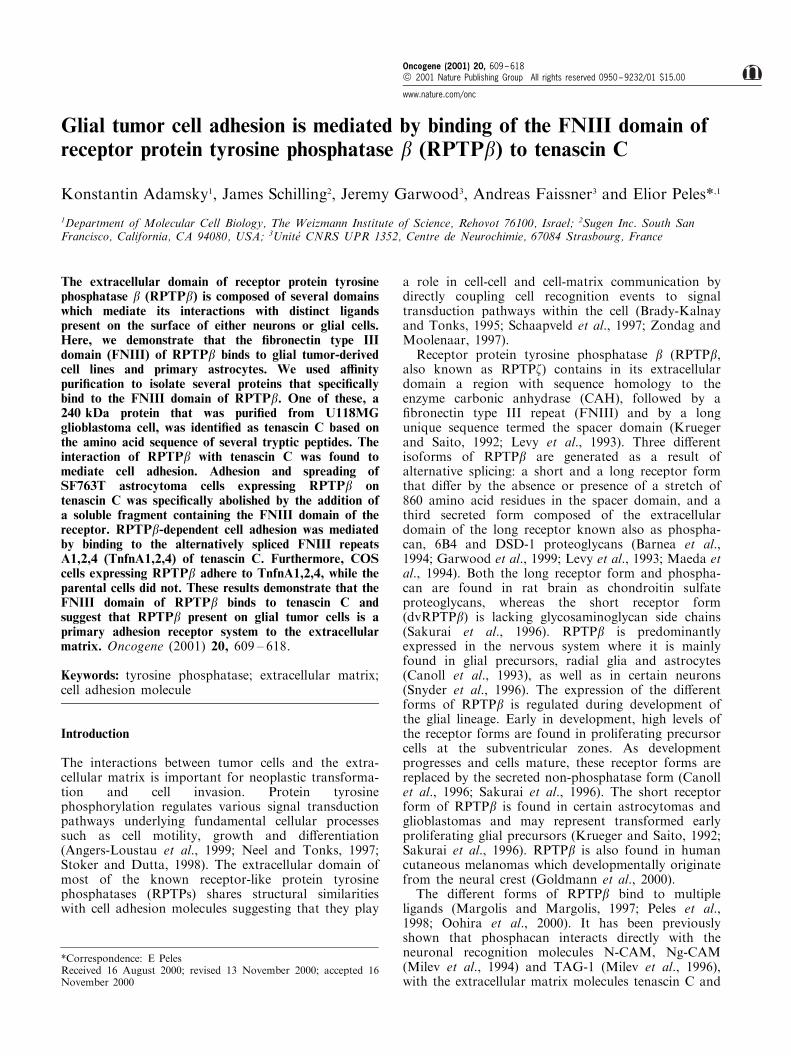
The extracellular domain of RPTPb is composed ofseveral domains that mediate its interactions withdistinct ligands that are present in neuronal and non-neuronal cells (Peles et al., 1998). To further identify suchligands, we produced soluble proteins that containeddi�erent subdomains of RPTPb fused to human IgG(Figure 1a). These recombinant proteins were used inbinding experiments using glioblastoma (U251MG,U373MG, C6) and astrocytoma (SF763T, U118MG)cell lines. As depicted in Figure 1b, all constructscontaining the FNIII domain of RPTPb bound well tothese cell lines. No binding was detected with thecarbonic anhydrase domain (bC), the spacer region(bS), or when an Fc-fusion protein containing thecarbonic anhydrase and the FNIII domains of therelated phosphatase RPTPg was used (Figure 1b anddata not shown). Similar results were obtained usingcultured primary astrocytes (Figure 2a). Staining ofGFAP-positive astrocytes with bF-Fc revealed a punc-tate staining pattern on the surface of the cells (Figure
2b). In agreement with our binding analysis, no stainingof astrocytes was detected with bC-Fc. In contrast, bC-Fc intensely labeled few oligodendrocytes that wereoccasionally present in the cultures. Given that contactinis expressed by oligodendrocytes (Koch et al., 1997), it islikely that this staining represents the interactionbetween the carbonic anhydrase domain of RPTPb andcontactin in these cells (Peles et al., 1995). Previous in situhybridization and immunohistochemistry data suggestedthat RPTPb is expressed by astrocytes (Canoll et al.,1996). As depicted in Figure 2b, we con®rmed thepresence of the receptor form of RPTPb in our culturesby staining with anti-RPTPb speci®c antibody, as well aswith a contactin-Fc fusion protein. Altogether, theseanalyses revealed the presence of a speci®c ligand for theFNIII domain of RPTPb on the surface of various glial-derived cell lines and primary cultured astrocytes.
Identification of a glial ligand for RPTPb
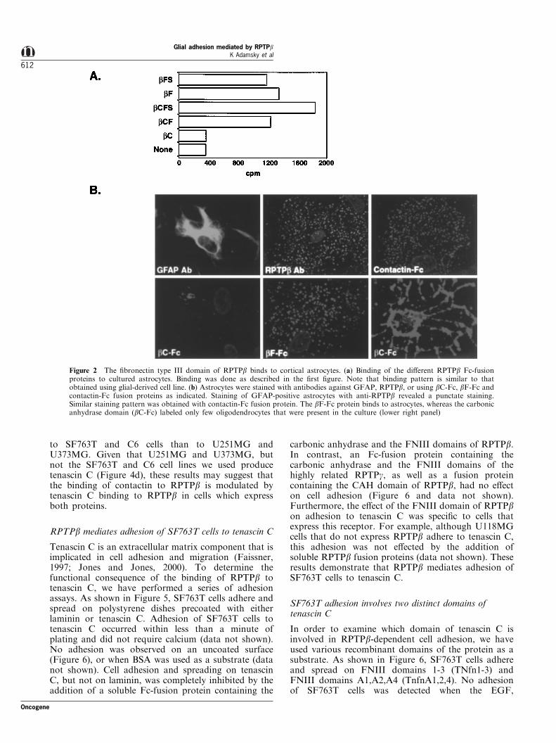
In order to isolate a ligand for the FNIII domain ofRPTPb from glial cells, membrane proteins wereprepared from 35S-methionine-labeled cell lines andincubated with bF-Fc or bC-Fc as a control (Figure3a). Several distinct sets of proteins speci®cally bound tothe bF-Fc a�nity matrix from the di�erent cell lines. InSF763T and C6 cells, bF-Fc precipitated four proteinswith the apparent molecular weight of 160, 170, 200 and210 kDa, while two proteins of 220 and 240 kDa weredetected from U138MG and U118MG cells. Theseproteins were not detected using bC-Fc, indicating thatthey interact speci®cally with the FNIII domain ofRPTPb. Furthermore, bF-Fc precipitated these proteinsonly from glial-derived, but not from several neuro-blastoma (neuro 2A and H4) or neuroendocrine (GH3)cell lines. In contrast, bC-Fc precipitated a 140 and a190 kDa proteins from GH3 that were identi®ed ascontactin and Caspr respectively (Peles et al., 1995,1997). We next used bF-Fc to purify the 240 kDaprotein from U118MG cells (Figure 3b). The 240 kDaprotein was excised from the gel, digested with trypsinand the resulting peptides were separated by HPLC(data not shown). Selected peptides were analysed byautomated Edman degradation. The sequence of one ofthe peptides (PVLSAEASTGETPNLGEVV) was iden-tical to positions 1151 ± 1169 of human tenascin C(accession #P24821). All other sequenced peptides werealso derived from human tenascin C (data not shown).Furthermore, we have con®rmed the identity of both the220 and 240 kDa proteins isolated by bF-Fc fromU118MG cells as tenascin C by immunoblot analysis(Figure 3c). In conclusion, the FNIII domain of RPTPbbinds to several distinct ligands present in glial cells, twoof which were identi®ed as isoforms of the extracellularmatrix protein tenascin C. The identity of the otherproteins is yet to be determined.
RPTPb is expressed in glial tumor cell lines
The identi®cation of tenascin C as a glial ligand forRPTPb raises the possibility that it may modulate the
Glial adhesion mediated by RPTPbK Adamsky et al
610
Oncogene

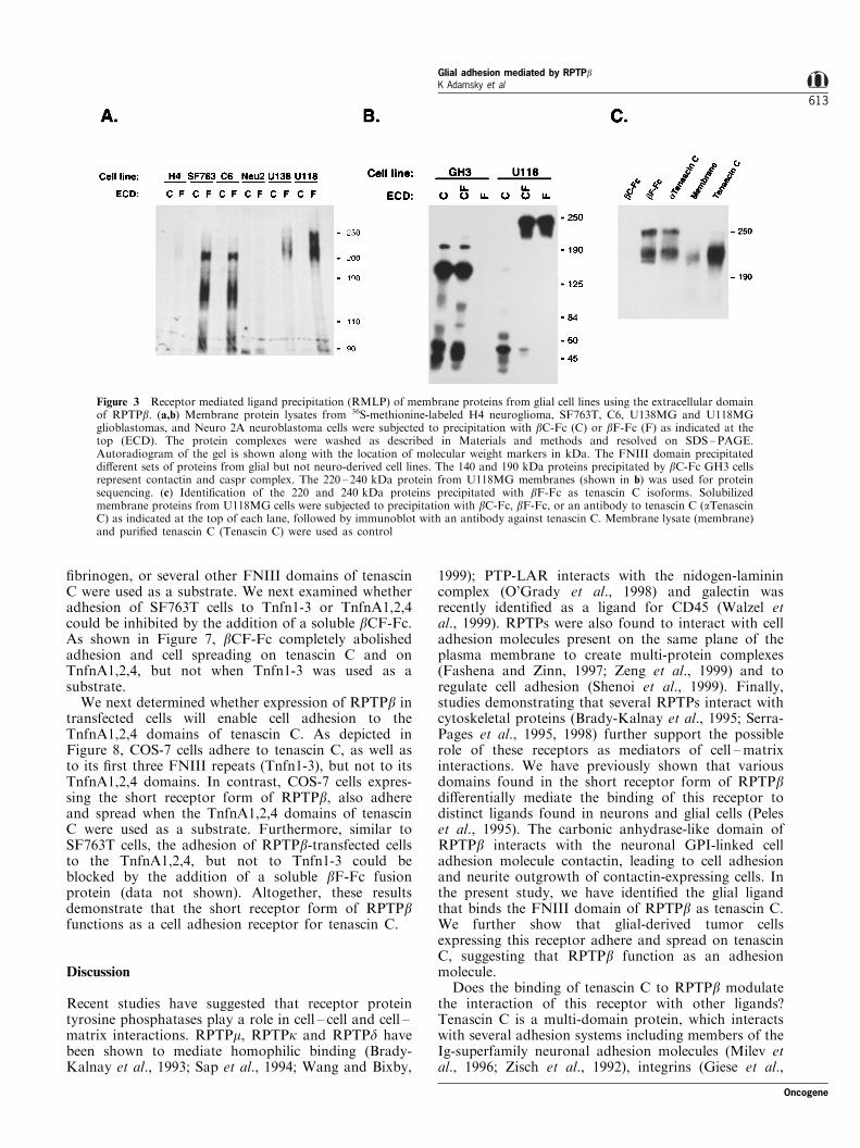
binding of RPTPb present in these cells with otherligands. We ®rst examined the expression of RPTPb inthe various cell lines. Immunoblot analysis revealedthat the short receptor form of RPTPb was found inU251MG, SF763, SF763T, U373MG, C6 and SF767T(Figure 4a). No expression of RPTPb transcript orprotein was detected in U118MG (Figure 4a and b) orU138MG cells (data not shown), the two cell linesfrom which tenascin C was precipitated by bF-Fc. Wenext compared the cell surface expression of RPTPb in
several cell lines using a speci®c antibody against itsextracellular domain with the binding of a solublecontactin-Fc, a known ligand of the carbonic anhy-drase domain (Peles et al., 1995). As depicted in Figure4c, strong binding of anti-RPTPb antibody wasdetected in SF763T, C6, U251MG and U373MG cells,indicating that RPTPb is expressed on the surface ofthese cells. However, in contrast to the antibodybinding which was equivalent in all RPTPb-expressingcells, we observed higher levels of Contactin-Fc binding
Figure 1 Binding of di�erent subdomains derived from the extracellular region of RPTPb. (a) Schematic presentation andexpression of the RPTPb Fc-fusion proteins. Abbreviations: C - carbonic anhydrase domain, F - ®bronectin type III, S - spacerdomain. Anti-human Fc immunoblot of the Fc-fusion proteins is shown on the right along with molecular weight markers in kDa.(b) Binding of the RPTPb Fc-fusion proteins to di�erent glial tumor-derived cell lines. Cells were incubated with the indicated Fc-fusion proteins. Bound proteins were detected using 125I-labeled protein A as described in Materials and methods. An average oftwo sets of experiments done in triplicates is shown
Oncogene
Glial adhesion mediated by RPTPbK Adamsky et al
611
to SF763T and C6 cells than to U251MG andU373MG. Given that U251MG and U373MG, butnot the SF763T and C6 cell lines we used producetenascin C (Figure 4d), these results may suggest thatthe binding of contactin to RPTPb is modulated bytenascin C binding to RPTPb in cells which expressboth proteins.
RPTPb mediates adhesion of SF763T cells to tenascin C
Tenascin C is an extracellular matrix component that isimplicated in cell adhesion and migration (Faissner,1997; Jones and Jones, 2000). To determine thefunctional consequence of the binding of RPTPb totenascin C, we have performed a series of adhesionassays. As shown in Figure 5, SF763T cells adhere andspread on polystyrene dishes precoated with eitherlaminin or tenascin C. Adhesion of SF763T cells totenascin C occurred within less than a minute ofplating and did not require calcium (data not shown).No adhesion was observed on an uncoated surface(Figure 6), or when BSA was used as a substrate (datanot shown). Cell adhesion and spreading on tenascinC, but not on laminin, was completely inhibited by theaddition of a soluble Fc-fusion protein containing the
carbonic anhydrase and the FNIII domains of RPTPb.In contrast, an Fc-fusion protein containing thecarbonic anhydrase and the FNIII domains of thehighly related RPTPg, as well as a fusion proteincontaining the CAH domain of RPTPb, had no e�ecton cell adhesion (Figure 6 and data not shown).Furthermore, the e�ect of the FNIII domain of RPTPbon adhesion to tenascin C was speci®c to cells thatexpress this receptor. For example, although U118MGcells that do not express RPTPb adhere to tenascin C,this adhesion was not e�ected by the addition ofsoluble RPTPb fusion proteins (data not shown). Theseresults demonstrate that RPTPb mediates adhesion ofSF763T cells to tenascin C.
SF763T adhesion involves two distinct domains oftenascin C
In order to examine which domain of tenascin C isinvolved in RPTPb-dependent cell adhesion, we haveused various recombinant domains of the protein as asubstrate. As shown in Figure 6, SF763T cells adhereand spread on FNIII domains 1-3 (TNfn1-3) andFNIII domains A1,A2,A4 (TnfnA1,2,4). No adhesionof SF763T cells was detected when the EGF,
Figure 2 The ®bronectin type III domain of RPTPb binds to cortical astrocytes. (a) Binding of the di�erent RPTPb Fc-fusionproteins to cultured astrocytes. Binding was done as described in the ®rst ®gure. Note that binding pattern is similar to thatobtained using glial-derived cell line. (b) Astrocytes were stained with antibodies against GFAP, RPTPb, or using bC-Fc, bF-Fc andcontactin-Fc fusion proteins as indicated. Staining of GFAP-positive astrocytes with anti-RPTPb revealed a punctate staining.Similar staining pattern was obtained with contactin-Fc fusion protein. The bF-Fc protein binds to astrocytes, whereas the carbonicanhydrase domain (bC-Fc) labeled only few oligodendrocytes that were present in the culture (lower right panel)
Glial adhesion mediated by RPTPbK Adamsky et al
612
Oncogene
®brinogen, or several other FNIII domains of tenascinC were used as a substrate. We next examined whetheradhesion of SF763T cells to Tnfn1-3 or TnfnA1,2,4could be inhibited by the addition of a soluble bCF-Fc.As shown in Figure 7, bCF-Fc completely abolishedadhesion and cell spreading on tenascin C and onTnfnA1,2,4, but not when Tnfn1-3 was used as asubstrate.
We next determined whether expression of RPTPb intransfected cells will enable cell adhesion to theTnfnA1,2,4 domains of tenascin C. As depicted inFigure 8, COS-7 cells adhere to tenascin C, as well asto its ®rst three FNIII repeats (Tnfn1-3), but not to itsTnfnA1,2,4 domains. In contrast, COS-7 cells expres-sing the short receptor form of RPTPb, also adhereand spread when the TnfnA1,2,4 domains of tenascinC were used as a substrate. Furthermore, similar toSF763T cells, the adhesion of RPTPb-transfected cellsto the TnfnA1,2,4, but not to Tnfn1-3 could beblocked by the addition of a soluble bF-Fc fusionprotein (data not shown). Altogether, these resultsdemonstrate that the short receptor form of RPTPbfunctions as a cell adhesion receptor for tenascin C.
Discussion
Recent studies have suggested that receptor proteintyrosine phosphatases play a role in cell ± cell and cell ±matrix interactions. RPTPm, RPTPk and RPTPd havebeen shown to mediate homophilic binding (Brady-Kalnay et al., 1993; Sap et al., 1994; Wang and Bixby,
1999); PTP-LAR interacts with the nidogen-laminincomplex (O'Grady et al., 1998) and galectin wasrecently identi®ed as a ligand for CD45 (Walzel etal., 1999). RPTPs were also found to interact with celladhesion molecules present on the same plane of theplasma membrane to create multi-protein complexes(Fashena and Zinn, 1997; Zeng et al., 1999) and toregulate cell adhesion (Shenoi et al., 1999). Finally,studies demonstrating that several RPTPs interact withcytoskeletal proteins (Brady-Kalnay et al., 1995; Serra-Pages et al., 1995, 1998) further support the possiblerole of these receptors as mediators of cell ±matrixinteractions. We have previously shown that variousdomains found in the short receptor form of RPTPbdi�erentially mediate the binding of this receptor todistinct ligands found in neurons and glial cells (Peleset al., 1995). The carbonic anhydrase-like domain ofRPTPb interacts with the neuronal GPI-linked celladhesion molecule contactin, leading to cell adhesionand neurite outgrowth of contactin-expressing cells. Inthe present study, we have identi®ed the glial ligandthat binds the FNIII domain of RPTPb as tenascin C.We further show that glial-derived tumor cellsexpressing this receptor adhere and spread on tenascinC, suggesting that RPTPb function as an adhesionmolecule.
Does the binding of tenascin C to RPTPb modulatethe interaction of this receptor with other ligands?Tenascin C is a multi-domain protein, which interactswith several adhesion systems including members of theIg-superfamily neuronal adhesion molecules (Milev etal., 1996; Zisch et al., 1992), integrins (Giese et al.,
Figure 3 Receptor mediated ligand precipitation (RMLP) of membrane proteins from glial cell lines using the extracellular domainof RPTPb. (a,b) Membrane protein lysates from 36S-methionine-labeled H4 neuroglioma, SF763T, C6, U138MG and U118MGglioblastomas, and Neuro 2A neuroblastoma cells were subjected to precipitation with bC-Fc (C) or bF-Fc (F) as indicated at thetop (ECD). The protein complexes were washed as described in Materials and methods and resolved on SDS±PAGE.Autoradiogram of the gel is shown along with the location of molecular weight markers in kDa. The FNIII domain precipitateddi�erent sets of proteins from glial but not neuro-derived cell lines. The 140 and 190 kDa proteins precipitated by bC-Fc GH3 cellsrepresent contactin and caspr complex. The 220 ± 240 kDa protein from U118MG membranes (shown in b) was used for proteinsequencing. (c) Identi®cation of the 220 and 240 kDa proteins precipitated with bF-Fc as tenascin C isoforms. Solubilizedmembrane proteins from U118MG cells were subjected to precipitation with bC-Fc, bF-Fc, or an antibody to tenascin C (aTenascinC) as indicated at the top of each lane, followed by immunoblot with an antibody against tenascin C. Membrane lysate (membrane)and puri®ed tenascin C (Tenascin C) were used as control
Oncogene
Glial adhesion mediated by RPTPbK Adamsky et al
613
1996; Phillips et al., 1998; Prieto et al., 1993; Schnappet al., 1995; Yokosaki et al., 1998), phosphacan(Barnea et al., 1994; Grumet et al., 1994; Milev etal., 1995, 1997) and the short receptor form of RPTPb(this study). Both tenascin C and RPTPb interact withcontactin (Peles et al., 1995; Revest et al., 1999; Zischet al., 1992), suggesting that the binding of each pair(i.e., RPTPb-contactin, RPTPb-tenascin and contactin-tenascin) could potentially be regulated by the presenceof the third component. Whether such regulationoccurs competitively or cooperatively may depend onwhich speci®c domain within each protein is directlyinvolved in binding. It has been shown that the ®rst Igdomains in contactin mediate its binding to bothtenascin C and phosphacan (Revest et al., 1999; Zischet al., 1992). In contrast, contactin, phosphacan and
RPTPb bind to distinct sites in tenascin C. Whilecontactin binds to the ®fth and sixth ®bronectinrepeats of tenascin C (TNfn5-6) (Jones and Jones,2000), and phosphacan binds to its ®brinogen domain(Milev et al., 1997), the short receptor for of RPTPbinteracts with TNfnA1,2,4. We have shown that twodistinct sites in RPTPb, the carbonic anhydrase and itsFNIII domains mediate its binding to contactin andtenascin C, respectively (Peles et al., 1995; and thisstudy). However, although RPTPb could potentiallybind contactin and tenascin simultaneously, we haveobserved reduced binding of contactin to cells whichexpress both RPTPb and tenascin C. This resultsuggests that the presence of tenascin may negativelyregulate the binding of contactin to RPTPb, and argueagainst the presence of a stable tertiary complex. This
Figure 4 RPTPb is expressed in human glial tumor-derived cell lines. (a) Cell lysates from the various cell lines were subjected toimmunoprecipitation and immunoblotting with anti- RPTPb antibody. The short receptor form of RPTPb appears as a doublet onthe gel. Molecular weight markers are shown in kDa on the left. (b) RT±PCR analysis of RPTPb expression. mRNA from humanhippocampus, isolated astrocytes, brain, or several glioblastoma cell lines was extracted and used as a template to amplify RPTPbor RPTPg as indicated. (c) Binding of antibody directed to the extracellular region of RPTPb. The di�erent indicated cell lines wereincubated with pre-immune serum as control (white bars), antibody against the extracellular region of RPTPb (black bars), or withhuman contactin-Fc fusion protein (gray bars). Bound proteins were detected using 125I-labeled anti mouse or protein A asdescribed in Material and methods. An average of two sets of experiments done in triplicates is shown. (d) Expression of tenascin Cin the various cell lines. Immunoprecipitation and immunoblot analyses were done using antibody against tenascin C as described in(a) for RPTPb
Glial adhesion mediated by RPTPbK Adamsky et al
614
Oncogene

is further supported by recent ®ndings which havedetected contactin-phosphacan and contactin-tenascinC, but not a tertiary complex that contains all of these
molecules in rat brain (Revest et al., 1999). Takentogether, these results suggest that the function of theshort receptor form of RPTPb in cell adhesion is highlyregulated by a complex interplay of other cell adhesionmolecules and extracellular matrix components such astenascin C.
We show that expression of the short receptor formof RPTPb in COS-7 cells results in adhesion of thesecells to the TNfnA1,2,4 domains of tenascin C,indicating that this phosphatase functions as areceptor system for cell adhesion. Previous experimentsshowed that the third FNIII repeat of tenascin Cmediates cell binding through interaction with integ-rins (Prieto et al., 1993; Schnapp et al., 1995;Yokosaki et al., 1998). These interactions were alsoreported to be important for astrocytes adhesion andastrocytoma and glioma migration (Giese et al., 1996;Phillips et al., 1998). Using di�erent domains oftenascin C as a substrate, we have found that adhesionof SF763T cells is mediated by TNfn1-3 andTNfnA1,2,4. Although these two distinct sites areinvolved in SF763T adhesion, only the adhesion onTnfnA1,2,4 could be inhibited by the FNIII domain of
Figure 5 RPTPb-dependent adhesion of SF763T cells totenascin C. Polystyrene dish that had been precoated withtenascin C (TnC; upper panels), or laminin (Lam; lower panels)was further incubated with an Fc-fusion containing the carbonicanhydrase and the FNIII domains of RPTPb (bCF), RPTPg(gCF), the FNIII domain of RPTPb (bF), or BSA (7) asindicated. SF763T cells (16105 cells) were placed in the centralregion of the dish for 1 h and non-adhered cells were removed bygentle washing. The plate was ®xed and viewed using invertedmicroscope. Magni®cation:620
Figure 6 Adhesion of SF763T cells is mediated by two distinct sites in tenascin C. (a) Schematic representation of tenascin C andthe di�erent recombinant tenascin C fragments used in this study. (b) Adhesion of SF763T cells to di�erent tenascin C domains. Theindicated fragments (10 mg/ml) were bound to a polystyrene dish and adhesion assay was carried out exactly as described in thelegend to Figure 5. Two distinct regions of tenascin C FNIII repeats mediated cell adhesion. While Tnfn1-3 is found in all knowntenascin C proteins, TnfnA1,2,4 are alternatively spliced FNIII repeats that are inserted between the ®fth and the sixth FNIIIrepeats only in several isoforms of tenascin C. Magni®cation:620
Oncogene
Glial adhesion mediated by RPTPbK Adamsky et al
615
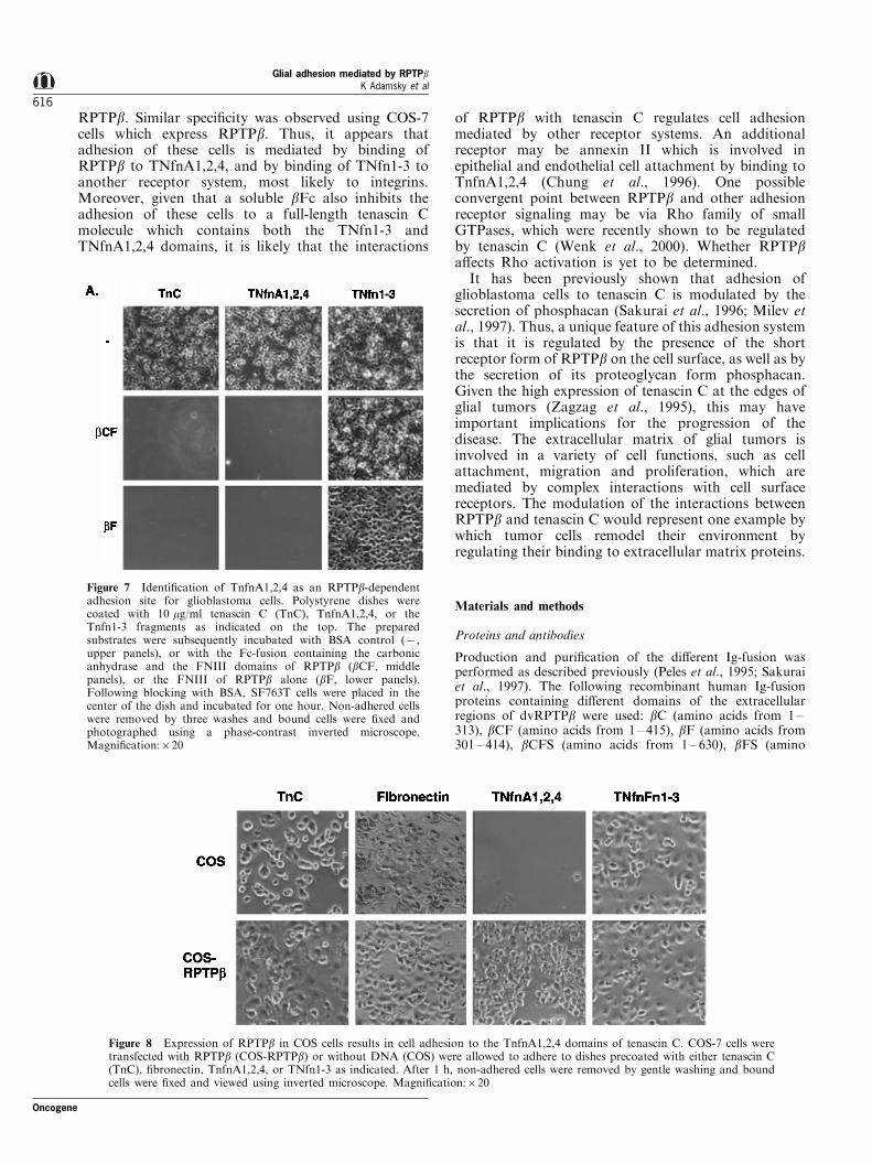
RPTPb. Similar speci®city was observed using COS-7cells which express RPTPb. Thus, it appears thatadhesion of these cells is mediated by binding ofRPTPb to TNfnA1,2,4, and by binding of TNfn1-3 toanother receptor system, most likely to integrins.Moreover, given that a soluble bFc also inhibits theadhesion of these cells to a full-length tenascin Cmolecule which contains both the TNfn1-3 andTNfnA1,2,4 domains, it is likely that the interactions
of RPTPb with tenascin C regulates cell adhesionmediated by other receptor systems. An additionalreceptor may be annexin II which is involved inepithelial and endothelial cell attachment by binding toTnfnA1,2,4 (Chung et al., 1996). One possibleconvergent point between RPTPb and other adhesionreceptor signaling may be via Rho family of smallGTPases, which were recently shown to be regulatedby tenascin C (Wenk et al., 2000). Whether RPTPba�ects Rho activation is yet to be determined.
It has been previously shown that adhesion ofglioblastoma cells to tenascin C is modulated by thesecretion of phosphacan (Sakurai et al., 1996; Milev etal., 1997). Thus, a unique feature of this adhesion systemis that it is regulated by the presence of the shortreceptor form of RPTPb on the cell surface, as well as bythe secretion of its proteoglycan form phosphacan.Given the high expression of tenascin C at the edges ofglial tumors (Zagzag et al., 1995), this may haveimportant implications for the progression of thedisease. The extracellular matrix of glial tumors isinvolved in a variety of cell functions, such as cellattachment, migration and proliferation, which aremediated by complex interactions with cell surfacereceptors. The modulation of the interactions betweenRPTPb and tenascin C would represent one example bywhich tumor cells remodel their environment byregulating their binding to extracellular matrix proteins.
Materials and methods
Proteins and antibodies
Production and puri®cation of the di�erent Ig-fusion wasperformed as described previously (Peles et al., 1995; Sakuraiet al., 1997). The following recombinant human Ig-fusionproteins containing di�erent domains of the extracellularregions of dvRPTPb were used: bC (amino acids from 1 ±313), bCF (amino acids from 1 ± 415), bF (amino acids from301 ± 414), bCFS (amino acids from 1 ± 630), bFS (amino
Figure 7 Identi®cation of TnfnA1,2,4 as an RPTPb-dependentadhesion site for glioblastoma cells. Polystyrene dishes werecoated with 10 mg/ml tenascin C (TnC), TnfnA1,2,4, or theTnfn1-3 fragments as indicated on the top. The preparedsubstrates were subsequently incubated with BSA control (7,upper panels), or with the Fc-fusion containing the carbonicanhydrase and the FNIII domains of RPTPb (bCF, middlepanels), or the FNIII of RPTPb alone (bF, lower panels).Following blocking with BSA, SF763T cells were placed in thecenter of the dish and incubated for one hour. Non-adhered cellswere removed by three washes and bound cells were ®xed andphotographed using a phase-contrast inverted microscope.Magni®cation:620
Figure 8 Expression of RPTPb in COS cells results in cell adhesion to the TnfnA1,2,4 domains of tenascin C. COS-7 cells weretransfected with RPTPb (COS-RPTPb) or without DNA (COS) were allowed to adhere to dishes precoated with either tenascin C(TnC), ®bronectin, TnfnA1,2,4, or TNfn1-3 as indicated. After 1 h, non-adhered cells were removed by gentle washing and boundcells were ®xed and viewed using inverted microscope. Magni®cation:620
Glial adhesion mediated by RPTPbK Adamsky et al
616
Oncogene

acids from 301 ± 630), and bS (amino acids from 415 ± 630).The generation of recombinant tenascin C fragments waspreviously described (Scholze et al., 1996). Puri®ed tenascinwas a generous gift from Dr Martin Grumet.Monoclonal antibodies to RPTPb were purchased from
Transduction Laboratories, Cy3 and FITC-conjugated sec-ondary antibodies were from Jackson laboratory, anti GFAPfrom Sigma and anti-tenascin C antibody (AB1906) fromChemicon. Polyclonal antibody to the extracellular domain ofRPTPb was obtained by immunizing mice with puri®ed bCF-Ig protein and clearing the serum from anti-human Fcantibodies by passing it through a normal human IgG-Sepharose column.
Cell culture
Growth and origin of the di�erent cell lines used in this studywere previously described (Peles et al., 1995). For culturingrat astrocytes, the cortex of newborn rat pups was dissectedand dissociated by incubation with 0.25% trypsin (Gibco)and collagenase (Sigma) for 20 min. Cells were washedseveral times in DMEM+10% fetal calf serum and trituratedwith a pasteur pipette. The cells were plated at 100 000 cells/cm2 in 75 cm2 Primaria ¯asks (Falcon) and grown inDMEM+10% fetal calf serum. After 8 days, con¯uentcultures were shaken for 14 h at 37C8 to remove oligoden-drocyte precursors and the attached cells were trypsinizedand replated. For immuno¯uorescence, cells were grown onglass coverslips precoated with 10 mg/ml polylysine for 3 daysbefore the experiment.
Protein purification and sequencing
Analytical scale puri®cation using the Ig-fusion proteins asa�nity reagents was performed from 35S methionine-labeledcells as previously described (Peles et al., 1997). In certaincases the complexes were resolved on an SDS gel andsubjected to Western blotting with anti-tenascin C antibody.Immunoprecipitation and immunoblotting (Poliak et al.,1999) and protein puri®cation were done essentially asdescribed previously (Peles et al., 1995).
Ig fusion proteins and antibody binding and immunofluorescence
Binding of Ig-fusion proteins to cultured cell lines was doneas described previously (Peles et al., 1995). Similar protocolwas used for antibody binding experiments but 125I-labeledanti-mouse IgG was used instead of protein-A. For stainingexperiments, astrocytes grown on coverslips in a 24-well dishwere incubated with conditioned media containing thevarious Ig-fusion proteins for 2 h at ambient temperature.The dish was placed on ice and unbound material wasremoved by three consecutive washes with cold PBS. Slideswere ®xed with 4% paraformaldehyde in 0.1 M phosphatebu�er for 30 min and subsequently stained with Cy3-conjugated anti-human Fc antibody as described previously(Poliak et al., 1999). Immuno¯uorescence slides were viewedand analysed using a Deltavision wide-®eld deconvolutionsystem (Applied Precision) connected to Zeiss ¯uorescencemicroscope.
Cell adhesion assays
SF763T cells were removed from the tissue culture plate bywashing with PBS containing 10 mM EDTA, washed oncewith DMEM containing 4% BSA and resuspended inDMEM/F12. 16105 cells in 0.5 ml of binding bu�er wereplaced in the central region of 35 mm polystyrene dish thathad been precoated with the various proteins. Substrates forcell adhesion were prepared by incubating 3 ml of protein for3 h followed by washes and blocking with BSA as previouslydescribed (Grumet et al., 1994). When double coating wasused, Ig-fusion proteins were applied before the blockingstep.
AcknowledgmentsThis work was supported by the Dorot foundation fromthe Israel Academy of Sciences and Humanities, the YadAbraham Research Center for Cancer Diagnostics andTherapy, and a Research Career Development Award fromthe Israel Cancer Research Fund. E Peles is an Incumbentof the Madeleine Haas Russell Career Development Chair.
References
Angers-Loustau A, Cote JF and Tremblay ML. (1999).Biochem. Cell. Biol., 77, 493 ± 505.
Barnea G, Grumet M, Milev P, Silvennoinen O, Levy JB, SapJ and Schlessinger J. (1994). J. Biol. Chem., 269, 14349 ±14352.
Brady-Kalnay SM, Flint AJ and Tonks NK. (1993). J. Cell.Biol., 122, 961 ± 972.
Brady-Kalnay SM, Rimm DL and Tonks NK. (1995). J.Cell. Biol., 130, 977 ± 986.
Brady-Kalnay SM and Tonks NK. (1995). Curr. Opin. Cell.Biol., 7, 650 ± 657.
Canoll PD, Barnea G, Levy JB, Sap J, Ehrlich M,Silvennoinen O, Schlessinger J and Musacchio JM.(1993). Brain Res. Dev. Brain Res., 75, 293 ± 298.
Canoll PD, Petanceska S, Schlessinger J and Musacchio JM.(1996). J. Neurosci. Res., 44, 199 ± 215.
Chung CY, Murphy-Ullrich JE and Erickson HP. (1996).Mol. Biol. Cell., 7, 883 ± 892.
Faissner A. (1997). Cell Tissue Res., 290, 385 ± 393.Fashena SJ and Zinn K. (1997). Mol. Cell. Biol., 17, 6859 ±
6867.
Garwood J, Schnadelbach O, Clement A, Schutte K, Bach Aand Faissner A. (1999). J. Neurosci., 19, 3888 ± 3899.
Giese A, Loo MA, Norman SA, Treasurywala S and BerensME. (1996). J. Cell. Sci., 109, 2161 ± 2168.
Goldmann T, Otto F and Vollmer E. (2000). FoliaHistochem. Cytobiol., 38, 19 ± 20.
Grumet M, Milev P, Sakurai T, Karthikeyan L, Bourdon M,Margolis RK and Margolis RU. (1994). J. Biol. Chem.,269, 12142 ± 12146.
Jones FS and Jones PL. (2000). Am. J. Respir. Crit. CareMed., 161, 2086 ± 2091.
Koch T, Brugger T, Bach A, Gennarini G and Trotter J.(1997). Glia, 19, 199 ± 212.
Krueger NX and Saito H. (1992). Proc. Natl. Acad. Sci.USA, 89, 7417 ± 7421.
Levy JB, Canoll PD, Silvennoinen O, Barnea G, Morse B,Honegger AM, Huang JT, Cannizzaro LA, Park SH,Druck T and Schlessinger J. (1993). J. Biol. Chem., 268,10573 ± 10581.
Maeda N, Hamanaka H, Shintani T, Nishiwaki T and NodaM. (1994). FEBS Lett., 354, 67 ± 70.
Oncogene
Glial adhesion mediated by RPTPbK Adamsky et al
617

Maeda N, Nishiwaki T, Shintani T, Hamanaka H and NodaM. (1996). J. Biol. Chem., 271, 21446 ± 21452.
Margolis RU and Margolis RK. (1997). Cell Tissue Res.,290, 343 ± 348.
Milev P, Friedlander DR, Sakurai T, Karthikeyan L, FladM, Margolis RK, Grumet M and Margolis RU. (1994). J.Cell. Biol., 127, 1703 ± 1715.
Milev P, Meyer-Puttlitz B, Margolis RK and Margolis RU.(1995). J. Biol. Chem., 270, 24650 ± 24653.
Milev P, Maurel P, Haring M, Margolis RK and MargolisRU. (1996). J. Biol. Chem., 271, 15716 ± 15723.
Milev P, Fische D, Haring M, Schulthess T & Margolis RK.(1997). J. Biol. Chem., 272, 15501 ± 15509.
Milev P, Chiba A, Haring M, Rauvala H, Schachner M,Ranscht B, Margolis RK and Margolis RU. (1998a). J.Biol. Chem., 273, 6998 ± 7005.
Milev P, Monnerie H, Popp S, Margolis RK and MargolisRU. (1998b). J. Biol. Chem., 273, 21439 ± 21442.
Neel BG and Tonks NK. (1997). Curr. Opin. Cell. Biol., 9,193 ± 204.
O'Grady P, Thai TC and Saito H. (1998). J. Cell. Biol., 141,1675 ± 1684.
Oohira A, Matsui F, Tokita Y, Yamauchi S and Aono S.(2000). Arch. Biochem. Biophys., 374, 24 ± 34.
Peles E, Nati M, Campbel PL, Sakurai T, Martinez R, Lev S,Clary DO, Schilling J, Barnea G, Plowman GD andSchlessinger J. (1995). Cell, 82, 251 ± 260.
Peles E, Nativ M, Lustig M, Grumet M, Schilling J, MartinezR, Plowman GD and Schlessinger J. (1997). EMBO J., 16,978 ± 988.
Peles E, Schlessinger J and Grumet M. (1998). TrendsBiochem. Sci., 23, 121 ± 124.
Phillips GR, Krushel LA and Crossin KL. (1998). Mol. Biol.Cell., 9, 1425 ± 1435.
Poliak S, Gollan L, Martinez R, Custer A, Einheber S, SalzerJ, Trimmer J, Shrager P and Peles E. (1999). Neuron, 24,1037 ± 1047.
Prieto AL, Edelman GM and Crossin KL. (1993). Proc. Natl.Acad. Sci. USA, 90, 10154 ± 10158.
Revest JM, Faivre-Sarrailh C, Maeda N, Noda M,Schachner M and Rougon G. (1999). Eur. J. Neurosci.,11, 1134 ± 1147.
Sakurai T, Friedlander DR and Grumet M. (1996). J.Neurosci. Res., 43, 694 ± 706.
Sakurai T, Lustig M, Nativ M, Hemperly JJ, Schlessinger J,Peles E and Grumet M. (1997). J. Cell. Biol., 136, 907 ±918.
Sap J, Jiang YP, Friedlander D, Grumet M and SchlessingerJ. (1994). Mol. Cell. Biol., 14, 1 ± 9.
Schaapveld R, Wieringa B and Hendriks W. (1997). Mol.Biol. Rep., 24, 247 ± 262.
Schnapp LM, Hatch N, Ramos DM, Klimanskaya IV,Sheppard D and Pytela R. (1995). J. Biol. Chem., 270,23196 ± 23202.
Scholze A, Gotz B and Faissner A. (1996). Int. J. Dev.Neurosci., 14, 315 ± 329.
Serra-Pages C, Kedersha NL, Fazikas L, Medley Q, DebantA and Streuli M. (1995). EMBO J., 14, 2827 ± 2838.
Serra-Pages C, Medley QG, Tang M, Hart A and Streuli M.(1998). J. Biol. Chem., 273, 15611 ± 15620.
Shenoi H, Seavitt J, Zheleznyak A, Thomas ML and BrownEJ. (1999). J. Immunol., 162, 7120 ± 7127.
Snyder SE, Li J, Schauwecker PE, McNeill TH and SaltonSR. (1996). Brain Res. Mol. Brain Res., 40, 79 ± 96.
Stoker A and Dutta R. (1998). Bioessays, 20, 463 ± 472.Walzel H, Schulz U, Neels P and Brock J. (1999). Immunol.
Lett., 67, 193 ± 202.Wang J and Bixby JL. (1999). Mol. Cell. Neurosci., 14, 370 ±
384.Wenk MB, Midwood KS and Schwarzbauer JE. (2000). J.
Cell. Biol., 150, 913 ± 919.Xiao ZC, Bartsch U, Margolis RK, Rougon G, Montag D
and Schachner M. (1997). J. Biol. Chem., 272, 32092 ±32101.
Yokosaki Y, Matsuura N, Higashiyama S, Murakami I,Obara M, Yamakido M, Shigeto N, Chen J and SheppardD. (1998). J. Cell. Sci., 111, 1095 ± 1104.
Zagzag D, Friedlander DR, Miller DC, Dosik J, CangiarellaJ, Kostianovsky M, Cohen H, Grumet M and Greco MA.(1995). Cancer Res., 55, 907 ± 914.
Zeng L, D'Alessandri L, Kalousek MB, Vaughan L andPallen CJ. (1999). J. Cell. Biol., 147, 707 ± 714.
Zisch AH, D'Alessandri L, Ranscht B, Falchetto R,Winterhalter KH and Vaughan L. (1992). J. Cell. Biol.,119, 203 ± 213.
Zondag GC and Moolenaar WH. (1997). Biochimie, 79,477 ± 483.
Glial adhesion mediated by RPTPbK Adamsky et al
618
Oncogene
Related Documents