Genotypic linkages of VP4, VP6, VP7, NSP4, NSP5 genes of rotaviruses circulating among children with acute gastroenteritis in Thailand Pattara Khamrin a,b , Niwat Maneekarn b , Rungnapa Malasao c , Tuan Anh Nguyen d , Shinichi Ishida a , Shoko Okitsu e , Hiroshi Ushijima a, * a Aino Health Science Center, Aino University, Tokyo, Japan b Department of Microbiology, Faculty of Medicine, Chiang Mai University, Chiang Mai, Thailand c School of Medical Sciences, Naresuan University, Phayao, Thailand d Children’s Hospital 1, Ho Chi Minh City, Viet Nam e Aino Health Science Center, Aino college, Tokyo, Japan 1. Introduction Group A rotaviruses are one of the major causes of severe gastroenteritis in young children in both developed and develop- ing countries. Globally, rotavirus infections account for an estimated of 22% (ranged from 17% to 28%) of children hospitalized with diarrhea, and are associated with 454,000–705,000 deaths annually among children under 5 years of age (Estes and Kapikian, 2007; Parashar et al., 2006). Rotavirus belongs to the Reoviridae family which contains 11 segments of double-stranded RNA genome. The two outer-layer proteins VP7 and VP4 form the basis of the current dual classification system of group A rotavirus into G and P genotypes (Estes and Kapikian, 2007). To date, at least 23 G (G1–G23) and 31 P (P[1]–P[31]) genotypes of rotaviruses have been identified globally, with various combinations of G and P genotypes (Khamrin et al., 2007a; Steyer et al., 2007; Matthijns- sens et al., 2008b; Schumann et al., 2009; Ursu et al., 2009). Based on reactivity of subgroup (SG)-specific monoclonal antibodies (MAbs) to VP6 protein, group A rotavirus could be classified into SG I, SG II, SG (I + II), and SG non-(I + II) (Greenberg et al., 1983a,b; Hoshino et al., 1987). Sequence analyses of the NSP4 genes revealed that at least six distinct NSP4 genetic groups exist among human and animal rotaviruses, termed genetic groups A to F (Horie et al., 1997; Ciarlet et al., 2000; Khamrin et al., 2008). The precise role(s) of the NSP5 proteins encoded by gene 11 has only been partially characterized and a formal classification system based on the NSP5 gene of rotavirus has not yet been well established. In recent decades, extensive epidemiological studies of rotavirus have been carried out worldwide and several unusual or novel genotypes have been detected increasingly (Santos and Hoshino, 2005; Khamrin et al., 2007a; Gray et al., 2008). Therefore, in order to assign a rotavirus strain to one of the established genotypes or as a new genotype, a standard procedure has been proposed by the Rotavirus Classification Working Group (RCWG) (Matthijnssens et al., 2008b). In Thailand, rotavirus is the leading pathogen that causes diarrhea in children, and it is responsible for about 20–61% of diarrheal diseases in hospitalized cases (Jiraphongsa et al., 2005; Khamrin et al., 2006b, 2007b; Maneekarn and Ushijima, 2000; Kittigul et al., 2009; Pongsuwannna et al., 2010). The study of Infection, Genetics and Evolution 10 (2010) 467–472 ARTICLE INFO Article history: Received 9 December 2009 Received in revised form 2 March 2010 Accepted 2 March 2010 Available online 17 March 2010 Keywords: Rotavirus Genotype VP4 VP6 VP7 NSP4 NSP5 Thailand ABSTRACT Rotavirus is the main cause of acute viral gastroenteritis in infants and young children worldwide. Surveillance of group A rotavirus has been conducted in Chiang Mai, Thailand since 1987 up to 2004 and those studies revealed that group A rotavirus was responsible for about 20-61% of diarrheal diseases in hospitalized cases. In this study, we reported the continuing surveillance of group A rotavirus in 2005 and found that group A rotavirus was detected in 43 out of 147 (29.3%) stool samples. Five different G and P genotype combinations were detected, G1P[8] (27 strains), G2P[4] (12 strains), G9P[8] (2 strains), G3P[8] (1 strain), and G3P[10] (1 strain). In addition, analysis of their genotypic linkages of G (VP7), P (VP4), I (VP6), E (NSP4), and H (NSP5) genotypes demonstrated that the rotaviruses circulating in Chiang Mai, Thailand carried 3 unique linkage patterns. The G1P[8], G3P[8], and G9P[8] strains carried their VP6, NSP4, NSP5 genotypes of I1, E1, H1, respectively. The G2P[4] strains were linked with I2, E2, H2 genotypes, while an uncommon G3P[10] genotype carried unique genotypes of I8, E3 and H6. These findings provide the overall picture of genotypic linkage data of rotavirus strains circulating in Chiang Mai, Thailand. ß 2010 Elsevier B.V. All rights reserved. * Corresponding author. Tel.: +81 3 3486 8481; fax: +81 3 3486 8481. E-mail address: [email protected] (H. Ushijima). Contents lists available at ScienceDirect Infection, Genetics and Evolution journal homepage: www.elsevier.com/locate/meegid 1567-1348/$ – see front matter ß 2010 Elsevier B.V. All rights reserved. doi:10.1016/j.meegid.2010.03.002

Welcome message from author
This document is posted to help you gain knowledge. Please leave a comment to let me know what you think about it! Share it to your friends and learn new things together.
Transcript

Infection, Genetics and Evolution 10 (2010) 467–472
Genotypic linkages of VP4, VP6, VP7, NSP4, NSP5 genes of rotaviruses circulatingamong children with acute gastroenteritis in Thailand
Pattara Khamrin a,b, Niwat Maneekarn b, Rungnapa Malasao c, Tuan Anh Nguyen d,Shinichi Ishida a, Shoko Okitsu e, Hiroshi Ushijima a,*a Aino Health Science Center, Aino University, Tokyo, Japanb Department of Microbiology, Faculty of Medicine, Chiang Mai University, Chiang Mai, Thailandc School of Medical Sciences, Naresuan University, Phayao, Thailandd Children’s Hospital 1, Ho Chi Minh City, Viet Name Aino Health Science Center, Aino college, Tokyo, Japan
A R T I C L E I N F O
Article history:
Received 9 December 2009
Received in revised form 2 March 2010
Accepted 2 March 2010
Available online 17 March 2010
Keywords:
Rotavirus
Genotype
VP4
VP6
VP7
NSP4
NSP5
Thailand
A B S T R A C T
Rotavirus is the main cause of acute viral gastroenteritis in infants and young children worldwide.
Surveillance of group A rotavirus has been conducted in Chiang Mai, Thailand since 1987 up to 2004 and
those studies revealed that group A rotavirus was responsible for about 20-61% of diarrheal diseases in
hospitalized cases. In this study, we reported the continuing surveillance of group A rotavirus in 2005
and found that group A rotavirus was detected in 43 out of 147 (29.3%) stool samples. Five different G and
P genotype combinations were detected, G1P[8] (27 strains), G2P[4] (12 strains), G9P[8] (2 strains),
G3P[8] (1 strain), and G3P[10] (1 strain). In addition, analysis of their genotypic linkages of G (VP7), P
(VP4), I (VP6), E (NSP4), and H (NSP5) genotypes demonstrated that the rotaviruses circulating in Chiang
Mai, Thailand carried 3 unique linkage patterns. The G1P[8], G3P[8], and G9P[8] strains carried their VP6,
NSP4, NSP5 genotypes of I1, E1, H1, respectively. The G2P[4] strains were linked with I2, E2, H2
genotypes, while an uncommon G3P[10] genotype carried unique genotypes of I8, E3 and H6. These
findings provide the overall picture of genotypic linkage data of rotavirus strains circulating in Chiang
Mai, Thailand.
� 2010 Elsevier B.V. All rights reserved.
Contents lists available at ScienceDirect
Infection, Genetics and Evolution
journal homepage: www.elsev ier .com/ locate /meegid
1. Introduction
Group A rotaviruses are one of the major causes of severegastroenteritis in young children in both developed and develop-ing countries. Globally, rotavirus infections account for anestimated of 22% (ranged from 17% to 28%) of children hospitalizedwith diarrhea, and are associated with 454,000–705,000 deathsannually among children under 5 years of age (Estes and Kapikian,2007; Parashar et al., 2006). Rotavirus belongs to the Reoviridae
family which contains 11 segments of double-stranded RNAgenome. The two outer-layer proteins VP7 and VP4 form the basisof the current dual classification system of group A rotavirus into Gand P genotypes (Estes and Kapikian, 2007). To date, at least 23 G(G1–G23) and 31 P (P[1]–P[31]) genotypes of rotaviruses havebeen identified globally, with various combinations of G and Pgenotypes (Khamrin et al., 2007a; Steyer et al., 2007; Matthijns-sens et al., 2008b; Schumann et al., 2009; Ursu et al., 2009). Basedon reactivity of subgroup (SG)-specific monoclonal antibodies
* Corresponding author. Tel.: +81 3 3486 8481; fax: +81 3 3486 8481.
E-mail address: [email protected] (H. Ushijima).
1567-1348/$ – see front matter � 2010 Elsevier B.V. All rights reserved.
doi:10.1016/j.meegid.2010.03.002
(MAbs) to VP6 protein, group A rotavirus could be classified into SGI, SG II, SG (I + II), and SG non-(I + II) (Greenberg et al., 1983a,b;Hoshino et al., 1987). Sequence analyses of the NSP4 genesrevealed that at least six distinct NSP4 genetic groups exist amonghuman and animal rotaviruses, termed genetic groups A to F (Horieet al., 1997; Ciarlet et al., 2000; Khamrin et al., 2008). The preciserole(s) of the NSP5 proteins encoded by gene 11 has only beenpartially characterized and a formal classification system based onthe NSP5 gene of rotavirus has not yet been well established.
In recent decades, extensive epidemiological studies ofrotavirus have been carried out worldwide and several unusualor novel genotypes have been detected increasingly (Santos andHoshino, 2005; Khamrin et al., 2007a; Gray et al., 2008). Therefore,in order to assign a rotavirus strain to one of the establishedgenotypes or as a new genotype, a standard procedure has beenproposed by the Rotavirus Classification Working Group (RCWG)(Matthijnssens et al., 2008b).
In Thailand, rotavirus is the leading pathogen that causesdiarrhea in children, and it is responsible for about 20–61% ofdiarrheal diseases in hospitalized cases (Jiraphongsa et al., 2005;Khamrin et al., 2006b, 2007b; Maneekarn and Ushijima, 2000;Kittigul et al., 2009; Pongsuwannna et al., 2010). The study of

P. Khamrin et al. / Infection, Genetics and Evolution 10 (2010) 467–472468
rotavirus infection in Thailand was initially performed approxi-mately two decades ago. The accumulated data from thosesurveillance studies indicate that the predominant serotype/genotype of rotaviruses in each epidemic season change overtime (Maneekarn and Ushijima, 2000). In Chiang Mai, Thailand,rotavirus surveillance in 1987–1988, G1 and G2 were co-predominant genotypes, while in 1988–1989, G1 became themost predominant genotype followed by G2 and G4. In addition,the first detection with low prevalence (1.98%) of human rotavirusG9 in Thailand had also been reported from this surveillance(Urasawa et al., 1992). The prevalence of G9 rotavirus increased to16.2% between 1996 and 1997 (Zhou et al., 2001). Subsequently,from 2000 to 2001, G9 was reported in Chiang Mai as the mostpredominant genotype with an exceptionally high frequency of91.6% in children hospitalized with rotavirus diarrhea (Khamrinet al., 2006b), but the prevalence rate decreased abruptly to 16.7%in 2003 and 32.1% in 2004. In addition, G2P[4] reemerged in theepidemic season of 2003, whereas G1P[8] became the mostpredominant strain in the year 2004 (Khamrin et al., 2007b).
In order to gain the overview genetic backgrounds of rotavirusdiversity and distribution in Chiang Mai, Thailand, we conductedthe follow-up study of the group A rotavirus distribution patterncirculating in children hospitalized with diarrhea in Chiang Mai,Thailand in 2005, and characterized their genotypic linkages of G,P, I, E, and H genotypes by analyses of the VP7, VP4, VP6, NSP4 andNSP5 genes, respectively.
2. Materials and methods
2.1. Specimen collection
A total of 147 stool specimens were collected from pediatricpatients at the age of under 5 years, who were hospitalized withdiarrhea at McCormick Hospital, Chiang Mai, Thailand. Only thepatients who had a clinical diagnosis of acute gastroenteritiswith watery diarrhea stool samples have been included. Thestool samples with mucus or blood were excluded from ourstudy. The study period was 1 year from January throughDecember 2005.
2.2. Detection of group A rotaviruses
The RNA genomes of rotavirus were first extracted from 10%fecal suspension supernatant using the QIAamp viral RNA Mini Kit(Qiagen, Germany). The presence of the group A rotaviruses in fecalspecimens was detected by reverse transcriptase-polymerasechain reaction (RT-PCR) using a protocol described previously(Yan et al., 2004). All of the rotavirus positive samples wereanalyzed further for their G and P genotypes by RT-PCR andmultiplex PCR genotyping methods.
2.3. RT-PCR and multiplex-PCR for G and P genotyping
Five microliters of the extracted dsRNA was added to 0.5 ml of50% dimethyl sulfoxide before being heated at 95 8C for 5 min andthen rapidly cooled on ice. The RT-PCR was carried out according tothe methods previously described by Gentsch et al. (1992), andGouvea et al. (1990). For PCR amplification of the VP7 gene, a1,062 bp fragment was generated using Beg9 (forward) and End9(reverse) primers. For PCR amplification of the VP4 gene, 876 bpfragment was generated using Con3 as a forward primer and Con2as a reverse primer. The G genotyping was performed using a poolof different primers specific for G1–G4, G8, and G9 (Gouvea et al.,1990). The VP4 gene was characterized by using a pool of differentP genotype specific primers for P[4], P[6], and P[8]–P[10] (Gentschet al., 1992).
2.4. Amplification of VP4, VP6, VP7, NSP4, and NSP5 genes
In order to determine the genetic backgrounds of theserotaviruses, the representative strains of rotavirus G1, G2, G3,and G9 genotypes were randomly selected based on the differentmonth of the collection throughout 1 year round study andselected from the samples which have enough stool materials. Allthe selected samples were examined for their genotypic linkageand genetic evolutionary relationships with those other strainscirculating in different regions of the world by sequence andphylogenetic analysis of the VP4, VP6, VP7, NSP4, and NSP5 genes.
The full-length VP7 gene was amplified with primers Beg9 andEnd9 (Gouvea et al., 1990), while the partial VP4 gene wasgenerated by Con3 and Con2 primers. The full-length of the VP6gene was amplified by VP6-5F and VP6-3R primer pair (Khamrinet al., 2006a). The NSP4 full-length gene was amplified by NSP4-1aand NSP4-2b primer pair (Kudo et al., 2001). The full-length NSP5/6gene was amplified with primers GEN-NSP5F and GEN-NSP5R(Matthijnssens et al., 2006).
2.5. Sequence and phylogenetic analyses
The PCR amplicons were purified with a Wizard SV Gel and PCRClean-Up System (Promega, Madison, WI) and sequenced in bothdirections using the BigDye Terminator Cycle Sequencing Kit(Applied Biosystems, Foster City, CA) on an automated sequencer(ABI 3100; Applied Biosystems, Foster City, CA). The nucleotidesequences of VP4, VP6, VP7, NSP4, and NSP5 genes were comparedwith those of reference strains available in the NCBI GenBankdatabase using BLAST server. Phylogenetic and molecular evolu-tionary analyses were conducted using MEGA, version 4 (Tamuraet al., 2007).
2.6. Nucleotide sequence accession numbers
The nucleotide sequences of rotavirus strains described in thepresent study have been deposited in the GenBank database. Theaccession numbers are as follows; VP7 (GU288621–GU288626,EU791924); VP4 (GU288627–GU288635, EU791922); VP6(GU288636–GU288641, EU791923); NSP4 (GU288642–GU288650, EU791925); NSP5 (GU288651–GU288659, EU791926).
3. Results
3.1. Detection of group A rotaviruses and distribution of G and P
genotypes
By RT-PCR screening method, 43 out of 147 (29.3%) stoolsamples were positive for group A rotaviruses. Of these, all of thegroup A rotavirus positive samples could be assigned for their Gand P genotypes, and mixed infections of G or P types were notfound. Five different G and P genotype combinations wererepresented in this study, G1P[8] (27 strains), G2P[4] (12 strains),G9P[8] (2 strains), G3P[8] (1 strain), and G3P[10] (1 strain).
3.2. Analysis of VP7, VP4, and VP6 gene sequences
The complete nucleotide sequences of the structural proteingenes VP7 and VP6, and partial sequence of VP4 gene from 10representative strains of rotavirus G1, G2, G3, and G9 weredetermined and compared to those of reference strains available inthe databank. The complete nucleotide sequences of VP7 genes ofthose strains confirmed the results of G genotyping obtained bymultiplex-PCR method. Interestingly, phylogenetic analysis of theVP7 genes of 3 G1 representative strains isolated in the presentstudy (CMH015/05, CMH032/05, and CMH146/05) belonged to 2
P. Khamrin et al. / Infection, Genetics and Evolution 10 (2010) 467–472 469
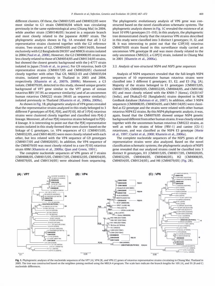
different clusters. Of these, the CMH015/05 and CMH032/05 weremost similar to G1 strain CMH036/04 which was circulatingpreviously in the same epidemiological area (Chiang Mai) in 2004,while another strain (CMH146/05) located in a separate branchand most closely related to the Japanese AU007 strain. Thephylogenetic analysis shown in Fig. 1A revealed that all 3 G2representative strains clustered together with the G2 referencestrains. Two strains of G2, CMH054/05 and CMH134/05, formedexclusively with G2 Bangladeshi DH397 and MMC6 strains isolatedin 2004 (Paul et al., 2008). However, the G2 CMH008/05 strain wasless closely related to those of CMH054/05 and CMH134/05 strains,but showed the closest genetic background with the J-4777 strainisolated in Japan (Trinh et al., in press). For G9 rotavirus, both G9representative strains (CMH017/05 and CMH020/05) clusteredclosely together with other Thai G9, NK022-03 and CMH035/04strains, isolated previously in Thailand in 2003 and 2004,respectively (Khamrin et al., 2007b, 2009b). Moreover, a G3isolate, CMH079/05, detected in this study, showed unique geneticbackground of VP7 gene similar to the VP7 genes of simianrotavirus RRV (97.9% aa sequence similarity) and of an uncommonhuman rotavirus CMH222 strain (99.6% aa sequence similarity)isolated previously in Thailand (Khamrin et al., 2006a, 2009a).
As shown in Fig. 1B, phylogenetic analysis of VP4 genes revealedthat the representative strains analyzed in this study belonged to 3different P genotypes of P[4], P[8], and P[10]. All of 3 P[4] rotavirusstrains were clustered closely together and classified into P[4]-2lineage. Moreover, all of our P[8] rotavirus strains belonged to P[8]-4 lineage. It is interesting to point out that the P[8] representativestrains isolated in this study formed their own cluster based on thelinkage of G genotypes, i.e. VP4 sequences of G1 (CMH015/05,CMH032/05, and CMH146/05) were more closely related with eachother, but less related with the VP4 sequence of G9 genotypes(CMH017/05 and CMH020/05). In addition, the VP4 sequence ofthe CMH079/05 was most closely related to a rare P[10] rotavirusstrain 69M (Khamrin et al., 2009a; Qian and Green, 1991).
The complete nucleotide sequences of VP6 genes of 7 strains(CMH008/05, CMH015/05, CMH017/05, CMH032/05, CMH054/05,CMH079/05, and CMH134/05) were obtained from sequencing.
Fig. 1. Phylogenetic analyses of the nucleotide sequences of the VP7 (A), VP4 (B), and VP
2005. The tree was constructed based on the neighbor-joining method using the MEGA
nucleotide differences.
The phylogenetic evolutionary analysis of VP6 gene was con-structed based on the novel classification schematic systems. Thephylogenetic analysis shown in Fig. 1C revealed the existence of atleast 10 VP6 I genotypes (I1–I10). In this analysis, the phylogenetictree demonstrated clearly that the rotavirus VP6 strains describedin this study were classified into 3 distinct I genotypes; I1, I2, andI8. It was interesting to point out that the G3P[10] rotavirusCMH079/05 strain found in this surveillance study carried anuncommon VP6 genotype I8 and was more closely related to theonly uncommon CMH222, a G3P[3] strain, isolated in Chiang Maiin 2001 (Khamrin et al., 2006a).
3.3. Analysis of non-structural NSP4 and NSP5 gene sequences
Analysis of NSP4 sequences revealed that the full-length NSP4sequences of 10 representative human rotavirus strains wereclassified into 3 different E genotypes; E1, E2, and E3 (Fig. 2A).Majority of the strains belonged to E1 genotypes (CMH015/05,CMH017/05, CMH020/05, CMH032/05, CMH044/05, and CMH146/05) and most closely related with the KNIH-7 (Korea), CH/D/147(India), and Dhaka25-02 (Bangladesh) strains deposited in NCBIGenBank database (Rahman et al., 2007). In addition, other 3 NSP4sequences (CMH008/05, CMH054/05, and CMH134/05) were classi-fied as E2 genotype and the strains were related with other humanrotavirus NSP4 E2 strains. By this NSP4 phylogenetic analysis, it was,again, found that the CMH079/05 showed unique NSP4 geneticbackground different from other human strains. Itwas closely relatedtogether with the uncommon human rotavirus CMH222 strains, aswell as with the strains of feline (FRV-1) and canine (CU-1)rotaviruses, and was classified as the NSP4 E3 genotype (Horieet al., 1997; Ciarlet et al., 2000; Khamrin et al., 2006a).
The complete nucleotide sequences of the NSP5 genes of therepresentative strains were also analyzed. Based on the novelclassification schematic systems, the phylogenetic analysis of NSP5gene revealed that our analyzed strains could be classified into 3distinct H genotypes, H1 (CMH015/05, CMH017/05, CMH020/05,CMH032/05, CMH044/05, CMH046/05), H2 (CMH008/05,CMH054/05, CMH124/05), and H6 (CMH079/05) (Fig. 2B).
6 (C) genes of rotavirus representative strains circulating in Chiang Mai, Thailand in
4 program. The scale bars indicate the branch lengths for 10% (A), and 5% (B and C)

Fig. 2. Phylogenetic analyses of the nucleotide sequences of the NSP4 (A) and NSP5 (B) genes of rotavirus representative strains circulating in Chiang Mai, Thailand in 2005.
The tree was constructed based on the neighbor-joining method using the MEGA 4 program. The scale bars indicate the branch lengths for 10% (A) and 5% (B) nucleotide
differences.
P. Khamrin et al. / Infection, Genetics and Evolution 10 (2010) 467–472470
3.4. Rotavirus genotypic linkages of G, P, I, E, and H genotypes
Recently, a new classification system for rotaviruses that basedon the sequences of all segments of rotavirus genome wasproposed (Matthijnssens et al., 2008a,b). Here, this new classifica-tion system was applied for analysis of 5 rotavirus gene segmentsof 10 representative strains. A nomenclature of rotavirusgenotypes was assigned as G, P, I, E, and H genotypes for VP7,VP4, VP6, NSP4, and NSP5, respectively. As demonstrated inTable 1, all of the selected strains, except for 3 VP6 gene sequencesof CMH020/05, CMH044/05, and CMH146/05, could be classifiedfor their genotypic linkages of G, P, I, E, H genotypes into 3 major
Table 1Genotype identification and genetic linkage between VP7, VP4, VP6, NSP4, and
NSP5 genes of group A rotaviruses circulating in Chiang Mai, Thailand in 2005.
Sample Genotypea
VP7 VP4 VP6 NSP4 NSP5
CMH008/05 G2 P[4] I2 E2 H2
CMH015/05 G1 P[8] I1 E1 H1
CMH017/05 G9 P[8] I1 E1 H1
CMH020/05 G9 P[8] ND E1 H1
CMH032/05 G1 P[8] I1 E1 H1
CMH044/05 G3 P[8] ND E1 H1
CMH054/05 G2 P[4] I2 E2 H2
CMH079/05b G3 P[10] I8 E3 H6
CMH134/05 G2 P[4] I2 E2 H2
CMH146/05 G1 P[8] ND E1 H1
ND, Sequence data was not available.a Rotavirus genotype classifications were assigned based on the novel
nomenclature purposed by RCWG (Matthijnssens et al., 2008b).b The data were published previously by Khamrin et al. (2009a).
patterns. Of these, G1P[8], G3P[8], and G9P[8] strains carried theirVP6, NSP4, NSP5 genotypes of I1, E1 and H1, respectively. TheG2P[4] strains were linked with I2, E2, H2 genotypes. In addition,an uncommon G3P[10] genotype carried the unique genotypes of I,E, H as I8, E3, H6, respectively.
4. Discussion
The studies of rotavirus infection carried out in Thailandrevealed that rotaviruses are the leading etiologic pathogen thatcauses diarrhea in children, and are responsible for about 20–61%of diarrheal diseases in hospitalized cases (Jiraphongsa et al., 2005;Khamrin et al., 2006a,b, 2007b; Maneekarn and Ushijima, 2000;Kittigul et al., 2009; Pongsuwannna et al., 2010). Previoussurveillance studies of group A rotaviruses in Thailand indicatethat the predominant genotypes of rotaviruses are changingovertime.
Epidemiological surveillance of group A rotavirus infectionconducted during the period of 2000–2004 (Khamrin et al., 2006b,2007b) revealed that G9P[8] emerged as the most predominantgenotype from 2000 to 2002. In addition, G2P[4] reemerged in theepidemic season of 2003, whereas G1P[8] became the mostpredominant genotype in 2004. In the present study (2005), groupA rotavirus was detected in 29.3% of the specimens collected fromchildren hospitalized with diarrhea, which was lower than thosereported previously at 34% in 2000–2001 and 37.3% in 2002–2004and G1P[8] genotype continues to be the most common genotypein 2005, followed by G2P[4], and G9P[8]. Moreover, sporadic casesof G3P[8] and G3P[10] were also detected in this surveillancestudy. The decrease of rotavirus prevalence and the changingpattern of rotavirus genotype distribution in this area are still

P. Khamrin et al. / Infection, Genetics and Evolution 10 (2010) 467–472 471
unclear although the sample size of the present study in 2005(n = 147) was not significantly different from the previous study in2004 (n = 160).
The classification of group A rotaviruses are normally based onthe molecular properties of two outer capsid proteins, VP7 andVP4, which allow classification of rotaviruses into G and Pgenotypes, respectively. Most recently, a novel classificationsystem of all 11 segments of rotavirus has been proposed by theRotavirus Classification Working Group (RCWG) (Matthijnssenset al., 2008b). This classification system relies on the percentage ofnucleotide sequence identity cut-off, and different genotypes areassigned for each genome segment. The data obtained by this novelclassification system represent the overall genetic backgroundsamong the circulating rotavirus strains better than that of theprevious conventional classification system. These data might beimportant for the future decision on a novel rotavirus vaccinedevelopment. In addition, the novel classification system is usefulfor minimizing an error of numbering or assigning new rotavirusgenotypes. By analysis of 5 gene segments (VP4, VP6, VP7, NSP4,and NSP5) of our rotavirus strains, it was observed clearly thatthose gene segments carried unique patterns of genetic linkages,such as G1P[8], G3P[8], and G9P[8] strains carrying their VP6,NSP4, NSP5 genotypes of I1, E1, H1, while G2P[4] strains werelinked with I2, E2, H2 genotypes. It was interesting to point out thatan uncommon human rotavirus strains G3P[10], which wasconsidered as the animal-like virus, carried VP6, NSP4, and NSP5gene segments related with the animal rotavirus strains and alsowith the uncommon human rotavirus G3P[3] (CMH222) strainpreviously identified in Chiang Mai, Thailand in 2001 (Khamrinet al., 2006a). This close evolutionary linkage between human andanimal rotaviruses emphasizes the need for simultaneous mon-itoring of emergence of novel rotavirus strains in both humans anddomestic animals. Since reassortment of rotavirus gene segmentsplays a key role in the generation of rotavirus diversity in nature,comprehensive genetic characterization of rotavirus genome isessential, especially for identifying reassortant strains (Ghoshet al., 2008; Matthijnssens et al., 2008b). The genetic linkageobserved in the present study resulted from comprehensivecharacterization of 5 genome segments of rotavirus strainscirculating in Thailand. Obviously, for a full recognition of theevolution of rotavirus strains in any geographical area analysis ofgenetic diversity should not be restricted only to VP7 and VP4genes but other structural and non-structural genes should also beanalyzed.
Acknowledgements
This research was supported by Grant-in-Aid for ScientificResearch under the JSPS Postdoctoral Fellowships and by Grants-in-Aid from the Ministry of Education and Sciences and theMinistry of Health, Labor and Welfare, Japan. In addition, the studywas also supported in part by the Research Fund from the Facultyof Medicine, Chiang Mai University, Thailand.
References
Ciarlet, M., Linprandi, F., Conner, M.E., Estes, M.K., 2000. Species specificity andinterspecies relatedness of NSP4 genetic groups by comparative NSP4 analysesof animal rotaviruses. Arch. Virol. 145, 371–387.
Estes, M.K., Kapikian, A.Z., 2007. Rotaviruses. In: Knipe, D.M., Howley, P.M. (Eds.),Fields Virology. 5th ed. Lippincott Williams & Wilkins, a Wolters KluwerBusiness, Philadelphia, pp. 1917–1974.
Gentsch, J.R, Glass, R.I., Woods, P., Gouvea, V., Gorziglia, M., Flores, J., Das, B.K., Bhan,M.K., 1992. Identification of group A rotavirus gene 4 types by polymerase chainreaction. J. Clin. Microbiol. 30, 1365–1373.
Ghosh, S., Samajdar, S., Sinha, M., Kobayashi, N., Taniguchi, K., Naik, T.N., 2008.Molecular characterization of rare bovine group A rotavirus G15P[11] andG15P[21] strains from eastern India: identification of simian SA11-like VP6genes in G15P[21] strains. Virus Genes 37, 241–249.
Gouvea, V., Glass, R.I., Woods, P., Taniguchi, K., Clark, H.F., Forrester, B., Fang, Z.Y.,1990. Polymerase chain reaction amplification and typing of rotavirus nucleicacid from stool specimens. J. Clin. Microbiol. 28, 276–282.
Gray, J., Vesikari, T., Van Damme, P., Giaquinto, C., Mrukowicz, J., Guarino, A., Dagan,R., Szajewska, H., Usonis, V., 2008. Rotavirus. J. Pediatr. Gastroenterol. Nutr. 46(Suppl. 2), S24–31.
Greenberg, H., McAuliffe, V., Valdesuso, J., Wyatt, R., Flores, J., Kalica, A., Hoshino, Y.,Singh, N., 1983a. Serological analysis of the subgroup protein of rotavirus, usingmonoclonal antibodies. Infect. Immun. 39, 91–99.
Greenberg, H.B., Valdesuso, J., van Wyke, K., Midthun, K., Walsh, M., McAuliffe, V.,Wyatt, R.G., Kalica, A.R., Flores, J., Hoshino, Y., 1983b. Production and preli-minary characterization of monoclonal antibodies directed at two surfaceproteins of rhesus rotavirus. J. Virol. 47, 267–275.
Horie, Y., Masamune, O., Nakagomi, O., 1997. Three major alleles of rotavirus NSP4proteins identified by sequence analysis. J. Gen. Virol. 78, 2341–2346.
Hoshino, Y., Gorziglia, M., Valdesuso, J., Askaa, J., Glass, R.I., Kapikian, A.Z., 1987. Anequine rotavirus (FI-14 strain) which bears both subgroup I and subgroup IIspecificities on its VP6. Virology 157, 488–496.
Jiraphongsa, C., Bresee, J.S., Pongsuwanna, Y., Kluabwang, P., Poonawagul, U.,Arporntip, P., Kanoksil, M., Premsri, N., Intusoma, U., Rotavirus SurveillanceProject Thailand Study Group, 2005. Epidemiology and burden of rotavirusdiarrhea in Thailand: results of sentinel surveillance. J. Infect. Dis. 192, 87–93.
Khamrin, P., Maneekarn, N., Peerakome, S., Yagyu, F., Okitsu, S., Ushijima, H., 2006a.Molecular characterization of a rare G3P[3] human rotavirus reassortant strainreveals evidence for multiple human-animal interspecies transmissions. J. Med.Virol. 78, 986–994.
Khamrin, P., Peerakome, S., Wongsawasdi, L., Tonusin, S., Sornchai, P., Maneerat, V.,Khamwan, C., Yagyu, F., Okitsu, S., Ushijima, H., Maneekarn, N., 2006b. Emer-gence of human G9 rotavirus with an exceptionally high frequency in childrenadmitted to hospital with diarrhea in Chiang Mai, Thailand. J. Med. Virol. 78,273–280.
Khamrin, P., Maneekarn, N., Peerakome, S., Chan-it, W., Yagyu, F., Okitsu, S.,Ushijima, H., 2007a. Novel porcine rotavirus of genotype P[27] shares newphylogenetic lineage with porcine rotavirus strain G2. Virology 361, 243–252.
Khamrin, P., Peerakome, S., Tonusin, S., Malasao, R., Okitsu, S., Mizuguchi, M.,Ushijima, H., Maneekarn, N., 2007b. Changing pattern of rotavirus G genotypedistribution in Chiang Mai, Thailand from 2002 to 2004: decline of G9 andreemergence of G1 and G2. J. Med. Virol. 79, 1775–1782.
Khamrin, P., Okitsu, S., Ushijima, H., Maneekarn, N., 2008. Novel nonstructuralprotein 4 genetic group in rotavirus of porcine origin. Emerg. Infect. Dis. 14,686–688.
Khamrin, P., Maneekarn, N., Peerakome, S., Malasao, R., Thongprachum, A., Chan-it,W., Mizuguchi, M., Okitsu, S., Ushijima, H., 2009a. Molecular characterization ofVP4, VP6, VP7, NSP4, and NSP5/6 genes identifies an unusual G3P[10] humanrotavirus strain. J. Med. Virol. 81, 176–182.
Khamrin, P., Thongprachum, A., Chaimongkol, N., Chusri, P., Okitsu, S., Ushijima, H.,Maneekarn, N., 2009b. Evolutionary consequences of G9 rotaviruses circulatingin Thailand. Infect. Genet. Evol. 9, 1394–1399.
Kittigul, L., Pombubpa, K., Taweekate, Y., Yeephoo, T., Khamrin, P., Ushijima, H.,2009. Molecular characterization of rotaviruses, noroviruses, sapovirus, andadenoviruses in patients with acute gastroenteritis in Thailand. J. Med. Virol. 81,345–353.
Kudo, S., Zhou, Y., Cao, X.R., Yamanishi, S., Nakata, S., Ushijima, H., 2001. Molecularcharacterization in the VP7, VP4 and NSP4 genes of human rotavirus serotype 4(G4) isolated in Japan and Kenya. Microbiol. Immunol. 45, 167–171.
Maneekarn, N., Ushijima, H., 2000. Epidemiology of rotavirus infection in Thailand.Pediatr. Int. 42, 415–421.
Matthijnssens, J., Rahman, M., Martella, V., Xuelei, Y., De Vos, S., De Leener, K.,Ciarlet, M., Buonavoglia, C., Van Ranst, M., 2006. Full genomic analysis of humanrotavirus strain B4106 and lapine rotavirus strain 30/96 provides evidence forinterspecies transmission. J. Virol. 80, 3801–3810.
Matthijnssens, J., Ciarlet, M., Heiman, E., Arijs, I., Delbeke, T., McDonald, S.M.,Palombo, E.A., Iturriza-Gomara, M., Maes, P., Patton, J.T., Rahman, M., Van Ranst,M., 2008a. Full genome-based classification of rotaviruses reveals a commonorigin between human Wa-Like and porcine rotavirus strains and human DS-1-like and bovine rotavirus strains. J. Virol. 82, 3204–3219.
Matthijnssens, J., Ciarlet, M., Rahman, M., Attoui, H., Banyai, K., Estes, M.K., Gentsch,J.R., Iturriza-Gomara, M., Kirkwood, C.D., Martella, V., Mertens, P.P., Nakagomi,O., Patton, J.T., Ruggeri, F.M., Saif, L.J., Santos, N., Steyer, A., Taniguchi, K.,Desselberger, U., Van Ranst, M., 2008b. Recommendations for the classificationof group A rotaviruses using all 11 genomic RNA segments. Arch. Virol. 153,1621–1629.
Parashar, U.D., Gibson, C.J., Bresse, J.S., Glass, R.I., 2006. Rotavirus and severechildhood diarrhea. Emerg. Infect. Dis. 12, 304–306.
Paul, S.K., Kobayashi, N., Nagashima, S., Ishino, M., Watanabe, S., Alam, M.M.,Ahmed, M.U., Hossain, M.A., Naik, T.N., 2008. Phylogenetic analysis of rota-viruses with genotypes G1, G2, G9 and G12 in Bangladesh: evidence for a closerelationship between rotaviruses from children and adults. Arch. Virol. 153,1999–2012.
Pongsuwannna, Y., Guntapong, R., Tacharoenmuang, R., Prapanpoj, M., Kameoka,M., Taniguchi, K., 2010. A long-term survey on the distribution of the humanrotavirus G type in Thailand. J. Med. Virol. 82, 157–163.
Qian, Y., Green, K.Y., 1991. Human rotavirus strain 69M has a unique VP4 asdetermined by amino acid sequence analysis. Virology 182, 407–412.
Rahman, M., Matthijnssens, J., Yang, X., Delbeke, T., Arijs, I., Taniguchi, K., Iturriza-Gomara, M., Iftekharuddin, N., Azim, T., Van Ranst, M., 2007. Evolutionary

P. Khamrin et al. / Infection, Genetics and Evolution 10 (2010) 467–472472
history and global spread of the emerging G12 human rotaviruses. J. Virol. 81,2382–2390.
Santos, N., Hoshino, Y., 2005. Global distribution of rotavirus serotypes/genotypesand its implication for the development and implementation of an effectiverotavirus vaccine. Rev. Med. Virol. 15, 29–56.
Schumann, T., Hotzel, H., Otto, P., Johne, R., 2009. Evidence of interspecies transmis-sion and reassortment among avian group A rotaviruses. Virology 386, 334–343.
Steyer, A., Poljsak-Prijatelj, M., Barlic-Maganja, D., Jamnikar, U., Mijovski, J.Z., Marin,J., 2007. Molecular characterization of a new porcine rotavirus P genotype foundin an asymptomatic pig in Slovenia. Virology 359, 275–282.
Tamura, K., Dudley, J., Nei, M., Kumar, S., 2007. MEGA4: Molecular EvolutionaryGenetics Analysis (MEGA) software version 4.0. Mol. Biol. Evol. 24, 1596–1599.
Trinh, Q.D., Pham, N.T., Nguyen, T.A., Phan, T.G., Yan, H., Hoang, P.L., Khamrin, P.,Maneekarn, N., Li, Y., Okitsu, S., Mizuguchi, M., Ushijima, H. Sequence analysis ofthe VP7 gene of human rotaviruses G2 and G4 isolated in Japan, China, Thailand,and Vietnam during 2001–2003. J. Med. Virol., in press.
Urasawa, S., Hasegawa, A., Urasawa, T., Taniguchi, K., Wakasugi, F., Suzuki, H.,Inouye, S., Pongprot, B., Supawadee, J., Suprasert, S., 1992. Antigenic and geneticanalyses of human rotaviruses in Chiang Mai, Thailand: evidence for a closerelationship between human and animal rotaviruses. J. Infect. Dis. 166, 227–234.
Ursu, K., Kisfali, P., Rigo, D., Ivanics, E., Erdelyi, K., Dan, A., Melegh, B., Martella, V.,Banyai, K., 2009. Molecular analysis of the VP7 gene of pheasant rotavirusesidentifies a new genotype, designated G23. Arch. Virol. 154, 1365–1369.
Yan, H., Nguyen, T.A., Phan, T.G., Okitsu, S., Li, Y., Ushijima, H., 2004. Development ofRT-multiplex PCR assay for detection of adenovirus and group A and C rota-viruses in diarrheal fecal specimens from children in China. KansenshogakuZasshi 78, 699–709.
Zhou, Y., Supawadee, J., Khamwan, C., Tonusin, S., Peerakome, S., Kim, B., Kaneshi, K.,Ueda, Y., Nakaya, S., Akatani, K., Maneekarn, N., Ushijima, H., 2001. Character-ization of human rotavirus serotype G9 isolated in Japan and Thailand from1995 to 1997. J. Med. Virol. 65, 619–628.
Related Documents


