JOURNAL OF VIROLOGY, 0022-538X/99/$04.0010 Nov. 1999, p. 9284–9293 Vol. 73, No. 11 Copyright © 1999, American Society for Microbiology. All Rights Reserved. Dual Infection of Gnotobiotic Calves with Bovine Strains of Group A and Porcine-Like Group C Rotaviruses Influences Pathogenesis of the Group C Rotavirus K. O. CHANG, P. R. NIELSEN, L. A. WARD, AND L. J. SAIF* Food Animal Health Research Program, Ohio Agricultural Research and Development Center, The Ohio State University, Wooster, Ohio 44691 Received 16 April 1999/Accepted 9 August 1999 There is serological evidence that bovine group C rotaviruses exist in the United States, but there are no reports of their isolation. Ninety fecal samples from calves with diarrhea, 81 samples from adult cows with diarrhea (winter dysentery), and 20 fecal samples from healthy adult cows were tested for group C rotaviruses by polyacrylamide gel electrophoresis, immune electron microscopy, and reverse transcription-PCR (RT-PCR). Three samples from adult cow diarrhea cases were positive only by RT-PCR, and a group C rotavirus was isolated from a positive sample in monkey kidney (MA104) cells (WD534tc/C). Genetically and serologically, the WD534tc/C strain was more closely related to the Cowden porcine group C strain than to the Shintoku bovine strain. Because the original cow feces also contained a group A rotavirus (detected after passage in cell culture), we hypothesized that such dual-rotavirus infections might play a role in the pathogenesis and host adaptation of rotaviruses. Thus, we examined the pathogenesis of WD534tc/C alone or combined with virulent (IND/A) or attenuated (NCDV/A) bovine group A rotaviruses in gnotobiotic calves. WD534tc/C alone induced diarrhea without (or with limited) virus shedding in inoculated calves (n 5 3). In contrast, all calves coinfected with WD534tc/C and IND/A (n 5 2) developed diarrhea and shed both viruses, whereas calves coinfected with WD534tc/C and NCDV/A (n 5 3) developed diarrhea but did not shed either virus. Infection with WD534tc/C or NCDV/A alone caused only mild villous atrophy (jejunum and/or ileum), whereas dual infection with both viruses induced lesions throughout the small intestine. Although IND/A alone caused villous atrophy, more- widespread small intestinal lesions occurred in calves coinfected with WD534tc/C and IND/A. In conclusion, coinfection of calves with group A rotaviruses enhanced fecal shedding of a bovine group C rotavirus and the extent of histopathological lesions in the small intestines. Thus, our findings suggest a potential novel hypothesis involving dual infections for the adaptation of heterologous rotaviruses to new host species. Rotaviruses are a major cause of diarrhea in young children and animals including cattle (21, 35) and belong to seven distinct antigenic groups (A to G) (7, 21, 24). Although group C rotaviruses are antigenically distinct from group A rotavi- ruses in serological tests, they are genetically related and they share a minor nonneutralizing epitope on VP6 detected with monoclonal antibodies (47). The VP6 genes of group C rota- viruses share 88.4 to 90.6% homology (porcine Cowden, bo- vine Shintoku, and human Bristol) and 41.3 and 16.3% homol- ogy with the VP6 gene of group A (bovine RF) and group B (human ADRV) rotaviruses, respectively (19). There is anti- genic and genetic variation within group C rotaviruses. There are at least three G types (VP7) identified by two-way cross- neutralization tests and sequence analysis of the VP7 genes (70 to 75% homology among the serotypically distinct strains), and the Cowden porcine and Shintoku bovine strains are different serotypes (45, 46). Group A rotaviruses are endemic, and group B rotaviruses cause sporadic cases of diarrhea in calves and cows in the United States (5, 31, 34, 35). However, although a moderate prevalence of antibodies to group C rotaviruses in sera of cattle (47 to 56%) in the United States (44) was reported, to date, the Shintoku group C bovine rotavirus, which was isolated from an adult cow in Japan, is the only bovine isolate of group C rotavirus (31, 48). The first group C rotavirus of any species was isolated in 1980 from nursing pigs (33) and more recently identified as a cause of enzootic diarrhea in neonates (27) and older finishing (fattening) pigs (22). Serological studies suggest that group C rotaviruses are endemic in U.S. swine herds (40, 44). In humans, group C rotaviruses are potential emerging enteric pathogens for all ages including adults (4, 17, 31). Since their first detection in humans in 1983 (28), group C rotavi- ruses have been associated with large outbreaks of diarrhea in Japan (4, 16), and with family or sporadic cases of diarrhea in children and adults worldwide including the United States (4, 17). Assays to detect group C rotaviruses, including polyacryl- amide gel electrophoresis (PAGE), immune electron micros- copy (IEM), enzyme-linked immunosorbent assay (ELISA), and cell culture immunofluorescence (CCIF) tests have been described elsewhere (3, 12, 18, 40, 44). However, because of the limited shedding or instability of group C rotaviruses in feces (4, 17, 31), highly sensitive assays are needed for their efficient detection (17). In this study, we surveyed fecal samples from calves and adult cows with diarrhea for the presence of group C rotaviruses by reverse transcription-PCR (RT-PCR). A group C rotavirus (WD534tc/C strain) was isolated from a diarrheic adult cow fecal sample, which also contained group A rotavirus. The WD534tc/C strain showed a higher relatedness to Cowden porcine group C rotavirus by serological and ge- netic analysis than to the Shintoku bovine group C rotavirus and was pathogenic in gnotobiotic pigs. During our preliminary study of the pathogenicity of the WD534tc/C infection, gnoto- * Corresponding author. Mailing address: Food Animal Health Re- search Program, Ohio Agricultural Research and Development Cen- ter, The Ohio State University, Wooster, OH 44691. Phone: (330) 263-3744. Fax: (330) 263-3677. E-mail: [email protected]. 9284 on August 17, 2015 by guest http://jvi.asm.org/ Downloaded from

Welcome message from author
This document is posted to help you gain knowledge. Please leave a comment to let me know what you think about it! Share it to your friends and learn new things together.
Transcript

JOURNAL OF VIROLOGY,0022-538X/99/$04.0010
Nov. 1999, p. 9284–9293 Vol. 73, No. 11
Copyright © 1999, American Society for Microbiology. All Rights Reserved.
Dual Infection of Gnotobiotic Calves with Bovine Strains ofGroup A and Porcine-Like Group C Rotaviruses Influences
Pathogenesis of the Group C RotavirusK. O. CHANG, P. R. NIELSEN, L. A. WARD, AND L. J. SAIF*
Food Animal Health Research Program, Ohio Agricultural Research and Development Center,The Ohio State University, Wooster, Ohio 44691
Received 16 April 1999/Accepted 9 August 1999
There is serological evidence that bovine group C rotaviruses exist in the United States, but there are noreports of their isolation. Ninety fecal samples from calves with diarrhea, 81 samples from adult cows withdiarrhea (winter dysentery), and 20 fecal samples from healthy adult cows were tested for group C rotavirusesby polyacrylamide gel electrophoresis, immune electron microscopy, and reverse transcription-PCR (RT-PCR).Three samples from adult cow diarrhea cases were positive only by RT-PCR, and a group C rotavirus wasisolated from a positive sample in monkey kidney (MA104) cells (WD534tc/C). Genetically and serologically,the WD534tc/C strain was more closely related to the Cowden porcine group C strain than to the Shintokubovine strain. Because the original cow feces also contained a group A rotavirus (detected after passage in cellculture), we hypothesized that such dual-rotavirus infections might play a role in the pathogenesis and hostadaptation of rotaviruses. Thus, we examined the pathogenesis of WD534tc/C alone or combined with virulent(IND/A) or attenuated (NCDV/A) bovine group A rotaviruses in gnotobiotic calves. WD534tc/C alone induceddiarrhea without (or with limited) virus shedding in inoculated calves (n 5 3). In contrast, all calves coinfectedwith WD534tc/C and IND/A (n 5 2) developed diarrhea and shed both viruses, whereas calves coinfected withWD534tc/C and NCDV/A (n 5 3) developed diarrhea but did not shed either virus. Infection with WD534tc/Cor NCDV/A alone caused only mild villous atrophy (jejunum and/or ileum), whereas dual infection with bothviruses induced lesions throughout the small intestine. Although IND/A alone caused villous atrophy, more-widespread small intestinal lesions occurred in calves coinfected with WD534tc/C and IND/A. In conclusion,coinfection of calves with group A rotaviruses enhanced fecal shedding of a bovine group C rotavirus and theextent of histopathological lesions in the small intestines. Thus, our findings suggest a potential novelhypothesis involving dual infections for the adaptation of heterologous rotaviruses to new host species.
Rotaviruses are a major cause of diarrhea in young childrenand animals including cattle (21, 35) and belong to sevendistinct antigenic groups (A to G) (7, 21, 24). Although groupC rotaviruses are antigenically distinct from group A rotavi-ruses in serological tests, they are genetically related and theyshare a minor nonneutralizing epitope on VP6 detected withmonoclonal antibodies (47). The VP6 genes of group C rota-viruses share 88.4 to 90.6% homology (porcine Cowden, bo-vine Shintoku, and human Bristol) and 41.3 and 16.3% homol-ogy with the VP6 gene of group A (bovine RF) and group B(human ADRV) rotaviruses, respectively (19). There is anti-genic and genetic variation within group C rotaviruses. Thereare at least three G types (VP7) identified by two-way cross-neutralization tests and sequence analysis of the VP7 genes (70to 75% homology among the serotypically distinct strains), andthe Cowden porcine and Shintoku bovine strains are differentserotypes (45, 46).
Group A rotaviruses are endemic, and group B rotavirusescause sporadic cases of diarrhea in calves and cows in theUnited States (5, 31, 34, 35). However, although a moderateprevalence of antibodies to group C rotaviruses in sera of cattle(47 to 56%) in the United States (44) was reported, to date, theShintoku group C bovine rotavirus, which was isolated from anadult cow in Japan, is the only bovine isolate of group C
rotavirus (31, 48). The first group C rotavirus of any specieswas isolated in 1980 from nursing pigs (33) and more recentlyidentified as a cause of enzootic diarrhea in neonates (27) andolder finishing (fattening) pigs (22). Serological studies suggestthat group C rotaviruses are endemic in U.S. swine herds (40,44). In humans, group C rotaviruses are potential emergingenteric pathogens for all ages including adults (4, 17, 31). Sincetheir first detection in humans in 1983 (28), group C rotavi-ruses have been associated with large outbreaks of diarrhea inJapan (4, 16), and with family or sporadic cases of diarrhea inchildren and adults worldwide including the United States (4,17).
Assays to detect group C rotaviruses, including polyacryl-amide gel electrophoresis (PAGE), immune electron micros-copy (IEM), enzyme-linked immunosorbent assay (ELISA),and cell culture immunofluorescence (CCIF) tests have beendescribed elsewhere (3, 12, 18, 40, 44). However, because ofthe limited shedding or instability of group C rotaviruses infeces (4, 17, 31), highly sensitive assays are needed for theirefficient detection (17). In this study, we surveyed fecal samplesfrom calves and adult cows with diarrhea for the presence ofgroup C rotaviruses by reverse transcription-PCR (RT-PCR).A group C rotavirus (WD534tc/C strain) was isolated from adiarrheic adult cow fecal sample, which also contained group Arotavirus. The WD534tc/C strain showed a higher relatednessto Cowden porcine group C rotavirus by serological and ge-netic analysis than to the Shintoku bovine group C rotavirusand was pathogenic in gnotobiotic pigs. During our preliminarystudy of the pathogenicity of the WD534tc/C infection, gnoto-
* Corresponding author. Mailing address: Food Animal Health Re-search Program, Ohio Agricultural Research and Development Cen-ter, The Ohio State University, Wooster, OH 44691. Phone: (330)263-3744. Fax: (330) 263-3677. E-mail: [email protected].
9284
on August 17, 2015 by guest
http://jvi.asm.org/
Dow
nloaded from

biotic calves inoculated with this strain developed diarrheawithout or with limited (1-day) virus shedding. Because theoriginal field sample was a mixed infection with group A rota-virus, and the isolate was potentially a porcine group C strain,we hypothesized that such dual-rotavirus infections might playa role in the pathogenesis and adaptation of heterologousgroup C rotaviruses to cattle. Thus, we investigated the coin-fection of gnotobiotic calves with group A bovine rotavirusesand the WD534tc/C bovine group C rotavirus isolate to deter-mine clinical signs, virus shedding patterns, and the extent ofhistopathological lesions in the intestines.
MATERIALS AND METHODS
Viruses and cells. Reference group C rotaviruses included the Shintoku bovinestrain (48) and the Cowden porcine strain (41). Group A rotaviruses included avirulent serotype G6 IND (P[5]G6) strain (6) and an attenuated vaccine NCDV(P[1]G6) strain from the American Tissue Culture Collection (Atlanta, Ga.). TheShintoku/C, attenuated Cowden/C, and NCDV/A strains were grown in monkeykidney cells (MA104 cells) in the presence of pancreatin (50 mg/ml) (36, 37, 41,48). The virulent IND/A and Cowden/C strains were prepared by virus passagein gnotobiotic calves or piglets, respectively, and infected feces or intestinalcontents were collected as a source of virus. Mock-infected cell culture samplesand mock-infected gnotobiotic calf feces or pig intestinal contents were used asnegative controls.
Field samples. Analysis for bovine group C rotaviruses was performed on thefollowing samples: 90 diarrheic calf fecal samples from Ohio, California, Wyo-ming, South Dakota, and Nebraska; 81 adult cow diarrheic fecal samples (winterdysentery cases) from Ohio, New York, and California; and 20 healthy adult cowfecal samples (from two Ohio herds that were matched case controls for thewinter dysentery cases) (39).
Extraction and electrophoresis of rotavirus dsRNA. Rotavirus double-stranded RNA (dsRNA) was extracted from cell culture-propagated viruses andfecal or intestinal viruses by previously described procedures (13). RotavirusdsRNA in extracted samples was analyzed by PAGE to confirm the presence ofdsRNA and to examine the genomic electropherotypes as previously described(6, 13, 31). The discontinuous buffer system of Laemmli was utilized, and dsRNAwas resolved on 10% polyacrylamide slab gels (23). Electrophoresis was con-ducted at 12 mA for 14 to 16 h. The dsRNA bands were visualized by silverstaining (13).
IEM and CCIF assay. Fifty-three fecal samples from diarrheic adult cows(field samples) and fecal samples after single or dual inoculation of gnotobioticcalves and pigs and cell culture isolates were processed for IEM as previouslydescribed (32). The processed samples were incubated separately with group A-,B-, C-, or bovine coronavirus-specific hyperimmune antisera, and then the mix-tures were pelleted, negatively stained with phosphotungstic acid, applied togrids, and examined by electron microscopy (29, 32). A CCIF assay was used toconfirm if group A or C rotaviruses were present in virus isolates and to detectvirus shedding after challenge of gnotobiotic calves and pigs with group A and/orC rotaviruses as described previously (20). Briefly, the fecal or cell-passagedrotavirus samples were serially diluted with minimal essential medium (MEM)from 1:10 to 1:107 and were inoculated onto monolayers of MA104 cells grownin 96-well plates. After adsorption at 37°C for 1 h, the mixtures were discardedand 200 ml (per well) of MEM containing 50 mg of pancreatin per ml was addedto the cells and incubated at 37°C for 20 h. The medium was discarded, and thecells were fixed with 80% acetone and stained with fluorescein isothiocyanate(FITC)-conjugated anti-OSU porcine group A or anti-Cowden porcine group Crotavirus serum (20, 40). The numbers of fluorescent focus units (FFU) in eachwell were counted, and titers were expressed as FFU per milliliter.
RT-PCR assay. Field fecal samples and fecal samples collected after viruschallenge of gnotobiotic calves and pigs were tested for group A and C rotavi-ruses by RT-PCR (6). The RT-PCR primers were designed to produce partial-length group C rotavirus VP6 based on Cowden and Shintoku VP6 genes and afull-length group A rotavirus VP7 gene. cDNA was synthesized and amplified inan RT-PCR mixture containing 103 RT-PCR buffer (200 mM Tris-HCl [pH 8.3],2.5 mM MgCl2, KCl, 0.05% gelatin), deoxynucleoside triphosphates (10 mM[each]), 200 ng of primer A (sense strands for group C VP6 [59-GAAGCTGTATGTGATGATGA-39] and group A VP7 [59-GGCCGGATTTAAAAGCGACAATTT-39]), 200 ng of primer B (antisense strands for group C VP6 [59-CATAGCAGCTGGTCTAATCA-39] and for group A VP7 [59-GGTCACATCATACAACTCTA-39]), (6), 5 U of RNasin (Boehringer Mannheim Biochemicals,Indianapolis, Ind.), 10 U of avian myeloblastosis virus reverse transcriptase(Boehringer Mannheim Biochemicals), and 2.5 U of Taq polymerase (Boehr-inger Mannheim Biochemicals). First-strand cDNA synthesis was accomplishedby incubating the above mixture for 90 min at 42°C. Thirty amplification cycleswere conducted, with each cycle consisting of 94°C for 1 min (denaturation),52°C for 1 min (annealing), and 72°C for 2 min (extension), followed by a 7-minextension step at 72°C. The PCR products were analyzed on 1% agarose gels bystandard procedures (38).
Isolation of a group C rotavirus from fecal samples. Two adult cow diarrheicfecal samples (from the same Ohio farm) which were group C positive byRT-PCR were inoculated into MA104 cells in roller tubes for virus isolation (36,37). They were blind passaged until cytopathic effects were seen and monitoredfor group A and C rotaviruses by CCIF (20, 40).
Cloning and sequence analysis of the full-length VP6 gene of WD534tc. Thefull-length VP6 gene of WD534tc was produced by RT-PCR with 59- and 39-endprimers and cloned in the pCRII vector (Invitrogen, San Diego, Calif.). Thesequence analysis was performed by the primer extension method (Sequenaseversion 2.0; Amersham, Arlington Height, Ill.), and the data were analyzed withthe DNASTAR program (DNASTAR Inc., Madison, Wis.).
Antigenic analyses of WD534tc, Cowden, Shintoku group C rotaviruses, andIND group A rotavirus by two-way FFN tests. The isolate WD534tc, Cowden andShintoku group C rotaviruses, and IND group A rotavirus were tested in two-wayfluorescent focus neutralization (FFN) tests using antisera to each virus, whichwere prepared by hyperimmunization of a gnotobiotic piglet with WD534tc/Cand of guinea pigs with Cowden, Shintoku, or IND strain, as described previously(20, 40). Briefly, the antisera were serially diluted with MEM from 1:10 to1:20,480, mixed with the homologous or heterologous virus strains (104 FFU/ml),and reacted at 37°C for 1 h. The mixtures (100 ml) were inoculated onto mono-layers of MA104 cells grown in 96-well plates. After adsorption at 37°C for 1 h,the mixtures were discarded and 200 ml (per well) of MEM containing 50 mg ofpancreatin per ml was added to the cells and incubated at 37°C for 20 h. Themedium was discarded, and the cells were fixed with 80% acetone and stainedwith FITC-conjugated pig anti-OSU group A or anti-Cowden group C rotavirusserum. Neutralizing antibody titers were expressed as the reciprocal of thehighest dilution that resulted in 80% or greater reduction in the numbers ofFFU. Serum samples from unexposed gnotobiotic pigs were used for negativecontrols.
Virus challenge of gnotobiotic calves and piglets. The challenge was eithersingle or dual inoculation (simultaneous) of group A and group C rotavirusstrains into 16 gnotobiotic calves and 6 gnotobiotic piglets (Table 1). All calvesand piglets were from 1 to 24 days old and were derived, fed, inoculated, andmaintained as previously described (25, 29). Briefly, the gnotobiotic calves andpiglets were oronasally inoculated with diluted (1:10) fecal filtrates or virus-infected cell culture lysates. Viruses were inoculated at similar doses (WD534/C,107 FFU for calves and 106 FFU for piglets; IND/A, 107 FFU; NCDV/A, 105
FFU for single inoculation and 105 or 107 FFU for dual inoculation; virulent andattenuated Cowden/C, 107 FFU for calves and 106 FFU for piglets) in a total of50 ml of calves and 10 ml for pigs. Dual inoculation (WD534tc/C and IND/A,WD534tc/C and NCDV/A or virulent Cowden/C and IND/A) of gnotobioticcalves was done with the same virus titers (except for WD534tc/C and NCDV/A,when either 105 or 107 FFU for NCDV/A was used) and volumes as singleinoculations. A titer of 105 FFU for single inoculation of calves with NCDV/Awas used because it is the recommended dose of commercial rotavirus vaccinesfor use in calves (42). After inoculation, calves and piglets were examined twicedaily for clinical signs and development of diarrhea, and the fecal consistency wasscored as follows: 0, normal; 1, pasty; 2, semiliquid; 3, liquid. For the datasummarized in the tables, diarrhea was noted as a fecal consistency of $2 andquestionable or mild diarrhea was noted as fecal consistency of 1. Feces and
TABLE 1. Experimental design for single and dual inoculation ofgnotobiotic calves and piglets with group C and group A rotaviruses
Group Virus inoculuma No. of animalsin group
Calvesb
1 IND/A 22 NCDV/A 33 WD534tc/C 44 Virulent Cowden/C 15 WD534tc/C 1 IND/A 26 Cowden/C (virulent) 1 IND/A 17 WD534tc/C 1 NCDV/A 28 None (buffer) (control) 1
Pigletsc
9 WD534tc/C 310 Virulent Cowden/C 111 Attenuated Cowden/C 2
a Viruses were inoculated at the following doses (WD534tc/C, 107 FFU forcalves and 106 FFU for piglets; IND/A, 107 FFU; NCDV/A, 105 or 107 FFU;virulent and attenuated Cowden, 107 FFU for the calf and 106 FFU for piglets).
b Calves were challenged at 3 to 8 days of age, except for one older calf (24days old) in group 6.
c Piglets were challenged at 4 days of age, except for one piglet (24 days old)in group 11.
VOL. 73, 1999 DUAL INFECTION OF GNOTOBIOTIC CALVES BY ROTAVIRUSES 9285
on August 17, 2015 by guest
http://jvi.asm.org/
Dow
nloaded from
serum samples were collected daily and weekly, respectively, and stored at 220°Cuntil tested. Each fecal sample was tested for the presence of groups A and Crotaviruses by CCIF, IEM, and RT-PCR, and each serum sample was tested forantibodies to group A and C rotaviruses by the FFN test as described previously(20, 40). Seroconversion was indicated as serum antibody titer increases from,50 for acute serum to $50 for convalescent serum.
Histopathological evaluation. Single or dual inoculated calves were eutha-nized within 12 h of diarrhea onset (postinoculation days [PID] 2 to 4) and theduodenum, jejunum, and ileum from the small intestines and the mesentericlymph nodes were collected and placed in fixative (Prefer; Anatech Ltd., BattleCreek, Mich.). The tissues were processed, embedded in paraffin, and stainedwith hematoxylin and eosin (50). Histological evaluation was done on codedsamples, and a comparison was made with tissues from age-matched controls(50).
Nucleotide sequence accession number. The GenBank accession number ofthe VP6 gene from WD534tc/C is AF162434.
RESULTS
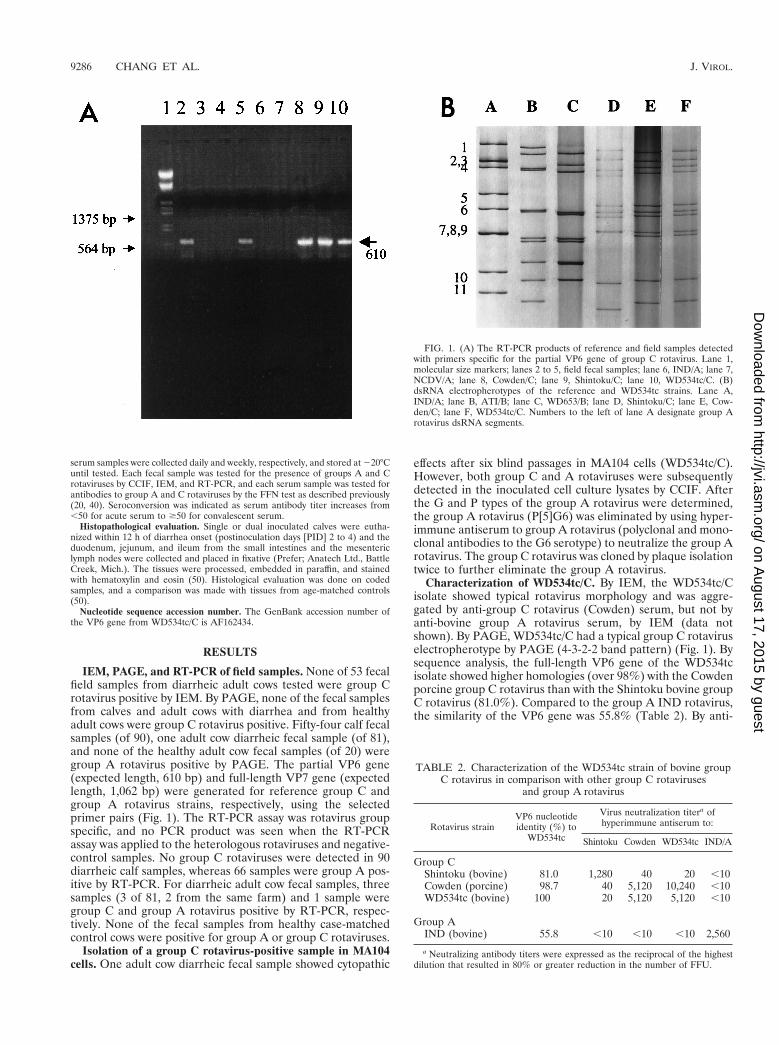
IEM, PAGE, and RT-PCR of field samples. None of 53 fecalfield samples from diarrheic adult cows tested were group Crotavirus positive by IEM. By PAGE, none of the fecal samplesfrom calves and adult cows with diarrhea and from healthyadult cows were group C rotavirus positive. Fifty-four calf fecalsamples (of 90), one adult cow diarrheic fecal sample (of 81),and none of the healthy adult cow fecal samples (of 20) weregroup A rotavirus positive by PAGE. The partial VP6 gene(expected length, 610 bp) and full-length VP7 gene (expectedlength, 1,062 bp) were generated for reference group C andgroup A rotavirus strains, respectively, using the selectedprimer pairs (Fig. 1). The RT-PCR assay was rotavirus groupspecific, and no PCR product was seen when the RT-PCRassay was applied to the heterologous rotaviruses and negative-control samples. No group C rotaviruses were detected in 90diarrheic calf samples, whereas 66 samples were group A pos-itive by RT-PCR. For diarrheic adult cow fecal samples, threesamples (3 of 81, 2 from the same farm) and 1 sample weregroup C and group A rotavirus positive by RT-PCR, respec-tively. None of the fecal samples from healthy case-matchedcontrol cows were positive for group A or group C rotaviruses.
Isolation of a group C rotavirus-positive sample in MA104cells. One adult cow diarrheic fecal sample showed cytopathic
effects after six blind passages in MA104 cells (WD534tc/C).However, both group C and A rotaviruses were subsequentlydetected in the inoculated cell culture lysates by CCIF. Afterthe G and P types of the group A rotavirus were determined,the group A rotavirus (P[5]G6) was eliminated by using hyper-immune antiserum to group A rotavirus (polyclonal and mono-clonal antibodies to the G6 serotype) to neutralize the group Arotavirus. The group C rotavirus was cloned by plaque isolationtwice to further eliminate the group A rotavirus.
Characterization of WD534tc/C. By IEM, the WD534tc/Cisolate showed typical rotavirus morphology and was aggre-gated by anti-group C rotavirus (Cowden) serum, but not byanti-bovine group A rotavirus serum, by IEM (data notshown). By PAGE, WD534tc/C had a typical group C rotaviruselectropherotype by PAGE (4-3-2-2 band pattern) (Fig. 1). Bysequence analysis, the full-length VP6 gene of the WD534tcisolate showed higher homologies (over 98%) with the Cowdenporcine group C rotavirus than with the Shintoku bovine groupC rotavirus (81.0%). Compared to the group A IND rotavirus,the similarity of the VP6 gene was 55.8% (Table 2). By anti-
FIG. 1. (A) The RT-PCR products of reference and field samples detectedwith primers specific for the partial VP6 gene of group C rotavirus. Lane 1,molecular size markers; lanes 2 to 5, field fecal samples; lane 6, IND/A; lane 7,NCDV/A; lane 8, Cowden/C; lane 9, Shintoku/C; lane 10, WD534tc/C. (B)dsRNA electropherotypes of the reference and WD534tc strains. Lane A,IND/A; lane B, ATI/B; lane C, WD653/B; lane D, Shintoku/C; lane E, Cow-den/C; lane F, WD534tc/C. Numbers to the left of lane A designate group Arotavirus dsRNA segments.
TABLE 2. Characterization of the WD534tc strain of bovine groupC rotavirus in comparison with other group C rotaviruses
and group A rotavirus
Rotavirus strainVP6 nucleotideidentity (%) to
WD534tc
Virus neutralization titera ofhyperimmune antiserum to:
Shintoku Cowden WD534tc IND/A
Group CShintoku (bovine) 81.0 1,280 40 20 ,10Cowden (porcine) 98.7 40 5,120 10,240 ,10WD534tc (bovine) 100 20 5,120 5,120 ,10
Group AIND (bovine) 55.8 ,10 ,10 ,10 2,560
a Neutralizing antibody titers were expressed as the reciprocal of the highestdilution that resulted in 80% or greater reduction in the number of FFU.
9286 CHANG ET AL. J. VIROL.
on August 17, 2015 by guest
http://jvi.asm.org/
Dow
nloaded from

genic analysis of WD534tc by the FFN test, the WD534tc/Crotavirus was more closely related to the Cowden (FFN anti-body titers were identical [5,120] or only twofold different[5,120 versus 10,240]) porcine group C rotavirus than to theShintoku (64-fold different [1,280 versus 20]) bovine group Crotavirus in two-way FFN tests (Table 2). The WD534tc/Crotavirus was antigenically unrelated to the IND group A ro-tavirus in the FFN test (FFN titers of ,10) (Table 2).
Single inoculation of gnotobiotic calves with IND/A,NCDV/A, WD534tc/C, or Cowden/C rotavirus. The virulentIND/A rotavirus caused diarrhea (from PID 2 or 3) and virusshedding (from PID 2; n 5 2) detected by RT-PCR, IEM, andCCIF (Table 3). The CCIF titers of IND/A shed were about105 to 106 FFU/ml (Table 3). With the attenuated NCDV/A,all calves (three calves but two had mild diarrhea before inoc-ulation) developed mild diarrhea (Table 3) without virus shed-ding and seroconverted to the homologous virus (n 5 2) (calf3 was euthanized at PID 3 for histopathology) 3 weeks afterinoculation. All calves (n 5 4) inoculated with WD534tc/Cdeveloped diarrhea from PID 1 to 3 with limited virus sheddingdetected by only RT-PCR or RT-PCR and IEM (PID 1 onlyfor two calves [calves 7 and 9]) or without virus shedding (n 52) and seroconverted to WD534tc/C (n 5 3; calf 9 was eutha-nized at PID 3 for histopathology) 3 weeks after inoculation
(Table 3). The calf inoculated with virulent Cowden/C porcinerotavirus had no diarrhea and limited virus shedding detectedby only IEM (PID 1 only) and seroconverted to homologousCowden/C virus 3 weeks after inoculation (Table 3). The con-trol calf (n 5 1) inoculated with diluent showed no diarrhea orvirus shedding (data not shown).
Dual inoculation of calves with WD534tc/C or Cowden/Cand virulent or attenuated group A rotavirus. Dual inoculationof calves with WD534tc/C and IND/A caused diarrhea (fromPID 1 or 3) with both group A and C virus shedding detectedby CCIF, IEM, and RT-PCR (n 5 2) (Table 4). The CCIFtiters of WD534tc/C and IND/A shed were about 102 and 105
to 106 FFU/ml, respectively (Table 4). Cowden/C and IND/Adual inoculation of one calf caused diarrhea after PID 2 withboth group A and C rotavirus shedding detected by RT-PCR,IEM, and CCIF (calf 13 [Table 4]). The CCIF titers of Cow-den/C and IND/A shed were about 102 and 105 to 106 FFU/ml,respectively (Table 4).
Dual inoculation of calves with WD534tc/C and attenuatedNCDV/A rotavirus induced diarrhea in all calves (n 5 3) withvariable, transient virus shedding depending on the inoculationtiter of the attenuated NCDV/A rotavirus. Using a low titer(105 FFU) of attenuated NCDV/A (calves 14 and 15), theeffects of dual inoculation were similar to those of single inoc-
TABLE 3. Pathogenesis of virulent IND/A, attenuated NCDV/A, and WD534tc/C bovine rotaviruses andCowden/C porcine rotavirus in gnotobiotic calves
Group, virus, andcalf no.
Age (days)at inoculation Clinical sign
Diarrheaa or virus sheddingb at PID: Serocon-versionc
1 2 3 4 5 6 7
Group 1, IND/A1 5 Diarrhea 2 1 1 EUT ND
Virus shedding 2/2/2 1/1/1 1/1/1
2 4 Diarrhea 2 1 1 1 EUT NDVirus shedding 2/2/2 1/1/1 1/1/1 1/1/1
Group 2,d NCDV/A3 5 Diarrhea 1 1 EUT ND
Virus shedding 2/2/2 2/2/2
4e 6 Diarrhea 1/2 1/2 1/2 1/2 1/2 1/2 1/2 1Virus shedding 2/2/2 2/2/2 2/2/2 2/2/2 2/2/2 2/2/2 2/2/2
5e 6 Diarrhea 1/2 1/2 1/2 1/2 1/2 1/2 1/2 1Virus shedding 2/2/2 2/2/2 2/2/2 2/2/2 2/2/2 2/2/2 2/2/2
Group 3, WD534tc/C6 6 Diarrhea 1 1 1 1 1 1 1f 1
Virus shedding 2/2/2 2/2/2 2/2/2 2/2/2 2/2/2 2/2/2 2/2/27 21 Diarrhea 1 1 1 1 1 1 1f 1
Virus shedding 1/2/2 1/2/2 2/2/2 2/2/2 2/2/2 2/2/2 2/2/28 2 Diarrhea 2 2 1 1 1 1 1f 1
Virus shedding 2/2/2 2/2/2 2/2/2 2/2/2 2/2/2 2/2/2 2/2/29 6 Diarrhea 2 2 1 EUT ND
Virus shedding 1/1/2 2/2/2 2/2/2
Group 4, Cowden/C10 8 Diarrhea 2 2 2 2 2 2 2 1
Virus shedding 2/1/2 2/2/2 2/2/2 2/2/2 2/2/2 2/2/2 2/2/2
a 1, fecal consistency of $2; 1/2, questionable or mild diarrhea (fecal consistency of 1); 2, no diarrhea (normal feces). EUT, euthanized for histopathology.b Virus shedding determined by three assays. Virus detected (1) or not detected (2) by RT-PCR, IEM, and CCIF assays (results shown in this order separated by
slashes in table). The CCIF titers were 105 to 106 FFU/ml.c Seroconversion to NCDV/A, WD534tc/C, or Cowden/C rotavirus after PID 21 determined by FFN test (antibody titers of $50 compared to ,50 for acute sera).
1, seroconversion detected; ND, not done.d All calves in this group were inoculated with 105 FFU.e Calf had mild diarrhea before challenge.f Diarrhea persisted from PID 8 to 11.
VOL. 73, 1999 DUAL INFECTION OF GNOTOBIOTIC CALVES BY ROTAVIRUSES 9287
on August 17, 2015 by guest
http://jvi.asm.org/
Dow
nloaded from

ulation of calves with only WD534tc/C: group C shedding wasdetected by only RT-PCR (calf 15) or there was no virusshedding (calf 14) (Table 4). However, the use of a higher titer(107 FFU) of NCDV/A rotavirus for dual inoculation inducedboth group A and group C virus shedding of limited amounts(detected by only RT-PCR and IEM for NCDV/A and by onlyRT-PCR for WD534tc/C) and for a limited period (PID 1 and2, calf 16 [Table 4]).
Inoculation of gnotobiotic piglets with WD534tc/C and vir-ulent or attenuated Cowden/C rotavirus. WD534tc/C and vir-ulent Cowden/C caused diarrhea and virus shedding in gnoto-biotic piglets (n 5 3 and n 5 1, respectively), whereas theattenuated Cowden/C strain (n 5 2) induced no diarrhea, andvirus shedding was detected only by RT-PCR (Table 5).
Histopathology studies. The morphologic changes observedin the small intestinal tissues of calves euthanized at PID 3 to4 following single or dual rotavirus inoculation are summarized(Table 6 and Fig. 2). Virulent IND/A induced lesions typical ofgroup A rotavirus infection characterized by loss of normalabsorptive cells, detachment of absorptive cells from theirbasement membranes (especially over villous tips), bluntingand fusion of villi, crypt hyperplasia, and lymphoreticular hy-perplasia within villous tip stroma, small intestinal submucosallymphoid aggregates, Peyer’s patches, and mesenteric lymphnodes. Inoculation with IND/A alone caused mild to moderateintestinal lymphoid hyperplasia and scattered foci of atrophied
villi throughout all regions of the small intestine examined.Inoculation with NCDV/A alone elicited mild intestinal lym-phoid hyperplasia and sparse villous atrophy in the jejunumand ileum. Inoculation with WD534tc/C alone elicited little tono lymphoid changes and only slight villous changes in thejejunum. Mild to moderate intestinal lymphoid hyperplasia wasobserved in the intestinal tissues in calves coinfected withWD534tc/C and either IND/A or NCDV/A, and villous atro-phy was more pronounced and more frequently observedthroughout all regions of the small intestine (duodenum, jeju-num, and ileum) at PID 2 and 4 (Table 6 and Fig. 2).
DISCUSSION
Group C rotaviruses have been reported in pigs, humans,and cattle. Unlike group A rotaviruses, group C rotaviruses areassociated with diarrhea in humans of all ages (infants, chil-dren, and adults) (4, 16, 17, 28, 31). They have been reportedworldwide since the early 1980s and are regarded as a potentialemerging pathogen (4, 31). Although there is serological evi-dence for group C rotaviruses in cattle, so far there are noreports of their detection or isolation from cattle in the UnitedStates (31, 44). When we screened adult cow diarrheal samplesfrom winter dysentery outbreaks for group C rotaviruses byRT-PCR (n 5 81), we found only three positive samples whichwere negative by IEM and PAGE. These results suggest that
TABLE 4. Pathogenesis of WD534tc/C in gnotobiotic calves coinfected with either virulent IND/A or attenuated NCDV/A bovine rotavirusesor virulent Cowden/C porcine rotaviruses
Group, viruses, and calf no. Age (days)at inoculation Clinical signa
Diarrheab or virus sheddingc at PID: Serocon-versiond
1 2 3 4 5 6 7
Group 5, WD534tc/C 1 IND/A11 6 Diarrhea 2 2 1 1 EUT ND
gpA shedding 1/1/1 1/1/1 1/1/1 1/1/1gpC shedding 2/2/2 1/1/1 1/1/1 1/1/2
12 24 Diarrhea 1 1 EUT NDgpA shedding 1/1/1 1/1/1gpC shedding 1/1/2 1/1/1
Group 6, Cowden/C 1 IND/A13 6 Diarrhea 2 1 1 1 1 1 1e 1
gpA shedding 1/1/1 1/1/1 1/1/1 1/1/1 1/1/1 1/1/1 1/1/1gpC shedding 1/1/1 1/1/2 1/1/2 1/2/2 1/2/2 1/2/2 2/2/2
Group 7, WD534tc/C 1 NCDV/A14f 5 Diarrhea 1 1 EUT ND
gpA shedding 2/2/2 2/2/2gpC shedding 2/2/2 2/2/2
15f 6 Diarrhea 2 1 1 1 1 1 1g 1gpA shedding 2/2/2 2/2/2 2/2/2 2/2/2 2/2/2 2/2/2 2/2/2gpC shedding 1/2/2 1/2/2 2/2/2 2/2/2 2/2/2 2/2/2 2/2/2
16h 6 Diarrhea 2 1 2 1 1 1 2 1gpA shedding 1/1/2 1/1/2 2/2/2 2/2/2 2/2/2 2/2/2 2/2/2gpC shedding 1/2/2 1/2/2 2/2/2 2/2/2 2/2/2 2/2/2 2/2/2
a gpA shedding, group A rotavirus shedding; gpC shedding, group C rotavirus shedding.b 1, fecal consistency of $2; 1/2, questionable or mild diarrhea (fecal consistency of 1); 2, no diarrhea (normal feces). EUT, euthanized for histopathology.c Virus shedding determined by three assays. Virus detected (1) or not detected (2) by RT-PCR, IEM, and CCIF assays (results shown in this order separated by
slashes in table). The CCIF titers of group A and group C rotavirus were 105 to 106 FFU/ml and about 102 FFU/ml, respectively.d Seroconversion to NCDV/A, WD534tc/C, or Cowden/C rotavirus after PID 21 determined by FFN test (antibody titers of $50 compared to ,50 for acute sera).
1, seroconversion detected; ND, not done.e Diarrhea lasted until PID 9.f Challenge dose of NCDV/A was 105 FFU.g Diarrhea lasted until PID 7.h Challenge dose of NCDV/A was 107 FFU.
9288 CHANG ET AL. J. VIROL.
on August 17, 2015 by guest
http://jvi.asm.org/
Dow
nloaded from
the prevalence of bovine group C rotaviruses in the field maybe low, the viruses may be unstable in feces, or virus sheddingfrom infected animals may be limited. Therefore, to survey forbovine group C rotaviruses in the field, highly sensitive assayslike RT-PCR are required as used in our study. Jiang et al. (17)also reported difficulty in detecting group C rotaviruses insamples from diarrheic children because of the low titers ofvirus in fecal samples and noted that the use of conventionalmethods such as PAGE may underestimate the true detectionrate of human group C rotaviruses.
From one diarrheic adult cow fecal sample, we isolated agroup C rotavirus (WD534tc/C strain) in MA104 cells. It wasinteresting that genetically and serologically, the WD534tc/Cstrain was more closely related to the Cowden/C porcine strainthan to the Shintoku/C bovine strain. Challenge of three gno-tobiotic calves with the WD534tc/C strain induced diarrheawithout virus shedding or with limited virus shedding, andcalves seroconverted to this virus, which indicates that theWD534tc/C strain is capable of limited replication in the in-
testine of the calves. The more-extensive diarrhea and shed-ding of WD534tc/C in piglets, comparable to that seen withvirulent Cowden porcine group C rotavirus, suggests that thenatural host for the WD534tc/C strain is more likely pigs,rather than cattle.
Because the original sample contained both group A and Crotaviruses, we were interested in the effects of their dualinfection on gnotobiotic calves. Single inoculation of calveswith the virulent IND/A strain caused diarrhea and virus shed-ding in the inoculated calves. Theil and McCloskey (42) re-ported that a titer of 105 FFU of NCDV/A in a commercialmodified live bovine rotavirus-coronavirus vaccine inducedmild diarrhea at PID 3 to 6 without virus shedding (except for1 rotavirus-positive sample of 41 fecal samples examined) byIEM in all three gnotobiotic calves, and all calves serocon-verted to NCDV/A rotavirus. In this study, although singleinoculation of calves with 105 FFU of the attenuated NCDV/Astrain caused mild diarrhea in one calf (the other two calveshad mild diarrhea preexposure) and seroconversion to homol-
TABLE 5. Pathogenesis of WD534tc/C in gnotobiotic piglets compared to that of virulent or attenuated Cowden/C porcine strains
Virus and pig no. Age (days) atinoculation Clinical sign
Diarrheaa or virus sheddingb at PID: Serocon-versionc
1 2 3 4 5 6 7
WD534tc/C1 4 Diarrhea 2 2 1 1 1 1 1d 1
Virus shedding 2/2/2 1/1/2 1/1/1 1/1/2 1/1/2 1/1/2 1/2/2
2 4 Diarrhea 2 2 1 1 EUT NDVirus shedding 2/2/2 1/1/1 1/1/1 1/1/1
3 4 Diarrhea 2 2 1 EUT NDVirus shedding 2/2/2 1/1/1 1/1/1
Virulent Cowden/C4 4 Diarrhea 2 2 1 1 EUT ND
Virus shedding 1/1/2 1/1/1 1/1/1 1/1/1
Attenuated Cowden/C5 26 Diarrhea 2 2 2 2 2 2 2 1
Virus shedding 1/2/2 2/2/2 2/2/2 2/2/2 2/2/2 2/2/2 2/2/2
6 4 Diarrhea 2 2 2 2 2 2 2 1Virus shedding 2/2/2 2/2/2 1/2/2 1/2/2 1/2/2 2/2/2 2/2/2
a 1, fecal consistency of $2; 1/2, questionable or mild diarrhea (fecal consistency of 1); 2, no diarrhea (normal feces). EUT, euthanized for histopathology.b Virus shedding determined by three assays. Virus detected (1) or not detected (2) by RT-PCR, IEM, and CCIF assays (results shown in this order separated by
slashes in table). The CCIF titers were about 104 FFU/ml.c Seroconversion to NCDV/A, WD534tc/C, or Cowden/C rotavirus after PID 21 determined by FFN test (antibody titers of $50 compared to ,50 for acute sera).
1, seroconversion detected; ND, not done.d Diarrhea lasted until PID 7.
TABLE 6. Intestinal morphologic changes in individual calves following single or dual inoculation with group A and C bovine rotaviruses
Virus inoculuma PIDEb
Lymphoid hyperplasiac Villous atrophyc
Mesentericlymph nodes
Smallintestined Duodenum Jejunum Ileum
IND/A 3 1 11 1 1 1NCDV/A 4 1 1/2 2 1 1/2WD534tc/C 4 2 1/2 2 1/2 2WD534tc/C 1 IND/A 2 or 4 1 11 11 11 11WD534tc/C 1 NCDV/A 4 1 1 1 1 1
a Calves were inoculated at 6 to 8 days of age.b PIDE, postinoculation day of euthanasia.c Degree of histologic change (lymphoid hyperplasia and villous atrophy) from age-matched control: 2, none (same as control); 1/2, minimal, focal; 1, mild,
scattered; 11, moderate, widespread.d Small intestinal lamina propria, submucosa, and Peyer’s patch lymphoid tissues evaluated.
VOL. 73, 1999 DUAL INFECTION OF GNOTOBIOTIC CALVES BY ROTAVIRUSES 9289
on August 17, 2015 by guest
http://jvi.asm.org/
Dow
nloaded from
ogous virus in all calves tested 3 weeks after inoculation, novirus shedding was detected in fecal samples, suggesting thelimited replication of this attenuated virus in the intestines ofcalves.
Using the virulent IND/A strain, dual infection withWD534tc/C consistently enhanced shedding of the WD534tc/Cstrain, as was seen also for porcine Cowden/C after dual inoc-ulation of a calf with Cowden/C and IND/A. Using a low titer(105 FFU) of attenuated NCDV/A, the effect of dual inocula-tion was similar to that seen after inoculation with onlyWD534tc (calves 14 and 15 [Table 4]): no synergistic effect wasseen by coinfection with both viruses. However, the use of ahigher titer (107 FFU) of NCDV/A for dual infection inducedlimited amounts of both group A and group C virus shedding(detected by RT-PCR and IEM for NCDV/A and by RT-PCR
for WD534tc/C) for a limited period (PID 1 and 2, calf 16[Table 4]). Differences between the virulent and attenuatedgroup A rotaviruses and different titers of attenuated group Arotavirus in their effects on dual infection could be due todifferences in their replication ability in the intestine and/or theextent of cytopathology (villous atrophy) induced.
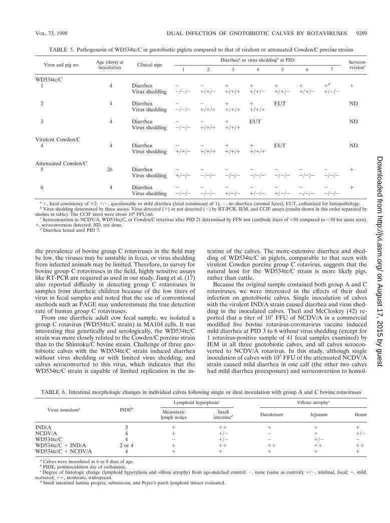
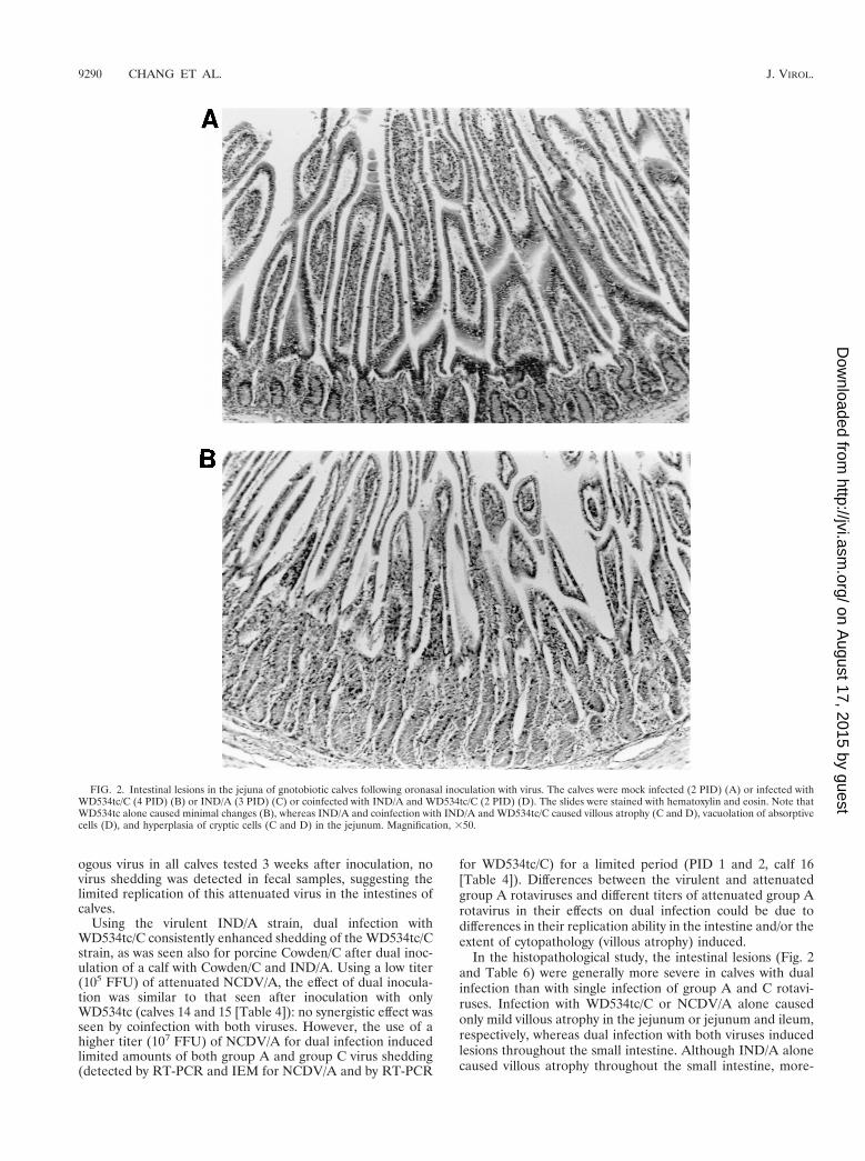
In the histopathological study, the intestinal lesions (Fig. 2and Table 6) were generally more severe in calves with dualinfection than with single infection of group A and C rotavi-ruses. Infection with WD534tc/C or NCDV/A alone causedonly mild villous atrophy in the jejunum or jejunum and ileum,respectively, whereas dual infection with both viruses inducedlesions throughout the small intestine. Although IND/A alonecaused villous atrophy throughout the small intestine, more-
FIG. 2. Intestinal lesions in the jejuna of gnotobiotic calves following oronasal inoculation with virus. The calves were mock infected (2 PID) (A) or infected withWD534tc/C (4 PID) (B) or IND/A (3 PID) (C) or coinfected with IND/A and WD534tc/C (2 PID) (D). The slides were stained with hematoxylin and eosin. Note thatWD534tc alone caused minimal changes (B), whereas IND/A and coinfection with IND/A and WD534tc/C caused villous atrophy (C and D), vacuolation of absorptivecells (D), and hyperplasia of cryptic cells (C and D) in the jejunum. Magnification, 350.
9290 CHANG ET AL. J. VIROL.
on August 17, 2015 by guest
http://jvi.asm.org/
Dow
nloaded from
widespread small intestinal lesions occurred in calves coin-fected with WD534tc/C and IND/A.
Interspecies transmission of group A rotaviruses has beensuggested, especially between humans and cattle. Gerna et al.(9) reported G6 serotype strains from humans (PA151 andPA169) (9, 10, 15), which were recognized by G6 monoclonalantibodies (bovine G6) and showed some genetic variation(about 10% in amino acid identity) with typical bovine G6strains (UK and NCDV strains). By RNA-RNA hybridization,most of the gene segments of PA151 G6 strains were closelyrelated to AU-1 human or NCDV bovine strains (15). Urasawaet al. (49) reported human G10 group A rotaviruses and sug-gested that this strain originated from a bovine host. Blackhallet al. (2) confirmed a bovine G1 strain analogous to human G1serotypes in Argentina by serological assays and sequenceanalysis of the VP7 genes. In addition, rotavirus strain 116E,isolated from the fecal specimen of a newborn asymptomatic
infant from India was identified as a P[11] type which waspreviously reported only in cattle (8).
Coinfection with group C and group A rotaviruses weredetected in diarrheic fecal samples from children and finishingpigs (17, 22). Kim et al. (22) reported a diarrhea outbreakassociated with group C rotaviruses in finishing (fattening) pigsand found these fecal samples also had group A rotavirusesdetectable only by a highly sensitive second-round PCR assay.Ishimaru et al. (16) studied the epidemiology of a gastroenter-itis outbreak associated with group C rotaviruses in the Mat-suyama district of Japan and noted that group C rotavirusgastroenteritis occurred following an epidemic of group A ro-tavirus infections, primarily in children aged 4 to 7 years butrarely in those aged 0 to 1 years.
Coinfections by group A rotaviruses and other enteropatho-gens including Escherichia coli in calves are common in nature(26, 30). The effects of coinfection of calves with group A
FIG. 2—Continued.
VOL. 73, 1999 DUAL INFECTION OF GNOTOBIOTIC CALVES BY ROTAVIRUSES 9291
on August 17, 2015 by guest
http://jvi.asm.org/
Dow
nloaded from

rotavirus and E. coli have been studied (11, 14, 30). Generallycoinfected calves had more severe diarrhea, lesions, and shed-ding of both agents than infection by either agent alone, butlittle information is available regarding the detailed mecha-nisms for these synergistic effects. When rotaviruses and E. coliwere coinoculated into calves, Runnels et al. (30) observed thatthere was more severe villous atrophy in the ilea of calvesinoculated with rotavirus and E. coli than in calves inoculatedwith only rotavirus. They also found more-intensive small in-testinal colonization by E. coli after coinfection with rotavirus.Gouet et al. (11) reported that experimental inoculation ofcalves with rotavirus, although not lethal in itself, followed byinoculation with a nonlethal dose of E. coli, led to dehydrationand death in newborn colostrum-deprived calves. Hess et al.(14) reported that coinfection of newborn colostrum-deprivedcalves with rotavirus and E. coli resulted in not only moresevere histological small intestinal lesions but also increasedshedding of both pathogens.
The mechanism for the enhancement of virus shedding ofthe WD534tc/C strain by coinfection with the virulent group Arotavirus is unclear. We hypothesize that the WD534tc/C strainis likely highly cell associated in the calf intestine as seen intissue culture (40), and following coinfection by a cytolyticgroup A rotavirus, the group C virus may be more readilyreleased by cytolysis from the infected cells. Alternatively,structural or nonstructural proteins of group A rotavirus couldassist in the replication or release of the WD534tc/C strain incoinfected cells or could have an indirect bystander effect ongroup C rotavirus-infected cells. A potential candidate is thenonstructural group A rotavirus protein NSP4, which has re-cently been shown to function as a viral enterotoxin in mousestudies and has membrane destabilization activity (1, 43).
Based on our results, which showed only limited or no virusshedding of the WD534tc/C strain in singly inoculated calvesand the genetic and antigenic similarity of this strain to porcinegroup C rotavirus, we propose that the WD534tc/C strain mayhave originated from a porcine host and thus replicates only toa limited extent in the calf intestine. However, if dual infec-tions with both group A and C rotaviruses occur in the bovinehost, synergic effects may cause the release of the WD534tc/Cstrain into the feces, leading to intraspecies (bovine) transmis-sion and further host adaptation after serial passage in the newhost. Interestingly, calves dually infected with WD534tc/C andattenuated group A rotavirus (analogous to currently used oralattenuated vaccine strain) also briefly shed both viruses. Thus,our findings suggest a potential novel hypothesis for the adap-tation of heterologous rotaviruses to new host species.
To explore potential mechanisms for the observed enhancedshedding of WD534tc/C rotavirus by coinfection with virulentgroup A rotavirus, in future studies, we plan to examine theeffects of group A and C rotavirus coinfection and of cotrans-fection and infection of a group A rotavirus NSP4 gene andgroup C rotavirus in an vitro cell culture system.
ACKNOWLEDGMENTS
We thank D. C. Hodgins for help with gnotobiotic calf work and C.Nielsen for help with histopathological studies.
Salaries and research support were provided by state and federalfunds appropriated to the Ohio Agricultural Research and Develop-ment Center (OARDC), The Ohio State University. This work waspartially supported by USDA NRICGP competitive grant 97-35204-4682 and grant RO1 AI 3356-04 from the National Institute of Allergyand Infectious Diseases, NIH.
REFERENCES1. Ball, J. M., P. Tian, C. Q. Zeng, A. P. Morris, and M. K. Estes. 1996. Age
dependent diarrhea induced by a rotaviral nonstructural glycoprotein. Sci-ence 272:101–104.
2. Blackhall, J., R. Bellinzoni, N. Mattion, M. K. Estes, J. L. LaTorre, and G.Magnusson. 1992. A bovine rotavirus serotype 1: serological characterizationof the virus and nucleotide sequence determination of the structural glyco-protein VP7 gene. Virology 189:833–837.
3. Bohl, E. H., L. J. Saif, K. W. Theil, A. G. Agnes, and R. F. Cross. 1982.Porcine pararotavirus: detection, differentiation from rotavirus, and patho-genesis in gnotobiotic pigs. J. Clin. Microbiol. 15:312–319.
4. Bridger, J. C. 1994. Non-group A rotaviruses, p. 369–407. In A. Z. Kapikian(ed.), Virus infections of the gastrontestinal tract. Marcel Dekker, NewYork, N.Y.
5. Chang, K. O., A. V. Parwani, D. Smith, and L. J. Saif. 1997. Detection ofgroup B rotaviruses in fecal samples from diarrheic calves and adult cowsand characterization of their VP7 genes. J. Clin. Microbiol. 35:2107–2110.
6. Chang, K. O., A. V. Parwani, and L. J. Saif. 1996. The characterization ofVP7 (G type) and VP4 (P type) genes of bovine group A rotaviruses fromfield samples using RT-PCR and RFLP analysis. Arch. Virol. 141:1727–1739.
7. Estes, M. K. 1990. Replication of rotaviruses, p. 1353–1404. In B. N. Fieldsand D. M. Knipe (ed.), Fields Virology, 2nd ed. Raven Press, New York,N.Y.
8. Gentsch, J. R., B. K. Das, B. Jiang, M. K. Bhan, and R. I. Glass. 1993.Similarity of the VP4 protein of human rotavirus strain 116E to that of thebovine B223 strain. Virology 194:424–430.
9. Gerna, G., A. Sarasini, M. Parea, S. Arista, P. Miranda, H. Brussow, Y.Hoshino, and J. Flores. 1994. Isolation and characterization of two distincthuman rotavirus strains with G6 specificity. J. Clin. Microbiol. 30:9–16.
10. Gerna, G., J. Sears, Y. Hoshino, A. D. Steele, O. Nakagomi, A. Sarasini, andJ. Flores. 1994. Identification of a new VP4 serotype of human rotaviruses.Virology 200:66–71.
11. Gouet, P., M. Contrepois, H. C. Dubourguier, Y. Riou, R. Scherrer, J.Laporte, J. F. Vautherot, J. Cohen, and R. L’Haridon. 1978. The experimen-tal production of diarrhoea in colostrum deprived axenic and gnotoxeniccalves with enteropathogenic Escherichia coli, rotavirus, coronavirus and ina combined infection of rotavirus and E. coli. Ann. Rech. Vet. 9:433–440.
12. Gouvea, V., J. R. Allen, R. I. Glass, Z. Fang, M. Bremont, J. Cohen, M. A.McCrae, L. J. Saif, P. Sinarachaatanant, and E. O. Caul. 1991. Detection ofgroup B and C rotaviruses by polymerase chain reaction. J. Clin. Microbiol.29:519–523.
13. Herring, A. J., N. F. Inglis, C. K. Ojeh, D. R. Snodgrass, and J. D. Menzies.1982. Rapid diagnosis of rotavirus infection by direct detection of viralnucleic acid in silver-stained polyacrylamide gels. J. Clin. Microbiol. 16:473–477.
14. Hess, R. G., P. A. Bachmann, G. Baljer, A. Mayr, A. Pospischil, and G.Schmid. 1984. Synergism in experimental mixed infections of newborn co-lostrum-deprived calves with bovine rotavirus and enterotoxigenic Esche-richia coli (ETEC). Zentbl. Vetmed. Reihe B 31:585–596.
15. Iizuka, M., M. Chiba, O. Masamune, G. Gerna, and O. Nakagomi. 1992.Molecular characterization of human rotavirus VP4 genes by polymerasechain reaction and restriction fragment length polymorphism assay. Micro-biol. Immunol. 37:729–735.
16. Ishimaru, Y., S. Nakano, H. Nakano, M. Oseto, and Y. Yamashita. 1991.Epidemiology of group C rotavirus gastroenteritis in Matsuyama, Japan.Acta Paediatr. Jpn. 33:50–56.
17. Jiang, B., P. H. Dennehy, S. Spangenberger, J. R. Gentsch, and R. I. Glass.1995. First detection of group C rotavirus in fecal specimens of children withdiarrhea in the United States. J. Infect. Dis. 172:45–50.
18. Jiang, B., Y. Qian, H. Tsunemitsu, K. Y. Green, and L. J. Saif. 1991. Analysisof the gene encoding the outer capsid glycoprotein (VP7) of group C rota-viruses by Northern and dot blot hybridization. Virology 184:433–436.
19. Jiang, B., H. Tsunemitsu, J. R. Gentsch, R. I. Glass, K. Y. Green, Y. Qian,and L. J. Saif. 1992. Nucleotide sequence of gene 5 encoding the inner capsidprotein (VP6) of bovine group C rotavirus: comparison with correspondinggenes of group C, A, and B rotaviruses. Virology 190:542–547.
20. Kang, S. Y., L. J. Saif, and K. L. Miller. 1989. Reactivity of VP4-specificmonoclonal antibodies to a serotype 4 porcine rotavirus with distinct sero-types of human (symptomatic and asymptomatic) and animal rotaviruses.J. Clin. Microbiol. 27:2744–2750.
21. Kapikian, A. Z., and R. M. Chanock. 1990. Rotaviruses, p. 1353–1404. InB. N. Fields and D. M. Knipe (ed.), Fields virology, 2nd ed. Raven Press,New York, N.Y.
22. Kim, Y., K. O. Chang, B. Straw, and L. J. Saif. 1999. Characterization ofgroup C rotaviruses associated with diarrhea outbreaks in feeder pigs.J. Clin. Microbiol. 37:1484–1488.
23. Laemmli, U. K. 1970. Cleavage of structural proteins during the assembly ofthe head of bacteriophage T4. Nature 27:680–685.
24. Mattion, N. M., J. Cohen, and M. K. Estes. 1994. The rotavirus proteins, p.169–249. In A. Z. Kapikian (ed.), Virus infections of the gastrontestinal tract.Marcel Dekker, New York, N.Y.
25. Meyer, R. C., E. H. Bohl, and E. M. Kohler. 1964. Procurement and main-tenance of germ-free swine for microbiological investigations. Appl. Micro-biol. 12:295–300.
26. Moon, H. W., A. W. McClurkin, R. E. Isaacson, J. Pohlenz, S. M. Skartvedt,K. G. Gillette, and A. L. Baetz. 1978. Pathogenic relationships of rotavirus,
9292 CHANG ET AL. J. VIROL.
on August 17, 2015 by guest
http://jvi.asm.org/
Dow
nloaded from

Escherichia coli, and other agents in mixed infections in calves. J. Am. Vet.Med. Assoc. 173:577–583.
27. Morin, M., R. Magar, and Y. Robinson. 1990. Porcine group C rotavirus asa cause of neonatal diarrhea in a Quebec swine heard. Can. J. Vet. Res.54:385–389.
28. Nicolas, J. C., J. Cohen, B. Fortier, M. H. Lourenco, and F. Bricout. 1983.Isolation of a human pararotavirus. Virology 124:181–184.
29. Parwani, A. V., A. Lucchelli, and L. J. Saif. 1996. Identification of group Brotaviruses with short genome electropherotypes from adult cows with diar-rhea. J. Clin. Microbiol. 34:1303–1305.
30. Runnels, P. L., H. W. Moon, P. J. Matthews, S. C. Whipp, and G. N. Woode.1986. Effects of microbial and host variables on the interaction of rotavirusand Escherichia coli infections in gnotobiotic calves. Am. J. Vet. Res. 47:1542–1550.
31. Saif, L. J., and B. Jiang. 1994. Nongroup A rotaviruses of humans andanimals, p. 339–371. In R. F. Ramig (ed.), Rotaviruses. Springer-Verlag,Berlin, Germany.
32. Saif, L. J., E. H. Bohl, E. M. Kohler, and J. H. Hughes. 1977. Immuneelectron microscopy of transmissible gastroenteritis virus and rotavirus (re-ovirus-like agent) of swine. Am. J. Vet. Res. 38:13–20.
33. Saif, L. J., E. H. Bohl, K. W. Theil, R. F. Cross, and J. A. House. 1980.Rotavirus-like, calicivirus-like and 23-nm virus-like particles associated withdiarrhea in young pigs. J. Clin. Microbiol. 12:105–111.
34. Saif, L. J., K. V. Brock, D. R. Redman, and E. M. Kohler. 1991. Winterdysentery in dairy herds: electron microscopic and serological evidence foran association with coronavirus infection. Vet. Rec. 128:447–449.
35. Saif, L. J., B. I. Rosen, and A. V. Parwani. 1994. Animal rotavirus, p.289–314. In A. Z. Kapikian (ed.), Virus infections of the gastrontestinal tract.Marcel Dekker, New York, N.Y.
36. Saif, L. J., B. I. Rosen, K. Y. Kang, and K. L. Miller. 1988. Cell culturepropagation of rotaviruses. J. Tissue Cult. Methods 11:147–156.
37. Saif, L. J., L. A. Terrett, K. L. Miller, and R. F. Cross. 1988. Serial propa-gation of porcine group C rotavirus (pararotavirus) in a continuous cell lineand characterization of the passaged virus. J. Clin. Microbiol. 26:1277–1282.
38. Sambrook, J., E. F. Fritsch, and T. Maniatis. 1989. Molecular cloning: alaboratory manual, 2nd ed. Cold Spring Harbor Laboratory Press, ColdSpring Harbor, N.Y.
39. Smith, D. R., P. J. Fedorka-Cray, R. Mohan, K. V. Brock, T. E. Wittum, P. S.Morley, K. H. Hoblet, and L. J. Saif. 1998. Epidemiologic herd-level assess-
ment of causative agents and risk factors for winter dysentery in dairy cattle.Am. J. Vet. Res. 59:994–1001.
40. Terrett, L. A., L. J. Saif, K. W. Theil, and E. M. Kohler. 1987. Physicochem-ical characterization of porcine pararotavirus and detection of virus and viralantibodies using cell culture immunofluorescence. J. Clin. Microbiol. 25:268–272.
41. Terrett, L. A., and L. J. Saif. 1987. Serial propagation of porcine group Crotavirus (pararotavirus) in primary porcine kidney cell cultures. J. Clin.Microbiol. 25:1316–1319.
42. Theil, K. W., and C. M. McCloskey. 1995. Rotavirus shedding in feces ofgnotobiotic calves orally inoculated with a commercial rotavirus-coronavirusvaccine. J. Vet. Diagn. Invest. 7:427–432.
43. Tian, P., J. M. Ball, C. Q. Zeng, and M. K. Estes. 1996. The rotavirusnonstructural glycoprotein NSP4 possesses membrane destabilization activ-ity. J. Virol. 70:6973–6981.
44. Tsunemitsu, H., B. Jiang, and L. J. Saif. 1992. Detection of group C rota-virus antigens and antibodies in animals and humans by enzyme-linkedimmunosorbent assays. J. Clin. Microbiol. 30:2129–2134.
45. Tsunemitsu, H., B. Jiang, and L. J. Saif. 1996. Sequence comparison of theVP7 gene encoding the outer capsid glycoprotein among animal and humangroup C rotaviruses. Arch. Virol. 141:705–713.
46. Tsunemitsu, H., B. Jiang, Y. Yamashita, M. Oseto, H. Ushijima, and L. J.Saif. 1992. Evidence of serologic diversity within group C rotaviruses. J. Clin.Microbiol. 30:3009–3012.
47. Tsunemitsu, H., C. K. Ojeh, B. Jiang, R. A. Simkins, P. A. Weilnau, and L. J.Saif. 1992. Production and characterization of monoclonal antibodies toporcine group C rotaviruses cross-reactive with group A rotaviruses. Virol-ogy 91:272–281.
48. Tsunemitsu, H., L. J. Saif, B. M. Jiang, M. Shimizu, M. Hiro, H. Yamaguchi,T. Ishiyama, and T. Hirai. 1991. Isolation, characterization, and serial prop-agation of a bovine group C rotavirus in a monkey kidney cell line (MA104).J. Clin. Microbiol. 29:2609–2613.
49. Urasawa, S., A. Hasegawa, T. Urasawa, K. Taniguchi, F. Wakasugi, H.Suzuki, S. Inouye, B. Pongprot, J. Supawadee, and S. Suprasert. 1992.Antigenic and genetic analyses of human rotaviruses in Chiang Mai, Thai-land: evidence for a close relationship between human and animal rotavi-ruses. J. Infect. Dis. 166:227–234.
50. Ward, L. A., B. I. Rosen, L. Yuan, and L. J. Saif. 1996. Pathogenesis of anattenuated and a virulent strain of group A human rotavirus in neonatalgnotobiotic pigs. J. Gen. Virol. 77:1431–1441.
VOL. 73, 1999 DUAL INFECTION OF GNOTOBIOTIC CALVES BY ROTAVIRUSES 9293
on August 17, 2015 by guest
http://jvi.asm.org/
Dow
nloaded from
Related Documents


