Fat Embolism Syndrome Dr. Ankit Desai Dr.B.L.Chandrakar

Welcome message from author
This document is posted to help you gain knowledge. Please leave a comment to let me know what you think about it! Share it to your friends and learn new things together.
Transcript
Fat Embolism Syndrome
Dr. Ankit Desai
Dr.B.L.Chandrakar
History
First diagnosed in 1873 by Dr Von Bergmann
Published in 1879 Fenger and Salisbury.
FE vs FES
Fat Embolism:
Traumatic fat embolism occurs in up to 90% of individuals with severe skeletal injuries, but < 10% of such patients have any clinical symptoms / signs
Fat Embolism Syndrome:
FE with clinical manifestation .
Some definitions…
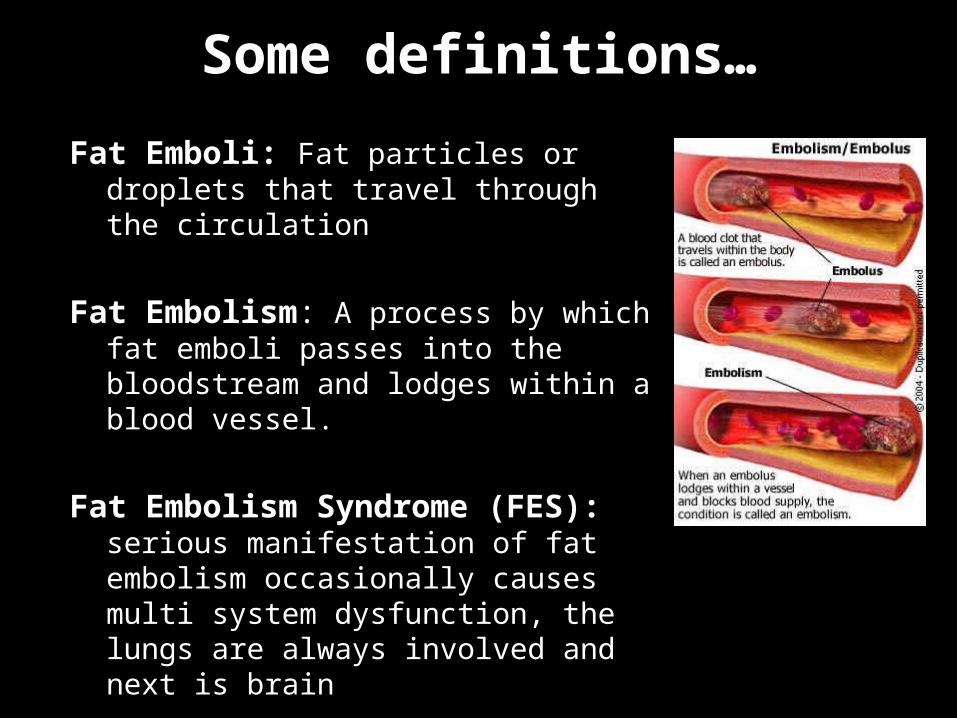
Fat Emboli: Fat particles or droplets that travel through the circulation
Fat Embolism: A process by which fat emboli passes into the bloodstream and lodges within a blood vessel.
Fat Embolism Syndrome (FES): serious manifestation of fat embolism occasionally causes multi system dysfunction, the lungs are always involved and next is brain
Incidence: 1-3% femur #, 5-10% if bilateral or multiple.
Mortality: 5-15%
Clinical diagnosis, No specific laboratory test is diagnostic
Mostly associated with long bone/pelvic #s, and more frequent in closed fractures. commanly with lower limb injury
Onset is 24-72 hours from initial insult

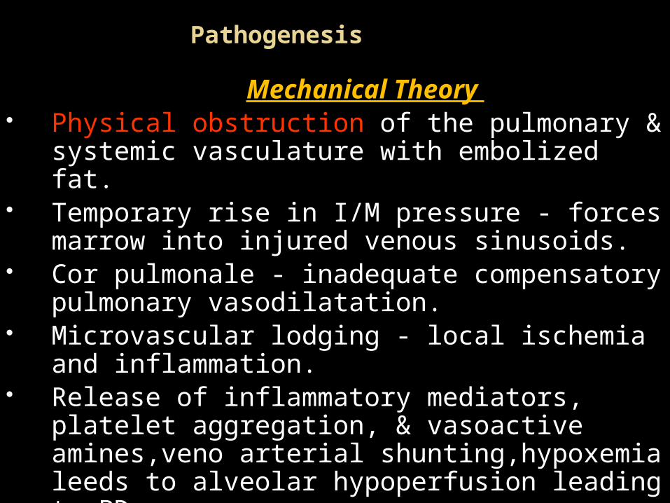
Pathogenesis
Mechanical Theory Physical obstruction of the pulmonary & systemic
vasculature with embolized fat. Temporary rise in I/M pressure - forces marrow into
injured venous sinusoids. Cor pulmonale - inadequate compensatory pulmonary
vasodilatation. Microvascular lodging - local ischemia and inflammation. Release of inflammatory mediators, platelet aggregation,
& vasoactive amines,veno arterial shunting,hypoxemia leeds to alveolar hypoperfusion leading to PD
Size of fat globules 2 to 200 micron Blood vessele <75 micron

The biochemical theory
Free fatty acid is culprit Circulating FFAs -directly toxic to Pneumocytes /
capillary Endothelium in the lung – decrease surfactant ,nterstitial hemorrhage, edema & chemical pneumonitis.
Local hydrolysis of Triglycerides and fat emboli by pneumocyte lipase -- Increase fatty acid .
Coexisting shock, hypovolemia and sepsis - reduce liver
flow exacerbate the toxic effects of FFAs.
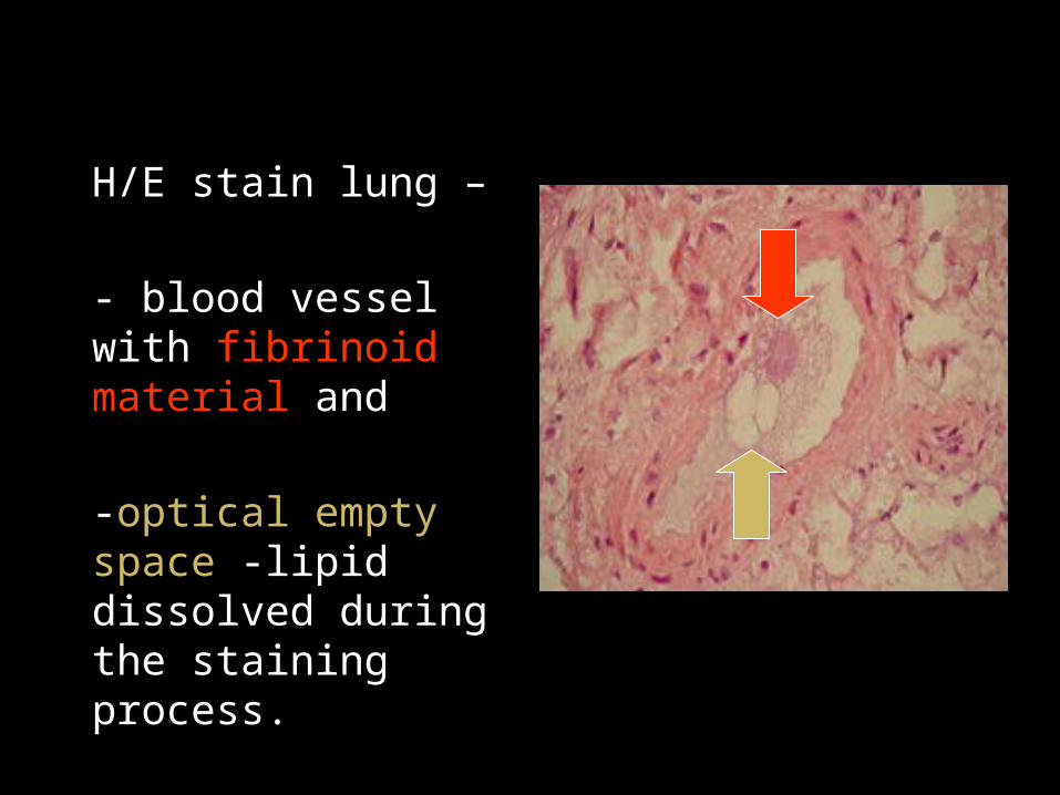
H/E stain lung –
- blood vessel with fibrinoid material and
-optical empty space -lipid dissolved during the staining process.
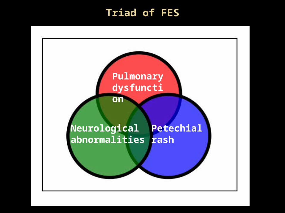
Triad of FES
Pulmonary dysfunction
Neurological abnormalities
Petechial rash

Early Signs
Dyspnea,
Tachypnea
Hypoxemia PaO2 < 60 mm Hg confusion

Pulmonary Picture
Clinically Tachpnea, Dyspnea, Hypoxia, rales, pleural friction rub & ARDS.
High spiking temperatures. Hypoxemia - ventilation-perfusion mismatch &
intrapulmonary shunting. Acute cor pulmonale -respiratory distress, hypoxemia, hypotension and elevated CVP.
½ of pts require mechanical ventilation

Imagining
• Chest x-ray– shows multiple flocculent shadows (snow storm appearance). picture
may be complicated by infection or pulmonary edema.– CT chest: ground glass opacification with interlobular septal thickening
• CT Scan brain – may be normal or may reveal diffuse white-matter petechial
haemorrhages
• Helical CT Scan chest– may be normal as the fat droplets are lodged in capillary beds. Can
detect lung contusion, acute lung injury, or ARDS may be evident.

Chest X-ray
ER admitAP & expiratory film so we cannot comment on cardiac shadow. However, there is no evidence of lung contusion, pneumo, haemo or pneumohaemothorax.
SICU admit (12 hours later)upper lobe diversion and bilateral pulmonary infiltrates
Altaf Hussain: “A Fatal Fat Embolism.” The Internet Journal of Anesthesiology, 2004. Volume 8 Number 2.

Neurological findings CNS signs usually occur after respiratory symptoms
- nonspecific - features of diffuse encephalopathy
Acute confusion, stupor, coma, rigidity, or convulsions - Transient and reversible in most cases
CT Head: general edema – nonspecific
MRI brain: Low density on T1 & High intensity T2 signal - correlates to degree of impairment

MRI showing foci of ischemia suggestive of fat embolism syndrome
post operative day 14 and shows evolving cortical infarctions
post operative day 2 showing multiple hyperintense areas consistent with multiple emboli
Source:http://www.ispub.com/journal/the_internet_journal_of_anesthesiology/volume_19_number_2/article/acute_fatal_fat_embolism_syndrome_in_bilateral_total_knee_arthroplasty_a_review_of_the_fat_embolism_syndrome.html
Rash Reddish-brown non-palpable Petechial rash - upper
anterior body, chest, neck, upper arm, axilla, shoulder, oral mucous membranes and conjunctivae in 20 - 50% patients.2 to 3re day
Resolvs in 7 day
Pathognomonic, however, it appears late and disappears within hours.
Results from occlusion of dermal capillaries by fat globules - extravasations of RBC
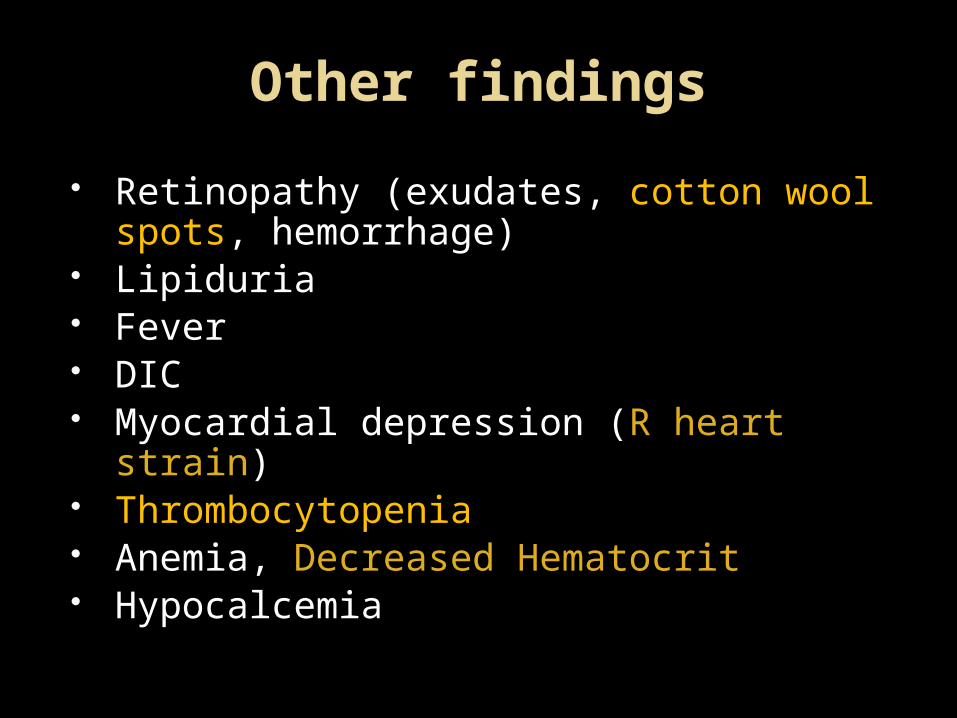
Other findings
Retinopathy (exudates, cotton wool spots, hemorrhage)
Lipiduria Fever DIC Myocardial depression (R heart strain) Thrombocytopenia Anemia, Decreased Hematocrit Hypocalcemia
Diagnostic Criteria
Gurd’s criteria
Most commonly used
1 major, plus 4 minor
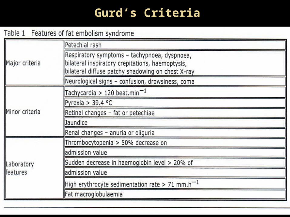
Gurd’s Criteria
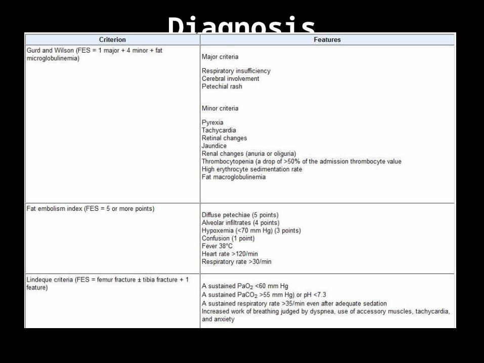
Diagnosis Clinical examination preferred over diagnostic
Continuous pulse oximetry monitoring - at-risk patients ( those patients with long bone fractures) - detecting desaturations early.
Consultations recommended include orthopedists, neurologists/ neurosurgeons, trauma care specialists, critical care specialists, pulmonologists, hematologists, and nutritionists.

Treatment
The most effective prophylactic measure - operative reduction/rigid fixation of long bone fractures as soon as possible. Higher incidence (5 fold) when fixation delayed greater than 24 hours.
Supportive care includes maintenance of adequate
oxygenation and ventilation, stable hemodynamics, blood products as clinically indicated, hydration, prophylaxis of DVT and stress-related GI bleeding.
Albumin has been recommended - not only restores blood volume / binds fatty acids - may decrease the extent of lung injury.
High dose corticosteroids have been effective in preventing development of FES in several trials, but controversy on this issue still persists.
Heparin has also been proposed as it activates lipase, but no evidence exists for its use in FES.
Ethanol/alcohol in bolus dose iv :as lipase inhibitor
DURATION OF FES
Difficult to predict –FES is frequently subclinical or overshadowed by other illnesses or injuries.
Increased alveolar-to-arterial oxygen gradient and neurologic deficits, including coma, may last days or weeks.
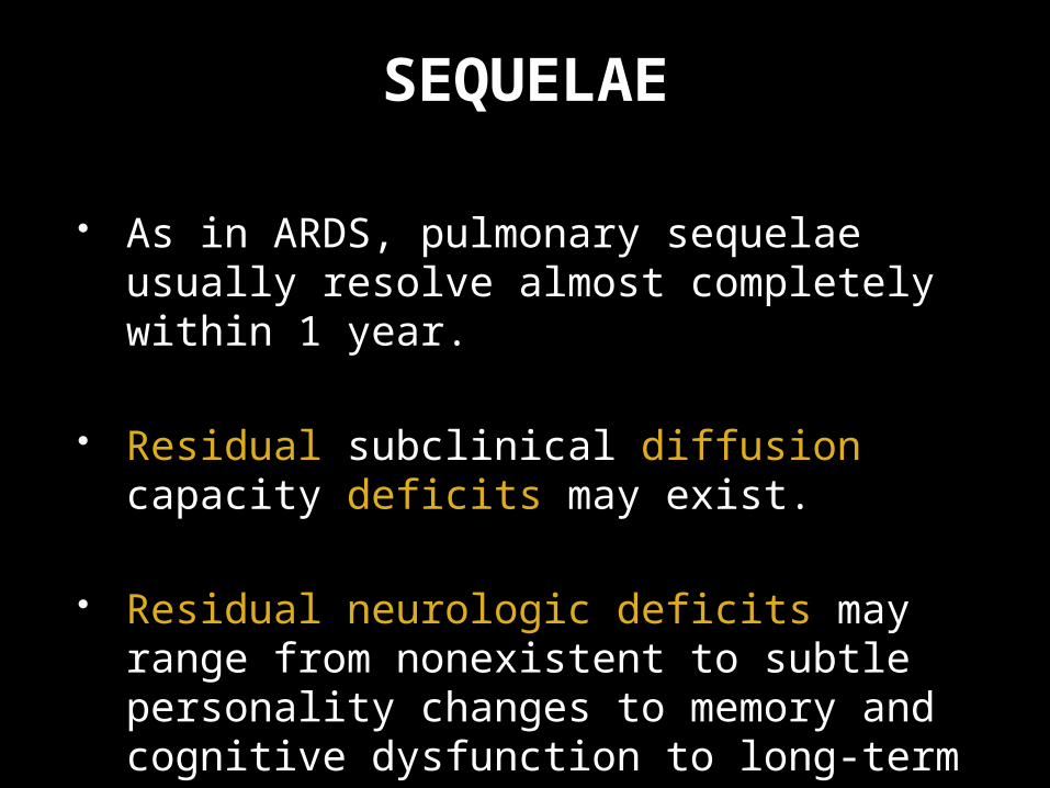
SEQUELAE
As in ARDS, pulmonary sequelae usually resolve almost completely within 1 year.
Residual subclinical diffusion capacity deficits may exist.
Residual neurologic deficits may range from nonexistent to subtle personality changes to memory and cognitive dysfunction to long-term focal deficits.
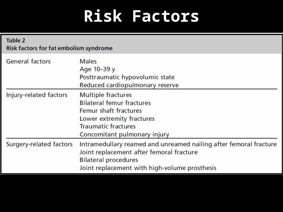
Risk Factors

THANK U
Related Documents