I Republic of Iraq Ministry of Higher Education And scientific Research University of Baghdad College of Dentistry Extraction in orthodontic A project Submitted to Collage of Dentistry, University of Baghdad. Department of orthodontics in fulfillment for the requirement to award the degree B.D.S Done by Montather khalouq Abraham 5th Grade Supervisor Dr. Haider Mohammed Ali B.D.S, M.S.C lecturer Baghdad Iraq 2017_1438

Welcome message from author
This document is posted to help you gain knowledge. Please leave a comment to let me know what you think about it! Share it to your friends and learn new things together.
Transcript

I
Republic of Iraq
Ministry of Higher Education
And scientific Research
University of Baghdad
College of Dentistry
Extraction in orthodontic
A project Submitted to Collage of Dentistry, University of Baghdad. Department of
orthodontics in fulfillment for the requirement to award the degree B.D.S
Done by
Montather khalouq Abraham 5th Grade
Supervisor
Dr. Haider Mohammed Ali
B.D.S, M.S.C lecturer
Baghdad Iraq
2017_1438

II
List of content
List of content II
List of figures IV
Introduction 1
Chapter one 2
Review of literature 2
1.1 EXTRACTION GUIDELINES 2
1.1.1 Less than 4 mm arch length discrepancy: 2
1.1.2 Arch length discrepancy 5 to 9 mm: 2
1.1.3 Arch length discrepancy 10 mm or more: 2
1.2 SELECTION OF TEETH TO BE EXTRACTED 3
1.3 TYPES OF EXTRACTION IN ORTHODONTICS 4
1.3.1 Balancing Extractions 4
1.3.2 Compensating Extractions 4
1.3.3 Enforced Extractions 5
1.3.4 Therapeutic extraction 6
1.3.5 Wilkinson extraction 6
1.4 TEETH TO BE EXTRACTED 6
1.4.1 Incisors 6
1.4.2 Canines 9
1.4.3 Permenant premolars 10
1.4.4 First permanent molars 13
1.4.5 Second permanent molars 15
1.4.6 Third permanent molars 17
1.5 SERIAL EXTRACTION 18
1.5.1 Advantages of Serial Extractions 19
1.5.2 Disadvantages of Serial Extractions 19
1.5.3 Complications of Serial Extractions 19

III
1.5.4 Indications For serial Extractions 20
1.5.5 Contra-indication for serial extractions 20
CHAPTER TWO 21
MATERIAL METHOD 21
CHAPTER THREE 22
RESULT 22
CHAPTER FOUR 27
DISCUSSION 27
CHAPTER FIVE 29
SUGGESTION 29
REFERENCE 30
IV
List of figures
Figure 1.2.balancing extraction ............................................................................................. 4
Figure 1.3.Compensating extraction .................................................................................... 5
Figure 1.6.Enforced extraction of carious tooth ............................................................... 5
Figure 1.5.Enforced extraction of fracture tooth.............................................................. 5
Figure 1.4.Enforced extraction of impacted second molar ............................................ 5
Figure 1.7.Poor periodontal teeth health ............................................................................ 5
Figure 1.8.Therapeutic extractions ....................................................................................... 6
Figure 1.9.Crowding of 5mm localized in lower labial segment .................................. 7
Figure 1.8.Single lower incisor excluded from the arch ................................................. 7
Figure 1.10.Deep openbite ....................................................................................................... 8
Figure 1.12.Maxillary central incisor with dilacerated root ......................................... 9
Figure 1.11.Peg shaped lateral inciser ................................................................................. 9
Figure 1.13.Impacted upper incisors .................................................................................... 9
Figure 1.14.Orthopantomgraphe view impacted canine ................................................ 9
Figure 1.15.Occlusal view of impacted canine ................................................................... 9
Figure 1.6.Extraction of first premolar for proper alignment of caine ....................11
Figure 1.17.Impacted second premolars ...........................................................................12
Figure 1.18.Hypomineralized permanent molars ...........................................................15
Figure 1.19.Impacted third and second molar ................................................................16
Figure 2.1 sample of survey paper ......................................................................................21
Table 3.1.How do you decide extraction in your clinic .................................................22
Chart 3.1.Percentage of answers of question one ...........................................................22
Table 3.2.In cl I molars and canines relationship, we have anterior crowding
more than 6 mm in upper arch do you prefer ....................................................... 23
Chart 3.2.Precentage of answers of questions two .........................................................23

V
Table 3.3. In cl I molars and canines relationship , we have have anterior
crowding more than 6 mm in lower arch do you prefer .................................... 24
Chart 3.3.Precentage of answers of questions two .........................................................24
Table 3.4.Which approach do you follow in your clinic ...............................................25
Chart 3.4.Precentage of answers of questions four ........................................................25
Table 3.5 Do you set your anchorage posteriorly (TPA or TADs) and placing
working stainless steel wires before doing extraction? ........................................ 26
Chart 3.5 precentage of answers of question five ...........................................................26
1
Introduction
The role of extractions in orthodontic treatment has been a controversial subject
for over a century. It is fair to say that even today, opinion is divided on whether
extractions are used too frequently in the correction of malocclusion.
Recently, the extraction debate has reopened, with some individuals believing
that expansion of the jaws and retraining of posture can obviate the need for
extractions and produce stable results (Travess et al. 2004).
These claims are for the most part unsubstantiated. If teeth are genuinely
crowded as opposed to being irregular then arch alignment can be achieved by
Enlargement of the arch form or Reduction in tooth size or Reduction in tooth number
The reduction in tooth number is usually achieved with extractions and these cases
ideally need to be compared with treated non extraction cases with spacing, cases
treated by arch expansion to accommodate crowding and untreated normal occlusions.
In a review of these issues it was concluded that arch length reduces in most cases,
including untreated normal occlusion (Travess et al.2004).
Any lateral expansion across the mandibular canines decreases after treatment but
this is also seen in those cases which have no orthodontic treatment. It was further
recognized that mandibular anterior crowding is a continuing phenomenon seen in
patients into the fourth decade and likely beyond. The degree of anterior crowding
seen at the end of retention is variable and unpredictable ( Travess et al.2004).

2
Chapter one
Review of literature
1.1 Extraction Guidelines
Extraction Contemporary guidelines for orthodontic in Class I crowding cases
can be summarized as follows:
1.1.1 Less than 4 mm arch length discrepancy:
Extraction rarely indicated (only if there is severe incisor protrusion or in a few
instances, a severe vertical discrepancy) (Phulari.2011).
Some cases, this amount of crowding can be managed without arch expansion by
slightly reducing the selected teeth, being careful to coordinate the amount of
reduction in the upper and lower arch (Proffit et al.2013).
1.1.2 Arch length discrepancy 5 to 9 mm:
Non-extraction or extraction treatment possible (Phulari.2011, Proffit et al
2013).
The decision depends on both the hard- and soft-tissue characteristics of the
patient and on how the final position of the incisors will be controlled, any of several
different teeth could be chosen for extraction. Non-extraction treatment usually
requires transverse expansion across the molars and premolars, and additional
treatment time if the posterior teeth are to be moved distally, to increase arch length
(Proffit et al. 2013) (Jeryl et al.2015).
1.1.3 Arch length discrepancy 10 mm or more:
Extraction almost always required (Phulari.2011, Proffit et al.2013).
For these patients, the amount of crowding virtually equals the amount of tooth mass
being removed, and there would be little or no effect on lip support and facial
appearance. The extraction choice is four first premolars or perhaps upper first

3
premolars and mandibular lateral incisors. Second premolar or molar extraction rarely
is satisfactory because it does not provide enough space near crowded anterior teeth or
options to correct midline discrepancies (Proffit et al.2013).
1.2 Selection of teeth to be extracted
Extraction for orthodontic reasons will be governed by:
1- Condition of the teeth: Fractured, high grossly carious teeth, root canal treated teeth
and teeth with large restorations are preferred for extraction over healthy teeth. The
main consideration is the long-term prognosis for the tooth rather than the appearance
(Phulari.2011, Millet et al.2005).
2- Position of the crowding: Crowding in one part of the arch is more readily corrected
if extractions are done in that part rather than a remote area of the arch. However,
incisor crowding is usually relieved by premolar extraction as it gives a more pleasing
appearance and occlusal balance than with incisor extraction. The first premolar,
positioned in the center of each quadrant, is usually near the area of crowding whether
Figure 1.1.Crowding level

4
in the anterior or buccal segment. Hence, it is also the tooth most frequently extracted
along with orthodontic treatment (Phulari.2011, Millet et al.2005).
3- Position of the teeth: Grossly malpositioned teeth which are difficult to align, may
often be the teeth of choice for extraction. The position of the apex of the tooth must
be considered as it is more difficult to move the apex than the crown (Phulari.2011,
Millet et al.2005).
1.3 Types of Extraction in orthodontics
1.3.1 Balancing Extractions
Balancing extractions may be defined as the removal of a tooth on the opposite
side the same arch (although not necessarily the same) in order to preserve symmetry
(Phulari. 2011).
1.3.2 Compensating Extractions
Removal of the equivalent tooth in the opposing arch to maintain buccal
occlusion, if the third molar is extracted in the right quadrant of the maxillary arch
then the third molar in the right quadrant of the mandibular arch is also extracted. This
type of extraction is called as compensatory extraction (Phulari.2011).
Figure 1.2.balancing extraction
5
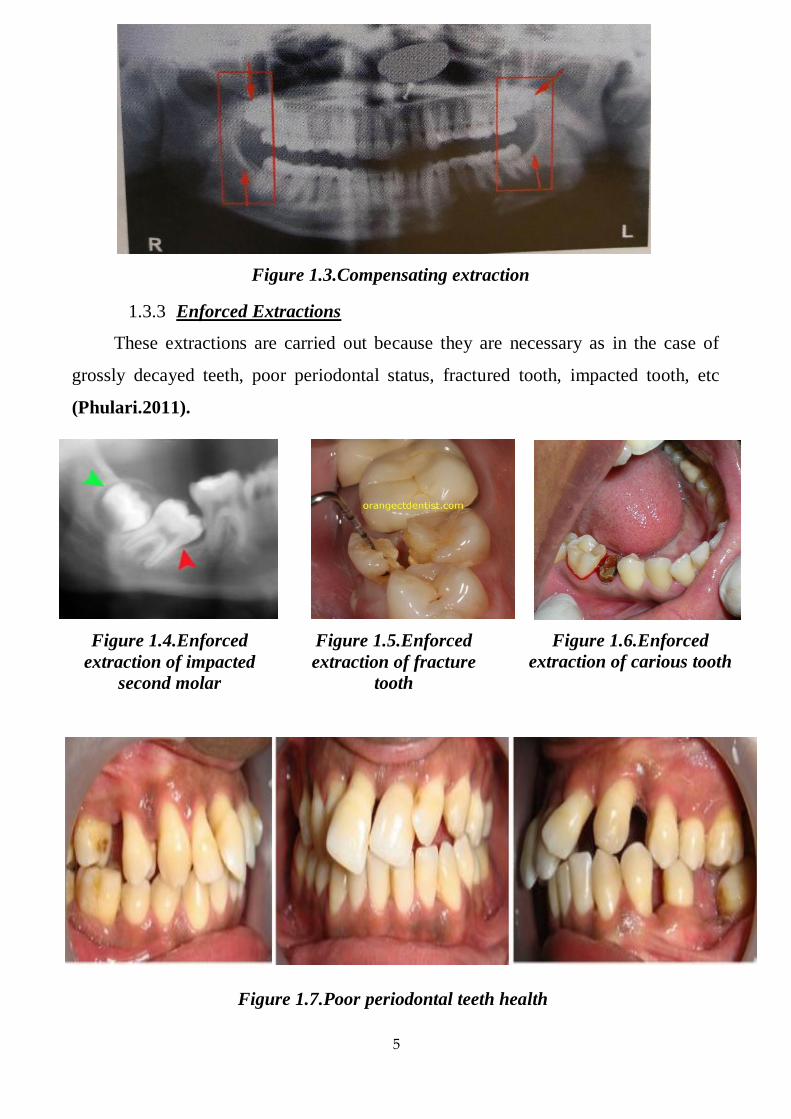
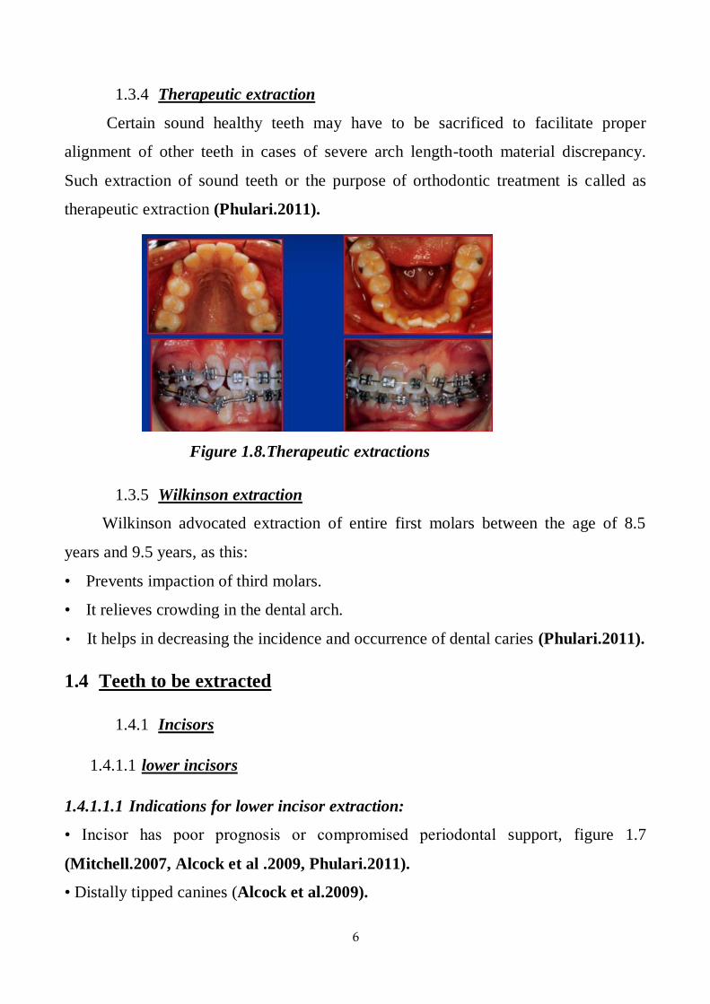
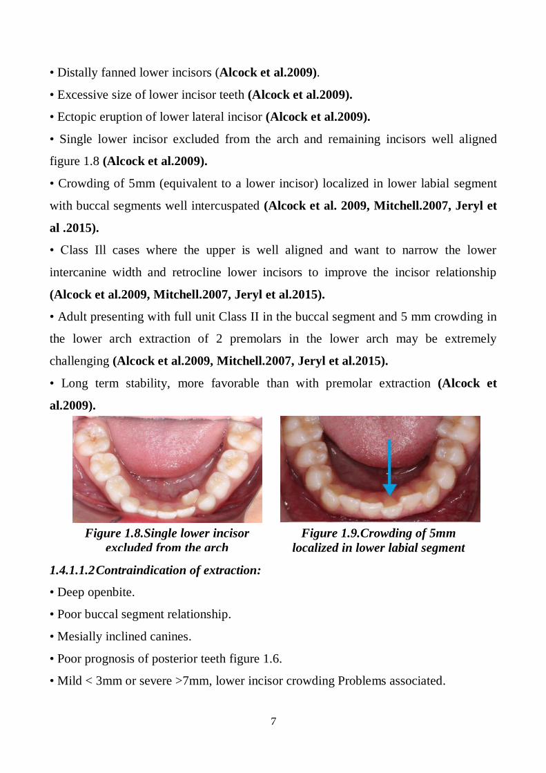
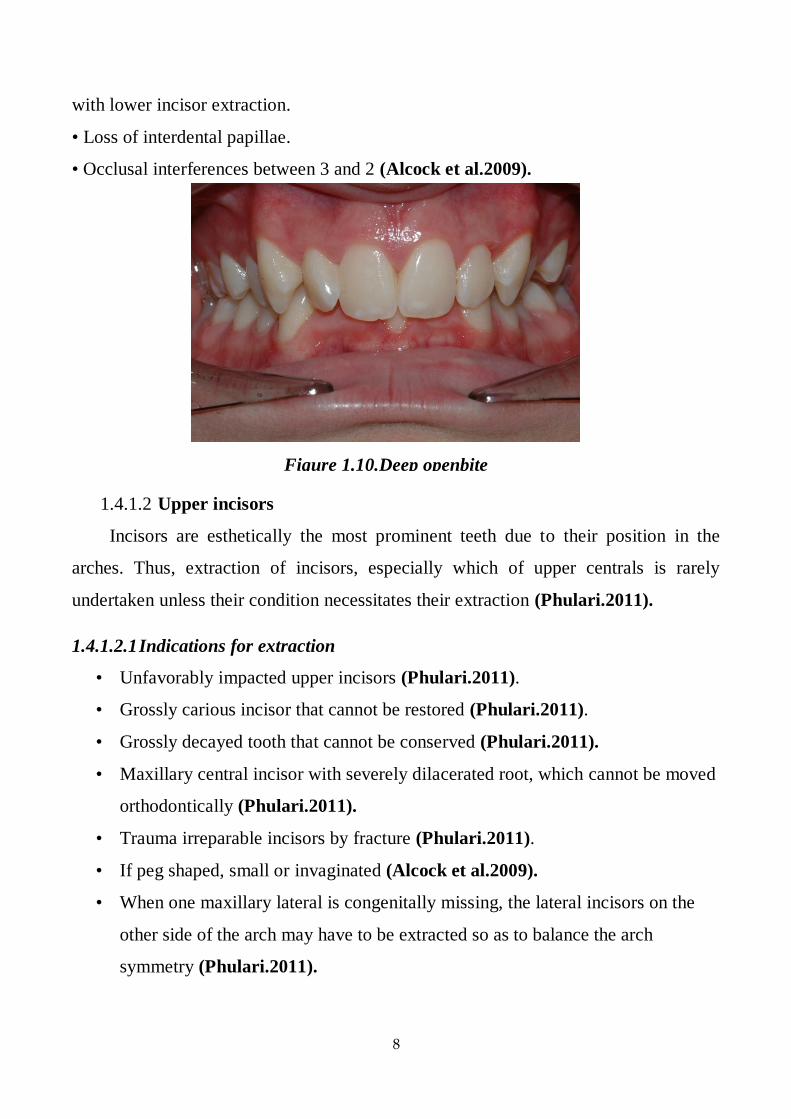
1.3.3 Enforced Extractions
These extractions are carried out because they are necessary as in the case of
grossly decayed teeth, poor periodontal status, fractured tooth, impacted tooth, etc
(Phulari.2011).
Figure 1.3.Compensating extraction
Figure 1.4.Enforced
extraction of impacted
second molar
Figure 1.5.Enforced
extraction of fracture
tooth
Figure 1.6.Enforced
extraction of carious tooth
Figure 1.7.Poor periodontal teeth health
6
1.3.4 Therapeutic extraction
Certain sound healthy teeth may have to be sacrificed to facilitate proper
alignment of other teeth in cases of severe arch length-tooth material discrepancy.
Such extraction of sound teeth or the purpose of orthodontic treatment is called as
therapeutic extraction (Phulari.2011).
1.3.5 Wilkinson extraction
Wilkinson advocated extraction of entire first molars between the age of 8.5
years and 9.5 years, as this:
• Prevents impaction of third molars.
• It relieves crowding in the dental arch.
• It helps in decreasing the incidence and occurrence of dental caries (Phulari.2011).
1.4 Teeth to be extracted
1.4.1 Incisors
1.4.1.1 lower incisors
1.4.1.1.1 Indications for lower incisor extraction:
• Incisor has poor prognosis or compromised periodontal support, figure 1.7
(Mitchell.2007, Alcock et al .2009, Phulari.2011).
• Distally tipped canines (Alcock et al.2009).
Figure 1.8.Therapeutic extractions
7
• Distally fanned lower incisors (Alcock et al.2009).
• Excessive size of lower incisor teeth (Alcock et al.2009).
• Ectopic eruption of lower lateral incisor (Alcock et al.2009).
• Single lower incisor excluded from the arch and remaining incisors well aligned
figure 1.8 (Alcock et al.2009).
• Crowding of 5mm (equivalent to a lower incisor) localized in lower labial segment
with buccal segments well intercuspated (Alcock et al. 2009, Mitchell.2007, Jeryl et
al .2015).
• Class Ill cases where the upper is well aligned and want to narrow the lower
intercanine width and retrocline lower incisors to improve the incisor relationship
(Alcock et al.2009, Mitchell.2007, Jeryl et al.2015).
• Adult presenting with full unit Class II in the buccal segment and 5 mm crowding in
the lower arch extraction of 2 premolars in the lower arch may be extremely
challenging (Alcock et al.2009, Mitchell.2007, Jeryl et al.2015).
• Long term stability, more favorable than with premolar extraction (Alcock et
al.2009).
1.4.1.1.2 Contraindication of extraction:
• Deep openbite.
• Poor buccal segment relationship.
• Mesially inclined canines.
• Poor prognosis of posterior teeth figure 1.6.
• Mild < 3mm or severe >7mm, lower incisor crowding Problems associated.
Figure 1.8.Single lower incisor
excluded from the arch
Figure 1.9.Crowding of 5mm
localized in lower labial segment
8
with lower incisor extraction.
• Loss of interdental papillae.
• Occlusal interferences between 3 and 2 (Alcock et al.2009).
1.4.1.2 Upper incisors
Incisors are esthetically the most prominent teeth due to their position in the
arches. Thus, extraction of incisors, especially which of upper centrals is rarely
undertaken unless their condition necessitates their extraction (Phulari.2011).
1.4.1.2.1 Indications for extraction
• Unfavorably impacted upper incisors (Phulari.2011).
• Grossly carious incisor that cannot be restored (Phulari.2011).
• Grossly decayed tooth that cannot be conserved (Phulari.2011).
• Maxillary central incisor with severely dilacerated root, which cannot be moved
orthodontically (Phulari.2011).
• Trauma irreparable incisors by fracture (Phulari.2011).
• If peg shaped, small or invaginated (Alcock et al.2009).
• When one maxillary lateral is congenitally missing, the lateral incisors on the
other side of the arch may have to be extracted so as to balance the arch
symmetry (Phulari.2011).
Figure 1.10.Deep openbite

9
• Buccally or lingually blocked out lateral incisor with good contact between
central incisor and canine (Phulari.2011).
• When one maxillary lateral is congenitally missing, the lateral incisors on the
other side of the arch may have to be extracted so as to balance the arch
symmetry (Phulari.2011).
1.4.2 Canines
Rarely tooth of choice to extract
• Aesthetic important - canine eminence
• Functionally important - canine guidance
• Long root - useful restoratively
• Contact between 2 and 4 is not ideal - occlusal interferences (Alcock et al.2009).
1.4.2.1 Indication:
Canine may be extracted in one of the following instances:
1 Mandibular canine may be extracted when it is likely to be very difficult to align, eg
when it is excluded from the arch and the apex is severe malpositioned or when it is
unfavorably impacted (Alcock et al.2009).
Figure 1.11.Peg shaped lateral
inciser Figure 1.12.Maxillary central
incisor with dilacerated root Figure 1.13.Impacted upper incisors
Figure 1.14.Orthopantomgraphe view impacted
canine
Figure 1.15.Occlusal view of impacted canine
10
2-Upper canines develop far away from their final location and have a long path of
eruption from their development site to their final position in the oral cavity.
Therefore, they are commonly impacted or ectopic and their alignment is difficult,
even impossible, Extraction may be required in such cases (Alcock et al.2009,
Phulari.2011).
3- When upper canine is completely excluded from the arch and approximal contact
between lateral incisor and first premolar is good, extraction of the canine may be
considered (Alcock et al.2009, Phulari.2011).
1.4.2.2 The following factors need to be taken into consideration in deciding
on treatment.
1 The general state of oral health and the patient's age and desire to accept treatment.
2 The position of the unerupted tooth.
3 The space available in the dental arch.
4 The condition, position and appearance of neighboring teeth, i.e. the lateral incisor,
first premolar and primary canine.
5 The occlusal relationship of the dental arches (Foster.1990).
1.4.3 Permenant premolars
1.4.3.1 .Lower first premolar
1.4.3.1.1 .Indications for extraction:
• Tooth most commonly extracted for relief of moderate to severe lower arch
crowding (figure 1.1) (Alcock et al.2009, Phulari.2011).
• Usefully sited to relieve anterior crowding and to correct molar relationship (figure
1.1) (Alcock et al.2009).
• In class II malocclusions spontaneous improvement in dental arch relationship
occurs with age if premolars are extracted in mixed/early permanent dentition (Alcock
et al.2009).
11
• Spontaneous improvement is rarely sufficient to correct Cl II molar relationship,
active treatment is required (Alcock et al.2009).
• Approximately 60% of lower 5/3 contact points are satisfactory without active
treatment (Alcock et al.2009).
• First premolars are also extracted as a part of serial extraction procedure
undertaken in early mixed dentition period to intercept the development of
crowding in the arches (Phulari. 2011).
1.4.3.1.2 Contraindication:
• Other teeth of poor prognosis, figure 1.4 (Alcock et al.2009).
• Mild crowding (Alcock et al.2009).
• Risk of excessive lingual movement of lower incisors (Alcock et al.2009)
1.4.3.2 Upper first premolar
1.4.3.2.1 Indications for extraction
• In high anchorage cases, it is preferred over second premolars.
• Commonest tooth to extract for upper arch crowding Bradbury, 1985
(Phulari.2011).
• Space is conveniently sighted to reduce overjet.
• It is the first tooth to erupt in the buccal segment - early extraction is possible
• First premolars are also extracted as a part of serial extraction procedure
undertaken in early mixed dentition period to intercept the development of
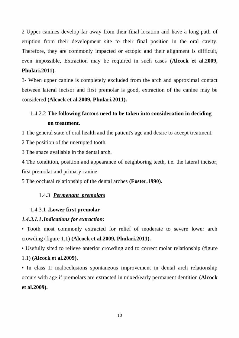
crowding in the arches (Phulari.2011).
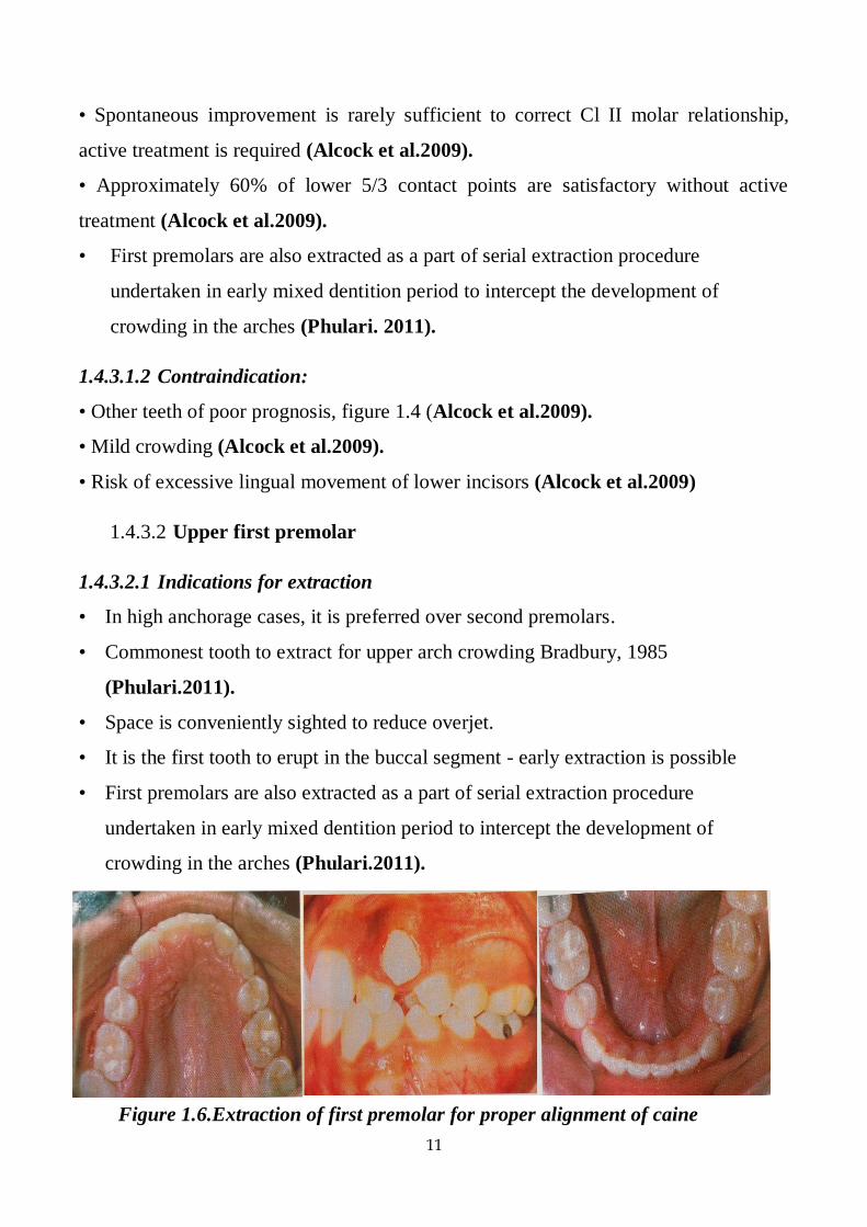
Figure 1.6.Extraction of first premolar for proper alignment of caine

12
1.4.3.2.2 Contra-indications
• Distally inclined canines (Choo Yew On et al .2011).
• Poor prognosis of canines - grossly carious or hypoplastic (extract canine instead)
(Choo Yew On et al.2011).
• Grossly displaced canines ; good contact between lateral incisor & 1st
• Premolar (extract canine) (Choo Yew On et al.2011).
• Class III incisor relationship (Choo Yew On et al.2011).
1.4.3.3 Lower second premolars
1.4.3.3.1 Indications for extraction of premolars.
• For relief of mild-moderate crowding Tulloch, 1978 (Michelle.2007,
Phulari.2011).
• May avoid excessive lingual movement of lower incisors (which may occur with
first premolar extraction), alters anchorage balance.
• Better sited than first premolar for correction of molar relationship and posterior
crowding.
• . Unfavorably impacted second premolars (Phulari.2011).
• If .5 is small or hypoplastic or grossly carious or periodentally compromised
second premolar (Alcock et al.2009).
1.4.3.3.2 Contraindication of extraction
• Fixed appliance usually necessary to establish good 6-4 contact.
Figure 1.17.Impacted second premolars

13
• Never tooth of choice to extract - functionally important (Alcock et al.2009,
Michelle.2007).
1.4.3.4 Upper second premolar
1.4.3.4.1 Indications for extraction:
• Extracted in preference to the 4 if crowding or overjet is less severe in upper arch
• Extraction of .5 rather than 4 may avoid over retraction of the labial segment, useful
with class Ill malocclusions.
• If .5 is excluded from the arch e.g. if early loss of deciduous second molar (Jeryl et
al .2015, Mitchell.2007, Phulari.2011).
• If .5 is small or hypoplastic or grossly carious or periodentally compromised second
premolar (Jeryl et al.2015, Phulari.2011).
1.4.3.4.2 Contraindication for extraction
• Fixed appliance often necessary to establish satisfactory contact between 4 and 6
• As with the lower first molar this is rarely the tooth of choice to extract (Alcock et
al.2009).
1.4.4 First permanent molars
First molars are regarded as the cornerstones of dental arches and are considered
to play a key role in the establishment of occlusion by Angle. They are usually not
extracted unless otherwise indicated (Phulari.2011).
Extraction of first permanent molars often makes orthodontic treatment more difficult
and prolonged. However, their extraction may need to be considered due to their
limited prognosis (Mitchell.2007).
Extraction of first molars may be advantageous in open bite cases as this may lead to
deepening of the bite (Phulari.2011).
14
1.4.4.1 Extraction of first permanent molars is not advisable due to the
following reasons:
• Extraction of first molars does not provide adequate space for the relief of anterior
crowding.
• First molar extraction can lead to deepening of the bite, which may not be
desirable in all cases.
• Following first molar extraction, the second premolar may tip into the extraction
space.
• Masticatory function of the patient may get affected. (Phulari.2011).
1.4.4.2 Lower first molar
1.4.4.2.1 Indications for extraction
• If tooth is carious consider the following Lower second molar:
1) Severity of crowding: Spaced (try to restore the tooth if possible).Mild-moderate
crowding (extract the tooth). Moderate-severe crowding (wait until definitive planning
in permanent dentition before extraction).
2) Stage of development of molar.
Ideal timing is between crown complete to root formed, i.e. when the bifurcation of
roots are forming.
3) Status of the other first permanent molars:
Extract all, do not worry about balancing in the lower arch, do consider compensating
because upper first molar will over erupt (Alcock et al.2009).
1.4.4.2.2 Contraindication for extraction
• Late extraction of first molars results in rotation and mesial tipping of lower second
molar tooth.
• Missing third molars (Alcock et al.2009).
15
1.4.4.3 Upper first molar
1.4.4.3.1 Indication for extraction
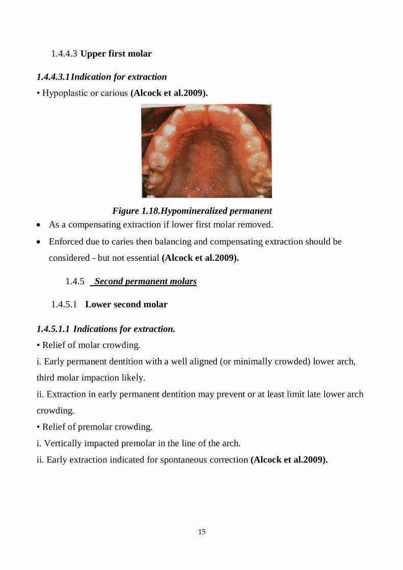
• Hypoplastic or carious (Alcock et al.2009).
As a compensating extraction if lower first molar removed.
Enforced due to caries then balancing and compensating extraction should be
considered - but not essential (Alcock et al.2009).
1.4.5 Second permanent molars
1.4.5.1 Lower second molar
1.4.5.1.1 Indications for extraction.
• Relief of molar crowding.
i. Early permanent dentition with a well aligned (or minimally crowded) lower arch,
third molar impaction likely.
ii. Extraction in early permanent dentition may prevent or at least limit late lower arch
crowding.
• Relief of premolar crowding.
i. Vertically impacted premolar in the line of the arch.
ii. Early extraction indicated for spontaneous correction (Alcock et al.2009).
Figure 1.18.Hypomineralized permanent
molars
16
1.4.5.1.2 Contraindication of extraction.
• Developmental absence or diminutive third molar.
• Lower anterior crowding > 1-2 mm.
1.4.5.1.3 Advantages of second molar extraction.
• Obviate the need for surgical removal of third molar: financial and patient morbidity
considerations.
• Shorter treatment = unsubstantiated.
• Facilitation of openbite reduction = unsubstantiated (Alcock et al.2009).
1.4.5.1.4 Disadvantages of second molar extraction
• Third molars may erupt into an unsatisfactory position, rarely with proper
angulation and contact relationship.
• Wide discrepancy between studies on the number of unsatisfactory thrid molar
eruptions: e.g. 25% Cryer, 20% Dacre, 4% Richardson.
• Difficult to predict which third molars will erupt unsatisfactorily.
• Second course of treatment to orthodontically upright the third molar may be
required.(Alcock et al.2009).
1.4.5.2 Upper second molar.
1.4.5.2.1 Indications for extraction.
• To aid distal movement of the upper buccal segments with EOT - good cooperation
with HG essential.
• Not indicated for relief of anterior crowding or overjet reduction.
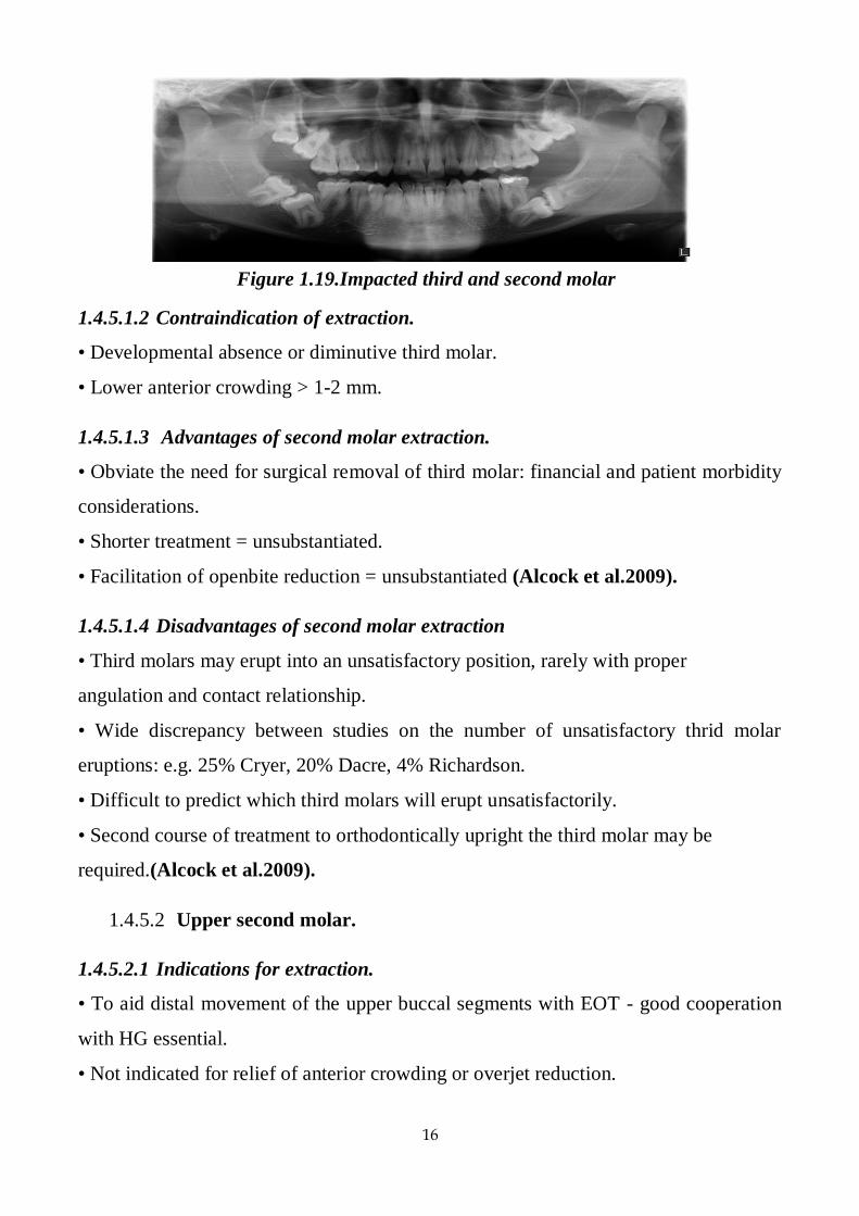
Figure 1.19.Impacted third and second molar
17
• Generally accepted that third molars erupt into satisfactory contact with first molar.
• Accelerated eruption of third molar into acceptable position often occurs (Alcock et
al. 2009).
1.4.5.2.2 Contra indications for extraction.
• Heavily restored first molar.
• Developmentally absent third molar.
• Prevalence of impaction may be increasing because fewer teeth extracted due to
caries (Alcock et al.2009).
1.4.6 Third permanent molars
In the past. Early extraction of these teeth has been advocated to prevent lower
labial segment crowding (Millet et al.2005, Mitchell.2007).
However, it is much more likely that late lower incisor crowding is caused by
subtle gromh and soft tissue changes that continue to occur throughout life It is now
not acceptable to extract third molars purely on the grounds of preventing crowding of
the lower labial segment (Mitchell.2007).
1.4.6.1 Indications for removal
• Third molars are the most common teeth to be impacted in oral cavity figure 1.19
(Phulari.2011).
• Impacted third molars, which are not likely to erupt into ideal position are
frequently extracted (Phulari.2011).
• Pericoronitis development may also necessitate third molar extraction
(Phulari.2011).
• Progressive crowding of lower anteriors is observed in adolescence and early adult
life. This has often been blamed on the erupting lower third molar teeth although it
is not proved. Some orthodontists advocate extraction of third molars to prevent
such late crowding (Phulari.2011)
• Teeth that present with symptoms other than transiently associated with eruption.
18
• Teeth unlikely to contribute to occlusal function and whose position jeopardizes the
continuing health of surrounding tissues:
i. Resorption of second molar.
ii. Follicular cyst.
iii. Bone loss due to chronic periodontitis.
iv. Concealed caries in distal surface of second molar (Alcock et al.2009).
1.5 Serial extraction
Serial extraction was first advocated in 1948 by Kjellgren, a Swedish
orthodontist, as a solution to a shortage of orthodontists. Kjellgren hoped that his
scheme would facilitate the treatment of patients with straight forward crowding by
their own dentists, thus minimizing demands upon the orthodontic service
(Mitchell.2007 ( .
This treatment technique involves the sequential removal of deciduous teeth to
facilitate the unimpeded eruption of the permanent teeth (Graber et al.2017).
Step 1: Extraction of deciduous canines
In this step, the deciduous canines are extracted at around 8-9 years to create
space for the alignment of the incisors. The main objective of extracting primary
canines is to establish the integrity of upper and lower incisors. This prevents the
development of lingual crossbite of maxillary laterals and resultant mesial migration
of maxillary canines.
Step 2: Extraction of deciduous first molars
In this step, deciduous first molars are extracted when first premolars reach half
of the root length as evidenced by radiographs. This would be some 12 months after
the extraction of deciduous canines at around 9-10 years of age. The objective of
deciduous first molar extraction is to accelerate the eruption of first premolars. This
ensures that the first premolars emerge into oral cavity, before the eruption of
permanent canines.
Step 3: Extraction of first premolars
19
In this step, first premolars are extracted as they are emerging into oral cavity
and when the permanent canines have developed beyond half of the root length.
Extraction of first premolars facilitates proper eruption and alignment of permanent
canines after serial extraction procedure, the teeth are fairly aligned. However, the
establishment of proper intercuspation usually requires orthodontic mechanotherapy of
minimal duration, although it may not be necessary in some cases (Phulari. 2011).
For lower arch if the level of eruption of permanent canine is at a level higher than
that of the first premolar, on radiographic evaluation, the enucleation of the
developing first premolar crown at the time of extraction of deciduous first molar must
do. Another modification advised in such clinical conditions is to extract the
deciduous second molars instead of first premolar enucleation after placement of a
lingual holding arch, so as to allow the first premolar to erupt distally. On eruption of
permanent canines the first premolars are extracted (Gurkeerat et el.2007).
1.5.1 Advantages of Serial Extractions
It brings about early self-induced alignment of the permanent teeth there is
improved overall oral health.
1.5.2 Disadvantages of Serial Extractions
Not indicated for class II and class III malocclusions, if at all extraction is
carried only in class II in upper arch It can have psychological impact on the child, if
totally 12 teeth have to be extracted. Deepening of bite can occur Requires prolonged
patient follow-up The procedure alone is not sufficient to bring impacted canine into
proper position. Early extraction can lead to the loss of space and delayed the eruption
of permanent teeth. (Phulari.2011).
1.5.3 Complications of Serial Extractions
It can result in flat face with prominent chin. Patient may look aged.
It can result in lingual inclination of incisors.
20
1.5.4 Indications For serial Extractions
• Minimum 7.0 mm of crowding in the anterior areas per arch (Jeryl et al.2015).
• Severe arch length-tooth material discrepancy of 10 mm or more in the arch
(Phulari.2011)
• Coincident upper and lower midlines (Jeryl et al.2015).
• Bilateral Class I molar relationship (Jeryl et al.2015, Phulari.2011).
• Balanced skeletal pattern in all three planes of space (Jeryl et al.2015).
1.5.5 Contra-indication for serial extractions
• Class III and Class II molar relationships (Phulari.2011, Jeryl et al .2015) .
• Unbalanced skeletal patterns of any kind (transverse, anteroposterior, or vertical)
(Jeryl et al.2015).
• Unequal crowding in the maxillary and mandibular arches (Jeryl et al.2015).
• Unequal crowding bilaterally in either arch (Jeryl et al.2015).
• Midline discrepancies (more than 2 mm) (Jeryl et al.2015, Phulari.2011).
• Open bites or impinging deep bite (Jeryl et al.2015, Phulari.2011).
•Presence of midline diastema (Phulari.2011).
21
Chapter two
Material method
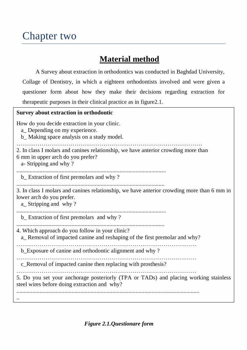
A Survey about extraction in orthodontics was conducted in Baghdad University,
Collage of Dentistry, in which a eighteen orthodontists involved and were given a
questioner form about how they make their decisions regarding extraction for
therapeutic purposes in their clinical practice as in figure2.1.
Survey about extraction in orthodontic
How do you decide extraction in your clinic.
a_ Depending on my experience.
b_ Making space analysis on a study model.
……………………………………………………………………………………
2. In class I molars and canines relationship, we have anterior crowding more than
6 mm in upper arch do you prefer?
a- Stripping and why ?
.......................................................................................................
b_ Extraction of first premolars and why ?
......................................................................................................
3. In class I molars and canines relationship, we have anterior crowding more than 6 mm in
lower arch do you prefer.
a_ Stripping and why ?
.......................................................................................................
b_ Extraction of first premolars and why ?
......................................................................................................
4. Which approach do you follow in your clinic?
a_ Removal of impacted canine and reshaping of the first premolar and why?
…………………………………………………………………………………
b_Exposure of canine and orthodontic alignment and why ?
…………………………………………………………………………………
c_Removal of impacted canine then replacing with prosthesis?
…………………………………………………………………………………
5. Do you set your anchorage posteriorly (TPA or TADs) and placing working stainless
steel wires before doing extraction and why?
..............................................................................................................................
..
iip
Figure 2.1.Questionare form

22
Chapter three
Result
Questioners were introduced to our samples about (How do you decide
extraction in your clinic?). Answers are 61.6%is depend on their experience and
38.8% is making space analysis on study model as the table 3.1 below.
Percentages
Answers
Depending on my experience 61.1%
Making space analysis on study model 38.8%
Table3.1.How do you decide extraction in your clinic
Chart 3.1.Percentage of answers of question one
0.00%
10.00%
20.00%
30.00%
40.00%
50.00%
60.00%
70.00%
depending on my experience making space analysis on study model
Series 2
Series 1
23
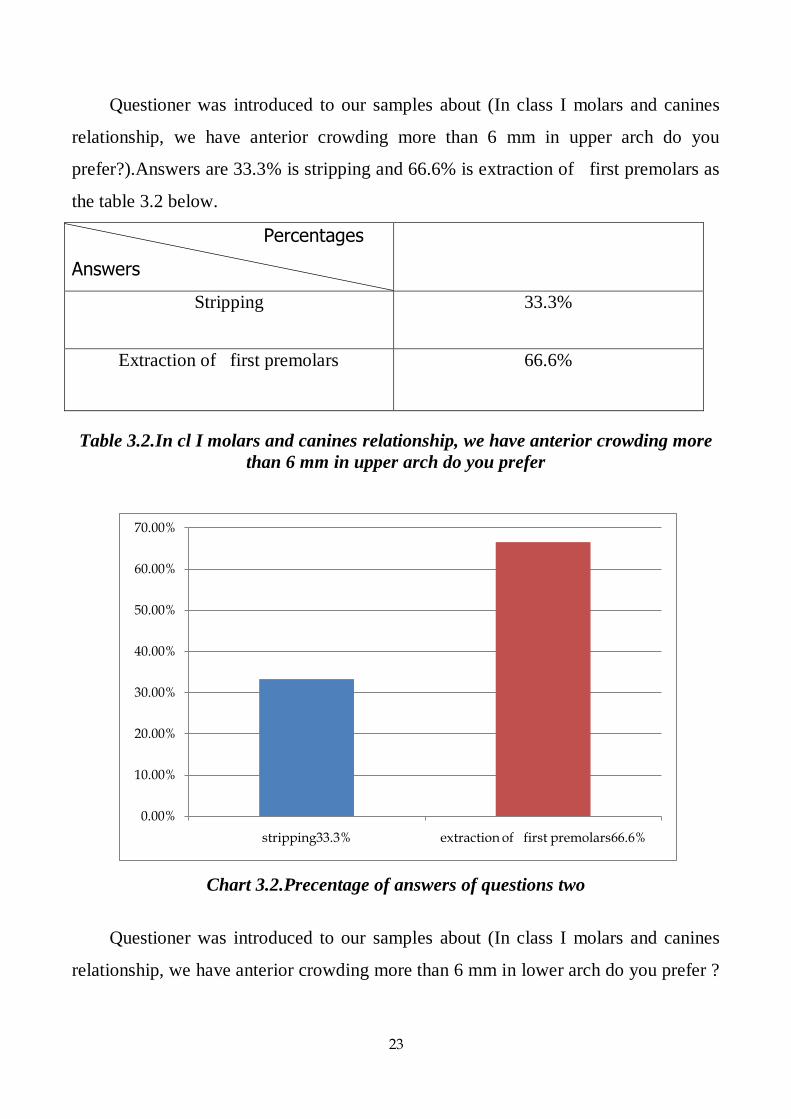
Questioner was introduced to our samples about (In class I molars and canines
relationship, we have anterior crowding more than 6 mm in upper arch do you
prefer?).Answers are 33.3% is stripping and 66.6% is extraction of first premolars as
the table 3.2 below.
Percentages
Answers
Stripping 33.3%
Extraction of first premolars 66.6%
Table 3.2.In cl I molars and canines relationship, we have anterior crowding more
than 6 mm in upper arch do you prefer
Chart 3.2.Precentage of answers of questions two
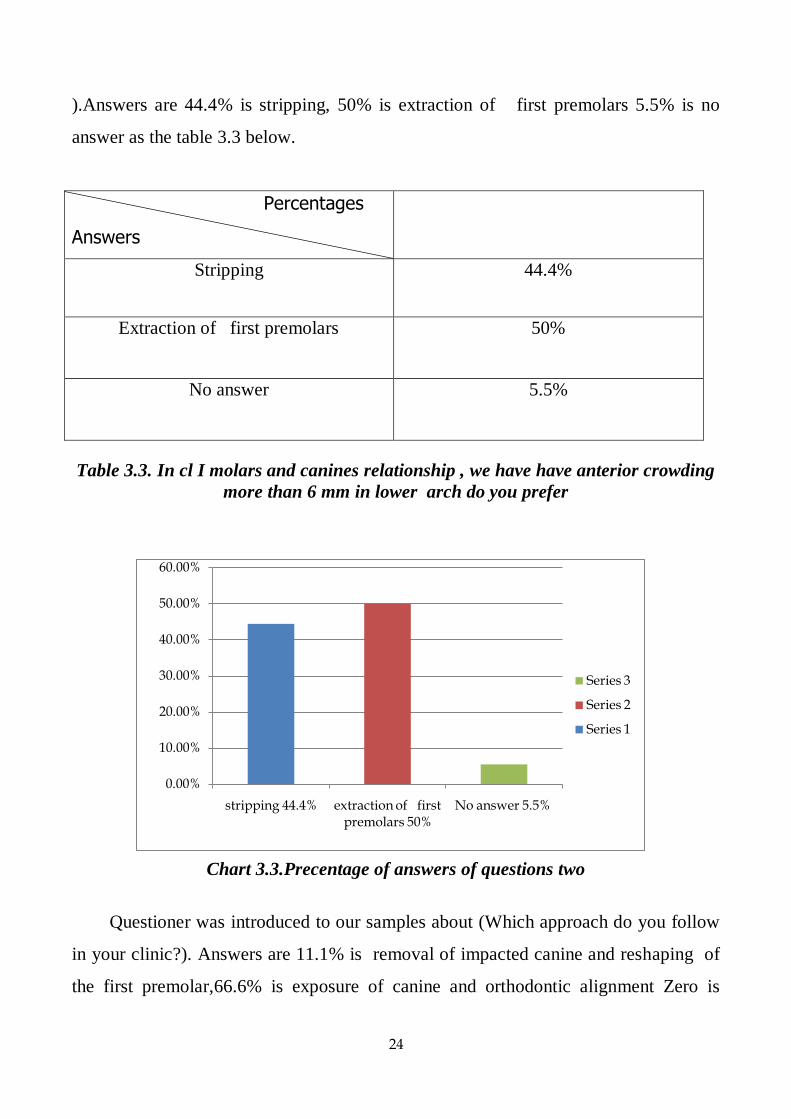
Questioner was introduced to our samples about (In class I molars and canines
relationship, we have anterior crowding more than 6 mm in lower arch do you prefer ?
0.00%
10.00%
20.00%
30.00%
40.00%
50.00%
60.00%
70.00%
stripping33.3% extraction of first premolars66.6%
24
).Answers are 44.4% is stripping, 50% is extraction of first premolars 5.5% is no
answer as the table 3.3 below.
Percentages
Answers
Stripping 44.4%
Extraction of first premolars 50%
No answer 5.5%
Table 3.3. In cl I molars and canines relationship , we have have anterior crowding
more than 6 mm in lower arch do you prefer
Chart 3.3.Precentage of answers of questions two
Questioner was introduced to our samples about (Which approach do you follow
in your clinic?). Answers are 11.1% is removal of impacted canine and reshaping of
the first premolar,66.6% is exposure of canine and orthodontic alignment Zero is
0.00%
10.00%
20.00%
30.00%
40.00%
50.00%
60.00%
stripping 44.4% extraction of first premolars 50%
No answer 5.5%
Series 3
Series 2
Series 1

25
removal of impacted canine then replacing with prosthesis and 22.2% is Depend on
case as the table 3.4 below.
Percentages
Answers
Removal of impacted canine and reshaping
of the first premolar
11.1%
Exposure of canine and orthodontic
alignment
66.6%
Removal of impacted canine then replacing
with prosthesis
Zero
Depend on case 22.2%
Table 3.4.Which approach do you follow in your clinic
Chart 3.4.Precentage of answers of questions four
11.10%
67%
22.20%
0.00%
10.00%
20.00%
30.00%
40.00%
50.00%
60.00%
70.00%
removal of impacted canine and reshaping of the first premolar
11.1%
exposure of canine and orthodontic alignment 66.6%
removal of impacted canine then replacing with prosthesis Zero
Depend on case 22.2%
Series 1 Series 2 Series 3 Series 4
26
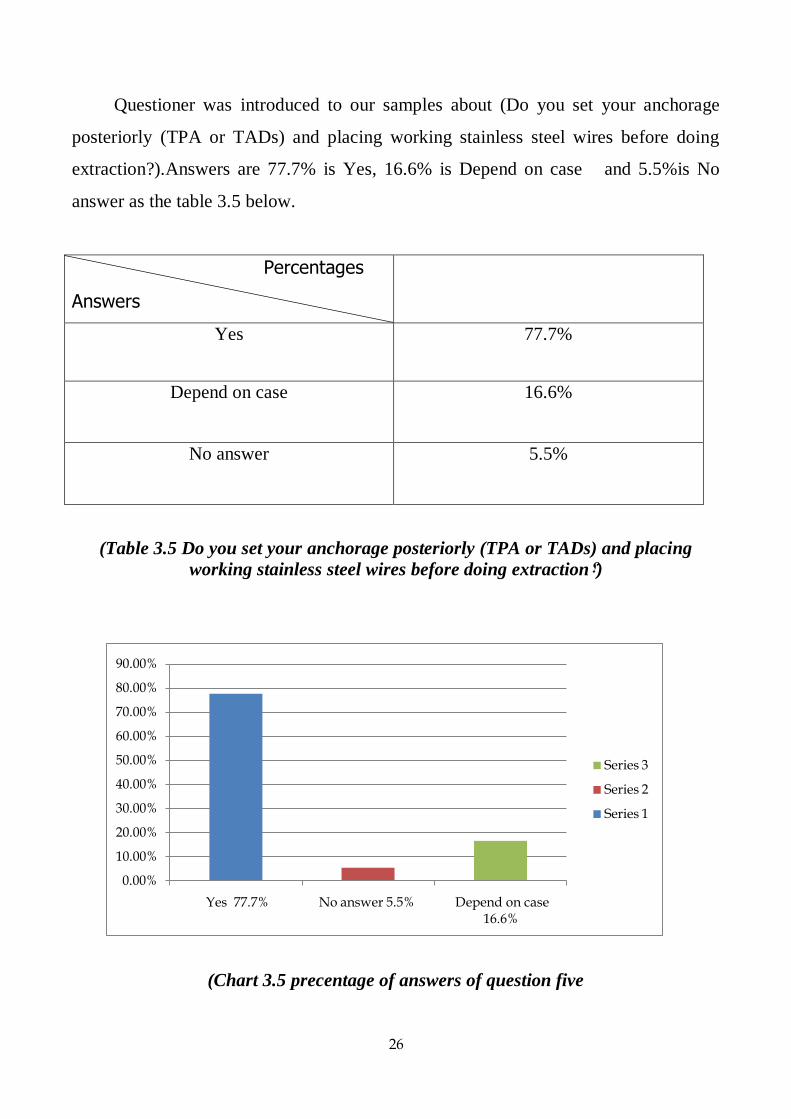
Questioner was introduced to our samples about (Do you set your anchorage
posteriorly (TPA or TADs) and placing working stainless steel wires before doing
extraction?).Answers are 77.7% is Yes, 16.6% is Depend on case and 5.5%is No
answer as the table 3.5 below.
Percentages
Answers
Yes 77.7%
Depend on case 16.6%
No answer 5.5%
(Table 3.5 Do you set your anchorage posteriorly (TPA or TADs) and placing
working stainless steel wires before doing extraction?)
(Chart 3.5 precentage of answers of question five
0.00%
10.00%
20.00%
30.00%
40.00%
50.00%
60.00%
70.00%
80.00%
90.00%
Yes 77.7% No answer 5.5% Depend on case 16.6%
Series 3
Series 2
Series 1
27
Chapter four
Discussion
In table 3.1 61.6% of orthodontists depend on their experience in deciding
extraction properly because they treat so many cases and that qualify them to decide
extraction without doing space analysis of the study models.
While 38.8% of orthodontists depend on space analysis of study model because
they follow the scientific method in order to be in the safe side so that they will not
end up with extra space and elongating treatment time.
In table 3.2 33.3% of orthodontist choose stripping as the stripping more
conservative, more esthetic in some cases and to avoid extraction of first premolar, as
it make canine relation CL II.
While 66.6% of orthodontists choice extraction of first premolars, because
having moderate to severe crowding is one of indication of extract first premolar
(Michelle.2007).also choosing stripping may cause flatting of contact area, tooth
sensitivity (0.5 mm reduction of each tooth) and sometimes the patients refuse
stripping.
In table 3.3 44.4% of orthodontists choice stripping as it More conservative,
more esthetic in some cases and to avoid extraction of first premolar, as it make canine
relation class II.
While 50% of orthodontist choice extraction of first premolars, because having
moderate to severe crowding is one of indication of extract first premolar
(Michelle.2007). Also they are not choose as stripping may cause flatting of contact
area, tooth sensitivity (0.5 mm reduction of each tooth) and sometimes the patients
refuse stripping.
5.5% of orthodontists refuse answer to this question.
28
In table 3.4 0% prefers extraction of impacted canine and replace with
prosthesis.
11.1% of orthodontists choice removal of impacted canine and reshaping of the
first premolar as this decrease treatment time, good contact between lateral incisor
and first premolar if the contact is poor we will not have good occlusion less time
consuming and the patient prefer it sometime.
On the other hand 66.6% of orthodontists choose exposure of canine and
orthodontic alignment as the canine form the cornerstone of the arch and are important
both aesthetically (for lip support) and functionally (providing canine guidance in
lateral movements).Their extraction causes flattening of the face, altered facial balance
and change in facial expression. While 22.2% depend on the case.
In table 3.5 77.7% the answer was yes in order to avoid loss of space that might
happen if the patient fail to attend his appointments.
16.6% depend on case.
5.5% no answer.

29
Chapter five
Suggestion
Run the same survey on larger sample.
Run the same survey in an advanced questioner.

30
Reference
1. Choo yew on . Ling chern chern. (2011). Orthodontic lecture manual (second
edition).
2. Declan millet and richard welbury. (2005). Clinical problem solving in
orthodontic and paediatric dentistry (1st edition). St louis, sydney
,london,toronto,philadelphia.oxford, new york: elsevier.
3. Foster, t. D. (1990). A textbook of orthodontics (third edition). Oxford
londonand edinburgh boston melbourne: blackwell scientific publications.
4. Gurkeerat singh,navjot singh,ankur kaul et el. (2007). Textbook of
orthodontics (second edition). New delhi: jaypee brothers medical publishers.
5. H travess, d roberts-harry and j sandy. (2004, february 28). Extractions in
orthodontics. Pp. 196:195 - 203.
6. Jeryl d. Sercan akyalcin. Timo peltomäki. Kate litschel. (2015). Mosby’s
orthodontic review (second edition). St. Louis, missouri: elsevier.
7. Joe alcock ,saud ai-anezi,nikki atack,sinaed barlow,matt clover,scott
deacon,tony ireland,nicky et el. (2009). Postgraduate notes in orthodontics
(fifth edition). The division of child dental health, bristol dental school,
university of bristol.
8. Laura mitchell. (2007). An introduction to orthodontic (third edition). United
states: oxford university press.
9. Lee w. Graber,robert l. Vanarsdall jr.,katherine w.l. Vig,greg j. Huang,.
(2017). Orthodontics current principles and techniques (6th edition). St. Louis,
missouri: elsevier.

31
10. Phulari, b. S. (2011). Orthodontics principles and practice (1st edition ed.).
New delhi: jaypee brothers medical publishers.
11. William r. Proffit, dds, phd, henry w. Fields, dds, ms, msd, and david m.
Sarver, dmd, ms. (2013). Contemporary orthodontics, (5th edition ed.). St.
Louis, missouri: elsevier.
Related Documents