International Journal of Molecular Sciences Article Evaluating the Effects of Subnormothermic Perfusion with AP39 in a Novel Blood-Free Model of Ex Vivo Kidney Preservation and Reperfusion Smriti Juriasingani 1,2 , Ashley Jackson 2,3 , Max Yulin Zhang 1,2 , Aushanth Ruthirakanthan 2,3 , George J. Dugbartey 2,4,5 , Emrullah Sogutdelen 6 , Max Levine 2,4 , Moaath Mandurah 2,4 , Matthew Whiteman 7 , Patrick Luke 2,3,4 and Alp Sener 1,2,4, * Citation: Juriasingani, S.; Jackson, A.; Zhang, M.Y.; Ruthirakanthan, A.; Dugbartey, G.J.; Sogutdelen, E.; Levine, M.; Mandurah, M.; Whiteman, M.; Luke, P.; et al. Evaluating the Effects of Subnormothermic Perfusion with AP39 in a Novel Blood-Free Model of Ex Vivo Kidney Preservation and Reperfusion. Int. J. Mol. Sci. 2021, 22, 7180. https://doi.org/10.3390/ ijms22137180 Academic Editors: Joan Roselló-Catafau, René Adam and Teresa Carbonell Camós Received: 16 May 2021 Accepted: 28 June 2021 Published: 2 July 2021 Publisher’s Note: MDPI stays neutral with regard to jurisdictional claims in published maps and institutional affil- iations. Copyright: © 2021 by the authors. Licensee MDPI, Basel, Switzerland. This article is an open access article distributed under the terms and conditions of the Creative Commons Attribution (CC BY) license (https:// creativecommons.org/licenses/by/ 4.0/). 1 Department of Microbiology and Immunology, Western University, London, ON N6A 5C1, Canada; [email protected] (S.J.); [email protected] (M.Y.Z.) 2 Matthew Mailing Center for Translational Transplant Studies, London Health Sciences Centre, London, ON N6A 5A5, Canada; [email protected] (A.J.); [email protected] (A.R.); [email protected] (G.J.D.); [email protected] (M.L.); [email protected] (M.M.); [email protected] (P.L.) 3 Department of Pathology & Laboratory Medicine, Western University, London, ON N6A 5C1, Canada 4 Multi-organ Transplant Program, London Health Sciences Center, London, ON N6A 5A5, Canada 5 Department of Pharmacology and Toxicology, School of Pharmacy, College of Health Sciences, University of Ghana, P.O. Box LG 43, Legon, Accra, Ghana 6 Department of Urology, Bolu Abant Izzet Baysal University, Bolu 14030, Turkey; [email protected] 7 St. Luke’s Campus, University of Exeter Medical School, Exeter EX1 2HZ, UK; [email protected] * Correspondence: [email protected]; Tel.: +1-519-663-3352 Abstract: The use of blood for normothermic and subnormothermic kidney preservation hinders the translation of these approaches and promising therapeutics. This study evaluates whether adding hydrogen sulfide donor AP39 to Hemopure, a blood substitute, during subnormothermic perfusion improves kidney outcomes. After 30 min of renal pedicle clamping, porcine kidneys were treated to 4 h of static cold storage (SCS-4 ◦ C) or subnormothermic perfusion at 21 ◦ C with Hemopure (H-21 ◦ C), Hemopure + 200 nM AP39 (H200nM-21 ◦ C) or Hemopure + 1 μM AP39 (H1μM-21 ◦ C). Then, kidneys were reperfused with Hemopure at 37 ◦ C for 4 h with metabolic support. Perfusate composition, tissue oxygenation, urinalysis and histopathology were analyzed. During preservation, the H200nM-21 ◦ C group exhibited significantly higher urine output than the other groups and significantly higher tissue oxygenation than the H1μM-21 ◦ C group at 1 h and 2h. During reperfusion, the H200nM-21 ◦ C group exhibited significantly higher urine output and lower urine protein than the other groups. Additionally, the H200nM-21 ◦ C group exhibited higher perfusate pO 2 levels than the other groups and significantly lower apoptotic injury than the H-21 ◦ C and the H1μM-21 ◦ C groups. Thus, subnormothermic perfusion at 21 ◦ C with Hemopure + 200 nM AP39 improves renal outcomes. Additionally, our novel blood-free model of ex vivo kidney preservation and reperfusion could be useful for studying other therapeutics. Keywords: kidney preservation; kidney transplantation; hydrogen sulfide (H 2 S); AP39; donation after cardiac death (DCD); subnormothermic 1. Introduction Kidney transplantation is the preferred treatment for end-stage kidney disease (ESKD) because it improves long-term survival and quality of life compared to hemodialysis [1–3]. However, there is a critical shortage of donor kidneys across the globe due to the increasing need for kidney transplants. In Canada, the prevalence of ESKD has risen by 33% between 2010 and 2019 [4]. Despite the 1648 adult kidney transplants that were performed in 2019, 3261 individuals remained on the waiting list [5]. To meet the rising demand, kidneys from marginal donors are being used more frequently. While donation after brain death does Int. J. Mol. Sci. 2021, 22, 7180. https://doi.org/10.3390/ijms22137180 https://www.mdpi.com/journal/ijms

Welcome message from author
This document is posted to help you gain knowledge. Please leave a comment to let me know what you think about it! Share it to your friends and learn new things together.
Transcript
International Journal of
Molecular Sciences
Article
Evaluating the Effects of Subnormothermic Perfusion withAP39 in a Novel Blood-Free Model of Ex Vivo KidneyPreservation and Reperfusion
Smriti Juriasingani 1,2, Ashley Jackson 2,3, Max Yulin Zhang 1,2, Aushanth Ruthirakanthan 2,3,George J. Dugbartey 2,4,5, Emrullah Sogutdelen 6 , Max Levine 2,4, Moaath Mandurah 2,4, Matthew Whiteman 7,Patrick Luke 2,3,4 and Alp Sener 1,2,4,*
�����������������
Citation: Juriasingani, S.; Jackson, A.;
Zhang, M.Y.; Ruthirakanthan, A.;
Dugbartey, G.J.; Sogutdelen, E.;
Levine, M.; Mandurah, M.;
Whiteman, M.; Luke, P.; et al.
Evaluating the Effects of
Subnormothermic Perfusion with
AP39 in a Novel Blood-Free Model of
Ex Vivo Kidney Preservation and
Reperfusion. Int. J. Mol. Sci. 2021, 22,
7180. https://doi.org/10.3390/
ijms22137180
Academic Editors:
Joan Roselló-Catafau, René Adam
and Teresa Carbonell Camós
Received: 16 May 2021
Accepted: 28 June 2021
Published: 2 July 2021
Publisher’s Note: MDPI stays neutral
with regard to jurisdictional claims in
published maps and institutional affil-
iations.
Copyright: © 2021 by the authors.
Licensee MDPI, Basel, Switzerland.
This article is an open access article
distributed under the terms and
conditions of the Creative Commons
Attribution (CC BY) license (https://
creativecommons.org/licenses/by/
4.0/).
1 Department of Microbiology and Immunology, Western University, London, ON N6A 5C1, Canada;[email protected] (S.J.); [email protected] (M.Y.Z.)
2 Matthew Mailing Center for Translational Transplant Studies, London Health Sciences Centre, London,ON N6A 5A5, Canada; [email protected] (A.J.); [email protected] (A.R.); [email protected] (G.J.D.);[email protected] (M.L.); [email protected] (M.M.); [email protected] (P.L.)
3 Department of Pathology & Laboratory Medicine, Western University, London, ON N6A 5C1, Canada4 Multi-organ Transplant Program, London Health Sciences Center, London, ON N6A 5A5, Canada5 Department of Pharmacology and Toxicology, School of Pharmacy, College of Health Sciences,
University of Ghana, P.O. Box LG 43, Legon, Accra, Ghana6 Department of Urology, Bolu Abant Izzet Baysal University, Bolu 14030, Turkey; [email protected] St. Luke’s Campus, University of Exeter Medical School, Exeter EX1 2HZ, UK; [email protected]* Correspondence: [email protected]; Tel.: +1-519-663-3352
Abstract: The use of blood for normothermic and subnormothermic kidney preservation hinders thetranslation of these approaches and promising therapeutics. This study evaluates whether addinghydrogen sulfide donor AP39 to Hemopure, a blood substitute, during subnormothermic perfusionimproves kidney outcomes. After 30 min of renal pedicle clamping, porcine kidneys were treatedto 4 h of static cold storage (SCS-4 ◦C) or subnormothermic perfusion at 21 ◦C with Hemopure(H-21 ◦C), Hemopure + 200 nM AP39 (H200nM-21 ◦C) or Hemopure + 1 µM AP39 (H1µM-21 ◦C).Then, kidneys were reperfused with Hemopure at 37 ◦C for 4 h with metabolic support. Perfusatecomposition, tissue oxygenation, urinalysis and histopathology were analyzed. During preservation,the H200nM-21 ◦C group exhibited significantly higher urine output than the other groups andsignificantly higher tissue oxygenation than the H1µM-21 ◦C group at 1 h and 2h. During reperfusion,the H200nM-21 ◦C group exhibited significantly higher urine output and lower urine protein thanthe other groups. Additionally, the H200nM-21 ◦C group exhibited higher perfusate pO2 levels thanthe other groups and significantly lower apoptotic injury than the H-21 ◦C and the H1µM-21 ◦Cgroups. Thus, subnormothermic perfusion at 21 ◦C with Hemopure + 200 nM AP39 improves renaloutcomes. Additionally, our novel blood-free model of ex vivo kidney preservation and reperfusioncould be useful for studying other therapeutics.
Keywords: kidney preservation; kidney transplantation; hydrogen sulfide (H2S); AP39; donationafter cardiac death (DCD); subnormothermic
1. Introduction
Kidney transplantation is the preferred treatment for end-stage kidney disease (ESKD)because it improves long-term survival and quality of life compared to hemodialysis [1–3].However, there is a critical shortage of donor kidneys across the globe due to the increasingneed for kidney transplants. In Canada, the prevalence of ESKD has risen by 33% between2010 and 2019 [4]. Despite the 1648 adult kidney transplants that were performed in 2019,3261 individuals remained on the waiting list [5]. To meet the rising demand, kidneys frommarginal donors are being used more frequently. While donation after brain death does
Int. J. Mol. Sci. 2021, 22, 7180. https://doi.org/10.3390/ijms22137180 https://www.mdpi.com/journal/ijms
Int. J. Mol. Sci. 2021, 22, 7180 2 of 16
not impact organ health, donation after cardiac death (DCD) leads to warm ischemic injury,as blood flow decreases and the heart stops. This is of concern because DCD kidneys arebeing transplanted more frequently and often lead to poorer patient outcomes comparedto kidneys from other donors [6]. Additionally, clinical cold preservation methods (4 ◦C),such as static cold storage (SCS), exacerbate DCD kidney outcomes due to the combinationof cold and warm ischemic injury [7–9].
Several strategies have been explored to improve cold preservation methods, such asthe addition of hydrogen sulfide (H2S), a gasotransmitter with cytoprotective properties, topreservation solutions [10–12]. Our group and others have previously shown that addingnanomolar concentrations of the H2S donor, AP39, to cold preservation solution improvesrenal and cardiac graft outcomes [11,13]. Additionally, there is growing interest in alter-natives to cold preservation that employ normothermic (36–37 ◦C) and subnormothermic(15–35 ◦C) temperatures [14]. Several porcine and discarded human kidney studies haveshown that normothermic and subnormothermic machine perfusion improve DCD renalgraft outcomes compared to SCS on ice [15–20]. However, a major challenge in this contextis the need for oxygenation and nutrients to meet the metabolic demand of the kidneyat these higher temperatures. Previous studies have primarily used erythrocyte-basedsolutions or autologous whole blood for this purpose [16,18,21], but the use of blood is achallenge for the clinical translation of these approaches. Pervasive shortages of bankedblood and the challenges associated with obtaining blood from DCD donors limit theavailability of blood for ex vivo kidney preservation.
The use of blood substitutes, especially hemoglobin-based oxygen carriers (HBOC),can solve the problem of oxygenation and circumvent the need for blood in the context ofnormothermic and subnormothermic preservation. One promising candidate is Hemopure(also known as HBOC-201), which is made of highly purified bovine hemoglobin [22,23]. Clinically, Hemopure is used to treat anemia in patients who cannot receive bloodtransfusions [24,25]. However, several recent studies have shown that ex vivo kidneypreservation with Hemopure can improve renal graft outcomes. In 2019, Aburawi et al. [26]reported that normothermic perfusion (37 ◦C) of discarded human kidneys with Hemopurevs. packed red blood cells led to comparable outcomes. Subsequently, a recent study byour center has shown that subnormothermic perfusion (22 ◦C) of DCD porcine kidneyswith Hemopure vs. whole blood exhibited similar outcomes, which further confirms thefeasibility of Hemopure for blood-free renal graft preservation [19].
While recent evidence supports the use of Hemopure for kidney preservation, itspotential as a platform to deliver therapeutics that could further enhance renal graft out-comes remains unexplored. We have previously shown that adding H2S donor, AP39, toUniversity of Wisconsin (UW) solution, a preservation solution that is commonly used forSCS, made it suitable for subnormothermic preservation [27]. Additionally, we have shownthat subnormothermic perfusion (21 ◦C) of DCD porcine kidneys with AP39-supplementedwhole blood improves renal graft outcomes and reduces tissue injury using an ex vivomodel of blood-based preservation and reperfusion [20]. In this study, we investigatewhether subnormothermic perfusion at 21 ◦C with AP39-supplemented Hemopure im-proves DCD porcine renal graft outcomes compared to SCS and subnormothermic perfu-sion with Hemopure alone. To evaluate this aim, we use a novel blood-free model of exvivo preservation and reperfusion. In this model, after 4 h of blood-free preservation, allkidneys are reperfused (37 ◦C) for 4 h with Hemopure, supplemented with a diuretic andmetabolic support, to mimic the post-transplant milieu.
2. Results2.1. Subnormothermic Perfusion with Hemopure + 200 nM AP39 at 21 ◦C Improves Gross KidneyMorphology and Perfusate pO2 Levels during Blood-Free Reperfusion
Kidneys were imaged prior to preservation (after flushing) and at the end of reper-fusion to evaluate differences in gross morphology (Figure 1). The images taken priorto preservation show that the kidneys in each group had similar gross morphology andwere thoroughly flushed following the induction of warm ischemia, which is reflected
Int. J. Mol. Sci. 2021, 22, 7180 3 of 16
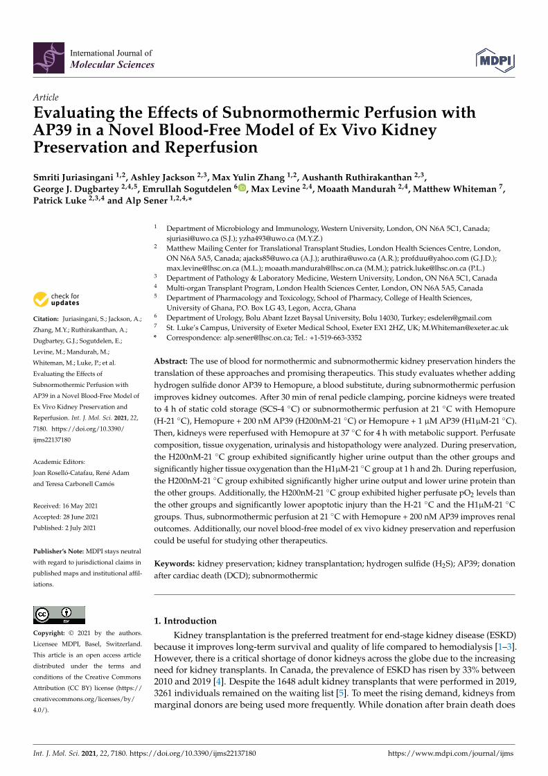
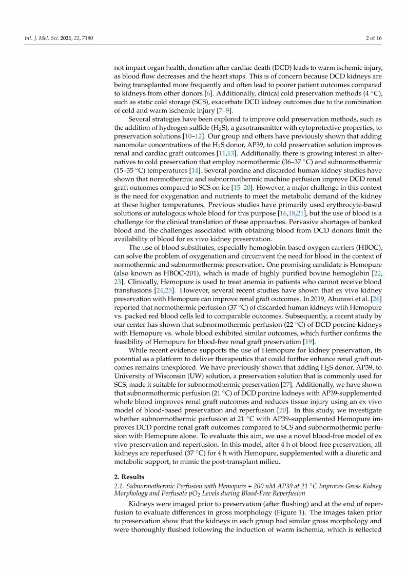
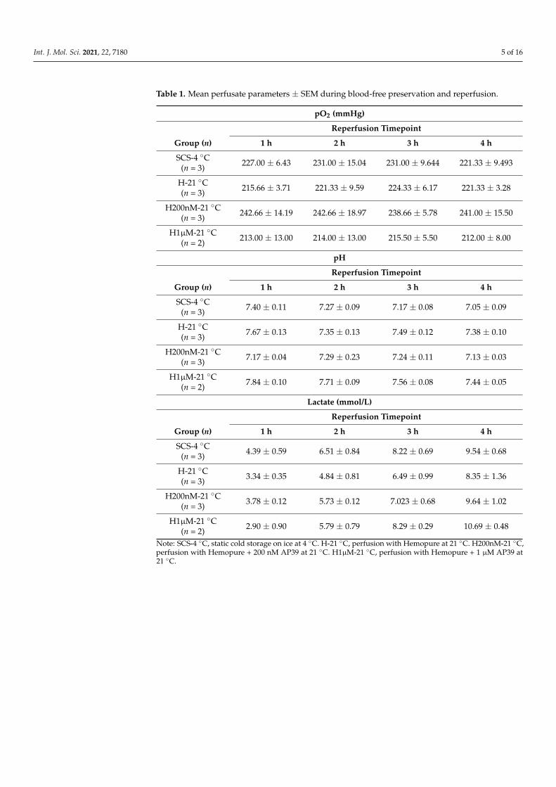
by the pale colour. At the end of reperfusion, the H200nM-21 ◦C kidneys looked the red-dest, which is an indicator of consistent perfusion and overall organ health. On the otherhand, the H1µM-21 ◦C kidneys looked much darker than the other groups at the end ofreperfusion, which is an indicator of thrombosis and tissue injury (Figure 1). Additionally,perfusate samples were analyzed hourly during reperfusion to evaluate relative changes inpO2, pH and lactate levels (Figure 2, Table 1). The H200nM-21 ◦C group exhibited higherperfusate pO2 levels than the SCS-4 ◦C, H-21 ◦C and H1µM-21 ◦C groups throughout thereperfusion period. Additionally, the perfusate pO2 levels stayed relatively consistent overtime for each group (Figure 2A). The H1µM-21 ◦C group exhibited higher perfusate pHthan the other three groups throughout the reperfusion period. Apart from the elevated pHof this group and the high variability at the 1 h timepoint, perfusate pH was maintainedwithin the range of 7.1 to 7.4. Interestingly, perfusate pH decreased over time for eachgroup (Figure 2B). Furthermore, all four groups exhibited comparable increases in lactatelevels throughout the reperfusion period (Figure 2C), which contributes to the decrease inperfusate pH described earlier (Figure 2B). None of the differences in perfusate parameterlevels were statistically significant.
Int. J. Mol. Sci. 2021, 22, x FOR PEER REVIEW 3 of 16
2. Results 2.1. Subnormothermic Perfusion with Hemopure + 200 nM AP39 at 21 °C Improves Gross Kidney Morphology and Perfusate pO2 Levels during Blood-Free Reperfusion
Kidneys were imaged prior to preservation (after flushing) and at the end of reperfusion to evaluate differences in gross morphology (Figure 1). The images taken prior to preservation show that the kidneys in each group had similar gross morphology and were thoroughly flushed following the induction of warm ischemia, which is reflected by the pale colour. At the end of reperfusion, the H200nM-21 °C kidneys looked the reddest, which is an indicator of consistent perfusion and overall organ health. On the other hand, the H1µM-21 °C kidneys looked much darker than the other groups at the end of reperfusion, which is an indicator of thrombosis and tissue injury (Figure 1). Additionally, perfusate samples were analyzed hourly during reperfusion to evaluate relative changes in pO2, pH and lactate levels (Figure 2, Table 1). The H200nM-21 °C group exhibited higher perfusate pO2 levels than the SCS-4 °C, H-21 °C and H1µM-21 °C groups throughout the reperfusion period. Additionally, the perfusate pO2 levels stayed relatively consistent over time for each group (Figure 2A). The H1µM-21 °C group exhibited higher perfusate pH than the other three groups throughout the reperfusion period. Apart from the elevated pH of this group and the high variability at the 1 h timepoint, perfusate pH was maintained within the range of 7.1 to 7.4. Interestingly, perfusate pH decreased over time for each group (Figure 2B). Furthermore, all four groups exhibited comparable increases in lactate levels throughout the reperfusion period (Figure 2C), which contributes to the decrease in perfusate pH described earlier (Figure 2B). None of the differences in perfusate parameter levels were statistically significant.
Figure 1. Gross morphology of the kidneys prior to preservation and at the end of reperfusion. Images were taken prior to perfusion to document the consistent flushing of the kidneys following the induction of warm ischemia. Images were taken at the end of reperfusion to document the gross morphology of kidneys by the end of the experiment. One pair of representative images were chosen
Figure 1. Gross morphology of the kidneys prior to preservation and at the end of reperfusion.Images were taken prior to perfusion to document the consistent flushing of the kidneys followingthe induction of warm ischemia. Images were taken at the end of reperfusion to document the grossmorphology of kidneys by the end of the experiment. One pair of representative images were chosenfor each preservation treatment group. Treatment groups: SCS-4 ◦C, static cold storage on ice at 4 ◦C.H-21 ◦C, perfusion with Hemopure at 21 ◦C. H200nM-21 ◦C, perfusion with Hemopure + 200 nMAP39 at 21 ◦C. H1µM-21 ◦C, perfusion with Hemopure + 1 µM AP39 at 21 ◦C.
Int. J. Mol. Sci. 2021, 22, 7180 4 of 16
Int. J. Mol. Sci. 2021, 22, x FOR PEER REVIEW 4 of 16
for each preservation treatment group. Treatment groups: SCS-4 °C, static cold storage on ice at 4 °C. H-21 °C, perfusion with Hemopure at 21°C. H200nM-21 °C, perfusion with Hemopure + 200 nM AP39 at 21 °C. H1µM-21°C, perfusion with Hemopure + 1 µM AP39 at 21 °C.
Figure 2. Perfusate parameters during blood-free reperfusion. (A) pO2 (mmHg), (B) pH and (C) Lactate (mmol/L) during blood-free reperfusion. Since pairs of kidneys were reperfused with shared perfusate, singular readings were obtained at each timepoint for each pair of kidneys. The iSTAT Analyzer was unable to detect tissue oxygenation for one pair of kidneys (n = 1) in the H1µM-21 °C group. Individual points on each graph represent the mean value ± SEM at a specific timepoint for pairs of kidneys within a single treatment group. Mean ± SEM values are listed in Table 1. After a Geisser–Greenhouse correction, values were compared using repeated measures two-way ANOVA followed by Tukey’s post-hoc test and no significant differences were found. Treatment groups: SCS-4 °C, static cold storage on ice at 4 °C (n = 3). H-21 °C, perfusion with Hemopure at 21 °C (n = 3). H200nM-21 °C, perfusion with Hemopure + 200 nM AP39 at 21 °C (n = 3). H1µM-21 °C, perfusion with Hemopure + 1 µM AP39 at 21 °C (n = 2).
Table 1. Mean perfusate parameters ± SEM during blood-free preservation and reperfusion.
pO2 (mmHg) Reperfusion Timepoint
Group (n) 1 h 2 h 3 h 4 h SCS-4 °C
(n = 3) 227.00 ± 6.43 231.00 ± 15.04 231.00 ± 9.644 221.33 ± 9.493
H-21 °C (n = 3)
215.66 ± 3.71 221.33 ± 9.59 224.33 ± 6.17 221.33 ± 3.28
H200nM-21 °C (n = 3)
242.66 ± 14.19 242.66 ± 18.97 238.66 ± 5.78 241.00 ± 15.50
H1µM-21 °C (n = 2)
213.00 ± 13.00 214.00 ± 13.00 215.50 ± 5.50 212.00 ± 8.00
pH
Figure 2. Perfusate parameters during blood-free reperfusion. (A) pO2 (mmHg), (B) pH and (C) Lactate (mmol/L) duringblood-free reperfusion. Since pairs of kidneys were reperfused with shared perfusate, singular readings were obtained ateach timepoint for each pair of kidneys. The iSTAT Analyzer was unable to detect tissue oxygenation for one pair of kidneys(n = 1) in the H1µM-21 ◦C group. Individual points on each graph represent the mean value ± SEM at a specific timepointfor pairs of kidneys within a single treatment group. Mean ± SEM values are listed in Table 1. After a Geisser–Greenhousecorrection, values were compared using repeated measures two-way ANOVA followed by Tukey’s post-hoc test and nosignificant differences were found. Treatment groups: SCS-4 ◦C, static cold storage on ice at 4 ◦C (n = 3). H-21 ◦C, perfusionwith Hemopure at 21 ◦C (n = 3). H200nM-21 ◦C, perfusion with Hemopure + 200 nM AP39 at 21 ◦C (n = 3). H1µM-21 ◦C,perfusion with Hemopure + 1 µM AP39 at 21 ◦C (n = 2).
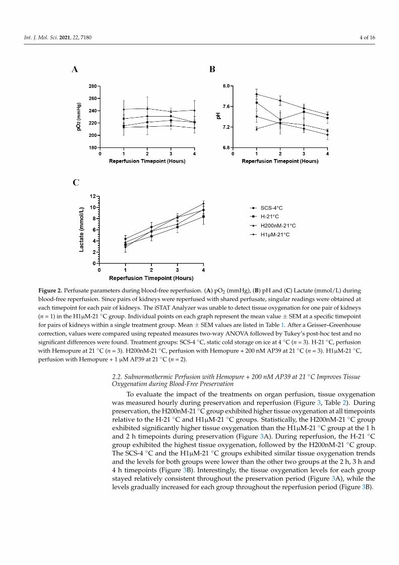
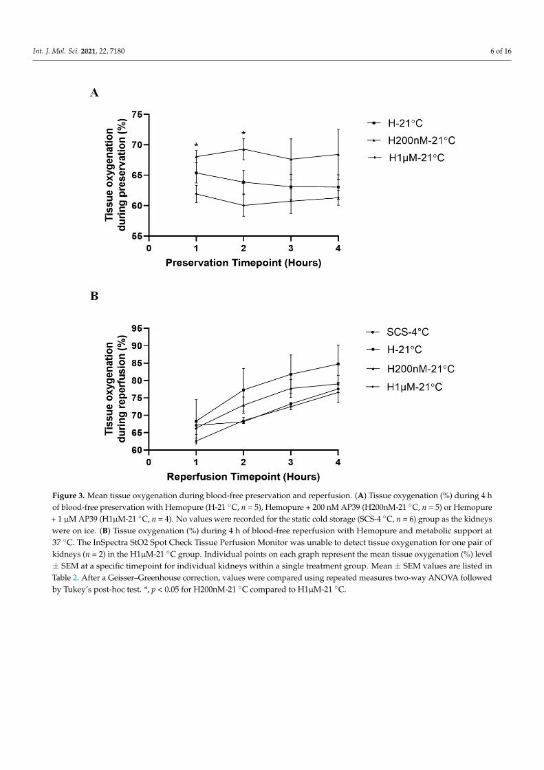
2.2. Subnormothermic Perfusion with Hemopure + 200 nM AP39 at 21 ◦C Improves TissueOxygenation during Blood-Free Preservation
To evaluate the impact of the treatments on organ perfusion, tissue oxygenationwas measured hourly during preservation and reperfusion (Figure 3, Table 2). Duringpreservation, the H200nM-21 ◦C group exhibited higher tissue oxygenation at all timepointsrelative to the H-21 ◦C and H1µM-21 ◦C groups. Statistically, the H200nM-21 ◦C groupexhibited significantly higher tissue oxygenation than the H1µM-21 ◦C group at the 1 hand 2 h timepoints during preservation (Figure 3A). During reperfusion, the H-21 ◦Cgroup exhibited the highest tissue oxygenation, followed by the H200nM-21 ◦C group.The SCS-4 ◦C and the H1µM-21 ◦C groups exhibited similar tissue oxygenation trendsand the levels for both groups were lower than the other two groups at the 2 h, 3 h and4 h timepoints (Figure 3B). Interestingly, the tissue oxygenation levels for each groupstayed relatively consistent throughout the preservation period (Figure 3A), while thelevels gradually increased for each group throughout the reperfusion period (Figure 3B).
Int. J. Mol. Sci. 2021, 22, 7180 5 of 16
Table 1. Mean perfusate parameters ± SEM during blood-free preservation and reperfusion.
pO2 (mmHg)
Reperfusion Timepoint
Group (n) 1 h 2 h 3 h 4 h
SCS-4 ◦C(n = 3) 227.00 ± 6.43 231.00 ± 15.04 231.00 ± 9.644 221.33 ± 9.493
H-21 ◦C(n = 3) 215.66 ± 3.71 221.33 ± 9.59 224.33 ± 6.17 221.33 ± 3.28
H200nM-21 ◦C(n = 3) 242.66 ± 14.19 242.66 ± 18.97 238.66 ± 5.78 241.00 ± 15.50
H1µM-21 ◦C(n = 2) 213.00 ± 13.00 214.00 ± 13.00 215.50 ± 5.50 212.00 ± 8.00
pH
Reperfusion Timepoint
Group (n) 1 h 2 h 3 h 4 h
SCS-4 ◦C(n = 3) 7.40 ± 0.11 7.27 ± 0.09 7.17 ± 0.08 7.05 ± 0.09
H-21 ◦C(n = 3) 7.67 ± 0.13 7.35 ± 0.13 7.49 ± 0.12 7.38 ± 0.10
H200nM-21 ◦C(n = 3) 7.17 ± 0.04 7.29 ± 0.23 7.24 ± 0.11 7.13 ± 0.03
H1µM-21 ◦C(n = 2) 7.84 ± 0.10 7.71 ± 0.09 7.56 ± 0.08 7.44 ± 0.05
Lactate (mmol/L)
Reperfusion Timepoint
Group (n) 1 h 2 h 3 h 4 h
SCS-4 ◦C(n = 3) 4.39 ± 0.59 6.51 ± 0.84 8.22 ± 0.69 9.54 ± 0.68
H-21 ◦C(n = 3) 3.34 ± 0.35 4.84 ± 0.81 6.49 ± 0.99 8.35 ± 1.36
H200nM-21 ◦C(n = 3) 3.78 ± 0.12 5.73 ± 0.12 7.023 ± 0.68 9.64 ± 1.02
H1µM-21 ◦C(n = 2) 2.90 ± 0.90 5.79 ± 0.79 8.29 ± 0.29 10.69 ± 0.48
Note: SCS-4 ◦C, static cold storage on ice at 4 ◦C. H-21 ◦C, perfusion with Hemopure at 21 ◦C. H200nM-21 ◦C,perfusion with Hemopure + 200 nM AP39 at 21 ◦C. H1µM-21 ◦C, perfusion with Hemopure + 1 µM AP39 at21 ◦C.
Int. J. Mol. Sci. 2021, 22, 7180 6 of 16Int. J. Mol. Sci. 2021, 22, x FOR PEER REVIEW 6 of 16
Figure 3. Mean tissue oxygenation during blood-free preservation and reperfusion. (A) Tissue oxygenation (%) during 4 h of blood-free preservation with Hemopure (H-21 °C, n = 5), Hemopure + 200 nM AP39 (H200nM-21 °C, n = 5) or Hemopure + 1 µM AP39 (H1µM-21 °C, n = 4). No values were recorded for the static cold storage (SCS-4 °C, n = 6) group as the kidneys were on ice. (B) Tissue oxygenation (%) during 4 h of blood-free reperfusion with Hemopure and metabolic support at 37 °C. The InSpectra StO2 Spot Check Tissue Perfusion Monitor was unable to detect tissue oxygenation for one pair of kidneys (n = 2) in the H1µM-21 °C group. Individual points on each graph represent the mean tissue oxygenation (%) level ± SEM at a specific timepoint for individual kidneys within a single treatment group. Mean ± SEM values are listed in Table 2. After a Geisser–Greenhouse correction, values were compared using repeated measures two-way ANOVA followed by Tukey’s post-hoc test. *, p < 0.05 for H200nM-21 °C compared to H1µM-21 °C.
Figure 3. Mean tissue oxygenation during blood-free preservation and reperfusion. (A) Tissue oxygenation (%) during 4 hof blood-free preservation with Hemopure (H-21 ◦C, n = 5), Hemopure + 200 nM AP39 (H200nM-21 ◦C, n = 5) or Hemopure+ 1 µM AP39 (H1µM-21 ◦C, n = 4). No values were recorded for the static cold storage (SCS-4 ◦C, n = 6) group as the kidneyswere on ice. (B) Tissue oxygenation (%) during 4 h of blood-free reperfusion with Hemopure and metabolic support at37 ◦C. The InSpectra StO2 Spot Check Tissue Perfusion Monitor was unable to detect tissue oxygenation for one pair ofkidneys (n = 2) in the H1µM-21 ◦C group. Individual points on each graph represent the mean tissue oxygenation (%) level± SEM at a specific timepoint for individual kidneys within a single treatment group. Mean ± SEM values are listed inTable 2. After a Geisser–Greenhouse correction, values were compared using repeated measures two-way ANOVA followedby Tukey’s post-hoc test. *, p < 0.05 for H200nM-21 ◦C compared to H1µM-21 ◦C.
Int. J. Mol. Sci. 2021, 22, 7180 7 of 16
Table 2. Mean tissue oxygenation ± SEM (%) during blood-free preservation and reperfusion.
Mean Tissue Oxygenation (%) ± SEM (%)
Preservation Timepoint
Group (n) 1 h 2 h 3 h 4 h
SCS-4 ◦C(n = 6) No data collected while kidneys were on ice
H-21 ◦C(n = 5) 65.37 ± 1.65 63.86 ± 1.94 63.11 ± 2.04 63.06 ± 2.02
H200nM-21 ◦C(n = 5) 68.04 ± 1.02 69.29 ± 1.76 67.62 ± 3.36 68.43 ± 4.10
H1µM-21 ◦C(n = 4) 61.93 ± 1.42 60.04 ± 1.78 60.75 ± 2.02 61.29 ± 1.21
Reperfusion Timepoint
Group (n) 1 h 2 h 3 h 4 h
SCS-4 ◦C(n = 6) 67.12 ± 0.88 68.12 ± 0.47 73.29 ± 0.40 77.57 ± 0.24
H-21 ◦C(n = 5) 68.31 ± 6.20 77.29 ± 6.15 81.80 ± 5.55 84.71 ± 5.52
H200nM-21 ◦C(n = 5) 66.35 ± 1.91 72.90 ± 2.34 77.73 ± 2.62 79.00 ± 2.44
H1µM-21 ◦C(n = 4) 62.61 ± 0.82 68.46 ± 0.89 72.46 ± 0.91 76.57 ± 2.91
Note: Bolded and highlighted values in each column were significantly different relative to each other (p < 0.05)as per repeated measures two-way ANOVA and Tukey’s post-hoc test after a Geisser–Greenhouse correction.SCS-4 ◦C, static cold storage on ice at 4 ◦C. H-21 ◦C, perfusion with Hemopure at 21 ◦C. H200nM-21 ◦C, perfusionwith Hemopure + 200 nM AP39 at 21 ◦C. H1µM-21 ◦C, perfusion with Hemopure + 1 µM AP39 at 21 ◦C.
2.3. Subnormothermic Perfusion with Hemopure + 200 nM AP39 at 21 ◦C Improves KidneyFunction during Blood-Free Preservation and Reperfusion
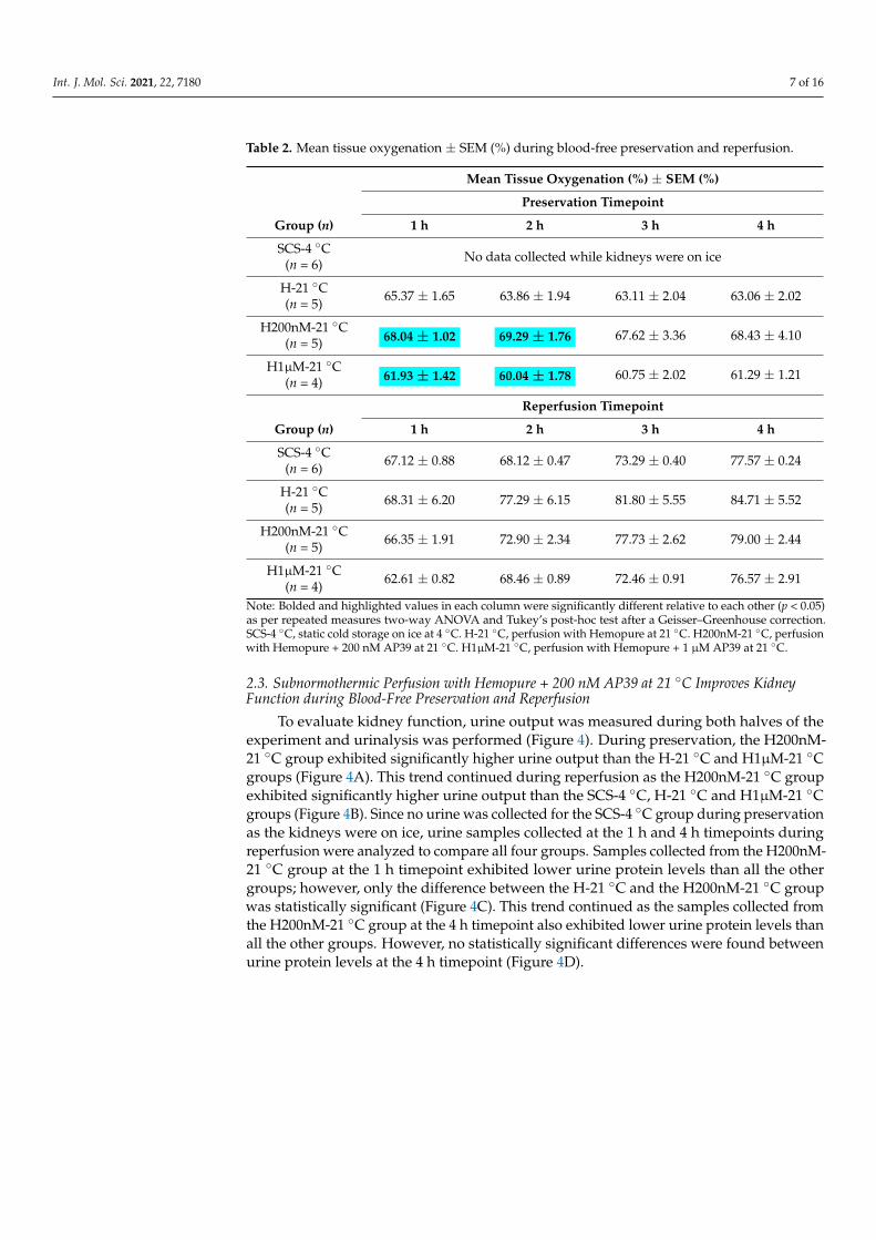
To evaluate kidney function, urine output was measured during both halves of theexperiment and urinalysis was performed (Figure 4). During preservation, the H200nM-21 ◦C group exhibited significantly higher urine output than the H-21 ◦C and H1µM-21 ◦Cgroups (Figure 4A). This trend continued during reperfusion as the H200nM-21 ◦C groupexhibited significantly higher urine output than the SCS-4 ◦C, H-21 ◦C and H1µM-21 ◦Cgroups (Figure 4B). Since no urine was collected for the SCS-4 ◦C group during preservationas the kidneys were on ice, urine samples collected at the 1 h and 4 h timepoints duringreperfusion were analyzed to compare all four groups. Samples collected from the H200nM-21 ◦C group at the 1 h timepoint exhibited lower urine protein levels than all the othergroups; however, only the difference between the H-21 ◦C and the H200nM-21 ◦C groupwas statistically significant (Figure 4C). This trend continued as the samples collected fromthe H200nM-21 ◦C group at the 4 h timepoint also exhibited lower urine protein levels thanall the other groups. However, no statistically significant differences were found betweenurine protein levels at the 4 h timepoint (Figure 4D).
Int. J. Mol. Sci. 2021, 22, 7180 8 of 16Int. J. Mol. Sci. 2021, 22, x FOR PEER REVIEW 8 of 16
Figure 4. Urine parameters during blood-free preservation and reperfusion. (A) Total urine output (mL) during 4 h of blood-free preservation with Hemopure (H-21 °C, n = 5), Hemopure + 200 nM AP39 (H200nM-21 °C, n = 5) or Hemopure + 1 µM AP39 (H1µM-21 °C, n = 6). No values were recorded for the static cold storage (SCS-4 °C, n = 6) group as the kidneys were on ice. (B) Total urine output (mL) during 4 h of blood-free reperfusion with Hemopure and metabolic support at 37 °C. (C) Urine protein (g/L) levels in urine samples collected 1 h after the start of reperfusion. (D) Urine protein (g/L) levels in urine samples collected in the final hour (4 h) of reperfusion. An outlier was excluded from the H1µM-21 °C group in (D). Individual points on each graph reflect values for individual kidneys and lines represent the mean ± SEM. Values were compared using one-way ANOVA followed by Tukey’s post-hoc test. *, p < 0.05. **, p < 0.01. ***, p < 0.001. ****, p < 0.0001.
2.4. Subnormothermic Perfusion with Hemopure + 200 nM AP39 at 21° Reduces Apoptotic Kidney Injury Following Blood-Free Preservation and Reperfusion
To evaluate apoptotic tissue injury, kidney sections were stained with TUNEL (Figure 5A) and scored by a blinded renal pathologist (Figure 5B). The H200nM-21 °C group received lower TUNEL scores than all the other groups, which indicates that this group exhibited the lowest apoptotic injury. Statistically, the TUNEL scores of the H200nM-21 °C group were significantly lower than the scores of the H-21 °C and H1µM-
Figure 4. Urine parameters during blood-free preservation and reperfusion. (A) Total urine output (mL) during 4 h ofblood-free preservation with Hemopure (H-21 ◦C, n = 5), Hemopure + 200 nM AP39 (H200nM-21 ◦C, n = 5) or Hemopure +1 µM AP39 (H1µM-21 ◦C, n = 6). No values were recorded for the static cold storage (SCS-4 ◦C, n = 6) group as the kidneyswere on ice. (B) Total urine output (mL) during 4 h of blood-free reperfusion with Hemopure and metabolic support at37 ◦C. (C) Urine protein (g/L) levels in urine samples collected 1 h after the start of reperfusion. (D) Urine protein (g/L)levels in urine samples collected in the final hour (4 h) of reperfusion. An outlier was excluded from the H1µM-21 ◦Cgroup in (D). Individual points on each graph reflect values for individual kidneys and lines represent the mean ± SEM.Values were compared using one-way ANOVA followed by Tukey’s post-hoc test. *, p < 0.05. **, p < 0.01. ***, p < 0.001.****, p < 0.0001.
2.4. Subnormothermic Perfusion with Hemopure + 200 nM AP39 at 21 ◦C Reduces ApoptoticKidney Injury Following Blood-Free Preservation and Reperfusion
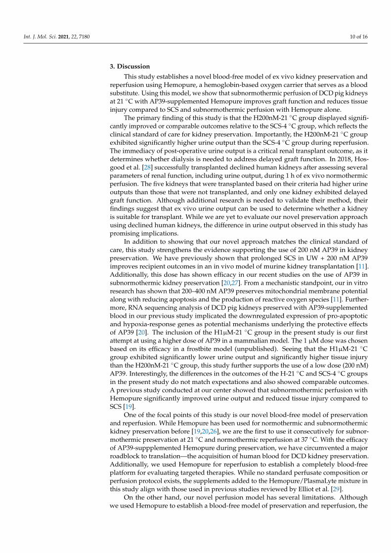
To evaluate apoptotic tissue injury, kidney sections were stained with TUNEL (Figure 5A)and scored by a blinded renal pathologist (Figure 5B). The H200nM-21 ◦C group receivedlower TUNEL scores than all the other groups, which indicates that this group exhibitedthe lowest apoptotic injury. Statistically, the TUNEL scores of the H200nM-21 ◦C groupwere significantly lower than the scores of the H-21 ◦C and H1µM-21 ◦C groups. Althoughthe TUNEL scores of the H200nM-21 ◦C group were also lower than the scores of the SCS-4 ◦C group, the difference was not statistically significant (Figure 5B). To evaluate acutetubular necrosis (ATN), kidney sections were stained with H&E (Figure 6A) and scoredby a blinded renal pathologist (Figure 6B). The SCS-4 ◦C, H-21 ◦C and H200nM-21 ◦C
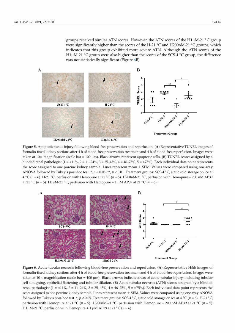
Int. J. Mol. Sci. 2021, 22, 7180 9 of 16
groups received similar ATN scores. However, the ATN scores of the H1µM-21 ◦C groupwere significantly higher than the scores of the H-21 ◦C and H200nM-21 ◦C groups, whichindicates that this group exhibited more severe ATN. Although the ATN scores of theH1µM-21 ◦C group were also higher than the scores of the SCS-4 ◦C group, the differencewas not statistically significant (Figure 6B).
Int. J. Mol. Sci. 2021, 22, x FOR PEER REVIEW 9 of 16
21 °C groups. Although the TUNEL scores of the H200nM-21 °C group were also lower than the scores of the SCS-4 °C group, the difference was not statistically significant (Figure 5B). To evaluate acute tubular necrosis (ATN), kidney sections were stained with H&E (Figure 6A) and scored by a blinded renal pathologist (Figure 6B). The SCS-4 °C, H-21 °C and H200nM-21 °C groups received similar ATN scores. However, the ATN scores of the H1µM-21 °C group were significantly higher than the scores of the H-21 °C and H200nM-21 °C groups, which indicates that this group exhibited more severe ATN. Although the ATN scores of the H1µM-21 °C group were also higher than the scores of the SCS-4 °C group, the difference was not statistically significant (Figure 6B).
Figure 5. Apoptotic tissue injury following blood-free preservation and reperfusion. (A) Representative TUNEL images of formalin-fixed kidney sections after 4 h of blood-free preservation treatment and 4 h of blood-free reperfusion. Images were taken at 10× magnification (scale bar = 100 µm). Black arrows represent apoptotic cells. (B) TUNEL scores assigned by a blinded renal pathologist (1= <11%, 2= 11–24%, 3= 25–45%, 4= 46–75%, 5= >75%). Each individual data point represents the score assigned to one porcine kidney sample. Lines represent mean ± SEM. Values were compared using one-way ANOVA followed by Tukey’s post-hoc test. *, p < 0.05. **, p < 0.01. Treatment groups: SCS-4 °C, static cold storage on ice at 4 °C (n = 6). H-21 °C, perfusion with Hemopure at 21 °C (n = 5). H200nM-21 °C, perfusion with Hemopure + 200 nM AP39 at 21 °C (n = 5). H1µM-21 °C, perfusion with Hemopure + 1 µM AP39 at 21 °C (n = 6).
Figure 6. Acute tubular necrosis following blood-free preservation and reperfusion. (A) Representative H&E images of formalin-fixed kidney sections after 4 h of blood-free preservation treatment and 4 h of blood-free reperfusion. Images
Figure 5. Apoptotic tissue injury following blood-free preservation and reperfusion. (A) Representative TUNEL images offormalin-fixed kidney sections after 4 h of blood-free preservation treatment and 4 h of blood-free reperfusion. Images weretaken at 10× magnification (scale bar = 100 µm). Black arrows represent apoptotic cells. (B) TUNEL scores assigned by ablinded renal pathologist (1 = <11%, 2 = 11–24%, 3 = 25–45%, 4 = 46–75%, 5 = >75%). Each individual data point representsthe score assigned to one porcine kidney sample. Lines represent mean ± SEM. Values were compared using one-wayANOVA followed by Tukey’s post-hoc test. *, p < 0.05. **, p < 0.01. Treatment groups: SCS-4 ◦C, static cold storage on ice at4 ◦C (n = 6). H-21 ◦C, perfusion with Hemopure at 21 ◦C (n = 5). H200nM-21 ◦C, perfusion with Hemopure + 200 nM AP39at 21 ◦C (n = 5). H1µM-21 ◦C, perfusion with Hemopure + 1 µM AP39 at 21 ◦C (n = 6).
Int. J. Mol. Sci. 2021, 22, x FOR PEER REVIEW 9 of 16
21 °C groups. Although the TUNEL scores of the H200nM-21 °C group were also lower than the scores of the SCS-4 °C group, the difference was not statistically significant (Figure 5B). To evaluate acute tubular necrosis (ATN), kidney sections were stained with H&E (Figure 6A) and scored by a blinded renal pathologist (Figure 6B). The SCS-4 °C, H-21 °C and H200nM-21 °C groups received similar ATN scores. However, the ATN scores of the H1µM-21 °C group were significantly higher than the scores of the H-21 °C and H200nM-21 °C groups, which indicates that this group exhibited more severe ATN. Although the ATN scores of the H1µM-21 °C group were also higher than the scores of the SCS-4 °C group, the difference was not statistically significant (Figure 6B).
Figure 5. Apoptotic tissue injury following blood-free preservation and reperfusion. (A) Representative TUNEL images of formalin-fixed kidney sections after 4 h of blood-free preservation treatment and 4 h of blood-free reperfusion. Images were taken at 10× magnification (scale bar = 100 µm). Black arrows represent apoptotic cells. (B) TUNEL scores assigned by a blinded renal pathologist (1= <11%, 2= 11–24%, 3= 25–45%, 4= 46–75%, 5= >75%). Each individual data point represents the score assigned to one porcine kidney sample. Lines represent mean ± SEM. Values were compared using one-way ANOVA followed by Tukey’s post-hoc test. *, p < 0.05. **, p < 0.01. Treatment groups: SCS-4 °C, static cold storage on ice at 4 °C (n = 6). H-21 °C, perfusion with Hemopure at 21 °C (n = 5). H200nM-21 °C, perfusion with Hemopure + 200 nM AP39 at 21 °C (n = 5). H1µM-21 °C, perfusion with Hemopure + 1 µM AP39 at 21 °C (n = 6).
Figure 6. Acute tubular necrosis following blood-free preservation and reperfusion. (A) Representative H&E images of formalin-fixed kidney sections after 4 h of blood-free preservation treatment and 4 h of blood-free reperfusion. Images
Figure 6. Acute tubular necrosis following blood-free preservation and reperfusion. (A) Representative H&E images offormalin-fixed kidney sections after 4 h of blood-free preservation treatment and 4 h of blood-free reperfusion. Images weretaken at 10× magnification (scale bar = 100 µm). Black arrows indicate areas of acute tubular injury, including tubularcell sloughing, epithelial flattening and tubular dilation. (B) Acute tubular necrosis (ATN) scores assigned by a blindedrenal pathologist (1 = <11%, 2 = 11–24%, 3 = 25–45%, 4 = 46–75%, 5 = >75%). Each individual data point represents thescore assigned to one porcine kidney sample. Lines represent mean ± SEM. Values were compared using one-way ANOVAfollowed by Tukey’s post-hoc test. *, p < 0.05. Treatment groups: SCS-4 ◦C, static cold storage on ice at 4 ◦C (n = 6). H-21 ◦C,perfusion with Hemopure at 21 ◦C (n = 5). H200nM-21 ◦C, perfusion with Hemopure + 200 nM AP39 at 21 ◦C (n = 5).H1µM-21 ◦C, perfusion with Hemopure + 1 µM AP39 at 21 ◦C (n = 6).
Int. J. Mol. Sci. 2021, 22, 7180 10 of 16
3. Discussion
This study establishes a novel blood-free model of ex vivo kidney preservation andreperfusion using Hemopure, a hemoglobin-based oxygen carrier that serves as a bloodsubstitute. Using this model, we show that subnormothermic perfusion of DCD pig kidneysat 21 ◦C with AP39-supplemented Hemopure improves graft function and reduces tissueinjury compared to SCS and subnormothermic perfusion with Hemopure alone.
The primary finding of this study is that the H200nM-21 ◦C group displayed signifi-cantly improved or comparable outcomes relative to the SCS-4 ◦C group, which reflects theclinical standard of care for kidney preservation. Importantly, the H200nM-21 ◦C groupexhibited significantly higher urine output than the SCS-4 ◦C group during reperfusion.The immediacy of post-operative urine output is a critical renal transplant outcome, as itdetermines whether dialysis is needed to address delayed graft function. In 2018, Hos-good et al. [28] successfully transplanted declined human kidneys after assessing severalparameters of renal function, including urine output, during 1 h of ex vivo normothermicperfusion. The five kidneys that were transplanted based on their criteria had higher urineoutputs than those that were not transplanted, and only one kidney exhibited delayedgraft function. Although additional research is needed to validate their method, theirfindings suggest that ex vivo urine output can be used to determine whether a kidneyis suitable for transplant. While we are yet to evaluate our novel preservation approachusing declined human kidneys, the difference in urine output observed in this study haspromising implications.
In addition to showing that our novel approach matches the clinical standard ofcare, this study strengthens the evidence supporting the use of 200 nM AP39 in kidneypreservation. We have previously shown that prolonged SCS in UW + 200 nM AP39improves recipient outcomes in an in vivo model of murine kidney transplantation [11].Additionally, this dose has shown efficacy in our recent studies on the use of AP39 insubnormothermic kidney preservation [20,27]. From a mechanistic standpoint, our in vitroresearch has shown that 200–400 nM AP39 preserves mitochondrial membrane potentialalong with reducing apoptosis and the production of reactive oxygen species [11]. Further-more, RNA sequencing analysis of DCD pig kidneys preserved with AP39-supplementedblood in our previous study implicated the downregulated expression of pro-apoptoticand hypoxia-response genes as potential mechanisms underlying the protective effectsof AP39 [20]. The inclusion of the H1µM-21 ◦C group in the present study is our firstattempt at using a higher dose of AP39 in a mammalian model. The 1 µM dose was chosenbased on its efficacy in a frostbite model (unpublished). Seeing that the H1µM-21 ◦Cgroup exhibited significantly lower urine output and significantly higher tissue injurythan the H200nM-21 ◦C group, this study further supports the use of a low dose (200 nM)AP39. Interestingly, the differences in the outcomes of the H-21 ◦C and SCS-4 ◦C groupsin the present study do not match expectations and also showed comparable outcomes.A previous study conducted at our center showed that subnormothermic perfusion withHemopure significantly improved urine output and reduced tissue injury compared toSCS [19].
One of the focal points of this study is our novel blood-free model of preservationand reperfusion. While Hemopure has been used for normothermic and subnormothermickidney preservation before [19,20,26], we are the first to use it consecutively for subnor-mothermic preservation at 21 ◦C and normothermic reperfusion at 37 ◦C. With the efficacyof AP39-suppplemented Hemopure during preservation, we have circumvented a majorroadblock to translation—the acquisition of human blood for DCD kidney preservation.Additionally, we used Hemopure for reperfusion to establish a completely blood-freeplatform for evaluating targeted therapies. While no standard perfusate composition orperfusion protocol exists, the supplements added to the Hemopure/PlasmaLyte mixture inthis study align with those used in previous studies reviewed by Elliot et al. [29].
On the other hand, our novel perfusion model has several limitations. Althoughwe used Hemopure to establish a blood-free model of preservation and reperfusion, the
Int. J. Mol. Sci. 2021, 22, 7180 11 of 16
lack of white blood cells in our perfusate largely excludes the inflammatory componentof subnormothermic preservation as well as reperfusion with blood. Additionally, thepigmented nature of Hemopure, due to the hemoglobin, prevented the use of colorimetricand fluorescent assays to detect inflammatory markers in the perfusate. Furthermore, pairsof kidneys were connected to the same circuit and perfused using a shared Hemopurereservoir due to the limitation of having only one organ perfusion pump. Although wecollected urine output separately for each kidney, the perfusate parameter readings wereless robust, as only one reading was obtained due to the perfusate being shared by pairs ofkidneys. Moreover, we were unable to detect perfusate parameters (pO2, pH and Lactate)with our iSTAT analysis platform since it was designed for use with blood (not Hemopure)at normothermic temperatures rather than subnormothermic temperatures. Our attempt touse the IDEXX analysis platform to detect perfusate levels during preservation also failed.Moreover, our study lacks a number of controls such as cold or subnormothermic perfusiongroup without Hemopure. This is because the high cost (CAD 5000/pig experiment) limitsthe number of groups we could evaluate. Thus, we designed our study around the premiseof comparing the outcomes of our novel approach to that of SCS, which is the clinicalstandard of care, and perfusion with Hemopure alone, which is the control for the effectsof temperature and oxygenated perfusion. Lastly, while perfusion was kept at constantpressure by adjusting the flow, we are unable to report flow data.
As mentioned above, this study advances our previous findings that showed theefficacy of preserving DCD pig kidneys using subnormothermic perfusion at 21 ◦C withAP39-supplemented blood [20]. There is some overlap in the strengths and limitationsof both studies due to the overlap in methodology. Per our previous study, we inducedwarm ischemic injury by clamping the renal pedicle. This approach mimics an extremeclinical DCD scenario where no oxygen is supplied to the kidneys due to a completecessation of blood flow. Thus, the positive outcomes observed would likely be heightenedin real clinical DCD scenarios, where there is a gradual reduction in blood flow as thedonor’s heart stops pumping blood. Additionally, the 4 h duration of the preservationand reperfusion periods is relatively short. However, this was deemed appropriate forestablishing a novel model and for facilitating comparisons to our previous study.
While no mechanistic advances were made, our methodology has improved withthe addition of new approaches to evaluate urine protein levels and tissue oxygenationin real time. It is important to note that whereas this study reports a novel approach forsubnormothermic kidney preservation with H2S-supplemented blood substitute, futureex vivo perfusion studies using declined human kidneys and longer perfusion times areneeded to support our findings. Additionally, more in vitro research is required to establishthe exact mechanisms underlying the protective effects of AP39. Furthermore, researchwith clinically approved H2S donors and in vivo models of renal transplantation is alsoneeded to facilitate the clinical translation of our novel approach.
In conclusion, this study demonstrates that subnormothermic perfusion at 21 ◦C withAP39-supplemented Hemopure improves ex vivo DCD porcine renal graft outcomes. Ourfindings contribute to the expanding body of literature that supports the use of H2S andsubnormothermic preservation to improve kidney outcomes following transplantation.Additionally, we have established a novel blood-free model of ex vivo kidney preserva-tion and reperfusion that will be useful for evaluating other therapeutics, such as othergasotransmitters and gene therapies.
4. Materials and Methods4.1. Animal Care and Surgery
Yorkshire pigs (60–70 kg), purchased from a regional farm, were tranquilized androutinely prepped for surgery. A midline incision was used to expose the kidneys. Fol-lowing intravenous infusion of 10,000 U of heparin, the renal pedicles were clamped insitu for 30 min to induce warm ischemia and mimic DCD injury. The complete cessationof renal blood flow replicates an extreme clinical DCD scenario where no oxygen is being
Int. J. Mol. Sci. 2021, 22, 7180 12 of 16
supplied to the kidneys. This approach has been used in many other studies within thefield and previous studies by our center. During the clamping period, the ureters andarteries were cannulated to facilitate ex vivo perfusion and urine collection. Subsequently,both kidneys were nephrectomized and the donor animal was euthanized. Surgeries wereperformed by transplant fellows at University Hospital, London, Canada. All procedureswere approved by the University of Western Ontario’s Animal Use Committee (AnimalUse Protocol 2018-090) on 28 March 2019.
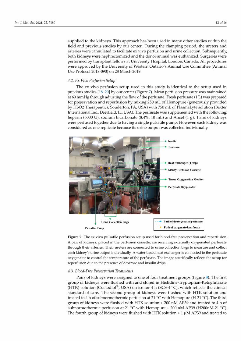
4.2. Ex Vivo Perfusion Setup
The ex vivo perfusion setup used in this study is identical to the setup used inprevious studies [18–20] by our center (Figure 7). Mean perfusion pressure was maintainedat 60 mmHg through adjusting the flow of the perfusate. Fresh perfusate (1 L) was preparedfor preservation and reperfusion by mixing 250 mL of Hemopure (generously providedby HbO2 Therapeutics, Souderton, PA, USA) with 750 mL of PlasmaLyte solution (BaxterInternational Inc., Deerfield, IL, USA). The perfusate was supplemented with the following:heparin (5000 U), sodium bicarbonate (8.4%, 10 mL) and Ancef (1 g). Pairs of kidneyswere perfused together due to having a single pulsatile pump. However, each kidney wasconsidered as one replicate because its urine output was collected individually.
Int. J. Mol. Sci. 2021, 22, x FOR PEER REVIEW 12 of 16
Additionally, we have established a novel blood-free model of ex vivo kidney preservation and reperfusion that will be useful for evaluating other therapeutics, such as other gasotransmitters and gene therapies.
4. Materials and Methods 4.1. Animal Care and Surgery
Yorkshire pigs (60–70 kg), purchased from a regional farm, were tranquilized and routinely prepped for surgery. A midline incision was used to expose the kidneys. Following intravenous infusion of 10,000 U of heparin, the renal pedicles were clamped in situ for 30 min to induce warm ischemia and mimic DCD injury. The complete cessation of renal blood flow replicates an extreme clinical DCD scenario where no oxygen is being supplied to the kidneys. This approach has been used in many other studies within the field and previous studies by our center. During the clamping period, the ureters and arteries were cannulated to facilitate ex vivo perfusion and urine collection. Subsequently, both kidneys were nephrectomized and the donor animal was euthanized. Surgeries were performed by transplant fellows at University Hospital, London, Canada. All procedures were approved by the University of Western Ontario’s Animal Use Committee (Animal Use Protocol 2018-090) on 28th March 2019.
4.2. Ex Vivo Perfusion Setup The ex vivo perfusion setup used in this study is identical to the setup used in
previous studies [18–20] by our center (Figure 7). Mean perfusion pressure was maintained at 60 mmHg through adjusting the flow of the perfusate. Fresh perfusate (1 L) was prepared for preservation and reperfusion by mixing 250 mL of Hemopure (generously provided by HbO2 Therapeutics, Souderton, PA, USA) with 750 mL of PlasmaLyte solution (Baxter International Inc., Deerfield, IL, USA). The perfusate was supplemented with the following: heparin (5000 U), sodium bicarbonate (8.4%, 10 mL) and Ancef (1 g). Pairs of kidneys were perfused together due to having a single pulsatile pump. However, each kidney was considered as one replicate because its urine output was collected individually.
Figure 7. The ex vivo pulsatile perfusion setup used for blood-free preservation and reperfusion. A pair of kidneys, placed in the perfusion cassette, are receiving externally oxygenated perfusate through their arteries. Their ureters are connected to urine collection bags to measure and collect
Figure 7. The ex vivo pulsatile perfusion setup used for blood-free preservation and reperfusion.A pair of kidneys, placed in the perfusion cassette, are receiving externally oxygenated perfusatethrough their arteries. Their ureters are connected to urine collection bags to measure and collecteach kidney’s urine output individually. A water-based heat exchanger is connected to the perfusateoxygenator to control the temperature of the perfusate. The image specifically reflects the setup forreperfusion due to the presence of dextrose and insulin drips.
4.3. Blood-Free Preservation Treatments
Pairs of kidneys were assigned to one of four treatment groups (Figure 8). The firstgroup of kidneys were flushed with and stored in Histidine-Tryptophan-Ketoglutarate(HTK) solution (Custodiol®, USA) on ice for 4 h (SCS-4 ◦C), which reflects the clinicalstandard of care. The second group of kidneys were flushed with HTK solution andtreated to 4 h of subnormothermic perfusion at 21 ◦C with Hemopure (H-21 ◦C). The thirdgroup of kidneys were flushed with HTK solution + 200 nM AP39 and treated to 4 h ofsubnormothermic perfusion at 21 ◦C with Hemopure + 200 nM AP39 (H200nM-21 ◦C).The fourth group of kidneys were flushed with HTK solution + 1 µM AP39 and treated to
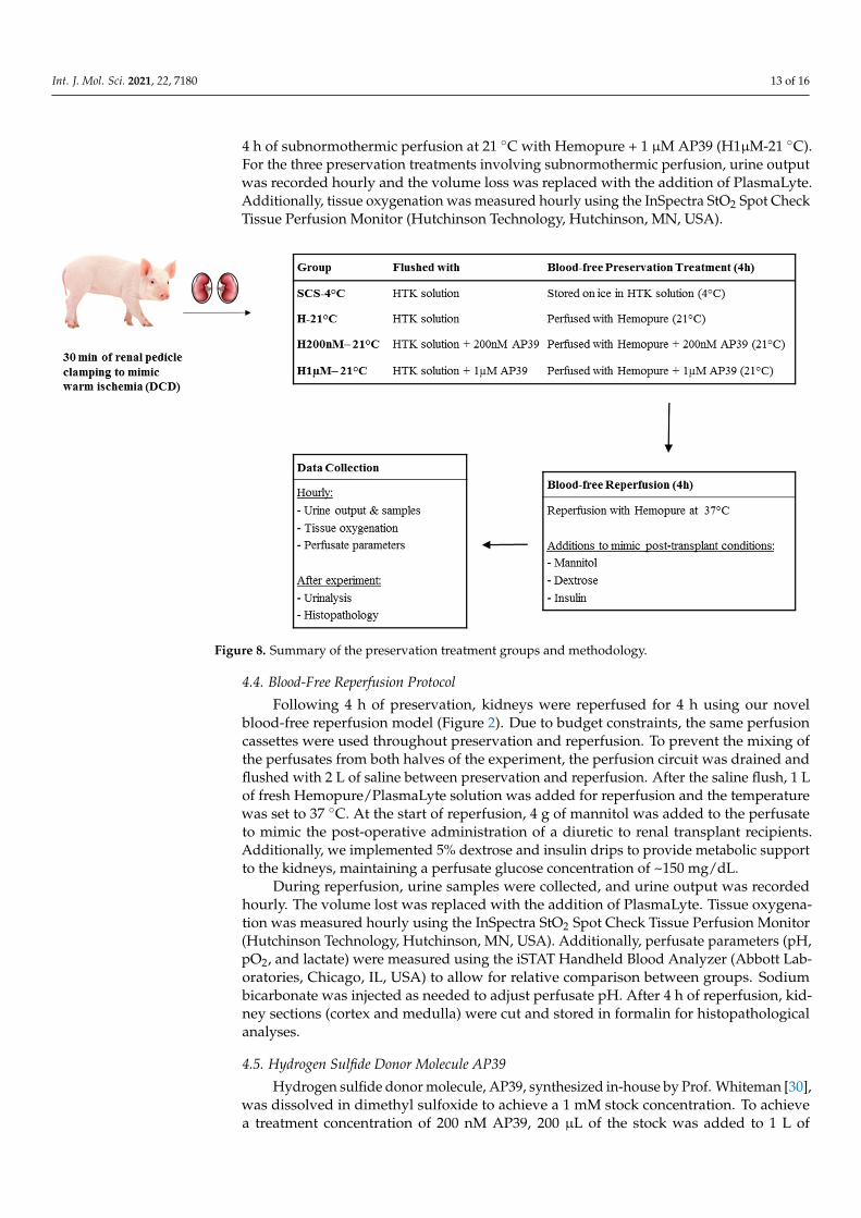
Int. J. Mol. Sci. 2021, 22, 7180 13 of 16
4 h of subnormothermic perfusion at 21 ◦C with Hemopure + 1 µM AP39 (H1µM-21 ◦C).For the three preservation treatments involving subnormothermic perfusion, urine outputwas recorded hourly and the volume loss was replaced with the addition of PlasmaLyte.Additionally, tissue oxygenation was measured hourly using the InSpectra StO2 Spot CheckTissue Perfusion Monitor (Hutchinson Technology, Hutchinson, MN, USA).
Int. J. Mol. Sci. 2021, 22, x FOR PEER REVIEW 13 of 16
each kidney’s urine output individually. A water-based heat exchanger is connected to the perfusate oxygenator to control the temperature of the perfusate. The image specifically reflects the setup for reperfusion due to the presence of dextrose and insulin drips.
4.3. Blood-Free Preservation Treatments Pairs of kidneys were assigned to one of four treatment groups (Figure 8). The first
group of kidneys were flushed with and stored in Histidine-Tryptophan-Ketoglutarate (HTK) solution (Custodiol®, USA) on ice for 4 h (SCS-4 °C), which reflects the clinical standard of care. The second group of kidneys were flushed with HTK solution and treated to 4 h of subnormothermic perfusion at 21 °C with Hemopure (H-21 °C). The third group of kidneys were flushed with HTK solution + 200 nM AP39 and treated to 4 h of subnormothermic perfusion at 21 °C with Hemopure + 200 nM AP39 (H200nM-21 °C). The fourth group of kidneys were flushed with HTK solution + 1 µM AP39 and treated to 4 h of subnormothermic perfusion at 21 °C with Hemopure + 1 µM AP39 (H1µM-21 °C). For the three preservation treatments involving subnormothermic perfusion, urine output was recorded hourly and the volume loss was replaced with the addition of PlasmaLyte. Additionally, tissue oxygenation was measured hourly using the InSpectra StO2 Spot Check Tissue Perfusion Monitor (Hutchinson Technology, Hutchinson, MN, USA).
Figure 8. Summary of the preservation treatment groups and methodology.
4.4. Blood-Free Reperfusion Protocol Following 4 h of preservation, kidneys were reperfused for 4 h using our novel blood-
free reperfusion model (Figure 2). Due to budget constraints, the same perfusion cassettes were used throughout preservation and reperfusion. To prevent the mixing of the perfusates from both halves of the experiment, the perfusion circuit was drained and flushed with 2 L of saline between preservation and reperfusion. After the saline flush, 1 L of fresh Hemopure/PlasmaLyte solution was added for reperfusion and the temperature was set to 37 °C. At the start of reperfusion, 4 g of mannitol was added to the perfusate to mimic the post-operative administration of a diuretic to renal transplant recipients. Additionally, we implemented 5% dextrose and insulin drips to provide metabolic support to the kidneys, maintaining a perfusate glucose concentration of ~150 mg/dL.
Figure 8. Summary of the preservation treatment groups and methodology.
4.4. Blood-Free Reperfusion Protocol
Following 4 h of preservation, kidneys were reperfused for 4 h using our novelblood-free reperfusion model (Figure 2). Due to budget constraints, the same perfusioncassettes were used throughout preservation and reperfusion. To prevent the mixing ofthe perfusates from both halves of the experiment, the perfusion circuit was drained andflushed with 2 L of saline between preservation and reperfusion. After the saline flush, 1 Lof fresh Hemopure/PlasmaLyte solution was added for reperfusion and the temperaturewas set to 37 ◦C. At the start of reperfusion, 4 g of mannitol was added to the perfusateto mimic the post-operative administration of a diuretic to renal transplant recipients.Additionally, we implemented 5% dextrose and insulin drips to provide metabolic supportto the kidneys, maintaining a perfusate glucose concentration of ~150 mg/dL.
During reperfusion, urine samples were collected, and urine output was recordedhourly. The volume lost was replaced with the addition of PlasmaLyte. Tissue oxygena-tion was measured hourly using the InSpectra StO2 Spot Check Tissue Perfusion Monitor(Hutchinson Technology, Hutchinson, MN, USA). Additionally, perfusate parameters (pH,pO2, and lactate) were measured using the iSTAT Handheld Blood Analyzer (Abbott Lab-oratories, Chicago, IL, USA) to allow for relative comparison between groups. Sodiumbicarbonate was injected as needed to adjust perfusate pH. After 4 h of reperfusion, kid-ney sections (cortex and medulla) were cut and stored in formalin for histopathologicalanalyses.
4.5. Hydrogen Sulfide Donor Molecule AP39
Hydrogen sulfide donor molecule, AP39, synthesized in-house by Prof. Whiteman [30],was dissolved in dimethyl sulfoxide to achieve a 1 mM stock concentration. To achievea treatment concentration of 200 nM AP39, 200 µL of the stock was added to 1 L of
Int. J. Mol. Sci. 2021, 22, 7180 14 of 16
preservation solution and perfusate. Similarly, for a concentration of 1 µM AP39, 1 mL ofthe stock was added to 1 L of preservation solution and perfusate. The doses were chosenbased on previous studies by our group [20].
4.6. Urinalysis
Most of the urine samples collected were heavily pigmented, due to the presenceof hemoglobin from the Hemopure, which prevented the use of conventional urinalysismethods. A 1:3 dilution of urine in Hemoglobind (Biotech Support Group, MonmouthJunction, NJ, USA) allowed us to obtain clearer urine samples after 10 min of vigorousshaking and centrifugation at 12,000× g. Urine protein and creatinine levels were analyzedusing the IDEXX Urine Analyzer (IDEXX Laboratories, Westbook, ME, USA), but creatininevalues remained undetectable.
4.7. Histopathology Imaging and Scoring
Formalin-fixed kidney sections, including cortex and medulla, were embedded inparaffin and mounted onto microscope slides. The sections were stained with Terminaldeoxynucleotidyl transferase dUTP nick end labeling (TUNEL) and Hematoxylin and Eosin(H&E) to determine the level of apoptosis and acute tubular necrosis, respectively. TUNELand H&E imaging was done using the Nikon Instruments Eclipse 90i digital microscopeat 10× magnification (Nikon Instruments, Melville, NY, USA). Both sets of slides werescored by a blinded renal pathologist as per the following scheme: 1 = <11%, 2 = 11–24%,3 = 25–45%, 4 = 46–75%, 5 = >75%.
4.8. Statistical Analyses
GraphPad Prism v9.0 (GraphPad Software, San Diego, CA, USA) was used to creategraphs and conduct statistical analyses. SEM is graphed as it accounts for the impact ofvarying n values in certain groups. One-way or two-way ANOVA followed by Tukey’spost-hoc test was used for comparisons of three or more experimental groups. Statisticalsignificance was accepted at p < 0.05.
5. Patents
The data reported in this study are a part of a US patent application (serial no.17/127,965 entitled “Method and Compositions for Protecting Tissue) that is pendingapproval. Additionally, Prof. Matthew Whiteman and the University of Exeter have intel-lectual property (patent filings) related to hydrogen sulfide delivery molecules and theirtherapeutic use.
Author Contributions: Conceptualization, A.S.; methodology, S.J., P.L. and A.S.; software, S.J.;animal surgeries, E.S., M.L. and M.M.; resources, M.W., P.L. and A.S.; data curation, S.J., A.J., M.Y.Z.and A.R.; writing—original draft preparation, S.J.; writing—review and editing, S.J., G.J.D. and A.S.;supervision, A.S. and funding acquisition, A.S. All authors have read and agreed to the publishedversion of the manuscript.
Funding: This research was supported by Physicians Services Incorporated (PSI) Foundation (Grant#18-17) and the Kidney Foundation of Canada (Grant #180015).
Institutional Review Board Statement: The study was conducted according to the guidelines of theDeclaration of Helsinki and approved by the Animal Use Committee of the University of WesternOntario (Animal Use Protocol 2018-090, approved 28 March 2019).
Informed Consent Statement: Not Applicable.
Data Availability Statement: The data presented in this study are available on request from thecorresponding author.
Conflicts of Interest: The authors declare no conflict of interest. The funders had no role in thedesign of the study; in the collection, analyses or interpretation of the data; in the writing of themanuscript or in the decision to publish the results.
Int. J. Mol. Sci. 2021, 22, 7180 15 of 16
References1. Wolfe, R.A.; Ashby, V.B.; Milford, E.L.; Ojo, A.O.; Ettenger, R.E.; Agodoa, L.Y.; Held, P.J.; Port, F.K. Comparison of mortality in all
patients on dialysis, patients on dialysis awaiting transplantation, and recipients of a first cadaveric transplant. N. Engl. J. Med.1999, 341, 1725–1730. [CrossRef]
2. Kaballo, M.A.; Canney, M.; O’Kelly, P.; Williams, Y.; O’Seaghdha, C.M.; Conlon, P.J. A comparative analysis of survival of patientson dialysis and after kidney transplantation. Clin. Kidney J. 2018, 11, 389–393. [CrossRef] [PubMed]
3. Laupacis, A.; Keown, P.; Pus, N.; Krueger, H.; Ferguson, B.; Wong, C.; Muirhead, N. A study of the quality of life and cost-utilityof renal transplantation. Kidney Int. 1996, 50, 235–242. [CrossRef]
4. Canadian Institute for Health Information. Organ Replacement in Canada: CORR Annual statistics|CIHI. 2020. Available online:https://www.cihi.ca/en/organ-replacement-in-canada-corr-annual-statistics-2020 (accessed on 22 March 2021).
5. Canadian Institute for Health Information. e-Statistics on Organ Transplants, Waiting Lists and Donors|CIHI. 2019. Availableonline: https://www.cihi.ca/en/e-statistics-on-organ-transplants-waiting-lists-and-donors (accessed on 22 March 2021).
6. Gill, J.; Rose, C.; Lesage, J.; Joffres, Y.; Gill, J.; O’Connor, K. Use and outcomes of kidneys from donation after circulatory deathdonors in the United States. J. Am. Soc. Nephrol. 2017, 28, 3647–3657. [CrossRef]
7. Maathuis, M.H.J.; de Groot, M.; Ploeg, R.J.; Leuvenink, H.G.D. Deterioration of endothelial and smooth muscle cell function indcd kidneys after static cold storage in IGL-1 or UW. J. Surg. Res. 2009, 152, 231–237. [CrossRef] [PubMed]
8. Salahudeen, A.K.; Haider, N.; May, W. Cold ischemia and the reduced long-term survival of cadaveric renal allografts. Kidney Int.2004, 65, 713–718. [CrossRef]
9. Salahudeen, A.K. Cold ischemic injury of transplanted kidneys: New insights from experimental studies. Am. J. Physiol. Physiol.2004, 287, F181–F187. [CrossRef]
10. Lobb, I.; Davison, M.; Carter, D.; Liu, W.; Haig, A.; Gunaratnam, L.; Sener, A. Hydrogen sulfide treatment mitigates renal allograftischemia-reperfusion injury during cold storage and improves early transplant kidney function and survival following allogeneicrenal transplantation. J. Urol. 2015, 194, 1806–1815. [CrossRef]
11. Lobb, I.; Jiang, J.; Lian, D.; Liu, W.; Haig, A.; Saha, M.N.; Torregrossa, R.; Wood, M.E.; Whiteman, M.; Sener, A. Hydrogen sulfideprotects renal grafts against prolonged cold ischemia-reperfusion injury via specific mitochondrial actions. Am. J. Transplant.2017, 17, 341–352. [CrossRef] [PubMed]
12. Dugbartey, G.J.; Bouma, H.R.; Saha, M.N.; Lobb, I.; Henning, R.H.; Sener, A. A hibernation-like state for transplantable organs: Ishydrogen sulfide therapy the future of organ preservation? Antioxid. Redox Signal. 2018, 28, 1503–1515. [CrossRef]
13. Zhu, C.; Su, Y.; Juriasingani, S.; Zheng, H.; Veramkovich, V.; Jiang, J.; Sener, A.; Whiteman, M.; Lacefield, J.; Nagpal, D.; et al.Supplementing preservation solution with mitochondria-targeted H2S donor AP39 protects cardiac grafts from prolonged coldischemia–reperfusion injury in heart transplantation. Am. J. Transplant. 2019, 19, 3139–3148. [CrossRef]
14. Juriasingani, S.; Akbari, M.; Luke, P.; Sener, A. Novel therapeutic strategies for renal graft preservation and their potential impacton the future of clinical transplantation. Curr. Opin. Organ Transplant. 2019, 24, 385–390. [CrossRef] [PubMed]
15. Hoyer, D.P.; Gallinat, A.; Swoboda, S.; Wohlschläger, J.; Rauen, U.; Paul, A.; Minor, T. Subnormothermic machine perfusion forpreservation of porcine kidneys in a donation after circulatory death model. Transpl. Int. 2014, 27, 1097–1106. [CrossRef]
16. Kaths, J.M.; Echeverri, J.; Chun, Y.M.; Cen, J.Y.; Goldaracena, N.; Linares, I.; Dingwell, L.S.; Yip, P.; John, R.; Bagli, D.; et al.Continuous normothermic ex vivo kidney perfusion improves graft function in donation after circulatory death pig kidneytransplantation. Transplantation 2017, 101, 754–763. [CrossRef] [PubMed]
17. Weissenbacher, A.; Lo Faro, L.; Boubriak, O.; Soares, M.F.; Roberts, I.S.; Hunter, J.P.; Voyce, D.; Mikov, N.; Cook, A.; Ploeg, R.J.;et al. Twenty-four-hour normothermic perfusion of discarded human kidneys with urine recirculation. Am. J. Transplant. 2019,19, 178–192. [CrossRef]
18. Bhattacharjee, R.N.; Ruthirakanthan, A.; Sun, Q.; Richard-Mohamed, M.; Luke, S.; Jiang, L.; Aquil, S.; Sharma, H.; Tun-Abraham,M.E.; Alharbi, B.; et al. Subnormothermic oxygenated perfusion optimally preserves donor kidneys ex vivo. Kidney Int. Rep. 2019,4, 1323–1333. [CrossRef]
19. Bhattacharjee, R.N.; Patel, S.V.; Sun, Q.; Jiang, L.; Richard-Mohamed, M.; Ruthirakanthan, A.; Aquil, S.; Al-Ogaili, R.; Juriasingani,S.; Sener, A.; et al. Renal protection against ischemia reperfusion injury: Hemoglobin-based oxygen carrier-201 versus blood as anoxygen carrier in ex vivo subnormothermic machine perfusion. Transplantation 2020, 104, 482–489. [CrossRef]
20. Juriasingani, S.; Ruthirakanthan, A.; Richard-Mohamed, M.; Akbari, M.; Aquil, S.; Patel, S.; Al-Ogaili, R.; Whiteman, M.; Luke, P.;Sener, A. Subnormothermic perfusion with H2S donor AP39 improves DCD porcine renal graft outcomes in an ex vivo model ofkidney preservation and reperfusion. Biomolecules 2021, 11, 446. [CrossRef] [PubMed]
21. Hosgood, S.A.; van Heurn, E.; Nicholson, M.L. Normothermic machine perfusion of the kidney: Better conditioning and repair?Transpl. Int. 2015, 28, 657–664. [CrossRef]
22. Jahr, J.S.; Nesargi, S.B.; Lewis, K.; Johnson, C. Blood substitutes and oxygen therapeutics: An overview and current status. Am. J.Ther. 2002, 9, 437–443. [CrossRef]
23. Jahr, J.S.; Guinn, N.R.; Lowery, D.R.; Shore-Lesserson, L.; Shander, A. Blood substitutes and oxygen therapeutics: A review.Anesth. Analg. 2021, 132, 119–129. [CrossRef] [PubMed]
24. Jordan, S.D.; Alexander, E. Bovine hemoglobin: A nontraditional approach to the management of acute anemia in a Jehovah’switness patient with autoimmune hemolytic anemia. J. Pharm. Pract. 2013, 26, 257–260. [CrossRef] [PubMed]
Int. J. Mol. Sci. 2021, 22, 7180 16 of 16
25. Davis, J.M.; El-Haj, N.; Shah, N.N.; Schwartz, G.; Block, M.; Wall, J.; Tidswell, M.; DiNino, E. Use of the blood substitute HBOC-201in critically ill patients during sickle crisis: A three-case series. Transfusion 2018, 58, 132–137. [CrossRef] [PubMed]
26. Aburawi, M.M.; Fontan, F.M.; Karimian, N.; Eymard, C.; Cronin, S.; Pendexter, C.; Nagpal, S.; Banik, P.; Ozer, S.; Mahboub, P.;et al. Synthetic hemoglobin-based oxygen carriers are an acceptable alternative for packed red blood cells in normothermickidney perfusion. Am. J. Transplant. 2019, 19, 2814–2824. [CrossRef] [PubMed]
27. Juriasingani, S.; Akbari, M.; Chan, J.Y.; Whiteman, M.; Sener, A. H2S supplementation: A novel method for successful organpreservation at subnormothermic temperatures. Nitric Oxide 2018, 81, 57–66. [CrossRef]
28. Hosgood, S.A.; Thompson, E.; Moore, T.; Wilson, C.H.; Nicholson, M.L. Normothermic machine perfusion for the assessmentand transplantation of declined human kidneys from donation after circulatory death donors. Br. J. Surg. 2018, 105, 388–394.[CrossRef] [PubMed]
29. Elliott, T.R.; Nicholson, M.L.; Hosgood, S.A. Normothermic kidney perfusion: An overview of protocols and strategies. Am. J.Transplant. 2021, 21, 1382–1390. [CrossRef]
30. Le Trionnaire, S.; Perry, A.; Szczesny, B.; Szabo, C.; Winyard, P.G.; Whatmore, J.L.; Wood, M.E.; Whiteman, M. The syn-thesis and functional evaluation of a mitochondria-targeted hydrogen sulfide donor, (10-oxo-10-(4-(3-thioxo-3H-1,2-dithiol-5-yl)phenoxy)decyl)triphenylphosphonium bromide (AP39). Med. Chem. Commun. 2014, 5, 728–736. [CrossRef]
Related Documents


