NUMBER 1 OF 1 AUTHOR QUERIES DATE 1/5/2015 JOB NAME TROPMED ARTICLE 140232 QUERIES FOR AUTHOR Alexandre Braga Libo ´ rio et al. THIS QUERY FORM MUST BE RETURNED WITH ALL PROOFS FOR CORRECTIONS AU1: Please provide city and country for the following affiliation: Laborato ´ rio de Investigac ¸a ˜ o Me ´ dica (LIM 12) do Hospital das Clı ´nicas da Faculdade de Medicina USP. AU2: Please provide company name, city, state, and country (if other than USA) for all manufacturers at first mention. AU3: Please spell out CKD-EPI, AKIN, and CPK at first use in the text. AU4: Please provide email addresses for all authors in addresses section.

Welcome message from author
This document is posted to help you gain knowledge. Please leave a comment to let me know what you think about it! Share it to your friends and learn new things together.
Transcript
NUMBER 1 OF 1
AUTHOR QUERIES
DATE 1/5/2015
JOB NAME TROPMED
ARTICLE 140232
QUERIES FOR AUTHOR Alexandre Braga Liborio et al.
THIS QUERY FORM MUST BE RETURNED WITH ALL PROOFS FOR CORRECTIONS
AU1: Please provide city and country for the following affiliation: Laboratorio de Investigacao Medica (LIM 12)
do Hospital das Clınicas da Faculdade de Medicina USP.
AU2: Please provide company name, city, state, and country (if other than USA) for all manufacturers at first mention.
AU3: Please spell out CKD-EPI, AKIN, and CPK at first use in the text.
AU4: Please provide email addresses for all authors in addresses section.

Am. J. Trop. Med. Hyg., 00(0), 2015, pp. 000–000doi:10.4269/ajtmh.14-0232Copyright © 2015 by The American Society of Tropical Medicine and Hygiene
Endothelial Glycocalyx Damage Is Associated with Leptospirosis Acute Kidney Injury
Alexandre Braga Liborio,*† Marcelo Boecker Munoz Braz,† Antonio Carlos Seguro, Gdayllon C. Meneses,Fernanda Macedo de Oliveira Neves, Danielle Carvalho Pedrosa, Luciano Pamplona de Goes Cavalcanti,
Alice Maria Costa Martins, and Elizabeth de Francesco Daher
Department of Clinical Medicine, Faculty of Medicine, Universidade Federal do Ceara, Fortaleza, Ceara, Brazil; Medical Sciences Post-GraduateProgram, Department of Clinical Medicine, Universidade Federal do Ceara, Fortaleza, Ceara, Brazil; Laboratorio de Investigacao Medica (LIM 12)
do Hospital das Clınicas da Faculdade de Medicina USPAU1 ; Department of Clinical and Toxicological Analysis, Faculty of Pharmacy, FederalUniversity of Ceara, Fortaleza, Ceara, Brazil; Departamento de Saude Comunitaria, Universidade Federal do Ceara, Fortaleza, Ceara, Brazil
Abstract. Leptospirosis is a common disease in tropical countries, and the kidney is one of the main target organs.Membrane proteins of Leptospira are capable of causing endothelial damage in vitro, but there have been no studies inhumans evaluating endothelial glycocalyx damage and its correlation with acute kidney injury (AKI). We performed acohort study in an outbreak of leptospirosis among military personnel. AKI was diagnosed in 14 of 46 (30.4%) patients.Leptospirosis was associated with higher levels of intercellular adhesion molecule-1 (ICAM-1; 483.1 ± 31.7 versus 234.9 ±24.4 mg/L, P < 0.001) and syndecan-1 (73.7 ± 15.9 versus 21.2 ± 7.9 ng/mL, P < 0.001) compared with exposed controls.Patients with leptospirosis-associated AKI had increased level of syndecan-1 (112.1 ± 45.4 versus 41.5 ± 11.7 ng/mL,P = 0.021) and ICAM-1 (576.9 ± 70.4 versus 434.9 ± 35.3, P = 0.034) compared with leptospirosis patients with no AKI.Association was verified between syndecan-1 and ICAM-1 with serum creatinine elevation and neutrophil gelatinase-associated lipocalin (NGAL) levels. This association remained even after multivariate analysis including other AKI-associated characteristics. Endothelial injury biomarkers are associated with leptospirosis-associated renal damage.
INTRODUCTION
Leptospirosis is caused by a microorganism of the genusLeptospira. In total, nine pathogenic species are known,including L. interrogans.1 The annual incidence is estimatedat 10–100 cases per 100,000 population in tropical countries.2
The early phase of leptospirosis manifestations lasts 3–7 days and includes fever, headaches, myalgia (especially inthe calves), nausea, vomiting, malaise, and conjunctival hyper-emia. Only 10% progress to the second phase: the Weil syn-drome.3 This phase lasts from 4 to 30 days, and more severesymptoms, such as jaundice, meningitis, pulmonary hemor-rhage, and acute kidney injury (AKI), can occur.4
The kidney is one of the main target organs of Leptospira,and AKI can occur in 20–85% of patients in the second phaseof the disease.5,6 Interstitial nephritis is the main pathologicalfinding in patients with leptospirosis, even in those withoutAKI.7,8 Leptospira or its fragments can be found in renaltubules and interstitium.9 Observational studies have indicatedthat Leptospira spread through the hematogenous route to thekidney, circulate to peritubular capillaries, migrate to the inter-stitium and renal tubule, and finally, remain in the proximaltubular lumen. In the kidney, proteins present in the outermembrane of Leptospira induce an inflammatory response.This response seems to be, at least in part, mediated by toll-like receptors.10
The outer membrane proteins (OMPs) of Leptospira arecapable of promoting up-regulation of cell adhesion molecules(for instance, intercellular adhesion molecule-1 [ICAM-1])expressed mainly in endothelial cells.11,12 Although these stud-ies were performed in vitro with umbilical vein endothelialcells, they suggested a role of endothelial activation in lepto-spiral pathogenesis.11,12 Also, experimental and autopsy studies
have shown endothelial necrosis and capillary thrombosis inlung tissue.13,14
Another important component of the endothelium is theglycocalyx, a 1-mm carbohydrate-rich structure located at thetop of the endothelium. It has antiadhesive and anticoagulantproperties that protect the endothelium and maintain the vas-cular barrier function. Glycocalyx damage is associated withpathophysiological sequelae, such as capillary leakage andedema formation, accelerated inflammation, platelet activa-tion and hypercoagulability, and loss of vascular responsive-ness.15,16 It is unknown whether glycocalyx lesions have anyrole in leptospirosis-related AKI pathophysiology.In this study, we evaluated young patients from a leptospi-
rosis outbreak among military personnel. Our objective wasto evaluate the association between the presence and severityof renal lesions with biomarkers representative of glycocalyxand endothelial injury.
METHODS
Subjects. In this study, we analyzed subjects involved in anoutbreak of leptospirosis among military personnel. Three mil-itary physicians were responsible for the patients’ medical care.They made all decisions regarding need for hospitalization,frequency of diagnostic procedures, and antibiotic therapy.Immunoglobulin M (IgM) enzyme-linked immunosorbentassay (ELISA; PanBio Kit AU2) and microagglutination (MAT)with 19 different serovars were carried out in all patients. TheCeara State Department of Health collected blood samplesfrom all subjects 3 weeks after convalescence. IgM-ELISAandMAT were performed by the Central Laboratory of PublicHealth (LACEN), a government-sponsored reference labora-tory. Positive cases were defined according to the Centers forDisease Control and Prevention (CDC) laboratory criteria forsupportive (Leptospira agglutination titer ³ 200 but < 800 byMAT in one serum specimen or detection of IgM antibodiesagainst Leptospira in one acute-phase serum specimen) or con-firmed (fourfold or greater increase inLeptospira agglutinationtiter between acute- and convalescent-phase serum specimensstudied at the same laboratory or Leptospira agglutination
*Address correspondence to Alexandre Braga Liborio, Departmentof Clinical Medicine, Faculty of Medicine, Universidade Federal doCeara, Avenue Abolicao 4043, Ap 1203, Fortaleza 60165083, Ceara,Brazil. E-mail: [email protected]†These authors contributed equally.
1
titer ³ 800 by MAT in one serum specimen) diagnosis. MATconfirmation criteria included seroconversion or a fourfold risein titers between acute- and convalescent-phase sera obtainedon the day of admission and after 14–30 days of convalescenceor a titer ³ 1:800 in one or more samples. This definition hasbeen used in other studies.17 Other infectious diseases (hepati-tis A, dengue, typhoid fever, and others) were excluded.Symptoms, physical findings, and routine laboratory tests
were retrospectively retrieved from medical records. Serumsamples from the first blood drawn from each in-hospitalpatient were processed and stored at −70°C for posterior anal-yses (see below). Patients with less than three serum creatinine(SCr) measurements during hospitalization were excluded.Anemia was defined as hemoglobin less than 12 g/dL, and lowplatelet count was defined as a value < 150 + 103/mL. Another31 soldiers exposed to the disease but with no laboratorydiagnosis of leptospirosis (neither supportive nor confirmed)were selected as the exposed control group. The institutionalethical committee approved the study, and participants signedinformed consent.AKI definition and classification. AKI was defined accord-
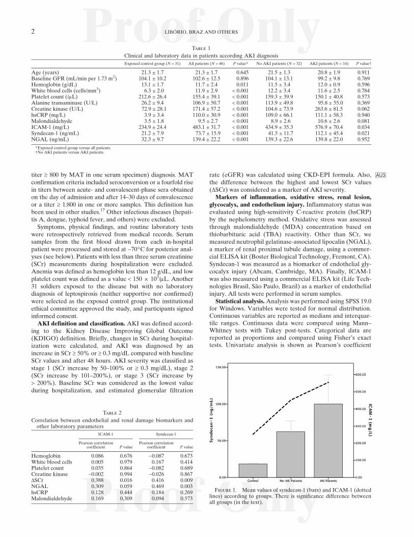
ing to the Kidney Disease Improving Global Outcome(KDIGO) definition. Briefly, changes in SCr during hospital-ization were calculated, and AKI was diagnosed by anincrease in SCr ³ 50% or ³ 0.3 mg/dL compared with baselineSCr values and after 48 hours. AKI severity was classified asstage 1 (SCr increase by 50–100% or ³ 0.3 mg/dL), stage 2(SCr increase by 101–200%), or stage 3 (SCr increase by> 200%). Baseline SCr was considered as the lowest valueduring hospitalization, and estimated glomerular filtration
rate (eGFR) was calculated using CKD-EPI formula AU3. Also,the difference between the highest and lowest SCr values(DSCr) was considered as a marker of AKI severity.Markers of inflammation, oxidative stress, renal lesion,
glycocalyx, and endothelium injury. Inflammatory status wasevaluated using high-sensitivity C-reactive protein (hsCRP)by the nephelometry method. Oxidative stress was assessedthrough malondialdehyde (MDA) concentration based onthiobarbituric acid (TBA) reactivity. Other than SCr, wemeasured neutrophil gelatinase-associated lipocalin (NGAL),a marker of renal proximal tubule damage, using a commer-cial ELISA kit (Boster Biological Technology, Fremont, CA).Syndecan-1 was measured as a biomarker of endothelial gly-cocalyx injury (Abcam, Cambridge, MA). Finally, ICAM-1was also measured using a commercial ELISA kit (Life Tech-nologies Brasil, Sao Paulo, Brazil) as a marker of endothelialinjury. All tests were performed in serum samples.Statistical analysis.Analysis was performed using SPSS 19.0
for Windows. Variables were tested for normal distribution.Continuous variables are reported as medians and interquar-tile ranges. Continuous data were compared using Mann–Whitney tests with Tukey post-tests. Categorical data arereported as proportions and compared using Fisher’s exacttests. Univariate analysis is shown as Pearson’s coefficient
Table 1
Clinical and laboratory data in patients according AKI diagnosis
Exposed control group (N = 31) All patients (N = 46) P value* No AKI patients (N = 32) AKI patients (N = 14) P value†
Age (years) 21.3 ± 1.7 21.3 ± 1.7 0.645 21.5 ± 1.3 20.8 ± 1.9 0.911Baseline GFR (mL/min per 1.73 m2) 104.1 ± 10.2 102.6 ± 12.5 0.896 104.1 ± 13.1 99.2 ± 9.8 0.769Hemoglobin (g/dL) 13.1 ± 1.7 11.7 ± 2.4 0.011 11.5 ± 3.4 12.0 ± 0.9 0.596White blood cells (cells/mm3) 6.3 ± 2.0 11.9 ± 2.9 < 0.001 12.2 ± 3.4 11.6 ± 2.5 0.784Platelet count (/mL) 212.6 ± 26.4 155.4 ± 39.1 < 0.001 159.3 ± 39.9 150.1 ± 40.8 0.573Alanine transaminase (U/L) 26.2 ± 9.4 106.9 ± 50.7 < 0.001 113.9 ± 49.8 95.8 ± 55.0 0.369Creatine kinase (U/L) 72.9 ± 28.1 171.4 ± 57.2 < 0.001 104.6 ± 73.9 263.6 ± 81.5 0.062hsCRP (mg/L) 3.9 ± 3.4 110.0 ± 30.9 < 0.001 109.0 ± 66.1 111.1 ± 58.3 0.940Malondialdehyde 3.5 ± 1.8 9.5 ± 2.7 < 0.001 8.9 ± 2.6 10.6 ± 2.6 0.081ICAM-1 (mg/L) 234.9 ± 24.4 483.1 ± 31.7 < 0.001 434.9 ± 35.3 576.9 ± 70.4 0.034Syndecan-1 (ng/mL) 21.2 ± 7.9 73.7 ± 15.9 < 0.001 41.5 ± 11.7 112.1 ± 45.4 0.021NGAL (ng/mL) 32.3 ± 9.7 139.4 ± 22.2 < 0.001 139.3 ± 22.6 139.8 ± 22.0 0.952
*Exposed control group versus all patients.†No AKI patients versus AKI patients.
Table 2
Correlation between endothelial and renal damage biomarkers andother laboratory parameters
ICAM-1 Syndecan-1
Pearson correlationcoefficient P value
Pearson correlationcoefficient P value
Hemoglobin 0.086 0.676 −0.087 0.673White blood cells 0.005 0.979 0.167 0.414Platelet count 0.035 0.864 −0.082 0.689Creatine kinase −0.002 0.994 −0.026 0.867DSCr 0.388 0.016 0.416 0.009NGAL 0.309 0.059 0.469 0.003hsCRP 0.128 0.444 0.184 0.269Malondialdehyde 0.169 0.309 0.094 0.573
Figure 1. Mean values of syndecan-1 (bars) and ICAM-1 (dottedlines) according to groups. There is significance difference betweenall groups (in the text).
2 LIBORIO, BRAZ AND OTHERS
when indicated. Two multiple linear regression analyses wereperformed to identify the independent factors associated withAKI markers (DSCr and NGAL). A P value < 0.05 was con-sidered statistically significant for all comparisons.
RESULTS
Subjects’ characteristics. During a 4-day period, 83 subjectsperformed military training activities in an open-air field inJuly of 2012 in Ceara in the northeast of Brazil. For personalreasons, 3 military individuals withdrew before any exposure tothe field, with 80 military soldiers remaining in the training.Among the military activities, those that entailed the highestexposure of skin or mucous membranes to water or soil con-taminated by Leptospira were performed on the second andthird days of training. On these days, transposition of water-courses and crawling exercises with water immersion were per-formed. At that place, there was stagnant water mixed with
feces from urban and wild animals found in the area. Duringtraining, the soldiers crawled close to the ground, leaning onforearms and legs, which resulted in immersion in mud.In total, 80 patients were exposed to Leptospira infection,
and 49 had leptospirosis confirmed. Of these, 3 patients wereexcluded, because they had fewer than three SCr measure-ments available. In the end, 46 patients were included in thefinal analysis. All patients were male and between 18 and24 years old, with a median age of 21 (19–23) years. No patienthad any comorbidity, and all patients had a basal eGFR higherthan 80 mL/min. The index case reported symptom onset 5 daysafter exposure, and the last case reported symptoms 21 daysafter exposure. The most common symptoms were headache(98%), myalgia, fever and prostration (96.1%), vomiting, diar-rhea, calf pain (30%), and abdominal pain (17.1%). Labora-tory data are disclosed in T1Table 1. Overall, 17 (36.9%) patientshad anemia, and 16 (34.8%) patients had low platelet count.AKI was diagnosed in 14 (30.4%) patients, and all patients
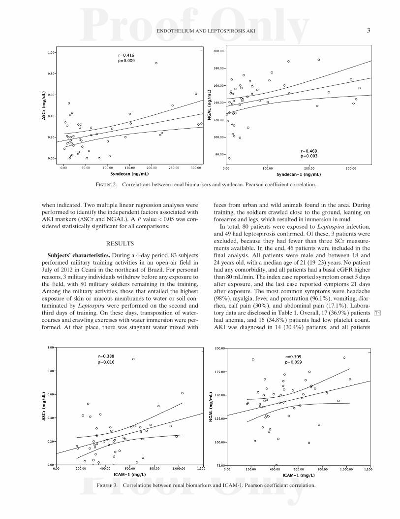
Figure 2. Correlations between renal biomarkers and syndecan. Pearson coefficient correlation.
Figure 3. Correlations between renal biomarkers and ICAM-1. Pearson coefficient correlation.
ENDOTHELIUM AND LEPTOSPIROSIS AKI 3

except for 1 had the milder form of AKI (AKIN stage 1). Nopatient had pulmonary hemorrhage. All patients were treatedwith ceftriaxone.Additionally, we selected 31 exposed soldiers without lep-
tospirosis diagnosis, and their data are show in Table 1. Asexpected, these subjects had lower levels of inflammatory,oxidative stress, and endothelial biomarkers. None of subjectsin the exposed control group had low platelet level or alteredrenal function.Endothelial biomarkers in leptospirosis. Patients with lep-
tospirosis had elevated serum levels of ICAM-1 (483.1 ± 31.7versus 234.9 ± 24.4 mg/L, P < 0.001) and syndecan-1 (73.7 ±15.9 versus 21.2 ± 7.9 ng/mL, P < 0.001) compared withpatients in the exposed control group. There was a significantcorrelation between syndecan-1 and ICAM-1 levels (r = 0.629,P < 0.0001). No significant correlation was observed betweensyndecan-1 or ICAM-1 and hsCRP, MDA, hemoglobin level,CPK, or platelet count (T2 Table 2).
Endothelial damage is associated with leptospirosis-associated AKI. Patients with leptospirosis-associated AKIhad increased levels of syndecan-1 (112.1 ± 45.4 versus 41.5 ±11.7 ng/mL, P = 0.021) and ICAM-1 (576.9 ± 70.4 versus434.9 ± 35.3, P = 0.034) compared with leptospirosis patientswith no AKI ( F1Figure 1). Also, a significant association was veri-fied between syndecan-1 and ICAM-1 levels with DSCr in bothabsolute and relative values in relation to basal SCr ( F2Figures 2and F33 and Table 2).NGAL was similar in leptospirosis patients with or without
AKI (139.8 ± 22.0 versus 139.3 ± 22.6 ng/mL, P = 0.952), andthere was no significant association between NGAL andDSCr. Although there was no correlation between serumNGAL and the presence of AKI, there was a positive correla-tion between syndecan-1 and serum NGAL levels (r = 0.469,P = 0.003) (Figure 2 and Table 2). A trend was observed in thepositive correlation between ICAM-1 and serum NGALlevels (r = 0.309, P = 0.059) (Figure 3).To further explore the association between syndecan/
ICAM-1 and renal damage markers, models of linear multi-variate analysis were performed to evaluate independentassociation between endothelium markers with DSCr andNGAL. In these models, we forced all variables potentiallyassociated with leptospirosis AKI: inflammatory status (whiteblood cells and hsCRP), oxidative stress (MDA), rhabdomyol-ysis (creatine kinase), serum liver enzymes, and platelet count.Even after forcing all variables into the model, syndecan andICAM-1 remained independently associated with DSCr andNGAL ( T3Table 3).
DISCUSSION
In this study, for the first time, endothelial lesion bio-markers in leptospirosis were studied in humans. Also, wedisclosed an important association between endothelial lesion(mainly glycocalyx) and the presence and severity of renallesion. There was a correlation with both reductions in GFRand markers of renal proximal tubular damage (DSCr andNGAL, respectively).The patients were extracted from an outbreak that
occurred among military personnel in 2011 in northeastern
Table 3
Multivariate analysis, including all laboratory parameters, shown toverify the independent association between syndecan-1 and renaldamage markers
Variable Standardized b-coefficient P value
Dependent variable: DSCr (mg/dL)Hemoglobin (g/dL) 0.077 0.793White blood cells ( +103/mm3) −0.224 0.479Platelet count ( +103/mm3) −0.329 0.484Alanine transaminase (U/L) −0.450 0.160Creatine kinase (U/L) 0.295 0.305hsCRP (mg/L) 0.145 0.613Malondialdehyde −0.043 0.893Syndecan-1 (ng/mL) 0.682 0.021ICAM-1 (mg/L) 0.560 0.032
Dependent variable: NGAL (ng/mL)Hemoglobin (g/dL) −0.102 0.738White blood cells ( +103/mm3) 0.145 0.683Platelet count ( +103/mm3) 0.234 0.444Alanine transaminase (U/L) −0.097 0.730Creatine kinase (U/L) 0.432 0.108hsCRP (mg/L) −0.101 0.712Malondialdehyde 0.149 0.629Syndecan-1 (ng/mL) 0.600 0.034ICAM-1 (mg/L) 0.480 0.047
Figure 4. Endothelial lesion, a suggested mechanism. Leptospira attaches to endothelium through cadherin followed by syndecan-1 releaseinto circulation. This figure appears in color at www.ajtmh.org.
4 LIBORIO, BRAZ AND OTHERS

Brazil. This fact provides support to our study mainly regardingtwo points: all patients had similar characteristics (same gen-der, similar age, and absence of comorbidities), and because allexposed individuals were submitted to medical assessment andlaboratory tests, there was no selection bias regarding diseaseseverity. Although the subjects were submitted to the sameexposure, Leptospira burden was not necessarily the same,because possible injuries and cuts on the skin can influencethe observed attack rate.Overall, the patients included in the study had mild forms
of leptospirosis. According to stringent definitions of severeleptospirosis (presence of at least one of the following criteria:AKI requiring dialysis, shock treated with vasoactive drugs,alveolar hemorrhage, bleeding requiring blood transfusion,respiratory failure needing mechanical ventilation, or deathduring hospitalization),18 no patient in this study had the severeform. Although 30% of included patients developed AKI,AKIN criteria was used, which is more sensitive than the pre-vious AKI definition. This AKI definition can significantlyoverestimate AKI incidence; however, in other leptospirosiscohorts, it has been associated with increasing mortality.19
Although it is known that leptospirosis can induce vesselinflammation, only few studies have evaluated the role of endo-thelium in the pathophysiology of leptospirosis. In an ancillarystudy, De Brito and others14 described swollen endotheliumand microvascular thrombosis in guinea pigs infected withLeptospira. Recently, a group of investigators described thatthe OMP of Leptospira can induce endothelial cell damagein vitro.12 To the best of our knowledge, this study is the first todisclose endothelial glycocalyx injury in human leptospirosis.Interestingly, these alterations could be seen, even in milderforms of leptospirosis.Migration of Leptospira to tissues involves the spirochetes
adhering to and escaping from vasculature. Recently, it hasbeen described that Leptospira adheres to vascular endothe-lium cadherin,20 which plays an important role in maintainingthe vasculature barrier properties. Attachment of Leptospirato the vasculature through cadherin may result in vasculardamage, facilitating the escape of the pathogen from thebloodstream into different tissues. One can speculate that gly-cocalyx and endothelial lesions occur during this translocationand that endothelial biomarkers are a marker of disease sever-ity in not only the kidney but also, other organs (F4 Figure 4).Alternatively, one can suppose that glycocalyx and endothe-lium lesions occur first, favoring spirochete adhesion and trans-location. These hypotheses cannot be further explored in ourfindings because of the study design.The most severely affected organs in leptospirosis are the
kidney, liver, and lungs. In our population, no patients hadpulmonary manifestations or liver failure, but almost allpatients had mild to moderate liver enzyme elevation, andnearly 30% had AKI. Although AKI was milder in all exceptfor one patient, there was a positive correlation betweensyndecan-1 and ICAM-1 levels and DSCr. This finding sup-ports a role of endothelium activation/damage in the patho-physiology of leptospirosis-associated AKI. As stated in theIntroduction, Leptospira affects mainly the proximal tubularcells after transmigration through the peritubular capillarynetwork. To further study this association, we have measureda renal biomarker that is more specific for proximal tubularinjury (NGAL), and it showed a positive correlation withboth syndecan-1 and ICAM-1.
Even after controlling for several laboratory findings,syndecan-1, but not ICAM-1, remained associated with AKIseverity. This highlights the potential and unexplored role ofendothelial glycocalyx in leptospirosis-associated AKI. Therehave been only two studies evaluating syndecan-1 levels inpatients with renal disease.21 In one study, patients undergoingmaintenance hemodialysis had higher levels of syndecan-1 thancontrols. The other study excluded any accumulation effect ofsyndecan-1 into the circulation because of reduced renal clear-ance,22 and thus, it is unlikely that only the reduced GFR maybe responsible for the increase in syndecan levels.Although not the main focus of the study, one point that
also deserves consideration is the lack of correlation betweenNGAL and AKI, contrary to what has been shown by manyother studies. Also, AKI in leptospirosis has been associatedwith low platelet count,23 rhabdomyolysis,24 and other clinicalparameters. In this study, we were unable to show these asso-ciations. The lack of associations can also be explained by themilder AKI developed by patients.Our study has some limitations, but two deserve consider-
ation. First, although we measured endothelial and glycocalyxbiomarkers during the first medical consultation, some patientsalready had some kidney damage and GFR decrease, making itdifficult to ascertain a causal association between endothelialdamage andAKI. Second, all except for one patient had milderAKI. It is highly probable the associations disclosed in thisstudy become stronger with increasing AKI severity, but thatcan only be speculated at this point.In conclusion, we studied a homogeneous group of young
male patients from the military community exposed to a lep-tospirosis outbreak and observed that endothelial and mainly,glycocalyx injury biomarkers are related with renal damage,even in patients with milder AKI.
Received April 14, 2014. Accepted for publication October 29, 2014.
Financial support: A.B.L., A.M.C.M. and E.d.F.D. are recipients of agrant from the Conselho Nacional de Desenvolvimento Cientıfico eTecnologico (CNPq).
Disclaimer: The funders had no role in study design, data collectionand analysis, decision to publish, or preparation of the manuscript.
Authors’ addresses: Alexandre AU4Braga Liborio and Elizabeth deFrancesco Daher, Department of Clinical Medicine, Faculty of Med-icine, Universidade Federal do Ceara, Fortaleza, Ceara, Brazil,E-mail: [email protected]. Marcelo Boecker MunozBraz, Fernanda Macedo de Oliveira Neves, and Danielle CarvalhoPedrosa, Medical Sciences Post-Graduate Program, Department ofClinical Medicine, Universidade Federal do Ceara, Fortaleza, Ceara,Brazil. Antonio Carlos Seguro, Laboratorio de Investigacao Medica(LIM 12) do Hospital das Clınicas da Faculdade de Medicina USP.Gdayllon C. Meneses and Alice Maria Costa Martins, Department ofClinical and Toxicological Analysis, Faculty of Pharmacy, FederalUniversity of Ceara, Fortaleza, Ceara, Brazil. Luciano Pamplonade Goes Cavalcanti, Departamento de Saude Comunitaria, Uni-versidade Federal do Ceara, Fortaleza, Ceara, Brazil.
REFERENCES
1. Cerqueira GM, Picardeau M, 2009. A century of Leptospirastrain typing. Infect Genet Evol 9: 760–768.
2. Pappas G, Papadimitriou P, Siozopoulou V, Christou L, AkritidisN, 2008. The globalization of leptospirosis: worldwide inci-dence trends. Int J Infect Dis 12: 351–357.
3. Ko AI, Goarant C, Picardeau M, 2009. Leptospira: the dawn ofthe molecular genetics era for an emerging zoonotic pathogen.Nat Rev Microbiol 7: 736–747.
ENDOTHELIUM AND LEPTOSPIROSIS AKI 5

4. Andrade L, de Francesco Daher E, Seguro AC, 2008. Leptospiralnephropathy. Semin Nephrol 28: 383–394.
5. Daher EF, Silva GB Jr, Lima RS, Mota RM, Rocha HA,de Abreu KL, Barreto AG, Pereira ED, Araujo SM, LiborioAB, 2011. Different patterns in a cohort of patients with severeleptospirosis (Weil syndrome): effects of an educational pro-gram in an endemic area. Am J Trop Med Hyg 85: 479–484.
6. Daher EF, Silva GB Jr, Karbage NN, Carvalho PC Jr, KataokaRS, Silva EC, Magalhaes MM,Mota RM, Araujo SM, Gutierrez-Adrianzen OA, Liborio AB, 2009. Predictors of oliguric acutekidney injury in leptospirosis. A retrospective study on 196 con-secutive patients. Nephron Clin Pract 112: c25–c30.
7. Sitprija V, Pipatanagul V, Mertowidjojo K, Boonpucknavig V,Boonpucknavig S, 1980. Pathogenesis of renal disease in leptospi-rosis: clinical and experimental studies. Kidney Int 17: 827–836.
8. Penna D, De Brito T, Pupo AA, Machado MM, Galvao PAA,de Almeida SS, 1963. Kidney biopsy in human leptospirosis.Am J Trop Med Hyg 12: 896–901.
9. De Brito T, Menezes LF, Lima DMC, Lourenco S, Silva AMG,Alves VAF, 2006. Immunohistochemical and in situ hybridiza-tion studies of the liver and kidney in human leptospirosis.Virchows Arch 448: 576–583.
10. Yang C-W, 2007. Leptospirosis renal disease: understanding theinitiation by Toll-like receptors. Kidney Int 72: 918–925.
11. Gomez RM, Vieira ML, Schattner M, Malaver E, WatanabeMM,Barbosa AS, Abreu PA, de Morais ZM, Cifuente JO, AtzingenMV, Oliveira TR, Vasconcellos SA, Nascimento AL, 2008.Putative outer membrane proteins of Leptospira interrogansstimulate human umbilical vein endothelial cells (HUVECS)and express during infection. Microb Pathog 45: 315–322.
12. Vieira ML, D’Atri LP, Schattner M, Habarta AM, Barbosa AS,de Morais ZM, Vasconcellos SA, Abreu PA, Gomez RM,Nascimento AL, 2007. A novel leptospiral protein increasesICAM-1 and E-selectin expression in human umbilical veinendothelial cells. FEMS Microbiol Lett 276: 172–180.
13. De Brito T, Aiello VD, da Silva LF, Goncalves da Silva AM,Ferreira da Silva WL, Castelli JB, Seguro AC, 2013. Humanhemorrhagic pulmonary leptospirosis: pathological findingsand pathophysiological correlations. PLoS ONE 8: e71743.
14. De Brito T, Bohm GM, Yasuda PH, 1979. Vascular damage inacute experimental leptospirosis of the guinea-pig. J Pathol128: 177–182.
15. Becker BF, Chappell D, Bruegger D, Annecke T, Jacob M,2010. Therapeutic strategies targeting the endothelial glyco-calyx: acute deficits, but great potential. Cardiovasc Res 87:300–310.
16. Palaiologou M, Delladetsima I, Tiniakos D, 2014. CD138(syndecan-1) expression in health and disease.Histol Histopathol29: 177–189.
17. Reis EA, Hagan JE, Ribeiro GS, Teixeira-Carvalho A, Martins-Filho OA, Montgomery RR, Shaw AC, Ko AI, Reis MG, 2013.Cytokine response signatures in disease progression and devel-opment of severe clinical outcomes for leptospirosis. PLoSNegl Trop Dis 7: e2457.
18. Tubiana S, Mikulski M, Becam J, Lacassin F, Lefevre P, GourinatAC, Goarant C, D’Ortenzio E, 2013. Risk factors and predic-tors of severe leptospirosis in New Caledonia. PLoS Negl TropDis 7: e1991.
19. Silva Junior GB, Abreu KL, Mota RM, Barreto AG, Araujo SM,Rocha HA, Liborio AB, Daher EF, 2011. RIFLE and AcuteKidney Injury Network classifications predict mortality inleptospirosis-associated acute kidney injury. Nephrology (Carlton)16: 269–276.
20. Evangelista K, Franco R, Schwab A, Coburn J, 2014. Leptospirainterrogans binds to cadherins. PLoS Negl Trop Dis 8: e2672.
21. Vlahu CA, Lemkes BA, Struijk DG, Koopman MG, Krediet RT,Vink H, 2012. Damage of the endothelial glycocalyx in dialysispatients. J Am Soc Nephrol 23: 1900–1908.
22. Padberg JS, Wiesinger A, di Marco GS, Reuter S, Grabner A,Kentrup D, Lukasz A, Oberleithner H, Pavenstadt H, BrandM, Kumpers P, 2014. Damage of the endothelial glycocalyx inchronic kidney disease. Atherosclerosis 234: 335–343.
23. Raoult D, Jeandel P, Mailloux M, Rougier Y, 1983. Thrombocy-topenia and renal failure in leptospirosis. Am J Trop Med Hyg32: 1464.
24. Coursin DB, Updike SJ, Maki DG, 2000. Massive rhabdomyoly-sis and multiple organ dysfunction syndrome caused by lepto-spirosis. Intensive Care Med 26: 808–812.
6 LIBORIO, BRAZ AND OTHERS
Related Documents