Vol. 5, No. 1 51 Copyright © 2005 by the Society for Biology of Reproduction Endoglin (cd105) and S100A13 as markers of active angiogenesis in endometriosis Soren Hayrabedyan, Stanimir Kyurkchiev, Ivan Kehayov 1 Department of Molecular Immunology, Institute of Biology and Immunology of Reproduction, Bulgarian Academy of Sciences, Sofia, Bulgaria Received: 10 July 2004; accepted: 5 February 2005 SUMMARY The aim of the present study was to evaluate the expression of the neo-an- giogenic marker endoglin and its localization in tissues of normal and endo- metriotic patients as well as to compare it with one new angiogenic marker candidate – S100A13. Human recombinant S100A13 and endoglin 35mer synthetic peptide of the intracellular domain were used for the production of rabbit polyclonal antisera. The antisera were characterized for specificity, using immunoenzyme assay (ELISA), Western blot and immunohistochem- istry. Formalin-fixed, paraffin-embedded tissue sections from normal endo- metrium, adenomyosis, ovarian endometriosis, eutopic endometrium from different endometriotic specimens were tested by immunohistochemistry. No endoglin specific staining was observed on the microvessels of the normal endometrium. In adenomyosis and ovarian endometriosis, the expression pattern was different – endoglin was expressed in all microvessels, with an 1 Corresponding author: Ivan Kehayov, Department of Molecular Immunology, Institute of Biology and Immunology of Reproduction, Bulgarian Academy of Sciences, Sofia 1113, 73, Tzarigradsko shosse blvd., Bulgaria; [email protected]

Welcome message from author
This document is posted to help you gain knowledge. Please leave a comment to let me know what you think about it! Share it to your friends and learn new things together.
Transcript

Vol. 5, No. 1 51
Copyright © 2005 by the Society for Biology of Reproduction
Endoglin (cd105) and S100A13 as markers of active angiogenesis
in endometriosisSoren Hayrabedyan, Stanimir Kyurkchiev, Ivan Kehayov1
Department of Molecular Immunology, Institute of Biology and Immunology of Reproduction,
Bulgarian Academy of Sciences, Sofia, Bulgaria
Received: 10 July 2004; accepted: 5 February 2005
SUMMARY
The aim of the present study was to evaluate the expression of the neo-an-giogenic marker endoglin and its localization in tissues of normal and endo-metriotic patients as well as to compare it with one new angiogenic marker candidate – S100A13. Human recombinant S100A13 and endoglin 35mer synthetic peptide of the intracellular domain were used for the production of rabbit polyclonal antisera. The antisera were characterized for specificity, using immunoenzyme assay (ELISA), Western blot and immunohistochem-istry. Formalin-fixed, paraffin-embedded tissue sections from normal endo-metrium, adenomyosis, ovarian endometriosis, eutopic endometrium from different endometriotic specimens were tested by immunohistochemistry. No endoglin specific staining was observed on the microvessels of the normal endometrium. In adenomyosis and ovarian endometriosis, the expression pattern was different – endoglin was expressed in all microvessels, with an
1Corresponding author: Ivan Kehayov, Department of Molecular Immunology, Institute of Biology and Immunology of Reproduction, Bulgarian Academy of Sciences, Sofia 1113, 73, Tzarigradsko shosse blvd., Bulgaria; [email protected]
Angiogenic markers in endometriosis52
even stronger expression in the myometrial compartment. Weak endoglin-positive staining was detected in the microvessels of eutopic endometrium specimens from different endometriosis cases. In comparison to endoglin, S100A13 exhibited a moderate expression in endometrial glands of nor-mal endometrium, but strong expression in endometriotic specimens. No S100A13 extensive staining of the microvessels was observed in normal endometrium, while in endometriosis, it exhibited very intense staining in microvascular endothelia and less intense in the perivascular area of middle to large-sized vessels. This study for the first time shows over-expression of S100A13 in endometriosis. These data show that the expression of en-doglin and S100A13 corresponds to the activation of the endothelial cells in the process of endometriotic angiogenesis, suggesting a beneficial role for these two molecules as markers for actively progressing endometriotic process. Reproductive Biology 2005 5(1): 51-67.Key words: endoglin, CD105, S100A13, endothelial marker, angiogenic status, microvascular density marker
INTRODUCTION
Endometriosis is characterized by development of endometriotic stroma and glands at sites different from the uterine endometrium [27]. Angiogenesis is considered to be a pivotal process in the pathogenesis of endometriosis and is required for establishment, development and maintenance of endometri-otic lesions as well as for their further propagation. Although endometriosis seems to be a unified pathology, it has many different forms. Adenomyosis and ovarian endometriosis are two of these. It has been currently considered by some authors [7], that these two subgroups have distinct pathogeneses and possibly different angiogenesis mechanisms.
Endoglin (Eng, CD105) is a transmembrane homodimer glycoprotein (190 kDa, [19]) known as a modulator of Transforming Growth Factor Re-ceptor I (TGF-β RI, [21]). Studies on gene knock-out mice [20] revealed that Eng is a vital factor for the angiogenesis early in development. Endoglin is expressed in human endothelial cells [8], macrophages [17], stromal cells

Hayrabedyan et al. 53
[25, 29], and vascular smooth muscle cells (VSMCs; [1, 5, 22]). Over--expression of Eng is characteristic in activated endothelial cells (ECs; [30]), but not quiescent ECs [15, 31]. Since it is used for microvascular density evaluation and ECs counting in tumours [2, 3], Eng is of primary interest as an angiogenic marker in endometriosis as well.
S100A13 is a newly discovered members of the S100 protein family, which is characterized by specificity to different forms of cancer [9, 11, 18]. S100A13 was reported to be co-expressed with the fibroblast growth factor 1 (FGF-1) in brain tumours [16] demonstrating a perivascular distribution. Translocation of S100A13 was observed in endothelial cells in response to an increase in intracellular calcium levels or to angiotensin II, and the process was dependent on the classic Golgi-endoplasmic reticulum pathway [10]. This implication of S100A13 as involved in multiple signalling pathways in the EC suggests for its future use as a possible marker of EC activation.
The aims of this study were to: 1. evaluate the expression of the neo-angiogenic marker Eng and its localization in tissues of normal and endo-metriotic patients; 2. compare the expression pattern of Eng with expression pattern of S100A13 – a possible candidate for angiogenic marker.
MATERIALS AND METHODS
A synthetic 35-mer peptide (corresponding to a portion of the intracellular domain of the Eng molecule, and referred to as Eng-peptide) and human recombinant S100A13 protein (hrS100A13) used for the production of anti-endoglin and anti-S100A13 polyclonal sera respectively, were kindly donated by the Maine Medical Centre Research Institute (USA).
Formalin-fixed paraffin embedded (FFPE) tissue sections from cases of adenomyosis (n=20), ovarian endometriosis (n=20), and eutopic endome-trium of women with endometriosis (n=5) and from normal endometrium (n=5) were provided by the Pathology Department of the University Hos-pital, University of Pleven, Bulgaria. All endometriosis cases were classi-fied according to the criteria of ASF (American Society for Fertility) and histologically graded after Noyes classification [23] by two pathologists.

Angiogenic markers in endometriosis54
Antisera production and characteristics
Chinchilla rabbits (males, 2.5 kg) were injected subcutaneously either with Eng-peptide (150 µg) or hrS100A13 (100 µg) every two weeks for three months. The polyclonal sera were subsequently collected and tested for specificity by ELISA and Western blot.
A. Antisera specificity assessed by immunoenzyme method (ELISA)
Indirect ELISA method was used to test the produced polyclonal antisera [9]. Briefly, the Eng-peptide (1 µg/ml) or hrS100A13 protein (1 µg/ml) were coated on 96 well microtiter plates (Costar, USA) in carbonate buffer (pH 9.4) during overnight incubation (4oC). Bovine serum albumin (1% BSA) was used to block the plates, and then serially diluted antisera – either anti-Eng-peptide (starting at 1/800) or anti-S100A13 (starting at 1/200) were distributed in each coated well. The assay was performed in duplicates.
An anti-rabbit IgG antiserum labelled with peroxidase (Sigma Co, USA), diluted 1/1000 in blocking buffer, was added to each well and kept for 1 hour at room temperature. The enzyme reaction was developed using ortho-phenylenedi-amine and the coloured product intensity was read using an ELISA reader (LKB, Sweden) at a wavelength of 492 nm. As an inhibition ELISA test for specificity, the same dilutions of anti-Eng-peptide or anti-hrS100A13 antibodies pre-incubated with endoglin peptide (1 µg/ml) and hrS100A13 (1 µg/ml) protein, respectively, were applied in parallel wells on the same plate and incubated at RT for 2 hours. Additionally, the sera were tested against a panel of various human proteins (teta-nus toxoid, normal human serum, human serum albumin, IL-1α, FGF-1, S100A1, S100A6, S100B, and calmodulin) used as antigens, by indirect ELISA.
B. Antisera specificity assessed by Western blot analysis
Electrophoresis in a 15% (w/v) polyacrylamide gel, in the presence of sodium dodecyl sulphate (SDS) under reducing conditions, was performed following

Hayrabedyan et al. 55
the well-established protocol of Laemmli (1970). Samples of endoglin peptide and hrS100A13 were added to serial dilutions (with an initial concentration of 5 μg/ml) and transferred to a “HyBond” nitrocellulose membrane (Sigma Co, USA) for 1 hour at 0.8V/cm2 using a Miniblotter apparatus (Pharmacia-Amersham, Sweden). The membrane was then blocked with 3% (w/v) non-fat dry milk and incubated in anti-Eng antibodies (1/200) or anti-hrS100A13 (1/400), respectively for 2 hours at room temperature. Each membrane was further treated with anti-rabbit IgG serum conjugated to alkaline phosphatase (Sigma Co, USA) diluted 1/1000 in blocking buffer. The reaction was devel-oped using 100 μl NBT, 100 μl BCIP in 5 ml alkaline phosphatase buffer (0.1 M Tris, 0.1 M NaCl, 0.005 M MgCl2, pH 5.0).
Immunohistochemistry
Immunohistochemistry was used to determine the tissue localization of the Eng and S100A13 and to evaluate the intensity of their expression. Sections were deparaffinized in xylene, sequentially rehydrated in gradients of ethanol, and treated with 1.2% (v/v) hydrogen peroxide in methanol solution for 30 min at room temperature to quench the endogenous peroxidase. In the case of Eng-peptide, antigen retrieval was performed using 1mM citric acid buffer, pH 6.2 [28]. No antigen retrieval was necessary for S100A13. All sections were then blocked with 3% BSA and 0.1% Tween 20 for 1 hour at room temperature. Anti-Eng-peptide and anti-S100A13 sera diluted in blocking buffer 1:800 and 1:100, respectively, were incubated on sections overnight at RT. The reac-tion was revealed using the streptavidin-biotin-peroxidase technique. Briefly, sections were incubated (1 h, RT) with biotin conjugated donkey anti-rabbit IgG (SAPU, diluted 1/500). This was followed by incubation (1 hour, room temperature) with streptavidin-conjugated horseradish-peroxidase (SAPU, diluted 1/500). Then, the sections were incubated with 3, 3-diaminobenzidine (0.05 diaminobenzidine in 0.05 M Tris buffer, pH 7.6 and 0.01% hydrogen peroxide) and counterstained with Mayer’s hematoxylin.
All bright-field observations were carried out on “Olympus BX-40” (Ja-pan) microscope system, equipped with “Sony CCD” (Japan) camera, and
Angiogenic markers in endometriosis56
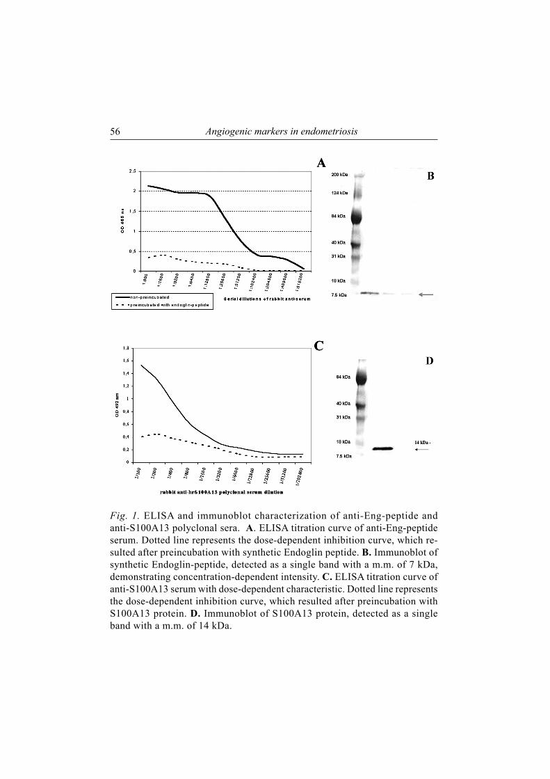
Fig. 1. ELISA and immunoblot characterization of anti-Eng-peptide and anti-S100A13 polyclonal sera. A. ELISA titration curve of anti-Eng-peptide serum. Dotted line represents the dose-dependent inhibition curve, which re-sulted after preincubation with synthetic Endoglin peptide. B. Immunoblot of synthetic Endoglin-peptide, detected as a single band with a m.m. of 7 kDa, demonstrating concentration-dependent intensity. C. ELISA titration curve of anti-S100A13 serum with dose-dependent characteristic. Dotted line represents the dose-dependent inhibition curve, which resulted after preincubation with S100A13 protein. D. Immunoblot of S100A13 protein, detected as a single band with a m.m. of 14 kDa.

Hayrabedyan et al. 57
digital image capture board mounted in Windows PC. “Adobe Photoshop ver.7” (Canada) software was used for image acquisition and processing. Se rial sections from each sample with no first antibody applied, were used as negative controls.
RESULTS
A. Antisera specificity assessment
Rabbit anti-Eng-peptide and anti-S100A13 polyclonal sera were tested by ELISA and Western blotting. Several tests were applied in order to confirm the specificity of the reaction. First, an indirect ELISA assay was performed and dose-dependent curve was made (fig. 1 A, C) Secondly, the antibod-ies were pre-incubated with free protein/peptide that caused significant inhibition of their reactivity against solid phase coated antigen. Thirdly, an additional ELISA test was done to check for cross-reactivity against the panel of human proteins but no reaction was observed. These results strongly demonstrated the specificity of the studied sera against the antigens.
The anti-sera specificity was also confirmed by Western blotting as de-scribed above. Using anti-Eng serum, Eng-peptide was detected as a single band with a m.m. of 7 kDa (fig. 1B). When polyclonal rabbit anti-S100A13 serum was applied, S100A13 protein migrated as a single band with m.m. of 14 kDa (fig. 1D). The band staining intensity had a dose-dependent char-acter and corresponded to molecular mass of studied antigens. The position of Eng bands corresponded to a m.m. of 7 kDa, rather than m.m. of 190 kDa as it should be for the whole endoglin homodimer molecule, since the synthetic 35-mer Eng-peptide was used instead.
B. Endoglin and S100A13 expression in endometriosis
Using immunohistochemistry, we investigated the expression of endoglin and S100A13 in three groups of specimens – normal secretory endometrium,

Angiogenic markers in endometriosis58
Fig. 2. Secretory phase normal endometrium microvessels were negative for endo-glin (white dotted arrows), while microvessels in the myometrium were regarded as internal positive controls (black solid arrow). G-glands, S-stroma, M-myometrium, C-capillaries; magnification x 100.
endometriotic lesions (adenomyosis and ovarian endometriosis) and eutopic endometrium from women with endometriosis.
Endoglin was not expressed in the capillaries of the normal endometrium. The borderline microvessels of the uterus exhibited only subtle Eng expres-sion, while myometrium had stronger endoglin expression (fig. 2). Eng was detected in the middle to large-sized vessels of the normal endometrium specimens. It was expressed predominantly in the pericyte component, rather than the endothelial component in the largest arterial vessels of the uterine myometrium.
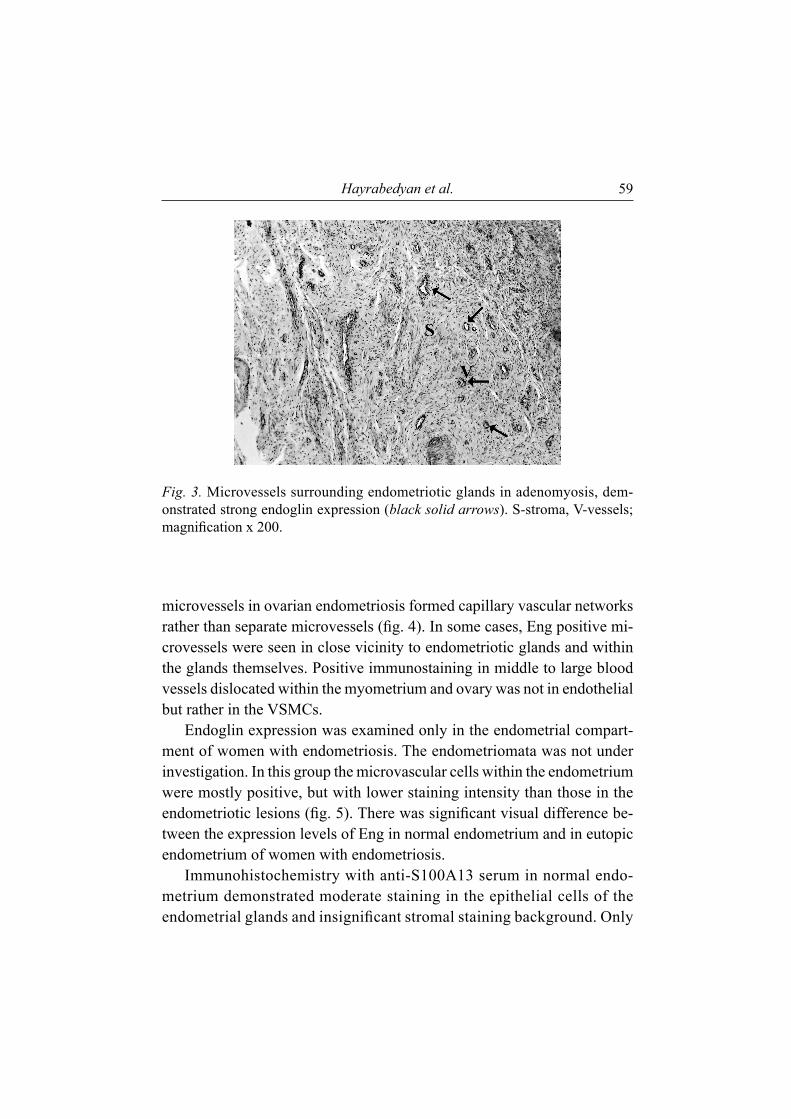
The immunohistochemical studies of specimens from adenomyosis and ovarian endometriosis revealed a distinctive expression of Eng. Endo-glin was expressed in all microvessels, with positively stained endothelial cells. Multiple single capillaries were stained with different intensity in adenomyosis, scattered within the endometriotic stroma. An even stronger - compared to the endometriotic lesion - vascular staining intensity was observed in the surrounding myometrial compartment (fig. 3). The stained
Hayrabedyan et al. 59
Fig. 3. Microvessels surrounding endometriotic glands in adenomyosis, dem-onstrated strong endoglin expression (black solid arrows). S-stroma, V-vessels; magnification x 200.
microvessels in ovarian endometriosis formed capillary vascular networks rather than separate microvessels (fig. 4). In some cases, Eng positive mi-crovessels were seen in close vicinity to endometriotic glands and within the glands themselves. Positive immunostaining in middle to large blood vessels dislocated within the myometrium and ovary was not in endothelial but rather in the VSMCs.
Endoglin expression was examined only in the endometrial compart-ment of women with endometriosis. The endometriomata was not under investigation. In this group the microvascular cells within the endometrium were mostly positive, but with lower staining intensity than those in the endometriotic lesions (fig. 5). There was significant visual difference be-tween the expression levels of Eng in normal endometrium and in eutopic endometrium of women with endometriosis.
Immunohistochemistry with anti-S100A13 serum in normal endo-metrium demonstrated moderate staining in the epithelial cells of the endometrial glands and insignificant stromal staining background. Only

Angiogenic markers in endometriosis60
Fig. 4. Endoglin stained moderately the microvessels network (white solid arrows) infiltrating the stroma of ovarian endometrioma. G-glands, C-capillaries; magnification x 200.
Fig. 5. Positive endoglin staining of microvessels (white solid arrows) in the en-dometrial compartment of eutopic proliferative endometrium from woman with endometriosis. G-glands, C-capillaries; magnification x 200.
Hayrabedyan et al. 61
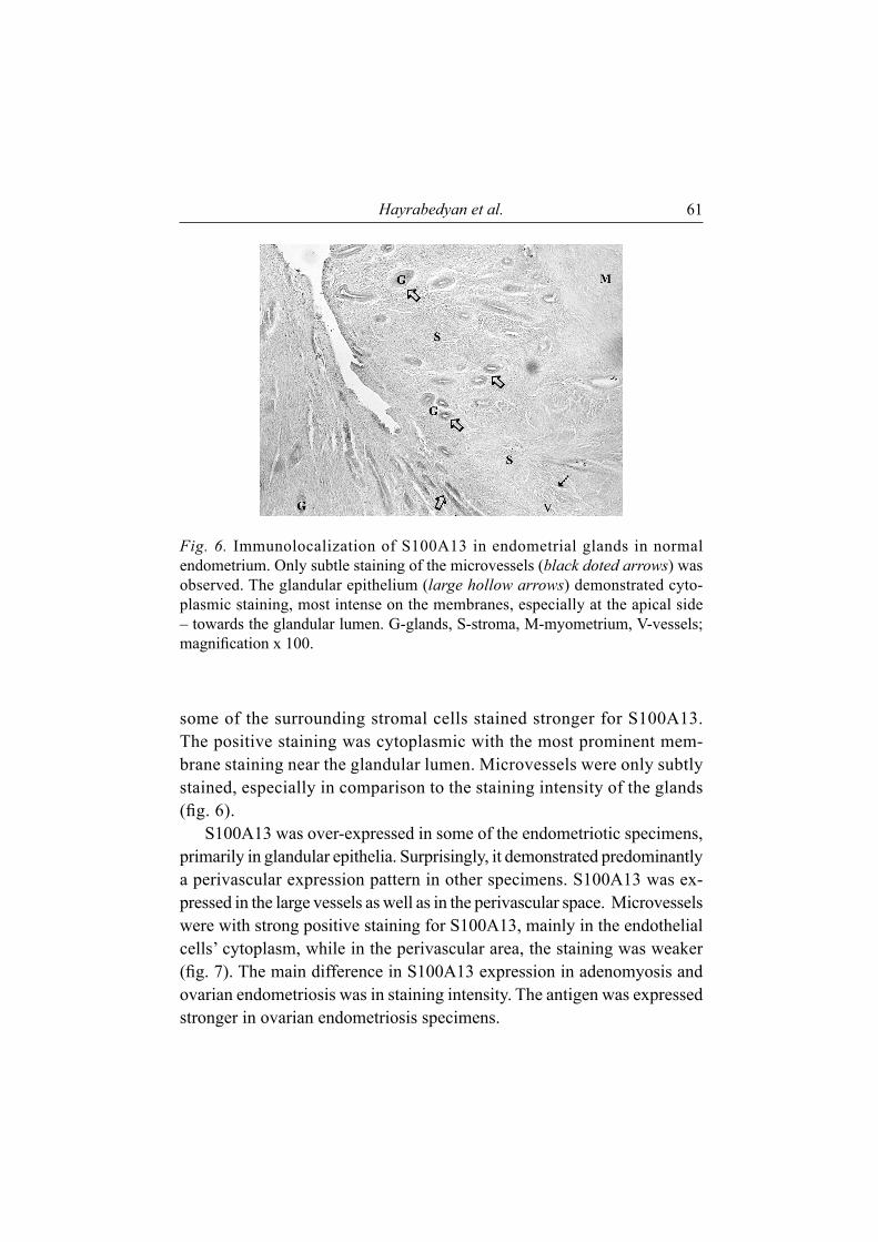
Fig. 6. Immunolocalization of S100A13 in endometrial glands in normal endometrium. Only subtle staining of the microvessels (black doted arrows) was observed. The glandular epithelium (large hollow arrows) demonstrated cyto-plasmic staining, most intense on the membranes, especially at the apical side – towards the glandular lumen. G-glands, S-stroma, M-myometrium, V-vessels; magnification x 100.
some of the surrounding stromal cells stained stronger for S100A13. The positive staining was cytoplasmic with the most prominent mem-brane staining near the glandular lumen. Microvessels were only subtly stained, especially in comparison to the staining intensity of the glands (fig. 6).
S100A13 was over-expressed in some of the endometriotic specimens, primarily in glandular epithelia. Surprisingly, it demonstrated predominantly a perivascular expression pattern in other specimens. S100A13 was ex-pressed in the large vessels as well as in the perivascular space. Microvessels were with strong positive staining for S100A13, mainly in the endothelial cells’ cytoplasm, while in the perivascular area, the staining was weaker (fig. 7). The main difference in S100A13 expression in adenomyosis and ovarian endometriosis was in staining intensity. The antigen was expressed stronger in ovarian endometriosis specimens.

Angiogenic markers in endometriosis62
Fig. 7. S100A13 was expressed in adenomyosis on midle- to large-sized arter-ies, in the para-vascular space of arterioles (black solid arrows) as well as in the endothelial cells of some capillaries (black thin arrows). G-glands, S-stroma, A-arteries, a-arterioles, V-veins, C-capillaries; magnification x 200.
DISCUSSION
Endometriosis has been considered an angiogenic disease in recent years. The establishment, propagation, and sustaining of endometriotic implants depend largely on proper angiogenic support. The molecules investigated in this study, endoglin and S100A13, are angiogenesis-related. They have an expression level corresponding to the state of activated endothelia. Although the exact mechanism of their involvement in endometriotic angiogenesis process is currently unknown, they both represent not only marker molecules, but also active participants in this process. S100A13 expression is coupled with the ex-pression of FGF-1, a proven angiogenic and growth factor, as well as with the expression of IL-1α, a cytokine also known to be pro-angiogenic. It has been established that Eng is an active participant in angiogenesis and its expression is highly restricted only to activated endothelial cells. [4, 6, 14, 26]
From a morphological point of view, both factors demonstrated distinct staining patterns in normal and pathologic conditions, suggesting their pos-

Hayrabedyan et al. 63
sible pathogenic role in endometriotic angiogenesis. It should be noted that when sections from non-endometriotic tissue were tested, no Eng staining of microvessels was observed. This fact seems to be of utmost importance considering Eng is assumed to have expression predominantly in endothelial cells of microvessels of tumours and other pathologies. On the other hand, the large vessels expressed Eng, as observed also by Zhang et al. [32]. Ac-cording to them, the Eng staining in normal endometrium, throughout the entire menstrual cycle, is confined primarily to the arterioles and arteries, with only weak positive staining of the veins and no staining of the capil-lary microvessels. The arterial staining is most intensive during the early secretory phase, followed by the early proliferative phase. We observed similar spatial pattern of Eng expression on normal endometrium, but this expression during the various menstruation cycle phases was not explored. In contrast to Zhang et al, there was no positive staining for the Eng mi-crovessels even in normal secretory endometrium in our study. Our results demonstrated negative staining of the eutopic endometrium capillaries in non-endometriosis patients and positive staining of eutopic endometrium in endometriosis patients. This observation has been currently stated only by Kim et al [12].
In this study, we evaluated the immunohistochemical expression of Eng and S100A13 in two main endometriosis subgroups – adenomyosis and ovarian endometriosis. Adenomyosis is characterised by the localization of endometrial glands and stroma within the uterine myometrium. When the same lesion types are localized within the ovarian tissue, forming frequently so-called ovarian “chocolate” cysts, the condition is denoted as ovarian endometriosis. The selection of these two groups was made because of the statements [7] that both these endometriosis subgroups have distinct pathogeneses.
The expression of Eng in the endometriotic lesions was investigated, and strong positive staining of the microvessels was observed. The inten-sity of staining between the two endometriotic subgroups was visually indistinguishable. The only difference was that single stained microvessels were observed in adenomyosis while these microvessels formed capillary networks in ovarian endometriosis. The specific anti-Eng-peptide serum
Angiogenic markers in endometriosis64
stained the cytoplasm of endothelial cells, corresponding to the intracellular localization of the selected peptide, recognized by the antibodies. The Eng expression in endometriotic endothelial cells was stronger, compared to normal endometrium where its expression was weaker which substantiate data published earlier [12]. We observed Eng expression in vascular smooth muscle cells that has been published by others [5, 24], with the assumption that myometrial vessels are useful as an internal positive control for Eng immunostaining [12].
It has been demonstrated that the expression of Eng is upregulated in endometriosis and it may be a better marker for evaluation of mi-crovessel density in comparison to CD34 and von Wielebrand factor (vWF; [3, 32]). The demonstration of positive Eng expression within the endometriomata, along with the data of its participation in the TGF-β pathway, suggests that Eng has a definite role in angiogenesis in endome-triosis. Angiogenesis is a pre-requisite for development of endometriotic lesions which implies that Eng is a better marker for both microvascular density evaluation and comparison between distinct endometriotic loci [3, 32].
The immunostaining of S100A13 demonstrated broader distribution pattern. It was expressed in endometrial glands, single stromal cells, the VSMCs and in the perivascular space. S100A13 has the ability to stain both large vessels and microvessels, giving a total vascular marking. This makes S100A13 preferable to vWF, which is reported to stain the ECs within the large vessels, while staining is weak or even absent within the capillaries [32]. Furthermore, it was differentially expressed in the microvessels located in the normal eutopic endometrium compared to endometriotic stromal compartment, exhibiting stronger staining pattern in the endometriotic microvessels. Another widely known and accepted endothelial cell marker – CD34, stains non-endothelial supportive cells around the endometrial glands in endometrium, forming their basal membrane [13, 32], suggesting that no vascular-restricted marker exists. S100A13, despite its staining het-erogeneity, proves to be useful tool in detecting the activated microvessels. This suggests its use as a universal angiogenic marker, at least as a marker of activated endometriosis.

Hayrabedyan et al. 65
In this paper we confirm, what had only been stated by Kim et al. [12], the positive expression of endoglin not only in endometriotic lesions, but also in eutopic endometrium of women with endometriosis. In our study, S100A13 overexpression was demonstrated for the first time in endome-triotic tissues, compared to normal endometrium.
In conclusions, both endoglin and S100A13 stained the activated mi-crovessel endothelia in endometriosis. S100A13 displayed positive expres-sion in normal endometrial microvessels while Eng did not. Only S100A13, but not Eng, was expressed in epithelia, with the highest staining intensity in endometriosis. Further studies to test the possibility to use endoglin and S100A13 as markers for monitoring the clinical status and the effect of treatment of patients with endometriosis are currently in progress.
ACKNOWLEDGEMENTS
This work was supported by Bulgarian National Science Fund (Grant No. K-1201/2000).
REFERENCES
1. Adam PJ, Clesham GJ, Weissberg PL 1998 Expression of endoglin mRNA and protein in human vascular smooth muscle cells. Biochemical and Biophysical Research Com-munications 247 33-7.
2. Bredow S, Lewin M, Hofmann B, Marecos E, Weissleder R 2000 Imaging of tumour neovasculature by targeting the TGF-beta binding receptor endoglin. European Journal of Cancer 36 675-81.
3. Brewer CA, Setterdahl JJ, Li MJ, Johnston JM, Mann JL, McAsey ME 2000 Endog-lin expression as a measure of microvessel density in cervical cancer. Obstetrics and Gynecology 96 224-8.
4. Charpin-Taranger C, Dales JP, Garcia S, Andrac-Meyer L, Ramuz O, Carpentier-Meunier S, Bonnier P 2003 The immunohistochemical expression of CD105 is a marker for high metastatic risk and worse prognosis in breast cancers. Bulletin De L’ Academie Nationale De Medecin (Paris) 187 1129-45; discussion 1145-6.
5. Conley BA, Smith JD, Guerrero-Esteo M, Bernabeu C, Vary CP 2000 Endoglin, a TGF-beta receptor-associated protein, is expressed by smooth muscle cells in human atherosclerotic plaques. Atherosclerosis 153 323-35.

Angiogenic markers in endometriosis66
6. Dales JP, Garcia S, Andrac L, Carpentier S, Ramuz O, Lavaut MN, Allasia C, Bon-nier P, Charpin C 2004 Prognostic significance of angiogenesis evaluated by CD105 expression compared to CD31 in 905 breast carcinomas: correlation with long-term patient outcome. International Journal of Oncology 24 1197-204.
7. Donnez J, Smoes P, Gillerot S, Casans-Roux F, Nissole M 1998 Vascular endothelial growth factor (VEGF) in endometriosis. Human Reproduction 13 1686-1690.
8. Gougos A, Letarte M 1988 Identification of a human endothelial cell antigen with monoclonal antibody 44G4 produced against a pre-B leukemic cell line. Journal of Immunology 141 1925-33.
9. Harlow E, Lane D 1988 Antibodies: A Laboratory Manual. In Antibodies: A Labora-tory Manual, pp 553-612. Cold Spring Harbor Laboratory Press, New York
10. Hsieh HL, Schafer BW, Cox JA, Heizmann CW 2002 S100A13 and S100A6 exhibit distinct translocation pathways in endothelial cells. Journal of Cell Science 115 3149-58.
11. Hsieh HL, Schafer BW, Sasaki N, Heizmann CW 2003 Expression analysis of S100 proteins and RAGE in human tumors using tissue microarrays. Biochemical and Biophysical Research Communications 307 375-81.
12. Kim SH, Choi YM, Chae HD, Kim KR, Kim CH, Kang BM 2001 Increased expres-sion of endoglin in the eutopic endometrium of women with endometriosis. Fertility and Sterility 76 918-22.
13. Kohnen G, Campbell S, Jeffers MD, Cameron IT 2000 Spatially regulated differentiation of endometrial vascular smooth muscle cells. Human Reproduction 15 284-292.
14. Korn T, Muller R, Kontermann RE 2004 Bispecific single-chain diabody-mediated killing of endoglin-positive endothelial cells by cytotoxic T lymphocytes. Journal of Immunotherapy 27 99-106.
15. Krupinski J, Kaluza J, Kumar P, Kumar S, Wang JM 1994 Role of angiogenesis in patients with cerebral ischemic stroke. Stroke 25 1794-8.
16. Landriscina M, Schinzari G, Cassano A, Leonardo GD, Quirino M, Pozzo C, Scerrati M, Barone C 2002 S100A13, a new marker of angiogenesis in brain tumors. Annals of Oncology 13 165 - 166.
17. Lastres P, Bellon T, Cabanas C, Sanchez-Madrid F, Acevedo A, Gougos A, Letarte M, Bernabeu C 1992 Regulated expression on human macrophages of endoglin, an Arg-Gly-Asp-containing surface antigen. European Journal of Immunology 22 393-7.
18. Lee WY, Su WC, Lin PW, Guo HR, Chang TW, Chen HH 2004 Expression of S100A4 and Met: potential predictors for metastasis and survival in early-stage breast cancer. Oncology 66 429-38.
19. Letamendia A, Lastres P, Botella LM, Raab U, Langa C, Velasco B, Attisano L, Bernabeu C 1998 Role of Endoglin in Cellular Responses to Transforming Growth Factor-beta. A comparative study with betaglycan. Journal of Biological Chemistry 273 33011-33019.
20. Li DY, Sorensen LK, Brooke BS, Urness LD, Davis EC, Taylor DG, Boak BB, Wendel DP 1999 Defective angiogenesis in mice lacking endoglin. Science 284 1534-7
21. Lopez-Casillas F, Wrana JL, Massague J 1993 Betaglycan presents ligand to the TGF beta signaling receptor. Cell 73 1435-44.
Hayrabedyan et al. 67
22. Ma X, Labinaz M, Goldstein J, Miller H, Keon WJ, Letarte M, O’Brien E 2000 En-doglin is overexpressed after arterial injury and is required for transforming growth factor-beta-induced inhibition of smooth muscle cell migration. Arteriosclerosis, Thrombosis, and Vascular Biology 20 2546-52.
23. Noyes RW, Hertwig AT, Rock J 1950 Dating the endometrial biopsy. Fertility and Sterility 1 3-25.
24. Paul M, Mazurek U, Witek A, Graniczka M, Wilczok T 2001 Histone H3 gene expres-sion level as markers of hyperplasia simplex of endometrium. Ginekologia Polska 72 1434-8.
25. Robledo MM, Hidalgo A, Lastres P, Arroyo AG, Bernabeu C, Sanchez-Madrid F, Teixido J 1996 Characterization of TGF-beta 1-binding proteins in human bone mar-row stromal cells. British Journal of Haematology 93 507-14.
26. Salvesen HB, Gulluoglu MG, Stefansson I, Akslen LA 2003 Significance of CD 105 expression for tumour angiogenesis and prognosis in endometrial carcinomas. Acta Pathologica, Microbiologica, et Immunologica Scandinavica. Section A, Pathology 111 1011-8.
27. Sampson JA 1927 Peritoneal endometriosis due to menstrual dissemination of endome-trial tissue into the peritoneal cavity. American Journal of Obstetrics and Gynecology 14 422-469.
28. Shi SR, Cote RJ, Taylor CR 2001 Antigen Retrieval Techniques: Current Perspectives. Journal of Histochemistry and Cytochemistry 49 931-937.
29. St-Jacques S, Cymerman U, Pece N, Letarte M 1994 Molecular characterization and in situ localization of murine endoglin reveal that it is a transforming growth factor-beta binding protein of endothelial and stromal cells. Endocrinology 134 2645-57.
30. Wang JM, Kumar S, Pye D, Haboubi N, al-Nakib L 1994 Breast carcinoma: compara-tive study of tumor vasculature using two endothelial cell markers. Journal of National Cancer Institute 86 386-8.
31. Westphal JR, Willems HW, Schalkwijk CJ, Ruiter DJ, de Waal RM 1993 A new 180-kDa dermal endothelial cell activation antigen: in vitro and in situ characteristics. Journal of Investigative Dermatology 100 27-34.
32. Zhang EG, Smith SK, Charnock-Jones DS 2002 Expression of CD105 (endoglin) in arteriolar endothelial cells of human endometrium throughout the menstrual cycle. Reproduction 124 703-11.
Related Documents