Expression of the TGF-b coreceptor endoglin in epidermal keratinocytes and its dual role in multistage mouse skin carcinogenesis Miguel Quintanilla* ,1,7 , Jose Ramo´ n Ramirez 2,7 , Eduardo Pe´rez-Go´ mez 1 , Diana Romero 1 , Beatriz Velasco 3 , Michelle Letarte 4,5 , Jose Miguel Lo´pez-Novoa 6 and Carmelo Bernabe´u 3 1 Instituto de Investigaciones Biome´dicas Alberto Sols, Consejo Superior de Investigaciones Cientı´ficas (CSIC), Universidad Auto´noma de Madrid (UAM), Arturo Duperier 4, Madrid 28029, Spain; 2 Departamento de Anatomı´a Patolo´gica, Hospital Gomez- Ulla, Glorieta del Ejercito, Madrid 28047, Spain; 3 Centro de Investigaciones Biolo´gicas, CSIC, Ramiro de Maeztu 9, Madrid 28040, Spain; 4 Blood and Cancer Research Program, The Hospital for Sick Children, Ontario, Canada; 5 Department of Immunology, University of Toronto, 555 University Avenue, Toronto, Ontario, Canada M5G 1X8; 6 Departamento de Fisiologı´a y Farmacologı´a, Facultad de Medicina, Universidad de Salamanca, Salamanca 37007, Spain Endoglin is an integral membrane glycoprotein primarily expressed in the vascular endothelium, but also found on macrophages and stromal cells. It binds several members of the transforming growth factor (TGF)-b family of growth factors and modulates TGF-b 1 -dependent cellular responses. However, it lacks cytoplasmic signaling motifs and is considered as an auxiliary receptor for TGF-b. We show here that endoglin is expressed in mouse and human epidermis and in skin appendages, such as hair follicles and sweat glands, as determined by immunohistochem- istry. In normal interfollicular epidermis, endoglin was restricted to basal keratinocytes and absent in differen- tiating cells of suprabasal layers. Follicular expression of endoglin was high in hair bulb keratinocytes, but decreased in parts distal from the bulb. To address the role of endoglin in skin carcinogenesis in vivo, Endoglin heterozygous mice were subjected to long-term chemical carcinogenesis treatment. Reduction in endoglin had a dual effect during multistage carcinogenesis, by inhibiting the early appearance of benign papillomas, but increasing malignant progression to highly undifferentiated carcino- mas. Our results are strikingly similar to those previously reported for transgenic mice overexpressing TGF-b 1 in the epidermis. These data suggest that endoglin might attenuate TGF-b 1 signaling in normal epidermis and interfere with progression of skin carcinogenesis. Oncogene (2003) 22, 5976–5985. doi:10.1038/sj.onc.1206841 Keywords: endoglin; TGF-b; keratinocyte; tumor progression Introduction The transforming growth factor-b (TGF-b) plays an important role in epithelial homeostasis, as it modulates the cellular phenotype and is a powerful inhibitor of cell growth (Derynck et al., 2001; Piek and Roberts, 2001). Members of the TGF-b superfamily exert their biologi- cal effects by binding to a heteromeric complex contain- ing two different transmembrane serine/threonine kinases known as type I and type II signaling receptors (Letterio and Roberts, 1998; Massague´ , 1998). The TGF-b receptor complex also contains two auxiliary receptors named endoglin and betaglycan (Massague´, 1998). These are transmembrane proteins with large extracellular domains and serine/threonine-rich cyto- plasmic regions without consensus signaling motifs. Endoglin binds TGF-b 1 , TGF-b 3 , activin-A, BMP-2, and BMP-7 in the presence of the signaling receptors types I and II (Cheifetz et al., 1992; Bello´n et al., 1993; Letamendia et al., 1998a; Barbara et al., 1999), and modulates TGF-b-dependent cellular responses (Lastres et al., 1996; Letamendia et al., 1998a; Li et al., 2000; Diez-Marques et al., 2002). Endoglin plays an important role in mammalian physiology as demonstrated by genetic inactivation, since Endoglin null (Eng / ) murine embryos die of vascular defects at 10–10.5 days post coitum (Bourdeau et al., 1999; Li et al., 1999; Arthur et al., 2000). Endoglin is a disulfide-linked homodimer of 180 kDa expressed in endothelial cells (Gougos and Letarte, 1988, 1990; Li et al., 2000), macrophages (Lastres et al., 1992), stromal cells (St-Jacques et al., 1994; Robledo et al., 1996), and other cellular lineages within the vascular system and connective tissues (Fonsatti et al., 2001). Several tumor cells also express endoglin, including pre-B-cell and monocytic leukemia (Quackenbush and Letarte, 1985; Haruta and Seon, 1986; Lastres et al., 1992; Zhang et al., 1996), melanoma (Altomonte et al., 1996), choriocarcinoma (Letamendia et al., 1998b), and ovarian carcinoma (Jindal et al., 1995). However, little is known about the possible expression and function of endoglin in epithelial cells. Approximately 90% of all human tumors are of epithelial origin. TGF-b has both positive and negative effects on carcinogenesis. It behaves as a tumor suppressor, early in tumorigenesis, but can also drive malignant progression, invasion, and metastasis (see Received 7 February 2003; revised 30 May 2003; accepted 2 June 2003 *Correspondence: M Quintanilla; E-mail: [email protected] 7 The first two authors contributed equally to this work Oncogene (2003) 22, 5976–5985 & 2003 Nature Publishing Group All rights reserved 0950-9232/03 $25.00 www.nature.com/onc

Welcome message from author
This document is posted to help you gain knowledge. Please leave a comment to let me know what you think about it! Share it to your friends and learn new things together.
Transcript
Expression of the TGF-b coreceptor endoglin in epidermal keratinocytes
and its dual role in multistage mouse skin carcinogenesis
Miguel Quintanilla*,1,7, Jose Ramon Ramirez2,7, Eduardo Perez-Gomez1, Diana Romero1, BeatrizVelasco3, Michelle Letarte4,5, Jose Miguel Lopez-Novoa6 and Carmelo Bernabeu3
1Instituto de Investigaciones Biomedicas Alberto Sols, Consejo Superior de Investigaciones Cientıficas (CSIC), UniversidadAutonoma de Madrid (UAM), Arturo Duperier 4, Madrid 28029, Spain; 2Departamento de Anatomıa Patologica, Hospital Gomez-Ulla, Glorieta del Ejercito, Madrid 28047, Spain; 3Centro de Investigaciones Biologicas, CSIC, Ramiro de Maeztu 9, Madrid 28040,Spain; 4Blood and Cancer Research Program, The Hospital for Sick Children, Ontario, Canada; 5Department of Immunology,University of Toronto, 555 University Avenue, Toronto, Ontario, Canada M5G 1X8; 6Departamento de Fisiologıa y Farmacologıa,Facultad de Medicina, Universidad de Salamanca, Salamanca 37007, Spain
Endoglin is an integral membrane glycoprotein primarilyexpressed in the vascular endothelium, but also found onmacrophages and stromal cells. It binds several membersof the transforming growth factor (TGF)-b family ofgrowth factors and modulates TGF-b1-dependent cellularresponses. However, it lacks cytoplasmic signaling motifsand is considered as an auxiliary receptor for TGF-b. Weshow here that endoglin is expressed in mouse and humanepidermis and in skin appendages, such as hair folliclesand sweat glands, as determined by immunohistochem-istry. In normal interfollicular epidermis, endoglin wasrestricted to basal keratinocytes and absent in differen-tiating cells of suprabasal layers. Follicular expression ofendoglin was high in hair bulb keratinocytes, butdecreased in parts distal from the bulb. To address therole of endoglin in skin carcinogenesis in vivo, Endoglinheterozygous mice were subjected to long-term chemicalcarcinogenesis treatment. Reduction in endoglin had adual effect during multistage carcinogenesis, by inhibitingthe early appearance of benign papillomas, but increasingmalignant progression to highly undifferentiated carcino-mas. Our results are strikingly similar to those previouslyreported for transgenic mice overexpressing TGF-b1 in theepidermis. These data suggest that endoglin mightattenuate TGF-b1 signaling in normal epidermis andinterfere with progression of skin carcinogenesis.Oncogene (2003) 22, 5976–5985. doi:10.1038/sj.onc.1206841
Keywords: endoglin; TGF-b; keratinocyte; tumorprogression
Introduction
The transforming growth factor-b (TGF-b) plays animportant role in epithelial homeostasis, as it modulates
the cellular phenotype and is a powerful inhibitor of cellgrowth (Derynck et al., 2001; Piek and Roberts, 2001).Members of the TGF-b superfamily exert their biologi-cal effects by binding to a heteromeric complex contain-ing two different transmembrane serine/threoninekinases known as type I and type II signaling receptors(Letterio and Roberts, 1998; Massague, 1998). TheTGF-b receptor complex also contains two auxiliaryreceptors named endoglin and betaglycan (Massague,1998). These are transmembrane proteins with largeextracellular domains and serine/threonine-rich cyto-plasmic regions without consensus signaling motifs.Endoglin binds TGF-b1, TGF-b3, activin-A, BMP-2,and BMP-7 in the presence of the signaling receptorstypes I and II (Cheifetz et al., 1992; Bellon et al., 1993;Letamendia et al., 1998a; Barbara et al., 1999), andmodulates TGF-b-dependent cellular responses (Lastreset al., 1996; Letamendia et al., 1998a; Li et al., 2000;Diez-Marques et al., 2002). Endoglin plays an importantrole in mammalian physiology as demonstrated bygenetic inactivation, since Endoglin null (Eng�/�) murineembryos die of vascular defects at 10–10.5 days postcoitum (Bourdeau et al., 1999; Li et al., 1999; Arthuret al., 2000). Endoglin is a disulfide-linked homodimerof 180 kDa expressed in endothelial cells (Gougos andLetarte, 1988, 1990; Li et al., 2000), macrophages(Lastres et al., 1992), stromal cells (St-Jacques et al.,1994; Robledo et al., 1996), and other cellular lineageswithin the vascular system and connective tissues(Fonsatti et al., 2001). Several tumor cells also expressendoglin, including pre-B-cell and monocytic leukemia(Quackenbush and Letarte, 1985; Haruta and Seon,1986; Lastres et al., 1992; Zhang et al., 1996), melanoma(Altomonte et al., 1996), choriocarcinoma (Letamendiaet al., 1998b), and ovarian carcinoma (Jindal et al.,1995). However, little is known about the possibleexpression and function of endoglin in epithelial cells.
Approximately 90% of all human tumors are ofepithelial origin. TGF-b has both positive and negativeeffects on carcinogenesis. It behaves as a tumorsuppressor, early in tumorigenesis, but can also drivemalignant progression, invasion, and metastasis (seeReceived 7 February 2003; revised 30 May 2003; accepted 2 June 2003
*Correspondence: M Quintanilla; E-mail: [email protected] first two authors contributed equally to this work
Oncogene (2003) 22, 5976–5985& 2003 Nature Publishing Group All rights reserved 0950-9232/03 $25.00
www.nature.com/onc
Akhurst and Balmain, 1999; Massague et al., 2000;Derynck et al., 2001, for reviews). In spite of the largenumber of studies addressing the role of TGF-b inepithelial carcinogenesis, the specific role played by theTGF-b coreceptor endoglin has not been evaluated invivo.
The mouse skin model of multistage carcinogenesishas been widely studied and is well characterizedwith respect to phenotypic and genetic changes(Yuspa, 1994; Akhurst and Balmain, 1999). Tumorinitiation is induced by a single topical applicationof the carcinogen 7,12-dimethylbenz(a)anthracene(DMBA), and involves a specific oncogenic mutationin the H-Ras gene (Quintanilla et al., 1986). Thereafter,repeated application of the tumor promoter 12-O-tetradecanoylphorbol-13-acetate (TPA) results in theoutgrowth of highly differentiated benign papillomas.A small subset of papillomas eventually progressesto malignant squamous cell carcinomas, some ofwhich undergo an epithelial–mesenchymal transition tospindle cell carcinomas (Klein-Szanto et al., 1989;Buchmann et al., 1991). The spindle cell, the mostinvasive and motile carcinoma cell type has altered theexpression of keratinocyte-specific markers, such ascytokeratins, and show loss of E-cadherin expression(Navarro et al., 1991; Dıaz-Guerra et al., 1992; Fronteloet al., 1998).
Here, we describe the presence of endoglin inkeratinocytes of basal epidermis and hair follicles, thetwo major cell compartments for tumor initiation in skincarcinogenesis (Akhurst and Balmain, 1999; Tayloret al., 2000). To investigate the role of endoglin incarcinogenesis, Endoglin heterozygous (Engþ /�) micewere subjected to long-term chemical carcinogenesistreatment with DMBA and TPA. We found thatendoglin haploinsufficiency had a dual effect duringmultistage skin carcinogenesis, inhibiting the earlyappearance of benign tumors, but increasing the latemalignant phenotype.
Results
Endoglin is expressed in keratinocytes of normal skin andcutaneous lesions
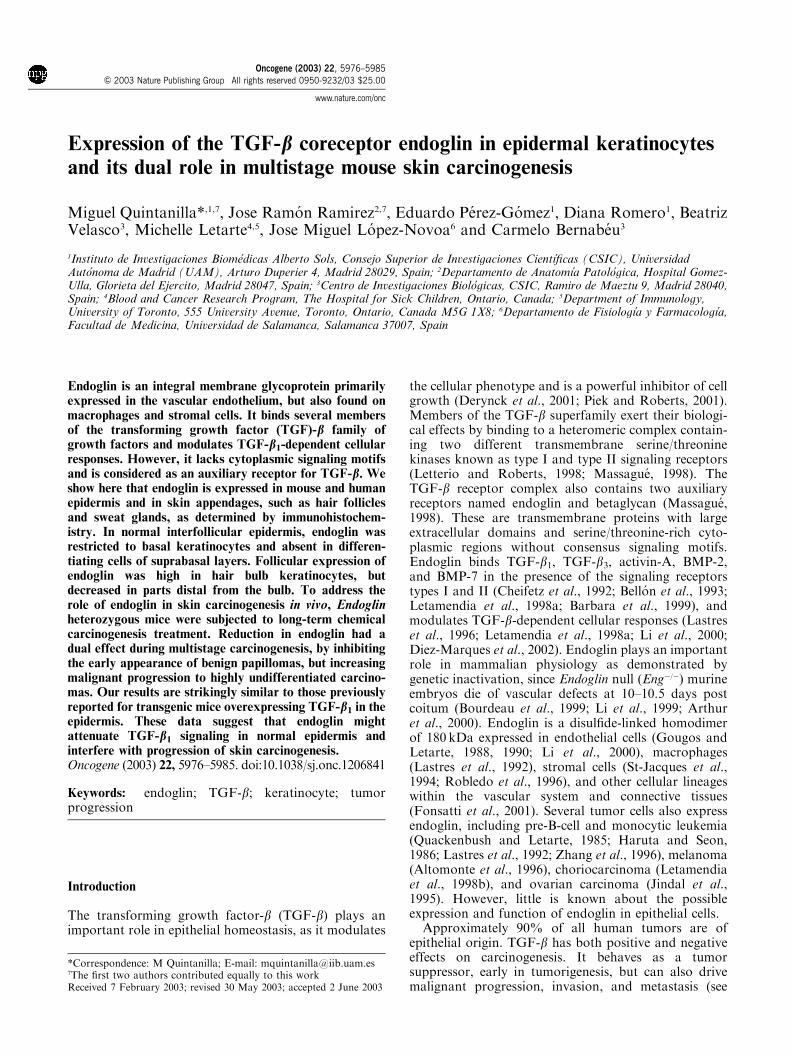
The presence of endoglin was analysed in human normalskin sections by immunohistochemistry using thespecific monoclonal antibody (mAb) SN6h. Endoglinwas expressed in the epidermis, restricted exclusively tobasal keratinocytes (Figure 1a, b). The immunostainingpattern was discontinuous in the basal layer with somecells staining weakly or not at all, and in patches incertain regions of the plasma membrane and/or cyto-
Figure 1 Immunohistochemical detection of endoglin in human skin and skin appendages. (a, b) Interfollicular skin. Endoglin isexpressed in basal keratinocytes of the epidermis (e) and in dermal (d) blood vessels (inset in panel b). (c) Hair follicles. Note staining inkeratinocytes of the hair bulb (hb) and outer (ors) and inner (irs) root sheaths. No staining is found in the dermal papilla (dp) and inparts distal from the hair bulb. The inset in this panel shows a higher magnification of the hair bulb. (d) Sweat glands showing positivestaining in basal and ductal cells. Original magnifications: � 100 (c), � 400 (a, b, d, insets in b and c)
Skin carcinogenesis in Endoglin heterozygous miceM Quintanilla et al
5977
Oncogene
plasm. This is in contrast to the continuous and cell-surface pattern of endoglin expression seen in endothe-lial cells of dermal blood vessels (Figure 1b, inset), usedas a positive control. The antibody did not stainsuprabasal keratinocytes and dermal fibroblastic cells.Endoglin was also present in keratinocytes of hairfollicles (Figure 1c). Intense staining was observed in thehair matrix and in the proximal regions of the outer andinner root sheaths, and decreased in parts distal fromthe bulb. Mesenchyme-derived dermal papilla fibro-blasts were endoglin negative. As seen for interfollicularepidermal cells, hair follicle keratinocytes expressedendoglin in a polarized fashion, with the protein locatedin certain regions of the plasma membrane and/orcytoplasm in the form of granules or aggregates(Figure 1c, inset). Basal and ductal cells of the sweatgland were found to express endoglin strongly(Figure 1d), while high nonspecific staining backgroundwas observed in cells of the sebaceous gland (notshown). Immunostaining of normal mouse skin sectionswith the rat mAb MJ7/18 specific for murine endoglinrevealed the same pattern of protein expression infollicular and interfollicular epidermis as in human skin(see below in Figure 6).
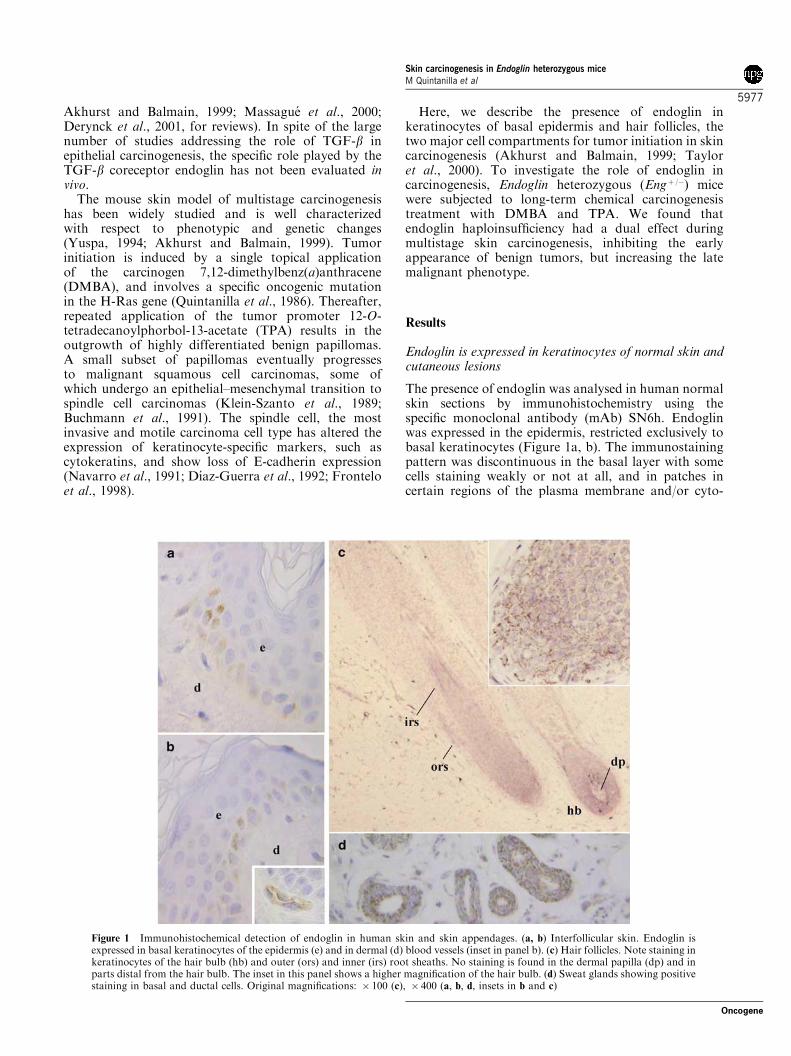
Since endoglin was present in cells of the epidermisand skin appendages, we analysed its expression inseveral human cutaneous disorders. To this aim,sections of verruca vulgaris (n¼ 3), seborrheic keratosis(n¼ 3), nodular basal cell carcinomas (n¼ 7), andsquamous cell carcinomas (n¼ 10) were stained withthe mAb SN6h. A pattern of staining similar to that of
normal epidermis was found in all cases of verrucavulgaris (not shown). In seborrheic keratosis, endoglinstaining was extended to proliferating keratinocytesabove the basal layer, while absent from terminallydifferentiating keratinocytes (Figure 2a). All specimensof nodular basal cell carcinomas were stained with thespecific mAb, revealing that endoglin was expressedmainly at the periphery of tumor nests, as dots, on thecell surface in contact with the basement membranezone. Reduced staining in the inner cell layers was seenin all cases (Figure 2b). Immunohistochemical analysisof squamous cell carcinomas showed that all tumorsexpressed endoglin at variable levels regardless of thedifferentation grade (Figure 2c, d). Positive staining wasobserved in the periphery of tumor nests, but not in thecentral keratinized areas of well-differentiated tumorsfrom which endoglin was excluded (Figure 2c).
Endoglin expression in murine keratinocyte cell lines
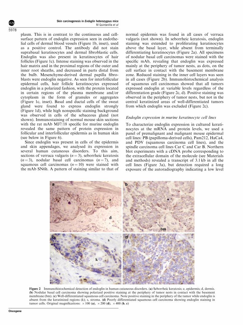
To characterize endoglin expression in cultured kerati-nocytes at the mRNA and protein levels, we used apanel of premalignant and malignant mouse epidermalcell lines: PB (papilloma-derived cells), Pam212, HaCa4,and PDV (squamous carcinoma cell lines), and thespindle carcinoma cell lines Car C and Car B. Northernblot experiments with a cDNA probe corresponding tothe extracellular domain of the molecule (see Materialsand methods) revealed a transcript of 3.1 kb in all thecell lines (Figure 3a), but detection required a longexposure of the autoradiography indicating a low level
Figure 2 Immunohistochemical detection of endoglin in human cutaneous disorders. (a) Seborrheic keratosis; e, epidermis; d, dermis.(b) Nodular basal cell carcinoma showing polarized positive staining at the periphery of tumor nests in contact with the basementmembrane (bm). (c) Well-differentiated squamous cell carcinoma. Note positive staining in the periphery of the tumor while endoglin isabsent from the keratinized regions (k); s, stroma. (d) Poorly differentiated squamous cell carcinoma showing endoglin staining intumor cells. Original magnifications: � 100 (a), � 200 (d), � 400 (b, c)
Skin carcinogenesis in Endoglin heterozygous miceM Quintanilla et al
5978
Oncogene
of endoglin mRNA expression in cultured keratinocytes.This transcript has already been described in othermurine tissues such as the ovary, thymus, placenta, andkidney (Ge and Butcher, 1994; St-Jacques et al., 1994;Rodriguez-Pena et al., 2002). The relative abundance ofthe endoglin mRNAs among the cell lines was roughlysimilar, except for PDV carcinoma cells that showedlower levels in this experiment. The presence of endoglintranscripts was confirmed by RT–PCR (Figure 3b).
Using specific primers to mouse endoglin, a single 1040-bp PCR product of the predicted size was obtained in allthe cell lines tested, including NIH3T3 fibroblasts usedas a control. To confirm that this band representedendoglin, it was purified and sequenced, demonstratingits identity with the published murine sequence (Ge andButcher, 1994; St-Jacques et al., 1994). Western blots(under reducing conditions) clearly detected a polypep-tide of about 95 kDa in PB, HaCa4, and PDVkeratinocytes (Figure 3c). A weaker band was presentin Pam212, whereas no specific signal could be detectedin Car C and Car B cell lines. Overall, the intensities ofthe bands detected in the keratinocyte cell lines werelow, although comparable to that of NIH3T3 fibro-blasts. Parallel blots challenged with preimmune ratIgG, as a negative control, confirmed the specificity ofthe mAb MJ7/18 (data not shown).
Reduced incidence of chemically induced benignpapillomas and enhanced progression to malignantcarcinomas in Endoglin heterozygous mice
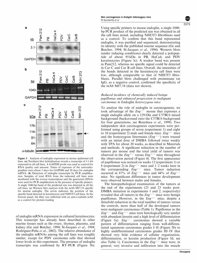
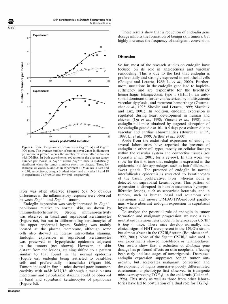
To analyse the role of endoglin in carcinogenesis, wetook advantage of the Engþ /� mouse that expresses asingle endoglin allele on a 129/Ola and C57Bl/6 mixedbackground (backcrossed onto the C57Bl/6 backgroundfor four generations, see Bourdeau et al., 1999). Twoindependent skin carcinogenesis experiments were per-formed using groups of seven (experiment 1) and eightto 10 (experiment 2) male and female mice. Engþ /� miceand the homozygous littermates (Engþ /þ) were treatedwith an initial dose of DMBA followed twice weeklywith TPA for about 20 weeks, as described in Materialsand methods. A significant reduction in the number oftumors per mouse and the total yield of tumors wasobserved in the Engþ /� versus Engþ /þ mice throughoutthe observation period (Figure 4). The first appearanceof papillomas was noticed on weeks 12 (experiment 1) or8 (experiment 2) in Engþ /þ mice and 1–2 weeks later inthe corresponding Engþ /� mice. Tumor inductionoccurred in 87% of Engþ /þ mice and 44% of Engþ /�
mice. No significant differences in tumor developmentwere observed between males and females.
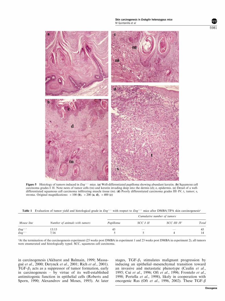
The histopathological examination of the tumors atthe end of the experiments (25 and 23 weeks post-DMBA initiation in experiments 1 and 2, respectively)revealed that all tumors in the Engþ /þ mice were benignpapillomas. However, in the Engþ /� mice, despite athreefold reduction in the total number of tumors versusthe controls, more than half of the developed tumorswere malignant carcinomas (Table 1). Papillomas in theEngþ /� and Engþ /þ mice were histologically very similarwith abundant keratin and a high level of differentiation(Figure 5a). Engþ /� carcinomas showed a variablepattern of differentiation ranging from well-differen-tiated squamous carcinomas grades I–II (Figure 5b) tohighly undifferentiated carcinomas grades III–IV thatshowed very little evidence of cellular organization,differentiation, or keratin production (Figure 5d, seealso Table 1). Carcinomas in the Engþ /� mice were, ingeneral, very invasive and infiltration into the muscle
Figure 3 Analysis of endoglin expression in mouse epidermal celllines. (a) Northern blot hybridization reveals a transcript of 3.1-kbexpressed in all cell lines. A GAPDH probe was used as control forRNA quality and amount. Times of exposure of the autoradio-graphies were about 3 days for GAPDH and 20 days for endoglinmRNA. (b) Detection of endoglin transcripts by PCR amplifica-tion. Samples of total RNA from the indicated cell lines wereincubated with the reverse transcriptase and the generated cDNAswere used for PCR amplification in the presence of specific primers.A single 1040-bp band of the predicted size was detected in all thecell lines. (c) Western blot analysis with the mAb MJ7/18 specificfor murine endoglin. The arrow indicates the position of thespecific band detected in keratinocyte and NIH3T3 cell lines. In thebottom panel, the filter was reblotted with an anti-a-tubulin mAbas a control for protein loading
Skin carcinogenesis in Endoglin heterozygous miceM Quintanilla et al
5979
Oncogene
layer was often observed (Figure 5c). No obviousdifferences in the inflammatory response were observedbetween Engþ /� and Engþ /þ tumors.
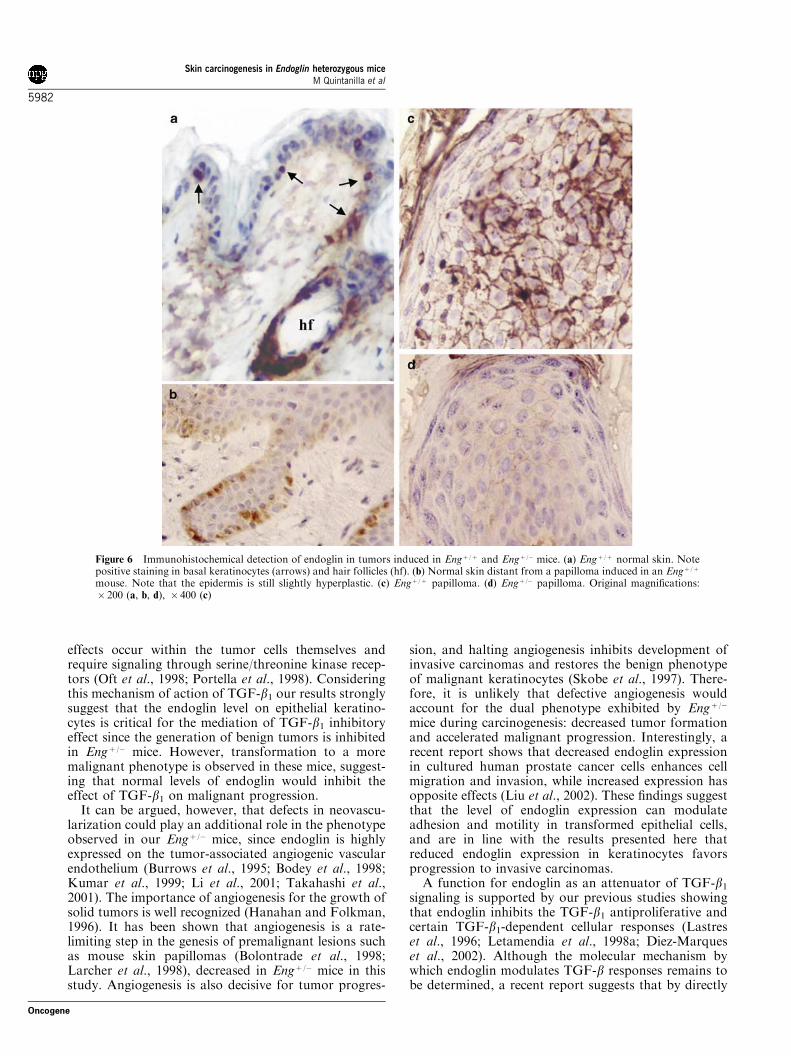
Endoglin expression was vastly increased in Engþ /þ
papillomas relative to normal skin, as shown byimmunohistochemistry. Strong immunoreactivitywas observed in basal and suprabasal keratinocytes(Figure 6c), but not in differentiating keratinocytes ofthe upper epidermal layers. Staining was mainlylocated at the plasma membrane, although somecells also showed an intense intracellular staining.Endoglin expression in suprabasal keratinocyteswas preserved in hyperplastic epidermis adjacentto the tumors (not shown). However, in skindistant from the lesions, staining shifted to a patternsimilar to that found in the normal epidermis(Figure 6a), endoglin being restricted to basal-likecells and preferentially intracellular (Figure 6b).Tumors induced in Engþ /� mice showed poor immunor-eactivity with mAb MJ7/18, although a weak plasmamembrane and cytoplasmic staining could be observedin basal and suprabasal keratinocytes of papillomas(Figure 6d).
These results show that a reduction of endoglin genedosage inhibits the formation of benign skin tumors, buthighly increases the frequency of malignant conversion.
Discussion
So far, most of the research studies on endoglin havefocused on its role in angiogenesis and vascularremodeling. This is due to the fact that endoglin ispreferentially and strongly expressed in endothelial cells(Gougos and Letarte, 1988; Li et al., 2000). Further-more, mutations in the endoglin gene lead to haploin-sufficiency and are responsible for the hereditaryhemorrhagic telangiectasia type 1 (HHT1), an auto-somal dominant disorder characterized by multisystemicvascular dysplasia, and recurrent hemorrhage (Guttma-cher et al., 1995; Shovlin and Letarte, 1999; Marchukand Lux, 2001). In addition, endoglin expression isregulated during heart development in human andchicken (Qu et al., 1998; Vincent et al., 1998); andendoglin-null mice obtained by targeted disruption ofthe endoglin gene die at 10–10.5 days post coitum due tovascular and cardiac abnormalities (Bourdeau et al.,1999; Li et al., 1999; Arthur et al., 2000).
Aside from the endothelial expression of endoglin,several laboratories have reported the presence ofendoglin in other cell types, mostly on cellular lineageswithin the vascular system and connective tissues (seeFonsatti et al., 2001, for a review). In this work, weshow for the first time that endoglin is expressed in theepidermis and skin appendages, such as hair follicles andsweat glands. The presence of endoglin in normalinterfollicular epidermis is restricted to keratinocytesof the basal, proliferative, layer, whereas none isdetected on suprabasal keratinocytes. This pattern ofexpression is disrupted in human cutaneous hyperpro-liferative lesions, such as seborrheic keratosis, and intumors, such as human basal and squamous cellcarcinomas and mouse DMBA/TPA-induced papillo-mas, where aberrant endoglin expression in suprabasallayers occurs.
To analyse the potential role of endoglin in tumorformation and malignant progression, we used a skinmultistage carcinogenesis model in heterozygous C57Bl/6 Engþ /� mice. These mice develop normally, andclinical signs of HHT were present in the 129/Ola strain,but almost absent in the C57Bl/6 strain (Bourdeau et al.,1999, 2001). None of the Engþ /� C57Bl/6 mice used inour experiments showed nosebleeds or telangiectases.Our results show that a reduction of Endoglin genedosage has profound effects on skin neoplasia, affectingboth early and late stages of tumorigenesis. Decreasedendoglin expression suppresses benign tumor out-growth, but accelerates malignant conversion anddevelopment of highly aggressive poorly differentiatedcarcinomas, a phenotype first observed in transgenicmice overexpressing TGF-b1 in the epidermis (Cui et al.,1996). This study as well as those from other labora-tories have led to postulation of a dual role for TGF-b1
6
4
2
05 10 15 2520
Experiment 1
6
4
2
05 10 15 2520
Experiment 2
Weeks post-DMBA initiation
Nu
mb
er o
f tu
mo
rsp
erm
ou
se
Figure 4 Rate of appearance of tumors in Engþ /þ (�) and Engþ /�
(J) mice. The average number of tumors (over 2mm in diameter)per mouse is plotted versus the number of weeks after initiationwith DMBA. In both experiments, reduction in the average tumornumber per mouse in Engþ /� versus Engþ /þ mice is statisticallysignificant when the tumor numbers reach the plateau. Thus, forexample, at weeks 22 and 23 in experiment 1 (P-values o0.05 ando0.01, respectively, using a Student t-test) and at weeks 17 and 18in experiment 2 (Po0.05 and P¼ 0.01, respectively)
Skin carcinogenesis in Endoglin heterozygous miceM Quintanilla et al
5980
Oncogene
in carcinogenesis (Akhurst and Balmain, 1999; Massa-gue et al., 2000; Derynck et al., 2001; Rich et al., 2001).TGF-b1 acts as a suppressor of tumor formation, earlyin carcinogenesis – by virtue of its well-establishedantimitogenic function in epithelial cells (Roberts andSporn, 1990; Alexandrov and Moses, 1995). At later
stages, TGF-b1 stimulates malignant progression byinducing an epithelial–mesenchymal transition towardan invasive and metastatic phenotype (Caulın et al.,1995; Cui et al., 1996; Oft et al., 1996; Frontelo et al.,1998; Portella et al., 1998), likely in cooperation withoncogenic Ras (Oft et al., 1996, 2002). These TGF-b
Figure 5 Histology of tumors induced in Engþ /� mice. (a) Well-differentiated papilloma showing abundant keratin. (b) Squamous cellcarcinoma grades I–II. Note nests of tumor cells (tn) and keratin invading deep into the dermis (d); e, epidermis. (c) Detail of a well-differentiated squamous cell carcinoma infiltrating muscle tissue (m). (d) Poorly differentiated carcinoma grades III–IV; t, tumor; s,stroma. Original magnifications: � 100 (b), � 200 (a, d), � 400 (c)
Table 1 Evaluation of tumor yield and histological grade in Eng+/� with respect to Eng+/+ mice after DMBA/TPA skin carcinogenesisa
Cumulative number of tumors
Mouse line Number of animals with tumors Papilloma SCC I–II SCC III–IV Total
Eng+/+ 13/15 45 — — 45Eng+/� 7/16 5 5 4 14
aAt the termination of the carcinogenesis experiment (25 weeks post DMBA in experiment 1 and 23 weeks post DMBA in experiment 2), all tumorswere enumerated and histologically typed. SCC, squamous cell carcinoma.
Skin carcinogenesis in Endoglin heterozygous miceM Quintanilla et al
5981
Oncogene
effects occur within the tumor cells themselves andrequire signaling through serine/threonine kinase recep-tors (Oft et al., 1998; Portella et al., 1998). Consideringthis mechanism of action of TGF-b1 our results stronglysuggest that the endoglin level on epithelial keratino-cytes is critical for the mediation of TGF-b1 inhibitoryeffect since the generation of benign tumors is inhibitedin Engþ /� mice. However, transformation to a moremalignant phenotype is observed in these mice, suggest-ing that normal levels of endoglin would inhibit theeffect of TGF-b1 on malignant progression.
It can be argued, however, that defects in neovascu-larization could play an additional role in the phenotypeobserved in our Engþ /� mice, since endoglin is highlyexpressed on the tumor-associated angiogenic vascularendothelium (Burrows et al., 1995; Bodey et al., 1998;Kumar et al., 1999; Li et al., 2001; Takahashi et al.,2001). The importance of angiogenesis for the growth ofsolid tumors is well recognized (Hanahan and Folkman,1996). It has been shown that angiogenesis is a rate-limiting step in the genesis of premalignant lesions suchas mouse skin papillomas (Bolontrade et al., 1998;Larcher et al., 1998), decreased in Engþ /� mice in thisstudy. Angiogenesis is also decisive for tumor progres-
sion, and halting angiogenesis inhibits development ofinvasive carcinomas and restores the benign phenotypeof malignant keratinocytes (Skobe et al., 1997). There-fore, it is unlikely that defective angiogenesis wouldaccount for the dual phenotype exhibited by Engþ /�
mice during carcinogenesis: decreased tumor formationand accelerated malignant progression. Interestingly, arecent report shows that decreased endoglin expressionin cultured human prostate cancer cells enhances cellmigration and invasion, while increased expression hasopposite effects (Liu et al., 2002). These findings suggestthat the level of endoglin expression can modulateadhesion and motility in transformed epithelial cells,and are in line with the results presented here thatreduced endoglin expression in keratinocytes favorsprogression to invasive carcinomas.
A function for endoglin as an attenuator of TGF-b1
signaling is supported by our previous studies showingthat endoglin inhibits the TGF-b1 antiproliferative andcertain TGF-b1-dependent cellular responses (Lastreset al., 1996; Letamendia et al., 1998a; Diez-Marqueset al., 2002). Although the molecular mechanism bywhich endoglin modulates TGF-b responses remains tobe determined, a recent report suggests that by directly
Figure 6 Immunohistochemical detection of endoglin in tumors induced in Engþ /þ and Engþ /� mice. (a) Engþ /þ normal skin. Notepositive staining in basal keratinocytes (arrows) and hair follicles (hf). (b) Normal skin distant from a papilloma induced in an Engþ /þ
mouse. Note that the epidermis is still slightly hyperplastic. (c) Engþ /þ papilloma. (d) Engþ /� papilloma. Original magnifications:� 200 (a, b, d), � 400 (c)
Skin carcinogenesis in Endoglin heterozygous miceM Quintanilla et al
5982
Oncogene
interacting with the signaling receptors types I and II,endoglin regulates their phosphorylation status andsubsequently their signaling activity (Guerrero-Esteoet al., 2002). TGF-b1 is an endogenous regulator ofepidermal homeostasis that is preferentially expressed insuprabasal layers, where it is thought to block prolifera-tion of differentiating keratinocytes (Akhurst et al.,1988; Fowlis et al., 1992; Patamalai et al., 1994).Interestingly, endoglin is synthesized in basal prolifera-tive keratinocytes, the appropriate cell type to counter-act the negative regulation of growth exerted by thisfactor. Anomalous endoglin expression in suprabasalkeratinocytes, as observed in papillomas and carcino-mas and other cutaneous hyperplastic disorders, is likelyassociated with expansion of the proliferative keratino-cyte compartment in these lesions (Yuspa, 1994). Thus,we postulate that the relative levels of endoglin andTGF-b1 production can mediate the strength of thecellular responses to the growth factor, pointing toendoglin as a potential new target to explore therapeuticstrategies to control TGF-b-mediated tumor progres-sion.
Materials and methods
Cell lines and culture
The origin of the mouse epidermal cell lines: Pam212, PB,PDV, HaCa4, Car C, and Car B used in this work has beendescribed (Yuspa et al., 1986; Dıaz-Guerra et al., 1992).NIH3T3 mouse fibroblast cell line was obtained from theAmerican Type Culture Collection (Rockville, MD, USA).Cells were grown in Ham’s F12 medium supplemented withamino acids and vitamins (Pam212, PB, HaCa4, PDV) orDulbecco’s modified Eagle’s medium (NIH3T3, Car C, andCar B), with 10% fetal calf serum (Gibco Invitrogen Corp.,Barcelona, Spain) and antibiotics (2.5 mg/ml amphotericin B,100mg/ml ampicillin, and 32mg/ml gentamicin; Sigma-Aldrich,Madrid, Spain). Cultures were maintained on plastic at 371Cin a 5% CO2-humidified atmosphere.
Endoglin heterozygous mice
Endoglin heterozygous (Engþ /�) mice were generated byhomologous recombination using embryonic stem cells of129/Ola (129) origin and by backcrossing onto the C57BL/6(B6) background, as previously described (Bourdeau et al.,1999). B6-Engþ /� mice of N4 generation were used in thepresent studies, as well as their B6-Engþ /þ littermate controls.Genotypes were determined by PCR analysis of tail DNAisolated using two sets of primers amplifying a 470 bp productin the mutated allele, and a 300 bp product in the wild-typeallele (Bourdeau et al., 1999). Mice were bred and kept inventilated rooms, in a germ-free facility, under controlledconditions of light, temperature, humidity, food, and water(Rodriguez-Pena et al., 2002). Engþ /� mice were fertile,showing a normal ratio of male to female progeny,had a normal lifespan, and did not show any visible vascularabnormality. In all procedures, mice were treated inaccordance with the international and national institution’sguidelines for the care and use of laboratory animals:Conseil de l’Europe (published in the Official DailyNo. L358/1-358/6, 18th December 1986), SpanishGovernment (published in Boletın Oficial del Estado No. 67,
pp. 8509–8512, 18th March 1988, and Boletın Oficial delEstado No. 256, pp. 31349–31362, 28th October 1990), and theGuide for the Care and Use of Laboratory Animals, publishedby the US National Institutes of Health (NIH PublicationNo. 85–23).
Chemical carcinogenesis
Tumors were induced on the shaved dorsal skin of 10-week-oldmice by a single topical application of 32 mg of DMBA in200 ml acetone, followed by treatment twice a weekwith 12.5mg. of TPA in 200 ml acetone (10�4
m). The numberof tumors (42mm in diameter) on each mouse was recordedevery week. Mice were anesthetized and killed at the endof the experiments, and tumors were fixed in formalin andembedded in paraffin. The tumors were histologically typed byhematoxylin and eosin staining of paraffin sections. Althoughthe pure B6 genetic background is refractory to DMBA/TPAcarcinogenesis (Reiners et al., 1984), the N4 generation of129�B6 mice used in this study yielded four to five tumors permouse.
Antibodies and immunohistochemical analysis
The human endoglin-specific mAb SN6h (Dako Diagnosticos,Barcelona, Spain) was used for staining formalin-fixed tissues.The mAb MJ7/18 recognizes mouse endoglin (Ge and Butcher,1994), and was purchased from BD Biosciences (Heidelberg,Germany). Immunohistochemical detection of endoglin wasperformed in deparaffinized human and mouse tissue sectionsby the Envision plus peroxidase method (Dako A/S, Glostrup,Denmark), as previously reported (Velasco et al., 2001). Thereaction product was developed with diaminobenzidine tetra-hydrochloride and H2O2. The sections were dehydrated ingraded ethanols, cleared in xylene, and mounted in Permountafter counterstaining with hematoxylin.
Northern, RT—PCR, and Western blot analyses
Northern blot hybridization experiments were performed withtotal RNA extracted from the cell lines by the guanidiniumthiocyanate procedure. RNA samples (25 mg) were fractio-nated on 1% agarose–formaldehyde gels and transferred toNylon membranes (Schleicher and Schuell, Dassel, Germany).The probe for endoglin mRNA detection was a fragment of1040 bp generated by RT–PCR from mouse keratinocytes (PBcell line), using oligonucleotides flanking the coding sequencecorresponding to the extracellular domain: 50-GTGTGTCTGGTCACAAGGAGGC-30 (nucleotides 1001–1022) and 50-GGACAAGGTGCTAGGGCGCAGA-30 (nu-cleotides 2019–2040). PCR conditions were 35 cycles: 951C for1min, 631C for 1min 30 s, 721C for 1min. For RT–PCRanalysis, total RNA samples were incubated with the Moloneymurine leukemia virus reverse transcriptase (Promega Cor-poration, Madison, WI, USA) and the generated cDNAs wereused for PCR amplification as described above. PCR productswere fractionated in 1% agarose–ethidium bromide gels. The1040-bp band was isolated using a gel extraction kit(QIAquick; QiagenGmbH, Hilden, Germany) and sequencedin an ABI PRISM 377 machine (PE Applied Biosystems,Foster City, CA, USA).
For detection of endoglin in Western blots, cells werelysed at 41C in buffer RIPA and a cocktail of proteinaseinhibitors as previously described (Frontelo et al., 1998).Aliquots of total cell lysates containing equivalent amounts ofprotein (30 mg) were separated by 7.5% SDS–PAGEunder reducing conditions and electrotransferred to Immobi-lon P membranes (Millipore Corporation, Bedford,
Skin carcinogenesis in Endoglin heterozygous miceM Quintanilla et al
5983
Oncogene
MA, USA) for immunodetection with the rat anti-mouseendoglin mAb MJ7/18 or preimmune rat IgG. As secondaryAb, goat anti-rat IgG coupled to horseradish peroxidase(Pierce, Perbio Science, Bonn, Germany) was used.Filters were reblotted with the anti-a-tubulin mAb DM1A(Sigma Aldrich). The peroxidase activity was developedusing an enhanced chemiluminescence kit (AmershamBiosciences).
AcknowledgementsWe thank Annette Duwell for helping with mice breeding andgenotyping, and Cristina Gonzalez for skilful technicalassistance. This work was supported by grants from Ministeriode Ciencia y Tecnologıa (SAF2001-2362 to MQ, SAF2000-0132 to CB, and SAF2001/1701 to JML-N), Fondo de Investi-gacion Sanitaria (PI020200) and Comunidad Autonoma deMadrid (CAM) to CB.
References
Akhurst RJ and Balmain A. (1999). J. Pathol., 187, 82–90.Akhurst RJ, Fee F and Balmain A. (1988). Nature, 331,
363–365.Alexandrov MG and Moses HL. (1995). Cancer Res., 55,
1452–1457.Altomonte M, Montagner R, Fonsatti E, Colizzi F, Cattarossi
I, Brasoveanu LI, Nicotra MR, Cattelan A, Natali PG andMaio M. (1996). Br. J. Cancer, 74, 1586–1591.
Arthur HM, Ure J, Smith AJH, Renforth G, Wilson DJ,Torsney E, Charlton R, Parums DV, Joweet T, MarchukDA, Burn J and Diamond AG. (2000). Dev. Biol., 217,42–53.
Barbara NP, Wrana JL and Letarte M. (1999). J. Biol. Chem.,274, 584–594.
Bellon T, Corbı A, Lastres P, Cales C, Cebrian M, Vera S,Cheifetz S, Massague J, Letarte M and Bernabeu C. (1993).Eur. J. Immunol., 23, 2340–2345.
Bodey B, Bodey Jr B, Siegel SE and Kaiser HE. (1998).Anticancer Res., 18, 2701–2710.
Bolontrade MF, Stern MC, Binder RL, Zenklusen JC,Gimenez-Conti IB and Conti CJ. (1998). Carcinogenesis,19, 2107–2113.
Bourdeau A, Dumont DJ and Letarte M. (1999). J. Clin.Invest., 104, 1343–1351.
Bourdeau A, Faughnan ME, McDonald ML, Paterson AD,Wanless IR and Letarte M. (2001). Am. J. Pathol., 158,2011–2020.
Buchmann A, Ruggeri B, Klein-Szanto AJP and Balmain A.(1991). Cancer Res., 51, 4097–4101.
Burrows FJ, Derbyshire EJ, Tazzari PL, Amlot P, Gazdar AF,King SW, Letarte M, Vitetta ES and Thorpe PE. (1995).Clin. Cancer Res., 1, 1623–1634.
Caulın C, Scholl FG, Frontelo P, Gamallo C and QuintanillaM. (1995). Cell Growth Differ., 6, 1027–1035.
Cheifetz S, Bellon T, Cales C, Vera S, Bernabeu C, Massague Jand Letarte M. (1992). J. Biol. Chem., 267, 19027–19030.
Cui W, Fowlis DJ, Bryson S, Duffie E, Ireland H, Balmain Aand Akhurst RJ. (1996). Cell, 86, 531–542.
Derynck R, Akhurst RJ and Balmain A. (2001). Nat. Genet.,29, 117–129.
Diaz-Guerra M, Haddow S, Bauluz C, Jorcano JL, Cano A,Balmain A and Quintanilla M. (1992). Cancer Res., 52,
680–687.Diez-Marques L, Ortega-Velazquez R, Langa C, Rodriguez-
Barbero A, Lopez-Novoa J M, Lamas S and Bernabeu C.(2002). Biochim. Biophys. Acta, 21, 1587:36–44.
Fonsatti E, Del Vecchio L, Altomonte M, Sigalotti L, NicotraMR, Coral S, Natali PG and Maio M. (2001). J. CellPhysiol., 188, 1–7.
Fowlis DJ, Flanders KC, Duffie E, Balmain A and AkhurstRJ. (1992). Cell Growth Differ., 3, 81–91.
Frontelo P, Gonzalez-Garrigues M, Vilaro S, Gamallo C,Fabra A and Quintanilla M. (1998). Exp. Cell Res., 244,
420–432.Ge AZ and Butcher EC. (1994). Gene, 138, 201–206.
Gougos A and Letarte M. (1988). J. Immunol., 141,
1925–1933.Gougos A and Letarte M. (1990). J. Biol. Chem., 265,
8361–8364.Guerrero-Esteo M, Sanchez-Elsner T, Letamendia A and
Bernabeu C. (2002). J. Biol. Chem., 277, 29197–29209.Guttmacher AE, Marchuck DA and White RIJ. (1995). N.
Eng. J. Med., 333, 918–924.Hanahan D and Folkman J. (1996). Cell, 86, 353–364.Haruta Y and Seon BK. (1986). Proc. Natl. Acad. Sci. USA,
83, 7898–7902.Jindal SK, Ishii E, Letarte M, Vera S, Teerds KJ and
Dorrington JH. (1995). Biol. Reprod., 52, 1027–1037.Klein-Szanto AJP, Larcher F, Bonfil RD and Conti CJ.
(1989). Carcinogenesis, 10, 2169–2172.Kumar S, Ghellal A, Li C, Byrne G, Haboubi N, Wang JM
and Bundred N. (1999). Cancer Res., 59, 856–861.Larcher F, Murillas R, Bolontrade M, Conti CJ and Jorcano
JL. (1998). Oncogene, 17, 303–311.Lastres P, Bellon T, Cabanas C, Saanchez-Madrid F, Acevedo
A, Gougos A, Letarte M and Bernabeu C. (1992). Eur. J.Immunol., 22, 393–397.
Lastres P, Letamendia A, Zhang H, Rius C, Almendro N,Raab U, Lopez LA, Langa C, Fabra A, Letarte M andBernabeu C. (1996). J. Cell Biol., 133, 1109–1121.
Letterio JJ and Roberts AB. (1998). Annu. Rev. Immunol., 16,137–161.
Letamendia A, Lastres P, Botella LM, Raab U, Langa C,Velasco B, Attisano L and Bernabeu C. (1998a). J. Biol.Chem., 273, 33011–33019.
Letamendia A, Lastres P, Almendro N, Raab U, Buhring H-J,Kumar S and Bernabeu C. (1998b). Int. J. Cancer, 76,
541–546.Li C, Guo B, Bernabeu C and Kumar S. (2001). Microsc. Res.
Technol., 52, 437–449.Li C, Hampson IN, Hampson L, Kumar P, Bernabeu C and
Kumar S. (2000). FASEB J., 14, 55–64.Li DY, Sorensen LK, Brooke BS, Urness LD, Davis EC,
Taylor DG, Boak BB and Wendel DP. (1999). Science, 284,
1534–1537.Liu Y, Jovanovic B, Pins M, Lee C and Bergan RC. (2002).
Oncogene, 21, 8272–8281.Marchuk DA and Lux A. (2001). The Metabolic and Molecular
Bases of Inherited Disease IV 8th Edition. Scriver CR,Beaudet AL, Sly WS, Andle D (eds). Medical PublishingDivision: McGraw-Hill, pp. 5419–5431.
Massague J. (1998). Annu. Rev. Biochem., 67, 753–791.Massague J, Blain SW and Lo RS. (2000). Cell, 103,
295–309.Navarro P, Gomez M, Pizarro A, Gamallo C, Quintanilla M
and Cano A. (1991). J. Cell Biol., 115, 517–533.Oft M, Peli J, Rudaz C, Schwarz H, Beug H and Reichmann
E. (1996). Genes Dev., 10, 2462–2477.Oft M, Akhurst RJ and Balmain A. (2002). Nat. Cell Biol., 4,
487–494.
Skin carcinogenesis in Endoglin heterozygous miceM Quintanilla et al
5984
Oncogene
Oft M, Heider K-H and Beug H. (1998). Curr. Biol., 8,
1243–1252.Patamalai B, Burrow DL, Gimenez-Conti I, Zenklusen JC,
Conti CJ, Klein-Szanto AJP and Fischer SM. (1994). Mol.Carcinog., 9, 220–229.
Piek E and Roberts AB. (2001). Adv. Cancer Res., 83, 1–54.Portella G, Cumming SA, Liddell J, Cui W, Ireland H,
Akhurst RJ and Balmain A. (1998). Cell Growth Differ., 9,
393–404.Qu R, Silver MM and Letarte M. (1998). Cell Tissue Res., 292,
333–343.Quackenbush EJ and Letarte M. (1985). J. Immunol., 134,
1276–1285.Quintanilla M, Brown K, Ramsden M and Balmain A. (1986).
Nature, 322, 78–80.Reiners JJ, Nesnow S and Slaga TJ. (1984). Carcinogenesis, 5,
301–307.Rich J, Borton A and Wang X. (2001). Microsc. Res. Technol.,
52, 363–373.Roberts AB and Sporn MB. (1990). Peptide Growth factors
and their Receptors – Handbook of Experimental Pharma-cology. Sporn MB, Roberts AB (eds). Spinger-Verlag:Heidelberg, pp. 419–472.
Robledo MM, Hidalgo A, Lastres P, Arroyo AG, Bernabeu C,Sanchez-Madrid F and Teixido J. (1996). Br. J. Haematol.,93, 507–514.
Rodriguez-Pena A, Eleno N, Duwell A, Arevalo M,Perez-Barriocanal F, Flores O, Docherty N, Bernabeu C,Letarte M and Lopez-Novoa JM. (2002). Hypertension, 40,
713–720.Shovlin CL and Letarte M. (1999). Thorax, 54,
714–729.Skobe M, Rockwell P, Goldstein N, Vosseler S and Fusenig
NE. (1997). Nat. Med., 3, 1222–1227.St-Jacques S, Cymerman U, Pece N and Letarte M. (1994).
Endocrinology, 134, 2645–2657.Takahashi N, Haba A, Matsuno F and Seon BK. (2001).
Cancer Res., 61, 7846–7854.Taylor G, Lehrer M, Jensen PJ, Sun T-T and Lavker RM.
(2000). Cell, 102, 451–461.Velasco B, Ramirez JR, Relloso M, Li C, Kumar S, Lopez-
Bote JP, Perez-Barriocanal F, Lopez-Novoa JM, Cowan PJ,d’Apice AJF and Bernabeu C. (2001). Gene Therapy, 8, 897–904.
Vincent EB, Runyan RB and Weeks DL. (1998). Dev. Dyn.,213, 237–247.
Yuspa SH. (1994). Cancer Res., 54, 1178–1189.Yuspa SH, Morgan D, Lichti U, Spangler EF, Michel D,
Kilkenny A and Hennings H. (1986). Carcinogenesis, 7,
949–958.Zhang H, Shaw AR, Mak A and Letarte M. (1996). J.
Immunol., 156, 564–573.
Skin carcinogenesis in Endoglin heterozygous miceM Quintanilla et al
5985
Oncogene
Related Documents