Endoglin differentially regulates TGF-b-induced Smad2/3 and Smad1/5 signalling and its expression correlates with extracellular matrix production and cellular differentiation state in human chondrocytes K.W. Finnson y, W.L. Parker y , Y. Chi y, C.D. Hoemann z, M.B. Goldring x, J. Antoniou k, A. Philip y * y Division of Plastic Surgery, Department of Surgery, McGill University, Montreal, Quebec, Canada z Department of Chemical Engineering, University of Montreal, Montreal, Quebec, Canada x Research Division, Hospital for Special Surgery, Weill Cornell Medical College, New York, NY 10021, USA k Division of Orthopaedic Surgery, Department of Surgery, McGill University, Montreal, Quebec, Canada article info Article history: Received 12 April 2010 Accepted 3 September 2010 Keywords: Endoglin Chondrocyte TGF-beta Smad Dedifferentiation Osteoarthritis summary Objective: Transforming growth factor-b (TGF-b) plays a critical role in cartilage homeostasis and deregulation of its signalling is implicated in osteoarthritis (OA). TGF-b isoforms signal through a pair of transmembrane serine/threonine kinases known as the type I and type II TGF-b receptors. Endoglin is a TGF-b co-receptor that binds TGF-b with high affinity in the presence of the type II TGF-b receptor. We have previously shown that endoglin is expressed in human chondrocytes and that it forms a complex with the TGF-b signalling receptors. However, the functional significance of endoglin expression in chondrocytes is unknown. Our objective was to determine whether endoglin regulates TGF-b/Smad signalling and extracellular matrix (ECM) production in human chondrocytes and whether its expression varies with chondrocyte differentiation state. Method: Endoglin function was determined by overexpression or antisense morpholino/siRNA knock- down of endoglin in human chondrocytes and measuring TGF-b-induced Smad phosphorylation, tran- scriptional activity and ECM production. Alterations in endoglin expression levels were determined during subculture-induced dedifferentiation of human chondrocytes and in normal vs OA cartilage samples. Results: Endoglin enhances TGF-b1-induced Smad1/5 phosphorylation and inhibits TGF-b1-induced Smad2 phosphorylation, Smad3-driven transcriptional activity and ECM production in human chondrocytes. In addition, the enhancing effect of endoglin siRNA knockdown on TGF-b1-induced Smad3-driven transcription is reversed by ALK1 overexpression. Furthermore, endoglin levels are increased in chondrocytes following subculture-induced dedifferentiation and in OA cartilage as compared to normal cartilage. Conclusion: Together, our results suggest that endoglin regulates the balance between TGF-b/ALK1/ Smad1/5 and ALK5/Smad2/3 signalling and ECM production in human chondrocytes and that endoglin may represent a marker for chondrocyte phenotype. Ó 2010 Osteoarthritis Research Society International. Published by Elsevier Ltd. All rights reserved. Introduction Transforming growth factor-b (TGF-b) is a multifunctional regulator of cartilage repair and homeostasis with critical roles in chondrocyte proliferation, differentiation and extracellular matrix (ECM) production 1e3 . Studies in mice have shown that disruption of TGF-b signalling leads to the development of degenerative joint disease resembling human osteoarthritis (OA) 4,5 . Furthermore, TGF-b receptor expression and TGF-b signalling are markedly reduced in murine models of OA 6 . Conversely, TGF-b treatment has been reported to result in OA-like lesions in rat knee joints sug- gesting that excessive TGF-b action may also play a role in OA pathogenesis 7,8 . Thus, factors that increase or decrease TGF- b activity may be important in maintaining chondrocyte phenotype and preventing cartilage diseases such as OA. TGF-b isoforms (b1, b2, b3) signal through a pair of trans- membrane serine/threonine kinases known as the type I (TbRI or activin receptor-like kinase-5; ALK5) and the type II (TbRII) * Address correspondence and reprint requests to: Anie Philip, Division of Plastic Surgery, Montreal General Hospital, 1650 Cedar Avenue, Room C9-158, Montreal, QC, Canada H3G 1A4. Tel: 1-514-934-1934x44533; Fax: 1-514-934-8289. E-mail address: [email protected] (A. Philip). 1063-4584/$ e see front matter Ó 2010 Osteoarthritis Research Society International. Published by Elsevier Ltd. All rights reserved. doi:10.1016/j.joca.2010.09.002 Osteoarthritis and Cartilage 18 (2010) 1518e1527

Welcome message from author
This document is posted to help you gain knowledge. Please leave a comment to let me know what you think about it! Share it to your friends and learn new things together.
Transcript
Osteoarthritis and Cartilage 18 (2010) 1518e1527
Endoglin differentially regulates TGF-b-induced Smad2/3 and Smad1/5signalling and its expression correlates with extracellular matrixproduction and cellular differentiation state in human chondrocytes
K.W. Finnson y, W.L. Parker y, Y. Chi y, C.D. Hoemann z, M.B. Goldring x, J. Antoniou k, A. Philip y*yDivision of Plastic Surgery, Department of Surgery, McGill University, Montreal, Quebec, CanadazDepartment of Chemical Engineering, University of Montreal, Montreal, Quebec, CanadaxResearch Division, Hospital for Special Surgery, Weill Cornell Medical College, New York, NY 10021, USAkDivision of Orthopaedic Surgery, Department of Surgery, McGill University, Montreal, Quebec, Canada
a r t i c l e i n f o
Article history:Received 12 April 2010Accepted 3 September 2010
Keywords:EndoglinChondrocyteTGF-betaSmadDedifferentiationOsteoarthritis
* Address correspondence and reprint requests to: ASurgery, Montreal General Hospital, 1650 Cedar AvenQC, Canada H3G 1A4. Tel: 1-514-934-1934x44533; Fa
E-mail address: [email protected] (A. Philip).
1063-4584/$ e see front matter � 2010 Osteoarthritidoi:10.1016/j.joca.2010.09.002
s u m m a r y
Objective: Transforming growth factor-b (TGF-b) plays a critical role in cartilage homeostasis andderegulation of its signalling is implicated in osteoarthritis (OA). TGF-b isoforms signal through a pair oftransmembrane serine/threonine kinases known as the type I and type II TGF-b receptors. Endoglin isa TGF-b co-receptor that binds TGF-b with high affinity in the presence of the type II TGF-b receptor. Wehave previously shown that endoglin is expressed in human chondrocytes and that it forms a complexwith the TGF-b signalling receptors. However, the functional significance of endoglin expression inchondrocytes is unknown. Our objective was to determine whether endoglin regulates TGF-b/Smadsignalling and extracellular matrix (ECM) production in human chondrocytes and whether its expressionvaries with chondrocyte differentiation state.Method: Endoglin function was determined by overexpression or antisense morpholino/siRNA knock-down of endoglin in human chondrocytes and measuring TGF-b-induced Smad phosphorylation, tran-scriptional activity and ECM production. Alterations in endoglin expression levels were determinedduring subculture-induced dedifferentiation of human chondrocytes and in normal vs OA cartilagesamples.Results: Endoglin enhances TGF-b1-induced Smad1/5 phosphorylation and inhibits TGF-b1-inducedSmad2 phosphorylation, Smad3-driven transcriptional activity and ECM production in humanchondrocytes. In addition, the enhancing effect of endoglin siRNA knockdown on TGF-b1-inducedSmad3-driven transcription is reversed by ALK1 overexpression. Furthermore, endoglin levels areincreased in chondrocytes following subculture-induced dedifferentiation and in OA cartilage ascompared to normal cartilage.Conclusion: Together, our results suggest that endoglin regulates the balance between TGF-b/ALK1/Smad1/5 and ALK5/Smad2/3 signalling and ECM production in human chondrocytes and that endoglinmay represent a marker for chondrocyte phenotype.
� 2010 Osteoarthritis Research Society International. Published by Elsevier Ltd. All rights reserved.
Introduction
Transforming growth factor-b (TGF-b) is a multifunctionalregulator of cartilage repair and homeostasis with critical roles inchondrocyte proliferation, differentiation and extracellular matrix(ECM) production1e3. Studies inmice have shown that disruption of
nie Philip, Division of Plasticue, Room C9-158, Montreal,x: 1-514-934-8289.
s Research Society International. P
TGF-b signalling leads to the development of degenerative jointdisease resembling human osteoarthritis (OA)4,5. Furthermore,TGF-b receptor expression and TGF-b signalling are markedlyreduced in murine models of OA6. Conversely, TGF-b treatment hasbeen reported to result in OA-like lesions in rat knee joints sug-gesting that excessive TGF-b action may also play a role in OApathogenesis7,8. Thus, factors that increase or decrease TGF-b activity may be important in maintaining chondrocyte phenotypeand preventing cartilage diseases such as OA.
TGF-b isoforms (b1, b2, b3) signal through a pair of trans-membrane serine/threonine kinases known as the type I (TbRI oractivin receptor-like kinase-5; ALK5) and the type II (TbRII)
ublished by Elsevier Ltd. All rights reserved.

K.W. Finnson et al. / Osteoarthritis and Cartilage 18 (2010) 1518e1527 1519
receptors9. TGF-b binds TbRII which, in turn, recruits ALK5 resultingin the formation of a heteromeric receptor complex10,11. TbRII isa constitutively active kinase and phosphorylates ALK5 within theTGF-b receptor complex leading to activation of ALK5 kinaseactivity. The activated ALK5 propagates the TGF-b signal by phos-phorylating intracellular Smad2 and Smad3. Besides signallingthrough the ALK5/Smad2/3 pathway, TGF-b has also been shown tosignal through another type I TGF-b receptor named ALK1 leadingto phosphorylation of Smad1 and Smad512,13. PhosphorylatedSmad2/3 and phosphorylated Smad1/5 associate with Smad4 andtranslocate to the nucleus where they interact with various tran-scriptional coactivators, corepressors and transcription factors tomodulate target gene expression14.
Endoglin is a TGF-b co-receptor that has been shown tomodulate TGF-b signalling in endothelial cells15,16. Endoglin bindsTGF-b1 and -b3 with high affinity in the presence of TbRII but doesnot bind TGF-b215,16. Although endoglin is not a signalling receptor,it has been shown to associate with the TGF-b signalling receptors(TbRII, ALK1 and ALK5) and to modulate their phosphorylationstatus as well as downstream Smad-dependent and Smad-inde-pendent signalling in endothelial cells17. We have previously shownthat endoglin is expressed in human chondrocytes and that it formsa heteromeric complex with TbRII and TbRI (ALK1 and ALK5)18.However, the functional significance of endoglin expression inchondrocytes is unknown. Here we show that endoglin enhancesTGF-b1-induced Smad1/5 phosphorylation but inhibits TGF-b1-induced Smad2 phosphorylation, Smad3-driven transcriptionalactivity and ECM protein (type II collagen and PAI-1) expression inhuman chondrocytes.We also demonstrate that the enhanced TGF-b-induced Smad3-driven transcription by endoglin siRNA knock-down is reversed by ALK1 overexpression. Furthermore, we showthat endoglin expression is increased in human chondrocytesfollowing subculture-induced dedifferentiation in vitro and in OAcartilage as compared to normal cartilage in vivo. Taken together,our results establish endoglin as a regulator of TGF-b/Smad sig-nalling and ECM production in human chondrocytes and suggestthat endoglin may represent a potential marker for chondrocytededifferentiation and/or phenotype.
Method
Cell culture
The SV40-immortalized human chondrocyte cell lines, C28/I2and tsT/AC62, have been described previously19. The C28/I2 cell linewas developed from chondrocytes isolated from juvenile humancostal cartilage and immortalized by retroviral infection of the SV40large T antigen20. The tsT/AC62 cells were developed from chon-drocytes isolated from adult human articular cartilage andimmortalized by retroviral infection of a temperature-sensitivemutant of SV40 large T antigen that is functional at 32�C but not at37�C19. Primary chondrocytes were isolated from human osteoar-thritic articular cartilage obtained intraoperatively from adultarthroplasty specimens or from normal adult cartilage obtainedfrom traumatic open joint injuries with no evidence of degeneratejoint disease (normal cartilage, N). All procedures were approvedby the Research Ethics Board of the McGill University Health Centerand all cartilage sampleswere obtainedwith informed consent. Thecartilage was rinsed with cold phosphate buffered saline (PBS),minced and then incubated for 1 h at 37�C with 1 mg/ml Pronasefollowed by overnight digestion at 37�C with 1 mg/ml collagenase.The suspension was passed through a 70 mm cell sieve and chon-drocytes were collected by 5 min centrifugation, washed with PBSand plated at a density of 2.5�105 cells/cm2. Alternatively, chon-drocyte cultures were established by outgrowth of explants as
described previously21. All cells were cultured in DMEM/F12 con-taining 10% fetal bovine serum and antibiotics and maintained at37�C in a humidified incubator under 5% CO2/air (Supplementaryinformation for details).
The 3-dimensional (3-D) alginate culture system for chon-drocyte redifferentiation has been described previously19,20. Briefly,chondrocytes (4�106 cells/ml) were suspended in a 1.2% alginatesolution and passed drop-wise through a 25-gauge needle intoa 102 mM CaCl2 solution resulting in the formation of alginatebeads. The beads were washed with 0.15 M NaCl and transferred toculture flasks with serum-containing media. The alginate beadswere cultured for 21 days and the media changed every third day.Alginate beads were depolymerised with 55 mM sodium citrate/0.15 M NaCl and the recovered chondrocytes washed in PBS,resuspended in serum-containing media, plated (2.5�105 cells/cm2) and grown to confluence. Subculture-induced dedifferentia-tionwas performed by serially passaging confluent chondrocytes ata 1:2 split ratio for up to 12 passages. Cells at passage 1e2 aredesignated as ‘early passage’ and cells at passage 10e12 aredesignated as ‘late passage’. Chondrocyte morphology was visual-ized by phase contrast microscopy and images were captured witha digital camera.
Affinity labelling
Affinity labelling was performed as described previously13.Briefly, chondrocytes were washed with cold binding buffer(Supplementary information for details) and incubated for 3 h at4�C with 100 pM of [125I]TGF-b1. [125I]TGF-b1-labelled complexeswere covalently cross-linked with 1 mM bis-sulfosuccinimidylsuberate. Membrane extracts were prepared in solubilisation buffer(Supplementary information for details). Protein concentrationswere determined using a Protein Assay Kit. Samples were resolvedby sodium dodecyl sulfate polyacrylamide gel electrophoresis (SDS-PAGE), the gels stained with Coomassie blue and radioactive bandswere visualized by autoradiography.
Transfections
Endoglin overexpression: Chondrocytes were transiently trans-fected with endoglin or its empty vector (EV) using Lipofectamine2000 according to the manufacturers’ instructions. Endoglinknockdown: Chondrocytes were transfected with endoglin-specificantisense morpholinos or control morpholinos using i-PEI trans-fection reagent according to the manufacturers’ instructions.Alternatively, chondrocytes were transfected with endoglin-specific siRNA, ALK1-specific siRNA or control siRNA as describedpreviously22.
Western blot analysis
Chondrocytes transfected with (1) endoglin or EV, (2) endoglin-specific or control morpholinos or (3) endoglin-specific siRNA,ALK1-specific siRNA or control siRNA were grown overnight inserum-containing media. Chondrocytes were washed with PBS,serum-starved for 3 h and then treated for 45 min or 24 h with0e100 pM TGF-b1. Cell lysates were prepared in RIPA buffer(Supplementary information for details). Samples were resolved bySDS-PAGE and transferred to nitrocellulose membranes.Membranes were blocked with Trisebuffered salineeTween 20(Supplementary information for details) and incubated overnightat 4�C with the indicated primary antibodies (Supplementary Table1 for antibody information). Membranes were washed with TBSTand incubated with a horseradish peroxidase (HRP)-conjugated
K.W. Finnson et al. / Osteoarthritis and Cartilage 18 (2010) 1518e15271520
secondary antibody. Detection was performed using the enhancedchemiluminescence (ECL) system.
Luciferase assays
C28/I2 cells were transfected with (1) Eg or EV plasmids or (2)endoglin-specific siRNA or control siRNA. In some experiments,endoglin-specific siRNA and control siRNA transfected cellswere co-transfected with wild-type (WT) ALK1, constitutivelyactive ALK1 (QD) or EV. Cells were co-transfected with Smad3-responsive CAGA12-lux23 or Smad2/3-responsive 3TP-lux24
luciferase reporter constructs. All cells were co-transfected withpCMV-b-galactosidase. Cells were then treated for 24 h with0e100 pM of TGF-b1 or TGF-b2 under serum-free conditions. Celllysates were prepared and analyzed for luciferase and b-galactosi-dase activity as described previously13. The data are presented asa fold-change in luciferase activity from control (absence of ligandaddition and/or transfection with control siRNA/EV alone).
Statistical analysis
Means calculated from replicates of three or more independentexperiments were used for statistical analysis. Differences betweenthe means were compared using Analysis of Variance (ANOVA)followed by post hoc pair-wise comparision using Bonferroni t-test(Systat 11.0; Systat, Chicago, IL).
Reverse transcriptase-polymerase chain reaction (RT-PCR)
Total RNA was extracted from chondrocytes and reverse tran-scribed using (MMLV)-RT and oligo-dT primer. PCR was performed
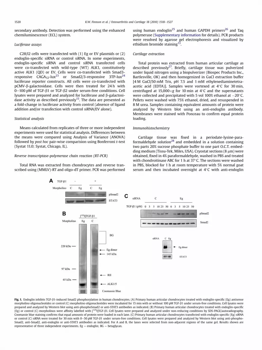
Fig. 1. Endoglin inhibits TGF-b1-induced Smad2 phosphorylation in human chondrocytes. (morpholino oligonucleotides or control (C) morpholino oligonucleotides were incubated forprepared and analyzed by Western blot using anti-phosphoSmad2 or anti-STAT3 antibodies(Eg) or control (C) morpholinos were affinity labelled with [125I]TGF-b1. Cell lysates wereCoomassie blue staining confirms that equal amounts of protein were loaded in each lane. (Cor control (C) siRNA were treated for 30 min with 0e50 pM TGF-b1 under serum-free condSmad2, anti-Smad2, anti-endoglin or anti-STAT3 antibodies as indicated. For A and B, therepresentative of three independent experiments. Eg ¼ endoglin; BG ¼ betaglycan.
using human endoglin25 and human GAPDH primers26 and Taqpolymerase (Supplementary information for details). PCR productswere resolved by agarose gel electrophoresis and visualized byethidium bromide staining13.
Cartilage extraction
Total protein was extracted from human articular cartilage asdescribed previously27. Briefly, cartilage tissue was pulverizedunder liquid nitrogen using a biopulverizer (Biospec Products Inc.,Bartlesville, OK) and then homogenized in GuCl extraction buffer[4 M GuCl/50 mM Tris, pH 7.5 and 1 mM ethylenediaminetetra-acetic acid (EDTA)]. Samples were vortexed at 4�C for 30 min,centrifuged at 15,000�g for 10 min at 4�C and the supernatantswere collected and precipitated with 5 vol 100% ethanol at �20�C.Pellets were washed with 75% ethanol, dried, and resuspended in8 M urea. Samples containing equivalent amounts of protein wereanalyzed by Western blot using an anti-endoglin antibody.Membranes were stained with Ponceau to confirm equal proteinloading.
Immunohistochemistry
Cartilage tissue was fixed in a periodate-lysine-para-formaldehyde solution28 and embedded in a solution containingtwo parts 20% sucrose phosphate buffer to one part O.C.T. embed-dingmedium (Tissu-Tek, Miles, USA). Cryostat sections (8 mm)wereobtained, fixed in 4% paraformaldehyde, washed in PBS and treatedwith chondroitinase ABC for 1 h at 37�C. The sections were washedin PBS, blocked for 1 h at room temperature with 5% normal goatserum and then incubated overnight at 4�C with anti-endoglin
A) Primary human articular chondrocytes treated with endoglin-specific (Eg) antisense15 min with or without 100 pM TGF-b1 under serum-free conditions. Cell lysates wereas indicated. (B) Primary human articular chondrocytes treated with endoglin-specificprepared and analyzed under non-reducing conditions by SDS-PAGE/autoradiography.) Primary human articular chondrocytes transfected with endoglin-specific (Eg) siRNAitions. Cell lysates were prepared and analyzed by Western blot using anti-phospho-lanes were selected from non-adjacent regions of the same gel. Results shown are
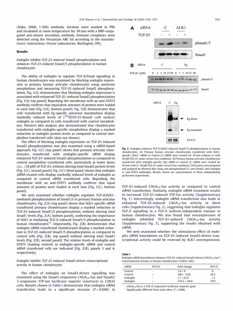
Fig. 2. Endoglin enhances TGF-b1/ALK1-induced Smad1/5 phosphorylation in humanchondrocytes. (A) Primary human articular chondrocytes transfected with ALK1-specific (ALK1) siRNA or control (C) siRNA were treated for 30 min without or with50 pM TGF-b1 under serum-free conditions. (B) Primary human articular chondrocytestransfected with endoglin-specific (Eg) siRNA or control (C) siRNA were treated for30 min with 0e50 pM TGF-b1 under serum-free conditions. Cell lysates were preparedand analyzed by Western blot using anti-phosphoSmad1/5, anti-Smad1, anti-endoglinor anti-STAT3 antibodies. Results shown are representative of three independentlyperformed experiments.
Table IEndoglin siRNA knockdown enhances TGF-b1-induced Smad3-driven (CAGA12-lux*)transcriptional activity in human chondrocytes (C28/I2 cells)
siRNA TGF-b1 Fold-change 95% CI
Control � 1.0� 0 0Control þ 100� 15.8y 39.2Endoglin � 1.7� 0.21 1.1Endoglin þ 276.2� 28.6y 70.9
* CAGA12-lux is a TGF-b responsive luciferase reporter gene.y Significantly different from each other; P¼ 0.006.
K.W. Finnson et al. / Osteoarthritis and Cartilage 18 (2010) 1518e1527 1521
(Dako, SN6h, 1:100) antibody. Sections were washed in PBSand incubated at room temperature for 30 min with a HRP-conju-gated anti-mouse secondary antibody. Immune complexes weredetected using the Vectastain ABC kit according to the manufac-turers’ instructions (Vector Laboratories, Burlington, ON).
Results
Endoglin inhibits TGF-b1-induced Smad2 phosphorylation andenhances TGF-b1-induced Smad1/5 phosphorylation in humanchondrocytes
The ability of endoglin to regulate TGF-b/Smad signalling inhuman chondrocytes was examined by blocking endoglin expres-sion in primary human articular chondrocytes using antisensemorpholinos and measuring TGF-b1-induced Smad2 phosphory-lation. Fig. 1(A) demonstrates that blocking endoglin expression isassociated with enhanced TGF-b1-induced Smad2 phosphorylation[Fig. 1(A) top panel]. Reprobing the membrane with an anti-STAT3antibody confirms that equivalent amounts of protein were loadedin each lane [Fig 1(A), bottom panel]. Fig. 1(B) demonstrates thatcells transfected with Eg-specific antisense morpholinos displaymarkedly reduced levels of [125I]TGF-b1-bound (cell surface)endoglin as compared to cells transfected with control morpholi-nos. Western blot analysis also demonstrated that chondrocytestransfected with endoglin-specific morpholinos display a markedreduction in endoglin protein levels as compared to control mor-pholino transfected cells (data not shown).
The effect of blocking endoglin expression on TGF-b1-inducedSmad2 phosphorylation was also examined using a siRNA-basedapproach. Fig. 1(C) (top panel) shows that primary articular chon-drocytes transfected with endoglin-specific siRNA displayenhanced TGF-b1-induced Smad2 phosphorylation as compared tocontrol morpholino transfected cells, particularly at lower doses(i.e.,�10 pM) of TGF-b1, without altering total Smad2 protein levels[Fig. 1(C), second panel]. Fig. 1(C) (third panel) shows that endoglinsiRNA treated cells display markedly reduced levels of endoglin ascompared to control siRNA transfected cells. Reprobing themembrane with an anti-STAT3 antibody confirms that equalamounts of protein were loaded in each lane [Fig. 1(C), bottompanel].
We next examined whether endoglin regulates TGF-b/ALK1-mediated phosphorylation of Smad1/5 in primary human articularchondrocytes. Fig. 2(A) (top panel) shows that ALK1-specific siRNAtransfected primary chondrocytes display a marked reduction inTGF-b1-induced Smad1/5 phosphorylation, without altering totalSmad1 levels [Fig. 2(A), bottom panel], confirming the importanceof ALK1 in mediating TGF-b-induced Smad1/5 phosphorylation inhuman chondrocytes13. Importantly, Fig. 2(B) demonstrates thatendoglin siRNA transfected chondrocytes display a marked reduc-tion in TGF-b1-induced Smad1/5 phosphorylation as compared tocontrol cells [Fig. 2(B), top panel] without altering total Smad1levels [Fig. 2(B), second panel]. The relative levels of endoglin andSTAT3 (loading control) in endoglin-specific siRNA and controlsiRNA transfected cells are indicated [Fig. 2(B), panels 3 and 4,respectively].
Endoglin inhibits TGF-b1-induced Smad3-driven transcriptionalactivity in human chondrocytes
The effect of endoglin on Smad3-driven signalling wasexamined using the Smad3-responsive CAGA12-lux and Smad2/3-responsive 3TP-lux luciferase reporter constructs in C28/I2cells. Results shown in Table I demonstrate that endoglin siRNAtransfection leads to a significant increase (P¼ 0.006) in
TGF-b1-induced CAGA12-lux activity as compared to controlsiRNA transfection. Similarly, endoglin siRNA treatment resultsin increased TGF-b1-induced 3TP-lux activity (SupplementaryFig. 1). Interestingly, endoglin siRNA transfection also leads toenhanced TGF-b2-induced CAGA12-lux activity in thesecells (Supplementary Fig. 2), suggesting that endoglin regulatesTGF-b signalling in a TGF-b isoform-independent manner inhuman chondrocytes. We also found that overexpression ofendoglin inhibited TGF-b1-induced CAGA12-lux activity(Supplementary Fig. 3), supporting the results obtained withsiRNA.
We next examined whether the stimulatory effect of endo-glin siRNA knockdown on TGF-b1-induced Smad3-driven tran-scriptional activity could be reversed by ALK1 overexpression.
K.W. Finnson et al. / Osteoarthritis and Cartilage 18 (2010) 1518e15271522
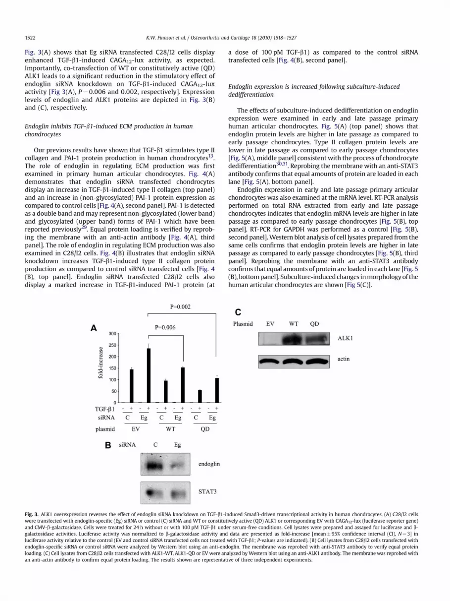
Fig. 3(A) shows that Eg siRNA transfected C28/I2 cells displayenhanced TGF-b1-induced CAGA12-lux activity, as expected.Importantly, co-transfection of WT or constitutively active (QD)ALK1 leads to a significant reduction in the stimulatory effect ofendoglin siRNA knockdown on TGF-b1-induced CAGA12-luxactivity [Fig 3(A), P¼ 0.006 and 0.002, respectively]. Expressionlevels of endoglin and ALK1 proteins are depicted in Fig. 3(B)and (C), respectively.
Endoglin inhibits TGF-b1-induced ECM production in humanchondrocytes
Our previous results have shown that TGF-b1 stimulates type IIcollagen and PAI-1 protein production in human chondrocytes13.The role of endoglin in regulating ECM production was firstexamined in primary human articular chondrocytes. Fig. 4(A)demonstrates that endoglin siRNA transfected chondrocytesdisplay an increase in TGF-b1-induced type II collagen (top panel)and an increase in (non-glycosylated) PAI-1 protein expression ascompared to control cells [Fig. 4(A), second panel]. PAI-1 is detectedas a double band andmay represent non-glycosylated (lower band)and glycosylated (upper band) forms of PAI-1 which have beenreported previously29. Equal protein loading is verified by reprob-ing the membrane with an anti-actin antibody [Fig. 4(A), thirdpanel]. The role of endoglin in regulating ECM production was alsoexamined in C28/I2 cells. Fig. 4(B) illustrates that endoglin siRNAknockdown increases TGF-b1-induced type II collagen proteinproduction as compared to control siRNA transfected cells [Fig. 4(B), top panel]. Endoglin siRNA transfected C28/I2 cells alsodisplay a marked increase in TGF-b1-induced PAI-1 protein (at
Fig. 3. ALK1 overexpression reverses the effect of endoglin siRNA knockdown on TGF-b1-iwere transfected with endoglin-specific (Eg) siRNA or control (C) siRNA and WT or constitutand CMV-b-galactosidase. Cells were treated for 24 h without or with 100 pM TGF-b1 undgalactosidase activities. Luciferase activity was normalized to b-galactosidase activity andluciferase activity relative to the control (EV and control siRNA transfected cells not treatedendoglin-specific siRNA or control siRNA were analyzed by Western blot using an anti-endloading. (C) Cell lysates from C28/I2 cells transfected with ALK1-WT, ALK1-QD or EV were anan anti-actin antibody to confirm equal protein loading. The results shown are representat
a dose of 100 pM TGF-b1) as compared to the control siRNAtransfected cells [Fig. 4(B), second panel].
Endoglin expression is increased following subculture-induceddedifferentiation
The effects of subculture-induced dedifferentiation on endoglinexpression were examined in early and late passage primaryhuman articular chondrocytes. Fig. 5(A) (top panel) shows thatendoglin protein levels are higher in late passage as compared toearly passage chondrocytes. Type II collagen protein levels arelower in late passage as compared to early passage chondrocytes[Fig. 5(A), middle panel] consistent with the process of chondrocytededifferentiation30,31. Reprobing the membrane with an anti-STAT3antibody confirms that equal amounts of protein are loaded in eachlane [Fig. 5(A), bottom panel].
Endoglin expression in early and late passage primary articularchondrocytes was also examined at the mRNA level. RT-PCR analysisperformed on total RNA extracted from early and late passagechondrocytes indicates that endoglin mRNA levels are higher in latepassage as compared to early passage chondrocytes [Fig. 5(B), toppanel]. RT-PCR for GAPDH was performed as a control [Fig. 5(B),second panel].Western blot analysis of cell lysates prepared from thesame cells confirms that endoglin protein levels are higher in latepassage as compared to early passage chondrocytes [Fig. 5(B), thirdpanel]. Reprobing the membrane with an anti-STAT3 antibodyconfirms that equal amounts of protein are loaded in each lane [Fig. 5(B), bottompanel]. Subculture-induced changes inmorphologyof thehuman articular chondrocytes are shown [Fig 5(C)].
nduced Smad3-driven transcriptional activity in human chondrocytes. (A) C28/I2 cellsively active (QD) ALK1 or corresponding EV with CAGA12-lux (luciferase reporter gene)er serum-free conditions. Cell lysates were prepared and assayed for luciferase and b-data are presented as fold-increase [mean� 95% confidence interval (CI), N¼ 3] in
with TGF-b1; P-values are indicated). (B) Cell lysates from C28/I2 cells transfected withoglin. The membrane was reprobed with anti-STAT3 antibody to verify equal proteinalyzed by Western blot using an anti-ALK1 antibody. The membrane was reprobed withive of three independent experiments.

Fig. 4. Endoglin inhibits TGF-b1-induced type II collagen and PAI-1 protein expression in human chondrocytes. (A) primary human articular chondrocytes or (B) C28/I2 cells weretransfected with endoglin-specific (Eg) or control (C) siRNA. The cells were treated for 24 h with 0e100 pM TGF-b1 under serum-free conditions. Cell lysates were prepared andanalyzed by Western blot using anti-type II collagen, anti-PAI-1, anti-endoglin and anti-actin antibodies. Results shown in A and B are each representative of two independentexperiments. Results shown are representative of four (A) and two (B) independent experiments, respectively.
K.W. Finnson et al. / Osteoarthritis and Cartilage 18 (2010) 1518e1527 1523
Endoglin expressionwas also examined in early and late passagetsT/AC62 cells. Western blot analysis indicates that total endoglinprotein levels are higher in late passage as compared to earlypassage tsT/AC62 cells [Fig. 6(A), top panel]. The type II collagenprotein levels are lower and the type I collagen (third panel) arehigher in late passage as compared to early passage tsT/AC62 cells[Fig. 6(A), second and third panel, respectively], consistent with theprocess of chondrocyte dedifferentiation30,31. Reprobing themembrane with an anti-actin antibody confirms that equalamounts of protein are loaded in each lane. Cell surface endoglin
Fig. 5. Endoglin expression is increased in primary human articular chondrocytes followiprimary human articular chondrocytes were analyzed by Western blot using anti-endoglin (twith an anti-STAT3 antibody (bottom panel) to confirm that equal amounts of protein welate passage primary human articular chondrocytes. Total RNA was analyzed by RT-PCR usiPCR product sizes for endoglin (e.g., 411 bp) and GAPDH (226 bp) are indicated. Cell lysates wpanel) antibodies. (C) Cell morphology: Phase contrast microscopy of early and late passarepresentative of three independent experiments.
levels were also examined in early (E) and late (L) passage tsT/AC62by affinity labelling with [125I]TGF-b1. Fig. 6(B) shows that latepassage tsT/AC62 cells display higher levels of [125I]TGF-b1-asso-ciated endoglin as compared to early passage cells. The morphol-ogies of early and late passage tsT/AC62 cells are shown in Fig. 6(C).Immediately (24 h) following transfer from alginate to monolayercultures, polygonal-shaped cells are observed [Fig 6(C), earlypassage]. However, the cells progressively acquire amore elongated‘fibroblastic’ morphology following several passages in monolayer[Fig 6(C), late passage].
ng subculture-induced dedifferentiation. (A) Cell lysates from early and late passageop panel) or anti-type II collagen (middle panel) antibodies. Membranes were reprobedre loaded in each lane. (B) Total RNA and cell lysates were prepared from early andng endoglin- (top panel) and GAPDH-specific (second panel) oligonucleotide primers.ere analyzed byWestern blot using anti-endoglin (third panel) and anti-STAT3 (bottomge primary human articular chondrocytes in monolayer culture. Results shown are

Fig. 6. Endoglin expression is increased in tsT/AC62 cells following subculture-induced dedifferentiation. (A) Cell lysates from early and late passage tsT/AC62 cells were analyzedby Western blot using anti-endoglin (top panel), anti-type II collagen (second panel) or anti-type I collagen (third panel) antibodies. Membranes were reprobed with an anti-actinantibody (bottom panel) to confirm that equal amounts of protein were loaded in each lane. (B) Early and late passage tsT/AC62 cells were affinity labelled with 100 pM of [125I]TGF-b1. Membrane extracts were prepared, electrophoretically separated by 3e11% gradient SDS-PAGE under non-reducing conditions and levels of [125I]TGF-b1-associated endoglinvisualized by autoradiography. The lanes were selected from non-adjacent regions of the same gel. Coomassie blue staining confirms that equal amounts of protein were loaded ineach lane. (C) Cell morphology: Phase contrast microscopy of early and late passage tsT/AC62 cells in monolayer culture. Results shown are representative of three independentexperiments.
K.W. Finnson et al. / Osteoarthritis and Cartilage 18 (2010) 1518e15271524
Endoglin protein expression in OA and normal cartilage
We next examined endoglin protein expression in vivo in cartilagesamples obtained from the knee of OA patients and normal controls.Cartilage protein was extracted as described in the Method sectionand analyzed by Western blot using an anti-endoglin antibody.Results shown in Fig. 7(A) demonstrate that endoglin protein levelsare higher in OA as compared to normal cartilage. Ponceau staining ofthemembrane confirms that equal amounts of proteinwere loaded ineach lane. Similar results were obtained when comparing endoglinprotein levels in three different pairs of OA and normal cartilagesamples (data not shown). Endoglin expressionwas also examined inhuman OA and normal cartilage by immunohistochemistry using ananti-endoglin (SN6h, Dako) antibody. Fig. 7(B) demonstrates that OAcartilage displays enhanced staining for endoglin as compared tonormal cartilage. No staining was detected when the primary anti-body was omitted in parallel experiments (data not shown).
Discussion
TGF-b is a pleiotropic cytokine that plays a critical role in main-taining cartilage homeostasis6,32. Although deregulation of TGF-b
signalling has been implicated in cartilage diseases such as OA6,32, thefactors that regulate TGF-b signalling in normal or diseased humanchondrocytes are unclear. We have previously shown that humanchondrocytes express several different TGF-b receptors including twoisoforms of TbRII (RII and RIIB), two different TbRIs (ALK5 and ALK1)and the TGF-b co-receptors endoglin and betaglycan13,18,33. We haveshown that endoglin forms a heteromeric complex with TbRII andTbRI (ALK5 and ALK1) in human chondrocytes13,18 as has beendemonstrated for endoglin in endothelial cells11,34e37. However, thefunctional significance of endoglin expression in human chon-drocytes has not been examined. Here we show that endoglinenhances TGF-b1-induced Smad1/5 phosphorylation but inhibitsTGF-b1-induced Smad2 phosphorylation, Smad3-driven (CAGA12-luxand 3TP-lux) transcriptional activity and type II collagen and PAI-1protein expression in human chondrocytes. Moreover, we presentevidence that endoglin expression is increased in human chon-drocytes following subculture-induced dedifferentiation in vitro andthat endoglin expression levels are increased in human OA cartilageas compared to normal cartilage in vivo. Collectively, our resultsestablish endoglin as a regulator of TGF-b signalling and ECM proteinproduction with opposing effects on the ALK1/Smad1/5 and ALK5/Smad2/3 pathways in human chondrocytes and suggest that endoglin

Fig. 7. Endoglin expression is increased in OA cartilage as compared to normal carti-lage. (A)Western blot: Protein was extracted from OA and normal cartilage as describedin Methods section. Samples were analyzed by Western blot using an anti-endoglinantibody (top panel). The membrane was stained with Ponceau S to confirm that equalamounts of protein were loaded in each lane (bottom panel). The lanes were selectedfrom non-adjacent regions of the same gel. Results shown are representative of threeindependent experiments performed on cartilage of three different OA and threedifferent normal donors. (B) Immunohistochemistry: Cryostat sections of OA andnormal cartilage were analyzed by immunohistochemistry using an anti-endoglin(Dako, SN6h, 1:100 dilution) antibody. Detection was performed using Vectostain ABCimmunostaining kit. Results shown are representative of two independently per-formed experiments.
K.W. Finnson et al. / Osteoarthritis and Cartilage 18 (2010) 1518e1527 1525
may represent a potential marker for chondrocyte dedifferentiationor the loss of chondrogenic phenotype.
Endoglin is a TGF-b co-receptor that has been shown to regulateTGF-b signalling in endothelial cells12,17,38. Our present resultsshow that endoglin enhances TGF-b1-induced Smad1/5 phos-phorylation but decreases TGF-b1-mediated Smad2 phosphoryla-tion in human chondrocytes. In comparison, other studies reportconflicting data on the role of endoglin in regulating TGF-b-medi-ated Smad1/5 and Smad2 signalling in different cell types. Forexample, ectopic expression of endoglin in rat myofibroblasts wasreported to increase phosphorylation (and total levels) of Smad239
whereas another study using ectopic expression of endoglin in ratmyofibroblasts reported no significant effect on TGF-b-inducedphosphorylation of Smad1 or Smad240. Conversely, studies usingendoglin-null mouse embryonic endothelial cells reported thatendoglin promotes TGF-b-mediated Smad1/5 signalling36,41
whereas another study using endoglin siRNA knockdown inhuman bone marrow stromal cells reported that endoglin acts asa positive regulator of both ALK1-induced Smad1 activation andALK5-induced Smad2 activation42. Taken together, these studiessuggest that endoglin differentially regulates TGF-b signalling viaALK1/Smad1/5 and ALK5/Smad2 depending on cell-type andcellular context. Our results suggest that endoglin differentiallyregulates TGF-b/Smad signalling in human chondrocytes bypromoting TGF-b signalling through the ALK1/Smad1/5 pathwayand decreasing signalling through the ALK5/Smad2/3 pathway.
Our previous work has demonstrated that ALK1 inhibitsTGF-b/ALK5-dependent Smad3-driven transcriptional activityand ECM production in human chondrocytes13. Our presentresults indicate that endoglin also inhibits TGF-b1/ALK5-inducedSmad3-driven transcriptional activity and ECM production inhuman chondrocytes. Because we have shown that endoglininteracts with ALK1 in human chondrocytes13 and that othershave shown endoglin to interact with, and promote signallingthrough, ALK1 in endothelial cells to inhibit ALK5 signalling36, itis plausible that endoglin regulates TGF-b/ALK5 signallingthrough a similar mechanism in human chondrocytes. Ourresults showing that endoglin promotes TGF-b1/ALK1 signalling(i.e., Smad1/5 phosphorylation) and that overexpression of ALK1can reverse the stimulatory effect of endoglin siRNA knockdownon TGF-b1-induced CAGA12-lux activity are consistent with thisnotion. Whether endoglin inhibits TGF-b/ALK5/Smad2/3 signal-ling by promoting the TGF-b/ALK1/Smad1/5 pathway or byregulating other pathways remains to be determined. Impor-tantly, our finding that endoglin inhibits TGF-b-induced Smad3-driven transcription provides further evidence for endoglin’s rolein inhibiting TGF-b-mediated ALK5/Smad2/3 signalling in humanchondrocytes.
Primary human articular chondrocytes maintain a rounded,polygonal morphology in monolayer culture but undergoa progressive change to an elongated fibroblast-like morphologyover time, particularly after subculture43. Accompanying thesemorphological changes is the loss of the chondrogenic (differenti-ated) phenotype or ‘chondrocyte dedifferentiation’ which is char-acterized by a decrease in type II collagen expression43. Our resultsdemonstrate that primary human articular chondrocytes and tsT/AC62 cells display such changes in morphology and reduction intype II collagen expression following serial passage in monolayersuggesting that these cells undergo subculture-induced dediffer-entiation. Human costal chondrocytes (C28/I2) also showa decrease in type II collagen expression after serial passage inmonolayer although morphological changes were not observed inthese cells (data not shown). Importantly, our results showing thatendoglin protein levels are higher in late passage as compared toearly passage chondrocytes suggest that endoglin expression isincreased following subculture-induced dedifferentiation. Theincreased endoglin protein expression in late passage chondrocytesis mirrored by an increase in endoglin mRNA expression indicatingthat the mechanism of its up-regulation involves an increase inendoglin gene expression and/or an increase in endoglin mRNAstability. Our results are consistent with a previous report identi-fying endoglin as one of many cell surface molecules upregulated inprimary articular chondrocytes during cell expansion in mono-layer44. Thus, our results suggest that endoglin may representa novel marker for chondrocyte dedifferentiation and/or the loss ofnormal ‘differentiated’ chondrocyte phenotype.
Factors that regulate TGF-b signalling in normal and ‘pheno-typically altered’ chondrocytes are ill-defined. A previous study hasshown that ALK1 mRNA levels are increased during in vitroexpansion of primary human articular chondrocytes45 and a morerecent study has reported an increase in the ALK1/ALK5 ratio asa cause for elevated MMP-13 expression in OA in humans andmice46. Our results showing that endoglin levels are increased inhuman chondrocytes following subculture-induced dedifferentia-tion in vitro and in human OA cartilage as compared to normalcartilage in vivo extend these findings and raise the possibility thatincreased expression of endoglin and ALK1 together leads to a shiftin TGF-b signalling away from the ALK5/Smad2/3 pathway in favourof ALK1/Smad1/5 pathway. Further studies using increasednumbers of age-, sex- and gender-matched OA and normal humancartilage samples will be needed to confirm these findings.

K.W. Finnson et al. / Osteoarthritis and Cartilage 18 (2010) 1518e15271526
In conclusion, our results establish endoglin as a regulator ofTGF-b signalling and ECM protein productionwith opposing effectson the ALK1/Smad1/5 and ALK5/Smad2/3 pathways in humanchondrocytes and suggest that endoglin may represent a potentialmarker for chondrocyte dedifferentiation or loss of chondrogenicphenotype. Delineation of the precise mechanism by which endo-glin may regulate the balance of ALK1 and ALK5 signalling inhuman chondrocytes will be a fruitful avenue for future studies.
Role of the funding source
This studywas supported by a CIHRGrantsMOP 69060 andMOP13732 (to AP) and the McGill Surgeon Scientist Training Award (toWLP).
Contributions
Kenneth Finnson contributed to the conception and design,collection and assembly of the data, analysis and interpretation ofdata, drafting of the article, critical revision of the article forimportant intellectual content and final approval of the article.
Wendy Parker contributed to the conception and design,collection and assembly of the data, analysis and interpretation ofdata, drafting of the article, critical revision of the article forimportant intellectual content and final approval of the article.
Yoonyoung Chi contributed to the collection and assembly of thedata, critical revision of the article for important intellectualcontent and final approval of the article.
Caroline Hoemann contributed to the collection of the data,critical revision of the article for important intellectual content andfinal approval of the article.
Mary Goldring contributed to analysis and interpretation ofdata, critical revision of the article for important intellectualcontent and final approval of the article.
John Antoniou contributed to the collection of the data, criticalrevision of the article for important intellectual content and finalapproval of the article.
Anie Philip contributed to the conception and design of thestudy, obtaining funding, the assembly of the data, analysis andinterpretation of data, drafting of the article, critical revision of thearticle for important intellectual content and final approval of thearticle.
Conflict of interestThe authors state that they have no conflicts of interest.
Acknowledgements
We thank Genzyme Inc. (Framingham,MA) for the gift of TGF-b1and TGF-b2, Calvin Vary (Maine Medical Center Research Institute,Scarborough, ME) for the endoglin cDNA cloned into pCDNA3.1/myc-HIS, Kohei Miyazono (The University of Tokyo, Tokyo, Japan)for ALK1-WT and ALK1-QD plasmids, Lucia Lesec (Jewish GeneralHospital, Montreal, QC) for collection of human articular cartilage,Mohammed Zafarullah (University of Montreal and ResearchCentre Notre-Dame Hospital, Montreal, QC) for assistance with thecalcium phosphate transfection method. Dr S. Huet (LaboratoireGlaxo Wellcome, Les Ulis Cedex, France) for (CAGA)12-lux.
Supplementary material
Supplementary data associated with this article can be found inthe online version, at doi:10.1016/j.joca.2010.09.002.
References
1. Blom AB, van der Kraan PM, van den Berg WB. Cytokinetargeting in osteoarthritis. Curr Drug Targets 2007;8:283e92.
2. Goldring MB. Update on the biology of the chondrocyte andnew approaches to treating cartilage diseases. Best Pract ResClin Rheumatol 2006;20:1003e25.
3. Grimaud E, Heymann D, Redini F. Recent advances inTGF-b effects on chondrocyte metabolism. Potential thera-peutic roles of TGF-b in cartilage disorders. Cytokine GrowthFactor Rev 2002;13:241e57.
4. Serra R, Johnson M, Filvaroff EH, LaBorde J, Sheehan DM,Derynck R, et al. Expression of a truncated, kinase-defectiveTGF-b type II receptor in mouse skeletal tissue promotesterminal chondrocyte differentiation and osteoarthritis. J CellBiol 1997;139:541e52.
5. Yang X, Letterio JJ, Lechleider RJ, Chen L, Hayman R, Gu H, et al.Targeted disruption of SMAD3 results in impaired mucosalimmunity and diminished T cell responsiveness to TGF-b.EMBO J 1999;18:1280e91.
6. Blaney Davidson E, van der Kraan P, van den Berg W. TGF-band osteoarthritis. Osteoarthritis Cartilage 2007;15:597e604.
7. Itayem R, Mengarelli-Widholm S, Hulth A, Reinholt FP. Ultra-structural studies on the effect of transforming growth factor-b1 on rat articular cartilage. Apmis 1997;105:221e8.
8. Itayem R, Mengarelli-Widholm S, Reinholt FP. The long-termeffect of a short course of transforming growth factor-b1 on ratarticular cartilage. APMIS 1999;107:183e92.
9. Massague J, Gomis RR. The logic of TGF-b signaling. FEBS Lett2006;580:2811e20.
10. Yamashita H, ten Dijke P, Franzen P, Miyazono K, Heldin CH.Formation of hetero-oligomeric complexes of type I and type IIreceptors for transforming growth factor-b. J Biol Chem1994;269:20172e8.
11. Lebrin F, Deckers M, Bertolino P, Ten Dijke P. TGF-b receptorfunction in the endothelium. Cardiovasc Res 2005;65:599e608.
12. Pardali E, ten Dijke P. Transforming growth factor-b signalingand tumor angiogenesis. Front Biosci 2009;14:4848e61.
13. Finnson KW, Parker WL, ten Dijke P, Thorikay M, Philip A.ALK1 opposes ALK5/Smad3 signaling and expression ofextracellular matrix components in human chondrocytes. JBone Miner Res 2008;23:896e906.
14. ten Dijke P, Hill C. New insights into TGF-b-Smad signalling.Trends Biochem Sci 2004;29:265e73.
15. Bernabeu C, Lopez-Novoa JM, Quintanilla M. The emerging roleof TGF-b superfamily co-receptors in cancer. Biochim BiophysActa 2009;1792:954e73.
16. Mokrosinski J, Krajewska WM. TGF-b signalling accessoryreceptors. Postepy Biochem 2008;54:264e73.
17. ten Dijke P, Goumans MJ, Pardali E. Endoglin in angiogenesisand vascular diseases. Angiogenesis 2008;11:79e89.
18. Parker WL, Goldring MB, Philip A. Endoglin is expressed onhuman chondrocytes and forms a heteromeric complex withbetaglycan in a ligand and type II TGFb receptor independentmanner. J Bone Miner Res 2003;18:289e302.
19. Robbins JR, Thomas B, Tan L, Choy B, Arbiser JL, Berenbaum F,et al. Immortalized human adult articular chondrocytesmaintain cartilage-specific phenotype and responses to inter-leukin-1b. Arthritis Rheum 2000;43:2189e201.
20. Goldring MB, Birkhead JR, Suen LF, Yamin R, Mizuno S,Glowacki J, et al. Interleukin-1 b-modulated gene expression inimmortalized human chondrocytes. J Clin Invest 1994;94:2307e16.
21. Qiu W, Murray MM, Shortkroff S, Lee CR, Martin SD, Spector M.Outgrowth of chondrocytes from human articular cartilage

K.W. Finnson et al. / Osteoarthritis and Cartilage 18 (2010) 1518e1527 1527
explants and expression of alpha-smooth muscle actin.Wound Repair Regen 2000;8:383e91.
22. Qureshi HY, Ahmad R, Zafarullah M. High-efficiency trans-fection of nucleic acids by the modified calcium phosphateprecipitation method in chondrocytes. Anal Biochem2008;382:138e40.
23. Dennler S, Huet S, Gauthier J-M. A short amino-acid sequencein MH1 domain is responsible for functional differencesbetween Smad2 and Smad3. Oncogene 1999;18:1643e8.
24. Wrana JL, Attisano L, Carcamo J, Zentella A, Doody J, Laiho M,et al. TGF-b signals through a heteromeric protein kinasereceptor complex. Cell 1992;71:1003e14.
25. Bellon T, Corbi A, Lastres P, Cales C, Cebrian M, Vera S, et al.Identification and expression of two forms of the humantransforming growth factor-b-binding protein endoglin withdistinct cytoplasmic regions. Eur J Immunol 1993;23:2340e5.
26. Yoshimura K, Uchida G, Okazaki M, Kitano Y, Harii K. Differ-ential expression of heparin-binding EGF-like growth factor(HB-EGF) mRNA in normal human keratinocytes induced bya variety of natural and synthetic retinoids. Exp Dermatol2003;12:28e34.
27. Hoemann CD, Sun J, Chrzanowski V, Buschmann MD. Amultivalent assay to detect glycosaminoglycan, protein,collagen, RNA, and DNA content in milligram samples ofcartilage or hydrogel-based repair cartilage. Anal Biochem2002;300:1e10.
28. McLean IW, Nakane PK. Periodate-lysine-paraformaldehydefixative. A new fixation for immunoelectron microscopy. JHistochem Cytochem 1974;22:1077e83.
29. Gils A, Pedersen KE, Skottrup P, Christensen A, Naessens D,Deinum J, et al. Biochemical importance of glycosylation ofplasminogen activator inhibitor-1. Thromb Haemost 2003;90:206e17.
30. Duval E, Leclercq S, Elissalde JM, Demoor M, Galera P,Boumediene K. Hypoxia-inducible factor 1a inhibits thefibroblast-like markers type I and type III collagen duringhypoxia-induced chondrocyte redifferentiation: hypoxia notonly induces type II collagen and aggrecan, but it also inhibitstype I and type III collagen in the hypoxia-inducible factor1alpha-dependent redifferentiation of chondrocytes. ArthritisRheum 2009;60:3038e48.
31. Domm C, Schunke M, Christesen K, Kurz B. Redifferentiation ofdedifferentiated bovine articular chondrocytes in alginateculture under low oxygen tension. Osteoarthritis Cartilage2002;10:13e22.
32. van der Kraan PM, Blaney Davidson EN, Blom A, van denBerg WB. TGF-b signaling in chondrocyte terminal differenti-ation and osteoarthritis modulation and integration ofsignaling pathways through receptor-Smads. OsteoarthritisCartilage 2009;17:1539e45.
33. Parker WL, Finnson KW, Soe-Lin H, Knaus P, Philip A.Expression and function of TbRII-B, a variant of the type II TGF-b receptor, in human chondrocytes. Osteoarthritis Cartilage2007;15:442e53.
34. Blanco FJ, Santibanez JF, Guerrero-Esteo M, Langa C, Vary CP,Bernabeu C. Interaction and functional interplay betweenendoglin and ALK-1, two components of the endothelialtransforming growth factor-b receptor complex. J Cell Physiol2005;204:574e84.
35. Koleva RI, Conley BA, Romero D, Riley KS, Marto JA, Lux A, et al.Endoglin structure and function: determinants of endoglinphosphorylation by transforming growth factor-b receptors. JBiol Chem 2006;281:25110e23.
36. Lebrin F, Goumans MJ, Jonker L, Carvalho RL,Valdimarsdottir G, Thorikay M, et al. Endoglin promotesendothelial cell proliferation and TGF-b/ALK1 signal trans-duction. Embo J 2004;23:4018e28.
37. Lee NY, Blobe GC. The interaction of endoglin with b-arrestin2regulates transforming growth factor-b-mediated ERK activa-tion and migration in endothelial cells. J Biol Chem2007;282:21507e17.
38. Garcia-Pozo L, Miquilena-Colina ME, Lozano-Rodriguez T,Garcia-Monzon C. Endoglin: structure, biological functions,and role in fibrogenesis. Rev Esp Enferm Dig 2008;100:355e60.
39. Santibanez J, Letamendia A, Perez-Barriocanal F, Silvestri C,Saura M, Vary C, et al. Endoglin increases eNOS expression bymodulating Smad2 protein levels and Smad2-dependentTGF-b signaling. J Cell Physiol 2007;210:456e68.
40. Velasco S, Alvarez-Munoz P, Pericacho M, Ten Dijke P,Bernabeu C, Lopez-Novoa JM, et al. L- and S-endoglin differ-entially modulate TGF-b1 signaling mediated by ALK1 andALK5 in L6E9 myoblasts. J Cell Sci 2008;121:913e9.
41. Lee NY, Ray BN, How T, Blobe GC. Endoglin promotes trans-forming growth factor-b-mediated Smad 1/5/8 signaling andinhibits endothelial cell migration through its association withGIPC. J Biol Chem 2008;283:32527e33.
42. O’Connor JC, Farach-Carson MC, Schneider CJ, Carson DD.Coculture with prostate cancer cells alters endoglin expressionand attenuates transforming growth factor-b signaling inreactive bone marrow stromal cells. Mol Cancer Res2007;5:585e603.
43. Goldring MB. Human chondrocyte cultures as models ofcartilage-specific gene regulation. Methods Mol Med2005;107:69e95.
44. Diaz-Romero J, Gaillard JP, Grogan SP, Nesic D, Trub T,Mainil-Varlet P. Immunophenotypic analysis of human artic-ular chondrocytes: changes in surface markers associated withcell expansion in monolayer culture. J Cell Physiol 2005;202:731e42.
45. Dell’Accio F, De Bari C, Luyten FP. Molecular markers predic-tive of the capacity of expanded human articular chondrocytesto form stable cartilage in vivo. Arthritis Rheum 2001;44:1608e19.
46. Blaney Davidson EN, Remst DF, Vitters EL, van Beuningen HM,Blom AB, Goumans MJ, et al. Increase in ALK1/ALK5 ratio asa cause for elevated MMP-13 expression in osteoarthritis inhumans and mice. J Immunol 2009;182:7937e45.
Related Documents


![Culture of Primary Bovine Chondrocytes on a Continuously ...passaging during standard methods causes ÒdedifferentiationÓ into fibroblastic cells [1]. To circumvent chondrocyte dedifferentiation](https://static.cupdf.com/doc/110x72/5fb19cc6166d0216b5433280/culture-of-primary-bovine-chondrocytes-on-a-continuously-passaging-during-standard.jpg)