Journal of Pineal Research 9:259-269 (1990) Evidence for an Effect of ELF Electromagnetic Fields on Human Pineal Gland Function Bary W. Wilson, Cherylyn W. Wright, James E. Morris, Raymond L. Buschbom, Donald P. Brown, Douglas L. Miller, Rita Sornmers-Flannigan, and Larry E. Anderson Battelle, Pacific Northwest Laboratories, Richland, Washington (B.W.W., C.W.W., J.E.M., R.L.B., D.P.B., D.L.M., L.E.A.); University of Montana, Missoula, Montana (RS.-F.) A study was carried out to determine possible effects of 60-Hz electromagnetic-field exposure on pineal gland function in humans. Overnight excretion of urinary 6- hydroxymelatonin sulfate (6-OHMS), a stable urinary metabolite of the pineal hor- mone melatonin, was used to assess pineal gland function in 42 volunteers who used standard (conventional) or modified continuous polymer wire (CPW) electric blan- kets for approximately 8 weeks. Volunteers using conventional electric blankets showed no variations in 6-OHMSexcretion as either a group or individuals during the study period. Serving as their own controls, 7 of 28 volunteers using the CPW blankets showed statistically significant changes in their mean nighttime 6-OHMS excretion. The CPW blankets switched on and off approximately twice as often when in service and produced magnetic fields that were 50% stronger than those from the conven- tional electric blankets. On the basis of these findings, we hypothesize that periodic exposure to pulsed DC or extremely low frequency electric or magnetic fields of sufficient intensity and duration can affect pineal gland function in certain in- dividuals. Key words: melatonin, electric blankets, electric field, magnetic field INTRODUCTION During the past two decades, interest has increased in the possibility that exposure to static or extremely low frequency (ELF: 10-100 Hz), including 50- or 60-Hz powerline-frequency electric and magnetic fields, may cause biologi- cal effects in human populations [Savitz and Calle, 19871. Much of our work has been directed toward understanding the association between ELF electric- and Received April 24, 1990; accepted August 23, 1990. Address reprint requests to Dr. Bary W. Wilson, Battelle, Pacific Northwest Laboratories, Richland, WA 99352.

Welcome message from author
This document is posted to help you gain knowledge. Please leave a comment to let me know what you think about it! Share it to your friends and learn new things together.
Transcript
Journal of Pineal Research 9:259-269 (1990)
Evidence for an Effect of ELF Electromagnetic Fields on Human Pineal
Gland Function
Bary W. Wilson, Cherylyn W. Wright, James E. Morris, Raymond L. Buschbom, Donald P. Brown, Douglas L. Miller,
Rita Sornmers-Flannigan, and Larry E. Anderson
Battelle, Pacific Northwest Laboratories, Richland, Washington (B.W.W., C.W.W., J.E.M., R.L.B., D.P.B., D.L.M., L.E.A.); University of Montana, Missoula, Montana (RS.-F.)
A study was carried out to determine possible effects of 60-Hz electromagnetic-field exposure on pineal gland function in humans. Overnight excretion of urinary 6- hydroxymelatonin sulfate (6-OHMS), a stable urinary metabolite of the pineal hor- mone melatonin, was used to assess pineal gland function in 42 volunteers who used standard (conventional) or modified continuous polymer wire (CPW) electric blan- kets for approximately 8 weeks. Volunteers using conventional electric blankets showed no variations in 6-OHMS excretion as either a group or individuals during the study period. Serving as their own controls, 7 of 28 volunteers using the CPW blankets showed statistically significant changes in their mean nighttime 6-OHMS excretion. The CPW blankets switched on and off approximately twice as often when in service and produced magnetic fields that were 50% stronger than those from the conven- tional electric blankets. On the basis of these findings, we hypothesize that periodic exposure to pulsed DC or extremely low frequency electric or magnetic fields of sufficient intensity and duration can affect pineal gland function in certain in- dividuals.
Key words: melatonin, electric blankets, electric field, magnetic field
INTRODUCTION
During the past two decades, interest has increased in the possibility that exposure to static or extremely low frequency (ELF: 10-100 Hz), including 50- or 60-Hz powerline-frequency electric and magnetic fields, may cause biologi- cal effects in human populations [Savitz and Calle, 19871. Much of our work has been directed toward understanding the association between ELF electric- and
Received April 24, 1990; accepted August 23, 1990.
Address reprint requests to Dr. Bary W. Wilson, Battelle, Pacific Northwest Laboratories, Richland, WA 99352.
260 Wilson et al.
magnetic-field exposure and alterations in pineal gland circadian rhythms [Wil- son et al., 19891.
Melatonin (N-acetyl-5-methoxytryptamine), the principal hormone of the pineal gland, is produced by the action of N-acetyltransferase (NAT) and hy- droxyindole-0-methyl transferase (HIOMT) on serotonin [Deguchi and Axel- rod, 19721. Melatonin concentrations normally increase during the hours of darkness in both the pineal gland and circulating blood. Maximum melatonin concentrations occur between approximately 0200 and 0400 h in humans. In all mammals, the internal clock that helps generate this pineal circadian rhythm resides in the suprachiasmatic nuclei. The pineal is richly innervated by fibers of the superior cervical ganglia (SCG) [Moore et al., 19681 as well as by fibers originating in the hypothalamus and optic regions of the brain [Zisapel et al., 19881. Neuronal input from the eyes acts via the SCG as the principal regulator of the melatonin circadian rhythm in the pineal.
Light of sufficient intensity is effective in suppressing melatonin synthesis in many animals [Wurtman et al., 19631. Lewy et al. 119821 reported that the light level required for suppression in humans is approximately 2,500 lux. It appears that the pineal gland of certain sensitive individuals, however, may respond to light levels as low as 200 lux [Mclntyre et al., 19901. Ingested alcohol [Wetterberg, 19781, P-adrenergic receptor-blocking drugs such as pro- r .olol [Wetterberg, 19791, and certain kinds of stress [Troiani et al., 19871 A - also been reported to reduce melatonin concentrations in the pineal and circulation of rats. Further, altering melatonin circadian rhythms by use of bright light has been effective in the treatment of seasonal affective disorder syndrome (SADS) [Lewy et a]., 19871.
In the circulation, melatonin acts to suppress the function of several other endocrine glands, including the gonads. Melatonin also suppresses the growth of certain cancers in both in vitro and in vivo models [Blask, 19901. Reduction in melatonin secretion has been associated with estrogen receptor-positive breast cancers [Sanchez Barcelo et a]., 19881 and prostate adenocarcinoma [Buzzell et al., 19881. Stevens [ 19871 proposed that, should there be increased cancer risk from ELF electromagnetic-field exposure, such risk may be a consequence of altered pineal gland function.
Chronic exposure to 60-Hz electric fields can reduce the normal nocturnal rise in both pineal NAT activity and melatonin concentration in laboratory rats [Wilson et a]., 1981, 19831. In 23-day-old rats maintained in a 60-Hz electric field for 20 Wday from conception, there was no difference among the pineal melatonin levels of animals exposed to field strengths of 10, 60, and 130 kV/m. Compared to controls, however, these exposed rats showed an approximate 40% reduction in maximal nighttime pineal melatonin levels and an approxi- mate 1.4-h delay in the occurrence of the nighttime melatonin peak [Reiter et al., 19881. Rats first exposed at 55 days of age to a 39-kV/m electric field showed no statistically significant difference between daytime and nighttime levels of pi- neal melatonin [i.e., no circadian rhythm in melatonin secretion) after 21 days of exposure. Within 3 days after cessation of ELF electric-field exposure, how- ever, strong pineal melatonin rhythms were reestablished. This effect appeared t! an "all-or-none" response to electric fields between approximately 2 and 13u kV/m [Wilson et al., 19861.
ELF Fields
Indeed, an accumulating bod) netic-field exposure can affect circ different species. The pineal @an& changes in the geomagnetic field [( showed that NAT activity and me1 suppressed by weak ELF magnetb marked changes in pineal seroton intermittent magnetic fields at nig consequence of daytime exposurr 50-Hz electric or magnetic fields c ening of the circadian cycle that nc temporal cues. However, we know electromagnetic-field exposure C ~ I
We have completed a study magnetic-field exposure from usin tonin secretion in humans. Use of sure to ELF fields that normally oc Exposure t o electric blankets, as u: the normal lifestyle or daily routir in pineal melatonin secretion, wc melatonin sulfate (6-OHMS) excrt
I I MATERIALS AND METHODS I Exposure Systems
1 Both conventional electric b
/ electric blankets were used. The
1 two parallel conductors separated f ing between the two conductors t I I to temperature at any point along
i for the thermal safety switches us vides some degree of auto tempe
I cause they can be safely heated by of AC and DC field effects. Our o blankets should have little or no studies were completed, however. DC magnetic fields can indeed a safety switches in the convention; DC power at temperatures greatel unacceptable fire hazard, and hen1 use with DC power.
Modifications to the CPW bl constructed in grounded metal bl the bed. AC and DC power supp appearance or weight, and both t controllers that the manufacturer ture control units were dimly lit t
3ns in pineal gland circadian rhythms [Wil.
1 . amine), the principal hormone of the tion of N-acetyltransferase (NAT) and hy- HIOMT) on serotonin [Deguchi and Axel- ns normally increase during the hours of nd circulating blood. Maximum melatonin ~ximately 0200 and 0400 h in humans. In all elps generate this pineal circadian rhythm :i. The pineal is richly innervated by fibers G) [Moore et al., 19681 as well as by fibers d optic regions of the brain [Zisapel et al., s acts via the SCG as the principal regulator in the pineal. effective in suppressing melatonh synthesis ,9631. Lewy et al. [I9821 reported that the I in humans is approximately 2,500 lux. It :ertain sensitive individuals, however, may 200 lux [Mclntyre et al., 19901. Ingested
nergic receptor-blocking drugs such as pro- :ertain kinds of stress [Troiani et al., 19871 melatonin concentrations in the pineal and
melatonin circadian rhythms by use of bright nent of seasonal affective disorder syndrome
~ c t s to suppress the function of several other lads. Melatonin also suppresses the growth of 3 . vo models [Blask, 19901. Reduction in ziahd with estrogen receptor-positive breast 881 and prostate adenocarcinoma [Buzzell et d that, should there be increased cancer risk Lposure, such risk may be a consequence of
ectric fields can reduce the normal nocturnal ~d melatonin concentration in laboratory rats i-day-old rats maintained in a ~ O - H Z electric n, there was no difference among the pineal ~d to field strengths of 10,60, and 130 kV/m. these exposed rats showed an approximate :ime pineal melatonin levels and an approxi- :of the nighttime melatonin peak [Reiter et al., ?s of age to a 39-kV/m electric field showed no between daytime and nighttime levels of pi- rhythm in melatonin secretion) after 21 days cessation of ELF electric-field exposure, how- thms were reestablished. This effect appeared > electric fields between approximately 2 and
ELF Fields and Human pineal Gland Function
Indeed, an accumulating body of data suggests that ELF electric- and netic-field exposure can affect circadian rhythms and pineal function in different species. The pineal glands of both pigeons and rats respond =cut changes in the geomagnetic field [Olcese et al., 19881, and Welker et al. [ showed that NAT activity and melatonin synthesis in pinealocyte cultur, suppressed by weak ELF magnetic fields. Lerchl et al. [1990] demons marked changes in pineal serotonin metabolism in rats and mice expo: intermittent magnetic fields at night, but no such changes were observe consequence of daytime exposure. Wever [I9681 reported that expos 50-HZ electric or magnetic fields can act as a "zeitgeber," arresting the 1. ening of the circadian cycle that normally occurs when humans are depri temporal cues. However, we know of no direct experimental evidence th electromagnetic-field exposure can affect human pineal gland function.
We have completed a study to determine if domestic ELF electri magnetic-field exposure from using electric blankets could affect pineal tonin secretion in humans. Use of electric blankets represents a periodic sure to ELF fields that normally occurs at night when the pineal is most Exposure to electric blankets, as used in this study, did not require alter2 the normal lifestyle or daily routine of the subjects. TO assess possible c in pineal melatonin secretion, we determined overnight urinary 6-h) melatonin sulfate (6-OHMS) excretion in healthy adult human voluntec
MATERIALS AND METHODS
Exposure Systems
Both conventional electric blankets and continuous polymer wire electric blankets were used. The heating element of CPW blankets col two parallel conductors separated by a resistive polymer material. Curre ing between the two conductors through the polymer is inversely prop to temperature at any point along the element. This feature eliminates t for the thermal safety switches used in conventional electric blankets ; vides some degree of auto temperature control. CPW blankets were 1
cause they can be safely heated by either AC or DC power, allowing cor of AC and DC field effects. Our original assumption was that the DC-j blankets should have little or no effect on pineal gland function. (ffi studies were completed, however, Lerchl et al. [1990] showed that intt DC magnetic fields can indeed affect pineal gland function in rats.) safety switches in the conventional electric blankets tested tended to a DC power at temperatures greater than about 1 40°F. This arcing const unacceptable f i e hazard, and hence these blankets were deemed unsu use with DC power.
Modifications to the CPW blankets consisted of power supplies I
constructed in grounded metal boxes that could fit near, or under th the bed. AC and DC power supply boxes could not be distinguishec appearance or weight, and both types allowed use of the bedside ter controllers that the manufacturer supplied with the blankets. Blanket ture control units were dimly lit by an internal bulb that was the Samm
262 Wilson et al.
Table 1. Measured Steady-State Magnetic Field Valuesa Generated at 10-cm Distance by Continuous Polymer Wire (CPW) Blanket in AC and DC Power Modes and by Conventional Electric Blanket in AC Power Mode
Head Chest Knees
Background 0.78 0.89 0.84 Conventional 2.4 4.4 5.6 CPW (AC)~ 4.2 6.6 5.6 CPW ( x ) ~ 0.56 0.56 0.57
'Values are in milligauss (measured approximately 10 cm from blanket surface). %dues were four to five times greater during warmup.
CPW and conventional electric blankets. When both husband and wife were participating in the study, a larger power supply was used to accommodate the individual temperature controllers for both sides of the bed. Subjects were not informed as to whether their blankets were powered by AC or DC at any given time. Nonfunctional (sham) power supply boxes were provided for use with the conventionally wired blankets.
Subjects
Volunteer subjects in the study consisted of 32 healthy, nonpregnant, pre- I._-aopausal women and 10 healthy men. Male and female participants were randomly divided into three groups. Each of the groups provided early evening and morning urine samples for 2 weeks (period 1-preexposure) before begin- ning exposure. When exposure began, group 1 (n = 12 women, 2 men) slept nightly for 4 to 5 weeks (period 2) under AC-powered CPW blankets. Group 2 (n = 10 women, 4 men) used DC-powered blankets in the same manner. After 4 to 5 weeks of exposure, power modes on the blankets for groups 1 and 2 were switched, and exposure continued for an additional 4 to 5 weeks (period 3). Because of differences in the fields produced by AC-powered CPW and con- ventional electric blankets (Table 1 ), one group of 14 volunteers (group 3: n = 10 women, 4 men) used AC-powered, conventionally wired blankets for a total of 7 weeks of exposure. Urine samples were also collected from all three groups for 2 weeks (period 4) after cessation of exposure.
Because of the anticipated large variation in melatonin excretion among individuals, the study was designed so that volunteers would act as their own control. The study population was selected from residents of southeastern Washington State, a region centered around 46O15' N latitude. At this latitude, winter solstice sunrise was at 0739 h and sunset at 1613 h. To control for possible changes in melatonin secretion arising from differences in the hours of daylight [Bojkowski and Arendt, 19881, study periods 1 and 2 were contiguous and ended just before the winter solstice. Periods 3 and 4 were contiguous and began just after the winter solstice. Because of the time required to change blanket power modes, there was essentially no break in exposure between periods 2 and 3.
The measure for assessing possible effects from ELF electromagnetic-field c sure was pineal gland function, as determined by radioimmunoassay (RIA) of urinary 6-OHMS. 6-OHMS is a stable metabolite of melatonin, and its levels in
ELF Fields ar
urine reflect pineal melatonin secret collection method did not allow gatl shifts in the melatonin peak that mig urine voiding before retiring and thc
Volunteers provided a set of n urine (generally around 1700 h) an between 0600 and 0700 h), three ti taken in the late afternoodeatly eve1 void urine, which was used to assess recorded the clock time of last urina well as that for the evening and mot ated by the volunteers immediately week, and processed in the lab w i u were measured and recorded; thre taken, one for analysis by RIA, one f held for archival purposes. In total, IT collected and analyzed by RIA. Level content and to urinary volume and expressed as nanograms of 6-OHMS of 6-OHMS per milligram of creatir lent. Cretainine normalization yield for further statistical analyses.
I Assay for Urinary 6-Hydroxymelat I I Urinary 6-OHMS excretion wa I CIDtech Research Inc. [Mississauga. ' tion of that described by Arendt [I9 ! using a method adapted from Vak
; (suspended in methanol) was separ phy plates using a butanol, water,
: ments in unknown samples were amounts of 6-OHMS antigen (0-20 fective working range for the assay 0.5 and 100 pg/ml. Within-assay v 9.5% ; berween-assay variance was or three different dilutions. Daytil 250:l and nighttime urines betwee
Statistical Analysis
Results of daytime and nightti for each subject and for the threl statistical analyses were performed for each group were analyzed separ the measured preexposure urinary the delay in the start of exposure (
Nested analysis of variance v OHMS means of preexposure, AC
senerated at 10-cm Distance by I Power Modes and by
- Chest Knees
0.89 0.84 4.4 5.6 6.6 5.6 0.56 0.57
Jrn blanket surface).
both husband and wife were was used to accommodate the of the bed. Subjects were not :red by AC or DC at any given vere provided for use with the
32 healthy, nonpregnant, pre- and female participants were ;roups provided early evening -preexposure) before begin- n = 12 women, 2 men) slept vered CPW blankets. Group 2 :ets in the same manner. After lnli for groups 1 and 2 were ,nL to 5 weeks (period 3). r AC-powered CPW and con- f 14 volunteers (group 3: n = lally wired blankets for a total ollected from all three groups re. 1 melatonin excretion among rteers would act as their own ,m residents of southeastern 5' N latitude. At this latitude, et at 1613 h. To control for 3m differences in the hours of iods 1 and 2 were contiguous 3 and 4 were contiguous and the time required to change break in exposure between
-om ELF electromagnetic-field d by radioimmunoassay (RIA) : of melatonin, and its levels in
ELF Fields and Human Pineal Gland Function 263
urine reflect pineal melatonin secretion over time [Arendt, 19861. The sample collection method did not allow gathering of information on possible temporal shifts in the melatonin peak that might occur in the time span between the last urine voiding before retiring and the first morning urination.
Volunteers provided a set of two samples, a late afternoon/early evening urine (generally around 1700 h) and the first morning void urine (generally between 0600 and 0700 h), three times each week during the study. Samples taken in the late afternoon/early evening were used as controls for the morning void urine, which was used to assess overnight melatonin excretion. Volunteers recorded the clock time of last urination before retiring (urine not retained), as well as that for the evening and morning urine samples. Samples were refriger- ated by the volunteers immediately after collection, picked up three times per week, and processed in the lab within a few hours of pickup. Total urine volumes were measured and recorded; three sets of aliquots ( 5 ml each) were then taken, one for analysis by RIA, one for creatinine determination, and one to be held for archival purposes. In total, more than 2,400 primary urine samples were collected and analyzed by RIA. Levels of 6-OHMS were normalized to creatinine content and to urinary volume and time. Excreted melatonin levels were thus expressed as nanograms of 6-OHMS per milliliters urinehour, or as nanograms of 6-OHMS per milligram of creatinine; the measures were essentially equiva- lent. Cretainine normalization yielded lower variance and was therefore used for further statistical analyses.
Assay for Urinary 6-Hydroxymelatonin Sulfate
Urinary 6-OHMS excretion was determined using an RIA kit supplied by CIDtech Research Inc. [Mississauga, Ontario, Canada]. The assay is a modifica- tion of that described by Arendt [ 19861 in which 6-OHMS is iodinated with '*'I using a method adapted from Vakkuri et al. 119841. The iodinated material (suspended in methanol) was separated on cellulose F thin-layer chromatogra- phy plates using a butanol, water, and acetic acid solvent (4:1.5:1). Measure- ments in unknown samples were based on a standard curve using known amounts of 6-OHMS antigen (0-200 pg/ml) diluted in stripped urine. The ef- fective working range for the assay (linear portion of the curve) was between 0.5 and 100 pg/ml. Within-assay variance among triplicate samples averaged 9.5%; between-assay variance was 14%. Samples were run in triplicate at two or three different dilutions. Daytime urines were diluted between 50:l and 250:l and nighttime urines between 2000:l and 8000:l.
Statistical Analysis
Results of daytime and nighttime 6-OHMS measurements were compiled for each subject and for the three groups of subjects during the study. All statistical analyses were performed on overnight 6-OHMS measurements. Data for each group were analyzed separately because of the significant difference in the measured preexposure urinary 6-OHMS excretion of groups 1 and 2, and the delay in the start of exposure of group 3.
Nested analysis of variance was used to test the hypothesis that the 6- OHMS means of preexposure, AC exposure, DC exposure, and postexposure
RESULTS
264 Wilson et al. ELF Fielk
periods are equal for each group [Winer, 19711. A subject within-period error 1.5 term was used to test this hypothesis. A natural logarithmic transformation of ( A ) the data was made before the analyses to achieve homogeneity of variances. Data for each subject were analyzed independently by one-way analysis of vari- DC ance to test the hypothesis that the 6-OHMS means of the four periods were o equal for that subject. The measurement within-period error term was used to
0 L
test the hypothesis. Differences among means were delineated using the least- o signi€icant-difference test [Fisher, 19491. Again, a natural logarithmic transfor- V) 1.0 - mation of the data was made before the analysis to achieve homogeneity of a variances. Also, the nonparametric procedure known as the sign test [Siege], - 19561 was used to evaluate the direction of the differences between pairs of V)
period means for each subject and for each group of subjects. All statistical hypotheses were tested at the 0.05 level of significance. The general linear
E s model (GLM) procedure from Statistical Analysis System (SAS, 1985) was em- ployed for analysis of variance.
3 g 0.5 -
Electric Blanket Magnetic and Electric Fields w c
Magnetic fields associated with the CPW and conventional electric blan- 2 L kets were measured on three orthogonal axes using a Denol meter magnetic- celd measuring device. The blankets were suspended from the ceiling for these 5
:asurements. Instrument probe design obviated making actual measurements closer than 10 cm from the blankets. Table 1 shows the steady-state magnetic \ fields measured for both types of blankets at the human head, torso, and knee I 0 ; regions. AC magnetic fields produced in the DC power mode were approxi- I
Fi.ie. 1. (A) Plot of current draw du
mately an order of magnitude less than those measured in the AC mode and I 1 .o
were not distinguishable from background. 5 Both the average and maximum magnetic fields associated with the CPW Z
CT blankets in the AC mode are approximately 50% higher than those for compa- I
rably sized conventional electric blankets. Florig and Holburg [1990] have car- V)
ried out detailed computer simulations of both the electric and magnetic fields associated with conventional and CPW blankets of several sizes. Data from their
B ! 5
work are in general agreement with our measurements. At initial switch-on, the 3 0.5-
CPW blanket may draw as much as five times its steady-state current, and during 2 this period produces a proportionally higher magnetic field. During steady-state 0
operation the modified CPW blankets had a slightly higher current just after w c switch-on than just before switch-off. Blanket duty cycles were characterized at 2 a room temperature of 23.5"C while the blankets were maintained at approxi- L
3 mately 26.5"C. A current shunt and a data-logging device were used to record 0
I - Table 2 shows the group means and corresponding log-transformed data, (cpw) electric blankets using AC pow
draw during 150-sec interval for con1 -pressed as nanograms of 6-OHMSImg creatinine, for each exposure period.
(B)
AC
I
'Deno is a registered trademark of Electric Field Measurements Co., West Stockbridge, MA.
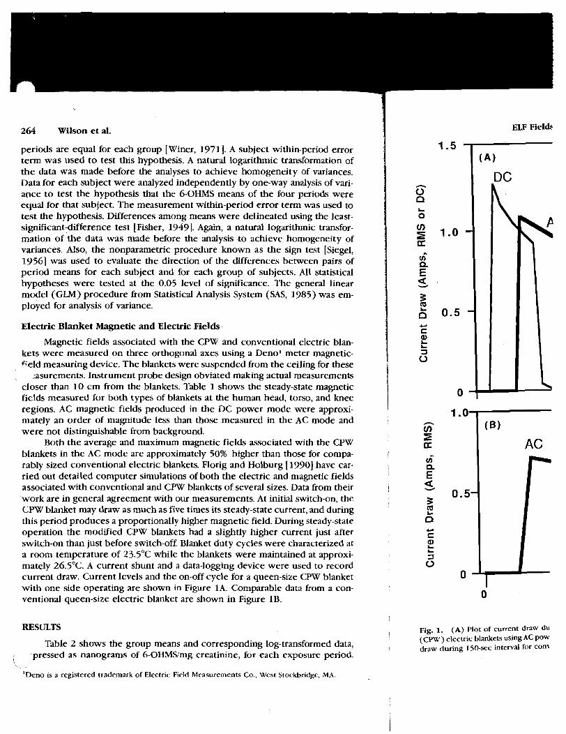
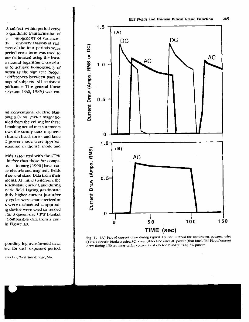
current draw. Current levels and the on-off cycle for a queen-size CPW blanket 0 with one side operating are shown in Figure 1A. Comparable data from a con- 1 ventional queen-size electric blanket are shown in Figure 1B. 0
A subject within-period error logarithmic transformation of ve - >mogeneity of variances. :Iy , one-way analysis of vari- zans of the four periods were period error term was used to ere delineated using the least- a natural logarithmic transfor- is to achieve homogeneity of nown as the sign test [Siegel, : differences between pairs of oup of subjects. All statistical pificance. The general linear s System (SAS, 1985) was em-
nd conventional electric blan- sing a Denol meter magnetic- lded firom the ceiling for these 1 making actual measurements ows the steady-state magnetic : human head, torso, and knee C power mode were approxi- leasured in the AC mode and
ields associated with the CPW h;-\er than those for compa- a. lolburg [ 19901 have car-
ne electric and magnetic fields )f several sizes. Data from their ments. At initial switch-on, the teady-state current, and during petic field. During steady-state @tly higher current just after :y cycles were characterized at s were maintained at approxi- lg device were used to record : for a queen-size CPW blanket . Comparable data from a con- in Figure 1B.
iponding log-transformed data, he, for each exposure period.
ents Co., West Stockbridge, MA.
ELF Fields and Human Pineal Gland Function 265
1.5
TIME (sec) Fig. 1. (A) Plot of current draw during typical 150-sec interval for continuous polymer wire (CPW) electric blankets using AC power (thick line) and DC power (thin line). (B) Plot of current draw during 150-sec interval for conventional electric blanket using AC power.
266 Wilson et al.
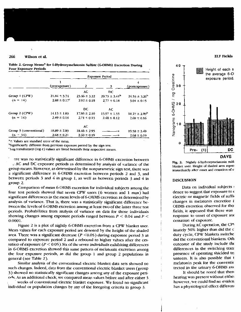
Table 2. Group Meansa for 6-Hydroxymelatonin Sulbte (6-OHMS) Excretion During Four Exposure Periods
Exposure Period
1 4 (preexposure) 2 3 (postexposure)
AC DC Group 1 (CPW) 21.84 2 3.74 23.46 2 3.22 20.73 & 3.41b 24.53 2 3.26b
( n = 14) 2 .8820 .17 2.92k0.18 2.7720.18 3.01 2 0.15
DC AC Group 2 (CFW) 14.1321.83 17.8622.10 13.97k1.55 1 8 . 2 7 2 . 8 9 ~
(n = 14) 2.49 2 0.14 2.71 C 0.13 2.48 k 0.12 2.69 * 0.16
AC Group 3 (conventional) 18.89 2 2.89 18.46 f 2.95 - 19.58 2 3.49
(n = 14) 2.68 2 0.21 2.60 k 0.19 - 2.68 2 0.19
"& Values are standard error of the mean. 'significantly different from previous exposure period by the sign test. 'Log-transformed (log e) values are listed beneath their respective means.
tre was no statistically significant difference in 6-OHMS excretion between , AC and DC exposure periods as determined by analysis of variance of the
group means. However, as determined by the nonparametric sign test, there was a significant difference in 6-OHMS excretion between periods 2 and 3, and between periods 3 and 4 in group 1, as well as between periods 3 and 4 in group 2.
Comparison of mean 6-OHMS excretion for individual subjects among the four test periods showed that seven CPW users ( 6 women and 1 man) had significant differences in the mean levels of 6-OHMS excretion as determined by analysis of variance. That is, there was a statistically significant difference be- [ tween the levels of 6-OHMS excretion among at least two of the latter three test periods. Probabilities from analysis of variance on data for those individuals I showing changes among exposure periods ranged between P < 0.04 and P < 1 0.0001. 1
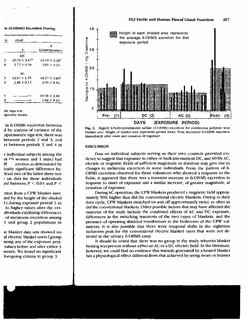
Figure 2 is a plot of nightly 6-OHMS excretion from a CPW blanket user. Mean values for each exposure period are denoted by the height of the shaded area. There was a significant decrease (P c0.05) during exposure period 3 as compared to exposure period 2 and a rebound to higher values after the ces- sation of exposure (P < 0.05). Six of the seven individuals exhibiting differences in 6-OHMS excretion showed this same pattern of melatonin excretion among the four exposure periods, as did the group 1 and group 2 populations in general (see Table 2).
Similar analysis of the conventional electric blanket data sets showed no such changes. Indeed, data from the conventional electric blanket users (group 3) showed no statistically significant changes among any of the exposure peri- ods. As an additional check, we compared mean values before and after either 3
weeks of conventional electric blanket exposure. We found no significant individual or population changes by any of the foregoing criteria in group 3
ELF Fields
Height of each s the average 6-O!
.- exposure period. ([I
2 30 0
-- - --
DAYS Fig. 2. Nightly 6-hydroxyrnelatonin sulf; blanket user. Height of shaded area repre: immediately after onset and cessation of e
DISCUSSION
Data on individual subjects : dence to suggest that exposure to t electric or magnetic fields of suffit changes in melatonin excretion i OHMS excretion observed for tho fields, it appeared that there was response to onset of exposure an< cessation of exposure.
During AC operation, the CPJ imately 50% higher than did the c duty cycle, CPW blankets switche did the conventional blankets. 0th outcome of the study include tht differences in the switching tran: presence of operating shielded tr; unteers. It is also possible that t melatonin peak for the conventic tected in the urinary 6-OHMS ass
It should be noted that then heating was present without eithe~ however, we could find no eviden has a physiological effect differenl
te (6-OHMS) Excretion During
,s eriod
the sign test. spective means.
in 6-OHMS excretion between d by analysis of variance of the nparametric sign test, there was between periods 2 and 3, and as between periods 3 and 4 in
r individud subjects among the rs (6 women and 1 man) had in rcretion as determined by ically sigmficant difference be- least two of the latter three test : on data for those individuals ;ed between P < 0.04 and P <
:tion from a CPW blanket user. ted by the height of the shaded 5) during exposure period 3 as to higher values after the ces-
ldividuals exhibiting differences of melatonin excretion among 1 and group 2 populations in
ic blanket data sets showed no a1 electric blanket users (group mong any of the exposure peri- values before and after either 3 msure. We found no significant foregoing criteria in group 3.
ELF Fields and Human Pineal Gland Function 267
Height of each shaded area represents the average 6-OHMS excretion for that
.- exposure period. .
$ 3 0
1 Pre- ( 1 ) 1 Dc (2) I AC (3) (post- (4) 1 DAYS (EXPOSURE PERIOD)
Fig. 2. Nightly 6-hydroxymelatonin sulfate (6-OHMS) excretion for continuous polymer wire blanket user. Height of shaded area represents period mean. Note increased 6-OHMS excretion immediately after onset and cessation of exposure.
DISCUSSION
Data on individual subjects serving as their own controls provided evi- dence to suggest that exposure to either or both intermittent DC, and 60-Hz AC, electric or magnetic fields of sufficient magnitude or duration may give rise to changes in melatonin excretion in some individuals. From the pattern of 6- OHMS excretion observed for those volunteers who showed a response to the fields, it appeared that there was a transient increase in 6-OHMS excretion in response to onset of exposure and a similar increase, of greater magnitude, at cessation of exposure.
During AC operation, the CPW blankets produced a magnetic field approx- imately 50% higher than did the conventional electric blankets. Owing to their duty cycle, CPW blankets switched on and off approximately twice as often as did the conventional blankets. Other possible factors that may have affected the outcome of the study include the combined effects of AC and DC exposure, differences in the switching transients of the two types of blankets, and the presence of operating shielded transformers in the bedrooms of the CPW vol- unteers. It is also possible that there were temporal shifts in the nighttime melatonin peak for the conventional electric blanket users that were not de- tected in the urinary 6-OHMS assay.
It should be noted that there was no group in the study wherein blanket heatingwas present without either an AC or a DC electric field. In the literature, however, we could find no evidence that warmth generated by a heated blanket has a physiological effect different from that achieved by using more or heavier
268 Wilson et al. ELF Fields
blankets. In addition, the conventional electric blanket users showed no changes in 6-OHMS levels, lending strength to the hypothesis that the electromagnetic fields associated with the CPW blankets, and not the heat that they generate, can affect human pineal function.
In further studies, it would be of interest to determine what, if any, phys- iological or genetic factors may be common to those individuals who exhibited change in 6-OHMS excretion as a consequence of electromagnetic field expo- sure. The report of McIntyre et al. [I9901 cited earlier illustrated large varia- tions in pineal gland sensitivity among individuals. Further work will be re- quired to determine more precisely those electromagnetic-field characteristics that may be responsible for the observed changes in 6-OHMS excretion for certain individuals in the study.
ACKNOWLEDGMENTS
This work was sponsored by the.Electric Power Research Institute under Contract RP-799-1 with Battelle, Pacific Northwest Laboratories.
LITERATLTRE CITED
.ndt, J. ( 1986) Assay of melatonin and its metabolites: Results in normal and usual environments. J. Neural Transm. (Suppl.) 21:11-35.
Blask, D.E. (1990) The emerging role of the pineal gland and melatonin in oncogenesis. In: Extremely Low Frequency Electromagnetic Fields: The Question of Cancer. B.W. Wilson, R.G. Stevens, and L.E. Anderson, eds. Battelle Press, Columbus, OH, pp. 319-335.
Bojkowski, CJ., J. Arendt (1988) Annual changes in 6-sulfatoxyrnelatonin excretion in man. Acta Endocrinol. (Copenh.) 117:470-476.
Buzzell, G.R., H.M. Arnerongen, J.G. Toma (1988) Melatonin and the growth of the Dunning R3327 rat prostatic adenocarcinoma. In: The Pineal Gland and Cancer. D. Gupta, A. Ananasio, and R.J. Reit er, eds. Brain Research Promotion, London, pp. 295-306.
Deguchi, T., J. Axelrod (1972) Control of circadian change-of serotonin N-acetyltransferase in the pineal organ by the P-adrenergic receptor. Proc. Natl. Acad. Sci. USA 69:2547-2550.
Florig, H.K, Holburg, J.F. (1990) Power-frequency magnetic fields from electric blankets. Health Phys. 58:493-502.
Fisher, R.A. (1949) The Design of Experiments. Oliver and Boyd Ltd., Edinburgh. Lerchl, A,, KO. Nonaka, KA. Stokken, RJ. Reiter (1990) Marked rapid alterations in nocturnal
pineal serotonin metabolism in mice and rats exposed to weak intermittent magnetic fields. Riochem. Biophy. Res. Commun. 169: 102-1 08.
Lewy, A.J., HA. Kern, N.F. Rosenthal, T.A. Wehr (1982) Bright artificial light suppresses melatonin secretion in humans. Science 210:1267-1269.
Lewy, AJ., R.L. Sack, LS. Miller, T.M. Boban (1987) Antidepressant and circadian phase-shifting effects of light. Science 235:352-354.
Mclntyre, I.M., T.R. Norman, G.B. Burrows, S.M. Armstrong (1990) Melatonin supersensitivity to dim light in seasonal affective disorder. Lancet 335:488.
Moore, R.Y., R. Heller, R.K Bhatnager, RJ. Wurtman, J. Axelrod (1968)Central control of the pineal gland: Visual pathways. Arch. Neurol. 18:208-218.
Pineal Gland and Cancer. D. Gupta, A London, pp. 22 1-232.
SAS (1985) SAS User's Guide: Statistics, Vel Savitz, D.A., E.E. Calle (1987) Leukemia :
Review and epidemiologic surveys.. Siegel, S. (1956) Non-Parametic Statistics f Stevens, R.G. (1987) Electric power use a
556-561. Troiani, M.E., S. Oaknin, RJ. Reiter, M.K. Vz
activity and melatonin content p r d dependent. J. Pineal Res 4.185-195
Vakkuri, 0.. J. Leppaluoto, 0. Vuolteenaht radioimmunoassay using radioidin 157.
Welker, HA., P. Semm, RP. Willig, J.C. COI artificial magnetic field on serotonir the rat pineal gland. Exp. Brain Res.
Wetterberg, L. (1978) Melatonin in huma (Suppl.) 13:289-310.
Wetterberg, L (1979) Clinical importance P. Paret, eds. Elsevier/North Hollan~
Wever, R. (1968) Einfluss schawcher elec Menschen. Naturwissenschaften 55:
Wilson, B.W., E.K Chess, L.E. Anderson ( rhythms: Time course of onset and
Wilson, B.W., R.G. Stevens, LE. Anderson ( netic field exposure: Possible role (
Wilson, B.W., L.E. Anderson, D.I. Hilton, electric fields: Effects on pineal fun
1 Wilson,-B.W., L.E. Anderson, D.1. Hilton, R fields: Effects on pineal function in
i Winer, BJ. ( 1971 ) Statistical Principles in Wurtman, R.J., J. Axelrd, LS Phillips ( 191
light. Science 142:1071-1073. Zisapel, N., M. Laudon, I. Nir ( 1988) Mela
aged male rats: Age-associated decn J. Physiol. Sci. 4392-393.
Olcese, J., S. Keuss, P. Semm (1988) Geomagnetic field detection in rodents. Life Sci. 42:605-613. Reiter, RJ., L.E. Anderson, R.L. Buschbom, B.W. Wilson (1988) Reduction of the nocturnal rise in
pineal melatonin levels in rats exposed to 60-Hz electric fields in utero and for 23 days after birth. Life Sci. 42:2203-2206.
chez Barcelo, E.J., S. Coscorral, M.D. Med~avilla (1988) Influence ofpineal gland function on the initiation and growth of hormone-dependent breast tumors: Possible mechanisms. In: The
ket users showed no changes fsis that the electromagnetic e ' . that they generate, can
determine what, if any, phys- ~se individuals who exhibited ' electromagnetic field expo- :arlier illustrated large varia- Is. Further work will be re- nagnetic-field characteristics es in 6-OHMS excretion for
wer Research Institute under ;t Laboratories.
s in normal and usual environments.
and melatonin in oncogenesis. In: : Question of Cancer. B.W. Wilson. :urnbus, OH, pp. 319-335. rymelatonin excretion in man. Acta
d the growth of the Dunning R3327 C, r. D. Gupta, A. Attanasio, and 2. -06. serotonin N-acetyltransferase in the \cad. Sci. USA 69:2547-2550. ields from electric blankets. Health
>yd Ltd., Edinburgh. rked rapid alterations in nocturnal > weak intermittent magnetic fields.
artificial light suppresses melatonin
vssant and circadian phase-shifting
990) Melatonin supersensitivity to 1. ( 1968) Central control of the pineal
on in rodents. Life Sci. 42605-613. ) Reduction of the nocturnal rise in fields in utero and for 23 days after
ence of pineal gland function on the nors: Possible mechanisms. In: The
ELF Fields and Human Pineal Gland Function 269
Pineal Gland and Cancer. D. Gupta, A. Attanasio. R. J. Reiter, eds. Brain Research Promotion, London, pp. 221-232.
SAS (1985) SAS User's Guide: Statistics, Version 5. SAS Institute Inc., Cary, North Carolina. Savitz, D.A., E.E. Calle (1987) Leukemia and occupational exposure to electromagnetic fields:
Review and epidemiologic surveys. J. Occup. Med. 29:47-51. Siegel, S. ( 1956) Non-Parametic Statistics for Behavioral Science. McGraw-Hill, New York. Stevens, R.G. (1987) Electric power use and breast cancer: A hypothesis. Am. J. Epidemiol 125:
556-561. Troiani, M.E., S. Oaknin, RJ. Reiter, M.K Vaughan, B.L. Cozzi (1987) Depression in rat pineal NAT
activity and melatonin content produced by hind leg saline injection is time and darkness dependent. J. Pineal Res. 4:185-195.
Vakkuri, O., J. Leppaluoto, 0 . Vuolteenaho (1984) Development and validation of a melatonin radioimmunoassay using radioiodinated melatonin as a tracer. Acta Endocrinol. 106:152- 157.
Welker, H.A., P. Semm, R.P. Willig, J.C. Commentz, W. Wiltschko, L. Vollrath (1983) Effects of an artificial magnetic field on serotonin N-acetyl transferase activity and melatonin content of the rat pineal gland. Exp. Brain Res. 50:426-432.
Wetterberg, L. (1978) Melatonin in human physiological and clinical studies. J. Neural Transm. (Suppl.) 13:289-310.
Wetterberg, L. (1975)) Clinical importance of melatonin. In: Progress in Brain Research. J. Kapper, P. Paret, eds. ElseviedNorth Holland, New York, Vol. 52, pp. 539-547.
Wever, R. (1968) Einfluss schawcher electromagnetischer Felder auf die circadiane Periodik des Menschen. Naturwissenschaften 55:29-32.
Wilson, B.W., E.K Chess, LE. Anderson (1986) 60-Hz electric field effects on pineal melatonin rhythms: Time course of onset and recovery. Bioelectromagnetics 7:239-242.
Wilson, B.W., RG. Stevens, LE. Anderson (1989) Neuroendocrine-mediated effects of electromag- netic field exposure: Possible role of the pineal gland. Life Sci. 45:1319-1332.
Wilson, B.W., L.E. Anderson, D.I. Hilton, RD. Phillips R.D. (1981) Chronic exposure to 60-Hz electric fields: Effects on pineal function in the rat. Bioelectromagnetics 2371-380.
Wilson,-B.W., L.E. Anderson, D.1. Hilton, R.D. Phillips (1983) Chronic exposure t o 60-Hz electric fields: Effects on pineal function in the rat (erratum). Bioelectromagnetics 4:293.
Winer, B.J. (1971) Statistical Principles in Experimental Design, 2d Ed. McGraw-Hill, New York Wurtman, R.J., J. Axelrod, LS. Phillips (1963) Melatonin synthesis in rat's pineal gland: Control by
light. Science 142:1071-1073. Zisapel, N., M. Laudon, I. Nir (1988) Melatonin receptors in discrete brain regions of mature and
aged male rats: Age-associated decrease in receptor density and circadian rhythmicity. Chin. J. Physiol. Sci. 4392-393.
/Pineal Res 1995:IR-1-11 Printed in the United Stares--all rights reserved
Copyright O Munksgoord. 1995 ------- Journal of Pineal Research
ISSN 0742-3OYH
A review of the evidence supporting melatonin's role as an antioxidant 23
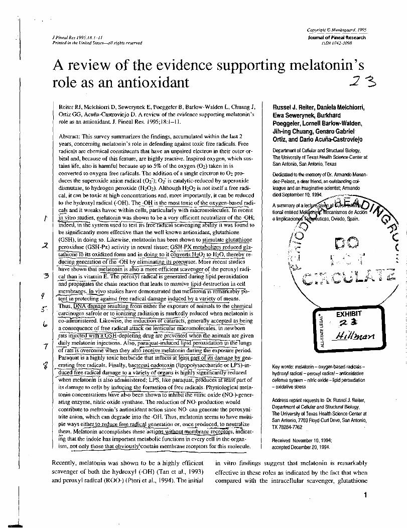
Reiter RJ, Melchiorri D, Sewerynek E, Poeggeler B, Barlow-Walden L, Chuang I, Ortiz GG, Acuiia-Castroviejo D. A review of the evidence supporting melatonin's role as an antioxidant. J. Pineal Res. 1995; 18: 1-1 1.
Abstract: This survey summarizes the findings, accumulated within the last 2 years, concerning melatonin's role in defending against toxic free radicals. Free radicals are chemical constituents that have an unpaired electron in their outer or- bital and, because of this feature, are highly reactive. Inspired oxygen, which sus- tains life, also is harmful because up to 5% of the oxygen (02) taken in is converted to oxygen-free radicals. The addition of a single electron to 0 2 pro- duces the superoxide anion radical ( 0 2 7 ) ; Of is catalytic-reduced by superoxide dismutase, to hydrogen peroxide (H202). Although Hz02 is not itself a free radi- cal, it can be toxic at high concentrations and, more importantly, it can be reduced - to the hydroxyl radical (.OH). The .OH is the most toxic of the oxygen-based radi- cals and it wreaks havoc within cells, particularly with macromolecules; In rec2nt j;-?itro studies, e a t o n i n was shown to be a very efficient neutralizer of the .OH; indeed, in thd system used to test its free radical scavenging ability it was found to be significantly more effective than the well known antioxidant, glutathione (GSH), in doing so. Likewise,.melatonin has been shown to timulate glutathione peroxidase (GSH-Px) activity in neural tissue; GSH-PX metabolizes reduced glu- tathione td its oxidized form and In doing so it&=~~02 to H20, thereby re- -f the .OH by eliminating its precu_rsor. ~ b r e ¢ studies --
*h-t melatonin is ;-re efficient scavenger of the peroxyl radi- cal than is vitamin E. The peroxyl radical is generated during lipid peroxidation
the chain reaction that leads to massive $id destruction in cell membranzs. In v p o studies have demonstrated that mei'atonin is remarkably go-
<ent in proteciing against free radical damagelsuced by a variety of means. I%-?ulting from eitherxe exposure of animals to the chhical carcinoiin saf&e or t o , i o m g radiatipn is markedly reduced when melatonin is -d. ~ i k e z < e , the induction of cataraz, ienerally accepted a .---- a consequenceof free radical attack on lenticular macromolecules, in newborn
'C --------/---
I% .. -m~-de~ l e t i n~ drug are prevented w h e n i e animals are given daily melatonin injections. Also, pzquat-ihduced lipid peroxidation in the lungs - of ratsXovercomahen they alsof receive melatonin during the exposure period.
-- ---- ----.--. Paraquat is a highly toxic herbicide that infl~cts at, l - p t p m d its darn&-en- erating free radicals. Finally, bacterial endotoxin (lipopolysaccharide or LPS)-in- - d u c e d e e radical damage to a &iety of organ: is hifisignificantly%~uced /-- 4__LI __--.- when melatonin is also administered; LPS, ~ i k e ' ~ a r a ~ u a ~ u c e s at leasf part of its damage to cells by inducing the formation of free radicals. Physiological mela- tonin concentrations hhve also been shown to inhibit the n~tric oxide (NO.)-gener- ating enzyme, nitric oxide synthase. The reduction of NO. production would contribute to melatonin's antioxidant action since NO. can generate the peroxyni- trite anion, which can degrade into the -OH. Thus, melatonin seems to have multi- ple ways either to reduce free radic@ generation or, oncsroduced, to neutralize them. Melatonin accomplishes these.actio-withr~ut-icat- Gg that the indole has important metabolic functions in ev&y cell in the organ- ism, not only those that obviouskkontain membrane receptors for this molecule.
Russel J. Reiter, Oaniela Melchiorri, Ewa Sewerynek, Burkhard Poeggeler, Lornell Barlow-Walden, Jih-ing Chuang, Genaro Gabriel Ortiz, and Dario Acutia-Castroviejo
Department of Cellular and Structural Biology, The University of Texas Health Science Center at San Antonio, San Antonio, Texas
Dedicated to the memory of Dr. Armando Menen- dezPelaez, a dear friend, an outstanding col- leaaue and an imaainative scientist: Armando died September 10,
" ,.._ . .. 7 .; . -"
Key words: melatmin - oxygen-based radicals - hydroxyl radical - peroxyl radical - antioxidative defense system - nitric oxide - lipid peroxidation - oxidative stress
Address reprint requests to Dr. Russel J. Reiter, Department of Cellular and Structural Biology. The University of Texas Health Science Center at San Antonio, 7703 Floyd Curl Drive, San Antonio, TX 78284-7?62
Received November 10,1994; accepted December 20,1994.
Recently, melatonin was shown to be a highly efficient in vitro findings suggest that melatonin is remarkably
scavenger of both the hydroxyl (.OH) (Tan et al., 1993) effective in these roles as indicated by the fact that when and peroxyl radical (ROO-) (Pieri e t al., 1994). The initial compared with the intracellular scavenger, glutathione
Reiter et al.
(GSH), melatonin proved five times better in neutralizing - - the .OH and, when compared to vitamin E, melatonin was
effective in inactivating the-0.. GSH (Meister, '1992) and vitamin E (Packer, 1994) are considered to be premier antioxidants within the cell. Besides these direct antioxidative actions of melatonin, there are indirect ef- fects as well. Thus, melatonin .stjmulates klgathione per- ,y----
oxidase (GSH-Px) a& (~arlow-Waldenet al., 1955) r- and inhibits nitric oxide synthase (NOS) (Pozo et al., 1994). GSH-PX is an important antioxidative enzyme because it metabolizes hydroperoxides including hydro- gen peroxide (H202), thereby reducing the formation of the highly toxic .OH (Liochev and Fridovich, 1994). By inhib- iting NOS, melatonin reduces the formation of the free radical nitric oxide (NO.) (Palmer et al., 1988). Although N O performs a variety of important functions in organ- isms (Moncada and Higgs, 1993), it also interacts with other radicals to produce the toxic peroxynitrite anion (ONOO-), which can generate reactive oxygen-based radi- cals by way of its interaction with the superoxide anion radical (027) (Beckman, 1991; Radi et al., 1991).
The purpose of this brief review is to summarize the newly discovered intracellular functions of melatonin that relate to free radical generation. Other reviews discuss the
,potential implications of these new findings for aging (Reiter, 1994a; Reiter et al., 1994a) and age-related dis- eases (Poeggeler et al., 1993; Reiter et al., 1993, 1994b, 1994~).
Free radical generation and antioxidative defense mechanisms
, A free radical is an atom or a molecule that contains an unpaired electron. Usually, electrons associated with at- oms or molecules are paired; pairing of electrons makes molecules relatively stable and unreactive. Conversely, the loss of or addition of an electron leaves the atom or
-nstable and, relatively more highly reactive than %s non-radical gunterpart. The chemical reactivity of ftee - '~radE& varies w T T h e simplest free radical is the
hydrogen radical (which is identical to the hydrogen atom); it contains a single proton and one unpaired elec- tron. Removal of a hydr%en radical (or alom) from 9 p o l y u ~ a c i d ( ~ ~ ~ ~ ) in a cell m e m b ~ s b y
/=B30ng - reducing agenzan initiate radical chain reactions Qch as in lipid perzdation) __ _. _ (Kanner el al., 1987). which are highly destructwe to cellular mqrphology and function.
Although there are a variety o f e d i c a l s produced in organisms, those that are by~roducts of - molecular oxy- gen (dioxygen or 02) have received a great deal of inves- tigative interest and they exkrt esensive damage, particularly over tims(Harman, 1994). Althoughestimates vary somewhat, it is believed that up to 5% of the 0 2 taken in by organisms may eventually end up as damaging
fez+ e- SOD d3+
0 2 ---4 02' ---4 Hz02 - ---------+ OH
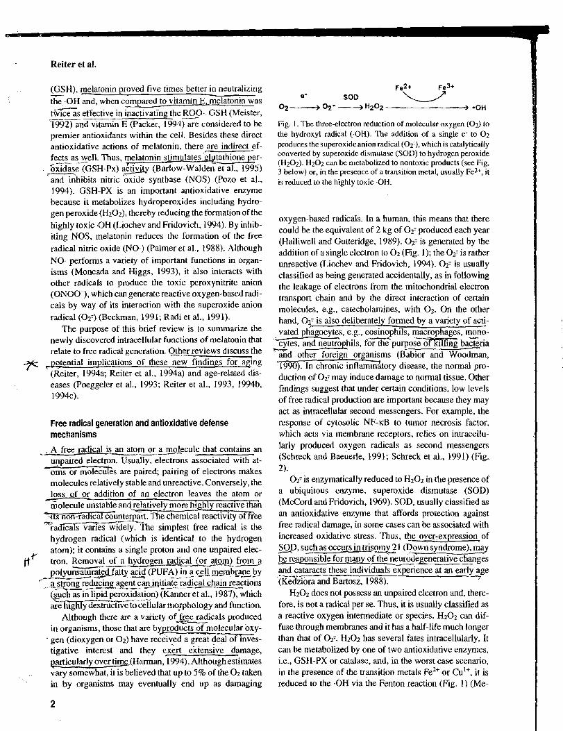
Fig. 1. The three-electron reduction of molecular oxygen ( 0 2 ) to the hydroxyl radical (.OH). The addition of a single e- to 0 2
produces the superoxideanion radical ( 0 2 3 , which is catalytically converted by superoxide disrnutase (SOD) to hydrogen peroxide (H202). H202 can be metabolized to nontoxic products (see Fig. 3 below) or, in the presence of a transition metal, usually Fez+, it is reduced to the highly toxic .OH.
oxygen-based radicals. In a human, this means that there could be the equivalent of 2 kg of 0 2 7 produced each year (Halliwell and Gutteridge, 1989). 02: is generated by the addition of a single electron to 0 2 (Fig. 1); the 0 2 7 is rather unreactive (Liochev and Fridovich, 1994). 0 2 7 is usually classified as being generated accidentally, as in following the leakage of electrons from the mitochondria1 electron transport chain and by the direct interaction of certain molecules, e.g., catecholamines, with 0 2 . On the other hand, 027 is also deliberately formed by a variety of acti- vated phagocy tes, e.g., eosinophils, macrophages, mond-
T t e s , a n n h i l s , f ~ ~ ~ u r ~ o s ~ b a z r i a t- -
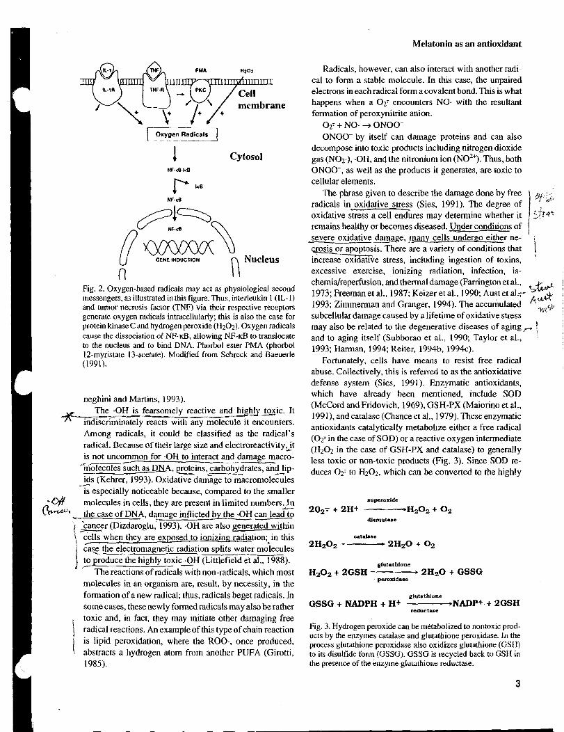
and other foreign organisms - (Babior and Woodman, '1990). In chronic inflammhory disease, the normal pro- duction of 0 2 7 may induce damage to normal tissue. Other findings suggest that under certain conditions, low levels of free radical production are important because they may act as intracellular second messengers. For example, the response of cytosolic NF-KB to tumor necrosis factor, which acts via membrane receptors, relies on intracellu- larly produced oxygen radicals as second messengers (Schreck and Baeuerle, 1991; Schreck et al., 1991) (Fig. 2).
O2'is enzymatically reduced to H202 in the presence of a ubiquitous enzyme, superoxide dismutase (SOD) (McCord and Fridovich, 1969). SOD, usually classified as an antioxidative enzyme that affords protection against free radical damage, in some cases can be associated with increased oxidative stress. Thus, the over-expression of SOD, such as occurs in t r i s p ~ ~ , 2 1 (Down syndrome), may - be responsible for many of the neur&egenerative changes and cataracts these i n d i ~ i x ~ r i e n c e at an early age (Kedziora and Bartosz, 1988). 7
H202 does not possess an unpaired electron and, there- fore, is not a radical per se. Thus, it is usually classifled as a reactive oxygen intermediate or species. H202 can dif- fuse through membranes and it has a half-life much longer than that of 027. Hz02 has several fates intracellularly. It can be metabolized by one of two antioxidative enzymes, i.e., GSH-PX or catalase, and, in the worst case scenario, in the presence of the transition metals ~ e ~ + or Cul+, it is reduced to the .OH via the Fenton reaction (Fig. 1) (Me-
1 Cytosol NF.&.l.%
Fig. 2. Oxygen-based radicals may act as physiological second messengers, as illustrated in this figure. Thus, interleukin 1 (IL- 1) and tumor necrosis factor (TNF) via their respective recepton generate oxygen radicals intracellularly; this is also the case for protein kinase C and hydrogen peroxide (H202). Oxygen radicals cause the dissociation of NPKB, allowing NF-KB to translocate to the nucleus and to bind DNA. Phorbol ester PMA (phorbol 12-myristate 13-acetate). Modified from Schreck and Baeuerle (1991).
Melatonin as an antioxidant
Radicals, however, can also interact with another radl- cal to form a stable molecule. In this case, the unpaired electrons in each radical fonn a covalent bond. This is what happens when a 0 2 7 encounters NO- with the resultant formation of peroxynitrite anion.
0 2 ' + NO- -3 ONOO- ONOO- by itself can damage proteins and can also
decompose into toxic products including nitrogen dioxide gas (NO2.), -OH, and the nitronium ion NO^+). Thus, both ONOO-, as well as the products it generates, are toxic to cellular elements.
The phrase given to describe the damage done by free radicals in oxidative stress (Sies, 1991). The degree of oxidative stress a cell endures may determine whether it remains healthy or becomes diseased. Under conditions of
i severe oxidative da2age, many cells under~o either ne- ,
osis or apoptasis. There are a variety of conditions that 1 h v e stress, including ingestion of toxins, excessive exercise, ionizing radiation, infection, is- chemidreperfusion, and thermal damage (Farrington et al., 1973; Freeman et al., 1987; Keizer et al., 1990; Aust et al.7 s w
'&'&& 1993; Zimmerman and Granger, 1994). The accumulated subcellular damage caused by a lifetime of oxidative stress
wGb
may also be related to the degenerative diseases of aging , 1 and to aging itself (Subborao et al., 1990; Taylor e t al., 1993; Harman, 1994; Reiter, 1994b, 1994~).
Fortunately, cells have means to resist free radical abuse. Collectively, this is referred to as the antioxidative defense system (Sies, 199 1). Enzymatic antioxidants, which have already been mentioned, include SOD neghini and Martins, 1993). (McCord and Fridovich, 1969), GSH-PX (Maiorino et al.,
-1 y reactive a n d h i g h _ l y ~ i c . It 199 I), and catalase (Chance et al., 1979). These enzymatic
indiscriminately reacts with any molecule it encounters. antioxidants catalytically metabol~ze either a free radical
Among radicals, it could be classified as the radical's (02T in the case of SOD) or a reactive oxygen intermediate radical. Because of their large size and electr~reactivity~lt (H202 in the case of GSH-PX and catalase) to is not unco3mon for -OH interact and damage macro- less toxic or non-toxic products (Fig. 3). Since SOD re-
'holecules such as DXA, =ins, carbohydrates, and lip- duces 02: to ~ ~ 0 ~ , which can be converted to the highly ids (Kehrer, 1993). Oxidative da&age to m~cromol&iles
A
is especially noticeable because, compared to the smaller -L)Y molecules in cells, they are present in limited n u m b e r s 2 h* the case of DNA, damage inflicted by the O H San lead to
'cancer ( ~ i z d a r o ~ l u , ' 1993). .OH are also ~ m t h i n \ % whep they are exposed to ionizing radiat~on; in this i cas: the electromagnetic radiation splits water molecules ' to produce the highly toxic -OH (Littlefield et al., 1988). 1 F _-
The reactions of radicals with non-radicals, which most molecules in an organism are, result, by necessity, in the formation of a new radical; thus, radicals beget radicals. In some cases, these newly formed radicals may also be rather
I toxic and, in fact, they may lnrtiate other damaging free radical reactions. An example of this type of chain reaction is lipid peroxidatlon, where the ROO-, once produced,
. abstracts a hydrogen atom from another PUFA (Girotti, 1985).
glutathione
Hz02 + 2GSH 2 H 2 0 + GSSG
glutathione GSSG + NADPH + H+ -NADP+ + 2GSH
reductrse
Fig. 3. Hydrogen peroxide can be metabolized to nontoxic prod- ucts by the enzymes catalase and glutathione peroxidase. In the process glutathione peroxidase also oxidizes glutathione (GSH) to its disulfide form (GSSG). GSSG is recycled back to GSH in the presence of the enzyme glutathione reductase.
Reiter et al.
toxic -OH, it is important that the antioxidative enzymes GSH-PX and catalase, both of which metabolize H202. work in concert with SOD (Chance et al., 1979).
In the process of the conversion of H202 to water by GSH-PX, the tripeptide GSH is converted to its disulfide oxidized form, GSSG (Fig. 3). GSH is an important anti- oxidant itself. It is found in millimolar concentrations within cells and it has important roles in xenobiotic meta- bolism and leukotriene synthesis (Chance et al., 1979). GSH-PX, which removes H202, is a selenium containing molecule; a related enzyme removes lipid hydroperoxides. which are formed during lipid peroxidation, from cellular membranes (Maiorino et al., 1991 ).
As shown in Figure 1, the reduction of H202 to .OH requires a transition metal, usually ~ e ~ + but occasionally Cul+. Because of this, it is important that these metals are not in the free state in cells and any' molecule that binds them and renders them incapable of interacting with H202 is classified as part of the antioxidative defense system. A common storage-form of iron in serum is transferrin (Win- terboum and Sutton, 1984), whereas-co~mr is often se-
- - --- questered by ceru loqmin (Goldstein et al., 1979). In these forms, the transition metals cannot promote free radical reactions. Besides those mentioned here, there are a wide variety of other antioxidative enzymes, free radial scavengers, and transition metal binders that contribute to the total antioxidant capacity of the organism.
The role of melatonin in the antioxidative defense system
For the last decade, some reports related to the actions of melatonin on metabolic processes have been considered inconsistent with the rather limited distribution of mem- brane receptors in cells (Reiter, 1991). It seemed likely that
&+&, certain actions of melatonin, e.g., those related to the re ulation of reproduction (Reiter, 1980) and those con- jj LY*; ;->-------- ---.- -------. cemed with circadian regulation (Armstrong, 1989), will prove to be mediated by membrane receptors on specific cells related to these functions (Vanecek et al., 1987; Morgan and Williams, 1989). However, the existence of melatonin in unicellular organisms (Poeggeler et al., 1991), as well as its widespread actions, described else- where (Reiter, 199 I), in multicellular organisms !gd-usto speculate that melatonin performed functions within cells that did not require an interaction with a receptor, particu- larly not a receptor located on the limiting membrane of the cell. ~ u r t h e i o r e , the high li&olubility of the indole a!- it ready acce&- to the 9 o ~ o f .all cells, also .- indicating that the melatonin's actions may not be limited to actions at the cell membrane level. Interestingly, the recent demonstration that melatonin is also quite soluble in aqueous media is consistent with the intracellular ac- tions of melatonin (Shida et al., 1994). Finally, the recent finding that melatonin-intracellularly may b e r a t h e r high - concentrations i? the nu$ei (Mennenga et al., 199 1; Me- -
nendez-Pelaez and Reiter, 1993; Menendez-Pelaez et al., 1993) and that there may be specific binding sites for melatonin associated with nucleoproteins (Acuiia Castroviejo et al., 1993, 1994), suggest the possibility that melatonin may function like some other hormones, e.g., steroid and thyroid hormones, on molecular events in the nuclei of cells.
The initial studies from which we deduced that mela- tonin may alter the !edox state of the cell were those of Chen et al. (1993). In this investigation ca2+-stimulated + ~ ~ ~ + - d e ~ e n d e n t ATPase ( ~ a ~ + - ~ u r n p ) activity in the heart was found to be influenced by the pineal gland and mela- tonin. Initially, a daylnight difference in ca2'-pump activ- ity was noted with highest levels at night. When animals were pinealectomized, the nighttime rise in the activity of the pump did not occur, so it was assumed that the rise was probably mediated by melatonin. When cardiomyocyte membranes were in fact incubated with melatonin, ca2+- ATPase activity increased in a dose-dependent manner (Chen et al., 1993). Since the activity of this enzyme is normally depressed in a high free radical atmosphere (Kaneko et al., 1989), wespeculated that melatonin altered the redox state of the cell by neutralizing toxic free radi- cals, which then allowed ca2+-pump activity to rise pas- sively. This idea is also supported by more recent studies wherein rats were treated with alloxan, which is known to generate free radials. This treatment significantly reduced ~ a ~ + - ~ u m ~ activity, which was again reversed by concur- rent melatonin treatment (Chen et al., 1994). Although the evidence is indirect, both studies indicated a potential involvement of melatonin with the oxidative status of cardiac cells.
These initial studies were followed by a series of inves- tigations that were designed to specifically examine the ability of melatonin to function as a free radical scavenger and antioxidant. Of specific interest was the interaction of melatonin with the highly toxic -OH. To check this, we developed a simple in vitro system in which H202 was exposed to 254 nm ultraviolet light to generate the .OH (Tan et al., 1993a). However, because of their extremely short half-life (1 x sec at 37OC), .OH are difficult to measure directly. To overcome this, a spin trapping agent, 53-dimethylpyrroline N-oxide, or DMPO, was added to the mixture. DMPO forms an adduct with the -OH and, since the adducts have a much longer half-life, they can be quantitated as an index of .OH generation. The adducts (DMPO--OH) were qualitatively and quantitatively evalu- ated using both high pressure liquid chromatography with electrochemical detection and electron spin resonance spectroscopy (Tan et al., 1993a). By also adding melatonin (or other known scavengers) to the mixture, it was possible to estimate the -OH scavenging capacity of the compounds of interest. In this system, melatonin proved to be very significantly more efficient than either GSH or mannitol
Melatonin as an antioxidant
TABLE 1. Concentration of various constituents required to scavenge 50% (ICs) of the .OH generated in vitm following the exposure of Hf12 to ultraviolet lghl
Scavenger -- ~CSO
Melatonin (N-acefyl-5-methoxytryptamine) 21pM
Reduced glutathione 123pM
Mannitol -- 183pM
in scavenging the -OH (Table 1). This finding generated considerable interest because both GSH and mannitol are very effective intracellular free radical scavengers, sug- gesting that melatonin may well have a physiologically significant role as an antioxidant. More importantly, of all the radicals produced in the organism, the .OH is consid- ered the most toxic; thus, any compound that neutralizes this radical could play an important role in the antioxida- tive defense system.
The free radical scavenging capacity of melatonin may extend to other radicals as well. A year following our reported demonstration of melatonin as a neutralizer of the .OH (Tan et al., 1993a), Pieri and colleagues (1994) claimed that the indole exhibits a similar action in refer- ence to the peroxyl radical (ROO.). Using a well estab- lished in vitro system for evaluating the radical scavenging capachy of a compound (Cao et al., 1993), Pieri et al. (1994) claimed that melatonin was better than vitamin E in scavenging the ROO-, which is a consequence of lipid
I peroxidation (Table 2). Clearly, in this system melatonin was twice as effective as vitamin E, a well known and important chain-breaking antioxidant (Packer, 1994). in halting llpid peroxidation. Thus, melatonin would be ex- pected to be highly effective against lipid peroxidation in
I vivo for several reasons: 1) melatonin is highly lipophilic and should, therefore, normally be found in rather high
I concentrations in cellular membranes; 2) melatonin, like I
I vitamin E, is an effective chain breaking antioxidant and, thus, it would reduce oxidation of lipids; and 3) melatonin,
I by virtue of its ability to scavenge the .OH, would also I reduce the initiation of lipid peroxidation. The .OH is one I
of the radicals that is sufficiently toxic to abstract a hydro- , gen atom, i.e., initiate lipid peroxidation, from a PUFA I (Niki et al., 1993).
I The demonstration that melatonin affords protection against oxidative stress in vivo followed soon after the in vitro studies indicating?hat melatonin is a potent scaven- ger of both the -OH (Tan et al., 1993a) and ROO. (Pieri et al., 1994). In reference to oxidative damage to nuclear DNA, Tan and co-workers (1 99313,1994) in a series of two reports found that-hepatic DNA damage inflicted by sa- frole, a chemlcal carcinogen, Gas highly significantly re- - duced when the rats also r e c c d y melatoijn.%&ole e ,- damages DNA at least in part because i
TABLE 2. Peroxyl radical (ROO ) scavenging capacity, as measured in oxygen rad~cal absorbing capacity (ORAC) units, of the four compounds indicateda
Scavenger - ORACperoqi Melatonin (N-acetyl-5-methoxylryptamne) 2.04
Vitamin C (ascorbate) 1.12
Trolox (water soluble vitamin E analogue) 1 .OO
Reduced glutathione 0.68
aThe findings suggest hat, of the four ROO- scavengers checked, melatonin is the most efficient.
production of toxic-free radicals (Boberg et al., 1983). w-.
' perhaps the most remarkable feature of melatonin's pro- tection against safrole-induced DNA damage was thAit was effective at verv low concentrations relative to the . . --- v e ~ ~ i s t e r e d T ; ' T h u s , even when the amount of melatonin administered was 1,000-fold lower than the dose of safrole, most of the DNA damage was prevented. Furthermore, when safrole was given either during the day or at night, in the latter case DNA damage was less. The implication of this obse-rvaiion i_s that - even the nighttims -d n s e i n o g e n o u s melatonin is -+
.- - sufficient to provide protection against oxygen toxicity %iting from xenobiotic administration (Tan et al., 1994). - -
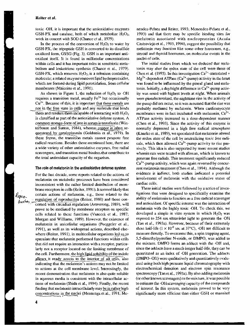
The protective effect of melatonin against oxygen radi- cal damage to DNA was also observed in another model system (Vijayalaxmi et al., 1995). In this case, we incu- bated human lymphocytes and subjected them to 150 cGy
- ionizing radiation with and w i t E t concurrent treatment -the cells with then
c ~ e n e t i c a l l y evaluated by an investigator who was un- aware of the experimental design of the study. Melaton& in a dose-response manner, significantly reduced the num- p r y ber of micronuclei, thi number of cells with exchange
%errations (both of which are indices of genomlc dam- v - -. - ---_---.-. _-__-
age), and the total number of cell with any type of ~ P O - 7 iiiosomal damage (Fig. 4). At a concentration of 2 mM /7 0 6 melatonin reduced ionizing radiation-induced da%e by ;,,$' ' ~ ~ Y 1 s ~ l f o x i d e (DMSO), a known ra- /B;;i dioprotective agent (Littlefield et al., 1988), to provide a similar level of DNA protection adose of 1 M was required (Fig. 4) (Vijayalaxmi et al., 1995). Thus, in this system melatonin seemed to be on the order of 500 times more effective than DMSO as a radioprotecto Free radicals +
induced by ionizing radiation3re the causative fact01 in damage to the genomic material (Okada et al., 1983).
Melatonin as a general protector against ionizing radia- tion is certainly also suggested by the observations of linke en staff and co-workers (1994). This group found that almost 50% of mice treated with melatonin prior to expo- sure to 950 cGy ionizing radiation survived at least 30 days, whereas within the same time frame all irradiated
Reiter et al.
Total Number of Cells with Chromosomal Damage 50 100 150 200 250 300
Mel(0.5mM)
Me1 (0.5mM) + 150 cGy 37.6%
Met (l.OmM)
Mel (1.OmM) + 150 cGy 51.5%
Met (2.0mM)
Mel(2.0mM) + 150 cGy 69.1%
OMS0 (1 .OM)
OMS0 (1 .OM) + 150 cGy 73.036
Fig.4. Percentage reductionofthenumber of human lymphocytes exhibiting chromosomal damage after their exposure to 150 cGy ionizing radiation. At aconcentration of 2.0 mM in the incubation mediu;, melatonin reduced the percentage of damaged cells by 69.1%. For the known radio~rotectordimethvlsu~foxide (DMSO) to reduce chromosomal daAage by roughly ;he same percentage (73%), its concentration had to be 1 M. Modified from Vijay- alaxmi et al. (1994).
mice that did not receive melatonin died. The protection of macromolecules from oxidative
stress by melatonin is not restricted to nuclear DNA. In a study where oxidative damage to the lens of the eye was assessed, we found that melatonin also provided signifi- cant protection against lenticular degeneration (Abe et al., 1994). Cataractogenesis is known to be a free radical-me- diated condition where the lens becomes cloudy following oxidative attack on lenticular protein and other macro- molecules (Spector, 1991). One of the major antioxidative defense constituents in the lens is GSH (Pau et al., 1990). One model in which to investigate the importance of GSH in protecting the lens from oxygen radical-based cataracts is to inject newborn rats with a drug (buthionine sulfoxi- mine or BSO) that depletes the organisms of this key antioxidant; BSO acts by inhibiting y-glutamylcysteine synthaqe, which regulates GSH formation (Martensson et al., 1989; Meister, 1992). When BSO is given shortly after birth, rats typically have cataracts at the time their eyes open (around 2 weeks of age). Interestingly, the pineal gland of newborn rats also produces only small amounts of melatonin during the first 2 weeks of life (Reiter, 1991). Thus in reality, following BSO administration, the new- born animals are really deficient in two important antioxi- dants, i.e., GSH and melatonin.
Considering this, we treated BSO-injected (to deplete their GSH levels) newborn rats with melatonin for the first 2 weeks of life to determine if the indole would alter cataractogenesis (Abe et al., 1994). The animals receiving BSO only exhibited the usual high incidence of cataracts, whereas those treated with BSO and melatonin had a very
low incidence of cataracts (Table 3). In these animals, BSO had indeed highly significantly reduced lenticular GSH levels whether or not they had been given melatonin. The clear implication is that melatonin was the active agent in reducing oxidative damage and suppressing cataract for- mation. Furthermore, although the evidence is obviously indirect it seems likely melatonin was effective in this model system because it reduced oxidative damage to protein (Spector, 1991).
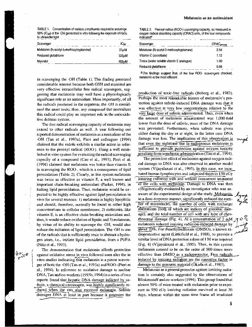
There is, of course, a great deal of interest in lipid peroxidation because it is devastating to cell membranes and it either disrupts the functions of these critical cellular organelles or, in the worst case scenario, it leads to the death of the cell (Ursini et al., 1991). As mentioned pre- viously, the best known lipid antioxidant is vitamin E, usually represented by a-tocopherol (Packer, 1994). How- ever, Pieri and colleagues' demonstration (1994) showing that, at least in an in vitro situation, melatonin is a more efficient scavenger of the ROO than is vitamin E itself, led us to examine melatonin's ability to reduce perox~da- tion of lipid in the lungs of rats treated with the highly toxic herbicide paraquat. Although the mechanisms by which paraquat inflicts its damage to lipid membranes is com- plex, the damage is believed in part to be a consequence of the induction of oxygen-free radicals (Ogata and Manobe, 1990). Thus, we administered paraquat to rats with and without concurrent melatonin treatment and biochemically evaluated the degree of oxidative damage in the lungs using three indices, i.e., the concentration of malondialde- hyde (MDA) and 4-hydroxyalkenals, total glutathione lev- els, and the ratio of oxidized glutathione (GSSG) to total glutathione (Melchiorri et ai., 1994). MDA and 4-hy- droxyalkenals are degraded lipid products in cell mem- branes that are taken as an index of oxidative damage (Ursini et al., 1990). In this experimental system, as in the others where it has been tested, melatonin provided re- markably potent protection against lipid peroxidation (Fig. 5). All indices of oxidative stress were returned to normal levels when paraquat-treated rats were also given mela-
- -
tonin. Furthermore, in yet-unpublished findings we have found that the lethal dose of paraquat rgquired to kill 50% of the&1mals ( ~ ~ 5 i j increases m d in melatonin pretkatedrats (D. MelcGorri and R.S. Reiter, unpublished 0ljziGzG).
TABLE 3. lncidence of cataracts in newborn rats after various treatments
Incidence of Percent of rats Treatment --- cataracts with cataracts ----
None (controls) 011 7 0
Buthionine sulfoximine 1811 8 100 Buthimine sulfoximine + Melatonin 1/15 7
Melatonin a s an antioxidant
MDA + HDA (nmoYmg protein)
Mel Mel 1 Fig. 5. Lipid peroxidation products (MDA + HDA) in lungs of
paraquat (PQ)-treated rats. One of two doses of paraquat (LoPQ = 20 mglkg and HiPQ = 70 m&g) was given to rats with or without concurrent melatonin (Me1 = I0 m a g ) treatment. Mela- tonin cotreatment overcame the effects of paraquat. Modified from Melchiorri et al. (1994).
1 This remarkably potent protection against paraquat tox- icity by melatonin certainly exceeded the most optimistic expectations. Seemingly, the results cannot be explained by the mere ability of melatonin to interrupt propagation of lipid peroxidation by scavenging the ROO. (Pieri et al., 1994). Protection is also likely afforded by melatonin's ability to scavenge the .OH (Tan et al., 1993a), which is certainly a sufficiently toxic radical to initiate lipid peroxi- dation. Even these two mechanisms alone may not account for the remarkable ability of melatonin to curtail the per- oxidative processes in the lungs of paraquat-treated rats. Several other potential mechanisms are currently being investigated. Pierrefiche and colleagues (1993), using an m vitro system and brain homogenates, also report that melatonin may prevent lipid peroxidation in the brain but the protection in this organ was reportedly not as great as that provided by its metabolite, 6-hydroxymelatonin. This leaves open the possibility that some of melatonin's anti- oxidative protection in vivo may follow its hepatic meta- bolism to its hydroxylated metabolite.
More recently, we have used another model system to examine melatonin's protective actions against peroxida- tive damage. Bacterial lipopolysaccharide (LPS) is a highly toxic endotoxin that induces extensive cellular damage in many organs (Ghezzi et al., 1986; Peavy and Fairchild, 1986) because of its ability to generate free
radicals (Gram et al., 1986). We have recently found that melatonin highly reslsts the peroxidative effects of LPS (Sewerynek et al., 1995) with the degree of efficiency being equal to that when paraquat is used as the free radical-generating molecule. The significance of the find- ings relates to the fact that LPS causes widespread oxida- tive damage in a number of organs, all of which are negated by melatonin treatment (Sewerynek et al., 1995); thus, the protection against free radical attack by melatonin is obvi- ously not confined to a single organ but probably extends to every organ and cell in an organism.
There are also several important enzymes that are part of the antioxidative defense system of animals that are influenced by melatonin. In the brain, GSH-PX is a pre- mier enzyme in warding off oxidative attack since this enzyme metabolizes H202 to water, thereby reducing the formation of the toxic .OH (Halliwell and Gutteridge, 1989). Indeed, GSH-PX activity is considered possibly the most important means by which neural tissue protects itself from the devastating actions of free radicals. In a series of studies, we have shown that melatonin greatly promotes GSH-PX activity in the brain (Fig. 6) (Barlow- Walden et al., 1995). This correlates with the rapid uptake of melatonin by the brain when it is administered to ani- mals (Menendez-Pelaezet al., 1993). The clear implication of the findings of Barlow-Walden and co-workers (1995) is that besides its direct scavenging ability, melatonin stimulates the most important antioxidative enzyme in the brain, GSH-PX, and thus provides indirect as well as direct protection against free radical attack.
We have also found that the activity of NOS, which controls the quantity of NO. produced (Mayer et al., 1990), is suppressed in the cerebellum by physiological concen- trations of melatonin (Pozo et al., 1994). This finding has numerous implications in terms of melatonin regulation of neural as well as cardiovascular physiology, but also could be another mechanism by which the indole quells free radical generation. NO., itself a free radical, can, in the presence of 0 2 7 induce the formation of ONOO-, which, although not a free radical itself, is rather toxic within cells and can also degrade to the .OH via peroxynitrous acid (Beckman et al., 1990). Thus, by virtue of melatonin's ability to reduce NO- formation by limiting NOS activity, free radical production from this source would be limited (Pozo et al., 1994) thereby reducing the likelihood of oxidative destruction.
Finally, another enzyme closely related to the antioxi- dative defense system of any organism is cytochrome P450. This microsomal complex enzyme often is involved in the metabolism of xenobiotics with the resultant produc- tion of free radicals (Gram et al., 1986; Coon e t al., 1992). Kothari and Subramanian (1992) have recently found that the activity of cytochrome P450 is reduced in the presence of melatonin; we have confirmed this finding by showing
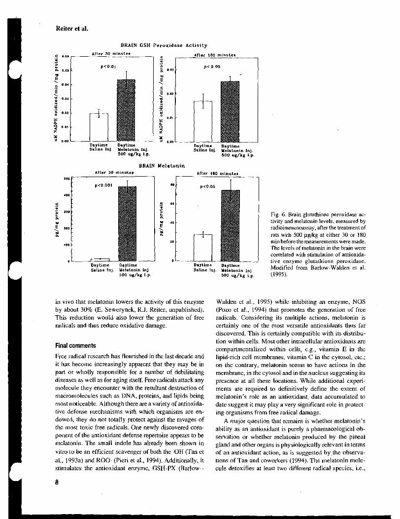
Reiter et al.
B R A I N GSH Peroxidase A c t i v i t y
51 0.01 Afler 30 minulea
.a c kfter 180 minutes a - .a a 0 4
t. 0 0. 0.05 :, 0.0,
6" \
0.04 E" \
'i c - \ E 0.02
1 0.0) \ v " .d II - 4 g 0.02
3
X O 0.01
n 2 n 2 0.01 P
z < z
2- 0.00 Daytime Daytime
2 0.00 Daytlme Daytime
Saline InJ. Yelatonin Inj. Saline la1. Yelatonin Inj. 500 ug/kg 1.p. 500 ug/kg i.p.
BRAIN Melatonin After 30 minuter After 180 minuter
1 I. I
Daytime Daytime Saline Inj. Yelatonin lnj.
500 ug/kg 1.p.
in vivo that melatonin lowers the activity of this enzyme by about 30% (E. Sewerynek, R.J. Reiter, unpublished). This reduction would also lower the generation of free radicals and thus reduce oxidative damage.
Final comments
Free radical research has flourished in the last decade and it has become increasingly apparent that they may be in part or wholly responsible for a number of debilitating diseases as well as for aging itself. Free radicals attack any molecule they encounter with the resultant destruction of macromolecules such as DNA, proteins, and lipids being most noticeable. Although there are a variety of antioxida- tive defense mechanisms with which organisms are en- dowed, they do not totally protect against the ravages of the most toxic free radicals. One newly discovered com- ponent of the antioxidant defense repertoire appears to be melatonin. The small indole has already been shown in vitro to be an efficient scavenger of both the .OH (Tan et al., 1993a) and K O 0 (Pieri et al., 1994). Additionally, it stimulates the antioxidant enzyme, GSH-PX (Barlow--
-- Daytime Daytime Saline lnj. Yelatonin lnj.
500 ug/kg i.p.
Fig. 6. Brain glutathione peroxidase ac- tivity and melatonin levels, measured by radioimmunoassay, after the treatment of rats with 500 ~.~g/kg at either 30 or 180 min before the measurements were made. The levels of melatonin in the brain were correlated with stimulation of antioxida- tive enzyme glutathione peroxidase. Modified from Barlow-Walden et al. (1995).
Walden et al., 1995) while inhibiting an enzyme, NOS (Pozo et al., 1994) that promotes the generation of free radicals. Considering its multiple actions, melatonin is certainly one of the most versatile antioxidants thus far discovered. This is certainly compatible with its distribu- tion within cells. Most other intracellular antioxidants are compartmentalized within cells, e.g., vitamin E in the lipid-rich cell membranes, vitamin C in the cytosol, etc.; on the contrary, melatonin seems to have actions in the membrane, in the cytosol and in the nucleus suggesting its presence at all these locations. While additional experi- ments are required to definitively define the extent of melatonin's role as an antioxidant, data accumulated to date suggest it may play a very significant role in protect- ing organisms from free radical damage.
A major question that remains is whether melatonin's ability as an antioxidant is purely a pharmacological ob- servation or whether melatonin produced by the pineal gland and other organs is physiologically relevant in terms of an antioxidant action, as is suggested by the observa- tions of Tan and coworkers (1994). The melatonin mole- cule detoxifies at least two different radical species, i.e.,
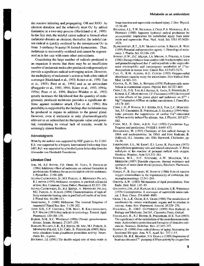
Melatonin as an antioxidant
the reactive initiating and propagating -OH and ROO-, by electron donation and the relatively inert 0 2 - by adduct formation in a two-step process (Hardeland et al., 1993). In the first step, the indolyl cation radical is formed when melatonin donates an electron; thereafter, the indolyl cat- ion radical is quickly oxidized by the omnipresent 0 2 - to form 5-methoxy-N-acetyl-N-formyl-kynuramine. Thus. melatonin is irreversibly oxidized and cannot be regener- ated as is the case with some other antioxidants.
Considering the large number of radicals produced in an organism it seems that there may be an insufficient number of melatonin molecules produced endogenously to provide a significant radical-scavenging action. However, the multiplicity of melatonin's action as both a free radical scavenger (Hardeland et al., 1993; Reiter et al., 1993; Tan et al., 1993); Pieri et al., 1994) and as an antioxidant (Poeggeler et al., 1993, 1994; Reiter et al., 1993; 1994a; 1994~: Pozo et al., 1994; Barlow-Walden et al., 1995) greatly increases the likelihood that the quantity of endo- genously produced melatonin provides a significant de- fense against oxidative attack (Tan et al., 1994); this possibility is supported by the findings that melatonin may be produced in organs in addition to the pineal gland. However, even if melatonin is only pharmacologically relevant as an antioxidant its therapeutic value and poten- tial, considering its virtual lack of toxicity, would be seemingly almost limitless.
Acknowledgments
Work by the authors was supported by NSF grant no. 9 1-21263. E.S. was supported by a Fogarty International Fellowship from NIH; B.P. was supported by a Feodor Lynen fellowship from the Alexander von Humboldt Foundation.
Literature Cited ABE, M., R.J. REITER, P.B. ORHII, M. HARA, B. POEGGELER
(1994) Inhibitory effect of melatonin on cataract formation in aewbom rats: Evidence for an antioxidative role for melatonin. J. Pineal Res. 17:94-100.
ACURA-CASTROVIEJO, D., M.I. PABLOS, A. MENENDEZ-PELAEZ, R.J. REITER (1993) Melatonin receptors in purlfled cell nuclei of liver. Res. Commun. Chem. Pathol. Pharmacol. 82:253-256.
ACUNA-CASTROVIEJO, D., R.J. REITER, A. MENENDU-PELAEZ, M.I. PABLOS, A. BURGOS (1994) Characterization of high-af- finity melatonin binding sites in purified cell nuclei of rat liver. J. Pineal Res. 16:lOO-113.
ARMSTRONG, S. (1989) Melatonin: The internal Zei~geber of mammals? Pineal Res. Rev. 7: 158-202.
AUST, S.D., C.F. CHIGNELL, T.M. BRAY, B. KALYANARAMAN, R.P. MASON (1993) Free radicals in toxicology. Toxicol. Appl. Phmacol. 120: 168-178.
BABIOR, B.M., R.C. WOODMAN (1990) Chronic granulornatous disease. Semin. Hematol. 27:247-259.
BARLOW-WALDEN, L.R., R.J. REITER, M. ABE, M.1. PABLOS, A. MENENDEZ-PELAEZ, L.D. CHEN, B. POEGGELER (1995) Mela- tonin stimulates brain glutathione peroxidase act~vity. Neuro- chern. Int.. in press.
BECKMAN, J.S. (1991) The double-edged role of nitric oxide ~n
brain function and superoxide mediated injury. J. Dev. Physiol. 155949.
BECKMAN, J.S., T.W. BECKMAN, J. CHEN, P.A. MARSHALL, B.A. FREEMAN (1990) Apparent hydroxyl radical production by peroxynitrite: Implication for endothelial injury from nitric oxide and superoxide. Proc. Natl. Acad. Sci. USA 87:1620- 1624.
BLINKENSTAFF, R.T., S.M. BRANDSTADTER, S. REDDY, R. WIT- (1994) Potential radioprotective agents. I . Homologs of mela- tonin. J. Pharm. Sci. 83:21&218.
BOBERG, E.W., E.C. MELER, J.A. MILLER, A. POLAND. A. LIEM (1983) Strong evidence from studies with brachyrnorphic mice and pentachlorophenol that 1'-sulfoxysafrole is the major ulti- mate electrophilic and carcinogenic metabolite of 1'-hy- droxysafrole in mouse liver. Cancer Res. 43:s 163-5 173.
CAO, G., K.M. ALESSIO, R.G. CUTLER (1993) Oxygen-radial absorbance capacity assay for antioxidants. Free Radical Biol. Med. 14:303-3 11.
CHANCE, B., H. SIES, A. BOVERIS (1979) Hydroperoxide meta- bolism in mammalian organs. Physiol. Rev. 62:527-605.
CHEN, L.D., D.X. TAN, R.J. REITER, K. YAGA, B. POEGGELER,~. KUMAR, L.C. MANCHESTER, J.P. CHAMBERS (1993) In vivo and in vitro effects of the pineal gland and melatonin on [C2+ + Mg2+] dependent ATPase in cardiac sarcolemma. J. Pineal Res. 14: 178-183.
CHEN, L.D., P. KUMAR, R.J. REITER, D.X. TAN, L.C. MANCHES- TER, J.P. CHAMBERS, B. POEGGELER, S. SAARELA (1994) Mela- tonin prevents the suppression of cardiac Caz+-stimulated ATPase activity induced by alloxan. Am. J. Physiol. 267:E57- E62.
COON, M.J., X. DING, A.D.N. VAZ (1992) Cytochrome P450: Progress and predictions. FASEB J.6:669473.
DIZDAROGLU, M. (1993) Chemistry of free radical damage to DNA and nucleoproteins. In: DNA and Free Radicals, B. Halliwell, 0.1. Aruoma, eds. Ellis Hawood, Chichester, pp. 19-39.
FARRINGTON, J.A., M. EBERT, E.J. LAND, K. FLETCIIBR (1973) Bipyridylium quaternary salts and related compounds. V. Pulse radiolysis of the reaction of paraquat radical with oxygen. Biochem. Biophys. Acta 314:372-381.
FREEMAN, M.L., N.C. SCIDMORE, A. W. MALCOLM, M.J. MEREDITH (1987) Diamide exposure, thermal resistance and synthesis of stress (heat shock) proteins. Biochem. Pharmacol. 36:21-28.
GIIEZZI, P., B. SACCARDO, M. BIANCHI (1986) Role of reactive oxygen intermediates in the hepatotoxicity of endotoxin. Im- munopharmacology l2:241-244.
G I R O ~ I , A.W. (1985) Mechanisms of lipid peroxidation. Free Radic. Biol. Med. 137-95.
GOLDSTEIN, I.M., H.B. KAPLAN, H.S. EDELSON, G.R. WEISSMAN (1979) Cemloplasmin: A scavenger of superoxide anion radi- cal. J. Biol. Chem. 254:404@4045.
GRAM, T.E., L.K. OKINE, R.A. GRAM (1986) The metabolism of xenobiotics by certain extrahepatic organs and its relation to toxicity. Annu. Rev. Pharmacol. Toxicol. 26:259-276.
HALLIWELL, B., J.M.C. GUITERIDGE (1989) Free Radicals in Biology and Medicine, 2nd Ed., Clarendon Press, Oxford.
HARDELAND, R., R.J. REITER, B. POEGGELER, D.X. TAN (1993) The significance of the metabolism of the neurohormone mela- tonin: Antioxidative protection and formation of bioactive sub- stances. Neurosci. Biobehav. Rev. 17:347-357.
HANNAN, D. (1994) Free radical theory of aging: Increasing the functional life span. Ann. N.Y. Acad. Sci. 717: 1-15.
KANEKO, M., R.E. BEAMISH, N.S. DHALLA (1989) Depression of heart sarcolernmaCz'-pumping ATPase activity by oxygen free
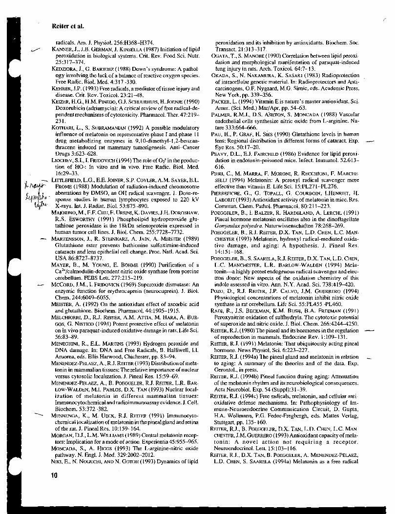
Reiter et al.
radicals. Am. J. Physiol. 256:H368-H374. J KANNER, J., J.B. GERMAN, J. K~NSELLA (1987) Initiation of lipid
peroxidation in biological systems. Crit. Rev. Food Sci. Nutr. 2 5 3 1 7-374.
KEDZIORA, J., G. BARMSZ (1988) Down's syndrome: A pathol- ogy involving the lack of a balance of reactive oxygen species. Free Radic. Biol. Med. 4:3 17-330.
KEHRER, J.P. ( 1993) Free radicals, a mediator of tissue injury and disease. Crit. Rev. Toxicol. 23:21-48.
KEIZER, H.G., H.M. PINEDO, G.J. SCI~UURHUIS, H. JOENJE (1990) Doxorubicin (adriamycin): A critical review of free radical-de- pendent mechanisms of cytotoxicity. Pharmacol. Ther. 47:2 19- 231.
KOTHARI, L., S. SUBRAMANIAN (1992) A possible modulatory influence of melatonin on representative phase I and phase 1 I drug metabolizing enzymes in 9,lO-dimethyl- l,2-benzan- thracene induced rat mammary tumorigenesis. Anti-Cancer Drugs 3:623628.
LIOCHEV, S.I., I. ~ruDoVlCH (1994) The role of 02: in the produc- tion of HO.: In vitro and in vivo. Free Radic. Biol. Med. 16:29-33.
-- L I ~ E F I E L D , L.G., E.E. JOINER, S.P. COYLER, A.M. SAYER, E.L. F R O ~ (1988) Modulation of radiation-induced chromosome aberrations by DMSO, an OH radical scavenger. 1. Dose-re- sponse studies in human lymphocytes exposed to 220 kV X-rays. Int. J. Radiat. Biol. 53:875-890.
~ ~ A I O R I N O , M., F.F. CHU,F. URSINI, K. DAVIES. J.H. DOROSHAW, R.S. ESWORTHY (1991) Phospholipid hydroperoxide glu- tathione peroxidase is the I8kDa selenoprotein expressed in human tumor cell lines. J. Biol. Chem. 255:7728-7732.
-- ARTEN ENS SON, J., R. STEINHARZ, A. JAIN, A. MEISTER (1989) Glutathione ester prevents buthionine sulfoximine-induced cataracts and lens epithelial cell change. Proc. Natl. Acad. Sci. USA 86~8727-8737.
MAYER, B., M. YOUNG, E. BOHME (1990) Purification of a Caz+/calmodulindependent nitric oxide synthase from porcine cerebellum. FEBS Lett. 277:215-219. - MCCORD, J.M., I. FRIDOVICH (1969) Superoxide dismutase: An enzymic function for erythrocuprein (neurocuprein). J. Biol. Chem. 244:60494055.
MEISTER, A. (1992) On the antioxidant effect of ascorbic acid and glutathione. Biochem. Pharmacol. 44: 1905- 19 15.
~ ~ L C H I O R R I , D., R.J. REITER, A.M. AT~IA, M. HARA, A. BUR- GOS, G. NISTICO (1994) Potent protective effect of melatonin on in vivo paraquat-induced oxidative damage in rats. Life Sci. 56:83-89.
MENEGHINI, R., E.L. MARTINS (1993) Hydrogen peroxide and DNA damage. In: DNA and Free Radicals, B. Halliwell, 1.1. Aruoma, eds. Ellis Harwood, Chichester, pp. 83-94.
MENENDEZ-PELAEZ, A., R.J. REITER ( 1993) Distribution of mela- tonin in mammalian tissues: The relative importance of nuclear versus cytosolic localization. J. Pineal Res. 15:5949.
MENENDEZ-PELAEZ, A., B. POEGGELER. R.J. REITER, L.R. BAR- LOW-WALDEN, M.I. PABLOS, D.X. TAN (1993) Nuclear local- ization of melatonin in different mammalian tissues: Immunocytochemical and radioimmunoassay evidence. J. Cell. Biochem. 53:372-382.
MENNENGA, K., M. UECK, R.J. REITER (1991) Immunocyto- chemical localization of melatonin in the pineal gland and retina of the rat. J. Pineal Res. 10: 159-164.
MORGAN, D.J., L.M. WILLIAMS (1989) Central melatonin recep- tors: Implication for a mode of action. Experientia 45:955-965.
MONCADA, S.. A. HIGGS (1993) The L-arginine-nitric oxide pathway. N. Engl. J. Med. 329:2002-20 12.
NIKI. E., N. NOGUCHI, AND N. GOTOH (1993) Dynamics of lipid
peroxidation and its inhibition by antioxidants. Biochem. Soc. Transact. 2 1 :3 13-3 17.
OCATA, T., S. MANOBE (1990) Correlation between lipid peroxi- dation and morphological manifestation of paraquat-induced lung injury in rats. Arch. Toxicol. 64:7-13.
OKADA, S.. N. NAKAMURA, K. SASAKI (1983) Radioprotection of intracellular genetic material. In: Radioprotectors and Anti- carcinogens, O.F. Nygaard, M.G. Simic, eds. Academic Press, New York, pp. 339-356.
PACKER, L. (1994) Vitamin E is nature's master antioxidant. Sci. Amer. (Sci. Med.) MarJApr, pp. 54-63.
PALMER, R.M.J., D.S. A'SHTON, S. MONCADA (1988) Vascular endothelial cells synthesize nitric oxide from L-arginine. Na- ture 333:664-666.
PAU, H., P. GRAF, H. SIES (1990) Glutathione levels in human lens: Regional distribution in different forms of cataract. Exp. Eye Res. 50: 17-20.
PEAVY, D.L., E.J. FAIRCHILD (1986) Evidence for lipid peroxi- dation in endotoxin-poisoned mice. Infect. Immunol. 52;613- 616.
PIERI, C., M. MARRA, F. MORONI, R. RECCHIONI, F. MARCHE- SELLI (1994) Melatonin: A peroxyl radical scavenger more effective than vitamin E. Life Sci. 15:PL271-PL276.
PIERREFICHE, G., G. TOPALL, G. COURBOIN, LHENRIET, H. L ~ ~ o ~ m ( 1 9 9 3 ) Antioxidant activity of melatonin in mice. Res. Commun. Chem. Pathol. Pharmacol. 80:211-223.
POEGGELER, B., I. BALZER, R. HARDELAND, A. LERCHL (1991) Pineal hormone melatonin oscillates also in the dinoflagellate Gonyaularpolyedra. Naturwissenschaften 78:268-269.
POEGGELER, B., R.J. REITER, D.X. TAN, L.D. CHEN, L.C. MAN- CHESTER (1993) Melatonin, hydroxyl radical-mediated oxida- tive damage, and aging: A hypothesis. J. Pineal Res. 14:151-168.
POEGGELER, B., S. SAARELA, R.J. REITER, D.X. TAN, L.D. CHEN, L.C. MANCHESTER, L.R. BARLOW-WALDEN (1994) Mela- tonin-a highly potent endogenous radical scavenger and elec- tron donor: New aspects of the oxidation chemistry of this indole assessed in vitro. Ann. N.Y. Acad. Sci. 738:419420.
Pozo, D., R.J. REITER, J.P. CALVO, J.M. GUERRERO (1994) Physiological concentrations of melatonin inhibit nitric oxide synthase in rat cerebellum. Life Sci. 55:PLA55-PL460.
RADI, R., J.S. BECKMAN, K.M. BUSH, B.A. FREEMAN (1991) Peroxynitrite oxidation of sulfiydryls. The cytotoxic potential of superoxide and nitric oxide. J. Biol. Chem. 266:4244-4250.
REITER, R.J. (1980) The pineal and its hormones in the regulation - of reproduction in mammals. Endocrine Rev. 1 : 109-1 3 1.
REITER, R.J. (1991) Melatonin: That ubiquitously acting pineal hormone. News Physiol. Sci. 6:223-227.
REITER, R.J. (1994a) The pineal gland and melatonin in relation to aging: A summary of the theories and of the data. Exp. Gerontol., in press.
REITER, R.J. (1994b) Pineal function during aging: Attenuation of the melatonin rhythm and its neurobiological consequences. Acta Neurobiol. Exp. 54 (Suppl):3 1-39.
REITER, R.J. (1994~) Free radicals, melatonin, and cellular anti- oxidative defense mechanisms. In: Pathophysiology of Im- mune-Neuroendocrine Communication Circuit, D. Gupta, H.A. Wollmann, P.G. Fedor-Fregbergh, eds. Mattes Verlag, Stuttgart, pp. 135-160.
REITER, R.J., B. POEGCELER, D.X. TAN, L.D. CHEN, L.C. MAN- CHESTER, J.M. GUERRERO (1993) Antioxidant capacity of mela- tonin: A novel action not requir ing a receptor. Neuroendocrinol. let^ 15: 103-1 16.
REITER, R.J., D.X. TAN, B. POEGGELER, A. MENENDEZ-PELAEZ, L.D. CHEN, S. SAARELA (1994a) Melatonin as a free radical
Melatonin as an antioxidant
scavenger: Implications for aging and age-related diseases. Ann. N.Y. Acad. Sci. 7 19: 1-12.
REITER, R.J., D.X. TAN, B. POEGGELER, L.D. CHEN, A. MENEN- DEZ-PELAEZ (1994b) Melatonin, free radicals and cancer initia- tion. In: Advances in Pineal Research, Vol. 7. G.J.M. Maestroni, A. Conti and R.J. Reiter, eds. John Libbey, London, pp. 2 1 1-228.
REITER, R.J., B. POEGGELER, L.D. CHEN. M. ABE, M. HARA, P.B. O~rur, A.M. AITIA, L.R. BARLOW-WALDEN (1994~) Melatonin as a free radical scavenger: Theoretical implications for neurodegenerative disorders in the aged. Acta Gerontol., in press.
SCHRECK, P., P.A. BAEUERLE (199 1) A role of oxygen radicals as second messengers. Trends Cell Biol., 1:39-42.
SCHRECK, P., P. RIEBER, P.A. BAEUERLE ( 199 1) Reactive oxygen intermediates a s apparently widely used messengers in the activation of the NF-KB transcription and HIV-I. EMBO J. 10:2247-2258.
SEWERYNEK, E., M. ABE, R.J. REITER, L.R. BARLOW-WALDEN, L.D. CHEN, T.J. MCCABE, L.J. ROMAN, B.DIAZ-LOPEZ (1995) Melatonin administration prevents Lipopolysaccharide- induced oxidative damage in phenobarbital-treated animals. J. Cell. Biochem., in press.
SHIDA, C.S., A.M.L. CASTRUCCI, M.T. LAMY-FREUND (1994) High melatonin solubility in aqueous medium. I. Pineal Res. 16: 198-201.
SIES, H. (1991) Oxidative stress. In: Oxidative Stress, 11: Oxi- dants and Antioxidants, H. Sies, ed. Academic Press, New York, pp. xv-xvii.
SPECTOR, A. (199 1) The lens and oxidative stress. In: Oxidative Stress, 11: Oxidants and Antioxidants. Academic Press, New York, pp. 529-558.
SUBBORAO, K.V., J.S. RICHARDSON, L.C. ANG (1990) Autopsy
samples of Alzheimer's cortex show increased peroxidation in vitro. J. Neurochem. 55:342-347.
TAN, D.X., L.D. CHEN, B. P O E ~ E L E R , L.C. MANCHESTER, R.J. REITER (1993a) Melatonin: A potent, endogenous hydroxyl radical scavenger. Endocrine J. 1 57-60.
TAN, D.X., B. POEGGELER, R.J. REITER, L.D. CHEN, S. CHEN, L.C. MANCHESTER, L.R. BARLOW-WALDEN ( 1993b)The pineal hormone melatonin inhibits DNA adduct formation induced by the chemical carcinogen safrole in vivo. Cancer Len: 70:65-7 1.
TAN, D.X, R.J. REITER, L.D. CHEN, B. POEGGELER, L.C. MAN- CHESTER, L.R. BARLOW-WALDEN (1994) Both physiological and pharmacological levels of melatonin reduce DNA adduct formation induced by the carcinogen safrole. Carcinogenesis 15:215-218.
TAYLOR, A., P.F. JACQUES, C.K. FOREY (1993) Oxidation in aging: Impact on vision. Toxicol. Indust. Health 9:349-37 1.
URSINI. F., M. MAIORINO, A. SEVANIAN (1991) Membrane hy- droperoxides. In: Oxidative Stress, 11: Oxidants and Antioxi- dants. Academic Press, New York, pp. 379-386.
VANECEK. J., A. PAVLIK. H. ILLNEROVA (1987) Hypothalamic melatonin receptor sites revealed by autoradiography. Brain Res. 435:359-362.
VUAYALAXMI, R.J. REITER, M.L. MELZ (1995) Melatonin pro- +-
tects human blood lymphocytes from radiation-induced chro- ,vt mosome damage. Mutat. Res., in press. WINTERBOUM, C.C., H.C. SUTTON (1984) Hydroxyl radical pro-
duction from hydrogen peroxide and enzymatically generated paraquat radicals: Catalytic requirements and oxygen depend- ence. Arch. Biochem. Biophys. 235: 116-126.
ZIMMERMAN, B.J., D.N. GRANGER (1994) Oxygen free radicals and the gastrointestinal tract: Role in ischemia-reperfusion injury. Hepato-Gastroenterology 41:337-342.

/ P i n e a l R;. ; 1996; ?1:200-213 Prrnred In :,i? L'nired Srores ojdrner icn-dl1 r i ~ h r s reserred
~ 1 7 U S ~
Mini Review
Melatonin in relation to physiology in adult humans
Cagnacci A. Melatonin in relation to physiology in adult humans. J. Pineal Res. 1996; 2 1 :3-00-213. O Munksgaard, Copenhagen
Abstract: The role exerted by melatonin in human physiology has not been completzly ascertained. Melatonin levels have been measured in different physiopathological conditions, but the effects induced by melatonin administration or withdrawal have been tested only recently. Some effects have been clearly documented. Melatonin has hypothermic properties, and its nocturnal secretion generates about 40% of the amplitude of the circadian body temperature rhythm. Melatonin has sleep inducing properties, and exerts important activities in the regulation of circadian rhythms. Melatonin is capable of phase shifting hliman circadian rhythms, of entraining free-running circadian rhythms, and of antagonizing phase shifts induced by nighttime exposure to light. Its effect on human reproduction is not completely clear, but stimulatory effects on gonadotropin secretion have been reported in the follicular phase of the menstrual cycle. Direct actions on ovarian cells and spermatozoa have been also documented. Beside these, new important actions for melatonin may be proved. Melatonin may exert protective effects on the cardiovascular system, by reducing the risk of atherosclerosis and hypertension, and may influence immune responses. Finally, by acting as an antioxidant, melatonin could be important in slowing the processes of ageing.
Cop?rr.ehr 0 Murrks~yoord. 1996
Journal 3 f Pineal Research ISSN 0742-3098
Angelo Cagnacci Institute of Obstetrics and Gynecology. University of Modena. 41 100 Modena, Italy
Key wolds: Melatonin -humans - reproduction -temperature - sleep - ageing -circadian rhythms
Address reprint requests to Dr. Angelo Cagnacci. lstituto di Fisiopatologia della Riproduzione Umana. via del Pouo 71, 41 10 Modena, Italy.
Received July 31. 1996; accepted September 16, 1996.
Introduction Reiter, 19911. Tryptophan is taken up by the pine-
Physiology of melatonin has been extensively stud- ied in animals. For years data obtained in animals have been extrapolated to humans without critical evaluation. Indeed, only more recent studies have tried to investigate the mechanisms of synthesis, regulation, and action of melatonin in humans. The present review will focus almost entirely on data obtained in humans that have defined mechanisms of melatonin production by the pineal gland, and the effects of melatonin on biological and endocrine functions.
Metatonin synthesis cr -
Studies performed in vitro and in animals have clari- fied the mechanisrns involved in the regulation of melatonin synthesis by the pineal gland [Cardinali m d Vacas, 1987; Krause and Dubocovich, 1990;
alocyte, is transformed to serotonin,-and seroionin is finally converted into melatonin by a two-step process that involves the sequential activities of two enzymes, N-acetyltransferase (NAT), which is be- lieved to be the limiting enzyme for the synthesis of melatonin, and hydroxyindole-0-methyltrans- ferase (HIOMT). The synthesis of melatonin is ini- tiated by the binding of norepinephrine to adrenergic Dl receptors, subsequent activation of pineal ade- nylate cyclase, increase in cyclic A M P (CAMP), binding, and de novo synthesis of N.4T or of its ac- tivator. The potent CAMP-induced gene transcrip- tion repressor (ICER) is activated in conjunction with NAT and represents a mechanism that limits the nocturnal production of melatonin [Stehle et al., 19931. The Dl adrenergic receptor stimulus is en- hanced by al- adrenoceptors, via calcium (ca2')- phospholipid-dependent protein kinase C (PKC) and by prostaglandins, whose synthesis is activated by
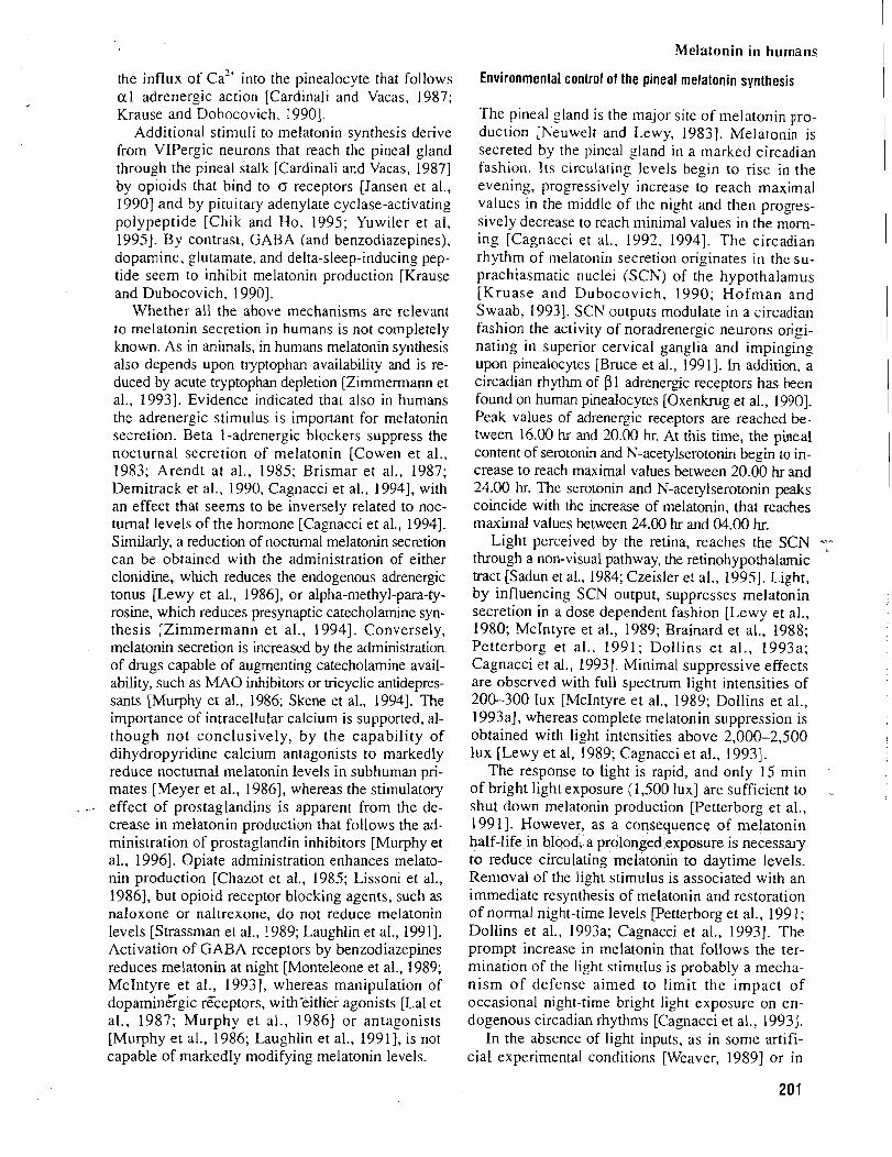
Melatonin in humans
the influx of ~ a " Into the pinealocyte that follows a1 adrenergic action [Cardinali and Vacas, 1987; Krause and Dobocovich. 19901.
Additional stimuli to melatonin synthesis derive from VIPergic neurons that reach the pineal gland through the pineal stalk [Cardinali and Vacas, 19871 by oploids that bind to o receptors [Jansen et al., 19901 and by pituitary adenylate cyclase-activating polypeptide [Chik and Ho, 1995; Yuw~ler et al, 19951. By contrast, GABA (and benzodiazepines), dopamine, ,ohtamate, and delta-sleep-inducing pep- tide seem to inhibit melatonin production [Krause and Dubocovich, 19901.
Whether all the above mechanisms are relevant to melatonin secretion in humans is not completely known. As in animals, in humans rnelatonin synthesis also depends upon tryptophan availability and is re- duced by acute tryptophan depletion [Zimmermann et al., 19931. Evidence indicated that also in humans the adrenergic stimulus is important for rnelatonin secretion. Beta 1-adrenergic blockers suppress the nocturnal secretion of melatonin [Cowen et al., 1983; Arendt at al., 1985; Brismar et al., 1987; Demitrack et a]., 1990, Cagnacci et al., 19941, with an effect that seems to be inversely related to noc- turnal levels of the hormone [Cagnacci et al., 19941. Similarly, a reduction of nocturnal melatonin secretion can be obtained with the administration of either clonidine, which reduces the endogenous adrenergic tonus [Lewy et al., 19861, or alpha-methyl-para-ty- rosine, which reduces presynaptic catecholarnine syn- thesis [Zimmermann et al., 19941. Conversely, melatonin secretion is increased by the administration of drugs capable of augmenting catecholarnine avail- ability, such as MA0 inhibitors or tricyclic antidepres- sants [Murphy et al., 1986; Skene et al., 19941. The importance of intracellular calcium is supported, al- though not conclusively, by the capability of dihydropyridine calcium antagonists to markedly reduce nocturnal melatonin levels in subhuman pri- mates [Meyer et al., 19861, whereas the stimulatory effect of prostaglandins is apparent from the de- crease in melatonin production that follows the ad- ministration of prostaglandin inhibitors [Murphy et al., 19961. Opiate administration enhances melato- nin production [Chazot et al., 1985; Lissoni et al., 19861, but opioid receptor blocking agents, such as naloxone or naltrexone, do not reduce melatonin levels [Strassman et al., 1989; Laughlin et al., 19911. Activation of GABA receptors by benzodiazepines reduces melatonin at night [Monteleone et al., 1989; McIntyre et al., 19931, whereas manipulation of dopamine"rgic receptors, withkither agonists [Lal et al., 1987; Murphy et al., 19861 or antagonists [Murphy et al., 1986; Laughlin et al., 19911, is not capable of markedly modifying melatonin levels.
Environmental control of the pineal melatonin synthesis
The pineal ?land is the major site of melatonin pro- duction [Neuwelt and Lewy, 19831. Melatonin 1s secreted by the pineal gland in a marked circadian fashion. Its circulating levels begin to rise in the evening. progressively increase to reach maximal values in the middle of the night and then progres- sively decrease to reach minimal values in the mom- ing [Cagnacci et al., 1992, 19941. The circadian rhythm of melatonin secretion originates in the su- prachiasmatic nuclei (SCN) of the hypothalamus [Kruase and Dubocovich, 1990; Hofman and Swaab, 19931. SCN outputs modulate in a circadian fashion the activity of noradrenergic neurons origi- nating in superior cervical ganglia and impinging upon pinealocytes [Bruce et al., 19911. In addition, a circadian rhythm of pl adrenergic receptors has been found on human pinealocytes [Oxenkrug et al., 19901. Peak values of adrenergic receptors are reached be- tween 16.00 hr and 20.00 hr. At this time, the pineal content of serotonin and N-acetylserotonin begin to in- crease to reach maximal values between 20.00 hr and 24.00 hr. The serotonin and N-acetylserotonin peaks coincide with the increase of melatonin, that reaches maximal values between 24.00 hr and 04.00 hr.
Light perceived by the retina, reaches the SCN q through a non-visual pathway, the retinohypothalarnic tract [Sadun et al., 1984; Czeisler et al., 19951. Light, by influencing SCN output, suppresses melatonin secretion in a dose dependent fashion [Lewy et al., 1980; McIntyre et al., 1989; Brainard et al., 1988; Petterborg et al., 1991; Dollins et al., 1993a; Cagnacci et al., 19931. Minimal suppressive effects are observed with full spectrum light intensities of 200-300 lux [McIntyre et al., 1989; Dollins et al., 1993a], whereas complete melatonin suppression is obtained with li,oht intensities above 2,000-2,500 lux [Lewy et al, 1989; Cagnacci et al., 19931.
The response to light is rapid, and only 15 min '
of bright light exposure (1,500 lux] are sufficient to shut down melatonin production [Petterborg et al., 19911. However, as a consequence of melatonin half-life in blood, a prolonged exposure is necessary to reduce circulating melatonin to daytime levels. Removal of the light stimulus is associated with an immediate resynthesis of melatonin and restoration of normal night-time levels [Petterborg et al., 199 1; Dollins et al., 1993a; Cagnacci et al., 19931. The prompt increase in melatonin that follows the ter- mination of the light stimulus is probably a mecha- nism of defense aimed to limit the impact of occasional nicght-time bright light exposure on en- dogenous circadian rhythms [Cagnacci et al., 19933.
In the absence of light inputs, as in some artifi- cial experimental conditions [Weaver, 19891 or in
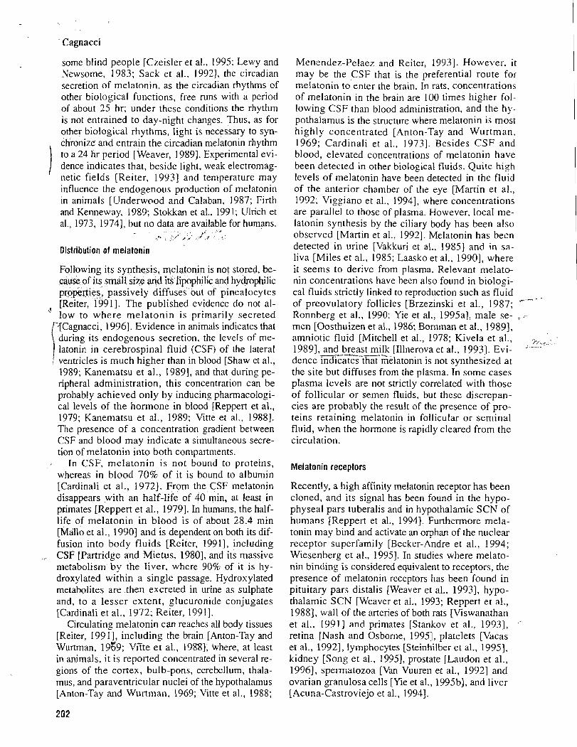
' Cagnacci
some blind people [Czeisler et al., 1995; Lewy and Newsome, 1983: Sack et al., 19931, the circadian secretion of melatonin, as the circadian rhythms of other biological functions, free runs with a period of about 35 hr; under these conditions the rhythm is not entrained to day-night changes. Thus, as for other biological rhythms, light is necessary to syn- chronize' and entrain the circadian melatonin rhythm to a 24 hr period [Weaver, 19891. Experimental evi- dence indicates that, beside light. weak electromag- netic fields [Reiter, 19931 and temperature may influence the endogenous production of melatonin in animals [Underwood and Calaban, 1987; Firth and Kenneway, 1989; Stokkan et al., 1991; Ulrich et al., 1973, 19741, but no data are available for humans.
,* < ~ * / , , .J, '
Distribution of melatonin
Following its synthesis, melatonin is not stored, be- cause of its small size and its lipophilic and hydrophilic properties, passively diffuses out of pinealocytes [Reiter, 19911. The published evidence do not al-
! low to where melatonin is primarily secreted [?Cagnacci, 19961. Evidence in animals indicates that \ during its endogenous secretion. the levels of me-
latonin in cerebrospinal fluid (CSF) of the lateral I ventricles is much higher than in blood [Shaw et al.. 1989; Kanematsu et al., 19891, and that during pe- ripheral administration, this concentration can be probably achieved only by inducing pharmacologi- cal levels of the hormone in blood [Reppert et al., 1979; Kanematsu et al., 1989; Vitte et al.. 19881. The presence of a concentration gradient between CSF and blood may indicate a simultaneous secre- tion of melatonin into both compartments.
, In CSF, melatonin is not bound to proteins, whereas in blood 70% of it is bound to albumin [Cardinali et al., 19721. From the CSF melatonin disappears with an haIf-life of 40 min, at least in primates [Reppert et al., 19791. In humans, the half- life of melatonin in blood is of about 28.4 min [Mallo et al., 19901 and is dependent on both its dif- fusion into body fluids [Reiter, 19911, including CSF [Partridge and Mietus, 19801, and its massive metabolism by the liver, where 90% of it is hy- droxylated within a single passage. Hydroxylated metabolites are then excreted in urine as sulphate and, to a lesser extent, glucuronide conjugates [Cardinali et al., 1972; Reiter, 19911.
Circulating melatonin can reaches all body tissues [Reiter, 199 I], including the brain [Anton-Tay and Wurtman, 1959; VGte et al., 19881, where, at least in animals, it is reported concentrated in several re- gions of the cortex, bulb-pons, cerebellum, thala- mus, and paraventricular nuclei of the hypothalamus [Anton-Tay and Wurtman, 1969; 'L'itte et al., 1988;
Menendez-Pelaez and Reiter, 19931. However. i t may be the CSF that is the preferential route for melatonin to enter the brain. In rats, concentrations of melatonin in the brain are 100 times higher fol- lowing CSF than blood administration. and the hy- pothalamus is the structure where melatonin is most highly concentrated [Anton-Tay and Wurtman. 1969; Cardinali et al., 19731. Besides CSF and blood, elevated concentrations of melatonin have been detected in other biolo_gical fluids. Quite high levels of melatonin have been detected in the fluid of the anterior chamber of the eye [Martin et al., 1992: Viggiano et al., 19941, where concentrations are parallel to those of plasma. However, local me- latonin synthesis by the ciliary body has been also observed [Martin et al., 19921. Melatonin has been detected in urine [Vakkuri et al., 19851 and in sa- liva [Miles et al., 1985; Laasko et al., 19901, where it seems to derive from plasma. Relevant melato- nin concentrations have becn also found in biologi- cal fluids strictly linked to reproduction such as fluid of preovulatory follicles [Brzezinski et al., 1987; /
Ronnberg et al., 1990; Yie et al.. 1995aI. male se- _ - men [Oosthuizen et al., 1986; Bornman et al., 19891, amniotic fluid [Mitchell et al., 1978; Kivela et al., ?,-,: 19891, and breast milk [Illnerova et al., 19931. Evi- " -
dence indicacs thafselatonin is not synthesized at the site but diffuses from the plasma. In some cases plasma levels are not strictly correlated with those of follicular or semen fluids, but these discrepan- cies are probably the result of the presence of pro- teins retaining melatonin in follicular or seminal fluid, when the hormone is rapidly cleared from the circulation.
Melatonin receptors
Recently, a high affinity melatonin receptor has been cloned, and its signal has been found in the hypo- physeal pars tuberalis and in hypothalamic SCN of humans [Reppert et al., 19941. Furthermore mela- tonin may bind and activate an orphan of the nuclear receptor superfamily [Becker-Andre et al., 1994; Wiesenberg et al., 19951. In studies where melato- nin binding is considered equivalent to receptors, the presence of melatonin receptors has been found in pituitary pars distalis [Weaver et al., 19931, hypo- thalamic SCN [Weaver et al., 1993; Reppert et al., 19881, wall of the aneries of both rats [Viswanathan et al., 19911 and primates [Stankov et al., 19931, a
retina [Nash and Osborne, 19951, platelets [Vacas et al., 19921, lymphocytes [Steinhilber et al., 19951, kidney [Song et al., 19951, prostate [Laudon et al., 19961, spermatozoa [Van Vuuren et al., 19921 and ovarian granulosa cells [Yie et al., 1995b1, and liver [Acuna-Castroviejo et al., 19941.
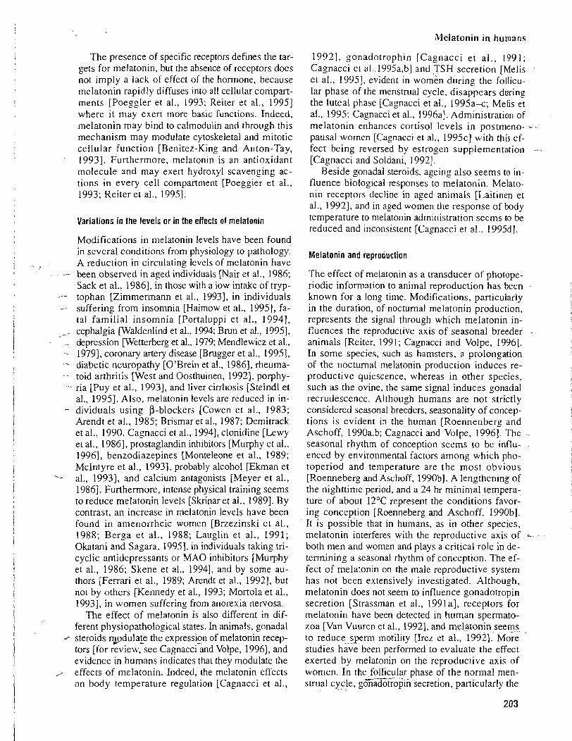
The presence of specific receptors defines the tar- gets for melatonin, but the absence of receptors does not imply a lack of effect of the hormone, because melatonin rapidly diffuses into all cellular compart- ments [Poeggler et al., 1993; Reiter et al., 19951 where it may exert more basic functions. Indeed, melatonin may bind to calmodulin and through this mechanism may modulate cytoskeletal and mitotic cellular function [Benitez-King and Anton-Tay, 19931. Furthermore, melatonin is an antioxidant molecule and may exert hydroxyl scavenging ac- tions in every cell compartment [Poeggler et al., 1993; Reiter et al., 19951.
Variations in the levels or in the effects of melatonin
Modifications in melatonin levels have been found in several conditions from physiology to pathology. A reduction in circulating levels of melatonin have
,- been observed in aged individuals [Nair et al., 1986; Sack et al., 19861, in those with a low intake of tryp- -- tophan [Zlmmermann et al., 19931, in individuals
.- suffering from insomnia [Haimow et al., 19951, fa- tal familial insomnia [Portaluppi et al., 19941,
- cephalgia waldenlind et al.. 1994; Brun et al., 19951, -. depression wetterberg et al., 1979; Mendlewicz et al., - 19791, coronary artery disease [Brugger et al., 199.51, -- diabetic neuropathy [O'Brein et al., 19861, rheuma- -- toid arthritis [West and Oosthuinen, 19921, porphy- - ria [Puy et al., 19931, and liver cirrhosis [Steindl et
al., 1995). Also, melatonin levels are reduced in in- - dividuals using P-blockers [Cowen et al., 1983;
Arendt et al., 1985; Brismar et al., 1987; Dernitrack et al., 1990, Cagnacci et al., 19941, clonidine [Lewy et al., 19861, prostaglandin inhibitors [Murphy et al., 19961, benzodiazepines [Monteleone et al., 1989; McIntyre et al., 19931, probably alcohol [Ekman et
L- al., 19931, and calcium antagonists [Meyer et al., 19861. Furthermore, intense physical training seems to reduce melatonin levels [Skrinaret al., 19891. By contrast, an increase in melatonin levels have been found in amenorrheic women [Brzezinski et al., 1988; Berga et al., 1988; Lauglln et al., 1991; Okatani and Sagara, 199.51, in individuals taking tri- cyclic antidepressants or MA0 inhibitors [Murphy et al., 1986; Skene et al., 19941, and by some au- thors [Ferrari et al., 1989; Arendt et al., 19921, but not by others (Kennedy et al., 1993; Mortola et al., 19931, in women suffering from anorexia nervosa.
The effect of melatonin is also different in dif- ferent physiopathological states. In animals, gonadal
1 steroids 30dulate the expressi~n of melatonin recep- tors [for review, see Cagnacci and Volpe, 19961, and evidence in humans indicates that they modulate the
, effects of melatonin. Indeed, the melatonin effects on body temperature regulation [Cagnacci et al.,
19921, gonadotrophin [Cagnacci et al., 1991; Cagnacci et a1..1995a,b] and TSH secretion [Melis et al., 19951, evident in women during the follicu- lar phase of the menstrual cycle, disappears during the luteal phase [Cagnacci et al., 1995a-c; Melis et al., 1995: Cagnacci et al., 1996al. Administration of melatonin enhances cortisol levels in postmeno- - . pausal women [Cagnacci et al., 1995~1 with this ef- fect being reversed by estrogen supplementation -- [Cagnacci and Soldani, 19921.
Beside gonadal steroids, ageing also seems to in- fluence biolog~cal responses to melatonin. Melato- nin receptors decline in aged animals [Laitinen et al., 19921, and in aged women the response of body temperature to melatonin administration seems to be reduced and inconsistent [Cagnacci et al., 1995dJ.
Melatonin and reproduction
The effect of melatonin as a transducer of photope- riodic information to animal reproduction has been known for a long time. Modifications, particularly in the duration, of nocturnal melatonin production, represents the signal through which melatonin in- fluences the reproductive axis of seasonal breeder -
animals [Reiter, 1991; Cagnacci and Volpe, 19961. In some species, such as hamsters, a prolongation of the nocturnal melatonin production induces re- productive quiescence, whereas in other species, such as the ovine, the same signal induces gonadal recrudescence. Although humans are not strictly considered seasonal breeders, seasonality of concep- tions is evident i n the human [Roennenberg and Aschoff, 1990a,b; Cagnacci and Volpe, 19961. The seasonal rhythm of conception seems to be influ- enced by environmental factors among which pho- toperiod and temperature are the most obvious [Roenneberg and Aschoff, 1990bl. A lengthening of the nighttime period, and a 24 hr minimal tempera- ture of about 12°C represent the conditions favor- ing conception [Roenneberg and Aschoff, 1990bl. It is possible that in humans, as in other species, melatonin interferes with the reproductive axis of both men and women and plays a critical role in de- termining a seasonal rhythm of conception. The ef- fect of melaton~n on the male reproductive system has not been extensively investigated. Although, melatonin does not seem to influence gonadotropin secretion [Strassman et al., 1991a1, receptors for melatonin have been detected in human spermato- zoa [Van Vuuren et al., 19921, and rngktonin seems to reduce sperm motility [Irez et al., 19921. More studies have been performed to evaluate the effect exerted by melatonin on the reproductive axis of women. In the follicular phase of the normal men- __--._ -. strual cycle, gonadotropin secretion, particularly the
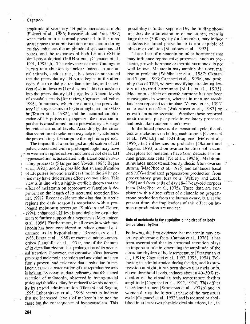
Cagnacci
amplitude of secretory LH pulse, increases at night [Filicori e t all, 1986; RosSmanith and Yen. 19871 when melatonin is normally secreted. In this men- strual phase the administratipn of melatonin during the day enhances the amplitude of spontaneous LH pulses, and the responses-of both-LH and FSH to simil-physiological GnRH stimuli [Cagnacci et al., 1991, 1995a,b]. The relevance of these findings to human reproduction is unclear. Indeed, in noctur- nal animals, such as rats, i t has been demonstrated that the preovulatory LH surge begins in the after- noon, due to a daily circadian stimulus, and is evi- dent also in diestms I1 or diestrus I; this is translated into the preovulatory LH surge by sufficient levels of gonadal steroids [for review, Cagnacci and Volpe, 19961. In humans, which are diurnal, the preovula- tory LH surge seems to begin at night, around 03.00 hr restart et al., 19821, and the noctumal amplifi- cation of LH pulses may represent the circadian in- put that is transformed into a preovulatory LH surge by critical estradiol levels. Accordingly, the circa- dian secretion of melatonin may help to synchronize the preovulatory LH surge to the nighttime period.
The impact that a prolonged amplification of LH pulses, associated with a prolonged night, may have
I -,-on women's reproductive functions is not clear. LH hypersecretion is associated with alterations in ovu-
I latory processes [Stanger and Yovich, 1985; Regan i ) et al., 19901, and it is possible that an amplification 1 of LH pulses beyond a critical time in the 24 hr pe- ----riod may have deleterious effects on ovulation. This
view is in line with a highly credible theory that the -- effect of melatonin on reproductive function is de-
pendent on the length of its nocturnal secretion [Re- iter, 19911. Recent evidence showing that in Arctic regions the dark season is associated with a pro- longed melatonin secretion [Stokkan and Reiter,
-. 19941, enhigced L H l e e b , a_nd defective ovulation., seem to further support this hypothesis [Martikainen et al., 19963. Furthermore, in a11 cases in which me- latonin has been considered to induce gonadal qui- escence, as in hypothalamic [Brzezinsky et al., 1988; Berza et al., 19881 or exercise induced-amen- orrhea [Laughlin et al., 1991.1, one of the features of its circadian rhytlmm is a prolongation of its noctur-
.- nal secretion. However, the causative effect between prolonged melatonin secretion and anovulation is not firmly proven, and evidence that a reduction in me- latonin causes a reactivation of the reproductive axis is lacking. By contrast, data indicating that tKe altered secretion of melatonin, observed in hypogonadal males and femsles, Gay be reduced towards normal- ity by steroid administration [Okatani and Sagara, 1995; Luboshitzky et al., 19961 seems to indicate _, that the increased levels of melatonin are not the
cause but the consequence of hypogonadism. This ..- -~
possibility is further supported by the finding show- ing that the administration of melatonin, even in large doses (300 mglday for 4 months), may induce -- a defective luteal phase but i t IS not capable of blocking ovulation [Voordouw et al., 19921.
The effect of melatonin on other hormones that may influence reproductive processes, such as pro- lactin, growth-hormone or thyroid hormones, is not well known. Melatonin may amplify the nocturnal rise in prolactin [Waldhauser et al., 1987; Okatan~ and Sagara, 1993; Cagnacci et al.. 1995e1, and prob- ably that of TSH, without modifying circulating lev- els of thyroid hormones [Melis et al., 19951. Melatonin's effect on growth hormone has not been investigated in women, whereas in men melatonin has been reported to stimulate [Valcavi et al., 19931 or to exert no effect [Waldhauser et al., 19871 on growth hormone secretion. Whether these reported modifications play any role in ovulatory processes and testicular function is unknown.
In the luteal phase of the menstrual cycle, the ef- fect of melatonin on both gonadotropins [Cagnacci et al., I995a,b] and TSH disappear [Melis et al., 19951, but influences on prolactin [Okatani and
,
Sagana, 19931 and on ovarian function still occur. Receptors for melatonin have been detected in hu- man granulosa cells [Yie et al., 1995bl. Melatonin stimulates androstenedione synthesis from ovarian - "
stroma [MacPhee et al., 19751 and enhances basal and hCG-stimulated progesterone production from preovulatory granulosa cells [Webley and Luck, 19861 and from cells of day 18-27-day-old corpora lutea [MacPhee et al., 19751. These data are con- sistent with a direct effect of melatonin on progest- erone production from the human ovary, but, at the present time, the implications of this effect on hu- man reproduction are unclear.
Role of melatonin in the regulation of the circadian body temperature rhythm
Following the first evidence that melatonin may ex- ert hypothermic effects [Carman et al., 19761, i t has been ascerrained that its nocturnal secretion plays an important role in generating the amplitude of the circadian rhythm of body temperature [Strassrnan et al., 1991 b; Cagnacci et a]., 1992, 1993, 19941. Fol- lowing its administration during the day, and its sup- pression at night, it has been shown that melatonin, above threshold levels, induces about a 40-50% re- duction of the circadian body temperature rhythm amplitude [Cagnacci et al., 1992, 19941. This effect is evident in men [Strassman et al., 199 1 b] and in women during the follicular phase of the menstrual cycle [Cagnacci et al., 19921, and is reduced or abol- ished in at least two physiological situations, i.e., in
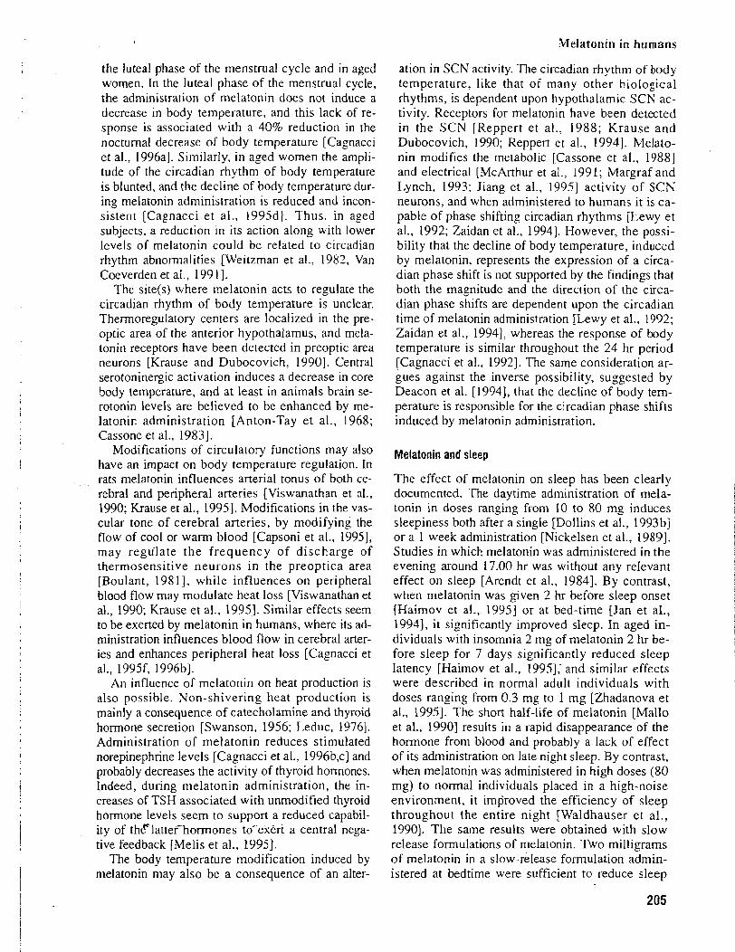
Melatonin in humans
the luteal phase of the menstrual cycle and in aged women. In the luteal phase of the menstrual cycle, the administration of melatonin does not induce a decrease in body temperature, and this lack of re- sponse is associated with a 40% reduction in the nocturnal decrease of body temperature [Cagnacci et al., 1996al. Similarly, in aged women the ampli- tude of the circadian rhythm of body temperature is blunted, and the decline of body temperature dur- ing melatonin administration is reduced and incon- sistent [Cagnacci et al., 1995dl. Thus. in aged subjects, a reduction in its action along with lower levels of melatonin could be related to circadian rhythm abnormalities [Weitzman et al., 1983, Van Coeverden et al., 199 11.
The site(s) where melatonin acts to regulate the circadian rhythm of body temperature is unclear. Thermoregulatory centers are localized in the pre- optic area of the anterior hypothalamus, and mela- tonin receptors have been detected in preoptic area neurons [Krause and Dubocovich, 19901. Central serotoninergic activation induces a decrease in core body temperature, and at least in animals brain se- rotonin levels are believed to be enhanced by me- latonin administration [Anton-Tay et al., 1968; Cassone et al., 19831.
Modifications of circulatory functions may also have an impact on body temperature regulation. In rats melatonin influences arterial tonus of both ce- rebral and peripheral arteries [Viswanathan et al., 1990; Krause et al., 19951. Modifications in the vas- cular tone of cerebral arteries, by modifying the flow of cool or warm blood [Capsoni et al., 19951, may regulate the frequency of discharge of thermosensitive neurons in the preoptica area [Boulant, 198 1 1, while influences on peripheral blood flow may modulate heat loss [Viswanathan et al., 1990; Krause et al., 19951. Similar effects seem to be exerted by melatonin in humans, where its ad- ministration influences blood flow in cerebral arter- ies and enhances peripheral heat loss [Cagnacci et al., 1995f, 1996bl.
An influence of melatonin on heat production is also possible. Non-shivering heat production is mainly a consequence of catecholamine and thyroid hormone secretion [Swanson, 1956; Leduc, 19761. Administration of melatonin reduces stimulated norepinephrine levels [Cagnacci et al., 1996b,c] and probably decreases the activity of thyroid hormones. Indeed, during melatonin administration, the in- creases of TSH associated with unmodified thyroid hormone levels seem to support a reduced capabil- ity of thclattefhormones toT'exert a central nega- tive feedback [Melis et al., 19951.
The body temperature modification induced by melatonin may also be a consequence of an alter-
ation in SCN activity. The circadian rhythm of body temperature, like that of many other biological rhythms, is dependent upon hypothalamic SCN ac- tivity. Receptors for melatonin have been detected in the SCN [Reppert et al., 1988; Krause and Dubocovich, 1990; Reppert et al., 19941. Melato- nin modifies the metabolic [Cassone et al., 19881 and electrical [McArthur et al., 199 1; Margraf and Lynch, 1993; Jiang et al., 199.51 activity of SCN neurons, and when administered to humans it is ca- pable of phase shifting circadian rhythms [Lewy et al., 1992; Zaidan et al., 19941. However, the possi- bility that the decline of body temperature, induced by melatonin, represents the expression of a circa- dian phase shift is not supported by the findings that both the magnitude and the direction of the circa- dian phase shifts are dependent upon the circadian time of melatonin administration [Lewy et al., 1992; Zaidan et al., 19941, whereas the response of body temperature is similar throughout the 24 hr period [Cagnacci et al., 19921. The same consideration ar- gues against the inverse possibility, suggested by Deacon et al. [1994], that the decline of body tem- perature is responsible for the circadian phase shifts induced by melatonin administration.
Melatonin and sleep
The effect of melatonin on sleep has been clearly documented. The daytime administration of mela- tonin in doses ranging from 10 to 80 mg induces sleepiness both after a single [Dollins et al., 1993bl or a 1 week administration [Nickelsen et al., 19891. Studies in which melatonin was administered in the evening around 17.00 hr was without any relevant effect on sleep [Arendt et al., 19841. By contrast, when melatonin was given 2 hr before sleep onset [Haimov et al., 19951 or at bed-time [Jan et al., 19941, it significantly improved sleep. In aged in- dividuals with insomnia 2 mg of melatonin 2 hr be- fore sleep for 7 days significantly reduced sleep latency [Haimov et al., 19951; and similar effects were described in normal adult individuals with doses ranging from 0.3 mg to 1 mg [Zhadanova et al., 199.51. The short half-life of melatonin [Mallo et al., 19901 results in a rapid disappearance of the hormone from blood and probably a lack of effect of its administration on late night sleep. By contrast, when melatonin was administered in high doses (80 mg) to normal individuals placed in a high-noise environment, it improved the efficiency of sleep throughout the entire night [Waldhauser et al., 19901. The same results were obtained with slow release formulations of melatonin. Two milligrams of melatonin in a slow-release formulation admin- istered at bedtime were sufficient to reduce sleep
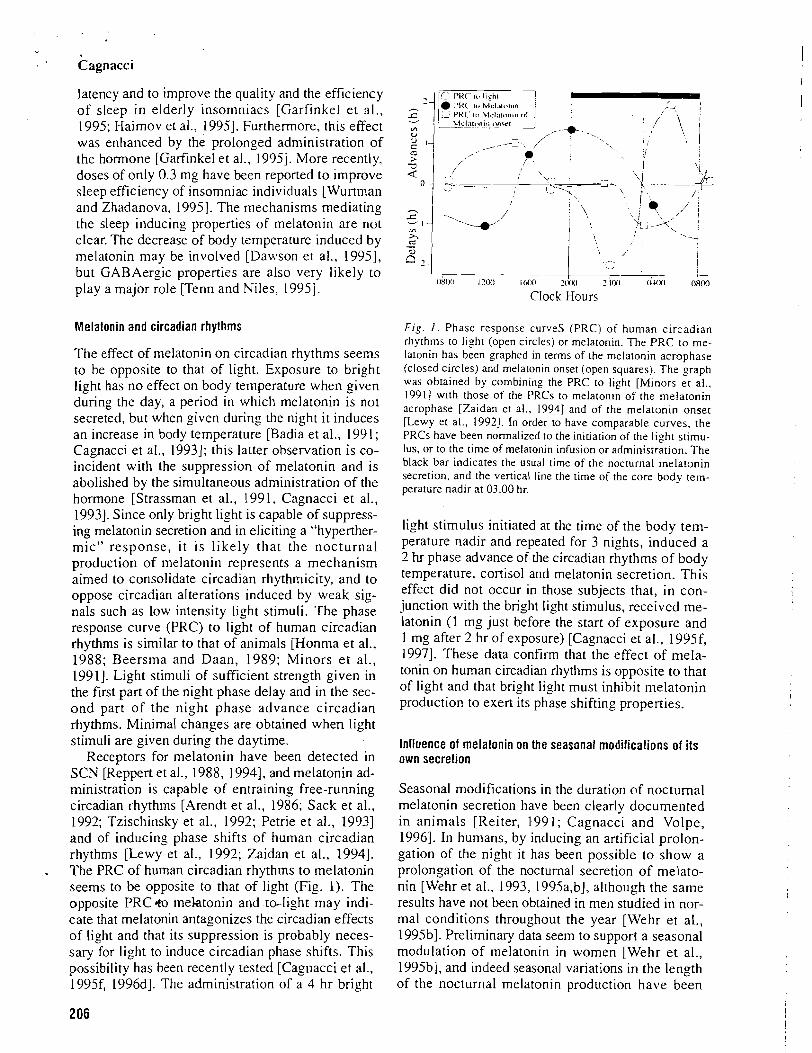
Cagnacci
latency and to improve the quality and the efficiency of sleep in elderly insomniacs [Garfinkel et al., 1995; Haimov et al., 19951. Furthermore. this effect was enhanced by the prolonged administration of the hormone [Garfinkel et al., 19951. More recently, doses of only 0.3 mg have been reported to improve sleep efficiency of insomniac individuals [Wurtman and Zhadanova, 19951. The mechanisms mediating the sleep inducing properties of rnelatonin are not clear. The decrease of body temperature induced by melatonin may be involved [Dawson et a]., 19951, but GABAergic properties are also very likely to play a major role [Tenn and Niles. 19951.
Melatonin and circadian rhythms
The effect of melatonin on circadian rhythms seems to be opposite to that of light. Exposure to bright light has no effecr on body temperature when given during the day, a period in which melatonin is nor secreted, but when given during the night it induces an increase in body temperature [Badia et al., 199 1 ; Cagnacci et al., 19931; this latter observation is co- incident with the suppression of melatonin and is abolished by the simultaneous administration of the hormone [Strassman et al., 1991, Cagnacci et al., 19931. Since only bright light is capable of suppress- ing melatonin secretion and in eliciting a "hyperther- mic" response, i t is likely that the nocturnal production of melatonin represents a mechanism aimed to consolidate circadian rhythmicity, and to oppose circadian aIterations induced by weak sig- nals such as low intensity light stimuli. The phase response curve (PRC) to light of human circadian rhythms is similar to that of animals [Honma et al., 1988; Beersma and Daan, 1989; Minors et al., 19911. Light stimuli of sufficient strength given in the first part of the night phase delay and in the sec- ond part of the night phase advance circadian rhythms. Minima1 changes are obtained when light stimuli are given during the dayrime.
Receptors for melatonin have been detected in SCN [Reppert et al., 1988, 19941, and melatonin ad- ministration is capable of entraining free-running circadian rhythms [Arendt et al., 1986; Sack et al., 1992; Tzischinsky et al., 1992; Petrie et al., 19931 and of inducing phase shifts of human circadian rhythms [Lewy et al., 1992; Zaidan et al., 19941. The PRC of human circadian rhythms to melatonin seems to be opposite to that of light (Fig. 1). The opposite PRC& mektonin and ta-li,oht may indi- cate that melatonin antagonizes the circadian effects of light and that its suppression is probably neces- sary for light to induce circadian phase shifts. This possibility has been recently tested [Cagnacci et al., 1995f, 1996dl. The administration of a 4 hr bright
wr- l L s 1iz"t -1 r - j
PWC' klclntnn~n ( d
m , , ,
,
1' .
a - +- ~ b o o 1.w Icdm zcKnl 2xx1 (iLoo o k ~ n
Clock Hours
Fig. I . Phase response curves (PRC) of human circadian rhythms to light (open circles) or melatonin. The PRC to me- latonin has been graphed in terms of the melatonin acrophase (closed circles) and rnelatonin onset (open squares). The graph was obtained by combining the PRC to light [Minors e t al., 19911 with those of the PRCs to melatonin of the melatonin acrophase [Zaidan et al., 19941 and of the melatonin onset [Lewy et al., 19921. In order to have comparable curves, the PRCs have been normalized to the initiation of the light stimu- lus, or to [he time of melatonin infusion or administration. The black bar indicates the usual time of the nocturnal melatonin secretion, and the vertical line the time of the core body tern- perature nadir at 03.00 hr.
light stimulus initiated ar the time of the body tem- perature nadir and repeated for 3 nights, induced a 2 hr phase advance of the circadian rhythms of body temperature, cortisol and rnelatonin secretion. This effect did not occur in those subjects that, in con- junction with the bright light stimulus, received me- latonin (1 mg just before the start of exposure and 1 mg after 2 hr of exposure) [Cagnacci et al., 1995f, 19971. These data confirm that the effect of mela- tonin on human circadian rhythms is opposite to that of light and that bright light must inhibit rnelatonin production to exert its phase shifting properties.
Influence of rnelatonin on the seasonal modifications of its own secretion
Seasonal modifications in the duration of noctumal melatonin secretion have been clearly documented in animals [Reiter, 199 1 ; Cagnacci and Volpe, 19961. In humans, by inducing an artificial prolon- gation of the night it has been possible to show a prolongation of the noctumal secretion of melato- nin [Wehr et al., 1993, 1995a,b], although the same results have not been obtained in men studied in nor- mal conditions throughout the year [Wehr et al., 1995bI. Preliminary data seem to support a seasonal modulation of melatonin in women [Wehr et al., 1995b1, and indeed seasonal variations in the length of the nocturnal melatonin production have been
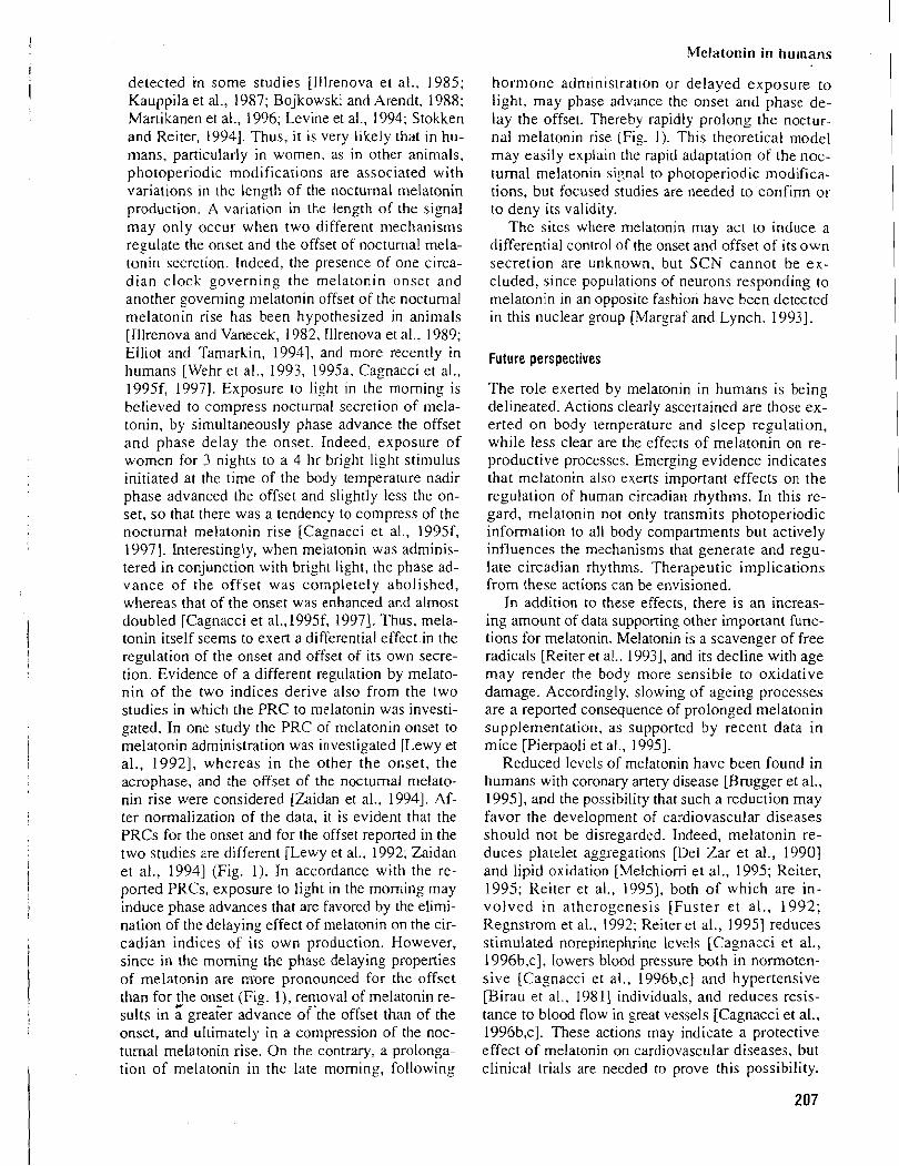
Melatonin in humans
detected in some studies [Illrenova et al.. 1985; Kauppila et al., 1987; Bojkowski and Arendt, 1988; Martikanen et a]., 1996; Levine et al., 1994; Stokken and Reiter, 19941. Thus, it is very likely that in hu- mans, particularly in women, as in other animals, photoperiodic modifications are associated with variations in the length of the nocturnal melatonin production. A variation in the length of the signal may only occur when two different mechanisms regulate the onset and the offset of nocturnal mela- tonin secretion. Indeed, the presence of one circa- dian clock governing the melatonin onset and another governing melatonin offset of the nocturnal melatonin rise has been hypothesized in animals [Illrenova and Vanecek, 1982, Illrenova et al., 1989; Elliot and Tamarkin, 19941, and more recently in humans [Wehr et al., 1993, 1995a. Cagnacci et al., 1995f, 19971. Exposure to light in the morning is believed to compress nocturnal secretion of rnela- tonin, by simultaneously phase advance the offset and phase delay the onset. Indeed, exposure of women for 3 nights to a 4 hr bright light stimulus initiated at the time of the body temperature nadir phase advanced the offset and slightly less the on- set, so that there was a tendency to compress of the nocturnal melatonin rise [Cagnacci et a!., 1995f, 19971. Interesringly, when melatonin was adminis- tered in conjunction with bright light, the phase ad- vance of the offset was completely abolished, whereas that of the onset was enhanced and almost doubled [Cagnacci et a1.,1995f, 19971. Thus, mela- tonin itself seems to exert a differential effect.in the regulation of the onset and offset of its own secre- tion. Evidence of a different regulation by melato- nin of the two indices derive also from the two studies in which the PRC to melatonin was investi- gated. In one study the PRC of melatonin onset to melatonin administration was investigated [Lewy et al., 19921, whereas in the other the onset, the acrophase, and the offset of the nocturnal melato- nin rise were considered [Zaidan et al., 19941. Af- ter normalization of the data, it is evident that the PRCs for the onset and for the offset reported in the two studies are different [Lewy et al.. 1992; Zaidan et al., 19941 (Fig. 1). In accordance with the re- ported PRCs, exposure to light in the morning may induce phase advances that are favored by the elimi- nation of the delaying effect of melatonin on the cir- cadian indices of its own production. However, since in the morning the phase delaying properties of melatonin are m-ore pronounced for the offset than for he onset (Fig. I), removal of melatonin re- sults in a greakr advance orthe offset than of the onset, and ultimately in a compression of the noc- turnal melatonin rise. On the contrary, a prolonga- tion of melatonin in the late morning, following
hormone administrat~on or delayed exposure to light, may phase advance the onset and phase de- lay the offset. Thereby rapidly prolong the noctur- nal melatonin rise (Fig. 1). This theoretical model may easily explain the rapid adaptation of the noc- turnal melatonin signal to photoperiodic modifica- tions, but focused studies are needed to confirm or to deny its validity.
The sites where melatonin may act to induce a differential control of the onset and offset of its own secretion are unknown, but SCN cannot be ex- cluded. since populations of neurons responding to melatonin in an opposite fashion have been detected in this nuclear group [Margraf and Lynch. 19931.
Future perspectives
The role exerted by melatonin in humans is being delineated. Actions clearly ascertained are those ex- erted on body temperature and sleep regulation, while less clear are the effects of melatonin on re- productive processes. Emerging evidence indicates that melatonin also exens important effects on the regulation of human circadian rhythms. In this re- gard, melatonin not only transmits photoperiodic information to all body compartments but actively influences the mechanisms that generate and regu- late circadian rhythms. Therapeutic implications from these actions can be envisioned.
In addition to these effects, there is an increas- ing amount of data supporting other important func- tions for melatonin. Melatonin is a scavenger of free radicals [Reiter et al., 19931, and its decline withage may render the body more sensible to oxidative damage. Accordingly, slowing of ageing processes are a reported consequence of prolonged melatonin supplementation, as supported by recent data in mice [Pierpaoli et al., 19951.
Reduced levels of melatonin have been found in humans with coronary artery disease [Brugger et al., 19951, and the possibility that such a reduction may favor the development of cardiovascular diseases should not be disregarded. Indeed, melatonin re- duces platelet aggregations [Del Zar et al., 19901 and lipid oxidation [Melchiorri et a]., 1995; Reiter, 1995; Reiter et al., 19951, both of which are in- volved in atherogenesis [Fuster .et al., 1992; Regnstrom et al., 1992; Reiter et al., 19951 reduces stimulated norepinephrine levels [Cagnacci et al., 1996b,c], lowers blood pressure both in normoten- sive [Cagnacci et al., 1996b,c] and hypertensive [Birau et al., 19811 individuals, and reduces resis- tance to blood flow in great vessels [Cagnacci et at., 1996b,c]. These actions may indicate a protective effect of melatonin on cardiovascular diseases, but clinical trials are needed to prove this possibility.
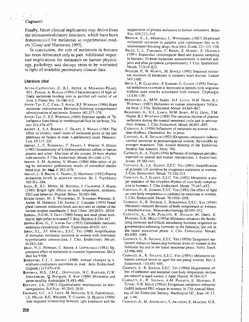
Finally, future clinical implications may derive from the immunomodulatory functions. which have been dernonstr2;cd for melatonin in experimental mod- els [Conti and Maestroni,l995].
In conclusion, the role of melatonin in humans has been delineated only in part. Additional impor- tant implications for melatonin on human physiol- ogy, pathology, and therapy seem to be warranted in light of available preliminary clinical data.
Literature cited
A C ~ ~ A - C A S T R O V ~ E ~ ~ . D., R.J., REITER, A. MENAKDEZ-PELAEZ, M.I., PABLOS. A. BURGOS (1994) Characterization of high af- finity melatonin binding sites in purified cell nuclei of rar liver. J. Pineal Res. lh:100-112.
Amos-TAY, F., C. CHOU. S. Amos, R.J. WURTMAK (1968) Brain serotonin concentration: Elevation following intraperitoneal administration of melatonin. Science 162:277-278.
A ~ o N - T A Y , F.. R.J. WURTMAV (1969) Regional uptake of 3 ~ - melatonin from blood or cerebrospinal fluid by rat brain. Na- ture 22 1:474-475.
ARENDT, J., A.A. BORBELY. C. FRANEY. J. WRIGHT (1984) The effect of chronic, small doses of melatonin given in the late afternoon on fatigue in man: A preliminary study. Neurosci. Lett. 45:3 17-32 1.
ARENDT, J., C. BOJKOWSKI, C. FRANEY. J. WRIGHT, V. MARKS (1985) Irnmunoassay of 6-hydroxymelatonin sulfate in human plasma and urine: Abolirion of the urinary 24-hour rhythm with atenolol. J. Clin. Endocrinol. Metab. 60: 11661 173.
ARENDT, J., M. ALDHOUS. V. MARKS (1986) Alleviation of jet lag by melatonin: preliminary results of conrrolled double blind trial. Br. Mad. J. 2921 170.
ARENDT. J., S. BHANJI, C. FRANEY, D. MATTIVGLY (1992) Plasma melatonin levels in anorexia nervosa. Br. J. Psychiatry 161:361-364.
BADIA, P,, B.L. MYERS, M. BOCCKER, J. CULPEPPER, J. HARSH (1991) Bright light effects on body temperature, alertness. EEG and behavior. Physiol. Behav. 50583-588.
BECKER-ANDRE, M.. I. WIESENBERG, N. SCHAEREN-WIEMERS, E. . ~ D R E , M. MISSBACH, J.H. SAURAT, C. CARLBERG (1994) Pineal gland hormone melatonin binds and activates an orphan of the nuclear receptor superfamily. J. Biol. Chem. 269:2853 1-28534.
Bwm, D.G.M., S. D m (1989) Strong and weak phase reset- ring by light pulses in humans? J. Biol. Rhythms 8:340-347.
BENTTEE-KING, G., F. ANTON-TAY (1993) Calmodulin mediates melatonin cytoskeletal effects. Experientia 49:635-64 1.
BERGA, S.L., J.F. MORTOLA, S.S.C. YES (1988) Amplification of nocturnal melatonin secretion in women with functional hypothalamic amenorrhea. J. Clin. Endocrinol. Metab. 66:242-244.
BIRAU, N.,U. PETRSSEN, C. MEYER, J. GOTTSCHALK (198 1 ) Hy- potensive effect of melatonin in essential hypertension. IRCS Med Sci 9:906
Boreows~r, C.J., J. ARENDT (1988) Annual changes in 6- sulphatoxymelatonin excretion in man. Acta Endocrinol. (Copenh.) I 17:470-476.
BORNMAN, M.S., J.M.C., OOSTHUIZEN, H.C. BARNARD, G.W. SCHULENBCRG, Q B O O ~ K E R . S. REIF (1989)-Melatonin and sperm motility. .4ndrologia 21:483486.
SOULANT, J.A. (198 1) Hypothalamic mechanisms in ther- moregulation. Fed Proc. 40:2843-2850.
BRAINARD, G.C., A.J. LEWY, M. MENAKER, R.H. FREDRICKSON, L.S. MILLER, R.G. WELEBER, V. CASSONE. D. HCDSOV (1988) Dose-response relationship between ltgtlt irradiance and the
suppression of plasma melatonin in human volunreers. Brain Res. 4542 17-2 18.
BRISMAR, K., L. ~IOGEXSEN, L. WETTERBERG (1987) Depressed rnelatonin secrerion in patienrs with nightmares due ro b- adrenoceptor blocking drugs. Acta Med. Scand. 12 I : 155-1 58.
BRUCE, J., L. TAMARKIX, C. RIEDEL. S. MARKEY. E. OLDFIELD ( 199 1 ) Sequential cerebrospinal fluid and plasma sampling in humans: 24-hour melatonin measurements in normal sub- jects and afrer peripheral syrnpathectomy. J. Clin. Endocrinol. Metab. 7 2 8 19-833.
BRUGGER, P., W. MARKTL, M. HEROLD (1995) Impaired nocrur- nal secretion of rnelatonin in coronary heart disease. Lancet 345: 1408.
BRLN, J., B. CLALSTRAT. P. SADDIER, G. CHAZOT (1995) Noctur- nal melatonin excretion is decreased in patients with migraine without aura attacks associared wirh menses. Cephalgia 15:136-139.
BRZEZINSKI. A.. M.M. SEIBEL, H.J. LYNCH. M.H. DENG, R.J. WL'RTMAN (1987) Melatonin in human preovulatory follicu- lar fluid. J. Clin. Endocrinol. Merab. 64:865-867.
BRZEZINSKI, A.. H.J.. LYNCH. M.M. SEIBEL, M.H. DENG. T.M. NADER, R.J. WURTMAN (1988) The circadian rhythm of plasma rnelaronin during the normal mensrmal cycle and in amenor- rheic women. J. Clin. Endocrinol. Metab. 66:891-895.
CAGNACCI, A. (1996) Influences of melatonin on human circa- dian rhythms. Chronobiol. Int. in press.
CAGNACCI, A., R. SOLDANI (1992) Exogenous melatonin induces cortisol secretion in posrmenopausal women: Reversible by estrogen Treatment. 74th Annual Meeting of [he Endocrine Sociery, San Antonio, Abstr. 796.
CAGNACCI, A., A. VOLPE (1996) Influence of melatonin and pho- toperiod on animal and human reproduction. J. Endocrinol. Invest. 19:382411.
CAGNACCI, A.. J.A. ELLIOTT. S.S.C. YES (1991) Amplification of pulsatile LH secretion by exogenous melatonin in women. J. Clin. Endocrinol. Metab. 73:210-212.
CAGPI'CCI. A., J. ELLIOTT. S.S.C. YES (1992) Melatonin: a rna- jor regulator of rhe circadian rhythm of core body rempera- ture in humans. J. Clin. Endocrinol. Metab. 75:147452.
CAGNACCI, A., R. SOLDANI, S.S.C. YEN (1993) The effect of light on core body remperarure is mediated by melatonin in women. J. Clin. Endocrinol. Metab. 76:103&-1038.
CAGNACCI, A., R. SOLDANI, C. ROMAGNOLO, S.S.C. YEN (1994) Melatonin-induced decrease of body temperature in women: a threshold event. Neuroendocrinology 60549-552.
CAGNACCI, A., A.M. PAOLETTI. R. SOLDANI, M. ORRO, E. MASCHIO, G.B. MELIS (1995a) Melatonin enhances the lutein- izing hormone and follicle stimulating hormone responses to gonadotropin-releasing hormone in the follicular, but not in the luteal mensrrual phase. J. Clin. Endocrinol. Metab. 80: 1095-1099.
CAGNACCI, A.. R. SOLDAXI, S.S.C. YEN (1995b) Exogenous me- latonin enhances luteinizing hormone levels of women in the follicular but not in the luteal mensrmal phase. Fertil. Steril. 63:99&999.
CAGNACCI. A.. R. SOLDANI, S.S.C. YEN (1995~) Melatonin en- hances cortisol levels in aged but not young women. Eur. J. Endocrinol. 133:69 1-695.
CAGNACC~, A., R. SOLD~NI. S.S.C. YEK (1995d) Hypothermic ef- fect of melatonin and nocturnal core body temperature decline are reduced in aged women. J. Appl. Physiol. 78:3 14-3 17.
CAGNACCI. A., R. SOLDANI, A.M. PAOLETTI, E. MASCHIO, F. TUVERI. G.B. MELIS (1995e) Exogenous melatonin enhances GnRH-induced PRL release in women. In 77th Annual Meet- ing of the Endocrine Society, Washington DC, June 14-17, pp. 1-96.
CAGKACCI, A,, M. AYGIOLUCCI, S. ARASGINO, E. ~ IASCHIO, G.B.
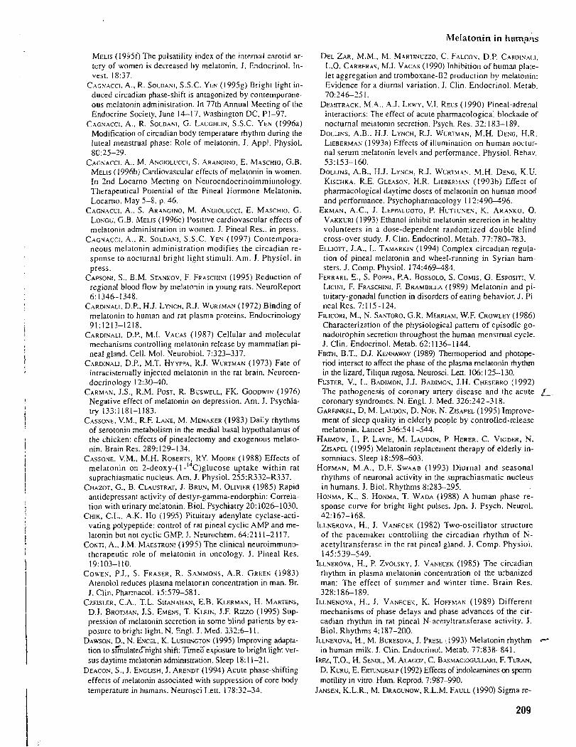
Melatonin in hump&
MELIS (1935f) The pulsatility index of the internal carotid ar- tery of women is decreased by melatonin. J. Endocrinol. In- vest. 18:37.
CAGSACCI. A., R. SOLDANI, S.S.C. YEN ( 1 995g) Bright light in- duced circadian phase-shift is antagonized by contemporane- ous melatonin administration. In 77th Annual Meeting of the Endocrine Society, June 14-17. Washington DC, PI-97.
CAGSACCI. A.. R. SOLDANI, G. LAUGHLIN, S.S.C. YEN (1996a) Modification of circadian body temperature rhythm during the iuteal menstrual phase: Role of melatonin. J. Appl. Physiol. 80:25-29.
CAGNACCI. A.. M. ANGIOLUCCI, S. ARANGISO. E. MASCHIO, G.B. MELIS ( 1996b) Cardiovascular effects of melatonin in women. In 2nd Locarno Meeting on Seuroendocrinoimmiunology. Therapeutical Potential of the Pineal Hormone Melatonin. Locarno. May 5-8. p. 46.
CACNACCI. A.. S. ARANCINO. M. AYGIOLUCCI. E. MASCHIO. G. LOYGG. G.B. MELIS ( I 996c) Positive cardiovascular effects of melatonin administration in women. J. Pineal Res.. in press.
C ~ o r ~ c c r . .4.. R. SOLDANI. S.S.C. YEY (1997) Contempora- neous melatonin administration modifies the circadian re- sponse to nocturnal bright light stimuli. Am. J. Physiol. in press.
CAPSONI, S.. B.M. STANKOV. F. FRASCHINI (1995) Reduction of regional blood flow by melatonin in young rats. NeuroReporr 6: 1346-1348.
CARDINALI. D.P.. H.J. LYNCH, R.J. WURTMAN (1972) Binding of melatonin to human and rat plasma proteins. Endocrinology 91:1213-1218.
CARDINALI, D.P., M.I. VACAS ( 1987) Cellular and molecular mechanisms controlling melatonin release by mammalian pi- neal gland. Cell. Mol. Neurobiol. 7:323-337.
CARDINALI, D.P., M.T. HYYPPA, R.J. WURTMAN (1973) Fate of intiacisternally injected melatonin in the rat brain. Neuroen- docrinology 12:30-40.
CARMAN, J.S., R.M. POST, R. BUSWELL, FK. GOODWIN (1976) Negative effect of melatonin on depression. Am. J. Psychia- try 133:1181-1183.
CASSONE, V.M., R.F. L ~ N E . M. MENAKER (1983) Daily rhythms of serotonin metabolism in the medial basal hypothalamus of the chicken: effects of pinealectomy and exogenous melato- nin. Brain Res. 289: 129-1 34.
CASSOXE, V.M., M.H. ROBERTS, RY. MOORE (1988) Effects of melatonin on 2 - d e o ~ ~ - ( l - ' ~ ~ ) ~ l u c o s e uptake within rat suprachiasmatic nucleus. Am. J. Physiol. 255:R332-R337.
CHAZOT, G., B. CLAUSTRAT, J. BRUN, M. OLIVIER (1985) Rapid antidepressant activity of destyr-gamma-endorphin: Correla- tion with urinary melatonin. Biol. Psychiatry 20: 102&-1030.
CHIK, C.L., A.K. HO (1995) Pituitary adenylate cyclase-acti- vating polypeptide: control of rat pineal cyclic AMP and me- latonin but not cyclic GMP. J. Neurochem. 64:2111-2117.
CONTI. A., J.M. MAESTRONJ (1995) The clinical neuroimmuno- therapeutic role of melatonin in oncology. J. Pineal Res. 19:103-110.
COWES, P.J., S. FRASER, R. SAMMONS, A.R. GREEN (1983) Atenolol reduces plasma melatonin concentration in man. Br. J. Clin. Pharmacol. 15579-58 1.
CZEISLER, C.A., T.L. SHANAHAN. E.B. KLERMAN, H. MARTENS, D.J. BROMAN, J.S. EMENS, T. KLEIN, J.F. RIZZO (1995) Sup- pression of melatonin secretion in some blind patients by ex- posure to bright light. N. Engl. J. Med. 3325-1 1.
DAWSON. D., N. EXCEL, K. LUSHKCTON (1995) Improving adapta- tion to s&ulatecTnight shift: Timea exposure to bright light ver- sus daytime melatonin administration. Sleep 18: 1 1-2 1.
DEACON, S., J. ENGLISH, J. ARENDT (1994) Acute phase-shifting effects of rnelatonin associated with suppression of core body temperature in humans. Neurosci Lett. 175:32-34.
DEL ZAR, M.M.. M. MARTINUZZO. C. FALCOV, D.P. CARDIYALI, L.O. CARRERAS. M.I. VACAS (1990) Inhibition of human plate- let aggregation and tromboxane-B2 production by melatonin: Evidence for a diurnal variation. J. Clin. Endocrinol. Metab. 70:24&25 1.
DEX~ITRACK. M.4.. A.J. LEWY. V.I. REUS (1990) Pineal-adrenal interactions: The effect of acute pharmacological blockade of nocturnal melatonin secretion. Psych. Res. 32: 183-1 89.
DOLLINS, A.B.. H.J. LYNCH, R.J. WLRTXIAN, M.H. DESG, H.R. LIEBERMAS (1993a) Effects of illumination on human noctur- nal serum melatonin levels and performance. Physiol. Behav. 53:153-160.
DOLLINS, A.B.. H.J. LYSCH, R.J. WURMAZ. M.H. DENG, K.U. KISCHKA. R.E. GLEASOS. H.R. LIEBERLI.A~ (1993b) Effect of pharmacological daytime doses of melatonin on human mood and performance. Psychopharmacology 11 2 4 9 0 3 9 6 .
EKMAN, A.C.. J. LEPPALI;OTO. P. HLTTUSEN, K. ARANKO, 0. V A K K ~ R I (1993) Ethanol inhibit melatonin secretion in healthy volunteers in a dose-dependent randomized double blind cross-over study. J. Clin. Endocnnol. Metab. 77:780-783.
ELLIOTT, J.A.. L. TAMARKIN (I 994) Complex circadian regula- tion of pineal melatonin and wheel-running in Syrian ham- sters. J. Comp. Physiol. 174:469-484.
FERRARI. E., S. POPPA, P.A. BOSSOLO. S. COMIS, G. ESPOSITI. V. LICINI. F. FRASCHINI. F. BRAMBILLA (1989) Melatonin and pi- tuitary-gonadal function in disorders of eating behavior. J. Pi- neal Res. 7: 1 15-1 24.
FILICORI, M.. N. SANTORO, G.R. MERRIAM, W.F. CROWLEY (1986) Characterization of the physiological pattern of episodic go- nadotrophin secretion throughout the human mensrmal cycle. J. CIin. Endocrinol. Metab. 62: 1136-1 114.
F m , B.T.. D.J. KENNAWAY (1989) Thermoperiod and photope- nod interact to affect the phase of the plasma melatonin rhythm in the lizard, Tiliqua rugosa. Neurosci. Lett. 106: 125-130.
FUSTER, V.. L. BADIMON, J.J. BADIMOX. J.H. CHESEBRO (1992) The pathogenesis of coronary anery disease and the acute A. coronary syndromes. N. Engl. J. Med. 326:242-3 18.
GARFINKEL, D. M. LAUDON, D. NOF, K. ZEAPEL (1 995) Improve- ment of sleep quality in elderly people by controlled-release melatonin. Lancet 346541-544.
HAIMOW, I., P. LAVIE, M. LAUDON, P. HERER. C. VIGDER, N. ZISAPEL (1995) Melatonin replacement therapy of elderly in- somniacs. Sleep 18598-603.
HOFMAN. M.A., D.F. S W A . ~ B (1993) Diurnal and seasonal rhythms of neuronal activity in the suprachiasmatic nucleus in humans. J. Biol. Rhythms 8:283-295.
HONMA, K., S. HONWA, T. WADA (1988) A human phase re- sponse curve for bright light pulses. Jpn. J. Psych. Neurol. 42:167-168.
ILLNEROVA. H.. J. VANECEK (1982) Two-oscillator structure of the pacemaker controlling the circadian rhythm of N- acetyltransferase in the rat pineal gland. J. Comp. Physiol. 145539-549.
ILLNEROVA, H.. P. ZVOLSKY, J. VANECEK (1985) The circadian rhythm in plasma melatonin concentration of the urbanized man: The effect of summer and winter time. Brain Res. 328: 186- 189.
ILLNENOVA, H.. J. VANECEK. K. HOFF~IAN (1989) Different mechanisms of phase delays and phase advances of the cir- cadian rhythm in rat pineal N-aceryltransferase activity. J. Biol. Rhythms 4: 237-200.
ILLNEROVA, H., M. BURESOVA, J. PRESL : i993) Melatonin rhythm +
in human milk. J. Clin. Endocrinol. Merab. 772338-841. Ircn, T.O., H. S m o ~ , M. A L A C ~ , C. B.-\S>LACIOGLZLARI. F. TURAN,
D. KLRU, E. E r n % c w (1992) Effects of indoleamines on sperm motility in vitro. Hum. Reprod. 7:987-990.
JANSEN, K.L.R.. M. DRAGLNOW, R.L.M. FAULL (1990) Sigma re-
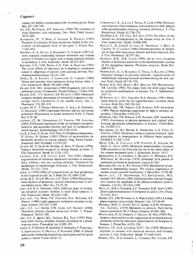
Cagnacci
ceptors are highly concentrated in the rat pineal gland. Brain Res. 507: 158-1 60.
JAN, J.E., H. ESPEZEL. R.E. APPLETOX (1994) The treatment of sleep disorders with melatonin. Dev. bled. Child Neurol. 36:97-107.
KANEMATSL, N.. Y. MORI, S. HAYASHI, K. HOSHISO (1989) Presence of a distinct 24-hour melatonin rhythm in the ven- tricular cerebrospinal fluid of the goat. J. Pineal Res. 7.143-151.
KAUPPILA. A., A. KIVELA. A. P.AKARINEN. 0. VAKKLRI ( 1987) In- verse seasonal relationship between melatonin and ovarian activity in humans in a region with a strong seasonal contrast in luminosity. J. Clin. Endcrinol. Metab. 65:823-827.
KENNEDY, S.H.. G.M. B ~ o w x , C.G. FORD, E. RALEVSKI (1993) The acute effects of starvation on 6-sulphatoxy-melatonin output in subgroups of patients with anorexia nervosa. Psy- choneuroendocrinolo,ov 18: 13 1-1 39.
KIVELA, K.. A. KACPPII.A. J. LEPPALUOTO. 0. VAKKLRI (1989) Serum and amniotic fluid melatonin during human labor. J. Clin. Endocrinol. Metab. 69: 1065- 1068.
KRAUSE. D.N.. M.L. DGBOCOVICH (1990) Regulatory sites in the melatonin system of mammals. Trends Neurosci. 13:464-470.
KRAUSE, D.N., V.E. BARRIOS, S.P. DUCKLES (1995) Melatonin receptors mediate potentiation of contractile responses to adr- energic nerve stimulation in rat caudal artery. Eur. J. Phannacol. 276:207-2 13.
LAAKSO, 1M.-L., T. PORKKA-HEISKANEN, A. ALILA, D. STENBERG, G. JOHANSSOS (1990) Correlation between salivary and serum melatonin: Dependence on serum melatonin levels. J. Pineal Res. 9:39-50.
LAITINEN, J.T., M. VISWANATHAK. 0. VAKKLRI, J.M. SAAVEDRA (1992) Differential regulation of the rat melatonin receptors: Selective age-associated decline and lack of melatonin-in- duced changes. Endocrinology 13012 139-2144.
LAL,~., I. ISAAC. C. PUPIL, N.O. NALR, N. HARISHARASUBRAMANIAN, H. GUYDA, R. QUIRON (1987) Effects of apomorphine on melatonin secretion in normal subjects. Prog. Neuro-Psycho- pharmacol. Biol. Psychiatry 11:219-233.
LA~DO~.- . M., E. GILAD, H. MATZKIK. Z. BRAF, N. ZISAPEL (1996) Putative melatonin receptors in benign human prostate. J. Clin. Endocrinol. Metab. 8 1 : 1336-1 341.
LALGHLIY, G.A., A.B. L o u c ~ s , S.S.C. YEN (1991) Marked augmentation of nocturnal melatonin secretion in amenor- rheic athletes, but not cycling athletes: Unaltered by opioidergic or dopaminergic blockade. J. Clin. Endocrinol. Metab. 73: 1321-1326.
LEDUC, J. (1976) Effect of sympatectomy on heat production in rats exposed to cold. Isr. J. Med. Sci. 12: 1099-1 103.
LEVINE, M.E., N.A.K. MILLIRO. L.K. DUFFY (1994) Diurnal sea- sonal rhythms of melatonin. cortisol and testosterone in inte- rior Alaska Arctic. Med. Res. 53:25-34.
LEWY. A.J, D.A. NEU'SOME (1983) Different types of melato- nin circadian secretory rhythms in some blind subjects. J. Clin. Endocrinol. Metab. 56: 1103-1 107.
LEWY. A.J., T.A. WEHR, F.K. Gooowt~ . D.A. NEWSOME, S.P. MARKEY (1 980) Light suppresses melatonin secretion in hu- mans. Science 2 10: 1267-1 269.
LEWY, A.J.. L.J. SIEVER, T.W. UHDE, S.P. MARKEY (1986) Clonidine reduces plasma melatonin levels. J. Pharm. Pharmacol. 38555-556.
LEWY, A.J., S. U~~ED. U.L. JACKSON,~,R.~. S+CK (1992) Mela- [onin shifts human circadian rhythms according to a phase- response curve. Chronobiol. Int. 9:38&392.
LasoxI. P., D. Es~osm. M. &E?.FTL%I. F. MOR~SITO, P. FUMAGALLI, A. S ~ ~ w s n N o , G. DELITALA, F. FR~SCHMI (1986) A clinical study on the relationship between the pineal gland and the opioid system. J. Neural Transm. 6563-73.
L~BOSHITZKY. R., S. LAW. I. THLMA. P. LAYIE (1996) Testoster- one treatment alters melatonin concentrations in male patients with gonadotrophin-releasing hormone deficiency. J. Clin. Endocrinol. Metab. 8 1 :77&774.
MACPHEE A.A., F.E. COLE, B.F. RICE (1975) The effect of me- latonin on steroidogenesis by the human ovary in vitro. J. Clin. Endocrinol. .Melab. 40:688-696.
MALLO, C., R. ZAID.AN, G. GALY, E. VERUELLEK. J. BRLS. G. CHAZOT. B. CLACSTRAT (1990) Pharmacokinetics of melato- nin in man after intravenous infusion and bolus injection. Eur. J. Clin. Pharmacol. 38:297-30 I .
MARGRAF. R.R., G.R. LYNCH (1993) An in vitro circadian rhythm of melatonin sensitivity in the suprachiasmatic nucleus of the djungarian hamster. Phodopus sungorus. Brain Res. 609:45-50.
MARTIKAISES. H., A. RCOKONEN, C. TOM.AS. .4. KALPPILA (1996) Seasonal chanses in pituitary function: Amplification of midfollicular luteining hormone secretion during the dark sea- son. Fenil. Steril. 65:7 18-720.
MARTIN, X.D., H.Z. MALIYA, M.C. BRESSAN, P.H. HENDRICKSON, P.R. LICHTER (1992) The ciliary body-the third or_ean found to synthesize indoleamines in humans. Eur. J. Ophthalmol. 2:67-71.
MCARTHLR, A.J., M.U. GILLETTE, R.A. PROSSER ( 199 1 ) Mela- tonin directly resets the rat suprachiasmatic circadian clock in vitro. Brain Res. 565:158-161.
MCINNRE, LM., T.R. XORMAS. G.D. BLRRO\VS. S.M. ARMSTROF;C (1989) Human melatonin suppression by light is intensity de- pendent. J. Pineal Res. 6 149-2 13.
M c I ~ E , I.M.. T.R. KOR.WAN, G.D. BLRROWS, S.M. ARMSTRONG (1993) Alterations to plasma melatonin and cortisol after evening alprazolam administration in humans. Chronobiol. Int. 10:205-2 13.
MELCHIORRI. D., R.J. REITER, E. SEWERYSEK, L.D. CHEN, G. NISTICO (1995) Melatonin reduces kainate-induced lipid peroxidation in homopenates of different brain regions. FASEB J. 9: 1205-1 2 10.
MELIS. G.B., A. CAGSACCI, A.M. PAOLETTI, R. SOLDANI, M. ORR~; , C. SECCI (1995) Melatonin administration increases TSH secretion in the follicular but not in the luteal phase of the woman menstrual cycle. J. Endocrinol. Invest. 18:37.
MENDLEWICZ. J., P. LISKOWSKI, L. BR.ANCHEY, U. WEINBERG, E.D. WEITZMAN, M. BR.ANCHEY (1979) Abnormal 24-h pattern of melatonin secretion in depression. Lancet ii: 1362.
MENANDEZ-PELAEZ. A.. R.J. REITER (1993) Distribution of me- latonin in mammalian tissues: The relative importance of nuclear versus cytosolic localization. J. Pineal Res. 15:59-69.
MEYER, A.C.. J.J. NIECWENHL'IS, V.J. KOCISZEWSKA, W.S. JOUBERT, B.J. MEYER (1986) Dihydropyridine calcium antago- nists depress the amplitude of the plasma melatonin cycle in baboons. Life Sci. 39:1563-1569.
MILES, A., D.R.S. PHILBRICK, S.F. TIDMARSH, D.M. SHAW (1985) Direct radioimmunoassay of melatonin in saliva. Clin. Chem. 31:1412-1413.
MINORS. D.S., J.M. WATERHOUSE, A. WIRZ-JUS~CE (1991) A human phase-response curve to light. Neurosci. Lett. 133:36-40.
MITCHELL, M.D., L. SAYERS, M.J..C. KEIRSE,A.B.M. ANDERSON. A.C. TURNBULL (1978) Melatonin in amniotic fluid during human parturition. Br. J. Obstet. Gynecol. 85:684-686.
MONTELEOSE, P., D. FORZIATI, C. ORAZZO, M. MAJ (1989) Pre- liminary observations on the suppression of nocturnal plasma melatonin levels by short-term administration of diazepam in humans. J. Pineal Res. 6253-258.
MORTOLA. J.F.. G.A. LACGHLIN, S.S.C. YES (1993) Melatonin rhythms in women with anorexia nervosa and bulimia nervosa. J. Clin. Endocrinol. Metab. 77:1540-1544.
MURPHY, D.L., N.A. GARRICK. L. TAMARKIS, P.L. TAYLOR, S.P.
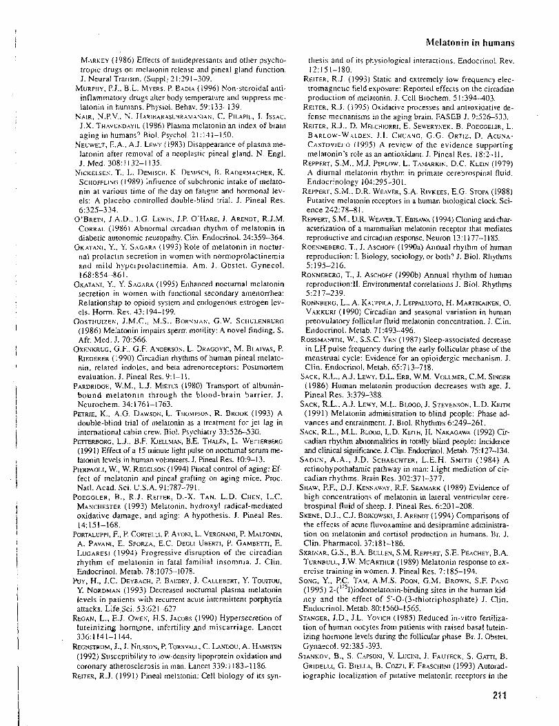
mel la ton in in humans
MARKEY (1986) Effects of antidepressants and other psycho- tropic drugs on melaronin release and pineal gland function. J. Neural Transm. !Suppl j 2 1 :29 1-309.
MURPHY. P.J.. B.L. MYERS. P. BADIA (1996) Non-steroidal anti- inflammatory drugs alter body temperature and suppress me- latonin in humans. Physiol. Behav. 59: 133-139.
NAIR. N.P.V.. N. HARIHARXSUBRA~IXSIAS, C. PILAPIL, I. ISSAC. J.X. THAVUSDAYIL (1986) Plasma melatonin an index of brain aging in humans? Biol. Psychol. 31 : 131-150.
NEUWELT, E.A.. A.J. LEVY (1983) Disappearance of plasma me- latonin after removal of a neoplastic pineal gland. N. Engl. J. Med. 308: 1 132-1 135.
NICKELSEN. T.. L. DEMISCH. K. DE\IISCH, B. RADERMACHER, K. SCHOFFLIVG (1989) Influence of subchronic intake of melato- nin at various time of the day on fatizue and hormonal lev- els: A placebo controlled double-blind trial. J. Pineal Res. 6:325-3 34.
O'BREIN, J.A.D.. 1.G. LEWIY, J.P. O'HARE, J. ARENDT, R.J.M. CORRAL (1986) Abnormal circadian rhythm of melatonin in diabetic autonomjc neuroparhy. Clin. Endocrinol. 24:359-364.
OKATANI, Y.. Y. SAGARA (1993) Role of melatonin in noctur- nal prolaclin secretion in women with normoprolactinemia and mild hyperprolactinemia. Am. J . Obstet. Gynecol. 168:854-861.
OKATANI. Y.., Y. SAGARA (1995) Enhanced nocturnal melatonin secretion in women with functional secondary amenorrhea: Relationship to opioid system and endogenous estrogen lev- els. H o r n . Res. 43: 194-199.
OOSTH~~ZES, J.M.C., M.S., B O R S ~ A S , G.W. SCHLLENBURG (1986) Melatonin impairs sperm motility: A novel finding. S. Afr. Med. J. 70566.
OXENKRLG. G.F., G.F. ANDERSON. L. DRAGOVIC, M. BLAIVAS, P. RIEDERER (1990) Circadian rhythms of human pineal melato- nin, related indoles, and beta adrenoreceptors: Postmonem evaluation. J. Pineal Res. 9: 1-1 1.
PARDRIDGE, W.M., L.J. MIETUS (1980) Transport of albumin- bound melatonin through the blood-brain barrier. J . Neurochem. 34: 176 1-1 763.
PETRIE, K.. A.G. DAWSOR, L. TIIOMPSOS, R. BROOK (1993) A double-blind trial of melatonin as a treatment for jet lag in international cabin crew. Biol. Psychiatry 33526-530.
FET~ERBORG. L.J., B.F. KJELLMAK, B.E. THALEN, L. W ~ R B E R G (1991) Effect of a 15 minule light pulse on nocturnal serum me- latonin levels in human volunteers. J. Pineal Res. 10:9-13.
PIERPAOLI, W., W. REGELSOK (1994) Pineal control of aging: Ef- fect of melatonin and pineal grafting on aging mice. Proc. Natl. Acad. Sci. U.S.A. 91:787-791.
POEGGLER, B., R.J. REITER, D.-X. TAN, L.D. CHEN, L.C. MANCHESTER (1993) Melaronin, hydroxyl radical-mediated oxidative damage, and aging: A hypothesis. J. Pineal Res. 14:151-168.
PORTALUPPI, F., P. CORTELLI, P. A\,osI. L. VERGNANI, P. ~ ' ~ ~ L T O N I N , A. PAVANI, E. SFORZA, E.C. DEGLI UBERTI, P. GAMBETTI, E. L U C A R E S ~ (1994) Progressive disruption of the circadian rhythm of melatonin in fatal familial insomnia. J. Clin. Endocrinol. Metab. 78:1075-1078.
Puy, H., J.C. DEYBACH, P. BAUDRY, J. CALLEBERT, Y. TOUITOU, Y. N O R D M A ~ (1993) Decreased noctumal plasma melatonin levels in patients with recurrent acute intermittent porphyria attacks. LifeSci. 53:621477.
REGAN, L., E.J. OWEN, H.S. JACOBS (1990) Hypersecretion of luteinizing hormone, infertility ~ n d .miscarriage. Lancet 336:1141-1144.
REGNSTROM. J., J. NILSSON, P. TORNWLL, C. LANDOL', A. HAMSTEY (1992) Susceptibility to low-density lipoprotein oxidation and coronary atherosclerosis in man. Lancet 339: 1 183-1 186.
REITER. R.J. (1991) Pineal melatonin: Cell biology of its syn-
thesis and of its physiological interactions. Endocrinol. Rev. 12:151-180.
REITER, R.J. (1993) Static and extremely low frequency elec- tromagnetic field exposure: Reported effects on the circadian produclion of melatonin. J. Cell Blochem. 5 1:391-403.
REITER. R.J. (1995) Oxidative processes and antioxidative de- fense mechanisms in the aging brain. FASEB J. 9526-533.
REITER, R.J., D. MELCHIORRI, E. SEWERYXEK, B. POEGGELER, L. B.ARLOW-W.ALDES. 1.1. CHUANG, G .G. ORTIZ, D. ACUNA- CASTOVIEJIO (1995) A review of the evidence supporting melatonin's role as an antioxidant. J. Pineal Res. 18:7-11.
REPPERT, S.M., M.J. PERLOW. L. TAMARKIS. D.C. KLEIN (1979) A diurnal melatonin rhythm in primate cerebrospinal fluid. Endocrinology 104:395-301.
REPPERT, S.M., D.R. WEAVER, S.A. RIVKEES. E.G. STOPA (1988) Putative melatonin receptors in a human biological clock. Sci- ence 242:78-8 1.
REPPERT, S.M.. D.R. WEA\ER.T. EBISAWA (1994) Cloning and char- acterization of a mammalian melalonin receptor that mediates reproductive and circadian response. Neuron 13: 1 177-1 185.
ROENNEBERG, T., J. ASCHOFF (1990a) Annual rhythm of human reproduction: I. Biology, sociology, or both? J: Biol. Rhythms 5:195-216.
RONNEBERG, T., J. ASCHOFF (1990b) Annual rhythm of human reproduction:II. Environmental correlations J. Biol. Rhythms 5:2 17-239.
ROPI'KBERG. L.. A. KA~PPILA. J. LEPPALUOTO, H. MARTIKAINEN, 0. VAKKURI (1990) Circadian and seasonal variation in human preovulatory follicular fluid melatonin concentration. J. Clin. Endocrinol. Metab. 7 1 :493-496.
ROSSMANITH. W.. S.S.C. YEN (1987) Sleep-associated decrease in L H pulse frequency during the early follicular phase of the menstrual cycle: Evidence for an opioidergic mechanism. J. Clin. Endocrinol. Metab. 65.7 13-718.
SACK, R.L., A.J. LEWY, D.L. ERE, W.M. VOLLMER, C.M. SINGER (1986) Human melatonin production decreases with age. J. Pineal Res. 3:379-388.
SACK, R.L., A.J. LEWY. M.L. BLOOD. J. STEVENSON, L.D. f i lm
(1991) Melatonin administration to blind people: Phase ad- vances and entrainment. J. Biol. Rhythms 6:249-261.
SACK. R.L., M.L. BLOOD, L.D. K E ~ , H. NAKAGAWA (1992) Cir- cadian rhythm abnormalities in totally blind people: Incidence and clinical significance. J. Clin. Endocrinol. Metab. 75: 127-134.
SADCN, A.A., J.D. SCHAECHTER, L.E.H. SMITH (1984) A retinohypothalamic pathway in man: Light mediation of cir- cadian rhythms. Brain Res. 302:37 1-377.
SHAW, P.F., D.J. KENYAWAY. R.F. SEAMARK (1989) Evidence of high concentrations of melatonin in lateral ventricular cere- brospinal fluid of sheep. J. Pineal Res. 6201-208.
SKENE, D.J., C.J. BOJKOWSKI, J. ARENDT (1994) Comparisons of the effects of acute fluvoxamine and desipramine administra- tion on melatonin and cortisol production in humans. Br. J. Clin. Pharmacol. 37: 181-186.
SKRI~VAR, G.S., B.A. BULLEN, S.M. REPPERT, S.E. PEACHEY, B.A. TURNBULL, J.W. MCARTHUR (1989) Melatonin response to ex- ercise training in women. J. Pineal Res. 7: 185-194.
SONG, Y., P.C. TAM. A.M.S. POON, G.M. BROWN, S.F. PANG (1995) 2-(12S~)iodomelatonin-binding sites in the human kid- ney and the effect of 5'-0-(3-thiotriphosphate) J. Clin. Endocrinol. Metab. 80:1560-1565.
STANGER, J.D., J.L. YOVICH (1985) Reduced in-vitro feniliza- tion of human oocytes from patients with raised basal lutein- izing hormone levels during the follicular phase. Br. J. Obstet. Gynaecol. 92:385-393.
STANKOV, B., S. C.APSOSI, V. LUCINI, J. FALTECK, S. GATTI, B. GR~DELLL, G. BIELLA, B. CoZ.21. F. FRASCH~NI (1993) Autorad- iographic localization of putative melatonin receptors in the
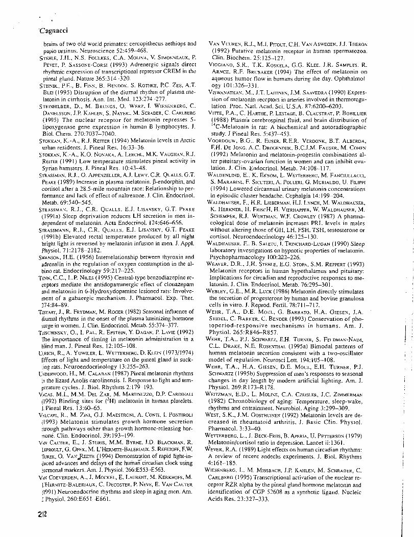
'Cagnacci
brains of two old world primates: cercopithecus aethiops and papio ursinus. Neuroscience 52:459168.
STEHLE, J.H., N.S. FOULKES. C.A. MOLINA. V. SISIOKVEAUX, P. PEVET. P. SASSOSE-CORSI (1993) Adrenergic signals direct rhythmic expression of transcriptional repressor CREM in the pineal gland. Nature 365:314-320.
STEISDL, P.E., B. FINN, B. BESDOK. S. ROTHKE, P.C. ZEE, A.T. BLEI ( I 995) Disruption of the diurnal rhythm of plasma me- latonin in cirrhosis. Ann. Int. Med. 113:774-277.
STEISHILBER. D., M. BRUNGS, 0. WERZ. I. WIESESBERG, C. DASIELSSON. J.P. KAHLEN, S. NAYERI, M. SCRADER. C. CARLBERG (1995) The nuclear receptor for melatonin represses 5- lipoxygenase gene expression in human B lymphocytes. J. Biol. Chem. 270:7037-7040.
STOKKAS. K.-A., R.J. R E ~ ~ E R (1994) Melatonin levels in Arctic urban residents. J. Pineal Res. 1633-36.
STOKKAN, K.-A.. K.O. NONAKA, A. LERCHL, M.K. VAUGHAN, R.J. RErrER (I 99 1) Low temperature stimulates pineal activity in Syrian hamsters. J. Pineal Res. 10:13-48.
STRASSSSMAN, R.J., 0. APPENZELLER, A.J. LEWY. C.R. QUALLS, G.T. PEAKE (1989) Increase in plasma melatonin, P-endorphin, and conisol after a 28.5-mile mountain race: Relationship to per- formance and lack of effect of naltrexone. J. Clin. Endocrinol. Metab. 69540-545.
STRASSMAN. R.J., C.R. QUALLS, E.J. LISANSKY, G.T. PEAKE (1991a) Sleep deprivation reduces LH secretion in men in- dependent of melatonin. Acta Endocrinol. 124:64&656.
STRASSMANK, R.J., C.R. QUALLS, E.J. LISANSKY, G.T. PEAKE (1991b) Elevated rectal temperature produced by all night bright light is reversed by melatonin infusion in men. J. Appl. Physiol. 7 12178-2182.
SWXNSON. H.E. (1956) Interrelationship between thyroxin and adrenalin in the regulation of oxygen consumption in the al- bino rat. Endocrinology 59:2 17-225.
TENS. C.C., L.P. NILES (I 995) Central-type benzodiazepine re- ceptors mediate the antidopaminergic effect of clonazepam and melatonin in 6-Hydroxydopamine lesioned rats: Involve- ment of a gabaergic mechanism. J. Pharmacol. Exp. Ther. ?74:84-89.
TSTAK, J., R. FRYDMAN, M. ROGER (1982) Seasonal influence of diurnal rhythms in the onset of the plasma luteinizing hormone surge in women. J. Clin. Endocrinol. Metab. 55:374-377.
TZISCHINSKY, O., I. PAL: R. EPSTEIN, Y. DAGAN, P. LAVIE (1992) The importance of timing in melatonin administration in a blind man. J. Pineal Res. 12: 105-108.
ULRICH, R., A. YUWILER, L. WETTERBERG, D. KLEIP.; (197311974) Effects of light and temperature on the pineal gland in suck- ling rats. Neuroendocrinology 13:255-263.
USDERWOOD. H., M. CALABAN (1987) Pineal melatonin rhythms in the lizard Anolis carolinensis. I. Response to light and tem- perature cycles. J. Biol. Rhythms 2: 179-193.
VACAS, M.I., M.M. DEL ZAR, M. MARTISUZZO, D.P. CARDINALI (1992) Binding sites for ('H) melatonin in human platelets. J. Pineal Res. 13:60-65.
VALCAVI, R., M. ZIKI, G.J. MAESTRONI, A. CONTI, I. PONTIROLI (1993) Iblelatonin stimulates growth hormone secretion through pathways other than growth hormone-releasing hor- mone. Clin. Endocrinol. 39: 193-199.
Vhn CALTER, E., J. STURIS, M.M. BYRNE, J.D. BLACKMAN, R. LEPROLLT, G. OFEK, M. L'Hm\m-BALERLALX, S. REFETOFF, F.W. TLXK, 0. V A N $ E E ~ H Fl994) Demonstration of rapid light-ii- duced advances and delays of the hum6circadian clock using hormonal markers. Am. J. Physiol. 266:E553-E563.
VAN COE\,ERDEN, A.. J. MOCKEL, E. LAURENT. M. KERKHOFS, M. ~ 'HER~!~TE-B.~LER~Aux, C. DECOSTER, P. NEVE, E. VAN CAUTER (1991) Neuroendocrine rhythms and sleep in aging men. Am. J. Physiot. 260:E65 1-E661.
VAN VUUREN, R.J., M.J. PTTOUT. C.H. VAN ASWEGEN. J.J. THERON (1992) Putative melatonin receptor in human spermatozoa. Clin. Biochem. 25: 125-127.
VlGGlASO, S.R.. T.K. KOSKELA, G.G. KLEE. J.R. SAMPLES, R. ARNCE. R.F. BRUBAKER (1994) The effect of melatonin on aqueous humor flow in humans during the day. Ophthalmol- ogy 101:321%331.
VISWAP~ATHAY. M., J.T. LAITINEN, J.M. SAAVEDRA (1990) Expres- sion of melatonin receptors in aneries involved in thermoregu- lation. Proc. Satl. Acad. Sci. U.S.A. 87:6200-6203.
VITTE, P.A., C. HARTHE. P. LESTAGE, B. CLAUSTRAT. P. BOBILLIER (1988) Plasm'a cerebrospinal fluid. and brain distribution of 1 3 C-Melatonin in rat: A biochemical and autoradiographic
study. J Pineal Res. 5:437153. VOORDOUW, B.G.. R. EUSER, R.E.R. VERDONK, B.T. ALBERDA,
F.H. DE JOP~G. .A.C. DROGENDIJK. B.C.J.,M. FAUSER, M. COHEF; (1992) Melatonin and melatonin-progestin combinations al- ter pituitary-ovarian function in women and can inhibit ovu- lation. J. Clin. Endocrinol. Metab. 74:108-117.
WALDENLIND, E.. K. EKBOM, L. W ~ E R B E R G . M. FANCIULLACCI, S. MARABINI, F. S I C ~ T E R I , .A. POLLERI. G. MURIALDO, U. FILIPPI (1994) Lowered circannual urinary melatonin concentrations in episodic cluster headache. Cephalgia 14: 199-204.
WALDHAUSER, F.. H.R. LIEBERMAN, H.J. LYNCH, M. WALDHAUSER, K. HERKNER, H. FRISCH, H. VIERHAPPER, W. WALDHAUSER, M. SCHEMPER, R.J. WURTMAN, W.F. CROWLEY (1987) A pharma- cological dose of melatonin increases PRL levels in males without altering those of GH, LH, FSH. TSH, testosterone or cortisol. Neuroendocrinology 46: 17-5-1 30.
WALDHAUSER, F., B. SALETU, I. TRINCHARD-LUGAN (1990) Sleep laboratory investi_gations on hypnotic properties of melatonin. Psychopharmacology 100:222-226.
WEAVER, D.R., J.H. STEHLE, E.G. STOP.^, S.M. REPPERT ( 1993) Melatonin receptors in human hypothalamus and pituitary: Implications for circadian and reproductive responses to me- latonin. J. Clin. Endocrinol. Metab. 76295-301.
WEBLEY, G.E., M.R. LUCK (1986) Melatonin directly stimulates the secretion of progesterone by human and bovine granulosa cells in vitro. J. Reprod. Fertil. 78:7 11-7 17.
WEHR, T.A., D.E. MOCL, G. BARBATO, H.A. GIESEN, J.A. SEIDEL, C. BARKER, C. BENDER (1993) Conservation of pho- toperiod-responsive mechanisms in humans. Am. J. Physiol. 265:R846-R857.
WEHR, T.A., P.J. SCHWARTZ, E.H. TURNER, S. FELDMAN-NAIM, C.L. DRAKE, N.E. ROSESTHAL (1995a) Bimodal patterns of human melatonin secretion consistent with a two-oscillator model of regulation. Neurosci Lett. 191:105-108.
WEHR, T.A., H.A. GIESEN, D.E. MOUL, E.H. TURNER, P.J. SCHWA^ (1 995b) Suppression of men's responses to seasonal changes in day length by modem artificial lighting. Am. J. Physiol. 269:R 173-R 178.
WEITZMAN, E.D.. L. MOLISE, C.A. CZELSLER, J.C. ZIMMERMAN (1982) Chronobiology of aging: Temperature, sleep-wake, rhythms and entrainment. Neurobiol. Aging 3299-309.
WEST, S.K., J.M. OOSTHUINEN (1992) Melatonin levels are de- creased in rheumatoid arthritis. J. Basic Clin. Physiol. Pharmacol. 3:33-40.
WETTERBERG, L., J. BECK-FRIIS, B. APERIA, U. PETTERSON (1979) Melatonin/cortisol ratio in depression. Lancet ii:1361.
WEVER. R.A. (1989) Light effects on human circadian rhythms: A review of recent andechs experiments. J. Biol. Rhythms 4:161-185.
WIESENBERG, I.. M. MISSBACH, J.P. KAHLES, M. SCHRADER, C. CARLBERG (1995) Transcriptional activation of the nuclear re- ceptor RZR alpha by the pineal gland hormone melatonin and identification of CGP 52608 as a synthetic ligand. Nucleic Acids Res. 23:327-333.
WURWAN, R.J., I. ZHDANOVA (1 995) Improvement of sleep qual- ity by melatonin. Lancet 346: 1491.
YIE, S.M., G.M. BROWN, G.Y. LIL'. J.A. COLLINS, S. DAYA, E.G. HUGHES, W.G. FOSTER, E.V. YOUGLAI (1995a) Melatonin and steroids in human preovulatory follicular fluid: Seasonal variation and granulosa cell steroid production. Human Reprod. 10:50-55.
YIE, S.-M., L.P. NILES, E.V. YOUNGLAI (1995b) Melatonin re- ceptors on human granulosa cell membranes. J. Clin. Endocrinol. Metab. 80: 1747- 1749.
YUWILER, A., G.L. BRAMMER. B.L. BENNET (1995) Interaction between adrenergic and peptide stimulation in the rat pineal: Pituitary adenylate cyclase-activating peptide. J. Neurochem. 64:2273-2280.
ZAIDAN, R., M. GEOFFRIAL', J. BRUN, J. TAILLARD, C. BUREAU, G. CHAZOT, B. CLAUSTRAT (1994) Melatonin is able to influ-
ence its secretion in humans: description of a phase-response- curve. Neuroendocrinology 60: 105-1 12.
ZHDANOVA. I.V., R.J. WURTMAN, H.J. LYNCH, J.R. IVES, A.B. DOLLINS. C. MORABITO. J.K. MATHESON, D.L. SCHOMER (1995) Sleep-inducing effects of low doses of melatonin ingested in the evening. Clin. Pharmacol. Ther. 57552-558.
ZIMMERMANN, R.C.. C.J. MCDOUGLE, M. SCHUMACHER, J. OLCESE, J.W. MASON, G.R. HENINGER, L.H. PRICE (1993) Effects of acute tryptophan depletion on nocturnal mela- tonin secretion in humans. J. Clin. Endocrinol. Metab. 76: 1160-1 164.
ZIMMERMANN, R.C., L. KRAHN. G. KLEE, P. DELGAW, S.J. ORY, S.C. LIN (1994) Inhibition of presynaptic catecholaminesyn- thesis with alpha-methyl-para-tyrosine attenuates nocturnal melatonin secretion in humans. J. Clin. Endocrinol. Metab. 79: 11 10-1 114.
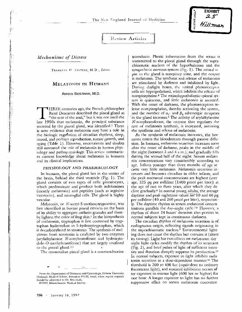
T h e N c ~ \ 2;ngland Journal c i i h , Icdicine - . . -. . . . .. - -- - ..
. -- - --
; .I<rries Articles -- )I
Mechanisms of Disease
F K A N K I . I S [ - I . ~<I'STEIN, M.D. , E l f i f 0 1
AMNON BRZEZINSKI. M.D.
T HREE centuries ago, the French philosopher Renk Descartes described the pineal gland as "the seat of the soul," but it was not until the
late 1950s that mclatonin, the principal substance secreted by the pineal gland, was identified.1 There is now evidence that melatonin may have a role in the biologic regidation of circadian rhythms, sleep, ~ n o o d , and perhaps ccproduction, tumor gro\vth, and aging (Table 1 ) . Ho\vever, uncertainties an3 doubts still surround the role of melatonin in human phys- iology and pathoph~*siology. This revie\\. summariz- es current knowledge about melatonin in humans and its clinical implications.
PHYSIO1,OGY AND PHARMACOLOGY
In humans, the pineal gland lies in the center of the brain, behind the third ventricle (Fig. 1). The gland consists of two types of cells: pinealocytes, which predominate and produce both indolamines (mostly melatonin) and peptides (such as arginine \.asotocin), and neuroglial cells. The gland is highly vascular.
Melatonin, or N-~cet!rl-5 -met l~osy t r ) lp ta~~~i~~e , was first identified in bo\~ine pineal extracts on the basis of its ability to aggregate melanin granules and there- by lighten the color of frog skin.1 In the biosynthesis of melatonin, tryptophan is first converted by tryp- tophan hydroxylase to 5-hydroxytryptophan, which is decarbosylated to serotoni~l. The synthesis of mel- atonin from serotonin is catalyzed by two enzymes (ar!;lalkylamine N-acetyltransferase and hydroxyin- dole-0-n~ctl~yltransferase) that are largely confined to the pineal t
TIlc ma~n~nal ian pineal glantl is a neuroenclocrine
From thc l>cpartmcnt of Ohstrrrics and Gynecology, Hcbrc\\. University Had.issali Medicat School, Jcruwlcni 91 120, [smcl, \\.licrc rcprinr requests shoubl he addressed to Dr Rrzczinski.
019'97, hlnss.ichuscra Medical Society.
transducer. Photic information from the retina is transmitted to the pineal gland through the supra- chiasmatic nucleus of the l~ypothalamus and the sympathetic nervous system (Fig. 1). The neuml in- put to the gland is norepincpl~rine, and the outpiit is melatonin. The synthesis and release of melatonin are stimulated by darkness and inhibited by light. During daylight hours, t l ~ c rctinal p h o t o r e c c p ~ ~ ) ~ - cells are hyperpolarized, \\d~ich inhibits the release o f norepinephrine.4 The retinohypothalamic-pineal sys- tem is quiescent, and little melatonin is secreted. With the onset of darkness, the photoreceptors re- lease norepinephrine, thereby activating the system, and the number of a , - and PI-adrenergic receptors
' in the land increase^.^ The activity of arylalkylamine N-acetyltransferase, the enqrn1e that regulates the rate of melatonin synthesis, is increased, initiating the synthesis and release of melatonin.
As the synthesis of melatonin increases, the hor- mone enters the bloodstream through passive diffil- sion. In humans, melatonin secretion increases soon after the onset of darkness, peaks in the middlc of the night (between 2 and 4 a.m.), and gradually t>ils during the second half of the night. Serum melato- nin concentrations vary considerably according to age. Infants younger than three months of age se- crete \.cry little rnelatonin. Melatonin sec re t io~~ in creases and becomes circadian in older ir~fants, a ~ ~ d the peak nocturnal concentrations are highest (aver age, 325 pg per milliliter [1400 pmol per liter]) at the age of one to three years, after which they de- cline gradually.6 In normal young adults, the average -/. daytime and peak nighttime values at-510 and 60 pg per n~illiliter (40 and 260 pmol per liter), -respective- ly. The daytime rhythm in serum melatonin conccn - trations parallels the day-night c y ~ l e . ~ . ~ Ho\vevcr, a rhythm of about 24 hours' duration also persists in normal subjects kept in continuous darkness.
The circadian rhythm of melatonin secretion is of endogenous origin, reflecting signals originating in the suprachiasrnatic nuclei~s.~ Environmental light ing does not cause the rhythm but entrains it (alters its timing). Light has two effects on melatonin: day--- night light cycles modify the rhythn~ of its secretion (Fig. 2), and brief pulses of light ofsufficie!~t inten- sity and duration abruptly suppress its production.1° In normal subjects, exposure to light inhibits mela- tonin secretion in a dose-dependent manner." The threshold is 200 to 400 lux (equivalent to ordinary fluorescent light), and maximal inhibition occurs af- ter exposure to incense light (600 lux or higher) for one hour. A longer exposure to light has no further suppressive effect on serum melatonin concentra-
186 . J a n u a r y 16, 1997
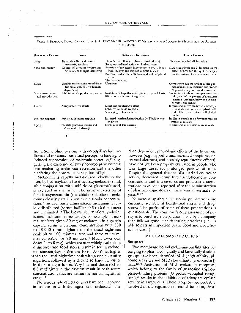
MECHANISMS OF DISEASE -- -- - -- - . -
Sleep
Circadian rhvthm
Mood
Sexual maturation and reproduction
Immune response
Agins
Hypnotic effect and increased propensity for sleep
Control of circadian rhythms and entrainment ro light-dark cycle
Possible role in cyclic mood disor- ders (season.11 .~ffccri\~e disorder, depression)
Inhibition of reproductive process
Antiproliferati\,e effects
Enhanced immune response
Possible protective effects and decreased cell damage
Hypothermic effect (at pharmacologic doses) Receptor-mediated action on limbic system Secretion of melatonin in response to lleural input
from thc eyes and suprachiasmatic nucleus Receptol-nicdiated effects on neural a ~ ~ c i peripheral
tissues Thermoregulation Unknown
Inhibition of hypothalamic-pimitary-gc~nadd axis Effect on ovarian steroidogencsis
Direct anriprolihrative dfect Enhanced immune response Scavenging of free radicals
Increased interleukirr'production by T-helper lym- phoc!;tcs
Scavcllg~~~g of free radicals
tions Some blind persons nwh no pupillary liglit re- flexes and no conscious visual perception have I~ght- induced suppression of melatonin secretion,l2 sug- gesting the existence of tnro photoreceptive systems: one mediating melatonin secretion and the other mediating the conscious perception of light
Melatonin is rapidly metabolized, chiefly in the liver, by hydroxylation (to 6-hydroxymelatonin) and, after conjugation with sulfuric o r glucuronic acid, is excreted in the ~lrine. The urinary excretion of 6-sulfatoxymelatonin (the chief n~ecabolite of mela- tonin) closely parallels serum melatonin concentra- tions.7 Intravenously administered melatonin is rap- idly distributed (serum half-life, 0.5 to 5.6 minutes) and eliminated.13 The bioavailability of orally adnun- istered melatonin varies widely. For example, in nor- mal subjects given 8 0 mg of melatonin in a gelatin capsule, serum melatonin concentrations were 350 to 10,000 times higher than the usual nighttime peak 60 to 150 minutes later, and these values re- mained stable for 9 0 minutes.14 Much lower oral doses ( 1 to 5 mg), which are now widely available in drugstores and food stores, result in serum melato- nin concentrations that are 10 to 100 tlmes higher than the usual nighttime peak within one hour after ingestion, followed by a decline to base-line values in four to eight hours. Very low oral doses (0.1 to 0.3 m g r g i v e ~ i n the daytime result in peak serum concentrations that are within the normal nighttime range.15
No serious side effects or risks have been reported in association with the ingestion of melatonin. The
Placebo-controlled clinical trials
Studies in animals and in humans on the effects of light and the light-dark cycle on the partern uf melatonin secretion
Comparative clinical studies o f the pat- tern of mel3to1iin iecretion and studies of photothcr~pj. for mood disorders
Studies in animals and comparative clini- cal studies of the pattern o f melatonin secretion (during pubcrty and in wom- en ulth amenorrhea)
In vitro and in viva studies in animals, in vitro studies of human neoplastic cells and cell lincs, and a fcw small clinical studies
Studies in animals and a few uncontrolled studies in humans
In vitro and in t-ivo studies in animals
dose-dependcnc physiologic effects of che hormone, however (e.g., I~ypothermia, increased sleepiness, de- creased alertness, and possibly reproductive effects), have not yet been properly evaluated in people who take large doses for prolonged periods of time. Despite the general absence of a marked endocrine action, decreased serum luteinizing-hormone con- centrations and increased serum prolactin concen- trations have been reported afier the administration of pharmacologic doses of melatonin in normal sub - jects.16J7
Numerous synthetic melatonin preparations are currently available at health-food stores and drug- stores. The purity of some of these preparations is questionable. The consumer's only guarantee of pu- rity is to purchase a preparation made by a company that follows good manufacturing practices (i.e., is able to pass an inspection by the Food and Drug Ad- ministration).
MECHANISMS OF ACTION Receptors
Tivo membrane-bound melatonin-binding sites be- longing to pharmacologically and kinetically distinct groups have been identified: ML1 (high-affinity [pi- comolar]) sites and ML2 (low-afinity [nanomolar]) sites.18.19 Activation of ML1 melatonin receptors, which belong to the family of guanosine. triphos- phate-binding proteins (G protein-coupled recep. tors),20 results in the inhibition of adenylate cyclase activity in target cells. These receptors are probably involved in the regulation of retinal function, circa-
\lolume 336 N u m b e r 3 - 187
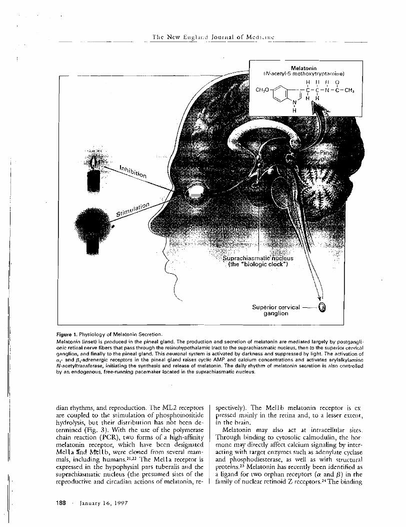
The New Engl .~~:c! Journal of M c d i i l n c - . - --
I Melatonin 1
~ - . . Suprachiasmatic nuc~eus 1\
Superior cervical -@ ganglion
Figure 1. Physiology of Melatonin Secretion.
Melatonin (inset) is produced in the pineal gland. The production and secretion of melatonin are mediated largely by postgangli- onic retinal nerve fibers that pass through the retinohypothalamic tract to the suprachiasmatic nucleus, then to the superior cervical ganglion, and finally to the pineal gland. This neuronal system is activated by darkness and suppressed by light. The activation o f cr1- and 8,-adrenergic receptors in the pineal gland raises cyclic AMP and calcium concentrations and activates arylalkylamine N-acetyltransferase, initiating the synthesis and release of melatonin. The daily rhythm of melatonin secretion is also controlled by an endogenous, free-running pacemaker located in the suprachiasmatic nucleus.
dian rhythms, and reproduction. The ML2 receptors are coupled to the stimulation of phos hoinositide P hydrolysis, but their distribution has not been de- termined (Fig. 3). With the use of the polymerase chain reaction (PCR), two forms of a high-affinity melatonin receptor, which have been designated Mella %rid m l l b , were cloned- from several mam- mals, including humans.21-22 The Mella receptor is expressed in the hypophysial pars tuberalis and the suprachiasmatic nucleus (the presumed sites of the reproducrive and circadian actions of melatonin, re-
spectively). The Mellb melatonin receptor is es- pressed mainly in the retina and, to a lesser extent, in the brain.
Melatonin may also act at intracellular sites. Through binding to cytosolic calmodulin, the hor mone may directly affect calcium signaling by inter- acting with target enzymes such as adenylate cyclase and phosphodiesterase, as well as with structural proteins.23 Melatonin has recently been identified as a ligand for two orphan receptors (a and 0 ) in the family of nuclear retinoid Z receptors.24The binding
488 . Janua ry 16, 1997
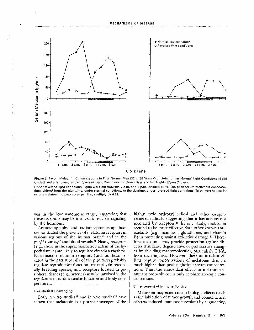
. - -- MECHANISMS OF DISEASE -- -- - - - - -
Normal light conditions o Reversed light conditions
Clock Time
Figure 2. Serum Melatonin Concer~trations i n Four Normal Men (22 to 35 Years Old) Living under Normal Light Conditions (Solid Circles) and after Living under Reversed Light Conditions for Seven Days and Six Nights (Open Circles).
Under reversed light conditions, lights were out between 7 a.m. and 3 p.m. (shaded bars). The peak serum melatonin concentra- tions shifted from the nighttime, under normal conditions, to the daytime, under reversed light conditions. To convert values for serum melatonin to picomoles per liter. multiply by 4.31.
was in the low nanomolar range, suggesting that these receptors may be involved in nuclear signaling by the hormone.
Autoradiography and radioreceptor assays have demonstrated the presence of melatonin receptors in various regions of the human brain25 and in the gut,26 ovaries:' and blood vessels.28 Neural receptors (e.g., those in the suprachiasmatic nucleus of the hy- pothalamus) are likely t o regulate circadian rhythms. Non-neural melatonin receptors (such as those lo- cated in the pars tuberalis of the pituitary) probably regulate reproductive function, especially~in season- ally breeding species, and receptors located in pe- ripheral tissues (e.g., arteries) may be involved in the regulation of cardiovascular function and body tem- perature, - - -
Free-Radical Scavenging
Both in vitro studies29 and in vivo studies3(' have shown that melatonin is a potent scavenger of the
highly toxic hydroxyl radical and other oxygen- centered radicals, suggesting that it has actions not mediated by receptors.3' In one study, melatonin seemed to be more effective than other known anti- oxidants (e,g., mannitol, glutathione, and vitamin E) in protecting against oxidative damage.31 There- fore, melatonin may provide protection against dis- eases that cause degenerative or proliferative chang- es by shielding macromolecules, particularly DNA, from such injuries. Ho~vever, these antioxidant ef- fects require concentrations of melatonin that are much higher than peak nighttime serum concentra- tions. Thus, the antioxidant effects o f melatonin in humans probably occur only at pharmacologic con- centrations.
Enhancement of Immune Funct ion
Melatonin may exert certain biologic effects (such as the inhibition of tumor growth and counteraction of stress-induced immunodepression) by augmenting
Volume 3 3 6 N u m b e r 3 . 189
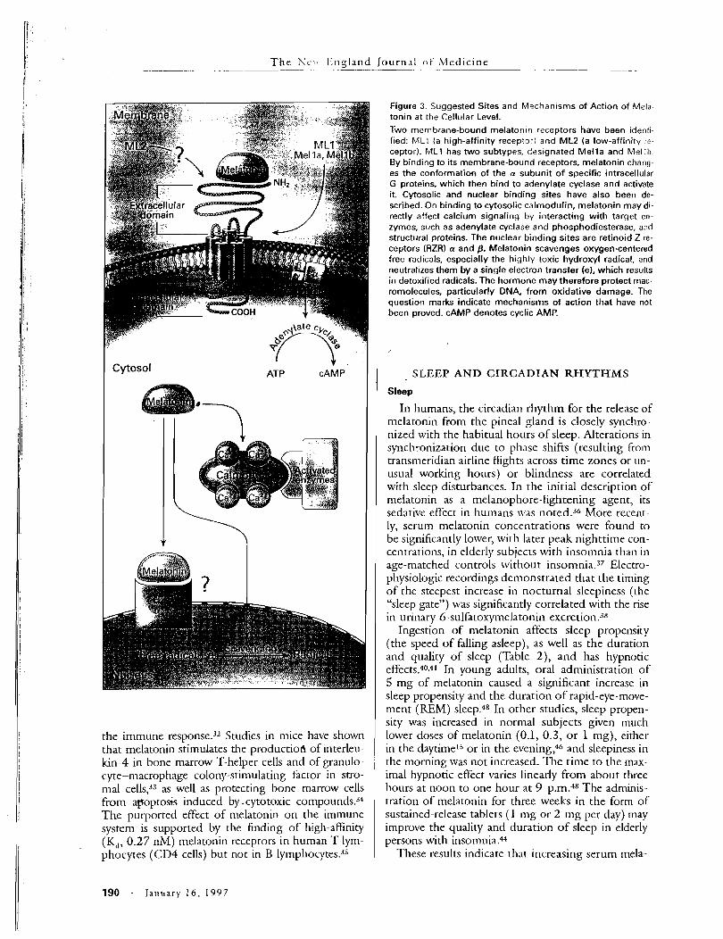
T h e S C : ~ l . ; t~gland J o u r n , ~ ! o f Medicine . .- - -- --
I
ATP CAMP
the immune resp0nse.3~ Studies in mice have shown that melatonin stimulates the productiofi of interleu- kin-4 in bone marrow T-helper cells and of granulo- cyte-macrophage colonjl-stimulating factor in stro- ma1 cells,33 as well as protecting bone marrow cells from apoptok induced by -cytotoxic compounds.34 The purported effect of melatonin on the immune system is supported by the finding of high-affinity (K,, 0.27 nM) melatonin receptors in human T lym- phocytes (CD4 cells) bat not in B lymphocytes.3j
Figure 3. Suggested Sites and Mechanisms of Action of Mela- tonin at the Cellular Level.
Two membrane-bound melatonin receptors have been identi fied: LlLi (a high-affinity recepro-1 and ML2 (a low-affinity .2-
ceptor). ML1 has two subtypes. dss~gnated Mella and Melib. By bind~ng to its membrane-bound receptors, melatonin chang- es the conformation of the a subunit of specific intracellular G proteins, which then bind to adenylate cyclase and activate it. Cytosolic and nuclear binding sites have also been de- scribed. On binding to cytosolic calmodulin, melatonin may dl- rectly affect calcium signaling by interacting with target en- zymes, such as adenylate cyclase and phosphodiesterase, and structural proteins. The nuclear binding sites are retinoid Z re- ceptors (RZR) a and p. Melatonin scavenges oxygen-centered free radicals, especially the highly toxic hydroxyl radical, and neutralizes them by a single electron transfer (el, which results in detoxified radicals. The hormone may therefore protect mac- romolecules, particularly DNA, from oxidative damage. The question marks indicate mechanisms of action that have not been proved. cAMP denotes cyclic AMP.
SLEEP AND CIRCADIAN RHYTHMS
Sleep
In humans, the circadian rhythm for the release of melatonin from the pineal gland is closely synchro- nized with the habitual hours of sleep. Alterations in synchronization due to phase shifts (resulting from transnleridian airline flights across time zones or un- usual working hours) or blindness are correlated with sleep disturbances. In the initial description of melatonin as a melanophore-lightening agent, its sedative effect in hurnans was noted.36 More recent- ly, serum melatonin concentrations were found to be significantly lower, with later peak nighttime con- centrations, in elderly subjects with insomnia than in age-matched controls without in~omnia .3~ Electro- physiologic recordings denlonstrated that the timing of the steepest increase in nocturnal sleepiness (the "sleep gate") was significantly correlated with the rise in urinary 6-sulfatox~~melatonin excretion.3"
Ingestion of melatonin affects sleep propensity (the speed of falling asleep), as well as the duration and quality of sleep (Table 2 ) ) and has hypnotic effects.40.41 In young adults, oral administration of 5 m g of melatonin caused a significant increase in sleep propensity and the duration o f rapid-eye-move- ment (REM) sleep.48 In other studies, sleep propen- sity was increased in normal subjects given much lower doses of melatonin (0.1, 0.3, or 1 mg), either in the daytimel%r in the evening,46 and sleepiness in the morning was not increased. The time to the max- imal hypnotic effect varies linearly from about three hours at noon to one hour at 9 p.m.48 The adminis- tration of melatonin for three weeks in the form of sustained-release tablets (1 mg or 2 mg per day) may improve the quality and duration o f sleep in elderly persons with insomnia.44
These results indicate that increasing serum mela-
190 - January 16, 1997
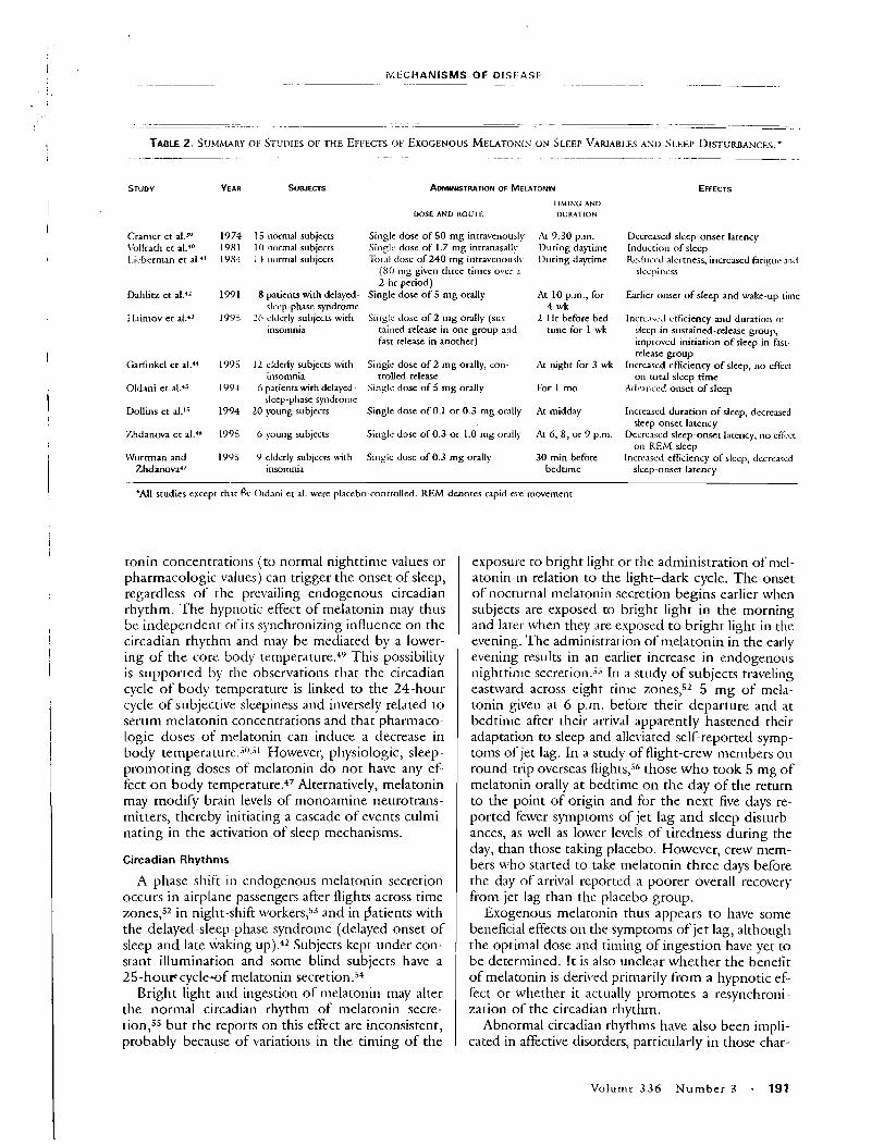
--- PAECHANISMS OF DISEASE
. . - . -
.. .. . .- - .- - .- . . - --. -. . - - - - .. - - .-
- . - . -- - - .
TABLE 2, S'JMMAKY OF STUI>IFS OF THE EFFECTS O F EXOGENOUS MELATOKIN O N SLEEP VARIABLES A S 0 ~ I . E E P DISTURBANCFS.~ . - .- . -- -. - - . . - -. - . - .- - - - -- - - - . .. - - - -
ADMINI~TRATION OF MELATONIN
I lh l lNG AVD
DOSE AND ROUTE DURATION
Cramer et al.- I'ollrath et al.'O L.icbcrnman e t al."
15 normal subjects 10 normal subjects I4 normal subjects
Single dose of 50 mg intravenously Singlc dose of 1.7 mg intranasall!. I'or.11 di~sc of 240 mg intravenousl\
(SO nig ~ i v e n three times over .I
2 - l ~ r period) Single dose of 5 mg orally
At 9:30 p.m. During daytime 1)uring daytime
Decreased sleep-onset latency Induction c~ f sleep R~du icd alertness, increased fatigue and
slccpincss
Dahlitz et al."
thirnov et al."
8 patients with delayed- clccp-phase syndrome
Z h clrlerly subjects with insomnia
At 10 p.m., for 4 wk
2 Hr before bed- time for 1 wk
Earlier onset of sleep and wake-up time
Singic dose of 2 mg orally (sus- tained release in one group and fast release in another)
Incrl-.!\c,i ctficiency and duration (I!.
sleep in sustained-release group, improved initiation o f sleep in fast- release group
Increased eficiency o f sleep, no effect on total sleep time
A ~ \ - R I ~ s c ~ onset of sleep
Garfinkel et al." 12 elderly subjects with insomnia
6 patients with delayed- sleep-phase syndrome
20 young subjects
Singlc dose of 2 mg orally, con trolled release
S i n ~ l c dose of 5 mg orally
At night for 3 wk
Oldani et al.'j
Dollins et a1.15
For 1 mo
Single dose of 0.1 or 0.3 mg orally At midday Increased duration o f sleep, decreased sleep-onset latency
Decrcdscd sleep-onset latency, no eKcct on REtU sleep
Increased efficiency of sleep, decrcascd sleep-onset latency
Zhdanova e t aLdn 6 young subjects Single dose of 0.3 o r 1.0 Ing orall!.
Wurtman and Zhdan~va '~
9 elderiy subjects with insomnia
S i n ~ l e dose of 0.3 mg orally 30 min before bedtime
*All studies except thar & Oldani et al. were placebo-controlled. REM denotes rapid eye movement.
tonin concentrations (to normal nighttime values or pharmacologic values) can trigger the onset of sleep, regardless of the prevailing endogenous circadian rhythm. The hypnotic effect of melatonin may thus be independent of its synchronizing influence on the circadian rhythm and may be mediated by a lower- ing of the core body temperat~re.4~ This possibility is supported by the observations that the circadian cycle of body temperature is linked to the 24-hour cycle of subjective sleepiness and inversely related to serum melatonin concentrations and that pharmaco- Logic doses of nlelatonin can induce a decrease in body temperature.j'J.3 However, physiologic, sleep- promoting doses of melatonin do not have any ef- fect on body temperature.4' Alternatively, melatonin may modify brain levels of monoamine neurotrans- mitters, thereby initiating a cascade of events culmi- nating in the activation of sleep mechanisms.
exp0sul.e to bright light or the administration of nlcl- atonin in relation to the light-dark cycle. The onset of nocturnal melatonin secretion begins earlier when subjects are exposed to bright light in the morning and later when they are esposed t o bright light in the evening. The administration of nielatonin in the early evening results in an earlier increase in endo, ~ ~ I I O L ~ S
nighttime secretion.j5 In a study of subjects traveling eastward across eight time 5 mg of mela- tonin given at 6 p.m. before their departure and at bedtime after their arrival apparently hastened their adaptation to sleep and alleviated self-reported symp- toms of jet lag. In a study of flight-crew members on round-trip overseas flights," those who took 5 mg of melatonin orally at bedtime on the day of the return to the point of origin and for the next five days re- ported fewer symptoms of jet lag and sleep disturb- ances, as well as lower levels of tiredness during the day, than those taking placebo. However, crew mem- bers who started to take melatonin three days before the day of arrival reported a poorer overall recovery from jet lag than the placebo group.
Exogenous melatonin thus appears to have some beneficial effects on the symptoms of jet lag, although the optimal dose and tinling of ingestion have yet to be determined. It is also unclear whether the benefit of melatonin is derived primarily from a hypnotic ef- fect or whether it actually promotes a resynchroni- zation of the circadian rhythm.
Abnormal circadian rhythms have also been impli- cated in affective disorders, particularly in those char-
Circadian Rhythms
A phase shift in endogenous melatonin secretion occurs in airplane passengers after flights across time zones,52 in night-shift ~vorkers,j3 and in Datients with the delayed-sleep-phase syndronle (delayed onset of sleep and late waking up).42 Subjects kept under con- stant illumination and some blind subjects have a 25-hou~cycle-of melatonin secretion.54
Bright light and ingestion of melatonin may alter the normal circadian rhythm of melatonin secre- tion,55 but the reports on this effect are inconsistent, probably because of variations in the timing of the
Volume 3 3 6 Number 3 - 191
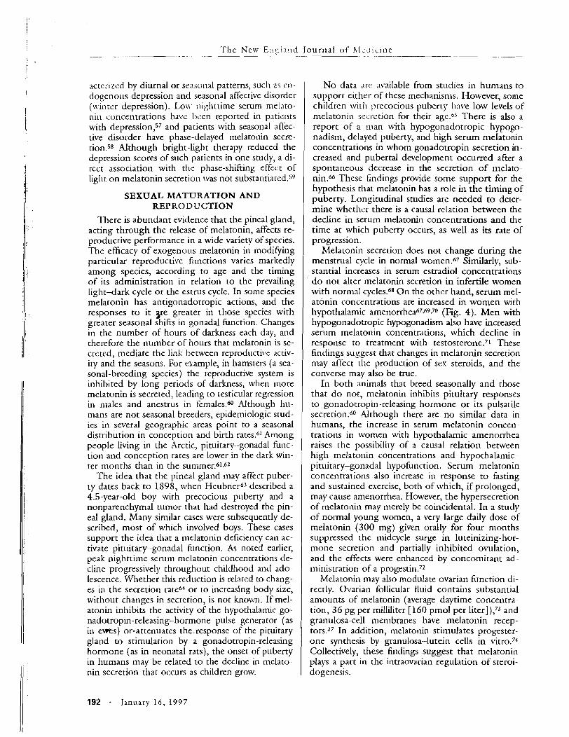
The New E I I ~ ! ~ I I ~ ~ - ~
actel l ~ c d by diurnal or srasonal patterns, such en- dogenous depression and seasonal affective disorder (n,inrcr depression). Lo\\ ~~igh t t ime serum mel.~to- nil1 concentrations have bzen reported in patiznts with depression,57 and patients with seasonal affec- tive disorder have phase-delayed melatonin secre- tion.58 Although bright-light therapy reduced the depression scores of such patients in one study, a di- rect association with the phase-shifting effect of liglrt o n melatonin secretion was not substantiated.59
SEXUAL MATURATION AND REPRODUCTION
There is abundant evidence that the pineal gland, acting through the release of melatonin, affects re- productive performance in a wide variety of species. The efficacy of exogenous melatonin in modifying particular reproductive functions varies markedly among species, according to age and the timing of its administration in relation to the prevailing light-dark cycle o r the estrus cycle. In some species melatonin has antigonadotropic actions, and the responses to it y e greater in those species with greater seasonal shifts in gonadal function. Changes in the number of hours of darkness each day, and therefore the number of hours that melatonin is se- creted, mediate the link between reproducti~c activ- ity and the seasons. For example, in hamsters (a sea- sonal-breeding species) the reproductive system is inhibited by long periods of darkness, when more melatonin is secreted, leading to testicular regression in 111ales and anestrus in females.60 Although hu- mans are not seasonal breeders, epidemiologic stud- ies in several geographic areas point to a seasonal distribution in conception and birth rates.61 Among people living in the Arctic, pituitary-gonadal func- tion and conception rates are lower in the dark win- ter months than in the sun1mer.61.6~
The idea that the pineal gland may affect puber- ty dates back to 1898, when Heubne~-63 described a 4.5-year-old boy with precocious puberty and a nonparenchymal tumor that had destroyed the pin- eal gland. Many similar cases were subsequently de- scribed, most of which involved boys. These cases support the idea that a melatonin deficiency can ac- tivate pituitary-gonadal Function. As noted earlier, peak nighttime serum melatonin concentrations de- cline progressively throughout childhood and ado- lescence. Whether this reduction is related to chang- es in the secretion rateM or to increasing body size, without changes in secretion, is not known. If mel- atonin inhibits the activity of the hypothalamic go- nadotropin-releasing-hormone pulse generator (as in e m s ) or-attenuates thcresponse of the pituitary gland to stimulation by a gonadotropin-releasing hormone (as in neonatal rats), the onset of puberty in humans may be related to the decline in melato- nin secretion that occurs as children grow.
No data are available from studles in humans to support either of these mechanisms. However, some children \vitl~ precocious puberty 11at.e low levels of melatonin szi~-ction for their agcss There is also a report of a inan with hypogonadotropic hypogo- nadism, delayed puberty, and high serum melatonin concentrations in whom gonadotropin secretion in- creased and pubertal development occurred after a spontaneous decrease in the secretion of melato- nin." These findings provide some support for the hypothesis that melatonin has a role in the timing of puberty. Longitudinal studies are needed to deter- mine whethzr there is a causal relation between the decline in serum melatonin concentrations and the time at which puberty occurs, as well as its rate of progression.
Melatonin secretion does not change during the menstrual cycle in normal women.67 Similarly, sub- stantial increases in serum estradiol concentrations do not alter melatonin secretion in infertile women with normal cycles.68 O n the other hand, serum mel- atonin concentrations are increased in women with hypothalamic amenorrhea6769.70 (Fig. 4). Men with hypogonadotropic hypogonadism also have increased serum melatonin concentrations, which decline in response to treatment with testosterone.71 These findings suggest that changes in melatonin secretion may affect the production of sex steroids, and the converse may also be true.
In both animals that breed seasonally and those that d o not, melatonin inhibits pituitary responses to gonadotropin-releasing hormone or its pulsatile secretion.60 Although there are no similar data in humans, the increase in serum melatonin concen- trations in women with hypothalamic amenorrhea raises the possibility of a causal relation between high rnelatonin concentrations and hypothalamic- pituitary-gonadal hypofunction. Serum melatonin concentrations also increase in response to fasting and sustained exercise, both of which, if prolonged, may cause amenorrhea. However, the hypersecretion of melatonin may merely be coincidental. In a study of normal young women, a very large daily dose of melatonin (300 mg) given orally for four months suppressed the midcycle surge in luteinizing-hor- mone secretion and partially inhibited ovulation, and the effects were enhanced by concomitant ad- ministration of a pr0gestin.~2
Melatonin may also modulate ovarian Function di- rectly. Ovarian follicular fluid contains substantial amounts of melatonin (average daytime concentra- tion, 36 pg per milliliter [160 pmol per liter]),73 and granulosa-cell membranes have melatonin recep- tors.27 In addition, melatonin stimulates progester- one synthesis by granulosa--1utein cells in vitro.74 Collectively, these findings suggest that melatonin plays a part in the intraovarian regulation of steroi- dogenesis.
19% - J anua ry 1 6 , 1997
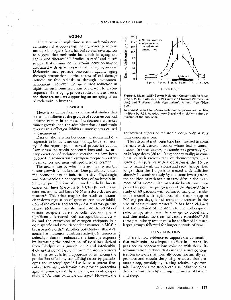
MECHANISWS O F DISEASE -- --- -- - -. . -- - - - -- - --- - -- -
I
AGING
The decrc.~sc in nighttime s c r~ : ! \~ melatonin con- centrations rl:nt occurs with aging, together with its multiple biologic effects, has led several investigators to suggest that melatonin has a role in aging and age-related di~eases.75~76 Studies in rats77 and mice78 suggest that diminished melatonin secretion may be associated \\.it11 an acceleration of the aging process. Melatonin mny provide protection against aging through attenuation of the effects of cell damage induced bv free radicals o r through immunoen- hancement. However, the age-related reduction in , 3 p.m. 7 p.m. 11 p.m. 3 a.m. 7 a.n>. 11 a.m.
nighttime nlelatonin secretio-n could well be a con- sequence of the aging process rather than its cause,
of melatonin in humans.
i and there are no data supporting an antiaging effect i
CANCER i There is evidence from experimental studies that 1
melatonin influences the growth of spontaneous and :
induced tumors in animals. Pinsalectomy enhances tumor growth, and the administration of melatonin reverses this effectlor inhibits tumorigenesis caused ,
by c a r c i n o ~ e n s . ~ ~ Data on the relation bet~veen melatonin and on-
cogenesis in humans are conflicting, but the major- ity of the ~.rports point toward protective action. Low serum melatonin concentrations and low uri- nary excretion of melatonin metabolites have been 1 reported in \xromen with estrogen-receptor-positive ! breast cancer and men with prostatic cancer.gO-8'
The mechanism by which melatonin may inhibit I tunlor gro~vth is not known. One possibility is that , the hormone has antimitotic activity. Physiologic and pharrnacologic concentrations of melatonin in- I hibit the proliferation of cultured epithelial breast- i cancer cell lines (particularly MCF-7)83 and malig- 1 nant-melanoma cell lines (M-6) in a dose-dependent 1 manner.84 This effect may be the result of intranu- I clear down-regulation of gene expression or inhibi- j tion of the release and activity ofstimulatory growth
'
factors. Melatonin may also modulate the actit~ity of i various receptors in tumor cells. For example, i t significantly decreased both estrogen-binding activ- ity and the expression of estrogen receptors in a dose-specific and time-dependent manner in MCF-7 breast-cancer cells.85 Another possibility is that mel- i
atonin has imn~unon~odulatory activity. In studies in / animals, melatonin enhanced the immupe response j by increasing the production of cytokines derived i from T-helper cells (interleukin-2 and interleukin- I 4),32 and as noted earlier, in mice melatonin protects , bone msrrow cells from apoptosis by enhancing the production of-colony-stimul5ing factor by granulo- cytes and ma~rophages.3~ Lastly, as a potent free- i radical scavenger, melatonin may provide protection j against tumor growth by shielding nlolecules, espe- I cially DNA, from oxidative dan~age .~ l However, the I
Clock Hour
Figure 4. Mean (-tSE) Serum Melatonin Concentrations Meas- ured at 2-Hour Intervals for 24 Hours in 14 Normal Women (Cir- cles) and 7 Women with Hypothalamic Amenorrhea (Trian- gles). To convert values for serum melatonin to picomoles per liter, multiply by 4.31. Adapted from Brzezinski et a1.6lwith the per- mission of the publisher.
antioxidant effects of melatonin occur only at very high concentrations.
The effects of n~el~ltonin have been studied in some patients with cancel; most of whom had advanced disease. In these studies, melatonin was generally giv- en in large doses (20 to 4 0 mg per day orally) in com- bination with radiotherapy or chemotherapy. In a study of 30 patients with glioblastomas, the 16 pa- tients treated with melatonin and radiotherapy lived longer than the 14 patients treated tvith radiation alone.86 In another study by the same investigators, the addition of nlelatonin to tanloxifen in the treat- ment of 14 \\lomen with metastatic breast cancer ap- peared to slotv the progression of the disease.87In a study of 40 patients ulith advanced malignant mela- noma treated \\lit11 high doses of melatonin (up to 700 rng per day), 6 had transient decreases in the size of some tunlor masses.88 It has been claimed that the addition of melatonin to chemotherapy o r radiotherapy attenuates the damage to blood cells and thus makes the treatment more tolerable.89 All these preliminary results must be confirmed in much larger groups follotved for longer periods of time.
CONCLUSIONS
There is evidence to support the contention that melatonin has a hypnotic effect in humans. Its peak serum concentrations coincide with sleep. Its administration in doses that raise the serum concen- trations to levels that norn~ally occur nocturnally can promote and sustain sleep. Higher doses also pro- mote sleep, possibly by causing relative hypother- mia. Exogenous ~nelatonin can also intluence circa- dian rhythms, thereby altering the timing of fatigue and sleep.
V o l u m e 336 Number 3 193
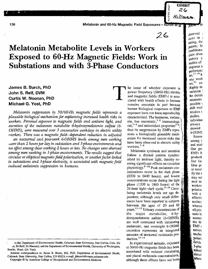
Melatonin and 60-Hz Magnetic Field Exposures
Melatonin Metabolite Levels in Workers Exposed to 60-Hz Magnetic Fields: Work in Substations and with 3-Phase Conductors
James B. Burch, PhD T he issue of whether exposure
John S. Reif, DVM power frequency (50/60-Hz) elec and magnetic fields (EMFs) is asso-
Curtis W. Noonan, PhD ciated with health effects in humans Michael G. Yost, PhD remains uncertain in part because
human biological responses to E Melatonin s u ~ e s s i o n by 50/6@Hz magnetic fields represents a exposure have not been reproduc
plausible biological mechanism for explaining increased health risks in characterized. The hormone, melato- workers. Personal exposure to magnetic fields and ambient light, and nin, has on costa ti^,'-^ irnmunologi- excretion of the melatonzn metabolite 6-hydroxymelatonin suEfate (6- ca1,3-4 and antioxidant OHMS), were measured oum 3 consecutiue workdays in electric utilzty thus its suppression by EMFs repre- n~mkers. Thme was a magnetic field-dependent reduction in adjusted Sents a biologic all^ plausible me&-
an nocturnal and post-work 6-OHMS h e k among men working for increased cancer risks that
,,'ore than 2 hounper day i n substation and 3-phase enuironments and have been observed in
n o effect among those working 2 hours or less. No changes were obsmed among men working in I?hase environments. The results suggest that Melatonin synthesis and secretion
follow a diurnal pattern synchro- circular or elliptical magnetic field polarizationy or anotherjactor linked nized by light, thereby ex- to substations and 3-phase electricity, is associated with magnetic field significant effects on circadian
induced melatonin suppession in humans. physiology.9-'0 Peak melatonin con- centrations occur in the dark phase ,
(0200 to 0400 hours), and lowest concentrations occur during the light phase (1200 to 1800 hours) of the 24-hour light-dark ~ ~ c l e . ~ - ' ~ Circu- lating melatonin levels are age de- pendent, although only small differ- ences have been reported in subjects between the ages of 20 and 60 years."-12 Urinary concentrations of the major metabolite, 6-hy- droxymelatonin sulfate (6-OHMS), are well correlated with circulating melatonin, and overnight &OHMS excretion represents an integrated measure of nocturnal melatonin pro- duction. I3-l4
m the Department of Environmental Health, Colorado State University, Port Collins, Colo. (Dr In experimental animals, exposure' -n. Dr Reif. Dr Noonan); and the Department of Environmental Health, University of Washington, 5 0 / 6 0 - ~ ~ magnetic fields has been
Seattle. Wash. (Dr Yost). associated with reduced circulating Address correspondence to: James B. Burch. MS. PhD, Department of Environmental Health,
Colorado State University, Fort Collins, CO 80523: e-mail: [email protected]. and pineal melatonin concentrations, Copynght O by American College of Occupational and Environmental Medicine although these effects have not been
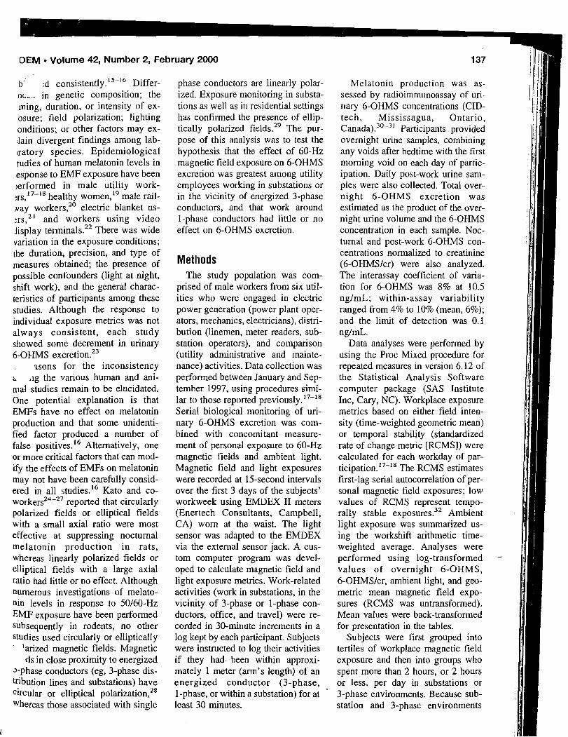
OEM Volume 42, Number 2, February 2000 137
b' :d c o n s i ~ t e n t l ~ . ' ~ - ' ~ Differ- nL,, in genetic composition; the ming, duration, or intensity of ex- osure; field polarization; lighting onditions; or other factors may ex- ,lain divergent findings among lab- ~ratory species. Epidemiological tudies of human melatonin levels in esponse to EMF exposure have been ~erformed in male utility work- : r ~ , ' ~ - l ~ healthy women,'g male rail- Nay workers,20 electric blanket us- :rs,2' and workers using video jisplay terminak2' There was wide variation in the exposure conditions; the duration, precision, and type of measures obtained; the presence of possible confounders (light at night, shift work), and the general charac- teristics of participants among these studies. Although the response to individual exposure metrics was not always consistent, each study showed some decrement in urinary 6-OHMS excretion.23
ssons for the inconsistency ; .ig the various human and ani- mal studies remain to be elucidated. One potential explanation is that EMFs have no effect on melatonin production and that some unidenti- fied factor produced a number of false positives.16 Alternatively, one or more critical factors that can mod- ify the effects of EMFs on melatonin may not have been carefully consid- ered in all studies.16 Kato and co- w o r k e r ~ ~ ~ - ~ ~ reported that circularly polarized fields or elliptical fields with a small axial ratio were most effective at suppressing nocturnal melatonin production in rats, whereas linearly polarized fields or elliptical fields with a large axial ratio had little or no effect. Although numerous investigations of melato- nin levels in response to 50160-Hz EMF exposure have been performed subsequently in rodents, no other studies used circularly or elliptically
'arized magnetic fields. Magnetic ds in close proximity to energized
J-phase conductors (eg, 3-phase dis- tribution lines and substations) have circular or elliptical po l a r i~a t ion ,~~ Whereas those associated with single
phase conductors are linearly polar- ized. Exposure monitoring in substa- tions as well as in residential settings has confirmed the presence of ellip- tically polarized fields.29 The pur- pose of this analysis was to test the hypothesis that the effect of 60-Hz magnetic field exposure on 6-OHMS excretion was greatest among utility employees working in substations or in the vicinity of energized 3-phase conductors, and that work around 1-phase conductors had little or no effect on 6-OHMS excretion.
Methods The study population was com-
prised of male workers from six util- ities who were engaged in electric power generation (power plant oper- ators, mechanics, electricians), distri- bution (linemen, meter readers, sub- station operators), and comparison (utility administrative and mainte- nance) activities. Data collection was performed between January and Sep- tember 1997, using procedures simi- lar to those reported previously.'7-'8 Serial biological monitoring of uri- nary 6-OHMS excretion was com- bined with concomitant measure- ment of personal exposure to 60-Hz magnetic fields and ambient light. Magnetic field and light exposures were recorded at 15-second intervals over the first 3 days of the subjects' workweek using EMDEX I1 meters (Enertech Consultants, Campbell, CA) worn at the waist. The light sensor was adapted to the EMDEX via the external sensor jack. A cus- tom computer program was devel- oped to calculate magnetic field and light exposure metrics. Work-related activities (work in substations, in the vicinity of 3-phase or I-phase con- ductors, office, and travel) were re- corded in 30-minute increments in a log kept by each participant. Subjects were instructed to log their activities if they had been within approxi- mately 1 meter (arm's length) of an energized conductor (3-phase, 1-phase, or within a substation) for at '
least 30 minutes.
Melatonin production was as- sessed by radioimmunoassay of uri- nary 6-OHMS concentrations (CID- tech, Mississagua, Ontario, ~anada) ."-~ ' Participants provided overnight urine samples, combining any voids after bedtime with the first morning void on each day of partic- ipation. Daily post-work urine sam- ples were also collected. Total over- night 6-OHMS excretion was estimated as the product of the over- night urine volume and the 6-OHMS concentration in each sample. Noc- turnal and post-work 6-OHMS con- centrations normalized to creatinine (6-OHMSIcr) were also analyzed. The interassay coefficient of varia- tion for 6-OHMS was 8% at 10.5 ng1mL; within-assay variability ranged from 4% to 10% (mean, 6%); and the limit of detection was 0.1 ng1mL.
Data analyses were performed by using the Proc Mixed procedure for repeated measures in version 6.12 of the Statistical Analysis Software computer package (SAS Institute Inc, Cary, NC). Workplace exposure metrics based on either field inten- sity (time-weighted geometric mean) or temporal stability (standardized rate of change metric [RCMS]) were calculated for each workday of par- t i ~ i ~ a t i o n . ' ~ - ' ~ The RCMS estimates first-lag serial autocorrelation of per- sonal magnetic field exposures; low values of RCMS represent tempo- rally stable exposures.32 Ambient light exposure was summarized us- ing the workshift arithmetic time- weighted average. Analyses were performed using log-transformed - values of overnight 6-OHMS, 6-OHMSIcr, ambient light, and geo- metric mean magnetic field expo- sures (RCMS was untransformed). Mean values were back-transformed for presentation in the tables.
Subjects were first grouped into tertiles of workplace magnetic field exposure and then into groups who spent more than 2 hours, or 2 hours or less, per day in substations or 3-phase environments. Because sub- station and 3-phase environments
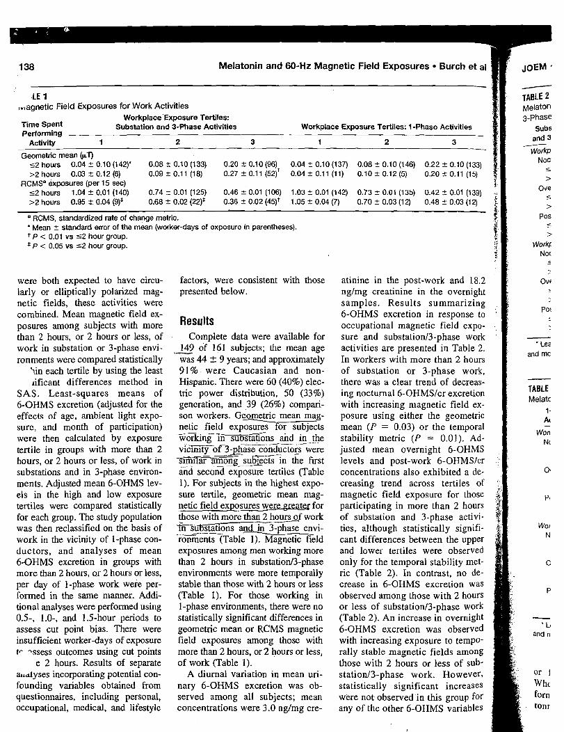
138 Melatonin and 60-Hz Magnetic Field Exposures Burch et a1
,LE 1 ,,(agnetic Field Exposures for Work Activities -
Workplace'Exposure Tertiles: Time Spent Substation and 3-Phase Activities Performing
Activity 1 2 3
Geometric mean bV 52 hours 0.04 + 0.10 (1 42)' 0.08 t 0.10 (133) 0.20 + 0.10 (96) >2 hours 0.03 2 0.12 (6) 0.09 2 0.1 1 (18) 0.27 2 0.1 1 (52)'
RCMSa exposures (per 15 sec) 52 hours 1.04 + 0.01 (140) 0.74 +- 0.01 (125) 0.46 2 0.01 (106) >2 hours 0.95 2 0.04 (9)* 0.68 2 0.02 (22)* 0.36 2 0.02 (45)'
Workplace Exposure Tertiles: I-Phase Activities
- TABLE 2 Melaton 3-Phase
Subs and 3 - workp
Noc s
> Ove 5
L >
a RCMS, standardized rate of change metric. Pos ' Mean +- standard error of the mean (worker-days of exposure in parentheses). c
t P < 0.01 vs 52 hour group. * P < 0.05 vs 52 hour group.
> r work^
were both expected to have circu- larly or elliptically polarized mag- netic fields, these activities were combined. Mean magnetic field ex- posures among subjects with more than 2 hours, or 2 hours or less, of work in substation or 3-phase envi- ronments were compared statistically
iin each tertile by using the least ~ificant differences method in
SAS. Least-squares means of 6-OHMS excretion (adjusted for the effects of age, ambient light expo- sure, and month of participation) were then calculated by exposure tertile in groups with more than 2 hours, or 2 hours or less, of work in substations and in 3-phase environ- ments. Adjusted mean 6-OHMS lev- els in the high and low exposure tertiles were compared statistically for each group. The study population was then reclassified on the basis of work in the vicinity of 1-phase con- ductors, and analyses of mean 6-OHMS excretion in groups with more than 2 hours, or 2 hours or less, per day of I-phase work were per- formed in the same manner. Addi- tional analyses were performed using 0.5, 1.0-, and 1.5-hour periods to assess cut point bias. There were insufficient worker-days of exposure tr -ssess outcomes using cut points
e 2 hours. Results of separate ailalyses incorporating potential con- founding variables obtained from questionnaires, including personal, occupational, medical, and lifestyle
factors, were consistent with those presented below.
Complete data were available for 149 of 161 subjects; the mean age .- was 44 +- 9 years; and approximately 91%. were Caucasian and non- Hispanic. There were 60 (40%) elec- tric power distribution, 50 (33%) generation, and 39 (26%) compari- son workers. Ge-c mean mag- netic field exposures for subjects - workingn s u b ~ G n s aid _---- in the viciZXjGT3-phase c o n d u c t ~ ~ ~ were
--.A
siiZiGr among subiects in the first . --
&d second exposure tertiles (Table 1). For subjects in the highest expo- sure tertile, geometric mean mag- netic field exposures were greatq for those with more than 2 hours of work w a t i o n s a n g - p h a s e envi- ronments (Table 1). Magnetic field exposures among men working more than 2 hours in substation/3-phase environments were more temporally stable than those with 2 hours or less (Table 1). For those working in 1-phase environments, there were no statistically significant differences in geometric mean or RCMS magnetic field exposures among those with more than 2 hours, or 2 hours or less, of work (Table 1).
A diurnal variation in mean uri- nary 6-OHMS excretion was ob- served among all subjects; mean concentrations were 3.0 nglmg cre-
atinine in the post-work and 18.2 ngJmg creatinine in the overnight samples. Results summarizing 6-OHMS excretion in response to occupational magnetic field expo- sure and substationl3-phase work activities are presented in Table 2. In workers with more than 2 hours of substation or 3-phase work, there was a clear trend of decreas- ing nocturnal 6-OHMSJcr excretion with increasing magnetic field ex- posure using either the geometric mean ( P = 0.03) or the temporal stability metric (P = 0.01). Ad- justed mean overnight 6-OHMS levels and post-work 6-OHMSJcr concentrations also exhibited a de- creasing trend across tertiles of magnetic field exposure for those participating in more than 2 hours of substation and 3-phase activi- ties, although statistically signifi- cant differences between the upper and lower tertiles were observed only for the temporal stability met- ric (Table 2). In contrast, no de- crease in 6-OHMS excretion was observed among those with 2 hours or less of substationl3-phase work (Table 2). An increase in overnight 6-OHMS excretion was observed with increasing exposure to tempo- rally stable magnetic fields among those with 2 hours or less of sub- stationl3-phase work. However, statistically significant increases were not observed in this group for any of the other 6-OHMS variables
and mc
- TABLE Melatc
1 - Ac
- La
and n
j@EM Volume 42, Number 2, February 2000 139
- - ?' - TABLE 2
Melatonin Metabolite Excretion* in Electric Utility Workers with Substation and $Phase Activities
Substation Diierence: and 3-Phase 1 2 3 Tertile I vs 3
Workplace geometric mean exposure tertiles Nocturnal 6-OHMWcr concentration (ng/mg cr)
5 2 hours 15.0 14.9 14.7 -2% >2 hours 23.5 18.0 13.5 -43%
Overnight 6-OHMS excretion (pg) 5 2 hours 7.9 7.9 8.2 +4% >2 hours 13.1 8.8 8.0 -39%
Post-work 6-OHMWcr concentration (ng/rng cr) 5 2 hours 2.1 2.4 2.5 +19% >2 hours 3.5 1.8 2.3 -34%
P-value: Tertile 1 vs 3
Workplace temporal stability exposure tertiles Nocturnal 6-OHMWcr concentration (ng/mg cr)
5 2 hours 13.7 15.2 15.7 +13% 0.1 1 >2 hours 23.6 16.1 13.8 -42% 0.01
Overnight 6-OHMS excretion (pg) 5 2 hours 7.2 8.1 8.8 +22% 0.05 >2 hours 13.5 8.9 7.8 -42% 0.03
Post-work 6-OHMWcr concentration (ng/mg cr) 5 2 hours 2.2 2.3 ' 2.3 +5% 0.87 >2 hours 3.5 2.7 1.8 -49% 0.02
-
* Least squares means adjusted for the effects of age, average workplace light exposure, and month of participation.
TABLE 3 Melatonin Metabolite Excretion* in Electric Utility Workers with 1-Phase Activities
I-Phase Difference: P-value: Activities 1 2 3 Tertile 1 vs 3 Tertile 1 vs 3
Worlplace geometric mean exposure tertiles Nocturnal 6-OHMWcr concentration (ng/mg cr)
5 2 hours 15.4 15.2 14.1 -8% 0.37 >2 hours 13.5 16.9 15.1 +12% 0.66
Overnight 6-OHMS excretion (pg) 5 2 hours 8.1 8.0 8.1 0% 0.99 >2 hours 8.4 7.8 8.4 0% 0.98
Post-work 6-OHMS/cr concentration (ng/mg cr) 5 2 hours 2.1 2.3 2.4 +14% 0.30 >2 hours 2.3 2.7 2.3 0% 0.96
W o ~ l a c e temporal stability exposure tertiles Nocturnal 6-OHMWcr concentration (ng/mg cr)
5 2 hours 14.3 14.9 15.4 +8% 0.35 >2 hours 13.4 20.0 12.7 -5% 0.84
Overnight 6-OHMS excretion (pg) 5 2 hours 7.5 8.0 8.5 +13% . 0.20 >2 hours 7.4 9.5 7.9 +7% 0.82
Post-work 6-OHMS/cr concentration (ng/mg cr) 2 hours 2.3 2.4 2.1 -9% 0.38 >2 hours 2.3 2.2 2.4 +4% 0.78
Least squares means adjusted for the effects of age, average workplace light exposure, and month of participation. '
or for magnetic field intensity. cally significant differences in When the same analysis was per- mean 6-OHMS excretion for those formed for work in 1-phase envi- with or without 2 hours of 1-phase ronments, there were no statisti- work when using either the geo-
metric mean or the temporal stabil- ity metric (Table 3).
Results obtained among workers with more than 1.0 or 1.5 hours of substatiod3-phase work (Table 4) were very similar to those obtained using the 2-hour cut point (Table 3). Differences between the upper and lower tertiles were progressively greater as the duration of time spent in substatiod3-phase environments increased. There were no statistically significant differences in mean 6-OHMS excretion among subjects below the chosen cut points for sub- statiod3-phase activities or among those with 1-phase work activities above or below the cut points (results not shown).
Discussion Decreased nocturnal or post-work
urinary 6-OHMS excretion have k e n associated with magnetic field .exposures in studies of electric rail- way workers2' and in our earlier studies of electric utility work- e r ~ . ' ~ - ' ~ In the present study, another population of male electric utility workers had decreased overnight 6-OHMS levels as well as lower nocturnal and post-work 6-OHMSIcr concentrations with increasing expo- sure to 60-Hz magnetic fields in substations o r near energized 3-phase conductors. Differences in mean 6-OHMS excretion between the upper and lower exposure tertiles became progressively greater as the cut point for the amount of time spent in substations and in 3-phase environments increased from 0.5 to 2 hours. These findings are consistent with the hypothesis that magnetic fields with circular or elliptical po- larization are more effective at sup- pressing melatonin production than linearly polarized field^.^^-^^ The lack of effects observed in those with 2 hours or less of substationl3-phase work or among those with 1-phase exposures further supports the hy- pothesis. Alternatively, this classifi- cation scheme may have simply se- lected those with more intense and temporally stable exposures. How-
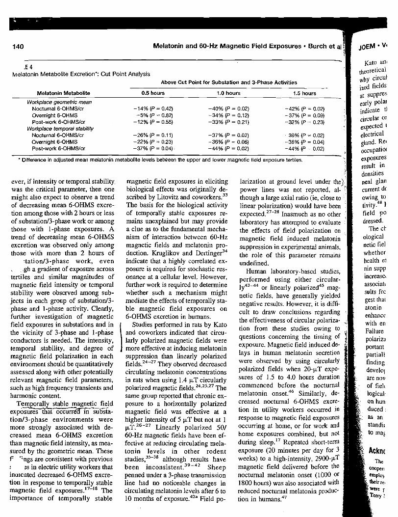
Melatonin and 60-Hz Magnetic Field Exposures Burch et a JOEM Vl v - . Kate ant .t 4
helatonin Metabolite Excretion*: Cut Point Analysis Above Cut Point for Substation and 3-Phase Activities
Melatonin Metabolite 0.5 hours 1.0 hours 1.5 hours
Workplace geometric mean Nocturnal 6-OHMS/cr -14% (P = 0.42) -40% (P = 0.02) -42% (P = 0.02) Overnight 6-OHMS -5% (P = 0.82) -34% (P = 0.12) -37% (P = 0.09) Post-work 6-OHMS/cr -12% (P= 0.55) -33% (P = 0.21) -32% (P = 0.23)
Workplace temporal stability Nocturnal 6-OHMS/cr -26% (P = 0.11) -37% (P = 0.02) -39% (P = 0.02) Overnight 6-OHMS -22% (P = 0.23) -36% (P = 0.06) -38% (P = 0.04) Post-work 6-OHMS/cr -37% (P = 0.04) -44% (P = 0.02) -44% (P = 0.02)
' Difference in adjusted mean melatonin metabolite levels beween the upper and lower magnetic f~eld exposure tert~les.
ever, if intensity or temporal stability magnetic field exposures in eliciting larization at ground level under the was the critical parameter, then one biological effects was originally de- power lines was not reported, al- might also expect to observe a trend scribed by Litovitz and coworkers.33 though a large axial ratio (ie, close to of decreasing mean 6-OHMS excre- The basis for the biological activity linear polarization) would have been tion among those with 2 hours or less of temporally stable exposures re- expected.27-28 Inasmuch as no other %
of substatiod3-phase work or among mains unexplained but may provide laboratory has attempted to evaluate those with 1-phase exposures. A a clue as to the fundamental mecha- the effects of field polarization on trend of decreasing mean 6-OHMS nism of interaction between 60-Hz magnetic field induced rnelatonin excretion was observed only among magnetic fields and melatonin pro- suppression in animals, those with more than 2 hours of duction. Kruglikov and ~ e r t i n ~ e ? ~ the role of this remains
~tationl3-phase work, even indicate that a highly correlated ex- undefined. . .gh a gradient of exposure across posure is required for stochastic res- H~~~~ laboratory-based studies, tertiles and similar magnitudes of onance at a cellular level. However, performed using either circular- magnetic field intensity or temporal further work is required to determine
1Y43-44 Or linearly polarized45 mag- stability were observed among sub- whether such a mechanism might netic fields, have generally yielded jects in each group of substatiod3- mediate the effects of temporally sta- negative results. However, it is diffi- phase and 1-phase activity. Clearly, ble magnetic field exposures on cult to draw conclusions regarding further investigation of magnetic 6-OHMS excretion in humans. the effectiveness of circular polariza- + field exposures in substations and in Studies perfonned in rats Kate tion from these studies owing to the vicinity of 3-phase and 1-phase and coworkers indicated that circu-
questions concerning the timing of conductors is needed. The intensity, larly polarized magnetic fields were temporal stability, and degree of more effective at inducing melatonin exposure. Magnetic field induced de-
magnetic field polarization in each suppression than linearly polarized lays in human melatonin secretion
environment should be quantitatively field^.^^-^^ They observed decreased were observed by using circularly
assessed along with other potentially circulating melatonin concentrations polarized fields when 20-pT expo-
relevant magnetic field parameters, in rats when using 1.4 pT circularly sures of 1.5 to 4.0 hours duration
such as high frequency transients and polarized magnetic field^.^^.^^" The before the
harmonic content. same group reported that chronic ex- melatonin onset.46 Similarly, de-
Temporally stable magnetic field posure to a horizontally polarized creased 6-oHMS excre- exposuz%at o c c u r r e m s t a - magnetic field was effective at a tion in Occurred in tion/3-phase environments were higher intensity of 5 pT but not at 1 to magnetic field exposures more strongly associated with de- p , ~ . ~ ~ - ~ ~ Linearly polarized 501 O C C U ' T ~ ~ ~ at homey or for work and creased mean 6-OHMS excretion 60-Hz magnetic fields have been ef- home exposures combined, but not than magnetic field intensity, as mea- fective at reducing circulating mela- during sleep.17 Repeated short-tern sured by the geometric mean. These tonin levels in other rodent exposure (20 minutes per day for 3 F ';rigs are consistent with previous s t ~ d i e s ? ~ " ~ although results have weeks) to a high-intensity, 2900-PT : zs in electric utility workers that been i n c ~ n s i s t e n t . ~ ' - ~ ~ Sheep magnetic field delivered before the inaicated decreased 6-OHMS excre- penned under a 3-phase transmission nocturnal melatonin onset (1000 or tion in response to temporally stable line had no noticeable changes in 1800 hours) was also associated with magnetic field exposures. 17-18 The circulating melatonin levels after 6 to reduced nocturnal melatonin produc- importance of temporally stable 10 months of exposure.42a Field po- tion in humans.47
:M Volume 42, Number 2, February 2000
:. nd Shigemitsu2' presented . ;a1 calculations to explain 1 circularly or elliptically polar- i fields would be more effective suppressing melatonin than lin- ly polarized fields. These authors .icate that magnetic fields with sular or elliptical polarization are ?ected to more effectively induce :ctrical currents in the rat pineal md. Recent estimates suggest that cupationally relevant electric field posures (10 kV/m) in humans may sult in average induced current mities of 1451 c L ~ l m 2 in the pi- :a1 gland compared with average merit densities of 6 p ~ / m 2 attained wing to endogenous electrical ac- vitye4' However, differences due to ~eld polarization were not ad- rased. The characterization of human bi-
logical responses to 60-Hz mag- ietic fields is critical for determining whether concern over potential IP effects is warranted. Melato-
.,ppression is a plausible link to increased cancer risks that have been associated with such exposures. Re- sults from the present analysis sug- gest that magnetic field induced mel- atonin suppression seems to be enhanced by work in substations and with energized 3-phase conductors. Failure to characterize magnetic field polarization or other potentially im- portant modifying fa~tors ' ' '~~ may Partially explain the inconsistent findings reported to date. Recently developed personal exposure devices are now available to evaluate the role of field polarization and other bio- logically based exposure parameters on human 6-OHMS e~cretion.~' Re- duced melatonin secretion may serve as an important model for under-
, standing human biological responses to magnetic field exposures.
Acknowledgments B me authors gratefully acknowledge the
pera at ion of the participating utilities, their cmPl~yees who participated in this study, and their representatives. Urinary &OHMS assays were performed under the direction of Dr Teq Nett, Director of the Radioimmunoas-
say Laboratory for the Colorado State Uni- versity Animal Research and Biotechnology Laboratories.
In particular, the authors thank Ms Jeanette Haddock for assistance with data collection, Ms Xiao Ming Sha for assistance with the &OHMS assay, Drs Lee Wilke and Martin Fettman for assistance with the creatinine assays, and Mr Travers Ichinose and Dr Annette Bachand for assistance with data processing. Dr Scott Davis of the Fred Hutchinson Cancer Research Center provided the design for adaptation of the light meters to the EMDEX monitors. Battelle Pacific North- west Laboratories and Platte River Power Authority provided light meters. Mr Ken Webster provided computer programming as- sistance.
This work was supported by research grant no. 1 ROlES08117 from the National Institute of Environmental Health Sciences, National Institutes of Health. Bethesda, Maryland.
References 1. Panzer A, Viljoen M. The validity of
melatonin as an oncostatic agent. J Pi- neal Res. 1997;22: 184-202.
2. Blask DE. Melatonin in oncology. In: Yu H, Reiter RJ, eds. Melatonin Biosynthe- sis. Physiological Effects, and Clinical Applications. Boca Raton: CRC Press; 1993447-475.
3. Fraschini F, Demartini G, Esposti D, Scaglione F. Melatonin involvement in immunity and cancer. Biol Signals Re- cept. 1998;7:61-72.
4. Conti A, Maestroni GJM. The clinical neuro-immunotherapeutic role of melato- nin in oncology. J Pineal Res. 1995;19: 103-1 10.
5. Reiter RJ, Melchiorri D, Sewerynek E. et al. A review of the evidence supporting melatonin's role as an antioxidant. J Pi- neal Res. 1995;18:1-11.
6. Reiter RJ. Oxidative damage in the cen- tral nervous system: protection by mela- tonin. Prog Neurobiol. 1998;56:359-38.
7. Savitz DA. Overview of epidemiological research on electric and magnetic fields and cancer. Am Ind Hyg Assoc J. 1993; 54: 197-204.
8. Kheiffets LI, Abdelmonem AA, Buffler PA, Zhang ZW. Occupational electric and magnetic field exposure and brain cancer: a meta-analysis. J Occ Environ Med. 1995;37: 1327-1 34 1.
9. Brezinski A. Melatonin in humans. New Engl J Med. 1997;336:186-195.
10. Reiter RI. Alterations of the circadian melatonin rhythm by the electromagnetic spectrum: a study in environmental toxi- cology. Reg Toxic01 Pharmacol. 1992; 15:226 -244.
11. Waldhauser F, Weizenbacher G, Tatzer E, et al. Alterations in nocturnal serum melatonin levels in humans with growth and aging. J Clin Endocr Metab. 1988; 66548-652.
12. Touitou Y, Fevre M, Lagoguey M, et al. Age- and mental health-related circadian rhythms of plasma levels of melatonin, prolactin, luteinizing hormone and folli- cle-stimulating hormone in man. J En- docr. 198 1;91:467-475.
13. Bojkowski CJ, Arendt JA, Shih MC, Markey SP. Melatonin secretion in hu- mans assessed by measuring its metabo- lite, 6-sulfatoxymelatonin. Clin Chem 1987;33:13431348.
14. Bartsch C, Bartsch H, Schmidt A, Ilg S, Bichler KH, Fluechter SH. Melatonin and 6-sulfatoxymelatonin circadian rhythms in serum and urine of primary prostate cancer patients: evidence for reduced pi- neal activity and relevance of urinary determinations. Clin Chim Acta. 1992; 209:153-167.
15. Reiter RJ. Melatonin in the context of the reported bioeffects of environmental electromagnetic fields. Bioelectrochem Bioenergetics. 1998;47: 135-1 42.
16. Brainard GC, Kavet R, Kheifets LI. The relationship between electromagnetic field and light exposures to melatonin and breast cancer risk: a review of the relevant literature. J Pineal Res. 1999:26: 65- 100.
17. Burch JB, Reif JS, Yost MG, Keefe TJ, Pitrat CA. Nocturnal excretion of a uri- nary melatonin metabolite in electric util- ity workers. Scand J Work Environ Health. 1998;24: 183-1 89.
18. Burch JB, Reif JS, Yost MG, Keefe TJ, Pitrat CA. Reduced excretion of a mela- tonin metabolite in workers exposed to 60 Hz magnetic fields. Am J Epidemiol. 1999;150:27-36.
19. Kaune W, Davis S, Stevens R Relation Between Residential Magnetic Fields. Light-at-Night, and Nocturnal Urine Melatonin Levels in Women. Palo Alto, CA: Electric Power Research Institute; 1997. TR-107242-V1, EmZL Report.
20. Pfluger DH, Minder CE. Effects of expo- sure to 16.7 Hz magnetic fields on uri- nary 6-hydroxymelatonin sulfate excre- ' tion of Swiss railway workers. J Pineal Res. 1996;21:91-100.
21. Wilson BW, Wright CW, Moms JE, et al. Evidence for an effect of ELF electro- magnetic fields on human pineal gland function. J Pineal Res. 1990;9:259-269.
22. Ametz BB, Berg M. Melatonin and ad- renocorticotropic hormone levels in video display unit workers during work and leisure. J Occup Med. 1996;38: 1108-1110.
142 Melatonin and 60-Hz Magnetic Field Exposures Burch et
National Institute of Environmental 33. Litovitz TA, Penafiel M, Krause D, Djungarian hamster. I Pineal Res. 1997; Health Sciences. Assessment of Health Zhang D. Mullins JM. The role of tem- 22: 177-1 83. Effects from Exposure to Power-Line poral sensing in bioelectromagnetic ef- 42a. Lee JM, Stormshak F, Thompson JM, Frequency Electric and Magnetic Fields. fects. Bioelectromagnetics. 1997; 18: Hess DL, Foster DL. Melatonin and Portier CJ. Wolfe MS. eds. Research 388-395. puberty in female lambs exposed to Triangle Park. NC: US Dept. Health and 34. Kruglikov IL, Dertinger H. Stochastic EMF: a replicate study. Bioelectromag. Human Services; 1998:311-313. Pub No. resonance as a possible mechanism of netics 1995; 16: I 19-23. 98-3981. amplification of weak electric signals in 43. Graham C, cook MR, Riffle DW, eerk-
24. Kato M. Honma K, Shigemitsu T. Shiga living cells. Bioelectromagnetics. 1994; ovich MM. Cohen HD. Nocturnal mela- Y. Effects of circularly polarized 50-Hz 15539-547. tonin levels in human volunteers exposed magnetic field on plasma and pineal 35. Selmaoui B. Touitou Y. Sinusoidal50 Hz to intermittent 60 Hz magnetic fields. atonin levels in rats. Bioelectromagnet- magnetic fields depress rat pineal NAT Bioelectrornagnerics. 1996; 17:263-273. ics. 1993; 14:97-106. activity and serum melatonin. Role of 44. craham C, cook MR. ~ i f f l ~ DW, Hu-
25. Kato M, Honma K, Shigemitsu T, Shiga duration and intensity of exposure. Life man melatonin during continuous mag- Y. Circularly polarized 50-Hz magnetic Sci. 1995;57:1351-1358. netic field exposure. Bioelecfromagnet- field exposure reduces pineal gland and 36. Loescher W, Wahnschaffe U, Mevissen blood melatonin concentrations in Long- ics. 1996;18:166-17!.
M. Lerchl A. Effects weak 45. Selmaoui B, Lambrozo J, Touitou Y. Evans rats. Neurosci k t t . 1994;166:59- alternating magnetic fields on nocturnal 62. Magnetic fields and pineal function in
melatonin production and mammary car- 26. Kato M, Honma K, Shigemitsu T, Shiga humans: evaluation of nocturnal expo-
cinogenesis in rats. Oncology. 1994;s 1 : Y. Horizontal or vertical 50-Hz 1-pT sure to extremely low frequency mag-
288-295. magnetic fields have no effect On pineal 37. Mevissen M, Lerchl A, Loescher W.
netic fields on serum melatonin and uri-
gland or plasma melatonin concentration Study of pineal function and DMBA- of albino rats. Neurosci IRtt. 1994;168: induced breast cancer formation in rats 205-208. 46. Wood AW, Armstrong SM. Sait ML,
during exposure to a 100-mG, 50 Hz 27. Kato M. Shigemitsu T. Effects of 50-Hz Devine L, Martin MI. Changes in human
magnetic field. J Toxic01 Environ Health. magnetic fields on pineal function in the rat. In: Stevens RG. Wilson BW, Ander-
1996;48:169-185. 38. Yellon SM. Acute 60 Hz magnetic field
son LE, eds. The Melatonin Hypothesis. Res. 1998;25: 1 16-127. exposure effects on the melatonin rhythm Columbus, OH: Batelle Press; 1997:337- 47. Karasek M, Woldanska-Okonska M, Cz- .:. in the pineal and circulation of the adult 376. ernicki J, Zylinska K, Swietoslawski
~ 8 . Deno DW. Transmission line fields. Djungarian hamster. J Pineal Res. 1994; 16~136-144. ZEEE Trans Power Appar Sys. 1976;95:
39. Loescher W, Mevissen M. Lerchl A. magnetic field reduces melatonin concen- 1600-1611.
Exposure of female rats to a 100-pT 50 trations in humans. J Pineal Res. 1998; 29. Dietrich FM, Feero WE, Robertson DC,
Hz magnetic field does not induce con- 25:240-244. Sicree RM. Measurement of Power Sys- tem Magnetic Fields by Waveform Cap- electric fields and currents induced in a ture. Palo Alto, CA: Electric Power Re- melatonin. Radiat Res. 1998;150:557-
millimeter-resolution human model at 60 search Institute; 1992. TR- 10006 1 EPRI 567.
Hz using the FDTD method. Bioeleclro- Report. 40. John TM, Liu GY, Brown GM. 60 Hz
magnetic field exposure and urinary magnetics. 1998; 19:293-299. 30. Arendt J, Bojkowski C, Franey C, Wright
J, Marks V. Immunoassay of 6-hy- 6-sulfatoxymelatonin levels in the rat. 49. Burch JB, Reif JS, Yost MG. Geomag- netic disturbances are associated with droxymelatonin sulfate in human plasma Bioelectromagnetics. 1998; 19: 172-1 80.
and urine: abolition of the urinary 24- 41. Heikkinen P, Kumlin T, Laitinen JT, reduced nocturnal excretion of a melato-
hour rhythm with atenolol. J Clin Endocr Komulainen H. Juutilainen J. Chronic nin metabolite in humans. Neurosci Lett.
Metab. 1985;60:1166-1173. exposure to 50-Hz magnetic fields or 1999;266:209 -2 12. 900-MHz electromagnetic fields does 50. Bowman JD. Methner MM. ~ a z a r r l Sur- 3 1. Aldous ME, Arendt J. Radioimmunoas- not alter nocturnal 6-hydroxymelatonin veillance for Workplace Magnetic Fields: say for 6-sulphatoxymelatonin in urine
Field Characteristics from Waveform using an iodinated tracer. Ann Clin Bio- sulfate secretion in CBNS mice. Elec- chem. 1988;25:298-303. fro- and Magnetobiology. 1999; 18:33- Measurements. Cinncinati, OH: ~ational
32. Yost MG. Alternative magnetic field ex- 42. Institute of Occupational Safety and - posure rnetrics: occupational measure- 42. Truong H, Yellon SM. Effect of various Health; 1998. Report to the U S Dept. of
ments in trolley workers. Radiation Pro- acute 60 Hz magnetic field exposures on Energy for Interagency Agreement NO, tection and Dosimetry. 1999;83:99-106. the nocturnal melatonin rise in the adult DE-AI01-94CE34008.
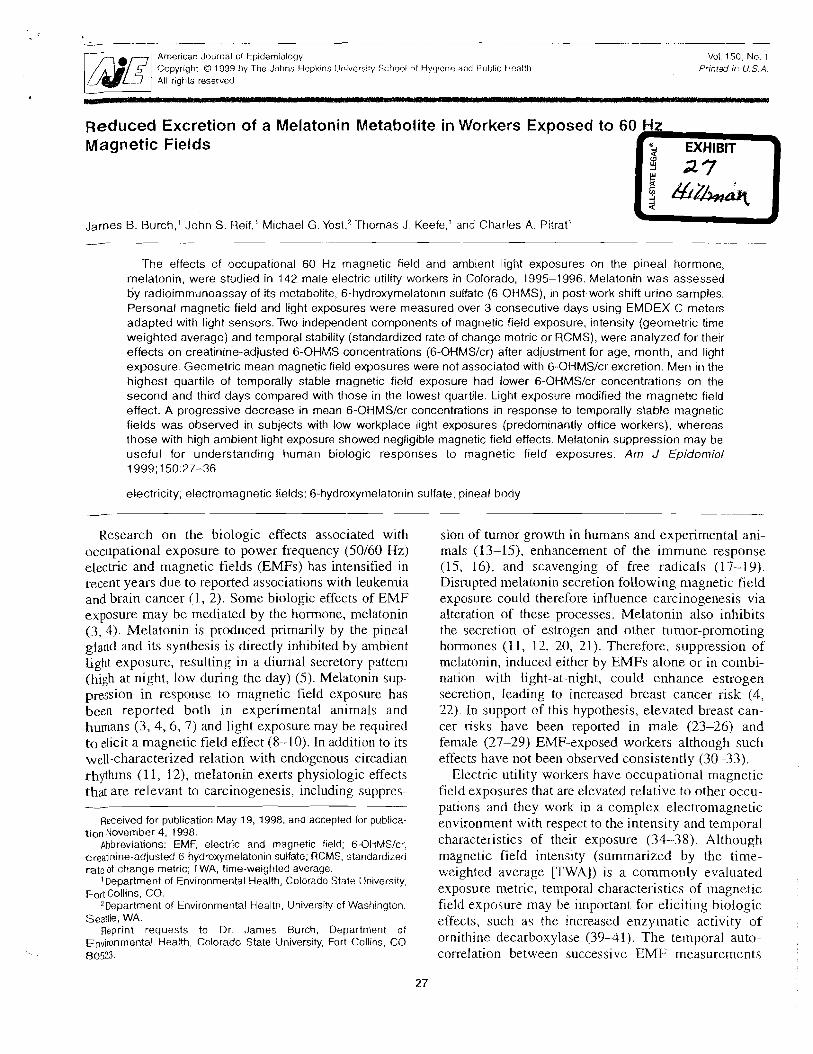
American Journal of Epidemislogy Copyright O I999 by The Johns Hopk~ns Univers~!y School of Hygiene and Public Ho-alth All rights reserved
Voi. 150. No. 1
Pr;ntsd jf! U. S A
Reduced Excretion of Magnetic Fields
James B. Burch,' John S. Reif,'
a Melatonin
Michael G. Y ~ s t , ~
Metabolite in
Thomas J Keefe,'
Workers
and Charles
Exposed to
A. Pitrat'
The effects of occupational 60 Hz magnetic field and ambient light exposures on the plneal hormone, melatonin, were studied in 142 male electric utility workers in Colorado, 1995-1 996. Melatonin was assessed by radioimmunoassay of its metabolite, 6-hydroxymelatonln sulfate (6-OHMS), rn post-work shift urine samples. Personal magnetic field and light exposures were measured over 3 consecutive days using EMDEX C meters adapted with light sensors. Two independent components of magnetic field exposure, ~ntensity (geometric time weighted average) and temporal stability (standardized rate of change metric or RCMS), were analyzed for their effects on creatinine-adjusted 6-OHMS concentrations (6-OHMSIcr) after adjustment for age, month, and light exposure. Geometric mean magnetic field exposures were not associated with 6-OHMSJcr excretion. Men in the highest quartile of temporally stable magnetic field exposure had lower 6-OHMSIcr concentrations on the second and third days compared with those in the lowest quartile. Light exposure modified the magnetic field effect. A progressive decrease in mean 6-OHMSJcr concentrations in response to temporally stable magnetic fields was observed in subjects with low workplace light exposures (predominantly office workers), whereas those with high ambient light exposure showed negligible magnetic field effects. Melatonin suppression may be useful for understanding human biologic responses to magnetic field exposures. Am J Epidemiol 1999; 150:27-36
electricity; electromagnetic fields; 6-hydroxymelatonin sulfate; pineal body
Research on the biologic effects associated with occupational exposure to power frequency (50160 Hz) electric and magnetic fields (EMFs) has intensified in recent years due to reported associations with leukemia and brain cancer ( 1 , 2). Some biologic effects of EMF exposure may be mediated by the hormone, melatonin (3, 4). Melatonin is produced primarily by the pinzal gland and its synthesis is directly inhibited by ambient light exposure, resulting in a diurnal secretory pattern (high at night, low during the day) (5) . Melatonin sup- pression in response to magnetic field exposure has been reported both in experimental animals and humans (3, 4 ,6 , 7) and light exposure may be required to elicit a magnetic field effect (8-10). In addition to its well-characterized relation with endogenous circadian rhythms (11, 12), melatonin exerts physiologic effects that are relevant to carcinogenesis, including suppres-
Received for publication May 19, 1998, and accepted for publica- tion November 4, 1998.
Abbreviations: EMF, electric and magnetic field; 6-OKMS!cr, creatinine-adjusted 6-hydroxymelatonin sulfate; RCMS, standardized rate of change metric; TWA, time-weighted average.
Department of Environmental Health, Colorado State Univers~ty. Fort Collins, CO.
2Department of Environmental Health, Universi!y of \Nashing:on. Seattle, WA.
Reprint requests to Dr. Janles Burch. Department of Environmental Health, Colorado State University, Fort Collins, CO 80523.
sion of tumor growth in humans and experimental ani- mals (13-15), enhancement of the immune response (15, 16), and scavenging of free radicals (17-19). Disrupted melatonin secretion following magnetic field exposure could therefore influence carcinogenesis via alteration of these processes. Melatonin also inhibits the secretion of estrogen and other tunlor-promoting hormones (1 1 , 12. 20, 21). Therefore, suppression of melatonin. induced either by EMFs alone or in combi- nation with light-at-night, could enhance estrogen secretion, leading to increased breast cancer risk (4, 22). In support of this hypothesis, elevated breast can- cer cisks have been repolted in male (23-26) and female (27-29) EMF-exposed workers although such effects have not been observed consistently (30-33).
Electric utility workers have occupational magnetic field exposures that are elevated relative to other occu- pations and they work in a complex electron~agnetic environment with respect to the intensity and temporal characteristics of their exposure (3638) . Although magnetic field intensity (summarized by the time- weighted average [TWA]) is a commorlly evaluated exposure metric, te~npoml characteristics of magnetic field exposure may be important for eliciting biologic effects, such as the increased enzy~natic activity of ornithine decarboxylase (3941) . The temporal auto- correlation between successive EMF measurements
28 Burch et al
has been identified as a component of personal EMF 7osure in clectric utility workers that is independent magnetic field intensity (33). Further, the temporal
autccorrelation of residential magnetic field exposures may be important for predicting childhood leukemia risk when combined with other EMF exposure metrics (42).
Reduced excretion of the major urinary melatonin metabolite. 6-hydroxymelatonin sulfate (6-OHMS), has been shown in two studies of occupational EMF exposure (6, 7). Swiss railway workers were found to have reduced evening 6-OHMS excretion after 5 days of exposure to 16.7 Hz fields (7). Recently, we demon- strated decreased nocturnal 6-OHMS excretion associ- ated with exposure to temporally stable 60 Hz magnetic fields in male electric utility workers (6). Temporal stability was assessed using an estimate of autocorre- lation, and the effect was most pronounced when both residential and occupational exposures were combined (6). The current study reports the effects of occupa- tional exposures to 60 Hz magnetic fields on post- work shift 6-OHMS excretion in the same population of electric utility workers using measures of field intensity and temporal autocorrelation.
MATERIALS AND METHODS
'he study population was derived from three munic- lPdl electric utilities in Colorado. All employees, aged 20-60 years, with at least one month of electric utility work experience were contacted via orientation meet- ings or by telephone. The goal, based on statistical power calculations, was to obtain 200 participants; 195 subjects were eventually recruited. Of those 195 work-. ers, data were available for 173, of which 142 were men. Workers with electric power generation, distribu- tion, or administrative job descriptions were studied over a one-year period during daytime work hours (approximately 7:00 a.m. to 6:00 p.m.). Data collec- tion was scheduled for the first 3 days of the work week to permit evaluation of changes in melatonin after time away from work (6, 7). Non-shift workers participated during the daytime after 2 days of non- occupational magnetic field exposure. In order to gen- erate comparable data for shift workers, they partick pated while they were working during the day; however, their scheclule provided 3 days off prior to comnlencing their clay shift.
A questionnair.e was ~ ~ s e d to collect additional infor- mation concerning factor-s that might intluence mag- netic field or light exposure and melatonin pi-oduction. Potential confounder, o!- modifiet-s included personal ' -e, race. body mass index). occupational (job title:
.rs work experience. physical activity. work with specific chemicals. 2.s.. creosote. solvel~ts. pesticides).
life-style (tobacco and alcohol consumption, light-at- night, electrical appliance use, exercise), and medical factors (medications, disease history). No subjects were taking exogenous melatonin during participation.
Subjects collected one urine sample immediately following their work shift on each of 3 consecutive days (usually Monday. Tuesday, and Wednesday) for determination of 6-OHMS. Subjects also collected four consecutive overnight urine samples; the Monday morning sample was used to evaluate baseline noctur- nal melatonin prior to resuming work. However, the logistics of having subjects collect a baseline "post- work shift" urine sample while off duty were consid- ered not practical due to concerns about subject com- pliance and quality assurance. The melatonin metabolite 6-OHMS was measured in urine by radioimmunoassay (43-45) using materials supplied by CIDtech (Mississauga, Ontario, Canada). The interassay coefficient of variation for the slope of the standard curves obtained during this study was 4 per- cent and the limit of detection for 6-OHMS was 0.1 nglml. Concentrations of 6-01-IMS were normalized to urinary creatinine concentrations (6-OHMSIcr) and are presented as nanograms 6-OHMS per milligram creatinine (nglmg cr).
Work shift personal magnetic field and ambient light exposures were logged daily for all subjects at a rate of once every 15 seconds using EMDEX C meters (Electric Field Measurements, Stockbridge, Massachusetts) worn at the waist. Light exposure was measured with a light sensor (model LX101, Grasby Optronics, Orlando, Florida) adapted to the meter's exteinal jack. This photoelectric detector produces a linear output current in proportion to light intensity, from less than 1 lux to approximately 100,000 lux. Exposure assessment was performed for 3 work days due to battery life and the capacity of digital memory in the meter. Subjects logged their work activities and hours on duty, permitting the calculation of daily workplace exposure metrics. Light exposure was sum- marized by calculating the work shift arithmetic TWA. The geometric TWA was used to assess the intensity of magnetic field exposure, and the standardized rate of change metric (RCMS), which estimates first-lag auto- correlation. was used to assess the tenlporal stability of exposure (6). Low values of RCMS represent relatively srnall differences between successive m:lgnetic field nieasiirements and are indicative of temporally stable exposures.
.\nalyscs were pertormed with the Statijtical Analysis Software (SAS) computer prograin (SAS Institute lnc., CrLi-y. North Carolinai using log-transformed values for 6-OHMSlcr, geometric mean inag~ietic field exposures (t:ntransfonned values f ~ r RCMSj, and light data. A
.Am J Epidemio: Voi 150, No. 1 , 1999
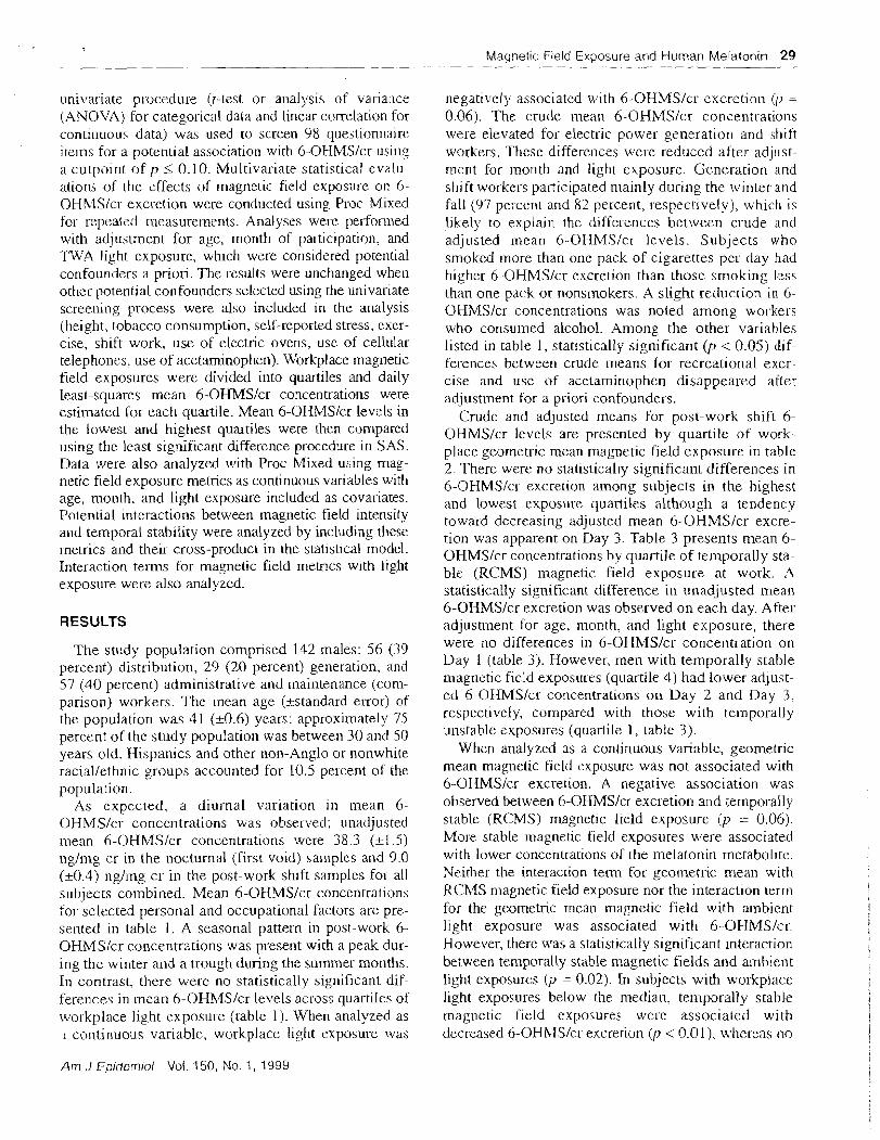
tula~rlet~c Field Exposure and Human tvlelatonin 29 --
itni.i/ariate procedure (t-test or analysis of variance (ANOVA) for carego~ical data and linear correlation for continuous data) was used to screen 98 questionnaire items for a potential association with 6-OHMSIcr using a cutpoint of p i 0.10. i\llultivariate statistical evalu- ations of the effects of magnetic field exposure on 6- OHMSIcr excretion were conducted using Proc Mixed for repeated measurements. Analyses were perfonned with adjustment for age, month of participation, and TWA light exposure, which were considered potential confounders a pliori. The results were unchanged when other potential confounders selected using the univariate screening process were also included in the analysis (height, tobacco consumption, self-reported stress, cxcr- cise, shift work, use of electric ovens, use of cellular telephones. use of acetaminophen). Workplace magnetic field exposures were divided into quartiles and daily least-squares mean 6-OHMSIcr concentrations were estimated for each quartile. Mean 6-OHMSIcr levels in the lowest and highest quartiles were then compared using the least significant difference procedure in SAS. Data were also analyzed with Proc Mixed using mag- netic field exposure metrics as continuous variables with age, month, and light exposure included as covaliates. Potential interactions between magnetic field intensity and temporal stability were analyzed by including these ~netrics and their cross-product in the statistical model. Interaction terms for magnetic field metncs with light exposure were also analyzed.
RESULTS
The study population comprised 142 males: 56 (39 percent) distribution, 29 (2.0 percent) generation, arid 57 (40 percent) administrative and ~nairltenance (com- parison) workers. The mean age (+standard error) of the population was 41 (k0.6) years; approximately 75 percent of the study population was between 30 and 50 years old. Hispanics and other non-Anglo or nonwhite raciallethnic groups accounted for 10.5 percent of the population.
As expected, a diurnal variation in mean 6- OHMSIcr concentrations was observed; unadjusted mean 6-OHMSIcr concentrations were 38.3 (-11.5) nglmg cr in the nocturnal (first void) sanlples and 9.0 (-10.4) nglmg cr in the post-work shift samples for all subjects combined. Mean 6-OHMSIcr concentrations for selected personal and occupational factors are pre- sented in table 1 . A seasonal pattern in post-work 6- OHMSlcr concentrations was present with a peak dur- ing the winter and a trough during the summer months. In contrast, there were no statistically significant dif- ferences in mean 6-OHh4SIcr levels across quartiles of \vorkplace light exposure (table 1). When analyzed as I co~ltinuotis variable, workplace light exposure was
negatively associated with 6-OHMSIcr excretion (0 = 0.06). The crude liiean 6-OHMSIcr concentrations were elevated for electric power generation and shift workers. These differences were reduced aftel- adjust- ment for month and light exposure. Generation and shift workers participated mainly during the winter and fall (97 pel-cent and 82 percent, respectively), which is likely to explain the differences between crude and adjusted mean 6-OHh4SIcr levels. Subjects who smoked inore than one pack of cigarettes per day had higher 6-OHMSIcr excretion than those smoking less than one pack or nonsinokers. A slight reduction in 6- OHMSlcr concentrations was noted among workers who consumed alcohol. Among the other variables listed in table. 1, statistically significant (p < 0.05) dif- ferences between crude means for recreational exer- cise and use of acetarnirlophen disappeared after adjustment for a priori confounders.
Crude and adjusted means for post-work shift 6- OHMSIcr levels are presented by quartile of work- place geometric mean magnetic field exposure in table 2. There were no statistically significant differences in 6-OHMSICK excretion among subjects in the highest and lowest exposure quartiles although a tendency toward decreasing adjusted mean 6-OHMSIcr excre- tion was apparent on Day 3. Table 3 presents mean 6- OHMSlcr concentrations by quartile of temporally sta- ble (RCMS) magnetic field exposure at work. A statistically significant difference in unadjusted mean 6-OHMS/cr excretion was observed on each day. After adjustment for age, month, and light exposure, there were no differences in 6-OHMSIcr concentration on Day 1 (table 3). However, men with temporally stable niagnetic field exposures (quartile 4) had lower adjust- ed 6-OHMSIcr concentrations on Day 2 and Day 3. respectively, compared with those with te~nporally unstable exposures (quartile 1, table 3).
When analyzed as a continuous variable, geometric mean magnetic field exposure was not associated with 6-OHMSIcr excretion. A negative association was observed between 6-OHMSIcr excretion and temporally stable (RCMS) magnetic field exposure (p = 0.06j. More stable magnetic field exposures \vere associated with lower concentrations of the melatonin metabolite. Neither the interaction term for geonletlic mean with RCh4S niagnetic field exposure nor the interaction term for the geometric mean magnetic field with ambient light exposure was associated with 6-OHMSIcr. However, there was a statistically significant interaction between temporally stable magnetic fields and ambient light exposures (p = 0.02). In subjects with workplace light exposures below the median. ten~porally stable magnetic field exposures were associated with decreased 6-OHhfSlcr excretion @ < 0.01). [vhereas no
Am J Epidemiol Voi. 150, No. I , 1999
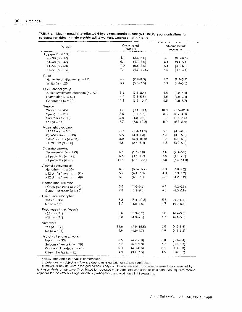
TABLE I. Mean* creatinine-adjusted 6-hydroxymelatonin sulfate (6-OHMSIcr) concentrations for selected variables in male electric utility workers, Colorado, 1995-1996t
Variable Crude mean$ Adjusted meanf -- (n@mg cr) (ng/mg cd
Age group (years) 20-30 (n = 17) 4.1 (2.G6.6) 4.8 (3.5-6 5) 31-40 (n = 47) 6.1 (4.7-7.9) 4.1 (3.4-5.1) 41-50 ( n = 59) 7.0 (5.5-8.9) 5.4 (4 .M.3) 51-60 ( n = 19) 7.4 (4.7-1 1.6) 4.6 (3.5-6.1)
Race Nonwhite or Hispanic (n = 15) White (n = 125)
Occupational group Administrativelmaintenance (n = 57) 6.5 (5.1-8.4) 4.6 (3.8-5.4) Distribution (n = 56) 4.6 (3.6-5.8) 4.5 (3.8-5.4) Generation (n = 29) 10.9 (8.8-1 3.5) 6.5 (4.8-8.7)
Season W~nter (n = 45) Spring (n = 21) Summer (n = 32) Fall ( n = 44)
Mean light exposure 5262 lux (n = 30) 263-572 lux (n = 30) 573-1,791 lux (n = 31) > I ,791 lux (n = 30)
Cigarette smoking Nonsmokers (n = 1 13) 51 packlday (n = 22) >1 packlday (n = 5)
Alcohol consumption Nondrinker (n = 39) 212 drinkshonth (n = 51) 212 drinksfmonth (n = 48)
Recreational Exercise >Once per week (n = 92) Seldom or never ( n = 50)
Use of acetaminophen Yes (n = 36) No (n = 105)
Body mass index (kg/rn2) 526 (n = 71 ) >26 (n = 71)
Shift work Yes ( n = 17) No (n = 124)
Use of cell phone at work Never ( n = 33) Seldom <Idweek (n = 38) Occasional I dday (n = 44) Often > 1 xlday ( n = 26)
p-
* 95% confidence interval in ~arentheses. t Variations in subject number are due to missing data for selec!ed variables. 4 lrrdividual results were averaged across 3 days of observation and crude means were then compared by f-
test or analysis of variance. Proc Mixed for repeated measurements was used to calculate least-squares means adjusted for the efiects of age. month of participation, and workplace light exposure.
Arrr J Epideiniol Vol. 150, No. 1 , 1999
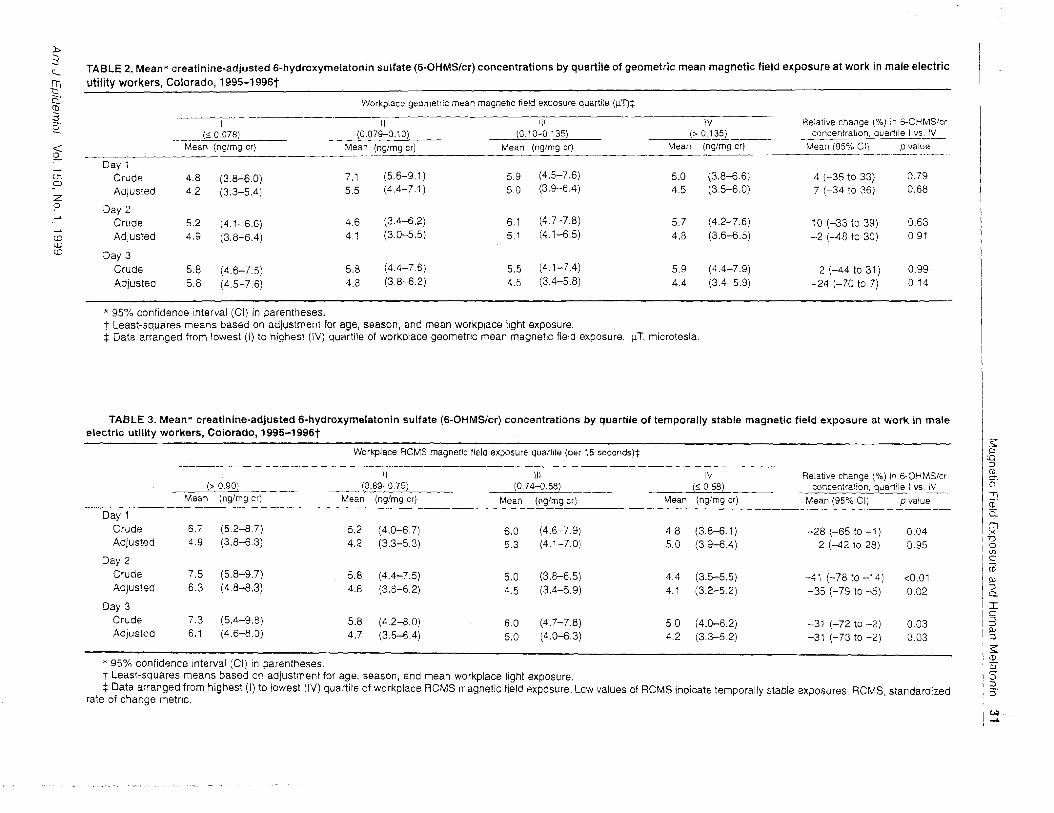
TABLE 2. Mean* creatinine-adjusted 6-hydroxyrnelatonin sulfate (6-OHMSlcr) concentrations by quartile of geometric mean magnetic field exposure at work in Inale electric uti l i ty workers, Colorado, 1995-1996t
Workpiace geometric mean magnetic field exposure quartile (p'T)$ -
I II Ill IV Relat~ve change (%) ir7 6-OHMSlcr (S 0.078) . . - - (0.079-0.10) (0.10-0.135) (> 0.135) __ concentration, quartile I vs. IV
Mean (nglmg cr) Mean (nglmg cr) Mean (nglmg cr) Mean (nglmg cr) Mean (95% CI; p value -- - -. .- -- -- - - . . - . . . . . , . -. -. .. . . . .- - - .
Day 1 Crude 4.8 (3.8-6.0) 7.1 (5.6-9.1) 5.9 (4.5-7.6) 5.0 (3.8-6.6) 4 (-35 to 33) 0.79 Adjusted 4.2 (3.3-5.4) 5.5 (4.4-7.1) 5.0 (3.9-6.4) 4.5 (3.5-6.0) 7 (-34 to 36) 0.68
Day 2 Crude 5.2 (4.1-6.6) 4.6 (3.4-6.2) 6.1 (4.7-7.8)
Adjusted 4.9 (3.8-6.4) 4.1 (3.0-5.5) 5.1 (4.1-6.5)
Day 3 Crude 5.8 (4.6-7.5) 5.8 (4.4-7.6) 5.5 (4.1-7.4) Adjusted 5.8 (4.5-7.6) 4.8 (3.8-6.2) 4.5 (3.4-5.8)
* 95% confidence interval (CI) in parentheses. t Least-squares means based on adjustment for age, season, and mean workplace light exposure. $ Data arranged from lowest (I) to highest (IV) quartile of workplace geometric mean magnetic field exposure. IT, microtesla.
TABLE 3. Mean* creatinine-adjusted 6-hydroxymelatonin sulfate (6-OHMSlcr) concentrations by quartile o f temporally stable magnetic field exposure at work in male electrlc uti l i ty workers, Colorado, 1995-1996t
-- Day 1
Crude Adjusted
Day 2 Crude Adjusted
Day 3 Crude Adjusted
- - - - - - - - - Workplace RCMS magnetic field exposure quartile (per 15 seconds)$
(> 0.90) -- - -- - Mean (nglmg cr)
(0.89-0.75) -- Mean (nglmg cr)
Ill (0.74-0.58)
Mean (nglmg cr)
I V (2 0.58)
Mean (nglmg cr)
Relative change ( O h ) in &OHMS/cr concentralion, quartile I v~s.-._
Mean (95% CI) D value
" 95% conf~dence interval (Cl) in parentheses. t Least-squares means based on adjustment for age, season, and mean workplace l~ght exposure. $ Data arranged from highest (I) to lowest (lV) quartile of workplace RCfdS magnetic field exposure. Low values of RCMS indicate temporally stable exposures. RCMS, standardized
rate of change metric.
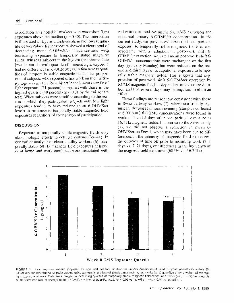
32 Burch kt al. -- ---
---ppp-pp
association was noted in workers wiih workplace light exposures above the median @ = 0.40). This interaction is illustrated in figure 1. Individuals in the lowest quar- tile of workplace light exposure showed a clear trend of decreasing mean 6-OHMSIcr concentrations with increasing exposure to temporally stable magnetic fields, whereas subjects in the highest (or inteimediate [results not shown]) quartile of ambient light exposure had no differences in 6-OHMSIcr excretion across quar- tiles of temporally stable magnetic fields. The propor- tion of subjects who reported office work on their activ- ity logs was greater for subjects in the lowest quartile of light exposure (7 1 percent) compared with those in the highest quartile (49 percent) @ < 0.01 by the chi-square test). When subjects were stratified according to the sea- son in which they participated, subjects with low light exposures tended to have reduced mean 6-OHMSIcr levels in response to temporally stable magnetic field exposures regardless of their season of participation.
DISCUSSION
Exposure to temporally stable magnetic fields may elicit biologic effects in cellular systems (39-41). In our earlier analysis of electric utility workers (6), tem- porally stable 60 Hz magnetic field exposures at home or at home and work combined were associated with
reductions in total overnight 6-OHMS excretion and nocturnal urinary 6-OHMSIcr concentration. In the current study, we provide evideme that occupational exposure to temporally stable magnetic fields is also associated with a reduction in post-work shift 6- OHMSIcr excretion. Adjusted mean post-work shift 6- OHMSIcr concentrations were unchanged on the first day (typically Monday) but were reduced on the sec- ond and third days of occupational exposure to tempo- rally stable magnetic fields. This suggests that sup- pression of post-work shift 6-OHMSIcr excretion by RCMS magnetic fields is dependent on exposure dura- tion and that several days may be required to elicit an effect.
These findings are reasonably consistent with those in Swiss railway workers (7), where statistically sig- nificant decreases in mean evening (samples collected at 6:00 p.m.) 6-OHMS concentrations were found in workers 1 and 5 days after occupational exposure to 16.7 Hz magnetic fields. In contrast to the Swiss study (7), we did not observe a reduction in mean 6- OHMSIcr on Day 1, which may have been due to dif- ferences in the intensity of magnetic field exposures, the duration of time off prior to resuming work (2-3 days vs. 7-21 days), or differences in the frequency of the- magnetic field exposures (60 Hz vs. 16.7 Hz).
1 2 3
W o r k R C M S E x p o s u r e Qaartile
FIGURE 1 . Least-sq~;ares m e m s (adjusted ior age and season] o i daytime urinary creatinine-adjusted 6 - h y d r o ; I a t i sulfate (6- OHMSIcr) concentrations for male electric utiiity workers in the lowest (black bars) and highest (white bars) quartiles of t i rne-~eightea average light exposure a: wnrk Dats are arranged by increasing quartile o f temporally stable magnetic fieid exposure at woric (i.e., 1 = highest quartile of standardized rate of change metric (RCMS), 4 = lowest quartile, etc.). "p < 0.05 vs. quartile 1; **p < 0.01 vs. quartile 1
Am J Epiden?io! Val. 1 50. No. 1 , 1 999
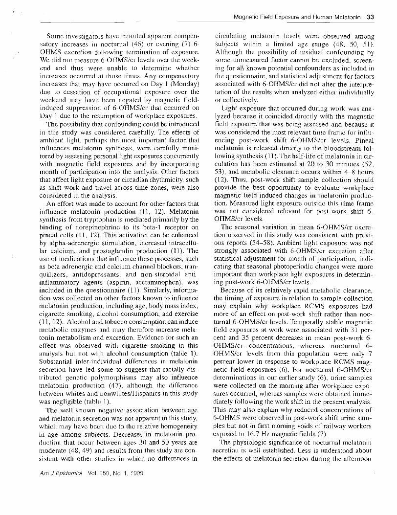
Magnetic Field Exposure and Human fvlelatonin 33
Some in\-estigacors have reported apparent compen- satory increases in ~locturnal (46) or evening (7) 6- OHMS excretion following termination of exposure. We did not measure 6-OHMSIcr levels over the week- znd and thus u:ere unable to determine whether increases occull-ed at those times. Any compensatory increases that may have occurred on Day 1 (Monday) due to cessation of occupational exposure over the weekend may have been negated by magnetic field- induced suppression of 6-OHMSIcr that occurred on Day I due to the resumption of workplace exposures.
The possibility that confounding could be introduced in this study was considered carefully. The effects of ambient light, perhaps the most important factor that influences melatonin synthesis, were carefully moni- tored by assessing persona1 light exposures collcurrently with magnetic field exposures and by incorporating month of participation into the analysis. Other factors that affect light exposure or circadian rhythmicity, such as shift work and travel across time zones, were also considered in the analysis.
An effort was made to account for other factors that influence melatonin production (1 1, 12). Melatonin synthesis from tryptophan is mediated primarily by the binding of norepinephrine to its beta-1 receptor on pineal cells (1 1, 12). This activation can be enhanced by alpha-adrenergic stimulation, increased intracellu- lar calcium, and prostaglandin production (11). The use of medications that influence these processes, such as beta adrenergic and calcium channel blockers, tran- quilizers, antidepressants, and non-steroidal anti- inflammatory agents (aspirin, acetaminophen), was included in the questionnaire (1 1). Similarly, informa- tion was collected on other factors known to influence melatonin production, including age, body mass index, cigarette smolung, alcohol consumption, and exercise (1 1, 12). Alcohol and tobacco consumption can induce metabolic enzymes and may therefore increase mela- tonin metabolism and excretion. Evidence for such an effect was observed with cigarette smoking in this analysis but not with alcohol consumption (table 1). Substantial inter-individual differences in melatonin secretion have led some to suggest that racially dis- tributed genetic polymol-phisms may also influence melatonin production (47), although the difference between whites and nonwhites/Hispanics in this study was negligible (table 1).
The well-known negative association between age and rnelatonin secretion was not apparent in this study, which may have been due to the relative homogeneity in age among subjects. Decreases in melatonin pro- ductioil that occur between ages 30 and 50 years are moderate (48, 49) and results from this study are con- sistent with other studies in which no differences in
circulating Inelatonin levels were observed among subjects within a limited age range (48, 50. 51). Although the possibility of residual confounding by some ullnleasured factor cannot be excluded, screen- ing for a11 known potential confounders as included in the questionnaire, and statistical adjustment for factors associated with 6-OHMSIcr did not alter the interpre- tation of the results when analyzed either individually or collectively.
Light exposure that occurred during work was ana- lyzed because it coincided directly with the magnetic field exposure that was being assessed and because it was considered the most relevant time frame for influ- encing post-work shift 6-OHMSIcr levels. Pineal melatonill is released directly to the bloodstream fol- lowing synthesis (1 I). The half-life of melatonin in cir- culation has been estimated at 20 to 30 minutes (52, 53), and metabolic clearance occurs within 4-8 hours (12). Thus, post-work shift sample collection should provide the best opportunity to evaluate workplace magnetic field induced changes in melatonin produc- tion. Measured light exposure outside this time frame was not considered relevant for post-work shift 6- OHMSIcr levels.
The seasonal variation in mean 6-OHMSIcr excre- tion observed in this study was consistent with previ- ous reports (54-58). Ambient light exposure was not strongly associated with 6-OHMSIcr excretion after statistical adjustment for month of participation, indi- cating that seasonal photoperiodic changes were more important than workplace light exposures in determin- ing post-work 6-OHMSIcr levels.
Because of its relatively rapid metabolic clearance, the timing of exposure in relation to sample collection may explain why workplace RCMS exposures had more of an effect on post-work shift rather than noc- turnal 6-OHMSIcr levels. Temporally stable magnetic field exposures at work were associated with 31 per- cent and 35 percent decreases in mean post-work 6- OHMSIcr concentrations, whereas nocturnal 6- OHMSIcr levels from this population were only 7 percent lower in response to workplace RCMS mag- netic field exposures (6). For nocturnal 6-OHMSIcr detenninations in our earlier study (6). urine samples were collected on the morning after workplace expo- sures occurred, whereas samples were obtained imme- diately following the work shift in the present analysis. This may also explain why reduced concentrations of 6-OHMS were observed in post-work shift urine sam- ples but not in first morning voids of railway workers exposed to 16.7 Hz magnetic fields (7).
The physiologic significance of noctilmal melatonill secretion is well established. Less is understood about the effects of melatonin secretion during the afternoon
Am J Epiderniol Val. 150, No. 1 , 1999
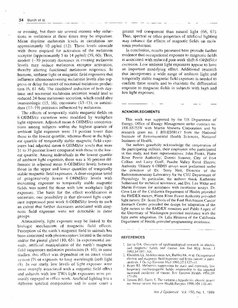
34 Burch et a1
or evening, but there are several reasons why reduc- ,
tions in melatonin at these times may be important. Mean daytime melatonin levels in circulation are approxiinately 10 pglml (12). These levels coincide with those required for activation of the melatonin receptor (approximately 5 to 14 pglml) (59, 60). Thus, modest (-30 percent) decreases in evening melatonin levels may reduce melatonin receptor activation, thereby altering functional melatonin responses. In humans, ambient light or magnetic field exposures that influence afternoon/evening melatonin levels also sup- press or delay the onset of nocturnal melatonin produc- tion (6, 61-64). The combined reduction of both day- time and nocturnal melatonin secretion would lead to reduced 24-hour melatonin secretion, which could alter immunologic (1 5, 16), oncostatic (13-1 5 ) , or antioxi- dant (17-19) processes influenced by melatonin.
The effects of temporally stable magnetic fields on 6-OHMSIcr excretion were modified by workplace light exposure. Adjusted mean 6-OHMSIcr concentra- tions among subjects within the highest quartile of ambient light exposure were 14 percent lower than those in the lowest quartile, whereas those in the high- est quartile of temporally stable magnetic field expo- sures had adjusted mean 6-OHMSlcr levels that were 3 1 to 35 percent lower compared with those in the low- est quartile. Among individuals in the lowest quartile of ambient light exposure, there was a 36 percent dif- ference in adjusted mean 6-OHMS/cr levels between those in the upper and lower quartiles of temporally stable magnetic field exposures. A dose-response trend of progressively lower 6-OHMSIcr levels with increasing exposure to temporally stable magnetic fields was noted for those with low workplace light exposure. The basis for the effect modification is uncertain; one possibility is that elevated light expo- sure suppressed post-work 6-OHMSIcr levels to such an extent that further decreases associated with mag- netic field exposure were not detectable in those groups.
Alternatively, light exposure xilay be linked to the biologic mechanism of magnetic field effects. Perception of the earth's magnetic tield in animals has bee11 associated with photoreceptors located in the retina ;uldIor the pineal gland (10, 65). In experimental ani- mals, artificial manipulation of the earth's magnetic field suppresses melatonin prod~lction (8-1 0); in some .jtuili~s, [his effect was dependerit on an intact visual system (9) or exposure to long wavelength jrecl) light (8). In our stutly. low levels of light exposure were milst strongly associated with a rnagnetic fislcl effect and subjects with low TWA light exposul.es were pri- :rial-ily engaged in office work. Artificial iighting has a dit'erent spectral composition and in some exes a
greater red component than natural light (66, 67). Thus, spectral or other properties of artificial lighting may enhance the effects of magnetic fields on rnela- tonin production.
In conclusion, results presented here provide further evidence that occupational exposure to magnetic fields is associated with reduced post-work shift 6-OHMSIcr excretion. Low ambient light exposures appear to have an important modifying effect. Additional research that incorporates a wide range of ambient light and temporally stable magnetic field exposure is needed to confiim these results and to elucidate the differential response to magnetic fields in subjects with high and low light exposure.
ACKNOWLEDGMENTS
This work was supported by the US Department of Energy: Office of Energy Management under contract no. 19X-SS755V with Martin Marietta Corporation and by research grant no. 1 ROlES08117 from the National Institute of Environmental Health Sciences, National Institutes of Health.
The authors gratefully acknowledge the cooperation of the participating utilities, their e~nployees who participated in this study, and their representatives: John Fooks, Platte River Power Authority; Dennis Sumner, City of Fort Collins; and Larry Graff. Poudre Valley Rural Electric Authority. Urinary 6-OHMS assays were performed under the direction of Dr. Terry Nett, Director of the Radioimmunoassay Laboratory for the CSU Department of Physiology. In particular, the authors thank Katherine Sutherland for technical assistance, and DIS. Lze Wilke and Martin Fzttman for assistance with creatinine assays. Dr. Gerri Lee of the California Department of Health provided the EMDEX meters; Platte River Power Authol-ity provided light meters; Dr. Scott Davis of the Fred Hutchinson Cancer Research Center provided the design for adaptation of the light meters to the EMDEX monitors and Pablo I-opez of the University of Washington provided assistance with the light meter adaptation. Dr. Lilia Hristova of the California Department of Health provided programming assistance.
REFERENCES
1. S : IV~~L DA. O v e r ~ . ~ e w of epidemiological reseal.ch on electic rtnd magnetic fieids and cancer. Am fnd Hyg .i\ssoc J 1993;54: I 97-2111.
7. Kheitt'e:~ LI; ,4h!Jeln?onem X.4. Buftler PA? et al. 0ccupationi:i elea::ric and rn~gneric tizld exposure and br~lin cancer: a mr,ta- analysis. J O z a p Envil-on Med 199537: 1327-1 1 .
7 Reiter RJ. Me!zion~n suppression by static and extremsiy io;x. - .
frequency e1ectrom;igiietic fields: relationship to the reported increased incidzncs of cancer. Rev Environ Health 1994;10: 17 1-86.
4. Sievens RG. Davis S. Ths melntonin hypothesis: eiectric pswer ancl brzast cancer. Environ Health Pel.sp-cr 1996;104: l jjA(j
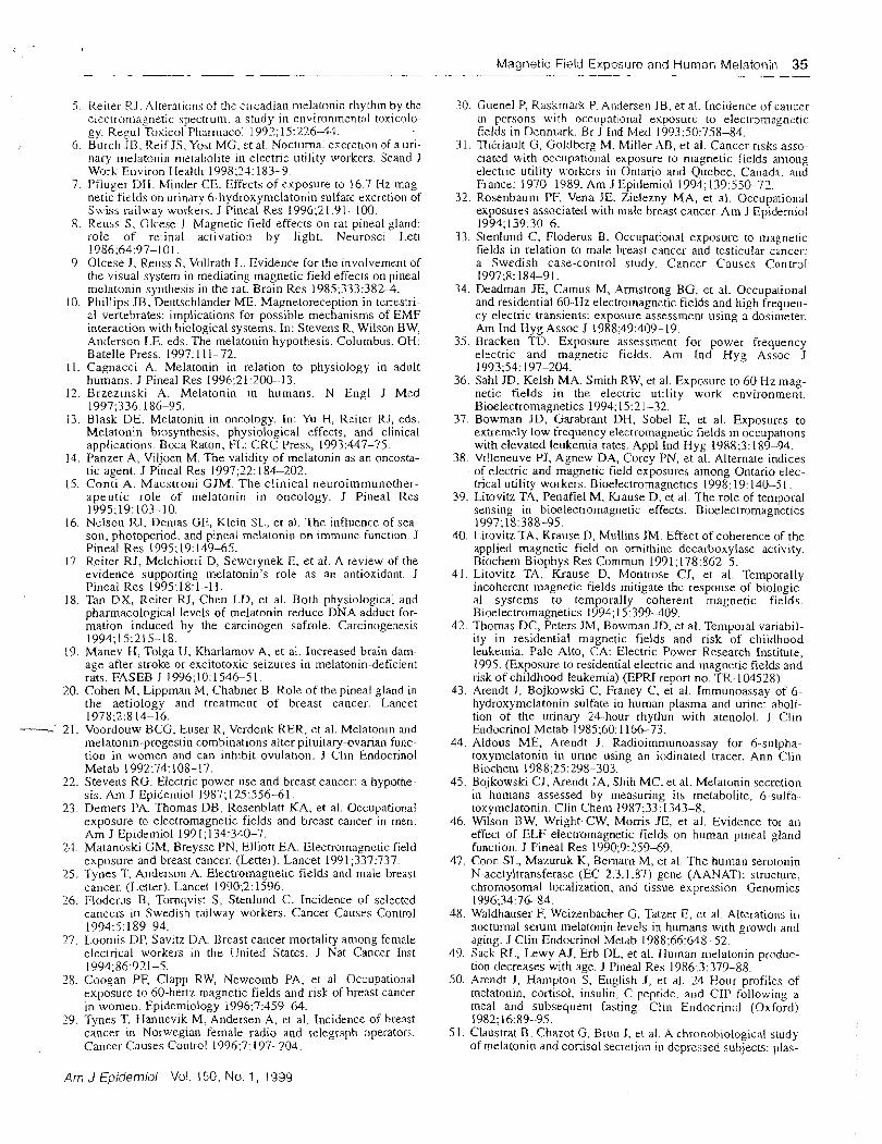
Magnetic Field Exposure and Human Ivlelatonin 35
5. Reiter R J . Alter-ations of the cir-cadian melatonic I-hythm by the elescroniagnetic spectrum: a study in environmental toxicolo- gy. Rep1 Toxic01 Pharr~~acol 1992:15:27643.
6 . Burch JB. Reif JS. Yost MG, et al. Nocturnal escretlon of a uri- nary ntelatonin metabolite in electric utility workers. Sca~td J Work Environ Health 1998124: 183-9.
7. Pflugel- DH. Miltder CE. Effects of exposure to 16 7 Hz mag- netic fields on urinary 6-liydroxymelatonin sulfate excreiion of Swiss railway wo~kers. J Pineal Res 1996;21:91-LOO.
8. Reuss S. Olcese J . Magnetic field effects on rat pineal gland: role of retinal activation by light. Neurosci Lett 1986;64:97-101.
9. Olcese J. Reuss S. Vollrath I.. Evidence for the involvement of the visual system in mediating magnetic field effects on pineal rnclatonin synthesis in the rat. Brain Res 1985;333:3824.
10. Phillips JB, Deutschlander ME. Magnetoreception in tcrrestri- al vertebrates: implications for possible mechanisms of EMF interaction with biological systems. In: Stevens R, Wilson BW, Anderson LE, eds. The melatonin hypothesis. Columbus, OH: Batelle Press, 1997: 11 1-72.
11. Cagnacci A. Melatonin in relation to physiology in adult humans. J Pineal Res 1996;21:20&13.
12. Brzezinski A. Melatonin in humans. N Engl J Med 1997:336: 186-95.
13. ~ l a s k DE. Melatonin in oncology. In: Yu H, Reiter RJ, eds. Melatonin biosynthesis, physiological effects, and clinical applications Boca Raton, FL: CRC Press, 1993:447-75.
14. Panzer A, Viljoen M. The validity of melatonin as an oncosta- tic agent. J Pineal Res 1997;22: 184-202.
15. Conti A. Maestroni GJM. The clinical neuroimmunother- aoeutic role of melatonin in oncolorv. J Pineal Res
-A
1$95;19:103-10. 16. Nelson RJ. Derrlas GE. Klein SL. et al. ' f i e influence of sea-
son, photoperiod. and pineal melatonin on immune. function. J Pineal Res 1995; 19: 149-65.
!7 Reiter RJ, Melchiorri D, Sewerynek E, et al. A review of the evidence supporting melatonin's role as an antioxidant. J Pineal Res 1995;lX:l-11.
18. Tan DX, Reitcr RJ, Chen LD, et al. Both physiological and pharmacological levels of melatonin reduce DNA adduct for- mation induced by the carcinogen safrole. Carcinogenesis 1994;15:215-18.
19. Manev H, Tolga U, Kharlamov A, et al. Increased brain dam- age after stroke or excitotoxic seizures in melatonin-deficient rats. FASEB J 1996; 10: 1 5 4 6 5 I .
20. Cohen M, Lippman M. Chabner B. Role of the pineal gland in the aetiology and treatment of breast cancer. Lancet 1978;2:8 14-16.
-- 21. Voordouw BCG, Euser R, Verdonk RER, et al. Melatonin and melatonin-progestin combinations alter pituitary-ovarian func- tion in women and can inhibit ovulation. J Clin Endocrinol Metab 1992;74: 108-17.
22. Stevens RG. Electric power use and breast cancer: a hypothe- sis. Am J Epidemiol 1987; 125:556-61.
23. Demers PA, Thomas DB. Rosenblatt KA, et al. Occupational exposure to electromagnetic fields and breast cancer in men. Am J Epidemiol 199 1 ; 134:340-7.
23. Matanoski GM, Breysse PN; Elliott EA. Electromagnctic field exposure and breast cancer. (Letter). Lancet 1991;337:737.
25. Tynes T, Anderson A. Electromagnetic fields and male breast cancer. (Letter). Lancet 1990;2: 1596.
26. Floderus B. Tornqvist S, Stenlund C. Incidence of selected cancers irt Swedish railway workers. Cancer Causes Control 1994:5: 189-94.
27. Looniis DP, Savitz DA. Breast cancer mortality among female electrical workers in the United States. J Nat Cancer Inst 1994;86:921-5.
28. Coogan PF, Clapp RW? Newcomb PA, et al. Occupational exposure to 60-hertz n~agnetic fields and risk of breast cancer in women. Epidemiology 1996:7:459-64.
29. Tynes T. Hannevik M; Andersen A, et al. Incidence of hreast cancer In Norwegian female radio and telegraph operators. Cancer. Causes Control 1996;7: 197-204.
30. Cuenel P, Raskrnark P. A~itlersen JB. et al. Incidence of cancer in persons with occupational exposure to electrolnagnetic fields in Denmark. Br J Ind Med 1993:50:758-84.
31. Tt~triault G: Goldberg M. Miller- XB, et al. Cancer- risks asso- ciated with occupational exposure to magnetic fields among electl-ic utility workers in Ontario and Quebec, Canada, and France: 197& 1589. Am J Epidemiol 1994; 139550-72.
32. Rosenbaum PF. Vena JE. Zielezny MA, et al. Occupational exposures associated with male breast cancer. Am J Epidemiol 1994;139:304.
33. Stenlund C , Floderus B. Occupational exposure to rnaenetic fields in relation to male breast cancer and testicular cancer: a Swedish case-control study. Cancer Causes Control 1997;8: 184-91.
34. Deadman JE, Camus M, Armstrong BG, et al. Occupational and residential 60-Hz electromagnetic fields and high frequen- cy electric transients: exposure assessment using a dosimeter. Am Ind Hyg Assoc J 1988;49:409-19.
35. Bracken TL). Exposure assessment for power frequency electric and magnetic fields. Am Ind Hyg Assoc J 1993;54: 197-204.
36. Sahl JD, Kelsh MA, Smith RW, et al. Exposure to 60 Hz mag- netic fields in the electric utility work environment. Bioelectromagnetics 1994; 15:2 1-32.
37. Bowman JD, Garabrant DH, Sobel E, et al. Exposures to extremely low frequency electromagnetic fields in occupations with elevated leukemia rates. Appl Ind Hyg 1988;3: 189-94.
38. Villeneuve PJ, Agnew DA, Corey PN, et al. Alternate indices of electric and magnetic field exposures among Ontario elec- trical utility workers. Bioelectromagnetics 1998: 19: 140-51.
39. Litovitz TA, Penafiel M, Krause D, et al. The role of temporal sensing in bioelectromagnetic effects. Bioelectromagnetics 1997;18:388-95.
40. LitovitzTA, Krause D, Mullins JM. Effect of coherence of the applied magnetic field on ornithine decarboxylase activity. Biochem Biophys Res Comrnun 1991; 178:862-5.
41. Litovitz TA, Krause D. Montrose CJ, et al. Temporally incoherent magnetic fields mitigate thc response of biologic- al systems to temporally coherent magnetic fields. Bioelectromagnetics 1994; 15:399-409.
42. Thomas DC, Peters JM, Bowman JD, et at. Temporal variabil- ity in residential magnetic fields and risk of childhood leukemia. Palo Alto, CA: Electric Power Research Institute, 1995. (Exposure to residential electric and magnetic fields and risk of childhood leukemia) (EPRI report no. TR-104528).
43. Arendt J, Bojkowski C. Franey C, et al. Immunoassay of 6- hydroxymelatonin sulfate in human plasma and urine: aboli- tion of the urinary 24-hour rhythm with atenolol. J Clin Endocrinol Metab 1985;60: 1 166-73.
44. Aldous ME, Arendt J. Radioimrnunoassay for 6-sulpha- toxymelatonin in urine using an iodinated tracer. Ann Clin Biochem 1988;25:298-303.
45. Bojkowski CJ, Arendt JA, Shih MC, et al. Melatonin secretion in humans assessed by measuring its metabolite. 6-sulfa- toxymelatonin. Clin Chem 1987r33: 1343-8.
46. Wilson RW, Wright CW, Morris JE, et al. Evidence for an effect of ELF electromagnetic fields on human pineal gland function. J Pineal Res 1990;9:259-69.
47. Coon SL, Mazuruk K, Bemarn M , et al. The human serotonin N-acetyltransferase (EC 2.3.1.87) Eerie (AANAT): structur-e, chromosomal localization, and tissue expression Genomics 1996;34:76-84.
48. Waldhauser F, Weizenbacher G. Tatzer E, et a!. Alterations in nocturnal serum melatonin levels in humans with growth and aging. J Clin Endocrinol Metab 1988;66:648-52.
49. Sack RL, Lewy AJ. Erb DL. et al. Human melatonin produc- tion decreases with age. J Pineal Res 1986;3:379-88.
50. Arendt J, Hanipton S, English J, et al. 24-Hour profiles of melatonin, cortisol, insulin, C-peptide. and GIP follouino a meal and subsequent fasting: elin Endocrinol (0xf;d) 1982: 16.89-95.
5 1 . ~laustrat B. Chazot G, Rrun J, et al. A chronobiological study of melatonin and cortisol secretion in depressed subjects: plas-
Am J Epidemiol Vol. 150, No. 1 , 1939
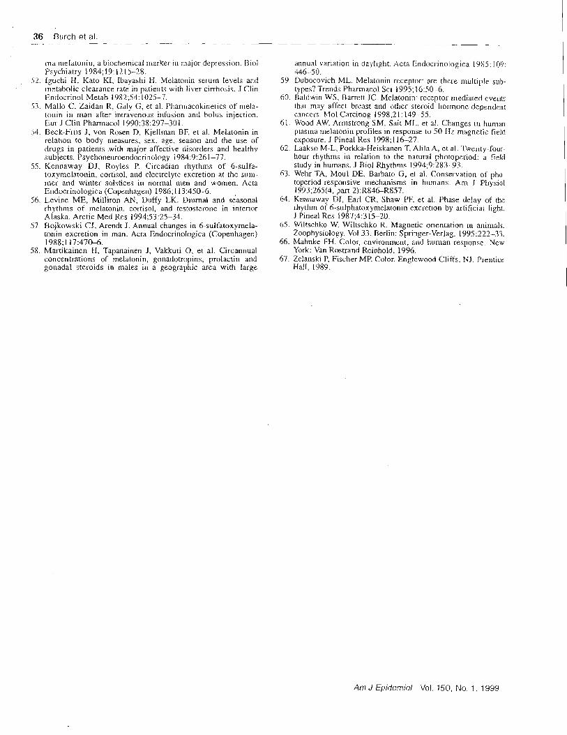
36 Burch et al. ~ -~
ma melatorlir~, a biochemical marker in Inajor depression. Biol Psychiatry 1984;19: 1215-28.
52. lguchi H, Kato KT, lbayastli H. Me1ato1:in serum levels and metabolic clearance rate in patients with liver cirrhosis. J Clin Endocrinol Metab 1982;54: 1025-7.
3 . Mallo C, Zaidan R, Galy G, et al. Phannacoiclnetics of niela- tonin in man after intravenous infusion and bolus injection. Eur J Clin Pharniacol 1990:38:297-30 I .
54. Beck-Friis J, von Rosen D, Kjellrnan BF: et al. Melatonin in relation to body measures. sex. age, season and the use of drugs in patients with major affective disorders and healthy subjects. Psychoneuroendocrinology 1984:9:7-61-77.
55. Kennaway DJ, Royles P. Circadian rhythms of 6-sulfa- toxyrnelatonin, cortisol, and electrolyte excretion at the sun- mer and winter solstices in nornlal men and women. Acta Endocrinologica (Coper~tiagen) 1986; 113:4506.
56. Levine ME, Milliron AN, Duffy LK. Diurnal and se'asonal rhythms of melatonin, cortisol, and testosterorre in interior Alaska. Arctic Med Res 1994;53:25-34.
57. Bojkowski CJ, Arendt J. Annual changes in 6-sulfatoxymela- tonin excretion in man. Acta Endocrinologica (Copenhagen) 1988;117:47&6.
58. Martikainen H, Tapanainen J, Vakkuri 0, et al. Circannual concentrations of melatonin, gonadotropins, prolactin and gonadal steroids in males in a geographic area with large
annual variation in daylight. Acta Endocrinologica L965.109: 446-5 0.
59. Dubocovich ML. Melatonin receptor: are thzl-e milltiple sub- types? Trends Pharmacol Sci 1995; 16:50-6.
60. Baldwin WS, Barrett JC. Melatonin: receptor-mzdiated evelltc thac may affect breast and other steroid liormone-dependell: cancers. Mol Carcinog 1998:2 1: 149-55.
61. Wood A\+'. Armstrong SM. Sait ML. et al. Changes in human plasma melatonin profiles in response to 5 0 Hz magnetic field exposure. J Pineal Res 1998; 116-27.
62. Laakso M-L, Porkka-Heiskanen T, Aliln A. et al. Twenty-four- hour rhythms in relation to the natural photopzriod: a field study in humans. J Biol Rhythms 1994;9:283-93.
63. Wehr TA, ~Moul DE, Barbato G, et al. Conservation of pho- toperiod-responsive mechanisms in humans. Am J Physiol 1993;265(4, pan 2):R84&R857.
64. Kennawav DJ, Earl CR, Shaw PF, et al. Phase delay of the rhythm of 6-sulphatoxymelatonin excretion by artifichl light. J Pineal Res 1987;4:3 15-20.
65. Wiltschko W Wiltschko R. Magnetic orientation in animals. Zoophysiology. Vol 33. Berlin: Springer-Verlag, 1995222-33.
66. Mahnke FH. Color, cnvironment, and human response. New York: Van Rostrand Reinhold. 1996.
67. Zelanski P, Fischer MP. Color. Englewood Cliffs, NJ. Prenticz Hall, 1989.
Am J Epidemiol Vol. 150, No. 1 , 1959
"-1 EXHIBIT * Original articles 4 2 8
Scand J Work Eqvrron t?ealt,h 1995,24(3). 183- 189 d
Nocturnal excretion of a urinary melatonin metabolite among electric utility workers by James B Burch. PhD. ' John S Reif, DVM,' Michael G Yost. PhD,? Thomas J Keefe, PhD,' Charles A Pitrat, MS1
Burch JB. itelf JS. Yost MG. Keefe TJ, Pitrat CA. Nocturnal axcretion of a ur~nary melatonin metabolite among ~ I ~ C T ~ I C util~ty ivorkers. Scand J Work Environ Health 1 !?98:24(3):183-189.
Objectives The effecn of 60-Hz m a ~ e t i c tieid and ambient light exposures on the pined normone meiatonin were smdied among eleciric ualiry wor~en. Methods Personal exposure was measured at 15-second intervals over 3 consecutive 71-hour periods. Exposure memcs based on magnetic field inrensicy. inrermitrence. or temporal stability were calculated for periods of work. home. and slzcp. A nre-of-change memc (RC.M) was used to e s h a t e imerminence. md the standardized RC.M (RCMS = RCbUsmdard deviarion! was used to evaluare temporal srabdiry. The effects of magpetic field exposure on toral overnight 6-hydroxymelaronin sulfate (6-OHMS) excretion and creaunine-adjusted nocturnal 6-OHMS (6- OHh.IS/cr, concenmtion were anaiyzed with ad:lusment for age. month. and light exposure. Results Magnetic field intensin.. intermirtenc-. or cumulative exposure had tide influence on nocrumal &OHMS excrenon. Residential RCMS magnetic field exposures were associated with lower nocrumd &OHMS/cr concenm- tions. In rndtivanate statistical analyses. the interaction ten for geomeuic mean and RC.MS mayetic field exposures at home was associared with lower nocturnal 6-OHMSIcr and overnight &OHMS levels. Modest reductions in the mean 6-OHMS levels occurred after RC.MS exposures during work. The greatest reducuons occurred when RCMS exposures both at work md ar home were combined: therefore the effecu of ternponlly suble magnetic fields may be inte--red over a large portton ofthe day. CancIusions Resulu from h s study provide evidence that temporally srable magnetic field exposures are associated with reduced nocrumal &OHMS excretion in humans.
Key terms electromagnetic fieids. human. 6-hydroxymeiatonin sulfate. 60 Hz. magneuc fields. pined.
The potential health eifecrs asociated with exposure to ! power frequency r 50160 Hz] mayetic fieids have received considerable attention in recent years. due in part to the pervasiveness o i such exposures in the home and work- place. The chxac:trizvion ofhuman biological respons- ,
es to magnetic 5e!d exposures is critical in determining I
whecher such exposures result in adverse health effecrs. i Some magnetic iisld sffects may be mediated through re- :
duced secretion of the hormone melatonin. iV1elatonin se- cretion follows a diurnal rin).rhm rnight high. day low) that is synchronized by ambient light exposure (1). Through this mechanism. meiatonin influences sleep and other i
physioiogical processes with circadian rh-mhms (7. 3). Mtlatonin is also 3ssocis~ed with suppressed tumor growth ( 3 - 4 . ennanced immunity (6-9). antioxidant : eliec? 110-13). and reduced secretion o i tumor-~romot- ing hormones 1!3. l l j . De:;t3sed melatonin production :
could ~ere r 'o re haye imponmt biologici consequences.
Althougi-I s reduction in melatonin synthesis follow- ing exposure to magnetic fields has been reported for a variety of experimental animal models i 151, only a few studies have attempted to determine whether such effects occur in humans. In a study of electric blanket users. Wil- son et a1 (. 16) found a reduction in nocturnal urinary con- centrations of the major melatonin metabolite 6-hy- droxymelatonin sulfate 16-OFEvlS or 6-sulfatoxymelaton- in) in some persons after 8 weeks of e!ecuic blanket use. Cessation of elect?c blanks: use was accompanied by an increase in 6-OH.kIS excretion 11 6i. -4 reduction in early evening. but not overnighr. 6-OffiVS e.~cretion was re- ported recently in a study of niiway workers with occu- pauonal exposure to 16.7 Hz magneuc fields (171.
Elevated magceric fieid txposures have becn report- ed for elecmc utility workers 11 8-2!1. Yumerous epide- miologic studies have identified rhis occupationd group as having an =le:ated risk for devzloping leukemia ( 2 2 )
1 D e p m e n r of Environmental Hedth. Colondo State University. Fon Collins. Colorado. United States. Depmment of Environrnencal Hedth, Universiy of Washngton. Seattie. Washington. United States.
Repint requesrs :a: Dr lames Burch. Dz?mment of Envuonmentai Hedrh. Colorado State Eniversiy. Fon Collins. CO 80513. United Scares.
Scand J Work 5wron Health ?!,098. 7 ~ c l 24. no 3 1 83
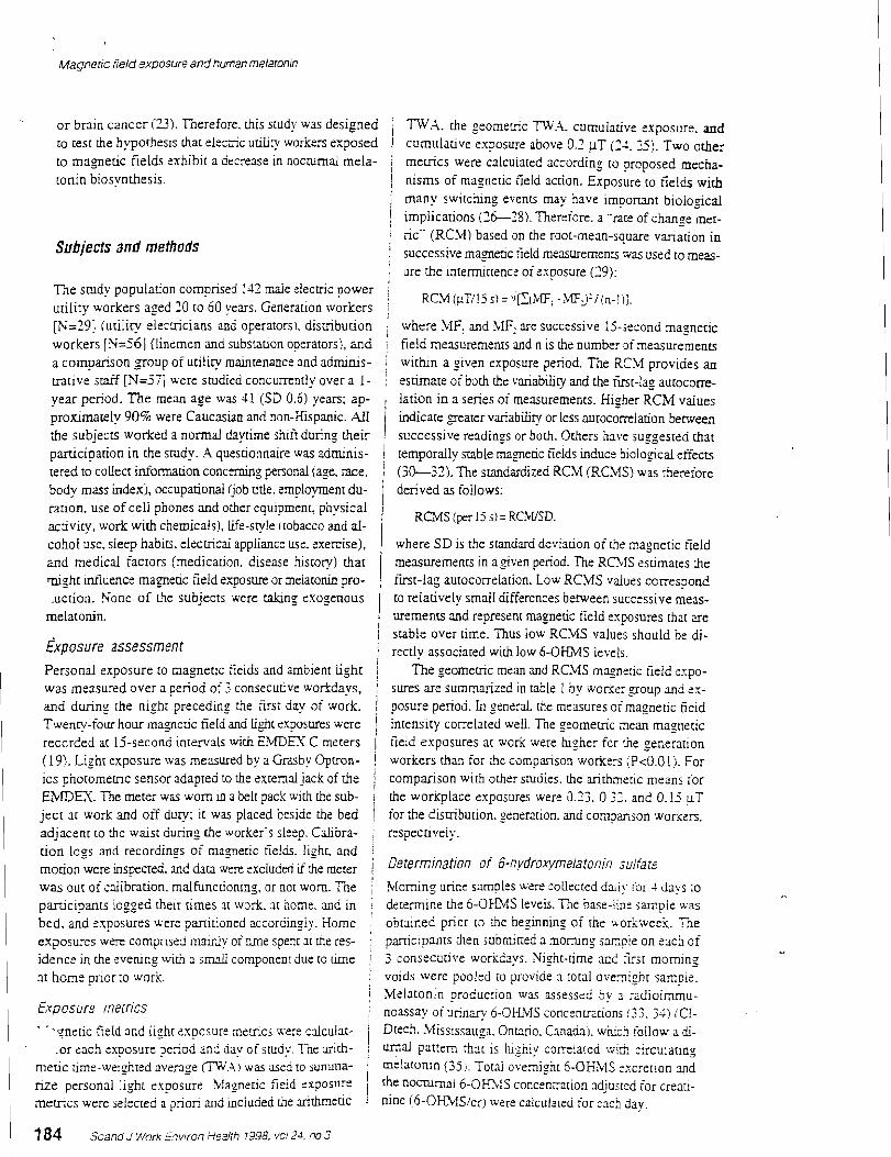
Magneric field exposure andhuman melaron~n
o r brain cancer (23). Therefore. this study was designed ! m.4. the geome~,c 77V.4. cumulative exposure. and I to test the hypothesis that elecsic utiliry wor~ers exposed ! cumulative exposure above 0.2 UT (11. 3 j . TWO other to magnetic fields -xhibir a decrease in nocturnal mela- i meciics were calculated according to proposed mecha- t o ~ n biosynt.hesis. / nisms of magnetic field action. Exposure to fields wi&
; many switching events may have imporrant biological ! implications ( 2 6 1 5 ) . Thereiore. a .-rate of change met- ! ric" (RCM) based on the root-mean-square vanation in
Subjects and methods I successive manetic tield mexuremencs was used to meas- ; ure the ~ntermittencz oizxposure (29):
The study population comprised 1-12 maie dectric power 8 RCM iuT/lS s) =\I[XiMF; -.MFlj-/(n-!ij.
utility workers aged 10 to 60 years. Generation woricers j [N=19! iutilicy e lec~rc ians and operators). distribution ; where MFI and 41F: are successive 15-jecond magnetic woriters [N=S6] (linemen and substation operators), and ; field measurements and n is the number ofinexurernenrs a comparison group of utility maintenance and adminis- j within a given exposure period. The RCM provides an u;lcive staff [N=57] were studied concurrently over a 1- ! estimate of both h e variability and che k t - l a g autocorn- Year period. The mean age was 41 (SD 0.6) years; ap- lation in a series of measurements. Hi= uher RCM values proximately 90% were Caucasian and non-Hispanic. All I indicate peater variability or less aurocorrelation berween the subiecrs worked a normal dayrime shift during their I successive readings or both. Others have suggested that i parciciparion in the study. .2 questionnaire was adminis- temporally m b l e magnetic fields induce biological effects tered to coUecr inionnation concerning personal (age. race. (30--32). The standardized RCM (RCMS) was herefore body mass index:^. occupational Gob tide. enployment du- derived as foliows: racion. use of cell phones and other equipment, physical I RCMS [per 15 s i = RCkVSD. activity, work with chemicals), life-style itobacco and al- cohol use. sleep habics. elecmcal appliancz use. exercise), where S D is the standard deviation of Be magnetic 5eld and medical factors (medication. disease history) that mesuremenrs in a$ven period. The R O l S esumates the I might mtluence magnetic field exposure or melatonin pro- first-lag autocorreiation. Low RCMS values ccrrespond uction. None of the subjects were *ng exogenous 1 to relatively small differences beween succtssive meas-
melatonin. urements and represent magnetic field exposures that are i i sriible over time. Thus low RCMS values should be di-
~ x ~ o s u r e assessment 1 rectly associated with low 6-OHMS levels. i
Personal exposure to magnetic fields and ambient light j The geomerric mean and RC.MS magns:ic field expo- was measured over a period of 3 consecuuve workdays, ! sures are summarized in cable 1 by worker group and zx-
i and during the night preceding the first day of work. i posure period. In general. the measures o i magnetic field Twenty-four hour magneuc fieid and light exposures were intensity correlated well. Tne seometric nean magnetic
1 recorded at 15-second intervals with E:MDEX C meters I field exposures at work were higher for h e generation (19). Lighr exposure was measured by a Grdby O p ~ o n - ! workers rhan for h e comparison workers (PcO.0 1 :J. For ics photomemc sensor adapted to h e zxternai jack of rhe / comparison with other studies. the arithme:ic means for EI\,lDEX. The mete: was worn in a hit pack with rhe sub- j the workpiace exposures were 0.23. 0.33. and 0.15 uT ject ar work and off duty; it was placed beside the bed for the dismbution. generation. and conpar:son workers, adjacent to the waist during the worker's sletp. Cdo ra - ; respectively. tion logs and recordings of magnetic fie!&. light. and ! morion weE inspecred. and were excluded *the j Determination of 3"-~~ydrox/melatonin suifate
was out of calibration. malfuncdoning, or not worn. Tne i Morning urine smoles were collected daiiy for 2 days to parricipants logged their times at work. 3t borne. aiid in i determine the 6-OI-blS le74e!s. T;le base-iix j a m ~ i e was bed. and exposures were pmitioned accordingiv. EIome . obtained prior to h e beginnins of the workweek. T3e exposucs were comprised rnainiy of dme sper.1 st h e res- participants h e n submined a momin: sarnpie on srich of idence in the wening wih 3 s m d cornconen: due to rime j 3 consecuuve workdays. Night-rime and 5rj t morning at home prior to work i voids were pooled to provide 3 total ovemi:nc sanpie.
i Meiaconin roduc:ion was assessed by 3 ; a d i o i m u - Ex~osurz meirics .' noassay of urinary 6-OHMS concenmtions ;31. 3) ; Ci- . - .pnetic field and !izht cxposurr merncs u e s niculat- Dtech. .ississauga. Onmio. Canada;. which follow a d -
.or each exposur-, seriod and day of study. Tne arirh- I urnal Pattern that is hishly corre!ated wisi zircuianng medc time-weighted average (TW.4) was nsed 10 summa- I melatonin (35.). Totai overnight 6-OHMS -xc:erion a d rize personal light exposure. h fane t ic k i d exposure
I the n o a n a l 6-0mIS concenntion sdjusrsd for creari- m c m c i were seiecred a p i o n and inciuded Be arithmetic nine (6-OEhlSicr) were caiculaicd for :ac: day.
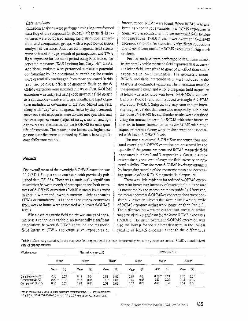
Table 1. Summary sratistlcsforthe magnetic field exposures of the male elec:ric arilir! *arkers i ~ y sxoosure perioa. (RCMS = standardized rate of change rnetricj
Data analyses I intermittence (RCM) were found. When RCMS was aria-
~tacisdcal analyses were performed using log-transformed / Iyzed as a continuous vkabie . l o w . ~ c h i s exposures at &[a (log of the reciprocal for RCMS j. blagnetic field ex- / home were associated with lower nocturnal ~ - o H M s / ~ ~ posures were compared among the dismbution, genera- j concenrradons (P<O.Olj and lower overnight 6 - O m ~ uon. and comparison groups with a repeated-measures ! excretion iPd.061. No s~tisrically ji-gifjcmt reducuons analysis of variancz. .Ualyses for magnetic field effects / ir, 6-OHMS were found for RCMS exposures during work were adjusted for age. month of parricipauon. and TWA or sleep. light exposure for rhe same period using Proc -Mixed for I Funher analyses were e r ia rmed to detenzine whe& repeated measures (SAS Insticute Inc. Gary, NC, USA). ' er t e m p o d y srable magnedc field exposures that occ-d .Addiuonal analyses were periomed io evaluate potential : at higher tieid jtrengms had more of an effect than suble confounding by rhz questionnaire variables; the results exposures at lowe: intensities. The ~ e o m e t r i c mean, were essentially unchanged from rhose presented in this 1 RCMS. and their interaction term were included in b e text. T i e potential effects of magnetic fields on the 6- j analysis as continuous variables. The interaction [ e m for OHMS excretion were modeled in 3 ways. First 6-OKblS ; the geomerric mean and RChlS magnetic field exposure excretion was analyzed using each magnetic field metric I at home was associated wich lower 6-OHMS/ci concen- as a conrinuous variable with age. month. and light expo- trations iP<O.Ol i and with reduced overnight 6 - O m s sure included as covariales in the R o c Mixed analysis. excretion (Pc0.01). Subjects with exposure to lugh-inten- along with .'day" and "magnetic fieldr by day". Second. / siry magnetic fields that were also temporally stable had m a ~ e d c field exposures were divided into quartiles. and ) the lowest 6-OHMS levels. Similar results were obtained the least-squares means (adjusted for age. month. and light 1 using the interacxion term for RCMS with other intensity
--
Worker group jeometnc mean (pT) 3C:ilS leer '5 s,
exposurei were estimated for the 6-OHMS for each quar- tile of exposure. The means in the lowest and highest ex-
:- SE '&an SE Mean Si Mean SE (M SE usan SE
metrics at home. Interaction terms for RChlS with ocher exposure metncs during work or sleep were not associa[-
0is;nbuuon !N=56) 0.10 0.02 0.1: 0.W 0.08 0.05 7 . 3 3 .y g.55- 3-23 0.50 3.M Genemion IN=29) 0.22- 1.07 0.:1 0.05 0.11 ' 0.07 0.39 0.C5 0.50 0.25 3-45' 0 . a Comoanson IN=5ii 0.10 0.03 0.09 0.04 0.06 0.05 2 . 0.03 0.68 g.04 8 0.31
posure quarriles were compared by Fisher's least signifi- ed wirh lower 6-OHIMS levels. cant difference method. I The mean nocturnal 6-OHMS/cr conc:nwtions and i
I total overnignt 6-OKVS excretion are presented by the I ! quanile of rhe geomeuic mean and RCMS magneric field I exposures in tables 3 and 3. respectively. Quamile 1 rep-
Results 1 resents rhe highest level of magnetic field intensiq or tern-
) p o d snbiliry. Thus the mean 6-OHMS levels are a m n g e d The overall mean of rhe overnight 6-OHMS excretion was j by increasing quarrile ofthe geomeaic mean and decreas- 23.7 (SD 1.3) pg, a value consistent with previously pub- 1 ing quanile of the RChlS magneric 5eld exposure. lished daca (35. 36). There was a statistically significant j There was little evidence for reduced 6-OHMS excre- associadon between monrh oiparticipation and both meas- 1 tion with increasins intensicy o i rnagnetic field exposure ures of 6-OHMS cxcrexion (PcO.01): mean levels were 1 as measured by the geomeric mean (table 1). However. higner in winter and lower in summer. Light exposures i the mean nocrurnal6-OH;LlS/cr concenrn~ons were con- (m,\ or cumulative lux) at home and during commutes i sistenrly lowest in subjects that were in the lowest quanile from work to home wer: associated with lower 6-OHMS i o i RCMS exposure durinz work. home. or sleep (:table 3). levels. / The difference between the highest and lowest quaniles
Wlen each magnetic fie!d meuic was analyzed sepa- / was sratisucdly signuicmt for h e home RCMS exposures rarely as a conmuous variabie. no srausdcally significant (P<0.01). The mean ovemizht 6-OHiilS excrerion was associations between 6-OH?VIS excretion and rnagnetic j also the lowest for the sub!ec:s that were in the lowest field intensiry (TWA and cumulative exposures) or ! q u m i i e o i RCMS exposure although the differences
a Mean and stanoar0 ermr si eacn excosure memi: :or days 1.2. m d 3 comb~ned. - t 5 0.05 versus c3moanson grouo. " ? 5 0.C: versus comparison group.
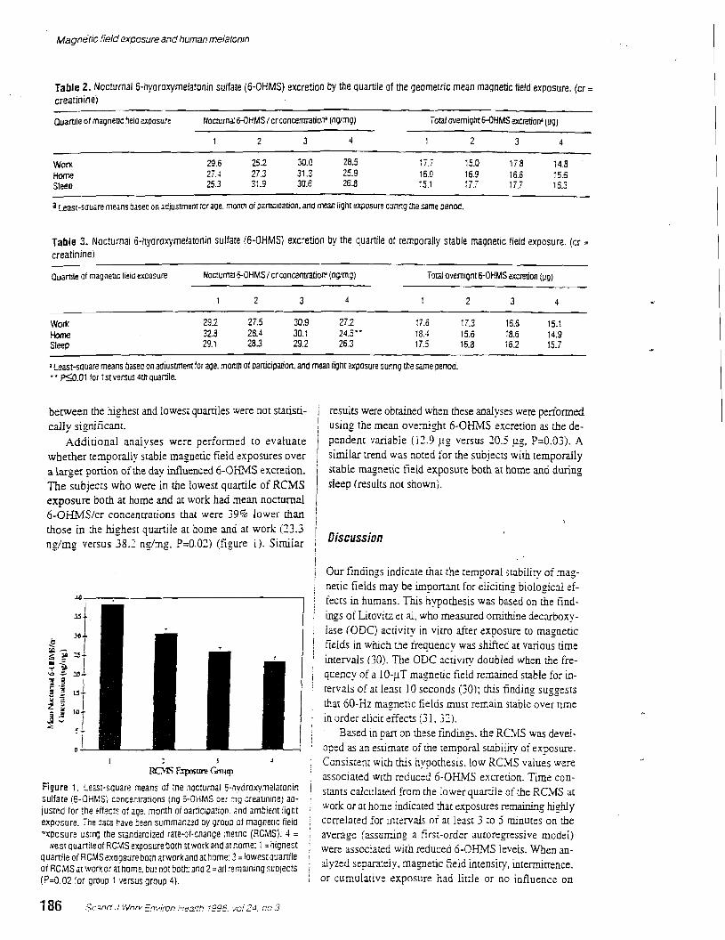
~agne't lc field ex,cosure and human melafonrn
Table 2. Nocturnal 5-hydroxymelatonin sulfate (6-OHMS) excretion by the quartile of the geometric mean magnetic field exposure. (cr = creatininej
Quartile of magnencfielo uposure Nocturnl WHMS /crconcemtioV (ngmg) Teal overnight &OHMS mre!jorr (pg)
1 2 3 4 1 2 3 4
won 29.6 252 30.0 28.5 17.; 15.0 17.8 14.8 Home 27.1 27.3 31.3 25.9 16.0 16.9 16.6 15.6 Sleep 25.3 31.a 30.6 26.8 15.1 17.; 17.; 16.3
a east-souare means aaseo on adjustmern braue. nonm oi parnc~oarion. and mean light exposure aunng the g m e onnod.
Table 3. Noclurnai 6-hydroxymelaronin sulfate @-OHMS) excretion by the quartile O i temporally stable magnetic field exposure. (cr . creatininei
puartjle or magnenc field excosure Ncmma &OHMS I c:concernratiorr (ngmg) Toral ovemtgnt 6-OHMS acretion (pg)
~
Work 292 27.5 30.9 272 l i . 6 17.3 16.6 15.1 Home 328 28.4 30.1 24.5.. 18.4 15.6 18.6 14.9 Sleep 29.1 28.3 29.2 26.3 17.5 16.8 i 6 2 15.7
~east-s~uare means basea on adiustmerrtfor age, month of partlcipalon. and mean light exposure dunng the same penoa. " a . 0 1 tor 1st versus Jth auartile.
between the highest and l owes~ quartiles were not stari.sti- j results were obtained when these analyses were penomed cally s i = ~ f i c m t . ) using the mean overnight 6-OHMS excretion as the de-
.4ddirional analyses were performed to evaluate / pendent variable (12.9 pg versus 20.5 ug. P=O.Oj). A whether temporally scable magnetic field exposures over similar trend was noted for the subjects with temporally a larger porcion of he day infiuenced 6-OHMS excretion. stable magnetic field exposure both ai home and during The subjects who were in the lowest quarrile of RCMS sleep (results not shown). exposure both at home and at work had mean nocturnal 6-OHMSIcr concentrations thar were 39% lower than i
I those in [he highest quartile at home and at work (13.3 ng/mg versus 38.1 ng/rng. E0 .02 ) (figure I ) . Similar iscussion
I i Our findings indicate t h a ~ the temporal stabiliv of mag- ! i netic fields may be important for eliciting biological ef- j fects in humans. This hypothesis was based on the find- : ings of Litovicz et al, who measured omithine decarboxy- 1 lase (ODC) activity in vitro after exposure to magnetic / fields in which che frequency was shifted at various time I I intervals (30). The ODC acrivicy doubled when the fre- 1 i quency of a 10-IT inaget ic fie!d remained stable for in- ! rervals of at least 10 seconds (30); dzls finding suggests
h a t 60-Hz magnetic fields musr remain srable over ume i in order elicit eifects (? I.;?). ! : Based in pan on rhese findings. the RC4lS was devel- ' aped as an esrirnare of rhe temporal subiiicy of exposurt.
I 3 J 3 I Consistent wich dihis nyoochesis. low RCMS values were R c m ~ r r r e G m u p I associated wich reduc~d b-OHMS excreuon. Time con-
Figure 1. Leasi-square rrieans ~f ;he nodumal 6-nv~roxymelatonin ! stants from the lower q u d i e ofthe RC>fS at sulfate (5-OHMS) concen~ralions ing ;-OHMS oe: ;ng zreal~ntne) aa- ,
jusred for the effec!s of age. month g i oarticipatjon, 2nd ambienr light i Or indicated remaining hJgdy exposure. The dam have hezn summanzad oy groua of rnagnerlc iieia correlated for intervals of a[ least 3 10 mrnuces on the wosure uslng the szndardized rare-or-cnange aemc (RCMSI: 4 = average (assuming a f i r - o rde r autoregressive model)
xest quanileof RC:dS exposure 90th atwork and Qnome: 1 = hignest j were associated with reduced 6 - 0 ~ i v f ~ levels. w%en an- quanile oi RCMSexogsureboth atworkan0 at home: 3 = iowestquaniie of RCMS l:,ork or at home. hr nor bott;: aria 2 =all remalnlng suo!ecn j d ~ ~ " d s e ? m [ e i ~ * manefic field i n t e n s i ~ , intermiaence. ( P d . 0 2 for group 1 versus group 3). ; or cumulative exposure nad litrle or no influence on
6 - O W S excretion although rhe inrensities were relative- 1 others (27. 78) suggest rhar fumre srudies should more ly low. However. the inreraction beween residential mag- ; carefully characrenze exposure to high-frequency tran- neuc field intensiry and temporal subilicy was associated ; sients. with a reducrion in both 6-OH3lS \.-xihies and therefore ! One strength of our jmdy was h e ability ro measure suggesrs rhar the tfiecrs of ternporslly srable magnetic i lighr exposure and adjusr for irs effects on melatonin pro- fields are enhanced at hlgher fieid suengths. I duction. However. the light sexor response w a matched
Our results indicate tkat h e riming O F exposure [O [em- ro that of the human eye and was nor maximal ar A wave porally srabie fields may be imponant for suppressing ien-ghs ha t produce the e a r e s r me!aroG d o i t i o n (60). 6-OHMS excrenon. Light 2sposures rhar occur at rimes
'
Thus the effects of the measured iipht on exae- when peopie are sxpecred to be at home !ie. near dawn tion may have been somewhat atrenuated due to the mis- and dusk) inrlucnc-, n o c m d melatonin producuon (37- , classificsuon of exposures. 39). and the m a z e d c field suppression of meiatonin may ' The reductions in mean nocturnal 6-OHMS excreuon be mediated by rednal pnororec:prors (4-3)- If so. i associated with RCMS rnagneric fie!d exposures in h s mage t i c 5eid exposures may need LO coincide wirhspe- : study (approximarely 10--iO%) were consisrenr w i h cific periods of photosensirivirl, for melaronin suppres- , those reponed elsewhere (15-17). and [he resulfi were sion to occur. In controlled human experiments. magnet- j in general agreemenr whether n o c m a l 6 - O m S / c r con- ic field exposures thac occurred prior [o the onset of noc- / centration or overnight 6-OKMS cxcreuon was used = tumal melaronin production resulted in a delay in onset I the outcome variable. Residential. ratherrhan occupation- and a suppression in peak nocrurnal plasma melatonin : al. magneric field exposures were most strongly associat- concentrations (13). Nocturnal melatonin onset usually ed with a reduccion in nocrmal6-OH3IS excretion. which occurs be twe~n 1600 and 2000 hours (2) . which corre- : does nor suppon h e hypothesis that workplacz exposures spends ro the aime of day wnen mosr of the subjects were j reduce 6-OHMS levels. Howeve:. the mean workplace at home. Othe: invesrigarors have faled to elicit a repro- ; exposures were !ower than those reponed by orher; (18- ducible suppression of nocrurnal meiaconin production in j 1 I), and they were only marginally $:her than the mean humans using only ovemignr magnetic field exposures residenrial exposures. Thus it was nor possible to deter- b a r starred at 1300 hours. after the nocturnal melaronin ! mine the effects of higher workplace magnetic field ex- onset (-6). Similarly. we found no statistically sig- / posures on 6 - O M S excrerion in our population. The nificanc reductions in 6-OEDlS in associarion with expo- : finding that remoorally stable magnetic field exposures, s u e s that occurred oniy during deep. 1 as measured by RCMS. are associated with reduced 6-
Reductions in mean nocmrnal 6-OHMS levels were ; O f i i S excrerion is unique and requires confirmation. modest after RCMS manedc field exposures at work. The : Funhcr work is also needed to derermine whecher 6- greatest reductions in the mean 6-OHMS levels were ob- i OHMS excrerion is c'nronicllly suppressed in elecmc util-
#
served when RCMS exposures at work and ar home were ; iry workers and co determine whether rhe tffecrs are due combined (figure 1). Tnis finding suggcsrs thac the cffrcts i to a reduccion ir, the biosvnthesis of melatonin. a phase of temporally stable magnetic fields are inregrated over a shift in nocturnal mrlaronin producuon. or an increase in longer h e span than rhe ~pproximate 8-hour periods rhat : melaronin metabolism. .\.leiatonin suppression may serve were used in kus smdy and that exposures occumng dur- : as a vaiuable tool for understanding human biololcal re- ing the day iniluence meiatonin production ar night. .hi- sponses to magnecic fields. mal experiments indicate rhar seven1 weeks of exposure ;
to 50- ro 60-Hz electric or magneric fields over a large j ponion of the dav appear ro be [he mosr eifecrlve means ; of suopresslng melatorun 117-51) alrhouzh there are . - some inconsisrencies (5:-561. Shori-[em exposures have been ineffective (57. 5 5 i unless repeated daily for : The authors gratzklly ac.how1edge -;?e coope:~tion of several weeks t 50) . the parriciparin~ xiiiries. their :mpioyz:s who ?arckipat-
>Ie!aconir. suppression has beta -?onec for Expen- ed LI Lhis srudy and ~ e i r ;=~resencadves: Jo'nn Foob. Plar- mental znimzlj after zxposure :o rapidly jwirched mag- re k v c r Power .Ailrhoriry: Dennis Sumzr . C i y of F o n netic iieids i17. 25 i . Our resuirs do no[ <upport a role for Collins: and Lz? Grsf . Pgudrc Valiey RurS Eiccmc intexniaent txposures k zuppresjing 6-OI%lS excretion. .Authonry. The usnary 6-OKMS m a y s wer- ?e$ormed However. mremnent ckxges in nagnenc fieids ar inre:- ! under the direcrion of Dr T z q Yet. Duecror of the Radi- vals of less : h a ! 5 sec2nos couid not he t~a lua tea . T l e . oimmunoassay Laboratory for [he CSL Deparrmenc of rare-of-change memc was designed ro 2apturz switching Physiolo~li. T'ne aurnors thank Ms R2therine Surfieriand evenu but. liik h e ESDEI: mere:. ir does ;lor specified- for her rechnicai xsisrance. md Drs L=e Wiikz and Mar- ly q~an t i fy rr~nsienr exFosures. Thus h e nesative find- : tin F-rtrnan for hen assismcz wich the creacinine assays. ings for RCXI in chis srudy and [he o s ~ r i v e rindings of Dr Gem Lze o i rhc California Depmmenr of Health
Magnefic field exposure anb human melaronrn
provided the EMDEX meters: the Plane kve r Power Au- 15. Re~ter IU. Meiatonin suppression by snuc and exmmcly
thtyrity provided rhe light meten: Dr Scon Davis of the 1 low frequency tiec.~.omagnetic fields: relationship ro the
Fred Hutchinson C a n c ~ r Research Center provided the reponed ~ncreasea incidence of cmcer. Rev Environ Health 1994:10:171-86.
design for adapting the light meters to the E!EY mon- 16- Wilson BW. Wnznt CW, Morns E. Buscbbom a Brown itors, and Pablo Lopez ofthe Universiy of Washing- DP. Miller DL. er a!. Evidence for m effect of ELF
- --
ton provided assistance with the lieht rnerer adapcacion. ( eIecuomagnetic fieids on h u m pined gland iunction. J
Dr Lilia Hnstova of the Caliiornia De~annent of Health 1 Pined Res 1990:?:259--69.
provided p rogmming assistance. ( 17. Ptluge: DH. blinder CE. Effects oi exposure to 16.7 Hz
n i s war rupoorred by the US D~~~~~~ of i mayeoc fieids XI u l n q 6-hydmxymelatonin sulfate ex- credon of SWISS ;yiway workers. J Pined Res 1996;21:9 1-
Ene rg . Office of Energy Mana~ernenr under contract I , ,, 1 uu.
1%-SS755V with Lhe hkmin Mariem Corporauon and / 18. Deaa rm E. C ~ U S M. . w m n g BG. Hemu P. C p D.
by research -pint ! ROlES08117 from the Nacional Insti- I Piante kt. er d. Occupational and residential 60-HZ eiecuo-
1 88 Scand J work E,-t,;~fi Heaith l9%, ~0124, no 3
cute of Environmental Health Sciences. National Institures of Health in h e United States.
References
1. Reiter RI. Atentions of the c d a n melatonin rhythm by the elecrromagnetic specuum: a study in environmental toxicology. R e d Toxic01 Pharmacol 1997-:15:11,6-44.
2. Cayacci A. ,Melatonin in reladon to physiology in adult humans. J Pined Res 1996:31:2&13.
3. Brezinski A. mel la ton in in humans. New Engl J Med 1997536: 1 8 6 9 5 .
4. Blask DE. Melatonin in oncology. In: Yu A. Reirer RJ, editors. .Melatonin biosynthesis. physiological effects, and clinicd applications. Bocs Raton (n): CRC Press. 1993.
5. Pmzer A. Viljoen M. The validity of melatonin as an oncostatic agent. J Pineal Res 1997:X: IS-202.
6. Conti .4. Maesuoni G M . The clinical aemoimmunothera- peutic role o i melatonin in oncoloey. J Pineal Rcs 1995;19: 103-10.
7. Maestroni G M . The immunoendocrine roie of melatonin. J Pineal Res 1993:lJ: 1-10.
8. Guenero M. Reirer RJ. A brief survey of pined gland- immune system interrelationships. Endoc Res 1992:18:91- 113.
9. Nelson RJ. Demas GE. Klein SL, Kriesfeld U. The i d u - ence of seson. pnotoperiod and pined melatonin on im- mune function. J Pined Res 1995:19:l49-65.
10. Reirer RJ. Melcluom D. Sewepek E. Poeggeler B. Bar- low-Walden L. Chuang J. er d. .4 review of the evidence supponing meiaromn's role as m and of id an^ J Pined Res 1995:18:1-11.
11. Tan DX. Re~ter IU. Chen LD. Poeggeier B, Manchesur LC. Barlow-Wdden LR. Both physiologicd md phannaco10~- c d levels of melatonin reduc: DNA ldducr formarion in-
magnedc fieids and high frequency eleczic nansienu: expo- sure assessment using a dosimerer. .Am Ind Hyg .hsoc 1 1988;49:409-19.
19. Bracken TD. Exposure assessment for power frequency electric and magneric fields. Am Ind Hyg Assoc J 1993:54: !97-2%.
20. Said ID. Kelsb MA. Snirh RW, Aseldne DA. Exposure to 60 Hz magnedc fields in the elecnic unliry work environ- menr Bioelecmmagnetics 1994:15:21--32.
21. Bowman JD, Ganbrant DH. Sobel E, P e w IM. Exposures to extremely low frequency elmmagnetic fie& in occu- pations with elevated leukemia rates. Appl Ind Hyg 1988;3: 189-94.
22. Savin DA. O v e ~ e w of epidemiological research on elec- sic and magnetic fields and cancer. Am Ind Hyg Xssoc J 199354: 197-204.
23. Kheiffets U. AMlmonem AG B a e r PA, Bang ZW. Occupational e1ecu-i~ and magnetic field exposure and brain cancer: a meta-analysis. J Occup Environ Med 1995:37: 1 3 2 7 1 1 .
24. Feychdng h4, M b o m A. Magnetic fields and cancer in children residing near Swedish high-voltaee power tines. Am J Epidemiol 1993;13:467-31. . .
Zi. Roderus B. P m n T, Stenlund C, l nde r G. Johansson C, Kiviranra J. et aL Occupauonal exposure ro elecuomagnetic fields in relation to leukemia and brain rumors: a case- control study in Sweden. Cancer Causes Conrrol 1993:4:465-76.
26. Maranoski GM. Ellion: E.4. Breysse PN. Lynberg MC. Leukemia in reiephone linemen. .4m J Epidemiol 1993;137:609-19.
27. Lcrchl A. Nonaka KO. Reiler RI. Pined gland "mgnero- xnsitivlry" to jnoc magnedc fields 1s a consequence of induced elecmc cxrrents (eddy currenu). J Pined Res 1991;10:109--16.
28. Rogen SVR. Reirer IU. Smirh HD. Bariow-Walden L. Rap- id+nser/oiiset. variabiy scheduled 64 Flz elecnic and mag- netic field exposure reduccs n o c m d serum meiatonin
I conccnmdon in nonhuman primates. Sioelecnomagnetics ducrd b y (he ca rc ino~en sairoie. Carcinogenesis 199513 suppi: I 19-32. 1994:!5:3.!5-8. 1 29. Wilson BW. Lcr GM. Yosi MG. Davis KC. H e ~ n b i p e r T.
12. Manev H. Tolga U. K ~ ~ i a m o v .4. Jco -7-Y. inmexed brain Buschbom .a. Mayeric 5eld ;hancrcnsdcs of eiecric bed damage h e r suoite or :x:irotoxic s e i m s in melatonin- 1 heating devices. Sialecuomagneocs 1996: 17: 12-9. deric~ent 3s. F-4SEB J !?96:!0:!5465 I . i 30. Litoviu T.4, b u s e 3. Mullins SM. Effect oi coherence of
13. Cohen .Lf. Lippmm M. Ckabner 8. Roie of me pined gland 1 the appiied magne9c rieid on o f i h e cieaboxylase activ- in the aetioio~y md treatment o i brmr cancer. h c t t ity. Biochem 9ioph"s Rcs Commun lQ91;178:862-7. !978:3:9 14-6. I 31. Litovie TA. Krause i). Monuose C:. Mullins M. Tempo-
14. Shah PN. .Mharre MC. Korhan Lz. E ~ Y : of melatonin on rally incoherent maeneuc .5e!ds mitigate the rcsponse of amm mar). c~ciiiogenesis i,, mmc: md ?meaiecrotnized rau bioiogcd svsterns :o rempratiy conecnt rnatpedc fields.
varying photoperiods Cmcer Res !98.1:u:3ull-I0 i Biacbcucma~euc i !994:!1:;9949.
rography mass specrromern. Biomed Mass Specrrom 1980;!:5.17.
37. Weir TA. .Maul DE. Barbaro G. Giesen H;\. Seidel JA, Barker C. er al. Conservation of phoroperiod-responsive rnecnanisms in humans. J Physiol 1993;765:R84657.
38. h k s o M-L, Porkka-Heiskanen T. Ahla .A. Stenberg D, Johansson G. Twenty-four-hour h@s in i-clarion ro the natural pboroperiod: a field study in humans. J Biol R h - W 1994;9:283-93.
39. KoUar M. Marma M. Laitinen JT. Kundi M. Piegler B. Haider ,M. Diffennt paKemS oi iighr cxposure in relation ro
32. Litoviu TA. Penarie! M. Knuse D. Zbmg D. Mullins IM. The role o i remponl sensing in bioeiccuomagneuc effects. Bioelecaomagnetics 1997: 1 8:388-95.
33. &end[ J. Bojkowski C. iraney C. Wn,oht J. Marks V. Immunoassav of 6-hydroxymelarom~ sulfate in human plas- ma and urine: abotiuon of rhe -xinap ?+hour r h w with atenolol. J C h Endociinol Mezab !985:60: 1166-73.
rnelaronin and cordsol r h w md sleep of mght workers. I Pined Res 1994; 16: 127-35.
40. Olcese J, Reuss S, Vollrath L. Evidence ior the involvemenr of rhe visual sysrem in mediatins mapetic field effecrs on
of durxion and inrensiry of exposure. Li fe s c i 1995:57: 1351-58.
48. Karo M. Honma K. Shgermuu T. Shiga Y. Effects of cucuixiy poianzed %-Hz magneric field on plasma and pineal melaronln levels in :ars. Bi~elec t romagnet ic~ 1993:14:97-106.
19. Karo M. Honma K. Shigemitsu T, Stuga Y. Circularly
~.
tion md DMBX-induced breast cancer formation in rau during exposure ro a 100-mG. 50 Hz magnetic iield. J Toxicoi Envuon Health 1996;a: 169--85.
52. Wilson BW. Chess EK. Anderson LE. 60-Hz elecmc field effects on pined melaronin h.ythm: time course for onser and recovery. Bioelecuomapedcs 1986:7:239-42.
53. Lee JM. S romhak F. Thompson M. Thinesen P. Painter U. Olenchek EG. Hess DL. Forbes R Foster DL. Melaton- in secretion and puberry in femaie lambs exposed to envi- ronmenral clecrnc and magn-rlc fields. Bioi Reprod 1993;19:857-4.
34. dou us ME. ,\rend[ J. Radio~mrnunoassav ior 6-suipha- I polarized 50-Hz magnetic field cxposurc reduces pined [oxymelarofi in mne uslng m lodinarea mcer. .a Clin i gland md blood melaronin conc~nmuons in Long-Evans Biochem !988:15:19&303. 1 n u . Neurosci Lerr 1994:166:5-2.
35. Bol~owski U. hrcnrjt JA. Shh MC. Markey SP. .Meiaroni.n j 50. Loescher W. Wahschaffe U. Mevissen !vl. Lerchl A. S w secreuon LD h u m s messed bv measuring its merabolire. 6- j A. Effecu of weai;. dternaung magnetic tieids on nocrumal suliaroxymeiatonin. Ciin Chem 1983::3:!343-8. / melaronin production and mammary cvcinogenesis in rats.
36. Fellenberg XI. Phiiiipou G. Seamark RF. Specific quanun- / Oncology 1994:51:18&95. tion o i urinary 6-hydroxymelzonin julfate by gas chroma- ! 51. Mevissen M. Lsrcd A. Loescher W. Study of pineal func-
54. Lee JM. Stomshak F. Thompson hi. Hess DL; Fosrer DL. Melaronin and puberry in female lambs exposed to EMF: a replicare study. Bioelecuomapeucs 1995: 16: 1 19-23.
55. Rogers WR. Reiter RT. Smith HD. Barlow-Walden L. Req- pineal melatonin synthesis in the rar. B:ain Res ularly scheduled. day-he . slow-onset 60 Hz elecmc and 1985;333:382-84. magneuc field exposure does not deprcss serum melatorun
41. Reuss S, Olcew J. Ma-metic field effects on rat pineal gland: role of retinal activarion by light. Neurosci Lett 1986:64:97-101.
42. Phdlips JB. Deutschlander .W. !4a~etoreception in terres- mal vertebrates: impticarions for possible mechanisms of EMF interaction with biological sysrems. In: Srevens R. Wilson BW. Anderson LE. ediron. Tne melatonin hypothe- sis and breasr cancer. Columbus (OH!: Batelle Press. 1997.
43. Wood AW. Xrmsmng SM. Sair .MI.. Devine L. 'Martin IW. Changes in human piasma melaroG profiies in response 10
50 Hz magnetic field exposure. J Pined Res. In press. 44. Graham C. Cook .WZ. Riffle DW. Gerjcovich LMM. Cohen
HD. Nocrumal melaronin leveis In human volunreen ex- posed to ~nrenninenr 60 Hz mageuc 5elds. Bioelecuomag- netics 1996: 17:763-73.
45. Gnham C. Cook iWZ. Riffle DW. Human melaronin during continuous magneuc field exposure. Bioelecuomagnetics 1996:18:16671.
46. Selmaoui B. Touitou Y. Magnetic 5eids and pineal function in numans: evaluarion of nocrurnai lcure cxposure ro ex- uemely low frequency rnagceuc ricics on serum melatonin m a urinxy 6-sulfaroxymelaronin :kaaian rh.vrhms. Liie Sci i996:58: ! 5 3 9 0 .
37. Seimaoui 3. Touitou Y. Sinuso~cd 53 Hz magnetic fieid3 depress zr ~ n e d ?;.AT a c n v i ~ v,c ;cxm rneiaronin: role
concenuauons in nonhuman primates. Bioelecuomagnetics 1995:3 suppl: 1 1 1 4 .
56. Grou U. Reirer RT. Keng P. LMichaelson S. Electric field exposure alters serum melaronin but not pineal melatonin synthesis in male rats. Bioelecuomagnetics 1'994:lj:427- 37 .
57. Bakos J, Nag? 5. Thuroczy G. Szaoo LD. Sinusoidal 50 Hz. 500 K T mageuc field has no acute effect on urinary 6- sulfaroxymeiaronin in Wistar rars. Bioelectromagnerics
1 1995:16:377-80. I 58. Truong H. Yellon SM. Effecr o i various acute 60 Hz
mapeuc field exposures on rhe nocrurnai meiaronin rise in the adulr Djun,oanan hamsrer. J Pined Res 1997;21:177- 83.
59. ~Maninez Sorimo F. Gimenez Gondez M. &manaza.s E, Ruiz Torner A. Pineal 's.ynaptic ribbons' and serum me!a- tonin leveis in h e nr following h e pulse action o i 52-Gs (50 Hzr magnetic fields: an evoluuve maivsis over 3 ldavs . Acra .Anarormc3 1'992: ll3:189-33.
60. B r s n v d GC. Lewv .Lf. Menaker hi. Freaenckson R=\. ,Miller LS. Welebe: RG. st d. Effec: o i iighr wave~engch on the suppression o i nocrumal pizsma melaronin in normai volunreers. .Ann ?ri Xcad Sci !985:153:37&K
Received for ouoiicauon: I: June 199-
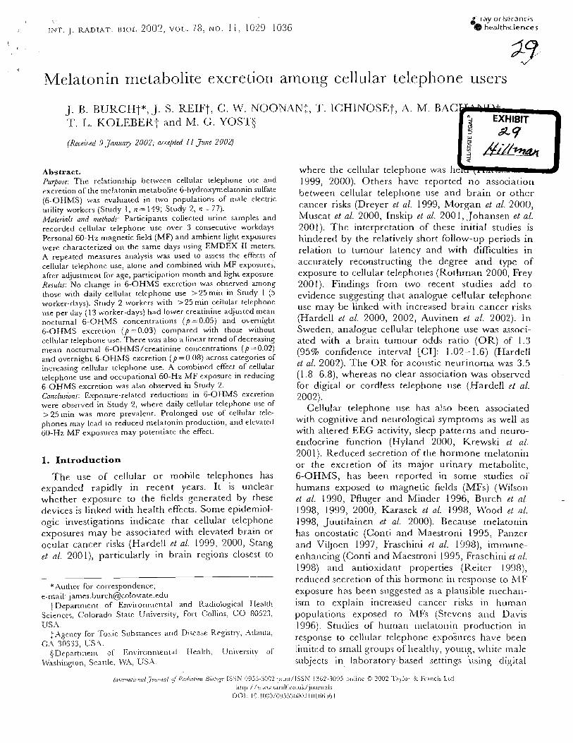
1, ..
I;.lr. J . RADIAT. BIOL. 2002, VOL.. 78: NO. 1 1, 1029.- 1636 g laylor&trancls @ healthsciences
Melatonin metabolite excretion among cellular telephone users
J. B. B U K C H t X , J. S. R E I F t , C. W. NOONAN:, T. I C H I N O S E t , A. M. BAG T . L. KO1,EBERT a n d IM. G. Y O S T $
(Receiued 9 Jnrlunly 2002; ncce/)tad I 1 Jzme 3002) ,Y d
rUiL- Abstract. where the cellular telephone was he Puqose: T h e relationship between cellular telephone use and 1999, 2000). Others have reported no association excretion of the melatonin metabolite 6-hydroxymelatonin sulfate between cellular telephone use and brain or other (6-OHMS) was evaluated in two populations of male electric utility workers (Study I, n = 149; Study 2, n=77). cancer risks (Dreyer et al. 1999, Morgan et nl. 2000, A,faterialr and n~thods: Participants collected urine samples and Muscat e t a/. 2000, lnskip et a/. 200 1, Johansell e t al. recorded cellular telephone use over 3 consecutive workdays. 2001). The interpretation of these initial studies is Personal 60-Hz magnetic field (MF) and ambient light exposures hindered by the relatively short follow-up periods in were characterized on the same days using EiVlDEX I1 meters. A repeated measures analysis was used to assess the effects of
relation to tumour latency and with difficulties in cellular telephone use, alone and combined with MF exposures, acc~lrately reconstructing the degree and type of after adjustment for age, par!icipation menth and light exposure. exposure to cellular telephorles (Rothman 2000, Frey Resulfr: NO change in 6-OHMS excretion was observed among 2001). Findings from two recent studies add to those with daily cellular telcpho1le use >25min in Stlldy 1 (5 evidence suggesting that analogue cellular telephone worker-days). Study 2 workers with >25 min ceilular telephone use per day (13 worker-days) had lower creatinine-adjusted mean
use may be linked with increased brain cancer risks nocturnal 6-OHMS concentrations (p=0.05) and overnight (Hardell e t a/ . 2000, 2002, Auvinen e t al. 2002). i n 6-OHMS excretion (p=0.03) compared with those without Sweden, analogue cellular telephone use was associ- cellular telephone use. There was also a linear trend of decreasing ated with a brain turnour odds ratio ( O R ) of 1.3 mean nocturnal 6-OHMS Icreatinine concentrations ( p =0.02j and overnight 6-OHXIS excretion ( p = 0.08) across categories o l
(95% confidence interval [CI]: 1.02- 1.6) (Hardell
iRcreasiog cellular telephone use. A combined effect cellular et 2002). The O R for acoustic neurinortIa was 3.5 telephone use and occupational 60-Hz MF exposure in reducing (1.8-6.8), whereas no clear association was observed 6-OHMS excretion was a!so observed in Study 2. for digital or cordless telephone use (Hardell e l nl. Conclusions: Exposure-related reductions in 6-OHMS excretion 2002). were observed in Study 2, where daily cellular telephone use of > 25 1cin was more prevalent. Prolonsed use of cellular tele-
Cellular telephone use has also been associated phones: [nay lead to reduced melatonin production, and elevated with cogllitive and neurological symptoms as well as 60-HZ MF exposures may potentiate the effect. with altered EEG activity, sleep patterns and neuro-
endocrine function (Hyland 2000, Krewski et a/. 2001). Reduced secretion of the hormone melatonin
1. Introduction or the excretion of its major urinary metabolite, The use of cellular or mobile telephones has 6-OHMS, has been reported in some studies oi'
expanded rapidly in recent years. It is unclear humans exposed to magnetic fields (MFs) (Wilsor~ whether exposure to the fields generated by these et al. 1990, Pfluger and Minder 1996, Burch et nl. devices is linked with health effects. Some epidemiol- 1998, 1999, 2000, Karasek el al. 1998, Mrood et ogic investigations indicate that cellular telephone 1998, Juutilainen e t al. 2000). Because rnelatonin exposures may be associated with elevated brain or has oncostatic (Conti and Maestroni 1995, Panzer ocular cancer risks (Hardell et al. 1999, 2000, Stang and Viljoen 1997, Fraschini et al. 1998), immune- et nl. 200 l ) , particularly in brain regions closest to enhancing (Conti and hlaestroni 1995, Fraschini et nl.
1998) and antioxidant properties (Reiter 19981, --
- -
reduced seci-etion of this hormone in response to MF *Author for correspondence;
e-mail: [email protected] exposure has been suggested as a plausible mechan- +Depal.tnlent of Environmental and Radiological Health ism to explain increased cancer risks in human
sciences. Colorado State Univel-sity, Fort Collins, 80523, populations exposed to MFs (Stevens and Davis . . USA. 1996). Studies of human inelatoni~ production in
:Agency for Toxic Substances and Disease Reo,istt-y, Atlanta, response to cellular telephone espoi~tres have been CA 30333, US..\.
4Departmenr ,,[ E ~ ~ , ~ ~ ~ ~ ~ ~ ~ ~ ~ [ Health, urliveriit!; limited to small groups of healthy, youno ?' white male \\lashington, Seattle. ~ V A . US.-\ subjects in laboratory-based settings using digital
[;,;n-,~n/i~~,1ni,,7001:i~rl ~ ~ i R 1 7 r l i ~ ~ i r o i i ElClii8,q:- [SSN 1)9jj-'j0n.) prinr/lSSX I:iij2-:309, o ~ ~ l ~ n e C 2002 .l 'aylo~ Lt F ~ r n c i s Ltd hrrp://\.in~. ra~idtco.uk/jou~-r~al;
001. I l l 108r!/OniS30002 IQI ticiiijl
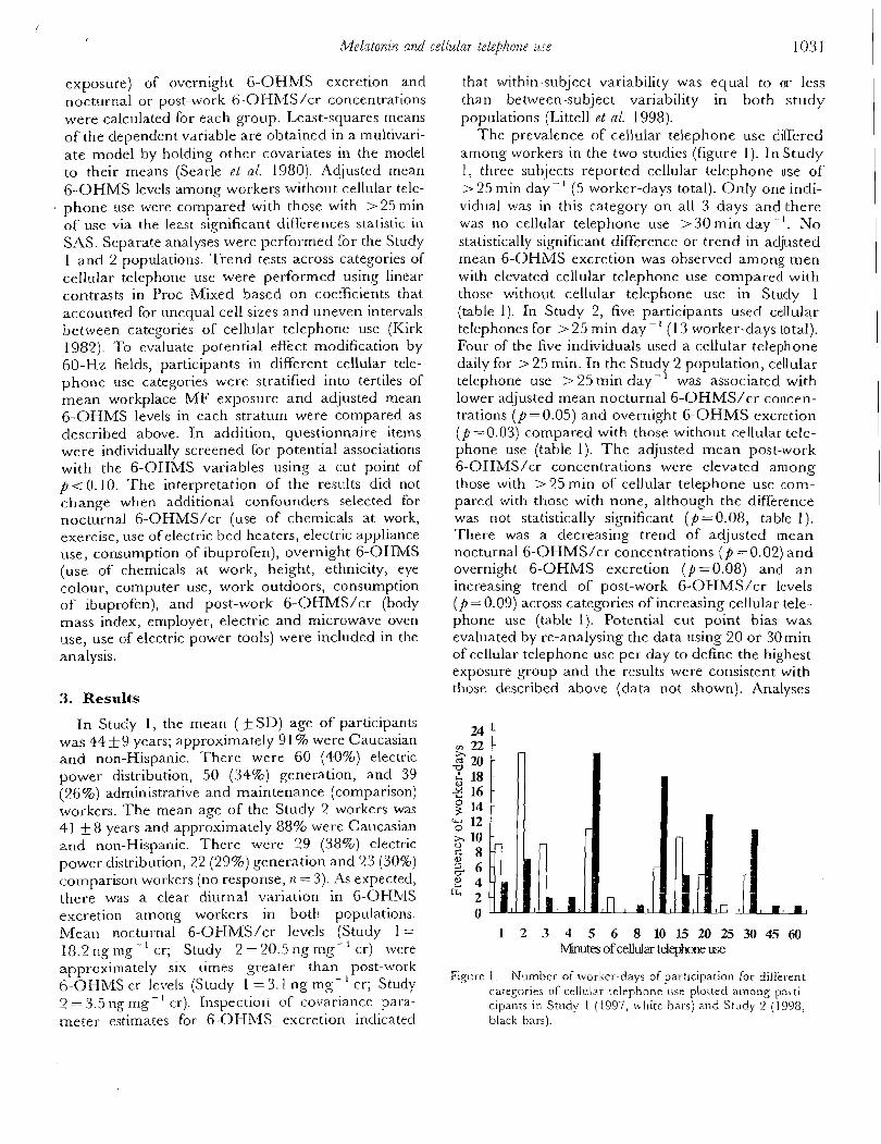
.ellz~far telephone iise 1031
exposure) of overnight 6-OHMS excretion and nocturnal or post-work 6-OHh/lS/cr concentrations were calculated for each group. Least-squares means of the dependent variable are obtained in a multivari- ate model by holding other covariates in the model to their means (Searle el a/. 1980). Adjusted mean 6-OHMS levels among workers without cellular tele- phone use were compared with those with >25 min of use via the least significant differences statistic in SAS. Separate analyses were performed for the Study 1 and 2 populations. Trend tests across categories of cellular telephone use were performed using linear contrasts in Proc Mixed based on coefficients that accounted for unequal cell sizes and uneven intervals between categories of cellular telephone use (Kirk 1982). To evaluate potential effect modification by 60-Hz fields, participants in different cellular tele- phone use categories were stratified into tertiles of mean workplace M F exposure and adjusted mean 6-OHMS levels in each stratum were compared as described above. In addition, questionnaire items were individually screened for potential associations with the 6-OHMS variables using a cut point of p t 0 . 1 0 . The interpretation of the results did not change when additional confounders selected for nocturnal 6-OHMS/cr (use of chemicals at work, exercise, use of electric bed heaters, electric appliance use, consumption of ibuprofen), overnight 6-OHMS (use of chemicals at work, height, ethnicity, eye colour, computer use, work outdoors, consumption of ibuprofen), and post-work 6-OHMS/cr (body mass index, employer, electric and microwave oven use, use of electric power tools) were included in the analysis.
3. Results
In Study I, the mean ( f SD) age of participants was 44 +9 years; approximately 9 1 % were Caucasian and non-Hispanic. There were 60 (40%) electric power distribution, 50 (34%) generation, and 39 (26%) administrative and maintenance (comparison) workers. The mean age of the Study I? workers was 41 f 8 years and approximately 88% were Caucasian and non-Hispanic. There were 29 (38%) electric power distribution, 22 (29%) generation and 23 (30%) cornparison workers (no response, n = 3) . As expected, there was a clear diurnal variation in 6-OHMS excretion among workers in both populations. Mean nocturnal 6-OHkIS/cr levels (Study 1 = 18.2 ng nlg-' cr; Study 2 =20.5 ng mg- ' cr) were approximately six times greater than post-work 6-OHMS cr levels (Study 1 = 3.1 ng mg- cr; Study 2 = 3.5 11g mg- ' cr). Inspectior1 of covariance para- meter estimates for 6-OH31fS excretion indicated
that within-subject variability was equal to or less than between-subject variability in both study populations (Littell et a f . 1998).
The prevalence of cellular telephone use differed among workers in the two studies (figure 1) . In Study 1 , three subjects reported cellular telephone use of > 25 min day - I (5 worker-days total). Only one indi- vidual was in this category on all 3 days and there was no cellular telephone use > 30 rnin day -'. No statistically significant difference or trend in adjusted mean 6-OHMS excretion was observed among men with elevated cellular telephone use compared with those without cellular telephone use in Study 1 (table 1 ) . In Study 2, five participants used cellular telephones for > 25 min day - ' (1 3 worker-days total). Four of the live individuals used a cellular telephone daily for >25 min. In the Study 2 population, cellular telephone use > 25 min day- was associated with lower adjusted mean nocturnal 6-OHMS/cr concen- trations ( p = 0.05) and overnight 6-OHMS excretion ( p =0.03) compared with those without cellular tele- phone use (table 1). The adjusted mean post-work 6-OHMS/cr concentrations were elevated among those with > 35 min of cellular telephone use com- pared with those with none, although the difference was not statistically significant (P=0.08, table 1). There was a decreasing trend of adjusted mean nocturnal 6-OHMS/cr concentrations ( p =0.02) and overnight 6-OHMS excretion (p=0.08) and a n increasing trend of post-work 6-OHMS/cr levels (p = 0.09) across categories of increasing cellular tele- phone use (table 1). Potential cut point bias was evaluated by re-analysing the data using 20 or 30min of cellular telephone use per day to define the highest exposure group and the results were consistent with those described above (data not shown). Analyses
1 2 3 4 5 6 8 1 0 1 5 2 0 2 5 3 0 4 5 ~ ~~ of ceMar telephone use
Figure I. Number of worker-days of participation Lbr different categories of cellular telephone Iise plorted among parti- cipants in Study 1 (1997, white bars) and Study 2 (1998, black bars).
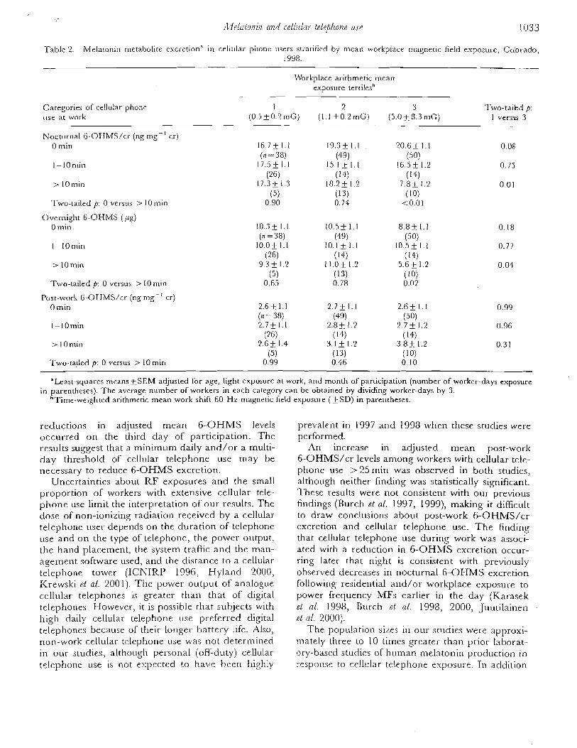
Table 2. Melatonin metabolite exci-etion" in cellular phone users st~.a~iiied by mean rvorkplace magnetic field esposul-e, Colorado, 1998.
Workplace arithmetic mean exposure tertilesh
Categories of cellular phone use at wcr-k
I 2 3 Two-tailed p: (0.5_+0.2mG) (1.1 f 0.2mG) (5.0+8.3nlG) I versus 3
Nocturnal 6-OHblS/cr (ng mg- ' cr) 0 min
Two-tailed p: 0 versus > 10 min
Over-night 6-OHMS (pg) 0 min
Two-tailed p: 0 versus > 10 min
Post-work 6-OHMS/cr (ng mg-' cr) 0 min
> IOmin
Two-tailed p: 0 versus > 10 min - --
"Least-squares means+SEM adjusted for age, light exposure at work, and month of participation (number of worker-days exposure in parentheses). The average number of workers in each category can bc obtained by dividing worker-days by 3.
"Time-weighted arithmetic mean work shift 60-Hz magnetic field exposure (+SD) in parentheses.
reductions in adjusted mean 6 -OHMS levels occurred on the third day of participation. The results suggest that a minimum daily and/or a multi- day threshold of cellular telephone use may be necessary to reduce 6-OHMS excretion.
Uncertainties about R F exposures and the small proportion of workers with extensive cellular tele- phone use limit the interpretation of our results. The dose of non-ionizing radiation received by a cellular telephone usel- depends on the duration of telephone use and on the type of telephone, the power output, the hand placement, the system traffic and the man- agement software used, and the distance to a cellular telephone tower (ICNIRP 1996, Hyland 2000, Krebvski rt ul. 2001). The power output of a~lalogue cellular telephones is greater than that of digital telephones. However, it is possible that subjects with high daily cellular telephone use preferred digital telephones because of their longer battery life. Also, non-work cellular telephone use was not determined in our studies, althougll personal (off-dutyj cellular te!ephone use is not expected to have been highly
prevalent in 1997 and 1998 when these studies were performed.
An increase in adjusted mean post-work 6-OHMS/cr levels among workers with cellular tele- phone use >25 min was observed in both studies, although neither finding was statistically significant. These results were not consistent with our previous findings (Burch et al. 1997, 1999), making it difficult to draw conclusions about post-work 6-OHMS/cr excretion and cellular telephone use. The finding that cellular telephone use during work was associ- ated with a reduction in 6-OHMS excretion occur- ring later that night is consistent with previously observed decreases in nocturnal 6-OHMS excretion following residential and/or workplace exposure to power rrequency MFs earlier in the day (Karasek PL 01. 1998, Burch eb 01. 1998, 2000, Juutilainen et nl. 2000).
The popu!ation sizes in our studies were approxi- mately three to 10 times greater than prior laborat- ory-based studies of human melatonin production in response to cc!lular telephone exposure. In addition
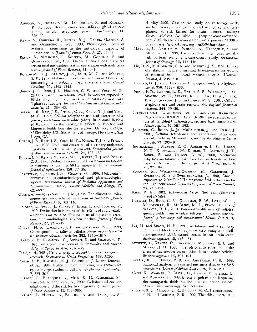
c211111a7 ttiel~hove use 1035
AUVINEN, A., HEITANEN, M., LUURKONEN, R. and KOSKELA, R. S., 2002, Brain tumors and salivary gland cancers among cellular telephone uesers. Epidnniolog, 13, 356-359.
BENOT, S., GOBERNA, R., REITER, R. J., GARCIA-MAURIXO, S. and GUERRERO, J. 1\/[ , 1999, Physiological levels of melatonin contribute to the antioxidant capacity of human sel-urn. Joicrnal of Pineal Research, 27, 59-64.
BENOT, S., ~ [ O L I N E R O , P., SOUTTO, M., GOBERNA, R. and GUERRERO, J. M., 1998, Circadian variations in the rat serum total antioxidant status: correlation with melatonin levels. JOUI-nal o/ Pineal Research, 25, 1-4.
BOJKOWSKI, C. J., ARENDT, J. A,, SHIH, kl . C. and MARREY, S. P., 1987, hfelatonin secretion in humans assessed by measuring its metabolite, 6-sulfatoxymelatoni~~. C'lUlical Cllenlisl,y, 33, 1343- 1348.
BURCH, J. B., REIF, J. S., NOONAN, C. W. and YOST, I\[. G., 2000, Melatonin metabolite levels in workers exposed to 60 Hz m a ~ n e t i c fields: work in substations and with - 3-phase conductors.journa1 of Occupa1ional and Enuironme~ital Medicine, 42, 136- 142.
BURCH, J. B., REIF, J. S., PITRAT, C . A,, KEEFE, T . J. and YOST, M. G., 1997, Cellular telephone use and excretion of a urinary melatonin metabolite (abst). In Annual Review of Research on the Biological Effects of Electric and Magnetic Fields from the Generation, Delivery and Use of Electricity. US Department of E n e r g , November, San Diego, CA.
BURCH, J. B., REIF, J. S., YOST, M. G., KEEFE, T. J. and PITRAT, C. A., 1398, Noctul-nal excretion of a urinary melatonin metabolite in electric utility workers. .Scandinauian Journal of l.%rk, Enuironrnent and Health, 24, 183- 189.
BURCH, J. B., REIF, J. S., YOST, M. G., KEEFE, T. J. and PITRAT, C. A., 1999, Reduced escretion of a melatonin metabolite in workers exposed to 60 H z magnetic fields. American journal ojpEpidetniology, 150, 27-36.
CLAUSTRAT, R., BRUN, J. and CHAZOT, G., 1990, Melatonin in humans: neuro-endocrinological and pharmacological aspects. Internntionul Journal o f Radialion Applications and 61strumetaion-Pat B, ~L~uclear Medicine und B i o l o ~ , 17, 625-632.
CONTI, A. and MAESTRONI, G. J. M., 1995, T h e clinical neuroim- munotherapeutic role of melatonin in oncology. Joumnl of Pineal Research, 9, 103- 1 10.
D E SEZE, R., AYOUB,J., PERAY, P., MIRO, L. and T o u r ~ o u , Y., 1999, Evaluation in humans of the effects of radiocellular telephones on the circadian patterns of melatonin secre- tion, a chronobiological rhythm marker. journul of Pzneal Research, 27, 237-242.
DREYER, N. A., LOUCHLIN, J. E. and ROTHMAN, K. J., 1999, Cause-specific mortality in cellular phone users. Jounznl of the Amn-ican lkferlical Associatiort, 282, 18 14- 18 16.
FRASCHINI, F., DEMARTINI, G., ESPOSTI, D. and SCAGLIONE, F., 1998, Melatonin involvement in immunity and cancer. Biological Signals Receptor, 7 , 61-72.
FREY, A. H., 2001, Cellular telephones and braill cancer: current 1,eszarch. Enuirorzrnental FIeutth Perspecliuts. 109, A20O.
FUNCH, D. P., ROTHMAN, I(. J., LOUCHLIN, J. E. and DREYER, N. A., 1996, Utility of telephone company records for epidemiologic studies of cellulal- telephones. Epide~niolo~, 7, 229-302.
HARDELL, L., HALLQUIST, .\., MILD, I\;. H., CARLBERG, kI., PAHLSON, '4. and LILIA, A, , 2002, Cellulal- and cordless telephones and the risk for brain tulno1.s. European Joitmai of Car~ctr PI-eoentio~i. 11, 3 7 7-386.
HARDELL, L.? NAS\I.AN, r\., PAHLSON. .A. arid H,\rrQurs~, A.!
4 May 2000, Case-control study on radiology work, medica! X-ray investigations, and use of cel111la1- tele- phones as risk factors for brain tumors. ~lledscape Gerlerol Medicine. Available at: [http://www.~nedscape. com / Medscape / Generalbledicine journal / 2000 / ~02.1103 mg- lrn0504.hard mg-'rn0504.hard.html].
HAROELL, L., NASMAN, A., PAHLSON, A., HALLQUIST, A. a n d MILD, K. N., 1999, Use of cellular telephones and the risk for brain turnours: a case-control study. hztena~iorznl Jou~-nrcl of Oncology, 15, 1 13- 1 16.
Hu, D. N., MCCORMICK, S. A. and ROBERTS, J. E., 1998, Effects oCmelatoni11, its precursors and derivatives on the ,orowth of cultured human weal melanoma cells. :War~omn Research, 8, 205-2 10
HYLAND, G. J., 2000, Physics and biology of mobile telephony Lancet, 356, 1833-1836.
INSKIP, P. D., THRONE, R. E., HATCH, E. E., WILCOSKY, T. C., SHAPIRO, W. R., SELKER, R. G., FINE, H. A., BLACK, P. IM., LOEFFLER, J. S and LINET, lh4. S., 2001, Ce l l~~la r telephone use and brain tumors. New England journal of Medicine, 344, 79-86.
INTERKATIONAL COMMISSION ON NON-IONIZING RADIATION PROTECTION (ICNIRP), 1996, Health issues related to the use of hand-held radiotelephones and base transmitters. Health Physics, 70, 587-593.
JOHANSEN, C., BOICE, J., JR, M C ~ U G H L I N , J. and OLSEN, J., 200 1 , Cellular telephones and cancer-a nationwide cohort study in Denmark. Journal of the ~Vfl&iotla/ Cancer Imtitule, 93, 203-207.
JUUTILAINEN, J., STEVENS, R. G. , ANDERSON, L. E., HANSEN, N. H., KILPELAINEN, M., KUMLIN, T. , LAITINEN, j. T. , SOBEL, E. and WILSON, B. W., 2000, Nocturnal 6-hydroxymelatonin sulfate excretion in female workers exposed to magnetic fields. Joumal of Pineal Research, 28, 97-104.
KARASEK, M., WOLDANSKA-OKONSKA, M., CZERNICKI, J., ZYLINSKA, K. and SWIETOSLAWSKI, J., 1998, Chronic exposure to 2.9mT, 40 Hz magnetic field reduces rnela- tonin concentrations in humans. Journal 4 Pineal Research, 25, 240-244.
KIRK, R. E., 1982, E.rperimenla1 Design. 2nd edn (Belmont: Brooks/Cole).
KREWSKI, D., BYUS, C. V., GLICKMAN, B. W., LOTZ, W. G., MANDEVILLE, R., MCBRIDE, M. L., PRATO, F. S. and WEAVER, D. F., 2001, Potential health risks of radiofre- quency fields from wireless telecommunication devices. Journal of Toxicology and Enuironmental Health, Part B, 4, 1-143.
LAI, H. and SINGH, N. P., 1997, Melatonin and a spin-trap compound block radiofrequency electromagnetic radi- ation-induced DNA strand breaks in rat brain cells. Bioelectronzagnefics, 18, 446-454.
LITOVITZ, T. , KRAUSE, D., PENAFIEL, L. M., ELSON, E. C. and MULLINS, J. M., 1993, The role of coherence time in the effect of micl-owaves on ornithine decarboxylase activity. Bioelectronlag~retics, 14, 395-403.
LITTELL, R. C., H'NRY, P. R. and A ~ ~ I E R M A N , C . B., 1998, Statistical analyses of repeated measures data using SAS procedul-es. Journal of dni7nal S~.iences, 76, 12 16.- 123 1 . .
L~,ANN. K., WAGNER, P., BRUNN; G., HAS.-\x, F., HIEAIKE! C. and ROSCHKE, J., 1998, Effects of pulsed high-fi.ecluel-\cy elect{-omagnetic fields OII the neuroenclocrine system. Clinical J~l~l~ron~docrinology, 67, 1 39- 1 44.
ARTIN, IN, S. D.: .I[.II.INA, H. Z., RREXN-IN, 1I. C.; HENDRICKSON, P. H. and ~ z ~ ~ ~ ~ ~ ~ , P. R., 1992, The ciliary body: the
Related Documents
![Light and electron microscopic examination of pineal gland in ......histological structure of the pineal gland [8–18]. So, in our study it was aimed to examine the pineal gland of](https://static.cupdf.com/doc/110x72/60c0abd73475835fb9682a43/light-and-electron-microscopic-examination-of-pineal-gland-in-histological.jpg)