PHARYNX

Edwards anatomy of Pharynx
Aug 07, 2015
Welcome message from author
This document is posted to help you gain knowledge. Please leave a comment to let me know what you think about it! Share it to your friends and learn new things together.
Transcript
PHARYNX
• Introduction:
• Pharynx is a wide muscular tube situated behind nose, mouth and larynx.
• Dimensions:• Length – 12Cms• Width - Upper part – 3.5Cms Middle part- Narrow Lower part - Narrowest 1.5Cms.

• Boundaries :
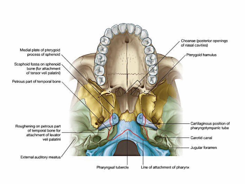
• Superiorly : Base of the skull, the posterior part of the body of the sphenoid bone and basilar part of occipital bone, in front of pharyngeal tubercle.
• Inferiorly : Pharynx is continuous with eusophagus at the level of 6th Cervical vertebra.
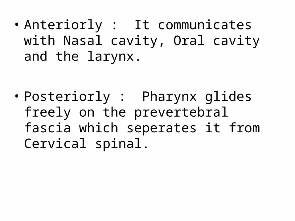
• Anteriorly : It communicates with Nasal cavity, Oral cavity and the larynx.
• Posteriorly : Pharynx glides freely on the prevertebral fascia which seperates it from Cervical spinal.

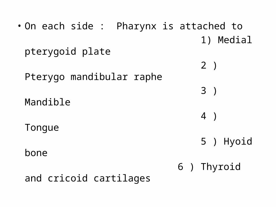
• On each side : Pharynx is attached to 1) Medial pterygoid plate 2 ) Pterygo mandibular raphe 3 ) Mandible 4 ) Tongue 5 ) Hyoid bone 6 ) Thyroid and cricoid cartilages
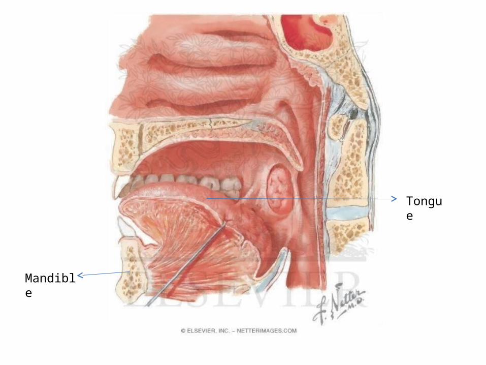
Tongue
Mandible
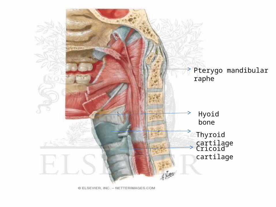
Pterygo mandibular raphe
Hyoid bone
Thyroid cartilage
Cricoid cartilage
• It communicates on each side with middle ear through auditory tube.
• Pharynx is related on either side to STYLOID PROCESS and muscles of it. COMMON CAROTID ARTERY EXTERNAL CAROTID ARTERY INTERNAL CAROTID ARTERY
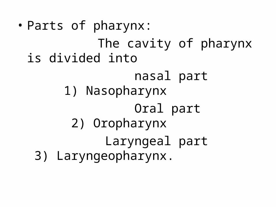
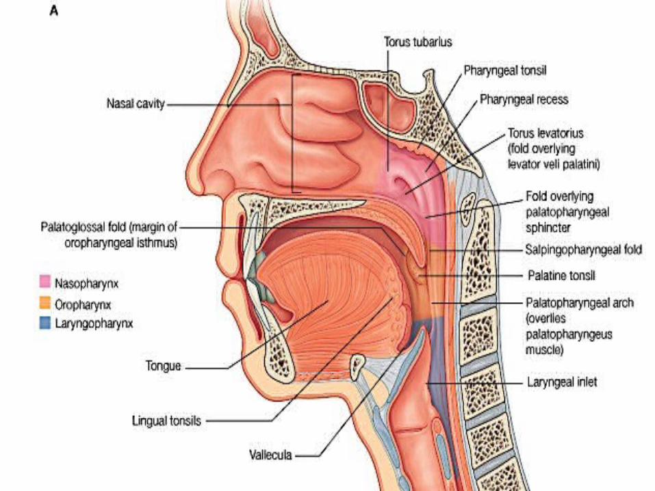
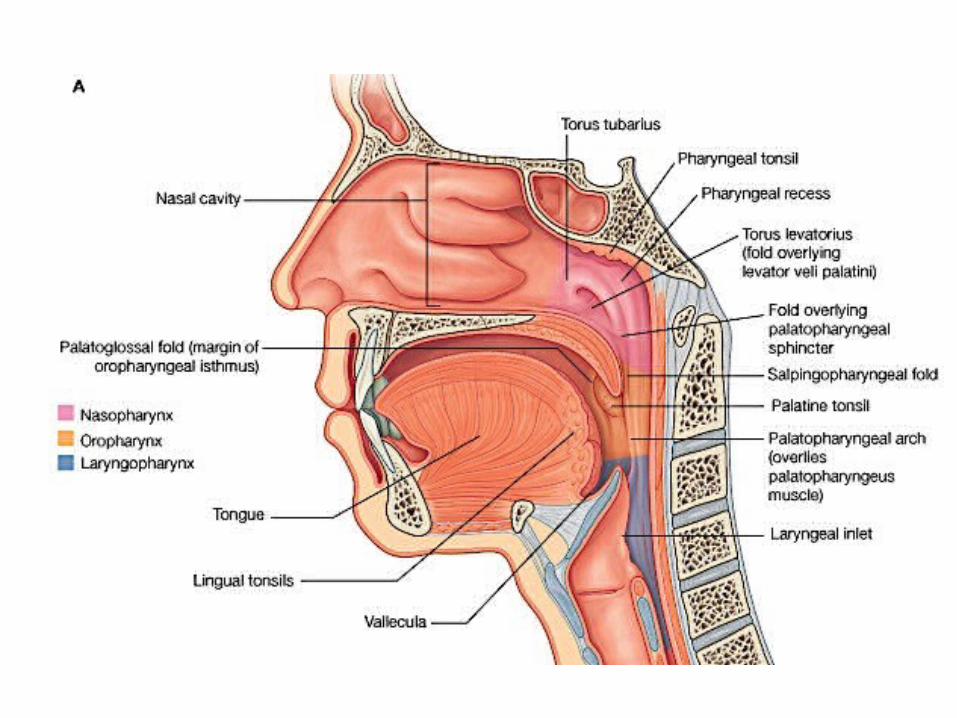
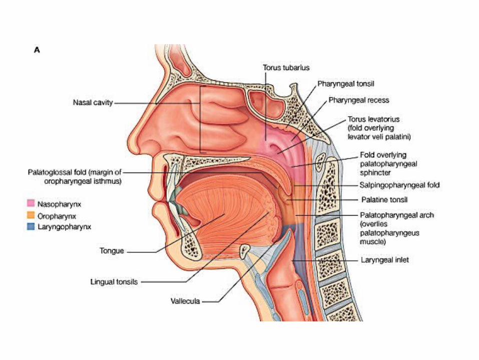
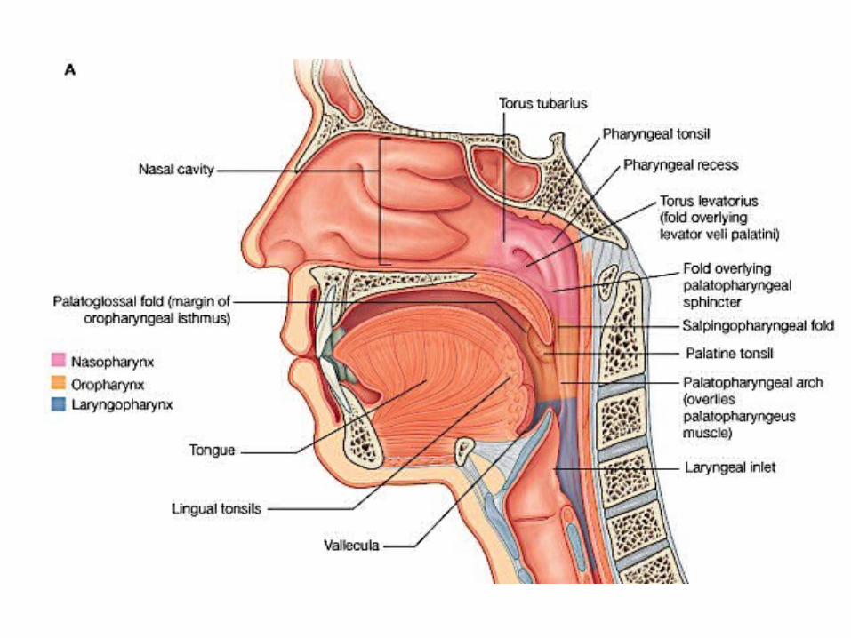
• Parts of pharynx: The cavity of pharynx is divided into nasal part 1) Nasopharynx Oral part 2) Oropharynx Laryngeal part 3) Laryngeopharynx.
Nasopharynx
• Its also called as Epipharynx.
• This is the upper part of pharynx situated behind the nose, and above the lower border of soft palate and passavant’s muscle.
• It resembles nose structurally and functionally,
1) Respiratory in function 2) Walls are rigid and non collapsable 3) lined by ciliated columnar
epithelium.
4) Mucous membrane is supplied by trigeminal nerve.
• Features : The rigid wall is formed by Pharyngobasilar-
fascia and posterior median Pharyngeal ligament.
• Anteriorly: It communicates with posterior nasal apertures.• Posteriorly:
Roof and Posterior wall form a continuous slope, opposite the posterior part of body of sphenoid bone, basioccuput, and anterior arch of atlas.
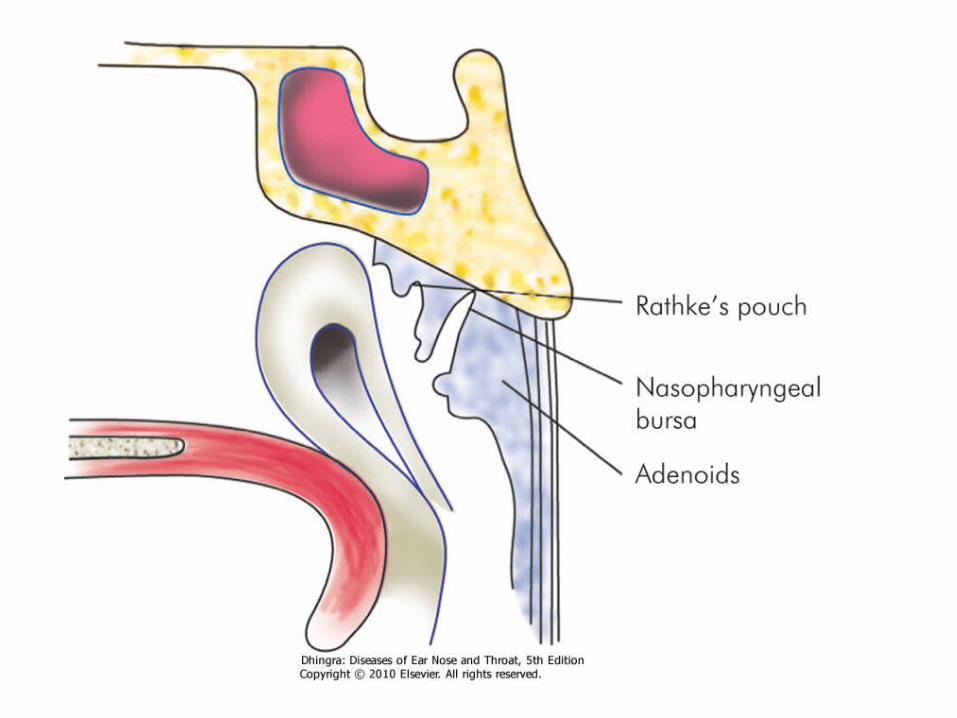
Under the mucous membrane, opposite the basiocciput , there is a collection of lymphoid tisue beneath the mucous membrane which projects downwards and forwards, it is called NASO-PHARYNGEAL TONSIL. It is better developed in children, small and absent in adult.
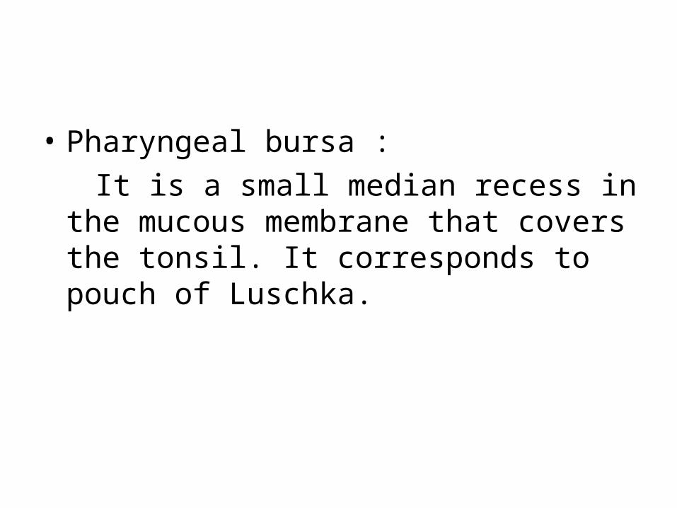
• Pharyngeal bursa : It is a small median recess in the mucous
membrane that covers the tonsil. It corresponds to pouch of Luschka.
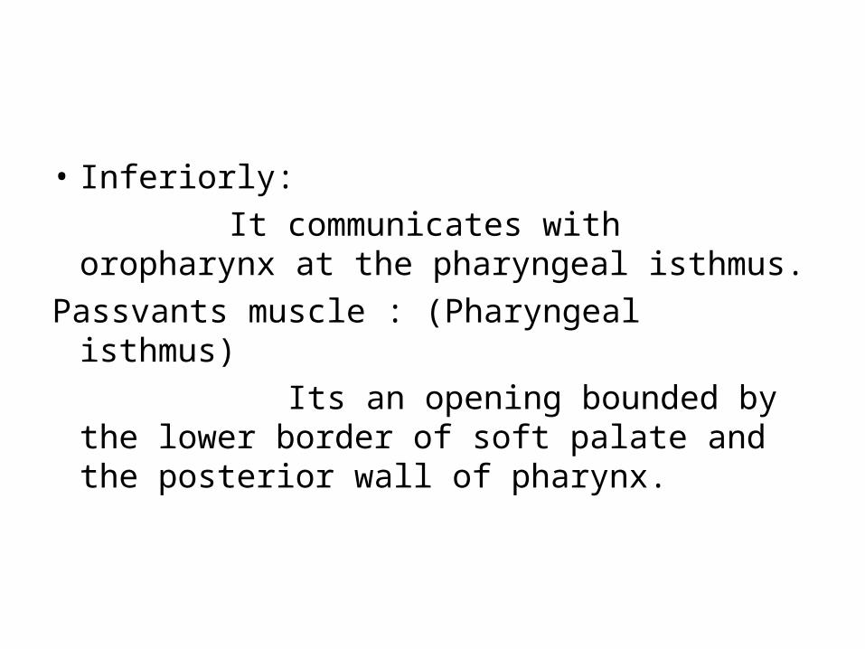
• Inferiorly: It communicates with oropharynx at the
pharyngeal isthmus.Passvants muscle : (Pharyngeal isthmus) Its an opening bounded by the lower
border of soft palate and the posterior wall of pharynx.
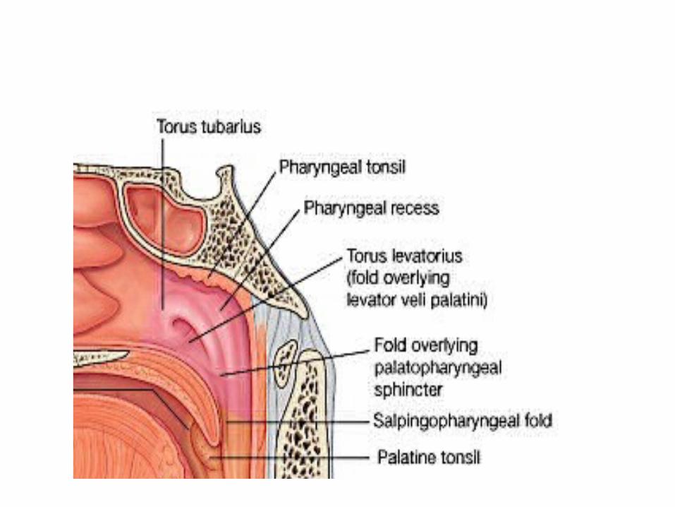
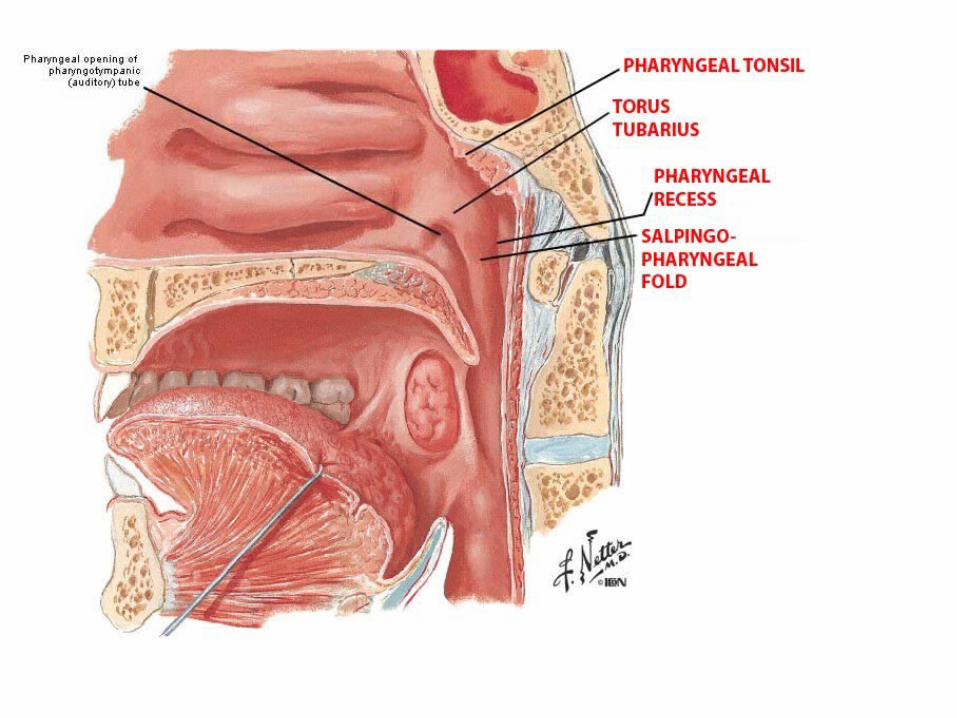
• Lateral wall : a) Pharyngeal opening of auditory tube, at the
level of inferior nasal concha and 1/2 inch behind it;
b) Tubal elevation bounds the tubal opening; c) Salpingopharyngeal fold is a vertial fold of
mucous membrane running downwards from the posterior margin of tubal elevation and gradually fading in the sides of pharynx. The fold is raised by a slip of muscle, the salpingopharyngeus.
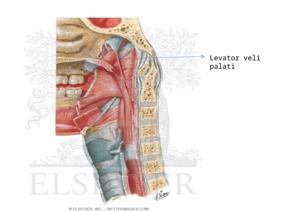
• The levator palati, as it enters soft palate, raises a fold of mucous membrane just below the tubal opening.
• Behind the tubal elevation and upper part of the fold there is a narrow vertical slit that leads into flat pocket of mucous membrane called Pharyngeal recess or lateral recess or fossa of rosenmuller, it is present above the superior constrictor muscle and below foramen lacerum and petrous part of temporal bone.
Levator veli palati
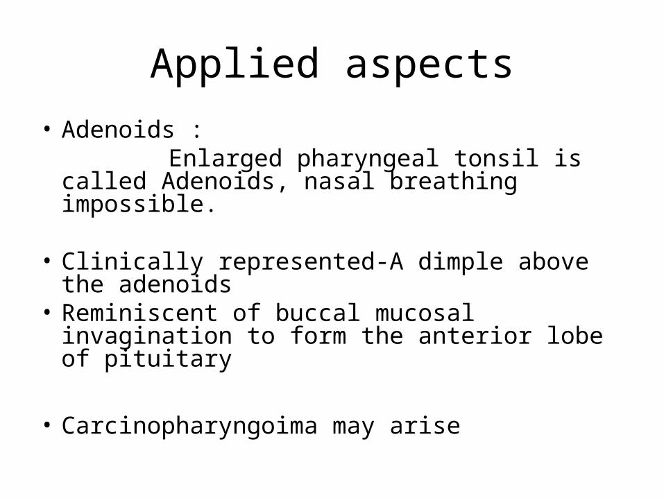
Applied aspects• Adenoids : Enlarged pharyngeal tonsil is called Adenoids,
nasal breathing impossible.
• Clinically represented-A dimple above the adenoids
• Reminiscent of buccal mucosal invagination to form the anterior lobe of pituitary
• Carcinopharyngoima may arise
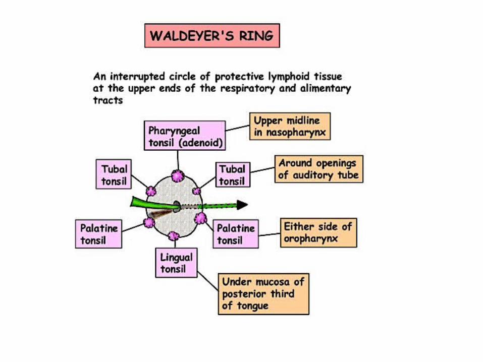
Oropharynx
• It is also called as mesopharynx, • It lies behind the oral cavity, and is supported
dorsally by the bodies of cervical vertebrae C2 and C3, and by the contents of retro pharyngeal space.
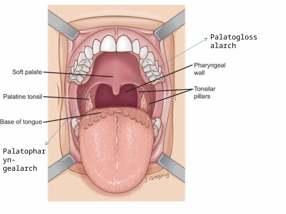
Palatoglossalarch
Palatopharyn-gealarch
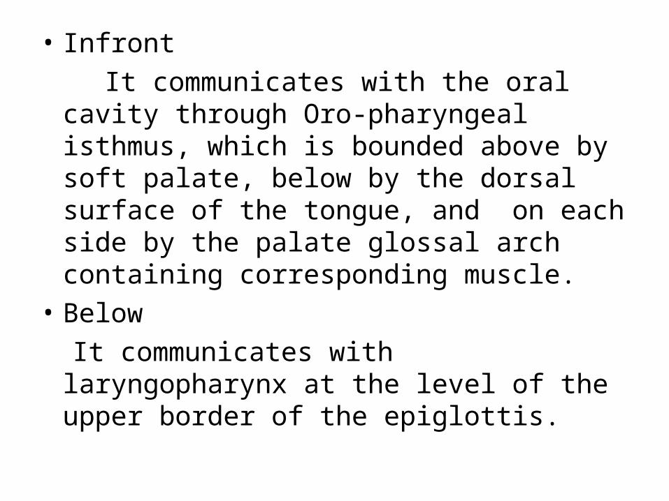
• Infront It communicates with the oral cavity through
Oro-pharyngeal isthmus, which is bounded above by soft palate, below by the dorsal surface of the tongue, and on each side by the palate glossal arch containing corresponding muscle.
• Below It communicates with laryngopharynx at the
level of the upper border of the epiglottis.
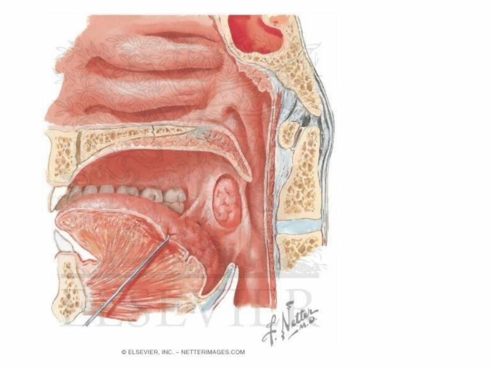
• Lateral wall: Lateral wall of the oropharynx presents on
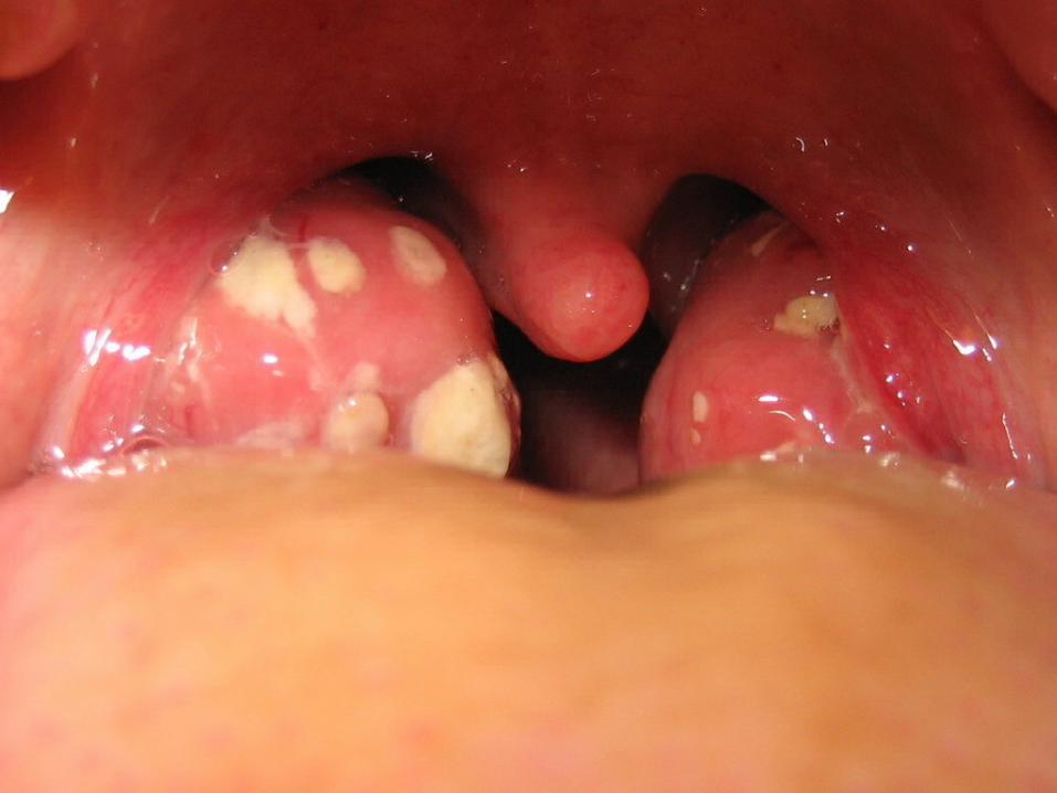
each side the palatine tonsil which lodges in a triangular tonsillar fossa.
Palatine tonsil
• Infront palatoglossalarch corresponding muscle;
• Behind palatopharyngeal arch corresponding muscle;
• Apex by the soft palate where both arches meet;
• Base, by the dorsal surface of the posterior 1/3rd of tongue;
• Lateral wall or the floor of the fossa, is formed by the superior constrictor and styloglossus muscles covered internally by the Pharyngobasilar fascia.
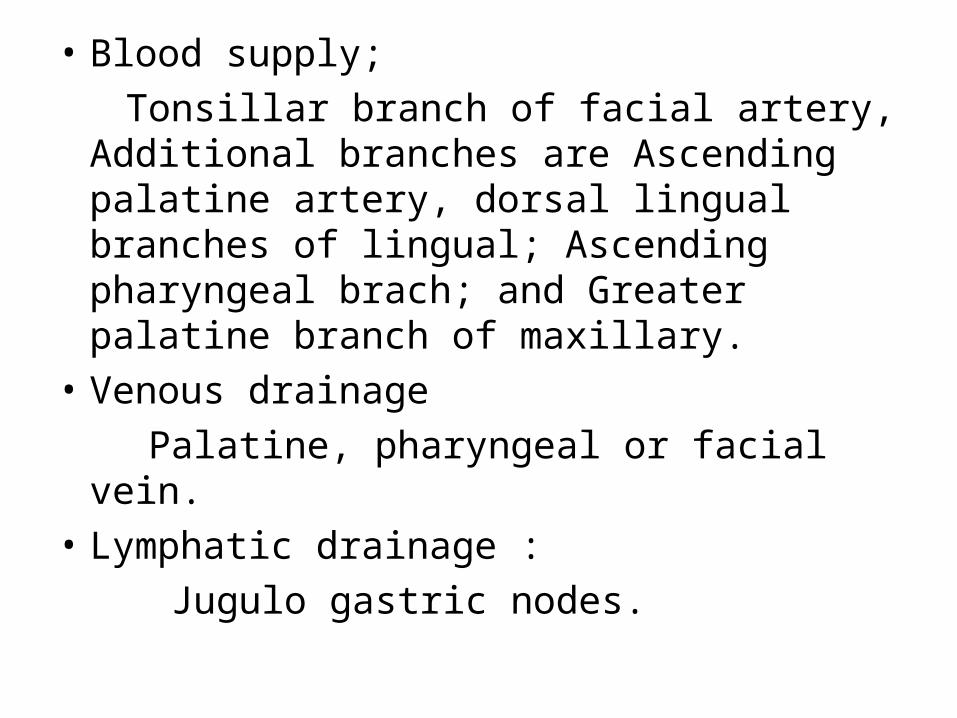
• Blood supply; Tonsillar branch of facial artery, Additional
branches are Ascending palatine artery, dorsal lingual branches of lingual; Ascending pharyngeal brach; and Greater palatine branch of maxillary.
• Venous drainage Palatine, pharyngeal or facial vein.• Lymphatic drainage : Jugulo gastric nodes.
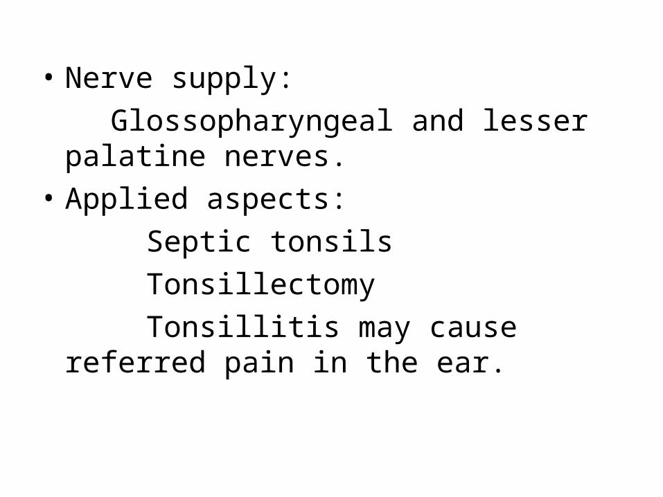
• Nerve supply: Glossopharyngeal and lesser palatine nerves.• Applied aspects: Septic tonsils Tonsillectomy Tonsillitis may cause referred pain in the ear.
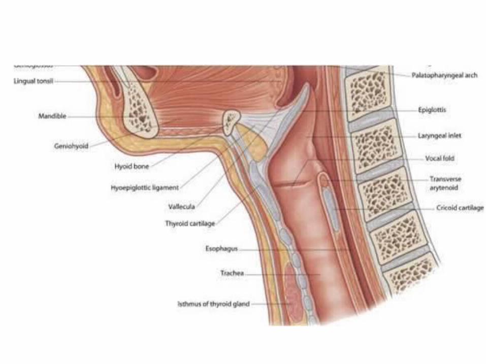
Laryngopharynx
• Laryngopharynx:• It is also called as Hypopharynx,• It is the lower part of the pharynx situated
behind the larynx. It extends from upper border of epiglottis to the lower border of cricoid cartilage
• Anterior wall: Inlet of larynx and posterior surface of cricoid
and arytenoid cartilages.• Posterior wall : It is supported by 4th and 5th cervical vertebrae
including the lower part of 3rd and upper part of 4th cervical vertebrae, the posterior pharyngeal wall is formed by the overlapping of 3 constrictors upto vocal cords.
• Lateral wall: It consists of piriform fossa one on each side of
the inlet of the larynx. The fossa medially bounded by the eryepiglottic fold, laterally by the thyroid cartilage and thyrohyoid membrane. Beneath the mucosa of the fossa there lies internal laryngeal nerve,
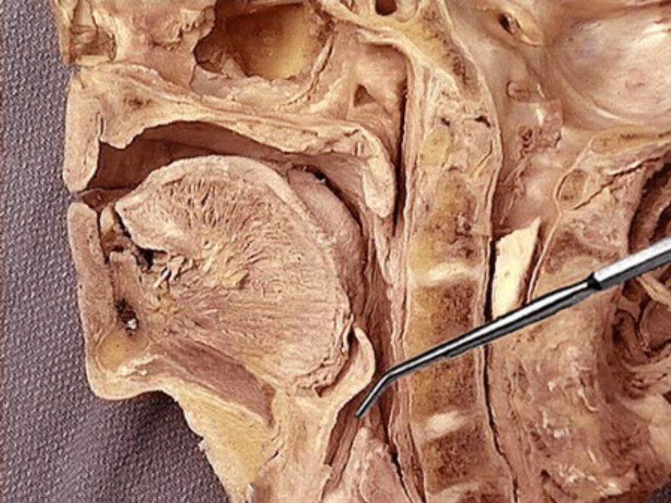
• Removal of foreign bodies from the piriform fossa may damage the internal laryngeal nerve, leading to anaesthetia in the supra epiglottic part of the larynx, this may cause aspiration pneumonia and death.
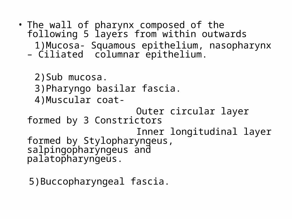
• The wall of pharynx composed of the following 5 layers from within outwards
1)Mucosa- Squamous epithelium, nasopharynx – Ciliated columnar epithelium.
2)Sub mucosa. 3)Pharyngo basilar fascia. 4)Muscular coat- Outer circular layer formed by 3 Constrictors Inner longitudinal layer formed by
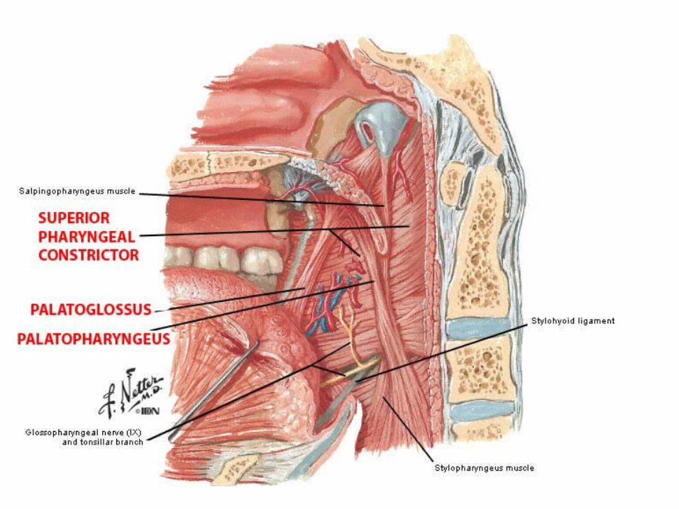
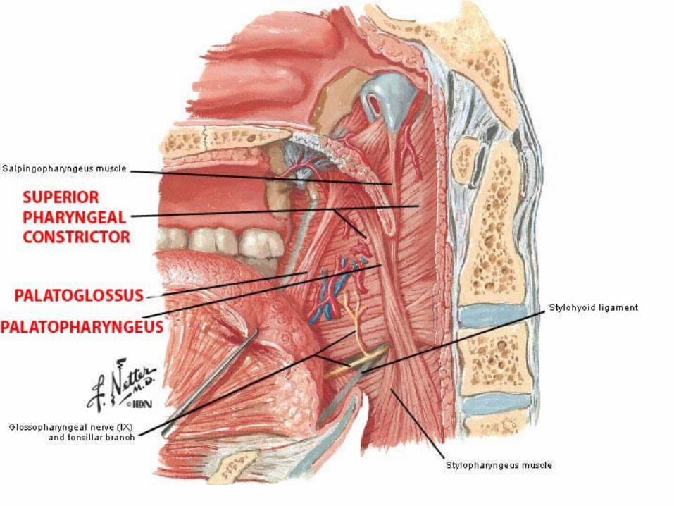
Stylopharyngeus, salpingopharyngeus and palatopharyngeus.
5)Buccopharyngeal fascia.
Muscles of the pharynx
• Superior constrictor- It arises at posterior border of medial pterygoid plate
and pterygoid hamulus , and pterygomandibular raphe and posterior end of mylohyoid line of mandible and inserts over pharyngeal tubercle supplied by pharyngeal plexus
• Middle constrictor:- It arises from lower part of stylohyoid ligament and
lesser cornu of hyoid bone and also upper part of greater cornu of hyoid bone and inserts to pharyngeal raphe supplied by pharyngeal plexus of nerves.
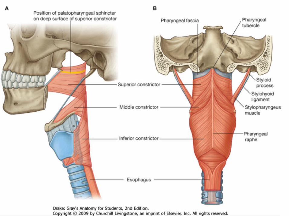
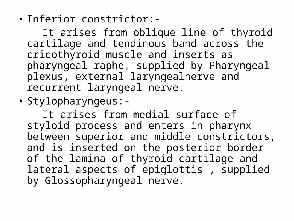
• Inferior constrictor:- It arises from oblique line of thyroid cartilage and
tendinous band across the cricothyroid muscle and inserts as pharyngeal raphe, supplied by Pharyngeal plexus, external laryngealnerve and recurrent laryngeal nerve.
• Stylopharyngeus:- It arises from medial surface of styloid process
and enters in pharynx between superior and middle constrictors, and is inserted on the posterior border of the lamina of thyroid cartilage and lateral aspects of epiglottis , supplied by Glossopharyngeal nerve.
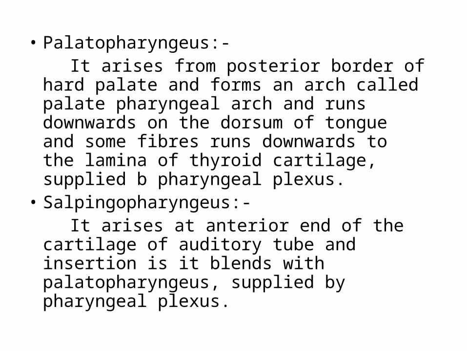
• Palatopharyngeus:- It arises from posterior border of hard palate
and forms an arch called palate pharyngeal arch and runs downwards on the dorsum of tongue and some fibres runs downwards to the lamina of thyroid cartilage, supplied b pharyngeal plexus.
• Salpingopharyngeus:- It arises at anterior end of the cartilage of
auditory tube and insertion is it blends with palatopharyngeus, supplied by pharyngeal plexus.
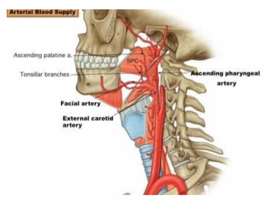
• Blood supply :• Ascending pharyngeal branch of External
carotid artery • Ascending palatine and tonsillar branches of
facial artery• Dorsal lingual branches of lingual; and Greater
palatine, pharyngeal and pterygoid branches of maxillary artery
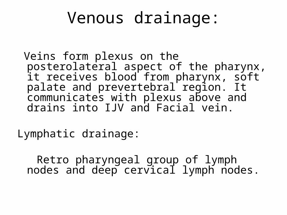
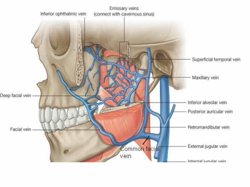
Venous drainage:
Veins form plexus on the posterolateral aspect of the pharynx, it receives blood from pharynx, soft palate and prevertebral region. It communicates with plexus above and drains into IJV and Facial vein.
Lymphatic drainage: Retro pharyngeal group of lymph nodes and deep
cervical lymph nodes.
Nerve supply:
• Pharynx is supplied by Pharyngeal plexus of
nerves which chiefly on the middle constrictor,
Plexus formed by Pharyngeal branch of Vagus (motor )nerve, pharyngeal branch of 9th
(Sensory) and superior cervical sympathetic ganglion
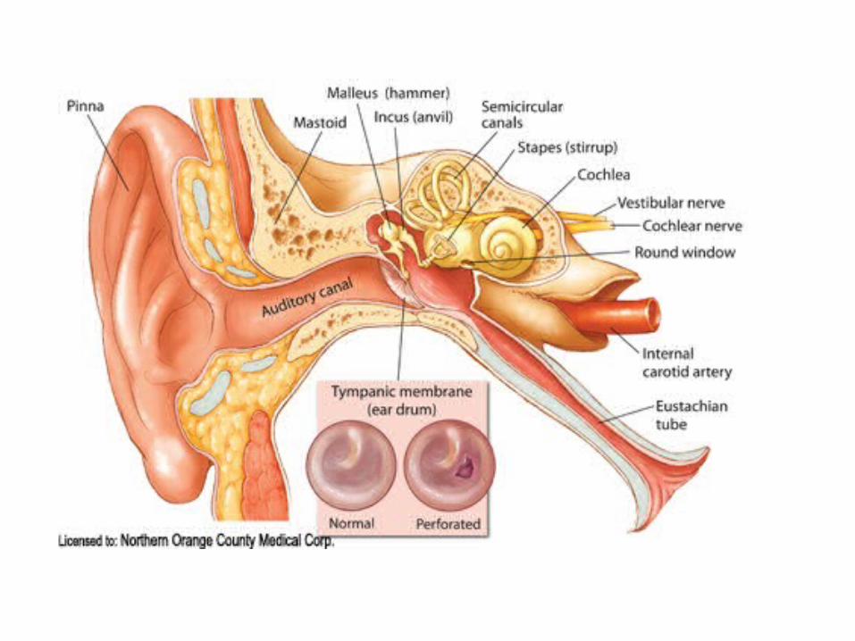
Auditory tube
• Its also called as pharyngo tympanic tube or Eustachian tube,
• It is trumpet shaped channel which connects
the middle ear with the nasopharynx,
• Length and direction:• It is about 1 ½ inches long, and is directed
downwards, forwards and medially. • Parts:• Posterior 1/3rd part is bony and anterior 2/3rd
part is cartilaginous
• Bony part:• It is half inch long, and lies in the petrous part of
temporal bone near tympanic plate.• Lateral end is wider and opens in the anterior
wall of middle ear cavity, the medial end is narrow and jagged for the attachment of cartilaginous part,
• Relations:• Superior : canal for tensor tympani• Medial: Carotid canal• Lateral: Chorda tympani, spine of sphenoid and
jaw joint• Lumen is oblong, being widest from side to side.
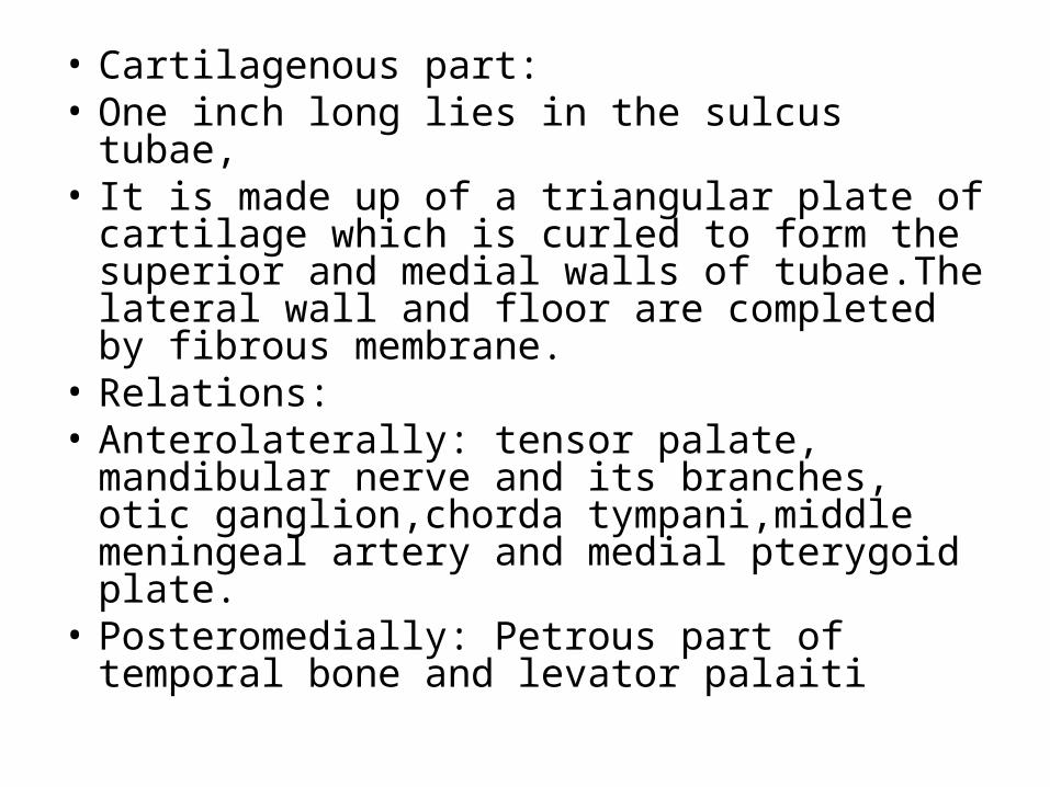
• Cartilagenous part:• One inch long lies in the sulcus tubae,• It is made up of a triangular plate of cartilage
which is curled to form the superior and medial walls of tubae.The lateral wall and floor are completed by fibrous membrane.
• Relations:• Anterolaterally: tensor palate, mandibular nerve
and its branches, otic ganglion,chorda tympani,middle meningeal artery and medial pterygoid plate.
• Posteromedially: Petrous part of temporal bone and levator palaiti
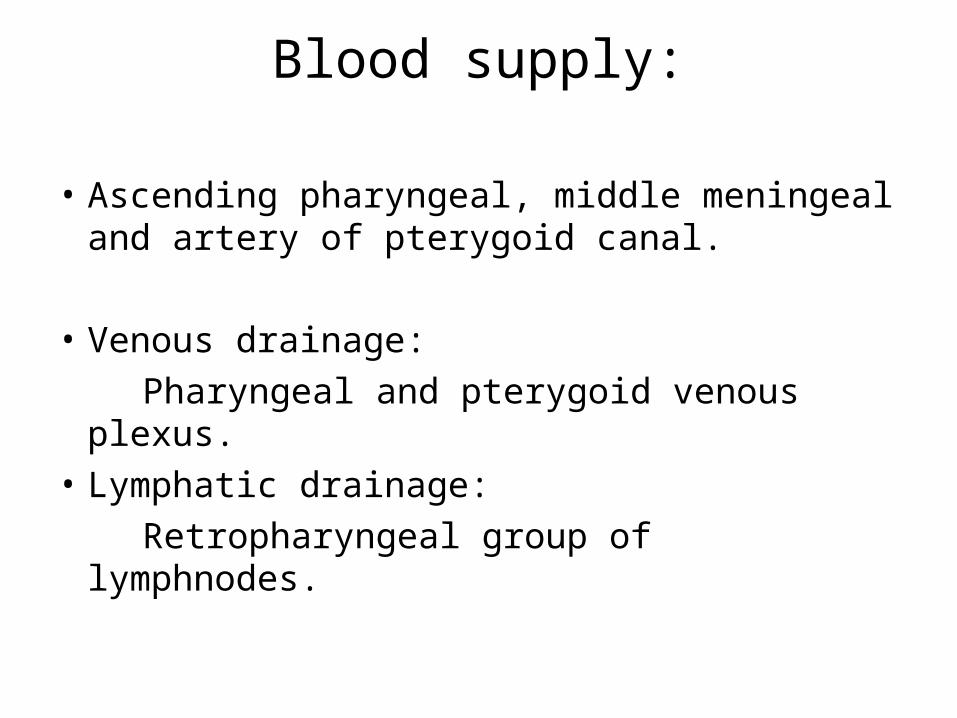
Blood supply:
• Ascending pharyngeal, middle meningeal and artery of pterygoid canal.
• Venous drainage: Pharyngeal and pterygoid venous plexus.• Lymphatic drainage: Retropharyngeal group of lymphnodes.
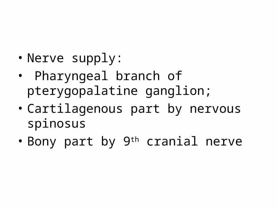
• Nerve supply:• Pharyngeal branch of pterygopalatine
ganglion;• Cartilagenous part by nervous spinosus • Bony part by 9th cranial nerve
• Function:• It maintains atmospheric pressure in the
middle ear cavity thus the air pressures on the twosides of the tympanic membrane are equalized, the tube is usually closed when relaxed. It opens during swallowing, yawning and sneezing by the action of tensor and levator veli palatine muscles.
Applied aspects:
• Inflammation of auditory tube.
Related Documents


![Pharynx [للقراءة فقط]fac.ksu.edu.sa/sites/default/files/pharynx_1.pdf · Pharynx • Anatomy (deep spaces) • Physiology • Pathology –Adenoid –Snoring & sleep apnea](https://static.cupdf.com/doc/110x72/5e011950d9ffa0689b5b92bb/pharynx-facksuedusasitesdefaultfilespharynx1pdf.jpg)


