Double Epitaxy as a Paradigm for Templated Growth of Highly Ordered Three-Dimensional Mesophase Crystals Yongsoon Shin, Jinhui Tao, Bruce W. Arey, Chongmin Wang, Gregory J. Exarhos, James J. De Yoreo, Maria L Sushko,* and Jun Liu* Pacific Northwest National Laboratory, Richland, Washington 99352, United States * S Supporting Information ABSTRACT: Molecular templating and self-assembly are fundamental mechanisms for controlling the morphology of biominerals, while in synthetic two-dimensional layered materials similar levels of control over materials structure can be achieved through the epitaxial relationship with the substrate. In this study these two concepts are combined to provide an approach for the nucleation and growth of three-dimensional ordered mesophases on solid surfaces. A combined experimental and theoretical study revealed how atomic ordering of the substrate controls the structure of surfactant template and the orientation and morphology of the epitaxially grown inorganic material. This dual epitaxial relationship between the substrate, surfactant template, and inorganic mesophase gives rise to a highly ordered porous mesophase with a well-defined cubic lattice of pores. The level of control over the material’s three-dimensional architecture achieved in this one-step synthesis is reminiscent of that in biomineralization. KEYWORDS: crystal growth, epitaxy, self-assembly, porous materials H eterogeneous substrate controlled nucleation is a widespread phenomenon in both the synthesis of materials and natural processes of biomineralization. In synthetic materials such substrate-controlled crystal growth often involves atomic-scale epitaxy between the substrate and the crystal. 1−5 This method is widely used for the growth of atomically controlled thin films, 6−8 However, achieving the same level of control over crystal morphology at the mesoscale remains a challenge despite a significant effort in developing synthesis approaches for achieving extended ordering in mesoporous materials 9−23 and the importance of such three- dimensional ordered materials for many applications including catalysis, separation, sensing, and energy conversion. 24−27 Here we demonstrate that mesoscale ordering of an organic− inorganic composite can be epitaxially controlled by the atomic ordering of the substrate. Detailed characterization and computer simulations are used to gain insights into the complex molecular processes leading to the formation of these atomically controlled organic−inorganic composite materials. In particular, we report a pathway for the synthesis of highly ordered mesophase crystals and porous materials, which utilizes dual epitaxial relationships with the substrate: (1) substrate- controlled three-dimensional surfactant templating and (2) metal oxide−substrate epitaxy. The method is demonstrated using the example of the epitaxial solution growth of uniform pyramidal silicates with ordered mesopores on Si(100) surfaces. RESULTS AND DISCUSSION The silicate films were synthesized using a two-step process (see Methods for more details). First, a BTME (1,2- bis(trimethoxysilyl)ethane)/CTAC (cetyltrimethylammonium chloride)/NaOH/H 2 O precursor solution was aged for 24 h at room temperature. Then Si(100) substrates were introduced into the solution, which was then heated to 100 °C and held at that temperature for times ranging from 0.5 to 2 h, with some samples exposed to repeated cycles of incubation in solution. Only a clear solution was used in order to avoid deposition onto the Si surface by silicate particles that had precipitated in the bulk solution. The substrates were then imaged ex situ using scanning electron microscopy (SEM) and atomic force microscopy (AFM) (Figure 1). The SEM images reveal the presence of coaligned pyramidal- shaped microscopic nanoparticles up to 20 μm in width on the Si(100) surfaces (Figure 1a−c). Figure 1d shows a typical SEM image of a large array of the pyramidal particles grown following repeated processing. The pyramids are uniform in shape, but the sizes vary over the range 3−20 μm. The studies of the same samples using AFM demonstrated that untreated, Received: June 16, 2016 Accepted: August 30, 2016 Published: August 30, 2016 Article www.acsnano.org © 2016 American Chemical Society 8670 DOI: 10.1021/acsnano.6b03999 ACS Nano 2016, 10, 8670−8675 Downloaded via PACIFIC NORTHWEST NATL LABORATORY on January 21, 2019 at 21:44:13 (UTC). See https://pubs.acs.org/sharingguidelines for options on how to legitimately share published articles.

Welcome message from author
This document is posted to help you gain knowledge. Please leave a comment to let me know what you think about it! Share it to your friends and learn new things together.
Transcript

Double Epitaxy as a Paradigm for TemplatedGrowth of Highly Ordered Three-DimensionalMesophase CrystalsYongsoon Shin, Jinhui Tao, Bruce W. Arey, Chongmin Wang, Gregory J. Exarhos, James J. De Yoreo,Maria L Sushko,* and Jun Liu*
Pacific Northwest National Laboratory, Richland, Washington 99352, United States
*S Supporting Information
ABSTRACT: Molecular templating and self-assembly are fundamental mechanismsfor controlling the morphology of biominerals, while in synthetic two-dimensionallayered materials similar levels of control over materials structure can be achievedthrough the epitaxial relationship with the substrate. In this study these twoconcepts are combined to provide an approach for the nucleation and growth ofthree-dimensional ordered mesophases on solid surfaces. A combined experimentaland theoretical study revealed how atomic ordering of the substrate controls thestructure of surfactant template and the orientation and morphology of theepitaxially grown inorganic material. This dual epitaxial relationship between thesubstrate, surfactant template, and inorganic mesophase gives rise to a highlyordered porous mesophase with a well-defined cubic lattice of pores. The level ofcontrol over the material’s three-dimensional architecture achieved in this one-stepsynthesis is reminiscent of that in biomineralization.
KEYWORDS: crystal growth, epitaxy, self-assembly, porous materials
Heterogeneous substrate controlled nucleation is awidespread phenomenon in both the synthesis ofmaterials and natural processes of biomineralization.
In synthetic materials such substrate-controlled crystal growthoften involves atomic-scale epitaxy between the substrate andthe crystal.1−5 This method is widely used for the growth ofatomically controlled thin films,6−8 However, achieving thesame level of control over crystal morphology at the mesoscaleremains a challenge despite a significant effort in developingsynthesis approaches for achieving extended ordering inmesoporous materials9−23 and the importance of such three-dimensional ordered materials for many applications includingcatalysis, separation, sensing, and energy conversion.24−27 Herewe demonstrate that mesoscale ordering of an organic−inorganic composite can be epitaxially controlled by the atomicordering of the substrate. Detailed characterization andcomputer simulations are used to gain insights into thecomplex molecular processes leading to the formation of theseatomically controlled organic−inorganic composite materials.In particular, we report a pathway for the synthesis of highlyordered mesophase crystals and porous materials, which utilizesdual epitaxial relationships with the substrate: (1) substrate-controlled three-dimensional surfactant templating and (2)metal oxide−substrate epitaxy. The method is demonstratedusing the example of the epitaxial solution growth of uniformpyramidal silicates with ordered mesopores on Si(100) surfaces.
RESULTS AND DISCUSSION
The silicate films were synthesized using a two-step process(see Methods for more details). First, a BTME (1,2-bis(trimethoxysilyl)ethane)/CTAC (cetyltrimethylammoniumchloride)/NaOH/H2O precursor solution was aged for 24 hat room temperature. Then Si(100) substrates were introducedinto the solution, which was then heated to 100 °C and held atthat temperature for times ranging from 0.5 to 2 h, with somesamples exposed to repeated cycles of incubation in solution.Only a clear solution was used in order to avoid depositiononto the Si surface by silicate particles that had precipitated inthe bulk solution.The substrates were then imaged ex situ using scanning
electron microscopy (SEM) and atomic force microscopy(AFM) (Figure 1).The SEM images reveal the presence of coaligned pyramidal-
shaped microscopic nanoparticles up to 20 μm in width on theSi(100) surfaces (Figure 1a−c). Figure 1d shows a typical SEMimage of a large array of the pyramidal particles grownfollowing repeated processing. The pyramids are uniform inshape, but the sizes vary over the range 3−20 μm. The studiesof the same samples using AFM demonstrated that untreated,
Received: June 16, 2016Accepted: August 30, 2016Published: August 30, 2016
Artic
lewww.acsnano.org
© 2016 American Chemical Society 8670 DOI: 10.1021/acsnano.6b03999ACS Nano 2016, 10, 8670−8675
Dow
nloa
ded
via
PAC
IFIC
NO
RT
HW
EST
NA
TL
LA
BO
RA
TO
RY
on
Janu
ary
21, 2
019
at 2
1:44
:13
(UT
C).
Se
e ht
tps:
//pub
s.ac
s.or
g/sh
arin
ggui
delin
es f
or o
ptio
ns o
n ho
w to
legi
timat
ely
shar
e pu
blis
hed
artic
les.
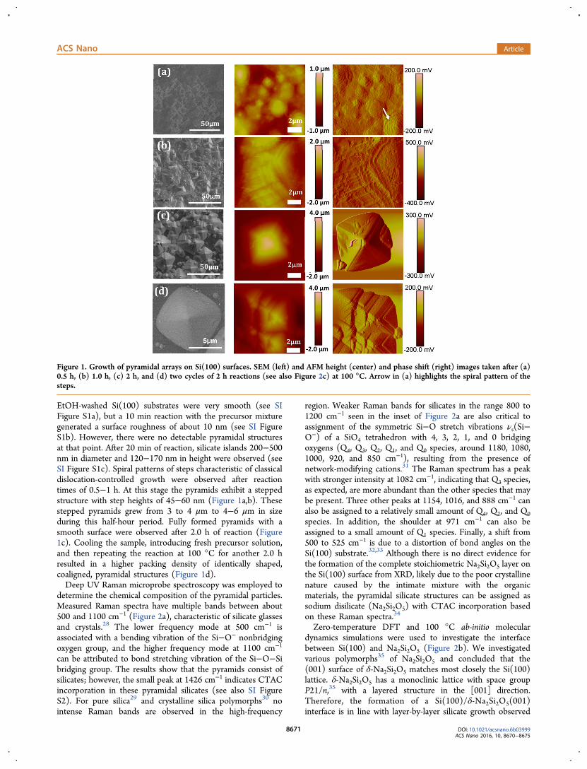
EtOH-washed Si(100) substrates were very smooth (see SIFigure S1a), but a 10 min reaction with the precursor mixturegenerated a surface roughness of about 10 nm (see SI FigureS1b). However, there were no detectable pyramidal structuresat that point. After 20 min of reaction, silicate islands 200−500nm in diameter and 120−170 nm in height were observed (seeSI Figure S1c). Spiral patterns of steps characteristic of classicaldislocation-controlled growth were observed after reactiontimes of 0.5−1 h. At this stage the pyramids exhibit a steppedstructure with step heights of 45−60 nm (Figure 1a,b). Thesestepped pyramids grew from 3 to 4 μm to 4−6 μm in sizeduring this half-hour period. Fully formed pyramids with asmooth surface were observed after 2.0 h of reaction (Figure1c). Cooling the sample, introducing fresh precursor solution,and then repeating the reaction at 100 °C for another 2.0 hresulted in a higher packing density of identically shaped,coaligned, pyramidal structures (Figure 1d).Deep UV Raman microprobe spectroscopy was employed to
determine the chemical composition of the pyramidal particles.Measured Raman spectra have multiple bands between about500 and 1100 cm−1 (Figure 2a), characteristic of silicate glassesand crystals.28 The lower frequency mode at 500 cm−1 isassociated with a bending vibration of the Si−O− nonbridgingoxygen group, and the higher frequency mode at 1100 cm−1
can be attributed to bond stretching vibration of the Si−O−Sibridging group. The results show that the pyramids consist ofsilicates; however, the small peak at 1426 cm−1 indicates CTACincorporation in these pyramidal silicates (see also SI FigureS2). For pure silica29 and crystalline silica polymorphs30 nointense Raman bands are observed in the high-frequency
region. Weaker Raman bands for silicates in the range 800 to1200 cm−1 seen in the inset of Figure 2a are also critical toassignment of the symmetric Si−O stretch vibrations νs(Si−O−) of a SiO4 tetrahedron with 4, 3, 2, 1, and 0 bridgingoxygens (Q4, Q3, Q2, Q1, and Q0 species, around 1180, 1080,1000, 920, and 850 cm−1), resulting from the presence ofnetwork-modifying cations.31 The Raman spectrum has a peakwith stronger intensity at 1082 cm−1, indicating that Q3 species,as expected, are more abundant than the other species that maybe present. Three other peaks at 1154, 1016, and 888 cm−1 canalso be assigned to a relatively small amount of Q4, Q2, and Q0species. In addition, the shoulder at 971 cm−1 can also beassigned to a small amount of Q1 species. Finally, a shift from500 to 525 cm−1 is due to a distortion of bond angles on theSi(100) substrate.32,33 Although there is no direct evidence forthe formation of the complete stoichiometric Na2Si2O5 layer onthe Si(100) surface from XRD, likely due to the poor crystallinenature caused by the intimate mixture with the organicmaterials, the pyramidal silicate structures can be assigned assodium disilicate (Na2Si2O5) with CTAC incorporation basedon these Raman spectra.34
Zero-temperature DFT and 100 °C ab-initio moleculardynamics simulations were used to investigate the interfacebetween Si(100) and Na2Si2O5 (Figure 2b). We investigatedvarious polymorphs35 of Na2Si2O5 and concluded that the(001) surface of δ-Na2Si2O5 matches most closely the Si(100)lattice. δ-Na2Si2O5 has a monoclinic lattice with space groupP21/n,35 with a layered structure in the [001] direction.Therefore, the formation of a Si(100)/δ-Na2Si2O5(001)interface is in line with layer-by-layer silicate growth observed
Figure 1. Growth of pyramidal arrays on Si(100) surfaces. SEM (left) and AFM height (center) and phase shift (right) images taken after (a)0.5 h, (b) 1.0 h, (c) 2 h, and (d) two cycles of 2 h reactions (see also Figure 2c) at 100 °C. Arrow in (a) highlights the spiral pattern of thesteps.
ACS Nano Article
DOI: 10.1021/acsnano.6b03999ACS Nano 2016, 10, 8670−8675
8671

in AFM experiments. The results predict a stable epitaxialinterface, based on long-time-scale dynamics with nodecomposition or significant relaxation of the interfacecompared to that obtained in static energy minimizationsimulations. A lattice mismatch between Si(100) and sodiumsilicate of 9.6% is accommodated through the deformation ofthe interfacial layer of sodium silicate. This deformation isassociated with rotation of SiO4 tetrahedra to form bonds withsurface Si atoms. The deformation is smaller for the nextsodium silicate layer. Nevertheless this degree of latticemismatch for inorganic solids is too high to support continuousfilm growth.36 However, due to the flexibility of sodium silicatelayers, such compressive strain can be accommodated duringlayer-by-layer growth of regular discrete particles,37 such as thepyramidal particles observed in our SEM and AFM experi-ments. Such gradual relief of strain within the layered structure,which the simulations predict is achieved by each layer havingdifferent two-dimensional lattice vectors compared to theirneighboring layers, suggests a loss of the overall bulkcrystallinity in the [100] and [010] directions of sodiumsilicate, while the native lattice spacing in the [001] direction isless affected. This prediction is supported by X-ray diffraction(XRD) data, which do not show characteristic diffraction peaksbetween 2θ = 15° and 2θ = 40° for crystalline sodium silicatephases with different SiO2/Na2O molar ratios38 (Figure S3).
Instead, only a dominant diffraction peak at 2θ = 69.16° and avery small diffraction peak at 2θ = 32.96°, characteristic ofSi(004) and Si(002) from the silicon substrate,39,40 wereobserved (see Figure S3). The incorporation of CTAC micellesinto the silicate structure, as evidenced by the appearance of thecharacteristic CH2 bending modes in Raman spectra at 1426cm−1 (Figure 2a), is likely to provide an additional mechanismfor strain relief within silicate layers. Surfactant incorporationalso suggests that the silicate layers are not continuous evenwithin a single pyramidal particle. Instead silicate layers areexpected to form around the CTAC scaffold and have an arrayof extended defects.Low-angle XRD indicates that, after reaction for 2 h at 100
°C, these pyramidal silicates have a cubic mesopore structurewith Pm3̅n symmetry (Figure 3a), similar to that seen inpreviously reported mesoporous silicas.41,42 Five well-resolvedfeatures indexed as (200), (210), (211), (320), and (400)
Figure 2. Structure of silicate on a Si(100) surface. (a) Ramanspectrum of epitaxially grown pyramidal silicates on Si(100). (b)Simulated structure of Na2Si2O5 on a Si(100) surface. Si atoms areshown as blue spheres, Na as yellow spheres, and O as red spheres.(c) Large-area SEM image of silicate taken after two cycles of 2 hreactions at 100 °C (see Figure 1d for the zoom-in image).
Figure 3. Structure and growth pathway of porous silicate pyramidson Si(100). (a) Low-angle XRD patterns of pyramidal silicates withcubic mesopores grown on a Si(100) surface at 100 °C for (1) 0.5h, black line; (2) 1 h, blue line; and (3) 2 h, red line. (b) Electrondiffraction pattern and (c) TEM image of cracked silicate withPm3̅n pore symmetry along the [110] direction. (d) Schematic ofsurfactant-directed silicate formation process showing trans-formations in the surfactant template. More detailed AFM imagesof surfactant layers on Si(100) and Si(111) surfaces are given in theSupporting Information (Figures S4−S6).
ACS Nano Article
DOI: 10.1021/acsnano.6b03999ACS Nano 2016, 10, 8670−8675
8672
reflections are also clearly observed. The cubic mesostructurewas not clear during early stages of the process, but afterreaction for over 1 h rapid transformation to a stable cubicstructure was found.43 The unit cell parameter of the Pm3 ̅nlattice, a, as calculated from the (210) diffraction peak, is 92 Å.This cubic micellar structure of the pyramidal silicates wasfurther confirmed by the transmission electron microscopy(TEM), which revealed regular periodic pores over a large area(Figure 3). These findings clearly indicate that the pyramidalsilicates exhibit a well-ordered mesoporous structure. Themesoscale structure can be calcined at above 500 °C to removethe surfactant without damaging the structural integrity toproduce ordered mesoporous materials.The mechanism for the growth of these organosilicates can
be understood based on the details of CTAC self-assembly insolution and on Si surfaces combined with the epitaxialrelationship of the Si(100)−sodium silicate interface. In theconcentration range of 45−70 wt %, CTAC is known to formcylindrical micelles in aqueous solution at room temperature.44
Our molecular simulations predicted that such cylindricalmicelle geometry is retained upon micelle adsorption on theSi(100) surface.45 Moreover, these micelles follow the top-ography of the Si substrate, resulting from the c(4 × 2)reconstruction of the (100) surface, and are arranged in aregular array with the long axis along Si[100] direction (Figure3d). Such micelle morphology and orientation with respect tothe protruding ridges on the reconstructed Si(100) surfacemaximizes the strength of the hydrophobic interactionsbetween CTAC and the substrate.45 AFM data confirm theformation of a highly oriented surfactant structure on theSi(100) surface (see Figure 3d and Figure S4). The 0.7 nmheight and 6.5 nm spacing between the neighboring stripes inthe AFM images are in-line with those previously observed forCTAB on graphite, an almost identical surfactant on ahydrophobic surface; this pattern was interpreted to be thatof hemicylindrical structures.46,47
On the basis of these experimental data and theoreticalpredictions for the initial and final stages of templated growthof sodium silicate pyramids on Si(100), we propose thefollowing pathway. The first stage of the reaction is likely toinvolve the transformation from a hexagonal micellar surfactantstructure to a bicontinuous cubic structure (space group Ia3̅d)upon heating to 100 °C. Although it is challenging toexperimentally observe this temperature-induced phase tran-sition on the Si surface, the transformation is expected based onthe CTAC phase diagram.44 This bicontinuous surfactant phasethen provides a three-dimensional template for the growth ofporous silicates. We note that, upon silicate growth, thesurfactant may be partially displaced (schematically shown inFigure 3d). Such local reduction in surfactant volume fractionwas reported to trigger transformation of the surfactant Ia3 ̅dphase to a micellar cubic Pm3 ̅n structure,48 as observed in thefinal product.In contrast to the situation on the Si(100) face, such a
pathway for transformation of the surfactant template fromhexagonal to bicontinuous cubic and to micellar cubic phase(Figure 3b) is not possible on a Si(111) surface. On thissurface, surfactants do not retain their hexagonal micellestructure. Instead, as revealed by our AFM data (Figure 3d andFigure S4) and molecular dynamics simulations, surfacesymmetry dictates the formation of hemispherical CTACmicelles.45 These micelles are randomly arranged on Si(111)and do not support the formation of a well-ordered three-
dimensional template. As a result, no silicate growth is observedon this substrate under otherwise identical conditions (FiguresS5 and S6).Similarly, if the CTAC concentration is too low (e.g., 100
mM) to support the transition to the bicontinuous phase at 100°C, no ordered porous silicate is formed on Si(100). Instead,irregular mesoporous particles with poorly developed morphol-ogy are observed (Figure S7). In contrast, increasing the CTACconcentration while within the hexagonal phase stability regionat room temperature produces more stable templates, hinderingfurther transformation to the surfactant cubic phase. As a result,the increase in CTAC concentration to 150 mM produceddodecahedral shape silicate precipitates, but with slightlydeformed cubic pore morphology (SI Figures S8 and S9).Elevation of temperature facilitates the transition fromhexagonal to bicontinuous cubic phase at lower CTACconcentrations. For a CTAC concentration of 125 mM, 100−110 °C is an optimal temperature for producing well-definedorganosilicate crystals. In these conditions the reaction of 20−30 mL of reaction mixture is completed in 2 h, and the repeatof the reaction produces homogeneous silicate pyramidalcrystal arrays.
CONCLUSION
In conclusion, Si(100) surfaces can act as templates for growthof long-range-ordered, uniform crystal-like pyramidal silicateswith cubic mesopores. Detailed characterization and modelingrevealed that the Si(100) surface plays the dual role of asubstrate for epitaxial growth of sodium silicate and one thatdirects CTAC self-assembly. The resulting highly orderedmesophase crystals and pore structure are a consequence of aseries of transformations in surfactant template geometry fromhexagonal phase to micellar cubic. Such complex nucleationand growth pathways to ordered three-dimensional architec-tures involving a series of transformations of organic templatingagents are reminiscent of biomineralization processes. Thelatter often involves complex transformations in organic(protein) and inorganic phases ultimately leading to highlyordered hierarchical materials.49 In this study we demonstratethe same level of control over materials architecture in a purelysynthetic system. The mechanistic model of the synthesispathway developed here broadens the horizons for knowledge-based design of ordered hierarchical materials for practicalapplications.
METHODSSynthesis. In a typical synthesis of silicate pyramids on a Si(100)
surface, 1,2-bis(trimethoxysilyl)ethane [(CH3O3Si-CH2CH2-Si-(OCH3)3)] was added to a mixture of cetyltrimethylammoniumchloride, sodium hydroxide, and water under vigorous stirring at roomtemperature, and this solution was aged for 24 h. The molar ratio ofBTME:CTAC:NaOH:H2O is 1.0:0.6−0.9:2.10:353. The aged solutionwas transferred to a Teflon container, and a small Si(100) piece wasplaced vertically into the solution. The Teflon container was placed inthe oven at 100 °C for 2 h. The Si(100) piece was taken out of thecontainer and washed with DI water.
Characterization. The XRD patterns were obtained on a PhilipsX’ert MPD X-ray diffractometer using Cu Kα (1.540 59 Å) radiationwith the X-ray generator operating at 45 kV and 40 mA. TEM imageswere obtained on a JEOL JEM 2010 microscope. For TEM analysis,pyramidal silicates were carefully scratched from the Si surface. Ramanspectra were excited using 244 nm cw radiation from a Lexel model85SHG laser equipped with harmonic doubling optics. For Ramanspectra measurement, approximately 10 mW of the filtered probe
ACS Nano Article
DOI: 10.1021/acsnano.6b03999ACS Nano 2016, 10, 8670−8675
8673

radiation was directed into a confocal microscope stage (Olympusmodel U-5RE-2) having a 40× UVB objective. Backscattered light wascollected, filtered to remove the excitation wavelength, and imagedthrough 100 μm slits into an 800 mm FL LabRAM HR spectrometerequipped with a 2400 g/mm grating blazed at 250 nm. A 5 s exposuretime was used, multiple spectra were acquired for each sample, and thesignal was averaged. All ex-situ AFM images were captured in tappingmode at room temperature (23 °C) with a NanoScope 8 atomic forcemicroscope (Digital Instruments J scanner, Bruker) with silicon tips(model AC160TS-R3, rectangular lever, k = 26 N/m, tip radius 9 ± 2nm; resonance frequency 300 kHz in air; Asylum Research). The driveamplitude was about 20 nm in air, and the signal-to-noise ratio wasmaintained above 10. The scanning speed was 1−2 Hz. The amplitudeset-point was carefully tuned to minimize the average loading force(∼50 pN) during imaging.Simulations. The first-principles simulations were performed using
the projector augmented wave method50,51 implemented in VASPcode.52−54 The generalized gradient approximation parametrized byPerdew, Burke, and Ernzerhof was used for the exchange−correlationterms,55,56 and wave functions were expanded by plane waves with acutoff energy of 500 eV. The atomic positions were optimized untilresidual ionic forces became less than 0.02 eV Å−1. A 2 × 2 × 1Monkhorst−Pack grid was used. A 3 × 4 Si(100) supercell (120atoms) and the corresponding 2 × 2 d-Na2Si2O5 supercell (144 atoms)were used to construct the interface. A 25 Å vacuum gap was used inthe direction normal to the interface.Ab-initio molecular dynamics simulations at 373 K (100 °C) were
performed within the canonical (NVT) ensemble. A Verlet algorithmwas integrated with Newton’s equations of motion at a time step of 2fs for a total simulation time of 30 ps. The frequency of thetemperature oscillations was controlled by the Nose ́ mass during thesimulations. K-point sampling at the Γ-point was used.
ASSOCIATED CONTENT*S Supporting InformationThe Supporting Information is available free of charge on theACS Publications website at DOI: 10.1021/acsnano.6b03999.
Additional AFM and TEM images of various stages oforganosilicate growth on Si surfaces; Raman and XRDspectra of silicate particles (PDF)
AUTHOR INFORMATIONCorresponding Authors*E-mail (M. L. Sushko): [email protected].*E-mail (J. Liu): [email protected].
NotesThe authors declare no competing financial interest.
ACKNOWLEDGMENTSThis work was supported by the Office of Basic EnergySciences, Division of Materials Sciences and Engineering, U.S.Department of Energy, under Award KC020105-FWP12152.The TEM and SEM studies were conducted at the Environ-mental Molecular Sciences Laboratory, a national scientific userfacility sponsored by the DOE’s Office of Biological andEnvironmental Research and located at Pacific NorthwestNational Laboratory (PNNL). Simulations were performedusing PNNL Institutional Computing resources. PNNL is amultiprogram national laboratory operated by Battelle for theDOE.
REFERENCES(1) Wu, X. J.; Chen, J. Z.; Tan, C. L.; Zhu, Y. H.; Han, Y.; Zhang, H.Controlled Growth of High-Density CdS and CdSe Nanorod Arrays
on Selective Facets of Two-Dimensional Semiconductor Nanoplates.Nat. Chem. 2016, 8, 470−475.(2) Fan, Z. X.; Bosman, M.; Huang, X.; Huang, D.; Yu, Y.; Ong, K.P.; Akimov, Y. A.; Wu, L.; Li, B.; Wu, J.; Huang, Y.; Liu, Q.; Png, C. E.;Gan, C. L.; Yang, P. D.; Zhang, H. Stabilization of 4H HexagonalPhase in Gold Nanoribbons. Nat. Commun. 2015, 6, 7684.(3) Tan, C. L.; Zhang, H. Epitaxial Growth of Hetero-NanostructuresBased on Ultrathin Two-Dimensional Nanosheets. J. Am. Chem. Soc.2015, 137, 12162−12174.(4) Fan, Z. X.; Zhu, Y. H.; Huang, X.; Han, Y.; Wang, Q. X.; Liu, Q.;Huang, Y.; Gan, C. L.; Zhang, H. Synthesis of Ultrathin Face-Centered-Cubic Au@Pt and Au@Pd Core-Shell Nanoplates fromHexagonal-Close-Packed Au Square Sheets. Angew. Chem., Int. Ed.2015, 54, 5672−5676.(5) Tan, C. L.; Zeng, Z. Y.; Huang, X.; Rui, X. H.; Wu, X. J.; Li, B.;Luo, Z. M.; Chen, J. Z.; Chen, B.; Yan, Q. Y.; Zhang, H. Liquid-PhaseEpitaxial Growth of Two-Dimensional Semiconductor Hetero-nanostructures. Angew. Chem., Int. Ed. 2015, 54, 1841−1845.(6) Mazet, L.; Yang, S. M.; Kalinin, S. V.; Schamm-Chardon, S.;Dubourdieu, C. A Review Of Molecular Beam Epitaxy of FerroelectricBaTiO3 Films on Si, Ge and GaAs Substrates and Their Applications.Sci. Technol. Adv. Mater. 2015, 16, 036005.(7) Li, G.; Wang, W.; Yang, W.; Wang, H. Epitaxial Growth of GroupIII-Nitride Films by Pulsed Laser Deposition and Their Use in theDevelopment of LED Devices. Surf. Sci. Rep. 2015, 70, 380−423.(8) Chambers, S. A. Molecular Beam Epitaxial Growth of DopedOxide Semiconductors. J. Phys.: Condens. Matter 2008, 20, 264004.(9) Monnier, A.; Schuth, F.; Huo, Q.; Kumar, D.; Margolese, D.;Maxwell, R. S.; Stucky, G. D.; Krishnamurty, M.; Petroff, P.; Firouzi,A.; Janicke, M.; Chmelka, B. F. Cooperative Formation of Inorganic-Organic Interfaces in the Synthesis of Silicate Mesostructures. Science1993, 261, 1299−1303.(10) Bagshaw, S. A.; Prouzet, E.; Pinnavaia, T. J. Templating ofMesoporous Molecular-Sieves by Nonionic Polyethylene OxideSurfactants. Science 1995, 269, 1242−1244.(11) Bao, X. Y.; Li, X.; Zhao, X. S. Synthesis of Large-PoreMethylene-Bridged Periodic Mesoporous Organosilicas and ItsImplications. J. Phys. Chem. B 2006, 110, 2656−2661.(12) Inagaki, S.; Guan, S.; Ohsuna, T.; Terasaki, O. An OrderedMesoporous Organosilica Hybrid Material with a Crystal-Like WallStructure. Nature 2002, 416, 304−307.(13) Guan, S.; Inagaki, S.; Ohsuna, T.; Terasaki, O. Cubic HybridOrganic-Inorganic Mesoporous Crystal with a Decaoctahedral Shape.J. Am. Chem. Soc. 2000, 122, 5660−5661.(14) Inagaki, S.; Guan, S.; Fukushima, Y.; Ohsuna, T.; Terasaki, O.Novel Mesoporous Materials with a Uniform Distribution of OrganicGroups and Inorganic Oxide in Their Frameworks. J. Am. Chem. Soc.1999, 121, 9611−9614.(15) Kim, J. M.; Kim, S. K.; Ryoo, R. Synthesis of MCM-48 SingleCrystals. Chem. Commun. 1998, 259−260.(16) Xia, Y. D.; Mokaya, R. To Stir or Not to Stir: Formation ofHierarchical Superstructures of Molecularly Ordered Ethylene-BridgedPeriodic Mesoporous Organosilicas. J. Mater. Chem. 2006, 16, 395−400.(17) Wang, J. W.; Xia, Y. D.; Wang, W. X.; Poliakoff, M.; Mokaya, R.Synthesis of Mesoporous Silica Hollow Spheres in Supercritical CO2/Water Systems. J. Mater. Chem. 2006, 16, 1751−1756.(18) Yang, Z. X.; Xia, Y. D.; Mokaya, R. Periodic MesoporousOrganosilica Mesophases Are Versatile Precursors for the DirectPreparation of Mesoporous Silica/Carbon Composites, Carbon andSilicon Carbide Materials. J. Mater. Chem. 2006, 16, 3417−3425.(19) Vercaemst, C.; de Jongh, P. E.; Meeldijk, J. D.; Goderis, B.;Verpoort, F.; Van Der Voort, P. Ethenylene-Bridged PeriodicMesoporous Organosilicas with Ultra-Large Mesopores. Chem.Commun. 2009, 4052−4054.(20) Morell, J.; Wolter, G.; Froba, M. Synthesis and Characterizationof Highly Ordered Thiophene-Bridged Periodic Mesoporous Organo-silicas with Large Pores. Chem. Mater. 2005, 17, 804−808.
ACS Nano Article
DOI: 10.1021/acsnano.6b03999ACS Nano 2016, 10, 8670−8675
8674

(21) Kapoor, M. P.; Yang, Q. H.; Inagaki, S. Self-Assembly ofBiphenylene-Bridged Hybrid Mesoporous Solid with Molecular-ScalePeriodicity in the Pore Walls. J. Am. Chem. Soc. 2002, 124, 15176−15177.(22) Yang, Q. H.; Kapoor, M. P.; Inagaki, S. Sulfuric Acid-Functionalized Mesoporous Benzene-Silica with a Molecular-ScalePeriodicity in the Walls. J. Am. Chem. Soc. 2002, 124, 9694−9695.(23) Dunphy, D. R.; Garcia, F. L.; Kaehr, B.; Khripin, C. Y.; Collord,A. D.; Baca, H. K.; Tate, M. P.; Hillhouse, H. W.; Strzalka, J. W.; Jiang,Z.; Wang, J.; Brinker, C. J. Tricontinuous Cubic Nanostructure andPore Size Patterning in Mesostructured Silica Films Templated withGlycerol Monooleate. Chem. Mater. 2011, 23, 2107−2112.(24) Ciriminna, R.; Fidalgo, A.; Pandarus, V.; Beland, F.; Ilharco, L.M.; Pagliaro, M. The Sol-Gel Route to Advanced Silica-BasedMaterials and Recent Applications. Chem. Rev. 2013, 113, 6592−6620.(25) Kruk, M. Access to Ultralarge-Pore Ordered MesoporousMaterials through Selection of Surfactant/Swelling-Agent MicellarTemplates. Acc. Chem. Res. 2012, 45, 1678−1687.(26) Mizoshita, N.; Tani, T.; Inagaki, S. Syntheses, Properties andApplications of Periodic Mesoporous Organosilicas Prepared fromBridged Organosilane Precursors. Chem. Soc. Rev. 2011, 40, 789−800.(27) Lei, X. L.; Jee, Y.; Huang, K. Amorphous Na2Si2O5 as a Fast Na
+
Conductor: an Ab Initio Molecular Dynamics Simulation. J. Mater.Chem. A 2015, 3, 19920−19927.(28) Simon, I. Modern Aspect of the Vitreous State; Butterworths:London, 1960; Vol. 1.(29) Galeener, F. L.; Lucovsky, G. Longitudinal Optical Vibrations inGlasses - GeO2 and SiO2. Phys. Rev. Lett. 1976, 37, 1474−1478.(30) Bates, J. B. Raman Spectra of Alpha-Cristobalite and Beta-Cristobalite. J. Chem. Phys. 1972, 57, 4042.(31) Virgo, D.; Mysen, B. O.; Kushiro, I. Anionic Constitution of 1-atm Silicate Melts - Implications for the Structure of Igneous Melts.Science 1980, 208, 1371−1373.(32) Furukawa, T.; Fox, K. E.; White, W. B. Raman-SpectroscopicInvestigation of the Structure of Silicate-Glasses 0.3. Raman Intensitiesand Structural Units in Sodium-Silicate Glasses. J. Chem. Phys. 1981,75, 3226−3237.(33) Hass, M. Raman Spectra of Vitreous Silica, Gerrnania andSodium Silicate Glasses. J. Phys. Chem. Solids 1970, 31, 415−422.(34) You, J. L.; Jiang, G. C.; Xu, K. D. Temperature Dependence ofthe Raman Spectra of Na2Si2O5. Chin. Phys. Lett. 2001, 18, 408−410.(35) Kahlenberg, V.; Dorsam, G.; Wendschuh-Josties, M.; Fischer, R.X. The Crystal Structure of delta-Na2Si2O5. J. Solid State Chem. 1999,146, 380−386.(36) Narayan, J.; Larson, B. C. Domain Epitaxy: A Unified Paradigmfor Thin Film Growth. J. Appl. Phys. 2003, 93, 278−285.(37) Fitzgerald, E. A. Gesi/Si Nanostructures. Annu. Rev. Mater. Sci.1995, 25, 417−454.(38) Woinaroschy, A.; Isopescu, R.; Filipescu, L. X-Ray PatternsIdentification of Crystalized Sodium Disilicates Mixtures. Cryst. Res.Technol. 2000, 35, 969−977.(39) Chen, Y.; Guo, L. P.; Chen, F.; Wang, E. G. Synthesis andCharacterization of C3N4 Crystalline Films on Silicon. J. Phys.:Condens. Matter 1996, 8, L685−L690.(40) Ozaki, K.; Hanatani, T.; Nakamura, T. Analysis of CrystallinePhases in Airborne Particulates by Grazing Incidence X-RayDiffractometry. Analyst 2005, 130, 1059−1064.(41) Huo, Q. S.; Margolese, D. I.; Ciesla, U.; Feng, P. Y.; Gier, T. E.;Sieger, P.; Leon, R.; Petroff, P. M.; Schuth, F.; Stucky, G. D.Generalized Synthesis of Periodic Surfactant Inorganic Composite-Materials. Nature 1994, 368, 317−321.(42) Sakamoto, Y.; Kaneda, M.; Terasaki, O.; Zhao, D. Y.; Kim, J. M.;Stucky, G.; Shim, H. J.; Ryoo, R. Direct Imaging of the Pores andCages of Three-Dimensional Mesoporous Materials. Nature 2000,408, 449−453.(43) Che, S. N.; Kamiya, S.; Terasaki, O.; Tatsumi, T. The Formationof Cubic Pm(3)over-barn Mesostructure by an Epitaxial PhaseTransformation from Hexagonal P6mm Mesophase. J. Am. Chem.Soc. 2001, 123, 12089−12090.
(44) Chen, Z. F.; Greaves, T. L.; Fong, C.; Caruso, R. A.;Drummond, C. J. Lyotropic Liquid Crystalline Phase Behaviour inAmphiphile-Protic Ionic Liquid Systems. Phys. Chem. Chem. Phys.2012, 14, 3825−3836.(45) Darkins, R.; Sushko, M. L.; Liu, J.; Duffy, D. M. The Effect ofSurface Topography on the Micellisation of Hexadecyltrimethylam-monium Chloride at the Silicon-Aqueous Interface. J. Phys.: Condens.Matter 2015, 27, 054008.(46) Manne, S.; Gaub, H. E. Molecular-Organization of Surfactants atSolid-Liquid Interfaces. Science 1995, 270, 1480−1482.(47) Manne, S.; Cleveland, J. P.; Gaub, H. E.; Stucky, G. D.; Hansma,P. K. Direct Visualization of Surfactant Hemimicelles by ForceMicroscopy of the Electrical Double-Layer. Langmuir 1994, 10, 4409−4413.(48) Faunce, C. A.; Paradies, H. H. Two New Colloidal CrystalPhases of Lipid a-Monophosphate: Order-to-Order Transition inColloidal Crystals. J. Chem. Phys. 2009, 131, 244708.(49) Meldrum, F. C.; Colfen, H. Controlling Mineral Morphologiesand Structures in Biological and Synthetic Systems. Chem. Rev. 2008,108, 4332−4432.(50) Blochl, P. E. Projector Augmented-Wave Method. Phys. Rev. B:Condens. Matter Mater. Phys. 1994, 50, 17953−17979.(51) Kresse, G.; Joubert, D. From Ultrasoft Pseudopotentials to theProjector Augmented-Wave Method. Phys. Rev. B: Condens. MatterMater. Phys. 1999, 59, 1758−1775.(52) Kresse, G.; Hafner, J. Ab-Initio Molecular-Dynamics for Open-Shell Transition-Metals. Phys. Rev. B: Condens. Matter Mater. Phys.1993, 48, 13115−13118.(53) Kresse, G.; Furthmuller, J. Efficiency of Ab-Initio Total EnergyCalculations for Metals and Semiconductors Using a Plane-Wave BasisSet. Comput. Mater. Sci. 1996, 6, 15−50.(54) Kresse, G.; Furthmuller, J. Efficient Iterative Schemes for AbInitio Total-Energy Calculations Using a Plane-Wave Basis Set. Phys.Rev. B: Condens. Matter Mater. Phys. 1996, 54, 11169−11186.(55) Perdew, J. P.; Burke, K.; Wang, Y. Generalized GradientApproximation for the Exchange-Correlation Hole of a Many-ElectronSystem. Phys. Rev. B: Condens. Matter Mater. Phys. 1996, 54, 16533−16539.(56) Perdew, J. P.; Burke, K.; Ernzerhof, M. Generalized GradientApproximation Made Simple. Phys. Rev. Lett. 1996, 77, 3865−3868.
ACS Nano Article
DOI: 10.1021/acsnano.6b03999ACS Nano 2016, 10, 8670−8675
8675
Related Documents


![eBook Production: A Templated Workflow [2013]](https://static.cupdf.com/doc/110x72/5596c5c01a28ab51408b46a5/ebook-production-a-templated-workflow-2013.jpg)