PALACKÝ UNIVERSITY OLOMOUC Dissertation Olomouc 2019 Zdeněk Škrott

Welcome message from author
This document is posted to help you gain knowledge. Please leave a comment to let me know what you think about it! Share it to your friends and learn new things together.
Transcript
PALACKÝ UNIVERSITY OLOMOUC
Dissertation
Olomouc 2019 Zdeněk Škrott
PALACKÝ UNIVERSITY OLOMOUCFACULTY OF MEDICINE AND DENTISTRY
Targeting the ubiquitin-proteasome system for cancer treatment:
the mechanism of action of drug disulfiram
Mgr. Zdeněk Škrott
Study programme: Pediatrics
Department: Laboratory of Genome Integrity, Institute of Molecular and Translational medicine
Supervisor: Mgr. Martin Mistrík, Ph.D.
Olomouc 2019

Statement:
I hereby declare that this thesis entitled: „Targeting the ubiquitin-proteasome system for
cancer treatment: the mechanism of action of drug disulfiram” was written by me, and all
relevant resources are included in the reference part. The work was mostly carried out at the
Laboratory of Genome Integrity, Institute of Molecular and Translational Medicine.
Acknowledgement:
First, I would like thank my supervisor Martin Mistrík, Ph.D. for his leadership, continuous
support, trust, and great scientific discussions and contribution. My thanks also belong to
prof. Jiří Bártek, Ph.D. for the opportunity to work on this exciting project. Second, I thank
my colleagues, namely Dušana Majera, Ph.D. for help with cell toxicity assays and stable cell
lines, Jan Gurský, Ph.D. for help with flow-cytometry, Tomáš Oždian Ph.D. for help with
HPLC-MS, Jing Li, Ph.D. from California Institute of Technology for help with 26S
proteasome assay, and MUDr. Andrea Miklovičová, MUDr. Petr Džubák, Ph.D., doc. MUDr.
Marian Hajdúch, Ph.D. for providing tissue samples from patients and animals. Third, I would
like to thank also to those who influenced my scientific thinking and career, namely to Boris
Cvek, Ph.D. and prof. Raymond Deshaies, Ph.D.
Finally, I would like to thank my family for continuous support.
Reasearch on the project was supported by: Palacky University (grants IGA_LF_2018_34;
IGA_LF_2019_026), Czech Ministry of Health (AZV 16-32030), Czech National Program of
Sustainability (LO1304).
Olomouc, June 2019. .....................................
Mgr. Zdeněk Škrott

Bibliografická identifikace:
Jméno a příjmení autora: Zdeněk Škrott
Název práce: Cílení ubiquitin-proteazomového systému při léčbě nádorových
onemocnění: mechanismus účinku léku disulfiramu
Typ práce: Dizertační
Pracoviště: Laboratoř integrity genomu, Ústav molekulární a translační
medicíny, Lékařská fakulta Univerzity Palackého v Olomouci
Vedoucí práce: Mgr. Martin Mistrík, Ph.D.
Rok obhajoby práce: 2019
Klíčová slova: ubiquitin-proteazomový systém, disulfiram, NPL4, p97, měď
Počet stran: 122
Jazyk: Anglický
Bibliographical identification:
Author´s name and surname: Zdeněk Škrott
Title: Targeting the ubiquitin-proteasome system for cancer treatment:
the mechanism of action of drug disulfiram
Type of thesis: Dissertation
Department: Laboratory of Genome Integrity, Institute of Molecular and
Translational Medicine, Faculty of Medicine and Dentistry,
Palacky University Olomouc
Supervisor: Mgr. Martin Mistrík, Ph.D.
The year of defence: 2019
Keywords: ubiquitin-proteasome system, disulfiram, NPL4, p97, copper
Number of pages: 122
Language: English

ABSTRACT
This thesis is focused on repurposing an old anti-alcohol drug disulfiram for cancer
therapy. Disulfiram has been shown to be effective in various preclinical cancer models,
but the unknown active metabolite, the unclear mechanism of action and unidentified
molecular target, all obstruct repurposing disulfiram as an anti-cancer drug. This thesis
describes
a new disulfiram metabolite found in humans, dithiocarbamate-copper complex, as the active
metabolite toxic to cancer cells and accumulating in tumours. Moreover, it shows,
that in the cells, dithiocarbamate-copper complex interferes with the NPL4 protein, an adaptor
of p97 segregase, which is essential for the degradation of proteins involved in several
regulatory and stress response pathways. After the treatment by dithiocarbamate-copper
complex, NPL4 forms aggregates, which subsequently attract p97 and other stress proteins
leading to induction of heat shock and unfolded protein responses, impairment of protein
degradation, ubiquitin stress, and cell death as a consequence. Collectively, observations
gathered in this thesis should encourage further clinical tests, help clinicians to monitor
the treatment and identify suitable patients benefiting from the disulfiram, all together
promoting eventual repurposing of this old, safe and cheap drug to safe lives of patients with
cancer worldwide.
ABSTRAKT
Tato disertační práce se týká znovuvyužití disulfiramu, starého léku používaného proti
alkoholismu, pro léčbu rakoviny. Protinádorový účinek disulfiramu byl prokázán na několika
preklinických modelech, ovšem nejasný mechanizmus účinku, neznámý aktivní metabolit
a také neznámý molekulární cíl, to vše brání nasazení disulfiramu pro léčbu nádorových
onemocnění. Tato práce popisuje nový metabolit disulfiramu nalezený u pacientů léčených
tímto lékem, a to komplex dithiokarbamátu s mědí. Jedná se o biologicky aktivní látku
toxickou pro rakovinové buňky a hromadící se v nádorech. Tato práce dále ukazuje, že tento
metabolit inhibuje protein NPL4, což je kofaktor proteinu p97, který je se podílí na degradaci
celé řady proteinů zapojených v mnoha regulačních a signalizačních drahách. Komplex
dithiokarbamátu s mědí vyvolává v buňkách agregaci NPL4. Tyto proteinové agregáty
následně přitahují vedle proteinu p97 také další stresové proteiny a indukují v buňce
specifickou stresovou odpověď. Navíc dochází k toxické akumulaci nezdegradovaných a

špatně poskládaných proteinů, ubiquitinovému stresu, a ve výsledku k buněčné smrti.
Poznatky v této práci a také v přiložených publikačních výstupech by měly podnítit další
klinické testy disulfiramu, usnadnit práci lékařům při hodnocení účinku léčby a také
identifikaci vhodných pacientů, pro něž by disulfiram mohl být přínosem. V důsledku by
mohly vést k zavedení tohoto starého, bezpečného a levného léku do protinádorové terapie.

TABLE OF CONTENTS
1 INTRODUCTION...................................................................................................................1
1.1 Overview of the Ubiquitin-proteasome system.................................................................1
1.2 The ubiquitin code.............................................................................................................4
1.3 The structure and function of the proteasome...................................................................7
1.4 Protein quality control....................................................................................................10
1.5 The role of the p97 complex............................................................................................15
1.6 The role of UPS in tumour development and treatment..................................................22
1.7 Anti-cancer activity of disulfiram...................................................................................30
2 AIMS......................................................................................................................................36
3 MATERIALS AND METHODS...........................................................................................37
3.1 HPLC/MS analysis of copper-dithiocarbamate complex (CuET)...................................37
3.2 Sample preparation for HPLC/MS analysis....................................................................37
3.3 Blood collection from humans for HPLC/MS analysis of CuET....................................38
3.4 Cell lines..........................................................................................................................38
3.5 Stable cell lines construction, transfection, siRNA.........................................................39
3.6 Colony formation assay...................................................................................................40
3.7 XTT assay........................................................................................................................40
3.8 Annexin V staining..........................................................................................................40
3.9 Caspases 3/7 assay...........................................................................................................40
3.10 Immunoblotting and antibodies.....................................................................................41
3.11 Immunofluorescence staining........................................................................................41
3.12 Microscopy, FRAP and image analysis........................................................................42
3.13 Cell fractionation for Triton X insoluble pellets...........................................................42
3.14 Isolation of microsomal fraction...................................................................................43
3.15 Ub(G76V)-GFP degradation..............................................................................................43
3.16 p97 ATPase activity assay.............................................................................................43
3.17 26S proteasome activity................................................................................................43
3.18 Affinity precipitation.....................................................................................................44
3.19 Protein expression and purification...............................................................................44

3.20 Chemicals......................................................................................................................45
3.21 Figures preparation, data analysis, used software.........................................................45
4 RESULTS..............................................................................................................................46
4.1 Ditiocarb-copper complex is a new metabolite of disulfiram.........................................46
4.2 CuET complex is highly toxic to cancer cells.................................................................48
4.3 CuET complex induces both apoptotic and non-apoptotic cell death.............................49
4.4 CuET complex does not inhibit the proteasome directly................................................53
4.5 CuET complex inhibits the function of p97 segregase....................................................55
4.6 Ubiquitinated proteins accumulated by CuET treatment are associated with insoluble
structures...............................................................................................................................59
4.7 CuET complex impairs ER-associated degradation leading to the activation of Unfolded
protein response.....................................................................................................................62
4.8 CuET complex immobilises p97 segregase.....................................................................63
4.9 CuET complex targets NPL4 cofactor............................................................................65
4.10 NPL4 protein forms aggregates after the treatment by CuET.......................................67
4.11 NPL4 protein mutated in putative zinc-finger domain resembles phenotypes induced
by CuET................................................................................................................................70
4.12 Aggregated NPL4 protein triggers heat shock response...............................................73
4.13 Disulfiram is converted to CuET in vitro......................................................................75
5 DISCUSSION........................................................................................................................78
6 CONCLUSION......................................................................................................................88
7 ABBREVIATIONS...............................................................................................................89
8 REFERENCES.......................................................................................................................93
9 BIBLIOGRAPHY................................................................................................................117
10 APPENDIX – FULL TEXT PUBLICATIONS RELATED TO THE THESIS................119

1 INTRODUCTION
Drugs often interact with more molecules than intended and such interaction with
these off-targets could manifests not only as adverse side effects, but importantly also as the
positive ones. In case the positive side effect is clinically relevant for any disease or medical
condition, a drug could be repurposed for clinical use. As drug repositioning accelerates
approval process and lower the financial cost, it is highly promising approach with growing
importance and interest.
Disulfiram, a drug used to treat alcoholism, could be an example of drug repurposing.
Disulfiram, also known as Antabuse, is used almost for seventy years as alcohol deterrent;
however, as suggest case reports and preclinical studies, disulfiram has also interesting anti-
cancer properties. While the active metabolite and the mechanism standing behind anti-
alcoholism effect of disulfiram are well known, such information is largely missing regarding
its impact on tumour cells. Nevertheless, it is generally accepted that anti-cancer activity of
disulfiram is dependent on the presence of copper, and inhibition of the proteasome has been
suggested as a plausible explanation for its toxicity towards malignant cells. The proteasome
is the multi-subunit protease responsible for degradation of vast majority of cellular proteins,
and is responsible not only for protein degradation, but also for cell signalling (Collins and
Goldberg, 2017). The proteasome represents particularly interesting target for cancer therapy
as demonstrated by three currently approved proteasome inhibitors, including bortezomib, a
drug that has significantly changed the outcome of multiple myeloma patients (Manasanch
and Orlowski, 2017).
To advance the repurposing of disulfiram for cancer, fresh insight to the role of copper
potentiation, the active metabolite, and the mechanism of action of disulfiram in cancer cells
is needed to be uncovered.
1.1 Overview of the Ubiquitin-proteasome system
Protein homeostasis within the cell is maintained by continuous cycle of protein
synthesis and degradation. On the one side, a cell invests up to 75% of total energy to create
billions of protein molecules using roughly 3 million of ribosomes, and on the other side, cells
developed costly and highly sophisticated mechanisms of protein degradation (Wolff et al.,
2014). While some proteins persist for months or even entire life of an individual, others,
1

somehow unreasonably, are degraded within minutes or even are designated to degradation
already during the translation. Degradation of proteins is equally important as transcription
and translation in regulation of protein functions, and in principle, it is crucial for all cellular
processes, including cell cycle, differentiation, signalling and cell death (Wolff et al., 2014).
Importantly, a considerable amount of proteins is unfolded, misfolded or damaged by radicals,
heat or other factors. To survive and preserve its functions, a cell must recognise and either
repair or eliminate all these proteins. As documented by a multitude of neurodegenerative
disorders, a malfunction of protein quality control mechanisms has severe consequences for
the entire organism (Hartl, 2017).
Cells evolved two complimentary systems responsible for the degradation of proteins.
The majority of proteins are degraded by the ubiquitin-proteasome system (UPS), a highly
specific system responsible for elimination of marked proteins. To be degraded, a designated
protein is first covalently linked with a chain of small proteins - ubiquitin, which serves as a
recognition signal for a large multi-subunit protease known as the proteasome (Finley, 2009).
In contrast to UPS, autophagy-lysosomal pathway is generally viewed as nonselective
mechanism for degradation of bulky molecules, aggregates, and even whole organelles. The
process involves the formation of double-membrane vesicles engulfing objects intended for
degradation, and the fusion of vesicles with lysosomes containing hydrolysing enzymes
resulting in breakdown of the content (Bento et al., 2016). Despite the “bulky-autophagy” is
more common, selective types of autophagy are also known, and plays important role in
cellular homeostasis. Its specificity is based on ubiquitin-like proteins of so-called Atg family
and ubiquitin, implying high degree of cross-talk between UPS and autophagy (Dikic, 2017).
In the light of the fascinating discoveries carried between 1950s and 1980s about how
genetic code is transcribed and translated into proteins, the opposite – a protein degradation,
remained largely neglected. In that time, scientists generally believed that proteins are long
lived and degraded non-specifically. However, later discoveries demonstrated that protein
degradation is rather selective and, paradoxically, energy-dependent process. With the
discovery of lysosome, it was supposed for two decades that this organelle is responsible for
hydrolysis of cellular proteins. Nevertheless, it became clear that besides the lysosome, other
mechanisms must be involved in the degradation of the majority of proteins. A series of
elegant experiments conducted by Aron Ciechanover and Avram Hershko in late 70s and
early 80s led to a discovery of the protein ubiquitin, which could be covalently linked to a
protein to serve as a signal for destruction (Ciechanover, 2009). A protease responsible for
2

degradation of ubiquitinated proteins was discovered later and named as the proteasome. For
the discovery of the ubiquitination Ciechanover, Hersko and his colleague Irvin Rove were
awarded by 2004 Nobel Prize in chemistry (Melino, 2005).
Figure 1│The Ubiquitin-proteasome system. Prior their degradation by the proteasome,
the majority of proteins must be ubiquitinated. First, ubiquitin is activated by E1 Ubiquitin-
activating enzyme and transferred to E2 Ubiquitin-conjugating enzyme. E3 Ubiquitin-ligases
than mediate the last step – the conjugation of ubiquitin from E2 to a substrate via isopeptide
bond. By repeating this cycle (dotted arrow), the substrate became polyubiquitinated. The
proteasome interacts with the substrate, removes the ubiquitin and translocates the substrate
into proteolytic chamber of the proteasome, where the substrate is cleaved to small peptides.
It is estimated that up to 80% of cytosolic proteins is degraded by UPS (Lee and
Goldberg, 1998). Remarkable selectivity and specificity of UPS is achieved mainly by
ubiquitination, a coordinated process involving three layers of steps each dependent on
different class of enzymes, collectively comprising more than 600 individual enzymes
involved in ubiquitination (Grabbe et al., 2011). In ATP-dependent process, ubiquitin is first
activated by E1 Ubiquitin-activating enzyme and transferred to E2 Ubiquitin-conjugating
3

enzyme. E3 Ubiquitin-ligases than mediate the last step – a conjugation of ubiquitin from E2
to target protein via iso-peptide bond. By repeating this cycle, a protein became
polyubiquitinated which is usually prerequisite to be recognised by the proteasome, a barrel-
like multi-subunit protease (Finley, 2009). The proteasome contains receptors for ubiquitin
enabling the interaction with client protein, as well as deubiquitinating enzymes (DUB),
which remove the ubiquitin chain from the protein. Deubiquitinated protein is than
translocated in ATP-dependent manner inside the proteolytic chamber of the proteasome,
where it is hydrolysed to small peptides (Collins and Goldberg, 2017) (Fig. 1).
1.2 The ubiquitin code
Ubiquitin is very stable, conserved 8,5 kDa protein containing 76 amino acids
assembled into compact globular structure. At the first step of ubiquitination, ubiquitin is
activated by E1 enzyme in ATP-dependent manner leading to attachment of ubiquitin to E1
by thioester bond between C-terminal glycine 76 and a cysteine in the active site of E1
(Pickart, 2004). While human genome contains at least two E1 enzymes able to activate
ubiquitin, Ubiquitin activating enzyme E1 (UBE1) is known to be responsible for nearly all
the biologically relevant ubiquitinations (Jin et al., 2007). Following activation, ubiquitin in
its thioester form is moved to E2 conjugating enzyme by transthiolation reaction. In contrast
to E1, tens of conjugating enzymes are known in humans and all interact with UBE1 and one
or more E3 ligases (Stewart et al., 2016). The third step involves a linkage of C-terminal
carboxyl group of ubiquitin via iso-peptide bond to ε-amino group of lysine residue of a
substrate protein, which is catalysed by E3 ligase (Zheng and Shabek, 2017).
The efficiency and specificity of ubiquitination is facilitated by enormous diversity
and regulation of E3 ligases. More than 600 different E3 enzymes are known in humans
spanning to two main families. The vast majority belongs to RING (Really Interesting New
Gene) family characteristic by their RING catalytic domain, cysteine rich sequence containing
two zinc ions, which promotes direct transfer of ubiquitin from E2 to lysine of the substrate
protein (Zheng and Shabek, 2017). RING E3 ligases operate is various states, as monomers,
homodimers or heterodimers, which includes also well-known ligases such as
MDM2/MDMX (Mouse double minute 2/X) regulating cancer-associated protein p53 or
BRCA1/BARD1 (Breast cancer 1/BRCA-associated RING domain protein 1) which is acting
on damaged DNA (Metzger et al., 2014). Alternatively, many RING ligases forms
4

multimeric complexes such as cullin-RING ubiquitin ligases (CRL) or Anaphase-promoting
complex/cyclosome (APC/C) (Lipkowitz and Weissman, 2011). Due to their complexity,
CRL shows huge variability and represents the largest subgroup of E3 ligases including SCF
(Skp1 – cullin – F-box protein) ligases. SCF consist of cullin protein (usually cullin-1), which
provides scaffold for other components such as Skp1 (S-phase kinase-associated protein 1), an
adaptor protein mediating recruitment of F-box proteins that are responsible for specificity of
substrate recognition. Cullins also mediate the interaction with RING proteins, namely Rbx1
(RING-box protein 1) possessing ubiquitin ligase activity (Deshaies and Joazeiro, 2009).
Because of their complex mode of regulation, it is not surprising that many substrates of SCF
ligases are stress responsive and signalling proteins, such as p27, I-κBα (Inhibitor of nuclear
factor kappa B alpha) or Cdc25A (Cell division cycle 25A) (Skaar et al., 2014).
The other, far less abundant family of E3 ligases, is known as HECT (Homologous to
the E6AP Carboxyl Terminus). In contrast to RING-type E3 ligases, which mediates direct
transfer of ubiquitin from E2 to substrate, HECT ligases first form an intermediate with
ubiquitin, which is subsequently transferred to the substrate. Moreover, while linkage
specificity of ubiquitin chains (e.g. K48 or K63 chains) is determined by E2 in case of RING
ligases, HECT ligases govern the type of ubiquitin linkage on their own (Berndsen and
Wolberger, 2014).
Despite being a quite simple polypeptide, the ubiquitin chains regulates myriads of
processes in highly specific and context dependent manner. This is achieved mainly due to its
ability to form multimeric chains of different lengths and types. The ubiquitin polypeptide
contains seven lysine residues (K-6, K-11, K-27, K-29, K-33, K-48, and K-63) each of them
serve as a possible site for linkage with other ubiquitin molecule forming polyubiquitin chain.
Moreover, also N-terminal methionine can mediate the binding with another ubiquitin. The
chains are usually homogenous, where only the same residue (e.g. K-48) is used during chain
elongation forming e.g. K48-polyubiquitin chain. Each type of chain has different structure
and topology defining the fate of modified substrate (Fig. 2). Such ubiquitin “code” is then
read by specific proteins containing ubiquitin-binding domains recognizing different types of
polyubiquitin chains. All possible polyubiquitin chains (seven different lysine linkages and N-
terminal methionine) have been found in eukaryotic cells, but the far most common are K48,
K63, and K11 chains (Komander and Rape, 2012). While K48 along with K11 chains are best
known as the mediators of protein degradation by the proteasomes, K63 chains play a role in
cell signalling such as NF-κB (Nuclear factor-kappa B) (Iwai, 2012) or upon DNA damage
5

(Liu et al., 2018; Schwertman et al., 2016), and in endosomal transport system (Nathan et al.,
2013). The role of others is far less known and it is difficult to make unifying conclusion as
many functions have been reported (in DNA damage response and mitochondria-related
functions for K6, in innate immunity for K27, in signalling for K29, in protein trafficking for
K33) (Akutsu et al., 2016).
Figure 2│The diversity of the ubiquitin code. Substrate proteins are modified by ubiquitin
in many ways. Polyubiquitin chains are linked via different lysine residues (e.g. lysine(K)-48
or lysine(K)-63). Proteins are also modified by ubiquitin at multiple sites (multi-
monoubiquitination) or by branched and mixed polyubiquitin chains.
The complexity of ubiquitination is even increased by multi-monoubiquitination of the
substrate or by the formation of heterogeneous chains. In these mixed chains, ubiquitins are
linked through different lysine residues, e.g. via K-48 and K-29. Additionally, more
ubiquitins could be linked to single ubiquitin forming so called branched chains (Yau and
Rape, 2016). The cellular functions of mixed and branched polyubiquitin chains still wait for
deeper insight, but functions of mixed chains in protein degradation, signalling and
endocytosis have been reported (Swatek and Komander, 2016). For branched chains, in has
been discovered that significantly increase the interaction with proteasome and promotes
protein degradation (Meyer and Rape, 2014).
Mixed chains are not limited only to ubiquitin, but also exist as heterotypic chains
consisting of ubiquitin and some of ubiquitin-like proteins. For instance, chains made of
ubiquitin and SUMO (Small ubiquitin-like modifier) or ubiquitin and NEDD8 have been
6

identified, but their physiological role remains largely unknown (Swatek and Komander,
2016).
On the basis of the above, it is clear that differentness and specificity of the “ubiquitin
code” is far larger than previously thought making ubiquitination probably the most complex
post-translational modification of proteins identified so far.
1.3 The structure and function of the proteasome
The ubiquitinated proteins dedicated for degradation are recognised by the
proteasome. The eukaryotic proteasome is multi-subunit barrel-like complex of molecular
weight ~2.5 MDa composed from two different components – a core particle (CP) called 20S
proteasome where a protein degradation takes place, and a regulatory particle (RP) known as
19S proteasome responsible for recognition, unfolding and translocation of substrates prior
their degradation. CP could interact with RP on one side (forming RP1-CP) or both sides
(forming RP2-CP). The complex of CP with RP is known as the 26S proteasome
(Budenholzer et al., 2017) (Fig. 3).
As stated above, the proteolysis of substrates is mediated by the 20S proteasome. CP
is ~700 kDa barrel-like structure composed by 28 subunits that are arranged into four layered
hetero-heptameric rings. Two outer rings consist of α-type closely related proteins and two
inner rings consist of β-type subunits altogether forming α-β-β-α fitting cylinder, which
contains three chambers. Subunits with proteolytic activity are located in the largest ~100 nm
central cavity that is formed by β-rings. Three of the β-subunits (β1, β2, and β5) are threonine
proteases possessing caspase-like (C-like), trypsin-like (T-like), and chymotrypsin-like (CT-
like) activities, respectively. Efficient protein cleavage by the proteasome is achieved by the
combination of three different proteases with relatively low specificity enabling them to
cleave almost any polypeptide. Proteasomal degradation of proteins is thus regulated solely by
the entrance of substrates into proteolytic chamber. The gate is guarded by N-termini of α-
type subunits, which close the pore under inactive state, so folded proteins cannot pass
through the entrance. To activate the proteasome, α-subunits interact with RP leading to
opening of the gate (Rousseau and Bertolotti, 2018; Tomko and Hochstrasser, 2013).
7
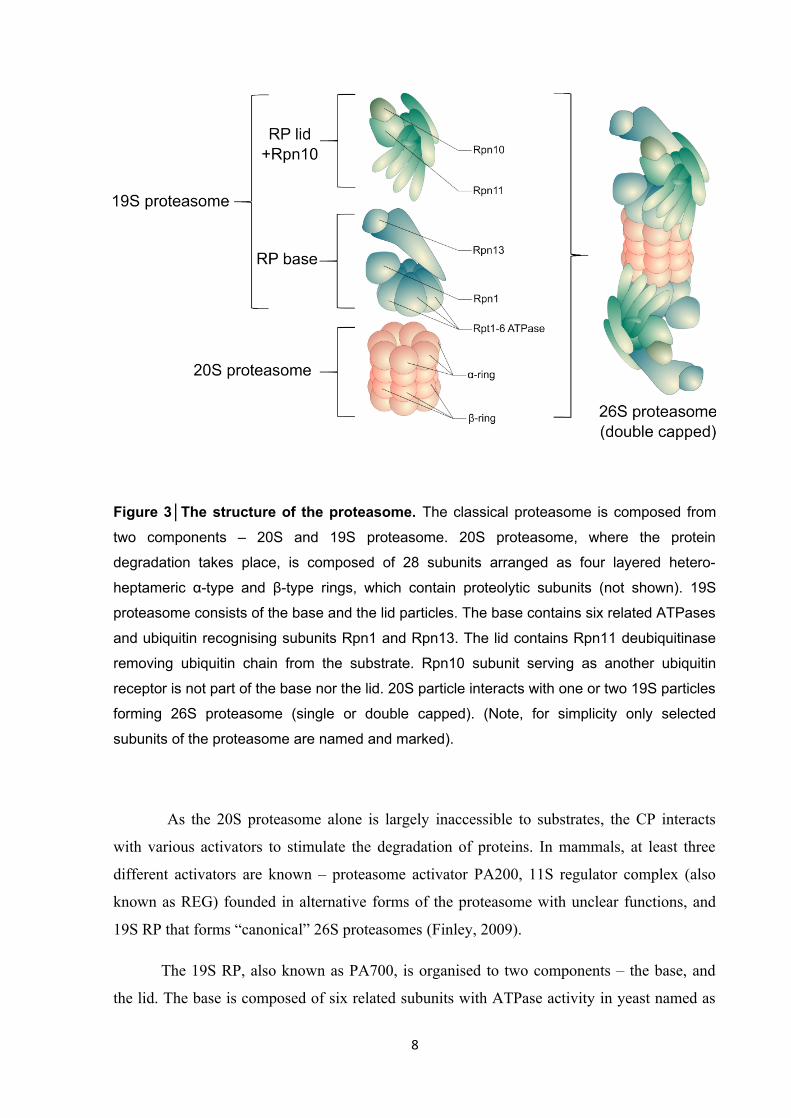
Figure 3│The structure of the proteasome. The classical proteasome is composed from
two components – 20S and 19S proteasome. 20S proteasome, where the protein
degradation takes place, is composed of 28 subunits arranged as four layered hetero-
heptameric α-type and β-type rings, which contain proteolytic subunits (not shown). 19S
proteasome consists of the base and the lid particles. The base contains six related ATPases
and ubiquitin recognising subunits Rpn1 and Rpn13. The lid contains Rpn11 deubiquitinase
removing ubiquitin chain from the substrate. Rpn10 subunit serving as another ubiquitin
receptor is not part of the base nor the lid. 20S particle interacts with one or two 19S particles
forming 26S proteasome (single or double capped). (Note, for simplicity only selected
subunits of the proteasome are named and marked).
As the 20S proteasome alone is largely inaccessible to substrates, the CP interacts
with various activators to stimulate the degradation of proteins. In mammals, at least three
different activators are known – proteasome activator PA200, 11S regulator complex (also
known as REG) founded in alternative forms of the proteasome with unclear functions, and
19S RP that forms “canonical” 26S proteasomes (Finley, 2009).
The 19S RP, also known as PA700, is organised to two components – the base, and
the lid. The base is composed of six related subunits with ATPase activity in yeast named as
8

Rpt1-6 (Regulatory particle triple-A protein) (in humans known as PSMC2,-1,-4,-6,-3,-5) that
belongs to family of AAA (ATPases Associated with diverse cellular Activities) ATP-ases
and form a ring directly interacting with outer ring of α-type subunits of the CP. The base also
contains three non-ATPase proteins – Rpn1, Rpn2, Rpn13 (Regulatory particle non-ATPase)
(in humans known as PSMD2, PSMD1 and ADRM1), that contains several binding sites for
ubiquitin-binding proteins or ubiquitin. The lid is composed of six Rpn subunits (Rpn3,-5,-6,-
7,-9,-12, in humans PSMD3,-12,-11,-6,-13,-8) with scaffolding function and two proteins
(Rpn8 and Rpn11, in humans PDMD7 and PSMD14) that cooperate during deubiquitination
of the substrate. Rpn10 (PSMD4) cofactor containing ubiquitin binding domain is not
assigned to the base nor the lid and it is assumed to mediate the bridge between both sub-
complexes (Bard et al., 2018; Lander et al., 2012). The proteasome contains three DUB –
Rpn11 (PSMD14 or POH1) necessary for the function of the proteasome located in the lid
(Yao and Cohen, 2002) and two other known as USP14 (Ubiquitin-specific protease 14) and
UCH37 (Ubiquitin C-terminal hydrolase 37) stably associated with the base that have rather
regulatory function (Lee et al., 2016).
It was previously thought that the ubiquitination solely determinates the fate and rate
of substrate degradation, nevertheless recent discoveries demonstrated that proteasome is not
just a passive machine for destruction, but rather highly organised and tightly regulated
complex defining the fate of proteins, as the degradation of proteins by the proteasome
involves several closely controlled steps (Collins and Goldberg, 2017).
First step involves recognition of ubiquitinated substrate. Initial binding of conjugates
is mediated mainly by Rpn10 and Rpn13 via UIM (Ubiquitin-interactive domain) domain,
and PRU (Pleckstrin-like receptor for ubiquitin) domain, respectively (Yu and Matouschek,
2017). Recently, new ubiquitin binding site was observed in Rpn1 explaining puzzling
observations that proteasomes defective in both Rpn10 and Rpn13 still readily interact with
ubiquitin conjugates (Shi et al., 2016). Ubiquitin conjugates bind proteasome with very high
affinity, but potentially reversibly as the proteasomal DUBs USP14 and UCH37 remove
ubiquitin from the substrate actually leading to its dissociation from the proteasome. On the
other hand, the interaction of ubiquitinted protein with USP14 and UCH37 leads to a major
conformational change enhancing substrate interaction with ATPases located in the base
promoting substrate entry to the proteasome. The competition between these two opposite
processes likely determines the fate of the substrates (Bard et al., 2018). Contrary to the early
ideas, the proteasome is not so selective for the type and length of ubiquitin chains. In vitro,
9

even monoubiquitinated protein could be degraded, or K63 linked conjugates are degraded
similarly like K48-marked proteins. In cells, the situation is likely more complicated as many
competing factors determine the fate of ubiquitinated substrate. For example, K63 conjugates
are efficiently captured by ESCORT system preventing its degradation by the proteasome
(Nathan et al., 2013); monoubiquitinated substrates interact with the proteasome less tightly
than substrates with long or branched chains, which are degraded very efficiently (Meyer and
Rape, 2014).
The transition from initial binding to tight binding is dependent on ATP and partially
unfolded or loosely folded region of the substrate, which activates ATPases and substrate
transport into proteasome. The sequence of further steps in not fully clear, but involves
deubiquitination of the substrate, unfolding and transport to proteolytic chamber (de la Peña et
al., 2018). Rpn11 (PSMD14 or POH1) is deubiquitinase located near the entry to proteasome
gate, which contains JAMM/MPN (Jab1/MPN/Mov34 metalloenzyme / Mpr1, Pad1 N-
terminal) domain binding Zn+2. This metalloprotease is essential for the degradation of
substrates as it removes ubiquitin chain close to the substrate enabling its entry to the channel
(Verma et al., 2002). Here, ATPase subunits Rpn1-Rpn6 unfold and transport the substrate to
the proteolytic chamber through the gate, which is opened by Rpt2, Rpt3, and Rpt5 proteins.
By the activity of β1, β2, and β5 subunits, substrate is degraded into small peptides
subsequently released to cytosol, where majority of them are digested by peptidases or used as
precursors for antigen presentation by the MHC-class I (Major histocompatibility complex)
molecules (Yu and Matouschek, 2017).
1.4 Protein quality control
Despite enormous quantity and rate of protein translation, the vast majority proteins do
not contain errors in their amino acid sequence as only one in 10 000 amino acids is
misincorporated. However, in the light of huge amount of proteome, millions of aberrant
proteins are produced by the cell. Additionally, the proper folding and maintaining of correct
structure of proteins is even far more challenging (Wolff et al., 2014). Proteins are constantly
exposed to many environmental stressors including oxygen radicals, heat, or metal ions. As
damaged, misfolded or aggregated proteins represent serious threat, documented by many
diseases associated with their accumulation, the cells evolved sophisticated multi-layered
system called protein quality control (PQC) for protection. PQC consists of systems for
10

identification of aberrant proteins, their refolding or, when necessary, for their destruction.
Therefore, UPS is one of the central components of PQC in cytoplasm, ER, nucleus or
mitochondria (Amm et al., 2014).
At least one third of all cellular proteins is translated on ER, which represents a key
hub of PQC and a sensor of protein stress. Despite the existence of many protein-folding
chaperones, it is estimated that more than one third of all proteins on ER does not fold
properly and must be removed by ER-associated degradation (ERAD) pathway that employ
the ubiquitination of unfolded proteins and their degradation by the proteasome (Christianson
and Ye, 2014). In case the folding or degradative capacity of the cell is overwhelmed or
impaired, accumulated defective proteins lead to ER-stress triggering so called Unfolded
Protein Response (UPR) (Hetz, 2012).
Three main sensors of UPR have been identified - IRE1α (Endoribonuclease inositol-
requiring enzyme 1-alpha), PERK (Protein kinase RNA-like endoplasmic reticulum kinase),
and ATF6α (Activating transcription factor 6 alpha) (Hetz et al., 2015). The main ER
chaperone, protein BIP (also known as GRP78 – glucose-regulated protein 78), plays
important role in the activation of the sensors. Under normal conditions, it binds the sensors,
but when unfolded proteins accumulate, BIP is sequestered and dissociates from the sensors
leading to IRE1α and PERK oligomerization and autophosphorylation, and to ATF6α export
to Golgi apparatus and nucleus, where it induces expression of target genes (Walter and Ron,
2011). Activated PERK phosphorylates translation incitation factor eIF2α (eukaryotic
translation initiation factor 2 alpha), which in turn disassembles polysomes and decreases total
protein synthesis to reduce the load of new proteins, and concomitantly, to allow preferential
translation of ATF4 (activating transcription factor 4), a transcription factor regulating genes
involved in protein folding, autophagy and apoptosis. Additionally, activated IRE1α induces
alternative splicing of transcription factor Xbp1 (X-box binding protein 1), which controls
mainly genes involved in protein folding (Fig. 4). Collectively, UPR activates proteins
helping the cell to cope with aberrant proteins, attenuate global protein synthesis a thus
promote the cell survival (Hetz, 2012). However, when unmitigated, the capacity of UPR is
overwhelmed and terminal UPR is triggered, which involves for instance protein CHOP
(CCAAT-enhancer-binding protein homologous protein), which serves as an activator of
apoptosis (Lu et al., 2014).
11

Figure 4│The Unfolded protein response (UPR). Three main branches of the UPR are
shown. Unfolded proteins in ER-lumen sequester BIP protein leading to activation of the
UPR sensors – PERK, IREα and ATF6. Autophosphorylated PERK phosphorylates eIF2α,
which in turn decreases total protein synthesis and activates ATF4 transcription factor
regulating UPR target genes. Activated IRE1α induces alternative spicing of Xbp1, resulting
in production of Xbp1s form acting as a transcription factor. Activated ATF6α is exported to
Golgi apparatus, where it is cleaved to form the active transcription factor translocating into
the nucleus to induce UPR-related genes.
12

To assist with folding of de novo synthetized proteins and to maintain proper structure
of other proteins, cells evolved numerous chaperones that are vital part of PQC. Many of the
chaperones belongs to the family of heat shock proteins (HSP), which have critical function in
preventing protein unfolding and aggregation especially under various stress conditions
(Sontag et al., 2017). Imbalance of protein homeostasis, accumulation of aberrant proteins or
formation of protein aggregates trigger intense cellular response known as Heat shock
response (HSR), as heat stress is common but not the only inductor of protein damage and
unfolding (Åkerfelt et al., 2010). HSR is characterised by rapid activation of roughly 100
genes in human cells, majority of them belonging to chaperones or proteins involved in
degradation, metabolism or DNA repair (Richter et al., 2010). HSR is regulated mainly by
transcription factors, among them HSF1 (Heat shock factor 1) is assumed as the critical one.
Under normal conditions, it is sequestered in the cytoplasm in inhibitory complex with HSP70
and HSP90 chaperones. Upon accumulation of aberrant proteins, HSP proteins dissociate and
liberated HSF1 is activated leading to its oligomerization (formation of trimeric form),
phosphorylation and translocation to nucleus, where it binds so called heat shock elements on
DNA (after binding to DNA HSF1 can be detected as typical nuclear foci called as nuclear
stress bodies) and triggers transcription of the target genes (Gomez-Pastor et al., 2017) (Fig.
5). Among them, genes of HSP70 family belong to the most important. Under physiological
conditions involved mainly in folding of de novo translated proteins, stress-overexpressed
HSP70 helps to prevent aggregation of unfolded proteins or even refold the aggregated one,
and assist with sequestration or degradation of protein aggregates, occupying a critical role of
cell response to stress conditions (Kim et al., 2013).
13

Figure 5│The activation of Heat shock factor 1 (HSF1). In non-stressed cells, HSF1 is
sequestered in the cytoplasm in inhibitory complex with Hsp70 and Hsp90 chaperones. Upon
stress conditions, accumulated aberrant proteins unbind Hsp70/Hsp90 chaperones from
HSF1, leading to liberation of HSF1, its phosphorylation, oligomerization and translocation to
the nucleus to induce the expression of target genes.
14

1.5 The role of the p97 complex
Intensive research about how proteins are degraded revealed the existence of other
layers of UPS except the ubiquitination and the proteasome, which are important or even
indispensable for protein degradation. For instance, several shuttling factors have been
discovered, which deliver ubiquitinated proteins to the proteasome or interact with the
proteasome to assist with the recognition of substrates. These factors contain UBL-UBA
(Ubiquitin-like and Ubiquitin-associated) domains enabling them to simultaneously interact
with both ubiquitinated proteins via UBA domain and proteasome through UBL domain. In
humans, the members of these factors include RAD23A/B, UBQLN1-4 (also known as
ubiquilin family) or DDI1/2 (Saeki, 2017). While the importance and exact roles of these
factors remained to be established, the physiological relevance is underlined by the fact that
mutations in UBQLN2 are associated with sever neurodegenerative disorder known as
amyotrophic-lateral sclerosis (Deng et al., 2011).
Another protein required for degradation of many substrates is p97, also known as
VCP (Valosin-containing protein) or Cdc48 in yeast. This protein is far more than just
shuttling factor, as growing evidence suggest p97 is involved in almost every aspect of cell
biology connected to the ubiquitination. Similarly to the base of proteasome, p97 belongs to
the family of hexameric AAA ATPases. It uses energy from ATP hydrolysis to perform many
key steps required for degradation and processing of ubiquitinated proteins – unfolding and
remodelling and segregase activity involving segregation of the ubiquitinated proteins from
various complexes or chromatin, or their extraction from membranes before the actual
transport to the proteasome (Meyer et al., 2012) (Fig. 6).
15

Figure 6│The cellular functions of the p97 complex. p97 segregase in association with
diverse cofactors operates in many essential processes. Together with NPL4-UFD1
heterodimer, p97 participates in ER-associated degradation, mitochondria-, chromatin-,
ribosome-associated degradation, or in protein unfolding prior proteasomal degradation. In
co-operation with p47 cofactor, p97 regulates also the biogenesis of Golgi apparatus, and
with UBXD1 adaptor, p97 is involved in autophagy and lysosome function. (Note, only
selected cellular activities of p97 are shown).
16

p97 contains N-terminal domain, two ATPase domains (D1 and D2) and flexible C-
terminal tail. In the active hexameric form, ATPase domains form two stacked rings with the
central pore. The exact functions of these domains is still not fully clear, but it seems that N-
terminal domain is important for the interaction with client proteins and cofactors, while
driving force is generated probably primarily by D2 (Bebeacua et al., 2012). Recently, a
molecular mechanism of substrate processing by p97 was revealed (Bodnar and Rapoport,
2017). The model suggests that ubiquitinated substrate is transferred through central pore by
the force produced by D2 domain. During the movement, the protein is unfolded, and with the
assistance of D1 domain and associated DUB it is partially deubiquitinated and released.
p97 segregase plays an essential role in many aspects of cellular physiology and to
perform a such variety of tasks, is interacts with a plethora of cofactors. Many of these
cofactors contain binding site for ubiquitin and mediate the interaction of client proteins with
p97, which itself has low affinity for ubiquitin; others are DUB enzymes or E3 ligases that
rearrange ubiquitin chains attached to the substrates. About 30 different adaptors of p97 have
been discovered according to a study published in 2016 (Xue et al., 2016).
The best studied cofactors are NPL4 (Nuclear protein localisation protein 4 homolog,
also known as NPLOC4) and UFD1 (Ubiquitin fusion degradation 1), which participate in
many functions of p97 including ERAD or chromatin associated degradation. NPL4 and
UFD1 acts as heterodimer, which interacts with N-terminal domain of p97 hexamer, and
recruits ubiquitinated client proteins (Meyer et al., 2002). The X-ray structure of NPL4 is
unavailable until now, most likely because of high structural flexibility of the protein
hampering the attempts to get it in crystal form and perform detailed X-ray analysis (Isaacson
et al., 2007). Very recently, a high resolution cryo-EM structure of NPL4/p97 complex from
thermophilic fungus Chaetomium thermophilum, which is presumably less sterically flexible,
was published (Bodnar et al., 2018). It complements previous cryo-EM and biochemical
studies (Bebeacua et al., 2012; Bruderer et al., 2004; Isaacson et al., 2007; Meyer et al., 2000;
Pye et al., 2007) revealing that only one UFD1-NPL4 (UN) heterodimer interacts with p97
hexamer via UBX-L domain (Ubiquitin regulatory X-like) domain on NPL4 and via SHP
(BS1, Binding segment 1) motive located on UFD1. Apart from UBX-L domain, NPL4 also
contain putative zinc finger domain (put-ZF) followed by MPN domain similar to that found
in POH1 (Rpn11) subunit of the proteasome. Last, C-terminal domain contain second zinc
finger domain called NPL4 zinc-finger (NZF), which is most likely involved in recognition of
the ubiquitinated proteins preferentially linked with K48 chains (Meyer et al., 2002). In
17

humans, NPL4 is expressed as two main isoforms, of which the “canonical” contains all
domains, while the “alternative” lacks C-terminal NZF domain, which is substituted by
another longer sequence making this variant little bit larger (visible on western blot as shifted
second band) (Meyer et al., 2000). The physiological function of the alternative isoform is
unknown. Besides SHP motive, UFD1 also contains UT3 domain recognising ubiquitin and
C-terminal UT6 domain mediating the interaction with NPL4 partner (Bruderer et al., 2004).
The exact site on NPL4, which UT6 interacts with, is not known, but according the study
using C. thermophilum NPL4, it is probably located near putative zinc-finger and MPN
domains (Bodnar et al., 2018); however, due to significant differences in sequence, it is
questionable if these results are relevant also for human orthologues.
Apart from UN, p97 interacts with p47 protein, other core cofactor. p47 belongs to the
family of UBA-UBX (Ubiquitin-associated; Ubiquitin regulatory X) domains containing
proteins enabling p47 to simultaneously recognise ubiquitinated proteins via N-terminal UBA
domain and p97 by C-terminal UBX domain. In contrast to UN, three p47 molecules bind p97
hexamer. p47 preferentially interacts with K63 linked ubiquitin chains enabling p97/p47
complex to regulate K63 chain dependent processes such as Golgi apparatus formation or
membrane fusion (Bruderer et al., 2004; Kondo et al., 1997).
p97 interacts with UN or p47 in a mutually exclusive manner, so these cofactors are
sometimes named as “core” adaptors. Apart of them, p97 binds plethora of other proteins,
some of them, such as FAF1 or UBXD7, seems to have preference to bind p97/UN complex
forming kind of secondary complex (Hänzelmann et al., 2011). On the other hands, Vms1
(VCP/Cdc48-associated mitochondrial stress-responsive 1) protein replaces UFD1 in UN
dimer and forms stable p97/NPL4/Vms1 complex, which is required for mitochondria-
associated degradation (Heo et al., 2010).
While the majority of p97 cofactors, represented mainly by UBA-UBX or UBX-L
protein families, mediates the interaction with ubiquitinated client proteins, other p97-
interacting proteins have different roles. For instance, p97 binds several E3-ubiquitin ligases
and DUB enzymes that plays a role in many processes. Two main E3 ligases central for
ERAD, HRD1 (HMG-CoA Reductase Degradation 1 Homolog) and gp78 (also known as
AMFR-Autocrine Motility Factor Receptor), both interact with p97 (Stach and Freemont,
2017), as well as ligases of CRL family (Alexandru et al., 2008). Additionally, p97 is found in
complex with several DUBs, including YOD1 (also known as OTU1), and ATX3 (Ataxin-3)
18

both involved in ERAD (Ernst et al., 2009; Wang et al., 2006), or VCIP135 (Valosin-
containing protein/p47 complex-interacting protein, p135), a DUB essential for Golgi
apparatus assembly (Uchiyama et al., 2002). Finally, there are also proteins with other
activities that interact with C-terminal domain of p97, including PNGase (Peptite:N
glycanase) involved in ERAD (Stach and Freemont, 2017), or PLAA (Phospholipase A-2-
activating protein) regulating clearance of damaged lysosomes (Papadopoulos et al., 2017).
Such plurality, diversity, and versatility of cofactors makes p97 far more than just a shuttling
factor upstream the proteasome, but rather another complex layer regulating the fate of
growing number of proteins and cellular processes.
Among many processes in which p97 plays a role, ERAD is one of the most studied.
Maturation-defective luminal and membrane proteins are dedicated to degradation in the
proteasome, an action dependent on p97 activity. Defective proteins must be first translocated
trough ER-membrane, and once exposed to cytoplasm, they are ubiquitinated and recognised
by p97 in complex with UN. p97 ATPase provides pulling force most likely required for final
dislocation of ubiquitinated substrate and its release to cytoplasm where it is available to
proteasomes (Stein et al., 2014). ERAD is multi-step process relying on coordinated action of
proteins with different functions, thus p97 interacts with many proteins apart from core UN
cofactors, including FAF1, UBXD8 recognising ubiquitin, with E3 ligases gp79 or HRD1,
and also with DUBs YOD1 or ATX3 (Christianson and Ye, 2014).
Analogously to its role in ERAD, p97 is also critical for degradation of proteins
located in outer mitochondrial membrane (OMM) during a process called mitochondria-
associated degradation (MAD). Again, p97 mediates the extraction of ubiquitinated proteins
out of OMM to facilitate their degradation by cytosolic proteasomes (Heo et al., 2010). The
close relationship between ERAD and MAD was further demonstrated quite recently. It was
shown that pro-apoptotic protein BOK (Bcl2 ovarian killer) regulating mitochondrial outer
membrane permeabilisation, a critical event of intrinsic apoptosis, is degraded by classical
ERAD components, including E3 ligase gp78 and p97 (Llambi et al., 2016).
p97 also act as a segregase using the pulling force to extract substrates from other
complexes or structures such as chromatin. Growing body of evidence suggests an essential
role of p97 in extraction and degradation of various proteins associated with chromatin
(Dantuma and Hoppe, 2012). The first such identified substrate was Aurora B kinase, which is
ubiquitinated and unloaded from the chromatin by p97-UN complex at the end of the mitosis
19

(Ramadan et al., 2007). p97-UN also mediates the degradation of Rpb1, the largest RNA
polymerase II subunit, upon transcriptional stalling or UV damage (Verma et al., 2011).
Additionally, according to published studies, p97 is recruited to the sites of DNA damage
(Meerang et al., 2011) and regulates extraction of several key players acting on double strand
breaks including L3MBTL1 (Acs et al., 2011), or presumably Ku70 and Ku80 (van den Boom
et al., 2016). Moreover, several critical steps required for proper replication are dependent on
p97-UN complex, comprising extraction of CDT1 replication origin licencing factor at the
onset of S-phase (Raman et al., 2011), or removal of MCM7 helicase subunit, when the
replication is completed (Moreno et al., 2014).
p97 complex is not linked only to the proteasomal degradation, but also to autophagy-
lysosomal degradation, further extending its complex role in cellular physiology. It has been
shown that p97 together with UBXD1 cofactor regulates endolysosomal sorting targeting the
proteins to degradation in lysosomes (Ritz et al., 2011). p97 is also recruited to damaged
lysosomes, where it removes K48 ubiquitin linked proteins to drive degradation of impaired
lysosomes via a process called lysophagy (Papadopoulos et al., 2017). Similarly, p97
mediates a degradation of damaged mitochondria trough mitophagy (Tanaka et al., 2010),
degradation of cellular stress granules (Buchan et al., 2013), and processing of various
cytoplasmic and nuclear aggregated proteins (Fujita et al., 2013; Gallagher et al., 2014;
Kitami et al., 2006; Seguin et al., 2014).
Moreover, many proteins involved in regulation of cell cycle or signalling are
substrates of p97. For instance, IκB-α (Inhibitor of nuclear factor kappa B), a critical negative
regulator of NF-κB (Nuclear factor kappa B) transcription factor, is segregated from NF-κB
by p97-UN complex, which enables IκB-α degradation by the proteasome and subsequent
activation of NF-κB (Li et al., 2014). Other protein, which degradation is dependent on p97-
UN complex, is Cdc25A (Cell division cycle 25 homolog A), a phosphatase regulating G2/M
checkpoint, which must be efficiently degraded upon DNA damage to ensure the cell will not
enter mitosis before the damage is repaired (Riemer et al., 2014). Further, the regulation of
HIF1α (Hypoxia inducible factor 1 alpha) transcription factor well illustrates the complexity
of the p97 system. Together with UBXD7 cofactor, p97 interacts with ubiquitinated HIF1α,
and upon depletion of p97, HIF1α accumulates as a high molecular mass species
corresponding to ubiquitinated HIF1α. However, the amount of accumulated HIF1α is far
lower than those observed after inhibition of the proteasome, suggesting that only a small
subset of the HIF1α is degraded in p97 dependent manner (Alexandru et al., 2008). This
20

indicates that perhaps p97 is involved only in degradation of protein molecules in some
particular states, in case of HIF1α presumably in complex with HIF1β or DNA and
transcription machinery (Alexandru et al., 2008; Bandau et al., 2012).
p97 complex does not need to be necessarily connected only to protein degradation, as
exemplified by transcription factor NRF1 (also known as NFE2L1 - Nuclear factor erythroid
derived 2-related factor 1, or TCF11 in humans). Cells evolved specialised response to
proteasome stress or malfunction involving rapid induction on new proteasome subunits to
compensate the insufficiency. This bounce-back response involves NRF1, a close relative of
well-known NRF2 transcription factor, which is involved in the anti-oxidant response (Koch
et al., 2011). The level of NRF1 is closely regulated mainly on the side of protein stability.
Under normal conditions, NRF1 is synthetized as luminal protein on ER, which is, however,
quickly translocated to cytosolic side of ER by p97 and presented for degradation to
proteasomes. Thus, in non-stressed cells, the basal level of NRF1 is very low. Conversely,
when the proteasome is inhibited or overloaded, translocated NRF1 cannot be degraded, but it
is processed by a protease and the cleaved product translocates into the nucleus, where it
induces expression of all proteasome subunits, and also p97/NPL4/UFD1 proteins
(Radhakrishnan et al., 2010, 2014; Sha and Goldberg, 2014; Steffen et al., 2010). The identity
of the protease responsible for the cleavage and release of NRF1 is still disputable, as some
argue it is actually the proteasome itself (Sha and Goldberg, 2014), others suggest it is
aspartyl protease DDI2 (DNA-damage inducible 1 homolog 2) (Koizumi et al., 2016;
Lehrbach and Ruvkun, 2016). It is clear, however, that upon proteasome inhibition, NRF1
accumulates as two different species – the unprocessed form of molecular weight
approximately 120 kDa (usually marked as p120), and the active cleaved form of 110 kDa
(known as p110). As the translocation of NRF1 is dependent on p97, the inhibition of p97
results in accumulation of only uncleaved p120 form of NRF1 prohibiting induction of
proteasome subunits (Le Moigne et al., 2017; Radhakrishnan et al., 2014). The physiological
role of proteasome regulation by NRF1, and its potential role in resistance to proteasome
inhibitors, is unclear so far, however, it has been shown that increased protein synthesis
induced by growth factors or feeding elevated also degradative capacity via NRF1 (Zhang et
al., 2014).
Taken together, p97 complex emerges as an important factor touching almost every
aspect of cellular physiology, which has also numerous implications for tumour biology and
therapeutic interventions.
21

1.6 The role of UPS in tumour development and treatment
Cancer cells harbour thousands of genetic alternations in their genomes including
large rearrangements, amplifications, deletions, and translocations, as well as numerous point
mutations. While a vast majority of these alternations is just accidental, some of them, such as
mutations of certain receptors or kinases, drives cancer progression and cancer cells are
highly dependent on their function, a phenomenon known as oncogene addiction (Weinstein,
2002). However, as the physiology of cancer cells is largely altered compared to their normal
counterparts, malignant cells are also highly dependent on many stress-supporting pathways
maintained by genes that are not classical oncogenes. Such dependency is called “non-
oncogene addiction” (Solimini et al., 2007).
While many responses supporting cancer growth and survival are known, pathways
regulating protein degradation and PQC systems are among the most important. Due to
numerous genetic, epigenetic and transcriptional alternations, cancer cell relay more on
mechanisms of proteostasis. These alternations likely challenge folding and degradative
capacity of cells (Deshaies, 2014). To cope with such stress, cancer cells activate supporting
pathways involving chaperones, UPS or autophagy. Many components of UPR response,
including BIP, XBP1 or ATF6, are overexpressed in several solid and haematological
malignancies; often correlate with progression and poor prognosis and have direct impact on
tumour growth (Wang and Kaufman, 2014). Moreover, protein chaperones and HSF1, a
master regulator of HSR, are upregulated in several cancers (Wu et al., 2017), and are
required for their growth (Fok et al., 2018; Mendillo et al., 2012; Trepel et al., 2010).
Similarly, the proteasome and p97, as critical components of UPS, are overexpressed in many
cancers, including malignancies of breast, prostate, pancreas, liver, lung, or colon (Cui et al.,
2015; Nakahara et al.; Petrocca et al., 2013; Tsujimoto et al., 2004; Valle et al., 2011;
Yamamoto et al., 2004b, 2004c, 2004a, 2005), and usually correlates with invasiveness and
poor prognosis.
The deregulation of UPS in cancer cells is not limited to their general demand to
degrade multitude of damaged, unfolded or unwanted proteins, but includes also control of
levels of specific proteins involved in signalling and cell-cycle regulation. Tumour suppressor
protein p53 serves as an illustrating example. While mutated or lost in approximately half of
all tumours, the second half of tumours still contains wild-type p53, which is relatively
22

frequently deregulated by increased degradation (Mandinova and Lee, 2011). For instance, in
cervical carcinoma caused by HPV (Human papilloma virus) infection, E6 oncogenic viral
protein induce rapid degradation of wt p53, as it triggers the interaction of p53 with E6AP (E6
associated protein) E3 ligase, which ubiquitinates p53 and commits it for degradation by the
proteasome (Martinez-Zapien et al., 2016). The situation in cervical carcinoma is rather
exception, as E6AP is not physiological regulator of p53, and the major E3 ligase regulating
p53 is MDM2 (Hock and Vousden, 2014). Under normal conditions, p53 is constantly
ubiquitinated by MDM2 and degraded by the proteasome, but various cellular stressors
abolish the MDM2/p53 interaction leading to p53 stabilization and activation of downstream
effects including cell-cycle arrest, senescence or cell death (Hock and Vousden, 2014).
However, MDM2 ligase is overexpressed in significant percentage of tumours, leading to
inactivation of wt p53 and inaccurate cellular response to stress (Wade et al., 2013). In sharp
contrast to wt p53, various mutant forms of p53 are stable and frequently found in high levels
in cancer tissues. While the explanation is not fully known, it is well accepted that MDM2
interacts with p53 mutant less tightly, and mut p53 is less efficiently ubiquitinated (Yue et al.,
2017). Moreover, mut p53 is stabilised by interaction with HSPs or histone deacetylases,
underwriting to high levels of mut p53, which probably promotes cancer growth, as gain-of
functions of mut p53 are known to contributing to malignant progression (Muller and
Vousden, 2014; Wiech et al., 2012).
Due to high dependency of cancer cells on protein degradation pathways, it is not
surprising that targeting UPS represents promising approach for the treatment of cancer,
especially for tumour types with secretory phenotype, such as neuroendocrine tumours,
prostate cancer, but mainly certain hematologic malignancies, particularly multiple myeloma
(Deshaies, 2014). Myeloma, as a malignancy originating from plasma cells producing
immunoglobulins, is characteristic by the high levels of paraprotein, an immunoglobulin
produced by a single cell clone in very high levels (Morgan et al., 2012). This secretory
phenotype associated with extremely high levels of proteosynthesis and increased endogenous
UPR stress is the likely explanation, why multiple myeloma is so far the most responsive
cancer to inhibition of UPS, as illustrated by bortezomib, first-in-class proteasome inhibitor,
which inhibits CT-like, and partially other proteolytic activities of the 20S proteasome
(Moreau et al., 2012).
The development of bortezomib is an example of very effective, fast, and fruitful
translational research based on collaboration of small group of scientist from academia and
23

private sector around Alfred L. Goldberg at Harvard Medical School. Originally, he intended
to study proteasome inhibitors as a potential drug for muscle wasting, which occurs upon
disease (e.g. cancer) or aging. For this purpose, he established biotech company MyoGenics,
and in cooperation with chemist J. Adams, developed the most important inhibitors of
proteasome such as MG-132 or bortezomib (MG-341) in less than one year solely based on
knowledge of substrate specificity of the proteasome, without any screening efforts of random
chemical libraries. In sharp contrast to usual practice, proteasome inhibitors were freely
distributed to universities for academic research, yielding many discoveries about importance
of the proteasome for cell cycle, apoptosis or cancer (Goldberg, 2011). For instance, the
proteasome was shown to be essential regulator of NF-κB, crucial transcription factor
regulating inflammation or carcinogenesis (Palombella et al., 1994). These discoveries shifted
the focus of company closer to cancer, however, as the proteasome was not accepted as a
potential target for cancer treatment, the MyoGenics (later renamed as ProScript) lost
financial support, and was sold for ridiculous price to company Millennium Pharmaceuticals
as a rather not profitable project. Yet, encouraging results from various xenograft studies
conducted by National Cancer Institute convinced Millennium Pharmaceuticals to invest to
small clinical trial phase I involving all cancer types (Goldberg, 2012). By serendipity, one
patient entering the trial had multiple myeloma, not so common cancer, and this patient
showed a complete remission after bortezomib (Goldberg, 2012; Orlowski et al., 2002). Such
promising result motivated to run phase II trial with multiple myeloma patients (Richardson et
al., 2003). Bortezomib was very effective against this type of cancer, for which no adequate
treatment was available at that time, leading to FDA approval of bortezomib (commercial
name Velcade) based just on phase II clinical trials (Kane, 2003).
Currently, bortezomib is approved as first line therapy for multiple myeloma or
mantle-cell lymphoma, and in clinical practice it is used to treat Waldenström’s
macroglobulinaemia, a disease characterised by high production of immunoglobulins
(Manasanch and Orlowski, 2017). Despite extreme effort (bortezomib has been tested in more
than 700 clinical trials) and promising preclinical results, its activity is limited only to few
cancer types, challenging the theoretical concept that cancer cells in general are more
dependent on UPS, and more broadly on PQC, and raising question what makes multiple
myeloma so exceptionally sensitive to proteasome inhibition (Deshaies, 2014).
The original explanation based on the inhibition of NF-κB transcription factor has
been questioned by several contradictory observations (Hideshima et al., 2002, 2009), and
24

nowadays it is more accepted that the sensitivity to bortezomib is dominantly determined by
proteotoxic stress, e.g. when the capacity of cells to handle and degrade unwanted proteins is
overcome (Bianchi et al., 2009). In multiple myeloma, the level of endogenous stress is so
high, that only partial and temporal inhibition of the proteasome, which is achieved in the
clinic, is sufficient to activate strong UPR leading to cell death (Deshaies, 2014). This
explanation is further supported by many observations that bortezomib is very effective as a
treatment of several non-malignant conditions associated with plasma cells producing huge
amount of antibodies. In pre-clinical models, and even in clinical practice, bortezomib was
demonstrated as effective drug to combat some antibody-mediated autoimmune diseases, such
as systemic lupus erythematosus, myasthenia gravis or autoimmune haemolytic anaemia,
rheumatic arthritis or other non-cancerous monoclonal gammopathies (reviewed in Skrott and
Cvek, 2014). In general, based on the efficacy of bortezomib against such diseases, which
share the characteristic of a high-rate antibody production, it seems highly probable that the
excessive production of proteins, and thus a strong need to efficiently degrade the damaged
ones determines the sensitivity of certain cell types, including plasma and multiple myeloma
cells, to proteasome inhibition (reviewed in Skrott and Cvek, 2014).
Despite significant improvement of prognosis of multiple myeloma patients after
introduction of bortezomib into clinic, the acquired or intrinsic resistance considerably limits
its benefit and durable response (McConkey and Zhu, 2008). There are many hypotheses to
explain resistance to proteasome inhibition, including overexpression or mutation within the
critical 20S subunit PSMB5, which is the main binding site for bortezomib, and possess CT-
like proteolytic activity. Such mode of resistance is very common in cell culture models,
where the resistance is induced artificially (Balsas et al., 2012; Franke et al., 2012; Oerlemans
et al., 2008), however, all attempts to identify such mutations in multiple myeloma cells
obtained from patients primarily or secondary resistant to bertozemib therapy failed, as no
association between any variation within PSMB genes and response to bortezomib has been
identified (Lichter et al., 2012; Politou et al., 2006). As an alternative scenario, it has been
proposed that less differentiated myeloma cells secreting lower amount of immunoglobulins
may stand behind the resistance. Using patient tumour samples, Xbp1s, a mediator of UPR
and plasma cell maturation, has been shown to play a role in the resistance to bortezomib in
patients. Xbp1s negative cells representing multiple myeloma B cells or pre-plasmablasts
seem to survive therapeutic application of bortezomib and are enriched in samples obtained
from patients refractory to bortezomib. These cells express lower amounts of
25

immunoglobulins and shows lower UPR stress, suggesting decreased dependency on the UPR
pathway than Xbp1s positive plasma cells (Leung-Hagesteijn et al., 2013). However, only
small fraction of patients not responding to bortezomib have non-secretory myeloma,
suggesting that other mechanism of resistance must be employed (Manasanch and Orlowski,
2017). Recently, a case report of a multiple myeloma patient, which was followed over
several years during the cycles of treatment with proteasome inhibitors, revealed that point
mutations within PSMB5 may indeed be involved in resistance to bortezomib in a subset of
patients (Barrio et al., 2019). During the cycles of therapy based on proteasome inhibitors, the
status of genes coding for proteasome subunits was checked, and some cell clones harbouring
different mutation within PSMB5 were detected. These mutations span to bortezomib binding
pocked a makes cell resistant to proteasome inhibitors, confirming the clinical relevance of
such mechanism of resistance (Barrio et al., 2019).
The unanswered question remains, however, what stands behind non-responsiveness
of all solid tumours to bortezomib therapy. It is not clear if these tumours are intrinsically less
sensitive to the inhibition of proteasome or the clinically achievable partial and transient
inhibition by bortezomib is not strong enough to trigger cell death. Together with frequent
acquired resistance in multiple myeloma, the lack of effectivity in solid tumours motivated the
development of second generation of proteasome inhibitors with better pharmacokinetic
properties and different mode of action (Deshaies, 2014).
Carfilzomib (Kyprolis), the first example of such drugs, is irreversible inhibitor of
primarily CT-like activity of 20S. Early clinical trials revealed that carfilzomib is effective in
relapsed and refractory multiple myeloma patients, leading to FDA approval for this group of
patients (Kortuem and Stewart, 2013). Phase III study also demonstrated that carfilzomib
combined with dexamethasone is at least equally efficient as bortezomib plus dexamethasone,
and even it is superior in case of progression-free and overall survival (Dimopoulos et al.,
2016, 2017). However, the potential activity of carfilzomib in solid tumour is unclear so far.
The last FDA approved proteasome inhibitor, ixazomib (Ninlaro), is first orally
available inhibitor, again targeting CT-like activity of 20S, but reversibly. It is used in the
clinic in pre-treated multiple myeloma patients. Due to better pharmacokinetic properties, and
faster dissociation from the proteasome, it is hoped that ixazomib will show better penetration
and activity in tissues outside circulation (Kupperman et al., 2010), which is supported by
preclinical models revealing better activity in solid tumour compared to bortezomib
26

(Kupperman et al., 2010). The activity of ixazomib towards solid tumours is largely unknown
until now, as trials are still running. However, the results from phase I involving patients with
various solid tumours do not raise much expectations (Smith et al., 2015).
The proteasome and whole UPS provides several other potential targets appropriate
for cancer treatment besides 20S proteolytic activities (Fig. 7). The relevance of these targets
is so far based mostly on pre-clinical and experimental data, but yet first inhibitors entered
clinical trials. Targeting DUBs associated with the proteasome represent one of such
approaches. Molecule b-AP15 is supposed to inhibit DUBs UCHL5 and USP14 leading to
impairment of proteasome activity, accumulation of non-degraded proteins, induction of
apoptosis, and reduction of tumour growth (D’Arcy et al., 2011). POH1, a central DUB of
19S proteasome, represents also intriguing target, as revealed by recent series of papers
describing various POH1 inhibitors (Li et al., 2017, 2018). Despite the activity in vivo needs
to be established, these compounds potently inhibit the activity of the proteasome, activate
UPR and apoptosis. The activity of 19S proteasome can be also compromised by molecules
targeting ubiquitin receptors recognising substrates for degradation, as exemplified by
compound RA190 that covalently binds to receptor Rpn13 (Anchoori et al., 2013). RA190
treatment leads to accumulation of ubiquitinated proteins, triggers apoptosis, and reduces
tumour growth in several models (Anchoori et al., 2013; Song et al., 2016). At the opposite
side of the whole UPS pathway lies E1 ubiquitin activating enzyme UBE1 necessary for
ubiquitination of vast majority of proteins. As revealed quite recently, small molecule
MLN7243 specifically targeting UBE1 has noteworthy anti-cancer activities in preclinical
models (Hyer et al., 2018). UBE1 inhibition leads to depletion of ubiquitin conjugates,
impairment of signalling cascades, and activation of proteotoxic stress, collectively leading to
profound activity in vivo motivating first clinical trials (Hyer et al., 2018). However,
preliminary results regarding anti-cancer activity from phase I are not so encouraging, yet
further trials will be needed to reveal the potential of MLN7243 (Sarantopoulos et al., 2017).
27

Figure 7│The anti-cancer agents targeting components of UPS. UPS provides many
attractive targets for small molecules intended to treat cancer. First of them are already in
clinical use (in green), others entered clinical trials (in red), and the rest is in preclinical stage
(in black). E1 ubiquitin activating enzyme (UBE1) is specifically inhibited by MLN7243.
Enormous diversity of E3 ligases provides bottomless source of possible targets. Small
molecule nutlin-3 targets MDM2 E3-ligase regulating p53 turnover. ATPase activity of p97 is
targeted by first-in-class inhibitor CB-5083. Clinically used proteasome inhibitors bortezomib,
carfilzomib and ixazomib target primarily β5 subunit. 19S proteasome also provides
promising targets. Rpn11 deubiquitinase is inhibited by experimental compound capzimin,
and ubiquitin binding subunit Rpn13 by small molecule RA190.
28

Given the importance of p97 in many pathways associated with malignant
transformation it is not surprising that recently emerged p97 inhibitors represent highly
promising class of compounds. The first small molecule thought to interfering with p97
activity was Eeyarestatin I (EerI) identified ten years ago. EerI does not inhibit p97 directly,
but rather it interferes with p97 associated DUB ATX3, leading to accumulation of
ubiquitinated proteins, impaired degradation, and cell death in various cancer models
including bortezomib-resistant multiple myeloma cells (Wang et al., 2008, 2009). However,
as a ATPase, it might be more reasonable to target p97 via compounds inhibiting this domain.
The first such compound, DBeQ, was identified by the screen involving the measurement of
ATPase activity of p97, and cell-based degradation assay confirming the ability of compounds
to specifically inhibit only degradation dependent on p97 (Chou and Deshaies, 2011; Chou et
al., 2011). DBeQ impaired both ubiquitin-dependent and autophagy degradation pathways
leading to death of cancer cells. More potent and specific derivatives of DBeQ, compounds
ML240 and ML241 were further synthetized and characterised (Chou et al., 2013). In parallel,
different group identified new more potent covalent and reversible p97 inhibitors NMS-859
and NMS-873 by high-throughput screen (Magnaghi et al., 2013). Particularly the later one,
NMS-873, the first allosteric p97 inhibitor, was the best and the most thoroughly
characterised inhibitor at that time, and became a common tool to study p97 function in cells.
Similarly to DBeQ, NMS-873 impaired degradation of several proteins and activated UPR
leading to death of dozens of cancer cell lines. Interestingly, no correlation between the
toxicity of bortezomib and those of NMS-873 across the panel of cell lines was detected, and
NMS-873 was not preferentially toxic to multiple myeloma cells indicating very different
mode of action of p97 inhibitors compared to the inhibitors of the proteasome (Magnaghi et
al., 2013).
To move p97 inhibitors to the clinics, pharmaceutical company Cleave Biosciences
build on ML240 scaffold, and identified the lead p97 inhibitor, CB-5083, with suitable drug-
like properties ensuring adequate pharmacokinetic properties and oral administration
(Anderson et al., 2015). Highly selective and potent, CB-5083 specifically inhibits D2
ATPase domain of p97 leading to impairment of ERAD, accumulation of ubiquitinated
proteins, and activation of UPR, autophagy, and finally apoptosis. CB-5083 shows activity in
more than 300 cancer cell lines, and in several in vivo mouse models surpassing bortezomib,
carfilzomib, and ixazomib in activity against solid tumours. Again, no correlation of cell
responsiveness to bortezomib and CB-5083 was observed (Anderson et al., 2015). CB-5083
29

shows also remarkable activity against multiple myeloma models (Le Moigne et al., 2017)
motivating currently running phase I clinical trial (NCT02223598). Another phase I trial
involving patients with various solid and haematological malignancies is also ongoing
(NCT02243917).
Interestingly, it has been observed that various ATPase inhibitors have diverse
potency against p97 associated with different cofactors such as p47 or NPL4-UFD1 (Fang et
al., 2015; Gui et al., 2016), raising question if p97 inhibitors specifically targeting p97 with
certain cofactor could be developed in future. However, so far no such inhibitor exists nor a
compound inhibiting the main p97 adaptors.
1.7 Anti-cancer activity of disulfiram
With the approval of bortezomib for clinical use and the proteasome as an established
target for anti-cancer drugs, many compounds were tested for their effect on UPS, among
them a number of drugs approved for different purposes Such effort led to several interesting
discoveries including repeated observations that a drug disulfiram impairs the function of
UPS leading to death of cancer cells.
Disulfiram (commercial name Antabuse) is FDA-approved drug used to treat alcohol
abuse for more than 60 years. Its metabolites irreversibly inhibit ALDH (Aldehyde
dehydrogenase), mainly ALDH2, an enzyme critically involved in the metabolism of alcohol.
ALDH mediates the conversion of toxic acetaldehyde, the main metabolite of ethanol, to
acetic acid (Koppaka et al., 2012). When ALDH is inhibited by the metabolite of disulfiram,
the level of acetaldehyde increase dramatically upon alcohol consumption leading to
unpleasing reaction (in rare cases can be life threatening), which include sweeting, flushing,
respiratory difficulty, nausea, tachycardia, and hypotension, known as disulfiram-alcohol
reaction. Consequently, such adverse effects preclude alcohol use under disulfiram therapy
(Ehrenreich and Krampe, 2004).
Disulfiram, chemically tetraethylthiuram disulfide, belongs to the family of thiuram
disulfides, organic compounds used frequently in industry as rubber accelerators or pesticides.
The surprising anti-alcoholic properties of disulfiram were discovered trough two independent
accidental observations. First, in 1937, E. E. Williams, a plant physician working in a
chemical company producing tetramethylthiuram disulfide, a compound closely related to
30

disulfiram, observed that workers were unable to drink any alcohol, since even one beer
caused unpleasant reaction (Williams, 1937). Second, when searching for potential vermicide,
two Danish scientists E. Jacobsen and J. Hald tried disulfiram, a compound with scabiescide
properties. To evaluate potential side effect, Jacobsen first tested the drug on himself,
revealing that the drug is safe, however, when combined with alcohol, it causes very
unpleasant reaction (Hald and Jacobsen, 1948). Jacobsen and Hald as a researches connected
to pharmaceutical company, immediately recognized the potential of disulfiram in the
treatment of alcoholism. In 1949, just four years after the surprising initial observation,
disulfiram was approved in Sweden followed by other countries. Nowadays, disulfiram is
used by approximately 120 000 patients worldwide, of which significant number comes from
Denmark (Kragh, 2008).
First indications about potential anti-cancer activity of disulfiram comes form 70´s,
when disulfiram was repeatedly shown to suppress chemically or UV induced tumours of
various organs in mice (Black et al., 1978; Irving et al., 1983; Wattenberg, 1978). However, it
was not clear if it did because of its direct effect on cancer or rather because it reacted
chemically with carcinogens. More relevant signs of its anti-tumour activity were reported by
a study conducted by National Cancer Institute aiming to identify potential chemical
carcinogens, which involved also disulfiram, and its main metabolite diethyldithiocarbamate
(ditiocarb, DTC), as the compounds frequently used in the industry. The study evaluated the
occurrence of tumours after long term exposition of chemicals added to a diet of treated
animals. Unexpectedly, the addition of disulfiram to diet significantly reduced the incidence
of spontaneous tumours of breast, hypophysis, liver, pancreatic islets, thyroid and lymphomas
in mice or rats (Program, 1979b, 1979a). The interest about disulfiram anti-cancer properties
increased in late 90´s, when a direct effect on cancer cells were reported (Liu et al., 1998)
initiating the efforts to identify its mechanism of action in cancer cells resulting in hundreds
of publications. In recent years, around 30 scientific papers a year are published about
disulfiram connection to cancer.
From a clinical perspective, anti-cancer activity of disulfiram is also supported by
the evidence from patients. First case report describes full regression of metastatic breast
cancer in alcoholic women taking disulfiram (Lewison, 1977). Moreover, ditiocarb, the main
disulfiram metabolite, was in late 80´s and 90´s very popular as a suspected modulator of
immune response, and was successfully tested in HIV patients (Hersh et al., 1991), and
produced under the commercial name Imuthiol. These positive results motivated to test
31

ditiocarb in cancer patients too. In randomised, double-blinded phase II trial involving high
risk breast cancer patients, ditiocarb significantly prolonged both overall and disease-free
survival as an adjuvant therapy (Dufour et al., 1993). Additionally, in a small phase II clinical
trial assessing the addition of disulfiram to chemotherapy for the treatment of metastatic non-
small cell lung cancer, disulfiram significantly prolonged overall survival, with two long term
survivals in disulfiram group (Nechushtan et al., 2015). Disulfiram seems to be effective also
in other types of cancers, including melanoma or glioblastoma, as suggest intriguing case
reports (Brar et al., 2004; Karamanakos et al., 2017). Potential anti-cancer activity of
disulfiram is also supported by epidemiological evidence, which indicates that it could have
protective effect against breast, and prostate cancer (Askgaard et al., 2014). Currently,
disulfiram is tested in several clinical trials involving different tumour types, including
glioblastoma (NCT02678975, NCT01777919), breast (NCT03323346), and prostate
(NCT02963051).
The anti-cancer activity of disulfiram is explained by several ways. Early studies
demonstrated, that disulfiram interferes with several pathways important for tumour
development, and spreading. These pathways include angiogenesis, which was reported to be
supressed by disulfiram via inhibition of superoxide dismutase (SOD-1), a zinc and copper
containing protein important for vessel formation (Marikovsky et al., 2001). Moreover, it was
reported that disulfiram blocks matrix metalloproteinases 2 and 9 (MMP) proteases playing a
role in degradation of extracellular matrix, a process enabling spreading of cancer cells to
surrounding tissues (Shian, 2003). Both proteins, SOD-1 and MMP, contain zinc, which is
critical for their activity. It is well known that disulfiram rapidly decomposes in vivo to
ditiocarb, a strong metal chelator, and the ejection of zinc from the active sites is believed to
stand behind the inhibition of these enzymes. Additionally, it was suggested that disulfiram
inhibits maturation of P-glycoprotein involved in resistance of cancer cells to conventional
chemotherapy (Loo, 2000). Disulfiram and related dithiocarbamates were also shown to
inhibit the activation of NF-κB transcription factor participating in various processes
promoting malignant transformation (Lövborg et al., 2006; Wang et al., 2003; Xu et al., 2017;
Ziegler-Heitbrock et al., 1993).
Later, it became apparent that toxicity of disulfiram towards cancer cells in greatly
potentiated by metal ions, namely zinc(II) and copper(II) (Allensworth et al., 2015; Brar et al.,
2004; Cen et al., 2004; Chen et al., 2006). It was shown that disulfiram slowly reacts with
copper(II) in distilled water forming high yields of bis-(diehtyldithiocarbamate)-copper
32

complex (CuET) in 24 hours. It was supposed that CuET facilitates intracellular uptake of
copper ions leading to apoptotic cell death (Cen et al., 2004). Alternatively, it is also possible
that in culture media disulfiram decomposes to ditiocarb, which is, in contrast to disulfiram,
extremely strong metal chelator (Tawari et al., 2015). Consequently, it was suggested that not
disulfiram or CuET itself is the active compound, but the toxicity is mediated solely by the
copper(II) ions, which disturb cellular homeostasis and induce strong oxidative stress (Tardito
et al., 2011). Alternative targets of disulfiram such phosphoinositide 3-kinase (Zhang et al.,
2010a) or DNA demethylase (Lin et al., 2011) have been also suggested. Chemists even
proposed that anti-cancer activity of disulfiram is likely just an artefact, since the toxic effect
in cell cultures is not mediated by disulfiram or copper ions, but just by the reaction between
these two compounds, which produce high amount of oxygen radicals toxic to cells in a petri
dish (Lewis et al., 2014).
One of the most popular hypotheses in recent years explaining the activity of
disulfiram in cancer cells relay on the first reported target of disulfiram – aldehyde
dehydrogenase (ALDH). It is widely accepted that ALDH is overexpressed in various stem
cells including cancer stem cells (CSC), and therefore, it is believed that ALDH is important
for this population of cells despite the clear explanation is still lacking (Clark and Palle,
2016). Numerous studies claim that disulfiram (especially when combined with copper)
inhibits various ALDH isoforms (mainly ALDH2 and ALDH1) in plenty of different cancer
types leading to death of cancer cells (Liu et al., 2012, 2013, 2016; MacDonagh et al., 2017;
Raha et al., 2014; Tacconi et al., 2017). Unfortunately, in these studies several logical gaps
can be found. Most importantly, it is well established that disulfiram itself is not the active
compound inhibiting ALDH in vivo (Koppaka et al., 2012). The metabolism of disulfiram was
deeply studied, so it is well known that disulfiram decomposes rapidly to ditiocarb that is
further metabolised to several intermediates including S-methyl-diethyldithiocarbamate
sulfoxide, and S-methyl-diethylthiocarbamate sulfoxide, which are most likely responsible for
ALDH inhibition as confirmed by in vitro and in vivo studies (Lipsky et al., 2001a, 2001b;
Mays et al., 1995; Shen et al., 2001). Despite this clear fact, for unknown reason, no study so
far used these direct and relevant metabolites to show if they are toxic to cancer cells.
Moreover, according to many studies (Allensworth et al., 2015; Cong et al., 2017; Liu et al.,
2012; MacDonagh et al., 2017), disulfiram must be combined with copper(II) to see the effect
on cancer stem cells, which is puzzling if ALDH should be the relevant target. Moreover, it is
not clear why disulfiram or disulfiram/copper(II) are toxic to all cells in the culture, if only a
33

few percent of them have detectable ALDH activity and are supposed to represent CSC (Liu
et al., 2012, 2016). However, the physiological role of potential ALDH inhibition in tumour
tissue by disulfiram metabolites remains completely unknown.
Probably the most accepted hypothesis explaining anti-cancer activity of disulfiram
involves the inhibition of the proteasome. In 2006 two groups independently demonstrated
that disulfiram or disulfiram/copper(II) combination efficiently inhibits protein degradation in
cancer cells leading to cell death, and suggest the proteasome as the target (Chen et al., 2006;
Lövborg et al., 2006). However, both studies disagree about the site of inhibition as the first
study claims that not the proteolytic activities of 20S, but the whole 26S proteasome is
impaired (Lövborg et al., 2006), while the second argues for CT-like activity of 20S as a
primary target (Chen et al., 2006). Both studies demonstrated that disulfiram inhibits NF-κB
activation depending on the proteasome, and accumulates various endogenous proteasome
substrates. As it was previously shown that dithiocarbamates inhibit proteasome function only
in the presence of metal ions (Chen et al., 2005; Kim et al., 2004), it is not surprising that also
disulfiram targets the proteasome in copper(II) dependent manner, as addition of copper(II)
ions to the culture media greatly enhance both the proteasome inhibition and the toxicity
(Chen et al., 2006). It was also shown that not only copper ions, but also other transition
metals like cadmium or zinc significantly potentiates disulfiram activity towards the
proteasome (Li et al., 2008). However, when tested synthetic CuET complex, as a suspected
metabolite of disulfiram in vivo (Johansson and Stankiewicz, 1985), the complex did not
inhibit 20S activity leading to a speculation that not 20S, but rather 19S particle is targeted by
CuET complex (Cvek et al., 2008).
Such discrepancy is actually symptomatic for research about anti-cancer activity of
disulfiram. The vast majority of publications relay on the combination of disulfiram (or
ditiocarb) with copper(II) ions, both extremely reactive compounds, rather on synthetic
complex of these two chemicals – CuET (Skrott and Cvek, 2012). This could lead easily to
confusing or even misleading results, as currently it is not known how disulfiram reacts with
copper(II) in the media, to which extent, and what is the identity of product(s). The use of
disulfiram/copper(II) combination could be especially problematic in case of in vitro
enzymatic assays, as both compound may non-specifically interact with the enzyme. For
instance, in the landmark paper about disulfiram effect on the proteasome (Chen et al., 2006),
disulfiram/copper(II) mixture is used to test the inhibition of the purified 20S proteasome, as
the crucial initial experiment. The assay clearly demonstrated the inhibition by
34

disulfiram/copper mixture, however, importantly, copper(II) alone was equally efficient
(Chen et al., 2006). How the contribution of disulfiram and copper(II) ions could be
dissected? On the other hand, synthetic CuET complex was ineffective in the same assay
(Cvek et al., 2008). From other point of view, the use of disulfiram could be also problematic
to some extent. It is well known that disulfiram is extremely unstable, and rapidly decompose
to other compounds, as minimal or zero levels of the drug could be detected in plasma from
patients taking disulfiram (Johansson B, 1986; Masso and Kramer, 1981). Similarly, however,
it is currently not known if CuET complex is indeed metabolite of disulfiram, as suggested 30
years ago (Johansson and Stankiewicz, 1985), but never confirmed. All these confusions
about relevant targets or metabolites preclude straightforward research aiming to repurpose
disulfiram for cancer, and urge for fresh insight and clarification.
35

2 AIMS
The aims of this thesis include:
1. To identify the active metabolite of disulfiram responsible for anti-cancer effect, and
to find if this metabolite is also present in organisms undergoing disulfiram’s therapy.
2. To uncover its mechanism of action in cancer cells especially in relation to protein
degradation and ubiquitin-proteasome system.
3. To discover the potential molecular target of this anti-cancer metabolite.
4. To describe the phenotypes associated with the impairment of the targeted protein.
36
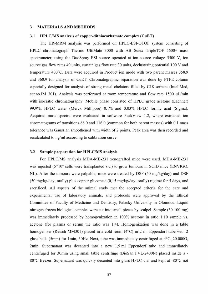
3 MATERIALS AND METHODS
3.1 HPLC/MS analysis of copper-dithiocarbamate complex (CuET)
The HR-MRM analysis was performed on HPLC-ESI-QTOF system consisting of
HPLC chromatograph Thermo UltiMate 3000 with AB Sciex TripleTOF 5600+ mass
spectrometer, using the DuoSpray ESI source operated at ion source voltage 5500 V, ion
source gas flow rates 40 units, curtain gas flow rate 30 units, declustering potential 100 V and
temperature 400°C. Data were acquired in Product ion mode with two parent masses 358.9
and 360.9 for analysis of CuET. Chromatographic separation was done by PTFE column
especially designed for analysis of strong metal chelators filled by C18 sorbent (IntellMed,
cat.no.IM_301). Analysis was performed at room temperature and flow rate 1500 µL/min
with isocratic chromatography. Mobile phase consisted of HPLC grade acetone (Lachner)
99.9%, HPLC water (Merck Millipore) 0.1% and 0.03% HPLC formic acid (Sigma).
Acquired mass spectra were evaluated in software PeakView 1.2, where extracted ion
chromatograms of transitions 88.0 and 116.0 (common for both parent masses) with 0.1 mass
tolerance was Gaussian smoothened with width of 2 points. Peak area was then recorded and
recalculated to ng/ml according to calibration curve.
3.2 Sample preparation for HPLC/MS analysis
For HPLC/MS analysis MDA-MB-231 xenografted mice were used. MDA-MB-231
was injected (5*106 cells were transplanted s.c.) to grow tumours in SCID mice (ENVIGO,
NL). After the tumours were palpable, mice were treated by DSF (50 mg/kg/day) and DSF
(50 mg/kg/day; orally) plus copper gluconate (0,15 mg/kg/day; orally) regime for 5 days, and
sacrificed. All aspects of the animal study met the accepted criteria for the care and
experimental use of laboratory animals, and protocols were approved by the Ethical
Committee of Faculty of Medicine and Dentistry, Palacky University in Olomouc. Liquid
nitrogen-frozen biological samples were cut into small pieces by scalpel. Sample (30-100 mg)
was immediately processed by homogenization in 100% acetone in ratio 1:10 sample vs.
acetone (for plasma or serum the ratio was 1:4). Homogenization was done in a table
homogenizer (Retsch MM301) placed in a cold room (4°C) in 2 ml Eppendorf tube with 2
glass balls (5mm) for 1min, 30Hz. Next, tube was immediately centrifuged at 4°C, 20.000G,
2min. Supernatant was decanted into a new 1,5 ml Eppendorf tube and immediately
centrifuged for 30min using small table centrifuge (BioSan FVL-2400N) placed inside a -
80°C freezer. Supernatant was quickly decanted into glass HPLC vial and kept at -80°C not
37

longer than 6 hours. Just before the HPLC analysis the vial was placed into the pre-cooled
(4°C) LC-sample rack and immediately analysed. To enable approximate quantification of
analyzed CuET, calibration curve was prepared. Various amounts of CuET were spiked to
plasma, frozen in liquid nitrogen, and placed at -80°C to exactly mimic sample processing.
Standards were then processed similarly as the samples described above. To measure
circulating CuET concentrations, mice were dosed with single per oral DSF (50 mg/kg) and
sacrificed at different time points. Serum was collected and frozen for analysis.
3.3 Blood collection from humans for HPLC/MS analysis of CuET
Blood samples were collected before and 3 hours after per oral application of the DSF
(Antabuse 400 mg) dissolved in water. Used needles were of special type for metal analysis -
Sarstedt Safety Kanule 21G x 1½´´ REF 85.1162.600. Collection tubes were of special type
for metal analysis - Sarstedt – S-Monovette 7,5 ml LH, REF 01.1604.400. Immediately after
the blood collection the samples were centrifuged in a pre-cooled centrifuge (4°C at 1300G
for 10min). After the spinning, tubes were placed on ice and the plasma fraction was
immediately aliquoted into the 1,5-ml Eppendorf tubes with approx. 500ul per tube. The tubes
with plasma were immediately frozen on dry ice and later stored in -80°C. Blood samples
were collected from volunteers who signed the informed consent for undergoing Antabuse
therapy due to alcohol abuse. Human participants were 4 males (ages 34, 38, 41, 60 years)
and 5 females (ages 37, 56, 46, 59, 63 years). All individuals were freshly diagnosed for
alcohol use disorder and were scheduled for Antabuse therapy. Blood samples were collected
before and after the first application of Antabuse. The study was approved by the Ethical
Committee of Faculty of Medicine and Dentistry, Palacky University in Olomouc.
3.4 Cell lines
Cell lines were cultured in appropriate growth media supplemented with 10% fetal
bovine serum and penicillin/streptomycin; and maintained at humidified, 5% CO2 atmosphere
at 37°C. Lines cultured in DMEM medium were: HCT116 (ATCC), DU145 (ECACC), PC3
(ECACC), T47D (NCI60), HS578T (NCI60), MCF7 (ECACC), MDA-MB-231 (ATCC), U-
2-OS (ECACC), HeLa (ATCC), NIH-3T3 (ATCC), CAPAN-1 (ATCC), A253 (ATCC),
FaDu (ATCC), , h-TERT-RPE1 (ATCC), HeLa-Ub(G76V)-GFP-ODD-Luc (kindly provided
by prof. R.J. Deshaies, Pasadena, California). Cell lines cultured in RPMI1640 medium were:
AMO-1 (WT and adapted to bortezomib kindly provided by prof. C. Driessen, St. Gallen,
Switzerland), MM-1S (kindly provided by prof. C. Driessen, St. Gallen, Switzerland), Cell
38

line A549 (ATCC) was cultured in F12K medium, RWPE-1 (ATCC) cells were cultured in a
keratinocyte serum-free medium supplemented with the bovine pituitary extract and human
recombinant epidermal growth factor (Thermo Scientific). DU145-RS (radio-surviving) cell
line was previously characterised (Kyjacova et al., 2015). Cell lines were tested for
mycoplasma contamination.
3.5 Stable cell lines construction, transfection, siRNA
For construction of all stably transfected cell lines U-2OS cell line (ECACC) was used.
For U-2OS-Ub-GFP the commercial Ub-GFP EGFP-C1 vector (Addgene) was used, for U-
2OS-NPL4-GFP the commercial NPLOC4-GFP pCMV6-AC-GFP vector (Origene), was
used, for U-2OS-p97-GFP the commercial VCP-GFP pCMV6-AC-GFP vector (Origene) was
used and for U-2OS-UFD1-GFP the commercial UFD1L-GFP pCMV6-AC-GFP vector
(Origene) was used. MDA-MB-231 cell line expressing POH1-myc was established using the
commercial PSMD14 vector (Origene). Cells were transfected using Promega FugeneHD
according to manufacturers’ instructions. Cells were further cultivated in the appropriate
antibiotics (geneticin, 400 μg/ml). Medium with geneticin was replaced every 2-3 days until
the population of resistant cells was fully established. Cells were further refined by sorting for
cells expressing GFP signal (BD FACS Aria). For preparation of inducible MUT-NPL4-GFP
cells, U-2OS cells were transfected with pcDNA6/TR plasmid (Invitrogen, V1025-20) using
FugeneHD transfection reagent (Promega, E2311) according to manufacturer's protocol. To
generate a cell line that stably expresses the Tet repressor, U-2OS cells were cultured in
selective medium with blasticidin (10 μg/ml) for 10 days. Blasticidin-resistant colonies were
picked, expanded and screened for clones that exhibit the lowest basal levels and highest
inducible levels of expression. Next, the most suitable clones were transfected with the
PCDNA4/TO expression vector encoding the mutated NPL4-GFP protein using the Fugene
transfection reagent. Cells were cultured in medium with Zeocin (500 μg/ml) to select clones
that contain pcDNA 4/TO-mutated NPL4-GFP. The MUT-NPL4-GFP-encoding plasmid
were obtained from GeneriBiotech. To induce expression of protein, cells were incubated
with doxycycline (Sigma) 1 μg/ml for 16-48 hours. For siRNA-mediated knock-down, U-2OS
cells were transfected with anti-TDP43 siRNA (Dharmacon; L-012394-00) or non-targeting
siRNA (Eurofins Genomics-UAA UGU AUU GGA ACG CAU A) using Lipofectamine
RNAiMAX transfection reagent (Invitrogen) according to manufacturer's protocol. After 48
hours, the knock-down efficiency was evaluated by immunofluorescence analysis.
39

3.6 Colony formation assay
Cells were seeded into 6-well plates at 100-300 cells per well (depending on the cell
line). Next day the cells were treated with compounds as indicated in the specific experiments
and kept in culture for 7-14 days. Colonies were visualized by crystal violet and counted.
3.7 XTT assay
10.000 cells were seeded into a 96-well plate. Next day, the cells were treated as
indicated. After 24 or 48 hours (as indicated in legends of individual figures) an XTT assay
was performed according to manufacturer’s instructions (Applichem). XTT solution was
added to media and incubated for 30-60 minutes, and then dye intensity was measured at the
475nm wavelength using spectrometer (TECAN, Infinite M200PRO). Results are shown as
mean values and standard deviations from 3 independent experiments, each performed in 3
replicates. IC50s are calculated using Graphpad Prism software based on survival curves from
at least two independent experiments.
3.8 Annexin V staining
Cell cultures were treated as indicated and harvested by trypsinization. Initial culture
medium and washing buffer were collected to include detached cells. Cells were centrifuged
(250G, 5min) and re-suspended in a staining buffer (140 mM NaCl, 4 mM KCl, 0.75 mM
MgCl2, 10 mM HEPES) containing 2.5 mM CaCl2, Annexin V-APC (1:20, BD Biosciences)
and 2.5 μg/ml 7-AAD (BD Biosciences) for 15 minutes on ice in the dark. Samples were
analysed by flow cytometry using BD FACSVerse (BD Biosciences), at least 10.000 events
were acquired per sample. Collected data were processed by BD FACSSuite (BD
Biosciences) and exported into Microsoft Excel.
3.9 Caspases 3/7 assay
Activity of caspase-3 and -7 was quantified by cleavage of fluorogenic substrate
CellEvent™ Caspase-3/7 Green Detection Reagent (ThermoFisher Scientific). Briefly,
samples prepared in the same staining buffer as described for Annexin V staining above were
supplemented with 2% FBS, 0.5 μM CellEvent™ Caspase-3/7 Green Detection Reagent and
incubated for 45 minutes at room temperature in the dark. Subsequently, 0.5 μg/mL DAPI
was added and samples were analysed by flow cytometry using BD FACSVerse (BD
40

Biosciences), at least 10.000 events were acquired per sample. Collected data were processed
by BD FACSSuite (BD Biosciences) and exported into Microsoft Excel.
3.10 Immunoblotting and antibodies
Equal amounts of cell lysates were separated by SDS-PAGE on hand-cast or precast
tris-glycine gradient (4-20%) gels (Life Technologies), and then transferred onto
nitrocellulose membrane. The membrane was blocked with 5% milk in Tris-buffered saline
containing 0.1% Tween 20 for 1 hour at room temperature, and then incubated overnight at
4°C or 1hour at room temperature, with one of the following primary antibodies (all
antibodies were used in the system under study (assay and species) according to the profile of
manufacturer): anti-ubiquitin (1:1000; Cell Signaling, cat.n.:3933), anti-HIF1α (1:1000; BD
Biosciences, cat. n.: 610958), anti-Cdc25A (1:500; Santa Cruz Biotechnology, clone DCS-
120), anti-NRF1 (1:1000, Cell Signaling, clone D5B10), anti-VCP (1:2000; Abcam, cat. n.:
ab11433), anti-VCP (1:1000; Novus Bio, cat. n.: NBP100-1557), anti-NPLOC4 (1:1000;
Novus Bio, cat. n.: NBP1-82166), anti-ubiquitin lys48-specific (1:1000; Merck Millipore,
clone Apu2), anti-β-actin (1:500, Santa Cruz Biotechnology, cat. n. sc-87778), anti-GAPDH
(1:1000,GeneTex, clone 1D4), anti-Lamin B (1:1000; Santa Cruz Biotechnology, cat. n.: sc-
6217), anti-calnexin (1:500; Santa Cruz Biotechnology, cat. n.: sc-11397), anti-α-Tubulin
(1:500; Santa Cruz Biotechnology, cat. n.: sc-5286), anti-Xbp1 (1:500; Santa Cruz
Biotechnology, cat. n.: sc-7160), Ufd1 (1:500; Abcam, cat. n.: ab155003), cleaved PARP1
(1:500; Cell Signaling, cat. n.: 9544), p-eIF2a (1:500; Cell Signaling, cat. n.: 3597), ATF4
(1:500; Merck Millipore, cat. n.: ABE387), HSP90 (1.500; Enzo, cat. n.: ADI-SPA-810),
HSP70 (1:500; Enzo, cat. n.: ADI-SPA-830), HSF1(1:500; Cell Signaling, cat. n.: 4356),
pHSP27 (1:1000; Abcam, cat. n.: 155987), HSP27 (1:1000; Abcam, cat. n.: 109376) CHOP
(1:500; cat. n.:2895, Cell Signaling) followed by detection by secondary antibodies: goat-anti
mouse IgG-HRP (GE Healthcare), goat-anti rabbit (GE Healthcare), donkey-anti goat IgG-
HRP (Santa Cruz Biotechnology, sc-2020). Bounded secondary antibodies were visualized by
ELC detection reagent (Thermo Scientific) and images were recorded by imaging system
equipped with CCD camera (ChemiDoc, Bio-Rad) operated by Image Lab software or
developed on film (Amersham).
3.11 Immunofluorescence staining
Cells were grown in 24-well plates with 0.170mm glass bottom (In Vitro Scientific).
Where indicated, the cells were pre-extracted before fixation with pre-extraction buffer (10
41

mM PIPES, pH 6.8, 100 mM NaCl, 1.5 mM MgCl2, 300 mM sucrose, 0.5% Triton-X 100,
1mM DTT, 5 μg/ml leupeptin, 2 μg/ml aprotinin, 0.1 mM PMSF) for 20 minutes at 4°C,
washed by PBS and then immediately fixed with 4% formaldehyde for 15 minutes at room
temperature. Cells were stained with primary antibodies: anti-ubiquitylated conjugateed
mouse FK2 antibody (1:500; Enzo, cat. n.: BML-PW8810), anti-VCP (1:500; Abcam; cat. n.:
ab11433), anti-NPL4 (1:500; Novus Bio, cat. n.: NBP1-82166), HSP70 (1:100; Enzo, cat. n.:
ADI-SPA-830), HSF1 (1:500; Cell Signaling, cat. n.: 4356) anti-ubiquitin lys48-specific
(1:500; Merck Millipore, clone Apu2), SC-35 (1:500; Abcam, cat. n.: ab11826), Sumo2/3
(1:500; Abcam, cat. n.: ab3742), TDP-43 (1:300; Proteintech, cat. n.: 10782-2-AP), PML
(1:300, Santa Cruz) and appropriate Alexa Fluor 488 and 568 secondary antibodies
(Invitrogen, 1:1000). Cytochrome c was stained by Alexa Fluor 555 conjugated mouse anti-
cytochrome c antibody according manufacture protocol (BD Pharmingen, cat. n.: 558700).
3.12 Microscopy, FRAP and image analysis
Samples were examined in a Zeiss Axioimager Z.1 platform equipped with the Elyra
PS.1 super-resolution module for structured illumination (SIM) and the LSM780 module for
CLSM. High resolution images were acquired in super-resolution mode using Zeiss Pln
Apo100x/1.46 oil objective (tot. mag. 1600x) with appropriate oil (Immersol 518F). SR-SIM
setup involved 5 rotations and 5 phases for each image layer and up to 7 Z-stacks (101nm)
were acquired per image. CLSM setup for FRAP and life cells acquisition involved c-Apo
40x/1.2W water immersion objective. Bleaching of regions of interest (ROI) was performed
using Argon 488nm laser. Lower resolution images of fixed samples were acquired using Plan
Apo 63x/1.4 Oil objective (tot. mag. 1008x). FRAP and image acquisitions were performed in
Zeiss Zen 11 software. For FRAP internal Zen’s “Bleach” and “Regions” modules were used.
Data from FRAP analysis involving multiple bleached ROI’s were exported into MS-Excel
and charted. Basic processing of acquired images such as contrast and brightens setting was
done in Adobe Photoshop on images exported as tiff. Quantitative microscopy-based
cytometry of the IF stained samples was performed using an automatic inverted fluorescence
microscope BX71 (Olympus) in the ScanR Acquisition software (Olympus), analyzed with
ScanR Analysis software (Olympus).
3.13 Cell fractionation for Triton X insoluble pellets
Cells were treated as indicated, washed in cold PBS and lysed in lysis buffer (50 mM
HEPES, pH 7.4, 150 mM NaCl, 2 mM MgCl2, 10% glycerol, 0.5% Triton-X100, protease
42

inhibitor cocktail by Roche) for 10 minutes gently agitating at 4°C. Then, cells were scraped
to Eppendorf tubes and kept for another 10 minutes on ice with intermittent vortexing. After
that, the lysate was centrifuged at 20.000G for 10 minutes at 4°C. Insoluble fraction and
supernatant, respectively, were re-suspended in 1x LSB buffer.
3.14 Isolation of microsomal fraction
After the desired treatment in cell culture, cells were washed with cold PBS and lysed
(250 mM sucrose, 20 mM HEPES pH 7.4, 10 mM KCl, 1.5 mM MgCl2, 1 mM EDTA, 1 mM
DTT, protease inhibitor cocktail). Lysates were homogenised by Potter-Elvejhem PTFE
homogeniser and kept on ice for 20 minutes. The homogenates were subjected to serial
centrifugation steps (720G and 10000G for 5 minutes both, and 100 000G for 1 hour). Pellets
and supernatants from the last ultracentrifugation step were re-suspended in the 1x LSB buffer
and used for WB analysis.
3.15 Ub(G76V)-GFP degradation
HeLa-Ub(G76V)-GFP-ODD-Luc cells expressing Ub(G76V)-GFP were treated with 5 μM
MG-132 for 4 hours. After that, the medium was discarded and cells were twice washed with
PBS and then incubated with tested compound in the presence of 50 μg/ml cycloheximide for
another 2 hours. GFP intensity was acquired using flow cytometry (BD FACSVerse-BD
Biosciences). The median of GFP intensity for each condition was used in calculation. The
percent of remaining Ub(G76V)-GFP for each compound was calculated using the following
formula: (Test compound/MG-132 treatment for 4 hours) * 100.
3.16 p97 ATPase activity assay
P97 ATPase assay was performed as described previously (Chou et al., 2011). 250 nM
of p97 protein was diluted in assay buffer (50 mM Tris-HCl pH 7.4, 20 mM MgCl2, 0.5 mM
DTT). Test compounds were added in DMSO (final concentration of DMSO was 5%). After
10 minutes of incubation, the reaction was started with ATP (100 μM final concentration)
followed by 1-hour incubation at room temperature. The reaction was stopped by adding
Biomol green solution (Enzo) and free phosphate was measured according to manufacturer
instructions. Results are expressed as a percent activity of the control (well containing only
DMSO).
43

3.17 26S proteasome activity
The Rpn11 assay wad done as described previously (Li et al., 2018). Briefly, a synthetic
fluorescent labeled substrate, Ub4pepOG was used to measure Rpn11 activity. Fluorescence
polarization assay was performed in a low-volume 384-well solid black plate in the presence
of 1) 5 µl compound (difference concentration of 1, 10 phenathroline or CuEt) in 3% DMSO
or 3% DMSO control 2) 5 µl of BioMol 26S proteasome and 3) 5µl of substrate (15 nM Ub4-
pepOG). Fluorescence polarization is measured using a plate reader with excitation of 480 nm
and emission of 520 nm filter set. The activity was normalized to DMSO control and fitted
using dose-response equation.
3.18 Affinity precipitation
For GFP immunoprecipitation, NPL4-GFP expressing U2-OS cells were lysed (50 mM
HEPES, pH 7.4, 150 mM NaCl, 2 mM MgCl2, 10% glycerol, 0.5% Triton-X100, protease
inhibitor cocktail by Roche) and centrifuged (20.000G for 10 minutes at 4°C). Supernatant
was incubated with anti-GFP agarose beads (Origene) overnight at 4°C. Beads ware than 3
times washed by lysis buffer and bound proteins eluted by Laemmli buffer for WB analysis.
For GST-precipitation, purified WT-NPL4-GST or MUT-NPL4-GST proteins were incubated
with glutathione sepharose 4B beads (Life Technologies) for 1 h at room temperature.
Unbound proteins were washed (50 mM Tris-HCl, pH 7.4, 150 mM NaCl, 2 mM MgCl2,
10% glycerol, 0.5% Triton-X100) and beads were incubated with purified p97-His or MDA-
MB-231 cell lysate (as a source of ubiquitinated proteins) for 1 h at room temperature. Beads
ware than 3 times washed by buffer and bound proteins eluted by Laemmli buffer for WB
analysis. For His-tag precipitation, purified UFD1-His protein was incubated with Ni-NTA
agarose beads (Qiagen) for 1 h at room temperature. Unbound proteins were washed (50 mM
Tris-HCl pH 7.5, 150 mM NaCl, 2.5 mM MgCl2, 20 mM imidazole, 5% glycerol) and
incubated with purified WT-NPL4-GST or MUT-NPL4-GST proteins for 1 h at room
temperature. Beads ware than 3 times washed by buffer and bound proteins eluted by
Laemmli buffer for WB analysis.
3.19 Protein expression and purification
All proteins were expressed in E. coli BL21 (DE3) cells (Novagen). p97-His (pET28a
vector) and Ufd1-His (pET28a vector) expression were induced by 1 mM IPTG (Life
Technologies) at an OD600 of 0.6 for 10 hours at 22°C. NPL4 WT and MUT (pGEX-2TK)
were induced by 0.4 mM IPTG at an OD600 of 0.8 overnight at 16°C. In case of p97 and
44

UFD1, bacterial pellet was suspended in buffer (50 mM Tris-HCl pH 8.0, 300 mM NaCl, 2.5
mM MgCl2, 20 mM imidazole, 5% glycerol) and lysed by sonication and centrifuged
(14000xg for 20 minutes). Proteins were purified by Ni-NTA chromatography (Qiagen)
according to manufacturer instructions. In case of p97, the protein was further purified by gel
filtration (Superdex 200, GE Healthcare). In case of WT and MUT GST-NPL4, bacterial
pellet was suspended in phosphate buffer (PBS, 0.1% Triton-X100, 300 mM NaCl) and lysed
by sonication and centrifuged (14000xg for 10 min). Proteins were purified by glutathione
sepharose 4B (Life Technologies) according manufacturer’s protocol. The proteins were
further purified by gel filtration (Superdex 200, GE Healthcare).
3.20 Chemicals
CuET was synthetized as described previously (Cvek et al., 2008). The following
chemicals were purchased from commercial vendors: tetraethylthiuram disulfide (disulfiram,
DSF) (Sigma), sodium diethyldithiocarbamate trihydrate (Sigma), copper chloride (Sigma),
copper gluconate (Sigma), bortezomib (Velcade, Janssen-Cilag International N.V.), MG-132
(Sigma), DBeQ (Sigma), NMS-873 (Abmole), cycloheximide (Sigma), 1,10-phenanthroline
(Sigma), MLN7243 (Active Biochem).
3.21 Figures preparation, data analysis, used software
All figures and drawings were prepared using Inkscape 0.17 and MS Office 2016
software. The data was analysed by MS Office 2016, STATISTICA 12, Graphpad Prism 4,
PeakView 1.2, Image Lab 4.1, Carl Zeiss Zen 2011 SP6 (black), Nano Analyze Software
2.3.6, Olympus ScanR Analysis 1.3.0.3 software.
45

4 RESULTS
4.1 Ditiocarb-copper complex is a new metabolite of disulfiram
The mechanism of the anti-cancer activity of disulfiram (DSF) is still controversial
and poorly defined, but many studies agreed on the essential role of copper ions for the
toxicity of disulfiram. Indeed, we confirmed and extended previous in vitro (Cen et al., 2004;
Chen et al., 2006) and in vivo (Allensworth et al., 2015) studies, and demonstrated that copper
supplementation in the form of copper gluconate significantly enhanced ability of disulfiram
to reduce the growth of mammary MDA-MB-231 xenografts in mice (Skrott et al., 2017).
However, it is not clear what mechanism is behind such property of copper. The metabolic
fate of disulfiram upon digestion was deeply studied, so it is well known that disulfiram is
quickly reduced to give two molecules of ditiocarb (diethyldithiocarbamate, DTC). Ditiocarb
is further processed to several metabolites finally to give S-methyl diethyldithiocarbamate
sulfoxide and S-methyl diethylthiocarbamate sulfoxide, proposed inhibitors of aldehyde
dehydrogenase (Johansson, 1992). Importantly, ditiocarb, as well as other members of
dithiocarbamate family, is a very strong metal chelator and its complex with copper is in fact
the most stable among biogenic metals (Hogarth, 2012). Ditiocarb readily reacts with copper
ions in vitro to form ditiocarb-copper complex (bis-(diethyldithiocarbamate)-copper, CuET).
However, the presence of CuET in the body after disulfiram intake was never clearly showed,
despite attempts dated back 30 years ago (Johansson and Stankiewicz, 1985).
To test if CuET really forms in the body and if it may represent a candidate metabolite
responsible for anti-cancer activity of disulfiram (Fig. 8a), I first developed method
employing High-pressure liquid chromatography coupled to mass spectrometry (HPLC-MS).
Optimised method was specific and sensitive enough to detect even low CuET concentrations
in tissue samples as documented by detection of spiked synthetic CuET to mouse serum (Fig.
8b). In following experiments, I confirmed that CuET metabolite was indeed present in mouse
serum after a single oral dose of disulfiram (50 mg/kg) even without copper supplementation
(Fig. 8b). The highest concentration of CuET was detected soon after disulfiram intake and
then dropped gradually (Fig. 8c). To further investigate if CuET penetrates to tissues and
more importantly to tumours, I analysed the extracts from plasma, liver, brain, and tumours of
mouse undergoing disulfiram therapy (50 mg/kg/day) with and without copper gluconate
supplementation (0,15 mg/kg/day) for 5 days. CuET was readily detected in livers or brains
and, importantly, in tumours as well (Fig. 8d). As hypothesised, copper addition leaded to
clearly elevated CuET levels compared to disulfiram alone. Intriguingly, CuET
46

concentrations in tumours were obviously higher compared to other organs, a phenomenon
even pronounced by copper supplementation. Finally, to prove CuET as a new metabolite of
disulfiram in humans, I analysed also plasma obtained from alcoholics undergoing disulfiram
therapy. Indeed, CuET was present in all samples albeit with diverse concentration (Fig. 8e).
Figure 8│CuET complex is new metabolite of disulfiram. a) A model of CuET formation in
the human body after orally administered disulfiram. b) Examples of mass-spectrometry
spectra of CuET visualised as peaks of 4 MRM transitions in murine serum after CuET
spikes, compared to orally applied disulfiram (single dose, 50 mg/kg). c) Pharmacokinetic
analysis of CuET levels in murine serum after orally applied disulfiram (50 mg/kg) (n=2
animals for each time point). d) CuET levels in murine tumours and tissues (mean; n=5
tissues; n=10 tumours). e) CuET levels in human plasma after disulfiram dose (400 mg) (n=9
patients).
47

Taken together, these results strongly argue that CuET is the ultimate metabolite
responsible for anti-cancer activity of disulfiram because CuET is the only known metabolite
of disulfiram containing copper, a metal that enhances the anti-tumour effects of disulfiram in
vitro and in vivo. As addition of copper further promotes CuET formation at the expense of
other DSF’s metabolites, the increased (rather than decreased) toxicity to cancer cells
correlates with elevated CuET and with likely lower levels of other metabolites.
4.2 CuET complex is highly toxic to cancer cells
To get further insight into the effect of CuET to cancer cells, I performed series of
experiments employing cancer cell lines. First, I compared the toxicity of disulfiram (DSF)
and its main primary metabolite ditiocarb (DTC) with CuET in short-term XTT-base assay
using mammary MDA-MB-231 cancer cell line (Fig. 9a). In sharp contrast to CuET, both
DSF and DTC were negligibly toxic, which was further corroborated by long-term colony-
forming assay (CFA) again verifying higher toxicity of CuET compared to DTC (Fig. 9b).
CFA assay revealed that CuET was also similarly potent (IC50 < 100 nM) to other three breast
cancer cell lines (Fig. 9c). To cover wider spectrum of human malignancies, CuET toxicity
was also tested by XTT assay on a panel of cell lines comprising 11 different cell lines (Fig.
9d). While CuET was toxic to all of them, IC50 values varied considerably (from ~80 nM to
~700 nM). Interestingly, among the most sensitive were identified multiple myeloma cell
lines (AMO1, MM1S) or BRCA2 deficient prostate adenocarcinoma line Capan-1. On the
other hand, non-cancerous prostate cell line RWPE1 was virtually insensitive with IC50 far
above 1 μM.
48

Figure 9│ Cytotoxicity of CuET complex. a) Toxicity of DSF, DTC and CuET in MDA-MB-
231 cells (24 h, mean, SD, and individual data from 3 experiments). b) Effect of DTC and
CuET on MDA-MB-231 cells analysed by colony formation assay (CFA) (mean, SD and
individual data from 3 experiments). c) CuET cytotoxicity measured by CFA in human breast
cancer cell lines (mean, SD and individual data from 3 experiments). d) Table summarising
IC50 values documenting cytotoxicity of CuET across a panel of cancer and non-cancer cell
lines (48 h treatment, 2 independent biological experiments).
4.3 CuET complex induces both apoptotic and non-apoptotic cell death
To get further insight into toxicity of CuET complex, I analysed in detail the mode of
cell death it triggers in cancer cells. Since several published studies (Allensworth et al., 2015;
Cen et al., 2004; Chen et al., 2006) state that disulfiram combined with copper induce
apoptosis, I first checked for this type of programed cell death. The main hallmark of
apoptosis is the activation of cysteine proteases responsible for cell death execution –
caspases, and especially the activity of effector caspase-3 or caspase-7 is measured frequently
(Kepp et al., 2011). To measure caspase-3/7 activity, cells were incubated with substrate that
becomes fluorescent after specific caspase-mediated cleavage, and fluorescence in cells was
49

analysed by flow cytometry. Unexpectedly, while a small molecule NMS-873, known
inductor of apoptosis (Magnaghi et al., 2013) here used as a positive control, clearly increased
caspase-3/7 activity in U-2OS or MDA-MB-231 cells, no such elevation was observed in
CuET treated samples despite massive death documented by increased number of
permeabilised cells (Fig. 10a, upper-right – e.g. positive for DNA stain).
To further corroborate these surprising results, caspase-3/7 activity was analysed by
other independent assays. First, Annexin V staining coupled with flow cytometry was
employed. During the apoptosis, activated caspases cleave handful of targets, among them
membrane associated flippase that maintain cytosolic orientation of a phospholipid
phosphatidylserine. Upon flippase cleavage, phosphatidylserine flips to extracellular surface
of the cell, where it can be recognized by macrophages to enable engulfing of the dying cell
(Fadok et al., 1992). Phosphatidylserine is also specifically bounded by a protein Annexin V,
which is used as a probe to detect apoptotic cells. Early apoptotic cells are positive for
Annexin V while negative for non-permeable DNA dyes such as propidium iodide or 7-AAD
confirming that the plasma membrane is still intact and Annexin V positivity is not due to
membrane rupture as typical for necrosis. Indeed, such population (Annexin V+/7-ADD–)
was clearly identified in NMS-873 treated samples, but again not in cells exposed to CuET
(Fig. 10b) confirming results obtained from direct measurement of caspase-3/7 activity. As a
second assay, I performed WB analysis of direct and well known caspase-3/7 substrate
PARP-1 protein. In apoptotic cells, activated caspase-3/7 cleave PARP-1 and cleaved product
of lower molecular weight can be easily detected (Chaitanya et al., 2010). In line with
previous results, PARP-1 cleavage was readily detected in NMS-873 treated sample, but not
in case of CuET (Fig. 10c). These data were further confirmed with specific antibody
recognizing only cleaved product of PARP-1 (Fig. 10c).
50

51
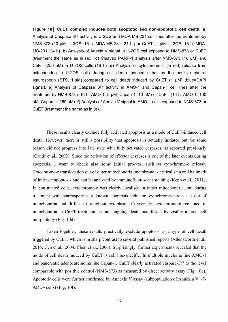
Figure 10│ CuET complex induces both apoptotic and non-apoptotic cell death. a)
Analysis of Caspase 3/7 activity in U-2OS and MDA-MB-231 cell lines after the treatment by
NMS-873 (10 µM; U-2OS: 16 h, MDA-MB-231: 24 h,) or CuET (1 µM; U-2OS: 16 h, MDA-
MB-231: 24 h). b) Analysis of Anexin V signal in U-2OS cell exposed to NMS-873 or CuET
(treatment the same as in (a). c) Cleaved PARP-1 analysis after NMS-873 (10 µM) and
CuET (250 nM) in U-2OS cells (16 h). d) Analysis of cytochrome c (in red) release from
mitochondria in U-2OS cells during cell death induced either by the positive control
staurosporin (STS, 1 µM) compared to cell death induced by CuET (1 µM) (blue=DAPI
signal). e) Analysis of Caspase 3/7 activity in AMO-1 and Capan-1 cell lines after the
treatment by NMS-873 ( 16 h; AMO-1: 5 µM; Capan-1: 10 µM) or CuET (16 h; AMO-1: 100
nM, Capan-1: 250 nM). f) Analysis of Anexin V signal in AMO-1 cells exposed to NMS-873 or
CuET (treatment the same as in (e).
These results clearly exclude fully activated apoptosis as a mode of CuET-induced cell
death. However, there is still a possibility that apoptosis is actually initiated but for some
reason did not progress into late state with fully activated caspases, as reported previously
(Cande et al., 2002). Since the activation of effector caspases is one of the later events during
apoptosis, I want to check also some initial process, such as cytochrome-c release.
Cytochrome-c translocation out of outer mitochondrial membrane is critical step and hallmark
of intrinsic apoptosis and can be analysed by immunofluorescent staining (Kepp et al., 2011).
In non-treated cells, cytochrome-c was clearly localised in intact mitochondria, but during
treatment with staurosporine, a known apoptosis inductor, cytochrome-c released out of
mitochondria and diffused throughout cytoplasm. Conversely, cytochrome-c remained in
mitochondria in CuET treatment despite ongoing death manifested by visibly altered cell
morphology (Fig. 10d).
Taken together, these results practically exclude apoptosis as a type of cell death
triggered by CuET, which is in sharp contrast to several published reports (Allensworth et al.,
2015; Cen et al., 2004; Chen et al., 2006). Surprisingly, further experiments revealed that the
mode of cell death induced by CuET is cell line-specific. In multiple myeloma line AMO-1
and pancreatic adenocarcinoma line Capan-1, CuET clearly activated caspase-3/7 to the level
comparable with positive control (NMS-873) as measured by direct activity assay (Fig. 10e).
Apoptotic cells were further confirmed by Annexin V assay (subpopulation of Annexin V+/7-
ADD– cells) (Fig. 10f).
52

The type of non-apoptotic cell death induced by CuET in cell lines such as MDA-MB-
231 or U-2OS is highly interesting and needs further investigation, as well as to find a factor
causing apoptosis in others cell lines. It is worth of mention that cell lines dying by apoptosis
are also the most CuET-sensitive (Fig. 9d).
4.4 CuET complex does not inhibit the proteasome directly
The toxicity of disulfiram to cancer cell has been explained by a plenty of hypothesis
(Cvek, 2011; Skrott and Cvek, 2012). The most likely explanation for so much heterogeneous
theories is the chemical nature of disulfiram itself. It contains two very reactive thiol groups
readily reacting with cysteine residues of various proteins, as reported for ALDH (Vallari and
Pietruszko, 1982) or MDR (Loo, 2000). Despite not listed as a typical example, disulfiram
shares several characteristics with pan-assay interfering compounds (PAINS) such as
curcumin or quinones (Baell and Walters, 2014). These compounds, as the name suggest,
score in various screening assays as positive hits, however such activity is often just an
artefact. PAINS, similarly to disulfiram, are usually very reactive compounds, metal chelators
or redox-cycling compounds. Due to their pleiotropic effect, it is extremely challenging to
validate the hit appropriately in cells and consequently such compounds are described as
having promising activity against a wide variety of targets (Baell and Walters, 2014). Such
scenario could be valid also for disulfiram.
It is also important to stress out, that disulfiram has very complex and rapid
metabolism, and very low or even undetectable plasma levels of circulating disulfiram
(Johansson, 1992) raise a question if it is even appropriate to test disulfiram in cancer cell
cultures, as it is not known if disulfiram reaches tumours in vivo. Conversely, majority of
studies agrees on the strong potentiation of disulfiram effect by copper. Therefore, to find the
mechanism standing behind disulfiram toxicity, it should be searched within the theories
involving the copper and CuET.
Notably, the only one hypothesis relies on the presence of copper consistently – the
inhibition of protein degradation by the interference with the activity of the proteasome
(Cvek, 2011). First reported in 2006 (Lövborg et al., 2006) and further confirmed (Chen et
al., 2006) by the group of prof. Q.P. Dou, this theory explaining the disulfiram´s mechanism
of action became the most accepted by the scientific community. However, further analysis
raised a direct inhibition of the proteasome questionable (Cvek et al., 2008). During my
53

research stay in prof. Dou’s lab, I have found that CuET complex did not directly inhibit any
of three proteolytic activities of the core proteasome particle (20S proteasome), but still,
CuET clearly inhibited degradation of proteins such as IκB or p53. These data are
summarised in my master thesis (Skrott, 2014). Such conflicting results urged for further
investigation and final answer.
In my previous results, I have confirmed that CuET induces accumulation of poly-
ubiquitinated proteins. However, the type of ubiquitin chain which determines the fate of the
substrate, was not known. Not all types of linkage commit the protein for the degradation in
proteasome, as ubiquitin has also many different roles. Lysine 48 (K-48) linkage is first and
foremost associated with proteasomal degradation (Komander and Rape, 2012), so I
performed WB analysis with an antibody specifically recognising K48-ubiquitin chains.
CuET treatment induced clear accumulation of this linkage type with similar potency and
kinetics like 20S proteasome inhibitor bortezomib used as positive control (Fig. 11a). As the
core particle of proteasome was excluded as a suspected target, the regulatory 19S part of the
proteasome was an obvious option. POH1 deubiquitinase (DUB) was especially interesting
(Cvek et al., 2008; Skrott and Cvek, 2012). This enzyme deubiquitinates proteins before their
translocation into the proteasome and its activity is crucial for proper proteasome function
(Verma et al., 2002). Since POH1 belongs to the family of JAMM domain DUBs, it contains
zinc in the catalytic site, and the reaction between the zinc and CuET was particularly
attractive (Cvek et al., 2008).
Figure 11│ The proteasome is not directly inhibited by CuET complex. a) Time-course
WB analysis of K48-ubiquitin in U-2OS cells treated by CuET or BTZ (both 1 µM). b) The
level of POH1 in WT and POH1-myc expressing MDA-MB-2231 cells. c) POH1
overexpression alleviates the effect of POH1 inhibitor 8-HQ (10 µM) in contrast to BTZ
54

(1 µM) or CuET (1 µM) as measured by ubiquitin and p21 protein levels in MDA-MB-231 cells
treated for 6 h. d) CuET does not inhibit POH1 activity in vitro (1,10-OPT used as a positive
control).
It is well known that overexpression of the protein targeted by its specific inhibitor can
elicit partial resistance to the inhibitor and such mechanism is frequently involved in the
acquired resistance to the chemotherapy (Morganti et al., 2000; Oerlemans et al., 2008). Such
approach was chosen to test a potential link between CuET and POH1 enzyme. Firstly, POH1
was overexpressed in MDA-MB-231 cell line by stable introduction of ectopic POH1. The
amount of POH1 was further analysed by WB confirming satisfactory overexpression (Fig.
11b). Control and POH1 cell lines were further compared in their response to CuET. Specific
POH1 inhibitor 8-HQ (8-quinolinethiol hydrochloride) was used as a positive control (Li et
al., 2017) while bortezomib as a negative. While 8-HQ was apparently less active in POH1
overexpressed cell line as measured by accumulation of polyubiquitinated proteins or
proteasome substrate protein p21, CuET behaved similarly to bortezomib, i.e. with similar
potency in both cell lines (Fig. 11c). Such results obviously argue against the hypothesis that
CuET targets POH1 deubiquitinase.
To get final answer, I visited the laboratory of professor R. J. Deshaies, the world-
leading expert in the family of JAMM domain deubiquitinases. In his lab, I employed
biochemical assay with artificial POH1 substrate measuring not only POH1 activity but
indirectly the whole 26S proteasome altogether (Li et al., 2017). Consistently with the data
obtained from cells, CuET failed to inhibit the deubiquitinase (Fig. 11d), in contrast to
positive control 1,10-phenantroline (Verma et al., 2002), finally excluding POH1 as well as
the whole proteasome as the direct target of CuET complex.
4.5 CuET complex inhibits the function of p97 segregase
Data gathered so far were quite confusing – while CuET complex induced the
accumulation of polyubiquitinated proteins and stabilisation of several proteins rapidly
degraded by proteasome, the proteasome seemed untouched. To find an explanation for such
puzzling discrepancy, I looked to the UPS more deeply. Proteasomal substrates could be
stabilised by impairment of its ubiquitination, as documented for ubiquitin-activating E1
enzyme inhibitors (Hyer et al., 2018). However, a such mechanism presumes a decrease of
polyubiquitinated proteins, and not an increase as observed in the case of CuET. Mammalian
55

cells also contain more than one hundred of deubiquitinases associated with several functions
including deubiquitination and thus stabilisation of proteins dedicated to degradation in the
proteasome (Harrigan et al., 2017). If CuET complex acts as a kind of pan-deubiquitinase
inhibitor, one would expect gross accumulation of polyubiquitinated proteins, however on the
other hand, in theory, the degradation of proteins should not be blocked, but should remained
the same or should be even promoted (Harrigan et al., 2017). Such speculations suggest that
suspected target of CuET should be somewhere between the polyubiquitinated substrate and
the proteasome.
It is well known that some substrates use adaptor proteins, such as RAD23 or DSK2,
that shuttle them to the proteasome (Saeki, 2017). However, it is not very likely that inhibition
of any of these factors could induce such global accumulation of polyubiquitinated proteins
and inhibit the degradation of several different substrates. Importantly, accumulating
evidence emphasises another crucial factor implicated in the multitude of processes associated
with ubiquitin – Valosin-containing protein (VCP or p97). p97 acts as a segregase pulling out
ubiquitinated proteins out of membranes, protein complexes or chromatin, and thus enables
their degradation by the proteasome (Meyer et al., 2012). Recent reports show that inhibitors
of p97 induce several phenotypes similar to the inhibition of the proteasome, including
accumulation and stabilisation of polyubiuitinated substrates dedicated to degradation
(Anderson et al., 2015; Chou et al., 2011).
Interestingly not all proteasome substrates are dependent on p97 activity, providing a
possible way how to distinguish between proteasome versus p97 inhibitors (Chou and
Deshaies, 2011). Importantly, all substrates reported so far to be stabilised by CuET, i.e. I-κB,
Ub(G76V)-GFP, p53 (Skrott, 2014) are all dependent on the activity of p97 segregase (Chou
et al., 2011; Li et al., 2014; Valle et al., 2011). To investigate if CuET specifically inhibits
only the degradation of proteins dependent on p97 activity, I analysed the behaviour of
Hypoxia-inducible factor 1 (HIF-1α). This transcription factor is continuously degraded by
the proteasome under normal conditions, and its degradation is largely independent on the
p97, as only transcriptionally active form of HIF-1α, representing just a small subset of the
protein, is degraded with the assistance of p97 (Alexandru et al., 2008). First, I treated the
cells with CuET or proteasome inhibitor MG-132 and analysed the level of HIF-1α.
Intriguingly, in contrast with MG-132, which significantly accumulated HIF-1α, CuET
induced only marginal elevation of the protein compared to non-treated cells (Fig. 12a). Such
results were consistent with the scenario presuming that CuET inhibits degradation of only
56

p97-dependent proteins and not all proteasome substrates as in case of MG-132. However, the
previous experiment is not strong enough for such statement – fail to accumulate the protein is
not the same as fail to inhibit its degradation, and can be interpreted by several ways. To get
more convincing data, I chose pulse-chase experiment, as a more appropriate. In such
experiment, the cells are first pre-treated with reversible proteasome inhibitor such as MG-
132 to completely block the degradation and to induce accumulation of looked-for proteins.
After desired time, MG-132 inhibitor is washed-out to restore the activity of the proteasome.
At the same time, cells are exposed to tested compounds and cycloheximide, an inhibitor of
ribosome, to stop protein synthesis. Under normal conditions, the level of accumulated protein
should decrease with time – the proteasome is again functional and de novo synthesis is
blocked. If the degradation of desired protein is inhibited by tested compound, the level of the
protein should remain the same or at least the decline should be significantly slowed down.
Such approach enables more direct assessment of the compound´s effect on protein
degradation with shorter exposure times lowering the possibility of unspecific cellular effects.
Employing for HIF-1α protein, results clearly shows that CuET, in contrast to bortezomib,
failed to stabilise HIF-1α similarly to DBeQ, a specific p97 inhibitor (Chou et al., 2011) (Fig.
12b). On the other hand, with the same conditions, all three compounds, CuET, bortezomib,
and DBeQ, prevented the degradation of a protein Cdc25A, a phosphatase involved in cell-
cycle regulation, which is degraded by p97-dependent manner (Riemer et al., 2014) (Fig.
12b). These result clearly shows that CuET blocs only the degradation of proteins dependent
on p97 activity.
p97 segregase is involved not only in protein degradation, but it plays a role also in
other processes such as activation of transcription factors, such as Nuclear respiratory factor 1
(NRF1) (Radhakrishnan et al., 2014). Closely related to the well-known NRF2 involved in
antioxidant response, NRF1 is a major regulator of protein degradation. It activates expression
of all proteasome units and p97 as well upon proteasome impairment or overload (Sha and
Goldberg, 2014). NRF1 is tethered on endoplasmic reticulum (ER) in the inactive form and it
is constitutively degraded by the proteasome (Steffen et al., 2010). After insufficient activity
of the proteasome, NRF1 accumulates at the ER membrane as inactive 120 kDa precursor.
This form is first pulled out of ER membrane by the translocase activity of p97 and then
cleaved to active 110 kDa form that translocates to the nucleus to start expression of
proteasome subunits (Radhakrishnan et al., 2014). Proteasome inhibitors induce accumulation
of both pre-processed 120 kDa and cleaved 110 kDa form of NRF1. On the other hand,
57

inhibitors of p97 blocks NRF1 translocation prior the cleavage, so only full-length 120 kDa
form accumulates (Le Moigne et al., 2017; Radhakrishnan et al., 2014; Sha and Goldberg,
2014). Therefore, NRF1 behaviour could be used as an elegant model substrate to monitor
p97 translocase activity to and distinguish p97 versus proteasome inhibition. As human cells
contain several isoforms of NRF1, which complicates the analysis, mouse cells expressing
only one variant are frequently used (Radhakrishnan et al., 2010, 2014). In NIH-3T3 mouse
fibroblast treated with bortezomib, both 120 kDa and 110 kDa species accumulated in
contrast to CuET treated cells, where only full-length form was present (Fig. 12c). When
combined with bortezomib, both CuET and NMS-873 (a specific p97 inhibitor) effectively
blocked the formation of cleaved 110 kDa form (Fig. 12d) further confirming that CuET
impairs p97 activity. The effect of CuET on NRF1 was also further corroborated in human
cancer cell lines (Fig. 12e). Collectively, such results confirmed that CuET impairs p97
segregase activity in cells but not the proteasome.
Figure 12│ CuET inhibits p97-dependent degradation and translocation. a) CuET (1
µM) induces only minor accumulation of HIF-1α in contrast to MG-132 (5 µM) or BTZ (1 µM)
in Hela cells treated for 2 h. b) Differential impact of BTZ (1 µM), CuET (1 µM) and DBeQ (10
µM) on Cdc25A vs HIF-1α. Hela cells were first pre-treated by MG-132 (4 h, 5 µM), then MG-
132 was washed-out and cells were exposed to cycloheximide (50 µg/ml) combined with
DMSO, BTZ, CuET or DBeQ for 1 h. c) BTZ (1 µM) induces NRF1 120 kDa (upper arrow)
58

and 110 kDa (lower arrow) forms; while CuET (0.5 µM) only the non-cleaved 120 kDa form
(NIH-3T3 cells treated for 8 h). d) Inhibition of the NRF1 cleavage process (appearance of
the lower band) by CuET and NMS-873 (5 µM) in NIH-3T3 cells co-treated with the
proteasome inhibitor MG-132 (5 µM for 6 h). e) Cells treated by MG-132 (5 µM, 6h)
accumulate both forms of NRF1 while CuET-treated cells (1 µM) accumulate only the non-
cleaved 120-KDa form in MDA-MB-231 and MCF7 cells. f) AMO-1 cells resistant to BTZ
(AMO-1 BTZ res.) are similarly sensitive to CuET as AMO-1 WT cells (48 h, representative
results, mean, SD from technical triplicate).
Such findings could be potentially of clinical relevance for the management of
multiple myeloma. Intrinsic or acquired resistance to bortezomib is a very frequent obstacle
limiting the applicability of proteasome inhibitors and the benefit of the treatment
(Manasanch and Orlowski, 2017). The inhibition of p97 segregase is one of the suggested
ways how to cope with the resistance (Le Moigne et al., 2017; Wang et al., 2009). To analyse
if CuET, a metabolite of readily available drug disulfiram, could be used to treat such
refractory disease, I tested its toxicity to AMO-1 cells adapted to bortezomib (Soriano et al.,
2016). Importantly, the potency of CuET was the same to WT cells and cells surviving
extremely high bortezomib concentrations (Fig. 12f), opening a new intriguing possibility for
the use of disulfiram with copper supplementation in patients not responding to proteasome
inhibitors.
4.6 Ubiquitinated proteins accumulated by CuET treatment are associated with
insoluble structures
Given that the most important function of p97 is to translocate or segregate
ubiquitinated proteins out of cellular structures including membranes, chromatin, organelles
and protein complexes for subsequent proteasomal degradation (Meyer et al., 2012), I
wondered if ubiquitinated proteins accumulated upon CuET treatment can be detected as a
part of such structures. First, I fractionated cell lysate to soluble and insoluble parts and
analysed for K48-ubiquitin. Interestingly, ubiquitinated proteins were highly enriched in the
insoluble fraction in CuET treated cells (Fig. 13a). To compare it with the inhibition of
proteasome or p97, I treated the cells with CuET, bortezomib or NMS-873. After the
treatment, I briefly pre-extracted the cells with Triton X-100 containing solution to wash out
59

all freely soluble proteins followed by fixation and staining for K48-ubiquitin. As shown in
Figure 13b, compared to bortezomib or untreated cells, stronger signal corresponding to
extraction-resistant insoluble ubiquitininated proteins was observed in both NMS-873 and
CuET treated cells, as further confirmed by image quantitative analysis (Fig. 13c).
Figure 13│ CuET induces accumulation of immobile ubiquitinated proteins. a)
Ubiquitinated proteins are part of TritonX-100 insoluble cellular fraction after CuET treatment
(1 µM, 3 h, U-2OS cells). b) IF analysis of K48-ubiquitin conjugates in not pre-extracted and
60

Triton X-100 pre-extracted U-2OS cells treated with DMSO, BTZ (1 µM), NMS873 (10 µM)
and CuET (1 µM) (Scale bar = 20 µm). c) Microscopic quantitative analysis of Triton X-100
pre-extracted U-2OS cells treated as in (b). d) Time-course images from a FRAP experiment.
U-2OS cells expressing GFP-ubiquitin were treated with NMS-873 (10 µM), CuET (1 µM) or
BTZ (1 µM) for 2 h (blue boxes mark areas before bleaching, arrows after bleaching), (Scale
bar = 10 µm). e) Quantification of FRAP experiment (relative mean signal of the bleached
region from 10 cells).
To analyse the mobility of ubiquitinated proteins after CuET by another independent
approach, I used fluorescence recovery after photobleaching (FRAP) microscopic method.
This method relies on the quantification of recovered fluorescent signal in specific area after
the photobleaching, and it is frequently used to measure protein mobility or transport
(Ishikawa-Ankerhold et al., 2012). A protein of interest must be fluorescent, thus it is usually
tagged with fluorescent protein such as green fluorescent protein (GFP). In this experiment, I
used GFP-tagged ubiquitin, that behaves as normal ubiquitin and it is attached to proteins
dedicated to degradation, as previously confirmed (Qian et al., 2002). In untreated cells, GFP-
ubiquitin was so mobile that it was even hard to bleach the signal completely as new GFP-
ubiquitin molecules diffused into bleached area extremely quickly (Fig. 13d, bleached area is
marked by blue box). The signal in the bleached area also recovered already within a few
seconds as quantified in Figure 13e. Bortezomib treated cells behaved the same, indicating
that polyubiquitinated proteins accumulated upon proteasome inhibition are fully mobile (Fig.
13d,e). Conversely, in NMS-873 or CuET treated samples, GFP-ubiquitin was visibly
bleached and low signal intensity persisted for a longer time in bleached areas indicating
slowed diffusion (Fig. 13d), as confirmed by quantitative analysis (Fig. 13e). Consistently
with previous experiments, these results indicate that after inhibition of p97 function at least a
subset of the accumulated polyubiquitinated proteins remains immobile and tightly associated
with cellular structures.
Collectively, these experiments demonstrated that significant fraction of
polyubiquitinated proteins accumulated after CuET treatment are largely immobile, consistent
with impairment of p97 segregase. They also suggest that experiments analysing the mobility
of ubiquitinated proteins could be used in future studies to dissect p97 and proteasome
inhibitors.
61

4.7 CuET complex impairs ER-associated degradation leading to the activation of
Unfolded protein response
While p97 plays a role in many cellular processes, probably the most important and
best understood is its function in endoplasmic reticulum-associated degradation (ERAD)
(Meyer et al., 2012). Prior degradation in proteasome, polyubiquitinated proteins associated
with ER must be first extracted from the membrane, a process dependent on p97 segregase
activity. As shown previously (Chou et al., 2011; Wang et al., 2009), several p97 inhibitors
inhibited a degradation of specific ERAD substrates and induced the accumulation of
polyubiquitinated proteins on ER-membrane (Locke et al., 2014). To investigate, if CuET
blocs also this p97-dependent process, I isolated microsomal fraction that contains mainly ER
membranes by ultracentrifugation and analysed it for K48-ubiquitin by WB. While
microsomes from untreated cells were only slightly positive for K48-ubiquitinated proteins,
CuET and NMS-873 treatments induced marked accumulation of non-degraded proteins
associated with ER-membrane in both tested cell lines (Fig. 14a). The same was observed also
for bortezomib, which is consistent with previous report (Locke et al., 2014) showing that
proteasome inhibition could to some extent also impair upstream steps including extraction of
proteins from ER-membrane by p97.
Figure 14│ CuET impairs ERAD and activates UPR. a) Western blot analysis of
accumulated K48-ubiquitin conjugates in microsomal fraction from U-2OS and MDA-MB-231
cells treated by DMSO, CuET (1 µM), NMS-873 (10 µM) or BTZ (1 µM) for 3 h. b) UPR in U-
2OS and MDA-MB-231 cell lines induced by 6 h treatment with CuET (125 nM, 250 nM,
62

500 nM) or positive controls (NMS-873 5 µM, tunicamycin 2 µg/ml, thapsigargin 1 µM)
manifested by increased levels of Xbp1s, ATF4 and p-eIF2α.
The efficacy and accuracy of protein synthesis, maturation and degradation in the ER
is tightly controlled. In case of increased protein load or accumulation of unfolded and non-
degraded proteins, compensatory program called unfolded protein response (UPR) is triggered
(Wang and Kaufman, 2014). UPR activates several pro-survival and adaptation processes to
deal with damaged proteins, however, in case a severe stress condition persists, it stimulates
cell death as well (Hetz, 2012). To examine if CuET triggers UPR, I compared its effect with
p97 inhibition by NMS-873 and with two commonly used activators of UPR, tunicamycin and
thapsigargin, inhibiting protein glycosylation and calcium ion pumping into ER, respectively
(Samali et al., 2010). Treated cells were than analysed for several effectors of UPR, including
phosphorylated eukaryotic initiation factor 2α (p-eIF2α) that negatively regulates polysome
formation, and two transcription factors ATF4 and Xbp1s that control UPR target genes (Hetz
et al., 2015). CuET treatment obviously activated all three markers in dose-dependent manner
to similar extend as positive controls, clearly indicating that it activated robust UPR in both
tested cell lines (Fig. 14b).
Taken together, these results suggest that CuET impairs p97-dependent translocation
of substrates during ERAD, causing an accumulation of damaged polyubiquitinated proteins
associated with ER-membrane ultimately triggering ER-stress and UPR activation. As ERAD
is indispensable process and UPR is tightly connected to cell death pathways, the induction of
UPR could contribute significantly to the toxicity of CuET complex.
4.8 CuET complex immobilises p97 segregase
The data gathered so far apparently demonstrates that CuET inhibits several processes
dependent on p97 function, but the mechanism involved is unclear. To possess its activities,
p97 hydrolyses ATP as a source of energy, so the inhibition of its ATP-ase activity was the
most likely hypothesis as illustrated for other p97 inhibitors (Anderson et al., 2015; Chou et
al., 2011; Magnaghi et al., 2013). Unexpectedly, in contrast to NMS-873, CuET failed to
inhibit p97-mediated ATP hydrolysis in vitro (Fig. 15a). As an alternative scenario, I checked
the protein levels of p97, as CuET treatment could potentially downregulate the amount of the
protein leading to malfunction of the system in cells. However, no effect was observed (Fig.
15b). p97 segregase does not operate alone, but it associates with plethora of cofactors.
63

Among the most important is a heterodimer consisting of NPL4 and UFD1 proteins that is
thought to mediate the interaction with client ubiquitinated proteins (Meyer et al., 2012). As
NPL4 and UFD1 are necessary for multitude of p97 activities, I checked also the level of
these proteins, however, again, no effect was observed (Fig. 15b).
Figure 15│ CuET complex immobilises p97 segregase. a) CuET (1 µM) does not inhibit
ATPase activity of p97, NMS-873 (5 µM) was used as a positive control (mean, SD and
individual data from 4 independent experiments). b) WB analysis of levels of p97, NPL4 and
UFD1 proteins in CuET-treated (8 h; 125 nM, 250 nM, 500 nM, 1000 nM) U-2OS and MDA-
64

MB-231 cells. c) IF analysis of p97 in pre-extracted U-2OS cells (CuET 1 µM for 3 h). d)
Dose-dependent immobilization of p97 in pre-extracted MDA-MB-231 cells treated by CuET
for 3 h. (Scale bar = 10 µm). e) Immobilization of p97, NPL4 and K48-ubiquitin conjugates in
Triton X-100 insoluble fraction in U-2OS and MDA-MB-231 cells treated by CuET (1 µM) for
3 h.
To get further insight into p97 behaviour after CuET treatment, I stained p97
segregase by immunofluorescence (IF) for analysis by confocal microscope. Interestingly,
when the cells were first pre-extracted before fixation and IF staining, that is the same
approach used for K48-ubiquitinated proteins, obvious effect of CuET on p97 was observed.
While in non-treated cells the signal disappears almost completely, consistent with p97 being
very mobile protein (Song et al., 2015), CuET induced very prominent immobilisation of p97
on insoluble cellular structures (Fig. 15c). Such effect was so intensive that extraction-
resistant pool of p97 was clearly visible also after the treatment with very low concentrations
of CuET (50 nM) (Fig. 15d). To assess if the immobilization effect of CuET complex is valid
also for NPL4 and UFD1 proteins, the most important cofactors of p97, I fractioned cell
lysate to soluble and insoluble pellet fraction and analysed by WB. As expected from previous
results, K48-ubiquitinated proteins and p97 were clearly detected in pellet fraction in CuET
treated cells (Fig. 15e). Interestingly, NPL4 cofactor was also visibly enriched in insoluble
fraction, while the second partner of heterodimer UFD1 seemed not (Fig. 15e).
These results suggest that CuET impairs p97 pathway by considerably unusual way. It
seems that CuET induce immobilisation of p97 itself and at least one of its essential cofactor,
NPL4, leading to a malfunction of the pathway.
4.9 CuET complex targets NPL4 cofactor
According to results shown in Fig. 15e, NPL4 enrichment in pellet fraction is very
pronounced, even leading to a visible decrease of total soluble pool of NPL4, which is not the
case for p97. To explore this interesting observation in more detail, I followed NPL4
transition from soluble into pellet fraction in time. Remarkably, CuET treatment induced
almost complete switch of NPL4 protein into insoluble fraction within 5 hours in both cell
lines (Fig. 16a), suggesting very prominent impact of CuET on this protein. NPL4, as the
essential cofactor of p97 necessary for its proper function and cell physiology (Meyer et al.,
65

2012), represents intriguing possible target of CuET complex. Since the resistance to the
drugs is frequently associated with amplification or overexpression of target protein, as seen
in case of bortezomib or methotrexate (Morganti et al., 2000; Oerlemans et al., 2008) for
instance, similar approach was employed to resolve the relevance of NPL4 protein as a target
of CuET complex. Stable cell lines overexpressing NPL4-GFP, p97-GFP, UFD1-GFP
proteins and cells transfected only with empty vector were generated (Fig. 16b), and the effect
of the overexpression of individual proteins on CuET toxicity was evaluated. Importantly,
NPL4 overexpression caused the cells more resistant to CuET treatment compared to p97,
UFD1 or control cells (Fig. 16c). To investigate the impact of NPL4 overexpression on
phenotypes induced by CuET, I also analysed the level of ubiquitinated proteins, a general
marker of protein degradation. Similarly, CuET-treated cells overexpressing NPL4-GFP had
visibly lower amount of accumulated K48-ubiquitinated proteins compared to controls (Fig.
16d).
To look in more detail to the behaviour of NPL4, cells expressing NPL4-GFP were
treated by CuET and followed in time by fluorescent microscopy. Interestingly, within 2-3
hours, NPL4-GFP formed clearly visible clusters in the nucleus and granular pattern in the
cytoplasm (Fig. 16f). Such effect was not observed in case of p97-GFP or UFD1-GFP (Fig.
16e). Subsequent FRAP analysis confirmed that NPL4-GFP is strongly immobilised in both
the nucleus and cytoplasm in CuET-treated cells (Fig. 16f). Similar nuclear clusters induced
by CuET were also detected by IF staining of endogenous NPL4 (Fig. 16g).
These findings indicate that NPL4 cofactor is prominently affected by CuET complex,
leading to its complete immobilization and formation of nuclear and cytoplasmic clusters,
explaining well the effect of CuET on p97 segregase and protein degradation. Importantly,
these data should be also viewed in the light of the biochemical experiments confirming the
direct interaction of CuET with purified NPL4 protein in vitro (Skrott et al., 2017).
66

Figure 16│ CuET complex targets NPL4 cofactor. a) WB analysis showing NPL4
enrichment in Triton X-100-insoluble fractions after CuET (1 µM) treatment. b) WB analysis
documenting levels of ectopic p97-GFP, NPL4-GFP and UFD1-GFP in stable U-2OS cell
lines used for the CuET-treatment rescue and cluster-formation experiments. c) Ectopic
NPL4-GFP, but not p97-GFP or UFD1-GFP rescues CuET toxicity (mean, SD, from 3
experiments, 24 h, U-2OS). d) Ectopic expression of NPL4-GFP alleviates CuET-induced
(125 nM, 4 h) accumulation of K48-ubiquitinated proteins in U-2OS cells. e) CuET (1 µM)
induces intra-nuclear clustering of NPL4-GFP, but not p97-GFP or UFD1-GFP. f) CuET (1
µM, 2 h)-induced immobilization of NPL4-GFP (FRAP, blue boxes: areas before bleaching,
arrows: after bleaching). g) Distribution of endogenous NPL4 nuclear clusters relative to
chromatin in cells treated by CuET (1 µM, 2 h). Scale bars = 10 µm, in (g) = 2 µm).
4.10 NPL4 protein forms aggregates after the treatment by CuET
Detailed picture of NPL4 nuclear clusters revealed that the clusters occupy areas
poorly labelled with DAPI and are not part of nucleoli (Fig. 16g). To explore their nature
67

more closely, several nuclear structures were stained and analysed by IF for a possible co-
localization. Unexpectedly, no overlap was observed with diverse nuclear structures including
nuclear speckles or PML bodies as revealed by staining for their markers (SC-35, PML) (Fig.
17a). In late-G2 cells, NPL4 was obviously segregated from the partially condensed
chromatin (Fig. 17b), suggesting NPL4 is not recruited into specific nuclear sites but rather
excluded, which is typical for aggregated proteins (Enam et al., 2018; Sontag et al., 2017).
Further experiments revealed that the immobilized cytoplasmic and nuclear signals of NPL4-
GFP co-localize with polyubiquitylated proteins (stained with anti-K48-ubiquitin antibody)
(Fig. 17c). The nuclear clusters are also positive for small ubiquitin-like modifier (SUMO2/3)
protein (Fig. 17d), which plays an important role in the recognition of aggregated nuclear
proteins (Guo et al., 2014). Interestingly, immobilised NPL4-GFP co-localised in addition
with TAR DNA binding protein 43 (TDP-43) (Fig. 17d), a protein involved in RNA
maturation that is found as a part of protein aggregates in several neurodegenerative diseases
(Becker et al., 2017; Buratti and Baralle, 2012; Guo et al., 2014; Vogler et al., 2018).
Such results suggest NPL4 is part of protein aggregates after CuET treatment, which
could be explained by two alternative scenarios. First, CuET could induce aggregation of
another unknown cellular protein(s) (such as TDP-43 for instance) and NPL4 is recruited into
such aggregates consequently to promote their degradation. Alternatively, NPL4 could be the
primary target of CuET, which aggregates and secondary attracts other proteins. To test these
hypotheses, I performed a couple of experiments. NPL4 protein, as a part of p97 complex
with UFD1 cofactor, is believed to recognise its client proteins via polyubiquitin chain
(Meyer et al., 2012, 2002). To investigate if NPL4 is recruited to the protein aggregates via
ubiquitin, I pre-treated the cells with ubiquitin-activating enzyme 1 inhibitor (MLN7243),
which should in principle block all ubiquitination in the cell (Hyer et al., 2018). Importantly,
while all ubiquitination was indeed completely stopped as revealed by immunofluorescent
staining for all ubiquitin conjugates by FK2 antibody, NPL4-GFP still formed clusters in cells
pre-treated by MLN7243 (Fig. 17e). Moreover, siRNA mediated knock-down of TDP-43 did
not prevent immobilization of NPL4-GFP (Fig. 17f), indicating that this protein is dispensable
for NPL4 clusters formation. These results strongly support the hypothesis that NPL4 is
aggregated first and independently on other factors like ubiquitinated proteins or TDP-43 are
rather recruited secondarily.
68

Figure 17│ NPL4 protein forms aggregates after CuET treatment. a) NPL4-GFP clusters
induced by CuET treatment (1 µM for 3h) do not co-localize with nuclear speckles (stained by
SC-35 antibody) or PML bodies. b) NPL4-GFP nuclear aggregates induced by CuET (1 µM,
69

3 h) are excluded from chromatin in early prometaphase U-2OS cells. c) NPL4-GFP co-
localizes with K48-ubiquitinated conjugates in cells treated by CuET (1 µM, 3 h; pre-
extracted). d) NPL4-GFP co-localizes with SUMO-2/3 and TDP-43 in cells treated by CuET
(1 µM, 3 h; pre-extracted). e) NPL4-GFP aggregates are formed independently of
ubiquitylations, as documented on CuET (1 µM, 3h) treated cells pre-treated with a chemical
UBE1 inhibitor (MLN7243, 10 µM for 1 h); The lack of the cellular FK2 staining for
ubiquitylated proteins validates the efficacy of the MLN7243 inhibitor. f) NPL4-GFP
aggregates are formed independently of TDP-43, as documented on CuET (1 µM, 3 h)
treated cells, in which TDP-43 was downregulated by siRNA. g) The amount of immobilised
p97 in CuET-treated cells (1 µM for 3 h) correlates with the intensity of NPL4-GFP
aggregates (pre-extracted). h) Detailed WB analysis of UFD1 behaviour in CuET-treated (1
µM) U-2OS cells reviling limited UFD1 immobilization compared to NPL4. i)
Immunoprecipitated soluble NPL4-GFP protein from cells treated by CuET (1 µM for 3h) still
interacts with p97, UFD1 binding partners and K48-ubiquitin conjugates. Scale bars = 10 µm.
Consistent with previous results (Fig. 15c,d,e), p97 also co-localise with NPL4-GFP,
indicating that subset of p97 is attracted to aggregates (Fig. 17g). The amount of p97 within
the NPL4-GFP clusters correlated with the GFP intensity suggesting that p97 is immobilized
via its interaction with NPL4. On the other hand, the virtual absence of UFD1 in pellet
fraction (Fig. 15e) suggests disruption of NPL4-UFD1 complex. Detailed analysis confirmed
very limited presence of UFD1 in insoluble fraction in contrast to NPL4 (Fig. 17h) raising a
question if CuET directly break the complex or if NPL4 aggregates interact with UFD1 less
tightly. To dissect between these hypotheses, I performed immunoprecipitation (IP) against
NPL4-GFP in soluble fraction. No difference in the levels of UFD1 and p97 associated with
NPL4 was observed after CuET treatment (Fig. 17i), indicating that CuET does not disrupt
soluble heterodimer directly, but rather aggregated NPL4 lost its affinity for UFD1 partner.
4.11 NPL4 protein mutated in putative zinc-finger domain resembles phenotypes
induced by CuET
NPL4 protein is a particularly interesting as a target for CuET because it contains two
zinc-finger domains termed as NZF (NPL4-zinc-finger) located at C-terminus and putative zf-
NPL4 (Lass et al., 2008). Importantly, zinc-finger domains are known to bind bi-valent metal
ions or complexes that might chemically resemble CuET (Voráčková et al., 2011).
70

Interestingly, NPL4 protein is expressed as two isoforms in human cells, one of them lacks C-
terminal NZF. In this isoform, NZF domain is completely substituted with different amino
acid sequence making this isoform larger, which is visible as an upper band as it migrates
more slowly in SDS-PAGE. Lacking NZF, yet this isoform responds to CuET treatment
normally by immobilization in the insoluble pellet fraction (Fig. 16a), suggesting c-terminal
NZF domain does not play a role in the response to CuET.
Putative zinc-finger domain is located closer to the N-terminus and its zinc-binding
pocked consist of two histidine and two cysteine residues (Lass et al., 2008). To test a
potential role of this domain, all four critical amino acids within the zinc-finger pocket were
mutated to alanine (Fig. 18a) and doxycycline (DOX) inducible cell line expressing mutated
form of NPL4 (MUT-NPL4-GFP) was established. Surprisingly, upon the induction, MUT-
NPL4-GFP spontaneously formed nuclear and cytoplasmic immobile aggregates (Fig. 18b),
reminiscent of those observed in cells ectopically expressing WT-NPL4-GFP and treated with
CuET. Moreover, in contrast to ectopic WT-NPL4-GFP (Fig. 16c), the ectopic MUT-NPL4-
GFP did not render the cells resistant towards CuET, and in fact it was rather toxic to the
acceptor cells and made them more susceptible to CuET (Fig. 18c). By further examination I
found that multiple CuET-induced phenotypes were shared with MUT-NPL4-GFP
overexpression. First, it induced accumulation of K48-ubiquitinated proteins and activation of
UPR (Fig. 18d). Second, similarly to CuET treatment, MUT-NPL4-GFP expression caused
immobilisation of polyubiquitinated proteins and p97, but not UFD1, as revealed by WB
analysis of soluble and pellet fractions (Fig. 18e). Third, MUT-NPL4-GFP aggregates were
also positive for several proteins found also in WT-NPL4-GFP aggregates induced by CuET,
such as ubiquitinated proteins, p97, SUMO or TDP-43 (Fig. 18f). Moreover, just like in the
case of CuET treatment, soluble MUT-NPL4 maintained its ability to bind ubiquitinated
proteins, p97 and UFD1 partners, as efficiently as WT-NPL4 protein, which was confirmed in
vitro by pull-down assay involving purified proteins (Fig. 18g). These results suggest that it is
not simply a gross misfolding that occurs in the MUT-NPL4, but rather a more restricted
folding alteration, with phenotypic consequences that are reminiscent of the scenario triggered
by CuET in the WT-NPL4.
71

Figure 18│ NPL4 mutated in putative zinc-finger domain resembles phenotypes
induced by CuET. a) Schematic representation of site-directed mutagenesis within the
amino acid sequence of the putative zinc finger domain of NPL4. b) Spontaneous intra-
72

nuclear clustering and immobilization of MUT-NPL4-GFP (FRAP, U-2OS cells, blue boxes:
areas before bleaching, arrows: after bleaching). c) Viability of cells expressing a
doxycycline-inducible MUT-NPL4-GFP, treated with CuET for 48 h (mean and SD, individual
points from 3 independent experiments are shown). d) Accumulation of K48-ubiquitinated
proteins and activation of UPR in cells expressing the doxycycline-inducible MUT-NPL4-
GFP. e) Immobilization of selected proteins in TritonX-100 insoluble pellet fractions from U-
2OS cells expressing doxycycline-inducible MUT-NPL4-GFP (48 h after induction). f) Co-
localization of spontaneous MUT-NPL4-GFP aggregates with ubiquitin conjugates (detected
by FK2 and anti-K48-ubiquitin antibodies), p97, SUMO-2/3 and TDP43 (pre-extracted). g)
Purified MUT-NPL4-GST retains ability to interact in vitro with ubiquitin conjugates (MDA-
MB-231 whole cell lysate was used as a source of ubiquitin conjugates), purified p97-His and
purified UFD1-His similarly like WT-NPL4-GST as revealed by GST or His precipitations.
Scale bars = 10 µm.
Collectively, it seems that CuET targets putative zinc-finger domain of NPL4. This
domain is important for proper conformational stability of NPL4 and its disruption causes the
aggregation of NPL4 protein resulting to several phenotypes induced by CuET treatment. As
MUT-NPL4-GFP is itself sufficient to induce all these phenotypes, it also strengthens NPL4
protein as a critical, and possibly dominant, target of CuET in human cells whose alteration is
likely sufficient to cause the observed toxic cellular effects.
4.12 Aggregated NPL4 protein triggers heat shock response
It is well known that aggregation of unfolded or damaged proteins triggers cellular
heat shock response (HSR), a mainly protective mechanism allowing the cells to handle
aggregates (Richter et al., 2010). Transcription factor HSF1 that regulates the expression of
several heat shock proteins (HSP) belongs to the most important proteins involved in HSR
(Gomez-Pastor et al., 2017). To induce the expression of HSP, phosphorylated HSF1
trimerises and binds to specific sequences of the genome known as heat shock elements
forming specific foci called nuclear stress bodies (Gomez-Pastor et al., 2017). Interestingly,
such stress bodies were clearly visible by IF after CuET treatment (Fig. 19a) as well as HSF1
shift in molecular weight detected by WB corresponding to phosphorylated form of HSF1
(Fig. 19b). HSP70 protein, the main effector managing aggregated proteins, was also
markedly induced by CuET treatment (Fig. 19b), and, as revealed by IF analysis after pre-
73

extraction of cells, HSP70 was actually directly associated with immobilised NPL4
aggregates (Fig. 19c). Importantly, robust HSR activation was also induced by expression of
MUT-NPL4-GFP protein, accompanied by formation of HSF1 stress bodies, co-localisation
of HSP70 with MUT-NPL4-GFP aggregates (Fig. 19d) and induction of HSF1 and HSP70 as
revealed by WB analysis (Fig. 19e). These results indicate that NPL4 aggregates induced
either by CuET or mutation are strong activators of HSR and HSP70 chaperone is involved in
their processing.
Figure 19│ Aggregated NPL4 triggers heat shock response (HSR). a) CuET treatment
(1 µM, 3 h) induces HSF1 stress bodies. b) CuET treatment (125 nM, 250 nM, 500 nM,
1000 nM for 8 h) triggers HSR as manifested by various markers: HSF1, HSP70 and p-
HSP27 detected by WB in U-2OS and MDA-MB-231 cells. c) NPL4-GFP co-localizes with
HSP70 in CuET-treated U-2OS cells (1 µM, 3 h, pre-extracted). d) HSF1 stress bodies and
HSP70 in U-2OS cells expressing MUT-NPL4-GFP. e) Activation of HSR markers in U-2OS
cells expressing doxycycline-inducible MUT-NPL4-GFP (24 h after induction). Scale bars =
10 µm.
74

4.13 Disulfiram is converted to CuET in vitro
The waste majority of research publications aiming to elucidate anti-cancer activity of
disulfiram and its potentiation by copper are based on combined treatment by disulfiram and
copper ions (Allensworth et al., 2015; Chen et al., 2006; Liu et al., 2013, 2016; Xu et al.,
2017). This approach has certain limitations (Skrott and Cvek, 2012), as for now it is
completely unknown what is happening in culture media and if disulfiram reacts with copper
in vitro. It has been even suggested that not disulfiram itself or disulfiram-copper complex is
responsible for toxic effect, but rather the reaction between disulfiram and copper yielding
high amount of oxygen radicals (Lewis et al., 2014). To bring more light into this poorly
explored area, I performed a series of experiments.
First, I incubated disulfiram or disulfiram with copper(ii) chloride (CuCl2) with
complete culture medium (DMEM with 10% FBS) and found that, in the presence of
equimolar CuCl2, a majority of disulfiram is quickly converted to CuET. Interestingly, even
without addition of extra copper ions, certain amount of CuET is also formed, as medium
contain traces of copper ions (Fig. 20a). According to these results, disulfiram combination
with copper should behave similarly like direct CuET treatment, and disulfiram alone should
have only negligible activity. To test this hypothesis, I compared the toxicity of disulfiram
and copper treatments with CuET. As expected, disulfiram combination with CuCl2 was
comparable toxic to CuET, in contrast to disulfiram or CuCl2 alone in different cell lines (Fig.
20b). Similarly, only CuET and disulfiram/Cu+2 combination markedly blocked a degradation
of Ub(G76V)-GFP, a reporter protein, which degradation is dependent on p97 activity (Fig.
20c). To follow other phenotypes associated with mechanism of action of CuET complex in
cells, I also confirmed that the combination of disulfiram with Cu+2 induced the aggregation
of NPL4 (Fig. 20d), immobilisation of NPL4, p97 and K48-ubiquitinated proteins (Fig. 20e)
and activation UPR as documented by elevated levels of several UPR markers (ATF4, eIF2α,
and CHOP) in various cancer cell lines (Fig. 20f). Finally, similarly to CuET, disulfiram/Cu+2
treatment activated HSR confirmed by the presence of HSF1 stress bodies (Fig. 20g,h) and
concomitant upregulation of HSP70 chaperone (Fig. 20i).
75

76

Figure 20│ Disulfiram is converted to CuET in vitro. a) The amount of CuET complex in
the media with added CuET, DSF or DSF combination with CuCl2 (each 1 µM). b) Toxicity of
CuET, DSF, CuCl2 and DSF combination with CuCl2 in various cell lines (48 h treatment,
mean, SD from technical triplicate, representative results). c) Stabilization of Ub(G76V)-GFP
reporter in Hela cell line. Reporter was pre-accumulated with MG-132 (5 µM for 6 h), followed
by MG-132 wash out and incubation of cells with cycloheximide (50 µg/ml) and tested
compounds for 2 h. Each compound was used at 1 µM (mean, SD and individual points from
3 independent experiments) d) Analysis of NPL4-GFP cluster formation after treatment with
indicated compounds (3 h treatment, 1 µM). e) WB analysis of K48-ubiquitinated proteins,
p97 and NPL4 in Triton X-100 insoluble cell fraction. Cells were treated with 1 µM
concentration of indicated compounds for 3 hours. f) UPR analysis in cell lines induced by 8
h treatment with CuET (0,5 µM) or DSF combination with CuCl2 (both 0,5 µM) or positive
controls (tunicamycin 5 µg/ml, thapsigargin 5 µM) manifested by increased levels of CHOP,
ATF4 and p-eIF2α. g) IF analysis of HSF1 stress bodies in various cell lines treated with
CuET (0,5 µM) or DSF combination with CuCl2 (both 0,5 µM) for 3 h. h) Microscopic
quantitative analysis of HSF1 stress bodies in various cell lines treated as in (g) (mean,
lower/upper quartile, n>300 cells). i) WB analysis of HSP70 induction in various cell lines
treated with CuET (0,5 µM) or DSF combination with CuCl2 (both 0,5 µM) for 6 h.
Collectively, these results suggest that disulfiram is converted to CuET in the media
containing copper ions, and thus such treatment induces phenotypes related to treatment with
synthetic CuET complex. If extrapolated, also other published observations based on
disulfiram/copper combination should be attributed to CuET complex itself. However, such
experiments should be interpreted with a caution, as the extent of contribution of CuET could
vary greatly and as disulfiram and copper ions are extremely reactive compounds, likely with
pleiotropic effects.
77

5 DISCUSSION
The repurposing of existing approved drugs for a treatment of other diseases is
currently relevant topic for academia, drug developers and regulatory officials (Collins,
2011). Owing to their considerable promiscuity, drugs have many “off-targets” that can be
relevant for widely unrelated diseases. Drug repurposing is especially applicable for rare
diseases or for diseases urgently needing new therapies. Drug thalidomide could serve as nice
example of such repositioning. Being infamous for its teratogenicity when used to treat
nausea in pregnant women, nowadays thalidomide found its place in the treatment of multiple
myeloma and even gave rise to new drug class, known as immunomodulatory drugs, with new
successors, lenalidomide and pomalidomide (Holstein and McCarthy, 2017).
Some upcoming years will show if drug disulfiram, also known as Antabuse, will join
thalidomide and others as an additional example of repurposing. Originally investigated as a
potential vermicide (disulfiram is used to supress scabies parasite until now), disulfiram was
accidentally found to be a potent anti-alcoholic drug, and it is approved to manage alcoholism
for more than 60 years (Kragh, 2008). Increasing body of evidence gathered during last 20
years now argues for its repurposing for cancer. Numerous preclinical studies, case reports
and small clinical trials are now extended by epidemiological evidence (Skrott et al., 2017).
Our analysis revealed that alcoholics continuing on disulfiram had lower cancer-related
mortality compared to alcoholics who cased disulfiram at the time of their cancer diagnosis
(Skrott et al., 2017). Although it is not possible to draw conclusions about causality, such
findings support the hypothesis that disulfiram has anti-cancer activity in patients suffering
from common cancers, prompting to perform more preclinical analyses and detailed
mechanical insight.
Despite intensive research, disulfiram metabolite responsible for observed anti-cancer
affects and its mechanism of action was largely unknown. In this thesis, I gather evidence
describing its mechanism of action. I propose a new model of disulfiram toxicity to cancer
cells, featuring rapid conversion of disulfiram into CuET, which accumulates in tumours.
After entering cells, CuET interacts with NPL4 protein and induces its aggregation,
consequently compromising the essential p97–NPL4–UFD1 pathway and inducing a complex
cellular phenotype finally leading to cell death (Fig. 21).
78

Figure 21│The mechanism of disulfiram´s action. Upon ingestion, disulfiram is rapidly
metabolised to disulfiram-copper complex (CuET), which is the active anti-cancer agent toxic
to transformed cells and accumulating in tumours. In cells, CuET interacts with NPL4 protein,
an adaptor of p97 segregase, leading to NPL4 aggregation. The aggregates subsequently
attract p97 and other stress proteins, including HSP70, TDP-43, ubiquitin or SUMO, and
induce heat shock and unfolded protein responses, impairs protein degradation, and trigger
cell death as a consequence.
79

Research community agrees that anti-cancer activity of disulfiram is strongly
dependent on the presence of copper ions, however, the reason for such phenomenon was
largely unclear. Here, I confirmed that CuET is new disulfiram’s metabolite in vivo and
represents the active compound responsible for toxicity to cancer cells (Fig. 8e). As additional
argument for this statement is a fact that CuET is the only known metabolite of DSF
containing copper ions, a metal that enhances the anti-tumour effects of disulfiram in vivo;
and it is unlikely that another disulfiram’s metabolite could represent the major anti-cancer
agent as levels of other metabolites besides CuET should be always lowered by copper
addition. Moreover, compared to disulfiram itself or its main metabolite,
diethyldithiocarbamate, CuET is far more potent anti-cancer agent in vitro (Fig. 9a) and even
the toxicity of disulfiram in vitro can be in fact attributed to CuET, which is formed rapidly
when disulfiram is added to culture media form available copper ions (Fig. 20a). Moreover,
strong toxicity to cancer cells and good tolerability of CuET was confirmed in vivo on mouse
models. Direct application of CuET formulated in albumin solution significantly reduced
growth of mammary MDA-MB-231 xenograft or prolonged survival of mice bearing multiple
myeloma AMO-1 xenograft (Skrott et al., 2017), finally proving CuET as the active anti-
cancer metabolite in vivo.
Interestingly, CuET levels were markedly higher in tumour tissue compared to liver,
brain or serum of disulfiram or disulfiram/copper treated animals (Fig. 8d). The reason for
such phenomenon is unknown so far. Disulfiram has been previously suggested (Chen et al.,
2006) to be specifically toxic to cancer cells, as tumour tissue contains elevated levels of
heavy metals including copper (Wang et al., 2010). Following such logic, the highest
concentrations of CuET should be, however, found rather in the liver, where copper is very
abundant (Linder and Hazegh-Azam, 1996). Specific transport or accumulation of CuET in
tumours represents more likely explanation. However, it is entirely unknown how CuET
enters cells. Cancer cells of different origins overexpress the main copper transporter Ctr1 to
meet high demand for copper, thus Ctr1-mediated transport of copper complex was suggested
as plausible way (Allensworth et al., 2015; Cai et al., 2014). Nevertheless, Ctr1 knock-down
failed to protect the cells from disulfiram/copper treatment or block the increase of
intracellular copper after disulfiram/copper treatment (Allensworth et al., 2015; Fujie et al.,
2016). As a highly lipophilic compound, CuET likely binds to plasma proteins in blood
stream, in that case a co-transport with such protein(s) consequently preferentially taken by
80

cancer cells may represent another intriguing explanation for higher CuET concentrations in
tumour tissue. It is of great importance, to explore the fate and behaviour of CuET in vivo and
its transport into tumours, as it may help to modulate the treatment or identify patients who
will more likely benefit from disulfiram medication.
Higher levels of CuET complex in tumour tissue compared to other organs may
provide clue to another puzzling issue: how disulfiram, as a drug with anti-cancer activity,
could be so well tolerated. It is known that disulfiram usage is associated with several side
effects and a subset of patients does not tolerate the treatment at all, however, the adverse
effects are usually rather marginal compared to the cytotoxic drugs used for cancer treatment.
If the level of CuET in normal tissues does not reach a critical value to induce toxicity,
disulfiram could be used safely, but still with significant anti-cancer effect. On the other hand,
it is well accepted that in fraction of patient disulfiram could be neurotoxic and induces
neuropathy (Huang et al., 2018; Kulkarni et al., 2013; Tran et al., 2016), for a reason
unknown so far. The mechanism of action of CuET uncovered in this thesis, involving
formation of protein aggregates and activation of HSR and UPR, may shed some light also on
this problem. It is well acknowledged that neurons are particularly susceptible to protein
aggregates, which are associated with several neurodegenerative diseases (Hartl, 2017). It
cannot be ruled out that not disulfiram itself or other metabolites, but rather CuET is
responsible for observed neurotoxicity. It would be very interesting to measure if affected
patients have higher levels of CuET complex and available copper (e.g. due to a diet) than
patients without neurological troubles. If CuET is accountable for neurotoxicity, this adverse
effect should be frequently observed in clinical trials (Huang et al., 2018) testing efficacy and
safety of disulfiram/copper combination, as circulating concentration of CuET should be
significantly higher than in case of disulfiram alone, as animal experiments suggest (Fig. 8d).
Peripheral neuropathy is also fundamental dose-limiting factor of therapy with
bortezomib, a proteasome inhibitor used to treat multiple myeloma (Argyriou et al., 2008).
Interestingly, the proteasome was also widely accepted as a target of disulfiram. Some 13
years ago, disulfiram was reported to block the degradation of several proteins and inhibition
of the proteasome was suggested as an explanation (Lövborg et al., 2006). Alongside,
disulfiram/copper combination was shown to directly inhibit chymotrypsin-like proteolytic
activity of the proteasome (Chen et al., 2006). However, me in this work, and others failed to
observe 20S proteasome inhibition by CuET complex (Cvek et al., 2008; Skrott, 2014; Skrott
et al., 2017). I further excluded also the whole 26S proteasome as a possible target of CuET
81

(Fig. 11d). There are some possible explanations for such discrepant results. First, and most
importantly, I used synthetic CuET complex and not mixture of disulfiram and copper, both
very reactive compounds, which react with many proteins in vitro (Skrott and Cvek, 2012),
making the interpretation of experiments more difficult. Indeed, it was observed that
copper(ii) chloride inhibits purified 20S proteasome itself, irrespectively of disulfiram (Chen
et al., 2006; Xiao et al., 2010). Second, it is not clear if observed decrease of 20S proteasome
activity in disulfiram/copper treated cells was a result of direct inhibition or rather just a
consequence of complex cellular phenotype and toxicity induced by CuET. It should be
stressed out that disulfiram/copper markedly inhibited chymotrypsin-like activity of 20S
proteasome only after prolonged incubation, but not within first few hours (Chen et al., 2006).
Actually, very nice correlation between cell death and proteasome inhibition was observed,
further supporting the hypothesis that proteasome malfunction is rather consequence of
ongoing cell stress and death. In a sharp contrast, bortezomib, as a prototypical compound
targeting 20S proteasome, inhibits the proteasome almost fully after one hour and cell death
occurs much later (Skrott, 2014). While a relationship between proteasome inhibition and
consequent cell death is well established, the effect of ongoing cell death on proteasome
function is far less clear. In a pioneering work (Sun et al., 2004), authors observed that several
subunits of the proteasome are efficiently cleaved by caspases, resulting in proteasome
malfunction, stabilisation of otherwise degraded pro-apoptotic proteins, and accumulation of
ubiquitinated proteins. These processes took place promptly after apoptosis induction; thus,
ongoing cell death cannot be ruled out as a possible explanation for a decrease of proteasome
activity observed in cells treated by disulfiram and copper.
As an alternative scenario, CuET was suggested (Cvek et al., 2008) to inhibit JAMM
domain of POH1 deubiquitinase of 19S regulatory particle, essential for proper function of the
proteasome (Verma et al., 2002). However, in this thesis, I present both in vitro and in cellulo
data (Fig. 11c,d) clearly excluding POH1 as a possible target of CuET. Together with
previous work (Skrott, 2014), this study confronts preceding publications (Chen et al., 2006;
Lövborg et al., 2006; Lun et al., 2016) that suggest the proteasome as the main target of
disulfiram.
Despite the lack of direct activity towards proteasome, CuET still targets UPS, as
previously suggested. Through the series of experiments, I have identified that CuET inhibits
segregase function of p97-Npl4-Ufd1 complex involved in processing of variety substrates to
proteasome for degradation (Meyer et al., 2012). After CuET treatment, large portion of
82

ubiquitinated proteins and p97 as well become immobilized on various cellular structures, in a
sharp contrast to proteasome inhibition (Fig. 13b, 15c). Moreover, CuET also blocked another
p97 dependent process such as translocation and processing of NRF1 to become an active
transcription factor triggering expression of proteasome subunits (Radhakrishnan et al., 2010;
Steffen et al., 2010). This observation can be of clinical relevance as proteasome inhibitors
(e.g. bortezomib) strongly activate NRF1, which in turn induce expression of new
proteasomes to compensate the inhibition. Although the role of NRF1 in resistance to
bortezomib in patients remains to be established, data from cell lines clearly demonstrated
that functional NRF1 is important for a tolerance to proteasome inhibition (Radhakrishnan et
al., 2010). Consistently, inhibition of p97 combines well with proteasome inhibitors in
multiple myeloma, which is likely owing to inhibition of NRF1 activation (Le Moigne et al.,
2017). A potential of p97 inhibitors for the treatment of multiple myeloma has been explored
with investigational inhibitors Eeyarestatin I and DBeQ. Both inhibitors were toxic to primary
myeloma cells as well as bortezomib-resistant cells, and, when combined with bortezomib,
the toxicity was further augmented suggesting non-redundant roles of p97 and the proteasome
(Wang et al., 2009). Similarly to these p97 inhibitors, CuET is effective in multiple myeloma
cells resistant to proteasome inhibitors with comparable potency to wild type cells (Fig.12f).
More importantly, CuET kills also myeloma cells obtained from a patient not responding to
bortezomib-based therapy (Skrott et al., 2017). In line with considerable effect of CuET
complex on multiple myeloma xenograft (Skrott et al., 2017), disulfiram combined with
copper should be promptly tested on patients with relapsed, bortezomib-resistant multiple
myeloma, as therapeutic options for this particular group of patients are limited.
From a broader perspective, targeting of p97 emerged as promising treatment
strategy for cancer. Due altered metabolism, genetic and proteomic changes, cancer cells
experience constant proteotoxic stress, which makes them highly dependent on protein-quality
control system including p97 segregase, as a vital part of UPS (Deshaies, 2014). Therefore, it
is not surprising, that overexpression of p97 has been observed in plethora of tumour types,
including cancers of breast (Cui et al., 2015), lung (Valle et al., 2011), colon (Yamamoto et
al., 2004c), prostate (Tsujimoto et al., 2004), liver (Yamamoto et al., 2003), and others
(Yamamoto et al., 2004b, 2004a, 2005), and its upregulation is associated with progression,
invasion, metastasis and poor prognosis in many of these cancers. As observed in pioneering
studies employing EerI and DBeQ, (Chou et al., 2011; Wang et al., 2008) the potency of p97
inhibitors is not limited to multiple myeloma, since they are toxic to wide range of cancer cell
83

lines in vitro. The phenotypes they induce are largely similar to those elicited by CuET, and
include accumulation of polyubiquitinated proteins, inhibition of degradation of several
proteins, activation of UPR leading to cell death; however, in contrast to CuET, only
apoptosis has been reported for p97 inhibitors. This difference is most likely due to different
mode of action, as CuET does not target p97 itself (Fig. 15a), but it causes NPL4 aggregation
(Fig. 16a,e). CB-5083 inhibitor, a derivative of DBeQ currently entering clinical trials
(NCT02243917, NCT02223598), and NMS-873, another well characterised inhibitor (both
are targeting ATP-ase domain of p97) (Anderson et al., 2015; Magnaghi et al., 2013) showed
very good potency across different cancer types, and particularly CB-5083 seems to be very
promising drug candidate. In preclinical models, it supresses growth of wide range of solid
tumours and shows spectacular potency against multiple myeloma model, as only single
application of the drug induced complete regression of the tumours (Anderson et al., 2015; Le
Moigne et al., 2017).
However, as nicely illustrated by kinase or other ATP-ase inhibitors, resistance to such
specifically targeted compounds is often inevitable usually due to single amino acid mutations
within the binding region. Indeed, cells resistant to either CB-5083 or NMS-873 have been
promptly identified harbouring point mutation spanning to ATP-ase domain of p97 (Anderson
et al., 2015; Magnaghi et al., 2013), raising a caution for further clinical use as acquired
resistance may severely limit the potential of these drugs. In a sharp contrast, CuET does not
inhibit a function of p97 enzyme, but it targets essential adaptor protein NPL4 by quite
unconventional mechanism, causing its aggregation. So far, it is not known if such
mechanism of toxicity could be easily overcome by cancer cells, or not. To my knowledge, no
observation reporting acquired resistance to disulfiram/copper treatment has been published
until now. Consistently, my attempts to establish cell lines resistant to CuET by conventional
methods involving long term dose escalation (McDermott et al., 2014) all failed (data not
shown). The only exception is data presented here showing moderate rescue of cells
overexpressing ectopically NPL4 (Fig. 16c).
The unique mode of action of CuET possibly explains the difficultness to generate
resistant cells. The integrity of putative zinc-finger domain, which is most likely targeted by
CuET, is necessary for proper NPL4 conformational stability and function. Mutation within
this domain causing loss of zinc abolished CuET interaction as assessed by isothermal
calorimetry and drug affinity responsive target stability methods (Skrott et al., 2017).
Nevertheless, NPL4 mutated in this domain (MUT-NPL4) aggregates spontaneously and fully
84

mimicks CuET-induced phenotypes including toxicity to cells (Fig. 18c). Thus, the mutation
precluding CuET binding is lethal per se. It remains to be explored, however, if a subtler
mutation not affecting zinc binding could simultaneously prevent CuET interaction,
presumably leading to resistance to CuET.
Observations gathered in this study support a hypothesis that NPL4 is directly targeted
by CuET complex, which induces aggregation of NPL4 concomitantly causing
immobilisation of its partner p97, and subsequent recruitment of several stress proteins
including ubiquitin, SUMO and HSP70. However, also alternative model proposing that
CuET treatment leads to aggregation of other protein(s) and NPL4, as a part of p97 complex,
is just one of many recruited stress proteins should be taken in consideration. While this
opposite scenario is not easy to unambiguously disprove, yet there are several arguments
suggesting that the first model is highly likely.
First, higher concentration of CuET (e.g. 1 μM) always induces complete aggregation/
immobilization of NPL4, no matter how strong the ectopic expression is. The alternative
model suggesting recruitment of NPL4 to aggregates of another cellular protein(s), the
putative primary target(s) of CuET, predicts that binding of NPL4 should be rate-limiting and
hence a subset of highly overexpressed NPL4 should remain unaffected and fully soluble
upon saturation of the primary target. Moreover, in the NPL4–GFP cells, the amount of p97
within NPL4 clusters is markedly higher than in non/low-expressing cells and correlates with
the GFP signal intensity (Fig. 17g), suggesting that p97 is immobilized via its interaction with
NPL4.
Second, TDP-43, another protein which is typically associated with protein aggregates
(Becker et al., 2017) and which can be detected within NPL4 clusters after CuET treatment
(Fig. 17f) could represent an imaginable candidate for putative CuET target. However, it
turned out to be fully dispensable for NPL4 aggregation as revealed by TDP-43 knock-down
experiment (Fig. 17f), rather suggesting TDP-43 being attracted to NPL4 aggregates
consequently.
Third, importantly, endogenous isoform of NPL4 lacking C-term NZF, which is
responsible for substrate recognition, is also aggregated/immobilised by CuET (Fig. 16a).
Consistently, inhibition of UBA1 enzyme which is indispensable for any ubiquitination within
the cell, does not affect NPL4 aggregation (Fig. 17e). According to current understanding,
85

NPL4 recognises only ubiquitinated proteins, thus its binding to hypothetical CuET target
would imply so-far unknown recognition mechanism.
Fourth, UFD1 partner of NPL4 is not markedly enriched in the insoluble fraction after
CuET suggesting a dissociation of the UFD1 from the complex (Fig. 15e). This would be
unreasonable if the p97-NPL4-UFD1 complex should be recruited to handle a protein
aggregate.
Finally, and most notably, the MUT-NPL4-GFP expression induces nearly identical
phenotypic responses as CuET treatment. These similarities include also the same pattern of
protein aggregates (Fig. 18b) and the same spectrum of attracted proteins, strongly suggesting
that NPL4 is the primary target of CuET and NPL4 itself is sufficient to induce almost
indistinguishable phenotype like the treatment by CuET.
These arguments are also well supported by the experimental data confirming the
direct interaction of CuET with purified NPL4 in vitro (Skrott et al., 2017).
While NPL4 protein, as a critical and possibly dominant target of CuET responsible
for the majority of observed phenotypes, seems reliable, additional potential targets of CuET
cannot be excluded. Actually, the mechanism of interaction with NPL4 is not known at all and
a binding of CuET to other zinc-finger containing proteins sounds plausible. Future studies
will be needed to uncover the specificity of CuET and to identify its other possible targets,
some of which might be relevant for certain medical conditions.
From a broader perspective, this study illustrates a feasibility of targeting NPL4
protein as a novel way to treat cancer. A potential clinical relevance of this approach was
underlined by a mouse model revealing that orally administrated disulfiram or disulfiram
combination with copper gluconate induced immobilisation of NPL4 and p97 leading to
suppression of tumour growth (Skrott et al., 2017). The unorthodox mechanism of the
inhibition of NPL4 function by CuET, which is based on the induction of NPL4
immobilization and aggregation, opens new possibilities for targeting relevant proteins not
possessing enzymatic activity yet containing structural elements sensitive to conformational
change, such as zinc-fingers. To my knowledge, similar mechanism of action of a small
molecule is extremely rare. To some extent, the mode of action of arsenic trioxide towards
acute promyelocytic leukaemia (APL) resembles the mechanism of CuET activity. In most
cases, a proliferation of APL cells is eminently dependent on the fusion oncogene PML-RAR
86

and its targeting by all-trans retinoic acid (ATRA) leads often to complete and durable
remissions (Wang and Chen, 2008). Besides ATRA, arsenic trioxide has remarkable activity
too, and it is FDA approved for APL treatment. It has been reported that arsenic binds directly
PML-RAR oncogene and induce its degradation leading to cell death. More specifically,
arsenic binds to zinc-finger within PML, causing a conformational change leading to
oligomerization of PML-RAR, subsequent SUMOylation and ubiquitination followed by
degradation by the proteasome (Zhang et al., 2010b). It would be very interesting to see if
there is any overlap in activity between CuET and arsenic trioxide, and weather disulfiram
could represent potential treatment option also for APL.
Finally, results presented in this thesis could be interesting also from a point of view of
medicinal chemistry. After great activity of cisplatin, and its platinum-based successors,
enormous effort has been spent to find new metal complexes with anti-cancer activity.
Thousands of complexes have been synthetized, characterised and evaluated, but with
negligible impact on cancer treatment. CuET, a metabolite of disulfiram, indicates that metal
complexes still have a place as a potential anti-cancer drugs. Somehow ironically, it is
possible that one of the most promising metal complexes, CuET, does not have to be
synthetized in chemical laboratory, but it is formed spontaneously in a body of a patient
taking disulfiram.
87

6 CONCLUSION
Collectively, this thesis, as a part of broader project, helps to explain anti-cancer effect of
alcohol-abuse drug disulfiram. It should encourage copper supplementation with disulfiram in
upcoming clinical trials. Validation of CuET as the active metabolite and development of
method for its detection in human samples should help clinical oncologist to set proper dose,
monitor impact of the treatment and explain potential variability of outcomes. Identification
of NPL4 protein as a molecular target of CuET is surprising given the NPL4 has never been
mentioned as a potential anti-cancer target in scientific literature. This study should promote
further research on NPL4 potentially leading to development of better inhibitors and
identification of subset of sensitive tumour types to NPL4 inhibition. Finally, with respect to
disulfiram safety, very good long-term tolerability and established clinical practice, these
results should motivate series of clinical trials to specifically identify cancer types responding
to disulfiram. Repurposing of disulfiram as anti-cancer drug could be especially important for
developing countries. Due to the financial demands of new drugs, they are inaccessible for
countries with poor health-care systems. The cost of a few US dollars for one-month therapy
makes disulfiram an ideal candidate for such countries to combat cancer.
88

7 ABBREVIATIONS
1,10-OPT – 1,10 orthophenanthroline
8-HQ – 8-quinolinethiol hydrochloride
AAA ATPase – ATPases associated with diverse cellular activities
ALDH – Aldehyde dehydrogenase
AMFR – Autocrine Motility Factor Receptor
APC/C – Anaphase-promoting complex/cyclosome
APL – Acute promyelocytic leukaemia
ATF4 – Activating transcription factor 4
ATF6α – Activating transcription factor 6 alpha
ATP – Adenosine tri phosphate
ATRA – All-trans retinoic acid
ATX3 – Ataxin-3
BARD1 – BRCA-associated RING domain protein 1
BOK – Bcl2 ovarian killer
BRCA1 – Breast cancer 1
C-like – Caspase-like
Cdc25A – Cell division cycle 25A
CHOP – CCAAT-enhancer-binding protein homologous protein
CP – Core particle of the proteasome
CRL – Cullin-RING ubiquitin ligases
CSC – Cancer stem cells
CT-like – Chymotrypsin-like
CuET – Bis(diethyldithiocarbamate)-copper complex
CuGlu – Copper gluconate
DDI2 – DNA-damage inducible 1 homolog 2
DNA – Deoxyribonucleic acid
89

DOX – Doxycycline
DSF – Disulfiram
DTC – Deithyldithiocarbamate, ditiocarb
DUB – Deubiquitinating enzyme
eIF2α – Eukaryotic translation initiation factor 2 alpha
ER – Endoplasmic reticulum
ERAD – ER-associated degradation
FRAP – Fluorescence recovery after photobleaching
GFP – Green fluorescent protein
GRP78 – Glucose-regulated protein 78
HECT – Homologous to the E6AP carboxyl terminus
HIF1α – Hypoxia inducible factor 1 alpha
HIV – Human Immunodeficiency virus
HPLC-MS – High-pressure liquid chromatography - mass spectrometry
HRD1 – HMG-CoA Reductase Degradation 1 Homolog
HSF1 – Heat shock factor 1
HSP – Heat shock protein
HSR – Heat shock response
I-κBα – Inhibitor of nuclear factor kappa B alpha
IP – Immunoprecipitation
IRE1α – Endoribonuclease inositol-requiring enzyme 1-alpha
JAMM – JAB1/MPN/Mov34 metalloenzyme
kDa – kilo Dalton
MAD – Mitochondria-associated degradation
MDa – mega Dlaton
MDM2/MDMX – Mouse double minute 2/X
MDR – Multidrug resistant
90

MPN – Mpr1, Pad1 N-terminal
NF-κB – Nuclear factor-kappa B
NPL4 – Nuclear protein localisation protein 4 homolog
NRF1 – Nuclear factor erythroid derived 2-related factor 1 (NFE2L1, or TCF11)
NZF – NPL4 zinc finger
OMM – Outer mitochondrial membrane
PAINS – Pan-assay interfering compounds
PERK – Protein kinase RNA-like endoplasmic reticulum kinase
PLAA – Phospholipase A-2-activating protein
PNGase – Peptite:N glycanase
PQC – Protein quality control
PRU – Pleckstrin-like receptor for ubiquitin
put-ZF – Putative zinc finger of NPL4
Rbx1 – RING-box protein 1
RING – Really interesting new gene
RP – Regulatory particle of the proteasome
Rpn – Regulatory particle non-ATPase
Rpt – Regulatory particle triple-A protein
SCF – Skp1-cullin-F-box protein ligases
siRNA – small interfering RNA
Skp1 – S-phase kinase-associated protein 1
SUMO – Small ubiquitin-like modifier
T-like – Trypsin-like
UBE1 – Ubiquitin activating enzyme E1
UCH37 – Ubiquitin C-terminal hydrolase 37
UBA – Ubiquitin-associated domain
91

UBA-UBX – Ubiquitin-associated; Ubiquitin regulatory X
UBL – Ubiquitin-like domain
UBX-L – Ubiquitin regulatory X-like
UFD1 – Ubiquitin fusion degradation 1
UIM – Ubiquitin-interactive domain
UPR – Unfolded protein response
UPS – Uubiquitin-proteasome system
USP14 – Ubiquitin-specific protease 14
VCIP135 – Valosin-containing protein/p47 complex-interacting protein, p135
VCP – Valosin-containing protein
Vms1 – VCP/Cdc48-associated mitochondrial stress-responsive 1
Xbp1 – X-box binding protein 1
92

8 REFERENCES
Acs, K., Luijsterburg, M.S., Ackermann, L., Salomons, F.A., Hoppe, T., and Dantuma, N.P.
(2011). The AAA-ATPase VCP/p97 promotes 53BP1 recruitment by removing L3MBTL1
from DNA double-strand breaks. Nat. Struct. Mol. Biol. 18, 1345.
Åkerfelt, M., Morimoto, R.I., and Sistonen, L. (2010). Heat shock factors: integrators of cell
stress, development and lifespan. Nat. Rev. Mol. Cell Biol. 11, 545–555.
Akutsu, M., Dikic, I., and Bremm, A. (2016). Ubiquitin chain diversity at a glance. J. Cell Sci.
129, 875–880.
Alexandru, G., Graumann, J., Smith, G.T., Kolawa, N.J., Fang, R., and Deshaies, R.J. (2008).
UBXD7 Binds Multiple Ubiquitin Ligases and Implicates p97 in HIF1α Turnover. Cell 134,
804–816.
Allensworth, J.L., Evans, M.K., Bertucci, F., Aldrich, A.J., Festa, R.A., Finetti, P., Ueno,
N.T., Safi, R., McDonnell, D.P., Thiele, D.J., et al. (2015). Disulfiram (DSF) acts as a copper
ionophore to induce copper-dependent oxidative stress and mediate anti-tumor efficacy in
inflammatory breast cancer. Mol. Oncol. 9, 1155–1168.
Amm, I., Sommer, T., and Wolf, D.H. (2014). Protein quality control and elimination of
protein waste: The role of the ubiquitin–proteasome system. Biochim. Biophys. Acta - Mol.
Cell Res. 1843, 182–196.
Anchoori, R.K., Karanam, B., Peng, S., Wang, J.W., Jiang, R., Tanno, T., Orlowski, R.Z.,
Matsui, W., Zhao, M., Rudek, M.A., et al. (2013). A bis-Benzylidine Piperidone Targeting
Proteasome Ubiquitin Receptor RPN13/ADRM1 as a Therapy for Cancer. Cancer Cell 24,
791–805.
Anderson, D.J., Le Moigne, R., Djakovic, S., Kumar, B., Rice, J., Wong, S., Wang, J., Yao,
B., Valle, E., Kiss von Soly, S., et al. (2015). Targeting the AAA ATPase p97 as an Approach
to Treat Cancer through Disruption of Protein Homeostasis. Cancer Cell 28, 653–665.
Argyriou, A.A., Iconomou, G., and Kalofonos, H.P. (2008). Bortezomib-induced peripheral
neuropathy in multiple myeloma: a comprehensive review of the literature. Blood 112, 1593–
1599.
Askgaard, G., Friis, S., Hallas, J., Thygesen, L.C., and Pottegard, A. (2014). Use of disulfiram
and risk of cancer: a population-based case-control study. Eur. J. Cancer Prev. 23, 225–232.
Baell, J., and Walters, M.A. (2014). Chemistry: Chemical con artists foil drug discovery.
93

Nature 513, 481–483.
Balsas, P., Galán-Malo, P., Marzo, I., and Naval, J. (2012). Bortezomib resistance in a
myeloma cell line is associated to PSMβ5 overexpression and polyploidy. Leuk. Res. 36,
212–218.
Bandau, S., Knebel, A., Gage, Z.O., Wood, N.T., and Alexandru, G. (2012). UBXN7 docks
on neddylated cullin complexes using its UIM motif and causes HIF1α accumulation. BMC
Biol. 10, 36.
Bard, J.A.M., Goodall, E.A., Greene, E.R., Jonsson, E., Dong, K.C., and Martin, A. (2018).
Structure and Function of the 26S Proteasome. Annu. Rev. Biochem. 87, 697–724.
Barrio, S., Stühmer, T., Da-Viá, M., Barrio-Garcia, C., Lehners, N., Besse, A., Cuenca, I.,
Garitano-Trojaola, A., Fink, S., Leich, E., et al. (2019). Spectrum and functional validation of
PSMB5 mutations in multiple myeloma. Leukemia 33, 447–456.
Bebeacua, C., Forster, A., McKeown, C., Meyer, H.H., Zhang, X., and Freemont, P.S. (2012).
Distinct conformations of the protein complex p97-Ufd1-Npl4 revealed by electron
cryomicroscopy. Proc. Natl. Acad. Sci. 109, 1098–1103.
Becker, L.A., Huang, B., Bieri, G., Ma, R., Knowles, D.A., Jafar-Nejad, P., Messing, J., Kim,
H.J., Soriano, A., Auburger, G., et al. (2017). Therapeutic reduction of ataxin-2 extends
lifespan and reduces pathology in TDP-43 mice. Nature 544, 367–371.
Bento, C.F., Renna, M., Ghislat, G., Puri, C., Ashkenazi, A., Vicinanza, M., Menzies, F.M.,
and Rubinsztein, D.C. (2016). Mammalian Autophagy: How Does It Work? Annu. Rev.
Biochem. 85, 685–713.
Berndsen, C.E., and Wolberger, C. (2014). New insights into ubiquitin E3 ligase mechanism.
Nat. Struct. Mol. Biol. 21, 301–307.
Bianchi, G., Oliva, L., Cascio, P., Pengo, N., Fontana, F., Cerruti, F., Orsi, A., Pasqualetto, E.,
Mezghrani, A., Calbi, V., et al. (2009). The proteasome load versus capacity balance
determines apoptotic sensitivity of multiple myeloma cells to proteasome inhibition. Blood
113, 3040–3049.
Black, H.S., Chan, J.T., and Brown, G.E. (1978). Effects of dietary constituents on ultraviolet
light-mediated carcinogenesis. Cancer Res. 38, 1384–1387.
Bodnar, N.O., and Rapoport, T.A. (2017). Molecular Mechanism of Substrate Processing by
94

the Cdc48 ATPase Complex. Cell 169, 722-735.e9.
Bodnar, N.O., Kim, K.H., Ji, Z., Wales, T.E., Svetlov, V., Nudler, E., Engen, J.R., Walz, T.,
and Rapoport, T.A. (2018). Structure of the Cdc48 ATPase with its ubiquitin-binding cofactor
Ufd1–Npl4. Nat. Struct. Mol. Biol. 25, 616–622.
van den Boom, J., Wolf, M., Weimann, L., Schulze, N., Li, F., Kaschani, F., Riemer, A.,
Zierhut, C., Kaiser, M., Iliakis, G., et al. (2016). VCP/p97 Extracts Sterically Trapped
Ku70/80 Rings from DNA in Double-Strand Break Repair. Mol. Cell 64, 189–198.
Brar, S.S., Grigg, C., Wilson, K.S., Holder, W.D.J., Dreau, D., Austin, C., Foster, M., Ghio,
A.J., Whorton, A.R., Stowell, G.W., et al. (2004). Disulfiram inhibits activating transcription
factor/cyclic AMP-responsive element binding protein and human melanoma growth in a
metal-dependent manner in vitro, in mice and in a patient with metastatic disease. Mol.
Cancer Ther. 1049–1060.
Bruderer, R.M., Brasseur, C., and Meyer, H.H. (2004). The AAA ATPase p97/VCP interacts
with its alternative co-factors, Ufd1-Np14 and p47, through a common bipartite binding
mechanism. J. Biol. Chem. 279, 49609–49616.
Buchan, J.R., Kolaitis, R.-M., Taylor, J.P., and Parker, R. (2013). Eukaryotic stress granules
are cleared by autophagy and Cdc48/VCP function. Cell 153, 1461–1474.
Budenholzer, L., Cheng, C.L., Li, Y., and Hochstrasser, M. (2017). Proteasome Structure and
Assembly. J. Mol. Biol. 429, 3500–3524.
Buratti, E., and Baralle, F.E. (2012). TDP-43: gumming up neurons through protein–protein
and protein–RNA interactions. Trends Biochem. Sci. 37, 237–247.
Cai, H., Wu, J. -s., Muzik, O., Hsieh, J.-T., Lee, R.J., and Peng, F. (2014). Reduced 64Cu
Uptake and Tumor Growth Inhibition by Knockdown of Human Copper Transporter 1 in
Xenograft Mouse Model of Prostate Cancer. J. Nucl. Med. 55, 622–628.
Cande, C., Cecconi, F., Dessen, P., and Kroemer, G. (2002). Apoptosis-inducing factor (AIF):
key to the conserved caspase-independent pathways of cell death? J. Cell Sci. 115, 4727–
4734.
Cen, D., Brayton, D., Shahandeh, B., Meyskens, F.L., and Farmer, P.J. (2004). Disulfiram
Facilitates Intracellular Cu Uptake and Induces Apoptosis in Human Melanoma Cells. J. Med.
Chem. 47, 6914–6920.
95

Chaitanya, G., Alexander, J.S., and Babu, P. (2010). PARP-1 cleavage fragments: signatures
of cell-death proteases in neurodegeneration. Cell Commun. Signal. 8, 31.
Chen, D., Peng, F., Cui, Q.C., Daniel, K.G., Orlu, S., Liu, J., and Dou, Q.P. (2005). Inhibition
of prostate cancer cellular proteasome activity by a pyrrolidine dithiocarbamate-copper
complex is associated with suppression of proliferation and induction of apoptosis. Front.
Biosci. 10, 2932–2939.
Chen, D., Cui, Q.C., Yang, H., and Dou, Q.P. (2006). Disulfiram, a Clinically Used Anti-
Alcoholism Drug and Copper-Binding Agent, Induces Apoptotic Cell Death in Breast Cancer
Cultures and Xenografts via Inhibition of the Proteasome Activity. Cancer Res. 66, 10425–
10433.
Chou, T.-F., and Deshaies, R.J. (2011). Quantitative Cell-based Protein Degradation Assays
to Identify and Classify Drugs That Target the Ubiquitin-Proteasome System. J. Biol. Chem.
286, 16546–16554.
Chou, T.-F., Brown, S.J., Minond, D., Nordin, B.E., Li, K., Jones, A.C., Chase, P., Porubsky,
P.R., Stoltz, B.M., Schoenen, F.J., et al. (2011). Reversible inhibitor of p97, DBeQ, impairs
both ubiquitin-dependent and autophagic protein clearance pathways. Proc. Natl. Acad. Sci.
108, 4834–4839.
Chou, T.-F., Li, K., Frankowski, K.J., Schoenen, F.J., and Deshaies, R.J. (2013). Structure-
Activity Relationship Study Reveals ML240 and ML241 as Potent and Selective Inhibitors of
p97 ATPase. ChemMedChem 8, 297–312.
Christianson, J.C., and Ye, Y. (2014). Cleaning up in the endoplasmic reticulum: ubiquitin in
charge. Nat. Struct. Mol. Biol. 21, 325–335.
Ciechanover, A. (2009). Tracing the history of the ubiquitin proteolytic system: The
pioneering article. Biochem. Biophys. Res. Commun. 387, 1–10.
Clark, D.W., and Palle, K. (2016). Aldehyde dehydrogenases in cancer stem cells: potential as
therapeutic targets. Ann. Transl. Med. 4, 518–518.
Collins, F.S. (2011). Mining for therapeutic gold. Nat. Rev. Drug Discov. 10, 397–397.
Collins, G.A., and Goldberg, A.L. (2017). The Logic of the 26S Proteasome. Cell 169, 792–
806.
Cong, J., Wang, Y., Zhang, X., Zhang, N., Liu, L., Soukup, K., Michelakos, T., Hong, T.,
96

DeLeo, A., Cai, L., et al. (2017). A novel chemoradiation targeting stem and nonstem
pancreatic cancer cells by repurposing disulfiram. Cancer Lett. 409, 9–19.
Cui, Y., Niu, M., Zhang, X., Zhong, Z., Wang, J., and Pang, D. (2015). High expression of
valosin-containing protein predicts poor prognosis in patients with breast carcinoma. Tumor
Biol. 36, 9919–9927.
Cvek, B. (2011). Targeting Malignancies with Disulfiram (Antabuse): Multidrug Resistance,
Angiogenesis, and Proteasome. Curr. Cancer Drug Targets 11, 332–337.
Cvek, B., Milacic, V., Taraba, J., and Dou, Q.P. (2008). Ni(II), Cu(II), and Zn(II)
Diethyldithiocarbamate Complexes Show Various Activities Against the Proteasome in
Breast Cancer Cells. J. Med. Chem. 51, 6256–6258.
D’Arcy, P., Brnjic, S., Olofsson, M.H., Fryknäs, M., Lindsten, K., De Cesare, M., Perego, P.,
Sadeghi, B., Hassan, M., Larsson, R., et al. (2011). Inhibition of proteasome deubiquitinating
activity as a new cancer therapy. Nat. Med. 17, 1636–1640.
Dantuma, N.P., and Hoppe, T. (2012). Growing sphere of influence: Cdc48/p97 orchestrates
ubiquitin-dependent extraction from chromatin. Trends Cell Biol. 22, 483–491.
Deng, H.-X., Chen, W., Hong, S.-T., Boycott, K.M., Gorrie, G.H., Siddique, N., Yang, Y.,
Fecto, F., Shi, Y., Zhai, H., et al. (2011). Mutations in UBQLN2 cause dominant X-linked
juvenile and adult-onset ALS and ALS/dementia. Nature 477, 211–215.
Deshaies, R.J. (2014). Proteotoxic crisis, the ubiquitin-proteasome system, and cancer
therapy. BMC Biol. 12, 94.
Deshaies, R.J., and Joazeiro, C.A.P. (2009). RING Domain E3 Ubiquitin Ligases. Annu. Rev.
Biochem. 78, 399–434.
Dikic, I. (2017). Proteasomal and Autophagic Degradation Systems. Annu. Rev. Biochem. 86,
193–224.
Dimopoulos, M.A., Moreau, P., Palumbo, A., Joshua, D., Pour, L., Hájek, R., Facon, T.,
Ludwig, H., Oriol, A., Goldschmidt, H., et al. (2016). Carfilzomib and dexamethasone versus
bortezomib and dexamethasone for patients with relapsed or refractory multiple myeloma
(ENDEAVOR): a randomised, phase 3, open-label, multicentre study. Lancet Oncol. 17, 27–
38.
Dimopoulos, M.A., Goldschmidt, H., Niesvizky, R., Joshua, D., Chng, W.-J., Oriol, A.,
97

Orlowski, R.Z., Ludwig, H., Facon, T., Hajek, R., et al. (2017). Carfilzomib or bortezomib in
relapsed or refractory multiple myeloma (ENDEAVOR): an interim overall survival analysis
of an open-label, randomised, phase 3 trial. Lancet Oncol. 18, 1327–1337.
Dufour, P., Lang, J.M., Giron, C., Duclos, B., Haehnel, P., Jaeck, D., Jung, J.M., and
Oberling, F. (1993). Sodium ditiocarb as adjuvant immunotherapy for high risk breast cancer:
A randomized study. Biotherapy 6, 9–12.
Ehrenreich, H., and Krampe, H. (2004). Does disulfiram have a role in alcoholism treatment
today? not to forget about disulfiram’s psychological effects. Addiction 99, 26–27.
Enam, C., Geffen, Y., Ravid, T., and Gardner, R.G. (2018). Protein Quality Control
Degradation in the Nucleus. Annu. Rev. Biochem. 87, 725–749.
Ernst, R., Mueller, B., Ploegh, H.L., and Schlieker, C. (2009). The Otubain YOD1 Is a
Deubiquitinating Enzyme that Associates with p97 to Facilitate Protein Dislocation from the
ER. Mol. Cell 36, 28–38.
Fadok, V.A., Voelker, D.R., Campbell, P.A., Cohen, J.J., Bratton, D.L., and Henson, P.M.
(1992). Exposure of phosphatidylserine on the surface of apoptotic lymphocytes triggers
specific recognition and removal by macrophages. J. Immunol. 148, 2207–2216.
Fang, C.-J., Gui, L., Zhang, X., Moen, D.R., Li, K., Frankowski, K.J., Lin, H.J., Schoenen,
F.J., and Chou, T.-F. (2015). Evaluating p97 Inhibitor Analogues for Their Domain
Selectivity and Potency against the p97-p47 Complex. ChemMedChem 10, 52–56.
Finley, D. (2009). Recognition and Processing of Ubiquitin-Protein Conjugates by the
Proteasome. Annu. Rev. Biochem. 78, 477–513.
Fok, J.H.L., Hedayat, S., Zhang, L., Aronson, L.I., Mirabella, F., Pawlyn, C., Bright, M.D.,
Wardell, C.P., Keats, J.J., De Billy, E., et al. (2018). HSF1 Is Essential for Myeloma Cell
Survival and A Promising Therapeutic Target. Clin. Cancer Res. 24, 2395–2407.
Franke, N.E., Niewerth, D., Assaraf, Y.G., van Meerloo, J., Vojtekova, K., van Zantwijk,
C.H., Zweegman, S., Chan, E.T., Kirk, C.J., Geerke, D.P., et al. (2012). Impaired bortezomib
binding to mutant β5 subunit of the proteasome is the underlying basis for bortezomib
resistance in leukemia cells. Leukemia 26, 757–768.
Fujie, T., Murakami, M., Yoshida, E., Tachinami, T., Shinkai, Y., Fujiwara, Y., Yamamoto,
C., Kumagai, Y., Naka, H., and Kaji, T. (2016). Copper diethyldithiocarbamate as an activator
of Nrf2 in cultured vascular endothelial cells. JBIC J. Biol. Inorg. Chem. 21, 263–273.
98

Fujita, K., Nakamura, Y., Oka, T., Ito, H., Tamura, T., Tagawa, K., Sasabe, T., Katsuta, A.,
Motoki, K., Shiwaku, H., et al. (2013). A functional deficiency of TERA/VCP/p97
contributes to impaired DNA repair in multiple polyglutamine diseases. Nat. Commun. 4,
1816.
Gallagher, P.S., Clowes Candadai, S. V, and Gardner, R.G. (2014). The requirement for
Cdc48/p97 in nuclear protein quality control degradation depends on the substrate and
correlates with substrate insolubility. J. Cell Sci. 127, 1980–1991.
Goldberg, A.L. (2011). Bortezomib’s Scientific Origins and Its Tortuous Path to the Clinic. In
Bortezomib in the Treatment of Multiple Myeloma, (Basel: Springer Basel), pp. 1–27.
Goldberg, A.L. (2012). Development of proteasome inhibitors as research tools and cancer
drugs. J. Cell Biol. 199, 583–588.
Gomez-Pastor, R., Burchfiel, E.T., and Thiele, D.J. (2017). Regulation of heat shock
transcription factors and their roles in physiology and disease. Nat. Rev. Mol. Cell Biol. 19,
4–19.
Grabbe, C., Husnjak, K., and Dikic, I. (2011). The spatial and temporal organization of
ubiquitin networks. Nat. Rev. Mol. Cell Biol. 12, 295.
Gui, L., Zhang, X., Li, K., Frankowski, K.J., Li, S., Wong, D.E., Moen, D.R., Porubsky, P.R.,
Lin, H.J., Schoenen, F.J., et al. (2016). Evaluating p97 Inhibitor Analogues for Potency
against p97-p37 and p97-Npl4-Ufd1 Complexes. ChemMedChem 11, 953–957.
Guo, L., Giasson, B.I., Glavis-Bloom, A., Brewer, M.D., Shorter, J., Gitler, A.D., and Yang,
X. (2014). A Cellular System that Degrades Misfolded Proteins and Protects against
Neurodegeneration. Mol. Cell 55, 15–30.
Hald, J., and Jacobsen, E. (1948). A DRUG SENSITISING THE ORGANISM TO ETHYL
ALCOHOL. Lancet 252, 1001–1004.
Hänzelmann, P., Buchberger, A., and Schindelin, H. (2011). Hierarchical Binding of
Cofactors to the AAA ATPase p97. Structure 19, 833–843.
Harrigan, J.A., Jacq, X., Martin, N.M., and Jackson, S.P. (2017). Deubiquitylating enzymes
and drug discovery: emerging opportunities. Nat. Rev. Drug Discov. 17, 57–78.
Hartl, F.U. (2017). Protein Misfolding Diseases. Annu. Rev. Biochem. 86, 21–26.
Heo, J.-M., Livnat-Levanon, N., Taylor, E.B., Jones, K.T., Dephoure, N., Ring, J., Xie, J.,
99

Brodsky, J.L., Madeo, F., Gygi, S.P., et al. (2010). A Stress-Responsive System for
Mitochondrial Protein Degradation. Mol. Cell 40, 465–480.
Hersh, E.M., Brewton, G., Abrams, D., Bartlett, J., Galpin, J., Gill, P., Gorter, R., Gottlieb,
M., Jonikas, J.J., and Landesman, S. (1991). Ditiocarb sodium (diethyldithiocarbamate)
therapy in patients with symptomatic HIV infection and AIDS. A randomized, double-blind,
placebo-controlled, multicenter study. JAMA 265, 1538–1544.
Hetz, C. (2012). The unfolded protein response: controlling cell fate decisions under ER stress
and beyond. Nat. Rev. Mol. Cell Biol. 13, 89–102.
Hetz, C., Chevet, E., and Oakes, S.A. (2015). Proteostasis control by the unfolded protein
response. Nat. Cell Biol. 17, 829–838.
Hideshima, T., Chauhan, D., Richardson, P., Mitsiades, C., Mitsiades, N., Hayashi, T.,
Munshi, N., Dang, L., Castro, A., Palombella, V., et al. (2002). NF- B as a Therapeutic Target
in Multiple Myeloma. J. Biol. Chem. 277, 16639–16647.
Hideshima, T., Ikeda, H., Chauhan, D., Okawa, Y., Raje, N., Podar, K., Mitsiades, C.,
Munshi, N.C., Richardson, P.G., Carrasco, R.D., et al. (2009). Bortezomib induces canonical
nuclear factor- B activation in multiple myeloma cells. Blood 114, 1046–1052.
Hock, A.K., and Vousden, K.H. (2014). The role of ubiquitin modification in the regulation of
p53. Biochim. Biophys. Acta 1843, 137–149.
Hogarth, G. (2012). Metal-dithiocarbamate complexes: chemistry and biological activity.
Mini Rev. Med. Chem. 12, 1202–1215.
Holstein, S.A., and McCarthy, P.L. (2017). Immunomodulatory Drugs in Multiple Myeloma:
Mechanisms of Action and Clinical Experience. Drugs 77, 505–520.
Huang, J., Campian, J.L., Gujar, A.D., Tsien, C., Ansstas, G., Tran, D.D., DeWees, T.A.,
Lockhart, A.C., and Kim, A.H. (2018). Final results of a phase I dose-escalation, dose-
expansion study of adding disulfiram with or without copper to adjuvant temozolomide for
newly diagnosed glioblastoma. J. Neurooncol. 138, 105–111.
Hyer, M.L., Milhollen, M.A., Ciavarri, J., Fleming, P., Traore, T., Sappal, D., Huck, J., Shi,
J., Gavin, J., Brownell, J., et al. (2018). A small-molecule inhibitor of the ubiquitin activating
enzyme for cancer treatment. Nat. Med. 24, 186–193.
Irving, C.C., Daniel, D.S., and Murphy, W.M. (1983). The effect of disulfiram on the
100

carcinogenicity of N-butyl-N-(3-carboxypropyl)nitrosamine in the rat. Carcinogenesis 4, 617–
620.
Isaacson, R.L., Pye, V.E., Simpson, P., Meyer, H.H., Zhang, X., Freemont, P.S., and
Matthews, S. (2007). Detailed structural insights into the p97-Npl4-Ufd1 interface. J. Biol.
Chem. 282, 21361–21369.
Ishikawa-Ankerhold, H.C., Ankerhold, R., and Drummen, G.P.C. (2012). Advanced
fluorescence microscopy techniques--FRAP, FLIP, FLAP, FRET and FLIM. Molecules 17,
4047–4132.
Iwai, K. (2012). Diverse ubiquitin signaling in NF-κB activation. Trends Cell Biol. 22, 355–
364.
Jin, J., Li, X., Gygi, S.P., and Harper, J.W. (2007). Dual E1 activation systems for ubiquitin
differentially regulate E2 enzyme charging. Nature 447, 1135–1138.
Johansson, B. (1992). A review of the pharmacokinetics and pharmacodynamics of disulfiram
and its metabolites. Acta Psychiatr. Scand. 86, 15–26.
Johansson, B., and Stankiewicz, Z. (1985). Bis-(diethyldithiocarbamato) copper complex: a
new metabolite of disulfiram? Biochem. Pharmacol. 34, 2989–2991.
Johansson B (1986). Rapid and sensitive on-line precolumn purification and high-
performance liquid chromatographic assay for disulfiram and its metabolites. J Chromatogr
419–426.
Kane, R.C. (2003). Velcade(R): U.S. FDA Approval for the Treatment of Multiple Myeloma
Progressing on Prior Therapy. Oncologist 8, 508–513.
Karamanakos, P.N., Trafalis, D.T., Papachristou, D.J., Panteli, E.S., Papavasilopoulou, M.,
Karatzas, A., Kardamakis, D., Nasioulas, G., and Marselos, M. (2017). Evidence for the
efficacy of disulfiram and copper combination in glioblastoma multiforme - A propos of a
case. J. BUON. 22, 1227–1232.
Kepp, O., Galluzzi, L., Lipinski, M., Yuan, J., and Kroemer, G. (2011). Cell death assays for
drug discovery. Nat. Rev. Drug Discov. 10, 221–237.
Kim, I., Kim, C.H., Kim, J.H., Lee, J., Choi, J.J., Chen, Z.A., Lee, M.G., Chung, K.C., Hsu,
C.Y., and Ahn, Y.S. (2004). Pyrrolidine dithiocarbamate and zinc inhibit proteasome-
dependent proteolysis. Exp. Cell Res. 298, 229–238.
101

Kim, Y.E., Hipp, M.S., Bracher, A., Hayer-Hartl, M., and Ulrich Hartl, F. (2013). Molecular
Chaperone Functions in Protein Folding and Proteostasis. Annu. Rev. Biochem. 82, 323–355.
Kitami, M.-I., Kitami, T., Nagahama, M., Tagaya, M., Hori, S., Kakizuka, A., Mizuno, Y.,
and Hattori, N. (2006). Dominant-negative effect of mutant valosin-containing protein in
aggresome formation. FEBS Lett. 580, 474–478.
Koch, A., Steffen, J., and Krüger, E. (2011). TCF11 at the crossroads of oxidative stress and
the ubiquitin proteasome system. Cell Cycle 10, 1200–1207.
Koizumi, S., Irie, T., Hirayama, S., Sakurai, Y., Yashiroda, H., Naguro, I., Ichijo, H.,
Hamazaki, J., and Murata, S. (2016). The aspartyl protease DDI2 activates Nrf1 to
compensate for proteasome dysfunction. Elife 5, e18357.
Komander, D., and Rape, M. (2012). The ubiquitin code. Annu. Rev. Biochem. 81, 203–229.
Kondo, H., Rabouille, C., Newman, R., Levine, T.P., Pappin, D., Freemont, P., and Warren,
G. (1997). p47 is a cofactor for p97-mediated membrane fusion. Nature 388, 75–78.
Koppaka, V., Thompson, D.C., Chen, Y., Ellermann, M., Nicolaou, K.C., Juvonen, R.O.,
Petersen, D., Deitrich, R.A., Hurley, T.D., and Vasiliou, V. (2012). Aldehyde Dehydrogenase
Inhibitors: a Comprehensive Review of the Pharmacology, Mechanism of Action, Substrate
Specificity, and Clinical Application. Pharmacol. Rev. 64, 520–539.
Kortuem, K.M., and Stewart, A.K. (2013). Carfilzomib. Blood 121, 893–897.
Kragh, H. (2008). From Disulfiram to Antabuse: The Invention of a Drug. Bull Hist Chem 33,
82–88.
Kulkarni, R.R., Pradeep, A. V., and Bairy, B.K. (2013). Disulfiram-Induced Combined
Irreversible Anterior Ischemic Optic Neuropathy and Reversible Peripheral Neuropathy: A
Prospective Case Report and Review of the Literature. J. Neuropsychiatry Clin. Neurosci. 25,
339–342.
Kupperman, E., Lee, E.C., Cao, Y., Bannerman, B., Fitzgerald, M., Berger, A., Yu, J., Yang,
Y., Hales, P., Bruzzese, F., et al. (2010). Evaluation of the proteasome inhibitor MLN9708 in
preclinical models of human cancer. Cancer Res. 70, 1970–1980.
Kyjacova, L., Hubackova, S., Krejcikova, K., Strauss, R., Hanzlikova, H., Dzijak, R.,
Imrichova, T., Simova, J., Reinis, M., Bartek, J., et al. (2015). Radiotherapy-induced
plasticity of prostate cancer mobilizes stem-like non-adherent, Erk signaling-dependent cells.
102

Cell Death Differ. 22, 898–911.
de la Peña, A.H., Goodall, E.A., Gates, S.N., Lander, G.C., and Martin, A. (2018). Substrate-
engaged 26S proteasome structures reveal mechanisms for ATP-hydrolysis-driven
translocation. Science 362.
Lander, G.C., Estrin, E., Matyskiela, M.E., Bashore, C., Nogales, E., and Martin, A. (2012).
Complete subunit architecture of the proteasome regulatory particle. Nature 482, 186.
Lang, J.M., Touraine, J.L., Trepo, C., Choutet, P., Kirstetter, M., Falkenrodt, A. H., Livrozet,
J.M., Retornaz, G., Touraine, F., et al. (1988). randomised, double-blind, placebo-controlled
trial of ditiocarb sodium (imuthiol) in human immunodeficiency virus infection. Lancet 332,
702–706.
Lass, A., McConnell, E., Fleck, K., Palamarchuk, A., and Wójcik, C. (2008). Analysis of
Npl4 deletion mutants in mammalian cells unravels new Ufd1-interacting motifs and suggests
a regulatory role of Npl4 in ERAD. Exp. Cell Res. 314, 2715–2723.
Lee, D.H., and Goldberg, A.L. (1998). Proteasome inhibitors: valuable new tools for cell
biologists. Trends Cell Biol. 8, 397–403.
Lee, B.-H., Lu, Y., Prado, M.A., Shi, Y., Tian, G., Sun, S., Elsasser, S., Gygi, S.P., King,
R.W., and Finley, D. (2016). USP14 deubiquitinates proteasome-bound substrates that are
ubiquitinated at multiple sites. Nature 532, 398–401.
Lehrbach, N.J., and Ruvkun, G. (2016). Proteasome dysfunction triggers activation of SKN-
1A/Nrf1 by the aspartic protease DDI-1. Elife 5, e17721.
Leung-Hagesteijn, C., Erdmann, N., Cheung, G., Keats, J.J., Stewart, A.K., Reece, D.E.,
Chung, K.C., and Tiedemann, R.E. (2013). Xbp1s-Negative Tumor B Cells and Pre-
Plasmablasts Mediate Therapeutic Proteasome Inhibitor Resistance in Multiple Myeloma.
Cancer Cell 24, 289–304.
Lewis, D.J., Deshmukh, P., Tedstone, A.A., Tuna, F., and O’Brien, P. (2014). On the
interaction of copper( <scp>ii</scp> ) with disulfiram. Chem. Commun. 50, 13334–13337.
Lewison, E.F. (1977). Spontaneous regression of breast cancer. Prog. Clin. Biol. Res. 12, 47–
53.
Li, J.-M., Wu, H., Zhang, W., Blackburn, M.R., and Jin, J. (2014). The p97-UFD1L-NPL4
Protein Complex Mediates Cytokine-Induced I B Proteolysis. Mol. Cell. Biol. 34, 335–347.
103

Li, J., Yakushi, T., Parlati, F., Mackinnon, A.L., Perez, C., Ma, Y., Carter, K.P., Colayco, S.,
Magnuson, G., Brown, B., et al. (2017). Capzimin is a potent and specific inhibitor of
proteasome isopeptidase Rpn11. Nat. Chem. Biol. 13, 486–493.
Li, J., Zhang, Y., Da Silva Sil Dos Santos, B., Wang, F., Ma, Y., Perez, C., Yang, Y., Peng, J.,
Cohen, S.M., Chou, T.-F., et al. (2018). Epidithiodiketopiperazines Inhibit Protein
Degradation by Targeting Proteasome Deubiquitinase Rpn11. Cell Chem. Biol. 25, 1350-
1358.e9.
Li, L., Yang, H., Chen, D., Cui, C., and Ping Dou, Q. (2008). Disulfiram promotes the
conversion of carcinogenic cadmium to a proteasome inhibitor with pro-apoptotic activity in
human cancer cells. Toxicol. Appl. Pharmacol. 229, 206–214.
Lichter, D.I., Danaee, H., Pickard, M.D., Tayber, O., Sintchak, M., Shi, H., Richardson, P.G.,
Cavenagh, J., Blade, J., Facon, T., et al. (2012). Sequence analysis of -subunit genes of the
20S proteasome in patients with relapsed multiple myeloma treated with bortezomib or
dexamethasone. Blood 120, 4513–4516.
Lin, J., Haffner, M.C., Zhang, Y., Lee, B.H., Brennen, W.N., Britton, J., Kachhap, S.K.,
Shim, J.S., Liu, J.O., Nelson, W.G., et al. (2011). Disulfiram is a DNA demethylating agent
and inhibits prostate cancer cell growth. Prostate 71, 333–343.
Linder, M.C., and Hazegh-Azam, M. (1996). Copper biochemistry and molecular biology.
Am. J. Clin. Nutr. 63, 797S-811S.
Lipkowitz, S., and Weissman, A.M. (2011). RINGs of good and evil: RING finger ubiquitin
ligases at the crossroads of tumour suppression and oncogenesis. Nat. Rev. Cancer 11, 629–
643.
Lipsky, J.J., Shen, M.L., and Naylor, S. (2001a). In vivo inhibition of aldehyde
dehydrogenase by disulfiram. Chem. Biol. Interact. 130–132, 93–102.
Lipsky, J.J., Shen, M.L., and Naylor, S. (2001b). Overview — In vitro inhibition of aldehyde
dehydrogenase by disulfiram and metabolites. Chem. Biol. Interact. 130–132, 81–91.
Liu, G.Y., Frank, N., Bartsch, H., and Lin, J.K. (1998). Induction of apoptosis by
thiuramdisulfides, the reactive metabolites of dithiocarbamates, through coordinative
modulation of NFkappaB, c-fos/c-jun, and p53 proteins. Mol. Carcinog. 22, 235–246.
Liu, P., Brown, S., Goktug, T., Channathodiyil, P., Kannappan, V., Hugnot, J.-P., Guichet, P.-
O., Bian, X., Armesilla, A.L., Darling, J.L., et al. (2012). Cytotoxic effect of
104

disulfiram/copper on human glioblastoma cell lines and ALDH-positive cancer-stem-like
cells. Br. J. Cancer 107, 1488–1497.
Liu, P., Kumar, I.S., Brown, S., Kannappan, V., Tawari, P.E., Tang, J.Z., Jiang, W.,
Armesilla, A.L., Darling, J.L., and Wang, W. (2013). Disulfiram targets cancer stem-like cells
and reverses resistance and cross-resistance in acquired paclitaxel-resistant triple-negative
breast cancer cells. Br. J. Cancer 109, 1876–1885.
Liu, P., Gan, W., Su, S., Hauenstein, A. V., Fu, T., Brasher, B., Schwerdtfeger, C., Liang,
A.C., Xu, M., and Wei, W. (2018). K63-linked polyubiquitin chains bind to DNA to facilitate
DNA damage repair. Sci. Signal. 11, eaar8133.
Liu, X., Wang, L., Cui, W., Yuan, X., Lin, L., Cao, Q., Wang, N., Li, Y., Guo, W., Zhang, X.,
et al. (2016). Targeting ALDH1A1 by disulfiram/copper complex inhibits non-small cell lung
cancer recurrence driven by ALDH-positive cancer stem cells. Oncotarget 7, 58516–58530.
Llambi, F., Wang, Y.-M., Victor, B., Yang, M., Schneider, D.M., Gingras, S., Parsons, M.J.,
Zheng, J.H., Brown, S.A., Pelletier, S., et al. (2016). BOK Is a Non-canonical BCL-2 Family
Effector of Apoptosis Regulated by ER-Associated Degradation. Cell 165, 421–433.
Locke, M., Toth, J.I., and Petroski, M.D. (2014). Lys 11 - and Lys 48 -linked ubiquitin chains
interact with p97 during endoplasmic-reticulum-associated degradation. Biochem. J. 459,
205–216.
Loo, T.W. (2000). Blockage of Drug Resistance In Vitro by Disulfiram, a Drug Used to Treat
Alcoholism. J. Natl. Cancer Inst. 92, 898–902.
Lövborg, H., Öberg, F., Rickardson, L., Gullbo, J., Nygren, P., and Larsson, R. (2006).
Inhibition of proteasome activity, nuclear factor-KB translocation and cell survival by the
antialcoholism drug disulfiram. Int. J. Cancer 118, 1577–1580.
Lu, M., Lawrence, D.A., Marsters, S., Acosta-Alvear, D., Kimmig, P., Mendez, A.S., Paton,
A.W., Paton, J.C., Walter, P., and Ashkenazi, A. (2014). Opposing unfolded-protein-response
signals converge on death receptor 5 to control apoptosis. Science 345, 98–101.
Lun, X., Wells, J.C., Grinshtein, N., King, J.C., Hao, X., Dang, N.-H., Wang, X., Aman, A.,
Uehling, D., Datti, A., et al. (2016). Disulfiram when Combined with Copper Enhances the
Therapeutic Effects of Temozolomide for the Treatment of Glioblastoma. Clin. Cancer Res.
22, 3860–3875.
MacDonagh, L., Gallagher, M.F., Ffrench, B., Gasch, C., Breen, E., Gray, S.G., Nicholson,
105

S., Leonard, N., Ryan, R., Young, V., et al. (2017). Targeting the cancer stem cell marker,
aldehyde dehydrogenase 1, to circumvent cisplatin resistance in NSCLC. Oncotarget 8,
72544–72563.
Magnaghi, P., D’Alessio, R., Valsasina, B., Avanzi, N., Rizzi, S., Asa, D., Gasparri, F., Cozzi,
L., Cucchi, U., Orrenius, C., et al. (2013). Covalent and allosteric inhibitors of the ATPase
VCP/p97 induce cancer cell death. Nat. Chem. Biol. 9, 548.
Manasanch, E.E., and Orlowski, R.Z. (2017). Proteasome inhibitors in cancer therapy. Nat.
Rev. Clin. Oncol. 14, 417–433.
Mandinova, A., and Lee, S.W. (2011). The p53 Pathway as a Target in Cancer Therapeutics:
Obstacles and Promise. Sci. Transl. Med. 3, 64rv1-64rv1.
Marikovsky, M., Nevo, N., Vadai, E., and Harris-Cerruti, C. (2001). Cu/Zn superoxide
dismutase plays a role in angiogenesis. Int. J. Cancer 97, 34–41.
Martinez-Zapien, D., Ruiz, F.X., Poirson, J., Mitschler, A., Ramirez, J., Forster, A., Cousido-
Siah, A., Masson, M., Vande Pol, S., Podjarny, A., et al. (2016). Structure of the
E6/E6AP/p53 complex required for HPV-mediated degradation of p53. Nature 529, 541–545.
Masso, P.D., and Kramer, P.A. (1981). Simultaneous determination of disulfiram and two of
its dithiocarbamate metabolites in human plasma by reversed-phase liquid chromatography. J.
Chromatogr. B Biomed. Sci. Appl. 224, 457–464.
Mays, D.C., Nelson, A.N., Fauq, A.H., Shriver, Z.H., Veverka, K.A., Naylor, S., and Lipsky,
J.J. (1995). S-Methyl N,N-diethylthiocarbamate sulfone, a potential metabolite of disulfiram
and potent inhibitor of low Km mitochondrial aldehyde dehydrogenase. Biochem. Pharmacol.
49, 693–700.
McConkey, D.J., and Zhu, K. (2008). Mechanisms of proteasome inhibitor action and
resistance in cancer. Drug Resist. Updat. 11, 164–179.
McDermott, M., Eustace, A.J., Busschots, S., Breen, L., Crown, J., Clynes, M., O’Donovan,
N., and Stordal, B. (2014). In vitro Development of Chemotherapy and Targeted Therapy
Drug-Resistant Cancer Cell Lines: A Practical Guide with Case Studies. Front. Oncol. 4, 40.
Meerang, M., Ritz, D., Paliwal, S., Garajova, Z., Bosshard, M., Mailand, N., Janscak, P.,
Hübscher, U., Meyer, H., and Ramadan, K. (2011). The ubiquitin-selective segregase
VCP/p97 orchestrates the response to DNA double-strand breaks. Nat. Cell Biol. 13, 1376.
106

Melino, G. (2005). Discovery of the ubiquitin proteasome system and its involvement in
apoptosis. Cell Death Differ. 12, 1155–1157.
Mendillo, M.L., Santagata, S., Koeva, M., Bell, G.W., Hu, R., Tamimi, R.M., Fraenkel, E.,
Ince, T.A., Whitesell, L., and Lindquist, S. (2012). HSF1 drives a transcriptional program
distinct from heat shock to support highly malignant human cancers. Cell 150, 549–562.
Metzger, M.B., Pruneda, J.N., Klevit, R.E., and Weissman, A.M. (2014). RING-type E3
ligases: Master manipulators of E2 ubiquitin-conjugating enzymes and ubiquitination.
Biochim. Biophys. Acta - Mol. Cell Res. 1843, 47–60.
Meyer, H.-J., and Rape, M. (2014). Enhanced Protein Degradation by Branched Ubiquitin
Chains. Cell 157, 910–921.
Meyer, H., Bug, M., and Bremer, S. (2012). Emerging functions of the VCP/p97 AAA-
ATPase in the ubiquitin system. Nat. Cell Biol. 14, 117–123.
Meyer, H.H., Shorter, J.G., Seemann, J., Pappin, D., and Warren, G. (2000). A complex of
mammalian ufd1 and npl4 links the AAA-ATPase, p97, to ubiquitin and nuclear transport
pathways. EMBO J. 19, 2181–2192.
Meyer, H.H., Wang, Y., and Warren, G. (2002). Direct binding of ubiquitin conjugates by the
mammalian p97 adaptor complexes, p47 and Ufd1-Npl4. EMBO J. 21, 5645–5652.
Le Moigne, R., Aftab, B.T., Djakovic, S., Dhimolea, E., Valle, E., Murnane, M., King, E.M.,
Soriano, F., Menon, M.-K., Wu, Z.Y., et al. (2017). The p97 Inhibitor CB-5083 Is a Unique
Disrupter of Protein Homeostasis in Models of Multiple Myeloma. Mol. Cancer Ther. 16,
2375–2386.
Moreau, P., Richardson, P.G., Cavo, M., Orlowski, R.Z., San Miguel, J.F., Palumbo, A., and
Harousseau, J.-L. (2012). Proteasome inhibitors in multiple myeloma: 10 years later. Blood
120, 947–959.
Moreno, S.P., Bailey, R., Campion, N., Herron, S., and Gambus, A. (2014).
Polyubiquitylation drives replisome disassembly at the termination of DNA replication.
Science 346, 477–481.
Morgan, G.J., Walker, B.A., and Davies, F.E. (2012). The genetic architecture of multiple
myeloma. Nat. Rev. Cancer 12, 335–348.
Morganti, M., Coronnello, M., Caciagli, B., Biondi, C., Quattrone, A., Capaccioli, S., Mazzei,
107

T., and Mini, E. (2000). Modulation of dihydrofolate reductase gene expression in
methotrexate-resistant human leukemia CCRF-CEM/E cells by antisense oligonucleotides.
Anticancer. Drugs 11, 285–294.
Muller, P.A.J., and Vousden, K.H. (2014). Mutant p53 in Cancer: New Functions and
Therapeutic Opportunities. Cancer Cell 25, 304–317.
Nakahara, T., Takeuchi, M., Kinoyama, I., Minematsu, T., Shirasuna, K., Matsuhisa, A., Kita,
A., Tominaga, F., Yamanaka, K., Kudoh, M., et al. YM155, a Novel Small-Molecule Survivin
Suppressant, Induces Regression of Established Human Hormone-Refractory Prostate Tumor
Xenografts. Cancer Res. 67, 8014–8021.
Nathan, J.A., Kim, H.T., Ting, L., Gygi, S.P., and Goldberg, A.L. (2013). Why do cellular
proteins linked to K63-polyubiquitin chains not associate with proteasomes? EMBO J. 32,
552–565.
Nechushtan, H., Hamamreh, Y., Nidal, S., Gotfried, M., Baron, A., Shalev, Y.I., Nisman, B.,
Peretz, T., and Peylan-Ramu, N. (2015). A Phase IIb Trial Assessing the Addition of
Disulfiram to Chemotherapy for the Treatment of Metastatic Non-Small Cell Lung Cancer.
Oncologist 20, 366–367.
Oerlemans, R., Franke, N.E., Assaraf, Y.G., Cloos, J., van Zantwijk, I., Berkers, C.R.,
Scheffer, G.L., Debipersad, K., Vojtekova, K., Lemos, C., et al. (2008). Molecular basis of
bortezomib resistance: proteasome subunit 5 (PSMB5) gene mutation and overexpression of
PSMB5 protein. Blood 112, 2489–2499.
Orlowski, R.Z., Stinchcombe, T.E., Mitchell, B.S., Shea, T.C., Baldwin, A.S., Stahl, S.,
Adams, J., Esseltine, D.-L., Elliott, P.J., Pien, C.S., et al. (2002). Phase I trial of the
proteasome inhibitor PS-341 in patients with refractory hematologic malignancies. J. Clin.
Oncol. 20, 4420–4427.
Palombella, V.J., Rando, O.J., Goldberg, A.L., and Maniatis, T. (1994). The
ubiquitinproteasome pathway is required for processing the NF-κB1 precursor protein and the
activation of NF-κB. Cell 78, 773–785.
Papadopoulos, C., Kirchner, P., Bug, M., Grum, D., Koerver, L., Schulze, N., Poehler, R.,
Dressler, A., Fengler, S., Arhzaouy, K., et al. (2017). VCP/p97 cooperates with YOD1,
UBXD1 and PLAA to drive clearance of ruptured lysosomes by autophagy. EMBO J. 36, 135
LP – 150.
108

Petrocca, F., Altschuler, G., Tan, S.M., Mendillo, M.L., Yan, H., Jerry, D.J., Kung, A.L.,
Hide, W., Ince, T.A., and Lieberman, J. (2013). A Genome-wide siRNA Screen Identifies
Proteasome Addiction as a Vulnerability of Basal-like Triple-Negative Breast Cancer Cells.
Cancer Cell 24, 182–196.
Pickart, C.M. (2004). Back to the Future with Ubiquitin. Cell 116, 181–190.
Politou, M., Karadimitris, A., Terpos, E., Kotsianidis, I., Apperley, J.F., and Rahemtulla, A.
(2006). No evidence of mutations of the PSMB5 (beta-5 subunit of proteasome) in a case of
myeloma with clinical resistance to Bortezomib. Leuk. Res. 30, 240–241.
Program, N.T. (1979a). Bioassay of tetraethylthiuram disulfide for possible carcinogenicity.
Natl. Cancer Inst. Carcinog. Tech. Rep. Ser. 166, 1–115.
Program, N.T. (1979b). Bioassay of sodium diethyldithiocarbamate for possible
carcinogenicity. Natl. Cancer Inst. Carcinog. Tech. Rep. Ser. 172, 1–115.
Pye, V.E., Beuron, F., Keetch, C.A., McKeown, C., Robinson, C. V., Meyer, H.H., Zhang, X.,
and Freemont, P.S. (2007). Structural insights into the p97-Ufd1-Npl4 complex. Proc. Natl.
Acad. Sci. U. S. A. 104, 467–472.
Qian, S.-B., Ott, D.E., Schubert, U., Bennink, J.R., and Yewdell, J.W. (2002). Fusion Proteins
with COOH-terminal Ubiquitin Are Stable and Maintain Dual Functionality in Vivo. J. Biol.
Chem. 277, 38818–38826.
Radhakrishnan, S.K., Lee, C.S., Young, P., Beskow, A., Chan, J.Y., and Deshaies, R.J.
(2010). Transcription Factor Nrf1 Mediates the Proteasome Recovery Pathway after
Proteasome Inhibition in Mammalian Cells. Mol. Cell 38, 17–28.
Radhakrishnan, S.K., den Besten, W., and Deshaies, R.J. (2014). p97-dependent
retrotranslocation and proteolytic processing govern formation of active Nrf1 upon
proteasome inhibition. Elife 3, e01856.
Raha, D., Wilson, T.R., Peng, J., Peterson, D., Yue, P., Evangelista, M., Wilson, C.,
Merchant, M., and Settleman, J. (2014). The Cancer Stem Cell Marker Aldehyde
Dehydrogenase Is Required to Maintain a Drug-Tolerant Tumor Cell Subpopulation. Cancer
Res. 74, 3579–3590.
Ramadan, K., Bruderer, R., Spiga, F.M., Popp, O., Baur, T., Gotta, M., and Meyer, H.H.
(2007). Cdc48/p97 promotes reformation of the nucleus by extracting the kinase Aurora B
from chromatin. Nature 450, 1258–1262.
109

Raman, M., Havens, C.G., Walter, J.C., and Harper, J.W. (2011). A Genome-wide Screen
Identifies p97 as an Essential Regulator of DNA Damage-Dependent CDT1 Destruction. Mol.
Cell 44, 72–84.
Richardson, P.G., Barlogie, B., Berenson, J., Singhal, S., Jagannath, S., Irwin, D., Rajkumar,
S.V., Srkalovic, G., Alsina, M., Alexanian, R., et al. (2003). A phase 2 study of bortezomib in
relapsed, refractory myeloma. N. Engl. J. Med. 348, 2609–2617.
Richter, K., Haslbeck, M., and Buchner, J. (2010). The Heat Shock Response: Life on the
Verge of Death. Mol. Cell 40, 253–266.
Riemer, A., Dobrynin, G., Dressler, A., Bremer, S., Soni, A., Iliakis, G., and Meyer, H.
(2014). The p97-Ufd1-Npl4 ATPase complex ensures robustness of the G2/M checkpoint by
facilitating CDC25A degradation. Cell Cycle 13, 919–927.
Ritz, D., Vuk, M., Kirchner, P., Bug, M., Schütz, S., Hayer, A., Bremer, S., Lusk, C., Baloh,
R.H., Lee, H., et al. (2011). Endolysosomal sorting of ubiquitylated caveolin-1 is regulated by
VCP and UBXD1 and impaired by VCP disease mutations. Nat. Cell Biol. 13, 1116–1123.
Rousseau, A., and Bertolotti, A. (2018). Regulation of proteasome assembly and activity in
health and disease. Nat. Rev. Mol. Cell Biol. 19, 697–712.
Saeki, Y. (2017). Ubiquitin recognition by the proteasome. J. Biochem. 161, 113–124.
Samali, A., FitzGerald, U., Deegan, S., and Gupta, S. (2010). Methods for Monitoring
Endoplasmic Reticulum Stress and the Unfolded Protein Response. Int. J. Cell Biol. 2010, 1–
11.
Sarantopoulos J, Infante J, Cohen R, Haeseong P, Lockhart AC, Gourdin T, et al. (2017). No
Title. In First-in-Human (FIH) Phase 1, Open-Label, Multicenter, Dose-Escalation Study to
Assess the Safety and Tolerability of TAK-243, a First-in-Class Investigational Inhibitor of
Ubiquitin (Ub)-Activating Enzyme (UAE),in Adult Patients (Pts) with Advanced Solid,
(Cavtat-Dubrovnik, Croatia: EMBO Conference Ubiquitin and SUMO: From molecular
mechanisms to system-wide responses).
Schwertman, P., Bekker-Jensen, S., and Mailand, N. (2016). Regulation of DNA double-
strand break repair by ubiquitin and ubiquitin-like modifiers. Nat. Rev. Mol. Cell Biol. 17,
379–394.
Seguin, S.J., Morelli, F.F., Vinet, J., Amore, D., De Biasi, S., Poletti, A., Rubinsztein, D.C.,
and Carra, S. (2014). Inhibition of autophagy, lysosome and VCP function impairs stress
110

granule assembly. Cell Death Differ. 21, 1838–1851.
Sha, Z., and Goldberg, A.L. (2014). Proteasome-mediated processing of Nrf1 is essential for
coordinate induction of all proteasome subunits and p97. Curr. Biol. 24, 1573–1583.
Shen, M.L., Johnson, K.L., Mays, D.C., Lipsky, J.J., and Naylor, S. (2001). Determination of
in vivo adducts of disulfiram with mitochondrial aldehyde dehydrogenase. Biochem.
Pharmacol. 61, 537–545.
Shi, Y., Chen, X., Elsasser, S., Stocks, B.B., Tian, G., Lee, B.-H., Shi, Y., Zhang, N., de Poot,
S.A.H., Tuebing, F., et al. (2016). Rpn1 provides adjacent receptor sites for substrate binding
and deubiquitination by the proteasome. Science. 351, aad9421–aad9421.
Shian, S.-G. (2003). Inhibition of Invasion and Angiogenesis by Zinc-Chelating Agent
Disulfiram. Mol. Pharmacol. 64, 1076–1084.
Skaar, J.R., Pagan, J.K., and Pagano, M. (2014). SCF ubiquitin ligase-targeted therapies. Nat.
Rev. Drug Discov. 13, 889–903.
Skrott, Z. (2014). Vliv komplexu disulfiramu s mědí na proteazom v buněčných liniích
rakoviny prsu. Univerzita Palackého v Olomouci, Přírodovědecká fakulta, Olomouc.
Skrott, Z., and Cvek, B. (2012). Diethyldithiocarbamate complex with copper: the mechanism
of action in cancer cells. Mini-Reviews Med. Chem. 12, 1184–1192.
Skrott, Z., and Cvek, B. (2014). Linking the activity of bortezomib in multiple myeloma and
autoimmune diseases. Crit. Rev. Oncol. Hematol. 92, 61–70.
Skrott, Z., Mistrik, M., Andersen, K.K., Friis, S., Majera, D., Gursky, J., Ozdian, T.,
Bartkova, J., Turi, Z., Moudry, P., et al. (2017). Alcohol-abuse drug disulfiram targets cancer
via p97 segregase adaptor NPL4. Nature 552, 194–199.
Smith, D.C., Kalebic, T., Infante, J.R., Siu, L.L., Sullivan, D., Vlahovic, G., Kauh, J.S., Gao,
F., Berger, A.J., Tirrell, S., et al. (2015). Phase 1 study of ixazomib, an investigational
proteasome inhibitor, in advanced non-hematologic malignancies. Invest. New Drugs 33,
652–663.
Solimini, N.L., Luo, J., and Elledge, S.J. (2007). Non-Oncogene Addiction and the Stress
Phenotype of Cancer Cells. Cell 130, 986–988.
Song, C., Wang, Q., Song, C., Lockett, S.J., Colburn, N.H., Li, C.-C.H., Wang, J.M., and
Rogers, T.J. (2015). Nucleocytoplasmic shuttling of valosin-containing protein (VCP/p97)
111

regulated by its N domain and C-terminal region. Biochim. Biophys. Acta 1853, 222–232.
Song, Y., Ray, A., Li, S., Das, D.S., Tai, Y.T., Carrasco, R.D., Chauhan, D., and Anderson,
K.C. (2016). Targeting proteasome ubiquitin receptor Rpn13 in multiple myeloma. Leukemia
30, 1877–1886.
Sontag, E.M., Samant, R.S., and Frydman, J. (2017). Mechanisms and Functions of Spatial
Protein Quality Control. Annu. Rev. Biochem. 86, 97–122.
Soriano, G.P., Besse, L., Li, N., Kraus, M., Besse, A., Meeuwenoord, N., Bader, J., Everts, B.,
den Dulk, H., Overkleeft, H.S., et al. (2016). Proteasome inhibitor-adapted myeloma cells are
largely independent from proteasome activity and show complex proteomic changes, in
particular in redox and energy metabolism. Leukemia 30, 2198–2207.
Stach, L., and Freemont, P.S. (2017). The AAA+ ATPase p97, a cellular multitool. Biochem.
J. 474, 2953–2976.
Steffen, J., Seeger, M., Koch, A., and Krüger, E. (2010). Proteasomal degradation is
transcriptionally controlled by TCF11 via an ERAD-dependent feedback loop. Mol. Cell 40,
147–158.
Stein, A., Ruggiano, A., Carvalho, P., and Rapoport, T.A. (2014). Key Steps in ERAD of
Luminal ER Proteins Reconstituted with Purified Components. Cell 158, 1375–1388.
Stewart, M.D., Ritterhoff, T., Klevit, R.E., and Brzovic, P.S. (2016). E2 enzymes: more than
just middle men. Cell Res. 26, 423–440.
Sun, X.-M., Butterworth, M., MacFarlane, M., Dubiel, W., Ciechanover, A., and Cohen, G.M.
(2004). Caspase Activation Inhibits Proteasome Function during Apoptosis. Mol. Cell 14, 81–
93.
Swatek, K.N., and Komander, D. (2016). Ubiquitin modifications. Cell Res. 26, 399–422.
Tacconi, E.M., Lai, X., Folio, C., Porru, M., Zonderland, G., Badie, S., Michl, J., Sechi, I.,
Rogier, M., Matía García, V., et al. (2017). BRCA1 and BRCA2 tumor suppressors protect
against endogenous acetaldehyde toxicity. EMBO Mol. Med. 9, 1398–1414.
Tanaka, A., Cleland, M.M., Xu, S., Narendra, D.P., Suen, D.-F., Karbowski, M., and Youle,
R.J. (2010). Proteasome and p97 mediate mitophagy and degradation of mitofusins induced
by Parkin. J. Cell Biol. 191, 1367–1380.
Tardito, S., Bassanetti, I., Bignardi, C., Elviri, L., Tegoni, M., Mucchino, C., Bussolati, O.,
112

Franchi-Gazzola, R., and Marchio, L. (2011). Copper Binding Agents Acting as Copper
Ionophores Lead to Caspase Inhibition and Paraptotic Cell Death in Human Cancer Cells. J.
Am. Chem. Soc. 133, 6235–6242.
Tawari, P.E., Wang, Z., Najlah, M., Tsang, C.W., Kannappan, V., Liu, P., McConville, C.,
He, B., Armesilla, A.L., and Wang, W. (2015). The cytotoxic mechanisms of disulfiram and
copper(ii) in cancer cells. Toxicol. Res. (Camb). 4, 1439–1442.
Tomko, R.J., and Hochstrasser, M. (2013). Molecular Architecture and Assembly of the
Eukaryotic Proteasome. Annu. Rev. Biochem. 82, 415–445.
Tran, A.T., Rison, R.A., and Beydoun, S.R. (2016). Disulfiram neuropathy: two case reports.
J. Med. Case Rep. 10, 72.
Trepel, J., Mollapour, M., Giaccone, G., and Neckers, L. (2010). Targeting the dynamic
HSP90 complex in cancer. Nat. Rev. Cancer 10, 537–549.
Tsujimoto, Y., Tomita, Y., Hoshida, Y., Kono, T., Oka, T., Yamamoto, S., Nonomura, N.,
Okuyama, A., and Aozasa, K. (2004). Elevated expression of valosin-containing protein (p97)
is associated with poor prognosis of prostate cancer. Clin. Cancer Res. 10, 3007–3012.
Uchiyama, K., Jokitalo, E., Kano, F., Murata, M., Zhang, X., Canas, B., Newman, R.,
Rabouille, C., Pappin, D., Freemont, P., et al. (2002). VCIP135, a novel essential factor for
p97/p47-mediated membrane fusion, is required for Golgi and ER assembly in vivo. J. Cell
Biol. 159, 855 LP – 866.
Vallari, R.C., and Pietruszko, R. (1982). Human aldehyde dehydrogenase: mechanism of
inhibition of disulfiram. Science 216, 637–639.
Valle, C.W., Min, T., Bodas, M., Mazur, S., Begum, S., Tang, D., and Vij, N. (2011). Critical
Role of VCP/p97 in the Pathogenesis and Progression of Non-Small Cell Lung Carcinoma.
PLoS One 6, e29073.
Verma, R., Aravind, L., Oania, R., McDonald, W.H., Yates, J.R., Koonin, E. V, and Deshaies,
R.J. (2002). Role of Rpn11 metalloprotease in deubiquitination and degradation by the 26S
proteasome. Science 298, 611–615.
Verma, R., Oania, R., Fang, R., Smith, G.T., and Deshaies, R.J. (2011). Cdc48/p97 mediates
UV-dependent turnover of RNA Pol II. Mol. Cell 41, 82–92.
Vogler, T.O., Wheeler, J.R., Nguyen, E.D., Hughes, M.P., Britson, K.A., Lester, E., Rao, B.,
113

Betta, N.D., Whitney, O.N., Ewachiw, T.E., et al. (2018). TDP-43 and RNA form amyloid-
like myo-granules in regenerating muscle. Nature 563, 508–513.
Voráčková, I., Suchanová, Š., Ulbrich, P., Diehl, W.E., and Ruml, T. (2011). Purification of
proteins containing zinc finger domains using immobilized metal ion affinity
chromatography. Protein Expr. Purif. 79, 88–95.
Wade, M., Li, Y.-C., and Wahl, G.M. (2013). MDM2, MDMX and p53 in oncogenesis and
cancer therapy. Nat. Rev. Cancer 13, 83–96.
Walter, P., and Ron, D. (2011). The unfolded protein response: from stress pathway to
homeostatic regulation. Science 334, 1081–1086.
Wang, M., and Kaufman, R.J. (2014). The impact of the endoplasmic reticulum protein-
folding environment on cancer development. Nat. Rev. Cancer 14, 581–597.
Wang, Z.-Y., and Chen, Z. (2008). Acute promyelocytic leukemia: from highly fatal to highly
curable. Blood 111, 2505–2515.
Wang, F., Jiao, P., Qi, M., Frezza, M., Dou, Q.P., and Yan, B. (2010). Turning Tumor-
Promoting Copper into an Anti-Cancer Weapon via High-Throughput Chemistry. Curr. Med.
Chem. 17, 2685–2698.
Wang, Q., Li, L., and Ye, Y. (2006). Regulation of retrotranslocation by p97-associated
deubiquitinating enzyme ataxin-3. J. Cell Biol. 174, 963–971.
Wang, Q., Li, L., and Ye, Y. (2008). Inhibition of p97-dependent protein degradation by
Eeyarestatin I. J. Biol. Chem. 283, 7445–7454.
Wang, Q., Mora-Jensen, H., Weniger, M.A., Perez-Galan, P., Wolford, C., Hai, T., Ron, D.,
Chen, W., Trenkle, W., Wiestner, A., et al. (2009). ERAD inhibitors integrate ER stress with
an epigenetic mechanism to activate BH3-only protein NOXA in cancer cells. Proc. Natl.
Acad. Sci. 106, 2200–2205.
Wang, W., McLeod, H.L., and Cassidy, J. (2003). Disulfiram-mediated inhibition of NF-kB
activity enhances cytotoxicity of 5-fluorouracil in human colorectal cancer cell lines. Int. J.
Cancer 104, 504–511.
Wattenberg, L.W. (1978). Inhibition of chemical carcinogenesis. J. Natl. Cancer Inst. 60, 11–
18.
Weinstein, I.B. (2002). Cancer. Addiction to oncogenes--the Achilles heal of cancer. Science
114

297, 63–64.
Wiech, M., Olszewski, M.B., Tracz-Gaszewska, Z., Wawrzynow, B., Zylicz, M., and Zylicz,
A. (2012). Molecular mechanism of mutant p53 stabilization: the role of HSP70 and MDM2.
PLoS One 7, e51426.
Williams, E.E. (1937). Effects of alcohol on workers with carbon disulfide. JAMA J. Am.
Med. Assoc. 109, 1472–1473.
Wolff, S., Weissman, J.S., and Dillin, A. (2014). Differential Scales of Protein Quality
Control. Cell 157, 52–64.
Wu, J., Liu, T., Rios, Z., Mei, Q., Lin, X., and Cao, S. (2017). Heat Shock Proteins and
Cancer. Trends Pharmacol. Sci. 38, 226–256.
Xiao, Y., Chen, D.I., Zhang, X., Cui, Q., Fan, Y., Bi, C., and Dou, Q.P. (2010). Molecular
study on copper-mediated tumor proteasome inhibition and cell death. Int. J. Oncol. 37, 81–
87.
Xu, B., Wang, S., Li, R., Chen, K., He, L., Deng, M., Kannappan, V., Zha, J., Dong, H., and
Wang, W. (2017). Disulfiram/copper selectively eradicates AML leukemia stem cells in vitro
and in vivo by simultaneous induction of ROS-JNK and inhibition of NF-κB and Nrf2. Cell
Death Dis. 8, e2797.
Xue, L., Blythe, E.E., Freiberger, E.C., Mamrosh, J.L., Hebert, A.S., Reitsma, J.M., Hess, S.,
Coon, J.J., and Deshaies, R.J. (2016). Valosin-containing protein (VCP)-Adaptor Interactions
are Exceptionally Dynamic and Subject to Differential Modulation by a VCP Inhibitor. Mol.
Cell. Proteomics 15, 2970–2986.
Yamamoto, S., Tomita, Y., Nakamori, S., Hoshida, Y., Nagano, H., Dono, K., Umeshita, K.,
Sakon, M., Monden, M., and Aozasa, K. (2003). Elevated Expression of Valosin-Containing
Protein (p97) in Hepatocellular Carcinoma Is Correlated With Increased Incidence of Tumor
Recurrence. J. Clin. Oncol. 21, 447–452.
Yamamoto, S., Tomita, Y., Hoshida, Y., Nagano, H., Dono, K., Umeshita, K., Sakon, M.,
Ishikawa, O., Ohigashi, H., Nakamori, S., et al. (2004a). Increased Expression of Valosin-
Containing Protein (p97) is Associated With Lymph Node Metastasis and Prognosis of
Pancreatic Ductal Adenocarcinoma. Ann. Surg. Oncol. 11, 165–172.
Yamamoto, S., Tomita, Y., Hoshida, Y., Iizuka, N., Kidogami, S., Miyata, H., Takiguchi, S.,
Fujiwara, Y., Yasuda, T., Yano, M., et al. (2004b). Expression level of valosin-containing
protein (p97) is associated with prognosis of esophageal carcinoma. Clin. Cancer Res. 10,
115

5558–5565.
Yamamoto, S., Tomita, Y., Hoshida, Y., Sakon, M., Kameyama, M., Imaoka, S., Sekimoto,
M., Nakamori, S., Monden, M., and Aozasa, K. (2004c). Expression of valosin-containing
protein in colorectal carcinomas as a predictor for disease recurrence and prognosis. Clin.
Cancer Res. 10, 651–657.
Yamamoto, S., Tomita, Y., Uruno, T., Hoshida, Y., Qiu, Y., Iizuka, N., Nakamichi, I.,
Miyauchi, A., and Aozasa, K. (2005). Increased expression of valosin-containing protein
(p97) is correlated with disease recurrence in follicular thyroid cancer. Ann. Surg. Oncol. 12,
925–934.
Yao, T., and Cohen, R.E. (2002). A cryptic protease couples deubiquitination and degradation
by the proteasome. Nature 419, 403–407.
Yau, R., and Rape, M. (2016). The increasing complexity of the ubiquitin code. Nat. Cell
Biol. 18, 579–586.
Yu, H., and Matouschek, A. (2017). Recognition of Client Proteins by the Proteasome. Annu.
Rev. Biophys. 46, 149–173.
Yue, X., Zhao, Y., Xu, Y., Zheng, M., Feng, Z., and Hu, W. (2017). Mutant p53 in Cancer:
Accumulation, Gain-of-Function, and Therapy. J. Mol. Biol. 429, 1595–1606.
Zhang, H., Chen, D., Ringler, J., Chen, W., Cui, Q.C., Ethier, S.P., Dou, Q.P., and Wu, G.
(2010a). Disulfiram treatment facilitates phosphoinositide 3-kinase inhibition in human breast
cancer cells in vitro and in vivo. Cancer Res. 70, 3996–4004.
Zhang, X.-W., Yan, X.-J., Zhou, Z.-R., Yang, F.-F., Wu, Z.-Y., Sun, H.-B., Liang, W.-X.,
Song, A.-X., Lallemand-Breitenbach, V., Jeanne, M., et al. (2010b). Arsenic trioxide controls
the fate of the PML-RARalpha oncoprotein by directly binding PML. Science 328, 240–243.
Zhang, Y., Nicholatos, J., Dreier, J.R., Ricoult, S.J.H., Widenmaier, S.B., Hotamisligil, G.S.,
Kwiatkowski, D.J., and Manning, B.D. (2014). Coordinated regulation of protein synthesis
and degradation by mTORC1. Nature 513, 440–443.
Zheng, N., and Shabek, N. (2017). Ubiquitin Ligases: Structure, Function, and Regulation.
Annu. Rev. Biochem. 86, 129–157.
Ziegler-Heitbrock, H.W., Sternsdorf, T., Liese, J., Belohradsky, B., Weber, C., Wedel, A.,
Schreck, R., Bäuerle, P., and Ströbel, M. (1993). Pyrrolidine dithiocarbamate inhibits NF-
kappa B mobilization and TNF production in human monocytes. J. Immunol. 151, 6986–
6993.
116
9 BIBLIOGRAPHY
ORIGINAL ARTICLES AND REVIEWS
Skrott Z, Mistrik M, Andersen KK, Friis S, Majera D, Gursky J, Ozdian T, Bartkova J, Turi
Z, Moudry P, Kraus M, Michalova M, Vaclavkova J, Dzubak P, Vrobel I, Pouckova P,
Sedlacek J, Miklovicova A, Kutt A, Li J, Mattova J, Driessen C, Dou QP, Olsen J, Hajduch
M, Cvek B, Deshaies RJ, Bartek J. Alcohol-abuse drug disulfiram targets cancer via p97
segregase adaptor NPL4. Nature. 2017 Dec 14;552(7684):194-199. IF(2017): 41.577
Skrott Z, Majera D, Gursky J, Buchtova T, Hajduch M, Mistrik M, Bartek J. Disulfiram's
anti-cancer activity reflects targeting NPL4, not inhibition of aldehyde dehydrogenase.
Oncogene. 2019 Aug 7. In press. IF(2018): 6.634
Skrott Z, Cvek B. Linking the activity of bortezomib in multiple myeloma and autoimmune
diseases. Crit Rev Oncol Hematol. 2014 Nov;92(2):61-70. IF(2014): 4.027
Majera D, Skrott Z, Bouchal J, Bartkova J, Simkova D, Gachechiladze M, Steigerova J,
Kurfurstova D, Gursky J, Korinkova G, Cwiertka K, Hodny Z, Mistrik M, Bartek J. Targeting
genotoxic and proteotoxic stress-response pathways in human prostate cancer by clinically
available PARP inhibitors, vorinostat and disulfiram. Prostate. 2019 Mar;79(4):352-362.
IF(2017): 3.347
Chroma K, Mistrik M, Moudry P, Gursky J, Liptay M, Strauss R, Skrott Z, Vrtel R, Bartkova
J, Kramara J, Bartek J. Tumors overexpressing RNF168 show altered DNA repair and
responses to genotoxic treatments, genomic instability and resistance to proteotoxic stress.
Oncogene. 2017 Apr 27;36(17):2405-2422. IF(2017): 6.854
CONFERENCE TALKS
Zdenek Skrott, Martin Mistrik, Boris Cvek, Pavla Pouckova, Jiri Bartek. The activity of
disulfiram towards breast cancer. X. Diagnostic, Predictive and Experimental Oncology Days.
2. - 3. 12. 2014 Olomouc.
Zdenek Skrott. Alcohol abuse drug disulfiram targets cancer via p97 segregate adaptor
NPL4. Sanofi prize for pharmacy. 25.4. 2018 Prague.
117

Zdenek Skrott, Martin Mistrik, Dusana Majera, Tomas Ozdian, Jiri Bartek. Alcohol abuse
drug disulfiram targets cancer via p97 segregate adaptor NPL4. 15th International Medical
Postgraduate Conference. 22 – 23. 11. 2018. Hradec Králové.
Zdenek Skrott, Martin Mistrik, Klaus Kaae Andersen, Soren Friis, Dusana Majera, Tomas
Ozdian, Jirina Bartkova, Peter Dzubak, Jindrich Sedlacek, Jing Li, Marian Hajduch, Boris
Cvek, Raymond J. Deshaies, Jiri Bartek. Alcohol abuse drug disulfiram targets cancer via p97
segregate adaptor NPL4. XIV. Diagnostic, Predictive and Experimental Oncology Days. 19.-
21. 11. 2018 Olomouc.
Zdenek Skrott, Martin Mistrik, Klaus Kaae Andersen, Soren Friis, Dusana Majera, Tomas
Ozdian, Jirina Bartkova, Peter Dzubak, Jindrich Sedlacek, Jing Li, Marian Hajduch, Boris
Cvek, Raymond J. Deshaies, Jiri Bartek. Alcohol abuse drug disulfiram targets cancer via p97
segregate adaptor NPL4. 10. PragueOnco. 23. -25. 1. 2019 Prague.
118

10 APPENDIX – FULL TEXT PUBLICATIONS RELATED TO THE THESIS
APPENDIX A
Skrott Z, Mistrik M, Andersen KK, Friis S, Majera D, Gursky J, Ozdian T, Bartkova J, Turi
Z, Moudry P, Kraus M, Michalova M, Vaclavkova J, Dzubak P, Vrobel I, Pouckova P,
Sedlacek J, Miklovicova A, Kutt A, Li J, Mattova J, Driessen C, Dou QP, Olsen J, Hajduch
M, Cvek B, Deshaies RJ, Bartek J. Alcohol-abuse drug disulfiram targets cancer via p97
segregase adaptor NPL4. Nature. 2017 Dec 14;552(7684):194-199. IF(2017): 41.577
119

1 9 4 | N A T U R E | V O L 5 5 2 | 1 4 D E C E M B E R 2 0 1 7
ARTICLEdoi:10.1038/nature25016
Alcohol-abuse drug disulfiram targets cancer via p97 segregase adaptor NPL4Zdenek Skrott1*, Martin Mistrik1*, Klaus Kaae Andersen2, Søren Friis2, Dusana Majera1, Jan Gursky1, Tomas Ozdian1, Jirina Bartkova2,3, Zsofia Turi1, Pavel Moudry1, Marianne Kraus4, Martina Michalova1, Jana Vaclavkova1, Petr Dzubak1, Ivo Vrobel1, Pavla Pouckova5, Jindrich Sedlacek6, Andrea Miklovicova7, Anne Kutt2, Jing Li8, Jana Mattova5, Christoph Driessen4, Q. Ping Dou9,10, Jørgen Olsen2, Marian Hajduch1, Boris Cvek6†, Raymond J. Deshaies8,11† & Jiri Bartek2,3
Despite advances in the understanding of cancer biology, malignant diseases have a high global toll. Furthermore, the increasing average human life expectancy is predicted to have demographic consequences, including an increase in the incidence of cancer. The high cancer- associated morbidity and mortality highlight the need for innovative treatments. Given the high costs, failure rate and long testing periods of developing new medicines, using drugs that are approved for the treatment of diverse diseases as candidate anti-cancer therapeutics represents a faster and cheaper alternative1, benefitting from available clinically suitable formulations and evidence of tolerability in patients. Among promising cancer-killing drugs2 is disulfiram (tetraethylthiuram disulfide, DSF), a drug that has been used for over six decades as a treat-ment for alcohol dependence3, with well-established pharma cokinetics, safety and tolerance at the US Food and Drug Administration (FDA)-recommended dosage4. In the body, DSF is metabolized to ditiocarb (diethyldithiocarbamate, DTC) and other metabolites, some of which inhibit liver aldehyde dehydrogenase5. Because DSF showed anti- cancer activity in preclinical models3,6–9 and because adjuvant DTC was used to treat high-risk breast cancer in a clinical trial10, DSF emerges as a candidate for drug repurposing in oncology. Additional advantages of DSF include a broad spectrum of malignancies sensitive to DSF, and its ability to also target the stem-like, tumour-initiating cells11. Although the mechanism of DSF’s anti-cancer activity remains unclear and it has been suggested that the drug inhibits proteasome activity6,12, it has been shown that DSF chelates bivalent metals and forms complexes with copper (Cu), which enhances its anti-tumour activity6,13. In addition to the lack of a well-defined mechanism of action in cancer cells, the main obstacles for DSF repurposing have
been: (i) uncertainty about the active metabolite(s) of DSF in vivo; (ii) the lack of assays to measure these active derivative(s) in tumours; (iii) missing biomarker(s) to monitor the impact of DSF in tumours and tissues; (iv) the lack of insights into the preferential toxicity towards cancer cells compared to normal tissues; and (v) the absence of a specific molecular target that could explain the potent anti-tumour activity of DSF. Here, we combine experimental approaches and epidemiology to address the important characteristics of DSF in relation to cancer, pursuing the goal of repurposing DSF for cancer therapy. We identify the active metabolite of DSF, and provide biological validation and mecha-nistic insights, including the discovery of a biologically attractive protein that has previously not been considered as the target for the anti-cancer activity of DSF.
Epidemiological analyses of DSF and cancerThe relative lack of cancer-related clinical trials with DSF10,14 prompted us to explore whether DSF use might reduce cancer mortality at a popu-lation level. Using the Danish nationwide demographic and health registries, we estimated hazard ratios of cancer-specific mortality associated with DSF use among patients with cancer for the first time during 2000–2013 (see Methods, Table 1 and Extended Data Fig. 1a). DSF users were categorized as (i) previous users, who were patients that were prescribed DSF for alcohol dependency only before their cancer diagnosis or (ii) continuing users, who were patients that were prescribed DSF both before and after diagnosis. As expected from the increase in cancer risk and the deleterious effect on prognosis15 caused by alcohol abuse, cancer-specific mortality was higher among previous DSF users than among patients with cancer who had never
Cancer incidence is rising and this global challenge is further exacerbated by tumour resistance to available medicines. A promising approach to meet the need for improved cancer treatment is drug repurposing. Here we highlight the potential for repurposing disulfiram (also known by the trade name Antabuse), an old alcohol-aversion drug that has been shown to be effective against diverse cancer types in preclinical studies. Our nationwide epidemiological study reveals that patients who continuously used disulfiram have a lower risk of death from cancer compared to those who stopped using the drug at their diagnosis. Moreover, we identify the ditiocarb–copper complex as the metabolite of disulfiram that is responsible for its anti-cancer effects, and provide methods to detect preferential accumulation of the complex in tumours and candidate biomarkers to analyse its effect on cells and tissues. Finally, our functional and biophysical analyses reveal the molecular target of disulfiram’s tumour-suppressing effects as NPL4, an adaptor of p97 (also known as VCP) segregase, which is essential for the turnover of proteins involved in multiple regulatory and stress-response pathways in cells.
1Institute of Molecular and Translational Medicine, Faculty of Medicine and Dentistry, Palacky University, Olomouc, Czech Republic. 2Danish Cancer Society Research Center, DK-2100 Copenhagen, Denmark. 3Science for Life Laboratory, Division of Genome Biology, Department of Medical Biochemistry and Biophysics, Karolinska Institute, Stockholm, Sweden. 4Kantonsspital St Gallen, Department Oncology/Hematology, St Gallen, Switzerland. 5Institute of Biophysics and Informatics, First Faculty of Medicine, Charles University, 120 00 Prague 2, Czech Republic. 6Department of Cell Biology & Genetics, Palacky University, Olomouc, Czech Republic. 7Psychiatric Hospital, 785 01 Šternberk, Czech Republic. 8Division of Biology and Biological Engineering, Caltech, Pasadena, California 91125, USA. 9Barbara Ann Karmanos Cancer Institute and Department of Oncology, School of Medicine, Wayne State University, Detroit, Michigan, USA. 10School of Basic Medical Sciences, Affiliated Tumor Hospital of Guangzhou Medical University, Guangzhou 511436, China. 11Howard Hughes Medical Institute, Caltech, Pasadena, California 91125, USA. †Present addresses: Olomouc University Social Health Institute, Palacky University, Olomouc, Czech Republic (B.C.); Amgen, Thousand Oaks, California 91320, USA (R.J.D.).*These authors contributed equally to this work.
© 2017 Macmillan Publishers Limited, part of Springer Nature. All rights reserved.

1 4 D E C E M B E R 2 0 1 7 | V O L 5 5 2 | N A T U R E | 1 9 5
ARTICLE RESEARCH
used DSF. Notably, we also found reduced cancer-specific mortality for cancer overall (Table 1), as well as for cancers of the colon, pros-tate and breast among continuing users compared to previous DSF users (Extended Data Fig. 1a). Stratification by clinical stage (Table 1) revealed reduced cancer-specific mortality with continuing use of DSF even among patients with metastatic disease. Although it is not possible to draw conclusions about causality, these findings supported the hypothesis that DSF may exert anti-cancer effects among patients suffering from common cancers, prompting us to perform pre-clinical analyses.
Anti-tumour activity of the DTC–copper complexBecause DSF anti-cancer activity has been suggested to be copper- dependent6,13, we compared groups of mice injected with human MDA-MB-231 cancer cells, fed with a (i) normal diet; (ii) normal diet plus copper gluconate (CuGlu); (iii) normal diet plus DSF; or (iv) nor-mal diet plus DSF and CuGlu (DSF/CuGlu); and tumour volume was measured over time (Fig. 1a and Extended Data Fig. 1b, c). Compared to matched controls, tumour volume in DSF- and DSF/CuGlu-treated groups at 32 days (at DSF doses equivalent to those used by alcoholics) were suppressed by 57% and 77%, respectively (P = 0.0038 in favour of the DSF/CuGlu treatment versus DSF alone). These results validate previous in vitro6,11,13 and in vivo6–9,13,16 studies, which indicated that DSF is an efficient anti-cancer agent and that copper potentiates its activity. As the reactive metabolite DTC forms complexes with metals, particularly copper17, we argued that a DTC–copper complex (bis (diethyldithiocarbamate)–copper (CuET)) forms in vivo (Extended Data Fig. 1d), providing the anti-cancer metabolite. To test this hypothesis, we deve loped a high-resolution
approach based on high-performance liquid chromatography–mass spectrometry to measure CuET in tissues, and readily detected CuET after a single oral dose of DSF (Extended Data Fig. 1e, f). Extracts from plasma, liver, brain and MDA-MB-231-xenografted tumours contained CuET in samples from mice treated for five days with DSF or DSF/CuGlu. The CuET levels in plasma and liver were slightly higher after DSF/CuGlu treatment compared to DSF alone. Notably, the CuET levels in the tumour specimens were almost an order of magnitude higher compared to corresponding levels in liver and brain tissues from the same animals (Fig. 1b), suggesting preferential accumulation of CuET in tumours. Importantly, we also confirmed formation of CuET in humans undergoing DSF treatment for alcoholism (Fig. 1c).
Next, we synthesized CuET and performed comparative cell culture and animal studies. Short-term (24-h) assays and long-term (colony-forming assay, CFA) assays consistently showed higher cyto-toxicity of CuET than of the primary DSF metabolite DTC in various cancer cell lines (Fig. 1d and Extended Data Fig. 1g). The half-maximal lethal dose (LD50) values of CuET in CFA experiments were ≤100 nM in three out of three tested breast cancer cell lines and similar potency was observed among cell lines derived from human lung, colon and prostate tumours (Extended Data Fig. 2a). These data were corrobo-rated by tetrazolium dye ((2,3-bis-(2-methoxy-4-nitro-5-sulfophenyl)-2h-tetrazolium-5-carboxanilide) (XTT))-based 48-h cytotoxicity tests on a wider panel of cell types (Extended Data Fig. 2b). Unexpectedly, only the most sensitive cell lines (for example, AMO-1, Capan1) showed markers of apoptosis18, which included annexin V and acti-vated caspases, whereas in most cell lines, for example, MDA-MB-231 and U2OS cells, CuET induced apoptosis-independent cell death (Extended Data Fig. 2c–f).
Direct therapeutic effects of CuET in vivo were then investigated using the MDA-MB-231 breast cancer (Fig. 1e) and AMO-1 myeloma (Fig. 1f) xenograft models treated intraperitoneally with a CuET–albumin formulation, with which the anti-tumour activity and good tolerability of this DSF metabolite was confirmed (Extended Data Fig. 1h, i).
CuET inhibits p97-dependent protein degradationNext, we investigated the interaction between CuET and cellular pro-tein degradation, one of the suggested explanations for anti-tumour effects of DSF6,12. We confirmed that CuET induces phenotypic fea-tures shared with proteasome inhibitors, such as MG132 or bortezomib (BTZ), including accumulation of poly-ubiquitylated (poly-Ub) pro-teins (Fig. 2a and Extended Data Fig. 3a), rapid deubiquitylation of histone H2A (uH2A)19 (Extended Data Fig. 3b) and accumulation of ubiquitylated proteins in the cytoplasm 19 (Extended Data Fig. 3c). Furthermore, TNF (also known as TNFα)-induced degradation of IκBα (ref. 20) was blocked after 1-h treatment with CuET or BTZ (Fig. 2b). Finally, CuET inhi bited degradation of Ub(G76V)–GFP (an ubiquitin-fusion degradation substrate)21 in a dose-dependent manner (Fig. 2c). However, although these data confirmed a defect in protein degradation, CuET had no effect on the CT-like, C-like or T-like activity of the 20S proteasome22 (Extended Data Fig. 3d, e). This was further corroborated by the lack of a stabilizing effect of CuET on p53 tumour suppressor protein in dicoumarol-treated cells, in which
Table 1 | Cancer-specific mortality associated with DSF use among Danish patients with cancer
Overall Localized stage Non-localized stage Unknown stage
Cancer type Number* HR 95% CI P value Number* HR 95% CI P value Number* HR 95% CI P value Number* HR 95% CI P value
Any cancer†Previous users 3,038 1.00 1,429 1.00 1,054 1.00 555 1.00Continuing users 1,177 0.66 0.58–0.76 0.000 602 0.69 0.64–0.74 0.000 355 0.71 0.59–0.87 0.001 220 0.65 0.57–0.75 0.000No prescriptions 236,950 0.68 0.64–0.73 0.000 113,354 0.59 0.57–0.61 0.000 73,933 0.80 0.73–0.88 0.000 49,663 0.66 0.62–0.71 0.000
Hazard ratios (HR) and 95% confidence intervals (CI) comparing continuing and previous users of DSF, relative to the time of their cancer diagnosis. For DSF exposure categories, statistics and clinical stages, see Methods.*Number of patients included. †Except cancers of the liver and kidney.
Day
P < 0.0001Vehicle
CuET
0
1.0
Surv
ival
0.2
0.4
0.6
0.8
1.2
1.0
1.4
1 4 8 11 15 18 22 25 29 32Days
Days
DSF + CuGlu
DSF
CuGluVehicle
Tum
our
vo
lum
e (cm
3)
1 155 10 19 24 29 33 40
0.5
1.0
1.5
2.0
Tum
our
vo
lum
e (cm
3)
VehicleCuET
e f
a b
Liver Brain Serum Tumour
DSFDSF + CuGlu
c
d
Patients
25
20
15
10
5
0
0
120
0.01 0.05 0.1 0.5 1 5
Via
bili
ty (%
)
CuET
DTC
0
35
Cu
ET
(n
g m
l–1)
Cu
ET
(n
g m
l–1)
1 96 110 124 138 159
(μM)
Figure 1 | Tumour-suppressing effects of DSF and CuET. a, Effects of orally administered DSF and CuGlu on subcutaneous growth of MDA-MB-231 tumours in mice. n = 8 mice per group. b, CuET levels in mouse tumours and tissues. n = 5 tissues, n = 10 tumours. c, CuET levels in human plasma after DSF treatment (n = 9 patients). d, Toxicity of DTC and CuET in MDA-MB-231 cells after 24 h treatment. n = 3 experiments. e, Effect of CuET on subcutaneous growth of MDA-MB-231 tumours in mice. n = 20 tumours. f, Survival of CuET- versus vehicle-treated mice with implanted AMO-1 xenografts. n = 10 animals per group. P value from a log-rank test. Data are mean ± s.d. (a, e) or mean (b) linked means with individual values (d) or individual values (c).
© 2017 Macmillan Publishers Limited, part of Springer Nature. All rights reserved.
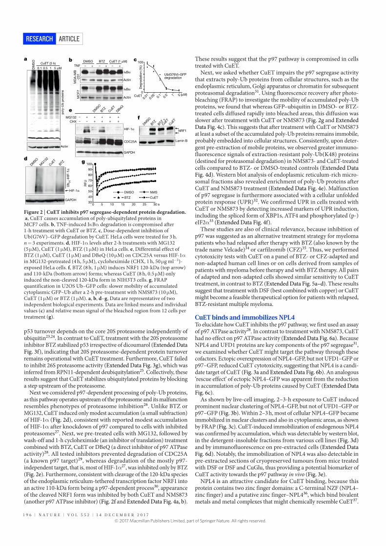
1 9 6 | N A T U R E | V O L 5 5 2 | 1 4 D E C E M B E R 2 0 1 7
ARTICLERESEARCH
p53 turnover depends on the core 20S proteasome independently of ubiquitin23,24. In contrast to CuET, treatment with the 20S proteasome inhibitor BTZ stabilized p53 irrespective of dicoumarol (Extended Data Fig. 3f), indicating that 20S proteasome-dependent protein turnover remains operational with CuET treatment. Furthermore, CuET failed to inhibit 26S proteasome activity (Extended Data Fig. 3g), which was inferred from RPN11-dependent deubiquitylation25. Collectively, these results suggest that CuET stabilizes ubiquitylated proteins by blocking a step upstream of the proteasome.
Next we considered p97-dependent processing of poly-Ub proteins, as this pathway operates upstream of the proteasome and its malfunction resembles phenotypes of proteasome inhibition26. Unlike BTZ or MG132, CuET induced only modest accumulation (a small subfraction) of HIF-1α (Fig. 2d), consistent with reported modest accumulation of HIF-1α after knockdown of p97 compared to cells with inhibited proteasomes27. Next, we pre-treated cells with MG132, followed by wash-off and 1-h cycloheximide (an inhibitor of translation) treatment combined with BTZ, CuET or DBeQ (a direct inhibitor of p97 ATPase activity)28. All tested inhibitors prevented degradation of CDC25A (a known p97 target)29, whereas degradation of the mostly p97- independent target, that is, most of HIF-1α27, was inhibited only by BTZ (Fig. 2e). Furthermore, consistent with cleavage of the 120-kDa species of the endoplasmic reticulum-tethered transcription factor NRF1 into an active 110-kDa form being a p97-dependent process30, appearance of the cleaved NRF1 form was inhibited by both CuET and NMS873 (another p97 ATPase inhibitor) (Fig. 2f and Extended Data Fig. 4a, b).
These results suggest that the p97 pathway is compromised in cells treated with CuET.
Next, we asked whether CuET impairs the p97 segregase activity that extracts poly-Ub proteins from cellular structures, such as the endoplasmic reticulum, Golgi apparatus or chromatin for subsequent proteasomal degradation31. Using fluorescence recovery after photo-bleaching (FRAP) to investigate the mobility of accumulated poly-Ub proteins, we found that whereas GFP–ubiquitin in DMSO- or BTZ-treated cells diffused rapidly into bleached areas, this diffusion was slower after treatment with CuET or NMS873 (Fig. 2g and Extended Data Fig. 4c). This suggests that after treatment with CuET or NMS873 at least a subset of the accumulated poly-Ub proteins remains immobile, probably embedded into cellular structures. Consistently, upon deter-gent pre-extraction of mobile proteins, we observed greater immuno-fluorescence signals of extraction-resistant poly-Ub(K48) proteins (destined for proteasomal degradation) in NMS873- and CuET-treated cells compared to BTZ- or DMSO-treated controls (Extended Data Fig. 4d). Western blot analysis of endoplasmic reticulum-rich micro-somal fractions also revealed enrichment of poly-Ub proteins after CuET and NMS873 treatment (Extended Data Fig. 4e). Malfunction of p97 segregase is furthermore associated with a cellular unfolded protein response (UPR)32. We confirmed UPR in cells treated with CuET or NMS873 by detecting increased markers of UPR induction, including the spliced form of XBP1s, ATF4 and phosphorylated (p-)eIF2α33 (Extended Data Fig. 4f).
These studies are also of clinical relevance, because inhibition of p97 was suggested as an alternative treatment strategy for myeloma patients who had relapsed after therapy with BTZ (also known by the trade name Velcade)34 or carfilzomib (CFZ)35. Thus, we performed cytotoxicity tests with CuET on a panel of BTZ- or CFZ-adapted and non-adapted human cell lines or on cells derived from samples of patients with myeloma before therapy and with BTZ therapy. All pairs of adapted and non-adapted cells showed similar sensitivity to CuET treatment, in contrast to BTZ (Extended Data Fig. 5a–d). These results suggest that treatment with DSF (best combined with copper) or CuET might become a feasible therapeutical option for patients with relapsed, BTZ-resistant multiple myeloma.
CuET binds and immobilizes NPL4To elucidate how CuET inhibits the p97 pathway, we first used an assay of p97 ATPase activity28. In contrast to treatment with NMS873, CuET had no effect on p97 ATPase activity (Extended Data Fig. 6a). Because NPL4 and UFD1 proteins are key components of the p97 segregase31, we examined whether CuET might target the pathway through these cofactors. Ectopic overexpression of NPL4–GFP, but not UFD1–GFP or p97–GFP, reduced CuET cytotoxicity, suggesting that NPL4 is a candi-date target of CuET (Fig. 3a and Extended Data Fig. 6b). An analogous ‘rescue effect’ of ectopic NPL4–GFP was apparent from the reduction in accumulation of poly-Ub proteins caused by CuET (Extended Data Fig. 6c).
As shown by live-cell imaging, 2–3-h exposure to CuET induced prominent nuclear clustering of NPL4–GFP, but not of UFD1–GFP or p97–GFP (Fig. 3b). Within 2–3 h, most of cellular NPL4–GFP became immobilized in nuclear clusters and also in cytoplasmic areas, as shown by FRAP (Fig. 3c). CuET-induced immobilization of endogenous NPL4 was confirmed by accumulation, which was detectable by western blot, in the detergent-insoluble fractions from various cell lines (Fig. 3d) and by immunofluorescence on pre-extracted cells (Extended Data Fig. 6d). Notably, the immobilization of NPL4 was also detectable in pre-extracted sections of cryopreserved tumours from mice treated with DSF or DSF and CuGlu, thus providing a potential biomarker of CuET activity towards the p97 pathway in vivo (Fig. 3e).
NPL4 is an attractive candidate for CuET binding, because this protein contains two zinc finger domains: a C-terminal NZF (NPL4–zinc finger) and a putative zinc finger–NPL436, which bind bivalent metals and metal complexes that might chemically resemble CuET37.
Lamin B
NRF1
DM
SO
BTZ
BTZ
+
CuE
T
CuE
TCHX
MG132
– – + + +
– ++
+
+ + +
DM
SO
BTZ
CuE
T
DBeQ
HIF-1α
CDC25A
GAPDH
DM
SO
BTZ
MG13
2
CuE
T
GAPDH
HIF-1α
Ub
(n)–
HIF
-1α
MC
F7
MD
A-M
B-2
31
IκBα
IκBα
Actin
Actin
– +– +– +
DMSO
TNF
BTZ CuET (1 μM)
0
100
0.01
0.05 0.
10.
5 1 5CuET (μM)
Ub(G76V)–GFPdegradation
Deg
rad
atio
n (%
)
CuET (3 h)
0.1 5 μM10.5
po
ly-U
b p
rote
ins
DM
SO
Actin
BTZ
NMS
CuET
DMSO
30
40
50
60
70
80
90
100
0 10 15 20 25 30 s
a b c
dg
e
f
RF
U
5
Figure 2 | CuET inhibits p97 segregase-dependent protein degradation. a, CuET causes accumulation of poly-ubiquitylated proteins in MCF7 cells. b, TNF-induced IκBα degradation is compromised after 1-h treatment with CuET or BTZ. c, Dose-dependent inhibition of Ub(G76V)–GFP degradation by CuET. HeLa cells were treated for 3 h. n = 3 experiments. d, HIF-1α levels after 2-h treatments with MG132 (5 µM), CuET (1 µM), BTZ (1 µM) in HeLa cells. e, Differential effect of BTZ (1 µM), CuET (1 µM) and DBeQ (10 µM) on CDC25A versus HIF-1α in MG132-pretreated (4 h, 5 µM), cycloheximide (CHX, 1 h, 50 µg ml−1)-exposed HeLa cells. f, BTZ (8 h, 1 µM) induces NRF1 120-kDa (top arrow) and 110-kDa (bottom arrow) forms; whereas CuET (8 h, 0.5 µM) only induced the non-cleaved 120-kDa form in NIH3T3 cells. g, FRAP quantification in U2OS Ub–GFP cells: slower mobility of accumulated cytoplasmic GFP–Ub after a 2-h pre-treatment with NMS873 (10 µM), CuET (1 µM) or BTZ (1 µM). a, b, d–g, Data are representative of two independent biological experiments. Data are linked means and individual values (c) and relative mean signal of the bleached region from 12 cells per treatment (g).
© 2017 Macmillan Publishers Limited, part of Springer Nature. All rights reserved.

1 4 D E C E M B E R 2 0 1 7 | V O L 5 5 2 | N A T U R E | 1 9 7
ARTICLE RESEARCH
Using isothermal calorimetry analysis (ITC)38, we observed a standard dose–response-dependent binding curve (Fig. 3f) compatible with one binding site for CuET on wild-type NPL4 (NPL4(WT)), and a Kd in nanomolar concentrations for the NPL4–CuET interaction. Next, we used mutagenesis to test whether the putative ZF–NPL4 domain has any role in the potential NPL4–CuET interaction. The putative zinc finger domain was preferred, because an endogenous larger form of NPL4 that lacks the C-terminal NZF sequence exists in human cells. This larger NPL4 form is detectable as an upper band on western blots (Fig. 3d) and it is immobilized after CuET treatment, suggesting that the C-terminal NZF is not necessary for the interac-tion with CuET. No ITC interaction was found with a NPL4 mutant (NPL4(MUT)) (Extended Data Fig. 6f) in which both histidines and both cysteines in the putative zinc finger domain were substituted by alanines (Extended Data Fig. 6e). We used drug affinity responsive target stability (DARTS) as another, independent approach, which is based on altered protease susceptibility of target proteins upon drug binding39. Consistently, exposure to CuET caused a differential pronase-dependent proteolysis pattern of NPL4(WT) but not NPL4(MUT) (Extended Data Fig. 6g). These results indicate that NPL4 is directly targeted by CuET and an intact putative zinc finger domain of NPL4 is essential for this interaction.
Notably, ectopically expressed NPL4(MUT)–GFP formed immobile nuclear clusters spontaneously in untreated cells, reminiscent of events seen in cells upon CuET treatment (Fig. 3c, g). Moreover, unlike ectopic NPL4(WT)–GFP, ectopically expressed NPL4(MUT)–GFP not only did not render cells resistant to CuET but also was toxic to the acceptor cells (Extended Data Fig. 6h). We also confirmed that multiple CuET-induced cellular phenotypes were mimicked by the ectopic NPL4(MUT)–GFP model, including accumulation of poly-Ub proteins and UPR activation (Extended Data Fig. 6i).
NPL4 aggregates trigger a heat-shock responseAlthough the nuclear NPL4 clusters occupied DAPI-unlabelled areas of chromatin (Extended Data Fig. 6d) co-localization with DAPI-excluded structures, such as nucleoli and nuclear speckles, were not found (Extended Data Fig. 7a). In late-G2 cells, NPL4 clusters were evidently excluded from the partially condensed chromatin (Extended Data Fig. 7b), suggesting that the NPL4 aggregates exclude chromatin rather than accumulating in specific nuclear areas. Both the nuclear clusters and the immobilized cytoplasmic NPL4 co-localized with poly-Ub proteins (confirmed by anti-Ub(K48) and FK2 antibodies), small ubiquitin-like modifiers (SUMOs) (only in nuclei) and with TDP43 protein40 (Fig. 4a and Extended Data Fig. 7d), which are all features typical of aggregated defective proteins41. The same co-localization patterns were observed for spontaneous clusters formed by NPL4(MUT)–GFP showing that NPL4 aggregation is sufficient for the induction of these phenotypes even without CuET treatment (Extended Data Fig. 7c, e). Blockade of cellular ubiquitylation with a chemical inhibitor (MLN7243) of the E1 ubiquitin-activating enzyme failed to prevent either NPL4–GFP nuclear aggregation or cytoplasmic immobi-lization (Extended Data Fig. 7d), excluding the immobilization of NPL4 via recognition of ubiquitylated and SUMOylated substrates, but rather suggesting that immobilized NPL4 attracts ubiquitylated proteins or proteins that subsequently become ubiquitylated and/or SUMOylated. A key protein commonly associated with intracellular protein aggre-gates is HSP70, a chaperone implicated in aggregate processing42. Indeed, pre-extracted cells showed co-localization of HSP70 with both CuET-induced NPL4(WT)–GFP and spontaneous NPL4(MUT)–GFP aggregates (Fig. 4b, c). Both the CuET-induced NPL4(WT) aggregates and spontaneous NPL4(MUT) aggregates also co-localized with p97 (Extended Data Fig. 7f, g), as is particularly evident after pre-extraction. In the NPL4–GFP model, the amount of p97 immunoreactivity within
Mo
ck
Cu
ET
2 h
125 250 350 500 1,000 (nM)CuET
80
60
40
20
0
100
d
g
e
1 μM CuET (h) 1 μM CuET (h)
Soluble fraction Insoluble fraction (pellet)
U2OS
1 12 23 34 45 5DM
SO
DM
SO
MDA-MB-231
NP
L4
NP
L4
f
NPL4–GFP
NPL4(MUT)–GFP
No extraction Pre-extracted
Veh
icle
DS
FD
SF
+ C
uG
lu
FRAP 15 s
FRAP 15 s
3 hCuET: 0 h
NP
L4
–G
FP
p9
7–G
FP
UF
D1
–G
FP
a b c
CuET to NPL4(WT)
Ratio
Heat
(μJ)
0
0.5 1.0 1.5 2.0 2.5
–1
–2
–3
VariableKa (M
–1)dH (kJ mol–1)n
Kd (M)dS (J mol–1 K)
Value2.1521 × 107
–94.411.2054.647 × 10–8
–176.3
Via
bili
ty (%
)
FRAP 15 s
Empty vectorp97
UFD1NPL4
Figure 3 | CuET binds to and immobilizes NPL4. a, Ectopic NPL4–GFP, but not p97–GFP or UFD1–GFP rescues CuET toxicity in U2OS cells treated for 24 h. n = 3 experiments. Data are mean ± s.d. b, CuET (1 µM) induces intranuclear clustering of NPL4–GFP, but not p97–GFP or UFD1–GFP. c, CuET-induced (1 µM) immobilization of NPL4–GFP (FRAP) in U2OS cells treated for 2 h. Blue boxes, areas before bleaching; arrows, after bleaching. d, NPL4 enrichment in Triton X-100-insoluble fractions
after CuET (1 µM) treatment. e, Immunohistochemistry demonstrates the non-extractable NPL4 in MDA-MB-231 tumours from mice treated with DSF or DSF and CuGlu. f, ICT shows that CuET binds to purified NPL4(WT). g, Spontaneous intranuclear clustering and immobilization of NPL4(MUT)–GFP using FRAP in U2OS cells. Blue boxes, areas before bleaching; arrows, after bleaching. Scale bars, 10 µm (b, c, g) or 50 µm (e). b–g, Data are representative of two independent experiments.
© 2017 Macmillan Publishers Limited, part of Springer Nature. All rights reserved.

1 9 8 | N A T U R E | V O L 5 5 2 | 1 4 D E C E M B E R 2 0 1 7
ARTICLERESEARCH
the NPL4–GFP clusters correlated with the GFP signal intensity, sug-gesting that p97 is immobilized via its interaction with NPL4. The other NPL4-binding partner, UFD1, was almost undetectable in detergent- insoluble pellets of CuET-treated or NPL4(MUT)–GFP-expressing cells despite clear p97 immobilization (Extended Data Fig. 8a, b), suggesting that UFD1 cannot bind to, or becomes only loosely attached to, the aggregated NPL4–p97 complex. Notably, non-extractable cellular p97 is detectable after CuET treatment (Extended Data Fig. 8c), including in stained tumour sections from mice treated with DSF or DSF and CuGlu, providing an additional candidate marker for CuET activity in vivo (Extended Data Fig. 8d).
Because aggregation of misfolded or damaged proteins triggers cellular heat-shock response (HSR) through an HSF1-dependent mechanism43, we confirmed that CuET treatment indeed triggered a robust HSR accompanied by characteristic HSF1 nuclear stress foci (Fig. 4d) that were also detectable in cells spontaneously aggregating NPL4(MUT)–GFP (Fig. 4e). HSR markers, including accumulation of heat-shock proteins and a phosphorylation shift in HSF1, were detect-able by western blot (Extended Data Fig. 8e, f).
DiscussionOur results help to explain the anti-cancer activity of the alcohol-abuse drug disulfiram. We propose a model for DSF cytotoxic activity, featur-ing rapid conversion of DSF into CuET, which accumulates in tumours. After entering cells, CuET binds NPL4 and induces its aggregation, con-sequently disabling the vital p97–NPL4–UFD1 pathway and inducing a complex cellular phenotype leading to cell death (Fig. 4f). Supporting CuET as the active metabolite is the correlation of CuET concentrations (active in the nanomolar range) with the biological effects and func-tional impact on the targeted pathway(s) in vivo. In addition, CuET is the only known metabolite of DSF containing copper ions, a metal
that enhances the anti-tumour effects of DSF; it is unlikely that another DSF metabolite could represent the major anti-cancer agent as levels of non-CuET metabolites should be lowered by copper addition. We also present a method for CuET detection in tissues and plasma, as well as data suggesting that preferential accumulation of CuET in tumours may contribute to cancer cell toxicity, consistent with the high therapeutic tolerability of DSF3, as documented even after years of daily administra-tion at doses comparable to those we used in our mouse experiments. Considering the numerous studies on DSF and diverse opinions about the potential target of its anti-cancer effects44, identification of NPL4, a key component of the p97–NPL4–UFD1 segregase complex, as the molecular target of CuET is surprising. The CuET–NPL4 interaction leads to rapid formation of protein aggregates and immobilization of this otherwise very mobile multifunctional protein complex, resulting in a severe phenotype, induction of HSR and eventually cell death. While additional potential targets of CuET cannot be excluded, the mal-function of the p97 pathway due to the NPL4–p97 aggregate formation explains the major cell phenotypes and the consequent cell death. Our work also reconciles the controversial studies6,12, suggesting that the proteasome is the DSF target, by demonstrating that neither 20S nor 26S proteasome, but the processing of ubiquitylated proteins by the NPL4-dependent segregase, is targeted by CuET. Our results support the notion that the p97–NPL4 pathway is a promising therapeutic target in oncology45,46. Indeed, reports on p97 overabundance correlating with progression and metastasis of carcinomas of the breast, colon and prostate47–49 are consistent with our present nationwide epidemio-logical analysis, which revealed an association between continued use of DSF and favourable prognosis, an intriguing finding that should be investigated further, particularly given the currently limited therapeutic options for patients with metastatic cancer. From a broader perspective, our study illustrates the potential of multifaceted approaches to drug repurposing, providing novel mechanistic insights, identification of new cancer-relevant targets and encouragement for further clinical trials, here with DSF, an old, safe and public domain drug4 that might help to save lives of patients with cancer worldwide.
Online Content Methods, along with any additional Extended Data display items and Source Data, are available in the online version of the paper; references unique to these sections appear only in the online paper.
Received 1 October 2015; accepted 8 November 2017.
Published online 6 December 2017.
1. Collins, F. S. Mining for therapeutic gold. Nat. Rev. Drug Discov. 10, 397 (2011).
2. Pantziarka, P. et al. The repurposing drugs in oncology (ReDO) project. Ecancermedicalscience 8, 442 (2014).
3. Iljin, K. et al. High-throughput cell-based screening of 4910 known drugs and drug-like small molecules identifies disulfiram as an inhibitor of prostate cancer cell growth. Clin. Cancer Res. 15, 6070–6078 (2009).
4. Cvek, B. Nonprofit drugs as the salvation of the world’s healthcare systems: the case of Antabuse (disulfiram). Drug Discov. Today 17, 409–412 (2012).
5. Shen, M. L., Johnson, K. L., Mays, D. C., Lipsky, J. J. & Naylor, S. Determination of in vivo adducts of disulfiram with mitochondrial aldehyde dehydrogenase. Biochem. Pharmacol. 61, 537–545 (2001).
6. Chen, D., Cui, Q. C., Yang, H. & Dou, Q. P. Disulfiram, a clinically used anti-alcoholism drug and copper-binding agent, induces apoptotic cell death in breast cancer cultures and xenografts via inhibition of the proteasome activity. Cancer Res. 66, 10425–10433 (2006).
7. Zha, J. et al. Disulfiram targeting lymphoid malignant cell lines via ROS–JNK activation as well as Nrf2 and NF-κB pathway inhibition. J. Transl. Med. 12, 163 (2014).
8. Safi, R. et al. Copper signaling axis as a target for prostate cancer therapeutics. Cancer Res. 74, 5819–5831 (2014).
9. Liu, P. et al. Liposome encapsulated disulfiram inhibits NFκB pathway and targets breast cancer stem cells in vitro and in vivo. Oncotarget 5, 7471–7485 (2014).
10. Dufour, P. et al. Sodium dithiocarb as adjuvant immunotherapy for high risk breast cancer: a randomized study. Biotherapy 6, 9–12 (1993).
11. Yip, N. C. et al. Disulfiram modulated ROS–MAPK and NFκB pathways and targeted breast cancer cells with cancer stem cell-like properties. Br. J. Cancer 104, 1564–1574 (2011).
e
c
dHSP70
a b
SUMO2/3
f
CuET
DSF
Organismal level
Tumour level
Cellular effects
Cu2+
p97
p97NPL4
UFD1
UFD1
Molecular targetNPL4 protein
p97
NPL4
HSP70
HSP70
HSF1NPL4(MUT)–GFP
NPL4–GFP Poly-Ub(K48)Overlay+ DAPI
Overlay+ DAPI
Overlay+ DAPI
TDP43NPL4–GFP
HSF1NPL4–GFP
Overlay+ DAPI
Overlay+ DAPI
HSF1NPL4–GFP
Mo
ck
CuE
T
Mo
ck
CuE
T
Overlay+ DAPINPL4–GFP Overlay
+ DAPINPL4–GFP
Overlay+ DAPINPL4–GFP
Overlay+ DAPINPL4(MUT)–GFP
Figure 4 | NPL4 protein aggregation triggers HSR. a, NPL4–GFP co-localizes with SUMO2/3, poly-Ub(K48) and TDP43 in U2OS cells. Cells were treated with 1 µM CuET for 3 h and pre-extracted. b, NPL4–GFP co-localizes with HSP70 in mock- and CuET-treated U2OS cells. Cells were treated with 1 µM CuET for 3 h and pre-extracted. c, NPL4(MUT)–GFP co-localizes with HSP70 in U2OS cells after pre-extraction. d, CuET-induced HSF1 stress bodies. NPL4–GFP U2OS cells were treated with 1 µM CuET for 3 h. e, HSF1 stress bodies in U2OS cells expressing NPL4(MUT)–GFP. f, Model of DSF anti-cancer activity in patients. Scale bars, 10 µm. a–e, Data are representative of two independent experiments.
© 2017 Macmillan Publishers Limited, part of Springer Nature. All rights reserved.

1 4 D E C E M B E R 2 0 1 7 | V O L 5 5 2 | N A T U R E | 1 9 9
ARTICLE RESEARCH
12. Lövborg, H. et al. Inhibition of proteasome activity, nuclear factor-κB translocation and cell survival by the antialcoholism drug disulfiram. Int. J. Cancer 118, 1577–1580 (2006).
13. Allensworth, J. L. et al. Disulfiram (DSF) acts as a copper ionophore to induce copper-dependent oxidative stress and mediate anti-tumor efficacy in inflammatory breast cancer. Mol. Oncol. 9, 1155–1168 (2015).
14. Nechushtan, H. et al. A phase IIb trial assessing the addition of disulfiram to chemotherapy for the treatment of metastatic non-small cell lung cancer. Oncologist 20, 366–367 (2015).
15. Jin, M. et al. Alcohol drinking and all cancer mortality: a meta-analysis. Ann. Oncol. 24, 807–816 (2013).
16. Li, Y. et al. Copper improves the anti-angiogenic activity of disulfiram through the EGFR/Src/VEGF pathway in gliomas. Cancer Lett. 369, 86–96 (2015).
17. Suzuki, Y. et al. The origin of an EPR signal observed in dithiocarbamate-loaded tissues. Copper(ii)-dithiocarbamate complexes account for the narrow hyperfine lines. Biochim. Biophys. Acta 1335, 242–245 (1997).
18. Kepp, O., Galluzzi, L., Lipinski, M., Yuan, J. & Kroemer, G. Cell death assays for drug discovery. Nat. Rev. Drug Discov. 10, 221–237 (2011).
19. Doil, C. et al. RNF168 binds and amplifies ubiquitin conjugates on damaged chromosomes to allow accumulation of repair proteins. Cell 136, 435–446 (2009).
20. Li, J. M., Wu, H., Zhang, W., Blackburn, M. R. & Jin, J. The p97–UFD1L–NPL4 protein complex mediates cytokine-induced IκBα proteolysis. Mol. Cell. Biol. 34, 335–347 (2014).
21. Chou, T. F. & Deshaies, R. J. Quantitative cell-based protein degradation assays to identify and classify drugs that target the ubiquitin–proteasome system. J. Biol. Chem. 286, 16546–16554 (2011).
22. Kisselev, A. F. & Goldberg, A. L. Monitoring activity and inhibition of 26S proteasomes with fluorogenic peptide substrates. Methods Enzymol. 398, 364–378 (2005).
23. Asher, G., Lotem, J., Cohen, B., Sachs, L. & Shaul, Y. Regulation of p53 stability and p53-dependent apoptosis by NADH quinone oxidoreductase 1. Proc. Natl Acad. Sci. USA 98, 1188–1193 (2001).
24. Asher, G., Tsvetkov, P., Kahana, C. & Shaul, Y. A mechanism of ubiquitin-independent proteasomal degradation of the tumor suppressors p53 and p73. Genes Dev. 19, 316–321 (2005).
25. Verma, R. et al. Role of Rpn11 metalloprotease in deubiquitination and degradation by the 26S proteasome. Science 298, 611–615 (2002).
26. Dai, R. M. & Li, C. C. Valosin-containing protein is a multi-ubiquitin chain-targeting factor required in ubiquitin–proteasome degradation. Nat. Cell Biol. 3, 740–744 (2001).
27. Alexandru, G. et al. UBXD7 binds multiple ubiquitin ligases and implicates p97 in HIF1α turnover. Cell 134, 804–816 (2008).
28. Chou, T. F. et al. Reversible inhibitor of p97, DBeQ, impairs both ubiquitin-dependent and autophagic protein clearance pathways. Proc. Natl Acad. Sci. USA 108, 4834–4839 (2011).
29. Riemer, A. et al. The p97–Ufd1–Npl4 ATPase complex ensures robustness of the G2/M checkpoint by facilitating CDC25A degradation. Cell Cycle 13, 919–927 (2014).
30. Radhakrishnan, S. K., den Besten, W. & Deshaies, R. J. p97-dependent retrotranslocation and proteolytic processing govern formation of active Nrf1 upon proteasome inhibition. eLife 3, e01856 (2014).
31. Meyer, H., Bug, M. & Bremer, S. Emerging functions of the VCP/p97 AAA-ATPase in the ubiquitin system. Nat. Cell Biol. 14, 117–123 (2012).
32. Magnaghi, P. et al. Covalent and allosteric inhibitors of the ATPase VCP/p97 induce cancer cell death. Nat. Chem. Biol. 9, 548–556 (2013).
33. Samali, A., Fitzgerald, U., Deegan, S. & Gupta, S. Methods for monitoring endoplasmic reticulum stress and the unfolded protein response. Int. J. Cell Biol. 2010, 830307 (2010).
34. Auner, H. W. et al. Combined inhibition of p97 and the proteasome causes lethal disruption of the secretory apparatus in multiple myeloma cells. PLoS ONE 8, e74415 (2013).
35. Soriano, G. P. et al. Proteasome inhibitor-adapted myeloma cells are largely independent from proteasome activity and show complex proteomic changes, in particular in redox and energy metabolism. Leukemia 30, 2198–2207 (2016).
36. Lass, A., McConnell, E., Fleck, K., Palamarchuk, A. & Wójcik, C. Analysis of Npl4 deletion mutants in mammalian cells unravels new Ufd1-interacting motifs and suggests a regulatory role of Npl4 in ERAD. Exp. Cell Res. 314, 2715–2723 (2008).
37. Vorác ková, I., Suchanová, S., Ulbrich, P., Diehl, W. E. & Ruml, T. Purification of proteins containing zinc finger domains using immobilized metal ion affinity chromatography. Protein Expr. Purif. 79, 88–95 (2011).
38. Holdgate, G. et al. Biophysical methods in drug discovery from small molecule to pharmaceutical. Methods Mol. Biol. 1008, 327–355 (2013).
39. Lomenick, B. et al. Target identification using drug affinity responsive target stability (DARTS). Proc. Natl Acad. Sci. USA 106, 21984–21989 (2009).
40. Becker, L. A. et al. Therapeutic reduction of ataxin-2 extends lifespan and reduces pathology in TDP-43 mice. Nature 544, 367–371 (2017).
41. Guo, L. et al. A cellular system that degrades misfolded proteins and protects against neurodegeneration. Mol. Cell 55, 15–30 (2014).
42. Kim, Y. E., Hipp, M. S., Bracher, A., Hayer-Hartl, M. & Hartl, F. U. Molecular chaperone functions in protein folding and proteostasis. Annu. Rev. Biochem. 82, 323–355 (2013).
43. Dai, C. & Sampson, S. B. HSF1: guardian of proteostasis in cancer. Trends Cell Biol. 26, 17–28 (2016).
44. Cvek, B. Targeting malignancies with disulfiram (Antabuse): multidrug resistance, angiogenesis, and proteasome. Curr. Cancer Drug Targets 11, 332–337 (2011).
45. Deshaies, R. J. Proteotoxic crisis, the ubiquitin–proteasome system, and cancer therapy. BMC Biol. 12, 94 (2014).
46. Anderson, D. J. et al. Targeting the AAA ATPase p97 as an approach to treat cancer through disruption of protein homeostasis. Cancer Cell 28, 653–665 (2015).
47. Cui, Y. et al. High expression of valosin-containing protein predicts poor prognosis in patients with breast carcinoma. Tumour Biol. 36, 9919–9927 (2015).
48. Yamamoto, S. et al. Expression of valosin-containing protein in colorectal carcinomas as a predictor for disease recurrence and prognosis. Clin. Cancer Res. 10, 651–657 (2004).
49. Tsujimoto, Y. et al. Elevated expression of valosin-containing protein (p97) is associated with poor prognosis of prostate cancer. Clin. Cancer Res. 10, 3007–3012 (2004).
Supplementary Information is available in the online version of the paper.
Acknowledgements We thank J. Škvor, M. Zadinová, J. Vec erka and D. Doležal for help with animal experiments, Jana Vrbkova for statistical analysis, D. Fridecky and T. Adam for help with HPLC, I. Protivankova and M. Grønvig Nielsen for technical assistance. This work was supported by grants from the Kellner Family Foundation, Czech National Program of Sustainability, Grant Agency of the Czech Republic, MEYS CR project Czech-BioImaging, the Czech Health Research Council, of the Danish Cancer Society, the Danish National Research Foundation (project CARD), the Danish Council for Independent Research, the Novo Nordisk Foundation, the Czech Cancer League, the Swedish Research Council, Cancerfonden of Sweden, the European Commission (EATRIS), the Czech Ministry of Education, youth and sports (OPVKCZ), Cancer Research Czech Republic and the Howard Hughes Medical Institute.
Author Contributions Z.S., M.Mis., B.C., R.J.D. and J.Barte. conceived the study. Z.S. and M.Mis. performed most biochemical and microscopy experiments and wrote the manuscript. D.M. established the expression cell lines and performed most cytotoxicity tests. T.O., P.D. and I.V. performed the HPLC experiments. K.K.A., S.F. and J.O. performed the epidemiological analyses. J.Bartk. performed the immunohistochemical analyses. J.V. and P.D. performed DARTS experiments. P.M. performed cell death analyses. Z.T. performed cytotoxicity tests and heat-shock response analyses. A.K. performed cytotoxicity tests. A.M. designed and performed phlebotomies of patients treated with Antabuse. M.Mic. performed the ITC. J.G. performed FACS analyses, cell death assays and cell sorting. J.S. performed 20S proteasome assays. J.L. performed 26S proteasome assays. M.K. and C.D. performed the cytotoxicity experiments on myeloid- and patient-derived cell lines. P.P., J.M. and M.H. performed mouse experiments. J.Barte., B.C., Q.P.D. and R.J.D. helped to design the experiments, interpreted the data and wrote/edited the manuscript. All authors approved the manuscript.
Author Information Reprints and permissions information is available at www.nature.com/reprints. The authors declare competing financial interests: details are available in the online version of the paper. Readers are welcome to comment on the online version of the paper. Publisher’s note: Springer Nature remains neutral with regard to jurisdictional claims in published maps and institutional affiliations. Correspondence and requests for materials should be addressed to J.Barte. ([email protected]), B.C. (cvekb@ seznam.cz) and R.J.D. ([email protected]).
Reviewer Information Nature thanks P. Brossart and the other anonymous reviewer(s) for their contribution to the peer review of this work.
© 2017 Macmillan Publishers Limited, part of Springer Nature. All rights reserved.

ARTICLERESEARCH
METHODSThe experiments were not randomized.Epidemiological analyses and access to health registers. We conducted a population- based cohort study by combining Danish nationwide demographic and health registers. This study was approved by the Danish Data Protection Agency and Statistics Denmark’s Scientific Board. As the epidemiological study was based solely on register data and did not involve any contact with patients, no ethical approval was required from the Danish Scientific Ethical Committee50. The cohort consisted of all Danes aged 35–85 years with a first-time diagnosis of cancer between January 2000 and December 2013. Because DSF (Antabuse) is a relative contra-indication among individuals with liver or kidney diseases, we excluded patients with cancers of the liver or kidney from the cohort. Cohort members were categorized according to use of DSF into two main groups: (i) those who filled at least one prescription of DSF within five years before the cancer diagnosis, but did not fill DSF prescrip-tion(s) during the first year after the diagnosis (previous users), that is, individu-als suffering from alcoholism but taking DSF only before their cancer diagnosis; and (ii) those who used DSF before their cancer diagnosis and also filled one or more DSF prescriptions during the first year after the cancer diagnosis (continuing users), that is, individuals who underwent DSF therapy both before and after the cancer diagnosis. We also defined a category of patients with cancer who did not fill prescription(s) for DSF either before or after (≤1 year) the cancer diagnosis (never users). In the main analyses, we calculated hazard ratios and 95% confidence inter-vals estimating cancer-specific mortality among continuing DSF users compared to previous DSF users based on a Cox model regressing on both propensity scores and disulfiram use. By including propensity scores in the regression, we used demo-graphic data and comorbid conditions/diagnostic codes as well as prescription data for selected concomitant drugs, to balance baseline characteristics of previous and continuing users of DSF and to adjust estimated hazard ratios of cancer-specific mortality associated with DSF use51. The patients with cancer were followed from one year after the diagnosis until death, migration or end of study (31 December 2014). The propensity scores thus estimate the probability of being treated with DSF in the exposure window 0–1 year after the cancer diagnoses conditional on the following other covariates in the calculation of propensity scores using logistic regression: gender, age at diagnosis, calendar time, highest achieved education and disposable income; medical histories of diabetes mellitus, chronic obstructive pulmonary disease, ischaemic heart disease, congestive heart failure, cerebrovascular disease, atrial fibrillation or atrial flutter, dementia and ulcer disease; and use of non-steroidal anti-inflammatory drugs (NSAIDs; including aspirin), non-aspirin antithrombotic agents (anticoagulants), statins, antihypertensive medication, other cardiovascular drugs, anti-diabetics and psychotropic drugs. In the Cox model, the propensity score is further included as a restricted cubic spline to model possible nonlinearities, in addition to the categorical disulfiram use, which is the variable of interest. Analyses were run for cancer overall and for breast, prostate and colon cancer, separately. Furthermore, all analyses were stratified by stage (localized, non-localized or unknown). Statistical significance of DSF use was evaluated by likelihood ratio tests. We used the software R for statistical computing52.In vivo tumour experiments. The human breast cancer cell line MDA-MB-231 was injected (107 cells transplanted subcutaneously) to grow tumours in athymic NU/NU female mice (AnLab Ltd) with a body weight of 23.6–26.9 g, aged 12 weeks. Inclusion criteria were: female, appropriate age and weight (15–30 g). Exclusion criteria were: tumour size must not exceed 20 mm (volume 4,000 mm3) in any direction in an adult mouse, the tumour mass should not proceed to the point where it significantly interferes with normal bodily functions, or causes pain or distress owing to its location, persistent self-induced trauma, rapid or progressive weight loss of more than 25%, for seven days. In none of the experiments were these approved ethical limits exceeded. After the tumours grew to 0.114–0.117 cm3 on average, mice were randomly divided into four groups, each of eight mice, and treated as follows: (i) normal diet; (ii) normal diet plus oral administration of 0.15 mg kg−1 copper gluconate (CuGlu); (iii) normal diet plus oral administra-tion of 50 mg kg−1 DSF; (iv) normal diet plus oral administration of 0.15 mg kg−1 CuGlu and 50 mg kg−1 DSF. Administration of compounds was carried out as a blinded experiment (all information about the expected outputs and the nature of used compounds were kept from the animal technicians). CuGlu was admini-stered each day in the morning (08:00) and DSF each day in the evening (19:00) to mimic a clinical trial combining DSF and CuGlu in treatment of tumours involving the liver (NCT00742911). After treatment began, mice were weighed and their tumours measured twice per week. At day 32, mice were euthanized, and the tumours were removed and frozen at −80 °C. The experiment was evaluated by comparing growth curves of tumours in the experimental groups with those in controls. The rates of tumour growth inhibition (TGI) were calculated by the formula TGI = (1 − Vtreated/Vcontrol where Vtreated is the mean of tumour volumes in the treated group and Vcontrol is the mean of tumour volumes in the control group).
Mean tumour volume values at specific time intervals were statistically evaluated. To test directly the effect of CuET, we used MDA-MB-231 and AMO-1 models. MDA-MB-231 was injected (5 × 10⁶ cells were transplanted subcutaneously) to grow tumours in SCID mice (ENVIGO) aged 10 weeks (±2 weeks). AMO-1 xenografts were expanded in SCID mice. Each group consisted of 10 animals, each bearing two tumours. CuET was formulated in bovine serum albumin solu-tion (1%) to a final concentration of 1 mg ml−1. CuET was applied intraperito-neally with a schedule of five days on and two days off. All aspects of the animal study met the accepted criteria for the care and experimental use of laboratory animals, and protocols were approved by the Animal Research Committee of the 1st Faculty of Medicine Charles University in Prague and Ethical Committee of Faculty of Medicine and Dentistry, Palacky University in Olomouc. For HPLC–MS and immunohistochemistry analysis, we used MDA-MB-231 xenografted mice treated with the same DSF and DSF plus copper gluconate regime as described for the anti-cancer activity assessment with the notable difference that mice were euthanized after five days of treatment.HPLC–MS analysis of CuET. The HR-MRM analysis was performed on a HPLC-ESI-QTOF system consisting of HPLC chromatograph Thermo UltiMate 3000 with AB Sciex TripleTOF 5600+ mass spectrometer, using the DuoSpray ESI source operated at an ion source voltage of 5,500 V, ion source gas flow rates of 40 units, curtain gas flow rate of 30 units, declustering potential of 100 V and temperature 400 °C. Data were acquired in product ion mode with two parent masses (358.9 and 360.9) for analysis of CuET. Chromatographic separation was done by PTFE column, which was especially designed for analysis of strong metal chelators filled by C18 sorbent (IntellMed, IM_301). Analysis was performed at room temperature and with a flow rate of 1,500 µl min−1 with isocratic chromatography. Mobile phase consisted of HPLC grade acetone (Lachner) 99.9%, HPLC water (Merck Millipore) 0.1% and 0.03% HPLC formic acid (Sigma-Aldrich). Acquired mass spectra were evaluated in software PeakView 1.2. Extracted ion chromatograms of transitions 88.0 and 116.0 (common for both parent masses) with a 0.1 mass tolerance were Gaussian smoothened with width of two points. Peak area was then recorded and recalculated to ng ml−1 according to the calibration curve.Sample preparation for HPLC–MS analysis. Liquid nitrogen-frozen biological samples were cut into small pieces using a scalpel. Samples (30–100 mg) were imme-diately processed by homogenization in 100% acetone in a ratio of 1:10 sample: acetone (for plasma or serum the ratio was 1:4). Homogenization was done in a table-top homogenizer (Retsch MM301) placed in a cold room (4 °C) in 2-ml Eppendorf tubes with 2 glass balls (5 mm) for 1 min at 30 Hz. The tube was imme-diately centrifuged at 4 °C, 20,000g for 2 min. Supernatant was decanted into a new 1.5-ml Eppendorf tube and immediately centrifuged for 30 min using a small table-top centrifuge (BioSan FVL-2400N) placed inside a −80 °C freezer. Supernatant was quickly decanted into a glass HPLC vial and kept at −80 °C for no longer than 6 h. Just before the HPLC analysis, the vial was placed into a pre-cooled (4 °C) LC-sample rack and immediately analysed. To enable an approximate quantifica-tion of analysed CuET, a calibration curve was prepared. Various amounts of CuET were spiked in plasma, frozen in liquid nitrogen, and placed at −80 °C to mimic sample processing. Standards were then processed as the samples described above. To measure circulating CuET concentrations, mice were given a single oral dose of DSF (50 mg kg−1) and euthanized at different time points. Serum was collected and frozen for analysis.Blood collection from humans for HPLC–MS analysis of CuET. Blood samples were collected before and 3 h after oral application of DSF (Antabuse, 400 mg) dissolved in water. Phlebotomy needles were specific for metal analysis—Sarstedt Safety Kanule 21G × 1½ inches, 85.1162.600. Collection tubes were specific for metal analysis —Sarstedt, S-Monovette 7.5 ml LH, 01.1604.400. Immediately after blood collection samples were centrifuged in a pre-cooled centrifuge (4 °C at 1,300g for 10 min). After centrifugation, tubes were placed on ice and the plasma fraction was immediately aliquoted into the 1.5-ml Eppendorf tubes with approximately 500 µl per tube. The tubes with plasma were immediately frozen on dry ice and later stored in −80 °C. Blood samples were collected from volunteers who gave informed consent and were undergoing Antabuse therapy because of alcohol abuse. Participants were four males (aged 34, 38, 41, 60 years) and five females (aged 37, 56, 46, 59, 63 years). All individuals were freshly diagnosed for alcohol-use disorder and were scheduled for Antabuse therapy. Blood samples were collected before and after the first use of Antabuse. All relevant ethical regulations were followed for the study. The study, including the collection of blood samples, was approved by the Ethical Committee of Faculty of Medicine and Dentistry, Palacky University in Olomouc.Cell lines. Cell lines were cultured in appropriate growth medium supplemented with 10% fetal bovine serum (FBS) and penicillin–streptomycin; and maintained in a humidified, 5% CO2 atmosphere at 37 °C. Cell lines cultured in DMEM medium were: HCT116 (ATCC), DU145 (ECACC), PC3 (ECACC), T47D (NCI60),
© 2017 Macmillan Publishers Limited, part of Springer Nature. All rights reserved.

ARTICLE RESEARCH
HS578T (NCI60), MCF7 (ECACC), MDA-MB-231 (ATCC), U2OS (ECACC), HeLa (ATCC), NIH-3T3 (ATCC), CAPAN-1 (ATCC), A253 (ATCC), FaDu (ATCC), h-TERT-RPE1 (ATCC), HeLa-Ub(G76V)-GFP-ODD-Luc21. Cell lines cultured in RPMI1640 medium were: NCI-H358 (ATCC), NCI-H52 (ATCC), HCT-15 (ATCC), AMO-1 (ATCC), MM-1S (ATCC), ARH77 (ATCC), RPMI8226 (ATCC), OVCAR-3 (NCI60), CCRF-CEM (ATCC), K562 (ATCC), 786-0 (NCI60). Cell lines cultured in EMEM medium were: U87-MG (ATCC), SiHA (ATCC). Cell line A549 (ATCC) was cultured in F12K medium, HT29 (ATCC) in McCoy’s medium and LAPC4 (provided by Z. Culig, University of Innsbruck) in IMDM medium supplemented with metribolone R1881 (Sigma-Aldrich). RWPE-1 (ATCC) cells were cultured in a keratinocyte serum-free medium supplemented with bovine pituitary extract and human recombinant epidermal growth factor (Thermo Fisher Scientific). BTZ- and CFZ-resistant multiple myeloma cell lines were previously described in ref. 35. Cell lines were tested for mycoplasma con-tamination and authenticated by STR method. None of the cell lines used in this study is listed in the database of commonly misidentified cell lines maintained by ICLAC.Stable cell line construction. For construction of all stably transfected cell lines we used the U2OS cell line (ECACC). For U2OS Ub–GFP, we used the commercial Ub–GFP EGFP-C1 vector (Addgene); for U2OS NPL4–GFP, we used the com-mercial NPLOC4–GFP pCMV6-AC-GFP vector (Origene); for U2OS p97–GFP, we used the commercial VCP–GFP pCMV6-AC-GFP vector (Origene); and for U2OS UFD1–GFP, we used the commercial UFD1L–GFP pCMV6-AC-GFP vector (Origene). Cells were transfected using Promega FugeneHD according to the manu-facturer’s instructions. Cells were further cultured in the appropriate antibiotics (geneticin, 400 µg ml−1). Medium with geneticin was replaced every 2–3 days until the population of resistant cells was fully established. Cells were further refined by sorting for cells expressing GFP (BD FACS Aria). For preparation of inducible NPL4(MUT)–GFP cells, U2OS cells were transfected with a pcDNA6/TR plas-mid (Invitrogen, V1025-20) using the FugeneHD transfection reagent (Promega, E2311) according to the manufacturer’s protocol. To generate a cell line that stably expressed the Tet repressor, U2OS cells were cultured in selective medium with blasticidin (10 µg ml−1) for 10 days. Blasticidin-resistant colonies were picked, expanded and screened for clones that exhibited the lowest basal levels and highest inducible levels of expression. Next, the most suitable clones were transfected with the PCDNA4/TO expression vector encoding the mutated NPL4–GFP protein using the Fugene transfection reagent. Cells were cultured in medium with zeo-cin (500 µg ml−1) to select clones that contain pcDNA4/TO-mutated NPL4–GFP. The NPL4(MUT)–GFP-encoding plasmid was obtained from Generi Biotech. To induce expression of protein, cells were incubated with doxycycline (Sigma-Aldrich) 1 µg ml−1 for 16–48 h.Colony-formation assay. Cells were seeded into six-well plates at 100–300 cells per well (depending on the cell line). The next day, cells were treated with compounds as indicated in the specific experiments and kept in culture for 7–14 days. Colonies were visualized by crystal violet and counted.XTT assay. Cells were plated at a density of 10,000 per well in a 96-well plate. The next day, cells were treated as indicated. After 24 h, an XTT assay was per-formed according to the manufacturer’s instructions (Applichem). XTT solution was added to the medium and incubated for 30–60 min, and then the dye intensity was measured at the 475 nm wavelength using a spectrometer (TECAN, Infinite M200PRO). Results are shown as mean ± s.d. from three independent experiments, each performed in triplicate. For LD50 analysis across the panel of cell lines listed in Extended Data Fig. 2b, cell lines were treated with various doses (at least five doses) for 48 h. LD50 values were calculated using Graphpad Prism software based on survival curves from at least two independent experiments.Annexin V staining. Cell cultures were treated as indicated and collected by trypsinization. Initial culture medium and washing buffer were collected to include detached cells. Cells were centrifuged (250g, 5 min) and re-suspended in a staining buffer (140 mM NaCl, 4 mM KCl, 0.75 mM MgCl2, 10 mM HEPES) containing 2.5 mM CaCl2, Annexin-V–APC (1:20, BD Biosciences) and 2.5 µg ml−1 7-AAD (BD Biosciences) for 15 min on ice in the dark. Samples were analysed by flow cytometry using BD FACSVerse (BD Biosciences) and at least 10,000 events were acquired per sample. Collected data were processed using BD FACSSuite (BD Biosciences) and exported into Microsoft Excel.Caspases 3/7 assay. Activity of caspase-3 and -7 was quantified by cleavage of fluorogenic substrate CellEvent Caspase-3/7 Green Detection Reagent (Thermo Fisher Scientific). In brief, samples prepared in the same staining buffer as described for annexin V staining above, supplemented with 2% FBS, 0.5 µM CellEvent Caspase-3/7 Green Detection Reagent and incubated for 45 min at room temperature in the dark. Subsequently, 0.5 µg ml−1 DAPI was added and samples were analysed by flow cytometry using BD FACSVerse (BD Biosciences) and at least 10,000 events were acquired per sample. Collected data were processed using BD FACSSuite (BD Biosciences) and exported into Microsoft Excel.
Viability assay of multiple myeloma cells. The CellTiter 96 MTS-assay (Promega) was used according to the manufacturer’s instructions to determine the cell viability of BTZ (Janssen Cilag), CFZ and CuEt in cell lines and the absorbance of the for-mazan product was measured in 96-well microplates at 492 nm. The assay measures dehydrogenase enzyme activity found in metabolically active cells.
For patient cells, the more sensitive luminescent CellTiterGlo assay (Promega) was used to determine cell viability, measured by ATP production of metaboli-cally active cells. The primary myeloma cell samples were obtained after written informed consent and approval by the independent ethics review board (St Gallen ethics committee—Ethikkommission Ostschweiz), in accordance with ICH-GCP and local regulations. Malignant plasma cells were retrieved by PBMC isolation from a patient with multiple myeloma progressing under BTZ-containing therapy, based on IMWG criteria (BTZ-resistant) and an untreated patient with multiple myeloma (BTZ-sensitive). The purity of the cell samples was >80% myeloma cells, as assessed by morphology.Immunoblotting and antibodies. Equal amounts of cell lysates were sepa-rated by SDS–PAGE on hand-cast or precast tris–glycine gradient (4–20%) gels (Life Technologies), and then transferred onto a nitrocellulose membrane. The membrane was blocked with 5% bovine milk in Tris-buffered saline containing 0.1% Tween-20 for 1 h at room temperature, and then incubated overnight at 4 °C or for 1 h at room temperature, with one of the following primary anti-bodies (all antibodies were used in the system under study (assay and species) according to the instructions of the manufacturer): anti-ubiquitin (1:1,000; Cell Signaling, 3933), anti-H2A, acidic patch (1:1,000; Merck Millipore, 07-146), anti- monoubiquityl-H2A (1:1,000; Merck Millipore, clone E6C5), anti-IκBα (1:500; Santa Cruz Biotechnology, sc-371), anti-p53 (1:500; Santa Cruz Biotechnology, clone DO-1), anti-HIF-1α (1:1,000; BD Biosciences, 610958), anti-CDC25A (1:500; Santa Cruz Biotechnology, clone DCS-120), anti-NRF1 (1:1,000; Cell Signaling, clone D5B10), anti-VCP (1:2,000; Abcam, ab11433), anti-VCP (1:1,000; Novus Bio, NBP100-1557), anti-NPLOC4 (1:1,000; Novus Bio, NBP1-82166), anti-ubiquitin lys48-specific (1:1,000; Merck Millipore, clone Apu2), anti- -actin (1:2,000; Santa Cruz Biotechnology, sc-1616; or 1:500, Santa Cruz Biotechnology, sc-87778), anti-GAPDH (1:1,000,GeneTex, clone 1D4), anti-lamin B (1:1,000; Santa Cruz Biotechnology, sc-6217), anti-calnexin (1:500; Santa Cruz Biotechnology, sc-11397), anti-α-tubulin (1:500; Santa Cruz Biotechnology, sc-5286), anti-XBP1 (1:500; Santa Cruz Biotechnology, sc-7160), UFD1 (1:500; Abcam, ab155003), cleaved PARP1 (1:500; Cell Signaling, 9544), p-eIF2α (1:500; Cell Signaling, 3597), ATF4 (1:500; Merck Millipore, ABE387), HSP90 (1.500; Enzo, ADI-SPA-810), HSP70 (1:500; Enzo, ADI-SPA-830), HSF1 (1:500; Cell Signaling, 4356), p-HSP27 (1:1,000; Abcam, 155987), HSP27 (1:1,000; Abcam, 109376) followed by detection by secondary antibodies: goat anti-mouse IgG–HRP (GE Healthcare), goat anti-rabbit (GE Healthcare), donkey anti-goat IgG–HRP (Santa Cruz Biotechnology, sc-2020). Bound secondary antibodies were visualized by ELC detection reagent (Thermo Fisher Scientific) and images were recorded by imaging system equipped with CCD camera (ChemiDoc, Bio-Rad) operated by Image Laboratory software or developed on film (Amersham).Immunofluorescence staining. Cells were grown in 24-well plates with a 0.170-mm glass bottom (In Vitro Scientific). Where indicated, the cells were pre-extracted before fixation with pre-extraction buffer (10 mM PIPES pH 6.8, 100 mM NaCl, 1.5 mM MgCl2, 300 mM sucrose, 0.5% Triton X-100, 1 mM DTT, 5 µg ml−1 leupep-tin, 2 µg ml−1 aprotinin, 0.1 mM PMSF) for 20 min at 4 °C, washed by PBS and then immediately fixed with 4% formaldehyde for 15 min at room temperature. Cells were stained with primary antibodies: anti-ubiquitylated conjugated mouse FK2 antibody (1:500; Enzo, BML-PW8810), anti-VCP (1:500; Abcam; ab11433), anti-NPL4 (1:500; Novus Bio, NBP1-82166), HSP70 (1:100; Enzo, ADI-SPA-830), HSF1 (1:500; Cell Signaling, 4356), anti-ubiquitin lys48-specific (1:500; Merck Millipore, clone Apu2), SUMO2/3 (1:500; Abcam, ab3742), TDP43 (1:300; Proteintech, 10782-2-AP) and appropriate Alexa Fluor 488- and 568-conjugated secondary antibodies (Invitrogen, 1:1,000). Cytochrome c was stained using an Alexa Fluor 555-conjugated mouse anti-cytochrome c antibody according the manufacturer’s protocol (BD Pharmingen, 558700).Microscopy, FRAP and image analysis. Samples were analysed using a Zeiss Axioimager Z.1 platform equipped with the Elyra PS.1 super-resolution module for structured illumination (SIM) and the LSM780 module for CLSM. High resolution images were acquired in super-resolution mode using a Zeiss Pln Apo100×/1.46 oil objective (total magnification, 1,600×) with appropriate oil (Immersol 518F). SR-SIM setup involved five rotations and five phases for each image layer and up to seven z-stacks (101 nm) were acquired per image. The CLSM setup for FRAP and life cells acquisition had a c-Apo 40×/1.2 W water immersion objective. Bleaching of regions of interest (ROIs) was performed using an Argon 488 nm laser. Lower resolution images of fixed samples were acquired using a Plan Apo 63×/1.4 oil objective (total magnification 1,008×). FRAP and image acquisitions were performed using Zeiss Zen 11 software. For FRAP, internal Zen’s ‘Bleach’
© 2017 Macmillan Publishers Limited, part of Springer Nature. All rights reserved.

ARTICLERESEARCH
and ‘Regions’ modules were used. Data from FRAP analysis involving multiple bleached ROIs were exported into Microsoft Excel and plotted. Basic processing of acquired images, such as contrast and brightness settings, was done in Adobe Photoshop on images exported as TIFFs. Quantitative microscopy-based cytome-try of the immunofluorescence-stained samples was performed using an automatic inverted fluorescence microscope BX71 (Olympus) using ScanR Acquisition soft-ware (Olympus) and analysed with ScanR Analysis software (Olympus).Cell fractionation for Triton-X100 insoluble pellets. Cells were treated as indi-cated, washed in cold PBS and lysed in lysis buffer (50 mM HEPES pH 7.4, 150 mM NaCl, 2 mM MgCl2, 10% glycerol, 0.5% Triton X-100, protease inhibitor cocktail by Roche) for 10 min gently agitating at 4 °C. Then, cells were scraped into Eppendorf tubes and kept for another 10 min on ice with intermittent vortexing. After that, the lysate was centrifuged at 20,000g for 10 min at 4 °C. The insoluble fraction and supernatant were separately re-suspended in 1× LSB buffer.Isolation of microsomal fraction. After the desired treatment in cell culture, cells were washed with cold PBS and lysed (250 mM sucrose, 20 mM HEPES pH 7.4, 10 mM KCl, 1.5 mM MgCl2, 1 mM EDTA, 1 mM DTT, protease inhibitor cocktail). Lysates were homogenized by Potter-Elvejhem PTFE homogenizer and kept on ice for 20 min. The homogenates were subjected to serial centrifugation steps (720g and 10,000g for 5 min each, and 100,000g for 1 h). Pellets and supernatants from the last ultracentrifugation step were resuspended in the 1× LSB buffer and used for western blot analysis.Immunoperoxidase staining of pre-extracted tissue sections. Frozen sections (4–5 µm thick) from xenograft-grown, cryopreserved tumour tissues were cut on a cryostat and placed on commercial adhesion slides (SuperFrost Plus, Menzel, Germany) and air-dried for 2 h at room temperature. The dried sections were care-fully covered with the cold extraction buffer: 50 mM Tris-HCL (pH 7.5), 150 mM NaCl, 1 mM MgCl2, 5% glycerol, 1 mM DTT, 1% Triton X-100, 1% IGEPAL, pro-tease inhibitor cocktail (Phos Stop Easy pack, 04906837001, Roche) or cold PBS (controls) and incubated in a cold room for 20 min. Pre-extracted and control PBS-treated sections were gently washed three times in cold PBS, and fixed in 4% paraformaldehyde fixative for 15 min, followed by another three washes in PBS. Washed sections were then subject to a sensitive immunoperoxidase staining protocol, using the primary rabbit monoclonal antibody against VCP antibody (EPR3307(2)) (1:10,000; ab109240, Abcam) and rabbit polyclonal antibody against NPLOC4 (1:500; NBP1-82166, Novus Biologicals) and Vectastain Elite kit as seco-ndary reagents (Vector Laboratories, USA), followed by a nickel-sulfate-enhanced diaminobenzidine reaction without nuclear counterstaining, mounted and micro-scopically evaluated and representative images documented by an experienced oncopathologist.Isothermal titration calorimetry (ITC). Experiments were performed at 25 °C with a Nano ITC Low Volume (TA Instruments) and analysed by Nano Analyze Software v.2.3.6. During all measurements, injections of 2.5 µl of ligand (16 µM) were titrated into 250 µl protein (2 µM) with time intervals of 300 s, a stirring speed of 250 r.p.m. All ITC experiments were conducted with degassed buffered solutions 20 mM HEPES buffer pH 7.3, in the presence of 1% DMSO. Purified GST–NPL4(WT) and GST–NPL4(MUT) proteins were used in ITC experiment.Drug affinity responsive target stability (DARTS). DARTS was performed according to a modified published protocol38. Purified GST–NPL4(WT) and GST–NPL4(MUT) proteins were diluted by 100 mM phosphate buffer, pH 7.4 to final concentration of 0.03 µg µl−1. The proteins were treated with CuET (final con-centration of 5 µM; dissolved in DMSO) for 1 h and equal amounts of DMSO were added to the solutions, which served as control samples. Pronase (Sigma-Aldrich) was dissolved in TNC buffer (50 mM Tris-Cl, 50 mM NaCl, 10 mM CaCl2, pH 7.5). The 0.025 µg of pronase was added to 50 µl of protein solution and incubated for 1 h at 37 °C. Samples without pronase served as the non-digested controls. The pronase reaction was stopped by addition of 5× SDS loading buffer; the samples were boiled at 95 °C for 15 min and loaded on SDS–PAGE gels. After SDS–PAGE, gels were silver-stained and scanned on a GS-800 Calibrated Densitometer (Bio-Rad) or used for western blot analysis.20S proteasome activity. To measure proteasome activity in cell extracts, cell lines were seeded in 100-mm Petri dishes at a density of 3 × 10⁶ cells per dish. After 24 h, cells were washed twice with 2 ml of ice-cold PBS and scraped in to 1,000 µl ice-cold PBS. The cells were then isolated and suspended in buffer (50 mM HEPES (pH 7.5), 150 mM NaCl, 1% Triton X-100 and 0.1 µM PMSF) and then centrifuged at 15,000 r.p.m. for 15 min at 4 °C. The cell lysates (10 µg) were incubated with 20 µM of substrates for measurement of chymotrypsin-like, trypsin-like and caspase-like activities (Suc-LLVT-AMC, Ac-RLR-AMC and Z-LLE-AMC (Boston Biochem)) in 90 µl of assay buffer (30 mM Tris-HCl, 0.035% sodium dodecylsulfate (pH 7.4)) in the presence CuET (1 µM and 5 µM) and BTZ (1 µM) for the investigation of proteasome inhibition; BTZ or an equivalent volume of solvent (DMSO) was used as a control. After 2 h of incubation at 37 °C, inhibition of proteasome activity was measured by the release of hydrolysed free AMC groups by fluorimeter at
380/460 nm (TECAN, Infinite M200PRO). To measure proteasome activity in live cells, the cells were seeded in 24-well plate at a density of 0.2 × 10⁶ cells per well. Cell lines were treated with CuET (1 µM and 5 µM), vehicle control or 1 µM BTZ for 1 h. After incubation, cells were twice washed with 0.5 ml of 1× ice-cold PBS and scraped into 100 µl ice-cold lysis buffer and then centrifuged at 15,000 r.p.m. for 15 min at 4 °C. Subsequently, the cell extract (10 µg) was incubated with 20 µM substrates to measure chymotrypsin-like, trypsin-like and caspase-like activities in assay buffer (30 mM Tris-HCl (pH 7.4)). After 2 h of incubation at 37 °C, inhibition of proteasome enzymatic activities was measured by the release of hydrolysed free AMC as described above.Ub(G76V)–GFP degradation. HeLa Ub(G76V)–GFP-ODD-Luc cells expressing Ub(G76V)–GFP were seeded at a density of 104 cells per well in 96-well plates. The next day, cells were treated with 4 µM MG132 for 3 h. After that, the medium was discarded and cells were washed twice with PBS and then incubated with the tested compound in the presence of 30 µg ml−1 cycloheximide for another 3 h. The GFP signal was acquired using an ImageXpress automated microscope. For each well, four images were taken (corresponding to 200–250 cells). Cells were analysed every 30 min during 3 h of treatment. Normalized GFP signal intensity was calculated using the following formula: (test compound − background)/(basal GFP signal intensity × background) where ‘test compound’ is defined as the mean GFP sig-nal intensity of Ub(G76V)–GFP-expressing cells treated with the test compound. ‘Background’ is defined as the background GFP signal intensity of HeLa cells. ‘Basal GFP signal intensity’ is defined as mean GFP signal intensity of Ub(G76V)–GFP-expressing cells treated with DMSO. The degradation rate constant (k) was obtained from the slope of the linear range of plotting ln(normalized GFP signal intensity) versus time ranging from 90 to 180 min. The percentage of remaining k for each compound is calculated using the following formula (test compound/DMSO control) × 100.p97 ATPase activity assay. P97 ATPase assay was performed as described pre-viously28. A total of 250 nM of p97 protein was diluted in assay buffer (50 mM Tris-HCl pH 7.4, 20 mM MgCl2, 0.5 mM DTT). Test compounds were added in DMSO (final concentration of DMSO was 5%). After 10 min of incubation, the reaction was started with ATP (100 µM final concentration) followed by a 1-h incubation at room temperature. The reaction was stopped by adding Biomol green solution (Enzo) and free phosphate was measured according to the manufacturer’s instructions. Results are expressed as the percentage of activity of the control (a well containing only DMSO).26S proteasome activity. The RPN11 assay is described in PubChem (AID588493). In brief, a synthetic fluorescently labelled substrate, Ub4pepOG, was used to measure RPN11 activity. Fluorescence polarization assay was performed in a low-volume 384-well solid black plate in the presence of (i) 5 µl of the compound 1,10-phenanthroline or CuEt in 3% DMSO or 3% DMSO control; (ii) 5 µl of BioMol 26S proteasome; and (iii) 5 µl of substrate (15 nM Ub4pepOG). Fluorescence polari zation is measured using a plate reader with excitation of 480 nm and emis-sion of 520 nm filter set. The activity was normalized to DMSO control and fit to a dose–response curve.Protein expression and purification. All proteins were expressed in E. coli BL21 (DE3) cells (Novagen). p97-His (pET28a vector) and Ufd1-His (pET28a vector) expression were induced by 1 mM IPTG (Life Technologies) at an OD600 of 0.6 for 10 h at 22 °C. NPL4(WT) and NPL4(MUT) (pGEX-2TK) were induced by 0.4 mM IPTG at an OD600 of 0.8 overnight at 16 °C. For p97 and UFD1, the bacterial pellet was suspended in buffer (50 mM Tris-HCl pH 8.0, 300 mM NaCl, 2.5 mM MgCl2, 20 mM imidazole, 5% glycerol) and lysed by sonication and centrifuged (14,000g for 20 min). Proteins were purified by Ni-NTA chromatography (Qiagen) according to the manufacturer’s instructions. For p97, the protein was further purified by gel filtration (Superdex 200, GE Healthcare). For GST–NPL4(WT) and GST–NPL4(MUT), the bacterial pellet was suspended in phosphate buffer (PBS, 0.1% Triton X-100, 300 mM NaCl) and lysed by sonication and centrifuged (14,000g for 10 min). Proteins were purified by glutathione sepharose 4B (Life Technologies) according the manufacturer’s protocol. The proteins were further purified by gel filtration (Superdex 200, GE Healthcare).Chemicals. CuET was prepared by direct synthesis from water solutions of diethyldithiocarbamate sodium salt and copper(ii) chloride as described previ-ously53. CuET for in vivo experiments was prepared equally with a slight modifi-cation. The reaction between diethyldithiocarbamate sodium salt and copper(ii) chloride was performed in a sterile 1% aqueous solution of bovine serum albumin. The resulting solution was used directly. The following chemicals were purchased from commercial vendors: tetraethylthiuram disulfide (disulfiram, DSF) (Sigma-Aldrich), sodium diethyldithiocarbamate trihydrate (Sigma-Aldrich), copper d-gluconate (Sigma-Aldrich), BTZ (Velcade, Janssen-Cilag International N.V.), MG132 (Sigma-Aldrich), DBeQ (Sigma-Aldrich), NMS873 (Abmole), cyclo-heximide (Sigma-Aldrich), dicoumarol (Sigma-Aldrich), 1,10-phenanthroline (Sigma) and MLN7243 (Active Biochem).
© 2017 Macmillan Publishers Limited, part of Springer Nature. All rights reserved.

ARTICLE RESEARCH
Statistical analyses and reproducibility. For the epidemiological study, we calculated hazard ratios and 95% confidence intervals estimating cancer-specific mortality, based on a Cox model regressing of both propensity scores and disulfiram use, balancing baseline characteristics of previous and continuing users of DSF and adjusting estimated hazard ratios of cancer-specific mortality associated with DSF use51. The propensity score estimates were conditional on multiple covari-ates, based on using logistic regression (see ‘Epidemiological analyses and access to health registers’ for specifics of cohorts and covariates). In the Cox model, the propensity score is further included as a restricted cubic spline to model possible nonlinearities, in addition to the categorical disulfiram use as the variable of interest. Statistical significance of DSF use was evaluated by likelihood ratio tests, using the software R for statistical computing52.
For evaluation of the animal studies, STATISTICA software, v.12 (StatSoft) was used to estimate sample size. For a power of 80%, the level of significance set at 5%, 4 groups and RMSSE = 0.8, seven mice per group were estimated. For usage of non-parametrical statistical methods, the number of eight mice per group was finally planned. The differences between tumour volumes were statistically ana-lysed by non-parametrical Kruskal–Wallis test, not requiring any assumptions of normality and homoscedascity. To test the effect of CuET treatment on survival of AMO-1-xenografted mice, a Kaplan–Meier graph and log-rank statistical test were
used. For other experiments, the statistics, such as number of repetitions, centre value and error bars, are specified in figure legends.Data availability. Most data generated or analysed during this study are included in the article and its Supplementary Information. Uncropped images of all gels and blots can be found in Supplementary Fig. 1. Source Data for all graphs are provided in the online version of the paper. Additional datasets generated during and/or analysed during the current study and relevant information are available from the corresponding authors upon reasonable request.
50. Thygesen, L. C., Daasnes, C., Thaulow, I. & Brønnum-Hansen, H. Introduction to Danish (nationwide) registers on health and social issues: structure, access, legislation, and archiving. Scand. J. Public Health 39 (Suppl), 12–16 (2011).
51. Rosenbaum, P. R. & Rubin, D. B. The central role of the propensity score in observational studies for causal effects. Biometrika 70, 41–55 (1983).
52. R Core Team. R: A language and environment for statistical computing. R Foundation for Statistical Computing https://www.R-project.org/ R v.3.2.3 (2015-12-10) (R Foundation for Statistical Computing, 2016).
53. Cvek, B., Milacic, V., Taraba, J. & Dou, Q. P. Ni(II), Cu(II), and Zn(II) diethyldithiocarbamate complexes show various activities against the proteasome in breast cancer cells. J. Med. Chem. 51, 6256–6258 (2008).
© 2017 Macmillan Publishers Limited, part of Springer Nature. All rights reserved.

�����V�8
� ����C,��.���8���&��&����0��#'�&.&�!��*�)����H�$&)�&��$ -�&"-��$!�-�$#�#���# ��&$$ ��
�&(�#(�(����&�� �.���'�-�?�$#��-��@DEJ��.XF@S@T%AE3KD���S@DEJT%�J�D@K
��

Critical Reviews in Oncology/Hematology 92 (2014) 61–70
Linking the activity of bortezomib in multiple myeloma
and autoimmune diseases
Zdenek Skrott, Boris Cvek ∗
Department of Cell Biology and Genetics, Faculty of Science, Palacky University, Slechtitelu 11, 78371 Olomouc, Czech Republic
Accepted 2 May 2014
Contents
1. Introduction. . . . . . . . . . . . . . . . . . . . . . . . . . . . . . . . . . . . . . . . . . . . . . . . . . . . . . . . . . . . . . . . . . . . . . . . . . . . . . . . . . . . . . . . . . . . . . . . . . . . . . . . . . . 61
2. Mechanism of action of bortezomib in vitro . . . . . . . . . . . . . . . . . . . . . . . . . . . . . . . . . . . . . . . . . . . . . . . . . . . . . . . . . . . . . . . . . . . . . . . . . . . . . . 62
3. Discrepancy between preclinical and clinical evaluations . . . . . . . . . . . . . . . . . . . . . . . . . . . . . . . . . . . . . . . . . . . . . . . . . . . . . . . . . . . . . . . . . . 62
4. Bortezomib’s activity in immunological disorders . . . . . . . . . . . . . . . . . . . . . . . . . . . . . . . . . . . . . . . . . . . . . . . . . . . . . . . . . . . . . . . . . . . . . . . . . 63
5. Proteostasis as a determining factor of sensitivity to bortezomib . . . . . . . . . . . . . . . . . . . . . . . . . . . . . . . . . . . . . . . . . . . . . . . . . . . . . . . . . . . . 64
6. Bortezomib resistance . . . . . . . . . . . . . . . . . . . . . . . . . . . . . . . . . . . . . . . . . . . . . . . . . . . . . . . . . . . . . . . . . . . . . . . . . . . . . . . . . . . . . . . . . . . . . . . . . 64
7. Conclusions. . . . . . . . . . . . . . . . . . . . . . . . . . . . . . . . . . . . . . . . . . . . . . . . . . . . . . . . . . . . . . . . . . . . . . . . . . . . . . . . . . . . . . . . . . . . . . . . . . . . . . . . . . . 66
Conflict of interest statement . . . . . . . . . . . . . . . . . . . . . . . . . . . . . . . . . . . . . . . . . . . . . . . . . . . . . . . . . . . . . . . . . . . . . . . . . . . . . . . . . . . . . . . . . . . 66
Reviewers . . . . . . . . . . . . . . . . . . . . . . . . . . . . . . . . . . . . . . . . . . . . . . . . . . . . . . . . . . . . . . . . . . . . . . . . . . . . . . . . . . . . . . . . . . . . . . . . . . . . . . . . . . . . 66
Acknowledgments . . . . . . . . . . . . . . . . . . . . . . . . . . . . . . . . . . . . . . . . . . . . . . . . . . . . . . . . . . . . . . . . . . . . . . . . . . . . . . . . . . . . . . . . . . . . . . . . . . . . . 66
References . . . . . . . . . . . . . . . . . . . . . . . . . . . . . . . . . . . . . . . . . . . . . . . . . . . . . . . . . . . . . . . . . . . . . . . . . . . . . . . . . . . . . . . . . . . . . . . . . . . . . . . . . . . . 66
Biographies . . . . . . . . . . . . . . . . . . . . . . . . . . . . . . . . . . . . . . . . . . . . . . . . . . . . . . . . . . . . . . . . . . . . . . . . . . . . . . . . . . . . . . . . . . . . . . . . . . . . . . . . . . . 70
Abstract
Since their introduction to the clinic 10 years ago, proteasome inhibitors have become the cornerstone of anti-multiple myeloma therapy.
Despite significant progress in understanding the consequences of proteasome inhibition, the unique activity of bortezomib is still unclear.
Disappointing results from clinical trials with bortezomib in other malignancies raise the question of what makes multiple myeloma so
sensitive to proteasome inhibition. Successful administration of bortezomib in various immunological disorders that exhibit high antibody
production suggests that the balance between protein synthesis and degradation is a key determinant of sensitivity to proteasome inhibition
because a high rate of protein production is a shared characteristic in plasma and myeloma cells. Initial or acquired resistance to bortezomib
remains a major obstacle in the clinic as in vitro data from cell lines suggest a key role for the b5 subunit mutation in resistance; however
the mutation was not found in patient samples. Recent studies indicate the importance of selecting for a subpopulation of cells that produce
lower amounts of paraprotein during bortezomib therapy.
© 2014 Elsevier Ireland Ltd. All rights reserved.
Keywords: Multiple myeloma; Bortezomib; Resistance; Ubiquitin-proteasome system; Autoimmune diseases; Proteostasis
1. Introduction
More than 10 years ago, the FDA (US Food and
Drug Administration) approved a first-in-class proteasome
∗ Corresponding author. Tel.: +420 585634904.
Email address: [email protected] (B. Cvek).
inhibitor, bortezomib (Velcade), for the treatment of
refractory and relapsed multiple myeloma. Subsequently,
proteasome inhibition-based regimens have become a front-
line therapeutic strategy for multiple myeloma patients [1].
Bortezomib, formerly known as PS-341, was first described
as an inhibitor of inflammation [2], but its strong cyto-
toxic effect toward tumor cell lines changed the research
http://dx.doi.org/10.1016/j.critrevonc.2014.05.003
1040-8428/© 2014 Elsevier Ireland Ltd. All rights reserved.

62 Z. Skrott, B. Cvek / Critical Reviews in Oncology/Hematology 92 (2014) 61–70
focus of this drug to cancer therapy. Preclinical investigations
and phase 1 trial results suggested that some malignancies,
including multiple myeloma, appeared to be sensitive to
bortezomib treatment [3,4]. After these initial studies, phase 2
trials [5,6] confirmed the positive effect on multiple myeloma
patient survival, and bortezomib treatment progressed to a
phase 3 trial, in which the superiority of bortezomib over the
standard of care was demonstrated [7]. Although originally
approved as a single agent, bortezomib is currently used pre-
dominantly in combination with other drugs [8]. The high
occurrence of initial and acquired resistance to bortezomib
treatment accelerated the development of a second gener-
ation of proteasome inhibitors with improved activity and
safety profiles. Recently, the FDA approved a new protea-
some inhibitor, carfilzomib (Kyprolis), for the treatment of
refractory and relapsed multiple myeloma patients who have
received at least two prior therapies [9,10], and proteasome
inhibitors developed by other groups have entered clinical
trials [11–13].
Multiple myeloma is not the only malignancy treatable
with bortezomib. It has been approved as a second-line ther-
apy also for mantle cell lymphoma [14], and has shown
promising activity in Waldenström’s macroglobulinemia as
a single agent [15,16] or as part of a combination therapy
[17] for MALT lymphoma [18,19] and cutaneous T-cell lym-
phoma [20]. Preclinical studies suggested bortezomib as a
favorable candidate for the treatment of solid tumors; how-
ever, these promising results did not translate to the clinic
[21]. Negative results from most of the clinical trials with
non-hematological and even hematological tumors raised the
question of what makes multiple myeloma so sensitive to pro-
teasome inhibition. This question is still not fully understood;
however, significant progress in recent years has brought
new light to the unique mechanism of bortezomib activity
in multiple myeloma.
2. Mechanism of action of bortezomib in vitro
It is generally believed that the ubiquitin-proteasome sys-
tem (UPS) is responsible for the degradation of the majority
of cellular proteins. Prior to destruction, proteins are usually
marked by a polyubiquitin chain that serves as a recog-
nition signal for proteasomes [22]. The constitutive 26S
proteasome is composed of a regulatory 19S particle that
mediates substrate recognition, deubiquitination, unfolding,
and protein hydrolysis by the 20S core particle. The 20S pro-
teasome contains three proteolytic subunits: b1, b2, and b5
(b5 is inhibited by bortezomib) with caspase-, trypsin-, and
chymotrypsin-like activity, respectively [23]. In addition to
inhibiting the b5 subunit, high concentrations of bortezomib
also target the b1subunit expressing caspase-like activity,
with a minimal effect on the trypsin-like activity of b2 [24].
Proteasome-mediated protein degradation is a fundamen-
tal process for maintaining the viability and homeostasis
of the cell. In addition to degrading short-lived regulatory
proteins, the proteasome prevents the accumulation of non-
functional, damaged or misfolded and thus potentially toxic
proteins. Moreover, the role of the UPS is not limited to
proteolysis but also includes the involvement in multiple
signaling cascades, cell cycle control, and DNA-damage
response [25]. Not surprisingly, by inhibiting the protea-
some, bortezomib has profound effects on a multitude of
cellular processes, some of which may contribute to its anti-
cancer activity. Bortezomib has been shown to cause the
accumulation of the cell cycle inhibitors p21 and p27 [26,27]
and to induce Bcl-2 protein family and p53-dependent or
-independent apoptosis [28–31]. As mentioned above, borte-
zomib was first described to suppress inflammation through
the inhibition of NF-kB, a key pro-inflammatory and tumor
promoting transcription factor [32]. Inactive NF-kB is bound
by inhibitory protein I-kB, sequestering NF-kB to its cyto-
plasmic localization. Upon activation, I-kB is ubiquitinated
and subsequently degraded in the proteasome, thus allowing
NF-kB to translocate to the nucleus and induce the transcrip-
tion of genes involved in proliferation, angiogenesis, or the
suppression of apoptosis [33]. As expected, bortezomib treat-
ment leads to the accumulation of I-kB, inhibition of NF-kB
nuclear translocation, and suppression of target genes, which
has been confirmed in multiple myeloma [34] and other can-
cer cell lines [35–38]. NF-kB is often overexpressed [39–41]
and constitutively active in multiple myeloma, providing the
rationale for bortezomib treatment; thus, NF-kB inhibition
was believed to be predominantly responsible for the anti-
cancer activity of bortezomib [42].
In addition, by mediating the degradation of misfolded
proteins by ERAD (endoplasmic reticulum-associated degra-
dation), proteasomes prevent cells from ER-stress and the
unfolded-protein response (UPR) [43], which triggers apo-
ptosis if unmitigated [44]. Not surprisingly, bortezomib
treatment induced terminal UPR in various cancer cell
lines [45–47], including multiple myeloma [48,49], revea-
ling another important aspect of the mechanism of action of
bortezomib.
3. Discrepancy between preclinical and clinical
evaluations
As mentioned above, bortezomib activity in multiple
myeloma was believed to be related to the transcription factor
NF-kB from the beginning of its clinical use. The protea-
some is required for both canonical and non-canonical NF-kB
activation [50], and as NF-kB is frequently upregulated in
multiple myeloma and further increased upon chemotherapy,
the efficacy of bortezomib was generally explained by the
inhibition of the transcription factor. Indeed, tumors contain-
ing an activating mutation in NF-kB signaling appeared to
have a better response to proteasome inhibitors [39]. How-
ever, further studies made this assumption more questionable.
First, in an intestinal epithelial cell line, various proteasome
inhibitors, including MG-132 or lactacystin, not only failed

Z. Skrott, B. Cvek / Critical Reviews in Oncology/Hematology 92 (2014) 61–70 63
to inhibit NF-kB but activated IKK kinase phosphorylation of
I-kB, thus inducing its degradation, which led to the nuclear
translocation of NF-kB [51]. Second, pharmacological inac-
tivation of NF-kB by the selective IKK inhibitor PS-1145
displayed lower toxic effects on multiple myeloma cells
compared to bortezomib treatment, possibly indicating that
NF-kB inhibition only accounts for a fraction of bortezomib’s
cell-killing activity [52]. Finally, Hideshima et al. illustrated
that bortezomib and other proteasome inhibitors significantly
inhibit I-kB expression, induce IKK kinase, and activate the
canonical NF-kB pathway in primary multiple myeloma cell
lines. Moreover, co-treatment of cells with bortezomib and
IKK inhibitors potentiated bortezomib anticancer effect [53].
Together, these results argue against a critical role for NF-kB
inhibition in bortezomib’s mechanism of action on multiple
myeloma, and suggest the need for another explanation.
In preclinical studies with cell lines derived from a wide
range of solid tumors, low concentrations of bortezomib have
been shown to be toxic for most of the cell lines in vitro
and in mouse xenografts [54,55]. Unexpectedly, bortezomib
failed as a monotherapy in almost all phase 2 clinical tri-
als with non-hematological malignancies [56]. One possible
explanation for this obvious discrepancy may be that can-
cer cell lines differ from their counterparts in tissues in
important aspects [57]; this type of affected process could
be the global turnover rate of cellular proteins. Comparative
transcriptomic and proteomic studies revealed a significantly
upregulated expression of genes involved in protein synthe-
sis and degradation, including proteasomes, in cultured cell
lines compared to tumor tissue or primary cells [58,59]. Most
of the upregulated genes in the cultured cell lines were asso-
ciated with higher proliferation rates, where macromolecule
processing and the degradation machinery play a critical role
[60]. These findings provide a possible reason for why cell
lines are so sensitive to proteasome inhibition and proteotoxic
stress compared to tumor tissues. Another explanation for
such disappointing results in clinical trials is insufficient drug
delivery and thus poor proteasome inhibition in solid and
poorly accessible tumors. Indeed, according to a study in
mice [61], proteasome inhibition and the anticancer effect
of bortezomib negatively correlates with tumor vasculariza-
tion and architecture. An additional possibility is that solid
tumors may be primarily resistant to the relatively short and
mild proteasome inhibition that is clinically achievable with
bortezomib, as higher drug doses and inhibition would likely
lead to serious adverse effects [62].
New light could be shed on to this question with
the second-generation proteasome inhibitor ixazomib, also
called MLN9078 [63], which showed better pharmaco-
dynamics compared to bortezomib in preclinical solid
tumor-derived xenograft models [64]. The improvement
is likely due to better physicochemical properties of
ixazomib, namely, a shorter half-life of proteasome dis-
sociation, enabling the molecule to be more sufficiently
distributed into tissues, sustaining its inhibitory activity [64].
The anticancer activity of ixazomib, already confirmed in
preclinical studies [65,66] and even in clinical trials [67,68]
with multiple myeloma, has also been evaluated in a phase 1
study with non-hematological malignancies [69]. Ixazomib
was present in all tumor biopsies, and 86% of them showed a
significant post-treatment accumulation of ATF-3, a marker
of the unfolded-protein response [69]. These results bring
promises to the further investigation of novel proteasome
inhibitors for the management of solid tumors despite the
poor activity of carfilzomib against various solid tumors in
phase 1/2 clinical trial [70].
Notwithstanding the future of new proteasome inhibitors
in the treatment of solid tumors, the sharp contrast in the
activity of bortezomib toward multiple myeloma and other
non-hematological malignancies may reveal critical char-
acteristics for determining tumor sensitivity to proteasome
inhibition. Importantly, multiple myeloma is not the only
disease treatable with bortezomib, as bortezomib was also
successfully used therapeutically for some immunological
disorders [71]. Clinical features shared by both groups of
relatively distinct illnesses may help us to understand the
mechanism of bortezomib’s action in a more detailed manner.
4. Bortezomib’s activity in immunological disorders
As it becomes clearer that it is mainly the high-rate of
protein production that determines the sensitivity of cer-
tain cell types to proteasome inhibition, bortezomib has
been suggested to specifically target non-malignant cells.
Plasma cells and their neoplasms are known to produce and
secrete extremely high amount of antibodies, i.e., >3000
molecules/cell/second, and the upregulation of the proteins
involved in ER stress and UPR indicates a strong dependency
of plasma cells on sufficient protein degradation [71,72].
Importantly, plasma cells, especially long-lived ones, play
a key role in several antibody-mediated autoimmune dis-
eases, such as systemic lupus erythematosus (SLE) [73],
myasthenia gravis (MG) [74] or autoimmune hemolytic ane-
mia [75], and as non-proliferating cells, they are particularly
difficult to target pharmacologically [76]. In preclinical mod-
els, bortezomib and other proteasome inhibitors have been
successfully used in SLE-like mice [77–80], experimental
autoimmune MG rats [81] and experimental hemophilia-A
mice that develop anti-factor VIII antibodies [82]. In the
SLE model, bortezomib depleted both short-lived and long-
lived plasma cells by the activation of terminal UPR, reduced
dsDNA-specific antibodies and prolonged the survival of
mice. Similar bortezomib activity toward plasma cells was
confirmed in others studies, highlighting the promises of clin-
ical application of proteasome inhibitors in these types of
disorders. More importantly, based on a few case reports and
some small trials (summarized in Ref. [71]), it seems that
bortezomib can be used in clinical practice, bringing benefits
to patients with rheumatoid arthritis, autoimmune hemolytic
anemia or SLE. The most discussed and extensively stud-
ied application of bortezomib is most likely in recipients of

64 Z. Skrott, B. Cvek / Critical Reviews in Oncology/Hematology 92 (2014) 61–70
renal transplantation to prevent antibody-mediated rejection
[83]. Interestingly, the efficacy of bortezomib treatment was
also confirmed in less frequent diseases. For example, borte-
zomib induced complete and partial responses in TEMPI
syndrome [84], a recently described illness that has char-
acteristics that include monoclonal gammopathy of IgGk,
suggesting a critical role for paraprotein in the pathophy-
siology of the syndrome [85]. Additionally, treatment with
bortezomib resulted in a rapid clinical response in a patient
with refractory thrombotic thrombocytopenic purpura asso-
ciated with the depletion of inhibitory autoantibodies against
ADAMTS13, a metalloproteinase that cleaves the von Wille-
brand factor, which is produced by plasma cells [86].
In summary, based on the efficacy of bortezomib against
the diseases mentioned above, which share the characteristic
of a high-rate antibody production, it seems highly probable
that the excessive production of proteins and thus a strong
need to efficiently degrade the damaged and misfolded ones
determines the sensitivity of certain cell types, including mul-
tiple myeloma cells, to proteasome inhibition.
5. Proteostasis as a determining factor of sensitivity
to bortezomib
Based on transcriptomic and proteomic studies, it seems
that the proteasome level is markedly upregulated in a vast
majority of cancers [87–90]. Despite tight co-regulation in
these cancers, there are also some differences in the protea-
some pool or overall activity, and interestingly, a consistently
higher proteasome activity was found in two of three breast
cancer cell lines that were relatively more resistant to borte-
zomib [91]. In line with these results, the balance between
the proteasomal load versus its capacity determines the sen-
sitivity of multiple myeloma cells to proteasome inhibitors
[92].
Proteasome expression varies among established cell lines
or primary patient-derived clones and a lower proteasome
level is negatively correlated with the workload, resulting in
higher stress and thus a higher sensitivity to bortezomib [92].
Importantly, even different rates of antibody synthesis can
determine the sensitivity of multiple myeloma cells to pro-
teasome inhibition, and an increase in Ig synthesis further
sensitizes these cells to bortezomib [93]. In addition, protein
synthesis imposes a large burden on proteasome-dependent
degradation, as almost 30% of newly synthesized proteins
are immediately degraded by the proteasome [94,95]; hence,
both sides of proteostasis, i.e., protein synthesis and degra-
dation, contribute to determine the sensitivity of certain cell
types to proteasome inhibitors [96]. Interestingly, plasma
cells lose a significant portion of their proteasome expression
during differentiation, whereas antibody synthesis increases,
resulting in imbalanced proteostasis, suggesting the involve-
ment of the exquisite sensitivity of plasma cells to proteasome
inhibition [77,97]. These results indicate a rationale for
combining proteasome inhibitors with other ER-stressors,
such as HDAC inhibitors [98,99], p97 inhibitors [100] or
HSPs inhibitors [101] to increase sensitivity or to overcome
resistance. This approach has been successful for multiple
myeloma and others malignancies [102,103].
6. Bortezomib resistance
The introduction of proteasome inhibitor bortezomib
and the immunomodulatory agent thalidomide to the clinic
resulted in the prolonged overall survival of multiple
myeloma patients, with a portion of these patients sustaining
remission for many years [104]. Despite these improve-
ments, initial and acquired resistance still represents a
major challenge because a majority of patients suffer from
relapse. Although the recently approved agents carfilzomib
and pomalidomide have brought promise to overcome drug
resistance [9,105], there is a strong need to identify the
physiological mechanisms underlying this critical but poorly
understood area. There are many hypotheses to explain
resistance to proteasome inhibition, including the altered
accumulation of pro-apoptotic proteins Noxa and Bim or the
activation of the AKT pathway [106]. However, the most dis-
cussed mechanism behind acquired resistance to bortezomib
is the up-regulation or mutation of proteasome subunits [107].
This type of data comes from many recent studies (reviewed
in Ref. [107]) elucidating bortezomib resistance in several
cancer cell lines by continued exposure to the drug. The most
prominent feature observed in these types of experiments was
a mutation in the bortezomib-binding pocket of the b5 subunit
of the proteasome core particle harboring CT-like activity.
Several mutations of the PSMB5 gene expressing b5 were
described, usually leading to the improper binding of the drug
and thus insufficient target inhibition [108–110]. Addition-
ally, bortezomib-resistant cell lines generated by continuous
exposure to the drug often express a high amount of the
b5 subunit, suggesting another mechanism of resistance to
proteasome inhibition. This mechanism for the development
of resistance was described for multiple myeloma cell lines
and cell lines derived from a variety of other hematologi-
cal and non-hematological malignancies [110–113]. Despite
throughput verification, under experimental conditions, the
clinical relevance of the PSMB5 mutation or overexpression
of the b5 subunit remains largely unclear.
Recently, a study [114] with patients participating in an
APEX clinical trial treated with single agent bortezomib
or high dose dexametasone has brought light to the poorly
understood area of clinical resistance to bortezomib. This
study addressed whether variations in the PSMB5 gene deter-
mine initial or acquired resistance to bortezomib and whether
they affect long-term outcomes of treatment. Interestingly,
the genotype frequency of non-synonymous SNPs (single
nucleotide polymorphism) in PSMB genes in pre- and post-
treatment multiple myeloma samples did not differ from the
average population and no unique non-synonymous SNPs
were found in post-treatment samples, including patients who

Z. Skrott, B. Cvek / Critical Reviews in Oncology/Hematology 92 (2014) 61–70 65
Fig. 1. The high-rate production of antibodies determines the sensitivity of plasma and myeloma cells to bortezomib.
were initially sensitive to bortezomib and then relapsed after
prolonged therapy. The study also did not find any correlation
between SNP variants of the PMSB5 gene and resistance or
clinical outcome, supporting the previous observation [115].
Moreover, the PSMB5 A108T variant commonly found in
many bortezomib-resistant cell lines was not observed in
any of the pre- or post-treatment samples collected from
10 patients deemed relatively insensitive to bortezomib or
from 6 patients who were initially sensitive but relapsed
prior to sample collection [114]. The results from this
study suggest that acquired resistance to bortezomib is not
linked to the PSMB5 gene variants and indicate a different
mechanism.
As bortezomib-induced apoptosis is associated with
a terminal unfolded-protein response, it has been sug-
gested [115,116] that aggresome formation, which normally
sequesters ubiquitinated misfolded proteins and leads them to
autophagy-mediated degradation, helps cells to survive while
under proteasome inhibition. Interestingly, HDAC inhibitors,
abrogating the formation of aggresomes, exhibit synergistic
cell-killing activity with bortezomib and are able to overcome
bortezomib resistance [98,117]. In accordance with these pre-
clinical results, several clinical trials in phase 1 or 2 with the
pan-HDAC inhibitors vorinostat or panobinostat revealed sig-
nificant responses in heavily pretreated patients, even those
who were bortezomib-refractory [99,118,119]. The ability of
HDAC inhibitors to overcome resistance to bortezomib, both
in experimental models and in patients, suggests a critical
role for the impaired accumulation of ubiquitinated proteins
in the acquired resistance to proteasome inhibition.
As multiple myeloma cells and their physiological
counterparts, plasma cells, produce high amounts of
immunoglobulin, it is not surprising that they depend
on ERAD (endoplasmic reticulum-associated degradation),
which is impeded by proteasome inhibitors (Fig. 1). It seems
probable that the elevated secretion of antibodies is a critical
factor underlying the unique sensitivity of multiple myeloma
to proteasome inhibition in the clinic. It is also possible
that the insufficient production of immunoglobulin proteins
mediates the resistance to bortezomib. Indeed, it has been
reported that bortezomib-resistant cell lines secrete lower
levels of proteins than bortezomib-sensitive ones [92]. In
a mouse model of multiple myeloma, bortezomib treat-
ment led to a selection of CD93 and CD69 negative cells,
which correspond to mature B cells expressing fewer Ig
molecules than CD93 and CD69 positive plasma cells. More-
over, bortezomib-sensitive cells are predominantly CD93
and CD69 positive, whereas the resistant cells are CD93
and CD69 negative (irrespectively if primarily or sec-
ondary). Additionally, LPS-prompted plasma cell maturation
re-sensitized bortezomib-resistant cells, accompanied by the
increased production of Ig and the expression of CD93 and
CD69 markers [120] (supporting a previous study [121] in
which 2-methoxyestrodiol induced plasma cell maturation to
overcome resistance to bortezomib). Notably, CD93 has been
revealed as a biomarker of outcome in multiple myeloma
patients [120].
The crucial role of plasma cell maturation in acquired
resistance to bortezomib has been supported in a recently
published study [122]. Using tumor samples from multiple
myeloma patients, the authors showed Xbp1s, a mediator
of UPR and plasma cell maturation, to be involved in clin-
ical resistance to bortezomib (Fig. 2). In accordance with
the requirement for Xbp1s signaling for bortezomib toxicity
in vitro [121,122], Xbp1s is suppressed in bortezomib-
refractory primary cells [121], and Xbp1s level correlates
with patient outcome [123]. Moreover, Xbp1s negative
cells, which correspond to multiple myeloma B cells or

66 Z. Skrott, B. Cvek / Critical Reviews in Oncology/Hematology 92 (2014) 61–70
Fig. 2. The role of Xbp1s in clinical resistance to bortezomib. Xbp1s induces genes involved in immunoglobulin production and UPR related to organelle
biogenesis, protein folding and ERAD. As Xbp1s positive myeloma cells produce high amounts of immunoglobulin, they depend on ERAD, which is impeded
by bortezomib. Xbp1s negative myeloma cells produce less immunoglobulins and exhibit less basal ER-stress, which makes them more vulnerable when ERAD
is inhibited by bortezomib. UPR-unfolded protein response; ERAD-endoplasmic reticulum-associated degradation.
pre-plasmablasts, are enriched in bortezomib-refractory
samples and seem to survive therapeutic application of borte-
zomib. A subpopulation of Xbp1s negative cells express
lower amounts of Ig and exhibits fewer UPR markers, sug-
gesting decreased ER-stress and less dependency on the UPR
pathway than Xbp1s positive plasma cells [122]. Whereas
no PMSB5 mutation was identified in 20 tumor samples,
two Xbp1 mutations were observed [124]. Together, these
results are of great importance, indicating that resistance to
bortezomib may be reversible and highlighting the need for a
new drug able to specifically target a selected Xbps1 negative
population after bortezomib treatment.
7. Conclusions
In summary, a growing body of evidence suggests that
the ratio of protein synthesis and degradation is a critical
determinant of the initial sensitivity to bortezomib-containing
therapies, and importantly, this ratio may also play a role
in the mechanisms conferring acquired resistance. Success-
ful administration of bortezomib in various immunological
diseases together with an improved dose schedule and sub-
cutaneous administration of bortezomib resulting in less
neurotoxicity opens the door for the introduction of protea-
some inhibitors to other non-malignant disorders. Despite
the fact that carfilzomib, alternative proteasome inhibitors ab-
154 [125] and RA190 [126] or a USP7 specific inhibitor [127]
are able to overcome the resistance to bortezomib in experi-
mental studies, the clinical experience with recently approved
carfilzomib and ongoing trials with various proteasome
inhibitors will most likely uncover the future of management
of bortezomib-refractory patients. Recent studies emphasize
the application of next generation of ER-stressors, such as
proteasome or HDAC inhibitors, or alternative treatment
approaches that are able to kill the bortezomib-selected pop-
ulation of pre-plasmablasts.
Conflict of interest statement
The author declares no conflict of interest.
Reviewers
Dr Rodger Tiedemann, Hematology/Oncology, Princess
Margaret Hospital, Toronto, ON, M5G 2C1, Canada.
Simone Cenci, M.D., Staff Scientist, San Raffaele Scien-
tific Institute, Genetics and Cell Biology, Via Olgettina 58,
I-20132 Milano, Italy.
Acknowledgments
This work was financed by project OP VK
CZ.1.07/2.3.00/20.0062 ‘An inexpensive drug Antabuse as
anticancer remedy: mechanism of action and clinical trials’
from resources of European Union and the Czech Republic.
References
[1] Moreau P, Richardson PG, Cavo M, Orlowski RZ, San Miguel
JF, Palumbo A, Harousseau JL. Proteasome inhibitors in multiple
myeloma: 10 years later. Blood 2012;120(5):947–59.
[2] Palombella VJ, Conner EM, Fuseler JW, et al. Role of the proteasome
and NF-kappaB in streptococcal cell wall-induced polyarthritis. Proc
Nat Acad Sci USA 1998;95(26):15671–6, 22.
[3] Aghajanian C, Soignet S, Dizon DS, et al. A phase I trial of the novel
proteasome inhibitor PS341 in advanced solid tumor malignancies.
Clin Cancer Res 2002;8(8):2505–11.
[4] Orlowski RZ, Stinchcombe TE, Mitchell BS, et al. Phase I trial of the
proteasome inhibitor PS-341 in patients with refractory hematologic
malignancies. J Clin Oncol 2002;20(22):4420–7.
[5] Richardson PG, Barlogie B, Berenson J, Singhal S, Jagannath S, Irwin
D, Rajkumar SV, Srkalovic G, Alsina M, Alexanian R, Siegel D,
Orlowski RZ, Kuter D, Limentani SA, Lee S, Hideshima T, Esseltine
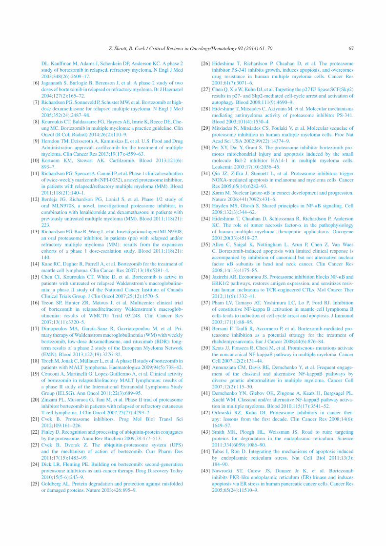
Z. Skrott, B. Cvek / Critical Reviews in Oncology/Hematology 92 (2014) 61–70 67
DL, Kauffman M, Adams J, Schenkein DP, Anderson KC. A phase 2
study of bortezomib in relapsed, refractory myeloma. N Engl J Med
2003;348(26):2609–17.
[6] Jagannath S, Barlogie B, Berenson J, et al. A phase 2 study of two
doses of bortezomib in relapsed or refractory myeloma. Br J Haematol
2004;127(2):165–72.
[7] Richardson PG, Sonneveld P, Schuster MW, et al. Bortezomib or high-
dose dexamethasone for relapsed multiple myeloma. N Engl J Med
2005;352(24):2487–98.
[8] Kouroukis CT, Baldassarre FG, Haynes AE, Imrie K, Reece DE, Che-
ung MC. Bortezomib in multiple myeloma: a practice guideline. Clin
Oncol (R Coll Radiol) 2014;26(2):110–9.
[9] Herndon TM, Deisseroth A, Kaminskas E, et al. U.S. Food and Drug
Administration approval: carfilzomib for the treatment of multiple
myeloma. Clin Cancer Res 2013;19(17):4559–63.
[10] Kortuem KM, Stewart AK. Carfilzomib. Blood 2013;121(6):
893–7.
[11] Richardson PG, SpencerA. Cannell P, et al. Phase 1 clinical evaluation
of twice-weekly marizomib (NPI-0052), a novel proteasome inhibitor,
in patients with relapsed/refractory multiple myeloma (MM). Blood
2011;118(21):140–1.
[12] Berdeja JG, Richardson PG, Lonial S, et al. Phase 1/2 study of
oral MLN9708, a novel, investigational proteasome inhibitor, in
combination with lenalidomide and dexamethasone in patients with
previously untreated multiple myeloma (MM). Blood 2011;118(21):
223.
[13] Richardson PG, Baz R, Wang L, et al. Investigational agent MLN9708,
an oral proteasome inhibitor, in patients (pts) with relapsed and/or
refractory multiple myeloma (MM): results from the expansion
cohorts of a phase 1 dose-escalation study. Blood 2011;118(21):
140.
[14] Kane RC, Dagher R, Farrell A, et al. Bortezomib for the treatment of
mantle cell lymphoma. Clin Cancer Res 2007;13(18):5291–4.
[15] Chen CI, Kouroukis CT, White D, et al. Bortezomib is active in
patients with untreated or relapsed Waldenstrom’s macroglobuline-
mia: a phase II study of the National Cancer Institute of Canada
Clinical Trials Group. J Clin Oncol 2007;25(12):1570–5.
[16] Treon SP, Hunter ZR, Matous J, et al. Multicenter clinical trial
of bortezomib in relapsed/refractory Waldenstrom’s macroglob-
ulinemia: results of WMCTG Trial 03-248. Clin Cancer Res
2007;13(11):3320–5.
[17] Dimopoulos MA, García-Sanz R, Gavriatopoulou M, et al. Pri-
mary therapy of Waldenstrom macroglobulinemia (WM) with weekly
bortezomib, low-dose dexamethasone, and rituximab (BDR): long-
term results of a phase 2 study of the European Myeloma Network
(EMN). Blood 2013;122(19):3276–82.
[18] Troch M, Jonak C, Müllauer L, et al. A phase II study of bortezomib in
patients with MALT lymphoma. Haematologica 2009;94(5):738–42.
[19] Conconi A, Martinelli G, Lopez-Guillermo A, et al. Clinical activity
of bortezomib in relapsed/refractory MALT lymphomas: results of
a phase II study of the International Extranodal Lymphoma Study
Group (IELSG). Ann Oncol 2011;22(3):689–95.
[20] Zinzani PL, Musuraca G, Tani M, et al. Phase II trial of proteasome
inhibitor bortezomib in patients with relapsed or refractory cutaneous
T-cell lymphoma. J Clin Oncol 2007;25(27):4293–7.
[21] Cvek B. Proteasome inhibitors. Prog Mol Biol Transl Sci
2012;109:161–226.
[22] Finley D. Recognition and processing of ubiquitin-protein conjugates
by the proteasome. Annu Rev Biochem 2009;78:477–513.
[23] Cvek B, Dvorak Z. The ubiquitin-proteasome system (UPS)
and the mechanism of action of bortezomib. Curr Pharm Des
2011;17(15):1483–99.
[24] Dick LR, Fleming PE. Building on bortezomib: second-generation
proteasome inhibitors as anti-cancer therapy. Drug Discovery Today
2010;15(5-6):243–9.
[25] Goldberg AL. Protein degradation and protection against misfolded
or damaged proteins. Nature 2003;426:895–9.
[26] Hideshima T, Richardson P, Chauhan D, et al. The proteasome
inhibitor PS-341 inhibits growth, induces apoptosis, and overcomes
drug resistance in human multiple myeloma cells. Cancer Res
2001;61(7):3071–6.
[27] Chen Q, Xie W, Kuhn DJ, et al. Targeting the p27 E3 ligase SCF(Skp2)
results in p27- and Skp2-mediated cell-cycle arrest and activation of
autophagy. Blood 2008;111(9):4690–9.
[28] Hideshima T, Mitsiades C, Akiyama M, et al. Molecular mechanisms
mediating antimyeloma activity of proteasome inhibitor PS-341.
Blood 2003;101(4):1530–4.
[29] Mitsiades N, Mitsiades CS, Poulaki V, et al. Molecular sequelae of
proteasome inhibition in human multiple myeloma cells. Proc Nat
Acad Sci USA 2002;99(22):14374–9.
[30] Pei XY, Dai Y, Grant S. The proteasome inhibitor bortezomib pro-
motes mitochondrial injury and apoptosis induced by the small
molecule Bcl-2 inhibitor HA14-1 in multiple myeloma cells.
Leukemia 2003;17(10):2036–45.
[31] Qin JZ, Ziffra J, Stennett L, et al. Proteasome inhibitors trigger
NOXA-mediated apoptosis in melanoma and myeloma cells. Cancer
Res 2005;65(14):6282–93.
[32] Karin M. Nuclear factor-kB in cancer development and progression.
Nature 2006;441(7092):431–6.
[33] Hayden MS, Ghosh S. Shared principles in NF-kB signaling. Cell
2008;132(3):344–62.
[34] Hideshima T, Chauhan D, Schlossman R, Richardson P, Anderson
KC. The role of tumor necrosis factor-a in the pathophysiology
of human multiple myeloma: therapeutic applications. Oncogene
2001;20(33):4519–27.
[35] Allen C, Saigal K, Nottingham L, Arun P, Chen Z, Van Waes
C. Bortezomib-induced apoptosis with limited clinical response is
accompanied by inhibition of canonical but not alternative nuclear
factor kB subunits in head and neck cancer. Clin Cancer Res
2008;14(13):4175–85.
[36] Jazirehi AR, Economou JS. Proteasome inhibition blocks NF-kB and
ERK1/2 pathways, restores antigen expression, and sensitizes resis-
tant human melanoma to TCR-engineered CTLs. Mol Cancer Ther
2012;11(6):1332–41.
[37] Pham LV, Tamayo AT, Yoshimura LC, Lo P, Ford RJ. Inhibition
of constitutive NF-kappa B activation in mantle cell lymphoma B
cells leads to induction of cell cycle arrest and apoptosis. J Immunol
2003;171(1):88–95.
[38] Bersani F, Taulli R, Accornero P, et al. Bortezomib-mediated pro-
teasome inhibition as a potential strategy for the treatment of
rhabdomyosarcoma. Eur J Cancer 2008;44(6):876–84.
[39] Keats JJ, Fonseca R, Chesi M, et al. Promiscuous mutations activate
the noncanonical NF-kappaB pathway in multiple myeloma. Cancer
Cell 2007;12(2):131–44.
[40] Annunziata CM, Davis RE, Demchenko Y, et al. Frequent engage-
ment of the classical and alternative NF-kappaB pathways by
diverse genetic abnormalities in multiple myeloma. Cancer Cell
2007;12(2):115–30.
[41] Demchenko YN, Glebov OK, Zingone A, Keats JJ, Bergsagel PL,
Kuehl WM. Classical and/or alternative NF-kappaB pathway activa-
tion in multiple myeloma. Blood 2010;115(17):3541–52.
[42] Orlowski RZ, Kuhn DJ. Proteasome inhibitors in cancer ther-
apy: lessons from the first decade. Clin Cancer Res 2008;14(6):
1649–57.
[43] Smith MH, Ploegh HL, Weissman JS. Road to ruin: targeting
proteins for degradation in the endoplasmic reticulum. Science
2011;334(6059):1086–90.
[44] Tabas I, Ron D. Integrating the mechanisms of apoptosis induced
by endoplasmic reticulum stress. Nat Cell Biol 2011;13(3):
184–90.
[45] Nawrocki ST, Carew JS, Dunner Jr K, et al. Bortezomib
inhibits PKR-like endoplasmic reticulum (ER) kinase and induces
apoptosis via ER stress in human pancreatic cancer cells. Cancer Res
2005;65(24):11510–9.

68 Z. Skrott, B. Cvek / Critical Reviews in Oncology/Hematology 92 (2014) 61–70
[46] Fribley A, Zeng Q, Wang CY. Proteasome inhibitor PS-341 induces
apoptosis through induction of endoplasmic reticulum stress-reactive
oxygen species in head and neck squamous cell carcinoma cells. Mol
Cell Biol 2004;24(22):9695–704.
[47] Fels DR, Ye J, Segan AT, et al. Preferential cytotoxicity of
bortezomib toward hypoxic tumor cells via overactivation of
endoplasmic reticulum stress pathways. Cancer Res 2008;68(22):
9323–30.
[48] Lee AH, Iwakoshi NN, Anderson KC, Glimcher LH. Proteasome
inhibitors disrupt the unfolded protein response in myeloma cells.
Proc Nat Acad Sci USA 2003;100:9946–51.
[49] Obeng EA, Carlson LM, Gutman DM, Harrington Jr WJ, Lee KP,
Boise LH. Proteasome inhibitors induce a terminal unfolded protein
response in multiple myeloma cells. Blood 2006;107(12):4907–16.
[50] Palombella VJ, Rando OJ, Goldberg AL, Maniatis T. The
ubiquitin-proteasome pathway is required for processing the NF-
kappa B1 precursor protein and the activation of NF-kB. Cell
1994;78(5):773–85.
[51] Nemeth ZH, Wong HR, Odoms K, et al. Proteasome inhibitors induce
inhibitory kappa B (I kappa B) kinase activation, I kappa B alpha
degradation, and nuclear factor kappa B activation in HT-29 cells.
Mol Pharmacol 2004;65(2):342–9.
[52] Hideshima T, Chauhan D, Richardson P, et al. NF-(B as a therapeu-
tic target in multiple myeloma. J Biol Chem 2002;277(19):16639–
47.
[53] Hideshima T, Ikeda H, Chauhan D, et al. Bortezomib induces canoni-
cal nuclear factor-kappaB activation in multiple myeloma cells. Blood
2009;114(5):1046–52.
[54] Holbeck SL, Collins JM, Doroshow JH. Analysis of Food and
Drug Administration-approved anticancer agents in the NCI60
panel of human tumor cell lines. Mol Cancer Ther 2010;9(5):
1451–60.
[55] Milano A, Iaffaioli RV, Caponigro F. The proteasome: a worth-
while target for the treatment of solid tumours? Eur J Cancer
2007;43(7):1125–33.
[56] Caravita T, de Fabritiis P, Palumbo A, Amadori S, Boccadoro M.
Bortezomib: efficacy comparisons in solid tumors and hematologic
malignancies. Nat Clin Pract Oncol 2006;3(7):374–87.
[57] Borrell B. How accurate are cancer cell lines? Nature
2010;463(7283):858.
[58] Sandberg R, Ernberg I. The molecular portrait of in vitro
growth by meta-analysis of gene-expression profiles. Genome Biol
2005;6(8):R65.
[59] Pan C, Kumar C, Bohl S, Klingmueller U, Mann M. Compara-
tive proteomic phenotyping of cell lines and primary cells to assess
preservation of cell type-specific functions. Mol Cell Proteomics
2009;8(3):443–50.
[60] Powers ET, Morimoto RI, Dillin A, Kelly JW, Balch WE. Biological
and chemical approaches to diseases of proteostasis deficiency. Annu
Rev Biochem 2009;78:959–91.
[61] Williamson MJ, Silva MD, Terkelsen J, et al. The relationship among
tumor architecture, pharmacokinetics, pharmacodynamics, and effi-
cacy of bortezomib in mouse xenograft models. Mol Cancer Ther
2009;8(12):3234–43.
[62] Driscoll JJ, Minter A, Driscoll DA, Burris JK. The ubiqui-
tin + proteasome protein degradation pathway as a therapeutic strategy
in the treatment of solid tumor malignancies. Anticancer Agents Med
Chem 2011;11(2):242–6.
[63] Cvek B. Ixazomib citrate, proteasome inhibitor oncolytic. Drug Future
2012;37(8):561–5.
[64] Kupperman E, Lee EC, Cao Y, et al. Evaluation of the proteasome
inhibitor MLN9708 in preclinical models of human cancer. Cancer
Res 2010;70(5):1970–80.
[65] Chauhan D, Tian Z, Zhou B, et al. In vitro and in vivo selec-
tive antitumor activity of a novel orally bioavailable proteasome
inhibitor MLN9708 against multiple myeloma cells. Clin Cancer Res
2011;17(16):5311–21.
[66] Tian Z, Zhao JJ, Tai YT, et al. Investigational agent MLN9708/2238
targets tumor-suppressor miR33b in MM cells. Blood
2012;120(19):3958–67.
[67] Richardson PG, Berdeja JG, Niesvizky R, et al. Oral weekly
MLN9708, an investigational proteasome inhibitor, in combina-
tion with lenalidomide and dexamethasone in patients (pts) with
previ- ously untreated multiple myeloma (MM): a phase I/II study.
J Clin Oncol 2012;30(suppl) (abstr 8033).
[68] Lonial S, Baz RC, Wang M, et al. Phase I study of twice-weekly dosing
of the investigational oral proteasome inhibitor MLN9708 in patients
(pts) with relapsed and/or refractory multiple myeloma (MM). J Clin
Oncol 2012;30(suppl) (abstr 8017).
[69] Smith DC, Sullivan D, Infante JR, et al. MLN9708, an investigational
proteasome inhibitor, in patients (pts) with solid tumors: Updated
phase I results. J Clin Oncol 2012;30(suppl) (abstr e13603).
[70] Papadopoulos KP, Burris 3rd HA, Gordon M, et al. A phase
I/II study of carfilzomib 2-10-min infusion in patients with
advanced solid tumors. Cancer Chemother Pharmacol 2013;72(4):
861–8.
[71] Gomez AM, Willcox N, Molenaar PC, et al. Targeting plasma cells
with proteasome inhibitors: possible roles in treating myasthenia
gravis. Ann NY Acad Sci 2012;1274:48–59.
[72] Lifter J, Kincade PW, Choi YS. Subpopulations of chicken B lym-
phocytes. J Immunol 1976;117(6):2220–5.
[73] Lipsky PE. Systemic lupus erythematosus: an autoimmune disease of
B cell hyperactivity. Nat Immunol 2001;2(9):764–6.
[74] Díaz-Manera J, Martinez-Hernandez E, Querol L, et al. Long-
lasting treatment effect of rituximab in MuSK myasthenia. Neurology
2012;78(3):189–93.
[75] Gehrs BC, Friedberg RC. Autoimmune hemolytic anemia. Am J
Hematol 2002;69(4):258–71.
[76] Hoyer BF, Manz RA, Radbruch A, Hiepe F. Long-lived plasma
cells and their contribution to autoimmunity. Ann NY Acad Sci
2005;1050:124–33.
[77] Cascio P, Oliva L, Cerruti F, et al. Dampening Ab responses using pro-
teasome inhibitors following in vivo B cell activation. Eur J Immunol
2008;38(3):658–67.
[78] Neubert K, Meister S, Moser K, et al. The proteasome inhibitor borte-
zomib depletes plasma cells and protects mice with lupus-like disease
from nephritis. Nat Med 2008;14(7):748–55.
[79] Ichikawa HT, Conley T, Muchamuel T, et al. Beneficial effect of
novel proteasome inhibitors in murine lupus via dual inhibition of
type I interferon and autoantibody-secreting cells. Arthritis Rheum
2012;64(2):493–503.
[80] Seavey MM, Lu LD, Stump KL, Wallace NH, Ruggeri BA.
Novel, orally active, proteasome inhibitor, delanzomib (CEP-18770),
ameliorates disease symptoms and glomerulonephritis in two pre-
clinical mouse models of SLE. Int Immunopharmacol 2012;12(1):
257–70.
[81] Gomez AM, Vrolix K, Martinez-Martinez P, et al. Proteasome
inhibition with bortezomib depletes plasma cells and autoanti-
bodies in experimental autoimmune myasthenia gravis. J Immunol
2011;186(4):2503–13.
[82] Meslier Y, Andre S, Dimitrov JD, et al. Bortezomib delays the onset
of factor VIII inhibitors in experimental hemophilia A, but fails to
eliminate established anti-factor VIII IgG-producing cells. J Thromb
Haemost 2011;9(4):719–28.
[83] Sadaka B, Alloway RR, Shields AR, Schmidt NM, Woodle ES. Pro-
teasome inhibition for antibody-mediated allograft rejection. Semin
Hematol 2012;49(3):263–9.
[84] Schroyens W, O’Connell C, Sykes DB. Complete and partial
responses of the TEMPI syndrome to bortezomib. N Engl J Med
2012;367(8):778–80.

Z. Skrott, B. Cvek / Critical Reviews in Oncology/Hematology 92 (2014) 61–70 69
[85] Sykes DB, Schroyens W, O’Connell C. The TEMPI syndrome—a
novel multisystem disease. N Engl J Med 2011;365(5):475–7.
[86] Shortt J, Oh DH, Opat SS. ADAMTS13 antibody depletion by
bortezomib in thrombotic thrombocytopenic purpura. N Engl J Med
2013;368(1):90–2.
[87] Deng S, Zhou H, Xiong R, et al. Over-expression of genes and proteins
of ubiquitin specific peptidases (USPs) and proteasome subunits (PSs)
in breast cancer tissue observed by the methods of RFDD-PCR and
proteomics. Breast Cancer Res Treat 2007;104(1):21–30.
[88] Geiger T, Wehner A, Schaab C, Cox J, Mann M. Compar-
ative proteomic analysis of eleven common cell lines reveals
ubiquitous but varying expression of most proteins. Mol Cell Pro-
teomics 2012;11(3) (M111.014050) http://www.ncbi.nlm.nih.gov/
pubmed/?term=Geiger+T%2C+Wehner+A
[89] Ross DT, Scherf U, Eisen MB, et al. Systematic variation in
gene expression patterns in human cancer cell lines. Nat Genet
2000;24(3):227–35.
[90] Moghaddas Gholami A, Hahne H, Wu Z, et al. Global proteome
analysis of the NCI-60 cell line panel. Cell Rep 2013;4(3):609–20.
[91] Codony-Servat J, Tapia MA, Bosch M, et al. Differential cellular and
molecular effects of bortezomib, a proteasome inhibitor, in human
breast cancer cells. Mol Cancer Ther 2006;5(3):665–75.
[92] Bianchi G, Oliva L, Cascio P, et al. The proteasome load versus capac-
ity balance determines apoptotic sensitivity of multiple myeloma cells
to proteasome inhibition. Blood 2009;113(13):3040–9.
[93] Meister S, Schubert U, Neubert K, et al. Extensive immunoglobu-
lin production sensitizes myeloma cells for proteasome inhibition.
Cancer Res 2007;67(4):1783–92.
[94] Schubert U, Anton LC, Gibbs J, Norbury CC, Yewdell JW, Bennink
JR. Rapid degradation of a large fraction of newly synthesized proteins
by proteasomes. Nature 2000;404(6779):770–4.
[95] Qian SB, Princiotta MF, Bennink JR, Yewdell JW. Characteriza-
tion of rapidly degraded polypeptides in mammalian cells reveals
a novel layer of nascent protein quality control. J Biol Chem
2006;281(1):392–400.
[96] Cenci S, Oliva L, Cerruti F, et al. Pivotal advance: protein synthe-
sis modulates responsiveness of differentiating and malignant plasma
cells to proteasome inhibitors. J Leukoc Biol 2012;92(5):921–31.
[97] Cenci S, Mezghrani A, Cascio P, et al. Progressively impaired protea-
somal capacity during terminal plasma cell differentiation. EMBO J
2006;25(5):1104–13.
[98] Santo L, Hideshima T, Kung AL, et al. Preclinical activity, pharma-
codynamic, and pharmacokinetic properties of a selective HDAC6
inhibitor, ACY-1215, in combination with bortezomib in multiple
myeloma. Blood 2012;119(11):2579–89.
[99] Richardson PG, Schlossman RL, Alsina M, et al. PANORAMA 2:
panobinostat in combination with bortezomib and dexamethasone in
patients with relapsed and bortezomib-refractory myeloma. Blood
2013;122(14):2331–7.
[100] Auner HW, Moody AM, Ward TH, et al. Combined inhibition of p97
and the proteasome causes lethal disruption of the secretory apparatus
in multiple myeloma cells. PLoS One 2013;8(9):e74415.
[101] Mitsiades CS, Mitsiades NS, McMullan CJ, et al. Antimyeloma activ-
ity of heat shock protein-90 inhibition. Blood 2006;107(3):1092–100.
[102] Yerlikaya A, Okur E, Eker S, Erin N. Combined effects of the pro-
teasome inhibitor bortezomib and Hsp70 inhibitors on the B16F10
melanoma cell line. Mol Med Rep 2010;3(2):333–9.
[103] Sonnemann J, Marx C, Becker S, et al. p53-dependent and p53-
independent anticancer effects of different histone deacetylase
inhibitors. Br J Cancer 2014;110(3):656–67.
[104] Orlowski RZ. Novel agents for multiple myeloma to overcome
resistance in phase III clinical trials. Semin Oncol 2013;40(5):
634–51.
[105] Lacy MQ, McCurdy AR. Pomalidomide. Blood 2013;122(14):
2305–9.
[106] McConkey DJ, Zhu K. Mechanisms of proteasome inhibitor
action and resistance in cancer. Drug Resist Updat 2008;11(4–5):
164–79.
[107] Kale AJ, Moore BS. Molecular mechanisms of acquired proteasome
inhibitor resistance. J Med Chem 2012;55(23):10317–27.
[108] Ri M, Iida S, Nakashima T, et al. Bortezomib-resistant myeloma
cell lines: a role for mutated PSMB5 in preventing the accumula-
tion of unfolded proteins and fatal ER stress. Leukemia 2010;24(8):
1506–12.
[109] Franke NE, Niewerth D, Assaraf YG, et al. Impaired borte-
zomib binding to mutant b5 subunit of the proteasome is the
underlying basis for bortezomib resistance in leukemia cells.
Leukemia 2012;26(4):757–68.
[110] Oerlemans R, Franke NE, Assaraf YG, et al. Molecular basis of
bortezomib resistance: proteasome subunit beta5 (PSMB5) gene
mutation and overexpression of PSMB5 protein. Blood 2008;112(6):
2489–99.
[111] Suzuki E, Demo S, Deu E, et al. Molecular mechanisms of bortezomib
resistant adenocarcinoma cells. PLoS One 2011;6(12):e27996.
[112] Balsas P, Galan-Malo P, Marzo I, Naval J. Bortezomib resistance
in a myeloma cell line is associated to PSMb5 overexpression and
polyploidy. Leuk Res 2012;36(2):212–8.
[113] Lu S, Yang J, Song X, et al. Point mutation of the proteasome
beta5 subunit gene is an important mechanism of bortezomib resis-
tance in bortezomib-selected variants of Jurkat T cell lymphoblastic
lymphoma/leukemia line. J Pharmacol Exp Ther 2008;326(2):
423–31.
[114] Lichter DI, Danaee H, Pickard MD, et al. Sequence analysis of
b-subunit genes of the 20S proteasome in patients with relapsed
multiple myeloma treated with bortezomib or dexamethasone. Blood
2012;120(23):4513–6.
[115] Politou M, Karadimitris A, Terpos E, Kotsianidis I, Apperley JF,
Rahemtulla A. No evidence of mutations of the PSMB5 (beta-5 sub-
unit of proteasome) in a case of myeloma with clinical resistance to
bortezomib. Leuk Res 2006;30(2):240–1.
[116] Catley L, Weisberg E, Kiziltepe T, et al. Aggresome induction by
proteasome inhibitor bortezomib and alpha-tubulin hyperacetylation
by tubulin deacetylase (TDAC) inhibitor LBH589 are synergistic in
myeloma cells. Blood 2006;108(10):3441–9.
[117] Mitsiades CS, Mitsiades NS, McMullan CJ, et al. Transcriptional
signature of histone deacetylase inhibition in multiple myeloma:
biological and clinical implications. Proc Nat Acad Sci USA
2004;101(2):540–5.
[118] Weber DM, Graef T, Hussein M, et al. Phase I trial of vorino-
stat combined with bortezomib for the treatment of relapsing
and/or refractory multiple myeloma. Clin Lymphoma Myeloma Leuk
2012;12(5):319–24.
[119] San-Miguel JF, Richardson PG, Gunther A, et al. Phase Ib study of
panobinostat and bortezomib in relapsed or relapsed and refractory
multiple myeloma. J Clin Oncol 2013;31(29):3696–703.
[120] Stessman HA, Mansoor A, Zhan F, Linden MA, Van Ness B, Baughn
LB. Bortezomib resistance can be reversed by induced expression of
plasma cell maturation markers in a mouse in vitro model of multiple
myeloma. PLoS One 2013;8(10):e77608.
[121] Gu JL, Li J, Zhou ZH, et al. Differentiation induction enhances borte-
zomib efficacy and overcomes drug resistance in multiple myeloma.
Biochem Biophys Res Commun 2012;420(3):644–50.
[122] Leung-Hagesteijn C, Erdmann N, Cheung G, et al. Xbp1s-negative
tumor B cells and pre-plasmablasts mediate therapeutic pro-
teasome inhibitor resistance in multiple myeloma. Cancer Cell
2013;24(3):289–304.
[123] Ling SC, Lau EK, Al-Shabeeb A, et al. Response of myeloma to
the proteasome inhibitor bortezomib is correlated with the unfolded
protein response regulator XBP-1. Haematologica 2012;97(1):
64–72.

70 Z. Skrott, B. Cvek / Critical Reviews in Oncology/Hematology 92 (2014) 61–70
[124] Chapman MA, Lawrence MS, Keats JJ, et al. Initial genome
sequencing and analysis of multiple myeloma. Nature
2011;471(7339):467–72.
[125] Tian Z, D’Arcy P, Wang X, et al. A novel small molecule inhibitor
of deubiquitylating enzyme USP14 and UCHL5 induces apoptosis
in multiple myeloma and overcomes bortezomib resistance. Blood
2014;123(5):706–16.
[126] Anchoori RK, Karanam B, Peng S, et al. A bis-benzylidine piperidone
targeting proteasome ubiquitin receptor RPN13/ADRM1 as a therapy
for cancer. Cancer Cell 2013;24(6):791–805.
[127] Chauhan D, Tian Z, Nicholson B, et al. A small molecule
inhibitor of ubiquitin-specific protease-7 induces apoptosis in
multiple myeloma cells and overcomes bortezomib resistance. Cancer
Cell 2012;22(3):345–58.
Biographies
Zdenek Skrott is a master’s student in cell and molecular
biology at Department of Cell Biology & Genetics, Palacky
University Olomouc. He is involved in research on an old
drug disulfiram as an anticancer drug inhibiting proteasome-
dependent degradation.
Boris Cvek (Ph.D.) is a researcher at Department of Cell
Biology & Genetics, Palacky University Olomouc. His main
research topics are proteasome inhibitors and repurposing of
old drugs for new uses.

�����V��
�#;��#��, �� ����C, �8� '0#- �6, �8#����.#�6,��&$��.#��,�+#'0�'0&-#�H���,����&����.#�6,
� �* �(��.#��,�+ �(�!�6,����&���.#�+,��5&����#��,�?���!��,��&(��&���,�8#�����6���#����&��
������:&'�#���"�������:&'�(���((3��("��(��"#�05#!(�&��0 $#��"��(�#���'#�'���)!�'-&�&'#--!
#.#&-#)-� � �� � � &�0&)&���(, � .��&��(�#� � #�� � �&( -*&�#$� � ���(�#��� � @DEF ��#�XKFSJT%PQ@3PA@�
�S@DEKT%�P�PJK
���

Received: 24 September 2018 | Accepted: 24 October 2018
DOI: 10.1002/pros.23741
ORIGINAL ARTICLE
Targeting genotoxic and proteotoxic stress-response
pathways in human prostate cancer by clinically available
PARP inhibitors, vorinostat and disulfiram
Dusana Majera PhD1 | Zdenek Skrott MSC1 | Jan Bouchal PhD2 |
Jirina Bartkova MD, PhD3,4 | Dana Simkova PhD2 | Mariam Gachechiladze MD, PhD2 |
Jana Steigerova PhD2 | Daniela Kurfurstova MD, PhD2 | Jan Gursky PhD1 |
Gabriela Korinkova PhD2 | Karel Cwiertka MD, PhD5 | Zdenek Hodny MD, PhD6 |
Martin Mistrik PhD1 | Jiri Bartek MD, PhD1,3,4,6
1 Laboratory of Genome Integrity, Institute of Molecular and Translational Medicine, Faculty of Medicine and Dentistry, Palacky University, Olomouc, Czech
Republic
2Department of Clinical and Molecular Pathology, Institute of Molecular and Translational Medicine, Faculty of Medicine and Dentistry, Palacky University,
Olomouc, Czech Republic
3Danish Cancer Society Research Center, Copenhagen, Denmark
4Division of Genome Biology, Department of Medical Biochemistry and Biophysics, Science for Life Laboratory, Karolinska Institute, Stockholm, Sweden
5Department of Oncology, Faculty of Medicine and Dentistry, Palacky University, University Hospital, Olomouc, Czech Republic
6Department of Genome Integrity, Institute of Molecular Genetics of the CAS, v.v.i., Prague, Czech Republic
Correspondence
Jiri Bartek, MD, PhD, Danish Cancer Society
Research Center, Copenhagen, Denmark
Email: [email protected]
Jan Bouchal, PhD, Department of Clinical and
Molecular Pathology, Institute of Molecular and
Translational Medicine, Faculty of Medicine and
Dentistry, Palacky University, Olomouc, Czech
Republic.
Email: [email protected]
Martin Mistrik, PhD, Laboratory of Genome
Integrity, Institute of Molecular and
Translational Medicine, Faculty of Medicine and
Dentistry, Palacky University, Olomouc, Czech
Republic
Email: [email protected]
Funding information
The Danish cancer society; the Novo Nordisk
Foundation; Palacky University, Grant numbers:
IGA-LF-2018-001, IGA-LF-2018-034; the
Swedish research council and Cancer Fonden;
the Danish council for independent research;
Czech Ministry of Education, Grant number:
DRO-61989592; Czech National Program of
Sustainability, Grant number: LO1304; Czech
Ministry of Health, Grant numbers: AZV 16-
32030, DRO-FNOL00098892, NV15-28628A;
Background: Castration-resistant prostate cancer (PCa) represents a serious health
challenge. Based on mechanistically-supported rationale we explored new thera-
peutic options based on clinically available drugs with anticancer effects, including
inhibitors of PARP1 enzyme (PARPi), and histone deacetylases (vorinostat),
respectively, and disulfiram (DSF, known as alcohol-abuse drug Antabuse) and its
copper-chelating metabolite CuET that inhibit protein turnover.
Methods: Drugs and their combination with ionizing radiation (IR) were tested in
various cytotoxicity assays in three human PCa cell lines including radio-resistant
stem-cell like derived cells. Mechanistically, DNA damage repair, heat shock and
unfolded protein response (UPR) pathways were assessed by immunofluorescence
and immunoblotting.
Results:We observed enhanced sensitivity to PARPi/IR in PC3 cells consistent with
lower homologous recombination (HR) repair. Vorinostat sensitized DU145 cells to
PARPi/IR and decreased mutant p53. Vorinostat also impaired HR-mediated DNA
repair, as determined by Rad51 foci formation and downregulation of TOPBP1
protein, andovercame radio-resistanceof stem-cell likeDU145-derived cells. All PCa
models responded well to CuET or DSF combined with copper. We demonstrated
that DSF interacts with copper in the culture media and forms adequate levels of
CuET indicating that DSF/copper and CuET may be considered as comparable
treatments. Both DSF/copper and CuET evoked hallmarks of UPR in PCa cells,
documented by upregulation of ATF4, CHOP and phospho-eIF2α, with ensuing heat
352 | © 2018 Wiley Periodicals, Inc. wileyonlinelibrary.com/journal/pros The Prostate. 2019;79:352–362.

Kellner Family Foundation; Czech-BioImaging,
Grant number: LM2015062shock response encompassing activation ofHSF1 andHSP70. Further enhancing the
cytotoxicity of CuET, combination with an inhibitor of the anti-apoptotic protein
survivin (YM155, currently undergoing clinical trials) promoted the UPR-induced
toxicity, yielding synergistic effects of CuET and YM155.
Conclusions:We propose that targeting genotoxic and proteotoxic stress responses
by combinations of available drugs could inspire innovative strategies to treat
castration-resistant PCa.
K E YWORD S
disulfiram, PARP, prostate cancer, proteotoxic stress, vorinostat
1 | INTRODUCTION
Prostate cancer (PCa) is the most frequently diagnosed malignancy in
men and one of the major causes of cancer-related death in developed
countries.1 PCa is initially androgen-dependent and responds to
androgen deprivation therapies, however, the disease ultimately
progresses into a hormone-independent and largely incurable stage
with metastases to the bones, lung, brain, or liver.
Aberrations in the DNA damage response (DDR) machinery are
common in cancer and represent potential targets for therapeutic
intervention.2 PARP1 activity is important in sensing and signaling DNA
damage that arises bothendogenously, for example, throughgeneration
of oxidative DNA lesions and DNA single-strand breaks (SSBs), or
exogenously, suchasdue to ionizing radiation (IR) exposureor treatment
with various chemotherapeutics. Exposure of cycling cells to inhibitors
of PARP1 (PARPi) causes excessive unrepaired SSBs and acceleration of
DNA replication3 leading to replication stress and formation of DNA
double-strand breaks (DSBs), toxic lesions preferentially repaired by
homologous recombination (HR). HR defects due to mutations or
silencing of factors such as BRCA1/2 sensitize cells to PARPi, as shown
for ovarian, breast and also metastatic prostate cancer.4
Defects in DNA damage sensors, signaling kinases or nucleotide
excision repair also sensitize to PARPi4 suggesting that the therapeutic
potential of PARPi might extend beyond the BRCA1/2-defective
tumors. There is also an urgent need to identify and validate potential
biomarkers to predict sensitivity of individual tumors to PARPi,
exemplified for PCa by the fusion oncogene TMPRSS2-ERG or loss of
the PTEN tumor suppressor.5,6
PCa is a heterogeneous disease reflecting both genetic and
epigenetic alterations.7 Six epigenetics-modulating drugs targeting
DNA methylation or histone deacetylation have been approved for
cancer treatment.7 Since epigenetic regulation is complex, preclinical
studies are required to generate patient stratification hypotheses and
identify predictive biomarkers. Epigenetic reprogramming after loss of
Rb and p53 tumor suppressors diminishes androgen receptor
expression and is associated with resistence to antiandrogen
therapy.8,9 From this point of view, both PC3 and DU145 cells,
lacking AR and possessing mutations in tumor suppressors, represent
relevant models of a subgroup of aggressive prostate cancer.
Another approach to PCa treatment could be drug repurposing,
with potentially multifaceted benefits for clinical implementation of
new treatment options. DSF is among such possible candidates,
showing anti-tumor activity in multiple studies. Recently, we discov-
ered the molecular target and mode of action of DSF, thereby
strengthening DSF's potential as an anticancer drug.10 Many cancers
become resistant to monotherapy through diverse mechanisms,
posing amajor challenge in contemporary oncology. Drug combination
could overcome resistance to single compounds, thus it is vital to find
the drugs that act synergistically and are well tolerated.11
Here, we describe differential responses to PARPi and IR in cellular
models of aggressive PCa: PC3 (typical for loss of p53 and PTEN),
DU145 (mutated p53 and Rb) and radioresistant stem-like PCa cells.12
Moreover, we show that HDAC inhibition alters expression of HR
proteins and potentiates cytotoxicity of IR, and that DSF's active
metabolite, diethyldithiocarbamate-copper complex (CuET), activates
heat shock response and UPR, showing synergistic toxic effect in
combination with a survivin inhibitor-YM155 in human PCa models.
2 | MATERIAL AND METHODS
2.1 | Cell lines
DU145 and PC3 cell lines were cultured in DMEM medium, LNCaP in
RPMI medium and LAPC4 in IMDM. DMEM, RPMI, and IMDM media
were supplemented with 10% fetal bovine serum and penicillin/
streptomycin. IMDM medium was additionally supplemented with
1 nMR1881. RWPE-1 cells were cultured in a keratinocyte serum-free
medium supplemented with the bovine pituitary extract and human
recombinant epidermal growth factor (Thermo Scientific, Waltham,
MA). EP156T cells were cultured as described previously.13 All cell
cultures were maintained in humidified 5% CO2 atmosphere at 37°C.
LAPC4, EP156T and RWPE1 cells were kindly provided by Prof. Zoran
Culig and Prof. Helmut Klocker (Innsbruck Medical University). Other
cell lines were purchased from the European Collection of Cell
MAJERA ET AL. | 353

Cultures (ECACC) and authenticated by AmpfISTR™ Identifiler PCR
Amplification Kit (Applied Biosystems, Foster City, CA).
2.2 | Colony forming and cell viability assays
For clonogenic cell survival assay, cells were plated in 6-well plates at
200-500 cells per plate. Next day the cells received appropriate
treatment and kept in culture for 7-14 days. Colonies of approximately
50 cells were visualized by 1% crystal violet in 96% ethanol, and their
number and total area were counted. Results were confirmed in three
independent experiments. For XTT assay, cells were plated at a density
of 10 000 perwell in a 96-well plate. The next day, cells were treated as
indicated. After 48 h, an XTT assay was performed according to the
manufacturer's instructions (Applichem, Darmstadt, Germany). XTT
solution was added to the medium and incubated for 30-60min, and
then the dye intensity wasmeasured at the 475 nmwavelength using a
spectrometer (TECAN, Infinite M200PRO, Mannedorf, Switzerland).
2.3 | Ionizing radiation and chemicals
The KU58948 inhibitor was obtained from AstraZeneca (London, UK).
Vorinostat, MK132, nutlin 3, DSF, tunicamycin, thapsigargin and CuCl2
were purchased fromSigma-Aldrich, YM155 fromSelleckchemand copper
diethyldithiocarbamate (CuET) from TCI Chemicals. Ionizing radiation
was delivered using Xstrahl RS research cabinet gamma irradiator.
2.4 | Immunoblotting
Equal amounts of cell lysates were separated by SDS-PAGE on
handcast or precast gel (Invitrogen, Carlsbad, CA), and then transferred
onto nitrocellulose membrane. The membrane was blocked with 5%
milk in Tris-buffered saline containing 0.1% Tween 20 for 1 h at room
temperature, and then incubated overnight at 4°C or 1 h at room
temperature with one of the following primary antibodies against: p53
(FL-393, Cell Signaling, Danvers, MA), Rad51 (ab63801, Abcam,
Cambridge, UK), GAPDH (GTX30666, GeneTex), alpha-tubulin (H-
300, Santa Cruz, Dallas, TX), BRCA1 (D-9, Santa Cruz), KU70 (N3H10,
Santa Cruz), KU80 (ab3107, Abcam), DNA-PKcs (clone 18-2 Thermo
Scientific), lamin B (M-20, Santa Cruz), TopBP1 (A300-111A, Bethyl,
Montgomery, TX), BRCA2 (A300-005A, Bethyl), ATF4 (ABE387,
Merck-Millipore), CHOP (L63F7, Cell Signaling), p-eIF2a (S51, Cell
signalling), HSP70(C92FBA-5, Enzo), followed by detection by
secondary antibodies: goat-anti mouse and goat-anti rabbit (GE
Healthcare). HRP conjugated secondary antibodies were visualized
by ECL detection reagent (Thermo Scientific).
2.5 | Immunofluorescence staining
After appropriate treatment cells were fixedwith 4% formaldehyde for
15min at room temperature, washedwith PBS and permeabilized with
0.5% Triton X-100 in PBS for 5min. The samples on the plastic inserts
cutted directly from cultivation plates using CNC machine were then
immunostained with primary antibodies against Rad51 (ab63201,
Abcam), cyclin A (6E6, Leica), BRCA1 (D-9, Santa Cruz), p53 (FL-393,
Santa Cruz), HSF1 (4356S Cell Signaling), followed by a fluorochrome-
conjugated secondary antibodies: Alexa Fluor-488 or Alexa Fluor-568
(Invitrogen). Nuclei were visualized by Hoechst 33342 at room
temperature for 5min before mounting. Images were automatically
recorded using an inverted fluorescence microscope BX71 (Olympus)
and ScanR Acquisition software (Olympus), analyzed with ScanR
Analysis software (Olympus), and evaluated with Statistica software
(StatSoft).
2.6 | Small RNA interference
DU145 cells were transfected with anti-p53 siRNA (Eurofins Geno-
mics-GUC CAG AUG AAG CUC CCA GAA) and NT siRNA (Eurofins
Genomics-UAA UGU AUU GGA ACG CAU A) using Lipofectamine
RNAiMAX transfection reagent (Invitrogen) according to manufac-
turer's recommended protocol. After 24 h, cells were either collected
for Western blot analysis or used for immunofluorescence analysis.
2.7 | Cell cycle analysis
Cells were harvested at indicated times after treatment (both adherent
and detached cells were collected) and fixed in cold 70% ethanol. After
treatment with RNaseA, samples were stainedwith propidium iodide (PI).
Cellular DNA content was analyzed using flow cytometer BD FACSVerse
(BD Biosciences), and collected data were processed using BD FACSuite
(BD Biosciences). At least 10 000 cells per sample were analyzed.
2.8 | Caspases 3/7 assay
Activity of caspase-3 and -7 was quantified by cleavage of fluorogenic
substrate CellEvent™ Caspase-3/7 Green Detection Reagent (Ther-
moFisher Scientific). Briefly, samples were prepared in staining buffer
(140mM NaCl, 4 mM KCl, 0.75mM MgCl2, 10 mM HEPES) supple-
mented with 2% FBS, 0.5 µM CellEvent™ Caspase-3/7 Green
Detection Reagent and incubated for 45min at room temperature in
the dark. Subsequently, 0.5 µg/mL DAPI was added and samples were
analyzed by flow cytometry using BD FACSVerse (BD Biosciences), at
least 10 000 events were acquired per sample. Collected data were
processed by BD FACSSuite (BD Biosciences).
2.9 | Measurement of CuET formation in vitro
To measure the formation of diethyldithiocarbamate-copper complex
(CuET) in vitro a complete cell culture medium (DMEM, 10% FBS, 1%
penicillin/streptomycin) was incubated with 1 µM disulfiram or 1 µM
disulfiram plus 1 µM copper (ii) chloride, and 1 µM CuET as a control.
After 3 h of incubation in 37 °C, 5% CO2, the samples were vortexed
and mixed with acetone in a ratio 1:250. The mixture was centrifuged
18 000g for 2 min at 4°C. The CuET complex in supernatant was
analyzed by HPLC-MS method as described previously.10 The
quantification of CuET complex was calculated according to the
calibration curve.
354 | MAJERA ET AL.

3 | RESULTS
3.1 | DU145 cells show more efficient HR repair
after PARPi and IR compared to more responsive
PC3 cells
The standard-of-care therapy for localized PCa is radical prostatectomy
followed by fractionated radiotherapy. In patients with disseminated
PCa, androgen deprivation is achieved either by surgical or chemical
castration. However, tumors often become castration-resistant as
disease progresses.14 Human PC3 and DU145 cell lines both lack
androgen receptors and thus represent useful models for PCa patients
with androgen-independent tumor growth.8
Recent findings showed high response rates to PARPi treatment in
patients with PCa defective in DNA repair genes.4 Using colony
formation assays that mimic effects of long-lasting therapy, we found
PC3 cells more sensitive to the PARPi than DU145 cells (Figure 1A),
while normal prostate epithelial RWPE1 and EP156T cells did not
respond within the 1000 nM range (Supplementary Figures S1A and
S1B). As PARP inhibitors are also candidate radiosensitizers, we tested
FIGURE 1 Rad51 foci formation is more effective in DU145 than in more responsive PC3 cells after KU58948 pre-treatment and IR. (A) PC3
and DU145 cells were treated with various concentrations of KU58948 and incubated for 8 days in colony formation assay. Next, cells were
irradiated with different doses after 24 h pre-treatment with the KU58948 and incubated for 8-days in colony formation assay. Error bars
represent SD of mean (n = 3). For immunofluorescence analysis, DU145 (B) and PC3 (C) cells were treated with 1 µM KU58948 inhibitor for 24 h
followed by IR (4Gy) and fixed at different time points (0, 1, 2, 5, and 10 h). Images are representative from 2 h time points. Rad51 foci
formation in cyclin A-positive DU145 cells was more effective than in PC3 cells (D and E). [Color figure can be viewed at wileyonlinelibrary.com]
MAJERA ET AL. | 355

combined PARPi and IR to explore potential additive/synergic effects.
DU145 and PC3 cells were pre-treated with 100 nM and 1 µM PARPi
and irradiated after 24 h. Although PC3 cells were less responsive than
DU145 to IR alone, the combination with PARPi was more effective in
PC3 than in DU145 cells (Figure 1A). These data suggest that PC3 cells
respond well to PARPi monotherapy or combined with IR, while
DU145 respond rather poorly, a phenomenon which we decided to
study further mechanistically.
PARPi is particularly effective in treatment of breast and ovarian
cancer with BRCA1/2mutations.15BRCA1 alongwith Rad51 and other
factors mediate HR, a high-fidelity DNA repair of DNA DSBs during S
and G2 phases of the cell cycle. As PC3 cells responded well to PARPi
and the combination with IR compared to DU145 cells, the functional
status of HR- repair was examined using immunofluorescence analysis
of RAD51 foci as marker of active HR. These experiments involved pre-
treatment of cells with PARPi for 24 h, subsequent IR (4 Gy) and further
incubation for 1, 2, 5, or 10 h. Fixed cells were then co-stained for
RAD51 and the S/G2 marker cyclin A to focus on the HR-relevant cell-
cycle phases (Figures 1B and 1C).16 Quantification showed reduced
RAD51 foci in PC3 cells compared to DU145 in cyclin A-positive cells
(Figures 1D and 1E) supporting the hypothesis of insufficient HR to
explain higher sensitivity of PC3 cells to PARPi. These data are
consistent with the notion that HR defects sensitize cancer cells to
PARPi, alone or combined with IR17 and extend this concept to PCa.
3.2 | Vorinostat treatment overcomes DU145 cell
resistance towards IR and PARPi
Since DU145 cells display relative resistance to PARPi and the
combined PARPi/IR treatment (Figure 1A), we sought to identify a drug
able to sensitise this PCa model to PARPi. DU145 harbours p53
mutations (P223L and V274F) thereby providing a model matching
PCa patients harbouring p53 mutation with limited treatment options
and adverse prognosis.18 We chose the FDA-approved histone
deacetylase inhibitor vorinostat (also known as SAHA), reportedly
preferentially cytotoxic towards cancer cells with mutated p53.19
Indeed, DU145 cells respondedwell to vorinostat (Figure 2A) andwere
more sensitive compared to PC3 (Supplementary Figure S2A). In
DU145 cells, vorinostat caused activation of apoptosis markers
caspases 3/7 (Supplementary Figure S2E) and G2/M arrest, as
determined by flow cytometry (Supplementary Figure S2D) and
accumulation of prometaphase cells (Supplementaty Figure S2B).
Unfortunately, in the short-term viability assay normal prostate
epithelial cells RWPE-1 and EP156T respond similarly, thereby
questioning the therapeutic window of vorinostat monotherapy
(Supplementary Figures S2F and S2G). Mechanistically, vorinostat
treatment should evoke degradation of the accumulated mutant p53
protein reverting its anti-apoptotic effect.19 Indeed, downregulation of
p53 by vorinostat (Figures 2C and 2E) was mediated by increased p53
degradation, rescuable by proteasome inhibitor MG132 or nutlin, an
inhibitor of MDM2 ubiquitin ligase for p53 (Figure 2D). Importantly,
pre-treatment with vorinostat also sensitized the DU145 cells to IR
and PARPi (Figures 2B and S2C) suggesting possible impact of
vorinostat on the DDR machinery. This phenomenon was further
explored as combinations of IR and/or PARPi with vorinostat could
potentially represent feasible treatment strategies.
3.3 | Vorinostat downregulates HR factors and
sensitizes radio-surviving PCa cells to IR
To elucidate how vorinostat potentiates the effects of IR and PARPi,
we assessed its impact on theDDR pathways. First, as HDACs regulate
gene expression, we examined the levels of multiple HR factors after
vorinostat treatment, and observed modest yet noticeable decreases
of BRCA1, BRCA2, Rad51, and TopBP1 proteins (Figure 3G).
Interestingly, despite the lower total BRCA1 level (Figure 2F), the
ability to form IR-induced BRCA1 foci remained unchanged
(Figures 2G and 2H). Notably, vorinostat pre-treatment prevented
formation of IR-induced Rad51 foci in cyclin A-positive cells
(Figures 3A and 3B), suggesting robust impairment of HR explaining
the acquired sensitivity to PARPi. This effect is unlikely attributable to
vorinostat-mediated downregulation of mutant p53, because direct
downregulation of mut-p53 in DU145 cells by siRNA did not
reproduce such phenotype (Figures 3C-E). Interestingly, Ku70,
Ku80, and DNA-PK, proteins involved in DSB repair via non-
homologous end joining (NHEJ), remained unaffected upon vorinostat
treatment (Figure 3F) consistent with differential transcription control
of genes involved in distinct DNA repair pathways.20 As radio-
resistance in PCa represents a significant issue that lacks suitable
cellular models, our team developed a model of radiosurviving PCa
cells obtained by exposure of parental DU145 cells to clinically
relevant daily fractions of IR to a cumulative dose of 64 Gy (2 Gy
applied every 24 h for 32 days). This treatment is not 100% toxic and
selects for a radiation-surviving, stem-like cell population.12,21
Importantly, pre-treatment with vorinostat sensitised such cells to
IR in colony formation assay (Supplementary Figure S2H) further
suggesting vorinostat as an interesting option for combined IR
treatment.
3.4 | Disulfiram as a candidate drug for PCa
treatment
Prostate, as a mainly secretory organ, is especially dependent on
proper function of endoplasmic reticulum (ER) and ER-associated
degradation (ERAD). ERAD malfunction or insufficiency leads to ER
stress and activation of the unfolded protein response (UPR).22 Several
factors of ERAD machinery are upregulated in PCa,23 and UPR
activation in PCa has been recently demonstrated, providing a possible
vulnerability exploitable therapeutically.24 We have recently shown
that DSF targets cancer via inhibition of the p97/NPL4 pathway,
essential for ERAD.10 DSF's anticancer activity depends on copper25
and we showed that in vivo, DSF becomes converted into
diethyldithiocarbamate, a strong copper chelator forming a stable
(CuET) the ultimate anticancermetabolite of DSF.10CuET accumulates
in tumors and paralyzes p97/NPL4-dependent processing of
proteins, leading to strong proteotoxic stress, UPR and heat shock
356 | MAJERA ET AL.

FIGURE 2 BRCA1 foci are formed in cyclin A-positive DU145 cells upon DNA damage after IR and vorinostat treatment, whereas p53
is downregulated. DU145 cell line was treated with vorinostat at indicated concentrations (A) and in combination with IR (2 Gy) and
500 nM vorinostat (B). Cell viability was measured by clonogenic cell survival assay for 8 days. Error bars represent SD of mean (n = 3).
Next, cells were treated with 5 µM vorinostat for 24 h, irradiated with 4 Gy and fixed after 5 h. Immunofluorescent staining of p53 and
BRCA1 (C) was quantified for their intensity (E and F). One-day treatment with 5 µM vorinostat dowregulated p53 levels in DU145 cells
which was abrogated by proteasome inhibitor MG132 or MDM2 inhibitor nutlin (D). Quantification of BRCA1 foci formation in cyclin
A-positive DU145 cells (H) after IR and/or vorinostat was evaluated by immunofluorescence analysis (G). [Color figure can be viewed at
wileyonlinelibrary.com]
MAJERA ET AL. | 357

responses (HSR).10 Since this drug is clinically used and well tolerated,
it is an ideal candidate for repurposing. Specifically for PCa, DSF might
be an interesting therapeutic candidate as it scored highly in PCa cell
line models.26
First, we treated DU145, PC3 and radiosurviving DU145 cells by
DSF, DSF + CuCl2, CuCl2 alone or CuET for 48 h to test for
cytotoxicity. All cell lines responded with similar sensitivity within
nanomolar range (IC50 around 200 nM) to DSF + CuCl2 and CuET
(Figure 4A). To further explore the comparable potencies of
DSF + CuCl2 and CuET, we assessed whether CuET forms also in
vitro, in media containing DSF and CuCl2. Indeed, we confirmed that
CuET complex forms efficiently, indicating that the cell culture
effects under DSF + CuCl2 treatment are attributable to CuET
(Figure 4B). DSF treatment alone was moderately toxic, likely
reflecting the presence of copper ions in standard growth media,
forming some CuET. Notably, unlike treatments with PARPi or
HDACi there was an obvious lack of differential responses among
the otherwise very heterogeneous cell lines, suggesting a mecha-
nisms of action independent of the p53 status or DNA repair defects.
To confirm that PCa cells treated by DSF + CuCl2 and CuET are
experiencing stress phenotypes similar to other cellular models,10
PCa cells were first examined for activation of HSR. Immunofluo-
rescence analysis confirmed a robust HSR manifested by formation
of HSF1 nuclear stress foci27 (Figures 4C and 4D) and increase of
heat shock protein 70 (HSP70), the main HSR effector, in all tested
cell lines (Figure 4E). The PCa cells also strongly activate UPR
manifested by elevated ATF4, CHOP, and phospho-eIF2α, estab-
lished UPR markers22 (Figures 5A and 5B).
FIGURE 3 Vorinostat downregulates BRCA1 and Rad51 proteins. (A) Cells were treated with 5 µM vorinostat for 24 h, irradiated with 4 Gy
and fixed after 5 h. Rad51 foci were determined by immunofluorescence analysis. (B) Quantification of Rad51 foci formation was assessed in
cyclin A-positive cells. (C) Next, cells were treated with siRNA (control or p53-targeting) for 48 h and formation of Rad51 was determined
after IR (4 Gy) followed by 5 h of incubation. (D) Quantification of Rad51 foci was measured in cyclin A-positive cells after downregulation of
p53. (E) Downregulation of mutated p53 had no impact on Rad51 protein levels. (G) Rad51, TopBP1, BRCA1 and BRCA2 were downregulated
after 24 h of treatment with 5 µM vorinostat, whereas Ku70, Ku80 and DNA-PK remained unchanged (F) as determined by Western blotting
analysis. [Color figure can be viewed at wileyonlinelibrary.com]
358 | MAJERA ET AL.

3.5 | Disulfiram toxicity synergizes with chemical
inhibition of survivin
DSF's toxicity for PCa cell lines26,28 inspired a small pharmacodynamic
clinical trial in PCa patients with non-metastatic recurrent prostate
cancer.29 The trial failed to show either global demethylation as a
presumed pharmacodynamic marker28 or significant changes in PSA
levels, consequently concluding that such DSF monotherapy was
inefficient in PCa patients. Such failure might reflect, at least in part,
the fact that copper was not included into this trial, thus limiting DSF's
anticancer activity that is otherwise apparent from preclinical studies
including mouse models.10,30 A new Phase Ib study of intravenous
copper loading combined with oral DSF administration in metastatic
castration resistant prostate cancer was lunched recently, which
should provide more conclusive information about DSF efficacy in
patients (ClinicalTrials.gov Identifier: NCT02963051). As DSF alone
could be insufficient for eradication of PCa cells in vivo combined
therapy could provide a better option. Because UPR, robustly induced
by DSF + CuCl2 and CuET treatments, strongly activates cell death,
such candidate combinational treatment strategy could exploit
inhibition of pro-survival proteins that are known to be overexpressed
in cancers, such as survivin.31 Chemical inhibitor of survivin, YM155,
showed anticancer activity in preclinical cancer models including
PCa32 and is being evaluated in clinical trials.33 Interestingly,
synergistic toxicity between YM155 and common UPR inductors
thapsigargin and tunicamycin has been recently reported.34 However,
these two UPR inducers are very toxic and unsuitable for clinical
applications.35 On the other hand, DSF (combined with copper) is
relatively well tolerated and thus provides a viable option to potentiate
survivin inhibitors. Motivated by this rationale, DSF + CuCl2 and CuET
were first compared with thapsigargin and tunicamycin and very good
potency in UPR induction was confirmed (Figures 5A and 5B). Next,
DU145 andPC3 cells were treatedwith indicated combinations ofDSF
(with copper) and YM155. Combination of the drugs led to reduced
survival of both DU145 and PC3 cells. (Figures 5C and 5D) revealing
moderate synergy as computed using CompuSyn algorithm36
(Figure 5E). Thus combination of two clinically available drugs,
YM155 and DSF (supplemented with copper) represents a readily
available and potentially efficient treatment option for PCa and also
other cancer patients.
4 | DISCUSSION
Therapy of advanced PCa still poses a serious challenge in oncology,
making any innovative and better alternative treatments highly
FIGURE 4 Disulfiram plus copper and CuET show cytotoxic effect in prostate cancer cell lines and activates heat shock reponse. (A)
DU145, PC3 and radiosurviving DU145 cell lines were treated with DSF, copper chloride, DSF plus copper chloride or CuET with indicated
concentrations and evaluated in 48 h by XTT assay. (B) Amount of CuET complex in the media was analyzed by HPLC-MS. (C) Cells were
treated with indicated compounds (500 nM all) or their combinations and stained for HSF1. (D) HSF1 stress nuclear foci were quantified using
ScanR. (E) Western blotting analysis revealed increase of HSP70, the main HSR effector, after the treatment with CuET or DSF + CuCl2.
[Color figure can be viewed at wileyonlinelibrary.com]
MAJERA ET AL. | 359

desirable. Here we chose two well-characterized cellular models (PC3,
DU145) and one experimentally derived model (termed radio-
surviving DU145) of castration resistant PCa to explore new
therapeutic options. We concentrated on anticancer drugs currently
entering or in clinical trials such as PARPi, vorinostat, and DSF, the
latterwith recently revealedmechanismof action through interference
with p97/NPL4- mediated protein turnover. IR was added in some
experimental setups as it is known that the standard androgen
deprivation treatment may benefit from combination with radiother-
apy in locally advanced prostate cancer.37
Compared to DU145, PC3 cells showed higher sensitivity to
PARPi and IR. Analogous observations were published by others
and the differential sensitivity was associated in part with PTEN
loss and induction of senescence in PC3 cells.38–40 Here, we add
another clue as PC3 cells show low Rad51 foci formation after
PARPi and IR suggesting defects in HR-promoted DNA repair.
Defects in HR are regarded as a major prerequisite for synthetic
lethality in combination with PARPi.41 Based on a phase II clinical
trial, combined with next-generation sequencing of DNA repair
genes, the PARPi olaparib (Lynparza) received an FDA designation
of breakthrough therapy.4,42 Our present results suggest a
potential for PARPi in treating PCa, guided by immunohistochemi-
cal and/or ex vivo biopsy evaluation of HR biomarkers such as
RAD51 foci formation.43 These approaches, while technically
challenging, have a potential for clinical implementation as
predictive biomarkers for treatment with PARPi, complementary
to genetic analyses of BRCA1/2, ATM or TMPRSS2-ERG.4,6,42
Olaparib has also been recently reported to be effective in
combination with, and as maintenance therapy after, first-line
endocrine therapy of prostate cancer.44
Different therapeutic approaches will be required for castration-
resistant PCa cases that are HR repair proficient. Based on our current
data,wepropose another treatment strategy, involvingHDAC inhibitors
such as vorinostat. DU145 are among the cell lineswith gain-of function
p53 mutations,18,45 associated with preferential sensitivity to HDAC
inhibition.19 Indeed, these cells responded well to vorinostat, particu-
larly when combined with IR, as also noticed by others.46 Consistent
with the literature, we observed reduced p53, and modest down-
regulation of BRCA1, BRCA2 and Rad51 after vorinostat treatment by
immunoblotting. For the first time, we report that vorinostat down-
regulates also TOPBP1 which is important for Rad51 loading to
chromatin during HR.47 Indeed, pre-treatment with vorinostat resulted
in less efficient DNA repair by HR, as documented by lower counts of
Rad51 foci in cyclin A-positive cells. Downregulation ofmutated p53 by
siRNAalteredneitherRad51foci nor theRAD51protein level, indicating
that the effect of HDAC inhibition by vorinostat is more pleiotropic
affecting also the HR-promoted DNA repair processes. Consistently,
other HDAC inhibitors, MS-275 and FK228, impaired HR repair.48 The
vorinostat-induced DNA repair defect was further corroborated in our
experiments of combined treatment with IR and PARPi.
FIGURE 5 Disulfiram activates UPR and kills DU145 and PC3 cell lines in a synergistic manner with survivin inhibitor YM155. DU145 (A)
and PC3 (B) cells were treated with 500 nM concentration of indicated compounds for 8 h and cell lysates were analyzed for ATF4, CHOP, p-
eIF2a and lamin B. (C and D) Next, cells were treated with 500 nM DSF + CuCl2 and with indicated concentrations of survivin inhibitor and
analyzed for synergy in CompuSyn (E). Circles and triangles refer to DU145 and PC3 cells, respectively. Fill and empty objects indicate
combinations of DSF + CuCl2 with lower and higher concentration of survivin inhibitor, respectively
360 | MAJERA ET AL.

As targeting proteotoxic stress pathways represents an emerging
promising therapeutic approach for PCa,24 we also tested DSF that
impairs protein degradation.10 DSF repurposing for cancer treatment
is currently tested in at least eight clinical trials (according to
ClinicalTrials.gov) involving various cancers including PCa. Despite
DSF monotherapy failed in a clinical trial in PCa patients with non-
metastatic recurrent PCa,29 this study did not combine DSF with
copper, which is required for DSF's anticancer activity in vitro25,49 and
potentiates its activity in vivo.10,30Another intriguing option for future
treatment is concomitant DSF (ideally supplementedwith copper) with
other anticancer drugs or IR. Such combinations show promising
results in preclinical models50,51 and also in a few clinical trials.52,53 In
this study, we demonstrated toxicity of the CuET complex, the main
anticancer metabolite of disulfiram in vivo10 as well as potency of DSF
in combination with copper. These treatments also induced cellular
responses which were reported for other cell lines, including UPR and
HSR pathway activation. Such strong activation ofUPR prompted us to
test the combination of DSF with a survivin inhibitor YM155, reported
as being highly potent in combination with UPR inducers thapsigargin
and tunicamycin.34 YM155 is a novel anticancer drug undergoing
clinical trials and it was already tested as a monotherapy in castration-
resistant PCa patients, yet with a rather limited effect.33 The observed
synergy between YM155 and CuET/DSF + CuCl2, reported in our
present study, provides a further rationale for additional preclinical
and/or clinical investigations, with potential implications also for other
human malignancies, beyond the treatment of PCa.
5 | CONCLUSIONS
Combined IR/PARPi effectively killed HR-impaired PCa cells. Vorino-
stat treatment reduced levels of HR factors including TOPBP1, with
ensuing enhanced sensitivity to IR and PARPi. DSF/copper was
effective against all PCamodels, triggering proteotoxic stress, UPR and
heat shock pathway activation, highlighting a rationale for combinato-
rial therapy blocking anti-apoptotic responses by survivin inhibitors.
We propose that targeting genotoxic stress and proteotoxic stress
responses by combinations of available drugs could inspire innovative
strategies to treat castration-resistant PCa.
ACKNOWLEDGMENTS
We thank Kamila Nemcova for technical assistance with cell culture
and immunoblotting. PARPi was kindly provided by Astra Zeneca. The
study was supported by grants from the Kellner Family Foundation,
Czech National Program of Sustainability LO1304, MEYS CR
(LM2015062 Czech-BioImaging and DRO-61989592), CzechMinistry
of Health NV15-28628A, DRO-FNOL00098892 and AZV 16-
32030A, University of Palacky IGA-LF-2018-001 and 034, the Danish
cancer society, the Novo Nordisk Foundation, the Danish council for
independent research, the Swedish research council and Cancer
Fonden.
CONFLICTS OF INTEREST
The authors have no conflicts of interest to disclose.
ORCID
Dusana Majera http://orcid.org/0000-0002-9238-3437
Jan Bouchal http://orcid.org/0000-0003-4842-1720
REFERENCES
1. Torre LA, Bray F, Siegel RL, Ferlay J, Lortet-Tieulent J, Jemal A. Global
cancer statistics, 2012. CA Cancer J Clin. 2015;65:87–108.
2. Kurfurstova D, Bartkova J, Vrtel R, et al. DNA damage signalling
barrier, oxidative stress and treatment-relevant DNA repair factor
alterations during progression of human prostate cancer. Mol Oncol.
2016;10:879–894.
3. Maya-Mendoza A, Moudry P, Merchut-Maya JM, Lee M, Strauss R,
Bartek J. High speed of fork progression induces DNA replication
stress and genomic instability. Nature. 2018;559:279–284.
4. Mateo J, Carreira S, Sandhu S, et al. DNA-repair defects and olaparib in
metastatic prostate cancer. N Engl J Med. 2015;373:1697–1708.
5. Mendes-Pereira AM, Martin SA, Brough R, et al. Synthetic lethal
targeting of PTEN mutant cells with PARP inhibitors. EMBO Mol Med.
2009;1:315–322.
6. Brenner JC, Ateeq B, Li Y, et al. Mechanistic rationale for inhibition of
Poly(ADP-Ribose) polymerase in ETS gene fusion-positive prostate
cancer. Cancer Cell 2011;19:664–678.
7. Graça I, Pereira-Silva E, Henrique R, PackhamG, Crabb SJ, JerónimoC.
Epigenetic modulators as therapeutic targets in prostate cancer. Clin
Epigenetics. 2016;8:98.
8. Ku SY, Rosario S, Wang Y, et al. Rb1 and Trp53 cooperate to suppress
prostate cancer lineage plasticity, metastasis, and antiandrogen
resistance. Science. 2017;355:78–83.
9. Mu P, Zhang Z, Benelli M, et al. SOX2 promotes lineage plasticity and
antiandrogen resistance in TP53 − and RB1−deficient prostate cancer.
Science. 2017;355:84–88.
10. Skrott Z, Mistrik M, Andersen KK, et al. Alcohol-abuse drug disulfiram
targets cancer via p97 segregase adaptor NPL4. Nature. 2017;552:
194–199.
11. Al-Lazikani B, Banerji U, Workman P. Combinatorial drug therapy for
cancer in the post-genomic era. Nat Biotechnol. 2012;30:679–692.
12. Kyjacova L, Hubackova S, Krejcikova K, et al. Radiotherapy-induced
plasticity of prostate cancer mobilizes stem-like non-adherent, Erk
signaling-dependent cells. Cell Death Differ. 2015;22:898–911.
13. Kogan I,GoldfingerN,MilyavskyM, et al.HTERT-immortalizedprostate
epithelial and stromal-derived Cells. Cancer Res. 2006;66:3531–3540.
14. Santer FR, Erb HH, McNeill RV. Therapy escape mechanisms in the
malignant prostate. Semin Cancer Biol. 2015;35:133–144.
15. Fong PC, Boss DS, Yap TA, et al. Inhibition of poly(ADP-Ribose)
polymerase in tumors from BRCA mutation carriers. N Engl J Med.
2009;361:123–134.
16. MistrikM,Oplustilova L, Lukas J, Bartek J. Low-doseDNAdamage and
replication stress responses quantified by optimized automated
single-cell image analysis. Cell Cycle. 2009;8:2592–2599.
17. Oplustilova L, Wolanin K, Mistrik M, et al. Evaluation of candidate
biomarkers to predict cancer cell sensitivity or resistance to PARP-1
inhibitor treatment. Cell Cycle. 2012;11:3837–3850.
18. Gurova KV, Rokhlin OW, Budanov AV, et al. Cooperation of two
mutant p53 alleles contributes to Fas resistance of prostate carcinoma
cells. Cancer Res. 2003;63:2905–2912.
MAJERA ET AL. | 361

19. Li D, Marchenko ND, Moll UM. SAHA shows preferential cytotoxicity
in mutant p53 cancer cells by destabilizing mutant p53 through
inhibition of the HDAC6-Hsp90 chaperone axis. Cell Death Differ.
2011;18:1904–1913.
20. PolkinghornWR, Parker JS, LeeMX, et al. Androgen receptor signaling
regulates DNA repair in prostate cancers. Cancer Discov.
2013;3:1245–1253.
21. Guggenberger F, van de Werken HJG, Erb HH, et al. Fractionated
radiation of primary prostate basal cells results in downplay of
interferon stem cell and cell cycle checkpoint signatures. Eur Urol.
2018;74:847–849.
22. Storm M, Sheng X, Arnoldussen YJ, Saatcioglu F. Prostate cancer and
the unfolded protein response. Oncotarget. 2016;7:54051–54066.
23. Erzurumlu Y, Ballar P. Androgen mediated regulation of endoplasmic
reticulum-associated degradation and its effects on prostate cancer.
Sci Rep. 2017;7:40719.
24. Nguyen HG, Conn CS, Kye Y, et al. Development of a stress response
therapy targeting aggressive prostate cancer. Sci Transl Med. 2018;10:
eaar2036.
25. Chen D, Cui QC, Yang H, Dou QP. Disulfiram, a clinically used anti-
alcoholism drug and copper-binding agent, induces apoptotic cell
death in Breast cancer cultures and xenografts via inhibition of the
proteasome activity. Cancer Res. 2006;66:10425–10433.
26. Iljin K, Ketola K, Vainio P, et al. High-throughput cell-based screening
of 4910 known drugs and drug-like small molecules identifies
disulfiram as an inhibitor of prostate cancer cell growth. Clin Cancer
Res. 2009;15:6070–6078.
27. Biamonti G, Vourc’hC.Nuclear stress bodies.Cold SpringHarb Perspect
Biol. 2010;2:a000695.
28. Lin J,HaffnerMC,ZhangY, et al.Disulfiram is aDNAdemethylating agent
and inhibits prostate cancer cell growth. Prostate. 2011;71:333–343.
29. Schweizer MT, Lin J, Blackford A, et al. Pharmacodynamic study of
disulfiram in men with non-metastatic recurrent prostate cancer.
Prostate Cancer Prostatic Dis. 2013;16:357–361.
30. Safi R, Nelson ER, Chitneni SK, et al. Copper signaling axis as a target
for prostate cancer therapeutics. Cancer Res. 2014;74:5819–5831.
31. Altieri DC. Survivin, cancer networks and pathway-directed drug
discovery. Nat Rev Cancer. 2008;8:61–70.
32. Nakahara T, Takeuchi M, Kinoyama I, et al. YM155, a novel small-
molecule survivin suppressant, induces regression of established
human hormone-Refractory prostate tumor xenografts. Cancer Res.
200: 8014–8021.
33. Tolcher AW, Quinn DI, Ferrari A, et al. A phase II study of YM155, a
novel small-molecule suppressor of survivin, in castration-resistant
taxane-pretreated prostate cancer. Ann Oncol. 2012;23:968–973.
34. Leli NM, Dey S, Brady L, Koumenis C. Identifying novel regulators of
the Unfolded Protein Response (UPR) by genome-scale CRISPR-Cas9
knockout screens. Cancer Res. 2017;77:Abstract nr 1247.
35. Foufelle F, Fromenty B. Role of endoplasmic reticulum stress in drug-
induced toxicity. Pharmacol Res Perspect. 2016;4:e00211.
36. Chou T-C. Theoretical basis, experimental design, and computerized
simulation of synergism and antagonism in drug combination studies.
Pharmacol Rev. 2006;58:621–681.
37. Warde P, Mason M, Ding K, et al. Combined androgen deprivation
therapy and radiation therapy for locally advanced prostate cancer.
Lancet. 2011;378:2104–2111.
38. Barreto-Andrade JC, Efimova EV, Mauceri HJ, et al. Response of
human prostate cancer cells and tumors to combining PARP inhibition
with ionizing radiation. Mol Cancer Ther. 2011;10:1185–1193.
39. Chatterjee P, Choudhary GS, Sharma A, et al. PARP inhibition
sensitizes to low dose-Rate radiation TMPRSS2-ERG fusion gene-
Expressing and PTEN-Deficient prostate cancer cells. Tofilon PJ, ed.
PLoS ONE. 2013;8:e60408.
40. Fraser M, Zhao H, Luoto KR, et al. PTEN deletion in prostate cancer
cells does not associate with loss of RAD51 function. Clin Cancer Res.
2012;18:1015–1027.
41. Lord CJ, Ashworth A. The DNA damage response and cancer therapy.
Nature. 2012;481:287–294.
42. Helleday T. PARP inhibitor receives FDA breakthrough therapy
designation in castration resistant prostate cancer: beyond germline
BRCA mutations. Ann Oncol. 2016;27:755–757.
43. Naipal KAT, Verkaik NS, Ameziane N, et al. Functional ex vivo assay to
select homologous recombination-deficient breast tumors for PARP
inhibitor treatment. Clin Cancer Res. 2014;20:4816–4826.
44. Feiersinger GE, Trattnig K, Leitner PD, et al. Olaparib is effective in
combination with, and as maintenance therapy after, first-line
endocrine therapy in prostate cancer cells. Mol Oncol. 2018;12:
561–576.
45. Goh AM, Coffill CR, Lane DP. The role of mutant p53 in human cancer.
J Pathol. 2011;223:116–126.
46. Chinnaiyan P, Vallabhaneni G, Armstrong E, Huang S-M, Harari PM.
Modulation of radiation response by histone deacetylase inhibition. Int
J Radiat Oncol. 2005;62:223–229.
47. Moudry P,Watanabe K,Wolanin KM, et al. TOPBP1 regulates RAD51
phosphorylation and chromatin loading and determines PARP
inhibitor sensitivity. J Cell Biol 2016;212:281–288.
48. Fukuda T,WuW,OkadaM, et al. Class I histone deacetylase inhibitors
inhibit the retention of BRCA1 and 53BP1 at the site of DNA damage.
Cancer Sci. 2015;106:1050–1056.
49. Brar SS, Grigg C, Wilson KS, et al. Disulfiram inhibits activating
transcription factor/cyclic AMP-responsive element binding protein
and humanmelanoma growth in ametal-dependentmanner in vitro, in
mice and in a patient with metastatic disease. Mol Cancer Ther.
2004:1049–1060.
50. Wang Y, Li W, Patel SS, et al. Blocking the formation of radiation
induced breast cancer stem cells. Oncotarget. 2014;5:3743–3755.
51. Lun X, Wells JC, Grinshtein N, et al. Disulfiram when combined
with copper enhances the therapeutic effects of temozolomide
for the treatment of glioblastoma. Clin Cancer Res. 2016;22:
3860–3875.
52. Nechushtan H, Hamamreh Y, Nidal S, et al. A phase IIb trial assessing
the addition of disulfiram to chemotherapy for the treatment of
metastatic non-Small cell lung cancer. Oncologist. 2015;20:
366–367.
53. Huang J, Campian JL, Gujar AD, et al. Final results of a phase I dose-
escalation, dose-expansion study of adding disulfiram with or without
copper to adjuvant temozolomide for newly diagnosed glioblastoma.
J Neurooncol. 2018;138:105–111.
SUPPORTING INFORMATION
Additional supporting information may be found online in the
Supporting Information section at the end of the article.
How to cite this article: Majera D, Skrott Z, Bouchal J, et al.
Targeting genotoxic and proteotoxic stress-response
pathways in human prostate cancer by clinically available
PARP inhibitors, vorinostat and disulfiram. The Prostate.
2019;79:352–362. https://doi.org/10.1002/pros.23741
362 | MAJERA ET AL.

�����V��
�0��$#��,��&(��&���,��� ��!��,�+ �(�!�6,��&"�#!��,����# ((� ,�� ����C,�����-� ,�8#����.#
6, ���#$#�# � 6, �8#���� � 6� � � $��( � �.���:"��((&�� � �EAU � (0�5 � #-����� ���� ��"#&� � #��
��("��(�(����������:&'����#�$���(, �����$&'�&�(�#)&-&�!�#�����(&(�#�'�����"�������:&'�(���((�
��'�������@DEK��"��@KXPASEKT%@JDQ3@J@@���S@DEKT%�A�UQJ
���

ORIGINAL ARTICLE
Tumors overexpressing RNF168 show altered DNA repair
and responses to genotoxic treatments, genomic instability
and resistance to proteotoxic stressK Chroma1, M Mistrik1,5, P Moudry1,2,5, J Gursky1, M Liptay1, R Strauss2, Z Skrott1, R Vrtel3, J Bartkova2,4, J Kramara1 and J Bartek1,2,4
Chromatin DNA damage response (DDR) is orchestrated by the E3 ubiquitin ligase ring finger protein 168 (RNF168), resulting in
ubiquitin-dependent recruitment of DDR factors and tumor suppressors breast cancer 1 (BRCA1) and p53 binding protein 1 (53BP1).
This ubiquitin signaling regulates pathway choice for repair of DNA double-strand breaks (DSB), toxic lesions whose frequency
increases during tumorigenesis. Recruitment of 53BP1 curbs DNA end resection, thereby limiting homologous recombination (HR)
and directing DSB repair toward error-prone non-homologous end joining (NHEJ). Under cancer-associated ubiquitin starvation
conditions reflecting endogenous or treatment-evoked proteotoxic stress, the ubiquitin-dependent accrual of 53BP1 and BRCA1 at
the DNA damage sites is attenuated or lost. Challenging this current paradigm, here we identified diverse human cancer cell lines
that display 53BP1 recruitment to DSB sites even under proteasome inhibitor-induced proteotoxic stress, that is, under substantial
depletion of free ubiquitin. We show that central to this unexpected phenotype is overabundance of RNF168 that enables more
efficient exploitation of the residual-free ubiquitin. Cells with elevated RNF168 are more resistant to combined treatment by
ionizing radiation and proteasome inhibition, suggesting that such aberrant RNF168-mediated signaling might reflect adaptation to
chronic proteotoxic and genotoxic stresses experienced by tumor cells. Moreover, the overabundant RNF168 and the ensuing
unorthodox recruitment patterns of 53BP1, RIF1 and REV7 (monitored on laser micro-irradiation-induced DNA damage) shift the
DSB repair balance from HR toward NHEJ, a scenario accompanied by enhanced chromosomal instability/micronuclei formation
and sensitivity under replication stress-inducing treatments with camptothecin or poly(ADP-ribose) polymerase (PARP) inhibitor.
Overall, our data suggest that the deregulated RNF168/53BP1 pathway could promote tumorigenesis by selecting for a more
robust, better stress-adapted cancer cell phenotype, through altered DNA repair, fueling genomic instability and tumor
heterogeneity. Apart from providing insights into cancer (patho)biology, the elevated RNF168, documented here also by
immunohistochemistry on human clinical tumor specimens, may impact responses to standard-of-care and some emerging
targeted cancer therapies.
Oncogene (2017) 36, 2405–2422; doi:10.1038/onc.2016.392; published online 14 November 2016
INTRODUCTION
Reflecting the process of oncogenic transformation and theensuing biological consequences, cancer cells are generallyexposed to enhanced endogenous stresses such as replicationstress/DNA damage and proteotoxic stress. Such environmentprovides selective pressures for tumors to adapt, throughselection of features that allow cancer cell survival and prolifera-tion at the expense of genomic instability and potentialvulnerabilities in the form of dependencies on various stress-support pathways.1–3 For example, nascent tumor cells in earlystages of tumorigenesis experience increased replication stressand incidence of DNA lesions including the highly toxic DNAdouble-strand breaks (DSBs), and such DNA damage is sensed andacted upon by the cell’s DNA damage response (DDR)machinery.2,4,5 Although such checkpoint response provides abiological anticancer barrier capable of preventing tumor growth
through induction of cellular senescence or cell death,6–8 some
tumors escape the barriers and progress to aggressive malig-
nancy. One way how cancers breach the DDR barrier is through
selection of mutations in the ataxia telangiectasia mutated (ATM)-
Chk2-p53 pathway,4,6 however, in many cases the adaptation
mechanisms that help cancer cells cope with diverse stresses and
thereby support tumor progression remain poorly understood.Given that tumor cells are exposed to higher loads of DSBs,
because of both endogenous replication stress and impact of
standard-of-care treatments including radiotherapy and multiple
chemotherapeutic drugs, cancer cell responses to DSBs are crucial
for cancer development and treatment response. Mammalian cells
respond to DSBs by activating a multi-component signaling
cascade that relies on several protein posttranslational modifica-
tions including phosphorylation, ubiquitination, methylation,
sumoylation and poly(ADP ribosylation) to orchestrate the DSB
1Institute of Molecular and Translational Medicine, Faculty of Medicine and Dentistry, Palacky University, Olomouc, Czech Republic; 2Genome Integrity Unit, Danish Cancer
Society Research Center, Copenhagen, Denmark; 3Department of Clinical Genetics, University Hospital Olomouc, Olomouc, Czech Republic and 4Department of Medical
Biochemistry and Biophysics, Science For Life Laboratory, Division of Translational Medicine and Chemical Biology, Karolinska Institute, Solna, Sweden. Correspondence:
Dr J Kramara, Institute of Molecular and Translational Medicine, Faculty of Medicine and Dentistry, Palacky University, Hnevotinska 5, Olomouc 775 15, Czech Republic or
Professor J Bartek, Genome Integrity Unit, Danish Cancer Society Research Center, Strandboulevarden 49, Copenhagen DK-2100, Denmark.
E-mail: [email protected] or [email protected] authors contributed equally to this work.
Received 6 June 2016; revised 14 August 2016; accepted 12 September 2016; published online 14 November 2016
Oncogene (2017) 36, 2405–2422
© 2017 Macmillan Publishers Limited, part of Springer Nature. All rights reserved 0950-9232/17
www.nature.com/onc

signaling and repair.9 Closely linked with DSB-induced phosphor-ylation signaling by the ATM kinase, ubiquitination of diverseproteins on damaged chromatin, mediated by E3 ubiquitin ligasesring finger protein 8 (RNF8) and ring finger protein 168 (RNF168),is critical for proper cellular response to DSBs.10 RNF8 is recruitedto DSB sites through binding to phosphorylated mediator of DNAdamage checkpoint 1 (MDC1), an adaptor protein that recognizesthe initial DSB signal—the ATM-phosphorylated histone variantH2A.X11 (γH2AX). RNF8 catalyzes lysine K63-linked ubiquitinationof histone H1, which promotes recruitment of the other key E3ligase, RNF168.12 The RNF8/RNF168-driven ubiquitinations createa platform for binding of two essential effectors (and tumorsuppressors) to the DSB site: p53 binding protein 1 (53BP1) andbreast cancer 1 (BRCA1).13,14 These two proteins control the DSBrepair pathway choice: 53BP1 promotes repair by the non-homologous end joining (NHEJ) pathway, whereas BRCA1 mayoppose or facilitate (depending on distinct protein complexes ofBRCA1) the homologous recombination (HR) repair. Both BRCA1and 53BP1 exert their control over the repair pathway choice byregulating DSB end resection. Although 53BP1 licenses NHEJ bylimiting resection and dominates in G1 phase, some BRCA1complexes counteract 53BP1 by removing it from the sites ofdamage in S phase thereby enabling DNA resection and HRinitiation.15–17 The exact mechanism of resection inhibition by53BP1 remains enigmatic, however several 53BP1 interactingfactors have been identified recently that have been implicated inresection control, including RIF1 and REV7.18–21 Upregulated53BP1 recruitment in S phase because of absence of functionalBRCA1 precludes the error-free HR and licenses inappropriatemutagenic NHEJ at replication-associated DSBs instead, resultingin enhanced chromosomal instability.22 Hence, cells with aberrantS-phase recruitment of 53BP1, such as BRCA1-deficient tumors,exhibit sensitivity toward chemotherapeutic agents that causedamage of replicating DNA, including poly(ADP-ribose) polymer-ase (PARP) inhibitors (PARPis).15
The exact nature of 53BP1 recruitment to the DNA damage siteshas been elucidated only recently. It has been shown that alongwith the dimethylated histone H4K20, 53BP1 also reads H2AK15monoubiquitination catalyzed by RNF168 upon DNA damage.23,24
Another layer of regulation represent proteins that compete forthe H4K20 mark with 53BP1 and thus oppose 53BP1’s recruitmentto chromatin. Three such proteins have been reported, theJMJD2A and JMJD2B demethylases and the polycomb proteinL3MBTL. All are removed from chromatin upon DNA damage bythe ubiquitin–proteasome system (UPS) in an RNF168-dependentmanner. Clearance of the competing proteins exposes the H4K20mark and allows 53BP1 binding to chromatin.25,26 Collectively,RNF168 appears to be crucial for both recruitment modes of53BP1 and thereby for shifting the DSB repair balance towardNHEJ. The central role of RNF168 in DSB signaling is alsoconsistent with the clinical phenotype of its homozygousinactivating mutation, leading to a grave human disease highlyreminiscent of the ATM kinase deficiency-associated neurodegen-eration, immunodeficiency and cancer-prone syndrome of AtaxiaTelangectasia.27 As a powerful signal amplifier at damagedchromatin, RNF168 requires a careful control over its abundanceand function, a requirement documented by negative regulationof RNF168 by two ubiquitin ligases—TRIP12 and UBR5 that targetRNF168 for proteasomal degradation.28 Depletion of theseproteins causes, in an additive manner, accumulation of RNF168to supraphysiological levels and enhances the accrual of 53BP1and other genome caretakers on chromatin.28
According to current understanding in the field, depletion ofthe cellular-free ubiquitin pool that occurs as a consequence ofproteotoxic stress abrogates the ubiquitin-dependent aspectsof DSB response such as recruitment of 53BP1.29,30 Underproteotoxic stress, ubiquitin is redistributed within the cell, thebulk being trapped in cytoplasmic protein conjugates because of
the limited recycling capacity of the proteasome. Consequently,the free ubiquitin level in the nucleus is depleted and ubiquitin-dependent nuclear processes such as the DSB signaling areattenuated.29 A typical phenotypic manifestation of DDR attenua-tion under ubiquitin depletion conditions is a failure to recruit the53BP1 and BRCA1 proteins to the sites of damage.30 As mentionedabove, most tumors experience at least partly enhancedendogenous proteotoxic stress, a scenario most prominent inmultiple myeloma.1 The endogenous proteotoxic stress is aconsequence of cancer-related gross changes in chromosomenumber, gene copy number, aberrant protein overproductionexemplified by the immunoglobulin-producing myelomas and/ortranscription variants that boost the production of aberrantproteins thus overloading the UPS.1 Hence, proteotoxic stressseems to be intimately linked to cancer and has been listed as oneof the emerging cancer hallmarks.3 Exacerbating the endogenousproteotoxic stress by proteasome inhibitors has proven to be aviable strategy in treatment of multiple myeloma and it may beapplicable also to other cancers.1 Nevertheless, a broader use ofproteasome inhibitors in cancer treatment has so far beenhampered by limited efficiency of proteasome inhibition in vivoand frequent emergence of resistance.1
While analyzing responses to diverse stresses among a rangeof human cell types, we identified a subset of cancer cell lines thatdid not follow the established pattern of limited DSB responseunder enhanced proteotoxic stress. Through a combination offunctional DDR-related, biochemical and cell biology approaches,we pinpointed aberrant ubiquitin signaling centered aroundoverabundance of RNF168 as the mechanistic basis of thisparadigm-shifting cancer-associated phenotype. These results, aswell as implications of these findings for our understanding oftumorigenesis and responses of cancer cells to diverse treatmentsare presented below.
RESULTS
53BP1 is recruited to DNA damage sites despite proteotoxic stressin MDA-MB-231 cells
In an attempt to identify vulnerabilities of triple-negativecarcinomas, a subset of breast tumors with poor prognosis, oftenaberrant DSB repair and currently lacking any targeted treatmentoption, we examined diverse aspects of the DDR machinery in thehuman triple-negative breast cancer model cell line MDA-MB-231.In sharp contrast to the current consensus in the field, inhibition ofproteasome activity that depletes the pool of free cellularubiquitin did not abrogate recruitment of the 53BP1 protein toionizing radiation induced foci (IRIF). Indeed, in the MDA-MB-231cell line exposed to IR after a 2-h pre-treatment with theproteasome inhibitor MG132 formation of 53BP1-positive IRIFwas not diminished compared with controls with active protea-some, as over 40 % of cells formed45 53BP1 IRIFs (Figures 1a and b).In contrast, in the control U2OS sarcoma cell line, the same treatmentabrogated 53BP1 IRIF formation completely (Figures 1a and b).Another control cell type, a primary diploid human fibroblaststrain (BJ) responded in the same manner as the U2OS cells(Figures 1a and b). Collectively, these results indicated that in theMDA-MB-231 cells, the 53BP1 DSB response pathway displays anexceptional resistance to depletion of free ubiquitin.
Unorthodox DSB response in MDA-MB-231 cells is limited todownstream steps of the pathway
We reasoned that the MDA-MB-231 cells might exhibit a non-standard response to core proteasome inhibition resulting in a lesspronounced drop in free ubiquitin levels thus enabling sustained53BP1 IRIF formation. Nevertheless, immunoblotting analysisof total ubiquitin showed accumulation of high-molecular weightubiquitin conjugates and depletion of free ubiquitin in both
Aberrant stress responses in RNF168-high tumors
K Chroma et al
2406
Oncogene (2017) 2405 – 2422 © 2017 Macmillan Publishers Limited, part of Springer Nature.
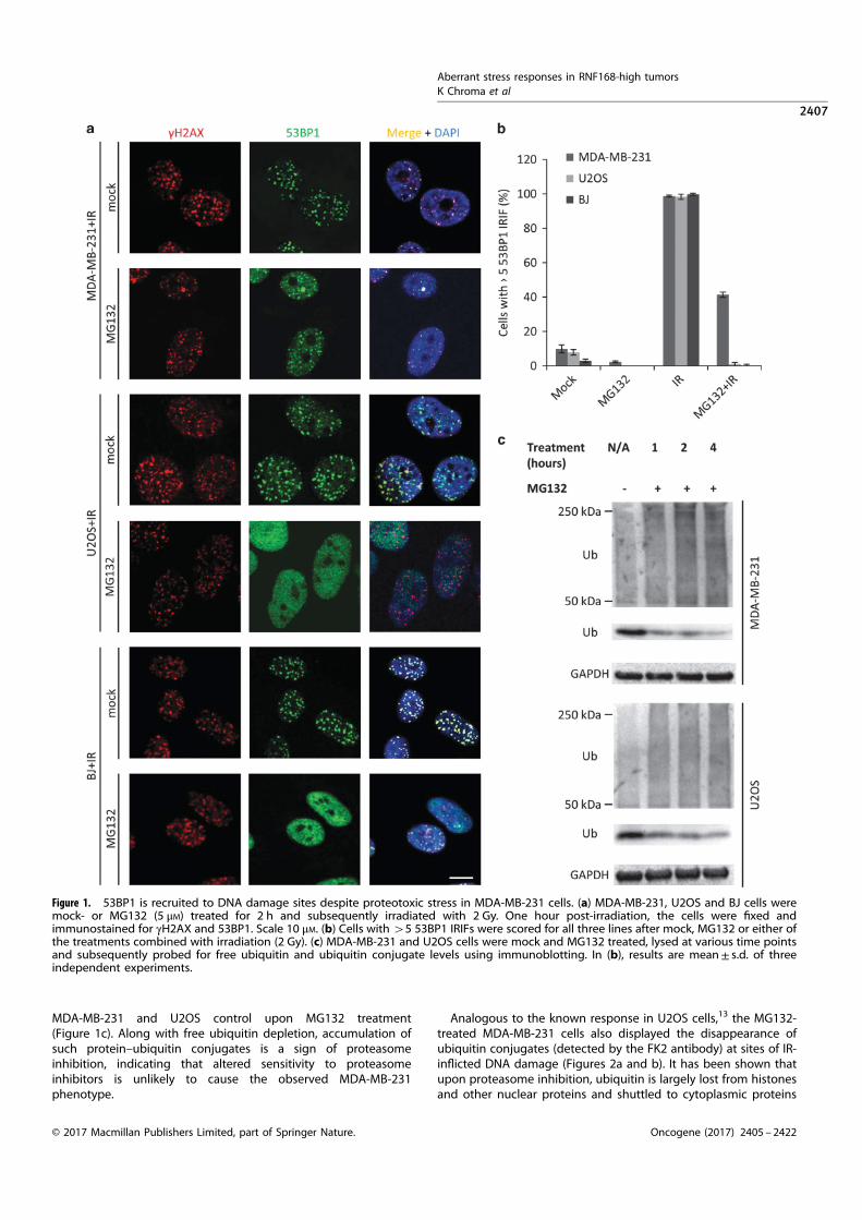
MDA-MB-231 and U2OS control upon MG132 treatment(Figure 1c). Along with free ubiquitin depletion, accumulation ofsuch protein–ubiquitin conjugates is a sign of proteasomeinhibition, indicating that altered sensitivity to proteasomeinhibitors is unlikely to cause the observed MDA-MB-231phenotype.
Analogous to the known response in U2OS cells,13 the MG132-treated MDA-MB-231 cells also displayed the disappearance ofubiquitin conjugates (detected by the FK2 antibody) at sites of IR-inflicted DNA damage (Figures 2a and b). It has been shown thatupon proteasome inhibition, ubiquitin is largely lost from histonesand other nuclear proteins and shuttled to cytoplasmic proteins
Figure 1. 53BP1 is recruited to DNA damage sites despite proteotoxic stress in MDA-MB-231 cells. (a) MDA-MB-231, U2OS and BJ cells weremock- or MG132 (5 μM) treated for 2 h and subsequently irradiated with 2 Gy. One hour post-irradiation, the cells were fixed andimmunostained for γH2AX and 53BP1. Scale 10 μM. (b) Cells with 45 53BP1 IRIFs were scored for all three lines after mock, MG132 or either ofthe treatments combined with irradiation (2 Gy). (c) MDA-MB-231 and U2OS cells were mock and MG132 treated, lysed at various time pointsand subsequently probed for free ubiquitin and ubiquitin conjugate levels using immunoblotting. In (b), results are mean± s.d. of threeindependent experiments.
Aberrant stress responses in RNF168-high tumors
K Chroma et al
2407
© 2017 Macmillan Publishers Limited, part of Springer Nature. Oncogene (2017) 2405 – 2422

awaiting degradation in the proteasome complex.29 This resultagain shows that MDA-MB-231 cells respond to proteasomeinhibition in an apparently standard way leading to depletion ofthe free nuclear ubiquitin pool, without any obvious compensa-tory mechanism that would facilitate the sustained 53BP1 accrualat the sites of DNA damage.Importantly, additional key DDR factors acting upstream of
53BP1 such as γH2AX and recruitment of MDC1 were observed in
IRIFs (Figures 2a and b) in both mock-treated and proteasomeinhibitor-treated cells. This suggested that the upstream steps ofthe DSB response pathway react to proteasome inhibition largelyin a standard mode in MDA-MB-231 cells.To further assess the chromatin DSB response pathway at the
level of 53BP1 and its associated proteins in the proteasome-inhibited cells, we probed the MDA-MB-231 and the control U2OScells for recruitment of two known 53BP1 effectors—RIF119,21 and
Figure 2. Probing DSB response upstream of 53BP1 in MG132-treated MDA-MB-231 and U2OS cells. (a) MDA-MB-231 cells were pretreatedwith MG132 for 2 h, irradiated with 2 Gy and 1h post-irradiation immunostained for the indicated proteins or protein modifications known tobe present in IRIFs. Scale 10 μM. (b) Graphical summary of nuclei with 45 γH2AX, FK2 or MDC1 IRIFs, scored in cells bearing 45 53BP1 IRIFs.Results are mean± s.d. of three independent experiments.
Aberrant stress responses in RNF168-high tumors
K Chroma et al
2408
Oncogene (2017) 2405 – 2422 © 2017 Macmillan Publishers Limited, part of Springer Nature.

REV7,20 to laser micro-irradiation induced DNA damage sites. Incontrast to U2OS, MG132 pretreated MDA-MB-231 cells showing53BP1 accumulation in laser-induced ‘stripes’ also displayed RIF1and REV7 accrual at such sites of micro-irradiation (Figures 3a and b).These results imply that the upstream steps of the DSB responsepathway operate normally, and the unorthodox DSB response inthe MDA-MB-231 cells under proteotoxic stress is shared by 53BP1and its downstream effectors.
UDR motif-mutated 53BP1 is not recruited to DSB sites underproteotoxic stress
53BP1 binds to two chromatin modifications at the DSBs—dimethylated histone H4 (H4K20) and ubiquitinated histone H2A(H2AK15Ub).23,24 The H2AK15Ub mark is recognized by theubiquitin damage response (UDR) domain at the C-terminal partof 53BP1.24 We utilized a UDR motif-mutated 53BP1 incapable ofbinding the H2AK15Ub mark to test whether ubiquitin was indeedrequired for 53BP1 accumulation at DSBs under conditions ofproteotoxic stress in the MDA-MB-231 line. Although a 53BP1 wild-type green fluorescent protein (GFP) fusion protein was recruitedto IRIFs, cells expressing the GFP-tagged UDR mutant (L1619A)24
did not form 53BP1 IRIFs (Figure 4). Furthermore, a GFP-taggedTudor domain 53BP1 mutant (D1521R)24 behaved similarly andwas not recruited to IRIFs (Figure 4). Taken together, this impliedthat in the MDA-MB-231 cells, 53BP1 recruitment still depends oneach of the two intact modules that recognize H4K20 andH2AK15Ub, respectively, even when levels of cellular-free ubiqui-tin become limiting.
The proteotoxic stress-resistant DSB response depends onubiquitin signaling, particularly RNF168
As the DSB response in the MDA-MB-231 cells is still fueled byubiquitin under proteotoxic stress, a mechanism should exist thatprovides sufficient amount of ubiquitin to sustain the process. Oneplausible way of bypassing an acute decrease in free ubiquitinlevels is overexpression of the E2 and/or E3 ubiquitin conjugatingenzymes/ligases. Elevated pool of an E2 conjugating enzyme thatwas charged with ubiquitin before the drop in free ubiquitin levelmight serve as a temporary reservoir for downstream processes.On the other hand, an overexpressed E3 ligase might outcompeteother E3 ligases in the uptake of residual ubiquitin underconditions of proteotoxic stress. Hence, we examined the levelsof the key DSB response related ubiquitin conjugating enzymeUBC13 (UBE2N) and E3 ligases RNF8 and RNF168 in the MDA-MB-231 cells. Strikingly, all three enzymes displayed elevatedlevels in this cell line (Figure 5a). When normalized toglyceraldehyde 3-phosphate dehydrogenase (GAPDH), bothRNF8 and RNF168 showed more than twofold higher levels thanthose in the U2OS cells, whereas UBC13 level was even higher—more than fivefold above the U2OS cells (Figure 5b). Theoverabundance of these three enzymes was even more profoundwhen the normal diploid BJ cells were compared with MDA-MB-231 cells: more than fourfold for UBC13, sixfold for RNF8 andmore than eightfold in the case of RNF168 (Figure 5b).Importantly, the level of the 53BP1 protein was comparable inall three cell types (Figure 5b).Quantitative PCR and cycloheximide chase experiments indi-
cated that the overabundance of RNF168 in the MDA-MB-231 cellsreflected transcriptional upregulation rather than increasedprotein stability (Supplementary Figures S1A and B). In additionto transcriptional upregulation, an increase in RNF168 translationefficiency and/or transcript stability likely contribute to theobserved RNF168 protein overabundance in MDA-MB-231 cellsas transcriptional upregulation alone (a 2.5-fold increase com-pared with U2OS, Supplementary Figure S1A) is unlikely toaccount for the high RNF168 protein levels given the fasterprotein turnover of RNF168 in these cells (deduced from the
almost fourfold shorter RNF168 protein half-life in MDA-MB-231compared with U2OS, Supplementary Figure S1B). Indeed, theaccelerated turnover of RNF168 protein was consistent withoverabundant TRIP12 and UBR5 (Supplementary Figure S1C), thetwo enzymes critical for ubiquitin/proteasome-mediated degrada-tion of RNF168.28 The elevated TRIP12 and UBR5 might reflect afine-tuning mechanism in MDA-MB-231 cells, possibly providing anegative feedback loop to limit the overabundant RNF168 tolevels that are not overly harmful to cells, a scenario that occursupon experimental gross overexpression of RNF168.28 Consis-tently, depletion of either TRIP12 or UBR5 in MDA-MB-231 led to aneven more pronounced DSB response phenotype resistant toproteasome inhibition (Supplementary Figure S1D), possibly due tofurther increase in the abundance of RNF168. As to additionalcomponents of the ubiquitin-mediated DSB signaling, we foundenhanced abundance of HERC2 (Supplementary Figure S1C), anotherubiquitin ligase that promotes 53BP1 recruitment at DSBs,31 whereasthere was little if any alteration of the negative regulators JMJD2A,L3MBTL1 or RNF169 proteins32 (negative data, not shown).Overall, these results supported the functional significance of
the RNF168-centered ubiquitin-mediated signaling pathway in thealtered DSB response in MDA-MB-231 cells. This notion was furthersupported by functional experiments, in which small interferingRNA (siRNA)-mediated knockdown of UBC13, RNF8 or RNF168completely abolished the proteotoxic stress-resistant DSB responsephenotype in the proteasome inhibitor-treated MDA-MB-231 cells(Figures 5c and d).Based on available mechanistic insights28 and the pronounced
clinical phenotype of RNF168 deficiency,27 we hypothesized thatthe RNF168 ligase could be central to the unorthodox DSBresponse phenotype in MDA-MB-231 cells. Partial knockdown ofRNF168 with increasing amounts of siRNA resulted in a gradualdecrease of cells capable of forming 45 53BP1 IRIFs (Figure 6a).Importantly, this phenotype could be rescued by expression ofsiRNA-resistant WT, but not the mutant version of RNF168 (C16S,in the RING domain) that abolishes the enzymatic activity of theprotein (Figure 6b).If RNF168 has a central role in the studied DSB response
phenotype, overexpression of the enzyme in a cell line incapableof sustaining DSB signaling under proteotoxic stress might mimicthe situation seen in MDA-MB-231 cells. Indeed, an U2OS-derivedcell line overexpressing a RNF168-GFP fusion protein exhibited53BP1 IRIF formation in nearly all nuclei even after proteasomeinhibition (Figures 6c–e). As in the case of MDA-MB-231 cells, thenumber of 53BP1 IRIF-positive cells correlated with the level ofRNF168 (Supplementary Figure S2) and, consistent with thepathway hierarchy,5,13 the phenotype was dependent on RNF8(Supplementary Figure S3). Of note, changes in RNF8 levels had aless profound effect on the phenotype compared with the moremarked impact of RNF168 abundance, thereby supportingthe major role of RNF168 in the proteotoxic stress-resistantDSB response (Supplementary Figure S3). Furthermore, ectopicexpression of the RNF168 (C16S) mutant in the U2OS cell linedid not result in the proteotoxic stress-resistant phenotype(Supplementary Figure S4), as opposed to expression of the WTprotein (Figures 6c and d and Supplementary Figure S4). Theseresults also parallel the scenario seen in MDA-MB-231 cells, where theectopic RNF168 C16S RING mutant was incapable of rescuing the lossof the phenotype caused by knockdown of endogenous RNF168.Overall, these data were consistent with the emerging key role ofRNF168 abundance in the proteotoxic stress-resistant DSB response.
The proteotoxic stress-resistant DSB response cancer phenotype ismore common
Next, we asked whether the emerging phenotype observed in thetriple-negative breast cancer cells MDA-MB-231 is unique or morewidespread, and tested a panel of proteasome inhibitor-treated
Aberrant stress responses in RNF168-high tumors
K Chroma et al
2409
© 2017 Macmillan Publishers Limited, part of Springer Nature. Oncogene (2017) 2405 – 2422
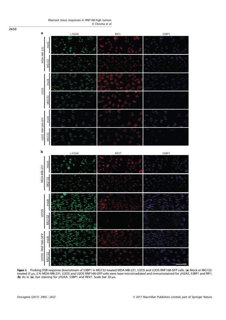
Figure 3. Probing DSB response downstream of 53BP1 in MG132-treated MDA-MB-231, U2OS and U2OS RNF168-GFP cells. (a) Mock or MG132-treated (5 μM, 2 h) MDA-MB-231, U2OS and U2OS RNF168-GFP cells were laser-microirradiated and immunostained for γH2AX, 53BP1 and RIF1.(b) As in (a), but staining for γH2AX, 53BP1 and REV7. Scale bar 50 μM.
Aberrant stress responses in RNF168-high tumors
K Chroma et al
2410
Oncogene (2017) 2405 – 2422 © 2017 Macmillan Publishers Limited, part of Springer Nature.

Figure 5. Elevated levels and impact of ubiquitin-mediated DSB signaling-related enzymes in MDA-MB-231 cells. (a) MDA-MB-231, U2OS andBJ cell lysates were analyzed by immunoblotting for abundance of 53BP1 and the major DSB ubiquitin signaling enzymes RNF8, RNF168 andUBC13. (b) Protein abundance was calculated using densitometric analysis of the immunoblot shown in a. Band intensities were normalized tocorresponding GAPDH bands. (c) MDA-MB-231 cells were transfected with indicated siRNAs, mock and MG132 treated (2 h, 5 μM) with andwithout irradiation (2 Gy) and 1 h post-irradiation stained for 53BP1. Cells with 45 53BP1 IRIF were scored. Results are mean± s.d. of threeindependent experiments (d) Knockdown efficiency in (c) was verified by probing corresponding cell lysates by immunobloting usingindicated antibodies.
Figure 4. 53BP1 recruitment to sites of damage in MDA-MB-231 is methylation and ubiquitination dependent. MDA-MB-231 cells transfectedwith siRNA against 53BP1 and expression vectors for the indicated siRNA-resistant GFP-tagged versions of 53BP1 were mock or MG132 treated(2 h, 5 μM), irradiated with 2 Gy and after 1h processed for GFP imaging. Scale 20 μM. Results are mean of two independent experiments.
Aberrant stress responses in RNF168-high tumors
K Chroma et al
2411
© 2017 Macmillan Publishers Limited, part of Springer Nature. Oncogene (2017) 2405 – 2422
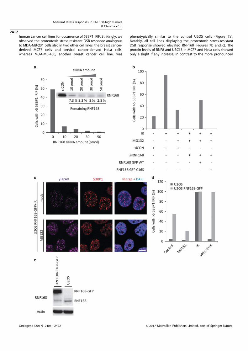
human cancer cell lines for occurrence of 53BP1 IRIF. Strikingly, weobserved the proteotoxic stress-resistant DSB response analogous
to MDA-MB-231 cells also in two other cell lines, the breast cancer-derived MCF7 cells and cervical cancer-derived HeLa cells,whereas MDA-MB-436, another breast cancer cell line, was
phenotypically similar to the control U2OS cells (Figure 7a).Notably, all cell lines displaying the proteotoxic stress-resistant
DSB response showed elevated RNF168 (Figures 7b and c). Theprotein levels of RNF8 and UBC13 in MCF7 and HeLa cells showedonly a slight if any increase, in contrast to the more pronounced
Aberrant stress responses in RNF168-high tumors
K Chroma et al
2412
Oncogene (2017) 2405 – 2422 © 2017 Macmillan Publishers Limited, part of Springer Nature.
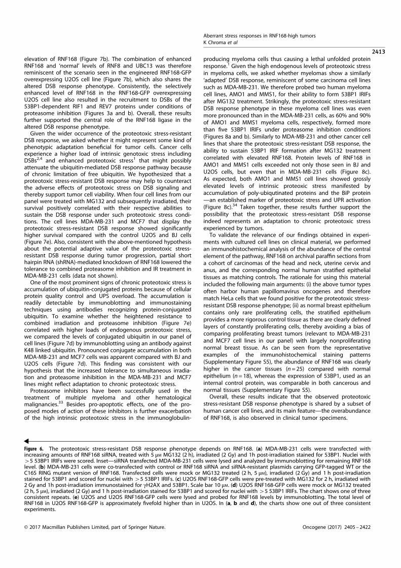
elevation of RNF168 (Figure 7b). The combination of enhancedRNF168 and ‘normal’ levels of RNF8 and UBC13 was thereforereminiscent of the scenario seen in the engineered RNF168-GFPoverexpressing U2OS cell line (Figure 7b), which also shares thealtered DSB response phenotype. Consistently, the selectivelyenhanced level of RNF168 in the RNF168-GFP overexpressingU2OS cell line also resulted in the recruitment to DSBs of the53BP1-dependent RIF1 and REV7 proteins under conditions ofproteasome inhibition (Figures 3a and b). Overall, these resultsfurther supported the central role of the RNF168 ligase in thealtered DSB response phenotype.Given the wider occurrence of the proteotoxic stress-resistant
DSB response, we asked whether it might represent some kind ofphenotypic adaptation beneficial for tumor cells. Cancer cellsexperience a higher load of intrinsic genotoxic stress includingDSBs2,4 and enhanced proteotoxic stress1 that might possiblyattenuate the ubiquitin-mediated DSB response pathway becauseof chronic limitation of free ubiquitin. We hypothesized that aproteotoxic stress-resistant DSB response may help to counteractthe adverse effects of proteotoxic stress on DSB signaling andthereby support tumor cell viability. When four cell lines from ourpanel were treated with MG132 and subsequently irradiated, theirsurvival positively correlated with their respective abilities tosustain the DSB response under such proteotoxic stress condi-tions. The cell lines MDA-MB-231 and MCF7 that display theproteotoxic stress-resistant DSB response showed significantlyhigher survival compared with the control U2OS and BJ cells(Figure 7e). Also, consistent with the above-mentioned hypothesisabout the potential adaptive value of the proteotoxic stress-resistant DSB response during tumor progression, partial shorthairpin RNA (shRNA)-mediated knockdown of RNF168 lowered thetolerance to combined proteasome inhibition and IR treatment inMDA-MB-231 cells (data not shown).One of the most prominent signs of chronic proteotoxic stress is
accumulation of ubiquitin-conjugated proteins because of cellularprotein quality control and UPS overload. The accumulation isreadily detectable by immunoblotting and immunostainingtechniques using antibodies recognizing protein-conjugatedubiquitin. To examine whether the heightened resistance tocombined irradiation and proteasome inhibition (Figure 7e)correlated with higher loads of endogenous proteotoxic stress,we compared the levels of conjugated ubiquitin in our panel ofcell lines (Figure 7d) by immunoblotting using an antibody againstK48 linked ubiquitin. Pronounced conjugate accumulation in bothMDA-MB-231 and MCF7 cells was apparent compared with BJ andU2OS cells (Figure 7d). This finding was consistent with ourhypothesis that the increased tolerance to simultaneous irradia-tion and proteasome inhibition in the MDA-MB-231 and MCF7lines might reflect adaptation to chronic proteotoxic stress.Proteasome inhibitors have been successfully used in the
treatment of multiple myeloma and other hematologicalmalignancies.33 Besides pro-apoptotic effects, one of the pro-posed modes of action of these inhibitors is further exacerbationof the high intrinsic proteotoxic stress in the immunoglobulin-
producing myeloma cells thus causing a lethal unfolded proteinresponse.1 Given the high endogenous levels of proteotoxic stressin myeloma cells, we asked whether myelomas show a similarly‘adapted’ DSB response, reminiscent of some carcinoma cell linessuch as MDA-MB-231. We therefore probed two human myelomacell lines, AMO1 and MMS1, for their ability to form 53BP1 IRIFsafter MG132 treatment. Strikingly, the proteotoxic stress-resistantDSB response phenotype in these myeloma cell lines was evenmore pronounced than in the MDA-MB-231 cells, as 60% and 90%of AMO1 and MMS1 myeloma cells, respectively, formed morethan five 53BP1 IRIFs under proteasome inhibition conditions(Figures 8a and b). Similarly to MDA-MB-231 and other cancer celllines that share the proteotoxic stress-resistant DSB response, theability to sustain 53BP1 IRIF formation after MG132 treatmentcorrelated with elevated RNF168. Protein levels of RNF168 inAMO1 and MMS1 cells exceeded not only those seen in BJ andU2OS cells, but even that in MDA-MB-231 cells (Figure 8c).As expected, both AMO1 and MMS1 cell lines showed grosslyelevated levels of intrinsic proteoxic stress manifested byaccumulation of poly-ubiqutinated proteins and the BiP protein—an established marker of proteotoxic stress and UPR activation(Figure 8c).34 Taken together, these results further support thepossibility that the proteotoxic stress-resistant DSB responseindeed represents an adaptation to chronic proteotoxic stressexperienced by tumors.To validate the relevance of our findings obtained in experi-
ments with cultured cell lines on clinical material, we performedan immunohistochemical analysis of the abundance of the centralelement of the pathway, RNF168 on archival paraffin sections froma cohort of carcinomas of the head and neck, uterine cervix andanus, and the corresponding normal human stratified epithelialtissues as matching controls. The rationale for using this materialincluded the following main arguments: (i) the above tumor typesoften harbor human papillomavirus oncogenes and thereforematch HeLa cells that we found positive for the proteotoxic stress-resistant DSB response phenotype; (ii) as normal breast epitheliumcontains only rare proliferating cells, the stratified epitheliumprovides a more rigorous control tissue as there are clearly definedlayers of constantly proliferating cells, thereby avoiding a bias ofcomparing proliferating breast tumors (relevant to MDA-MB-231and MCF7 cell lines in our panel) with largely nonproliferatingnormal breast tissue. As can be seen from the representativeexamples of the immunohistochemical staining patterns(Supplementary Figure S5), the abundance of RNF168 was clearlyhigher in the cancer tissues (n= 25) compared with normalepithelium (n= 18), whereas the expression of 53BP1, used as aninternal control protein, was comparable in both cancerous andnormal tissues (Supplementary Figure S5).Overall, these results indicate that the observed proteotoxic
stress-resistant DSB response phenotype is shared by a subset ofhuman cancer cell lines, and its main feature—the overabundanceof RNF168, is also observed in clinical tumor specimens.
Figure 6. The proteotoxic stress-resistant DSB response phenotype depends on RNF168. (a) MDA-MB-231 cells were transfected withincreasing amounts of RNF168 siRNA, treated with 5 μM MG132 (2 h), irradiated (2 Gy) and 1h post-irradiation stained for 53BP1. Nuclei with45 53BP1 IRIFs were scored. Inset—siRNA transfected MDA-MB-231 cells were lysed and analyzed by immunoblotting for remaining RNF168level. (b) MDA-MB-231 cells were co-transfected with control or RNF168 siRNA and siRNA-resistant plasmids carrying GFP-tagged WT or theC16S RING mutant version of RNF168. Transfected cells were mock or MG132 treated (2 h, 5 μM), irradiated (2 Gy) and 1 h post-irradiationstained for 53BP1 and scored for nuclei with 45 53BP1 IRIFs. (c) U2OS RNF168-GFP cells were pre-treated with MG132 for 2 h, irradiated with2 Gy and 1h post-irradiation immunostained for γH2AX and 53BP1. Scale bar 10 μM. (d) U2OS RNF168-GFP cells were mock or MG132 treated(2 h, 5 μM), irradiated (2 Gy) and 1 h post-irradiation stained for 53BP1 and scored for nuclei with45 53BP1 IRIFs. The chart shows one of threeconsistent repeats. (e) U2OS and U2OS RNF168-GFP cells were lysed and probed for RNF168 levels by immunoblotting. The total level ofRNF168 in U2OS RNF168-GFP is approximately fivefold higher than in U2OS. In (a, b and d), the charts show one out of three consistentexperiments.
Aberrant stress responses in RNF168-high tumors
K Chroma et al
2413
© 2017 Macmillan Publishers Limited, part of Springer Nature. Oncogene (2017) 2405 – 2422

Overabundant RNF168 shifts DSB repair toward NHEJ,enhances genomic instability and vulnerability to PARPis andcamptothecin
The results obtained so far suggested that MDA-MB-231 and someother cancer cell lines capable of DSB signaling despiteproteotoxic stress may deviate from normal cells and from othercancer cell lines in various aspects of their genome integritycontrol. To explore this emerging concept further, we firstassessed the response of MDA-MB-231 cells to PARPi, a strategy
that causes DNA damage mainly during S phase and whichshowed promise in treatment of a subset of triple-negative breasttumors in clinical trials.35,36 Immunofluorescence analysis showedthat while 460% of S-phase MDA-MB-231 cells treated by a PARPidisplayed over 10 53BP1-positive foci per nucleus, in U2OS thefraction of such cells was significantly lower (Figures 9a and b).Given the similar cell cycle phase profiles of both cell lines (datanot shown) and the fact that the DSBs caused by PARPi commonlyoccur during S phase and are preferentially repaired by HR the
Figure 7. The proteotoxic stress-resistant DSB response phenotype is shared by other cancer cell lines. (a) Indicated cells lines were mock- andMG132 treated (2 h, 5 μM), either with or without irradiation (2 Gy) and 1 h post-irradiation stained for 53BP1. Nuclei with 45 53BP1 IRIFs werescored. (b) Lysates prepared from the lines in (a) were probed for RNF8, RNF168 and UBC13 levels by immunoblotting. (c) RNF168 band intensitywas quantified and normalized according to the total protein levels in the indicated lines. (d) Indicated cell lines were probed for the level ofconjugated K48 linked ubiquitin by immunoblotting. Equal protein amounts were loaded for all the cell lines. (e) Indicated cell lines displayingvarious levels of RNF168 expression were pretreated with 5 μM MG132 (2 h), irradiated with 2 Gy and 1 h post-treatment seeded to Petri dishes.Six days post-irradiation, the cells were trypsinized and counted using an automated cell counter. In (a and e), results are mean± s.d. of threeindependent experiments. Statistical significance was determined with two-tailed unpaired Student's t-test; **,##Po0.005.
Aberrant stress responses in RNF168-high tumors
K Chroma et al
2414
Oncogene (2017) 2405 – 2422 © 2017 Macmillan Publishers Limited, part of Springer Nature.

Figure 8. Multiple myeloma cell lines exhibit the RNF168-fueled proteotoxic stress-resistant DSB response. (a and b) AMO1 and MMS1 celllines were mock- and MG132-treated (2 h, 5 μM), either with or without irradiation (2 Gy) and 1h post-irradiation stained for 53BP1. Nuclei with45 53BP1 IRIFs were scored. Scale 10 μM. (c) Indicated cell lines were probed for the level of conjugated K48 linked ubiquitin, RNF168 andBiP by immunoblotting. Equal protein amounts were loaded for all the cell lines. In (b), results are mean± s.d. of three independentexperiments.
Aberrant stress responses in RNF168-high tumors
K Chroma et al
2415
© 2017 Macmillan Publishers Limited, part of Springer Nature. Oncogene (2017) 2405 – 2422
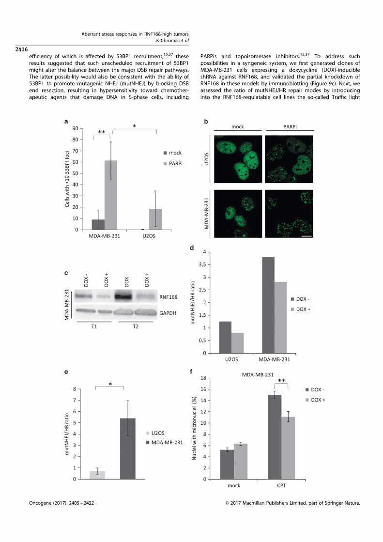
efficiency of which is affected by 53BP1 recruitment,15,37 theseresults suggested that such unscheduled recruitment of 53BP1
might alter the balance between the major DSB repair pathways.The latter possibility would also be consistent with the ability of53BP1 to promote mutagenic NHEJ (mutNHEJ) by blocking DSBend resection, resulting in hypersensitivity toward chemother-
apeutic agents that damage DNA in S-phase cells, including
PARPis and topoisomerase inhibitors.15,37 To address suchpossibilities in a syngeneic system, we first generated clones of
MDA-MB-231 cells expressing a doxycycline (DOX)-inducibleshRNA against RNF168, and validated the partial knockdown ofRNF168 in these models by immunoblotting (Figure 9c). Next, weassessed the ratio of mutNHEJ/HR repair modes by introducing
into the RNF168-regulatable cell lines the so-called Traffic light
Aberrant stress responses in RNF168-high tumors
K Chroma et al
2416
Oncogene (2017) 2405 – 2422 © 2017 Macmillan Publishers Limited, part of Springer Nature.

system,38 a reporter that enables flow cytometric analysis of repairpathway choice at individual I-SceI induced DNA breaks.Quantification of red (mutNHEJ) and green (HR) events thenprovides information on the overall activity proportion of the twopathways in the analyzed cell population. A representativeexample of such experiment shown in Figure 9d indeed supportsthe RNF168-dependent repair shift, as the cells with DOX-inducedpartial RNF168 knockdown showed a lower mutNHEJ/HR ratio.Furthermore, consistent with the high and low levels of RNF168,respectively, the mutNHEJ/HR ratio was more than sixfold higherin the parental MDA-MB-231 cells compared with the parentalU2OS cells (Figure 9e).Excessive mutNHEJ leads to frequent chromosome aberrations
and genome rearrangements that might contribute to tumorheterogeneity.22,39 To examine whether the RNF168-drivenupregulation of mutNHEJ makes the MDA-MB-231 line moreprone to genome rearrangements, we used the DOX-inducibleRNF168 knockdown model in MDA-MB-231 cells and comparednumbers of micronuclei in DOX-induced and non-induced cellspretreated by a topoisomerase I inhibitory drug camptothecin(CPT). The number of micronuclei was indeed significantly lower inthe DOX-induced cells with lowered RNF168 level and hence amore proficient HR repair because of less robust recruitment of53BP1 (Figure 9f). These results support a plausible scenario thatthe aberrantly upregulated RNF168 protects cancer cells fromadverse effects of proteotoxic stress on the DDR, however, only atthe cost of increased genomic instability.As the altered balance of DSB repair pathway choice toward
higher mutNHEJ and the ensuing chromosomal instability canimpact cell viability under exposure to S-phase genotoxic insultsthat require HR for efficient DNA repair,22 we next tested sensitivityof the MDA-MB-231 cells with inducible RNF168 knockdown towardCPT. Strikingly, the DOX-induced cells with decreased RNF168 levelswere significantly less sensitive to CPT (Figure 10a) than the non-induced counterpart cells. We interpret the observed decrease inCPT sensitivity upon RNF168 knockdown as further evidence forupregulation of NHEJ and the ensuing genomic instability in theMDA-MB-231 cells driven by RNF168 overabundance.Surprisingly, the MDA-MB-231 knockdown cell line did not show
a significant change in sensitivity toward PARP1 inhibition, whichis also known to be particularly toxic to cells with deregulatedNHEJ.15 We reasoned that this might be caused by only moderatedegree of RNF168 knockdown achieved in the MDA-MB-231 cells.To address this possibility, we also established and tested a MCF7-derived RNF168 knockdown cell line for sensitivity to CPT and theKU58948 PARP1 inhibitor. Indeed, MCF7 cells that share withMDA-MB-231 cells also the RNF168-fueled proteotoxic stress-resistant DSB response proved to be more amenable to the DOX-inducible RNF168 knockdown as the RNF168 level dropped 43.5-fold upon DOX treatment (Figure 10b). Another important reasonfor including MCF7 was the fact that, along with MDA-MB-231,MCF7 cells exhibited significant PARPi sensitivity, despite both
these cell lines are BRCA1/BRCA2 proficient.15 We hypothesizedthat the observed sensitivity to PARPi might be at least partlyattributable to the RNF168 overabundance and the ensuing shiftof the mutNHEJ/HR ratio, thereby creating a partial, relative‘HR deficiency’ despite the proficient BRCA1/2 genes. Consistentwith such possibility, the MCF7 cells showed significantlydecreased sensitivity toward both CPT and the PARPi uponinduction of RNF168 knockdown (Figure 10c). Thus, apart fromproviding another piece of evidence for aberrant upregulation ofNHEJ in these cell lines, this result might also represent animportant clue for better understanding of PARPi sensitivity inBRCA1/2-proficient tumors.
DISCUSSION
From a broader perspective, our present study contributes tobetter understanding of genome integrity maintenance andpoints to previously unrecognized wide occurrence and impactof aberrant ubiquitin-mediated signaling of DNA damage underproteotoxic stress, with the ensuing consequences for genomicinstability and responses to cancer treatment. Our results suggestthat human tumors can be widely categorized into two subsets,featuring ‘standard’ and ‘proteotoxic stress-resistant’ responses toDNA breakage, respectively. The latter tumor category, discoveredand characterized here, may represent an adaptive scenario of‘conditional/secondary’ rather than ‘genetically caused/primary’HR deficiency, with implications for genomic instabilityand selective advantages, but also potential vulnerabilities ofsuch cancers.First, from the mechanistic point of view of the chromatin
response to DSB, we show that overabundance of the RNF168ubiquitin ligase, sometimes accompanied by enhanced levels ofadditional E2/E3 enzymes, renders the DSB signaling insensitive todepletion of free ubiquitin levels resulting from proteotoxic stress.According to the current paradigm scenario typical for normalcells and some cancers, exemplified by the widely used humanU2OS sarcoma cell line model, DSB signaling is grossly attenuatedwhen free ubiquitin levels become limiting upon proteasomeinhibition-induced proteotoxic stress. Therefore, it seemed coun-terintuitive that we could observe sustained recruitment of 53BP1and its partner proteins REV7 and RIF1 after proteasomeinhibition. Although 53BP1 recruitment is regulated also by othermodifications including NEDDylation and acetylation40,41 andNEDDylation was suggested to compensate for ubiquitinationwhen proteasome is inhibited42 our own experiments usinginhibitors of NEDD conjugation and deacetylation did not supportthis possibility (our unpublished data). Based on our results, wepropose a model whereby ubiquitin is still used under proteotoxicstress to relay the DSB chromatin signaling, provided that theRNF168 E3 ligase is overabundant and hence can preferentiallychannel the remaining available ubiquitin to the RNF168-mediated pathway (Figure 11). Notably, whereas experimentally
Figure 9. Overabundant RNF168 causes unscheduled 53BP1 recruitment, increased mutNHEJ pathway activity and micronuclei formation.(a) MDA-MB-231 and U2OS cells were mock or PARPi (10 μM, 24 h) treated, immunostained for 53BP1 and cyclin A. Cyclin A-positive cells with410 53BP1 foci were scored. (b) Representative images of 53BP1 immunostained cells from (a). Scale 10 μM. (c) The MDA-MB-231DOX-inducible knockdown cells were pretreated with DOX (DOX, 100 ng/ml; 72 h: T1 or 96 h: T2), lysed and probed for RNF168. After the 72 hpre-treatment, the RNF168 levels were 42.5 times lower in the DOX-treated cells compared with controls (T1). The endpoint(96-h knockdown) RNF168 levels are shown in the T2 panel. (d) The effect of RNF168 level on the mutNHEJ/HR ratio was assessed in theMDA-MB-231 and U2OS cell lines bearing the DOX-inducible RNF168 knockdown and the Traffic light reporter. Stable reporter cell lines werepretreated with DOX as in (a) and subsequently transduced with a lentivirus carrying an HR repair template and an I-SceI gene. Five days post-transduction, cells were examined by flow cytometry for mCherry and GFP signal. The NHEJ/HR ratio was calculated by correlating thenumbers of red (NHEJ) and green (HR). (e) Analogous to (d), assessed in the parental U2OS and MDA-MB-231 cell lines only. (f) MDA-MB-231cells were pretreated with DOX as above, then mock or CPT treated (10 nM. 24 h) and nuclei/micronuclei were counterstained with DAPI.Fraction of micronuclei in the DAPI-stained objects was determined. In (a, e and f), results are mean± s.d. of three independent experiments.Statistical significance was determined with two-tailed unpaired Student's t-test; *Po0.05; **Po0.005.
Aberrant stress responses in RNF168-high tumors
K Chroma et al
2417
© 2017 Macmillan Publishers Limited, part of Springer Nature. Oncogene (2017) 2405 – 2422

induced ectopic overexpression of RNF168 can rescue theotherwise abolished DSB recruitment of 53BP1 (as well asrecruitment of RIF1 and REV7) under proteotoxic stress in cellswith moderate, physiological levels of endogenous RNF168(Figures 6c and d, 3a and b), recruitment of the RAP80-BRCA1complex to DSB lesions is not rescued under such circumstances(our unpublished results). This striking difference between the twobranches of the chromatin response to DSBs further supports ourrecent report on a functional interplay between JMJD1Cdemethylase, RNF8 and the MDC1 scaffold protein as a selectivemechanism required to recruit the RAP80-BRCA1 complex, but not53BP1.43 Considered in the context of our present study, the‘hyper-activity’ of the overabundant RNF168 that is sufficient torescue the 53BP1 recruitment under proteotoxic stress is notenough to allow recruitment of RAP80-BRCA1, as the latter branchof the DSB chromatin response critically depends on RNF8-mediated ubiquitination of MDC1, rather than histone ubiquitina-tion by RNF168, as well as on additional protein modifications.43
Such a dichotomy in ubiquitin-mediated recruitment of 53BP1versus RAP80-BRCA1 is also consistent with the recent report fromthe Halazonetis laboratory that 53BP1 recruitment in proteasomeinhibitor-treated cells may be partially rescued by fusing a bulkymoiety to the H2AX histone.44 This presumably opens upchromatin in the vicinity of DSBs and thus partially restoresresidual chromatin ubiquitination that in turn enables 53BP1accrual at DNA lesions.44 Analogous to the differential responsesto overexpression of RNF168 in our present study, recruitment of
the RAP80-BRCA1 complex to IRIFs under proteasome inhibitionconditions was also not rescued by the chromatin openingstrategy. Furthermore, our data are also consistent with the notionthat the FK2 antibody detected ubiquitin conjugates at the DSBsites may reflect preferential reactivity with RNF8-mediatedubiquitination of MDC1, whereas the histone ubiquitin productscatalyzed by RNF168 may not be accessible to antibodies becauseof nucleosome compaction.43 Such interpretation can also helpexplain that upon replacement of endogenous RNF168 with acatalytically inactive RNF168 variant, the FK2 antibody fociremained detectable, whereas ubiquitination of histone H2AX wasabolished.45
We suggest that our experiments can shed some light also onthe competition based mode of 53BP1 recruitment that reportedlyrequires the two competing demethylases, JMJD2A and JMJD2Bto be removed from chromatin flanking the DSBs and degraded inorder to expose the H4K20me2 that can be subsequently boundby the 53BP1’s tandem TUDOR domains.26 As we have observedsustained 53BP1 recruitment under conditions of proteasomeinhibition, it seems unlikely that the two demethylases have to bedegraded to allow for 53BP1 recruitment to chromatin. We favor amodel in which the clearance of the competing proteins from theDSB-flanking chromatin is sufficient and does not have to beaccompanied by their degradation in order to permit 53BP1recruitment. According to such modified model, the RNF168-mediated ubiquitination of JMJD2A and JMJD2B would serveprimarily as a chromatin eviction signal and the subsequent
Figure 10. RNF168 overabundance sensitizes MDA-MB-231 and MCF7 cells to CPT and PARPi. (a) Sensitivity of the MDA-MB-231 RNF168knockdown cells toward CPT was assessed by a cell survival assay. The cells were pretreated with DOX as above and then treated with 1 nM
CPT. After 6 days, the cells were trypsinized and counted using an automated cell counter. (b) The MCF7 DOX-inducible knockdown cell linewas pretreated with DOX (DOX, 100 ng/ml; 72 h: T1 or 96 h: T2), lysed and probed by immunoblotting for RNF168. After the 72 hpre-treatment, the RNF168 levels were43.5-fold lower in the DOX-induced cells than in the non-treated control cells (T1). (c) Sensitivity of theMCF7 RNF168 knockdown cells toward CPT and KU58948 was assessed as in (a). In (a and c), results are mean± s.d. of three independentexperiments. Statistical significance was determined with two-tailed unpaired Student's t-test; **Po0.005.
Aberrant stress responses in RNF168-high tumors
K Chroma et al
2418
Oncogene (2017) 2405 – 2422 © 2017 Macmillan Publishers Limited, part of Springer Nature.

degradation of these demethylases is not essential for 53BP1recruitment.In the absence of BRCA1 that limits 53BP1 chromatin loading
during S phase, the RNF168-driven 53BP1 recruitment precludesDSB end resection and thereby HR, whereas boosting DNA repairby the mutagenic NHEJ pathway.22,46,47 Unexpectedly, ourfindings show that BRCA1-proficient cells bearing overabundantRNF168 mimic, at least to some extent, the BRCA1-deficientphenotype by displaying lower levels of HR at the expense ofupregulated mutNHEJ. We show that this is most likely caused byaberrantly enhanced 53BP1 recruitment in S-phase cells that isfueled by the excess of RNF168. Albeit not tested in our presentstudy, it is predictable based on the published work in mouseB cells and embryonic fibroblasts, which the overabundantRNF168 inhibits efficient DSB end resection and fuels DSB repairby the mutagenic NHEJ pathway.22 The RNF168 overexpressionseems to derail the physiological balance of the DSB repairpathways toward 53BP1 recruitment and mutNHEJ. We speculatethat this imbalance leads to ‘conditional HR deficiency’ especiallyunder chronic proteotoxic stress conditions, and might accountfor (or contribute to) the observed increased sensitivity of certainBRCA1-proficient (and principally also HR-proficient) tumorssuch as subsets of triple-negative breast carcinomas, towardPARPis.35,48
The unexpectedly wide occurrence of the proteotoxic stress-resistant DDR among different tumor cell lines raises a question
whether it might represent a means of adaptation or providesome selective advantage(s) during tumorigenesis. Cancer cellssuffer from increased endogenous proteotoxic stress that stemsfrom such features as aneuploidy, mutation overload and henceaccumulation of altered proteins, and variation of gene copynumber and levels of transcription.1,3 We propose that apart fromplacing a significant burden on the protein quality controlmechanisms,3 proteotoxic stress also impacts on DSB responsevia attenuating the ubiquitin driven signaling at damagedchromatin. Of note, the load of endogenous DSBs increasesduring cell transformation and tumor progression because ofenhanced replication stress evoked by diverse oncogenes and lossof some tumor suppressors.6,7,49–51 Given its pathophysiologicalsignificance, aberrations in the DSB ubiquitination signalingpathway might profoundly affect genome integrity of tumor cells.Our findings show that attenuation of DSB signaling because ofproteotoxic stress might be circumvented by upregulation of oneor more key ubiquitin ligases involved in the DDR, particularlyRNF168. Importantly, this concept was further supported byobservation of the proteotoxic stress-resistant DSB response inmultiple myeloma cells, an established model of cancer-relatedproteotoxic stress. It has been also reported that breast cancersexhibit elevated levels of some E2 ubiquitin conjugatingenzymes.52 Taken together, this implies that upregulation ofsome ubiquitin-mediated cellular processes might represent amore general strategy to overcome adverse effects of cancer-
Figure 11. Model summarizing the proteotoxic stress-resistant DSB response and its impact on cancer cells. Changes in chromosome or genecopy number and transcription (de)regulation in cancer cells result in protein overproduction that overwhelms the cellular protein qualitycontrol, causing chronic proteotoxic stress and diminishing levels of free ubiquitin. The limited free ubiquitin supply has to be shared bydiverse ubiquitin-dependent processes whose efficiency, including that of DSB signaling, is impaired. This is manifested by increasedradiosensitivity. Overexpression of RNF168 (and other key DSB response ubiquitin-related enzymes) in the proteotoxic stress-resistant cellsshifts the free ubiquitin equilibrium toward DSB signaling thus increasing radioresistance. Overexpression of RNF168 and concomitant robust53BP1 recruitment promotes mutNHEJ at the expense of HR repair, rendering the cells sensitive to topoisomerase and PARPis, and leading toenhanced genomic instability. Such changes collectively impact tumor heterogeneity, progression and responses to therapy.
Aberrant stress responses in RNF168-high tumors
K Chroma et al
2419
© 2017 Macmillan Publishers Limited, part of Springer Nature. Oncogene (2017) 2405 – 2422

associated proteotoxic stress. UPS has a major role in theregulation of several key tumorigenesis driving processes, suchas cellular proliferation, apoptosis and stress tolerance.1,53 Hence,it is likely that these pathways are sensitive to proteotoxic stressand tumor cells have evolved compensatory mechanisms such asthe upregulation of specific enzymes of the UPS. In terms ofpotential selective advantages during tumorigenesis, the acquiredoverabundance of RNF168 can help enhance survival of cancercells under combined proteotoxic and replication stresses, fuelerror-prone DNA repair, genomic instability and thereby intra-tumor heterogeneity (Figure 11), all features likely to promotetumor progression and aggressivity.It remains to be elucidated how cancer cells acquire the
elevated expression of RNF168 and/or other ubiquitin ligases andconjugating enzymes. Analogous to other tumor-associatedchanges in gene expression, the most likely candidates aremutations in gene regulatory sequences, genome rearrangementsor transcription suppressor/activator mutations. One of the likelycandidates that might drive the cancer-related RNF168 over-expression is the family of FOXO transcription factors known toregulate various stress response genes including components ofthe DDR machinery.54,55 Dysregulation of the FOXO3a transcrip-tion factor occurs in both breast cancer and hematologicalmalignancies,54 which implies that this protein (and possibly otherFOXO family members) might fuel the elevated RNF168 expres-sion in tumors. Regardless of the molecular mechanism, it will alsobe conceptually interesting to find out when during tumorprogression such overexpression of RNF168 occurs, relative to thereported activation of the DSB-responsive checkpoint anticancerbarrier and its interplay with the ARF-p53 checkpointpathway.2,4,8,53
Last but not least, our present results indicate that such possiblyadaptive upregulation of RNF168 may have important implicationsfor responses of tumors to standard-of-care as well as someemerging targeted treatments. On one hand, we show that tumorcells with the proteotoxic stress-resistant DSB response phenotypeare more resistant to ionizing radiation under conditions ofenhanced proteotoxic stress. At the same time, however, thealtered balance among the DSB repair pathways appears togenerate a kind of adaptive, conditional HR deficiency, andthereby unmask some potentially exploitable vulnerabilities toS-phase genotoxic drugs such as CPT or PARPis. In the light of ourpresent findings, combined immunohistochemical detection ofRNF168 and markers of proteotoxic stress such as conjugatedubiquitin or the BiP chaperone might be exploited as candidatebiomarkers to identify the subsets of patients whose tumors maydisplay the proteotoxic stress-resistant phenotype described here,and possibly help decisions about personalized cancer therapy inthe future.
MATERIALS AND METHODS
Cell culture and generation of DSBs
Most cell lines used in this work were cultured in Dulbecco’s modified Eagle’smedium, supplemented with 10% fetal bovine serum (PAA, Pasching, Austria)and penicillin/streptomycin (Sigma-Aldrich, St Louis, MO, USA) in a humidifiedatmosphere of 5% CO2 at 37 °C. For MCF7, AMO1 and MMS1 culturing, thestandard cell culture medium was RPMI-1640 with the same supplements asabove. All cell lines were purchased from ATCC (Manassas, VA, USA) with theexception of U2OS RNF168-GFP that was established previously.13
X-ray irradiation was done using the YXLON.SMART 160E/1.5 device(YXLON, Horsens, Denmark) at the following settings: 150 kV, 6 mA,11 mGy/s.
Micro-irradiation
Laser micro-irradiation was performed on a Zeiss Axioimager Z.1 instrumentequipped with a laser scanning LSM780 module (Zeiss, Oberkochen,Germany). A UV-A laser (355 nm 65 mW) was used to induce the DNA
damage. BrDU presensitization and irradiation of the cells was done asdescribed previously.56 Subsequent immunofluorescent detection of recruitedproteins was essentially done as in Xu et al.20
Plasmids and RNA interference
Most plasmids were transfected using the FuGENE 6 (Roche, Basel,Switzerland) reagent following the manufacturer’s instructions. Whenrequired, plasmid DNA was transfected by nucleofection using the Neon(Life Technologies, Carlsbad, CA, USA) device at settings recommended bythe manufacturer for the respective cell line. The pGFP-53BP1-Fl-wt,pGFP-53BP1-Fl-L1619A and pGFP-53BP1-Fl-D1521 plasmids carrying the53BP1 UDR and Tudor domains mutations were a gift from D Durocher(Samuel Lunenfeld Research Institute, Ontario, Canada). The pAcGFP-C1-RNF168 plasmids harboring the C16S RING and MIU mutations weredescribed previously.13 The Traffic light repair template, the I-SceI lentiviralconstructs38 as well as the lentivirus production plasmids pMD2.Gand psPAX2 (D Trono, unpublished) were purchased from Addgene(Cambridge, MA, USA; plasmids no’s 31476, 31482, 12259 and 12260). Theinducible shRNA RNF168 knockdown lentiviral plasmids were constructedas described in Wiederschain et al.57 using following oligonucleotides(5'–3'): shRNA RNF168 sense CCGGGGCGAAGAGCGATGGAGGACTCGAGTCCTCCATCGCTCTTCGCCTTTTT; shRNA RNF168 antisense AATTAAAAAGGCGAAGAGCGATGGAGGACTCGAGTCCTCCATCGCTCTTCGCC (Generi Biotech,Hradec Kralove, Czech Republic). The backbone pLKO-Tet-On Puro57,58
plasmid was obtained from Addgene (plasmid no. 21915).siRNA’s were transfected with the Lipofectamine RNAiMAX (Invitrogen,
Carlsbad, CA, USA) reagent following the manufacturer’s instructions.siRNAs were purchased from Thermo Fisher Scientific (Waltham, MA, USA):siCON-negative control, siRNA#1 (ID#4390843), siRNF168 (ID #126171),siRNF8 (ID#17200) and from MWG Operon (Ebersberg, Germany):53BP1DD2013 GAGAGCAGAUGAUCCUUUAtt (5'–3').
Oligonucleotides and quantitative PCR
The abundance of RNF168 mRNA level was probed by quantitative PCRusing a Nano LightCycler (Roche) instrument and following oligos (5'–3'):RNF168qPCR_F1 CAGGGCAAGACACAGAAATAGA; RNF168qPCR_R1 GGCACCACAGGCACATAA; RNF168qPCR_F2 CTCCCTACAGCCTAGCATTTC and RNF168qPCR_R2 AGATCACAAAGCACTCCCTTTA (Generi Biotech). FollowingGAPDH, primers were used as an internal control: GAPDH—F GAAGATGGTGATGGGATTTC; GAPDH—R GAAGGTGAAGGTCGGAGT (GeneriBiotech) PCR product abundance was quantified using the LighCyclerNano software (Roche).
Chemicals and antibodies
The Bortezomib (PS-341), MG132 and CPT inhibitors were purchased fromSigma-Aldrich. The KU58948 PARP1 inhibitor was obtained from AstraZe-neca (London, UK). Antibodies used in this study included following mousemonoclonal antibodies: γH2AX (Merck Millipore, Billerica, MA, USA), RNF8(B‐2) (Santa Cruz Biotechnology, Santa Cruz, CA, USA), Ubc13, JMJD2A(KDM4A) (Thermo Fisher Scientific, Waltham, MA, USA), HERC2(BD Transduction Laboratories, San Jose, CA, USA), MDC1, USP34 (Abcam,Cambridge, UK), GAPDH (GT239) (GeneTex, Hsinchu, Taiwan), β-actin(Sigma-Aldrich) and polyclonal rabbit: 53BP1, BRCA1 (Santa Cruz Biotech-nology), TRIP12 (Abcam), UBR5 (Sigma-Aldrich), FK2 (Enzo Life Sciences,Farmingdale, NY, USA), RIF1 (Bethyl Laboratories, Montgomery, TX, USA),REV7 (BD Transduction Laboratories). The rabbit polyclonal antibodies toRNF168 and RNF169 were a gift from N Mailand (Center for ProteinResearch, Copenhagen, Denmark).
Immunoblotting
Cells were lysed in Laemmli sample buffer and the whole-cell lysates weresubsequently separated on a 10% sodium dodecyl sulfate–polyacrylamidegel electrophoresis gel and transferred to a nitrocellulose membrane (GEHealthcare, Little Chalfont, UK). The membrane was blocked in 5 % (w/v)skim milk in Tris-buffered saline supplemented with in 0.1% (v/v) Tween-20and probed with a primary antibody. Subsequently, the membrane wasincubated with horseradish peroxidase-labeled secondary anti-mouse oranti-rabbit antibodies (Santa Cruz Biotechnology) and the signals werevisualized using ECL detection reagents (Thermo Fisher Scientific) and theChemiDoc system (Bio-Rad, Hercules, CA, USA). Band intensity quantifica-tion was performed in the ImageJ software (http://imagej.nih.gov/ij/).
Aberrant stress responses in RNF168-high tumors
K Chroma et al
2420
Oncogene (2017) 2405 – 2422 © 2017 Macmillan Publishers Limited, part of Springer Nature.

Immunofluorescence and micronuclei staining, microscopicanalysis
Cells grown on 12-mm coverslips were fixed with 4% paraformaldehyde inphosphate-buffered saline (PBS) for 15 min and then permeabilized withPBS containing 0.2% (v/v) Triton X-100 for 5 min. Suspension cells werecytospinned onto microscopic slides before fixation using the Cyto-TekSakura instrument (Sakura Finetek, Torrance, CA, USA). Fixed cells wereblocked with 5% (v/v) fetal bovine serum in PBS for 30 min and incubatedovernight at 4 °C with primary antibodies (diluted in 5% (w/v) bovineserum albumin in PBS). Coverslips were washed three times in PBSsupplemented with 0.1% (v/v) Tween-20, once with PBS and thenincubated with an appropriate secondary goat anti-rabbit or goat anti-mouse Alexa Fluor 488 or Alexa Fluor 568 conjugated (Invitrogen)secondary antibody (diluted in in 5% (w/v) bovine serum albumin inPBS) for 60 min at room temperature. Slips were then washed as aboveand mounted onto slides using the 4,6-diamidino-2-phenylindole (DAPI)containing Vectashield mounting reagent (Vector Laboratories, Burlin-game, CA, USA). Coverslips for micronuclei analysis were fixed and washedas above, stained with DAPI diluted in PBS and subsequently mountedwith the Vectashield reagent (without DAPI).Slides were visualized by the Axio Observer.Z1/Cell Observer Spinning
Disc microscopic system (Yokogawa, Tokyo, Japan and Zeiss) equippedwith an Evolve 512 (Photometrix, Tucson, AZ, USA) EMCCD camera. ZeissPlan Apochromat 63x and 100x/1.40 NA objectives were used.For quantitative image analysis, a series of random fields were recorded
automatically using the ScanR imaging workstation (Olympus, Tokyo,Japan; with an EM charge-coupled device camera (C9100; HamamatsuPhotonics, Hamamatsu City, Japan), a U Plan S Apochromat 40× /0.9 NAobjective, and an image resolution of 200× 200 nm/pixel). The numberand intensity of micronuclei and IR-induced nuclear foci were quantifiedusing the ScanR image analysis software (Olympus).
Generation of lentiviruses and lentiviral transduction
Lentiviruses were generated by co-transfecting 293T cells with 4 μg ofpMD2.G, 7 μg of psPAX2 and 9 μg of a lentiviral plasmid of interest usingthe CaPO4 precipitation method.59 Six to eight hours post-transfection, thecells were washed briefly with pre-warmed PBS and medium was changed.Lentivirus containing supernatant was collected 48 h later. Target cellswere transduced at multiplicity of infection of 1–10 with the supernatantsupplemented with 4 μg/ml polybrene (Sigma-Aldrich). Twenty-four hourspost-transduction, the medium was changed and when required, the cellswere selected in 1 μg/ml puromycin.
Flow cytometric analysis of DNA repair pathway choice
Cells harboring the Traffic light reporter were seeded in a 12-well plate and24-h later transduced with the I-SceI and GFP repair template containingconstruct using the procedure above. Seven days later, the cellswere trypsinized, fixed with formaldehyde and analyzed by an Influx(BD Biosciences, San Jose, CA, USA) instrument. GFP was measured using a488 nm laser for excitation and a 530/40 filter, whereas mCherry wasexcited using a 561 nm laser and acquired with a 610/20 filter. Data wereanalyzed using the FACS Sortware (BD Biosciences) software.
Cell cycle analysis
Cells were fixed in 70% ethanol and stained with propidium iodide for flowcytometric analysis. Fixed cells were analyzed on a FACS Verse instrument(BD Biosciences) and cell cycle distribution was assigned using theFACSuite software (BD Biosciences).
Long-term cell survival assay
In all, 1 × 10⁵ cells were seeded in triplicate to ø 6 cm plates and left toattach overnight. Next day, the medium was replaced by inhibitor ordimethylsulfoxide (mock) containing medium. Seven days later, the cellswere trypsinized and cell number was scored using a Vi-Cell XR CellViability Analyzer (Beckman Coulter, Brea, CA, USA) equipped with theViCELL XR software (Beckman Coulter).The IR resistance of proteotoxic stress DDR-resistant lines was assessed
as above with following modifications: attached cells were pretreated with5 μM MG132 or dimethylsulfoxide (mock) for 2 h and subsequentlyirradiated with 2 Gy. Then medium was changed and cell survival wasassayed as above 7 days later.
Statistical analysis
Differences in DNA repair pathway efficiency and cell survival assays were
analyzed by Student’s t-test. Variability and reproducibility among
repeated experiments subjected to quantitative evaluations, such as
immunofluorescence IRIF counts or quantitative PCR products is indicated
by mean ± s.d. and shown as error bars in graphical summaries in the
relevant figures.
ABBREVIATIONS
53BP1, p53 binding protein 1; BRCA1, breast cancer 1; DDR, DNA damage
response; DSB, double-strand break; HR, homologous recombination; IRIF,
ionizing radiation induced foci; NHEJ, non-homologous end joining; PARP1,
poly(ADP-ribose) polymerase 1; RNF168, ring finger protein 168.
CONFLICT OF INTEREST
The authors declare no conflict of interest.
ACKNOWLEDGEMENTS
We thank Jan Bouchal, Katerina Bouchalova and our colleagues from the Laboratory of
Genome Integrity for technical assistance, suggestions and comments on the
manuscript. This work was supported by grants from the following foundations: Grant
Agency of the Czech Republic 13-17555S, Czech National Program of Sustainability
LO1304, the Kellner Family Foundation, the Norwegian Financial Mechanism CZ09
(Project PHOSCAN 7F14061), MEYS CR (LM2015062 Czech-BioImaging), the internal grant
IGA-LF-2016-030, the EU operation program CZ.1.07/2.3.00/30.0004, the Danish National
Research Foundation (DNRF125, project CARD), Danish Cancer Society, the Swedish
Research Council, the Lundbeck Foundation, Cancer Fonden, and the Danish Council for
Independent Research.
REFERENCES
1 Deshaies RJ. Proteotoxic crisis, the ubiquitin-proteasome system, and cancer
therapy. BMC Biol 2014; 12: 94.
2 Halazonetis TD, Gorgoulis VG, Bartek J. An oncogene-induced DNA damage
model for cancer development. Science 2008; 319: 1352–1355.
3 Luo J, Solimini NL, Elledge SJ. Principles of cancer therapy: oncogene and
non-oncogene addiction. Cell 2009; 136: 823–837.
4 Bartek J, Bartkova J, Lukas J. DNA damage signalling guards against activated
oncogenes and tumour progression. Oncogene 2007; 26: 7773–7779.
5 Jackson SP, Bartek J. The DNA-damage response in human biology and disease.
Nature 2009; 461: 1071–1078.
6 Bartkova J, Horejsi Z, Koed K, Kramer A, Tort F, Zieger K et al. DNA damage
response as a candidate anti-cancer barrier in early human tumorigenesis.
Nature 2005; 434: 864–870.
7 Bartkova J, Rezaei N, Liontos M, Karakaidos P, Kletsas D, Issaeva N et al. Oncogene-
induced senescence is part of the tumorigenesis barrier imposed by DNA damage
checkpoints. Nature 2006; 444: 633–637.
8 Evangelou K, Bartkova J, Kotsinas A, Pateras IS, Liontos M, Velimezi G et al.
The DNA damage checkpoint precedes activation of ARF in response to escalating
oncogenic stress during tumorigenesis. Cell Death Differ 2013; 20: 1485–1497.
9 Zhao Y, Brickner JR, Majid MC, Mosammaparast N. Crosstalk between ubiquitin
and other post-translational modifications on chromatin during double-strand
break repair. Trends Cell Biol 2014; 24: 426–434.
10 Lukas J, Lukas C, Bartek J. More than just a focus: the chromatin response to DNA
damage and its role in genome integrity maintenance. Nat Cell Biol 2011; 13:
1161–1169.
11 Reinhardt HC, Yaffe MB. Phospho-Ser/Thr-binding domains: navigating the cell
cycle and DNA damage response. Nat Rev Mol Cell Biol 2013; 14: 563–580.
12 Thorslund T, Ripplinger A, Hoffmann S, Wild T, Uckelmann M, Villumsen B et al.
Histone H1 couples initiation and amplification of ubiquitin signalling after
DNA damage. Nature 2015; 527: 389–393.
13 Doil C, Mailand N, Bekker-Jensen S, Menard P, Larsen DH, Pepperkok R et al.
RNF168 binds and amplifies ubiquitin conjugates on damaged chromosomes to
allow accumulation of repair proteins. Cell 2009; 136: 435–446.
14 Stewart GS, Panier S, Townsend K, Al-Hakim AK, Kolas NK, Miller ES et al.
The RIDDLE syndrome protein mediates a ubiquitin-dependent signaling cascade
at sites of DNA damage. Cell 2009; 136: 420–434.
15 Bunting SF, Callen E, Wong N, Chen HT, Polato F, Gunn A et al. 53BP1 inhibits
homologous recombination in Brca1-deficient cells by blocking resection of
DNA breaks. Cell 2010; 141: 243–254.
Aberrant stress responses in RNF168-high tumors
K Chroma et al
2421
© 2017 Macmillan Publishers Limited, part of Springer Nature. Oncogene (2017) 2405 – 2422

16 Daley JM, Sung P. 53BP1, BRCA1, and the choice between recombination and end
joining at DNA double-strand breaks. Mol Cell Biol 2014; 34: 1380–1388.
17 Densham RM, Garvin AJ, Stone HR, Strachan J, Baldock RA, Daza-Martin M et al.
Human BRCA1-BARD1 ubiquitin ligase activity counteracts chromatin barriers to
DNA resection. Nat Struct Mol Biol 2016; 23: 647–655.
18 Callen E, Di Virgilio M, Kruhlak MJ, Nieto-Soler M, Wong N, Chen HT et al. 53BP1
mediates productive and mutagenic DNA repair through distinct phosphoprotein
interactions. Cell 2013; 153: 1266–1280.
19 Chapman JR, Barral P, Vannier JB, Borel V, Steger M, Tomas-Loba A et al. RIF1 is
essential for 53BP1-dependent nonhomologous end joining and suppression of
DNA double-strand break resection. Mol Cell 2013; 49: 858–871.
20 Xu G, Chapman JR, Brandsma I, Yuan J, Mistrik M, Bouwman P et al. REV7
counteracts DNA double-strand break resection and affects PARP inhibition.
Nature 2015; 521: 541–544.
21 Zimmermann M, Lottersberger F, Buonomo SB, Sfeir A, de Lange T. 53BP1
regulates DSB repair using Rif1 to control 5' end resection. Science 2013; 339:
700–704.
22 Zong D, Callen E, Pegoraro G, Lukas C, Lukas J, Nussenzweig A. Ectopic expression
of RNF168 and 53BP1 increases mutagenic but not physiological non-
homologous end joining. Nucleic Acids Res 2015; 43: 4950–4961.
23 Botuyan MV, Lee J, Ward IM, Kim JE, Thompson JR, Chen J et al. Structural basis for
the methylation state-specific recognition of histone H4-K20 by 53BP1 and Crb2
in DNA repair. Cell 2006; 127: 1361–1373.
24 Fradet-Turcotte A, Canny MD, Escribano-Diaz C, Orthwein A, Leung CC, Huang H
et al. 53BP1 is a reader of the DNA-damage-induced H2A Lys 15 ubiquitin mark.
Nature 2013; 499: 50–54.
25 Acs K, Luijsterburg MS, Ackermann L, Salomons FA, Hoppe T, Dantuma NP. The
AAA-ATPase VCP/p97 promotes 53BP1 recruitment by removing L3MBTL1 from
DNA double-strand breaks. Nat Struct Mol Biol 2011; 18: 1345–1350.
26 Mallette FA, Mattiroli F, Cui G, Young LC, Hendzel MJ, Mer G et al. RNF8- and
RNF168-dependent degradation of KDM4A/JMJD2A triggers 53BP1 recruitment to
DNA damage sites. EMBO J 2012; 31: 1865–1878.
27 Devgan SS, Sanal O, Doil C, Nakamura K, Nahas SA, Pettijohn K et al. Homozygous
deficiency of ubiquitin-ligase ring-finger protein RNF168 mimics the radio-
sensitivity syndrome of ataxia-telangiectasia. Cell Death Differ 2011; 18:
1500–1506.
28 Gudjonsson T, Altmeyer M, Savic V, Toledo L, Dinant C, Grofte M et al. TRIP12 and
UBR5 suppress spreading of chromatin ubiquitylation at damaged chromosomes.
Cell 2012; 150: 697–709.
29 Dantuma NP, Groothuis TA, Salomons FA, Neefjes J. A dynamic ubiquitin
equilibrium couples proteasomal activity to chromatin remodeling. J Cell Biol
2006; 173: 19–26.
30 Jacquemont C, Taniguchi T. Proteasome function is required for DNA damage
response and fanconi anemia pathway activation. Cancer Res 2007; 67:
7395–7405.
31 Bekker-Jensen S, Rendtlew Danielsen J, Fugger K, Gromova I, Nerstedt A, Lukas C
et al. HERC2 coordinates ubiquitin-dependent assembly of DNA repair factors on
damaged chromosomes. Nat Cell Biol 2010; 12: 80–86. sup pp 1-12.
32 Panier S, Boulton SJ. Double-strand break repair: 53BP1 comes into focus. Nat Rev
Mol Cell Biol 2014; 15: 7–18.
33 Chauhan D, Hideshima T, Anderson KC. Proteasome inhibition in multiple
myeloma: therapeutic implication. Annu Rev Pharmacol Toxicol 2005; 45: 465–476.
34 Gething MJ. Role and regulation of the ER chaperone BiP. Semin Cell Dev Biol 1999;
10: 465–472.
35 Livraghi L, Garber JE. PARP inhibitors in the management of breast cancer: current
data and future prospects. BMC Med 2015; 13: 188.
36 Ricks TK, Chiu HJ, Ison G, Kim G, McKee AE, Kluetz P et al. Successes and chal-
lenges of PARP inhibitors in cancer therapy. Front Oncol 2015; 5: 222.
37 Bouwman P, Aly A, Escandell JM, Pieterse M, Bartkova J, van der Gulden H et al.
53BP1 loss rescues BRCA1 deficiency and is associated with triple-negative and
BRCA-mutated breast cancers. Nat Struct Mol Biol 2010, biology 17: 688–695.
38 Certo MT, Ryu BY, Annis JE, Garibov M, Jarjour J, Rawlings DJ et al. Tracking
genome engineering outcome at individual DNA breakpoints. Nat Methods 2011;
8: 671–676.
39 Rodgers K, McVey M. Error-prone repair of DNA double-strand breaks. J Cell
Physiol 2016; 231: 15–24.
40 Ma T, Chen Y, Zhang F, Yang CY, Wang S, Yu X. RNF111-dependent
neddylation activates DNA damage-induced ubiquitination. Mol Cell 2013; 49:
897–907.
41 Tang J, Cho NW, Cui G, Manion EM, Shanbhag NM, Botuyan MV et al. Acetylation
limits 53BP1 association with damaged chromatin to promote homologous
recombination. Nat Struct Mol Biol 2013; 20: 317–325.
42 Hjerpe R, Thomas Y, Chen J, Zemla A, Curran S, Shpiro N et al. Changes in the ratio
of free NEDD8 to ubiquitin triggers NEDDylation by ubiquitin enzymes. Biochem J
2012; 441: 927–936.
43 Watanabe S, Watanabe K, Akimov V, Bartkova J, Blagoev B, Lukas J et al. JMJD1C
demethylates MDC1 to regulate the RNF8 and BRCA1-mediated chromatin
response to DNA breaks. Nat Struct Mol Biol 2013; 20: 1425–1433.
44 Kocylowski MK, Rey AJ, Stewart GS, Halazonetis TD. Ubiquitin-H2AX fusions render
53BP1 recruitment to DNA damage sites independent of RNF8 or RNF168.
Cell Cycle 2015; 14: 1748–1758.
45 Mattiroli F, Vissers JH, van Dijk WJ, Ikpa P, Citterio E, Vermeulen W et al. RNF168
ubiquitinates K13-15 on H2A/H2AX to drive DNA damage signaling. Cell 2012;
150: 1182–1195.
46 Munoz MC, Laulier C, Gunn A, Cheng A, Robbiani DF, Nussenzweig A et al.
RING finger nuclear factor RNF168 is important for defects in homologous
recombination caused by loss of the breast cancer susceptibility factor BRCA1.
J Biol Chem 2012; 287: 40618–40628.
47 Munoz MC, Yanez DA, Stark JM. An RNF168 fragment defective for focal accu-
mulation at DNA damage is proficient for inhibition of homologous recombina-
tion in BRCA1 deficient cells. Nucleic Acids Res 2014; 42: 7720–7733.
48 Inbar-Rozensal D, Castiel A, Visochek L, Castel D, Dantzer F, Izraeli S et al.
A selective eradication of human nonhereditary breast cancer cells by
phenanthridine-derived polyADP-ribose polymerase inhibitors. Breast Cancer Res
2009; 11: R78.
49 Burrell RA, McClelland SE, Endesfelder D, Groth P, Weller MC, Shaikh N et al.
Replication stress links structural and numerical cancer chromosomal instability.
Nature 2013; 494: 492–496.
50 Di Micco R, Fumagalli M, Cicalese A, Piccinin S, Gasparini P, Luise C et al.
Oncogene-induced senescence is a DNA damage response triggered by DNA
hyper-replication. Nature 2006; 444: 638–642.
51 Gorgoulis VG, Vassiliou LV, Karakaidos P, Zacharatos P, Kotsinas A, Liloglou T et al.
Activation of the DNA damage checkpoint and genomic instability in human
precancerous lesions. Nature 2005; 434: 907–913.
52 Chen L, Madura K. Increased proteasome activity, ubiquitin-conjugating enzymes,
and eEF1A translation factor detected in breast cancer tissue. Cancer Res 2005; 65:
5599–5606.
53 Velimezi G, Liontos M, Vougas K, Roumeliotis T, Bartkova J, Sideridou M et al.
Functional interplay between the DNA-damage-response kinase ATM and ARF
tumour suppressor protein in human cancer. Nat Cell Biol 2013; 15: 967–977.
54 Greer EL, Brunet A. FOXO transcription factors at the interface between longevity
and tumor suppression. Oncogene 2005; 24: 7410–7425.
55 Tran H, Brunet A, Grenier JM, Datta SR, Fornace AJ Jr., DiStefano PS et al.
DNA repair pathway stimulated by the forkhead transcription factor FOXO3a
through the Gadd45 protein. Science 2002; 296: 530–534.
56 Mistrik M, Vesela E, Furst T, Hanzlikova H, Frydrych I, Gursky J et al. Cells and
stripes: a novel quantitative photo-manipulation technique. Sci Rep 2016; 6:
19567.
57 Wiederschain D, Wee S, Chen L, Loo A, Yang G, Huang A et al. Single-vector
inducible lentiviral RNAi system for oncology target validation. Cell Cycle 2009; 8:
498–504.
58 Wee S, Wiederschain D, Maira SM, Loo A, Miller C, deBeaumont R et al.
PTEN-deficient cancers depend on PIK3CB. Proc Natl Acad Sci USA 2008; 105:
13057–13062.
59 Tiscornia G, Singer O, Verma IM. Production and purification of lentiviral vectors.
Nat Protoc 2006; 1: 241–245.
Supplementary Information accompanies this paper on the Oncogene website (http://www.nature.com/onc)
Aberrant stress responses in RNF168-high tumors
K Chroma et al
2422
Oncogene (2017) 2405 – 2422 © 2017 Macmillan Publishers Limited, part of Springer Nature.

APPENDIX E
Skrott Z, Majera D, Gursky J, Buchtova T, Hajduch M, Mistrik M, Bartek J. Disulfiram's anti-
cancer activity reflects targeting NPL4, not inhibition of aldehyde dehydrogenase. Oncogene.
2019 Aug 7. In press. IF(2018): 6.634

Oncogene
https://doi.org/10.1038/s41388-019-0915-2
ARTICLE
Disulfiram’s anti-cancer activity reflects targeting NPL4, notinhibition of aldehyde dehydrogenase
Zdenek Skrott1 ● Dusana Majera1 ● Jan Gursky 1● Tereza Buchtova1 ● Marian Hajduch 1
● Martin Mistrik1 ●
Jiri Bartek1,2,3
Received: 18 April 2019 / Revised: 27 June 2019 / Accepted: 22 July 2019
© The Author(s), under exclusive licence to Springer Nature Limited 2019
Abstract
Aldehyde dehydrogenase (ALDH) is a proposed biomarker and possible target to eradicate cancer stem cells. ALDH
inhibition as a treatment approach is supported by anti-cancer effects of the alcohol-abuse drug disulfiram (DSF, Antabuse).
Given that metabolic products of DSF, rather than DSF itself inhibit ALDH in vivo, and that DSF’s anti-cancer activity is
potentiated by copper led us to investigate the relevance of ALDH as the suggested molecular cancer-relevant target of DSF.
Here we show that DSF does not directly inhibit ALDH activity in diverse human cell types, while DSF’s in vivo metabolite,
S-methyl-N,N-diethylthiocarbamate-sulfoxide inhibits ALDH activity yet does not impair cancer cell viability. Our data
indicate that the anti-cancer activity of DSF does not involve ALDH inhibition, and rather reflects the impact of DSF’s
copper-containing metabolite (CuET), that forms spontaneously in vivo and in cell culture media, and kills cells through
aggregation of NPL4, a subunit of the p97/VCP segregase. We also show that the CuET-mediated, rather than any ALDH-
inhibitory activity of DSF underlies the preferential cytotoxicity of DSF towards BRCA1- and BRCA2-deficient cells. These
findings provide evidence clarifying the confusing literature about the anti-cancer mechanism of DSF, a drug currently tested
in clinical trials for repositioning in oncology.
Introduction
Cancer stem cells (CSCs) are believed to represent a major
challenge to successful cancer therapy [1], due to CSCs’
ability to resist standard-of-care treatment modalities and
fuel post-treatment relapse and metastatic spread [2]. CSCs
can be detected through expression of several markers
including aldehyde dehydrogenases (ALDHs) [3]. There are
19 putatively functional ALDH genes in the human genome
[4], and several ALDH isoenzymes are used as markers of
stem cells including CSC [5]. ALDH have diverse functions
in normal tissues, including the pivotal role in catalysing
endogenous and exogenous aldehydes into carboxylic acids
[6]. If aldehydes are not metabolized, they may cause severe
toxicity to the cells, including DNA damage by forming
adducts [7]. Numerous studies reported that ALDH is
overexpressed in cancer cells and implicated in metastatic
spread [8–10]. Despite the above-mentioned reports on
ALDH in CSCs, however, it remains unclear whether
ALDH may serve as an actionable target for cancer treat-
ment, and whether tumours are indeed addicted to ALDH
function.
Recent efforts to eradicate CSCs have exploited the old
anti-alcoholism drug DSF, used for decades as an ALDH
inhibitor in clinical care [4]. Eradication of CSCs by DSF
has been reported in numerous studies, the first of which
reported DSF’s toxicity for breast cancer cells with CSC-
These authors contributed equally: Zdenek Skrott, Dusana Majera
* Martin Mistrik
* Jiri Bartek
1 Institute of Molecular and Translational Medicine, Faculty of
Medicine and Dentistry, Palacky University, Olomouc, Czech
Republic
2 Danish Cancer Society Research Center, Copenhagen, Denmark
3 Division of Genome Biology, Department of Medical
Biochemistry and Biophysics, Science for Life Laboratory,
Karolinska Institute, Stockholm, Sweden
Supplementary information The online version of this article (https://
doi.org/10.1038/s41388-019-0915-2) contains supplementary
material, which is available to authorized users.
1234567890();,:
1234567890();,:

like properties [11]. In recent years, multiple studies
reported DSF as a drug toxic to cancer cells via inhibition of
ALDH in a range of tumour types and models [12–20] and
other studies have later build on these findings and used
DSF combined with copper ions to target cancer cells [21–
24]. However, the mechanism of ALDH inhibition by DSF
is more complex, as metabolic products of DSF, not DSF
itself, inhibit ALDH in vivo [4]. While well accepted in
pharmacology, the latter fact has often been overlooked in
the cancer-related studies focusing on DSF and ALDH,
thereby causing potentially misleading interpretations of the
results.
In vivo DSF is rapidly metabolized to diethyldithio-
carbamate (DDTC), which is further converted to
S-methyl-N,N-diethyldithiocarbamate (DETC) and
S-methyl-N,N-diethyldithiocarbamate (Me-DDTC). Sub-
sequent P450-catalyzed oxidation of DETC and
Me-DDTC produces DETC-sulfoxide (DETC-SO) and S-
methyl-N,N-diethylthiocarbamate-sulfoxide (Me-DTC-
SO) and -sulfone (Me-DTC-SO2), metabolites that are
most likely directly involved in ALDH inhibition [25–29].
Importantly, when downstream steps of DSF metabolism
are blocked by a chemical P450 inhibitor, liver ALDH
remains uninhibited [30], thus unambiguously proving
that not DSF itself, but its metabolites are the genuine
inhibitors of ALDH in vivo. Despite this knowledge is
published and accepted in some research fields, most
cancer-focused studies regard DSF as a direct ALDH
inhibitor. Notably, there are no published data with regard
to any anti-cancer effects of the DSF metabolites that are
responsible for ALDH inhibition. Further fuelling the
confusion in this field, the vast majority of cancer-related
studies report that DSF inhibits ALDH only when com-
bined with copper ions [12–16], a fact that further
underlines the extent of misunderstanding and lack of
logic behind such approach with respect to the known
mechanism of ALDH inhibition, a process that does not
involve copper at all. On the other hand, it is well known
that copper does potentiate DSF’s anti-cancer toxicity
[12, 31, 32], and we have recently uncovered that this
reflects the in vivo formation of a copper-containing CuET
(bis-diethyldithiocarbamate-copper) complex, the ultimate
anti-cancer metabolite derived from DSF [33]. This con-
undrum surrounding the links among DSF, ALDH, copper
and cancer toxicity prompted us to assess the role of
ALDH as a potential target of DSF’s anti-cancer activity
in more detail, using genuine validated inhibitors of
ALDH enzymatic activity, and thereby help to reconcile
the often mis-interpreted findings in this field, with the
goal to facilitate the future repositioning of DSF for
treatment of cancer.
Results
DSF’s toxicity for cancer cells is mediated by CuETformed in the culture media
Despite numerous pre-clinical studies and ongoing clinical
trials, the mechanism of anti-cancer activity of DSF is still
debated, as several targets and hypotheses have been pro-
posed. Among them, the inhibition of ALDH is probably
currently the most prevalent and accepted theory. ALDH is
attractive not only as a generally accepted marker of stem
cells, but also as an important protective enzyme metabo-
lising potentially harmful aldehydes. However, the
hypothesis that ALDH may represent a promising avenue to
target cancer stem cells or cancer in general, remains to be
rigorously tested.
Recently, preferential cytotoxicity of DSF for homo-
logous recombination (HR) deficient cells have been
reported [20]. Since DSF is regarded by some as a direct
inhibitor of ALDH, the reported cytotoxicity in this study
was attributed to increased acetaldehyde levels ensuing
ALDH inhibition, and subsequent DNA damage induced by
the crosslinking activity of the aldehydes. As direct inhi-
bition of cellular ALDH by DSF is in fact highly unlikely
(see Introduction) we decided to reproduce and re-analyse
those intriguing results. First, we tried to recapitulate the
reported preferential sensitivity of BRCA1 and BRCA2-
deficient cell lines to DSF [20]. Indeed, the H1299 cell lines
with doxycycline (DOX)-inducible shBRCA1 or
shBRCA2 show efficient knockdown of these genes after
DOX induction (Fig. 1a) and both models show also
hypersensitivity of BRCA-depleted cells to olaparib (Sup-
plementary Fig. 1a) a PARP1 inhibitor effective against HR
deficient cancers [34]. In agreement with Tacconi et al. [20],
we confirmed that BRCA1- and BRCA2-deficient cells are
indeed more sensitive to DSF treatment compared to their
BRCA-proficient counterparts (Fig. 1b). Importantly, we
have recently described a new metabolite of DSF, CuET,
which is formed in vivo and is responsible for DSF’s anti-
cancer activity [33], providing a meaningful explanation for
why is the toxicity of DSF potentiated by copper supple-
mentation. Thus we sought to investigate whether the CuET
complex forms also in vitro, since standard cell culture
media contain significant amounts of copper ions [35] and
the complex biochemical environment in the medium may
allow spontaneous formation of such complex. Indeed, we
have confirmed that CuET is detectable in DSF-containing
medium even without any additional copper supplementa-
tion (Fig. 1c). As predicted, addition of more copper to the
medium increased the amount of formed CuET; conversely
chelation of copper ions by a metal chelator,
Z. Skrott et al.

bathocuproinedisulfonic acid (BCDS), markedly reduced
the levels of spontaneously formed CuET (Fig. 1c).
Importantly, in line with our hypothesis and results of
spontaneous formation of CuET, chelation of copper by
BCDS completely reversed the cytotoxic effect of DSF in
all tested cell lines irrespective of their BRCA1/2 status
(Fig. 1d, Supplementary Fig. 1b). Another interesting aspect
described by Tacconi et al. [20] was the observation that the
cytotoxic effect of DSF reaches a certain plateau, which
cannot be overcome by increasing concentrations of the
drug, a phenomenon attributed by the authors to limited
solubility of the DSF. We also confirmed this plateau effect
but we argued that this might be explained by an alternative
mechanism, namely reflecting the limiting amounts of
copper in culture media, which would enable only limited
formation of CuET irrespectively of increasing concentra-
tions of DSF. To test the two alternative hypotheses, we
added non-toxic extra amounts of copper ions to culture
Fig. 1 Preferential cytotoxicity of disulfiram to BRCA1- and BRCA2-
depleted H1299 cells is copper dependent. a H1299 cells expressing
DOX-inducible shBRCA1 or shBRCA2 were cultivated for at least
3 days in DOX and protein expression was evaluated by Western
blotting, confirming efficient knockdown of BRCA1 and BRCA2,
respectively. b H1299 cells expressing DOX-inducible shBRCA1 or
shBRCA2 were treated with DSF at indicated concentration for 5 days.
c HPLC-MS analysis of CuET complex formed in the media
containing DSF, DSF with copper, or DSF with BCDS. d Cells as in b
were treated with the combination of 10 μM BCDS and DSF at indi-
cated concentration for 5 days. e H1299 cells were treated with DSF or
the combination of 1 μM CuCl2 and DSF at indicated concentrations
for 5 days. f Cells as in b were treated with CuET at indicated con-
centration for 5 days. All graphs represent at least three independent
experiments. Error bars represent SD
Disulfiram’s anti-cancer activity reflects targeting NPL4, not inhibition of aldehyde. . .

medium. Supporting our hypothesis, we observed a reversal
of the plateau effect, along with a striking potentiation of
DSF toxicity (Fig. 1e). Finally, we directly tested the syn-
thetic CuET complex. As expected, CuET treatment was
highly potent and also recapitulated the preferential toxicity
toward BRCA-impaired cell lines (Fig. 1f, Supplementary
Fig. 1c). Taken together, these results demonstrate that
DSF’s cytotoxicity is fully dependent on copper ions and is
mediated by the CuET complex, which is spontaneously
formed in the medium, proportionally to the amounts of
DSF and copper ions present in the cell culture
environment.
Neither DSF nor CuET inhibit ALDH activity, contraryto DSF metabolite Me-DTC-SO
Given the fact that DSF’s anti-cancer activity is commonly
attributed to inhibition of ALDH, we wanted to test this
hypothesis further. An important aspect of DSF as a drug is
that it undergoes extensive metabolism resulting in several
compounds, including S-methyl-N, N-diethylthiocarbamate-
sulfoxide (Me-DTC-SO), which represents the most likely
DSF’s metabolite responsible for the inhibition of liver
ALDH in vivo [25, 26]. However, no potential effect of Me-
DTC-SO on cancer cells has so far been reported. First, we
investigated the impact of CuET, DSF and Me-DTC-SO on
ALDH activity. We selected two human cancer cell lines
with high ALDH expression, K562 and A549, and used the
well-established ALDEFLUOR assay to measure total
ALDH activity in these cells [36]. Strikingly, in K562 cells
ALDH activity was not impaired by either CuET or DSF
treatment, in contrast to the Me-DTC-SO metabolite that
inhibited ALDH with an efficacy similar to D-
aminobenzaldehyde (DEAB), a commonly employed
ALDH inhibitor used here as a positive control for ALDH
inhibition (Fig. 2a, b). Consistently, neither CuET nor DSF
decreased the percentage of ALDH positive cells, in contrast
to Me-DTC-SO and DEAB (Fig. 2c). The same scenario was
reproduced also in the A549 cells as neither CuET nor DSF
mimicked the impact of the ALDH inhibitors, while Me-
DTC-SO completely blocked the ALDH activity in all cells
(Fig. 2d–f). Analogous data were seen in the BRCA1/2
knockdown H1299 cells whose overall ALDH activity is
lower compared to K562 or A549 cell lines, again con-
firming that only Me-DTC-SO potently inhibited ALDH,
while CuET and DSF had no direct measurable effect on
ALDH activity (Supplementary Fig. 2a, b).
ALDH inhibitors are not toxic to cancer cells
Next, we tested the toxicity of Me-DTC-SO and DEAB, in
concentrations efficiently inhibiting the ALDH activity.
Strikingly, both compounds failed to supress growth of
K562 and A549 cells (Fig. 3a). In contrast, CuET which
does not inhibit ALDH reduced the growth of both cancer
cell lines (Fig. 2b). Furthermore, the H1299 cells were
highly responsive to CuET but fully resistant to both DEAB
and Me-DTC-SO inhibitors, irrespectively of their BRCA1/
2 status (Fig. 3c, d). Given the fact that DSF undergoes
rapid transformation in vivo, it is very likely that both
metabolites, CuET and Me-DTC-SO, exist in the body at
the same time and their effects may potentially influence
each other. To test if ALDH inhibition augments the toxi-
city of CuET, we combined CuET with Me-DTC-SO and
DEAB at concentrations efficiently inhibiting ALDH and
analysed the viability of cancer cells; however, no poten-
tiation was observed (Fig. 3e). Taken together, these results
clearly exclude ALDH inhibition as a possible explanation
for DSF’s anti-cancer activity and call for an alternative,
mechanistically justified explanation. At the same time, our
data caution that targeting the ALDH as an approach to
cancer treatment should be further scrutinized.
Reduced ALDH activity readout of the ALDEFLUORassay upon long-term exposure to DSF is an indirectconsequence of toxicity
Our results excluding direct inhibition of ALDH by CuET
and/or DSF sharply contrast with numerous previous stu-
dies claiming that DSF or DSF combined with copper
inhibits ALDH activity in cultured cells [12–14, 19, 20],
thereby raising the notion of how can such conflicting
conclusions be reconciled. One key aspect shared by the
studies that reported apparent effects of DSF or DSF/Cu
treatments on ALDH activity were long exposure times to
the drug (from many hours to several days). This seemed to
us a rather odd approach for aiming to test direct enzymatic
inhibitors for which a few-hour exposure should be suffi-
cient. We argued that such long exposure times to a toxic
and metabolized compound might generate confounding
indirect effects and thereby complicate the interpretation of
the final outcome, as many important cellular functions can
be already hampered due to rather broad, non-specific
phenotypes. Such late indirect effects could also bias the
readout of the commonly used ALDEFLUOR assay, which
requires cellular import of a fluorescent probe and its
intracellular retention after cleavage by the ALDH enzyme.
To test this idea, we compared ALDH activity at different
time-points of drug exposure to evaluate the potential effect
of reduced cellular fitness on the ALDEFLUOR assay
readout. First, we measured ALDH activity after 3 h of
incubation with the four relevant drugs, which was suffi-
cient to supress ALDH activity when the direct ALDH
inhibitors DEAB and Me-DTC-SO were used, yet with no
detectable ALDH-inhibitory effect of either CuET or DSF
used in parallel experiments (Fig. 2a). Next, we tested not
Z. Skrott et al.

only CuET as a compound of interest, but also bortezomib
(BTZ), a compound that exerts its toxicity through specific
inhibition of the 20 S proteasome and partly resembles the
cellular effects induced by CuET [33]. Notably, BTZ´s
mechanism of action is completely unrelated to ALDH.
Consistent with our previous results, DSF and CuET failed
to inhibit ALDH activity after 3 h of exposure despite other
typical cellular phenotypes such as accumulation of poly-
ubiquitinylated proteins [33] are already well detectable in
the cells treated for 3 h with the same concentration of DSF
or CuET (Fig. 4a). As expected, also BTZ failed to score in
the ALDH inhibition assay (Fig. 4b). Strikingly, however,
after a prolonged treatment (20 h), both CuET and BTZ
markedly reduced the ALDEFLUOR-assessed ALDH
Fig. 2 ALDH activity in cells is inhibited by DSF’s metabolite Me-
DTC-SO, but not affected by DSF and CuET. a, b K562 cells were
treated with indicated compounds and ALDH activity was quantified
by ALDEFLUOR™ assay. Representative graphs and flow cytometry
profile from three independent experiments are shown. c Number of
ALDH positive K562 cells. d, e A549 cells were treated with indicated
compounds and ALDH activity was measured. Representative graphs
and flow cytometry profile from four independent experiments are
shown. f Number of ALDH positive A549 cells
Disulfiram’s anti-cancer activity reflects targeting NPL4, not inhibition of aldehyde. . .

activity readout (Fig. 4b) and clearly decreased the numbers
of ALDH-positive cells (Fig. 4c, d). Such prolonged treat-
ments also increased the numbers of permeabilised cells,
an indirect marker of reduced cell fitness and increased
cell death (Fig. 4e). Given that even BTZ, a compound
never reported as an ALDH inhibitor, behaved similarly to
CuET, we propose that the decrease of ALDH activity in
such long-term treatment experiments is not caused by any
direct interference with ALDH enzymatic activity, but it is
rather a consequence of impaired cell fitness. All per-
meabilised cells were totally negative for ALDH activity
(Supplementary Fig. 3b), which is understandable con-
sidering the principle of the ALDEFLUOR assay. Even
the seemingly still ‘intact’ (nonpermeabilized) cells
showed a lower ALDH activity readout suggesting that
prolonged cellular stresses (at least the proteotoxic stress
caused by CuET- or BTZ-induced protein turnover
impairment) is sufficient to indirectly affect the outcome
of the ALDEFLUOR assay (Supplementary Fig. 3a), a fact
that has been incorrectly interpreted by many as direct
inhibition of ALDH by DSF. These results help explain
the previous conflicting studies and exclude ALDH inhi-
bition as a mechanism underlying DSF’s toxicity to cancer
cells.
DSF toxicity is linked to NPL4 aggregation
We have recently reported that DSF is metabolised in vivo
into the CuET complex, and showed that CuET represents
the ultimate anti-cancer metabolite. CuET interferes with
the cellular protein degradation machinery via targeting the
NPL4 cofactor of the p97/VCP segregase, leading to NPL4
Fig. 3 Cytotoxicity of ALDH inhibitors and CuET. a Cytotoxicity of
DEAB or Me-DTC-SO in A549 and K562 cells after 5 days of
treatment. b Cytotoxicity of CuET in A549 and K562 cells after 5 days
of treatment. All graphs represent at least three independent experi-
ments. Error bars represent SD. c H1299 cells expressing DOX-
inducible shBRCA1 or shBRCA2 were treated with ALDH inhibitors
DEAB and d Me-DTC-SO at indicated concentrations for 5 days.
e Cytotoxicity of CuET, Me-DTC-SO and DEAB or their combination
in A549 cells
Z. Skrott et al.

aggregation, activation of stress responses and cell death
[33]. Since CuET is formed from DSF also in vitro in
culture media (Fig. 1c) and DSF’s toxicity strictly depends
on available copper ions (Fig. 1d, e), we examined whether
DSF’s cytotoxicity is also accompanied by NPL4 aggre-
gation. We treated the cells with CuET, DSF, DSF com-
bined with a copper chelator BCDS, and the two ALDH
inhibitors: Me-DTC-SO and DEAB and assessed the NPL4
protein status. As expected, both CuET and DSF treatment
led to formation of insoluble aggregated endogenous NPL4
resistant to pre-extraction (Fig. 5a). Chelation of copper
ions by BCDS completely supressed DSF’s effect on NPL4
aggregation, thereby preserving the normal diffuse staining
pattern of NPL4 that was sensitive to cell pre-extraction.
The same un-altered, extraction-sensitive diffuse staining of
NPL4 was furthermore observed for mock treated cells, but
also upon treatment by the two ALDH inhibitors Me-DTC-
SO and DEAB (Fig. 5a, see Fig. 5b for signal quantifica-
tion). These results were further corroborated using a NPL4-
GFP expressing cell line showing the same effects on GFP
tagged NPL4 protein (Supplementary Fig. 4a, b). Together
with the other results of our present study, these data
demonstrate that DSF’s cytotoxicity does not involve
ALDH inhibition, but rather it is attributable to CuET
causing NPL4 aggregation, as recently described for the
synthetic CuET complex [33].
Fig. 4 CuET reduces ALDH activity after prolonged treatment.
a Western blot analysis of immobilized and accumulated K48 poly-
ubiquitin in CuET/DSF (1 μM, 3 h) treated A549 and K562 cells.
b K562 cells were treated with DEAB (50 μM), CuET (1 μM), BTZ
(1 μM) for indicated time and ALDH activity was quantified by
ALDEFLUOR™ assay. Representative graphs from three independent
experiments are shown. c Number of ALDH positive K562 cells after
indicated treatments. Error bars represent SD of three independent
experiments. d Representative flow cytometry profile of K562 cells
treated as in b. e)Percentage of permeabilized K562 cells after indi-
cated treatments was measured by DAPI staining using flow cytometry
Disulfiram’s anti-cancer activity reflects targeting NPL4, not inhibition of aldehyde. . .

Discussion
The alcohol-abuse drug DSF is a promising candidate for
repurposing in cancer therapy, as documented by many pre-
clinical studies and ongoing clinical trials. Proper knowl-
edge of drug´s mechanism of action is essential for both
development of suitable biomarkers and selection of indi-
vidual patients who might most benefit from such treatment.
In this study, we therefore critically assessed the commonly
accepted theory about DSF´s mechanism of action in cancer
cells, namely the inhibition of ALDH enzymes. Many
publications attribute the anti-cancer effect of DSF to
interference with ALDH, and others build their subsequent
work on such conclusions. As ALDH was widely proposed
to be a cancer target [37, 38], the hypothesis that DSF kills
cancer cells via inhibition of ALDH seemed plausible and
was widely accepted by experimental cancer researchers
and oncologists. DSF is indeed well known as a drug
averting alcoholism through ALDH enzyme inhibition in
the human body. On the other hand, it is not DSF itself but
rather some of its metabolites that directly inhibit ALDH, an
important fact that is much less appreciated and completely
overlooked in cancer-related studies, thereby fuelling the
misleading claims that DSF directly inhibits cellular ALDH.
In sharp contrast, here we show that neither DSF nor CuET
(which forms spontaneously both in vivo and in cell culture
due to available copper ions) inhibits ALDH, contrary to the
appropriate DSF’s metabolite Me-DTC-SO that does inhibit
ALDH. Importantly, the results of the cytotoxicity assays
showed that the genuine ALDH inhibitors DEAB and Me-
DTC-SO were not toxic to cancer cells at a concentration
range that robustly inhibited the ALDH activity while
treatments with CuET and DSF killed cancer cells effi-
ciently. Furthermore, despite only a minor fraction of cells
in culture (~20% in the H1299 cell line) are positive for
ALDH activity, all cells respond well to CuET or DSF
treatment. Furthermore, we have proven here that the
cytotoxicity of DSF for human cancer cells requires
the availability of copper and spontaneous formation of the
CuET complex. The frequently observed potentiation of
DSF activity by copper has been often attributed to ALDH
inhibition, an unproven conclusion that lacks any rational
Fig. 5 DSF toxicity is caused by NPL4 aggregation. a A549 cells were
treated with DMSO, DSF (1 μM), CuET (500 nM), Me-DTC-SO
(20 μM), DSF with BCDS (10 μM) and DEAB (50 μM) for 4 h and
NPL4 aggregation was visualized by immunofluorescence staining
after pre-extraction. b Quantification of nuclear NPL4 signal in more
than 200 cells. c Schematic representation of the mechanism of action
of disulfiram and its metabolites
Z. Skrott et al.

basis. Yet such major discrepancy has remained
overlooked.
While some potential contribution of ALDH inhibition to
the in vivo anti-cancer activity of DSF by the relevant
metabolites cannot be entirely excluded, it should be
emphasized that also under in vivo conditions, DSF’s
toxicity for cancer cells is potentiated by copper supple-
mentation [12, 33] that leads to increased formation of
CuET [33] at the expense of lower formation of the ALDH-
inhibitory metabolites. Moreover, the ALDH hypothesis is
also based only on results obtained in cell culture experi-
ments, and it has so far not been proven under in vivo
conditions, in tumour tissues. Importantly, in the Asian
population a large number of people (approximately 540
million) carry a mutation in the ALDH2 gene producing a
defective enzyme [39] that causes alcohol-related symptoms
largely resembling the therapeutic exposure to DSF. Yet,
these people still suffer from common cancers with a similar
frequency as the matched normal population [40]. In addi-
tion, normal stem cells physiologically express ALDH
activity, but patients treated with DSF to prevent use of
alcohol are medicated for many years with no evidence of
stem cell exhaustion demonstrated by myelodysplasia or
bone marrow failure [41]. As studies that employ DSF have
become a key part of cancer research aiming to target
ALDH in CSCs, our present results provide a fresh insight
into this field that should motivate further thorough exam-
ination of the role played by ALDH in cancer cells. From
the clinical perspective, inhibition of liver ALDH in cancer
patients treated by DSF represents serious limitation for its
widespread use. DSF must be excluded in all patients whose
overall treatment requires administration of some alcohol-
based substances. Those include patients receiving therapy
with drugs where alcohol is used as an excipient (such as
commonly used anti-cancer drugs gemcitabine or pacli-
taxel) or disinfectants (e.g., before surgery). Moreover,
moderate alcohol consumption might be regarded by some
patients as an important aspect of life quality, incompatible
with a concomitant DSF treatment. Sensitive patients even
do not tolerate alcohol-containing cosmetics or mouthwash
under DSF therapy, again interfering with quality of live.
Our results suggest that this limitation of DSF could be
possibly overcome by direct application of CuET. However,
on its own the CuET complex is highly lipophilic, water
insoluble and thus unsuitable for clinical applications. In
our previous work, we have overcome this limitation and
developed an albumin-based formulation of CuET, which is
prepared by single in situ reaction yielding CuET-albumin
composition in an aqueous solution suitable for in vivo
applications and demonstrating promising anti-tumour
effects [33]. Consequently, other groups also reported for-
mulations of CuET based on a similar principle and using
other pharmaceutically acceptable excipients [42, 43].
These results indicate, that direct application of CuET is in
principle feasible and represents a potential strategy to tar-
get cancer. At the same time, our present study should
inspire further research into the proposed ability of DSF to
target CSCs, in light of our findings that the anti-cancer
effect of DSF is mediated by CuET and involves NPL4 as
the relevant target. Similarly, the observed hypersensitivity
of BRCA1/2- deficient cancer cell models to CuET via
NPL4 protein aggregation opens new avenues for further
exploration in clinical scenarios associated with some DNA
repair deficiencies.
Materials and methods
Cell lines
Human non-small cell lung carcinoma H1299 cells expressing
a doxycycline (DOX)-inducible BRCA1 and BRCA2
shRNAs, U-2-OS cells expressing NPL4-GFP [33], human
lung adenocarcinoma cells A549 (ATCC) and human chronic
myelogenous leukemia K562 cells (ATCC) were cultured and
maintained in DMEM medium (Lonza), supplemented with
10% fetal bovine serum (Thermo Fisher Scientific) and 1%
penicillin/streptomycin (Sigma–Aldrich). H1299 expressing a
DOX-inducible BRCA1 and BRCA2 shRNA were kindly
provided [20]. For efficient BRCA1 and BRCA2 knockdown
cells were cultivated in the presence of 2 μg/ml DOX for at
least 3 days. Cell lines were tested for mycoplasma con-
tamination and authenticated by CTR method.
Cell viability assays
H1299 and A549 cells were plated at a density
80,000–100,000 cells per well in six-well plates and in case
of Olaparib treatment 20000 per well in 12-well plate. Next
day cells were treated with compounds at indicated con-
centrations and left in culture for 5–7 days before analysing.
On the day of analysing, growth medium was removed,
cells were fixed in ice cold 70% ethanol and stained with
1% crystal violet in 96% ethanol and total growth area was
calculated. Results are shown as mean values and standard
deviations from at least three independent experiments.
K562 cell viability was analysed by XTT assay. Cells
were plated at a density of 5000 per well in a 96-well plate.
The next day, cells were treated as indicated. After 5 days,
an XTT assay was performed according to the manu-
facturer’s instructions (Applichem). XTT solution was
added to the medium and incubated for 30–60min, and then
the dye intensity was measured at the 475 nm wavelength
using a spectrometer (TECAN, Infinite M200PRO).
Disulfiram’s anti-cancer activity reflects targeting NPL4, not inhibition of aldehyde. . .

Immunoblotting
Equal amounts of cell lysates were separated by SDS-PAGE
on NuPAGE™ 3–8% Tris-Acetate protein gels (Thermo
Fisher Scientific) or hand-casted gels and then transferred
onto nitrocellulose membrane. The membrane was blocked
in Tris-buffered saline containing 5% milk in and 0.1%
Tween 20 for 1 h at room temperature, and then incubated
1 h at room temperature with primary antibodies, followed
by detection with secondary antibodies: Secondary anti-
bodies were visualized by ELC detection reagent (Thermo
Fisher Scientific).
Immunofluorescence
Cells were seeded on plastic inserts in 12-well dishes. Next
day cells were treated with compounds at indicated con-
centrations and subsequently pre-extracted (0.1% Triton X
100 in PBS, for 2 min) and fixed with −20 °C methanol for
15 min at room temperature, washed with PBS and per-
meabilized with 0.5% Triton X-100 in PBS for 5 min. After
PBS washes, the cells on the plastic inserts were then
immunostained with primary antibody for 120 min at room
temperature, followed by a PBS washes and staining with
fluorescently-conjugated secondary antibody for 60 min at
room temperature. NPL4-GFP expressing cells were pre-
extracted (0.2% Triton X 100 in PBS, for 2 min) and fixed
with 4% formaldehyde for 15 min at room temperature,
washed with PBS. Nuclei were visualized by DAPI staining
at room temperature for 2 min. Dried plastic inserts with
cells were mounted using Vectashield mounting medium
(Vector Laboratories) and images were acquired using Zeiss
Axioimager Z.1 platform.
Image quantification
Images were acquired using the Olympus IX81 fluorescence
microscope and ScanR Acquisition software. The scans
were quantified in automated image and data analysis
software ScanR Analysis. The data were further analysed in
the STATISTICA 13 software tool.
ALDEFLUOR assay
ALDH activity in cells were analysed by ALDEFLUOR
assay (Stemcell) preformed according to manufacturer
protocol. Briefly, cells were incubated with ALDH reagent
in supplied buffer for 45 min at 37 °C. After that, cells were
centrifuged, resuspended in fresh assay buffer and kept on
ice until measured by flow cytometry using BD FACSVerse
(BD Biosciences), at least 10.000 events were acquired per
sample. Collected data were processed by BD FACSSuite
(BD Biosciences).
Measurement of CuET formation in vitro
To measure the formation of diethyldithiocarbamate-copper
complex (CuET) in vitro, a complete cell culture medium
(DMEM, 10% FBS) was incubated with 1 μM DSF or 1 μM
DSF plus 1 μM copper (ii) chloride, or DSF with BCDS
(20 μM). After 3 h of incubation in 37 °C, the samples were
vortexed and mixed with acetone in a ratio 1:4. The mixture
was centrifuged 18 000 × g for 2 min at 4 °C and immedi-
ately spinned for 30 min using small table centrifuge (Bio-
San FVL-2400N) placed inside −80 °C freezer. Supernatant
was quickly transferred into glass HPLC vial and kept at
−80 °C not longer than 6 h. The CuET complex was ana-
lysed by HPLC-MS method described previously (Skrott
et al. 2017). The quantification of CuET complex was cal-
culated according to the calibration curve.
Cell fractionation for Triton X insoluble pellets
Cells were treated as indicated, washed in cold PBS and
lysed in lysis buffer (50 mM Tris-HCl, pH 7.5, 150 mM
NaCl, 2 mM MgCl2, 10% glycerol, 0.5% Triton-X100,
protease inhibitor cocktail by Roche) for 2 min gently agi-
tating at 4 °C. Then, cells were scraped to eppendorf tubes
and kept for another 10 min on ice with vortex steps. After
that, the lysate was centrifuged at 20,000 × g for 10 min at
4 °C. Insoluble fraction and supernatant were resuspended
in LSB buffer.
Antibodies and chemicals
The following antibodies were used for immunoblotting:
mouse monoclonal antibody against BRCA1 antibody
(Santa Cruz, D-9, sc-6954), rabbit polyclonal antibody
against BRCA2 (Bethyl, A300–005 A) antibody and mouse
monoclonal antibody against β-actin (Santa Cruz, C4, sc-
47778), lamin B (Santa Cruz, sc-6217), α-Tubulin (Santa
Cruz, B-7, sc-5286), anti-ubiquitin lys48-specific (Merck
Millipore, clone Apu2, 05–1307). For immunofluorescence
were used following antibodies: mouse monoclonal anti-
body against NPL4 (Santa Cruz, D-1, sc-365796), Alex-
aFuor 488 goat anti-mouse (Invitrogen, A-11001).
Chemicals used in this study were as follows: CuET (bis-
dietnyldithiocarbamate-copper complex, TCI chemicals),
disulfiram (Sigma), copper chloride (Sigma), bortezomib
(Velcade, Janssen-Cilag International N.V.), DEAB
(Sigma), bathocuproinedisulfonic acid (Sigma), S-methyl-
N,N-diethylthiocarbamate-sulfoxide (Santa Cruz).
Acknowledgements We thank Dr. M. Tarsounas (Oxford, UK) for the
human H1299 cell lines with regulatable expression of shBRCA1 and
shBRCA2. The study was supported by grants from: Grant agency of
Czech Rep. GACR 17–25976 S, MEYS CR (LM2015062 Czech‐
BioImaging and DRO‐61989592), Internal grant of University of
Z. Skrott et al.

Palacky IGA_LF_2019_026, Cancer Research Czech Republic, Min-
istry of School, Education, Youth and Sports of the Czech Republic
(EATRIS-CZ No. LM2015064 and ENOCH No. CZ.02.1.01/0.0/0.0/
16_019/0000868), the Novo Nordisk Foundation (no. 16854), the
Danish National Research Foundation (project CARD: no. DNRF125),
the Danish Cancer Society (R204-A12617) the Swedish Research
Council (VR-MH 2014–46602–117891–30), and the Swedish Cancer
Society (no. 170176).
Compliance with ethical standards
Conflict of interest The authors declare that they have no conflict of
interest.
Publisher’s note: Springer Nature remains neutral with regard to
jurisdictional claims in published maps and institutional affiliations.
References
1. Marques DS, Sandrini JZ, Boyle RT, Marins LF, Trindade GS.
Relationships between multidrug resistance (MDR) and stem cell
markers in human chronic myeloid leukemia cell lines. Leuk Res.
2010;34:757–62.
2. Batlle E, Clevers H. Cancer stem cells revisited. Nat Med.
2017;23:1124–34.
3. Tirino V, Desiderio V, Paino F, De Rosa A, Papaccio F, La Noce
M, et al. Cancer stem cells in solid tumors: an overview and new
approaches for their isolation and characterization. FASEB J.
2013;27:13–24.
4. Koppaka V, Thompson DC, Chen Y, Ellermann M, Nicolaou KC,
Juvonen RO, et al. Aldehyde dehydrogenase inhibitors: a com-
prehensive review of the pharmacology, mechanism of action,
substrate specificity, and clinical application. Pharm Rev.
2012;64:520–39.
5. Pors K, Moreb JS. Aldehyde dehydrogenases in cancer: an
opportunity for biomarker and drug development? Drug Disco
Today. 2014;19:1953–63.
6. Marchitti SA, Brocker C, Stagos D, Vasiliou V. Non-P450 alde-
hyde oxidizing enzymes: the aldehyde dehydrogenase super-
family. Expert Opin Drug Metab Toxicol. 2008;4:697–720.
7. Brooks PJ, Theruvathu JA. DNA adducts from acetaldehyde:
implications for alcohol-related carcinogenesis. Alcohol.
2005;35:187–93.
8. van den Hoogen C, van der Horst G, Cheung H, Buijs JT, Lippitt
JM, Guzmán-Ramírez N, et al. High aldehyde dehydrogenase
activity identifies tumor-initiating and metastasis-initiating cells in
human prostate cancer. Cancer Res. 2010;70:5163–73.
9. Charafe-Jauffret E, Ginestier C, Iovino F, Tarpin C, Diebel M,
Esterni B, et al. Aldehyde dehydrogenase 1-positive cancer stem
cells mediate metastasis and poor clinical outcome in inflamma-
tory breast cancer. Clin Cancer Res. 2010;16:45–55.
10. Ginestier C, Hur MH, Charafe-Jauffret E, Monville F, Dutcher J,
Brown M, et al. ALDH1 is a marker of normal and malignant
human mammary stem cells and a predictor of poor clinical out-
come. Cell Stem Cell. 2007;1:555–67.
11. Yip NC, Fombon IS, Liu P, Brown S, Kannappan V, Armesilla
AL, et al. Disulfiram modulated ROS–MAPK and NFκB path-
ways and targeted breast cancer cells with cancer stem cell-like
properties. Br J Cancer. 2011;104:1564–74.
12. Allensworth JL, Evans MK, Bertucci F, Aldrich AJ, Festa RA,
Finetti P, et al. Disulfiram (DSF) acts as a copper ionophore to
induce copper-dependent oxidative stress and mediate anti-tumor
efficacy in inflammatory breast cancer. Mol Oncol.
2015;9:1155–68.
13. Liu X, Wang L, Cui W, Yuan X, Lin L, Cao Q, et al. Targeting
ALDH1A1 by disulfiram/copper complex inhibits non-small cell
lung cancer recurrence driven by ALDH-positive cancer stem
cells. Oncotarget. 2016;7:58516–30.
14. Liu P, Kumar IS, Brown S, Kannappan V, Tawari PE, Tang JZ,
et al. Disulfiram targets cancer stem-like cells and reverses resis-
tance and cross-resistance in acquired paclitaxel-resistant triple-
negative breast cancer cells. Br J Cancer. 2013;109:1876–85.
15. Liu P, Brown S, Goktug T, Channathodiyil P, Kannappan V,
Hugnot J-P, et al. Cytotoxic effect of disulfiram/copper on human
glioblastoma cell lines and ALDH-positive cancer-stem-like cells.
Br J Cancer. 2012;107:1488–97.
16. Liu P, Wang Z, Brown S, Kannappan V, Tawari PE, Jiang W,
et al. Liposome encapsulated Disulfiram inhibits NFkappaB
pathway and targets breast cancer stem cells in vitro and in vivo.
Oncotarget. 2014;5:7471–85.
17. Jin N, Zhu X, Cheng F, Zhang L. Disulfiram/copper targets stem
cell-like ALDH(+) population of multiple myeloma by inhibition
of ALDH1A1 and Hedgehog pathway. J Cell Biochem.
2018;119:6882–93.
18. Choi SA, Choi JW, Wang K-C, Phi JH, Lee JY, Park KD, et al.
Disulfiram modulates stemness and metabolism of brain tumor
initiating cells in atypical teratoid/rhabdoid tumors. Neuro Oncol.
2015;17:810–21.
19. MacDonagh L, Gallagher MF, Ffrench B, Gasch C, Breen E, Gray
SG, et al. Targeting the cancer stem cell marker, aldehyde dehy-
drogenase 1, to circumvent cisplatin resistance in NSCLC.
Oncotarget. 2017;8:72544–63.
20. Tacconi EM, Lai X, Folio C, Porru M, Zonderland G, Badie S,
et al. BRCA1 and BRCA2 tumor suppressors protect against
endogenous acetaldehyde toxicity. EMBO Mol Med.
2017;9:1398–414.
21. Raha D, Wilson TR, Peng J, Peterson D, Yue P, Evangelista M,
et al. The cancer stem cell marker aldehyde dehydrogenase is
required to maintain a drug-tolerant tumor cell subpopulation.
Cancer Res. 2014;74:3579–90.
22. Wu L, Meng F, Dong L, Block CJ, Mitchell AV, Wu J, et al.
Disulfiram and BKM120 in combination with chemotherapy
impede tumor progression and delay tumor recurrence in tumor
initiating cell-rich TNBC. Sci Rep. 2019;9:236.
23. Wang N-N, Wang L-H, Li Y, Fu S-Y, Xue X, Jia L-N, et al.
Targeting ALDH2 with disulfiram/copper reverses the resistance
of cancer cells to microtubule inhibitors. Exp Cell Res.
2018;362:72–82.
24. Bista R, Lee DW, Pepper OB, Azorsa DO, Arceci RJ, Aleem E.
Disulfiram overcomes bortezomib and cytarabine resistance in
Down-syndrome-associated acute myeloid leukemia cells. J Exp
Clin Cancer Res. 2017;36:22.
25. Lipsky JJ, Shen ML, Naylor S. In vivo inhibition of aldehyde
dehydrogenase by disulfiram. Chem Biol Inter.
2001;130–132:93–102.
26. Shen ML, Johnson KL, Mays DC, Lipsky JJ, Naylor S. Deter-
mination of in vivo adducts of disulfiram with mitochondrial
aldehyde dehydrogenase. Biochem Pharm. 2001;61:537–45.
27. Mays DC, Nelson AN, Fauq AH, Shriver ZH, Veverka KA,
Naylor S, et al. S-Methyl N,N-diethylthiocarbamate sulfone, a
potential metabolite of disulfiram and potent inhibitor of low Km
mitochondrial aldehyde dehydrogenase. Biochem Pharm.
1995;49:693–700.
28. Lam JP, Mays DC, Lipsky JJ. Inhibition of recombinant human
mitochondrial and cytosolic aldehyde dehydrogenases by two
candidates for the active metabolites of disulfiram †. Biochem-
istry. 1997;36:13748–54.
29. Hart BW, Faiman MD. Bioactivation of S-methyl N,N-Diethyl-
thiolcarbamate to S-methyl N,N-diethylthiolcarbamate sulfoxide.
Biochem Pharm. 1993;46:2285–90.
Disulfiram’s anti-cancer activity reflects targeting NPL4, not inhibition of aldehyde. . .

30. Yourick JJ, Faiman MD. Disulfiram metabolism as a requirement
for the inhibition of rat liver mitochondrial low Km aldehyde
dehydrogenase. Biochem Pharm. 1991;42:1361–6.
31. Chen D, Cui QC, Yang H, Dou QP. Disulfiram, a clinically used
anti-alcoholism drug and copper-binding agent, induces apoptotic
cell death in breast cancer cultures and xenografts via inhibition of
the proteasome activity. Cancer Res. 2006;66:10425–33.
32. Majera D, Skrott Z, Bouchal J, Bartkova J, Simkova D, Gache-
chiladze M, et al. Targeting genotoxic and proteotoxic stress-
response pathways in human prostate cancer by clinically avail-
able PARP inhibitors, vorinostat and disulfiram. Prostate.
2019;79:352–62.
33. Skrott Z, Mistrik M, Andersen KK, Friis S, Majera D, Gursky J,
et al. Alcohol-abuse drug disulfiram targets cancer via
p97 segregase adaptor NPL4. Nature. 2017;552:194–9.
34. Farmer H, McCabe N, Lord CJ, Tutt ANJ, Johnson DA,
Richardson TB, et al. Targeting the DNA repair defect in BRCA
mutant cells as a therapeutic strategy. Nature. 2005;434:917–21.
35. Bryan N, Andrews KD, Loughran MJ, Rhodes NP, Hunt JA.
Elucidating the contribution of the elemental composition of fetal
calf serum to antigenic expression of primary human umbilical-
vein endothelial cells in vitro. Biosci Rep. 2011;31:199–210.
36. Zhou L, Sheng D, Wang D, Ma W, Deng Q, Deng L, et al.
Identification of cancer-type specific expression patterns for active
aldehyde dehydrogenase (ALDH) isoforms in ALDEFLUOR
assay. Cell Biol Toxicol. 2019;35:161–77.
37. Huddle BC, Grimley E, Buchman CD, Chtcherbinine M, Debnath
B, Mehta P, et al. Structure-based optimization of a novel class of
aldehyde dehydrogenase 1A (ALDH1A) subfamily-selective
inhibitors as potential adjuncts to ovarian cancer chemotherapy.
J Med Chem. 2018;61:8754–73.
38. Duan J-J, Cai J, Guo Y-F, Bian X-W, Yu S-C. ALDH1A3, a
metabolic target for cancer diagnosis and therapy. Int J Cancer.
2016;139:965–75.
39. Lai C-L, Yao C-T, Chau G-Y, Yang L-F, Kuo T-Y, Chiang C-P,
et al. Dominance of the inactive Asian variant over activity and
protein contents of mitochondrial aldehyde dehydrogenase 2 in
human liver. Alcohol Clin Exp Res. 2014;38:44–50.
40. Chang JS, Hsiao J-R, Chen C-H. ALDH2 polymorphism and
alcohol-related cancers in Asians: a public health perspective. J
Biomed Sci. 2017;24:19.
41. Suh JJ, Pettinati HM, Kampman KM, O’Brien CP. The status of
disulfiram: a half of a century later. J Clin Psychopharmacol.
2006;26:290–302.
42. Chen W, Yang W, Chen P, Huang Y, Li F. Disulfiram copper
nanoparticles prepared with a stabilized metal ion ligand complex
method for treating drug-resistant prostate cancers. ACS Appl
Mater Interfaces. 2018;10:41118–28.
43. Peng X, Pan Q, Zhang B, Wan S, Li S, Luo K, et al. Highly stable,
coordinated polymeric nanoparticles loading copper(II) diethyl-
dithiocarbamate for combinational chemo/chemodynamic therapy
of cancer. Biomacromolecules. 2019;20:2372–83.
Z. Skrott et al.
Related Documents