Differential Expression of the Angiogenic Factor Genes Vascular Endothelial Growth Factor (VEGF) and Endocrine Gland-Derived VEGF in Normal and Polycystic Human Ovaries Napoleone Ferrara,* Gretchen Frantz, † Jennifer LeCouter,* Lisa Dillard-Telm, † Thinh Pham, † Aparna Draksharapu, † Thomas Giordano, ‡ and Franklin Peale † From the Departments of Molecular Oncology * and Pathology, † Genentech Incorporated, South San Francisco, California; and the Department of Pathology, ‡ University of Michigan, Ann Arbor, Michigan Angiogenesis is a key aspect of the dynamic changes occurring during the normal ovarian cycle. Hyperpla- sia and hypervascularity of the ovarian theca interna and stroma are also prominent features of the poly- cystic ovary syndrome (PCOS), a leading cause of infertility. Compelling evidence indicated that vascu- lar endothelial growth factor (VEGF) is a key mediator of the cyclical corpus luteum angiogenesis. However, the nature of the factor(s) that mediate angiogenesis in PCOS is less clearly understood. Endocrine gland- derived (EG)-VEGF has been recently identified as an endothelial cell mitogen with selectivity for the endo- thelium of steroidogenic glands and is expressed in normal human ovaries. In the present study , we com- pared the expression of EG-VEGF and VEGF mRNA in a series of 13 human PCOS and 13 normal ovary specimens by in situ hybridization. EG-VEGF expres- sion in normal ovaries is dynamic and generally com- plementary to VEGF expression in both follicles and corpora lutea. A particularly high expression of EG- VEGF was detected in the Leydig-like hilus cells found in the highly vascularized ovarian hilus. In PCOS ova- ries, we found strong expression of EG-VEGF mRNA in theca interna and stroma in most of the specimens examined, thus spatially related to the new blood vessels. In contrast , VEGF mRNA expression was most consistently associated with the granulosa cell layer and sometimes the theca, but rarely with the stroma. These findings indicate that both EG-VEGF and VEGF are expressed in PCOS ovaries, but in different cell types at different stages of differentiation, thus sug- gesting complementary functions for the two factors in angiogenesis and possibly cyst formation. (Am J Pathol 2003, 162:1881–1893) Angiogenesis is a key aspect of normal cyclical ovarian function. Follicular growth and the development of the corpus luteum (CL) are dependent on the proliferation of new capillary vessels. 1 The process of selection of a dominant follicle in monovular species has been also associated with angiogenesis, as there is evidence that selected follicles possess a more elaborate microvascu- lar network than other follicles. 2 The angiogenesis that accompanies CL development also plays a key role in the delivery of cholesterol to luteal cells for progesterone biosynthesis. 3 Subsequently, the blood vessels regress, suggesting the coordinated action of inducers as well as inhibitors of angiogenesis in the course of the ovarian cycle. 4,5 Angiogenesis is also a prominent feature of the poly- cystic ovary syndrome (PCOS), a leading cause of infer- tility affecting as many as 5 to 10% of women of repro- ductive age. PCOS was originally described as a disorder characterized by the association of hirsutism, obesity, reduced fertility, and enlarged polycystic ova- ries. 6 Hyperplasia of the theca interna and stroma, with excessive production of androgens, are hallmarks of PCOS. 7 . Indeed, the ultrasonographic assessment of stromal area 8 and blood flow 9 is currently used as diag- nostic test. Although PCOS was described more than 50 years ago, its etiology has remained primarily unclear. However, increased luteinizing hormone/follicle-stimulat- ing hormone ratio, defective selection of a dominant fol- licle, and anovulation are considered to be key aspects of the pathogenesis. Recent evidence also indicates that PCOS is a part of a complex endocrine/metabolic disor- der in which insulin resistance plays a major role. 10 Previous studies have shown that the vascular endo- thelial growth factor (VEGF) mRNA expression is tempo- rally and spatially related to the proliferation of blood vessels in the normal rat, mouse, and primate ovary, suggesting that VEGF may be a mediator of the cyclical growth of blood vessels that occurs in the female repro- ductive tract. 11,12 Administration of VEGF inhibitors sup- presses luteal angiogenesis 13–15 and delays follicular de- Accepted for publication February 28, 2003. Address reprint requests to Napoleone Ferrara, M.D. (E-mail: [email protected]) or Franklin Peale, M.D. (E-mail: [email protected]), Genentech Inc., 1 DNA Way, South San Francisco, CA 94080. American Journal of Pathology, Vol. 162, No. 6, June 2003 Copyright © American Society for Investigative Pathology 1881

Welcome message from author
This document is posted to help you gain knowledge. Please leave a comment to let me know what you think about it! Share it to your friends and learn new things together.
Transcript
Differential Expression of the Angiogenic FactorGenes Vascular Endothelial Growth Factor (VEGF)and Endocrine Gland-Derived VEGF in Normal andPolycystic Human Ovaries
Napoleone Ferrara,* Gretchen Frantz,†
Jennifer LeCouter,* Lisa Dillard-Telm,†
Thinh Pham,† Aparna Draksharapu,†
Thomas Giordano,‡ and Franklin Peale†
From the Departments of Molecular Oncology * and Pathology,†
Genentech Incorporated, South San Francisco, California; and
the Department of Pathology,‡ University of Michigan, Ann
Arbor, Michigan
Angiogenesis is a key aspect of the dynamic changesoccurring during the normal ovarian cycle. Hyperpla-sia and hypervascularity of the ovarian theca internaand stroma are also prominent features of the poly-cystic ovary syndrome (PCOS), a leading cause ofinfertility. Compelling evidence indicated that vascu-lar endothelial growth factor (VEGF) is a key mediatorof the cyclical corpus luteum angiogenesis. However,the nature of the factor(s) that mediate angiogenesisin PCOS is less clearly understood. Endocrine gland-derived (EG)-VEGF has been recently identified as anendothelial cell mitogen with selectivity for the endo-thelium of steroidogenic glands and is expressed innormal human ovaries. In the present study, we com-pared the expression of EG-VEGF and VEGF mRNA ina series of 13 human PCOS and 13 normal ovaryspecimens by in situ hybridization. EG-VEGF expres-sion in normal ovaries is dynamic and generally com-plementary to VEGF expression in both follicles andcorpora lutea. A particularly high expression of EG-VEGF was detected in the Leydig-like hilus cells foundin the highly vascularized ovarian hilus. In PCOS ova-ries, we found strong expression of EG-VEGF mRNAin theca interna and stroma in most of the specimensexamined, thus spatially related to the new bloodvessels. In contrast, VEGF mRNA expression was mostconsistently associated with the granulosa cell layerand sometimes the theca, but rarely with the stroma.These findings indicate that both EG-VEGF and VEGFare expressed in PCOS ovaries, but in different celltypes at different stages of differentiation, thus sug-gesting complementary functions for the two factorsin angiogenesis and possibly cyst formation. (Am JPathol 2003, 162:1881–1893)
Angiogenesis is a key aspect of normal cyclical ovarianfunction. Follicular growth and the development of thecorpus luteum (CL) are dependent on the proliferation ofnew capillary vessels.1 The process of selection of adominant follicle in monovular species has been alsoassociated with angiogenesis, as there is evidence thatselected follicles possess a more elaborate microvascu-lar network than other follicles.2 The angiogenesis thataccompanies CL development also plays a key role in thedelivery of cholesterol to luteal cells for progesteronebiosynthesis.3 Subsequently, the blood vessels regress,suggesting the coordinated action of inducers as well asinhibitors of angiogenesis in the course of the ovariancycle.4,5
Angiogenesis is also a prominent feature of the poly-cystic ovary syndrome (PCOS), a leading cause of infer-tility affecting as many as 5 to 10% of women of repro-ductive age. PCOS was originally described as adisorder characterized by the association of hirsutism,obesity, reduced fertility, and enlarged polycystic ova-ries.6 Hyperplasia of the theca interna and stroma, withexcessive production of androgens, are hallmarks ofPCOS.7. Indeed, the ultrasonographic assessment ofstromal area8 and blood flow9 is currently used as diag-nostic test. Although PCOS was described more than 50years ago, its etiology has remained primarily unclear.However, increased luteinizing hormone/follicle-stimulat-ing hormone ratio, defective selection of a dominant fol-licle, and anovulation are considered to be key aspects ofthe pathogenesis. Recent evidence also indicates thatPCOS is a part of a complex endocrine/metabolic disor-der in which insulin resistance plays a major role.10
Previous studies have shown that the vascular endo-thelial growth factor (VEGF) mRNA expression is tempo-rally and spatially related to the proliferation of bloodvessels in the normal rat, mouse, and primate ovary,suggesting that VEGF may be a mediator of the cyclicalgrowth of blood vessels that occurs in the female repro-ductive tract.11,12 Administration of VEGF inhibitors sup-presses luteal angiogenesis13–15 and delays follicular de-
Accepted for publication February 28, 2003.
Address reprint requests to Napoleone Ferrara, M.D. (E-mail:[email protected]) or Franklin Peale, M.D. (E-mail: [email protected]),Genentech Inc., 1 DNA Way, South San Francisco, CA 94080.
American Journal of Pathology, Vol. 162, No. 6, June 2003
Copyright © American Society for Investigative Pathology
1881
velopment16 in rodents and primates. Furthermore, a fewstudies have implicated VEGF also in the angiogenesisassociated with PCOS.17
More recently, an endothelial cell mitogen with an evengreater level of specificity than VEGF has been identified.This molecule, termed endocrine gland-derived (EG)-VEGF, is expressed in the human and primate ovary.18
Intriguingly, adenovirus-mediated delivery of EG-VEGFinduced a strong angiogenic response, accompanied byextensive cyst formation in the ovary, whereas it fails tohave significant effects when delivered in other organssuch as the skeletal muscle.18 Similar to VEGF, the ex-pression of EG-VEGF mRNA is up-regulated by hypoxiaby a HIF-1�-dependent mechanism.19 EG-VEGF repre-sents one of a structurally related class of peptides as-cribed multiple regulatory functions, including regulationof gastrointestinal motility and circadian rhythms.19 Thefirst of these molecules, venom protein A, (VPRA),20 waspurified from the venom of the black mamba snake as anontoxic component. The other members of this familyinclude the digestive enzyme, colipase,21 the Xenopushead-organizer, dickkopf,22 and the secreted proteinfrom the toad Bombina variegata, designated Bv8.23 EG-VEGF, 80% homologous to VPRA, is most likely the hu-man orthologue of this molecule. EG-VEGF and VPRA areclosely related, 71% and 76% homologous, respectively,to the Bv8 peptide. Mouse and human orthologues of Bv8(also known as prokineticin-2)24 have been recently de-scribed.
In the present study we have examined the expressionof VEGF and EG-VEGF mRNA by in situ hybridization in aseries of normal ovaries and PCOS specimens. The ex-pression of KDR (VEGFR-2) mRNA and CD34 and CD31proteins were used as markers of the endothelium ofblood vessels. Results of these studies show that EG-VEGF may play a critical role, along with VEGF, in bothnormal and pathological ovarian function.
Materials and Methods
Specimens from 13 patients with PCOS and 13 patientswith normal ovaries were used in this study. Tissues wereobtained from the University of Michigan and GenomicsCollaborative Inc. (Cambridge, MA). Normal and patho-logical specimens were removed from patients for thera-peutic procedures unrelated to this study; they were pro-vided after the appropriate institutional review boardreview. The PCOS tissues were selected because theovaries displayed the histological features of PCOS, ie,fibrosis of the ovarian cortex and numerous maturing andatretic follicles. The ages of the PCOS patients rangedfrom 24 to 38 years, with a mean age of 33 years. Normalovaries were chosen from women in their third and fourthdecade of life. Cycle status was determined by histolog-ical criteria. All tissues were fixed in 4% buffered formalinand paraffin-embedded. Sections 5 �m thick weredeparaffinized, deproteinated in 4 �g/ml proteinase K for30 minutes at 37°C, and further processed for in situhybridization as previously described.25,26 [33P]UTP-la-beled sense and anti-sense riboprobes were hybridized
at 55°C overnight, followed by a high stringency wash at55°C in 0.1� standard saline citrate for 2 hours. Beforedipping the slides in photographic emulsion, the dryglass slides were exposed for 3 days at room tempera-ture to Kodak BioMax MR autoradiographic film. Filmsfrom parallel sections of ovaries hybridized with VEGFand EG-VEGF probes were digitized on a flat-bed scan-ner (Canon FB1200S), selected images overlaid usingPhotoShop, and false colored in red or green, and ad-justed to enhance the signal-to-noise ratio. The slideswere dipped in NTB2 nuclear track emulsion (EastmanKodak, Rochester, NY), exposed in sealed plastic slideboxes containing desiccant for 2 to 4 weeks at 4°C,developed, and counterstained with hematoxylin and eo-sin (H&E). The VEGF probe was prepared as previouslydescribed.27 Polymerase chain reaction primers weredesigned to amplify fragments of the genes of interest. Asequence spanning nucleotides 668 to 1617 of humanVEGF (GenBank accession no. AF022375) was cloned inreverse orientations into two transcription vectors. Linear-ization of each with HindIII and transcription with SP6generated sense and anti-sense transcripts. For EG-VEGF, KDR (VEGFR-2), and the human homologue of theBv8 protein23 (known also as prokineticin 224), the follow-ing probe templates were polymerase chain reaction am-plified using the primers described below. Upper primersand lower primers had 27 nucleotide extensions ap-pended to the 5� ends encoding T7 RNA polymerase andT3 RNA polymerase promoters, respectively, for genera-tion of sense and anti-sense transcripts. EG-VEGF probe:length 759 nucleotides; upper primer 5�-GCTTC-GAGGGCTGCGGATGT-3�; lower primer 5�-TGCCTT-GGGGTGACTGTCTGC-3�. KDR probe: length 814 nucleo-tides; upper primer 5�-AAATGTGAAGCGGTCAACAA-3�;lower primer 5�-CTGTCTTCAG TTCCCCTCCA T-3�. Bv8probe: length 599 nucleotides; upper primer 5�-CTGCCG-CACAAATACACCTAC-3�; lower primer 5�-TACAGGTA-CAAACGGCAATCC-3�.
Immunohistochemical staining for CD34 and CD31was done using Target antigen retrieval (DAKO, India-napolis, IN), 20 minutes at 99°C, anti-CD34 monoclonalQBEnd/10 (Novocastro, Newcastle, UK) at 0.125 �g/ml,or the anti-CD31 monoclonal JC/70A (DAKO) at 13 �g/ml,each detected sequentially with 2.5 �g/ml biotinylatedhorse anti-mouse secondary, Vectastain Elite (VectorLaboratories, Burlingame, CA), biotinyl-tyramide (DAKO)and Vectastain Elite. Reaction product was generatedusing metal-enhanced DAB (Pierce Chemical, Rockford,IL). Sections were lightly counterstained with hematoxy-lin, dehydrated, and coverslipped.
Results
Normal Ovaries
We have expanded our previous analysis of EG-VEGFexpression in human and primate ovarian follicles18 toinclude a wider range of human preovulatory and atreticfollicular stages, and a range of CL stages. Expression ofVEGF and EG-VEGF mRNA was detected by in situ hy-
1882 Ferrara et alAJP June 2003, Vol. 162, No. 6
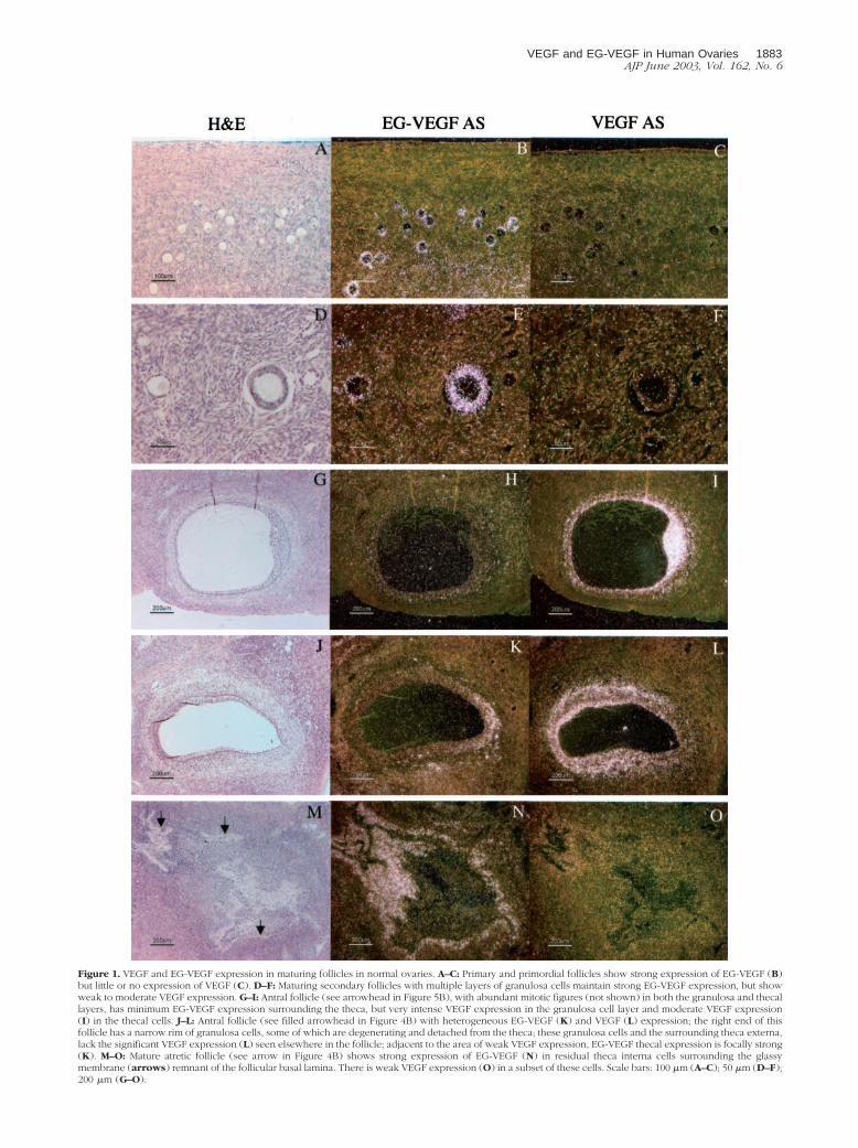
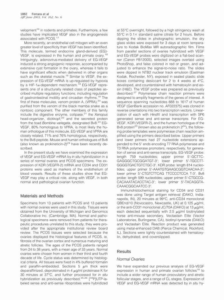
Figure 1. VEGF and EG-VEGF expression in maturing follicles in normal ovaries. A–C: Primary and primordial follicles show strong expression of EG-VEGF (B)but little or no expression of VEGF (C). D–F: Maturing secondary follicles with multiple layers of granulosa cells maintain strong EG-VEGF expression, but showweak to moderate VEGF expression. G–I: Antral follicle (see arrowhead in Figure 5B), with abundant mitotic figures (not shown) in both the granulosa and thecallayers, has minimum EG-VEGF expression surrounding the theca, but very intense VEGF expression in the granulosa cell layer and moderate VEGF expression(I) in the thecal cells. J–L: Antral follicle (see filled arrowhead in Figure 4B) with heterogeneous EG-VEGF (K) and VEGF (L) expression; the right end of thisfollicle has a narrow rim of granulosa cells, some of which are degenerating and detached from the theca; these granulosa cells and the surrounding theca externa,lack the significant VEGF expression (L) seen elsewhere in the follicle; adjacent to the area of weak VEGF expression, EG-VEGF thecal expression is focally strong(K). M–O: Mature atretic follicle (see arrow in Figure 4B) shows strong expression of EG-VEGF (N) in residual theca interna cells surrounding the glassymembrane (arrows) remnant of the follicular basal lamina. There is weak VEGF expression (O) in a subset of these cells. Scale bars: 100 �m (A–C); 50 �m (D–F);200 �m (G–O).
VEGF and EG-VEGF in Human Ovaries 1883AJP June 2003, Vol. 162, No. 6
bridization in all of the specimens examined. Figure 1, Ato I, illustrates representative examples of EG-VEGF andVEGF expression in preovulatory follicles from normalovaries. Granulosa cells in primordial and primary folli-cles express EG-VEGF strongly (Figure 1B), whereasVEGF expression is very weak or undetectable (Figure1C). VEGF expression is more uniformly detectable butstill weak in secondary follicles with two to three layers ofgranulosa cells (Figure 1F). As preovulatory follicles ma-ture, VEGF expression appears to progressively in-
crease, so that antral follicles show intense granulosa cellsignal (Figure 1I) that is often associated with moderateor weak VEGF expression in the adjacent thecal layers(both the theca interna and externa can be VEGF-posi-tive; Figure 1I). As the secondary follicle matures, EG-VEGF expression in granulosa cells declines (Figure 1H),although in a previous series we detected some EG-VEGFexpression in granulosa cells in the cumulus oophorus sur-rounding the oocyte.18 In antral follicles, variable EG-VEGFexpression can be seen in the surrounding theca.
Figure 2. EG-VEGF and VEGF expression in normal ovary early-stage CL. An early-stage (approximately day 2 to 3 after ovulation) CL, characterized byincompletely developed vascularity in the granulosa lutein layer and by inapparent theca lutein cell differentiation (I, J), shows strong VEGF expression in thegranulosa lutein cells. A: False-colored autoradiographic film results show intense VEGF expression (red) in the wall of the large cystic CL (B, arrow). Microscopicresults show granulosa lutein cells are intensely VEGF-positive (C, dark field; J, bright field), but only weakly positive for EG-VEGF (E); the surrounding thecais only weakly positive for both VEGF and EG-VEGF. VEGFR-2 (KDR) expression (G) is present in small vessels at the boundary between the theca interna andgranulosa cell layer, and in vessels invading the outermost granulosa cell layers (I, arrows). Other atretic follicles (A, B) with (closed arrowheads) and without(open arrowhead) intact granulosa cell linings (detail not shown) show prominent EG-VEGF expression in the theca interna. Scale bars: 5 mm (B); 100 �m (C–I);50 �m (J).
1884 Ferrara et alAJP June 2003, Vol. 162, No. 6
Approximately 0.1% of the follicles present at birthmature to the point of ovulation, subsequently progress-ing to form CL. The remaining follicles mature to variouspreovulatory stages, then undergo degenerative changes,becoming atretic.28 We examined expression of VEGF andEG-VEGF in atretic follicles at different stages of their evo-lution. Figure 1, M to O, illustrates a representative exampleof a mature (collapsed) atretic follicle, which typicallystrongly expresses EG-VEGF in the residual thecal cellssurrounding the dense hyaline remnant of the follicularbasal lamina. VEGF is only weakly expressed (Figure 1O) in
a subset of these cells immediately adjacent to the follicularbasal lamina. Atretic follicles in a less mature stage of evo-lution may retain a central lumen lacking intact granulosacells, but surrounded by luteinized thecal cells. Follicles atthis stage (not shown), typically expressed high levels ofEG-VEGF in the thecal cells, but lacked VEGF expression.Follicles with a large central lumen lined by an intact gran-ulosa cell layer are occasionally found to lack VEGF expres-sion in the granulosa cell layer, but retain significant VEGFexpression (and have EG-VEGF expression) in the thecainterna (not shown). It is unclear whether follicles with this
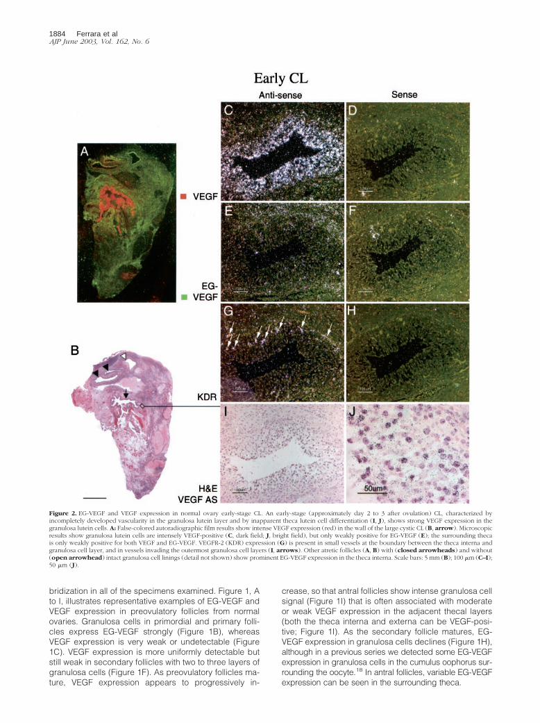
Figure 3. EG-VEGF and VEGF expression in normal ovary early- to mid-stage CL. A CL at approximately day 5 after ovulation, characterized microscopically bywell-developed vascularity and early theca lutein cell differentiation (I), shows strong VEGF (C) and EG-VEGF (E) expression in spatially overlapping CL cellpopulations. A: False-colored autoradiographic film results show yellow-orange overlapping of VEGF (red) and EG-VEGF (green) expression in a large cystic CL.It was not possible to determine from this result whether VEGF and EG-VEGF were co-expressed in the same cells, or in separate cells in the CL. The distributionof EG-VEGF and VEGF signal across the entire CL is consistent with expression by theca granulosa cells, but theca lutein cells may also express EG-VEGF at thisstage. Vascular VEGFR-2 (KDR) expression is intense in the CL (G). Scale bars: 5 mm (B); 100 �m (C–I); 50 �m (J).
VEGF and EG-VEGF in Human Ovaries 1885AJP June 2003, Vol. 162, No. 6
pattern represent a later stage of preovulatory follicle thanillustrated in Figure 1, G to I, or whether they represent anearly stage of follicular atresia.
CL derived from ovulatory follicles mature in a canon-ical 14-day pattern.29 We examined EG-VEGF and VEGFexpression in a series of CL representing time points �2days to 14 days after ovulation. To convey a sense of theoverall distribution of EG-VEGF and VEGF expression inindividual ovaries, autoradiographic film results of paral-lel sections were digitized and the images correspondingto EG-VEGF and VEGF signals from representative ovary
samples were false-colored green and red, respectively.At �2 to 3 days after ovulation (Figure 2; time points areinferred, according to the histological criteria of Cor-ner29), the EG-VEGF and VEGF expression resemble thepattern seen in the late preovulatory follicle: granulosacells are intensely VEGF-positive, but lack significant EG-VEGF expression (Figure 2; C to F). At �5 days afterovulation (Figure 3), both VEGF (Figure 3, C and D) andEG-VEGF (Figure 3, E and F) are strongly expressed in aportion of granulosa lutein cells (theca lutein cells are notclearly distinct histologically at this stage; they may also
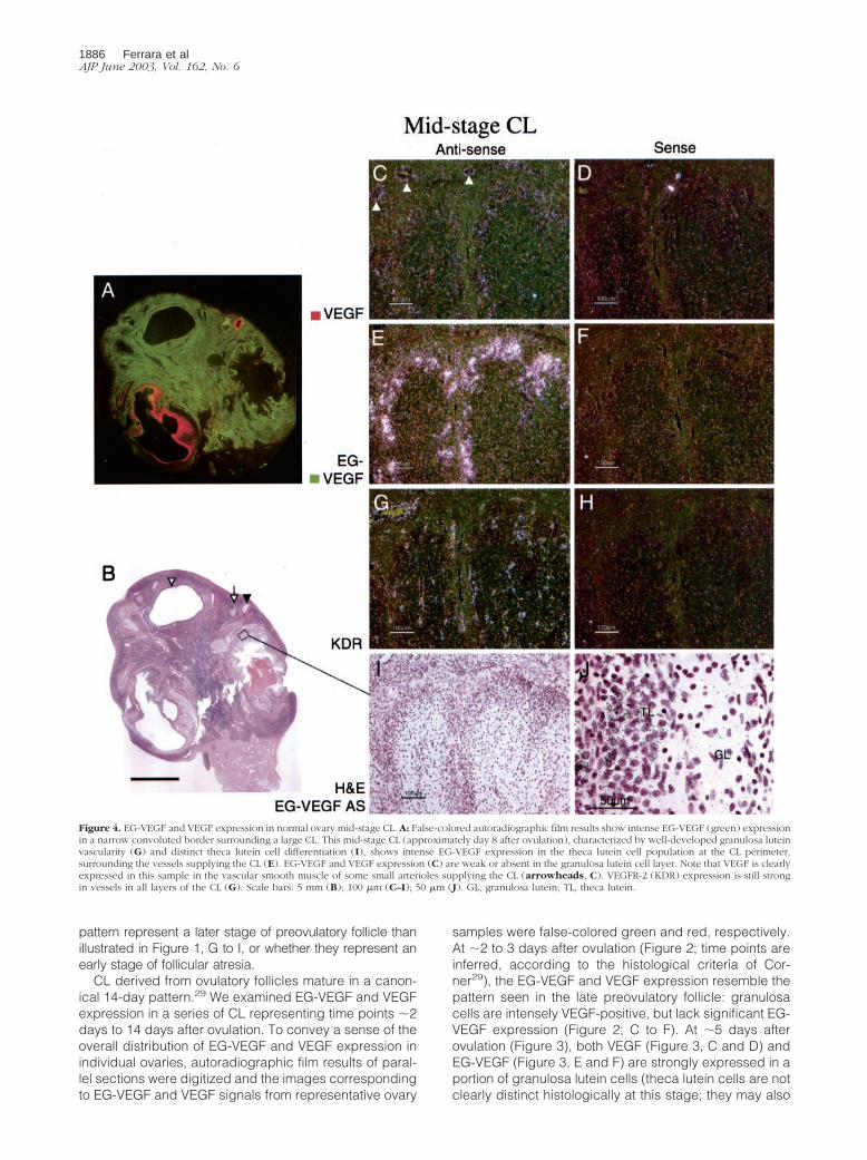
Figure 4. EG-VEGF and VEGF expression in normal ovary mid-stage CL. A: False-colored autoradiographic film results show intense EG-VEGF (green) expressionin a narrow convoluted border surrounding a large CL. This mid-stage CL (approximately day 8 after ovulation), characterized by well-developed granulosa luteinvascularity (G) and distinct theca lutein cell differentiation (I), shows intense EG-VEGF expression in the theca lutein cell population at the CL perimeter,surrounding the vessels supplying the CL (E). EG-VEGF and VEGF expression (C) are weak or absent in the granulosa lutein cell layer. Note that VEGF is clearlyexpressed in this sample in the vascular smooth muscle of some small arterioles supplying the CL (arrowheads, C). VEGFR-2 (KDR) expression is still strongin vessels in all layers of the CL (G). Scale bars: 5 mm (B); 100 �m (C–I); 50 �m (J). GL, granulosa lutein; TL, theca lutein.
1886 Ferrara et alAJP June 2003, Vol. 162, No. 6
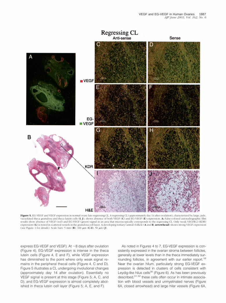
express EG-VEGF and VEGF). At �8 days after ovulation(Figure 4), EG-VEGF expression is intense in the thecalutein cells (Figure 4, E and F), while VEGF expressionhas diminished to the point where only weak signal re-mains in the peripheral thecal cells (Figure 4, C and D).Figure 5 illustrates a CL undergoing involutional changes(approximately day 14 after ovulation). Essentially noVEGF signal is present at this stage (Figure 5; A, C, andD), and EG-VEGF expression is almost completely abol-ished in theca lutein cell layer (Figure 5; A, E, and F).
As noted in Figures 4 to 7, EG-VEGF expression is con-sistently expressed in the ovarian stroma between follicles,generally at lower levels than in the theca immediately sur-rounding follicles, in agreement with our earlier report.18
Near the ovarian hilum, particularly strong EG-VEGF ex-pression is detected in clusters of cells consistent withLeydig-like hilus cells30 (Figure 6). As has been previouslydescribed,31,32 these cells often occur in intimate associa-tion with blood vessels and unmyelinated nerves (Figure6A, closed arrowhead) and large hilar vessels (Figure 6A,
Figure 5. EG-VEGF and VEGF expression in normal ovary late-regressing CL. A regressing CL (approximately day 14 after ovulation), characterized by large, pale,vacuolated theca granulosa and theca lutein cells (I, J), shows absence of both VEGF (C) and EG-VEGF (E) expression. A: False-colored autoradiographic filmresults show absence of VEGF (red) and EG-VEGF (green) signal in an area that microscopically corresponds to the regressing CL. Only weak VEGFR-2 (KDR)expression (G) is noted in scattered vessels in the granulosa cell layer. A developing tertiary (antral) follicle (A and B, arrowhead) shows strong VEGF expression(see Figure 1 for details). Scale bars: 5 mm (B); 100 �m (C–I); 50 �m (J).
VEGF and EG-VEGF in Human Ovaries 1887AJP June 2003, Vol. 162, No. 6
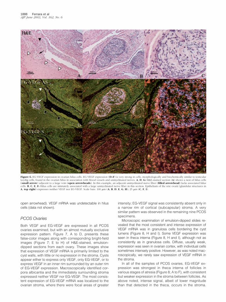
open arrowhead). VEGF mRNA was undetectable in hiluscells (data not shown).
PCOS Ovaries
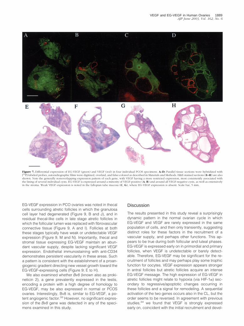
Both VEGF and EG-VEGF are expressed in all PCOSovaries examined, but with an almost mutually exclusiveexpression pattern. Figure 7, A to D, presents thesefalse-color images along with corresponding bright-fieldimages (Figure 7; E to H) of H&E-stained, emulsion-dipped sections from each ovary. These images showthat expression of VEGF mRNA is primarily limited to thecyst walls, with little or no expression in the stroma. Cystsappear either to express only VEGF, only EG-VEGF, or toexpress VEGF in an inner rim surrounded by an outer rimof EG-VEGF expression. Macroscopically identified cor-pora albicantia and the immediately surrounding stromaexpressed neither VEGF nor EG-VEGF. The most consis-tent expression of EG-VEGF mRNA was localized to theovarian stroma, where there were focal areas of greater
intensity; EG-VEGF signal was consistently absent only ina narrow rim of cortical (subcapsular) stroma. A verysimilar pattern was observed in the remaining nine PCOSspecimens.
Microscopic examination of emulsion-dipped slides re-vealed that the most consistent and intense expression ofVEGF mRNA was in granulosa cells bordering the cystlumens (Figure 8, H and I). Some VEGF expression wasseen in theca interna (Figure 8, H and I), although not asconsistently as in granulosa cells. Diffuse, usually weak,expression was seen in ovarian cortex, with individual cellssometimes intensely positive. However, as was noted mac-roscopically, we rarely saw expression of VEGF mRNA inthe stroma.
In all of the samples of PCOS ovaries, EG-VEGF ex-pression was strongest in theca interna of follicles invarious stages of atresia (Figure 8; A to F), with consistentbut weaker expression in the stroma between follicles. Asabove noted, intense signal, albeit of lower magnitudethan that detected in the theca, occurs in the stroma.
Figure 6. EG-VEGF expression in ovarian hilus cells. EG-VEGF expression (D–F) is very strong in cells, morphologically and biochemically similar to testicularLeydig cells, found in the ovarian hilus in association with blood vessels and unmyelinated nerves. A, D, G: H&E-stained section (A) shows a nest of hilus cells(small arrow) adjacent to a large vein (open arrowheads). In this example, an adjacent unmyelinated nerve fiber (filled arrowhead) lacks associated hiluscells. B, C, E, F: Hilus cells are intimately associated with a large unmyelinated nerve fiber in this section. Epithelium of the rete ovarii (glandular structures inA, top right) expresses neither VEGF nor EG-VEGF. Scale bars: 100 �m (A, B, D, E, G, H); 25 �m (C, F, I).
1888 Ferrara et alAJP June 2003, Vol. 162, No. 6
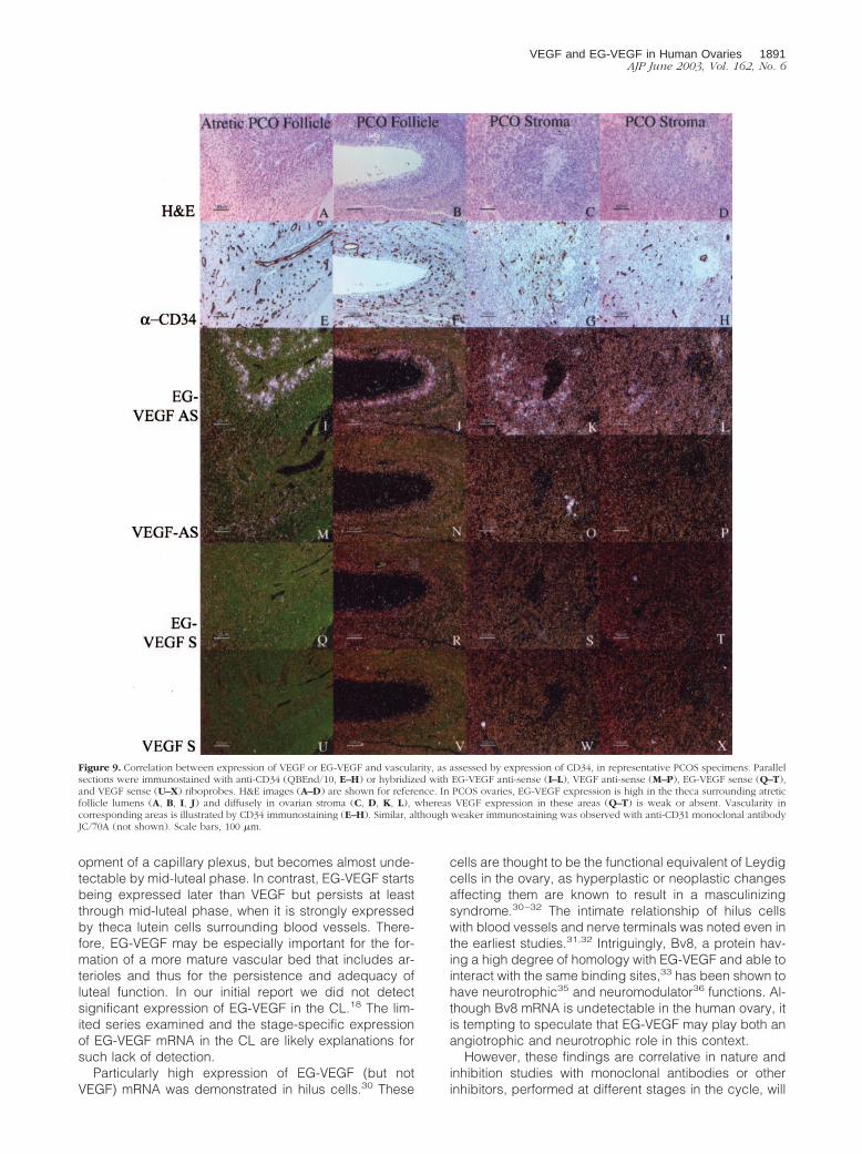
EG-VEGF expression in PCO ovaries was noted in thecalcells surrounding atretic follicles in which the granulosacell layer had degenerated (Figure 9, B and J), and inresidual thecal-like cells in late stage atretic follicles inwhich the follicular lumen was replaced with fibrovascularconnective tissue (Figure 9, A and I). Follicles at boththese stages typically have weak or undetectable VEGFexpression (Figure 9, M and N). Importantly, thecal andstromal tissue expressing EG-VEGF maintain an abun-dant vascular supply, despite lacking significant VEGFexpression. Endothelial immunostaining with anti-CD34demonstrates persistent vascularity in these areas. Sucha pattern is consistent with the establishment of a proan-giogenic gradient directing new vessel growth toward theEG-VEGF-expressing cells (Figure 9; E to H).
We also examined whether Bv8 (known also as proki-neticin 2), a gene prevalently expressed in the testis,encoding a protein with a high degree of homology toEG-VEGF, may be also expressed in normal or PCOSovaries. Interestingly, Bv8 is, similar to EG-VEGF, a po-tent angiogenic factor.33 However, no significant expres-sion of the Bv8 gene was detected in any of the speci-mens examined in this study.
Discussion
The results presented in this study reveal a surprisinglydynamic pattern in the normal ovarian cycle in whichEG-VEGF and VEGF are rarely expressed in the samepopulation of cells, and then only transiently, suggestingdistinct roles for these factors in the recruitment of avascular supply, and perhaps other functions. This ap-pears to be true during both follicular and luteal phases.EG-VEGF is expressed early on in primordial and primaryfollicles, when VEGF is undetectable or barely detect-able. Therefore, EG-VEGF may be significant for the re-cruitment of follicles and may perhaps play some trophicfunction for oocytes. VEGF expression appears strongerin antral follicles but atretic follicles acquire an intenseEG-VEGF message. The high expression of EG-VEGF inatretic follicles might relate to hypoxia (via HIF-1�) sec-ondary to regressive/apoptotic changes occurring inthese follicles and a signal for remodeling. A sequentialactivation of the two genes occurs also in the CL, but theorder seems to be reversed. In agreement with previousstudies,34 we found that VEGF is strongly expressedearly on, coincident with the initial recruitment and devel-
Figure 7. Differential expression of EG-VEGF (green) and VEGF (red) in four individual PCOS specimens. A–D: Parallel tissue sections were hybridized with[33P]-labeled probes, autoradiographic films were digitized, overlaid, and false-colored as described in Materials and Methods. H&E-stained sections (E–H) are alsoshown. Note the generally nonoverlapping expression pattern of each gene, with VEGF having a more restricted expression, most consistently associated withthe lining of several individual cysts. EG-VEGF is expressed around a minority of VEGF-positive (A, B) and around all VEGF-negative cysts, as well as extensivelyin the stroma. Weak VEGF expression is noted in the fallopian tube mucosa (C, G), where EG-VEGF expression is absent. Scale bar, 5 mm.
VEGF and EG-VEGF in Human Ovaries 1889AJP June 2003, Vol. 162, No. 6
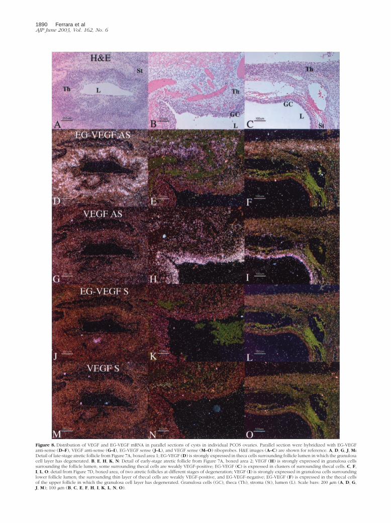
Figure 8. Distribution of VEGF and EG-VEGF mRNA in parallel sections of cysts in individual PCOS ovaries. Parallel section were hybridized with EG-VEGFanti-sense (D–F), VEGF anti-sense (G–I), EG-VEGF sense (J–L), and VEGF sense (M–O) riboprobes. H&E images (A–C) are shown for reference. A, D, G, J, M:Detail of late-stage atretic follicle from Figure 7A, boxed area 1; EG-VEGF (D) is strongly expressed in theca cells surrounding follicle lumen in which the granulosacell layer has degenerated. B, E, H, K, N: Detail of early-stage atretic follicle from Figure 7A, boxed area 2; VEGF (H) is strongly expressed in granulosa cellssurrounding the follicle lumen; some surrounding thecal cells are weakly VEGF-positive; EG-VEGF (C) is expressed in clusters of surrounding thecal cells. C, F,I, L, O: detail from Figure 7D, boxed area, of two atretic follicles at different stages of degeneration; VEGF (I) is strongly expressed in granulosa cells surroundinglower follicle lumen; the surrounding thin layer of thecal cells are weakly VEGF-positive, and EG-VEGF-negative; EG-VEGF (F) is expressed in the thecal cellsof the upper follicle in which the granulosa cell layer has degenerated. Granulosa cells (GC), theca (Th), stroma (St), lumen (L). Scale bars: 200 �m (A, D, G,J, M); 100 �m (B, C, E, F, H, I, K, L, N, O).
1890 Ferrara et alAJP June 2003, Vol. 162, No. 6
opment of a capillary plexus, but becomes almost unde-tectable by mid-luteal phase. In contrast, EG-VEGF startsbeing expressed later than VEGF but persists at leastthrough mid-luteal phase, when it is strongly expressedby theca lutein cells surrounding blood vessels. There-fore, EG-VEGF may be especially important for the for-mation of a more mature vascular bed that includes ar-terioles and thus for the persistence and adequacy ofluteal function. In our initial report we did not detectsignificant expression of EG-VEGF in the CL.18 The lim-ited series examined and the stage-specific expressionof EG-VEGF mRNA in the CL are likely explanations forsuch lack of detection.
Particularly high expression of EG-VEGF (but notVEGF) mRNA was demonstrated in hilus cells.30 These
cells are thought to be the functional equivalent of Leydigcells in the ovary, as hyperplastic or neoplastic changesaffecting them are known to result in a masculinizingsyndrome.30–32 The intimate relationship of hilus cellswith blood vessels and nerve terminals was noted even inthe earliest studies.31,32 Intriguingly, Bv8, a protein hav-ing a high degree of homology with EG-VEGF and able tointeract with the same binding sites,33 has been shown tohave neurotrophic35 and neuromodulator36 functions. Al-though Bv8 mRNA is undetectable in the human ovary, itis tempting to speculate that EG-VEGF may play both anangiotrophic and neurotrophic role in this context.
However, these findings are correlative in nature andinhibition studies with monoclonal antibodies or otherinhibitors, performed at different stages in the cycle, will
Figure 9. Correlation between expression of VEGF or EG-VEGF and vascularity, as assessed by expression of CD34, in representative PCOS specimens. Parallelsections were immunostained with anti-CD34 (QBEnd/10, E–H) or hybridized with EG-VEGF anti-sense (I–L), VEGF anti-sense (M–P), EG-VEGF sense (Q–T),and VEGF sense (U–X) riboprobes. H&E images (A–D) are shown for reference. In PCOS ovaries, EG-VEGF expression is high in the theca surrounding atreticfollicle lumens (A, B, I, J) and diffusely in ovarian stroma (C, D, K, L), whereas VEGF expression in these areas (Q–T) is weak or absent. Vascularity incorresponding areas is illustrated by CD34 immunostaining (E–H). Similar, although weaker immunostaining was observed with anti-CD31 monoclonal antibodyJC/70A (not shown). Scale bars, 100 �m.
VEGF and EG-VEGF in Human Ovaries 1891AJP June 2003, Vol. 162, No. 6
be required to dissect the physiological roles of EG-VEGFin the ovary.
It is well established that increased ovarian mass, sup-ported by new blood vessel proliferation in stroma andtheca, is a key feature of PCOS. Indeed, there has beenconsiderable interest in the identification of the mediatorsof such hypervascularity, but surprisingly little is knownabout the nature and distribution of such mediators. Thepresent study may represent the most extensive seriesreported so far examining the in situ expression of can-didate angiogenic factor genes in PCOS. Recent litera-ture has focused on VEGF as one of the most likelycandidates. Indeed, as previously mentioned, VEGF is akey regulator of normal and abnormal proliferation ofblood vessels and has been shown to play a central rolein ovarian angiogenesis.37 Interestingly, VEGF levelshave been reported to be elevated in the serum of PCOSpatients compared to normal controls, although the de-gree of increase varied among different studies, being aslittle as 25%38 or approximately twofold.39 Furthermore,Kamat and colleagues40 have reported in a series ofthree PCOS ovaries the expression of VEGF mRNA.
Given the hyperplasia and hypervascularity of thestroma in PCOS and the finding that EG-VEGF is ex-pressed in the theca of atretic follicles and in the ovarianstroma, we were prompted to compare the expression ofEG-VEGF and VEGF mRNAs in specimens of such dis-order. A consistent finding of our study is that both VEGFand EG-VEGF are expressed in PCOS ovaries, but with apattern that is almost mutually exclusive. The most in-tense and consistent expression of VEGF was in thegranulosa cell layer of follicles, with a lower expression inthe theca of some follicles. In contrast, EG-VEGF in PCOSfollicles is never seen in the granulosa cells, but fre-quently in the theca surrounding follicles. This expressionpattern is an exaggeration of the pattern seen in normalcycling ovaries, where our results show intense VEGFexpression in the granulosa cells of antral follicles, withlower expression in the theca some atretic follicles; acomplementary pattern of EG-VEGF expression showsstrong granulosa cell signal in primordial and primaryfollicles, and strong thecal signal in atretic follicles. Thearrested follicular development in PCOS reflects the lackof follicular maturation and CL development and acyclicalgonadotropin stimulation.41 Although there is debatewhether most PCOS follicles are truly atretic,42 theyclearly have several features of atresia.43 We detected avery low or undetectable VEGF hybridization signal in thestroma, a component that, like the theca, undergoesdramatic hyperplastic changes in PCOS. This is in con-trast to the generally high expression of EG-VEGF mRNAin the stroma. Although we cannot rule out the possibilitythat matrix metalloproteinase-mediated proteolyticevents may result in enhancement in the activity of low,constitutive, levels of VEGF,44,45 our findings suggest thatthe hyperplastic/angiogenic changes occurring in PCOSare not likely solely because of VEGF and most likelyEG-VEGF also participates in these events. In fact, ouranalysis indicates that, at least in terms of mRNA expres-sion, EG-VEGF is the molecule that shows an even stron-ger correlation with hyperplasia and angiogenesis in this
condition. We suggest that, although VEGF is an essen-tial player in normal cycling ovaries, EG-VEGF might beof even greater pathophysiological importance in theacyclical angiogenesis occurring during chronic anovu-lation. Additional studies are clearly needed to verify thishypothesis. The availability of antibodies suitable for im-munohistochemistry as well as sensitive assays to mea-sure the EG-VEGF protein levels in the serum or otherbiological fluids will be helpful to extend these findings.
Previous studies have shown that adenovirus-medi-ated delivery of EG-VEGF in the ovary elicits angiogeniceffects as well as cyst formation of similar magnitude asthat induced by VEGF.18 Therefore, our findings suggestthat EG-VEGF is potentially an important contributor tothe angiogenesis and hyperplasia of PCOS. Thus, it istempting to speculate that blockade of VEGF and EG-VEGF may be effective at reducing ovarian mass andandrogen output before induction of ovulation with cur-rent protocols.
Finally, previous studies46 have implicated VEGF alsoin the pathogenesis of ovarian hyperstimulation syn-drome (OHSS), a potentially fatal condition characterizedby ovarian enlargement, with multiple follicular cysts andincreased vascular permeability.47,48 PCOS is a well-established risk factor for OHSS.49 However, other stud-ies have cast doubt on the hypothesis that VEGF may bethe causative factor in the vascular permeability associ-ated with OHSS.50 It is tempting to speculate that suchdiscrepancies are because, at least in part, of the factthat although VEGF may be an important mediator inOHSS, it is by itself insufficient and the symptoms reflectthe contribution of other factors, including EG-VEGF.
References
1. Bassett DL: The changes in the vascular pattern of the ovary of thealbino rat during the estrous cycle. Am J Anat 1943, 73:251–278
2. Zeleznik AJ, Schuler HM, Reichert Jr LE: Gonadotropin-binding sitesin the rhesus monkey ovary: role of the vasculature in the selectivedistribution of human chorionic gonadotropin to the preovulatory fol-licle. Endocrinology 1981, 109:356–362
3. Carr BR, MacDonald PC, Simpson ER: The role of lipoproteins in theregulation of progesterone secretion by the corpus luteum. Fertil Steril1981, 38:303–311
4. Goede V, Schmidt T, Kimmina S, Kozian D, Augustin HG: Analysis ofblood vessel maturation processes during cyclic ovarian angiogene-sis. Lab Invest 1998, 78:1385–1394
5. Maisonpierre PC, Suri C, Jones PF, Bartunkova S, Wiegend SJ, Radz-iejewski C, Compton D, McClain J, Aldrich TH, Papadopulos N, DalyTJ, Davis S, Sato TN, Yancopoulos GD: Angiopoietin-2, a naturalantagonist for Tie-2 that disrupts in vivo angiogenesis. Science 1997,277:55–60
6. Goldziher JW, Green JA: The polycistic ovary. I. Clinical and histo-logic features. J Clin Endocrinol Metab 1962, 22:325–332
7. Yen, SSC: A contemporary overview. Reproductive Endocrinology,Surgery, and Technology. Edited by EY Adashi, JA Rock, Z Rosen-waks. Philadelphia, Lippincott-Raven, 1996, pp 1117–1126
8. Dewailly D, Robert Y, Helin I, Ardaens Y, Thomas-Desrousseaux P,Lemaitre L, Fossati P: Ovarian stromal hypertrophy in hyperandro-genic women. Clin Endocrinol (Oxf) 1964, 41:557–562
9. Fulghesu AM, Ciampelli M, Belosi C, Apa R, Pavone V, Lanzone A: Anew ultrasound criterion for the diagnosis of polycystic ovarysyndrome: the ovarian stroma/total area ratio. Fertil Steril 2001, 76:326–331
1892 Ferrara et alAJP June 2003, Vol. 162, No. 6
10. Dunaif A, Thomas A: Current concepts in the polycystic ovary syn-drome. Annu Rev Med 2001, 52:401–419
11. Phillips HS, Hains J, Leung DW, Ferrara N: Vascular endothelialgrowth factor is expressed in rat corpus luteum. Endocrinology 1990,127:965–967
12. Ravindranath N, Little-Ihrig L, Phillips HS, Ferrara N, Zeleznik AJ:Vascular endothelial growth factor messenger ribonucleic acid ex-pression in the primate ovary. Endocrinology 1992, 131:254–260
13. Ferrara N, Chen H, Davis-Smyth T, Gerber, H-P, Nguyen T-N, PeersD, Chisholm V, Hillan KJ, Schwall RH: Vascular endothelial growthfactor is essential for corpus luteum angiogenesis. Nat Med 1998,4:336–340
14. Ryan AM, Eppler DB, Hagler KE, Bruner RH, Thomford PJ, Hall RL,Shopp GM, O’Neill CA: Preclinical safety evaluation of rhuMAbVEGF,an antiangiogenic humanized monoclonal antibody. Toxicol Pathol1999, 27:78–86
15. Fraser HM, Dickson SE, Lunn SF, Wulff C, Morris KD, Carroll VA,Bicknell R: Suppression of luteal angiogenesis in the primate afterneutralization of vascular endothelial growth factor. Endocrinology2000, 141:995–1000
16. Zimmermann RC, Xiao E, Husami N, Sauer MV, Lobo R, Kitajewski J,Ferin M: Short-term administration of antivascular endothelial growthfactor antibody in the late follicular phase delays follicular develop-ment in the rhesus monkey. J Clin Endocrinol Metab 2001, 86:768–772
17. Ferrara N: Vascular endothelial growth factor: a key regulator ofphysiological angiogenesis. Vascular Morphogenesis in the FemaleReproductive System. Edited by HG Augustin, L Iruela-Arispe, PAWRogers, SK Smith. Boston, Birkhauser, 2001, pp 149–165
18. LeCouter J, Kowalski J, Foster J, Hass P, Zhang Z, Dillard-Telm L,Frantz G, Rangell L, DeGuzman L, Keller G-A, Peale F, Gurney A,Hillan KJ, Ferrara N: Identification of an angiogenic mitogen selectivefor endocrine gland endothelium. Nature 2001, 412:877–884
19. LeCouter J, Lin R, Ferrara N: Endocrine gland-derived VEGF and theemerging hypothesis of organ-specific regulation of angiogenesis.Nat Med 2002, 8:913–917
20. Joubert FJ, Strydom DJ: Snake venom. The amino acid sequence ofprotein A from Dendroaspis polylepis polylepis (black mamba)venom. H-S Z Physiol Chem 1980, 361:1787–1794
21. Aravind L, Koonin EV: A colipase fold in the carboxy-terminal domainof the Wnt antagonists—the Dickkopfs. Curr Biol 1998, 8:477–478
22. Glinka A, Wu W, Delius H, Monaghan AP, Blumenstock C, Niehrs C:Dickkopf-1 is a member of a new family of secreted proteins andfunctions in head induction. Nature 1998, 391:357–362
23. Wechselberger C, Puglisi R, Engel E, Lepperdinger G, Boitani C, KreilG: The mammalian homologues of frog Bv8 are mainly expressed inspermatocytes. FEBS Lett 1999, 462:177–181
24. Li M, Bullock CM, Knauer DJ, Ehlert FJ, Zhou QY: Identification of twoprokineticin cDNAs: recombinant proteins potently contract gastroin-testinal smooth muscle. Mol Pharmacol 2001, 59:692–698
25. Lu LH, Gillette N: An optimized protocol for in situ hybridization usingPCR-generated 33P-labeled riboprobes. Cell Vis 1994, 7:53–64
26. Holcomb IN, Kabakoff RC, Chan B, Baker TW, Gurney A, Henzel W,Nelson C, Lowman HB, Wright BD, Skelton NJ, Frantz GD, Tumas DB,Peale Jr FV, Shelton DL, Hebert CC: FIZZ1, a novel cysteine-richsecreted protein associated with pulmonary inflammation, defines anew gene family. EMBO J 2000, 19:4046–4055
27. Warren RS, Yuan H, Matli MR, Gillett NA, Ferrara N: Regulation byvascular endothelial growth factor of human colon cancer tumorigen-esis in a mouse model of experimental liver metastasis. J Clin Invest1995, 95:1789–1797
28. Clement PB: Histology of the ovary. Am J Surg Pathol 1987, 11:277–303
29. Corner GWJ: The histological dating of the human corpus luteum ofmenstruation. Am J Anat 1956, 9:377–401
30. Clement PB: Histology of the ovary. Histology for Pathologists, ed 2.Edited by SS Sternberg. Philadelphia, Lippincott, 1997, pp 929–959
31. Sternberg WH: The morphology, androgenic function, hyperplasiaand tumors of the ovarian hilus cells. Am J Pathol 1949, 25:493–521
32. Merrill JA: Ovarian hilus cells. Am J Obstet Gynecol 1959, 78:1258–1271
33. LeCouter J, Lin R, Frantz G, Tejada M, Peale F, Hillan KJ, Ferrara N:The endocrine gland-derived VEGF homologue Bv8 promotes angio-genesis in the testis: localization of Bv8 receptors to endothelial cells.Proc Natl Acad Sci USA 2003, 100:2685–2690
34. Yamamoto S, Konishi I, Tsuruta Y, Nanbu K, Mandai M, Kuroda H,Matsushita K, Hamid AA, Yura Y, Mori T: Expression of vascularendothelial growth factor (VEGF) during folliculogenesis and corpusluteum formation in the human ovary. Gynecol Endocrinol 1997, 11:371–381
35. Melchiorri D, Bruno V, Besong G, Ngomba RT, Cuomo L, De Blasi A,Copani A, Moschella C, Storto M, Nicoletti F, Lepperdinger G, Pas-sarelli F: The mammalian homologue of the novel peptide Bv8 isexpressed in the central nervous system and supports neuronalsurvival by activating the MAP kinase/PI-3-kinase pathways. EurJ Neurosci 2001, 13:1694–1702
36. Cheng MY, Bullock CM, Li C, Lee AG, Bermak JC, Belluzzi J, WeaverDR, Leslie FM, Zhou QY: Prokineticin 2 transmits the behavioralcircadian rhythm of the suprachiasmatic nucleus. Nature 2002, 417:405–410
37. Ferrara N: Role of vascular endothelial growth factor in the regulationof angiogenesis. Kidney Int 1999, 56:794–814
38. Tulandi T, Saleh A, Morris D, Jacobs HS, Payne NN, Tan SL: Effectsof laparoscopic ovarian drilling on serum vascular endothelial growthfactor and on insulin responses to the oral glucose tolerance test inwomen with polycystic ovary syndrome. Fertil Steril 2000, 74:585–588
39. Agrawal R, Sladkevicius P, Engmann L, Conway GS, Payne NN, BekisJ, Tan SL, Campbell S, Jacobs HS: Serum vascular endothelialgrowth factor concentrations and ovarian stromal blood flow areincreased in women with polycystic ovaries. Hum Reprod 1998,13:651–655
40. Kamat BR, Brown LF, Manseau EJ, Senger DR, Dvorak HF: Expres-sion of vascular permeability factor/vascular endothelial growth factorby human granulosa and theca lutein cells. Role in corpus luteumdevelopment. Am J Pathol 1995, 146:157–165
41. Yen SSC: Polycystic ovary syndrome (hyperandrogenic chronicanovulation). Reproductive Endocrinology. Edited by SSC Yen, RBJaffe, RL Barbieri. Philadelphia, W. B. Saunders, 1999, pp 436–78
42. Almahbobi G, Anderiesz C, Hutchinson P, McFarlane JR, Wood C,Trounson AO: Functional integrity of granulosa cells from polycysticovaries. Clin Endocrinol 1996, 44:571–580
43. Cataldo NA, Dumesic DA, Goldsmith PC, Jaffe RB: Immunolocaliza-tion of Fas and Fas ligand in the ovaries of women with polycysticovary syndrome: relationship to apoptosis. Hum Reprod 2000, 15:1889–1997
44. Bergers G, Brekken R, McMahon G, Vu TH, Itoh T, Tamaki K, Tan-zawa K, Thorpe P, Itohara S, Werb Z, Hanahan D: Matrix metallopro-teinase-9 triggers the angiogenic switch during carcinogenesis. NatCell Biol 2000, 2:737–744
45. Inoue M, Hager JH, Ferrara N, Gerber HP, Hanahan D: VEGF-A hasa critical, nonredundant role in angiogenic switching and pancreaticbeta cell carcinogenesis. Cancer Cell 2002, 1:193–202
46. McClure N, Healy DL, Rogers PA, Sullivan J, Beaton L, Haning RV,Connolly DT, Robertson DM: Vascular endothelial growth factor as acapillary permeability agent in ovarian hyperstimulation syndrome.Lancet 1994, 344:235–269
47. Engel T, Jewelewicz R, Dyrenfurth I, Speroff L, Vande Wiele RL:Ovarian hyperstimulation syndrome: report of one case with notes onpathogenesis and treatment. Am J Obstet Gynecol 1972, 112:1052–1057
48. Haning RJ, Strawn E, Nolten W: Pathophysiology of the ovarian hy-perstimulation syndrome. Obstet Gynecol 1985, 166:220–224
49. Kemmann E, Tavakoli F, Shelden RM, Jones JR: Induction of ovula-tion with menotropins in women with polycystic ovary syndrome. Am JObstet Gynecol 1981, 141:58–64
50. Kobayashi H, Okada Y, Asahina T, Gotoh J, Terao T: The kallikrein-kinin system, but not vascular endothelial growth factor, plays a rolein the increased vascular permeability associated with ovarian hyper-stimulation syndrome. J Mol Endocrinol 1998, 20:363–374
VEGF and EG-VEGF in Human Ovaries 1893AJP June 2003, Vol. 162, No. 6
Related Documents

![Mesenchymal stem cells in cardiac regeneration: a detailed ...angiogenic factors such as vascular endothelial growth fac-tor (VEGF) [13, 14], stromal cell-derived factor-1α (SDF-1α)](https://static.cupdf.com/doc/110x72/609f71b062df4a0989617ef4/mesenchymal-stem-cells-in-cardiac-regeneration-a-detailed-angiogenic-factors.jpg)