Combined Angiogenic and Osteogenic Factor Delivery Enhances Bone Marrow Stromal Cell-Driven Bone Regeneration Yen-Chen Huang, 1 Darnell Kaigler, 2 Kevin G Rice, 3 Paul H Krebsbach, 1,4 and David J Mooney 1,2,5 ABSTRACT: Bone formation is a coordinated process involving various biological factors. We have developed a scaffold system capable of sustained and localized presentation of osteogenic (BMP-4) and angiogenic (VEGF) growth factors and human bone marrow stromal cells to promote bone formation at an ectopic site. Combined delivery of these factors significantly enhanced bone formation compared with other conditions. Introduction: Tissue regeneration entails complex interactions between multiple signals and materials plat- forms. Orchestrating the presentation of these signals may greatly enhance the regeneration of lost tissue mass. Bone formation, for example, is dependent on the signaling of BMPs, molecules initiating vascularization (e.g., vascular endothelial growth factor [VEGF]), and osteogenic precursor cells capable of responding to these cues and forming bone tissue. It was hypothesized that combined and concerted delivery of these factors from biodegradable scaffolds would lead to enhanced bone formation. Materials and Methods: Poly(lactic-co-glycolic acid) scaffolds containing combinations of condensed plasmid DNA encoding for BMP-4, VEGF, and human bone marrow stromal cells (hBMSCs) were implanted into the subcutaneous tissue of SCID mice. Implants (n 6) were retrieved at 3, 8, and 15 weeks after implantation. Bone and blood vessel formation was determined qualitatively and quantitatively by methods including histology, immmunostaining, and CT. Results: Scaffolds delivering VEGF resulted in a prominent increase in blood vessel formation relative to the conditions without VEGF. BMP-4 expression in scaffolds encapsulating condensed DNA was also confirmed at the 15-week time-point, showing the characteristic of long-term delivery in this system. Combined delivery of all three types of factors resulted in a significant increase in the quantity of regenerated bone compared with any factor alone or any two factors combined, as measured with DXA, X-ray, and histomorphometric analysis. Furthermore, bone formed with all three factors had elastic moduli significantly higher than any other con- dition. Conclusions: Concerted delivery of BMP-4, VEGF, and hBMSCs promoted greater bone formation relative to any single factor or combination of two factors. Materials systems that allows multifactorial presentation more closely mimic natural developmental processes, and these results may have important implications for bone regeneration therapeutics. J Bone Miner Res 2005;20:848–857. Published online on December 20, 2004; doi: 10.1359/JBMR.041226 Key words: vascular endothelial growth factor, BMP-4, gene therapy, bone marrow stromal cell, poly(ethylenimine), poly(lactic-co-glycolic acid) INTRODUCTION T ISSUE DEVELOPMENT AND regeneration are regulated by an interplay among various tissue inductive growth fac- tors, formation of an appropriate vascular bed to support the metabolic needs of the forming tissue mass, and a cell population capable of responding to the chemical cues and creating the new tissue. There has been considerable inter- est in understanding this signaling interplay in bone, be- cause of its limited ability to heal on serious fracture or trauma and the limitations in common treatments for bone morbidity or replacement. BMPs are responsible for initi- ating cartilage and bone progenitor cell differentiation (1) and sequencing new bone formation through endochondral ossification. (2) Angiogenesis, the growth of new capillary blood vessels from pre-existing host vasculature, is also in- volved in the initiation of fracture healing (3) and promotion of endochondral and intramembranous ossification in bone growth. (4–6) Vascular endothelial growth factor (VEGF), a potent angiogenic molecule, seems to be particularly im- The authors have no conflict of interest. 1 Department of Biomedical Engineering, University of Michigan, Ann Arbor, Michigan, USA; 2 Department of Biologic and Materials Sciences, University of Michigan, Ann Arbor, Michigan, USA; 3 College of Pharmacy, University of Iowa, Iowa City, Iowa, USA; 4 Department of Oral Medicine, Pathology and Oncology, University of Michigan, Ann Arbor, Michigan, USA; 5 Department of Chemical Engineering, University of Michigan, Ann Arbor, Michigan, USA. JOURNAL OF BONE AND MINERAL RESEARCH Volume 20, Number 5, 2005 Published online on December 20, 2004; doi: 10.1359/JBMR.041226 © 2005 American Society for Bone and Mineral Research 848

Welcome message from author
This document is posted to help you gain knowledge. Please leave a comment to let me know what you think about it! Share it to your friends and learn new things together.
Transcript
Combined Angiogenic and Osteogenic Factor Delivery Enhances BoneMarrow Stromal Cell-Driven Bone Regeneration
Yen-Chen Huang,1 Darnell Kaigler,2 Kevin G Rice,3 Paul H Krebsbach,1,4 and David J Mooney1,2,5
ABSTRACT: Bone formation is a coordinated process involving various biological factors. We have developeda scaffold system capable of sustained and localized presentation of osteogenic (BMP-4) and angiogenic(VEGF) growth factors and human bone marrow stromal cells to promote bone formation at an ectopic site.Combined delivery of these factors significantly enhanced bone formation compared with other conditions.
Introduction: Tissue regeneration entails complex interactions between multiple signals and materials plat-forms. Orchestrating the presentation of these signals may greatly enhance the regeneration of lost tissue mass.Bone formation, for example, is dependent on the signaling of BMPs, molecules initiating vascularization (e.g.,vascular endothelial growth factor [VEGF]), and osteogenic precursor cells capable of responding to thesecues and forming bone tissue. It was hypothesized that combined and concerted delivery of these factors frombiodegradable scaffolds would lead to enhanced bone formation.Materials and Methods: Poly(lactic-co-glycolic acid) scaffolds containing combinations of condensed plasmidDNA encoding for BMP-4, VEGF, and human bone marrow stromal cells (hBMSCs) were implanted into thesubcutaneous tissue of SCID mice. Implants (n ! 6) were retrieved at 3, 8, and 15 weeks after implantation.Bone and blood vessel formation was determined qualitatively and quantitatively by methods includinghistology, immmunostaining, and !CT.Results: Scaffolds delivering VEGF resulted in a prominent increase in blood vessel formation relative to theconditions without VEGF. BMP-4 expression in scaffolds encapsulating condensed DNA was also confirmedat the 15-week time-point, showing the characteristic of long-term delivery in this system. Combined deliveryof all three types of factors resulted in a significant increase in the quantity of regenerated bone compared withany factor alone or any two factors combined, as measured with DXA, X-ray, and histomorphometric analysis.Furthermore, bone formed with all three factors had elastic moduli significantly higher than any other con-dition.Conclusions: Concerted delivery of BMP-4, VEGF, and hBMSCs promoted greater bone formation relativeto any single factor or combination of two factors. Materials systems that allows multifactorial presentationmore closely mimic natural developmental processes, and these results may have important implications forbone regeneration therapeutics.J Bone Miner Res 2005;20:848–857. Published online on December 20, 2004; doi: 10.1359/JBMR.041226
Key words: vascular endothelial growth factor, BMP-4, gene therapy, bone marrow stromal cell,poly(ethylenimine), poly(lactic-co-glycolic acid)
INTRODUCTION
TISSUE DEVELOPMENT AND regeneration are regulated byan interplay among various tissue inductive growth fac-
tors, formation of an appropriate vascular bed to supportthe metabolic needs of the forming tissue mass, and a cellpopulation capable of responding to the chemical cues andcreating the new tissue. There has been considerable inter-est in understanding this signaling interplay in bone, be-
cause of its limited ability to heal on serious fracture ortrauma and the limitations in common treatments for bonemorbidity or replacement. BMPs are responsible for initi-ating cartilage and bone progenitor cell differentiation(1)
and sequencing new bone formation through endochondralossification.(2) Angiogenesis, the growth of new capillaryblood vessels from pre-existing host vasculature, is also in-volved in the initiation of fracture healing(3) and promotionof endochondral and intramembranous ossification in bonegrowth.(4–6) Vascular endothelial growth factor (VEGF), apotent angiogenic molecule, seems to be particularly im-The authors have no conflict of interest.
1Department of Biomedical Engineering, University of Michigan, Ann Arbor, Michigan, USA; 2Department of Biologic and MaterialsSciences, University of Michigan, Ann Arbor, Michigan, USA; 3College of Pharmacy, University of Iowa, Iowa City, Iowa, USA;4Department of Oral Medicine, Pathology and Oncology, University of Michigan, Ann Arbor, Michigan, USA; 5Department of ChemicalEngineering, University of Michigan, Ann Arbor, Michigan, USA.
JOURNAL OF BONE AND MINERAL RESEARCHVolume 20, Number 5, 2005Published online on December 20, 2004; doi: 10.1359/JBMR.041226© 2005 American Society for Bone and Mineral Research
848
JO407399 848 857 May
portant in bone formation.(5,7) Ultimately, these factorsmust act on a population of cells capable of responding tolocal factors and forming bone tissue. Multipotent stemcells originating from the bone marrow stroma, or bonemarrow stromal cells (BMSCs), are a particularly attractivesource for osteogenic precursors for bone tissue engineer-ing,(8) because they can be easily harvested and expandedin vitro and induced to differentiate into bone-formingcells.
A major goal of research in tissue regeneration is thedevelopment of appropriate delivery approaches for thevarious factors and cells involved in the process.(9) Gener-ally, approaches have focused on developing appropriatecarriers for transplanting cells(10) or for delivering inductivemolecules.(11,12) Combining these functions by geneticallymodifying cells with viruses before transplantation, to allowthe transplanted cells to secrete the desired inductive mol-ecules, is feasible.(13) However, ex vivo gene therapy usingviral vectors presents both practical and safety challenges,as highlighted by recent adverse effects noted in genetherapy trials.(14,15) In contrast, we report a material systemcapable of combined delivery of inductive molecules andcells. This system can be used for either direct protein de-livery or delivery of plasmid DNA encoding for the proteinof interest. We hypothesized that the delivery of multiplefactors involved in tissue formation mimics the mechanismof tissue development and have used this system to showthat combining delivery of BMP-4, VEGF, and humanbone marrow stromal cells (hBMSCs) significantly en-hances bone formation compared with each element aloneor combined with one other component.
MATERIALS AND METHODS
Chemicals
Branched poly(ethylenimine) (PEI; molecular weight, 25kDa) was purchased from Sigma/Aldrich (St Louis, MO,USA). Sucrose and calcium quantification kits were pur-chased from Sigma. Poly(D,L-lactic-co-glycolic acid)(PLGA) 85:15 was purchased from Alkermes (Cincinnati,OH, USA). MVG sodium alginate was purchased fromPronova Bioploymers (Oslo, Norway). FCS and penicillin-streptomycin were purchased from Gibco BRL (Gaithers-burg, MD, USA). VEGF165 was purchased from Intergen(Purchase, NY, USA). Tissue culture medium ("-MEM)was purchased from Life Technologies (Grand Island, NY,USA). Reagents for von Kossa, H&E, and Goldner’s stain-ing were purchased from Fisher Scientific (Springfield, NJ,USA) or Sigma. Staining for von Willebrand Factor (vWF)was accomplished using a primary antibody rabbit anti-human vWF (A0082) and a secondary antibody biotinyl-ated anti-rabbit IgG (K1498 rabbit link) purchased fromDAKO (Carpinteria, CA, USA). BMP-4 ELISA kit waspurchased from R & D Systems (Minneapolis, MN, USA).Male CB-17 4- to 6-week-old SCID mice were purchasedfrom Taconic Farms (Germantown, NY, USA). Antibodiesfor BMP-4 were purchased from Chemicon (Temecula,CA, USA). Plasmid DNA encoding BMP-4 (pcDNA3-BMP-4) was supplied by Fermentas (Hanover, MD, USA),
and the gene encoding for BMP-4 was under the control ofthe cytomegalovirus promoter. The vector pcDNA-3 usedfor the construction of the plasmids was obtained from In-vitrogen (Carlsbad, CA, USA). In brief, the plasmids wereharvested in Escherichia coli strain DH5-" and purified,followed by ethanol precipitation. The plasmids were resus-pended in Tris-EDTA buffer and quantified by absorbanceat 260 nm, with the purity shown by a 260/280 absorbanceratio ranging from 1.80 to 1.98.
In vitro studies
Isolation and culture of hBMSCs: Human bone marrowwas collected, with University of Michigan Institutional Re-view Board approval, as previously described, from patientsundergoing iliac bone graft procedures. Briefly, bone mar-row aspirates was placed in ice cold "-MEM with sodiumheparin (100 U/ml) and centrifuged at 1000 rpm for 10minutes, and the cell pellet was resuspended in fresh"-MEM. All preparations were pipetted repeatedly tobreak up cell aggregates. Subsequently, marrow cell sus-pensions were passed consecutively through 16.5- and 20.5-gauge needles before culture. Cells were cultured in"-MEM, 2 mM glutamine, penicillin (100 U/ml), strepto-mycin sulfate (100 !g/ml), and 10% FBS supplementedwith 10!7 M dexamethasone, 10 mM #-glycerophosphate,and 10!4 M L-ascorbic acid–phosphated magnesium salt n-hydrate to verify that the cells can be induced into osteo-genic cells. However, the cells used in studies reported herewere incubated in basal media (i.e., without dexametha-sone, #-glycerophosphate, and ascorbic acid) to examinethe effect of BMP-4 secreted by transfected cells on thedifferentiation of hBMSCs.
Preparation of PLGA scaffolds: Scaffolds incorporatingcondensed DNA were prepared as follows. PEI DNA con-densates were prepared by combining 200 !g of plasmid in4 ml HEPES buffer (5 mM, pH 7.4) with 4 ml of PEI (10mM). Sucrose (1 wt/vol%) was added, and condensateswere rapidly frozen using dry ice in ethanol and lyophilizedfor 72 h. Freeze-dried PEI DNA condensates were com-bined with milled sucrose (250–425 !m) and 85:15 polylac-tide:glycolide co-polymer particles (106–250 !m) to fabri-cate scaffolds as previously described.(16) Sponges wereprepared at 91:9 weight ratio of porogen:PLGA by com-bining 728 mg of sucrose and PEI DNA condensate with 72mg of PLGA. Sponges were fabricated by compressionmolding 800 mg of the dry mixture at 1500 psi for 1 minuteusing a 13-mm die set from Pike Technologies (Madison,WI, USA) and a Carver Model “C” hydraulic press (PikeTechnologies). The compressed pellet (2 " 13 mm) wasfoamed into a scaffold using a gas foaming process(17) in acustom-designed stainless steel high-pressure vessel usingdry CO2 gas at 800 psi for 24 h. A rapid reduction in pres-sure causes the polymer particles to expand and fuse into aninterconnected structure. Sucrose was leached from thesponge by immersion in PBS. Scaffolds incorporating un-condensed plasmid DNA were also prepared by mixing ly-ophilized 200 !g plasmid DNA, sucrose, and PLGA, fol-lowed by the gas foaming and particulate leaching process.For sponges containing VEGF, 250 !l of 1% alginate solu-tion in ddH2O and 3 !g of VEGF were vortexed and ly-
DELIVERY OF MULTI-BIOFACTORS PROMOTES REGENERATION 849
ophilized overnight before mixing with PLGA, sucrose, andcondensed DNA before the gas foaming process. Alginatewas used because it has been shown to attenuate release ofVEGF from PLGA scaffolds and enhance the bioactivity ofthe released VEGF.(12)
In vitro cell seeding on scaffolds: hBMSCs (between pas-sages 4 and 6) were seeded onto scaffolds with the followingthree conditions, to assess the expression of BMP-4 andextent of mineralization induced by gene transfer: scaffoldsincorporating condensed DNA, scaffolds incorporating un-condensed DNA, and blank scaffolds. The plasmid DNAused was pcDNA3-BMP-4. Before cell seeding, scaffoldswere sterilized through immersion in 100% ethanol for 1 h,followed by three rinses and an overnight incubation inPBS. On the day of seeding, 50 !l of a cell suspension (3.5" 106 cells/ml) was added to each polymer scaffold in a24-well plate. The cells remaining in each well after cellseeding were quantified using a ZM Coulter counter todetermine the efficiency of cell seeding into each scaffold.The medium in the wells was changed every 2 days, andscaffolds with seeded cells were cultured for 3 weeks. Thescaffolds were fixed with 10% zinc-buffered formalin, par-affin embedded, sectioned, and stained with H&E to detectgeneral cellular organization. Sections were stained withvon Kossa to detect mineralization. One scaffold from eachcondition was also used for scanning electron microscopy(SEM). BMP-4 production from cells in the scaffolds wasdetermined using a human BMP-4 ELISA kit.
In vivo bone formation
The ability of the combined delivery system to enhancebone formation in vivo was assessed by the implantation ofPLGA sponges containing combinations of condensedDNA, VEGF, and human bone marrow stromal cells intothe subcutaneous tissue of SCID mice. The plasmid DNAused was pcDNA3-BMP-4. Eight experimental conditionswere used: (1) blank scaffolds, (2) scaffolds deliveringVEGF only, (3) scaffolds delivering hBMSCs only, (4) scaf-folds delivering BMP-4 only, (5) scaffolds delivering bothhBMSCs and VEGF, (6) scaffolds delivering both BMP-4and VEGF, (7) scaffolds delivering both BMP-4 andhBMSCs, and (8) scaffolds delivering BMP-4, VGEF, andhBMSCs. For conditions involving hBMSC transplantation,the cells were precultured on the scaffold in basal mediumwithout osteogenic supplements for 2 h before implantationto allow for cell adhesion. Six mice were used for eachcondition at each time-point. Implants were retrieved at 3,8, and 15 weeks after implantation. Treatment of experi-mental animals was in accordance with University of Michi-gan animal care guidelines, and all NIH animal handlingprocedures were observed. For implantations, SCID mice(Charles River Laboratories, Raleigh, NC, USA) wereanesthetized with an intraperitoneal injection of 80 mg/kgketamine and 10 mg/kg xylazine. Midlongitudinal skin in-cisions !15 mm in length were made on the dorsal surfaceof each mouse using a scalpel blade, and four subcutaneouspockets were created per animal through blunt dissection.One transplant was placed into each pocket with four trans-plants per animal. Incisions were closed with black filament
nylon sutures (Ethicon, Somerville, NJ, USA). After 3, 8,and 15 weeks, the animals were killed with carbon dioxide,and the implants were retrieved.
On retrieval, implants were measured and weighed, andbiomechanical testing was performed. Elastic moduli of thespecimens (N ! 6) were measured using an MTS Bionix100 mechanical testing machine (MTS Systems) with Test-pad interface software as follows. Specimen thickness weremeasured with calipers. After two flat aluminum plateswere placed in contact with the specimens, compressiontests were performed at a constant displacement rate of 1mm/min. Assuming the specimens initially behaved elasti-cally, the elastic moduli were determined from the slope ofthe linear first 5% of strain in the resulting stress-strainplots. After the mechanical testing, each implant was di-vided into three equal pieces with a scalpel, and one piecewas fixed in Z-fix (10% phosphate-buffered formalin) for24 h and moved to 70% ethanol before performing histo-logical analysis and immunohistochemical analysis, and onepiece was flash frozen in liquid nitrogen and stored at –80°Cfor biochemical assay (calcium, phosphate, and DNA con-tent analysis). The other portion of the sample was used forDXA (pDEXA; Norland, White Plains, NY, USA) and 3D!CT (MS-8 specimen microCT scanner; GE/EVS, London,Ontario, Canada) analysis. DXA images were scanned todetermine BMD and BMC. One specimen from each con-dition at 15 weeks was imaged at a resolution of 9 !m using!CT and reconstructed at a resolution of 18 !m to visualizethe volume of new bone formation and qualitatively exam-ine the microarchitecture of the regenerated bone tissue.
Histological analysis of in vivo bone formation
Specimens fixed in 10% buffered formalin were dehy-drated in increasing concentrations of ethanol and embed-ded in paraffin. Serial sections were stained with H&E,Masson’s Trichrome, and von Kossa at the Histology Coreof the University of Michigan Dental School. Histomor-phometry of sections stained with H&E was performed toquantitatively determine bone formation. Using a light mi-croscope, histologic sections from each implant at eachtime-point were scanned and imported into Adobe Photo-shop (Adobe Systems, Mountain View, CA, USA). Bonetissue area for each section was determined by dividing thetotal number of bone pixels, as based on color, by the totalnumber of implant pixels using Image Pro Plus Software(located in the dental imaging facility). Fifteen-week, non-decalcified tissue sections were stained with von Kossa formineralized tissue and imaged digitally at 100" magnifica-tion. Fully mineralized tissue, indicated by black staining,were identified using a Nikon Eclipse E800 light micro-scope.
Analysis of blood vessel ingrowth
To determine the extent of blood vessel ingrowth, 3-weektissue sections were immunostained for vWF, a proteinpresent in large quantities in subendothelial matrices suchas blood vessel basement membranes, and imaged using aNikon Eclipse E800 light microscope and a Spot RT digitalcamera from Diagnostic Instruments (Sterling Heights, MI,
HUANG ET AL.850
USA). Blood vessels, indicated by vWF staining, werecounted manually in the total scaffold area, at 100" magni-fication, and normalized to tissue area using Image Pro PlusSoftware from Media Cybernetics (Silver Spring, MD,USA). Only circular vWF staining was taken to indicate ablood vessel subendothelium, because vWF also becomesimmobilized on extracellular matrix (ECM) proteins at sitesof vascular injury and is present in platelet "-granules.
Statistical analysis
Six scaffolds were prepared, implanted, and analyzed percondition per time-point, and statistical analysis was per-formed using Instat software. Statistically significant differ-ences in histomorphometric analysis were determined usingone-tailed Student’s t-test, and statistical significance wasdefined by p < 0.05.
RESULTS
System characterization
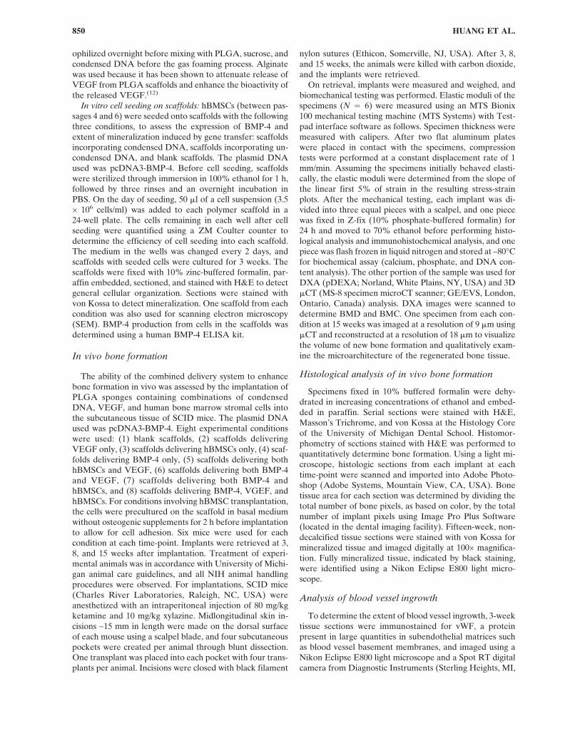
VEGF delivery: The effect of VEGF delivery on the ex-tent of angiogenesis was determined by immunostainingtissue sections for vWF, a component of the blood vesselECM. Scaffolds without VEGF showed relatively lowblood vessel densities at 3 weeks (Fig. 1A). In contrast,scaffolds delivering VEGF displayed high densities ofblood vessels interspersed throughout the scaffolds at thistime (Fig. 1B). Quantification of blood vessel densities
throughout the total scaffold area confirmed the histologi-cal examination, because scaffolds delivering VEGF in allexperimental conditions had a significantly higher bloodvessel density than scaffolds without VEGF (Fig. 1C).
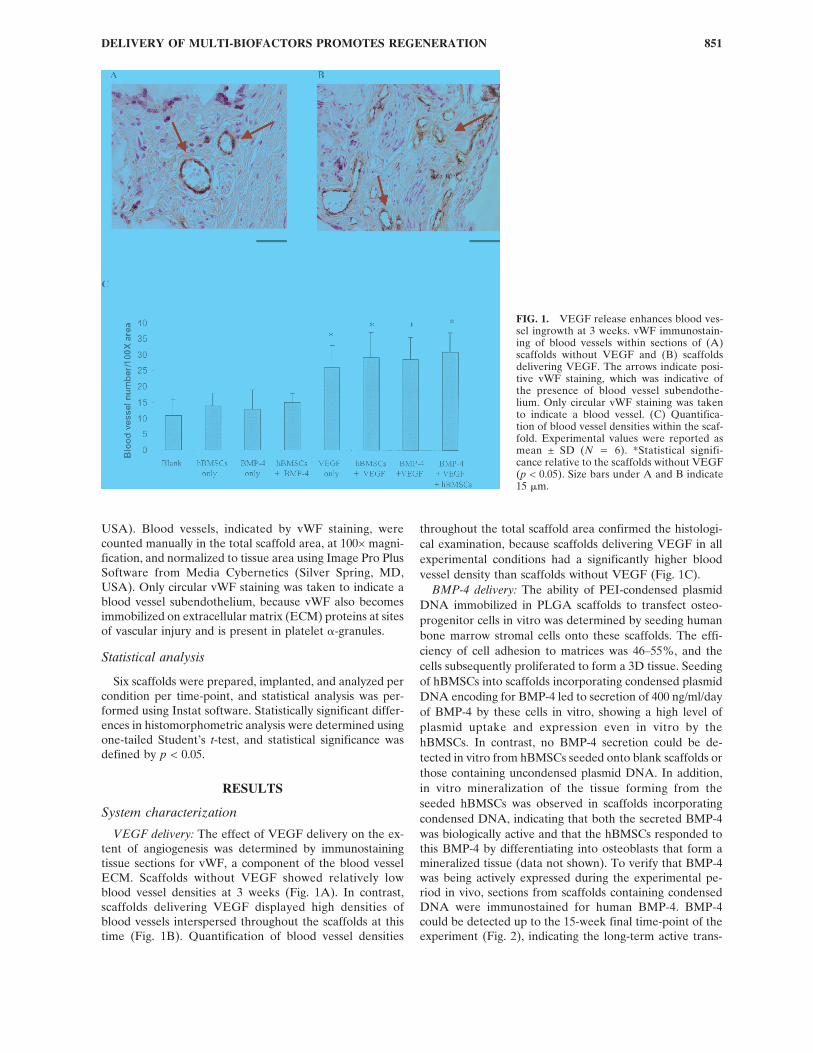
BMP-4 delivery: The ability of PEI-condensed plasmidDNA immobilized in PLGA scaffolds to transfect osteo-progenitor cells in vitro was determined by seeding humanbone marrow stromal cells onto these scaffolds. The effi-ciency of cell adhesion to matrices was 46–55%, and thecells subsequently proliferated to form a 3D tissue. Seedingof hBMSCs into scaffolds incorporating condensed plasmidDNA encoding for BMP-4 led to secretion of 400 ng/ml/dayof BMP-4 by these cells in vitro, showing a high level ofplasmid uptake and expression even in vitro by thehBMSCs. In contrast, no BMP-4 secretion could be de-tected in vitro from hBMSCs seeded onto blank scaffolds orthose containing uncondensed plasmid DNA. In addition,in vitro mineralization of the tissue forming from theseeded hBMSCs was observed in scaffolds incorporatingcondensed DNA, indicating that both the secreted BMP-4was biologically active and that the hBMSCs responded tothis BMP-4 by differentiating into osteoblasts that form amineralized tissue (data not shown). To verify that BMP-4was being actively expressed during the experimental pe-riod in vivo, sections from scaffolds containing condensedDNA were immunostained for human BMP-4. BMP-4could be detected up to the 15-week final time-point of theexperiment (Fig. 2), indicating the long-term active trans-
FIG. 1. VEGF release enhances blood ves-sel ingrowth at 3 weeks. vWF immunostain-ing of blood vessels within sections of (A)scaffolds without VEGF and (B) scaffoldsdelivering VEGF. The arrows indicate posi-tive vWF staining, which was indicative ofthe presence of blood vessel subendothe-lium. Only circular vWF staining was takento indicate a blood vessel. (C) Quantifica-tion of blood vessel densities within the scaf-fold. Experimental values were reported asmean ± SD (N ! 6). *Statistical signifi-cance relative to the scaffolds without VEGF(p < 0.05). Size bars under A and B indicate15 !m.
DELIVERY OF MULTI-BIOFACTORS PROMOTES REGENERATION 851
Fig 1 live 4/C
fection of cells resulting from condensed DNA delivery inthis system.
In vivo bone formation
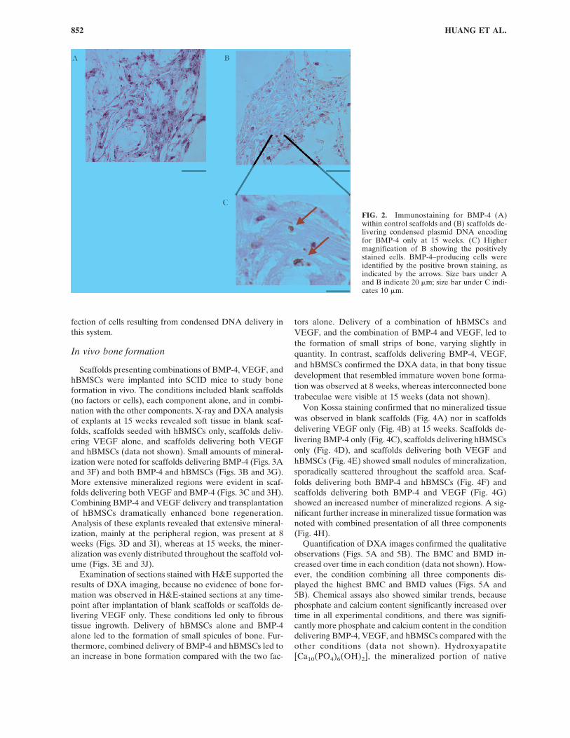
Scaffolds presenting combinations of BMP-4, VEGF, andhBMSCs were implanted into SCID mice to study boneformation in vivo. The conditions included blank scaffolds(no factors or cells), each component alone, and in combi-nation with the other components. X-ray and DXA analysisof explants at 15 weeks revealed soft tissue in blank scaf-folds, scaffolds seeded with hBMSCs only, scaffolds deliv-ering VEGF alone, and scaffolds delivering both VEGFand hBMSCs (data not shown). Small amounts of mineral-ization were noted for scaffolds delivering BMP-4 (Figs. 3Aand 3F) and both BMP-4 and hBMSCs (Figs. 3B and 3G).More extensive mineralized regions were evident in scaf-folds delivering both VEGF and BMP-4 (Figs. 3C and 3H).Combining BMP-4 and VEGF delivery and transplantationof hBMSCs dramatically enhanced bone regeneration.Analysis of these explants revealed that extensive mineral-ization, mainly at the peripheral region, was present at 8weeks (Figs. 3D and 3I), whereas at 15 weeks, the miner-alization was evenly distributed throughout the scaffold vol-ume (Figs. 3E and 3J).
Examination of sections stained with H&E supported theresults of DXA imaging, because no evidence of bone for-mation was observed in H&E-stained sections at any time-point after implantation of blank scaffolds or scaffolds de-livering VEGF only. These conditions led only to fibroustissue ingrowth. Delivery of hBMSCs alone and BMP-4alone led to the formation of small spicules of bone. Fur-thermore, combined delivery of BMP-4 and hBMSCs led toan increase in bone formation compared with the two fac-
tors alone. Delivery of a combination of hBMSCs andVEGF, and the combination of BMP-4 and VEGF, led tothe formation of small strips of bone, varying slightly inquantity. In contrast, scaffolds delivering BMP-4, VEGF,and hBMSCs confirmed the DXA data, in that bony tissuedevelopment that resembled immature woven bone forma-tion was observed at 8 weeks, whereas interconnected bonetrabeculae were visible at 15 weeks (data not shown).
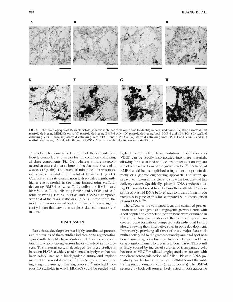
Von Kossa staining confirmed that no mineralized tissuewas observed in blank scaffolds (Fig. 4A) nor in scaffoldsdelivering VEGF only (Fig. 4B) at 15 weeks. Scaffolds de-livering BMP-4 only (Fig. 4C), scaffolds delivering hBMSCsonly (Fig. 4D), and scaffolds delivering both VEGF andhBMSCs (Fig. 4E) showed small nodules of mineralization,sporadically scattered throughout the scaffold area. Scaf-folds delivering both BMP-4 and hBMSCs (Fig. 4F) andscaffolds delivering both BMP-4 and VEGF (Fig. 4G)showed an increased number of mineralized regions. A sig-nificant further increase in mineralized tissue formation wasnoted with combined presentation of all three components(Fig. 4H).
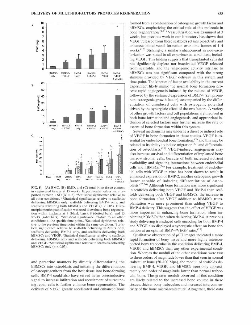
Quantification of DXA images confirmed the qualitativeobservations (Figs. 5A and 5B). The BMC and BMD in-creased over time in each condition (data not shown). How-ever, the condition combining all three components dis-played the highest BMC and BMD values (Figs. 5A and5B). Chemical assays also showed similar trends, becausephosphate and calcium content significantly increased overtime in all experimental conditions, and there was signifi-cantly more phosphate and calcium content in the conditiondelivering BMP-4, VEGF, and hBMSCs compared with theother conditions (data not shown). Hydroxyapatite[Ca10(PO4)6(OH)2], the mineralized portion of native
FIG. 2. Immunostaining for BMP-4 (A)within control scaffolds and (B) scaffolds de-livering condensed plasmid DNA encodingfor BMP-4 only at 15 weeks. (C) Highermagnification of B showing the positivelystained cells. BMP-4–producing cells wereidentified by the positive brown staining, asindicated by the arrows. Size bars under Aand B indicate 20 !m; size bar under C indi-cates 10 !m.
HUANG ET AL.852
Fig 2 live 4/C
bone, has a calcium to phosphate molar ratio of 1.67. Theaverage of this ratio for conditions that displayed mineral-ized tissue formation (1.63–1.72) was similar to that of hy-droxyapatite at 15 weeks, but only scaffolds delivering bothBMP-4 and hBMSCs, scaffolds delivering both BMP-4 andVEGF, and scaffolds delivering BMP-4, VEGF, andhBMSCs achieved values (1.69–1.74) close to this ratio by 8weeks.
Histomorphometric analysis of all conditions (Fig. 5C)supported the qualitative findings based on observation ofhistology. Co-delivery of BMP-4 and hBMSCs led to anincrease in bone formation equivalent to adding the effectsof each factor alone. In addition, quantification revealed a
dramatic effect on bone formation when VEGF was com-bined with either hBMSCs or BMP-4. Scaffolds deliveringboth VEGF and hBMSCs led to a 2.4-fold increase in boneformation over scaffolds delivering hBMSCs only (Fig. 5C).Furthermore, delivery of BMP-4 and VEGF led to a 19–24% increase on bone formation compared with the deliv-ery of BMP-4 alone. Scaffolds delivering all three factorsled to significantly greater bone formation than any of theother conditions (Fig. 5C).
To determine if the combined delivery of all three com-ponents also increased the quality of newly formed bonetissue, !CT images were taken of the explants over time,and the elastic moduli of the implants were determined at
FIG. 3. DXA images of (A)scaffold delivering BMP-4, (B)scaffold delivering both BMP-4and hBMSCs, (C) scaffold de-livering both BMP-4 andVEGF at 15 weeks, and scaf-fo lds de l iver ing BMP-4 ,VEGF, and hBMSCs at (D) 8weeks and (E) 15 weeks. InDXA images, yellow is thehighest density, red is medium,and the rest is low or back-ground. X-ray of (F) scaffolddelivering BMP-4, (G) scaffolddelivering both BMP-4 andhBMSCs, (H) scaffold deliver-ing both BMP-4 and VEGF at15 weeks, and scaffolds deliv-ering BMP-4, VEGF, andhBMSCs at (I) 8 weeks and (J)15 weeks. Size bars under thefigures indicate 5 mm.
DELIVERY OF MULTI-BIOFACTORS PROMOTES REGENERATION 853
Fig 3 live 4/C
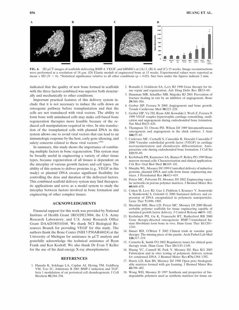
15 weeks. The mineralized portion of the explants wasloosely connected at 3 weeks for the condition combiningall three components (Fig. 6A), whereas a more intercon-nected structure similar to bony trabeculae was observed at8 weeks (Fig. 6B). The extent of mineralization was moreextensive, consolidated, and solid at 15 weeks (Fig. 6C).Constant strain rate compression tests revealed significantlyhigher elastic moduli in the tissue formed using scaffoldsdelivering BMP-4 only, scaffolds delivering BMP-4 andhBMSCs, scaffolds delivering BMP-4 and VEGF, and scaf-folds delivering BMP-4, VEGF, and hBMSCs comparedwith that of the blank scaffolds (Fig. 6D). Furthermore, themoduli of tissues created with all three factors was signifi-cantly higher than any other single or dual combinations offactors.
DISCUSSION
Bone tissue development is a highly coordinated process,and the results of these studies indicate bone regenerationsignificantly benefits from strategies that mimic concomi-tant interactions among various factors involved in this pro-cess. The material system developed for these studies isbased on PLGA, a widely used biomedical polymer that hasbeen safely used as a biodegradable suture and implantmaterial for several decades.(18) PLGA was fabricated, us-ing a high pressure gas foaming process,(17) into highly po-rous 3D scaffolds in which hBMSCs could be seeded with
high efficiency before transplantation. Proteins such asVEGF can be readily incorporated into these materials,allowing for a sustained and localized release at an implantsite of a bioactive form of the growth factor.(19) Delivery ofBMP-4 could be accomplished using either the protein di-rectly or a genetic engineering approach. The latter ap-proach was taken in this study to show the flexibility of thisdelivery system. Specifically, plasmid DNA condensed us-ing PEI was delivered to cells from the scaffolds. Conden-sation of plasmid DNA before leads to orders of magnitudeincreases in gene expression compared with uncondensedplasmid DNA.(20)
The effects of the combined local and sustained presen-tation of an osteogenic and angiogenic growth factors witha cell population competent to form bone were examined inthis study. Any combination of the factors displayed in-creased bone formation, compared with individual factorsalone, showing their interactive roles in bone development.Importantly, providing all three of these major factors si-multaneously led to the greatest quantity and quality of newbone tissue, suggesting the three factors acted in an additiveor synergistic manner to regenerate bone tissue. This resultis likely caused by increased survival of transplanted cellsbecause of VEGF-mediated angiogenesis, in concert withthe direct osteogenic action of BMP-4. Plasmid DNA po-tentially can be taken up by both hBMSCs and the infil-trating surrounding host cells (e.g., fibroblasts). The BMP-4secreted by both cell sources likely acted in both autocrine
FIG. 4. Photomicrographs of 15-week histologic sections stained with von Kossa to identify mineralized tissue. (A) Blank scaffold, (B)scaffold delivering hBMSCs only, (C) scaffold delivering BMP-4 only, (D) scaffold delivering both BMP-4 and hBMSCs, (E) scaffolddelivering VEGF only, (F) scaffold delivering both VEGF and hBMSCs, (G) scaffold delivering both BMP-4 and VEGF, and (H)scaffold delivering BMP-4, VEGF, and hBMSCs. Size bars under the figures indicate 20 !m.
HUANG ET AL.854
and paracrine manners by directly differentiating thehBMSCs into osteoblasts and initiating the differentiationof osteoprogenitors from the host tissue into bone-formingcells. BMP-4 could also have served as an osteoinductivesignal to increase infiltration and recruitment of surround-ing repair cells to further enhance bone regeneration. Thedelivery of VEGF greatly accelerated and enhanced bone
formed from a combination of osteogenic growth factor andhBMSCs, emphasizing the critical role of this molecule inbone regeneration.(4,21) Vascularization was examined at 3weeks, but previous work in our laboratory has shown thatVEGF released from these scaffolds retains bioactivity andenhances blood vessel formation over time frames of 1–4weeks.(12) Strikingly, a similar enhancement in neovascu-larization was noted in all experimental conditions, includ-ing VEGF. This finding suggests that transplanted cells didnot significantly deplete nor inactivated VEGF releasedfrom scaffolds, and the angiogenic activity intrinsic tohBMSCs was not significant compared with the strongstimulus provided by VEGF delivery in this system andtime-point. The kinetics of factor availability in the currentexperiment likely mimic the normal bone formation pro-cess: rapid angiogenesis induced by the release of VEGF,followed by the sustained expression of BMP-4 (i.e., promi-nent osteogenic growth factor), accompanied by the differ-entiation of uninduced cells with osteogenic potentialdriven by the synergistic effect of the two factors. A varietyof other growth factors and cell populations are involved inboth bone formation and angiogenesis, and appropriate in-clusion of selected factors may further increase the rate orextent of bone formation within this system.
Several mechanisms may underlie a direct or indirect roleof VEGF in bone formation in these studies. VEGF is es-sential for endochondral bone formation,(5) and this may berelated to its ability to induce migration(22) and differentia-tion of osteoblasts.(23) VEGF-induced angiogenesis mayalso increase survival and differentiation of implanted bonemarrow stromal cells, because of both increased nutrientavailability and signaling interactions between endothelialcells and hBMSCs.(24) For example, treatment of endothe-lial cells with VEGF in vitro has been shown to result inenhanced expression of BMP-2, another osteogenic growthfactor capable of inducing differentiation of osteo-blasts.(25,26) Although bone formation was more significantin scaffolds delivering both VEGF and BMP-4 than scaf-folds delivering both VEGF and hBMSCs, the increase inbone formation after VEGF addition to hBMSCs trans-plantation was more prominent than adding VEGF toBMP-4 delivery. This suggests that the effect of VEGF wasmore important in enhancing bone formation when im-planting hBMSCs than when delivering BMP-4. A previousstudy delivering transduced cells encoding for both BMP-4and VEGF also displayed a synergistic effect on bone for-mation at an optimal BMP-4/VEGF ratio.(27)
Qualitative observation of !CT images indicated a morerapid formation of bony tissue and more highly intercon-nected bony trabeculae in the condition delivering BMP-4,VEGF, and hBMSCs than any other experimental condi-tion. Whereas the moduli of the other conditions were twoto three orders of magnitude lower than that seen in normaltrabecular bone (50–100 Mpa), the moduli of scaffolds de-livering BMP-4, VEGF, and hBMSCs were only approxi-mately one order of magnitude lower than normal trabec-ular bone. The greater moduli observed in this conditionare likely related to the increased bone volume in thesetissues, thicker bony trabeculae, and increased interconnec-tivity of the bone microarchitecture. Altogether, these data
FIG. 5. (A) BMC, (B) BMD, and (C) total bone tissue contentin engineered tissues at 15 weeks. Experimental values were re-ported as mean ± SD (N ! 6). *Statistical significance relative toall other conditions. **Statistical significance relative to scaffoldsdelivering hBMSCs only, scaffolds delivering BMP-4 only, andscaffolds delivering both hBMSCs and VEGF (p < 0.05). Histo-morphometric quantification was used to evaluate bone regenera-tion within implants at 3 (blank bars), 8 (dotted bars), and 15weeks (solid bars). aStatistical significance relative to all otherconditions at the specific time-point., bStatistical significance rela-tive to the previous time-point within the same condition. cStatis-tical significance relative to scaffolds delivering hBMSCs only,scaffolds delivering BMP-4 only, and scaffolds delivering bothhBMSCs and VEGF. dStatistical significance relative to scaffoldsdelivering hBMSCs only and scaffolds delivering both hBMSCsand VEGF. eStatistical significance relative to scaffolds deliveringhBMSCs only (p < 0.05).
DELIVERY OF MULTI-BIOFACTORS PROMOTES REGENERATION 855
indicated that the quality of new bone formed in scaffoldswith the three factors combined was superior both structur-ally and mechanically to other conditions.
Important practical features of this delivery system in-clude that it is not necessary to induce the cells down anosteogenic pathway before transplantation and that thecells are not transduced with viral vectors. The ability toform bone with uninduced cells may make cell-based boneregeneration therapies more feasible because of the re-duced cell manipulations required in vitro. In situ transfec-tion of the transplanted cells with plasmid DNA in thissystem allows one to avoid viral vectors that can lead to animmunologic response by the host, early gene silencing, andsafety concerns related to these viral vectors.(28)
In summary, this study shows the importance of combin-ing multiple factors in bone regeneration. This system maybe broadly useful in engineering a variety of other tissuetypes, because regeneration of all tissues is dependent onthe interplay of various growth factors and cell types. Theability of this system to deliver proteins (e.g., VEGF in thisstudy) or plasmid DNA creates significant flexibility forcontrolling the dose and duration of the delivered factors.This combined scaffold delivery system may find therapeu-tic applications and serve as a model system to study theinterplay between factors involved in bone formation andengineering of other complex tissues.
ACKNOWLEDGMENTS
Financial support for this work was provided by NationalInstitute of Health Grant 1RO1DE13004, the U.S. ArmyResearch Laboratory, and U.S. Army Research OfficeGrant DAAD190310168. We thank NCI Biological Re-sources Branch for providing VEGF for this study. Theauthors thank the Bone Center (NIH 5 P30AR46024) at theUniversity of Michigan for assistance in !CT analysis andgratefully acknowledge the technical assistance of RyanFrank and Ken Kozloff. We also thank Dr Evan T Kellerfor the use of his dual-energy X-ray absorptiometer.
REFERENCES
1. Hanada K, Solchaga LA, Caplan AI, Hering TM, GoldbergVM, Yoo JU, Johnstone B 2001 BMP-2 induction and TGF-beta 1 modulation of rat periosteal cell chondrogenesis. J CellBiochem 81:284–294.
2. Bonadio J, Goldstein SA, Levy RJ 1998 Gene therapy for tis-sue repair and regeneration. Adv Drug Deliv Rev 33:53–69.
3. Hausman MR, Schaffler MB, Majeska RJ 2001 Prevention offracture healing in rats by an inhibitor of angiogenesis. Bone29:560–564.
4. Gerber HP, Ferrara N 2000 Angiogenesis and bone growth.Trends Cardiovasc Med 10:223–228.
5. Gerber HP, Vu TH, Ryan AM, Kowalski J, Werb Z, Ferrara N1999 VEGF couples hypertrophic cartilage remodeling, ossifi-cation and angiogenesis during endochondral bone formation.Nat Med 5:623–628.
6. Thompson TJ, Owens PD, Wilson DJ 1989 Intramembranousosteogenesis and angiogenesis in the chick embryo. J Anat166:55–65.
7. Carlevaro MF, Cermelli S, Cancedda R, Descalzi Cancedda F2000 Vascular endothelial growth factor (VEGF) in cartilageneovascularization and chondrocyte differentiation: Auto-paracrine role during endochondral bone formation. J Cell Sci113:59–69.
8. Krebsbach PH, Kuznetsov SA, Bianco P, Robey PG 1999 Bonemarrow stromal cells: Characterization and clinical application.Crit Rev Oral Biol Med 10:165–181.
9. Murphy WL, Mooney DJ 1999 Controlled delivery of inductiveproteins, plasmid DNA and cells from tissue engineering ma-trices. J Periodontal Res 34:413–419.
10. Peters MC, Polverini PJ, Mooney DJ 2002 Engineering vascu-lar networks in porous polymer matrices. J Biomed Mater Res60:668–678.
11. Cohen H, Levy RJ, Gao J, Fishbein I, Kousaev V, SosnowskiS, Slomkowski S, Golomb G 2000 Sustained delivery and ex-pression of DNA encapsulated in polymeric nanoparticles.Gene Ther 7:1896–1905.
12. Sheridan MH, Shea LD, Peters MC, Mooney DJ 2000 Bioad-sorbable polymer scaffolds for tissue engineering capable ofsustained growth factor delivery. J Control Release 64:91–102.
13. Krebsbach PH, Gu K, Franceschi RT, Rutherford RB 2000Gene therapy-directed osteogenesis: BMP-7-transduced hu-man fibroblasts form bone in vivo. Hum Gene Ther 11:1201–1210.
14. Simari RD, O’Brien T 2002 Clinical trials in vascular genetherapy: The missing piece of the puzzle. Arch Pathol Lab Med126:317–319.
15. Cornetta K, Smith FO 2002 Regulatory issues for clinical genetherapy trials. Hum Gene Ther 13:1143–1149.
16. Huang YC, Connell M, Park Y, Mooney DJ, Rice KG 2003Fabrication and in vitro testing of polymeric delivery systemfor condensed DNA. J Biomed Mater Res 67A:1384–1392.
17. Harris LD, Kim BS, Mooney DJ 1998 Open pore biodegrad-able matrices formed with gas foaming. J Biomed Mater Res42:396–402.
18. Wong WH, Mooney D 1997 Synthesis and properties of bio-degradable polymers used as synthetic matrices for tissue en-
FIG. 6. 3D !CT images of scaffolds delivering BMP-4, VEGF, and hBMSCs at (A) 3, (B) 8, and (C) 15 weeks. Image reconstructionswere performed at a resolution of 18 !m. (D) Elastic moduli of engineered bone at 15 weeks. Experimental values were reported asmean ± SD (N ! 6). *Statistical significance relative to all other conditions (p < 0.05). Size bars under the figures indicate 2 mm.
HUANG ET AL.856
gineering. In: Atala A, Mooney D (eds.) Synthetic Biodegrad-able Polymer Scaffolds. Birkhauser, Boston, MA, USA, pp.51–82.
19. Richardson TP, Peters MC, Ennett AB, Mooney DJ 2001 Poly-meric system for dual growth factor delivery. Nat Biotechnol19:1029–1034.
20. Huang YC, Rice KG, Mooney DJ 2005 Long-term in vivo geneexpression via delivery of PEI-DNA condensates from porouspolymer scaffolds. Hum Gene Ther (in press).
21. Street J, Bao M, deGuzman L, Bunting S, Peale FV Jr, FerraraN, Steinmetz H, Hoeffel J, Cleland JL, Daugherty A, vanBruggen N, Redmond HP, Carano RA, Filvaroff EH 2002Vascular endothelial growth factor stimulates bone repair bypromoting angiogenesis and bone turnover. Proc Natl Acad SciUSA 99:9656–9661.
22. Mayr-Wohlfart U, Waltenberger J, Hausser H, Kessler S, Gun-ther KP, Dehio C, Puhl W, Brenner RE 2002 Vascular endo-thelial growth factor stimulates chemotactic migration of pri-mary human osteoblasts. Bone 30:472–477.
23. Zelzer E, McLean W, Ng YS, Fukai N, Reginato AM, LovejoyS, D’Amore PA, Olsen BR 2002 Skeletal defects inVEGF(120/120) mice reveal multiple roles for VEGF in skel-etogenesis. Development 129:1893–1904.
24. Kaigler D, Krebsbach PH, Polverini PJ, Mooney DJ 2003 Roleof vascular endothelial growth factor in bone marrow stromalcell modulation of endothelial cells. Tissue Eng 9:95–103.
25. Wozney JM 1992 The bone morphogenetic protein family andosteogenesis. Mol Reprod Dev 32:160–167.
26. Bouletreau PJ, Warren SM, Spector JA, Peled ZM, GerretsRP, Greenwald JA, Longaker MT 2002 Hypoxia and VEGFup-regulate BMP-2 mRNA and protein expression in micro-vascular endothelial cells: Implications for fracture healing.Plast Reconstr Surg 109:2384–2397.
27. Peng H, Wright V, Usas A, Gearhart B, Shen H-C, CumminsJ, Huard J 2002 Synergistic enhancement of bone formationand healing by stem cell-expressed VEGF and bone morpho-genetic protein-4. J Clin Invest 110:751–759.
28. Romano G, Pacilio C, Giordano A 1999 Gene transfer tech-nology in therapy: Current applications and future goals. StemCells 17:191–202.
Address reprint requests to:David Mooney, PhD
Harvard UniversityDivision of Engineering and Applied Sciences
Room 325, Pierce Hall29 Oxford Street
Cambridge, MA 02138, USAE-mail: [email protected]
Received in original form July 12, 2004; revised form October 25,2004; accepted December 15, 2004.
DELIVERY OF MULTI-BIOFACTORS PROMOTES REGENERATION 857
Related Documents