thematic review Thematic Review Series: Sphingolipids Ganglioside GM3 suppresses the proangiogenic effects of vascular endothelial growth factor and ganglioside GD1a Purna Mukherjee, Anthony C. Faber, Laura M. Shelton, Rena C. Baek, Thomas C. Chiles, and Thomas N. Seyfried 1 Department of Biology, Boston College, Chestnut Hill, MA 02467 Abstract Gangliosides are sialic acid-containing glyco- sphingolipids that have long been associated with tumor malignancy and metastasis. Mounting evidence suggests that gangliosides also modulate tumor angiogenesis. Tumor cells shed gangliosides into the microenvironment, which produces both autocrine and paracrine effects on tumor cells and tumor-associated host cells. In this study, we show that the simple monosialoganglioside GM3 counteracts the proangiogenic effects of vascular endothelial growth factor (VEGF) and of the complex disialoganglioside GD1a. GM3 suppressed the action of VEGF and GD1a on the prolifer- ation of human umbilical vein endothelial cells (HUVECs) and inhibited the migration of HUVECs toward VEGF as a chemoattractant. Enrichment of added GM3 in the HUVEC membrane also reduced the phosphorylation of vascular endothelial growth factor receptor 2 (VEGFR-2) and downstream Akt. Moreover, GM3 reduced the proan- giogenic effects of GD1a and growth factors in the in vivo Matrigel plug assay. Inhibition of GM3 biosynthesis with the glucosyl transferase inhibitor, N-butyldeoxynojirimycin (N B-DNJ), increased HUVEC proliferation and the phos- phorylation of VEGFR-2 and Akt. The effects of N B-DNJ on HUVECs were reversed with the addition of GM3. We conclude that GM3 has antiangiogenic action and may possess therapeutic potential for reducing tumor angio- genesis.—Mukherjee, P., A. C. Faber, L. M. Shelton, R. C. Baek, T. C. Chiles, and T. N. Seyfried. Ganglioside GM3 suppresses the proangiogenic effects of vascular endothelial growth factor and ganglioside GD1a. J. Lipid Res. 2008. 49: 929–938. Supplementary key words glycosphingolipid & human umbilical endothelial cell migration & matrigel plug assay & growth factor receptor & phosphorylated Akt Gangliosides are a family of sialic acid-containing glycosphingolipids that are enriched in the outer surface of plasma membranes and have long been associated with tumor malignancy and metastasis (1–3). These molecules contain an oligosaccharide head group that is attached to a lipophilic ceramide, consisting of a sphingosine base and a long-chain fatty acid. Gangliosides can be shed from the surface of tumor cells into the microenvironment, where they can influence tumor host cell interactions to include angiogenesis (1, 4–13). Ganglioside GM3, a sim- ple monosialoganglioside (NeuAca2Y3Galb1Y4Glcb1Y 1¶-ceramide), modulates cell adhesion, proliferation, and differentiation (2, 5, 12, 14). The antiproliferative and proapoptotic effects of GM3 were observed in glioma cells grown both in vivo and in vitro (15–17). In contrast to GM3, complex gangliosides like GM2, GM1, GD1a, GD1b, GT1b, and GD3, which contain longer oligosaccharide chains than that of GM3, enhance tumor cell prolifer- ation, invasion, and metastasis (1, 3, 14, 18, 19). Increased tumorigenic effects of complex gangliosides were ob- served in a variety of tumor cells, including bladder, lym- phoma, glioma, neuroblastoma, and melanoma (7, 11, 14, 20–22). Specific inhibitors of ganglioside biosynthesis also reduced tumor growth (23–25), whereas gene-linked shifts in ganglioside distribution changed tumor growth and angiogenesis in vivo (4, 8, 12). Endothelial cell signaling is important in cancer- associated vascularity (angiogenesis). The proliferation and migration of endothelial cells in response to growth factors is one of the major determinants of tumor growth and progression. Dysregulation of the balance between proangiogenic and antiangiogenic factors contributes to Manuscript received 3 January 2008 and in revised form 15 February 2008. Published, JLR Papers in Press, February 20, 2008. DOI 10.1194/jlr.R800006-JLR200 Abbreviations: bFGF, basic fibroblast growth factor; EBM, endo- thelial basal medium; EGFR, epidermal growth factor receptor; EGM- 2, endothelial growth medium; HPTLC, high-performance thin-layer chromatography; HUVEC, human umbilical vein endothelial cell; MTT, 3-(4,5-dimethylthiazol-2-yl)-2,5-diphenyltetrazolium bromide; N B-DNJ, N-butyldeoxynojirimycin; VEGF, vascular endothelial growth factor; VEGFR-2, vascular endothelial growth factor receptor 2. 1 To whom correspondence should be addressed. e-mail: [email protected] Copyright D 2008 by the American Society for Biochemistry and Molecular Biology, Inc. This article is available online at http://www.jlr.org Journal of Lipid Research Volume 49, 2008 929 by guest, on May 12, 2016 www.jlr.org Downloaded from

Welcome message from author
This document is posted to help you gain knowledge. Please leave a comment to let me know what you think about it! Share it to your friends and learn new things together.
Transcript

thematic review
Thematic Review Series: Sphingolipids
Ganglioside GM3 suppresses the proangiogenic
effects of vascular endothelial growth factor and
ganglioside GD1a
Purna Mukherjee, Anthony C. Faber, Laura M. Shelton, Rena C. Baek, Thomas C. Chiles,and Thomas N. Seyfried1
Department of Biology, Boston College, Chestnut Hill, MA 02467
Abstract Gangliosides are sialic acid-containing glyco-sphingolipids that have long been associated with tumormalignancy and metastasis. Mounting evidence suggeststhat gangliosides also modulate tumor angiogenesis. Tumorcells shed gangliosides into the microenvironment, whichproduces both autocrine and paracrine effects on tumorcells and tumor-associated host cells. In this study, we showthat the simple monosialoganglioside GM3 counteracts theproangiogenic effects of vascular endothelial growth factor(VEGF) and of the complex disialoganglioside GD1a. GM3suppressed the action of VEGF and GD1a on the prolifer-ation of human umbilical vein endothelial cells (HUVECs)and inhibited the migration of HUVECs toward VEGF asa chemoattractant. Enrichment of added GM3 in theHUVEC membrane also reduced the phosphorylation ofvascular endothelial growth factor receptor 2 (VEGFR-2)and downstream Akt. Moreover, GM3 reduced the proan-giogenic effects of GD1a and growth factors in the in vivoMatrigel plug assay. Inhibition of GM3 biosynthesis withthe glucosyl transferase inhibitor, N-butyldeoxynojirimycin(NB-DNJ), increased HUVEC proliferation and the phos-phorylation of VEGFR-2 and Akt. The effects of NB-DNJon HUVECs were reversed with the addition of GM3.We conclude that GM3 has antiangiogenic action and maypossess therapeutic potential for reducing tumor angio-genesis.—Mukherjee, P., A. C. Faber, L. M. Shelton, R. C.Baek, T. C. Chiles, and T. N. Seyfried. Ganglioside GM3suppresses the proangiogenic effects of vascular endothelialgrowth factor and ganglioside GD1a. J. Lipid Res. 2008. 49:929–938.
Supplementary key words glycosphingolipid & human umbilicalendothelial cell migration & matrigel plug assay & growth factor receptor& phosphorylated Akt
Gangliosides are a family of sialic acid-containingglycosphingolipids that are enriched in the outer surface
of plasma membranes and have long been associated withtumor malignancy and metastasis (1–3). These moleculescontain an oligosaccharide head group that is attachedto a lipophilic ceramide, consisting of a sphingosine baseand a long-chain fatty acid. Gangliosides can be shed fromthe surface of tumor cells into the microenvironment,where they can influence tumor host cell interactions toinclude angiogenesis (1, 4–13). Ganglioside GM3, a sim-ple monosialoganglioside (NeuAca2Y3Galb1Y4Glcb1Y1¶-ceramide), modulates cell adhesion, proliferation, anddifferentiation (2, 5, 12, 14). The antiproliferative andproapoptotic effects of GM3 were observed in glioma cellsgrown both in vivo and in vitro (15–17). In contrast toGM3, complex gangliosides like GM2, GM1, GD1a, GD1b,GT1b, and GD3, which contain longer oligosaccharidechains than that of GM3, enhance tumor cell prolifer-ation, invasion, and metastasis (1, 3, 14, 18, 19). Increasedtumorigenic effects of complex gangliosides were ob-served in a variety of tumor cells, including bladder, lym-phoma, glioma, neuroblastoma, and melanoma (7, 11,14, 20–22). Specific inhibitors of ganglioside biosynthesisalso reduced tumor growth (23–25), whereas gene-linkedshifts in ganglioside distribution changed tumor growthand angiogenesis in vivo (4, 8, 12).
Endothelial cell signaling is important in cancer-associated vascularity (angiogenesis). The proliferationand migration of endothelial cells in response to growthfactors is one of the major determinants of tumor growthand progression. Dysregulation of the balance betweenproangiogenic and antiangiogenic factors contributes to
Manuscript received 3 January 2008 and in revised form 15 February 2008.
Published, JLR Papers in Press, February 20, 2008.DOI 10.1194/jlr.R800006-JLR200
Abbreviations: bFGF, basic fibroblast growth factor; EBM, endo-thelial basal medium; EGFR, epidermal growth factor receptor; EGM-2, endothelial growth medium; HPTLC, high-performance thin-layerchromatography; HUVEC, human umbilical vein endothelial cell;MTT, 3-(4,5-dimethylthiazol-2-yl)-2,5-diphenyltetrazolium bromide;NB-DNJ, N-butyldeoxynojirimycin; VEGF, vascular endothelial growthfactor; VEGFR-2, vascular endothelial growth factor receptor 2.
1 To whom correspondence should be addressed.e-mail: [email protected]
Copyright D 2008 by the American Society for Biochemistry and Molecular Biology, Inc.
This article is available online at http://www.jlr.org Journal of Lipid Research Volume 49, 2008 929
by guest, on May 12, 2016
ww
w.jlr.org
Dow
nloaded from

the abnormal vasculature in tumors. The targeting oftumor endothelial cells, therefore, is considered impor-tant for managing tumor growth (1, 8, 26).
Vascular endothelial cells are responsive to a number ofproangiogenic growth factors, including basic fibroblastgrowth factor (bFGF) and vascular endothelial growthfactor (VEGF), which promote endothelial cell survival,growth, and migration (18, 27, 28). Interestingly, complexgangliosides enhance the response of endothelial cellsto the proangiogenic action of bFGF and VEGF (4, 13,18, 29). GD1a enrichment of endothelial cells enhancedVEGF receptor dimerization, autophosphorylation, anddownstream signaling pathways for endothelial cell pro-liferation and migration (13). The involvement of gan-gliosides in angiogenesis is dependent on the intactmolecules, as neither asialo species nor sialic acid aloneinfluences angiogenesis (30). In contrast to the enhanc-ing effects of complex gangliosides on angiogenesis, GM3reduces endothelial cell proliferation and migration (5, 8,12, 31). Little is known, however, about the molecularmechanism by which GM3 inhibits angiogenesis.
The interactions of gangliosides with cell surface re-ceptor molecules in tumor cells as well as in endothelialcells may be critical for the tumor-induced progression ofthe microenvironment (8, 13, 14). The inhibitory effectsof GM3 on the epidermal growth factor receptor (EGFR)tyrosine kinase are well studied (32–34). Yoon et al. (34)showed that GM3 inhibits the EGFR tyrosine kinasethrough interactions with N-acetylglucosamine residueson the glycan units of the receptor. In contrast to the in-formation available on the influence of GM3 on the EGFR,little is known about the influence of GM3 on othergrowth factor receptors, including the vascular endothe-lial growth factor receptor (VEGFR). VEGFR-2 or KDRis phosphorylated upon VEGF stimulation, which inducesthe phosphoinositide-3 kinase/Akt pathway, resulting inenhanced endothelial proliferation and migration (35, 36).
In this study, we examined the influence of the exog-enous addition of GM3 on the proliferation of humanumbilical vein endothelial cells (HUVECs) in the presenceof VEGF and the VEGF enhancer, GD1a. We show thatGM3 suppresses angiogenesis both in vitro and in vivo.We also found that GM3 enrichment of HUVECs inhibitsmigration toward VEGF as a chemoattractant. Addition-ally, we found that GM3 reduced VEGFR-2 phosphoryla-tion and downstream Akt signaling in HUVECs, suggestinga mechanism by which GM3 reduces endothelial cell pro-liferation and migration. Moreover, pharmacological deple-tion of endogenous GM3 significantly increased HUVECproliferation and VEGFR-2 and Akt phosphorylation.
MATERIALS AND METHODS
Reagents
Purified disialoganglioside GD1a (bovine brain), recombinanthuman vascular endothelial growth factor (VEGF165), and BSAwere purchased from Sigma (St. Louis, MO). Highly purifiedmonosialoganglioside GM3 was purchased from Matreya (Pleasant
Gap, PA). The commercial ganglioside preparations were thesame as those used by other groups (13, 14) but were not checkedfor purity. MatrigelTM basement membrane matrix was purchasedfrom BD Biosciences (Bedford, MA). The CellTiter 96 non-radioactive cell proliferation 3-(4,5-dimethylthiazol-2-yl)-2,5-diphenyltetrazolium bromide (MTT) assay kit was from Promega(Madison, WI). Endothelial growth medium (EGM-2), BulletKitmedium, and supplement-free endothelial basal medium (EBM)were purchased from Cambrex (Charles City, IA). GangliosideGD1a and VEGF were dissolved in EBM and sonicated, and ali-quots stored at 220jC. Ganglioside GM3 was prepared in thesame manner but was first dissolved in chloroform-ethanol(1:1, v/v) and was then evaporated and dissolved in EBM. Anti-bodies against total and phosphorylated VEGFR-2 and Akt werepurchased from Cell Signaling (Beverly, MA). FITC-labeled Iso-lectin B4 was purchased from Vector Laboratories (Burlingame,CA). Calcein was purchased from Invitrogen (San Diego, CA).Anti-GM3 DH2 antibody was a gift from Dr. S. I. Hakomori(University of Washington, Seattle). Cy5-labeled anti-mouse anti-body was purchased from Jackson ImmunoResearch (West Grove,PA).N-butyldeoxynojirimycin (NB-DNJ; molecular weight, 219.3)was obtained as a gift from Oxford Glycosciences (Abigdon, UK).[14C]galactose was obtained from Sigma.
Cell culture
HUVECs (pooled) were purchased from Cambrex and weremaintained at 37jC in EGM-2 in humidified air containing 5%CO2. All experiments with HUVECs were conducted at passagestwo to six.
Animals
BALBc/J-SCID mice were obtained from the Jackson Labora-tory (Bar Harbor, ME) and were used for the Matrigel plug study.All animal experiments were carried out with ethical committeeapproval in accordance with the National Institutes of HealthGuide for the Care and Use of Laboratory Animals and approvedby the institutional care committee.
Proliferation assay
In vitro HUVEC proliferation was analyzed using the CellTiter96 nonradioactive cell proliferation assay according to the man-ufacturer’s protocol. Briefly, 5 3 103 HUVECs in EGM-2 wereseeded in each well of a 96-well plate. After 24 h, the HUVECswere washed and treated with 20 mM GD1a and GM3 in EBM for24 h. After this incubation, the ganglioside-containing mediumwas removed. The HUVECs were then washed with EBM andwere stimulated with 4 ng/ml VEGF in EBM for 24 h. For the NB-DNJ study, cells were treated with 200 mM NB-DNJ in the absenceand presence of GM3 for 48 h and were then stimulated with4 ng/ml VEGF in EBM for an additional 24 h. Tetrazolium saltdye solution (15 ml) was added to each well, and the plate wasreturned to the incubator for 4 h. Stop solution (100 ml) was thenadded to solubilize the metabolite and to lyse the HUVECs. Theplate was incubated for 24 h. The plate was shaken for 30 s afterincubation and was read in a microplate reader (SpectraMax M5;Molecular Devices) at 595 nm. This procedure was performedin triplicate.
Flow cytometry
HUVECs were treated with 0, 0.5, 2.5, or 5.0 mM GM3 in EBMfor 24 h at 37jC in 5% CO2-humidified conditions. Confluentcells were detached with trypsin/EDTA and were washed withPBS. HUVECs (2 3 105) were incubated with anti-GM3 DH2antibody (1:50) for 1 h on ice, washed with PBS, and incubated
930 Journal of Lipid Research Volume 49, 2008
by guest, on May 12, 2016
ww
w.jlr.org
Dow
nloaded from

with 1:500 Cy5 anti-mouse secondary antibody for 30 min onice in darkness. Cells were then washed twice with PBS, re-suspended, and analyzed with FACSDiva software (BeckmanCoulter). Control experiments included HUVECs treated withonly secondary antibody.
Migration assay
Confluent HUVECs in T75 flasks were rinsed and treatedwith 20 mM GM3 in EBM containing 0.1% BSA for a minimumof 4–5 h at 37jC in 5% CO2-humidified conditions. A controlflask of HUVECs was treated with EBM containing 0.1% BSA.Cells were washed twice in EBM and were harvested in EBMcontaining 0.1% BSA. According to the manufacturer’s protocol,equal numbers of control and treated cells (4 3 105 cells/ml)in 250 ml of EBM were seeded on the upper insert of a 24-wellmigration chamber (BD Falcon Fluoroblok Insert system; 3.0 mmpore size). EBM (750 ml) containing 100 ng/ml VEGF was thenplaced in the lower chamber according to the protocol. ThisVEGF concentration was recommended and necessary for aneffective migration of HUVECs. Serum-containing medium wasused as a positive control for these experiments. The plates wereincubated for 24 h under the humidified conditions as above.HUVECs that migrated to the underside of the membrane werewashed with HBSS, and the inserts were transferred in the com-panion wells containing calcein (4–5 mg/ml) solution. HUVECswere incubated in calcein for 1 h and washed, and the fluo-rescently labeled cells were photographed using a fluorescencemicroscope. Fluorescence was measured using a SpectraMax M5microplate reader (Molecular Devices).
In vivo Matrigel plug assay
Angiogenesis was analyzed using the in vivo Matrigel plug assayas we described previously (12, 37). Briefly, Matrigel (200 ml) andEBM (100 ml) containing GD1a and/or GM3 (at 20 or 40 mM)were thoroughly mixed at 4jC. Control plugs contained only theMatrigel/EBM solution. Male BALBc/SCID mice were anesthe-tized with Avertin (0.1 ml/10 g body weight) and then injectedwith Matrigel with or without gangliosides subcutaneously in thedorsal midline using a prechilled tuberculin syringe (27 gaugeneedle). Seven days after implantation and 30 min before nec-ropsy, mice were injected intravenously with 100 ml of FITC-conjugated Griffonia simplicifolia Isolectin B4 (0.25 mg/ml) tostain vascular endothelial cells (38). Matrigel plugs with the sur-rounding skin were removed as described previously, and vas-cularity was photographed (37). Lectin-FITC was extracted fromplugs by homogenizing in 500 ml of radioimmunoprecipitationbuffer. The homogenate was centrifuged at 1,000 g, and fluores-cence was measured at 490 nm using a SpectraMax M5 micro-plate reader as above.
Ganglioside biosynthesis in HUVECs
Synthesized gangliosides were isolated from control and NB-DNJ-treated HUVECs as described previously (2). Briefly,HUVECs were grown for 72 h in EGM-2 containing 5 mCi of[14C]galactose and 200 mM NB-DNJ. The radiolabeled cells wereremoved from the flask with a cell scraper in PBS and were col-lected as a pellet. Before ganglioside isolation, unlabeled mouseependymoblastoma gangliosides, containing GM3, were addedas unlabeled carrier (2). Total lipids were extracted from theradiolabeled cells in chloroform-methanol (2:1, v/v), and dH2Owas added (20%, v/v). The radiolabeled gangliosides were sep-arated from the total lipids by Folch partitioning into an upperaqueous phase as described (2). The upper phase gangliosides
were dried under nitrogen, resuspended in chloroform-methanol(2:1, v/v), and spotted on a high-performance thin-layer chro-matography (HPTLC) plate. The concentration of radiolabeledgangliosides spotted on the HPTLC plate was determined byscintillation counting. The amount of disintegrations per minuteequivalent to 10,000 dpm was spotted per lane on the HPTLCplate. The HPTLC plate was developed in one ascending elutionwith C/M/H2O (50:45:10, v/v) containing 0.02% CaCl2IdH2O.After autoradiography, the plates were sprayed with the resorcinolreagent to identify ganglioside standards. The amount of radio-label incorporated into GM3 was determined from Bioscan anal-ysis as we described previously (39).
Immunoblot analysis of HUVEC lysates
HUVECs were seeded in EGM-2 at 1 3 105 cells/well on six-well plates. When confluent, the HUVECs were washed with EBMand then incubated with 80 nM (100 ng/ml) GM3 in serum-freeEBM for 24 h. The HUVECs were then washed and stimulatedwith 100 ng/ml VEGF in EBM for 5 min at 37jC. For the NB-DNJstudy, cells were treated with 200 mM NB-DNJ in the presenceand absence of GM3 for 48 h and then stimulated with VEGF for5 min at 37jC. The HUVECs were next washed twice with PBSand treated with lysis buffer (100 ml/well) containing 20 mMTris-HCl (pH 7.5), 150 mM NaCl, 1 mM Na2EDTA, 1 mM EGTA,1% Triton, 2.5 mM NaPPi, 1 mM b-glycerophosphate, 1 mMNa3VO4, 1 mg/ml leupeptin, and 1 mM phenylmethylsufonylfluoride. The lysates were centrifuged at 12,000 g for 20 min at4jC. Supernatants were collected, and protein concentrationswere estimated using the Bio-Rad DC protein assay. Approxi-mately 50–100 mg of total protein from each sample was loadedonto a 12% sodium dodecyl polyacrylamide gel (Invitrogen) andanalyzed by electrophoresis. Proteins were transferred to a poly-vinylidene difluoride Immobilon TM-P membrane (Millipore).The membrane was blocked in 5% nonfat powdered milk inTris-buffered saline with Tween 20 (pH 7.6) for 1 h at roomtemperature. Blots were then probed with primary antibodyagainst phosphorylated VEGFR-2 (tyrosine-1175) and reprobedwith primary antibody against mouse monoclonal Akt (serine-473) overnight at 4jC. The membrane was probed again withtotal VEGFR-2 and Akt under optimal conditions.
Statistical analysis
All data are presented as means 6 SEM. Significance ofdifferences between groups was evaluated with one-way ANOVA.
RESULTS
GM3 suppresses VEGF- and GD1a-inducedHUVEC proliferation
To examine the influence of GM3 on angiogenesis, westudied the proliferation of HUVECs in culture. We eval-uated the influence of GM3 on HUVEC proliferationeither alone or in combination with GD1a in the pres-ence of VEGF stimulation (Fig. 1). We found that GD1a(20 mM) significantly increased VEGF-induced HUVECproliferation. In contrast, GM3 (20 mM) significantly re-duced VEGF- or GD1a-induced HUVEC proliferation by?50%. A control study was done to confirm that GM3did not alter the uptake of MTT into HUVECs. The inhib-itory effect of GM3 on HUVEC proliferation was also ob-served at a lower GM3 concentration of 80 nM.
Antiangiogenic effects of GM3 931
by guest, on May 12, 2016
ww
w.jlr.org
Dow
nloaded from

Detection of GM3 on HUVEC membranes
To determine whether the suppressive effects of GM3on HUVEC proliferation were associated with the ex-pression of GM3 on the membrane, we added differentamounts of GM3 to the incubation medium and analyzedthe HUVECs using flow cytometry with DH2 anti-GM3antibody (Fig. 2). No DH2 binding occurred in the un-treated HUVECs, indicating that DH2 does not detectthe endogenous GM3 epitope. Incubation of HUVECswith GM3 caused a dose-dependent increase in DH2 bind-ing. Because GM3 pretreatment produced no apparentcell death or toxicity, it is likely that GM3, located on theHUVEC membrane, inhibits proliferation and reduces thestimulatory effects of VEGF and/or GD1a. These findingsindicate that GM3 modulates the response of HUVECs tothe proangiogenic effects of VEGF and GD1a.
GM3 reduces HUVEC migration toward VEGFas a chemoattractant
We also examined whether GM3 could influence HUVECmigration toward VEGF as a chemoattractant. HUVECs,which migrated from the upper chamber to the lowerchamber of the migration assay and were labeled with thefluorescent dye calcein-AM, were photographed (Fig. 3A)and the amount of fluorescence was quantified (Fig. 3B).HUVEC migration was significantly less in the absencethan in the presence of VEGF, indicating that VEGF is achemoattractant for HUVECs in this assay. Migration wassignificantly less for GM3-treated HUVECs than for non-
treated HUVECs. These findings show that GM3 reducedHUVEC migration in response to VEGF.
GM3 inhibits GD1a-induced vascularization inMatrigel in vivo
Because GM3 suppressed the GD1a- and/or VEGF-induced HUVEC proliferation and migration in culture,it was of interest to examine the influence of GM3 onangiogenesis in the in vivo Matrigel plug assay. The in vivoMatrigel angiogenesis model represents early events ofangiogenesis and tumor progression and is dependent onthe activation and infiltration of host stromal cells, whichinclude monocytes, macrophages, and endothelial cell pre-cursors (12, 27, 37). Matrigel contains growth factors,which induce the infiltration of blood vessels as seen inthe control plugs (Fig. 4A). The addition of GD1a to theMatrigel enhanced blood vessel formation into the plugs.The number and size of vessels was also greater at a GD1aconcentration of 40 mM than at 20 mM, consistent withthe known angiogenesis-enhancing effects of GD1a (13).The addition of equal amounts of GM3 and GD1a (40 mM)markedly reduced blood vessel formation in the plugs.Indeed, the appearance of blood vessels was noticeably lessin the plugs containing GM3 and GD1a than in the controlplugs, which contained no added gangliosides (Fig. 4A).Fluorescently labeled vessels were noticeably less in theGM3 1 GD1a-containing plugs than in the plugs contain-ing GD1a alone (Fig. 4B). The ratio of FITC-labeled ves-sels to arbitrary units was also significantly lower in theGM3 1 GD1a-containing plugs than in the plugs con-taining GD1a alone (Fig. 4C). These results indicate thatGM3 suppressed the proangiogenic effects of GD1a in anin vivo environment.
Fig. 2. Detection of added GM3 on HUVEC membranes. HUVECswere incubated with GM3 for 24 h in EBM. Flow cytometry usinganti-GM3 DH2 and Cy5-labeled anti-mouse IgG was used to eval-uate GM3 on the membranes of HUVECs. The concentration ofadded GM3 is shown above each peak: orange, 0.0 mM; green,0.5 mM; pink, 2.5 mM; brown, 5.0 mM. The 0 mM concentrationpeak was the same as that for the secondary Cy5 antibody withoutDH2. These experiments were done in triplicate.
Fig. 1. GM3 inhibits vascular endothelial growth factor (VEGF)-and GD1a-induced human umbilical vein endothelial cell(HUVEC) proliferation. HUVECs were pretreated for 24 h in endo-thelial basal medium (EBM) in the absence (control) or in thepresence of GD1a (20 mM) and/or GM3 (20 mM) and then in-cubated for an additional 24 h in the presence of VEGF (4 ng/ml).The 3-(4,5-dimethylthiazol-2-yl)-2,5-diphenyltetrazolium bromide(MTT) assay was used to measure HUVEC proliferation as de-scribed in Materials and Methods. Proliferation is expressed as apercentage of the untreated, VEGF-stimulated controls. GD1a pre-treatment significantly enhanced HUVEC proliferation (P, 0.01).GM3 pretreatment significantly suppressed VEGF- and GD1a-induced proliferation (P , 0.001). Values are expressed as means6 SEM (n 5 3 independent experiments).
932 Journal of Lipid Research Volume 49, 2008
by guest, on May 12, 2016
ww
w.jlr.org
Dow
nloaded from

GM3 inhibits VEGFR-2 and Akt phosphorylation
VEGF stimulates HUVEC proliferation through down-stream phosphoinositide-3 kinase/Akt signaling by in-ducing the phosphorylation of VEGFR-2 (KDR, Flk-1)(35, 36). We used a 100 ng/ml VEGF concentration forthese experiments to ensure robust VEGFR-2 phosphor-ylation (ratio of pVEGFR-2 to total). VEGFR-2 phosphor-ylation was 6-fold greater and Akt phosphorylation atserine-473 (ratio of pAkt to total) was ?2-fold greater inthe presence than in the absence of VEGF after 5 minof stimulation in EBM (Fig. 5A, B). No VEGF phosphor-ylation was observed for HUVECs grown in the basalmedium (EBM), which contained no serum or growth fac-tors. In contrast, phosphorylation of VEGFR-2 and Aktwas reduced significantly by 60% and 75%, respectively,in HUVECs incubated with GM3 (80 nM or 100 ng/ml)for 24 h before VEGF stimulation (Fig. 5A, B). We usedthe 100 ng/100 ng GM3/VEGF concentration ratioin these experiments to remain consistent with the 1:1GM3/bFGF concentration ratio used previously in therabbit cornea model (31). This GM3 concentration wasmore effective at reducing phosphorylation than was thehigher concentration (20 mM) of GM3 used in the in vivoassay (data not shown). Collectively, these findings indi-
cate that low concentrations of GM3 inhibit the VEGF-induced phosphorylation of VEGFR-2 and downstreamAkt in cultured HUVECs.
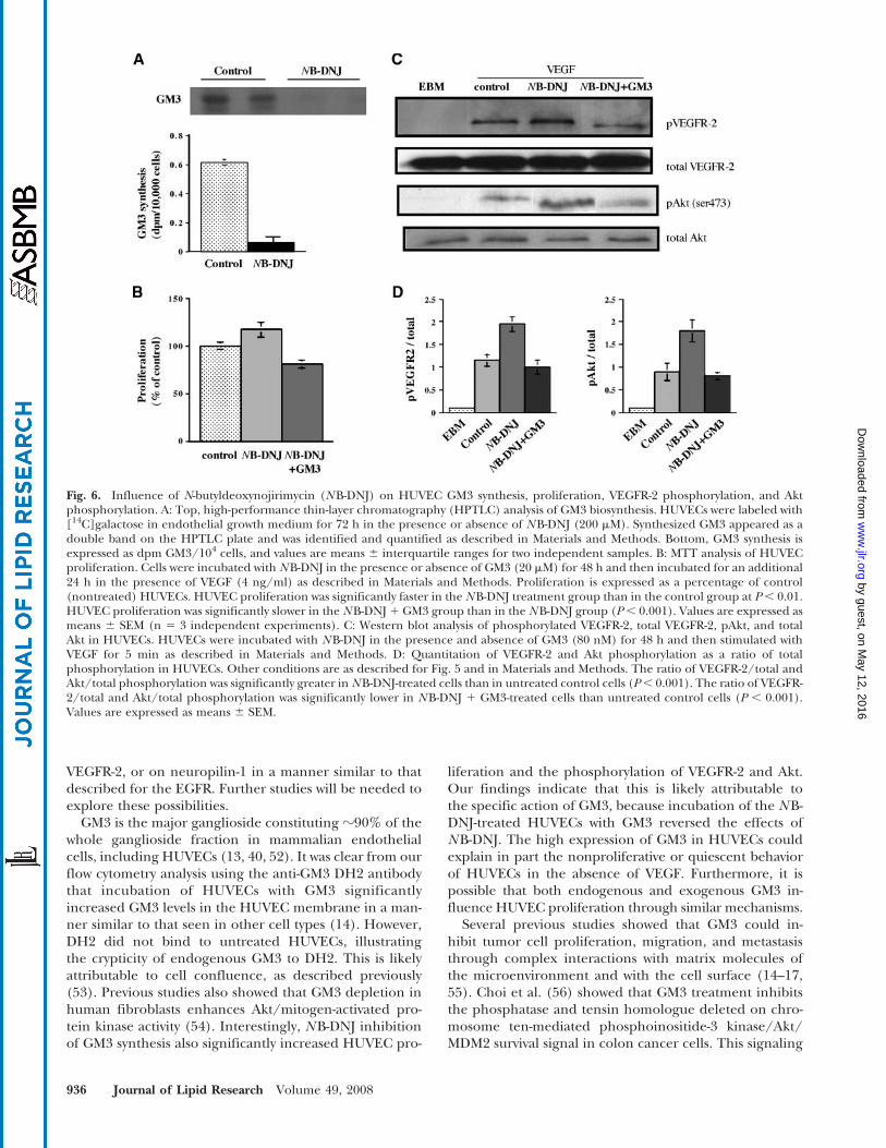
Influence of NB-DNJ on GM3 synthesis, HUVECproliferation, and VEGFR-2 and Akt phosphorylation
NB-DNJ is a nontoxic competitive inhibitor of theceramide-specific glucosyltransferase that catalyzes thefirst step in ganglioside biosynthesis (23). Treatment ofHUVECs with NB-DNJ (200 mM) reduced GM3 synthesis,as revealed by the incorporation of 14C-labeled galactoseinto newly synthesized GM3 (Fig. 6A). The gangliosidepattern of HUVECs was similar to that described previ-ously with GM3 as the predominant species (13, 40). NB-DNJ treatment significantly increased VEGF-inducedproliferation over untreated control cells (Fig. 6B). In par-allel, NB-DNJ significantly increased the VEGFR-2 andAkt phosphorylation (Fig. 6C, D). Because NB-DNJ re-duces not only GM3 synthesis but also that of most otherglycosphingolipids in HUVECs, we evaluated the effectsof the exogenous addition of GM3 in NB-DNJ-treatedcells. We found that GM3 addition reversed the NB-DNJ-induced increases in HUVEC proliferation and VEGFR-2and Akt phosphorylation (Fig. 6B–D). These findings fur-
Fig. 3. GM3 inhibits VEGF-induced HUVEC migration. A: Calcein-labeled HUVECs were photographed under fluorescence microscopy(2003). B: The amount of fluorescence incorporated was measured. The HUVECs were either untreated or treated with GM3 (20 mM) andwere seeded in EBM onto the upper chamber of the fibronectin-coated insert. VEGF (100 ng) in EBM was placed in the lower chamber.After 24 h, HUVECs that migrated through the filter were stained with calcein. VEGF significantly increased HUVEC migration comparedwith VEGF-untreated control (C) cells at P, 0.01. GM3 significantly reduced VEGF-induced migration at P, 0.01. Values are expressed asmeans 6 SEM (n 5 3 independent experiments, three wells per group per experiment).
Antiangiogenic effects of GM3 933
by guest, on May 12, 2016
ww
w.jlr.org
Dow
nloaded from

ther support the specific role of GM3 in HUVEC prolifer-ation and signaling.
DISCUSSION
The objective of this study was to revisit the role of gan-glioside GM3 in angiogenesis in relationship to HUVECproliferation and migration in vitro and to blood vesselformation in vivo. Gullino and coworkers (5, 6, 30, 41) firstshowed that changes in the relative concentrations ofGM3 to complex gangliosides could stimulate or suppressangiogenesis in vitro or in vivo. Using the rabbit corneamodel of angiogenesis, they found that GM3 reduced thegrowth and motility of microvascular endothelium whilerepressing the proangiogenic effects of prostaglandinE1 and bFGF (6, 30, 31). Moreover, ganglioside GD3 andother complex gangliosides (GM1 and GD1a) reduced theantiangiogenic effects of GM3. No further reports haveappeared addressing the mechanism by which GM3 mightinhibit angiogenesis.
We previously showed that gene-linked changes inthe distribution of GM3 to GD1a significantly influenced
tumor growth and angiogenesis in mouse brain tumors(8, 12). Specifically, reductions in the GM3/GD1a ratioenhanced angiogenesis in an experimental ependymo-blastoma, whereas increases in the ratio decreasedangiogenesis in the highly vascularized CT-2A astro-cytoma. Zeng et al. (4) reported similar findings in F-11neuroblastoma tumors with respect to the ratio of GM3 toGD3. It was not clear from these studies, however, whetherit was the increase of GM3 or the reduction of GD1a orGD3 that altered angiogenesis in these tumors. We nowshow for the first time that GM3 binds to culturedHUVECs and reduces the phosphorylation of VEGFR-2and Akt in the downstream signaling pathway, suggestingthat GM3 by itself can suppress angiogenesis. These find-ings provide insight on the mechanism by which GM3,shed from tumor cells into the microenvironment, sup-presses angiogenesis.
In contrast to the few reports on the antiangiogenic roleof GM3, several reports have described the proangiogeniceffects of complex gangliosides (4, 6, 12, 29, 42). Ladischand coworkers (13, 42) recently found that GD1a andother complex gangliosides (GM1 and GD3) could sen-sitize fibroblasts or HUVECs to low concentrations of pro-
Fig. 4. GM3 inhibits the proangiogenic effects of GD1a in the in vivo Matrigel plug assay. Matrigel alone (control) or containing GD1a orGD1a with GM3 was injected subcutaneously in SCID mice as described in Materials and Methods. A: Plugs were photographed (12.53) onday 7 after Matrigel injection to evaluate blood vessels. B: The plug vasculature was also evaluated under fluorescence microscopy (2003) inmice injected intravenously at 30 min before necropsy with FITC-labeled Isolectin B4. C: The amount of fluorescence in the plugs wasmeasured by fluorimetry as described in Materials and Methods. Six mice per group were evaluated, and the values are expressed as means6 SEM. Vascularization as detected by fluorescence was significantly less in the plugs with GD1a and GM3 than in the plugs with only GD1aat P , 0.01. AU, arbitrary units.
934 Journal of Lipid Research Volume 49, 2008
by guest, on May 12, 2016
ww
w.jlr.org
Dow
nloaded from

angiogenic growth factors. GM3, in contrast to the morecomplex gangliosides, did not sensitize VEGFR-2 phos-phorylation (13). The effects of gangliosides on growthfactor-induced angiogenesis are dependent on the type ofmicroenvironment (in vitro or in vivo) and on the con-centrations of both gangliosides and growth factors in themicroenvironment (1, 13, 30, 31, 34, 43). In the presentstudy, we found that 20–40 mM GM3 was effective at sup-pressing angiogenesis in the in vivo Matrigel assay andin the functional migration assay. Furthermore, GM3 waseffective at suppressing HUVEC proliferation at bothhigher (20 mM) and lower (80 nM) concentrations, but itwas most effective at suppressing VEGFR-2 phosphoryla-tion and Akt signaling at the lower concentration. Ourresults also support the findings of Ziche et al. (31) in thecorneal angiogenesis assay, showing that a GM3/growthfactor concentration ratio of 1:1 could arrest the pro-angiogenic effects of angiogenesis promoters. Numerousfactors can influence the incorporation and the rate oftransfer of exogenously added gangliosides into cell mem-branes. Under some conditions, gangliosides added atlower concentrations could be more effective at enteringmembranes than when added at higher concentrations,as a result of micelle formation at higher concentrations(44). Collectively, these findings indicate that the influ-
ence of gangliosides on angiogenesis and cell signalingevents is dependent to a large extent on the type of micro-environment and on the concentrations of gangliosidesand growth factors in the microenvironment.
In response to VEGF, VEGFR-2 undergoes dimeriza-tion and tyrosine phosphorylation, which alter endothelialcell proliferation, chemotaxis, and survival (45). The lo-calization of VEGFR-2 in HUVEC caveolae is involved inVEGF-induced downstream phosphorylation events (46).Receptors for epidermal growth factor and platelet-derived growth factor are also associated with “caveolarmembranes” or glycolipid-enriched microdomains (47–49). GM3 modulates EGFR function in epithelial cellsthrough carbohydrate-carbohydrate interaction with N-acetylglucosamine terminal residues of N-linked glycanslocated on the EGFR itself (34). It is also interesting thatheparan sulfate proteoglycans, which contain N -linkedoligosaccharides, can also enhance the response of endo-thelial VEGFR-2 to the VEGF165 isoform (50). Althoughlittle is known about the glycosylation of VEGFR-2, theVEGFR-2 coreceptor, neuropilin-1, is glycosylated (51).Moreover, GM3 reduces neuropilin-1 expression in CT-2Aastrocytoma cells (8). It is possible, therefore, that GM3could influence angiogenesis through interactions withcarbohydrate residues on specific proteoglycans, on the
Fig. 5. GM3 inhibits vascular endothelial growth factor receptor 2 (VEGFR-2) and Akt phosphorylationin HUVECs. HUVECs were incubated with GM3 (80 nM) in EBM for 24 h and then stimulated with VEGF(100 ng/ml) for 5 min as described in Materials and Methods. Cell lysates were prepared and phos-phorylation was measured. A: Detection of phosphorylated VEGFR-2, total VEGFR-2, pAkt, and total Akt byWestern blot. B: Quantitation of VEGFR-2 and Akt phosphorylation over total. VEGFR-2 and Akt phos-phorylation was significantly lower in GM3-treated HUVECs than in control HUVECs (P, 0.001). Values areexpressed as means 6 SEM (n 5 3 independent experiments).
Antiangiogenic effects of GM3 935
by guest, on May 12, 2016
ww
w.jlr.org
Dow
nloaded from
VEGFR-2, or on neuropilin-1 in a manner similar to thatdescribed for the EGFR. Further studies will be needed toexplore these possibilities.
GM3 is the major ganglioside constituting ?90% of thewhole ganglioside fraction in mammalian endothelialcells, including HUVECs (13, 40, 52). It was clear from ourflow cytometry analysis using the anti-GM3 DH2 antibodythat incubation of HUVECs with GM3 significantlyincreased GM3 levels in the HUVEC membrane in a man-ner similar to that seen in other cell types (14). However,DH2 did not bind to untreated HUVECs, illustratingthe crypticity of endogenous GM3 to DH2. This is likelyattributable to cell confluence, as described previously(53). Previous studies also showed that GM3 depletion inhuman fibroblasts enhances Akt/mitogen-activated pro-tein kinase activity (54). Interestingly, NB-DNJ inhibitionof GM3 synthesis also significantly increased HUVEC pro-
liferation and the phosphorylation of VEGFR-2 and Akt.Our findings indicate that this is likely attributable tothe specific action of GM3, because incubation of the NB-DNJ-treated HUVECs with GM3 reversed the effects ofNB-DNJ. The high expression of GM3 in HUVECs couldexplain in part the nonproliferative or quiescent behaviorof HUVECs in the absence of VEGF. Furthermore, it ispossible that both endogenous and exogenous GM3 in-fluence HUVEC proliferation through similar mechanisms.
Several previous studies showed that GM3 could in-hibit tumor cell proliferation, migration, and metastasisthrough complex interactions with matrix molecules ofthe microenvironment and with the cell surface (14–17,55). Choi et al. (56) showed that GM3 treatment inhibitsthe phosphatase and tensin homologue deleted on chro-mosome ten-mediated phosphoinositide-3 kinase/Akt/MDM2 survival signal in colon cancer cells. This signaling
Fig. 6. Influence of N-butyldeoxynojirimycin (NB-DNJ) on HUVEC GM3 synthesis, proliferation, VEGFR-2 phosphorylation, and Aktphosphorylation. A: Top, high-performance thin-layer chromatography (HPTLC) analysis of GM3 biosynthesis. HUVECs were labeled with[14C]galactose in endothelial growth medium for 72 h in the presence or absence of NB-DNJ (200 mM). Synthesized GM3 appeared as adouble band on the HPTLC plate and was identified and quantified as described in Materials and Methods. Bottom, GM3 synthesis isexpressed as dpm GM3/104 cells, and values are means 6 interquartile ranges for two independent samples. B: MTT analysis of HUVECproliferation. Cells were incubated with NB-DNJ in the presence or absence of GM3 (20 mM) for 48 h and then incubated for an additional24 h in the presence of VEGF (4 ng/ml) as described in Materials and Methods. Proliferation is expressed as a percentage of control(nontreated) HUVECs. HUVEC proliferation was significantly faster in the NB-DNJ treatment group than in the control group at P, 0.01.HUVEC proliferation was significantly slower in the NB-DNJ 1 GM3 group than in the NB-DNJ group (P, 0.001). Values are expressed asmeans 6 SEM (n 5 3 independent experiments). C: Western blot analysis of phosphorylated VEGFR-2, total VEGFR-2, pAkt, and totalAkt in HUVECs. HUVECs were incubated with NB-DNJ in the presence and absence of GM3 (80 nM) for 48 h and then stimulated withVEGF for 5 min as described in Materials and Methods. D: Quantitation of VEGFR-2 and Akt phosphorylation as a ratio of totalphosphorylation in HUVECs. Other conditions are as described for Fig. 5 and in Materials and Methods. The ratio of VEGFR-2/total andAkt/total phosphorylation was significantly greater in NB-DNJ-treated cells than in untreated control cells (P, 0.001). The ratio of VEGFR-2/total and Akt/total phosphorylation was significantly lower in NB-DNJ 1 GM3-treated cells than untreated control cells (P , 0.001).Values are expressed as means 6 SEM.
936 Journal of Lipid Research Volume 49, 2008
by guest, on May 12, 2016
ww
w.jlr.org
Dow
nloaded from

pathway is also considered a target for the control ofbrain tumor angiogenesis (57). Together, these findingsare consistent with our observations that GM3 inhibitsendothelial cell migration and proliferation in vitro andin vivo through growth factor receptor inactivation andinhibition of Akt signaling. We suggest that GM3 mayhave broad therapeutic potential for targeting cell-signaling events in both tumor cells and tumor-associatedhost cells in relationship with tumor progression, metas-tasis, and angiogenesis. Preclinical trials are warranted toassess the therapeutic potential of GM3 as an antiangio-genic agent.
The authors thank Hong-Wei Bai, Daniel Kirschner, WeihuaZhou, and Jeremy Marsh for technical assistance. This re-search was supported by National Institutes of Health GrantsNS-055195 and CA-102135 and by the Boston College Re-search Expense Fund.
REFERENCES
1. Alessandri, G., S. Filippeschi, P. Sinibaldi, F. Mornet, P. Passera,F. Spreafico, P. M. Cappa, and P. M. Gullino. 1987. Influenceof gangliosides on primary and metastatic neoplastic growth inhuman and murine cells. Cancer Res. 47: 4243–4247.
2. Bai, H., and T. N. Seyfried. 1997. Influence of ganglioside GM3and high density lipoprotein (HDL) on the cohesion of mousebrain tumor cells. J. Lipid Res. 38: 160–172.
3. Hakomori, S. 1996. Tumor malignancy defined by aberrant gly-cosylation and sphingo (glyco) lipid metabolism. Cancer Res. 56:5309–5318.
4. Zeng, G., L. Gao, S. Birkle, and R. K. Yu. 2000. Suppression ofganglioside GD3 expression in a rat F-11 tumor cell line reducestumor growth, angiogenesis, and vascular endothelial growth factorproduction. Cancer Res. 60: 6670–6676.
5. Alessandri, G., P. Cornaglia Ferraris, and P. M. Gullino. 1997.Angiogenic and angiostatic microenvironment in tumors—role ofgangliosides. Acta Oncol. 36: 383–387.
6. Alessandri, G., G. De Cristan, M. Ziche, A. P. Cappa, and P. M.Gullino. 1992. Growth and motility of microvascular endotheliumare modulated by the relative concentration of gangliosides in themedium. J. Cell. Physiol. 151: 23–28.
7. Valentino, L. A., and S. Ladisch. 1992. Localization of shed humantumor gangliosides: association with serum lipoproteins. Cancer Res.52: 810–814.
8. Abate, L. E., P. Mukherjee, and T. N. Seyfried. 2006. Gene-linkedshift in ganglioside distribution influences growth and vascularityin a mouse astrocytoma. J. Neurochem. 98: 1973–1984.
9. Olshefski, R., and S. Ladisch. 1996. Intercellular transfer of shedtumor cell gangliosides. FEBS Lett. 386: 11–14.
10. Ladisch, S., B. Gillard, C. Wong, and L. Ulsh. 1983. Shedding andimmunoregulatory activity of YAC-1 lymphoma cell gangliosides.Cancer Res. 43: 3808–3813.
11. Ladisch, S., S. Kitada, and E. F. Hays. 1987. Gangliosides shed bytumor cells enhance tumor formation in mice. J. Clin. Invest. 79:1879–1882.
12. Manfredi, M. G., S. Lim, K. P. Claffey, and T. N. Seyfried. 1999.Gangliosides influence angiogenesis in an experimental mousebrain tumor. Cancer Res. 59: 5392–5397.
13. Liu, Y., J. McCarthy, and S. Ladisch. 2006. Membrane gangliosideenrichment lowers the threshold for vascular endothelial cellangiogenic signaling. Cancer Res. 66: 10408–10414.
14. Mitsuzuka, K., K. Handa, M. Satoh, Y. Arai, and S. Hakomori. 2005.A specific microdomain (“glycosynapse 3”) controls phenotypicconversion and reversion of bladder cancer cells through GM3-mediated interaction of alpha3beta1 integrin with CD9. J. Biol.Chem. 280: 35545–35553.
15. Fujimoto, Y., S. Izumoto, T. Suzuki, M. Kinoshita, N. Kagawa, K.Wada, N. Hashimoto, M. Maruno, Y. Nakatsuji, and T. Yoshimine.
2005. Ganglioside GM3 inhibits proliferation and invasion ofglioma. J. Neurooncol. 71: 99–106.
16. Bassi, R., P. Viani, P. Giussani, L. Riboni, and G. Tettamanti. 2001.GM3 ganglioside inhibits endothelin-1-mediated signal transduc-tion in C6 glioma cells. FEBS Lett. 507: 101–104.
17. Noll, E. N., J. Lin, Y. Nakatsuji, R. H. Miller, and P. M. Black. 2001.GM3 as a novel growth regulator for human gliomas. Exp. Neurol.168: 300–309.
18. De Cristan, G., L. Morbidelli, G. Alessandri, M. Ziche, A. P. Cappa,and P. M. Gullino. 1990. Synergism between gangliosides andbasic fibroblastic growth factor in favouring survival, growth, andmotility of capillary endothelium. J. Cell. Physiol. 144: 505–510.
19. Ravindranath, M. H., T. Tsuchida, D. L. Morton, and R. F. Irie.1991. Ganglioside GM3:GD3 ratio as an index for the managementof melanoma. Cancer. 67: 3029–3035.
20. Shochat, S. J., N. L. Corbelletta, M. A. Repman, and C. L.Schengrund. 1987. A biochemical analysis of thoracic neuroblasto-mas: a Pediatric Oncology Group study. J. Pediatr. Surg. 22: 660–664.
21. Chu, S. H., Y. B. Ma, H. Zhang, D. F. Feng, Z. A. Zhu, Z. Q. Li, andX. H. Yuan. 2007. Hepatocyte growth factor production is stim-ulated by gangliosides and TGF-beta isoforms in human gliomacells. J. Neurooncol. 85: 33–38.
22. Koochekpour, S., A. Merzak, and G. J. Pilkington. 1996. Vascularendothelial growth factor production is stimulated by gangliosidesand TGF-beta isoforms in human glioma cells in vitro. Cancer Lett.102: 209–215.
23. Ranes, M. K., M. El-Abbadi, M. G. Manfredi, P. Mukherjee,F. M. Platt, and T. N. Seyfried. 2001. N-Butyldeoxynojirimycin re-duces growth and ganglioside content of experimental mousebrain tumours. Br. J. Cancer. 84: 1107–1114.
24. Weiss, M., S. Hettmer, P. Smith, and S. Ladisch. 2003. Inhibition ofmelanoma tumor growth by a novel inhibitor of glucosylceramidesynthase. Cancer Res. 63: 3654–3658.
25. Deng, W., R. Li, and S. Ladisch. 2000. Influence of cellularganglioside depletion on tumor formation. J. Natl. Cancer Inst. 92:912–917.
26. Folkman, J. 1992. The role of angiogenesis in tumor growth. Semin.Cancer Biol. 3: 65–71.
27. Claffey, K. P., K. Abrams, S. C. Shih, L. F. Brown, A. Mullen, andM. Keough. 2001. Fibroblast growth factor 2 activation of stromalcell vascular endothelial growth factor expression and angiogen-esis. Lab. Invest. 81: 61–75.
28. Ferrara, N., and T. Davis Smyth. 1997. The biology of vascularendothelial growth factor. Endocr. Rev. 18: 4–25.
29. Lang, Z., M. Guerrera, R. Li, and S. Ladisch. 2001. GangliosideGD1a enhances VEGF-induced endothelial cell proliferation andmigration. Biochem. Biophys. Res. Commun. 282: 1031–1037.
30. Ziche, M., G. Alessandri, and P. M. Gullino. 1989. Gangliosidespromote the angiogenic response. Lab. Invest. 61: 629–634.
31. Ziche, M., L. Morbidelli, G. Alessandri, and P. M. Gullino. 1992.Angiogenesis can be stimulated or repressed in vivo by a change inGM3:GD3 ganglioside ratio. Lab. Invest. 67: 711–715.
32. Wang, X. Q., P. Sun, and A. S. Paller. 2003. Ganglioside GM3 blocksthe activation of epidermal growth factor receptor induced byintegrin at specific tyrosine sites. J. Biol. Chem. 278: 48770–48778.
33. Bremer, E. G., J. Schlessinger, and S. Hakomori. 1986. Ganglioside-mediated modulation of cell growth. J. Biol. Chem. 261: 2434–2440.
34. Yoon, S. J., K. Nakayama, T. Hikita, K. Handa, and S. I. Hakomori.2006. Epidermal growth factor receptor tyrosine kinase is modu-lated by GM3 interaction with N-linked GlcNAc termini of thereceptor. Proc. Natl. Acad. Sci. USA. 103: 18987–18991.
35. Veikkola, T., M. Karkkainen, L. Claesson-Welsh, and K. Alitalo.2000. Regulation of angiogenesis via vascular endothelial growthfactor receptors. Cancer Res. 60: 203–212.
36. Gille, H., J. Kowalski, B. Li, J. LeCouter, B. Moffat, T. F. Zioncheck,N. Pelletier, and N. Ferrara. 2001. Analysis of biological effects andsignaling properties of Flt-1 (VEGFR-1) and KDR (VEGFR-2). Areassessment using novel receptor-specific vascular endothelialgrowth factor mutants. J. Biol. Chem. 276: 3222–3230.
37. Mukherjee, P., M. M. El-Abbadi, J. L. Kasperzyk, M. K. Ranes, andT. N. Seyfried. 2002. Dietary restriction reduces angiogenesis andgrowth in an orthotopic mouse brain tumour model. Br. J. Cancer.86: 1615–1621.
38. Wakasugi, K., B. M. Slike, J. Hood, A. Otani, K. L. Ewalt,M. Friedlander, D. A. Cheresh, and P. Schimmel. 2002. A humanaminoacyl-tRNA synthetase as a regulator of angiogenesis. Proc.Natl. Acad. Sci. USA. 99: 173–177.
Antiangiogenic effects of GM3 937
by guest, on May 12, 2016
ww
w.jlr.org
Dow
nloaded from
39. Seyfried, T. N., A. M. Novikov, R. A. Irvine, and J. V. Brigande. 1994.Ganglioside biosynthesis in mouse embryos: sialyltransferase IVand the asialo pathway. J. Lipid Res. 35: 993–1001.
40. Muthing, J., S. Duvar, D. Heitmann, F. G. Hanisch, U. Neumann,G. Lochnit, R. Geyer, and J. Peter-Katalinic. 1999. Isolation andstructural characterization of glycosphingolipids of in vitro prop-agated human umbilical vein endothelial cells. Glycobiology. 9:459–468.
41. Gullino, P. M., M. Ziche, and G. Alessandri. 1990. Gangliosides,copper ions and angiogenic capacity of adult tissues. CancerMetastasis Rev. 9: 239–251.
42. Liu, Y., R. Li, and S. Ladisch. 2004. Exogenous ganglioside GD1aenhances epidermal growth factor receptor binding and dimeriza-tion. J. Biol. Chem. 279: 36481–36489.
43. Slevin, M., S. Kumar, X. He, and J. Gaffney. 1999. Physiologicalconcentrations of gangliosides GM1, GM2 and GM3 differentiallymodify basic-fibroblast-growth-factor-induced mitogenesis and theassociated signalling pathway in endothelial cells. Int. J. Cancer. 82:412–423.
44. Lauc, G., and M. Heffer-Lauc. 2006. Shedding and uptake ofgangliosides and glycosylphosphatidylinositol-anchored proteins.Biochim. Biophys. Acta. 1760: 584–602.
45. Ferrara, N., H. P. Gerber, and J. LeCouter. 2003. The biology ofVEGF and its receptors. Nat. Med. 9: 669–676.
46. Cho, C. H., C. S. Lee, M. Chang, I. H. Jang, S. J. Kim, I. Hwang,S. H. Ryu, C. O. Lee, and G. Y. Koh. 2004. Localization of VEGFR-2and PLD2 in endothelial caveolae is involved in VEGF-inducedphosphorylation of MEK and ERK. Am. J. Physiol. Heart Circ. Physiol.286: H1881–H1888.
47. Li, R., J. Manela, Y. Kong, and S. Ladisch. 2000. Cellular gan-gliosides promote growth factor-induced proliferation of fibro-blasts. J. Biol. Chem. 275: 34213–34223.
48. Hakomori, S. I. 2002. Inaugural article. The glycosynapse. Proc.Natl. Acad. Sci. USA. 99: 225–232.
49. Mineo, C., G. L. James, E. J. Smart, and R. G. Anderson. 1996.
Localization of epidermal growth factor-stimulated Ras/Raf-1interaction to caveolae membrane. J. Biol. Chem. 271: 11930–11935.
50. Kaji, T., C. Yamamoto, M. Oh-i, Y. Fujiwara, Y. Yamazaki, T. Morita,A. H. Plaas, and T. N. Wight. 2006. The vascular endothelial growthfactor VEGF165 induces perlecan synthesis via VEGF receptor-2in cultured human brain microvascular endothelial cells. Biochim.Biophys. Acta. 1760: 1465–1474.
51. Shintani, Y., S. Takashima, Y. Asano, H. Kato, Y. Liao, S. Yamazaki,O. Tsukamoto, O. Seguchi, H. Yamamoto, T. Fukushima, et al.2006. Glycosaminoglycan modification of neuropilin-1 modulatesVEGFR2 signaling. EMBO J. 25: 3045–3055.
52. Kanda, T., T. Ariga, M. Yamawaki, H. Yoshino, X. B. Gu, andR. K. Yu. 1997. Glycosyltransferase activities in cultured endothe-lial cells of bovine brain microvascular origin. Neurochem. Res. 22:463–466.
53. Tatewaki, K., T. Yamaki, Y. Maeda, H. Tobioka, H. Piao, H. Yu,Y. Ibayashi, N. Sawada, and K. Hashi. 1997. Cell density regulatescrypticity of GM3 ganglioside on human glioma cells. Exp. Cell Res.233: 145–154.
54. Toledo, M. S., E. Suzuki, K. Handa, and S. Hakomori. 2005. Effectof ganglioside and tetraspanins in microdomains on interaction ofintegrins with fibroblast growth factor receptor. J. Biol. Chem. 280:16227–16234.
55. Gu, J., A. Fujibayashi, K. M. Yamada, and K. Sekiguchi. 2002.Laminin-10/11 and fibronectin differentially prevent apoptosis in-duced by serum removal via phosphatidylinositol 3-kinase/Akt- andMEK1/ERK-dependent pathways. J. Biol. Chem. 277: 19922–19928.
56. Choi, H. J., T. W. Chung, S. K. Kang, Y. C. Lee, J. H. Ko, J. G. Kim,and C. H. Kim. 2006. Ganglioside GM3 modulates tumor suppres-sor PTEN-mediated cell cycle progression—transcriptional in-duction of p21(WAF1) and p27(kip1) by inhibition of PI-3K/AKT pathway. Glycobiology. 16: 573–583.
57. Castellino, R. C., and D. L. Durden. 2007. Mechanisms of disease:the PI3K-Akt-PTEN signaling node–an intercept point for the con-trol of angiogenesis in brain tumors. Nat. Clin. Pract. 3: 682–693.
938 Journal of Lipid Research Volume 49, 2008
by guest, on May 12, 2016
ww
w.jlr.org
Dow
nloaded from
Related Documents