Pathology of interstitial lung disease Kevin O. Leslie, MD a,b, * a Department of Pathology, Mayo Clinic College of Medicine, Rochester, MN, USA b Department of Laboratory Medicine and Pathology, Mayo Clinic Scottsdale, 13400 East Shea Boulevard, Scottsdale, AZ 85259, USA A large and diverse group of pathologic conditions manifests clinically and radiologically as diffuse parenchymal lung disease. In practice, this group of disorders has been categorized on the basis of clinical dysfunction (‘‘restrictive lung disease’’) or radiologic appearance (‘‘interstitial lung disease [ILD]’’), neither of which accurately reflects the pathologic processes involved [1]. Diffuse ILDs encompass mainly inflam- matory processes that involve the structural elements of this organ. Some ILDs are caused by infections, but most are the result of immunologic, environmental, or toxic mechanisms. These diseases are discussed together because they have in common the tendency to produce bilateral abnormalities on chest imaging studies and are mainly nonneoplastic conditions [2]. Currently, less morbid sampling techniques have increased dramatically the probability that pulmo- nologists and their general pathology colleagues will be faced with establishing a specific and clinically relevant diagnosis using surgical lung biopsy material. Most of the concepts presented in this article have been established using this type of specimen. In the early years of surgical lung biopsy, a small number of diffuse inflammatory conditions came to light that exclusively involved the lungs and did not seem to be caused by infection, toxin, sarcoidosis, pneumoconiosis, or neoplasm. Liebow is credited with recognizing these conditions and devising a classification system for them. These disorders came to be known as the idiopathic interstitial pneumonias [3]. The original classification proposed by Liebow is presented for historical purposes in Box 1. Much has changed in medical science over the years, and none of the entities proposed in Liebow’s original classi- fication is viewed today exactly as he described them more than 30 years ago. A recent international consensus conference updated the classification of idiopathic interstitial pneumonias (Box 2) [4]. In this article, these ‘‘idiopathic’’ disorders are discussed in the context of their dominant pathologic findings rather than presented as a separate group of entities (as has been traditional in past). A comparison of the pathologic manifestations of the idiopathic ILDs is presented in Table 1. Interpretation of lung biopsies in a patient with ILD is best accomplished using a multidisciplinary approach that results in a composite clinico-radio- logic-pathologic diagnosis. Unfortunately, this is not always realistic in many clinical practice settings. For diffuse lung diseases, a pathologist must have some essential information regarding the clinical and radiologic findings to arrive at a clinically meaningful diagnosis. In many instances, more extensive clinical and radiologic consultation may be necessary. The pulmonologist who is conversant with the pathology of ILD is a powerful ally in this process. Pattern analysis approach to surgical lung biopsies The concept of ‘‘losing the forest for the trees’’ becomes evident in the evaluation of lung wedge biopsies. The age-old training method of requiring 0272-5231/04/$ – see front matter D 2004 Elsevier Inc. All rights reserved. doi:10.1016/j.ccm.2004.05.002 * Department of Laboratory Medicine and Pathology, Mayo Clinic Scottsdale, 13400 East Shea Boulevard, Scottsdale, AZ 85259. E-mail address: [email protected] Clin Chest Med 25 (2004) 657 – 703

Welcome message from author
This document is posted to help you gain knowledge. Please leave a comment to let me know what you think about it! Share it to your friends and learn new things together.
Transcript

Clin Chest Med
Pathology of interstitial lung disease
Kevin O Leslie MDab
aDepartment of Pathology Mayo Clinic College of Medicine Rochester MN USAbDepartment of Laboratory Medicine and Pathology Mayo Clinic Scottsdale 13400 East Shea Boulevard
Scottsdale AZ 85259 USA
A large and diverse group of pathologic conditions
manifests clinically and radiologically as diffuse
parenchymal lung disease In practice this group of
disorders has been categorized on the basis of clinical
dysfunction (lsquolsquorestrictive lung diseasersquorsquo) or radiologic
appearance (lsquolsquointerstitial lung disease [ILD]rsquorsquo) neither
of which accurately reflects the pathologic processes
involved [1] Diffuse ILDs encompass mainly inflam-
matory processes that involve the structural elements
of this organ Some ILDs are caused by infections but
most are the result of immunologic environmental or
toxic mechanisms These diseases are discussed
together because they have in common the tendency
to produce bilateral abnormalities on chest imaging
studies and are mainly nonneoplastic conditions [2]
Currently less morbid sampling techniques have
increased dramatically the probability that pulmo-
nologists and their general pathology colleagues will
be faced with establishing a specific and clinically
relevant diagnosis using surgical lung biopsy material
Most of the concepts presented in this article have
been established using this type of specimen
In the early years of surgical lung biopsy a small
number of diffuse inflammatory conditions came to
light that exclusively involved the lungs and did not
seem to be caused by infection toxin sarcoidosis
pneumoconiosis or neoplasm Liebow is credited
with recognizing these conditions and devising a
classification system for them These disorders came
0272-523104$ ndash see front matter D 2004 Elsevier Inc All rights
doi101016jccm200405002
Department of Laboratory Medicine and Pathology
Mayo Clinic Scottsdale 13400 East Shea Boulevard
Scottsdale AZ 85259
E-mail address lesliekevinmayoedu
to be known as the idiopathic interstitial pneumonias
[3] The original classification proposed by Liebow is
presented for historical purposes in Box 1 Much has
changed in medical science over the years and none
of the entities proposed in Liebowrsquos original classi-
fication is viewed today exactly as he described them
more than 30 years ago A recent international
consensus conference updated the classification of
idiopathic interstitial pneumonias (Box 2) [4] In this
article these lsquolsquoidiopathicrsquorsquo disorders are discussed in
the context of their dominant pathologic findings
rather than presented as a separate group of entities
(as has been traditional in past) A comparison of the
pathologic manifestations of the idiopathic ILDs is
presented in Table 1
Interpretation of lung biopsies in a patient with
ILD is best accomplished using a multidisciplinary
approach that results in a composite clinico-radio-
logic-pathologic diagnosis Unfortunately this is not
always realistic in many clinical practice settings For
diffuse lung diseases a pathologist must have some
essential information regarding the clinical and
radiologic findings to arrive at a clinically meaningful
diagnosis In many instances more extensive clinical
and radiologic consultation may be necessary The
pulmonologist who is conversant with the pathology
of ILD is a powerful ally in this process
Pattern analysis approach to surgical lung
biopsies
The concept of lsquolsquolosing the forest for the treesrsquorsquo
becomes evident in the evaluation of lung wedge
biopsies The age-old training method of requiring
25 (2004) 657 ndash 703
reserved
Box 1 Liebow classification of interstitialpneumonia (1975)
Usual interstitial pneumonia (UIP)Bronchiolitis obliterans with usual
interstitial pneumonia (BIP)Desquamative interstitial pneumonia
(DIP)Lymphoid interstitial pneumonia (LIP)Giant cell interstitial pneumonia (GIP)
Adapted from Liebow A Carrington CThe interstitial pneumonias In Simon MPotchen E LeMay M editors Frontiers ofpulmonary radiology pathophysiologicroentgenographic and radioisotopic con-siderations Orlando Grune amp Stratton1969 p 109ndash42
Box 2 International ConsensusCommittee classification of idiopathicinterstitial pneumonia (2002)
Acute interstitial pneumoniaDIPrespiratory bronchiolitisndashasso-
ciated interstitial disease (RB-ILD)Cryptogenic organizing pneumonia
(COP)Nonspecific interstitial pneumonia
fibrosis (NSIPF)a
LIP
a ProvisionalAdapted from Travis W King T Bate-man E Lynch DA Capron F Colby TVet al ATSERS international multidisci-plinary consensus classification of theidiopathic interstitial pneumonias Am JRespir Crit Care Med 2002165(2)277ndash304
KO Leslie Clin Chest Med 25 (2004) 657ndash703658
that the microscope slide be evaluated first by the
naked eye may seem overly methodical but it does
force the interpreter to see the lsquolsquobig picturersquorsquo before
getting lost in the fine details For nonneoplastic lung
diseases the scanning low power objective (2 or
4) is useful if not essential because different
diseases give rise to different architectural patterns
which may immediately raise a narrow differential
diagnosis For diffuse lung diseases several helpful
patterns emerge
Pattern 1 acute lung injury
The prototype of this pattern is diffuse alveolar
damage (DAD) with hyaline membranes classically
encountered in the clinical setting of adult respiratory
distress syndrome (ARDS) (Fig 1)
Pattern 2 fibrosis
Lung diseases that lead to the accrual of collagen
in the lung with permanent structural remodeling
are represented by this pattern (Fig 2) Idiopathic
pulmonary fibrosis (IPF) (pathologic usual intersti-
tial pneumonia [UIP]) is the prototype and is often
the diagnosis of greatest clinical concern in older
adult patients because of the dismal prognosis of
this condition
Pattern 3 cellular interstitial infiltrates
Lymphocytes plasma cells and macrophages
are present in the alveolar walls in Pattern 3 (Fig 3)
Hypersensitivity pneumonitis (extrinsic allergic al-
veolitis) is the prototype of this pattern
Pattern 4 airspace filling
This pattern is characterized by the presence of
cells or other material filling the alveolar spaces
(Fig 4) Organizing pneumonia is the prototype of
this pattern The airspace filling pattern also includes
infectious bronchopneumonias (neutrophils in the al-
veoli) classic Pneumocystis infection in the immu-
nocompromised host (foamy casts in alveoli)
pulmonary alveolar proteinosis (PAP) (proteinaceous
material in alveoli) diffuse pulmonary hemorrhage
(blood siderophages and patchy organizing pneumo-
nia in alveoli) and DIP in which lightly pigmented
lsquolsquosmokersrsquorsquo-type macrophages are the dominant intra-
alveolar element
Pattern 5 nodules
The presence of discrete nodules (Fig 5) in the
lung parenchyma raises a differential diagnosis that
includes nodular infections benign and malignant
neoplasms sarcoidosis Langerhansrsquo cell histiocyto-
sis and various bronchiolocentric diseases The
prototype is Wegenerrsquos granulomatosis (large nodular
pattern) but small (miliary) patterns of disease also
are included
Table 1
Contrasting pathologic features of idiopathic interstitial pneumonias
Features NSIP UIP DIP AIP LIP COP
Temporal appearance Uniform Variegated Uniform Uniform Uniform Uniform
Interstitial inflammation Prominent Scant Scant Scant Prominent Scant
Interstitial fibrosis (collagen) Variable diffuse Patchy Variable
diffuse
No Some cases No
Interstitial fibrosis (fibroblasts) Occasional diffuse No No Yes diffuse No No
Organizing pneumonia pattern Occasional focal Occasional
focal
No Occasional
focal
No Prominent
Fibroblast foci Occasional focal Typical No No No No
Honeycomb areas Rare Yes No No Sometimes No
Intra-alveolar macrophages Occasional patchy Occasional
focal
Yes
diffuse
No Occasional
patchy
No
Hyaline membranes No No No Yes focal No No
Granulomas No No No No Focal poorly
formed
No
Abbreviation AIP acute interstitial pneumonia
Data from Katzenstein A Fiorelli R Nonspecific interstitial pneumoniafibrosis histologic features and clinical significance
Am J Surg Pathol 199418136ndash47 and Trans W Colby T Koss M Rosado-Christenson ML Muller NL King TE et al Non-
neoplastic disorders of the lower respiratory tract In King D editor Atlas of nontumor pathology Washington DC American
Registry of Pathology and the Armed Forces Institute of Pathology 2002 p 939
KO Leslie Clin Chest Med 25 (2004) 657ndash703 659
Pattern 6 near normal lung
The surgical lung biopsy that has barely discern-
ible abnormalities is often the result of diseases that
affect the airways and blood vessels of the lung The
changes may be subtle at low magnification The
prototype is small airways disease in which pruning
dilatation and generalized scarring of the small
airways occur and this may be difficult to appreciate
Fig 1 Pattern 1 acute lung injury DAD with hyaline
membranes classically encountered in the clinical setting of
ARDS is the prototype of the acute lung injury pattern
at scanning magnification Vascular diseases (eg pul-
monary hypertension) cystic diseases (eg lymphan-
gioleiomyomatosis [LAM]) and conditions with
patchy scarring also can produce subtle disease that
results in what seems to be lsquolsquonormalrsquorsquo lung from
scanning magnification (Fig 6)
Once the dominant pattern is determined addi-
tional microscopic findings help narrow the diagnos-
tic possibilities A list of these findings with their
Fig 2 Pattern 2 fibrosis Lung diseases that lead to the
accrual of collagen in the lung with permanent structural
remodeling are represented by this pattern IPF (pathologic
UIP) often is the diagnosis of greatest clinical concern
in older adult patients because of the dismal prognosis of
this condition
Fig 3 Pattern 3 cellular interstitial infiltrates Lymphocytes
plasma cells and macrophages are present in the alveolar
walls in Pattern 3 Hypersensitivity pneumonitis (extrinsic
allergic alveolitis) is the prototype of this pattern
Fig 5 Pattern 5 nodules The presence of discrete nod-
ules in the lung parenchyma raises a narrow differen-
tial diagnosis
KO Leslie Clin Chest Med 25 (2004) 657ndash703660
respective differential diagnosis is presented inTable 2
Overlap between patterns occurs and may be a use-
ful clue in the differential diagnosis For example
when nearly all of the six patterns are present in the
same biopsy specimen rheumatoid arthritis is often
the correct diagnosis Acute lung injury also proceeds
through several distinctive histopathologic patterns
during the repair phase after injury If a lung biopsy is
performed in the subacute phase of DAD airspace
Fig 4 Pattern 4 airspace filling The alveolar spaces are
filled with cells or other material Organizing pneumonia is
the prototype of this pattern
organization may dominate the picture and poten-
tially cause confusion with organizing pneumonia
Acute lung injury pattern (days to weeks in
evolution rapid onset of symptoms)
The pattern of acute lung injury is characterized
by variable interstitial and alveolar edema fibrin in
airspaces and reactive type-II cell hyperplasia (Fig 7)
Hyaline membranes neutrophils necrosis eosino-
Fig 6 Pattern 6 near normal lung The surgical lung biopsy
that has barely discernible abnormalities is often the result of
diseases that affect the airways and blood vessels of the lung
or produce cysts The changes may be subtle at low
magnification The prototype is small airways disease in
which pruning dilatation and generalized scarring of the
small airways occur and may be difficult to appreciate at
scanning magnification
Table 2
Pattern-based approach to interstitial lung diseases
Acute lung injury Fibrosis Cellular interstitial pneumonia Alveolar filling Nodular Minimal change
With hyaline membranes
Infection
CVD
With variable fibrosis
(normal to HC)
UIPIPF
With lymphs and plasma cells
C-NSIP CVD
HSP drug
With macrophages
Smoking-related
Local fibrosis
With lymphoid
Follicular bronch
Wegenerrsquos
With SAD
Constrictive bronchiolitis
Drug Asbestosis Infection Lymphoma
Idiopathic RA Lymphoma
Chronic HSP
With eosinophils With honeycombing only With neutrophils With neutrophils With necrosis With vascular
AEP Diffuse Infection Infection Infections pathology
Drug Late UIP CVD DPH Tumor PHT
DAD in smoker Focal Hemorrhage Wegenerrsquos VOD
Many causes
With necrosis With diffuse fibrosis With granulomas With OP With atypical cells With cysts
Infections
Viral
Bacterial
CVD
Drug
Sarcoid (with granulomas)
Infection HSP
sarcoidberylliosis
aspiration
With focal OP
Infection drug
CVD
With eosinophilic material
Infections Ca
Lymphomas
Sarcomas
PLCH
LAM
With no findings
Fungal PLCH (with stellate scars)
Infection
Infection CVD
Drug DPH
With stellate scars Sampling error
Pneumoconiosis
F-NSIP CVD CHF PAP
PLCH
With siderophages With pleuritis With pleuritis With hemorrhage With OP
DPH CVD CVD CVD Infections CVD
CVD DPH Drug Wegenerrsquos
Infarct
Abbreviations AEP acute eosinophilic pneumonia bronch bronchiolitis CHF congestive heart failure C-NSIP cellular NSIP CVD collagen vascular disease DPH diffuse pulmonary
hemorrhage Drug drug toxicity F-NSIP fibrotic NSIP HC honeycomb HSP hypersensitivity pneumonitis OP organizing pneumonia PHT pulmonary hypertension PLCH
pulmonary Langerhans cell histiocytosis RA rheumatoid arthritis SAD small airways disease VOD veno-occlusive disease
KOLeslie
Clin
Chest
Med
25(2004)657ndash703
661
Fig 7 Acute lung injury The pattern of acute lung injury is
characterized by variable interstitial and alveolar edema
fibrin in alveolar spaces and reactive type II cells
Box 3 Causes of diffuse alveolar damage
InfectionsPneumocystis jiroveciViruses (eg influenza cytomegalo-
virus varicella and adenovirus)Fungi (eg blastomycosis
aspergillus)Legionella sp
ToxinsInhaled toxins (eg O2 NO2
household ammonia and bleachmercury vapor)
Ingested toxins (eg paraquat)
DrugsCytotoxic (eg azothioprine
carmustine [BCNU] bleomycinbusulfan lomustin [CCNU]cyclophosphamide melphelanmethotrexate mitomycinprocarbazine teniposidevinblastin and zinostatin)
Noncytotoxic (eg amiodaroneamitriptyline colchicine goldsalts hexamethoniumnitrofurantoin penicillaminestreptokinase sulphathiozole)
Illicit (heroin)
ShockTraumaSepsisCardiogenesisRadiation
KO Leslie Clin Chest Med 25 (2004) 657ndash703662
phils and siderophages are the qualifying elements to
be searched for once this pattern is identified When
hyaline membranes are present (Fig 8) the term
lsquolsquodiffuse alveolar damagersquorsquo is appropriate (see later
discussion) The differential diagnosis in the setting of
DAD always includes infection at the top of the list
but several other causes must be considered once
infection has been reasonably excluded (Box 3)
Adult respiratory distress syndrome and diffuse
alveolar damage
The clinical prototype of acute lung disease is
ARDS ARDS is a relatively common condition in
Fig 8 DAD When hyaline membranes are present the term
DAD is appropriate
MiscellaneousAcute pancreatitis
Data from Myers JL Colby TV YousemSA Common pathways and patternsof injury In Dail D Hammer S editorsPulmonary pathology 2nd edition NewYork Springer-Verlag 1994 p 59
the United States where it is estimated to occur at a
rate of 150000 cases per year The pathologic
manifestation of ARDS is DAD Although DAD is
the prototypic manifestation of ARDS pathologic
DAD does not necessarily correspond to the clinical
entity of ARDS In current practice in the United
States most cases of DAD arise as a consequence of
lung infection or immunologically mediated acute
KO Leslie Clin Chest Med 25 (2004) 657ndash703 663
lung injury related to drug toxicity or connective
tissue disease In the immunocompromised patient
infection dominates this picture
Infections
A complete discussion of pulmonary infections
that produce acute lung injury is beyond the scope of
this article Bacteria fungi and viruses can produce
acute lung injury and are the diagnosis of exclusion in
this setting Viruses are the most common of these
infections to cause diffuse acute lung injury The
more common viruses that cause pneumonia and their
susceptible hosts are presented in Table 3
Drugs and radiation reactions
Medications taken orally or by injection may
produce various lesions within the lung including
DAD pulmonary edema asthma eosinophilic pneu-
monia and even advanced fibrosis [56] For many
drugs acute and chronic forms of toxicity have been
reported This discussion emphasizes a few reactions
that classically manifest as acute lung disease and
highlight those that may produce chronic disease
Nitrofurantoin
Nitrofurantoin is an antimicrobial agent used in
the treatment of urinary tract infections This agent is
responsible for more cases of pulmonary toxicity than
any other drug with acute and chronic reactions
reported [78] Acute reactions are accompanied by
Table 3
Viral pneumonias
Virus Usual patient
RNA NLH (adults)
Influenza ICH
Measles
Respiratory syncytial virus
NLH (infants) ICH
adults (rare)
Hantavirus
NLH
DNA NLH NLH (children) IC
Adenovirus ICH
Herpes simplex NLH (adults) ICH
Varicella-zoster ICH
Cytomegalovirus
Abbreviations ICH immunocompromised host NLH
normal host
Data from Miller RR Muller LM Thurlbeck WM Diffuse
diseases of the lungs In Silverberg SG DeLellis RA Frable
WJ editors Silverbergrsquos principles and practice of surgical
pathology and cytopathology 3rd edition New York
Churchill-Livingstone 1997 p 1116
fever dyspnea and peripheral eosinophilia which
typically appear within 2 weeks of initiating therapy
The histopathologic findings are similar to those of
acute eosinophilic pneumonia Chronic reactions
occur in a few patients taking the drug and clinical
manifestations appear after 1 to 6 months of treat-
ment The chronic cases are more often subjected to
biopsy and show interstitial inflammation and fibrosis
accompanied by vascular sclerosis
Cytotoxic chemotherapeutic drugs
The most common group of drugs that produces
acute lung injury includes the antineoplastic agents
From a clinical standpoint some drugs (eg 5-fluoro-
uracil vinblastine cytarabine adriamycin thiotepa
azathioprine) almost never produce pulmonary dis-
ease With increasing numbers of newer antineo-
plastic agents being used pulmonary toxicity
undoubtedly will increase Excellent on-line re-
sources that provide comprehensive and up-to-date
lists of these agents are available [9]
Analgesics
Heroin [10] methadone propoxyphene and even
aspirin can produce acute lung reactions [1112]
Toxicity typically results from overdose and is
characterized by pulmonary edema sometimes com-
plicated by aspiration of gastric contents When pill
binding agents such as talc or microcrystalline
cellulose are injected with a drug intravenously a
foreign body giant cell reaction may be seen in lung
tissue in a characteristic perivascular distribution
Radiation pneumonitis
Radiation therapy was a common cause of acute
lung injury before improved technology and modi-
fications in dosing were instituted [13] Radiation
injury can be exacerbated by infection [14] and
chemotherapeutic drugs [15] Initial clinical signs and
symptoms often are absent or mild In the acute
phase chest radiographs and high-resolution CT
(HRCT) reveal ground-glass opacities or airspace
consolidation with some loss of lung volume
Acute eosinophilic lung disease
Acute lung injury that occurs in the presence of
significant numbers of tissue eosinophils is referred
to as lsquolsquoacute eosinophilic lung diseasersquorsquo Peripheral
blood and bronchoalveolar lavage eosinophils are
commonly elevated in these conditions Eosinophilia
may not be persistent throughout the disease and
eosinophilic vasculitis is not a prerequisite for the
diagnosis in lung tissue Several forms have been
Fig 9 Eosinophilic pneumonia The histopathologic features of eosinophilic pneumonia are characterized by intra-alveolar
eosinophils fibrin and plump eosinophilic macrophages surrounded by striking reactive type II cell hyperplasia (A) Low
magnification with parenchymal consolidation (B) Prominent fibrin in airspaces with eosinophils and reactive type II cells
Fig 10 Eosinophilic pneumonia Eosinophilic microab-
scesses and eosinophilic vasculitis may be present but are
not necessary for the diagnosis
KO Leslie Clin Chest Med 25 (2004) 657ndash703664
described over the years the mildest of which has
been referred to as Loeffler syndrome or simple
eosinophilic pneumonia Ascaris infestation was
documented eventually in the initial series by
Loeffler which led to the hypothesis that simple
eosinophilic pneumonia was a manifestation of
hypersensitivity to Ascaris antigens
The second form occurs commonly in patients
with asthma presumably as an allergic manifestation
to an unknown antigen The clinical course is more
chronic and typically evolves slowly over many
months Patients with the lsquolsquochronicrsquorsquo form of eosino-
philic pneumonia may have a typical clinical syn-
drome and radiographic appearance [16]
Finally a dramatic new manifestation of idio-
pathic eosinophilic lung disease has been described
that is characterized by rapid onset of breathlessness
in an otherwise healthy young adult without asthma
[17] This form may mimic DAD clinically and patho-
logically even with the presence of hyaline mem-
branes The importance of recognizing this entity lies
in its excellent prognosis and characteristic rapid
response to corticosteroid therapy
Some other well-recognized associations have
been described with eosinophilic pneumonia The
best example is that produced by sensitivity to nitro-
furantoin and other drugs Eosinophilic pneumonia in
the presence of asthma may be a manifestation of
hypersensitivity to aspergillus and other fungal organ-
isms (eg allergic bronchopulmonary fungal disease)
The histopathologic features of eosinophilic pneu-
monia include intra-alveolar eosinophils fibrin and
plump eosinophilic macrophages surrounded by
striking reactive type II cell hyperplasia (Fig 9)
Acute fibrinous pleuritis may occur Eosinophilic
microabscesses and eosinophilic vasculitis may be
present but are not necessary for the diagnosis
(Fig 10)
Acute pulmonary manifestations of the collagen
vascular diseases
The most common acute manifestation of the
collagen vascular diseases is DAD but diffuse
pulmonary hemorrhage also occurs The more com-
mon collagen vascular diseases that produce acute
manifestations are presented herein
Fig 11 Acute lupus pneumonitis is a serious complication of SLE The pattern is acute lung injury (A) with or without hyaline
membranes Diffuse pulmonary hemorrhage also may occur usually accompanied by vasculitis (B) and capillaritis
Fig 12 Acute fibrinous and organizing pneumonia This
condition typically lacks hyaline membranes but is rich in
fibrinous alveolar exudates
KO Leslie Clin Chest Med 25 (2004) 657ndash703 665
Rheumatoid arthritis
Nearly one-half of all patients with rheumatoid
arthritis (RA) develop one or more forms of
rheumatoid lung disease [18] and patients with more
severe joint involvement are more likely to develop
pleuropulmonary manifestations Lung disease typi-
cally follows the development of joint disease but
occasionally the lung or pleura may herald the
disease DAD is a well-recognized complication of
RA [19]
Systemic lupus erythematosus
Systemic lupus erythematosus (SLE) also com-
monly involves the lungs and pleura [18] Painful
pleuritis with or without effusion is the most common
abnormality [20] but acute lupus pneumonitis is a
potentially disastrous complication with a mortality
rate of 50 [21] Acute lupus pneumonitis is
characterized morphologically by DAD Diffuse
pulmonary hemorrhage also may occur usually
accompanied by vasculitis and capillaritis (Fig 11)
Immune complexes may be identified on capillary
basement membranes in this setting [22]
Dermatomyositis-polymyositis
DAD is not common in dermatomyositis-poly-
myositis but the clinical presentation may be
particularly dramatic Tazelaar et al [23] presented
14 patients with dermatomyositis-polymyositis who
developed lung disease Three patients developed
DAD all of whom died most frequently in the acute
episode The authors also reviewed 27 additional
cases of dermatomyositis-polymyositis lung disease
reported in the literature and found similar results
DAD may be the first clinical manifestation of
dermatomyositis-polymyositis and may precede the
clinical and serologic diagnosis of the disease by
many months
Acute fibrinous and organizing pneumonia
A new entity with some similarities to DAD
recently has been described and it is termed lsquolsquoacute
fibrinous and organizing pneumoniarsquorsquo [24] Acute
fibrinous and organizing pneumonia can be patchy
and typically lacks hyaline membranes but is rich in
fibrinous alveolar exudates (Fig 12) without evi-
Box 4 Causes of diffuse alveolarhemorrhage
Goodpasturersquos syndrome (antiglo-merular basement membraneantibody disease)
Vasculitides (especially Wegenerrsquosgranulomatosis)
Mitral stenosisIgA nephropathyBehcetrsquos syndromeCertain systemic collagen vascular dis-
eases (especially SLE)HIV infectionAntiphospholipid syndromePulmonary veno-occlusive diseaseIdiopathic pulmonary hemosiderosisDrug reactions including toxic reac-
tions and anticoagulantsAcute lung allograft rejectionUnclassified forms
Data from Leslie K Colby T Swenson SAnatomic distribution and histopathologicpatterns of interstitial lung disease InSchwarz M King TE editors Interstitiallung disease NewYork BCDecker 2003p 31ndash53
KO Leslie Clin Chest Med 25 (2004) 657ndash703666
dence of infection Like DAD acute fibrinous and
organizing pneumonia can be idiopathic or associated
with several underlying or associated conditions
such as collagen vascular disease drug reaction
and occupational exposures Survival is similar to
DAD in general but the requirement for mechanical
ventilation was associated with a worse prognosis
Acute diffuse alveolar hemorrhage
Diffuse alveolar hemorrhage (DAH) is character-
ized by a triad of (1) hemoptysis (2) anemia and
(3) bilateral ground-glass opacities (or consolidation)
that rapidly wax and wane Hemorrhage and hemo-
siderin-laden macrophages in alveolar spaces are
essential to the pathologic diagnosis [25ndash27] In
practice artifactual hemorrhage can occur commonly
in lung biopsy specimens Hemosiderin-laden macro-
phages (with coarsely granular golden-brown refrac-
tile pigment) always should be present in the alveolar
spaces before one invokes the diagnosis of DAH
(Fig 13) The differential diagnosis of DAH is pre-
sented in Box 4
Antiglomerular basement membrane disease
(Goodpasturersquos syndrome)
When diffuse pulmonary hemorrhage occurs with
renal disease in the presence of circulating antibodies
against glomerular basement membranes the con-
dition is referred to as antiglomerular basement
membrane disease [28ndash31] Lung biopsy is less
desirable than kidney as a diagnostic specimen in
Fig 13 DAH Fresh blood in the lung is not sufficient
evidence for a diagnosis of DAH Hemosiderin-laden
macrophages with coarsely granular golden-brown refractile
pigment always should be present
antiglomerular basement membrane disease but
because renal disease is commonly occult at the time
of presentation the lung is often the first tissue
sample examined by the pathologist Unfortunately
the lung findings are relatively nonspecific and
consist of fresh alveolar hemorrhage hemosiderin
deposition in macrophages (siderophages) and vari-
able interstitial inflammation with delicate interstitial
fibrosis (Fig 14) The presence of capillaritis in the
alveolar wall is also helpful in distinguishing anti-
glomerular basement membrane disease from idio-
pathic pulmonary hemosiderosis (IPH) and chronic
passive lung congestion The results of immunofluo-
rescent studies on lung tissue are not as reliable as
they are on kidney tissue [30] and for cost-effective
practice we generally recommend serologic confir-
mation (radioimmunoassay or ELISA) even when
appropriately preserved lung tissue is available
Diffuse alveolar hemorrhage associated with the
systemic collagen vascular diseases
DAH may occur as a consequence of several
immune-mediated vasculitides including those that
Fig 14 Antiglomerular basement membrane disease The lung findings consist of fresh alveolar hemorrhage hemosiderin
deposition in macrophages (siderophages) and variable interstitial inflammation with delicate interstitial fibrosis (A) At higher
magnification hemosiderin-laden macrophages are present (B)
KO Leslie Clin Chest Med 25 (2004) 657ndash703 667
occur in the setting of collagen vascular disease
Potential causes of DAH in this setting include
microscopic polyangiitis SLE Wegenerrsquos granulo-
matosis cryoglobulinemia RA crescentic glomeru-
lonephritis and scleroderma [25272930] The
common histopathologic feature is acute capillaritis
with or without larger vessel vasculitis (Fig 15)
Idiopathic pulmonary hemosiderosis
In the absence of renal disease or demonstrable
immunologic disease DAH has been termed IPH
Fig 15 DAH in the collagen vascular diseases The common histo
disease is acute capillaritis (A) with or without larger vessel vascu
IPH occurs most commonly in children younger
than 10 years and young adults in the second and
third decades of life Anemia is accompanied by
bilateral areas of consolidation on the chest radio-
graph The sexes are equally affected in the younger
age group but men predominate in the older age
group The histopathology is similar to that of
antiglomerular basement membrane disease namely
alveolar hemorrhage and hemosiderin-laden macro-
phages but in IPH there is less interstitial inflam-
mation and more fibrosis (Fig 16) By definition
pathologic feature of DAH in the setting of connective tissue
litis (B)
Fig 16 IPH The pathologic changes seen in IPH are similar
to those of antiglomerular basement membrane disease
namely alveolar hemorrhage and hemosiderin-laden macro-
phages In IPH there tends to be less interstitial inflamma-
tion and more fibrosis
KO Leslie Clin Chest Med 25 (2004) 657ndash703668
tissue immunoglobulin studies and electron micros-
copy are nondiagnostic
Idiopathic diffuse alveolar damage acute interstitial
pneumonia
The term lsquolsquoacute interstitial pneumoniarsquorsquo was first
introduced in 1986 to describe a syndrome of rapidly
evolving acute respiratory failure that occurred in
immunocompetent individuals [32] The patients
described included three men and five women (two
of whom were pregnant) who developed sudden
unexplained respiratory failure Six reported a viral-
like prodrome None of the patients was reported to
have underlying collagen vascular disease By
definition acute interstitial pneumonia is of unknown
cause and is a diagnosis of exclusion The usual
causes of ARDS must be absent (ie shock sepsis
trauma aspiration or drug toxicity)
Surgical lung biopsies show DAD in varying
stages (Fig 17) The changes observed in biopsy
specimens depend on the stage at which the biopsy is
taken and tend to be relatively diffuse throughout the
specimen Like other forms of DAD the early stages
show an exudative phase with edema and hyaline
membranes Bronchioles may show squamous meta-
plasia that extend peripherally to involve adjacent
alveolar walls Organizing arterial thrombi were seen
in five of the seven patients who died in the Kat-
zenstein series [32] In the last stages fibrosis distorts
the lung architecture
Collagen vascular disease or allergic disorders
may be responsible for many cases of acute inter-
stitial pneumonia although they may not be clinically
apparent at the time of presentation acute interstitial
pneumonia has been formally added to the classi-
fication of the idiopathic interstitial pneumonias by a
recent international consensus committee [4]
Pattern 2 interstitial lung disease dominated by
fibrosis (typically months to years in evolution)
A large number of systemic diseases inhalational
exposures toxins and drugs and even genetic
disorders are well known to cause scarring in the
lungs with permanent structural remodeling A list of
these diseases is presented in Box 5 UIP is the most
notorious of these diseases and is the diagnosis of
exclusion for patients over the age of 50 because of
the dismal prognosis of this idiopathic condition In
younger patients the systemic connective tissue
diseases figure prominently as causes of chronic lung
disease with fibrosis
Pulmonary fibrosis in the systemic connective tissue
diseases
The collagen vascular diseases as a group involve
the respiratory system frequently Each of these
diseases may involve the lung and pleura in several
different ways Although the lung morphologic
abnormalities are not specific for any one of these
diseases some features are more commonly mani-
fested than others in each of them (Table 4) A few of
the more prominent collagen vascular diseases known
to produce fibrosis are presented herein
Rheumatoid arthritis
The most common thoracic complication of RA is
pleural disease (effusion or pleuritis) which is seen in
as much as 50 of patients in autopsy studies
According to a study by Walker and Wright [33]
approximately one-third of the patients with pleural
effusions also have pulmonary manifestations of RA
in the form of nodules or interstitial disease Nodules
may be seen in the lung parenchyma and occasionally
in the walls of airways in persons with RA which
represents lymphoid hyperplasia with germinal cen-
ters in most instances (Fig 18) The interstitial
pneumonia of RA may be cellular with little fibrosis
(cellular NSIP-like see later discussion) fibrotic with
honeycomb cystic remodeling (UIP-like see later
discussion) and occasionally may have a macro-
phage-rich DIP pattern (discussed in Pattern 4) [19]
Fig 17 Acute interstitial pneumonia Surgical lung biopsies show diffuse alveolar damage in varying stages In the earliest
manifestation (A) edema in the alveolar spaces and interstitium is typical with hyaline membranes and preservation of the
alveolar spaces As the process evolves (2ndash4 days after onset) hyaline membranes become thicker and there is greater cellularity
in the interstitium as inflammatory cells begin to accrue (B) By the end of the first week (C) alveolar spaces are overwhelmed
by reparative changes with myofibroblasts that produce an organizing pneumonia pattern Over the next weeks (D) the
myofibroblasts become incorporated into the interstitium as the best outcome with reconstitution of the alveolar architecture
KO Leslie Clin Chest Med 25 (2004) 657ndash703 669
Systemic lupus erythematosus
Similar to RA SLE also commonly involves the
respiratory system [18] Painful pleuritis with or
without effusion is the most common abnormality
[20] Noninfectious organizing pneumonia also has
been reported and advanced fibrosis with honey-
comb remodeling occurs (Fig 19) [34]
Progressive systemic sclerosis
The most notable feature of lsquolsquoscleroderma lungrsquorsquo
is the presence of extensive alveolar wall fibrosis
without much inflammation (Fig 20) [35] Some
degree of diffuse lung fibrosis occurs in nearly every
patient with pulmonary involvement [18] Patients
with longstanding progressive systemic sclerosisndash
related lung fibrosis are at high risk of developing
bronchoalveolar carcinoma Vascular sclerosis usu-
ally without true vasculitis is typical if sufficiently
severe it produces pulmonary hypertension [36]
Pleural disease is less common in progressive
systemic sclerosis than in RA or SLE
Mixed connective tissue disease
Mixed connective tissue disease is relatively
common in producing interstitial pulmonary disease
or pleural effusions [18] In many cases the
abnormalities respond well to corticosteroid therapy
but severe and progressive pulmonary disease with
Box 5 Diseases with fibrosis andhoneycombing
Idiopathic pulmonary fibrosis(idiopathic UIP)
DIPLymphocytic interstitial pneumoniaSystemic collagen vascular diseaseChronic drug reactionsPneumoconioses (eg asbestosis
berylliosis silicosis hard metalpneumoconiosis)
SarcoidosisPulmonary Langerhansrsquo cell histiocyto-
sis (PLCH histiocytosis X)Chronic granulomatous infectionsChronic aspirationChronic hypersensitivity pneumonitisOrganized chronic eosinophilic
pneumoniaOrganized and organizing DADChronic interstitial pulmonary edema
passive congestionRadiation (chronic)Healed infectious pneumonias and
other inflammatory processesNSIPF
Data from Leslie K Colby T Swenson SAnatomic distribution and histopathologicpatterns of interstitial lung disease InSchwarz M King TE editors Interstitiallung disease NewYork BCDecker 2003p 31ndash53
KO Leslie Clin Chest Med 25 (2004) 657ndash703670
fibrosis does occur A pattern of fibrosis that re-
sembles the pattern seen in UIP (see later discussion)
occurs and pulmonary hypertension may occur
accompanied by plexiform lesions similar to those
seen in persons with primary pulmonary hyperten-
sion [37]
DermatomyositisPolymyositis
Several forms of ILD have been reported in der-
matomyositispolymyositis and the histologic find-
ings seen on biopsy seem to be better predictors of
prognosis than clinical or radiologic features [23] A
subacute presentation with a noninfectious organizing
pneumonia pattern has been associated with the best
prognosis whereas the worst prognosis has been
associated with advanced lung fibrosis [23]
Sjogrenrsquos syndrome
The common pulmonary lesions of Sjogrenrsquos
syndrome generally evolve over weeks to months
and are analogous to the disease manifestations in the
salivary glands The range of disease patterns in
Sjogrenrsquos syndrome is broad especially when Sjog-
renrsquos syndrome is accompanied by other connective
tissue disease A hallmark of pure Sjogrenrsquos syndrome
in the lung is marked lymphoreticular infiltrates in
the submucosal glands of the tracheobronchial tree
(Fig 21) [18] Patients with Sjogrenrsquos syndrome also
are at risk for LIP and occasionally develop lympho-
proliferative disorders that involve the pulmonary
interstitium ranging from relatively low-grade extra-
nodal marginal zone lymphoma (MALToma) to a
high-grade lymphoma Advanced lung fibrosis also
occurs as pleuropulmonary manifestation in Sjogrenrsquos
syndrome (Fig 22) [3839]
Certain chronic drug reactions
Many drugs are reported to produce lung fibrosis
among them bleomycin carmustine penicillamine ni-
trofurantoin tocainide mexiletine amiodarone aza-
thioprine methotrexate melphalan and mitomycin C
Unfortunately the list of agents is growing rapidly
and the reader is referred to on-line resources such
as wwwpneumotoxcom [188] for continuously
updated information on reported drug reactions Bleo-
mycin is presented in this article because it causes sub-
acute and chronic toxicity and has been used widely
as an experimental model of pulmonary fibrosis
Bleomycin
Bleomycin is an antineoplastic agent that becomes
concentrated in skin lungs and lymphatic fluid
Pulmonary lesions may be dose-related [4041] and
prior radiotherapy seems to predispose to toxicity
[42] The initial site of injury in experimental models
seems to be the venous endothelial cell [43] but type I
cell injury allows fibrin and other serum proteins to
leak into the alveolus Type II cell hyperplasia occurs
as a regenerative phenomenon that results in atypical
enlarged forms and intra-alveolar fibroplasia occurs
(often in a subpleural distribution) eventually result-
ing in alveolar septal widening (Fig 23)
Hermansky-Pudlak syndrome
The Hermansky-Pudlak syndromes are a group of
autosomal-recessive inherited genetic disorders that
share oculocutaneous albinism platelet storage
pool deficiency and variable tissue lipofuschinosis
[44ndash46] The most common form of Hermansky-
Table 4
Lung manifestations of the collagen vascular diseases
Lung manifestations RA J-RA SLE PSS DM-PM MCTD
Sjogrenrsquos
syndrome
Ankylosing
spondylitis
Pleural inflammation fibrosis effusions X X X X X X X X
Airway disease inflammation obstruction
lymphoid hyperplasia follicular bronchiolitis
X X X X X
Interstitial disease X X X X X X X
Acute (DAD) with or without hemorrhage X X X X X X
Subacuteorganizing (OP pattern) X X X X X
Subacute cellular X X X
Chronic cellular X X X X X X X
Eosinophilic infiltrates X
Granulomatous interstitial pneumonia X X X
Vascular diseases hypertensionvasculitis X X X X X X X
Parenchymal nodules X X
Apical fibrobullous disease X X
Lymphoid proliferation (reactive neoplastic) X X X
Abbreviations DMPM dermatomyositispolymyositis J-RA juvenile rheumatoid arthritis MCTD mixed connective
tissue disease OP organizing pneumonia PSS progressive systemic sclerosis RA rheumatoid arthritis SLE systemic
lupus erythematosus
Data from Colby T Lombard C Yousem S Kitaichi M Atlas of pulmonary surgical pathology In Bordin G editor Atlases in
diagnostic surgical pathology Philadelphia WB Saunders 1991 p 380 and Trans W Colby T Koss M Rosado-Christenson
ML Muller NL King TE et al Non-neoplastic disorders of the lower respiratory tract In King D editor Atlas of nontumor
pathology Washington DC American Registry of Pathology and the Armed Forces Institute of Pathology 2002 p 939
KO Leslie Clin Chest Med 25 (2004) 657ndash703 671
Pudlak syndrome arises from a 16-base pair duplica-
tion in the HPS1 gene at exon 15 on the long arm of
chromosome 10 (10q23) [47] This form is referred to
as HPS1 and is associated with progressive lethal
pulmonary fibrosis HPS1 affects between 400 and
500 individuals in northwest Puerto Rico [4849]
Pulmonary fibrosis typically begins in the fourth
Fig 18 RA Nodules of hyperplastic lymphoid tissue with germina
RA and occasionally in the walls of airways (follicular bronchiolitis
(B) the distribution may suggest UIP of idiopathic pulmonary fibr
diffuse alveolar wall fibrosis throughout the lobule
decade and results in death from respiratory failure
within 1 to 6 years of onset [50] No effective therapy
has been identified for patients with Hermansky-
Pudlak syndrome with lung fibrosis but newer
antifibrotic therapies are being explored [51] HRCT
findings include peribronchovascular thickening
ground-glass opacification and septal thickening
l centers may be seen in the lung parenchyma in persons with
) (A) When advanced fibrosis and remodeling occurs in RA
osis but typically with more chronic inflammation and more
Fig 19 SLE Advanced fibrosis with honeycomb remodel-
ing may occur in SLE No residual alveolar parenchyma is
present in the example of honeycomb remodeling
Fig 21 Sjogrenrsquos syndrome A hallmark of pure Sjogrenrsquos
syndrome in the lung is marked lymphoreticular infiltrates
in the submucosal glands of the tracheobronchial tree All
of the small blue nodules seen in this illustration are lym-
phoid follicles with germinal centers (secondary follicles)
KO Leslie Clin Chest Med 25 (2004) 657ndash703672
[52] A granulomatous colitis also may occur in
patients with Hermansky-Pudlak syndrome
Histopathologically the findings in Hermansky-
Pudlak syndrome are distinctive At scanning mag-
nification broad irregular zones of fibrosis are seen
some of which are pleural based whereas others are
centered on the airways (Fig 24) Alveolar septal
thickening is present and associated with prominent
clear vacuolated type II pneumocytes (Fig 25) Con-
Fig 20 Progressive systemic sclerosis The most notable
feature of lsquolsquoscleroderma lungrsquorsquo is the presence of extensive
alveolar wall thickening by fibrosis without much inflam-
mation Like advanced fibrosis in RA the disease may
mimic UIP on occasion Note that all of the alveolar walls in
this photograph are abnormal although the walls located
centrally in the illustrated lobule are less involved than those
at the periphery
strictive bronchiolitis occurs and microscopic honey-
combing is present without a consistent distribution
Ultrastructurally numerous giant lamellar bodies can
be found in the vacuolated macrophages and type II
cells The phospholipid material in the vacuoles is
weakly positive with antibodies directed against
surfactant apoprotein by immunohistochemistry
Idiopathic nonspecific interstitial pneumonia
In the 30 years after the original Liebow clas-
sification of the idiopathic interstitial pneumonias a
lsquolsquonewrsquorsquo category of interstitial pneumonia emerged
and was informally referred to as lsquolsquounclassified or
Fig 22 Sjogrenrsquos syndrome Advanced lung fibrosis also
occurs as a pleuropulmonary manifestation in Sjogrenrsquos syn-
drome often with abundant chronic lymphoid infiltration
Fig 25 Hermansky-Pudlak syndrome Alveolar septal
thickening is present and is associated with prominent
clear vacuolated type II pneumocytes in Hermansky-
Pudlak syndromeFig 23 Bleomycin toxicity Advanced lung fibrosis may
occur after bleomycin therapy which is one of the main
reasons that bleomycin is used in experimental models
of IPF
KO Leslie Clin Chest Med 25 (2004) 657ndash703 673
unclassifiablersquorsquo interstitial pneumonia by some or
simple lsquolsquocellular interstitial pneumoniarsquorsquo by others In
an effort to group these lsquolsquounclassifiablersquorsquo patterns of
interstitial pneumonia Katzenstein and Fiorelli [53]
published in 1994 a review of 64 patients whose
biopsies showed diffuse interstitial inflammation or
fibrosis that did not fit Liebowrsquos classification
scheme The pathologic findings for this group of
patients were referred to as lsquolsquononspecific interstitial
pneumoniafibrosisrsquorsquo or simply NSIP NSIP was not a
Fig 24 Hermansky-Pudlak syndrome The histopathologic
findings in Hermansky-Pudlak syndrome are distinctive At
scanning magnification broad irregular zones of fibrosis are
seenmdashsome pleural based and others centered on the
airways A focus of metaplastic bone is present in the upper
left portion of this image (a nonspecific sign of chronicity in
fibrotic lung disease)
specific disease entity but likely represented several
unrelated diseases and conditions
Katzenstein and Fiorelli subdivided their cases
into three groups group I had diffuse interstitial
inflammation alone (Fig 26) group II had interstitial
inflammation and early interstitial fibrosis occurring
together (Fig 27) and group III had denser diffuse
interstitial fibrosis without significant active inflam-
mation (Fig 28) These uniform injury patterns were
judged to be separable from the lsquolsquotemporally hetero-
geneousrsquorsquo injury seen in UIP (transitions from
uninvolved lsquolsquonewrsquorsquo lung to lsquolsquooldrsquorsquo injury with fibrosis
and honeycombing) Group I NSIP (cellular NSIP) is
discussed under Pattern 3 later in this article
Fig 26 NSIP group I Katzenstein and Fiorelli subdivided
their cases into three groups Group I had diffuse interstitial
inflammation alone (without fibrosis) In this photograph
there is only mild interstitial thickening by small lympho-
cytes and a few plasma cells
Fig 27 NSIP Group II had interstitial inflammation and
early interstitial fibrosis occurring together
KO Leslie Clin Chest Med 25 (2004) 657ndash703674
Several significant systemic disease associations
were identified in their population Connective tissue
disease was identified in 16 of patients including
RA SLE polymyositisdermatomyositis sclero-
derma and Sjogrenrsquos syndrome Pulmonary disease
preceded the development of systemic collagen
vascular disease in some of their casesmdasha phenome-
non well documented for some collagen vascular
diseases such as dermatomyositispolymyositis
Other autoimmune diseases that occurred in their
series included Hashimotorsquos thyroiditis glomerulo-
nephritis and primary biliary cirrhosis Beyond these
systemic associations another subset of patients was
found to have a history of chemical organic antigen
Fig 28 NSIP Group III had denser diffuse interstitial fibrosis w
inflammation may be present (B)
or drug exposures which suggested the possibility of
a hypersensitivity phenomenon Two additional
patients were status post-ARDS and two patients
had suffered pneumonia months before their biopsies
were performed
Perhaps the most important finding in the Katzen-
stein and Fiorelli study was that their population of
patients had morbidity and mortality rates signifi-
cantly different from that of UIP in which reported
mortality figures were more in the range of 90 with
median survival in the range of 3 years Only 5 of 48
patients with clinical follow-up died of progressive
lung disease (11) whereas 39 patients either
recovered or were alive with stable lung disease
For the patients with follow-up no deaths were
reported in group I patients whereas 3 patients from
group II and 2 patients from group III died
Unfortunately a significant number of patients were
lost to follow-up and mean lengths of follow-up
varied Since 1994 there have been several additional
reported series of patients with NSIP [54ndash61] with
variable reported survival rates (Table 5) Deaths
occurred in patients with NSIP who had fibrosis
(groups II and III) analogous to results reported by
Katzenstein and Fiorelli Nagai et al [58] restricted
the scope of NSIP to patients with idiopathic disease
primarily by excluding patients with known collagen
vascular diseases and environmental exposures Two
of 31 patients in their study (65) died of pro-
gressive lung disease both of whom had group III
disease By contrast the highest mortality rate was re-
ported in the series by Travis et al [61] in which 9 of
22 patients (41) died with group II and III disease
These deaths occurred after 5 years somewhat
ithout significant active inflammation (A) Mild interstitial
Table 5
Literature review of deaths or progression related to nonspecific interstitial pneumonia
Authors No of patients Sex Progression () Deaths (NSIP) ()
Katzenstein and Fiorelli 1994 [53] 64 26 M 38 F 13 11
Nagai et al 1998 [58] 31 15 M 16 F 16 6
Cottin et al 1998 [55] 12 6 M 6 F 33 0
Park et al 1995 [59] 7 1 M 6 F 29 29
Hartman et al 2000 [60] 39 16 M 23 F 19 29
Kim et al 1998 [57] 23 1 M 22 F Not given Not given
Travis et al 2000 [61] 29 10 M 9 F 41 (at least) 41
Daniil et al 1999 [56] 15 7 M 8 F 33 13
Bjoraker et al 1998 [54] 14 8 M 6 F Not given 25 (5 yr) 35 (10 yr)
Abbreviations F female M male
KO Leslie Clin Chest Med 25 (2004) 657ndash703 675
different from the course of most patients with UIP
Travis et al also reported 5- and 10-year survival rates
of 90 and 35 respectively in their patients with
NSIP compared with 5- and 10-year survival rates of
43 and 15 respectively for patients with UIP
Idiopathic usual interstitial pneumonia (cryptogenic
fibrosing alveolitis)
UIP is a chronic diffuse lung disease of
unknown origin characterized by a progressive
tendency to produce fibrosis UIP has had many
names over the years including chronic Hamman-
Rich syndrome fibrosing alveolitis cryptogenic
fibrosing alveolitis idiopathic pulmonary fibrosis
widespread pulmonary fibrosis and idiopathic inter-
stitial fibrosis of the lung For Liebow UIP was the
Fig 29 Cryptogenic fibrosing alveolitis (A) At scanning magnif
peripheral fibrosis There is tractional emphysema centrally in lob
appearance of UIP in the setting of cryptogenic fibrosing alveolitis
and has a consistent tendency to leave lung fibrosis and honeycom
illustrated Note the presence of subpleural fibrosis immediately
can be seen at the lower left as paler zones of tissue
most common or lsquolsquousualrsquorsquo form of diffuse lung
fibrosis According to Liebow UIP was idiopathic
in approximately half of the patients originally
studied In the other half the disease was lsquolsquohetero-
geneous in terms of structure and causationrsquorsquo [3]
Currently UIP has been restricted to a subset of the
broad and heterogeneous group of diseases initially
encompassed by this term [114]
UIP is a disease of older individuals typically
older than 50 years [62] Men are slightly more
commonly affected than women Characteristic clini-
cal findings include distinctive end-inspiratory
crackles (lsquolsquoVelcro cracklesrsquorsquo) at the lung bases and
the eventual development of lung fibrosis with cor
pulmonale Clubbing occurs commonly with the
disease Many patients die of respiratory failure
The average duration of symptoms in one series was
ication the lung lobules are accentuated by the presence of
ules which further adds to the distinctive low magnification
The disease begins at the periphery of the pulmonary lobule
b cystic lung remodeling in its wake (B) An entire lobule is
adjacent to thin and delicate alveolar septa Fibroblast foci
Fig 30 Cryptogenic fibrosing alveolitis The renowned fibroblast focus (A) is not unique to cryptogenic fibrosing alveolitis but
is distinctive and occurs at the interface between dense peripheral fibrosis and more normal lung centrally in the lobule Another
consistent finding seen even in early examples of cryptogenic fibrosing alveolitis is microscopic honeycombing (B) This focus
was presumably an intact lobule once but was replaced by cysts partially filled with mucus No alveoli remain
Fig 31 Cryptogenic fibrosing alveolitis Smooth muscle is
typically present within areas of fibrosis
KO Leslie Clin Chest Med 25 (2004) 657ndash703676
3 years [3] and the mean survival after diagnosis has
been reported as 42 years in a population-based
study [63] Different from other chronic inflamma-
tory lung diseases immunosuppressive therapy im-
proves neither survival nor quality of life for patients
with UIP [62]
HRCT has added a new dimension to the diagnosis
of UIP The abnormalities are most prominent at the
periphery of the lungs and in the lung bases
regardless of the stage [64] Irregular linear opacities
result in a reticular pattern [64] Advanced lung
remodeling with cyst formation (honeycombing) is
seen in approximately 90 of patients at presentation
[65] Ground-glass opacities can be seen in approxi-
mately 80 of cases of UIP but are seldom extensive
The gross examination of the lung often reveals a
characteristic nodular external surface (Fig 29)
Histopathologically UIP is best envisioned as a
smoldering alveolitis of unknown cause accompanied
by microscopic foci of injury repair and lung
remodeling with dense fibrosis The disease begins
at the periphery of the pulmonary lobule and has a
consistent tendency to leave lung fibrosis and honey-
comb cystic lung remodeling in its wake as it
progresses from the periphery to the center of the
lobule (Fig 30) This transition from dense fibrosis
with or without honeycombing to near normal lung
through an intermediate stage of alveolar organization
and inflammation is the histologic hallmark of so-
called lsquolsquotemporal heterogeneityrsquorsquo Thick irregular
bundles of smooth muscle typically are present within
areas of fibrosis (Fig 31) presumably arising as a
consequence of progressive parenchymal collapse
with incorporation of native airway and vascular
smooth muscle into fibrosis Less well-recognized
additional features of UIP are distortion and narrow-
ing of bronchioles together with peribronchiolar
fibrosis and inflammation This observation likely
accounts for the functional evidence of small airway
obstruction that may be found in UIP [66] Wide-
spread bronchial dilation (traction bronchiectasis)
may be present at postmortem examination in ad-
vanced disease and is evident on HRCT late in the
course of IPF
KO Leslie Clin Chest Med 25 (2004) 657ndash703 677
Acute exacerbation of idiopathic pulmonary fibrosis
Episodes of clinical deterioration are expected in
patients with UIP Although lsquolsquorespiratory failurersquorsquo is
the cause of death in approximately one half of
affected individuals for a small subset death is
sudden after acute respiratory failure This manifes-
tation of the disease has been termed lsquolsquoacute exa-
cerbation of IPFrsquorsquo when no infectious cause is
identified The typical history is that of a patient
being followed for IPF who suddenly develops acute
respiratory distress that often is accompanied by
fever elevation of the sedimentation rate marked
increase in dyspnea and new infiltrates that often
have an lsquolsquoalveolarrsquorsquo character radiologically For
many years this manifestation was believed to be
infectious pneumonia (possibly viral) superimposed
on a fibrotic lung with marginal reserve Because
cases are sufficiently common organisms are rarely
identified and a small percentage of patients respond
to pulse systemic corticosteroid therapy many inves-
tigators consider such exacerbation to be a form of
fulminant progression of the disease process itself
Overall acute exacerbation has a poor prognosis and
death within 1 week is not unusual Pathologically
acute lung injury that resembles DAD or organizing
pneumonia is superimposed on a background of
peripherally accentuated lobular fibrosis with honey-
combing This latter finding can be highlighted in
tissue sections using the Masson trichrome stain for
collagen (Fig 32) That acute exacerbation is a real
phenomenon in IPF is underscored by the results of a
recent large randomized trial of human recombinant
interferon gamma 1b in IPF In this study of patients
with early clinical disease (FVC 50 of predicted)
Fig 32 In acute exacerbation of cryptogenic fibrosing alveolitis ac
is superimposed on a background of peripherally accentuate lobula
highlighted in tissue sections using the Masson trichrome stain fo
44 of 330 enrolled subjects died unexpectedly within
the 48-week trial period Eighty percent of deaths in
the experimental and control groups were respiratory
in origin and without a defined cause [67]
Pattern 3 interstitial lung diseases dominated by
interstitial mononuclear cells (chronic
inflammation)
The most classic manifestation of ILD is em-
bodied in this pattern in which mononuclear in-
flammatory cells (eg lymphocytes plasma cells and
histiocytes) distend the interstitium of the alveolar
walls The pattern is common and has several
associated conditions (Box 6)
Hypersensitivity pneumonitis
Lung disease can result from inhalation of various
organic antigens In most of these exposures the
disease is immunologically mediated presumably
through a type III hypersensitivity reaction although
the immunologic mechanisms have not been well
documented in all conditions [68] The prototypic
example is so-called lsquolsquofarmerrsquos lungrsquorsquo which is
caused by hypersensitivity to thermophilic actino-
mycetes (Micromonospora vulgaris and Thermophyl-
liae polyspora) that grow in moldy hay
The radiologic appearance depends on the stage of
the disease In the acute stage airspace consolidation
is the dominant feature In the subacute stage there is
a fine nodular pattern or ground-glass opacification
The chronic stage is dominated by fibrosis with
ute lung injury that resembles DAD or organizing pneumonia
r fibrosis with honeycombing (A) This latter finding can be
r collagen (B)
Box 6 Lung diseases with diffuse cellularinterstitial infiltrates
NSIPSystemic collagen vascular diseases
that manifest in the lungHypersensitivity pneumonitisCertain toxic or hypersensitivity
drug reactionsLymphocytic interstitial pneumonia in
HIV infectionLymphoproliferative diseases
Data from Leslie K Colby T Swenson SAnatomic distribution and histopathologicpatterns of interstitial lung disease InSchwarz M King TE editors Interstitiallung disease NewYork BCDecker 2003p 31ndash53
KO Leslie Clin Chest Med 25 (2004) 657ndash703678
irregular linear opacities resulting in a reticular
pattern The HRCT reveals bilateral 3- to 5-mm
poorly defined centrilobular nodular opacities or
symmetric bilateral ground-glass opacities which
are often associated with lobular areas of air trapping
[69] The chronic phase is characterized by irregular
linear opacities (reticular pattern) that represent
fibrosis which are usually most severe in the mid-
lung zones [70]
Table 6
Summary of morphologic features in pulmonary biopsies of 60 fa
Morphologic criteria Present
Interstitial infiltrate 60 100
Unresolved pneumonia 39 65
Pleural fibrosis 29 48
Fibrosis interstitial 39 65
Bronchiolitis obliterans 30 50
Foam cells 39 65
Edema 31 52
Granulomas 42 70
With giant cellsb 30 50
Without giant cells 35 58
Solitary giant cells 32 53
Foreign bodies 36 60
Birefringentb 28 47
Non-birefringent 24 40
a Degree of involvement rated on an arbitrary but documentedb The discrepancy in the total numbers is caused by the fact tha
be found This discrepancy also applies with the foreign bodies
Data from Reyes C Wenzel F Lawton D Emanuel DA The pul
142ndash51
The classic histologic features of hypersensitivity
pneumonia are presented in Table 6 Because biopsy
is typically performed in the subacute phase the
picture is usually one of a chronic inflammatory
interstitial infiltrate with lymphocytes and variable
numbers of plasma cells Lung structure is preserved
and alveoli usually can be distinguished A few
scattered poorly formed granulomas are seen in the
interstitium (Fig 33) The epithelioid cells in the
lsquolsquogranulomasrsquorsquo are loosely aggregated and mixed with
lymphocytes Characteristically scattered giant cells
of the foreign body type are seen around terminal
airways and may contain cleft-like spaces or small
particles that are doubly refractile (Fig 34) Terminal
airways display chronic inflammation of their walls
(bronchiolitis) often with destruction distortion and
even occlusion Pale or lightly eosinophilic vacuo-
lated macrophages are typically found in alveolar
spaces and are a common sign of bronchiolar
obstruction Similar macrophages also are seen within
alveolar walls
In the largest series reported the inciting allergen
was not identified in 37 of patients who had
unequivocal evidence of hypersensitivity pneumo-
nitis on biopsy [71] even with careful retrospective
search [72] As the condition becomes more chronic
there is progressive distortion of the lung architecture
by fibrosis and microscopic honeycombing occa-
sionally attended by extensive pleural fibrosis At this
stage the lesions are difficult to distinguish from
rmerrsquos lung patients
Degree of involvementa
plusmn 1+ 2+ 3+
0 14 19 27
mdash mdash mdash mdash
mdash mdash mdash mdash
10 24 5 mdash
3 mdash mdash mdash
6 24 6 3
mdash mdash mdash mdash
mdash mdash mdash mdash
mdash mdash mdash mdash
mdash mdash mdash mdash
mdash mdash mdash mdash
mdash mdash mdash mdash
mdash mdash mdash mdash
mdash mdash mdash mdash
scale for each criterion
t in some cases granulomas with and without giant cells may
monary pathology of farmerrsquos lung disease Chest 198281
Fig 33 Hypersensitivity pneumonitis The picture is usually one of a chronic inflammatory interstitial infiltrate (cellular
interstitial pneumonia) with lymphocytes and variable numbers of plasma cells (A) Lung structure is preserved and alveoli
usually can be distinguished A few scattered poorly formed granulomas can be seen in the interstitium (B)
KO Leslie Clin Chest Med 25 (2004) 657ndash703 679
other chronic lung diseases with fibrosis because the
lymphocytic infiltrate diminishes and only rare giant
cells may be evident The differential diagnosis of
hypersensitivity pneumonitis is presented in Table 7
Bioaerosol-associated atypical mycobacterial
infection
The nontuberculous mycobacteria species such
as Mycobacterium kansasii Mycobacterium avium
Fig 34 Hypersensitivity pneumonitis The epithelioid cells
in the lsquolsquogranulomasrsquorsquo are loosely aggregated and mixed with
lymphocytes Characteristically scattered giant cells of the
foreign body type are seen around terminal airways and
may contain cleft-like spaces or small particles that are
refractile in plane-polarized light
intracellulare complex and Mycobacterium xenopi
often are referred to as the atypical mycobacteria [73]
Being inherently less pathogenic than Myobacterium
tuberculosis these organisms often flourish in the
setting of compromised immunity or enhanced
opportunity for colonization and low-grade infection
Acute pneumonia can be produced by these organ-
isms in patients with compromised immunity Chronic
airway diseasendashassociated nontuberculous mycobac-
teria pose a difficult clinical management problem
and are well known to pulmonologists A distinctive
and recently highlighted manifestation of nontuber-
culous mycobacteria may mimic hypersensitivity
pneumonitis Nontuberculous mycobacterial infection
occurs in the normal host as a result of bioaerosol
exposure (so-called lsquolsquohot tub lungrsquorsquo) [74] The
characteristic histopathologic findings are chronic
cellular bronchiolitis accompanied by nonnecrotizing
or minimally necrotizing granulomas in the terminal
airways and adjacent alveolar spaces (Fig 35)
Idiopathic nonspecific interstitial
pneumonia-cellular
A pure lsquolsquocellularrsquorsquo (chronic inflammatory) form of
NSIP (group I) was identified in Katzenstein and
Fiorellirsquos original report In the absence of fibrosis
the prognosis of NSIP seems to be good The
distinction of cellular NSIP from hypersensitivity
pneumonitis LIP (see later discussion) some mani-
festations of drug and a pulmonary manifestation of
collagen vascular disease may be difficult on histo-
pathologic grounds alone
Table 7
Differential diagnosis of hypersensitivity pneumonitis
Histologic features Hypersensitivity pneumonitis Sarcoidosis
Lymphocytic interstitial
pneumonia
Granulomas
Frequency Two thirds of open biopsies 100 5ndash10 of cases
Morphology Poorly formed Well formed Well formed or poorly formed
Distribution Mostly random some peribronchiolar Lymphangitic
peribronchiolar
perivascular
Random
Intraluminal fibrosis Two thirds of open biopsies Rare Unusual
Lymphocyte infiltrates Mild-moderate peribronchiolar Absent or minimal Extensive diffuse
Dense fibrosis In advanced cases In advanced cases Unusual
BAL lymphocytosis CD8 gt CD4 CD4 gtCD8 Usually B cells
Abbreviation BAL bronchoalveolar lavage
Data from Travis W Colby T Koss M Rosado-Christenson ML Muller NL King TE et al Non-neoplastic disorders of
the lower respiratory tract In King D editor Atlas of nontumor pathology Washington DC American Registry of Pathology
and the Armed Forces Institute of Pathology 2002 p 939
KO Leslie Clin Chest Med 25 (2004) 657ndash703680
Drug reactions
Methotrexate
Methotrexate seems to manifest pulmonary tox-
icity through a hypersensitivity reaction [75] There
does not seem to be a dose relationship to toxicity
although intravenous administration has been shown
to be associated with more toxic effects Symptoms
typically begin with a cough that occurs within the
first 3 months after administration and is accompanied
by fever malaise and progressive breathlessness
Peripheral eosinophilia occurs in a significant number
of patients who develop toxicity A chronic interstitial
infiltrate is observed in lung tissue with lymphocytes
plasma cells and a few eosinophils (Fig 36) Poorly
Fig 35 Bioaerosol-associated atypical mycobacterial infection The
bronchiolitis (A) accompanied by nonnecrotizing or minimally nec
airways into adjacent alveolar spaces (B)
formed granulomas without necrosis may be seen and
scattered multinucleated giant cells are common
(Fig 37) Symptoms gradually abate after the drug
is withdrawn [76] but systemic corticosteroids also
have been used successfully
Amiodarone
Amiodarone is an effective agent used in the
setting of refractory cardiac arrhythmias It is
estimated that pulmonary toxicity occurs in 5 to
10 of patients who take this medication and older
patients seem to be at greater risk Toxicity is
heralded by slowly progressive dyspnea and dry
cough that usually occurs within months of initiating
therapy In some patients the onset of disease may
characteristic histopathologic findings are a chronic cellular
rotizing granulomas that seemingly spill out of the terminal
Fig 36 Methotrexate A chronic interstitial infiltrate is
observed in lung tissue with lymphocytes plasma cells and
a few eosinophils
KO Leslie Clin Chest Med 25 (2004) 657ndash703 681
mimic infectious pneumonia [77ndash80] Diffuse infil-
trates may be present on HRCT scans but basalar and
peripherally accentuated high attenuation opacities
and nonspecific infiltrates are described [8182]
Amiodarone toxicity produces a cellular interstitial
pneumonia associated with prominent intra-alveolar
macrophages whose cytoplasm shows fine vacuola-
tion [7783ndash85] This vacuolation is also present in
adjacent reactive type 2 pneumocytes Characteristic
lamellar cytoplasmic inclusions are present ultra-
structurally [86] Unfortunately these cytoplasmic
changes are an expected manifestation of the drug so
their presence is not sufficient to warrant a diagnosis
of amiodarone toxicity [83] Pleural inflammation
and pleural effusion have been reported [87] Some
patients with amiodarone toxicity develop an orga-
Fig 37 Methotrexate Poorly formed granulomas without
necrosis may be seen and scattered multinucleated giant
cells are common
nizing pneumonia pattern or even DAD [838889]
Most patients who develop pulmonary toxicity
related to amiodarone recover once the drug is dis-
continued [777883ndash85]
Idiopathic lymphoid interstitial pneumonia
LIP is a clinical pathologic entity that fits
descriptively within the chronic interstitial pneumo-
nias By consensus LIP has been included in the
current classification of the idiopathic interstitial
pneumonias despite decades of controversy about
what diseases are encompassed by this term In 1969
Liebow and Carrington [3] briefly presented a group
of patients and used the term LIP to describe their
biopsy findings The defining criteria were morphol-
ogic and included lsquolsquoan exquisitely interstitial infil-
tratersquorsquo that was described as generally polymorphous
and consisted of lymphocytes plasma cells and large
mononuclear cells (Fig 38) Several associated
clinical conditions have been described including
connective tissue diseases bone marrow transplanta-
tion acquired and congenital immunodeficiency
syndromes and diffuse lymphoid hyperplasia of the
intestine This disease is considered idiopathic only
when a cause or association cannot be identified
The idiopathic form of LIP occurs most com-
monly between the ages of 50 and 70 but children
may be affected Women are more commonly
affected than men Cough dyspnea and progressive
shortness of breath occur and often are accompanied
by weight loss fever and adenopathy Dysproteine-
Fig 38 Lymphoid interstitial pneumonia (LIP) Liebowrsquos
LIP was characterized by dense inflammation accompanied
by variable fibrosis at scanning magnification Multi-
nucleated giant cells small granulomas and cysts may
be present
Fig 39 LIP The histopathologic hallmarks of the LIP
pattern include lsquolsquoan exquisitely interstitial infiltratersquorsquo that
must be proven to be polymorphous (not clonal) and consists
of lymphocytes plasma cells and large mononuclear cells
Fig 40 Pattern 4 alveolar filling neutrophils When
neutrophils fill the alveolar spaces the disease is usually
acute clinically and bacterial pneumonia leads the differ-
ential diagnosis Neutrophils are accompanied by necrosis
(upper right)
KO Leslie Clin Chest Med 25 (2004) 657ndash703682
mia with abnormalities in gamma globulin production
is reported and pulmonary function studies show
restriction with abnormal gas exchange The pre-
dominant HRCT finding is ground-glass opacifica-
tion [90] although thickening of the bronchovascular
bundles and thin-walled cysts may be seen [90]
LIP is best thought of as a histopathologic pattern
rather than a diagnosis because LIP as proposed
initially has morphologic features that are difficult to
separate accurately from other lymphoplasmacellular
interstitial infiltrates including low-grade lymphomas
of extranodal marginal zone type (maltoma) The LIP
pattern requires clinical and laboratory correlation for
accurate assessment similar to organizing pneumo-
nia NSIP and DIP The histopathologic hallmarks of
the LIP pattern include diffuse interstitial infiltration
by lymphocytes plasmacytoid lymphocytes plasma
cells and histiocytes (Fig 39) Giant cells and small
granulomas may be present [91] Honeycombing with
interstitial fibrosis can occur Immunophenotyping
shows lack of clonality in the lymphoid infiltrate
When LIP accompanies HIV infection a wide age
range occurs and it is commonly found in children
[92ndash95] These HIV-infected patients have the same
nonspecific respiratory symptoms but weight loss is
more common Other features of HIV and AIDS
such as lymphadenopathy and hepatosplenomegaly
are also more common Mean survival is worse than
that of LIP alone with adults living an average of
14 months and children an average of 32 months
[96] The morphology of LIP with or without HIV
is similar
Pattern 4 interstitial lung diseases dominated by
airspace filling
A significant number of ILDs are attended or
dominated by the presence of material filling the
alveolar spaces Depending on the composition of
this airspace filling process a narrow differential
diagnosis typically emerges The prototype for the
airspace filling pattern is organizing pneumonia in
which immature fibroblasts (myofibroblasts) form
polypoid growths within the terminal airways and
alveoli Organizing pneumonia is a common and
nonspecific reaction to lung injury Other material
also can occur in the airspaces such as neutrophils in
the case of bacterial pneumonia proteinaceous
material in alveolar proteinosis and even bone in
so-called lsquolsquoracemosersquorsquo or dendritic calcification
Neutrophils
When neutrophils fill the alveolar spaces the
disease is usually acute clinically and bacterial
pneumonia leads the differential diagnosis (Fig 40)
Rarely immunologically mediated pulmonary hem-
orrhage can be associated with brisk episodes of
neutrophilic capillaritis these cells can shed into the
alveolar spaces and mimic bronchopneumonia
Organizing pneumonia
When fibroblasts fill the alveolar spaces the
appropriate pathologic term is lsquolsquoorganizing pneumo-
KO Leslie Clin Chest Med 25 (2004) 657ndash703 683
niarsquorsquo although many clinicians believe that this is an
automatic indictment of infection Unfortunately the
lung has a limited capacity for repair after any injury
and organizing pneumonia often is a part of this
process regardless of the exact mechanism of injury
The more generic term lsquolsquoairspace organizationrsquorsquo is
preferable but longstanding habits are hard to
change Some of the more common causes of the
organizing pneumonia pattern are presented in Box 7
One particular form of diffuse lung disease is
characterized by airspace organization and is idio-
pathic This clinicopathologic condition was previ-
ously referred to as lsquolsquoidiopathic bronchiolitis obliterans
organizing pneumoniarsquorsquo (idiopathic BOOP) The name
of this disorder recently was changed to COP
Idiopathic cryptogenic organizing pneumonia
In 1983 Davison et al [97] described a group of
patients with COP and 2 years later Epler et al [98]
described similar cases as idiopathic BOOP The pro-
cess described in these series is believed to be the
same [1] as those cases described by Liebow and
Carrington in 1969 as lsquolsquoidiopathic bronchiolitis oblit-
erans interstitial pneumoniarsquorsquo [3] Currently a rea-
Box 7 Causes of the organizingpneumonia pattern
Organizing infectionsOrganizing DADDrug and toxic reactionsCollagen vascular diseasesHypersensitivity pneumonitisChronic eosinophil pneumoniaAirway diseases (eg bronchitis and
emphysema bronchiectasis cysticfibrosis aspiration pneumoniachronic bronchiolitis) complicatedby infection
Airway obstructionPeripheral reaction around abscesses
infarcts Wegenerrsquos granulomato-sis and others
Idiopathic (likely immunologic) lungdisease (COP)
Data from Leslie K Colby T Swenson SAnatomic distribution and histopathologicpatterns of interstitial lung disease InSchwarz M King TE editors Interstitiallung disease NewYork BCDecker 2003p 31ndash53
sonable consensus has emerged regarding what is
being called COP [97ndash100] King and Mortensen
[101] recently compiled the findings from 4 major
case series reported from North America adding 18
of their own cases (112 cases in all) Based on
these compiled data the following description of
COP emerges
The evolution of clinical symptoms is subacute
(4 months on average and 3 months in most) and
follows a flu-like illness in 40 of cases The average
age at presentation is 58 years (range 21ndash80 years)
and there is no sex predominance Dyspnea and
cough are present in half the patients Fever is
common and leukocytosis occurs in approximately
one fourth The erythrocyte sedimentation rate is
typically elevated [102] Clubbing is rare Restrictive
lung disease is present in approximately half of the
patients with COP and the diffusing capacity is
reduced in most Airflow obstruction is mild and
typically affects patients who are smokers
Chest radiographs show patchy bilateral (some-
times unilateral) nonsegmental airspace consolidation
[103] which may be migratory and similar to those of
eosinophilic pneumonia Reticulation may be seen in
10 to 40 of patients but rarely is predominant
[103104] The most characteristic HRCT features of
COP are patchy unilateral or bilateral areas of
consolidation which have a predominantly peribron-
chial or subpleural distribution (or both) in approxi-
mately 60 of cases In 30 to 50 of cases small
ill-defined nodules (3ndash10 mm in diameter) are seen
[105ndash108] and a reticular pattern is seen in 10 to
30 of cases
The major histopathologic feature of COP is
alveolar space organization (so-called lsquolsquoMasson
bodiesrsquorsquo) but it also extends to involve alveolar ducts
and respiratory bronchioles in which the process has
a characteristic polypoid and fibromyxoid appearance
(Fig 41) The parenchymal involvement tends to be
patchy All of the organization seems to be recent
Unfortunately the term BOOP has become one of the
most commonly misused descriptions in lung pathol-
ogy much to the dismay of clinicians Pathologists
use the term to describe nonspecific organization that
occurs in alveolar ducts and alveolar spaces of lung
biopsies Clinicians hear the term BOOP or BOOP
pattern and often interpret this as a clinical diagnosis
of idiopathic BOOP Because of this misuse there is a
growing consensus [101109] regarding use of the
term COP to describe the clinicopathologic entity for
the following reasons (1) Although COP is primarily
an organizing pneumonia in up to 30 or more of
cases granulation tissue is not present in membra-
nous bronchioles and at times may not even be seen
Box 8 Conditions associated with adesquamative interstitial pneumoniandashlikereaction
Idiopathic DIPRB-ILDPulmonary histiocytosis X (PLCH)Drug reactions (especially associated
with amiodarone)Chronic alveolar hemorrhageEosinophilic pneumonia (especially
after corticosteroid therapy)Certain pneumoconioses (especially
talcosis hard metal disease andasbestosis)
Obstructive pneumonias (with foamyalveolar macrophages)
Exogenous lipoid pneumonia and lipidstorage diseases
Infection in immunosuppressedpatients (histiocytic pneumonia)
Data from Leslie K Colby T Swenson SAnatomic distribution and histopathologicpatterns of interstitial lung disease InSchwarz M King TE editors Interstitiallung disease NewYork BCDecker 2003p 31ndash53
Fig 41 Pattern 4 alveolar filling COP The major
histopathologic feature of COP is alveolar space organiza-
tion (so-called Masson bodies) but this also extends to
involve alveolar ducts and respiratory bronchioles in which
the process has a characteristic polypoid and fibromyxoid
appearance (center)
KO Leslie Clin Chest Med 25 (2004) 657ndash703684
in respiratory bronchioles [97] (2) The term lsquolsquobron-
chiolitis obliteransrsquorsquo has been used in so many
different ways that it has become a highly ambiguous
term (3) Bronchiolitis generally produces obstruction
to airflow and COP is primarily characterized by a
restrictive defect
The expected prognosis of COP is relatively good
In 63 of affected patients the condition resolves
mainly as a response to systemic corticosteroids
Twelve percent die typically in approximately
3 months The disease persists in the remaining sub-
set or relapses if steroids are tapered too quickly
Patients with COP who fare poorly frequently have
comorbid disorders such as connective tissue disease
or thyroiditis or have been taking nitrofurantoin
[110] A recent study showed that the presence of
reticular opacities in a patient with COP portended
a worse prognosis [111]
Macrophages
Macrophages are an integral part of the lungrsquos
defense system These cells are migratory and
generally do not accumulate in the lung to a
significant degree in the absence of obstruction of
the airways or other pathology In smokers dusty
brown macrophages tend to accumulate around the
terminal airways and peribronchiolar alveolar spaces
and in association with interstitial fibrosis The
cigarette smokingndashrelated airway disease known as
respiratory bronchiolitisndashassociated ILD is discussed
later in this article with the smoking-related ILDs
Beyond smoking some infectious diseases are
characterized by a prominent alveolar macrophage
reaction such as the malacoplakia-like reaction to
Rhodococcus equi infection in the immunocompro-
mised host or the mucoid pneumonia reaction to
cryptococcal pneumonia Conditions associated with
a DIP-like reaction are presented in Box 8
Eosinophilic pneumonia
Acute eosinophilic pneumonia was discussed
earlier with the acute ILDs but the acute and chronic
forms of eosinophilic pneumonia often are accom-
panied by a striking macrophage reaction in the
airspaces Different from the macrophages in a
patient with smoking-related macrophage accumula-
tion the macrophages of eosinophilic pneumonia
tend to have a brightly eosinophilic appearance and
are plump with dense cytoplasm Multinucleated
forms may occur and the macrophages may aggre-
gate in sufficient density to suggest granulomas in the
alveolar spaces When this occurs a careful search
for eosinophils in the alveolar spaces and reactive
KO Leslie Clin Chest Med 25 (2004) 657ndash703 685
type II cell hyperplasia is often helpful in distinguish-
ing eosinophilic lung disease from other conditions
characterized by a histiocytic reaction
Idiopathic desquamative interstitial pneumonia
In 1965 Liebow et al [112] described 18 cases of
diffuse lung diseases that differed in many respects
from UIP The striking histologic feature was the pre-
sence of numerous cells filling the airspaces Liebow
et al believed that the cells were chiefly desquamated
alveolar epithelial lining cells and coined the term
lsquolsquodesquamative interstitial pneumoniarsquorsquo It is currently
known that these cells are predominately macro-
phages however [113] DIP and the cigarette smok-
ingndashrelated disease known as RB-ILD are believed to
be similar if not identical diseases possibly repre-
senting different expressions of disease severity [115]
RB-ILD is discussed later in this article in the section
on smoking-related diffuse lung disease
The patients described by Liebow et al [112] were
on average slightly younger than patients with UIP
and their symptoms were usually milder Clubbing
was uncommon but in later series some patients with
clubbing were identified [4] Most patients have a
subacute lung disease of weeks to months of evo-
lution The predominant finding on the radiograph and
HRCT in patients with DIP consists of ground-glass
opacities particularly at the bases and at the costo-
phrenic angles [115] Some patients have mild reticu-
lar changes superimposed on ground-glass opacities
In lung biopsy the scanning magnification
appearance of DIP is striking (Fig 42) The alveolar
spaces are filled with lightly pigmented (brown)
macrophages and multinucleated cells are commonly
Fig 42 DIP The scanning magnification appearance of DIP is strik
(brown) macrophages and multinucleated cells are commonly pre
present Additional important features include the
relative preservation of lung architecture with only
mild thickening of alveolar walls and absence of
severe fibrosis or honeycombing [116ndash118] Inter-
stitial mononuclear inflammation is seen sometimes
with scattered lymphoid follicles The histologic
appearance of DIP is not specific It is commonly
present in other diffuse and localized lung diseases
including UIP asbestosis [119] and other dust-
related diseases [120] DIP-like reactions occur after
nitrofurantoin therapy [121122] and in alveolar
spaces adjacent to the nodules of PLCH (see later
section on smoking-related diseases)
Cases have been reported in which classic DIP
lsquolsquoprogressed to fibrosing alveolitisrsquorsquo [117123] It
seems clear that DIP represents a nonspecific reaction
and more commonly occurs in smokers It is critical
to distinguish between DIP and UIP especially
because these diseases are regarded as different from
one another Research has shown conclusively that
the clinical features are different the prognosis is
much better in DIP and DIP may respond to
corticosteroid administration [124] whereas UIP
does not [62]
Proteinaceous material
When eosinophilic material fills the alveolar
spaces the differential diagnosis includes pulmonary
edema and alveolar proteinosis
Pulmonary alveolar proteinosis
PAP (alveolar lipoproteinosis) is a rare diffuse
lung disease characterized by the intra-alveolar
ing (A) The alveolar spaces are filled with lightly pigmented
sent (B)
Fig 44 PAP Embedded clumps of dense globular granules
and cholesterol clefts are seen
KO Leslie Clin Chest Med 25 (2004) 657ndash703686
accumulation of lipid-rich eosinophilic material
[125] PAP likely occurs as a result of overproduction
of surfactant by type II cells impaired clearance of
surfactant by alveolar macrophages or a combination
of these mechanisms The disease can occur as an
idiopathic form but also occurs in the settings of
occupational disease (especially dust-related) drug-
induced injury hematologic diseases and in many
settings of immunodeficiency [125ndash128] PAP is
commonly associated with exposure to inhaled
crystalline material and silica although other sub-
stances have been implicated [126] The idiopathic
form is the most common presentation with a male
predominance and an age range of 30 to 50 years
The usual presenting symptom is insidious dyspnea
sometimes with cough [129] although the clinical
symptoms are often less dramatic than the radio-
logic abnormalities
Chest radiographs show extensive bilateral air-
space consolidation that involves mainly the perihilar
regions CT demonstrates what seems to be smooth
thickening of lobular septa that is not seen on the
chest radiograph The thickening of lobular septae
within areas of ground-glass attenuation is character-
istic of alveolar proteinosis on CT and is referred to as
lsquolsquocrazy pavingrsquorsquo [130] The areas of ground-glass
attenuation and consolidation are often sharply
demarcated from the surrounding normal lung with-
out an apparent anatomic correlation [130ndash132]
Histopathologically the scanning magnification
appearance is distinctive if not diagnostic Pink
granular material fills the airspaces often with a
rim of retraction that separates the alveolar wall
slightly from the exudate (Fig 43) Embedded
clumps of dense globular granules and cholesterol
clefts are seen (Fig 44) The periodic-acid Schiff
Fig 43 PAP Pink granular material fills the airspaces in
PAP often with a rim of retraction that separates the alveolar
wall slightly from the exudate
stain reveals a diastase-resistant positive reaction in
the proteinaceous material of PAP Dramatic inflam-
matory changes should suggest comorbid infection
The idiopathic form of PAP has an excellent
prognosis Many patients are only mildly symptom-
atic In patients with severe dyspnea and hypoxemia
treatment can be accomplished with one or more
sessions of whole lung lavage which usually induces
remission and excellent long-term survival [133]
Pattern 5 interstitial lung diseases dominated by
nodules
Some ILDs are dominated by or significantly
associated with nodules For most of the diffuse
ILDs the nodules are small and appreciated best
under the microscope In some instances nodules
may be sufficiently large and diffuse in distribution
that they are identified on HRCT In others cases a
few large nodules may be present in two or more
lobes or bilaterally (eg Wegener granulomatosis) For
neoplasms that diffusely involve the lung the nodular
pattern is overwhelmingly represented (eg lymphan-
gitic carcinomatosis) The differential diagnosis of the
nodular pattern is presented in Box 9
Nodular granulomas
When granulomas are present in a lung biopsy the
differential diagnosis always includes infection
sarcoidosis and berylliosis aspiration pneumonia
and some lymphoproliferative diseases Hypersensi-
tivity pneumonitis is classically grouped with lsquolsquogran-
Box 9 Diffuse lung diseases with anodular pattern
Miliary infections (bacterial fungalmycobacterial)
PLCHSarcoidosisSilicosisWegenerrsquos granulomatosisCarcinomas and sarcomasLymphomas
Data from Leslie K Colby T Swenson SAnatomic distribution and histopathologicpatterns of interstitial lung disease InSchwarz M King TE editors Interstitiallung disease NewYork BCDecker 2003p 31ndash53
Box 10 Diffuse diseases associated withgranulomatous inflammation
SarcoidosisHypersensitivity pneumonitis (gener-
ally not well formed)Drug reactionsGranulomatous infectionsIntravenous talcosisPneumoconioses (eg inhalation talco-
sis berylliosis)Sjogrenrsquos syndromeAspiration pneumoniaCertain tumors especially lymphomas
Data from Leslie K Colby T Swenson SAnatomic distribution and histopathologicpatterns of interstitial lung disease InSchwarz M King TE editors Interstitiallung disease NewYork BCDecker 2003p 31ndash53
KO Leslie Clin Chest Med 25 (2004) 657ndash703 687
ulomatous lung diseasersquorsquo but this condition rarely
produces well-formed granulomas Hypersensitivity
pneumonia is discussed under Pattern 3 because the
pattern is more one of cellular chronic interstitial
pneumonia with granulomas being subtle
Granulomatous infection
Most nodular granulomatous reactions in the lung
are of infectious origin until proven otherwise
especially in the presence of necrosis The infectious
diseases that characteristically produce well-formed
granulomas are typically caused by mycobacteria
fungi and rarely bacteria Sometimes Pneumocystis
infection produces a nodular pattern A list of the
diffuse lung diseases associated with granulomas is
presented in Box 10
Sarcoidosis
Sarcoidosis is a systemic granulomatous disease
of uncertain origin The disease commonly affects the
lungs [134135] The origin pathogenesis and
epidemiology of sarcoidosis suggest that it is a
disorder of immune regulation [136ndash138] The
observation that sarcoid granulomas recur after lung
transplantation [139ndash141] seems to underscore fur-
ther the notion that this is an acquired systemic
abnormality of immunity It also emphasizes the fact
that even profound immunosuppression (such as that
used in transplantation) may be ineffective in halting
disease progression for the subset whose condition
persists and progresses to lung fibrosis
Sarcoidosis occurs most frequently in young
adults but has been described in all ages There is a
decreased incidence of sarcoidosis in cigarette smok-
ers Many patients with intrathoracic sarcoidosis are
symptom free Systemic manifestations may be
identified (in decreasing frequency) in lymph nodes
eyes liver skin spleen salivary glands bone heart
and kidneys Breathlessness is the most common
pulmonary symptom
The chest radiographic appearance is often char-
acteristic with a combination of symmetrical bilateral
hilar and paratracheal lymph node enlargement
together with a varied pattern of parenchymal
involvement including linear nodular and ground-
glass opacities [142] In approximately 25 of the
patients the radiographic appearance is atypical and
in approximately 10 it is normal [143] Staging of
the disease is based on pattern of involvement on
plain chest radiographs only [135142]
The histopathologic hallmark of sarcoidosis is the
presence of well-formed granulomas without necrosis
(Fig 45) Granulomas are classically distributed
along lymphatic channels of the bronchovascular
bundles interlobular septa and pleura (Fig 46) The
area between granulomas is frequently sclerotic and
adjacent small granulomas tend to coalesce into larger
nodules Because of involvement of the broncho-
vascular bundles and the characteristic histology
sarcoidosis is one of the few diffuse lung diseases
that can be diagnosed with a high degree of success
by transbronchial biopsy (Fig 47) [144] Although
necrosis is not a feature of the disease sometimes
Fig 45 Sarcoidosis The histopathologic hallmark of
sarcoidosis is the presence of well-formed granulomas
without necrosis
Fig 47 Sarcoidosis Because of involvement of the
bronchovascular bundles and the characteristic histology
sarcoidosis is one of the few diffuse lung diseases that can
be diagnosed with a high degree of success by trans-
bronchial biopsy An interstitial granuloma is present at the
bifurcation of a bronchiole which makes it an excellent
target for biopsy
KO Leslie Clin Chest Med 25 (2004) 657ndash703688
foci of granular eosinophilic material may be seen at
the center of the granulomas The lsquolsquodirtyrsquorsquo necrosis so
typical of mycobacterial and fungal disease granu-
lomas is not seen Distinctive inclusions may be
present within giant cells in the granulomas such as
asteroid and Schaumannrsquos bodies (Fig 48) but these
can be seen in other granulomatous diseases There
is a generally held belief that a mild interstitial inflam-
matory infiltrate accompanies granulomas in sar-
coidosis [145ndash147] If this lsquolsquointerstitial pneumoniarsquorsquo
of sarcoidosis exists it is subtle in the best example
and consists of a few lymphocytes mononuclear
cells and macrophages
The prognosis for patients with sarcoidosis is
excellent The disease typically resolves or improves
Fig 46 Sarcoidosis Granulomas are classically distributed
along lymphatic channels in sarcoidosis that involves the
bronchovascular bundles interlobular septae and pleura
with only 5 to 10 of patients developing signifi-
cant pulmonary fibrosis Most patients recover com-
pletely with minimal residual disease
Berylliosis
Occupational exposure to beryllium was first
recognized as a health hazard in fluorescent lamp
factory workers The use of beryllium in this industry
was discontinued but because of berylliumrsquos remark-
able structural characteristics it continues to be used
in metallic alloy and oxide forms in numerous
industries Berylliosis may occur as acute and chronic
forms The acute disease is usually seen in refinery
Fig 48 Sarcoidosis Distinctive inclusions may be present
within giant cells in the granulomas such as this asteroid
body These are not specific for sarcoidosis and are not seen
in every case
Fig 50 Diffuse panbronchiolitis A characteristic low-
magnification appearance is that of nodular bronchiolocen-
tric lesions
KO Leslie Clin Chest Med 25 (2004) 657ndash703 689
workers and produces DAD Chronic berylliosis is a
multiorgan disease but the lung is most severely
affected The radiologic findings are similar to
sarcoidosis except that hilar and mediastinal aden-
opathy is seen in only 30 to 40 of cases compared
with 80 to 90 in sarcoidosis [148149] Beryllio-
sis is characterized by nonnecrotizing lung paren-
chymal granulomas indistinguishable from those of
sarcoidosis [150]
Nodular lymphohistiocytic lesions (lymphoid cells
lymphoid follicles variable histiocytes)
Follicular bronchiolitis
When lymphoid germinal centers (secondary
lymphoid follicles) are present in the lung biopsy
(Fig 49) the differential diagnosis always includes a
lung manifestation of RA Sjogrenrsquos syndrome or
other systemic connective tissue disease immuno-
globulin deficiency diffuse lymphoid hyperplasia
and malignant lymphoma When in doubt immuno-
histochemical studies and molecular techniques may
be useful in excluding a neoplastic process
Diffuse panbronchiolitis
Diffuse panbronchiolitis can produce a dramatic
diffuse nodular pattern in lung biopsies This
condition is a distinctive form of chronic bronchi-
olitis seen almost exclusively in people of East
Asian descent (ie Japan Korea China) Diffuse
panbronchiolitis may occur rarely in individuals in
the United States [151ndash153] and in patients of non-
Asian descent
Fig 49 Follicular bronchiolitis Lymphoid germinal cen-
ters (secondary lymphoid follicles) are present around a
severely compromised bronchiole in this case of follicu-
lar bronchiolitis
Severe chronic inflammation is centered on
respiratory bronchioles early in the disease followed
by involvement of distal membranous bronchioles
and peribronchiolar alveolar spaces as the disease
progresses A characteristic low magnification ap-
pearance is that of nodular bronchiolocentric lesions
(Fig 50) The characteristic and nearly diagnostic
feature of diffuse panbronchiolitis is the accumulation
of many pale vacuolated macrophages in the walls
and lumens of respiratory bronchioles and in adjacent
airspaces (Fig 51) Japanese investigators suspect
that the condition occurs in the United States and has
been underrecognized This view was substantiated
Fig 51 Diffuse panbronchiolitis The accumulation of many
pale vacuolated macrophages in the walls and lumens of
respiratory bronchioles and in adjacent airspaces is typical of
diffuse panbronchiolitis This appearance is best appreciated
at the upper edge of the lesion
Fig 52 Lymphangitic carcinomatosis Histopathologically
malignant tumor cells are typically present in small
aggregates within lymphatic channels of the bronchovascu-
lar sheath and pleura
Box 11 Causes of a normal biopsy inclinically apparent interstitial lung disease
Subtle interstitial inflammatoryinfiltrate or early diffusealveolar damage
Small airway diseasePulmonary edemaPulmonary emboli (including
fat emboli)LAM with inconspicuous lesionsPulmonary vascular diseaseSampling error (eg histiocytosis X
lesions may not be included)
Data from Leslie K Colby T Swenson SAnatomic distribution and histopathologicpatterns of interstitial lung disease InSchwarz M King TE editors Interstitiallung disease NewYork BCDecker 2003p 31ndash53
KO Leslie Clin Chest Med 25 (2004) 657ndash703690
by a study of 81 US patients previously diagnosed
with cellular chronic bronchiolitis [151] On review 7
of these patients were reclassified as having diffuse
panbronchiolitis (86)
Nodules of neoplastic cells
Isolated nodules of neoplastic cells occur com-
monly as primary and metastatic cancer in the lung
When nodules of neoplastic cells are seen in the
radiologic context of ILD lymphangitic carcinoma-
tosis leads the differential diagnosis LAM also can
produce diffuse ILD typically with small nodules
and cysts LAM is discussed later in this article under
Pattern 6 PLCH also can produce small nodules and
cysts diffusely in the lung (typically in the upper lung
zones) and this entity is discussed with the smoking-
related interstitial diseases
Lymphangitic carcinomatosis
Pulmonary lymphangitic carcinomatosis (lym-
phangitis carcinomatosa) is a form of metastatic
carcinoma that involves the lung primarily within
lymphatics The disease produces a miliary nodular
pattern at scanning magnification Lymphangitic
carcinoma is typically adenocarcinoma The most
common sites of origin are breast lung and stomach
although primary disease in pancreas ovary kidney
and uterine cervix also can give rise to this
manifestation of metastatic spread Patients often
present with insidious onset of dyspnea that is
frequently accompanied by an irritating cough The
radiographic abnormalities include linear opacities
Kerley B lines subpleural edema and hilar and
mediastinal lymph node enlargement [154] The
HRCT findings are highly characteristic and accu-
rately reflect the microscopic distribution in this
disease with uneven thickening of the bronchovas-
cular bundles and lobular septa which gives them a
beaded appearance [155156]
Histopathologically malignant tumor cells are
typically present in small aggregates within lym-
phatic channels of the bronchovascular sheath and
pleura (Fig 52) Variable amounts of tumor may be
present throughout the lung in the interstitium of the
alveolar walls in the airspaces and in small muscular
pulmonary arteries This latter finding (microangio-
pathic obliterative endarteritis) may be the origin of
the edema inflammation and interstitial fibrosis that
frequently accompany the disease and likely accounts
for the clinical and radiologic impression of nonneo-
plastic diffuse lung disease [154157]
Pattern 6 interstitial lung disease with subtle
findings in surgical biopsies (chronic evolution)
A limited differential diagnosis is invoked by the
relative absence of abnormalities in a surgical lung
biopsy (Box 11) Three main categories of disease
emerge in this setting (1) diseases of the small
Fig 53 Rheumatoid bronchiolitis In this example of
rheumatoid bronchiolitis complex bronchiolar metaplasia
involves a membranous bronchiole accompanied by fol-
licular bronchiolitis Small rheumatoid nodules (similar to
those that occur around the joints) also can be seen
occasionally in the walls of airways which results in partial
or total occlusion
KO Leslie Clin Chest Med 25 (2004) 657ndash703 691
airways (eg constrictive bronchiolitis) (2) vasculo-
pathic conditions (eg pulmonary hypertension) and
(3) two diseases that may be dominated by cysts the
rare disease known as LAM and PLCH in the in-
active or healed phase of the disease All of these may
be dramatic in biopsy specimens but when con-
fronted with the lsquolsquonearly normalrsquorsquo lung biopsy in a pa-
tient with significant clinical disease these three
groups of diseases dominate the differential diagnosis
Small airways disease and constrictive bronchiolitis
Obliteration of the small membranous bronchioles
can occur as a result of infection toxic inhalational
exposure drugs systemic connective tissue diseases
and as an idiopathic form Outside of the setting of
lung transplantation in which so-called lsquolsquobronchio-
litis obliteransrsquorsquo (having histopathology similar to
constrictive bronchiolitis) occurs as a chronic mani-
festation of organ rejection the diagnosis presents a
challenge for pulmonologists and pathologists alike
In this section we present a few recognized forms of
nonndashtransplant-associated constrictive bronchiolitis
Irritants and infections
Many irritant gases can produce severe bronchi-
olitis This inflammatory injury may be followed by
the accumulation of loose granulation tissue and
finally by complete stenosis and occlusion of the
airways The best known of these agents are nitrogen
dioxide [158] sulfur dioxide [159] and ammonia
[160] Viral infection also can cause permanent
bronchiolar injury particularly adenovirus infection
[161] Mycoplasma pneumonia is also cited as a
potential cause [162] The course of events is similar
to that for the toxic gases Variable degrees of
bronchiectasis or bronchioloectasis may occur sec-
ondarily up- and downstream from the area of
occlusion Lung biopsy is performed rarely and then
usually because the patient is young and unusual
airflow obstruction is present Occasionally mixed
obstruction and restriction may occur presumably on
the basis of diffuse peribronchiolar scarring This
airway-associated scarring may produce CT findings
of lsquolsquointerstitialrsquorsquo disease The airway lesions are subtle
but can be recognized by variable reduction in
bronchiolar luminal diameter compared with the
adjacent pulmonary artery branch (Normally these
should be roughly equal in diameter when viewed
as cross-sections) The diagnosis depends on careful
clinical correlation and sometimes the addition of a
comparison between inspiratory and expiratory
HRCT scans which typically shows prominent
mosaic air trapping
Rheumatoid bronchiolitis
Patients with RA may develop constrictive bron-
chiolitis as a consequence of their disease In some
patients small rheumatoid nodules can be seen in the
walls of airways which results in their partial or total
occlusion (Fig 53) From a practical point of view
the lesions are focal within the airways often in small
bronchi and may not be visualized easily in the
biopsy specimen Because of the widespread recog-
nition of rheumatoid bronchiolitis biopsy is rarely
performed in these patients Morphologically scat-
tered occlusion of small bronchi and bronchioles is
observed and is associated with the presence of loose
connective tissue in their lumens
Neuroendocrine cell hyperplasia with occlusive
bronchiolar fibrosis
In 1992 Aguayo et al [163] reported six patients
with moderate chronic airflow obstruction all of
whom never smoked Diffuse neuroendocrine cell
hyperplasia of the bronchioles associated with partial
or total occlusion of airway lumens by fibrous tissue
was present in all six patients (Fig 54) Three of the
patients also had peripheral carcinoid tumors and
three had progressive dyspnea
In a study of 25 peripheral carcinoid tumors that
occurred in smokers and nonsmokers Miller and
Muller [164] identified 19 patients (76) with
neuroendocrine cell hyperplasia of the airways which
occurred mostly in bronchioles Eight patients (32)
Fig 54 Neuroendocrine cell hyperplasia with occlusive bronchiolar fibrosis (A) Diffuse neuroendocrine cell hyperplasia of the
bronchioles associated with partial or total occlusion of airway lumens by fibrous tissue is a rare cause of unexplained airflow
obstruction (B) An immunohistochemical stain for the neuroendocrine marker synaptophysin stains the hyperplastic
neuroendocrine cells red (Immuno-alkaline phosphatase method red chromogen)
Fig 55 Cryptogenic constrictive bronchiolitis is commonly
recognized as an expression of chronic organ rejection in the
setting of lung transplantation (bronchiolitis obliterans
syndrome) It also occurs on the basis of many other injuries
and exists as an idiopathic form In this photograph taken
from a biopsy in a lung transplant patient the bronchiole can
be seen at center right but the lumen is filled with loose
fibroblasts (note the adjacent pulmonary artery upper left)
KO Leslie Clin Chest Med 25 (2004) 657ndash703692
were found to have occlusive bronchiolar fibrosis
Four of the 8 had mild chronic airflow obstruction
and 2 of these 4 patients were nonsmokers
An increase in neuroendocrine cells was present in
more than 20 of bronchioles examined in lung
adjacent to the tumor and in tissue blocks taken well
away from tumor Less than half of these airways
were partially or totally occluded The mildest lesion
consisted of linear zones of neuroendocrine cell
hyperplasia with focal subepithelial fibrosis The
most severely involved bronchioles showed total
luminal occlusion by fibrous tissue with few visible
neuroendocrine cells
In both of these studies most of the patients with
airway neuroendocrine hyperplasia were women Pre-
sumably fibrosis in this setting of neuroendocrine
hyperplasia is related to one or more peptides se-
creted by neuroendocrine cells possibly these cells are
more effective in stimulating airway fibrosis inwomen
Cryptogenic constrictive bronchiolitis
Unexplained chronic airflow obstruction that
occurs in nonsmokers may be a result of selective
(and likely multifocal) obliteration of the membra-
nous bronchioles (constrictive bronchiolitis) In a
study of 2094 patients with a forced expiratory
volume in the first second (FEV1) of less than
60 of predicted [165] 10 patients (9 women) were
identified They ranged in age from 27 to 60 years
Five were found to have RA and presumably
rheumatoid bronchiolitis The other 5 had airflow
obstruction of unknown cause believed to be caused
by lsquolsquobronchiolitisrsquorsquo Other studies also identified a
cryptogenic form of bronchiolar disease that produces
airflow obstruction [166167] When biopsies have
been performed constrictive bronchiolitis seems to
be the common pathologic manifestation (Fig 55)
It is fair to conclude that a rare but fairly distinct
clinical syndrome exists that consists of mild airflow
obstruction and usually affects middle-aged women
who manifest nonspecific respiratory symptoms
Fig 56 Vasculopathic disease Diseases that involve the small arteries and veins of the lung can be subtle when viewed from low
magnification under the microscope (A) At higher magnification scattered plexiform vascular lesions (B) are present in this
example of primary pulmonary hypertension
Fig 57 Vasculopathic disease This is not to imply that the
entities of pulmonary hypertension capillary hemangioma-
tosis and veno-occlusive disease are always subtle This
example of pulmonary veno-occlusive disease resembles an
inflammatory ILD at scanning magnification
KO Leslie Clin Chest Med 25 (2004) 657ndash703 693
such as cough and dyspnea It is possible that these
cryptogenic cases of constrictive bronchiolitis are
manifestations of undeclared systemic connective
tissue disease the sequelae of prior undetected
community-acquired infections (eg viral myco-
plasmal chlamydial) or exposure to toxin
Interstitial lung disease dominated by
airway-associated scarring
A form of small airway-associated ILD has been
described in recent years under the names lsquolsquoidiopathic
bronchiolocentric interstitial pneumoniarsquorsquo [168] and
lsquolsquoairway-centered interstitial fibrosisrsquorsquo [169] Affected
patients have more of a restrictive than obstructive
functional deficit and the process is characterized
histopathologically by the presence of significant
small airwayndashassociated scarring similar to that seen
in forms of chronic hypersensitivity pneumonia
certain chronic inhalational injuries (including sub-
clinical chronic aspiration pneumonia) and even
some examples of late-stage inactive PLCH (which
typically lacks characteristic Langerhansrsquo cells) This
morphologic group may pose diagnostic challenges
because of the absence of interstitial inflammatory
changes despite the radiologic and functional impres-
sion of ILD
Vasculopathic disease
Diseases that involve the small arteries and veins
of the lung can be subtle when viewed from low
magnification under the microscope (Fig 56) This is
not to imply that the entities of pulmonary hyper-
tension capillary hemangiomatosis and veno-occlu-
sive disease are always subtle (Fig 57) A complete
discussion of these disease conditions is beyond the
scope of this article however when the lung biopsy
has little pathology evident at scanning magnifica-
tion a careful evaluation of the pulmonary arteries
and veins is always in order
Lymphangioleiomyomatosis
Pulmonary LAM is a rare disease characterized by
an abnormal proliferation of smooth muscle cells in
Fig 59 LAM The walls of these spaces have variable
amounts of bundled spindled and slightly disorganized
smooth muscle cells
KO Leslie Clin Chest Med 25 (2004) 657ndash703694
the pulmonary interstitium and associated with the
formation of cysts [170ndash173] The disease is
centered on lymphatic channels blood vessels and
airways LAM is a disease of women typically in
their childbearing years The disease does occur in
older women and rarely in men [174] There is a
strong association between the inherited genetic
disorder known as tuberous sclerosis complex and
the occurrence of LAM Most patients with LAM do
not have tuberous sclerosis complex but approxi-
mately one fourth of patients with tuberous sclerosis
complex have LAM as diagnosed by chest HRCT
[175] The most common presenting symptoms are
spontaneous pneumothorax and exertional dyspnea
Others symptoms include chyloptosis hemoptysis
and chest pain The characteristic findings on CT are
numerous cysts separated by normal-appearing lung
parenchyma The cysts range from 2 to 10 mm in
diameter and are seen much better with HRCT
[171176]
The appearance of the abnormal smooth muscle in
LAM is sufficiently characteristic so that once
recognized it is rarely forgotten Cystic spaces are
present at low magnification (Fig 58) The walls of
these spaces have variable amounts of bundled
spindled cells (Fig 59) The nuclei of these spindled
cells (Fig 60) are larger than those of normal smooth
muscle bundles seen around alveolar ducts or in the
walls of airways or vessels Immunohistochemical
staining is positive in these cells using antibodies
directed against the melanoma markers HMB45 and
Mart-1 (Fig 61) These findings may be useful in the
evaluation of transbronchial biopsy in which only a
Fig 58 LAM Cystic spaces are present at low
magnification
few spindled cells may be present Actin desmin
estrogen receptors and progesterone receptors also
can be demonstrated in the spindled cells of LAM
[177] Other lung parenchymal abnormalities may be
present including peculiar nodules of hyperplastic
pneumocytes (Fig 62) that lack immunoreactivity
for HMB45 or Mart-1 but show immunoreactivity for
cytokeratins and surfactant apoproteins [178] These
epithelial lesions have been referred to as lsquolsquomicro-
nodular pneumocyte hyperplasiarsquorsquo
The expected survival is more than 10 years
All of the patients who died in one large series did
Fig 60 LAM The nuclei of these spindled cells are larger
than those of normal smooth muscle bundles seen around
alveolar ducts or in the walls of airways or vessels
Fig 61 LAM Immunohistochemical staining is positive
in these cells using antibodies directed against the mela-
noma markers HMB45 and Mart-1 (immunohistochemical
stain for HMB45 immuno-alkaline phosphatase method
brown chromogen)
KO Leslie Clin Chest Med 25 (2004) 657ndash703 695
so within 5 years of disease onset [179] which
suggests that the rate of progression can vary widely
among patients
Interstitial lung disease related to cigarette
smoking
DIP was discussed earlier in this article as an
idiopathic interstitial pneumonia In this section we
Fig 62 Micronodular pneumocyte hyperplasia in LAM
Other lung parenchymal abnormalities may be present
including peculiar nodules of hyperplastic pneumocytes
referred to as micronodular pneumocyte hyperplasia These
cells do not show reactivity to HMB45 or MART1 but do
stain positively with antibodies directed against epithelial
markers and surfactant
present two additional well-recognized smoking-
related diseases the first of which is related to DIP
and likely represents an earlier stage or alternate
manifestation along a spectrum of macrophage
accumulation in the lung in the context of cigarette
smoking Conceptually respiratory bronchiolitis
RB-ILD DIP and PLCH can be viewed as interre-
lated components in the setting of cigarette smoking
(Fig 63)
Respiratory bronchiolitisndashassociated interstitial lung
disease
Respiratory bronchiolitis is a common finding in
the lungs of cigarette smokers and some investiga-
tors consider this lesion to be a precursor of centri-
acinar emphysema Respiratory bronchiolitis affects
the terminal airways and is characterized by delicate
fibrous bands that radiate from the peribronchiolar
connective tissue into the surrounding lung (Fig 64)
Dusty appearing tan-brown pigmented alveolar
macrophages are present in the adjacent airspaces
and a mild amount of interstitial chronic inflamma-
tion is present Bronchiolar metaplasia (extension of
terminal airway epithelium to alveolar ducts) is
usually present to some degree In the bronchioles
submucosal fibrosis may be present but constrictive
changes are not a characteristic finding When
respiratory bronchiolitis becomes extensive and
patients have signs and symptoms of ILD use of
the term RB-ILD has been suggested [180181] The
exact relationship between RB-ILD and DIP is
unclear and in smokers these two conditions are
probably part of a continuous spectrum of disease
Symptoms of RB-ILD include dyspnea excess
sputum production and cough [182] Rarely patients
may be asymptomatic Men are slightly more
Fig 63 Smoking-related ILD RB-ILD DIP and PLCH
can be viewed as interrelated components in the setting of
cigarette smoking
Fig 64 Respiratory bronchiolitis affects the terminal
airways of smokers and is characterized by delicate fibrous
bands that radiate from the peribronchiolar connective tissue
into the surrounding lung Scant peribronchiolar chronic
inflammation is typically present and brown pigmented
smokers macrophages are seen in terminal airways and
peribronchiolar alveoli
Fig 65 In RB-ILD denser aggregates of lightly pigmented
macrophages are present in the airspaces around the
terminal airways with variable bronchiolar metaplasia
and more interstitial fibrosis than seen in simple respira-
tory bronchiolitis
Fig 66 RB-ILD The relatively patchy (nonconfluent)
nature of the disease is important in differentiating RB-
ILD from DIP
KO Leslie Clin Chest Med 25 (2004) 657ndash703696
commonly affected than women and the mean age of
onset is approximately 36 years (range 22ndash53 years)
The average pack year smoking history is 32 (range
7ndash75)
Most patients with respiratory bronchiolitis alone
have normal radiologic studies The most common
findings in RB-ILD include thickening of the
bronchial walls ground-glass opacities and poorly
defined centrilobular nodular opacities [183] Be-
cause most patients with RB-ILD are heavy smokers
centrilobular emphysema is common
On histopathologic examination lightly pig-
mented macrophages are present in the airspaces
around the terminal airways with variable bronchiolar
metaplasia (Fig 65) Iron stains may reveal delicate
positive staining within these cells The relatively
patchy nature of the disease is important in differ-
entiating RB-ILD from DIP (Fig 66) A spectrum of
pathologic severity emerges with isolated lesions of
respiratory bronchiolitis on one end and diffuse
macrophage accumulation in DIP on the other RB-
ILD exists somewhere in between The diagnosis of
RB-ILD should be reserved for situations in which
respiratory bronchiolitis is prominent with associated
clinical and pathologic ILD [184] No other cause for
ILD should be apparent The prognosis is excellent
and there does not seem to be evidence for pro-
gression to end-stage fibrosis in the absence of other
lung disease
Pulmonary Langerhansrsquo cell histiocytosis
PLCH (formerly known as pulmonary eosino-
philic granuloma or pulmonary histiocytosis X) is
currently recognized as a lung disease strongly
associated with cigarette smoking Proliferation of
Langerhansrsquo cells is associated with the formation of
stellate airway-centered lung scars and cystic change
in affected individuals The incidence of the disease is
unknown but it is generally considered to be a rare
complication of cigarette smoking [185]
Fig 67 PLCH The classic lsquolsquoMedusa-headrsquorsquo lesion of PLCH
is illustrated in this figure Tractional emphysema with cyst
formation is typical at the perimeter of the lesions Fig 69 PLCH The Langerhansrsquo cell has a slightly pale
basophilic nucleus with characteristic sharp nuclear folds
that resemble crumpled tissue paper
KO Leslie Clin Chest Med 25 (2004) 657ndash703 697
PLCH affects smokers between the ages of 20 and
40 The most common presenting symptom is cough
with dyspnea but some patients may be asymptom-
atic despite chest radiographic abnormalities Chest
pain fever weight loss and hemoptysis have been
reported to occur HRCT scan shows nearly patho-
gnomonic changes including predominately upper
and middle lung zone nodules and cysts [185186]
The classic lesion of PLCH is illustrated in
Fig 67 Characteristically the nodules have a stellate
shape and are always centered on the bronchioles
Fig 68 PLCH Immunohistochemistry using antibodies
directed against S100 protein and CD1a is helpful in
highlighting numerous positively stained Langerhansrsquo cells
within the cellular lesions (immunohistochemical stain using
antibodies directed against S100 protein) (immuno-alkaline
phosphatase method brown chromogen)
Pigmented alveolar macrophages and variable num-
bers of eosinophils surround and permeate the
lesions Immunohistochemistry using antibodies
directed against S100 proteinCD1a highlight numer-
ous positive Langerhansrsquo cells at the periphery of the
cellular lesions (Fig 68) The Langerhansrsquo cell has a
slightly pale basophilic nucleus with characteristic
sharp nuclear folds that resemble crumpled tissue
paper (Fig 69) One or two small nucleoli are usually
present Late lesions (so-called lsquolsquoinactiversquorsquo or
resolved PLCH) consist only of fibrotic centrilobular
scars [187] with a stellate configuration (Fig 70)
Microcysts and honeycombing may be present
Fig 70 PLCH Late lesions (so-called lsquolsquoinactiversquorsquo or
resolved PLCH) consist only of fibrotic centrilobular scars
with a stellate configuration
KO Leslie Clin Chest Med 25 (2004) 657ndash703698
Immunohistochemistry for S-100 protein and CD1a
may be used to confirm the diagnosis but this is
usually unnecessary and even may be confounding in
late lesions in which Langerhansrsquo cells may be
sparse and the stellate scar is the diagnostic lesion
Up to 20 of transbronchial biopsies in patients
with Langerhansrsquo cell histiocytosis may have diag-
nostic changes The presence of more than 5
Langerhansrsquo cells in bronchoalveolar lavage is
considered diagnostic of Langerhansrsquo cell histiocy-
tosis in the appropriate clinical setting Unfortunately
cigarette smokers without Langerhansrsquo cell histiocy-
tosis also may have increased numbers of Langer-
hansrsquo cells in the bronchoalveolar lavage
References
[1] Colby TV Carrington CB Interstitial lung disease
In Thurlbeck W Churg A editors Pathology of the
lung 2nd edition New York7 Thieme Medical
Publishers 1995 p 589ndash737
[2] Carrington CB Gaensler EA Clinical-pathologic
approach to diffuse infiltrative lung disease In
Thurlbeck W Abell M editors The lung structure
function and disease Baltimore7 Williams amp Wilkins
1978 p 58ndash67
[3] Liebow A Carrington C The interstitial pneumonias
In Simon M Potchen E LeMay M editors Fron-
tiers of pulmonary radiology pathophysiologic
roentgenographic and radioisotopic considerations
Orlando7 Grune amp Stratton 1969 p 109ndash42
[4] Travis W King T Bateman E Lynch DA Capron F
Colby TV et al ATSERS international multidisci-
plinary consensus classification of the idiopathic
interstitial pneumonias Am J Respir Crit Care Med
2002165(2)277ndash304
[5] Gillett D Ford G Drug-induced lung disease In
Thurlbeck W Abell M editors The lung structure
function and disease Baltimore7 Williams amp Wilkins
1978 p 21ndash42
[6] Myers JL Diagnosis of drug reactions in the lung
Monogr Pathol 19933632ndash53
[7] Sovijarvi A Lemola M Stenius B Nitrofurantoin-
induced acute subacute and chronic pulmonary re-
actions Scand J Respir Dis 19775841ndash50
[8] Cooper JAD White DA Mathay RA Drug-induced
pulmonary disease (Parts 1 and 2) Am Rev Respir
Dis 1986133321ndash38 488ndash502
[9] Camus PH Foucher P Bonniaud PH et al Drug-
induced infiltrative lung disease Eur Respir J Suppl
20013293sndash100s
[10] Siegel H Human pulmonary pathology associated
with narcotic and other addictive drugs Hum Pathol
1972355ndash70
[11] Rosenow E Drug-induced pulmonary disease Clin
Notes Respir Dis 1977163ndash12
[12] Davis P Burch R Pulmonary edema and salicylate
intoxication letter Ann Intern Med 197480553ndash4
[13] Abid SH Malhotra V Perry M Radiation-induced
and chemotherapy-induced pulmonary injury Curr
Opin Oncol 200113(4)242ndash8
[14] Bennett DE Million PR Ackerman LV Bilateral
radiation pneumonitis a complication of the radio-
therapy of bronchogenic carcinoma A report and
analysis of seven cases with autopsy Cancer 1969
231001ndash18
[15] Phillips T Wharham M Margolis L Modification of
radiation injury to normal tissues by chemotherapeu-
tic agents Cancer 1975351678ndash84
[16] Gaensler E Carrington C Peripheral opacities in
chronic eosinophilic pneumonia the photographic
negative of pulmonary edema AJR Am J Roentgenol
19771281ndash13
[17] Buchheit J Eid N Rodgers GJ et al Acute eo-
sinophilic pneumonia with respiratory failure a new
syndrome Am Rev Respir Dis 1992145716ndash8
[18] Hunninghake G Fauci A Pulmonary involvement in
the collagen vascular diseases Am Rev Respir Dis
1979119471ndash503
[19] Yousem S Colby T Carrington C Lung biopsy in
rheumatoid arthritis Am Rev Respir Dis 1985131
770ndash7
[20] Sahn S The pleura Am Rev Respir Dis 1988138
184ndash234
[21] Matthay R Schwarz M Petty T et al Pulmonary
manifestations of systemic lupus erythematosus re-
view of twelve cases with acute lupus pneumonitis
Medicine 197454397ndash409
[22] Myers JL Katzenstein AA Microangiitis in lupus-
induced pulmonary hemorrhage Am J Clin Pathol
198685(5)552ndash6
[23] Tazelaar HD Viggiano RW Pickersgill J et al
Interstitial lung disease in polymyositis and dermato-
myositis clinical features and prognosis as correlated
with histologic findings Am Rev Respir Dis 1990
141(3)727ndash33
[24] Beasley MB Franks TJ Galvin JR et al Acute
fibrinous and organizing pneumonia a histological
pattern of lung injury and possible variant of diffuse
alveolar damage Arch Pathol Lab Med 2002126(9)
1064ndash70
[25] Albelda SM Gefter WB Epstein DM et al Diffuse
pulmonary hemorrhage a review and classification
Radiology 1984154289ndash97
[26] Colby TV Fukuoka J Ewaskow SP et al Pathologic
approach to pulmonary hemorrhage Ann Diagn
Pathol 20015(5)309ndash19
[27] Miller R Diffuse pulmonary hemorrhage In Thurl-
beck W Churg A editors Pathology of the lung 2nd
edition New York7 Thieme Medical Publishers 1995
p 365ndash73
[28] Wilson CB Recent advances in the immunological
aspects of renal disease Fed Proc 197736(8)2171ndash5
[29] Leatherman J Davies S Hoida J Alveolar hemor-
rhage syndromes diffuse microvascular lung hemor-
KO Leslie Clin Chest Med 25 (2004) 657ndash703 699
rhage in immune and idiopathic disorders Medicine
(Baltimore) 198463343ndash61
[30] Leatherman J Immune alveolar hemorrhage Chest
198791891ndash7
[31] Young KJ Pulmonary-renal syndromes Clin Chest
Med 198910655ndash72
[32] Katzenstein A Myers J Mazur M Acute interstitial
pneumonia a clinicopathologic ultrastructural and
cell kinetic study Am J Surg Pathol 198610256ndash67
[33] Walker W Wright V Rheumatoid pleuritis Ann
Rheum Dis 196726467ndash73
[34] Gammon R Bridges T Al-Nezir H et al Bronchi-
olitis obliterans organizing pneumonia associated
with systemic lupus erythematosus Chest 1992102
1171ndash4
[35] Harrison N Myers A Corrin B et al Structural
features of interstitial lung disease in systemic scle-
rosis Am Rev Respir Dis 1991144706ndash13
[36] Yousem SA The pulmonary pathologic manifesta-
tions of the CREST syndrome Hum Pathol 1990
21(5)467ndash74
[37] Wiener-Kronish J Solinger A Warnock M et al Se-
vere pulmonary involvement in mixed connective tis-
sue disease Am Rev Respir Dis 1981124499ndash503
[38] Baruch HH Firooznia H Sackler JP et al Pulmonary
disorders associated with Sjogrenrsquos syndrome Rev
Interam Radiol 19772(2)77ndash81
[39] Deheinzelin D Capelozzi VL Kairalla RA et al
Interstitial lung disease in primary Sjogrenrsquos syn-
drome clinical-pathological evaluation and response
to treatment Am J Respir Crit Care Med 1996
154(3 Pt 1)794ndash9
[40] Holoye P Luna M MacKay B et al Bleomycin
hypersensitivity pneumonitis Ann Intern Med 1978
847ndash9
[41] Borzone G Moreno R Urrea R et al Bleomycin-
induced chronic lung damage does not resemble
human idiopathic pulmonary fibrosis Am J Respir
Crit Care Med 2001163(7)1648ndash53
[42] Samuels M Johnson D Holoye P et al Large-dose
bleomycin therapy and pulmonary toxicity a possible
role of prior radiotherapy JAMA 19762351117ndash20
[43] Adamson I Bowden D The pathogenesis of bleo-
mycin-induced pulmonary fibrosis in mice Am J
Pathol 197477185ndash98
[44] Davies BH Tuddenham EG Familial pulmonary
fibrosis associated with oculocutaneous albinism and
platelet function defect a new syndrome Q J Med
197645(178)219ndash32
[45] DePinho RA Kaplan KL The Hermansky-Pudlak
syndrome report of three cases and review of patho-
physiology and management considerations Medi-
cine (Baltimore) 198564(3)192ndash202
[46] Dimson O Drolet BA Esterly NB Hermansky-
Pudlak syndrome Pediatr Dermatol 199916(6)
475ndash7
[47] Huizing M Gahl WA Disorders of vesicles of
lysosomal lineage the Hermansky-Pudlak syn-
dromes Curr Mol Med 20022(5)451ndash67
[48] Anikster Y Huizing M White J et al Mutation of a
new gene causes a unique form of Hermansky-Pudlak
syndrome in a genetic isolate of central Puerto Rico
Nat Genet 200128(4)376ndash80
[49] Hermos CR Huizing M Kaiser-Kupfer MI et al
Hermansky-Pudlak syndrome type 1 gene organiza-
tion novel mutations and clinical-molecular review of
non-Puerto Rican cases Hum Mutat 200220(6)482
[50] Okano A Sato A Chida K et al Pulmonary
interstitial pneumonia in association with Herman-
sky-Pudlak syndrome Nihon Kyobu Shikkan Gakkai
Zasshi 199129(12)1596ndash602
[51] Gahl WA Brantly M Troendle J et al Effect of
pirfenidone on the pulmonary fibrosis of Hermansky-
Pudlak syndrome Mol Genet Metab 200276(3)
234ndash42
[52] Avila NA Brantly M Premkumar A et al Herman-
sky-Pudlak syndrome radiography and CT of the
chest compared with pulmonary function tests and
genetic studies AJR Am J Roentgenol 2002179(4)
887ndash92
[53] Katzenstein A Fiorelli R Nonspecific interstitial
pneumoniafibrosis histologic features and clinical
significance Am J Surg Pathol 199418136ndash47
[54] Bjoraker JA Ryu JH Edwin MK et al Prognostic
significance of histopathologic subsets in idiopathic
pulmonary fibrosis Am J Respir Crit Care Med 1998
157(1)199ndash203
[55] Cottin V Donsbeck AV Revel D et al Nonspecific
interstitial pneumonia individualization of a clinico-
pathologic entity in a series of 12 patients Am J
Respir Crit Care Med 1998158(4)1286ndash93
[56] Daniil ZD Gilchrist FC Nicholson AG et al A
histologic pattern of nonspecific interstitial pneumo-
nia is associated with a better prognosis than usual
interstitial pneumonia in patients with cryptogenic
fibrosing alveolitis Am J Respir Crit Care Med 1999
160(3)899ndash905
[57] Kim T Lee K Chung M Kwon OJ Kim TS Hwang
JH et al Nonspecific interstitial pneumonia with
fibrosis high resolution CT and pathologic findings
Roentgenol 1998171949ndash53
[58] Nagai S Kitaichi M Itoh H et al Idiopathic non-
specific interstitial pneumoniafibrosis comparison
with idiopathic pulmonary fibrosis and BOOP Eur
Respir J 199812(5)1010ndash9
[59] Park J Lee K Kim J et al Nonspecific interstitial
pneumonia with fibrosis radiographic and CT find-
ings in 7 patients Radiology 1995195645ndash8
[60] Hartman TE Swensen SJ Hansell DM et al Non-
specific interstitial pneumonia variable appearance at
high-resolution chest CT Radiology 2000217(3)
701ndash5
[61] Travis WD Matsui K Moss J et al Idiopathic
nonspecific interstitial pneumonia prognostic signifi-
cance of cellular and fibrosing patterns Survival
comparison with usual interstitial pneumonia and
desquamative interstitial pneumonia Am J Surg
Pathol 200024(1)19ndash33
KO Leslie Clin Chest Med 25 (2004) 657ndash703700
[62] American Thoracic Society Idiopathic pulmonary
fibrosis diagnosis and treatment International con-
sensus statement of the American Thoracic Society
(ATS) and the European Respiratory Society (ERS)
Am J Respir Crit Care Med 2000161(2 Pt 1)646ndash64
[63] Mapel DW Hunt WC Utton R et al Idiopathic
pulmonary fibrosis survival in population based and
hospital based cohorts Thorax 199853(6)469ndash76
[64] Muller N Miller R Webb W et al Fibrosing al-
veolitis CT-pathologic correlation Radiology 1986
160585ndash8
[65] Staples C Muller N Vedal S et al Usual interstitial
pneumonia correlations of CT with clinical func-
tional and radiologic findings Radiology 1987162
377ndash81
[66] Ostrow D Cherniack R Resistance to airflow in
patients with diffuse interstitial lung disease Am Rev
Respir Dis 1973108205ndash10
[67] Raghu G Brown KK Bradford WZ et al A placebo-
controlled trial of interferon gamma-1b in patients
with idiopathic pulmonary fibrosis N Engl J Med
2004350(2)125ndash33
[68] Bourke SJ Dalphin JC Boyd G et al Hyper-
sensitivity pneumonitis current concepts Eur Respir
J Suppl 20013281sndash92s
[69] Hansell DM High-resolution computed tomography
in chronic infiltrative lung disease Eur Radiol 1996
6(6)796ndash800
[70] Adler BD Padley SPG Muller NL et al Chronic
hypersensitivity pneumonitis high resolution CT and
radiographic features in 16 patients Radiology 1992
18591ndash5
[71] Reyes C Wenzel F Lawton B et al Pulmonary
pathology in farmerrsquos lung Chest 198281142ndash6
[72] Coleman A Colby TV Histologic diagnosis of
extrinsic allergic alveolitis Am J Surg Pathol 1988
12(7)514ndash8
[73] Marchevsky A Damsker B Gribetz A et al The
spectrum of pathology of nontuberculous mycobacte-
rial infections in open lung biopsy specimens Am J
Clin Pathol 198278695ndash700
[74] Khoor A Leslie KO Tazelaar HD et al Diffuse
pulmonary disease caused by nontuberculous myco-
bacteria in immunocompetent people (hot tub lung)
Am J Clin Pathol 2001115(5)755ndash62
[75] Clarysse AM Cathey WJ Cartwright GE et al
Pulmonary disease complicating intermittent therapy
with methotrexate JAMA 19692091861ndash4
[76] Imokawa S Colby TV Leslie KO et al Methotrexate
pneumonitis review of the literature and histopatho-
logical findings in nine patients Eur Respir J 2000
15(2)373ndash81
[77] Kennedy JI Myers JL Plumb VJ et al Amiodarone
pulmonary toxicity clinical radiologic and patho-
logic correlations Arch Intern Med 1987147(1)
50ndash5
[78] Dusman RE Stanton MS Miles WM et al Clinical
features of amiodarone-induced pulmonary toxicity
Circulation 199082(1)51ndash9
[79] Weinberg BA Miles WM Klein LS et al Five-year
follow-up of 589 patients treated with amiodarone
Am Heart J 1993125(1)109ndash20
[80] Fraire AE Guntupalli KK Greenberg SD et al
Amiodarone pulmonary toxicity a multidisciplinary
review of current status South Med J 199386(1)
67ndash77
[81] Nicholson AA Hayward C The value of computed
tomography in the diagnosis of amiodarone-induced
pulmonary toxicity Clin Radiol 198940(6)564ndash7
[82] Kuhlman JE Teigen C Ren H et al Amiodarone
pulmonary toxicity CT findings in symptomatic
patients Radiology 1990177(1)121ndash5
[83] Myers JL Kennedy JI Plumb VJ Amiodarone lung
pathologic findings in clinically toxic patients Hum
Pathol 198718(4)349ndash54
[84] Martin II WJ Rosenow III EC Amiodarone pulmo-
nary toxicity recognition and pathogenesis (part I)
Chest 198893(5)1067ndash75
[85] Martin II WJ Rosenow III EC Amiodarone pulmo-
nary toxicity recognition and pathogenesis (part 2)
Chest 198893(6)1242ndash8
[86] Liu FL Cohen RD Downar E et al Amiodarone
pulmonary toxicity functional and ultrastructural
evaluation Thorax 198641(2)100ndash5
[87] Gonzalez-Rothi RJ Hannan SE Hood CI et al
Amiodarone pulmonary toxicity presenting as bilat-
eral exudative pleural effusions Chest 198792(1)
179ndash82
[88] Wood DL Osborn MJ Rooke J et al Amiodarone
pulmonary toxicity report of two cases associated
with rapidly progressive fatal adult respiratory dis-
tress syndrome after pulmonary angiography Mayo
Clin Proc 198560(9)601ndash3
[89] Van Mieghem W Coolen L Malysse I et al
Amiodarone and the development of ARDS after
lung surgery Chest 1994105(6)1642ndash5
[90] Johkoh T Muller NL Pickford HA et al Lympho-
cytic interstitial pneumonia thin-section CT findings
in 22 patients Radiology 1999212(2)567ndash72
[91] Liebow AA Carrington CB Diffuse pulmonary
lymphoreticular infiltrations associated with dyspro-
teinemia Med Clin North Am 197357809ndash43
[92] Joshi V Oleske J Pulmonary lesions in children with
the acquired immunodeficiency syndrome a reap-
praisal based on data in additional cases and follow-
up study of previously reported cases Hum Pathol
198617641ndash2
[93] Joshi V Oleske J Minnefor A et al Pathologic pulmo-
nary findings in children with the acquired immuno-
deficiency syndrome Hum Pathol 198516241ndash6
[94] Solal-Celigny P Coudere L Herman D et al
Lymphoid interstitial pneumonitis in acquired immu-
nodeficiency syndrome-related complex Am Rev
Respir Dis 1985131956ndash60
[95] Grieco M Chinoy-Acharya P Lymphoid interstitial
pneumonia associated with the acquired immune
deficiency syndrome Am Rev Respir Dis 1985131
952ndash5
KO Leslie Clin Chest Med 25 (2004) 657ndash703 701
[96] Saldana M Mones J Lymphoid interstitial pneumo-
nia in HIV infected individuals Progress in Surgical
Pathology 199112181ndash215
[97] Davison A Heard B McAllister W et al Crypto-
genic organizing pneumonitis Q J Med 198352
382ndash94
[98] Epler GR Colby TV McLoud TC et al Bronchiolitis
obliterans organizing pneumonia N Engl J Med
1985312(3)152ndash8
[99] Guerry-Force M Muller N Wright J et al A
comparison of bronchiolitis obliterans with organiz-
ing pneumonia usual interstitial pneumonia and
small airways disease Am Rev Respir Dis 1987
135705ndash12
[100] Katzenstein A Myers J Prophet W et al Bronchi-
olitis obliterans and usual interstitial pneumonia a
comparative clinicopathologic study Am J Surg
Pathol 198610373ndash6
[101] King TJ Mortensen R Cryptogenic organizing
pneumonitis Chest 19921028Sndash13S
[102] Yoshinouchi T Ohtsuki Y Kubo K et al Clinico-
pathological study on two types of cryptogenic orga-
nizing pneumonia Respir Med 199589271ndash8
[103] Muller NL Guerry-Force ML Staples CA et al
Differential diagnosis of bronchiolitis obliterans with
organizing pneumonia and usual interstitial pneumo-
nia clinical functional and radiologic findings
Radiology 1987162(1 Pt 1)151ndash6
[104] Chandler PW Shin MS Friedman SE et al Radio-
graphic manifestations of bronchiolitis obliterans with
organizing pneumonia vs usual interstitial pneumo-
nia AJR Am J Roentgenol 1986147(5)899ndash906
[105] Muller N Staples C Miller R Bronchiolitis organiz-
ing pneumonia CT features in 14 patients AJR Am J
Roentgenol 1990154983ndash7
[106] Nishimura K Itoh H High-resolution computed
tomographic features of bronchiolitis obliterans
organizing pneumonia Chest 199210226Sndash31S
[107] Bouchardy LM Kuhlman JE Ball WC et al CT
findings in bronchiolitis obliterans organizing pneu-
monia (BOOP) with radiographic clinical and his-
tologic correlation J Comput Assist Tomogr 1993
17352ndash7
[108] Lee K Kullnig P Hartman T et al Cryptogenic
organizing pneumonia CT findings in 43 patients
AJR Am J Roentgenol 199462543ndash6
[109] Myers JL Colby TV Pathologic manifestations of
bronchiolitis constrictive bronchiolitis cryptogenic
organizing pneumonia and diffuse panbronchiolitis
Clin Chest Med 199314(4)611ndash22
[110] Cohen AJ King TEJ Downey GP Rapidly pro-
gressive bronchiolitis obliterans with organizing
pneumonia Am J Respir Crit Care Med 1994149
1670ndash5
[111] Yousem SA Lohr RH Colby TV Idiopathic
bronchiolitis obliterans organizing pneumoniacryp-
togenic organizing pneumonia with unfavorable out-
come pathologic predictors Mod Pathol 199710(9)
864ndash71
[112] Liebow A Steer A Billingsley J Desquamative in-
terstitial pneumonia Am J Med 196539369ndash404
[113] Farr G Harley R Henningar G Desquamative
interstitial pneumonia an electron microscopic study
Am J Pathol 197060347ndash54
[114] Katzenstein AL Myers JL Idiopathic pulmonary
fibrosis clinical relevance of pathologic classifica-
tion Am J Respir Crit Care Med 1998157(4 Pt 1)
1301ndash15
[115] Hartman TE Primack SL Swensen SJ et al
Desquamative interstitial pneumonia thin-section
CT findings in 22 patients Radiology 1993187(3)
787ndash90
[116] Yousem S Colby T Gaensler E Respiratory bron-
chiolitis and its relationship to desquamative inter-
stitial pneumonia Mayo Clin Proc 1989641373ndash80
[117] Patchefsky A Israel H Hock W et al Desquamative
interstitial pneumonia relationship to interstitial
fibrosis Thorax 197328680ndash93
[118] Carrington C Gaensler EA et al Natural history and
treated course of usual and desquamative interstitial
pneumonia N Engl J Med 1978298801ndash9
[119] Corrin B Price AB Electron microscopic studies in
desquamative interstitial pneumonia associated with
asbestos Thorax 197227324ndash31
[120] Coates EO Watson JHL Diffuse interstitial lung
disease in tungsten carbide workers Ann Intern Med
197175709ndash16
[121] Bone RC Wolfe J Sobonya RE et al Desquamative
interstitial pneumonia following chronic nitrofuran-
toin therapy Chest 197669(Suppl 2)296ndash7
[122] Lundgren R Back O Wiman L Pulmonary lesions
and autoimmune reactions after long-term nitrofuran-
toin treatment Scand J Respir Dis 197556208ndash16
[123] McCann B Brewer D A case of desquamative in-
terstitial pneumonia progressing to honeycomb lung
J Pathol 1974112199ndash202
[124] Carrington CB Gaensler EA Coutu RE et al Natural
history and treated course of usual and desquamative
interstitial pneumonia N Engl J Med 1978298(15)
801ndash9
[125] Singh G Katyal S Bedrossian C et al Pulmonary
alveolar proteinosis staining for surfactant apoprotein
in alveolar proteinosis and in conditions simulating it
Chest 19838382ndash6
[126] Miller R Churg A Hutcheon M et al Pulmonary
alveolar proteinosis and aluminum dust exposure Am
Rev Respir Dis 1984130312ndash5
[127] Bedrossian CWM Luna MA Conklin RH et al
Alveolar proteinosis as a consequence of immuno-
suppression a hypothesis based on clinical and
pathologic observations Hum Pathol 198011(Suppl
5)527ndash35
[128] Wang B Stern E Schmidt R et al Diagnosing
pulmonary alveolar proteinosis Chest 1997111
460ndash6
[129] Davidson J MacLeod W Pulmonary alveolar protein-
osis Br J Dis Chest 19696313ndash6
[130] Murch C Carr D Computed tomography appear-
KO Leslie Clin Chest Med 25 (2004) 657ndash703702
ances of pulmonary alveolar proteinosis Clin Radiol
198940240ndash3
[131] Godwin J Muller N Tagasuki J Pulmonary al-
veolar proteinosis CT findings Radiology 1989169
609ndash14
[132] Lee K Levin D Webb W et al Pulmonary al-
veolar proteinosis high resolution CT chest radio-
graphic and functional correlations Chest 1997111
989ndash95
[133] Claypool W Roger R Matuschak G Update on the
clinical diagnosis management and pathogenesis of
pulmonary alveolar proteinosis (phospholipidosis)
Chest 198485550ndash8
[134] Carrington CB Gaensler EA Mikus JP et al
Structure and function in sarcoidosis Ann N Y Acad
Sci 1977278265ndash83
[135] Hunninghake G Staging of pulmonary sarcoidosis
Chest 198689178Sndash80S
[136] Daniele R Rossman M Kern J et al Pathogenesis of
sarcoidosis Chest 198689174Sndash7S
[137] Sharma OP Alam S Diagnosis pathogenesis and
treatment of sarcoidosis Curr Opin Pulm Med 1995
1(5)392ndash400
[138] Moller DR Cells and cytokines involved in the
pathogenesis of sarcoidosis Sarcoidosis Vasc Diffuse
Lung Dis 199916(1)24ndash31
[139] Johnson B Duncan S Ohori N et al Recurrence of
sarcoidosis in pulmonary allograft recipients Am Rev
Respir Dis 19931481373ndash7
[140] Martinez FJ Orens JB Deeb M et al Recurrence of
sarcoidosis following bilateral allogeneic lung trans-
plantation Chest 1994106(5)1597ndash9
[141] Judson MA Lung transplantation for pulmonary
sarcoidosis Eur Respir J 199811(3)738ndash44
[142] Muller NL Kullnig P Miller RR The CT findings of
pulmonary sarcoidosis analysis of 25 patients AJR
Am J Roentgenol 1989152(6)1179ndash82
[143] McLoud T Epler G Gaensler E et al A radiographic
classification of sarcoidosis physiologic correlation
Invest Radiol 198217129ndash38
[144] Wall C Gaensler E Carrington C et al Comparison
of transbronchial and open biopsies in chronic
infiltrative lung disease Am Rev Respir Dis 1981
123280ndash5
[145] Judd PA Finnegan P Curran RC Pulmonary sarcoid-
osis a clinicopathological study J Pathol 1975115
191ndash8
[146] Rosen Y Athanassiades T Moon S et al Non-granu-
lomatous interstitial inflammation in sarcoidosis
relationship to development of epithelioid granulo-
mas Chest 197874122ndash5
[147] Takemura T Hiraga Y Oomechi M et al Ultra-
structural features of alveolitis in sarcoidosis Am J
Respir Crit Care Med 1995152367ndash73
[148] Aronchik JM Rossman MD Miller WT Chronic
beryllium disease diagnosis radiographic findings
and correlation with pulmonary function tests Radi-
ology 1987163677ndash8
[149] Newman L Buschman D Newell J et al Beryllium
disease assessment with CT Radiology 1994190
835ndash40
[150] Matilla A Galera H Pascual E et al Chronic
berylliosis Br J Dis Chest 197367308ndash14
[151] Iwata M Colby TV Kitaichi M Diffuse panbron-
chiolitis diagnosis and distinction from various
pulmonary diseases with centrilobular interstitial
foam cell accumulations Hum Pathol 199425(4)
357ndash63
[152] Randhawa P Hoagland M Yousem S Diffuse
panbronchiolitis in North America Am J Surg Pathol
19911543ndash7
[153] Baz MA Kussin PS Davis RD et al Recurrence of
diffuse panbronchiolitis after lung transplantation
Am J Respir Crit Care Med 1995151895ndash8
[154] Janower M Blennerhassett J Lymphangitic spread of
metastatic cancer to the lung a radiologic-pathologic
classification Radiology 1971101267ndash73
[155] Munk P Muller N Miller R et al Pulmonary
lymphangitic carcinomatosis CT and pathologic
findings Radiology 1988166705ndash9
[156] Stein M Mayo J Muller N et al Pulmonary lymph-
angitic spread of carcinoma appearance on CT scans
Radiology 1987162371ndash5
[157] Heitzman E The lung radiologic-pathologic correla-
tions St Louis7 CV Mosby 1984
[158] Horvath E DoPico G Barbee R et al Nitrogen
dioxide-induced pulmonary disease J Occup Med
197820103ndash10
[159] Woodford DM Gaensler E Obstructive lung disease
from acute sulfur-dioxide exposure Respiration
(Herrlisheim) 197938238ndash45
[160] Close LG Catlin FI Gohn AM Acute and chronic
effects of ammonia burns of the respiratory tract
Arch Otolaryngol 1980106151ndash8
[161] Becroft DMO Bronchiolitis obliterans bronchiecta-
sis and other sequelae of adenovirus type 21 infection
in young children J Clin Pathol 19712472ndash9
[162] Edwards C Penny M Newman J Mycoplasma
pneumonia Stevens-Johnson syndrome and chronic
obliterative bronchiolitis Thorax 198338867ndash9
[163] Aguayo SM Miller YE Waldron JAJ et al Brief
report idiopathic diffuse hyperplasia of pulmonary
neuroendocrine cells and airways disease N Engl J
Med 19923271285ndash8
[164] Miller R Muller N Neuroendocrine cell hyperplasia
and obliterative bronchiolitis in patients with periph-
eral carcinoid tumors Am J Surg Pathol 199519
653ndash8
[165] Turton C Williams G Green M Cryptogenic
obliterative bronchiolitis in adults Thorax 198136
805ndash10
[166] Kraft M Mortensen R Colby T et al Cryptogenic
constrictive bronchiolitis a clinicopathologic study
Am Rev Respir Dis 19921481093ndash101
[167] Edwards C Cayton R Bryan R Chronic transmural
bronchiolitis a nonspecific lesion of small airways J
Clin Pathol 199245993ndash8
[168] Yousem SA Dacic S Idiopathic bronchiolocentric
KO Leslie Clin Chest Med 25 (2004) 657ndash703 703
interstitial pneumonia Mod Pathol 200215(11)
1148ndash53
[169] Churg A Myers J Suarez T et al Airway-centered
interstitial fibrosis a distinct form of aggressive dif-
fuse lung disease Am J Surg Pathol 200428(1)62ndash8
[170] Carrington CB Cugell DW Gaensler EA et al
Lymphangioleiomyomatosis physiologic-pathologic-
radiologic correlations Am Rev Respir Dis 1977116
977ndash95
[171] Templeton P McLoud T Muller N et al Pulmonary
lymphangioleiomyomatosis CT and pathologic find-
ings J Comput Assist Tomogr 19891354ndash7
[172] Kitaichi M Itch H Izumi T Pulmonary lymphangio-
leiomyomatosis a report of 46 patients including a
clinicopathologic study of prognostic factors Am J
Respir Crit Care Med 1995151527ndash33
[173] Chu S Horiba K Usuki J et al Comprehensive
evaluation of 35 patients with lymphangioleiomyo-
matosis Chest 19991151041ndash52
[174] Aubry MC Myers JL Ryu JH et al Pulmonary
lymphangioleiomyomatosis in a man Am J Respir
Crit Care Med 2000162(2 Pt 1)749ndash52
[175] Costello L Hartman T Ryu J High frequency of
pulmonary lymphangioleiomyomatosis in women
with tuberous sclerosis complex Mayo Clin Proc
200075591ndash4
[176] Lenoir S Grenier P Brauner M et al Pulmonary
lymphangiomyomatosis and tuberous sclerosis com-
parison of radiographic and thin section CT Radiol-
ogy 1989175329ndash34
[177] Ohori N Yousem S Sonmez-Alpan E et al Estrogen
and progesterone receptors in lymphangioleiomyo-
matosis epithelioid hemangioendothelioma and scle-
rosing hemangioma of the lung Am J Clin Pathol
199196(4)529ndash35
[178] Muir TE Leslie KO Popper H et al Micronodular
pneumocyte hyperplasia Am J Surg Pathol 1998
22(4)465ndash72
[179] Taylor JR Ryu J Colby TV et al Lymphangioleio-
myomatosis clinical course in 32 patients N Engl J
Med 1990323(18)1254ndash60
[180] Myers J Katzenstein A Wegenerrsquos granulomatosis
presenting with massive pulmonary hemorrhage and
capillaritis Am J Surg Pathol 198711895ndash8
[181] Yousem S Colby T Gaensler E Respiratory bron-
chiolitis-associated interstitial lung disease and its
relationship to desquamative interstitial pneumonia
Mayo Clin Proc 1989641373ndash80
[182] Myers J Veal C Shin M et al Respiratory bron-
chiolitis causing interstitial lung disease a clinico-
pathologic study of six cases Am Rev Respir Dis
1987135880ndash4
[183] Heyneman LE Ward S Lynch DA et al Respiratory
bronchiolitis respiratory bronchiolitis-associated
interstitial lung disease and desquamative interstitial
pneumonia different entities or part of the spectrum
of the same disease process AJR Am J Roentgenol
1999173(6)1617ndash22
[184] Moon J du Bois RM Colby TV et al Clinical
significance of respiratory bronchiolitis on open lung
biopsy and its relationship to smoking related inter-
stitial lung disease Thorax 199954(11)1009ndash14
[185] Vassallo R Ryu JH Colby TV et al Pulmonary
Langerhansrsquo-cell histiocytosis N Engl J Med 2000
342(26)1969ndash78
[186] Brauner M Grenier P Tijani K et al Pulmonary
Langerhansrsquo cell histiocytosis evolution of lesions on
CT scans Radiology 1997204497ndash502
[187] Basset F Soler P Wyllie L et al Langerhansrsquo cells
and lung interstitium Ann N Y Acad Sci 1976278
599ndash611
[188] Foucher P Camus P and Groupe drsquoEtudes de la
Pathologie Pulmonaire Iatrogene (GEPPI) The drug-
induced lung diseases Available at httpwww
pneumotoxcom Accessed September 24 2004
- Pathology of interstitial lung disease
-
- Pattern analysis approach to surgical lung biopsies
-
- Pattern 1 acute lung injury
- Pattern 2 fibrosis
- Pattern 3 cellular interstitial infiltrates
- Pattern 4 airspace filling
- Pattern 5 nodules
- Pattern 6 near normal lung
-
- Acute lung injury pattern (days to weeks in evolution rapid onset of symptoms)
-
- Adult respiratory distress syndrome and diffuse alveolar damage
- Infections
- Drugs and radiation reactions
-
- Nitrofurantoin
- Cytotoxic chemotherapeutic drugs
- Analgesics
- Radiation pneumonitis
-
- Acute eosinophilic lung disease
- Acute pulmonary manifestations of the collagen vascular diseases
-
- Rheumatoid arthritis
- Systemic lupus erythematosus
- Dermatomyositis-polymyositis
-
- Acute fibrinous and organizing pneumonia
- Acute diffuse alveolar hemorrhage
-
- Antiglomerular basement membrane disease (Goodpastures syndrome)
- Diffuse alveolar hemorrhage associated with the systemic collagen vascular diseases
- Idiopathic pulmonary hemosiderosis
-
- Idiopathic diffuse alveolar damage acute interstitial pneumonia
-
- Pattern 2 interstitial lung disease dominated by fibrosis (typically months to years in evolution)
-
- Pulmonary fibrosis in the systemic connective tissue diseases
-
- Rheumatoid arthritis
- Systemic lupus erythematosus
- Progressive systemic sclerosis
- Mixed connective tissue disease
- DermatomyositisPolymyositis
- Sjgrens syndrome
-
- Certain chronic drug reactions
-
- Bleomycin
-
- Hermansky-Pudlak syndrome
- Idiopathic nonspecific interstitial pneumonia
- Idiopathic usual interstitial pneumonia (cryptogenic fibrosing alveolitis)
-
- Acute exacerbation of idiopathic pulmonary fibrosis
-
- Pattern 3 interstitial lung diseases dominated by interstitial mononuclear cells (chronic inflammation)
-
- Hypersensitivity pneumonitis
- Bioaerosol-associated atypical mycobacterial infection
- Idiopathic nonspecific interstitial pneumonia-cellular
- Drug reactions
-
- Methotrexate
- Amiodarone
-
- Idiopathic lymphoid interstitial pneumonia
-
- Pattern 4 interstitial lung diseases dominated by airspace filling
-
- Neutrophils
- Organizing pneumonia
-
- Idiopathic cryptogenic organizing pneumonia
-
- Macrophages
-
- Eosinophilic pneumonia
- Idiopathic desquamative interstitial pneumonia
-
- Proteinaceous material
-
- Pulmonary alveolar proteinosis
-
- Pattern 5 interstitial lung diseases dominated by nodules
-
- Nodular granulomas
-
- Granulomatous infection
- Sarcoidosis
- Berylliosis
-
- Nodular lymphohistiocytic lesions (lymphoid cells lymphoid follicles variable histiocytes)
-
- Follicular bronchiolitis
- Diffuse panbronchiolitis
-
- Nodules of neoplastic cells
-
- Lymphangitic carcinomatosis
-
- Pattern 6 interstitial lung disease with subtle findings in surgical biopsies (chronic evolution)
-
- Small airways disease and constrictive bronchiolitis
-
- Irritants and infections
- Rheumatoid bronchiolitis
- Neuroendocrine cell hyperplasia with occlusive bronchiolar fibrosis
- Cryptogenic constrictive bronchiolitis
- Interstitial lung disease dominated by airway-associated scarring
-
- Vasculopathic disease
- Lymphangioleiomyomatosis
-
- Interstitial lung disease related to cigarette smoking
-
- Respiratory bronchiolitis-associated interstitial lung disease
- Pulmonary Langerhans cell histiocytosis
-
- References
-
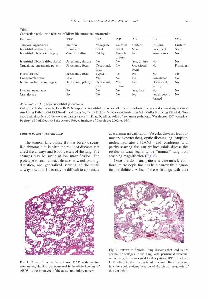
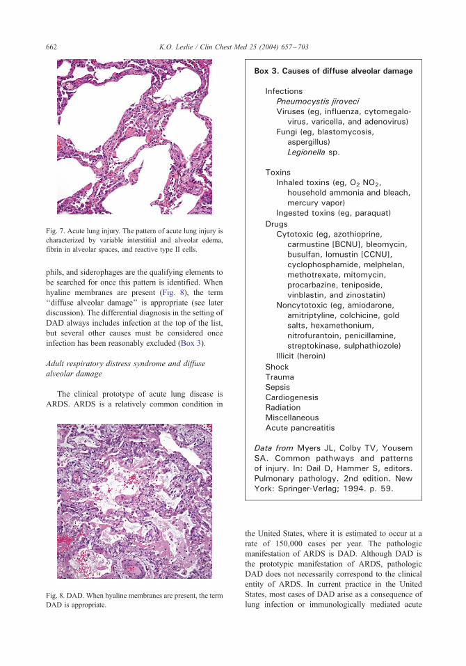
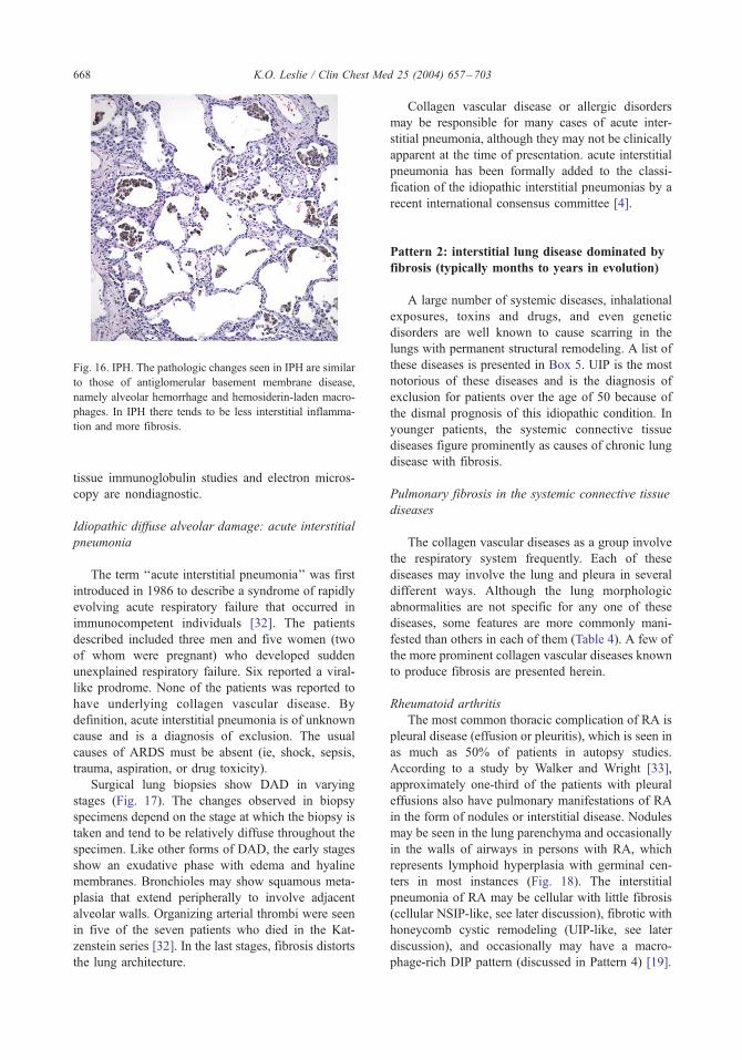
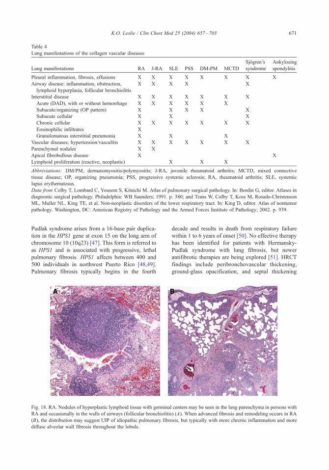
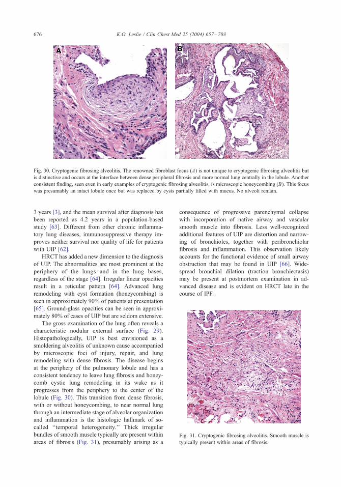
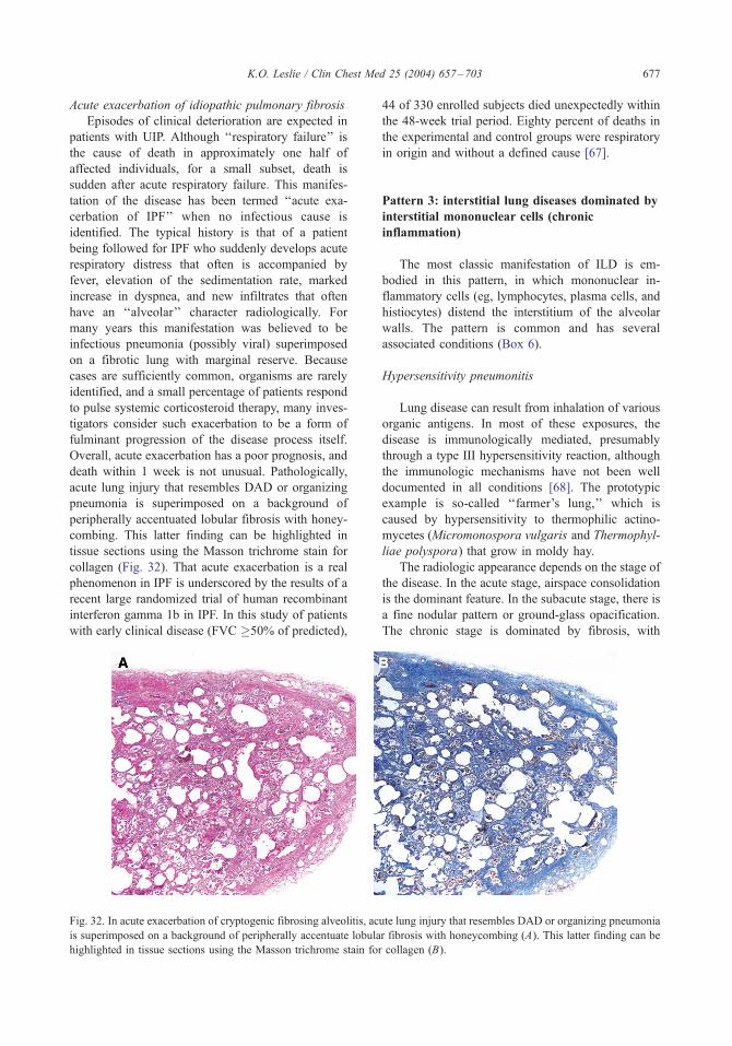
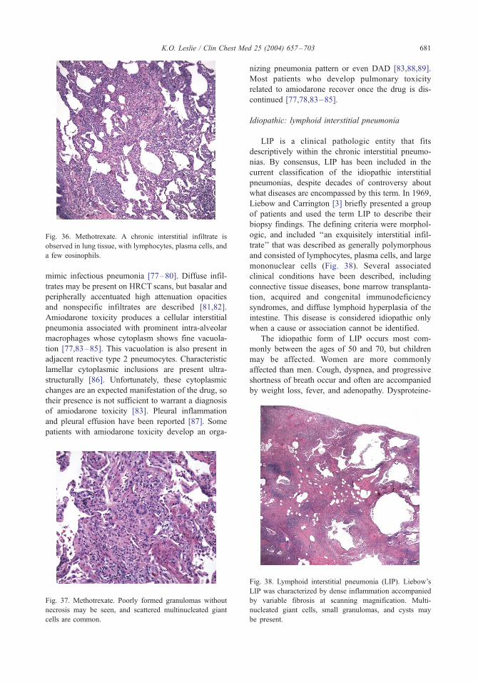
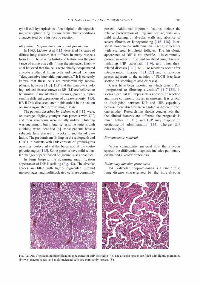
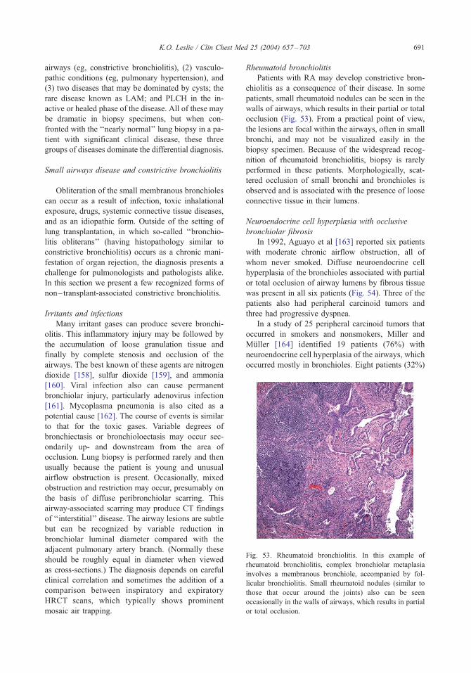
Related Documents