Decay-Accelerating Factor 1 Deficiency Exacerbates Leptospiral-Induced Murine Chronic Nephritis and Renal Fibrosis Marı ´a F. Ferrer 1 , Emilia Scharrig 1 , Lucrecia Alberdi 1 , Maia Cedola 1 , Gabriela Pretre 1 , Ricardo Drut 2 , Wen- Chao Song 3 , Ricardo M. Gomez 1 * 1 Instituto de Biotecnologı ´a y Biologı ´a Molecular, CONICET-UNLP, La Plata, Buenos Aires, Argentina, 2 Ca ´tedra de Patologı ´a ‘‘A’’, Facultad de Medicina, UNLP, La Plata, Buenos Aires, Argentina, 3 Department of Pharmacology, Perelman School of Medicine, University of Pennsylvania, Philadelphia, Pennsylvania, United States of America Abstract Leptospirosis is a global zoonosis caused by pathogenic Leptospira, which can colonize the proximal renal tubules and persist for long periods in the kidneys of infected hosts. Here, we characterized the infection of C57BL/6J wild-type and Daf1 2/2 mice, which have an enhanced host response, with a virulent Leptospira interrogans strain at 14 days post-infection, its persistence in the kidney, and its link to kidney fibrosis at 90 days post-infection. We found that Leptospira interrogans can induce acute moderate nephritis in wild-type mice and is able to persist in some animals, inducing fibrosis in the absence of mortality. In contrast, Daf1 2/2 mice showed acute mortality, with a higher bacterial burden. At the chronic stage, Daf1 2/2 mice showed greater inflammation and fibrosis than at 14 days post-infection and higher levels at all times than the wild-type counterpart. Compared with uninfected mice, infected wild-type mice showed higher levels of IL-4, IL-10 and IL-13, with similar levels of a-smooth muscle actin, galectin-3, TGF-b1, IL-17, IFN-c, and lower IL-12 levels at 90 days post- infection. In contrast, fibrosis in Daf1 2/2 mice was accompanied by high expression of a-smooth muscle actin, galectin-3, IL- 10, IL-13, and IFN-c, similar levels of TGF-b1, IL-12, and IL-17 and lower IL-4 levels. This study demonstrates the link between Leptospira-induced murine chronic nephritis with renal fibrosis and shows a protective role of Daf1. Citation: Ferrer MF, Scharrig E, Alberdi L, Cedola M, Pretre G, et al. (2014) Decay-Accelerating Factor 1 Deficiency Exacerbates Leptospiral-Induced Murine Chronic Nephritis and Renal Fibrosis. PLoS ONE 9(7): e102860. doi:10.1371/journal.pone.0102860 Editor: Yung-Fu Chang, Cornell University, United States of America Received March 14, 2014; Accepted June 24, 2014; Published July 17, 2014 Copyright: ß 2014 Ferrer et al. This is an open-access article distributed under the terms of the Creative Commons Attribution License, which permits unrestricted use, distribution, and reproduction in any medium, provided the original author and source are credited. Funding: This work was supported by grants from Universidad Nacional de La Plata (X592), Agencia Nacional de Promocio ´ n Cientı ´fica y Tecnolo ´ gica (ANPCyT) PICT 07-00642, 07-00028 and 12-0434 (RMG). The funders had no role in study design, data collection and analysis, decision to publish, or preparation of the manuscript. Competing Interests: The authors have declared that no competing interests exist. * Email: [email protected] Introduction Renal fibrosis is a reliable predictor of prognosis, a major determinant of renal insufficiency and a common final outcome of almost all progressive chronic kidney diseases (CKD) [1,2]. In USA the prevalence of CKD is increasing and affects an estimated 13% of the population [3] or even more in developing countries [4]. The etiology of CKD in developed countries is associated with atherosclerosis, diabetes, and autoimmune glomerulonephritis [2]. However, it is possible that in developing countries infections have also a significant impact. Leptospirosis is a global zoonosis caused by spirochetes of the genus Leptospira [5]. Human infection commonly occurs through direct contact with infected animal urine or indirectly through contaminated water. Almost every mammal can serve as a carrier of leptospires, harboring the spirochete in the proximal renal tubules of the kidneys, leading to urinary shedding. Rodents, mainly rats, serve as the major carriers in most human leptospirosis, excreting high concentrations of leptospires (10 7 organisms per ml) months after their initial infections [6]. Consequently, leptospirosis can be considered a disease with an endemo-epidemic pattern associated with slum settlements [7]. Leptospirosis is usually a biphasic disease with an early bacteremic phase during which leptospires are disseminated rapidly through- out the body during the first 7–10 days of infection. This is followed by a leptospiruric phase during which specific antibodies arise in parallel with the disappearance of the bacteria from the blood and most organs with the exception of the kidneys, where it can persist for long periods of time [8]. Although it can cause death or very severe symptoms (Weil’s syndrome), most docu- mented cases are mild and self-limiting [9]. It is accepted that an accurate diagnosis of leptospirosis is frequently lacking. In experimental leptospirosis, animal species and strain, age, dose inoculum, bacterial history, passage and Leptospira serovar influence the infection outcome [9,10]. Guinea pigs and hamsters are the most commonly used animal experimental models for studying acute parameters [11,12], whereas rats are considered a suitable model for studying persistent renal colonization [13,14]. In contrast, mice are relatively more resistant, presenting fewer symptoms and lesions and lower mortality, and are therefore used less as a model [10]. However, mice may become more susceptible via immunosuppression [15], or by deletion of specific cytokine genes [16], suggesting that the immune response plays a major role in murine infection. Decay-accelerating factor 1 (Daf1 or CD55) is a glycosylpho- sphatidylinositol-anchored membrane protein of the complement- PLOS ONE | www.plosone.org 1 July 2014 | Volume 9 | Issue 7 | e102860

Welcome message from author
This document is posted to help you gain knowledge. Please leave a comment to let me know what you think about it! Share it to your friends and learn new things together.
Transcript

Decay-Accelerating Factor 1 Deficiency ExacerbatesLeptospiral-Induced Murine Chronic Nephritis and RenalFibrosisMarıa F. Ferrer1, Emilia Scharrig1, Lucrecia Alberdi1, Maia Cedola1, Gabriela Pretre1, Ricardo Drut2, Wen-
Chao Song3, Ricardo M. Gomez1*
1 Instituto de Biotecnologıa y Biologıa Molecular, CONICET-UNLP, La Plata, Buenos Aires, Argentina, 2 Catedra de Patologıa ‘‘A’’, Facultad de Medicina, UNLP, La Plata,
Buenos Aires, Argentina, 3 Department of Pharmacology, Perelman School of Medicine, University of Pennsylvania, Philadelphia, Pennsylvania, United States of America
Abstract
Leptospirosis is a global zoonosis caused by pathogenic Leptospira, which can colonize the proximal renal tubules andpersist for long periods in the kidneys of infected hosts. Here, we characterized the infection of C57BL/6J wild-type andDaf12/2 mice, which have an enhanced host response, with a virulent Leptospira interrogans strain at 14 days post-infection,its persistence in the kidney, and its link to kidney fibrosis at 90 days post-infection. We found that Leptospira interroganscan induce acute moderate nephritis in wild-type mice and is able to persist in some animals, inducing fibrosis in theabsence of mortality. In contrast, Daf12/2 mice showed acute mortality, with a higher bacterial burden. At the chronic stage,Daf12/2 mice showed greater inflammation and fibrosis than at 14 days post-infection and higher levels at all times thanthe wild-type counterpart. Compared with uninfected mice, infected wild-type mice showed higher levels of IL-4, IL-10 andIL-13, with similar levels of a-smooth muscle actin, galectin-3, TGF-b1, IL-17, IFN-c, and lower IL-12 levels at 90 days post-infection. In contrast, fibrosis in Daf12/2 mice was accompanied by high expression of a-smooth muscle actin, galectin-3, IL-10, IL-13, and IFN-c, similar levels of TGF-b1, IL-12, and IL-17 and lower IL-4 levels. This study demonstrates the link betweenLeptospira-induced murine chronic nephritis with renal fibrosis and shows a protective role of Daf1.
Citation: Ferrer MF, Scharrig E, Alberdi L, Cedola M, Pretre G, et al. (2014) Decay-Accelerating Factor 1 Deficiency Exacerbates Leptospiral-Induced Murine ChronicNephritis and Renal Fibrosis. PLoS ONE 9(7): e102860. doi:10.1371/journal.pone.0102860
Editor: Yung-Fu Chang, Cornell University, United States of America
Received March 14, 2014; Accepted June 24, 2014; Published July 17, 2014
Copyright: � 2014 Ferrer et al. This is an open-access article distributed under the terms of the Creative Commons Attribution License, which permitsunrestricted use, distribution, and reproduction in any medium, provided the original author and source are credited.
Funding: This work was supported by grants from Universidad Nacional de La Plata (X592), Agencia Nacional de Promocion Cientıfica y Tecnologica (ANPCyT)PICT 07-00642, 07-00028 and 12-0434 (RMG). The funders had no role in study design, data collection and analysis, decision to publish, or preparation of themanuscript.
Competing Interests: The authors have declared that no competing interests exist.
* Email: [email protected]
Introduction
Renal fibrosis is a reliable predictor of prognosis, a major
determinant of renal insufficiency and a common final outcome of
almost all progressive chronic kidney diseases (CKD) [1,2]. In
USA the prevalence of CKD is increasing and affects an estimated
13% of the population [3] or even more in developing countries
[4]. The etiology of CKD in developed countries is associated with
atherosclerosis, diabetes, and autoimmune glomerulonephritis [2].
However, it is possible that in developing countries infections have
also a significant impact.
Leptospirosis is a global zoonosis caused by spirochetes of the
genus Leptospira [5]. Human infection commonly occurs through
direct contact with infected animal urine or indirectly through
contaminated water. Almost every mammal can serve as a carrier
of leptospires, harboring the spirochete in the proximal renal
tubules of the kidneys, leading to urinary shedding. Rodents,
mainly rats, serve as the major carriers in most human
leptospirosis, excreting high concentrations of leptospires (107
organisms per ml) months after their initial infections [6].
Consequently, leptospirosis can be considered a disease with an
endemo-epidemic pattern associated with slum settlements [7].
Leptospirosis is usually a biphasic disease with an early bacteremic
phase during which leptospires are disseminated rapidly through-
out the body during the first 7–10 days of infection. This is
followed by a leptospiruric phase during which specific antibodies
arise in parallel with the disappearance of the bacteria from the
blood and most organs with the exception of the kidneys, where it
can persist for long periods of time [8]. Although it can cause
death or very severe symptoms (Weil’s syndrome), most docu-
mented cases are mild and self-limiting [9]. It is accepted that an
accurate diagnosis of leptospirosis is frequently lacking.
In experimental leptospirosis, animal species and strain, age,
dose inoculum, bacterial history, passage and Leptospira serovar
influence the infection outcome [9,10]. Guinea pigs and hamsters
are the most commonly used animal experimental models for
studying acute parameters [11,12], whereas rats are considered a
suitable model for studying persistent renal colonization [13,14].
In contrast, mice are relatively more resistant, presenting fewer
symptoms and lesions and lower mortality, and are therefore used
less as a model [10]. However, mice may become more susceptible
via immunosuppression [15], or by deletion of specific cytokine
genes [16], suggesting that the immune response plays a major role
in murine infection.
Decay-accelerating factor 1 (Daf1 or CD55) is a glycosylpho-
sphatidylinositol-anchored membrane protein of the complement-
PLOS ONE | www.plosone.org 1 July 2014 | Volume 9 | Issue 7 | e102860
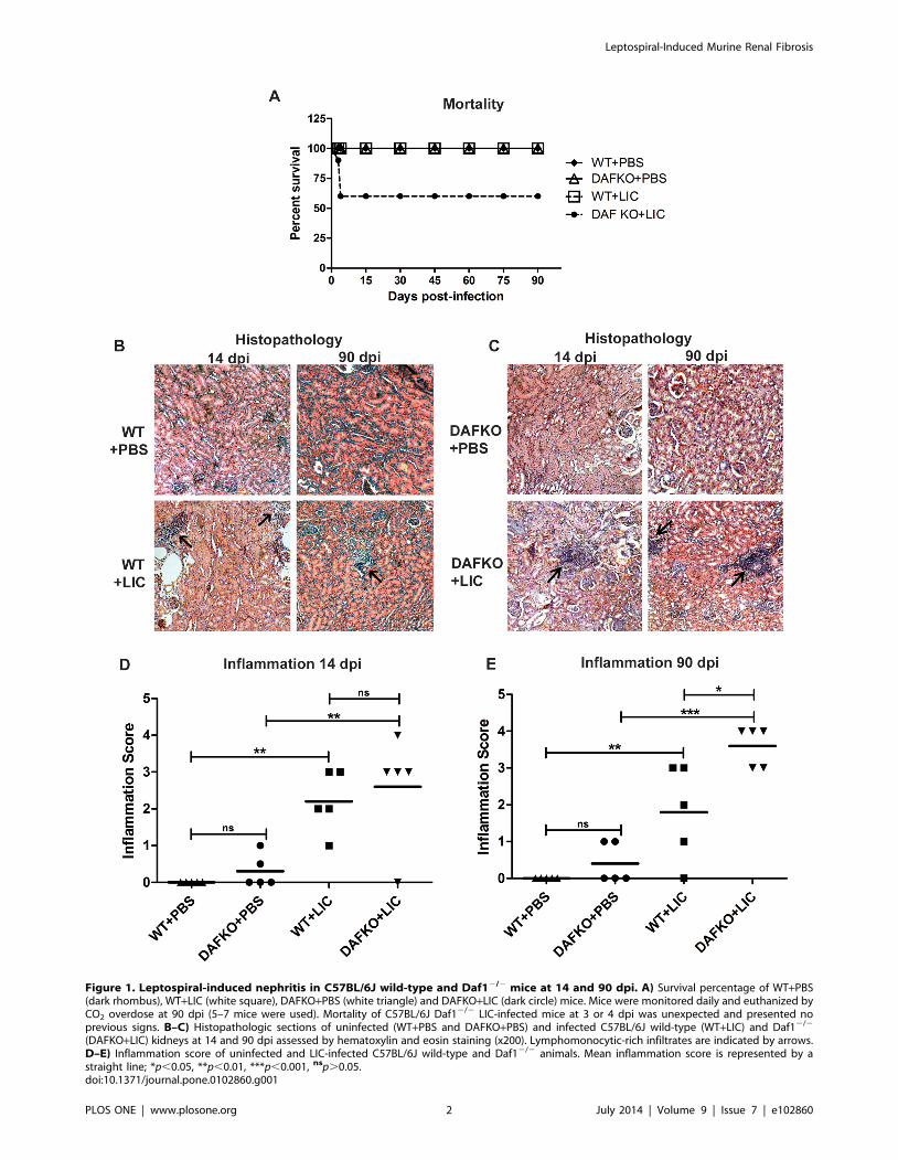
Figure 1. Leptospiral-induced nephritis in C57BL/6J wild-type and Daf12/2 mice at 14 and 90 dpi. A) Survival percentage of WT+PBS(dark rhombus), WT+LIC (white square), DAFKO+PBS (white triangle) and DAFKO+LIC (dark circle) mice. Mice were monitored daily and euthanized byCO2 overdose at 90 dpi (5–7 mice were used). Mortality of C57BL/6J Daf12/2 LIC-infected mice at 3 or 4 dpi was unexpected and presented noprevious signs. B–C) Histopathologic sections of uninfected (WT+PBS and DAFKO+PBS) and infected C57BL/6J wild-type (WT+LIC) and Daf12/2
(DAFKO+LIC) kidneys at 14 and 90 dpi assessed by hematoxylin and eosin staining (x200). Lymphomonocytic-rich infiltrates are indicated by arrows.D–E) Inflammation score of uninfected and LIC-infected C57BL/6J wild-type and Daf12/2 animals. Mean inflammation score is represented by astraight line; *p,0.05, **p,0.01, ***p,0.001, nsp.0.05.doi:10.1371/journal.pone.0102860.g001
Leptospiral-Induced Murine Renal Fibrosis
PLOS ONE | www.plosone.org 2 July 2014 | Volume 9 | Issue 7 | e102860

regulatory family that protects cells from autologous complement
attack [17]. Daf1 inhibits the assembly and accelerates the rapid
decay of C3 and C5 convertases in both the classical and
alternative complement activation pathways [17]. Interestingly,
many kidney pathologies have been linked to abnormal comple-
ment activation [18]. It has been reported that Daf12/2 mice are
more susceptible to complement-mediated inflammatory injury
and have significantly enhanced T-cell responses to active
immunization. This phenotype is characterized by hypersecretion
of interferon (IFN)-c and IL-2 as well as down-regulation of the
inhibitory cytokine IL-10 during antigen restimulation of lympho-
cytes in vitro [19]. Furthermore, several studies have shown that
the absence of Daf1 exacerbates disease in a variety of
autoimmune models, including systemic autoimmune disease in
the MLR-Faslpr mouse [20,21], focal and segmental glomerulo-
sclerosis [22] and mercury-induced autoimmunity [23], by
enhancing T-cell and autoimmune responses associated with
hypersecretion of IFN-c, IL-12 and IL-17 [19,24,25,26].
Galectin-3 (Gal-3) is a b-galactoside-binding animal lectin [27].
Gal-3 expression and secretion by macrophages is a major
mechanism linking macrophages to fibroblast activation and
myofibroblast accumulation, as demonstrated by their synthesis
of a-smooth muscle actin (a-SMA), thus driving renal fibrosis in
the unilateral ureteric obstruction model (UUO) [28].
We hypothesized that by using a reference strain with known
virulence, mice may become susceptible to chronic experimental
leptospirosis. If successful, we could then explore whether the
persistent renal infection affects the renal extracellular matrix
(ECM) and explore some of the mechanisms involved, particularly
the influence of an enhanced host response. In the present paper,
we report the establishment of a murine model of chronic
leptospirosis followed by fibrosis in wild-type and Daf12/2 mice.
Results
Leptospira interrogans induces discrete to moderatenephritis in mice
In order to investigate whether Leptospira interrogans, serovar
Copenhageni (LIC), was able to induce murine renal inflamma-
tion, and to study how an enhanced host response could impact in
the disease, C57BL/6J wild-type (WT) and Daf12/2 mice of 3–4
weeks of age were infected with 106 bacteria and groups of five
animals were euthanized and necropsied at 14 and 90 days post-
infection (dpi).
None of the uninfected (WT+PBS and Daf12/2+PBS) or WT+LIC mice died during the experiment. In contrast, the Daf12/2+LIC mice had 40% mortality at 4 dpi (Figure 1 A). No pathology
was observed in uninfected mice (Figure 1 B–C). In contrast,
infected animals developed interstitial nephritis in the form of
multifocal lymphomonocytic infiltrates (Figure 1 B–C). The degree
of inflammation was slightly higher (although not significant) in the
Daf12/2+LIC than in WT littermates at 14 dpi, but significantly
higher at 90 dpi (p,0.05, Figure 1 D–E), when only 80% of WT+LIC mice showed inflammation. Taken together, these results
show that Daf12/2+LIC mice have greater susceptibility to
infection than their WT littermates.
Bacterial burden is significantly reduced at later timepoints
Kidney bacterial burden showed an unexpected 200-fold
increase (3.4861021 bacteria/56104 cells) in Daf12/2 infected
mice compared with WT mice at 14 dpi (1.5361023 bacteria/
56104 cells, p,0.01, Figure 2 A). In contrast, the kidney bacterial
burden was markedly reduced in both Daf12/2 mice
(1.461025bacteria/56104 cells) and WT infected animals
(3.961025 bacteria/56104 cells) at 90 dpi (Figure 2 A).
Immunohistochemistry (IHC) showed that the tissue distribution
of leptospiral antigen was present mostly on the luminal surface of
the proximal tubules (Figure 2 B–C), and occasionally inside
macrophages and in the interstitium. A moderate number of
antigen spots were observed in samples of mice harvested at 14
dpi, but leptospiral antigen became almost undetectable in samples
from both groups of mice at 90 dpi (Figure 2 B–C).
LIC-induced chronic nephritis produces renal fibrosisIn order to investigate whether the chronic nephritis induced in
C57BL/6J mice infected with LIC was linked to fibrosis, renal
collagen deposition was observed with Masson’s trichrome and
digitally analyzed using Picro sirius red (PS) staining.
Uninfected animals had no signs of fibrosis during the
experiment (Figure 3 A–B). On the other hand, renal interstitial
fibrosis was evident with increased numbers of red collagen fibers,
frequently observed away from lymphocyte-rich infiltrates in WT+LIC animals and even more in Daf12/2 infected mice (p,0.05
Figure 2. Bacterial burden is significantly reduced at later time points. A) Quantitative measurement of leptospiral DNA (16S) in kidneysamples from WT (WT+LIC) or Daf12/2 (DAFKO+LIC) infected animals with 106 bacteria at 14 and 90 dpi. Bars represent standard error mean (SEM) ofassays from a group of five to seven mice. Three pieces of each organ were analyzed in triplicate q-PCR and normalized to host cell number; **p,0.01, nsp.0.05. B–C) Immunohistochemistry with antiserum specific for LipL32 (x200) of kidney sections of uninfected (WT+PBS and DAFKO+PBS)and infected WT (WT+LIC) and Daf12/2 (DAFKO+LIC) animals at 14 and 90 dpi. Arrows indicate representative positive foci.doi:10.1371/journal.pone.0102860.g002
Leptospiral-Induced Murine Renal Fibrosis
PLOS ONE | www.plosone.org 3 July 2014 | Volume 9 | Issue 7 | e102860

and p,0.01, respectively, Figure 3 A–C), involving all animals in
correlation with the higher inflammation observed but in contrast
to the low Leptospira presence at 90 dpi.
The levels of pro-collagen I mRNA were similarly increased in
both groups of infected mice compared with uninfected animals at
90 dpi (Figure 3 D, p,0.05).
Leptospira interrogans-induced humoral responseAnti-leptospiral IgM levels in serum samples of Daf12/2
infected mice at 14 dpi were higher than in uninfected mice
(Figure 4 A, p,0.001), but similar to WT mice. In contrast, the
IgG-specific humoral immune response was higher in WT than in
the Daf12/2 infected group at 14 dpi, but reached similarly high
values in the infected groups at 90 dpi (Figure 4 B, p,0.01). In
order to study if anti-leptospiral antibody production and
enhanced complement activation contributed to kidney damage,
we analyzed the membrane attack complex (MAC) tissue
distribution by IHC and observed minimal MAC presence in
Daf12/2 infected mice at 90 dpi (Figure 4 C–H).
Chronic fibrosis in Daf12/2 LIC-infected mice correlateswith myofibroblast activation and enhanced Gal-3expression
Next, we investigated whether the observed chronic fibrosis was
accompanied by myofibroblast activation and enhanced Gal-3
expression, as observed in models of renal fibrosis [29]. Enhanced
a-SMA expression was observed only in Daf12/2 infected mice
(Figure 5 A). In contrast, both WT and Daf12/2 infected mice
showed enhanced Gal-3 expression in the renal tissue of infected
mice when compared with the uninfected control group by IHC
analysis (Figure 5 A). The analysis by qPCR showed significantly
higher values only for Daf12/2 infected mice (Figure 5 B–C, p,
0.05).
Figure 3. LIC-induced chronic nephritis produces renal fibrosis. A–B) Renal collagen deposition (indicated by arrows) was analyzed byMasson’s trichrome (MS) and Picro sirius red (PS) staining in uninfected (WT+PBS and DAFKO+PBS) and infected (WT+LIC and DAFKO+LIC) animals at90 dpi (x200). C) Fibrosis score of WT (WT+PBS and WT+LIC) and Daf12/2 (DAFKO+PBS and DAFKO+LIC) animals. Mean fibrosis score is representedby a straight line, *p,0.05, **p,0.01, nsp.0.05. D) Quantitative measurement of pro-collagen I mRNA in kidney samples from uninfected or infectedanimals at 90 dpi. Bars represent the standard error of the mean (SEM) of assays from a group of five mice. Three pieces of each organ were analyzedin triplicate for q-PCR and normalized to host b-actin expression; *p,0.05, nsp.0.05.doi:10.1371/journal.pone.0102860.g003
Leptospiral-Induced Murine Renal Fibrosis
PLOS ONE | www.plosone.org 4 July 2014 | Volume 9 | Issue 7 | e102860

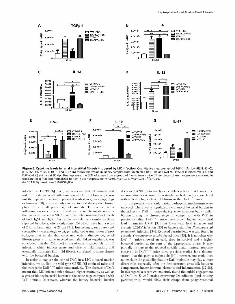
Cytokine levels in renal chronic fibrosis triggered by LICinfection
Previous studies have implicated TGF-b1 as an important
mediator of renal fibrosis [2,29]. However, TGF-b1 mRNA
expression was similar in uninfected and infected mice at 90 dpi
(Figure 6 A). Meanwhile, WT mice showed higher IL-4 mRNA
expression than uninfected mice, while Daf12/2 infected mice
showed lower levels compared to Daf12/2 uninfected animals
(Figure 6 B, p,0.001 and p,0.01, respectively). IL-13 mRNA
expression was up-regulated in both groups of infected mice
(Figure 6 C, p,0.05). Significant down-regulation of IL-12 was
observed in infected WT mice (p,0.05); meanwhile Daf12/2
mice showed similar levels with and without infection (Figure 6 D).
Significant up-regulation of IFN-c was observed only in Daf12/2+LIC animals (Figure 6 E, p,0.05). Finally, although elevated IL-
10 mRNA levels were found in both WT and Daf12/2 LIC-
infected mice (Figure 6 F, p,0.01 and p,0.05, respectively), IL-
17 expression was not different among the groups (Figure 6 G).
Discussion
The kidney is the primary target of Leptospira during both
subacute and chronic infection [5,30]. In our model of LIC
Figure 4. Anti-leptospiral antibodies followed by enhanced complement activation in Daf12/2 mice suggest a minimal contributionto kidney damage. A) Total anti-leptospiral IgM from serum samples of uninfected and infected animals at 14 dpi was analyzed by ELISA. Barsrepresent standard error mean (SEM) of assays from a group of five to seven mice; ***p,0.001. B) Total anti-leptospiral IgG from serum samples ofuninfected and infected animals at 14 and 90 dpi was analyzed by ELISA. Bars represent the SEM of assays from a group of five to seven mice; ***p,0.001 with respect to WT+PBS at 14 dpi; +++p,0.001 with respect to DAFKO+PBS at 14 dpi; &&p,0.01 between both WT and Daf12/2 LIC-infectedgroups. Values under the baseline (dashed line) are considered negligible. Immunohistochemistry with antiserum specific for MAC (membrane attackcomplex) at 90 dpi (6200). C) Uninfected pancreas as the negative control, D) coxsackievirus B3-infected pancreas as the positive control, WT+LICwithout (E) or with anti-MAC (F), DAFKO+LIC without (G) or with anti-MAC (H). Only in panel H there is minimal, positive labeling in interstitial cellsindicated by arrows. Samples were treated according to Abcam’s suggested protocol including unmasking. Unmasking usually strongly increases thestaining of kidney acinar cells which are rich in endogenous peroxidase, but this staining is intracytoplasmic.doi:10.1371/journal.pone.0102860.g004
Figure 5. Chronic fibrosis in Daf12/2 +LIC mice induces myofibroblast activation and enhanced galectin-3 expression. A)Immunohistochemistry of kidney sections from uninfected (WT+PBS and DAFKO+PBS) and LIC-infected (WT+LIC and DAFKO+LIC) mice withantiserum specific for a-smooth muscle actin (a-SMA) or galectin-3 (Gal-3) at 90 dpi (x200). Arrows indicate foci of antigen expression. Quantitativemeasurement of a-SMA (B) and Gal-3 (C) mRNA expression in kidney samples from uninfected (WT+PBS and DAFKO+PBS) and infected (WT+LIC andDAFKO+LIC) animals at 90 dpi. Bars represent the SEM of assays from a group of five to seven mice. Three pieces of each organ were analyzed intriplicate for q-PCR and normalized to host b-actin expression; *p,0.05, nsp.0.05.doi:10.1371/journal.pone.0102860.g005
Leptospiral-Induced Murine Renal Fibrosis
PLOS ONE | www.plosone.org 5 July 2014 | Volume 9 | Issue 7 | e102860
infection in C57BL/6J mice, we observed that all animals had
mild to moderate renal inflammation at 14 dpi. However, it was
not the typical interstitial nephritis described in guinea pigs, dogs
or humans [30], and was only discrete to mild during the chronic
phase in a small percentage of animals. This reduction in
inflammation over time correlated with a significant decrease in
the bacterial burden at 90 dpi and inversely correlated with levels
of both IgM and IgG. Our results are relatively similar to those
reported by others, where only some C57BL/6J mice had a score
of 2 for inflammation at 28 dpi [31]. Interestingly, such restricted
susceptibility was enough to trigger enhanced transcription of pro-
collagen I at 90 dpi that correlated with a discrete degree of
fibrosis present in some infected animals. Taken together, it was
concluded that the C57BL/6J strain of mice is susceptible to LIC
infection, which induces acute and chronic inflammation, and
eventually translates into mild fibrosis correlated to some degree
with the bacterial burden.
In order to explore the role of Daf1 in a LIC-induced murine
infection, we studied the wild-type C57BL/6J strain of mice and
the transgenic Daf12/2 mice comparatively. The absence of Daf1
meant that LIC-infected mice showed higher mortality, as well as
a greater kidney bacterial burden in the acute stage compared with
WT animals. Moreover, whereas the kidney bacterial burden
decreased at 90 dpi to barely detectable levels as in WT mice, the
inflammation score rose. Interestingly, such differences correlated
with a clearly higher level of fibrosis in the Daf12/2 mice.
In the present work, only partial pathogenic mechanisms were
unveiled. There was a significantly enhanced bacterial burden in
the kidneys of Daf12/2 mice during acute infection but a similar
burden during the chronic stage. In comparison with WT, in
previous studies, Daf12/2 mice have shown higher acute viral
load in murine CMV [32] but lower viral load in acute and
chronic LCMV infection [33] or bacteremia after Pneumococcalpneumonia infection [34]. Reduced parasite load was also found in
chronic Trypanosome cruzi-infected mice [35]. It is not clear why
Daf12/2 mice showed an early drop in survival and a higher
bacterial burden at the start of the leptospiruric phase. It may
partially be due to the reduced specific acute humoral response
observed in Daf12/2 mice since previous studies have demon-
strated that this plays a major role [36]; however, our study does
not exclude the possibility that the Daf1 molecule may play a more
direct role, especially after the demonstrated cross-talk between
complement, innate immune elements and inflammation [37,38].
In this regard, a recent ex vivo study found that initial engagement
of Daf1 by E. coli strains expressing Dr adhesins (and causing
pyelonephritis) would allow their escape from phagolysosomal
Figure 6. Cytokine levels in renal interstitial fibrosis triggered by LIC infection. Quantitative measurement of TGF-b1 (A), IL-4 (B), IL-13 (C),IL-12 (D), IFN-c (E), IL-10 (F) and IL-17 (G) mRNA expression in kidney samples from uninfected (WT+PBS and DAFKO+PBS) or infected (WT+LIC andDAFKO+LIC) animals at 90 dpi. Bars represent the SEM of assays from a group of five to seven mice. Three pieces of each organ were analyzed intriplicate for q-PCR and normalized to host b-actin expression; *p,0.05, **p,0.01, ***p,0.001, nsp.0.05.doi:10.1371/journal.pone.0102860.g006
Leptospiral-Induced Murine Renal Fibrosis
PLOS ONE | www.plosone.org 6 July 2014 | Volume 9 | Issue 7 | e102860

fusion, leading to non-destructive parasitism that allows bacteria to
persist intracellularly [39]. The precise mechanism involved in our
model will hopefully be clarified in future studies.
Previous studies have shown that outside membrane proteins
(OMPs) from pathogenic leptospires are recognized by TLR2 of
murine kidney tubular cells [40]. This recognition activates the
transcription factor kB (NF-kB) and the mitogen-activated protein
kinase (MAPK) pathway, triggering early inflammation and
leukocyte recruitment [41] and later in vitro HeK-2 cell produc-
tion of collagens through activation of the TGF-b1/Smad3
pathway [42]. However, by using several transgenic mice, a very
recent study elegantly showed that TLR and NLR receptors as
well as T lymphocytes are not required to generate Leptospira-
induced renal fibrosis. Instead, the iNOS enzyme, known to play a
role in Leptospira-induced interstitial nephritis [43,44], was
associated with the induction of renal fibrosis [45]. Given that in
Daf12/2 mice the increase in inflammation and fibrosis was not
correlated with an increase in the bacterial burden present in the
kidneys of chronically infected mice, we conclude that the major
factors driving fibrosis in this model rely on the presence of
leptospires during the early stages, and on the host response in the
chronic phase without excluding a role for enhanced complement-
mediated injury and/or reduced turnover of extracellular matrix
components.
Gal-3 expression and infiltration of macrophages occurs early in
LIC-induced nephritis and remains up-regulated, as shown in the
UUO model [28]. However, enhanced a-SMA expression was
observed only in the kidneys of infected Daf12/2 mice. Both Gal-3
and a-SMA expression levels remain higher in the UUO model
[28], continuously increasing macrophage recruitment, and
probably justifying the clearly higher level of renal fibrosis
observed in the UUO model by directly acting through TGF-
b1-mediated myofibroblast activation and extracellular matrix
production [46]. However, since it has been shown that Gal-3
forms lattices that promote cell-surface residence or the retention
of cytokine and growth factor receptors, including receptors for
TGF-b1, by interfering with their endocytosis, and that this
retention of receptors leads to increased signaling [47], it should
also be considered that enhanced levels of Gal-3 could modulate
fibrosis indirectly. In addition, TGF-b1-independent mechanisms
of renal fibrosis have also been reported in the UUO model [48].
Regarding IL-4, this cytokine it has been described in humans as a
profibrotic cytokine promoting fibrocyte differentiation [2],
whereas it has an inhibitory effect in mice [49] and therefore
may explain the low levels at 90 dpi in Daf12/2 mice. Hopefully,
future studies will clarify if it has a role in WT mice. As expected,
increased IL-13 levels were detected in both WT and Daf12/2
infected mice at 90 dpi, since this cytokine has been reported to be
a major profibrotic molecule that promotes fibrocyte differentia-
tion [50]. The high levels of IFN-c, IL-12 and IL-17 (although not
significant) observed in the Daf12/2 mice are probably a major
contributor to the T-cell hyper-responsiveness of these mice
[19,22] and therefore may explain the similar or lower levels
observed in the WT mice. IL-10 levels were elevated in both WT
and Daf12/2 infected mice. Interestingly, early enhanced IL-10
expression has been shown in other murine models of leptospirosis
[51] and has been linked to death in humans [52] and hamsters
[53] infected with leptospires.
Based on the results of our experiments and very recent studies
performed by others [45], it may be speculated that the presence
of bacteria triggers the recruitment and activation of an early
cellular exudate in the kidney [2]. It may be hypothesized that
bacterial persistence contributes to non-resolving inflammation
and a cellular exudate that sets the fibrogenic stage (priming) and
triggers the activation and recruitment of extracellular matrix
(ECM)-producing cells such as interstitial fibroblasts and circulat-
ing fibrocytes through Gal-3 and probably other molecules such as
nitric oxide. Upon activation, ECM-producing cells assemble a
Figure 7. Model of leptospiral-induced murine chronic nephritis and renal fibrosis. Upon LIC colonization of proximal renal tubules ofeither C57BL/6J wild-type (WT) or Daf12/2 mice, cellular exudate recruitment and activation occurs and precedes the arrival of macrophages, whichafter stimulation produce mediators such as galectin-3 (Gal-3) that activate quiescent fibroblasts and convert them into an a-SMA+ myofibroblastpopulation. Myofibroblasts produce several molecules and orchestrate the production of ECM components and their extracellular assembly. In WTmice, down-regulation of IL-12 as well as up-regulation of IL-4, IL-10 and profibrotic cytokines such as IL-13 contributes to ECM accumulation. Ofnote, LIC-infected Daf12/2 mice have a higher bacterial burden at the acute stage of infection, but barely detectable bacterial burden, increased IL-13, IL-10 and IFN-c expression and decreased IL-4 expression during the chronic stages; these correlate with chronic inflammation, ECM depositionand renal fibrosis. Also, an alternative possibility is that the increased adaptive immune response against infection leads to the deposition of anti-leptospiral antibody followed by complement activation, which then contributes to kidney damage.doi:10.1371/journal.pone.0102860.g007
Leptospiral-Induced Murine Renal Fibrosis
PLOS ONE | www.plosone.org 7 July 2014 | Volume 9 | Issue 7 | e102860

multicomponent, integrin-associated protein complex that inte-
grates input from various fibrogenic signals and orchestrates the
production of ECM components and their extracellular assembly.
Multiple cellular and molecular events, such as tubular atrophy
and microvascular rarefaction, may promote scar formation and
ensure a vicious progression to end-stage kidney failure [54]
(Figure 7). Taken together, our results demonstrate that Daf1 plays
a protective role in experimental leptospiral-induced fibrosis
in vivo. Moreover, Daf12/2+LIC animals constitute a suitable
murine model for the study of leptospiral infections followed by the
development of renal fibrosis.
It has been recently shown that asymptomatic renal coloniza-
tion of leptospires in a region of high disease transmission is
common, and may include people without serological or clinical
evidence of recent infection with Leptospira persistence in the
kidney [55]. However, the pathogenic significance of this finding,
and particularly its connection with renal fibrosis, remains
unknown [55]. Although scarce, some studies on human biopsies
have shown an association between leptospirosis, interstitial
nephritis and late-stage fibrosis [56]. In a recent case report of a
young male patient, leptospirosis evolved to irreversible tubuloin-
terstitial fibrosis that required chronic dialysis treatment [57]. In
other studies of human biopsies, tubulointerstitial nephritis was
associated indirectly with fibrosis based on increased collagen I,
IV, TGF-b1 and Smad levels in the kidney, particularly when the
disease was not treated with antibiotics [58]. In conclusion, there
are no data showing to what degree Leptospira infection impacts
on the incidence of human CKD, but the fact that it is a major
human zoonosis, is frequently undiagnosed, and has a major
impact on low social-economic populations, our and other [47]
results associating even low bacterial persistence with renal fibrosis
strongly suggest that it could be higher than expected, making
Leptospira-triggered fibrosis a neglected event in a neglected
disease. This should encourage more studies in order to clarify this
issue.
Materials and Methods
BacteriaThe virulent Leptospira interrogans serovar Copenhageni
(LIC) strain Fiocruz L1-130 and the culture conditions used have
been described previously [59].
Ethics StatementAll animal experiments were in compliance with the Argentine
animal protection Law 14346 ‘‘Malos tratos y actos de crueldad a
los animales’’. The ethics committee of the ‘‘Instituto de
Biotecnologıa y Biologıa Molecular, CONICET-UNLP’’, in
agreement with the International Guiding Principles for Biomed-
ical Research Involving Animals (NIH, 1985), did not raise any
concerns and approved our research protocol (identification
Table 1. Primers used in q-PCR assays.
Gene product Primer Primer sequence (59-39) Amplicon length (bp)
16S F CATTCATGTTTCGAATCATTTCAAA 331
R GAAACACGGACACCCAAAGTA
Pro-collagen type I F TTCACCTACAGCACCCTTGTG 66
R GATGACTGTCTTGCCCCAAGTT
a-SMA F GCTCTGCCTCTAGCACACAA 150
R GCCAGGGCTACAAGTTAAGG
Gal-3 F GACCACTGACGGTGCCCTAT 149
R GGGGTTAAAGTGGAAGGCAA
TGF-b1 F TGCGCTTGCAGAGATTAAAA 82
R AGGTAACGCCAGGAATTGTTGCTA
IFN-c F CTTGGATATCTGGAGGAACTGGC 234
R GCGCTGGACCTGTGGGTTGTTGA
IL-4 F CATCGGCATTTTGAACGAGGTCA 240
R CTTATCGATGAATCCAGGCATCG
IL-10 F CCAGTTTTACCTGGTAGAAGTGATG 324
R TGTCTAGGTCCTGGAGTCCAGCAGACTCAA
IL-12 F ATGGCCATGTGGGAGCTGGAGAAAG 225
R GTGGAGCAGCAGATGTGAGTGGCT
IL-13 F GACCAGACTCCCCTGTGCAA 121
R TGGGTCCTGTAGATGGCATTG
IL-17 F ACCGCAATGAAGACCCTGAT 83
R TCCCTCCGCATTGACACA
b-actin (DNA) F GGCTGTATTCCCCTCCATCG 241
R CCAGTTGGTAACAATGCCATGT
b-actin (cDNA) F CGTCATCCATGGCGAACTG 60
R GCTTCTTTGCAGCTCCTTCGT
doi:10.1371/journal.pone.0102860.t001
Leptospiral-Induced Murine Renal Fibrosis
PLOS ONE | www.plosone.org 8 July 2014 | Volume 9 | Issue 7 | e102860

number 001/12). All animals received water and food ad libitum.
All efforts were made to minimize suffering.
Animals and experimental designC57BL/6J wild-type or C57BL/6J Daf12/2 mice, aged 3–4
weeks, were injected intraperitoneally (ip) with 0.2 ml of PBS
(uninfected control group) or 0.2 ml of PBS containing 106 LIC
strain Fiocruz L1-130. Mice were monitored daily and euthanized
by CO2 overdose at 14 and 90 dpi (5–7 mice were used for each
time point) and their blood and kidneys were then harvested.
Routinely, one part was frozen at 280uC for further studies and
the other was fixed with buffered 4% paraformaldehyde for
histological examination and immunoperoxidase labeling. Mor-
tality of C57BL/6J Daf12/2 LIC-infected mice at 3 or 4 dpi was
unexpected and presented no previous signs.
Histopathology and immunohistochemistryNephritis was graded blindly by a pathologist on a scale of 0–4
in a whole longitudinal section of the organ following previously
published criteria for scoring kidney injury present in leptospirosis
[31]. The PS technique was carried out as previously described
[60,61,62]. Digital image analysis was used to quantify the amount
of red-stained collagen fibers as previously described [28] using a
Nikon E200 microscope with a Tucsen TCC 5.0 digital camera
and the software provided by the manufacturer. The IHC
procedure have been previously described [43] using anti a-
SMA, (Clone 1A4, Dako), Gal-3 (Clone M3/38), MAC (Abcam
55811) and an anti LipL32 (a gift from Dr Nascimento, Butantan
Institute). Acute necrotizing pancreatitis was used as a positive
control for the MAC IHC [63,64].
DNA-RNA isolation and RT-PCRTotal DNA or RNA was isolated from the kidney by
mechanical homogenization and Trizol (Invitrogen), as recom-
mended by the manufacturer. The DNA or RNA was quantified
with a Nanodrop spectrophotometer ND-1000. Prior to cDNA
synthesis, DNase treatment was performed with an RNasefree
DNase Kit (Qiagen). cDNA was synthesized from 500 ng of total
RNA with 15 mM of random hexamers and MMLV reverse
transcriptase (Promega), according to the manufacturer’s instruc-
tions.
Real-time PCRThe q-PCR studies were performed with a Line-Gene K
instrument and software (Bioer). The 5x HOT FIREPol
EvaGreenqPCR Mix Plus was used for all reactions, following
the manufacturer’s instructions. Standard cDNA samples with 10-
fold serial dilutions were used for PCR efficiency calculations.
Cycle threshold (Ct) values were obtained for each individual
reaction, and the Ct of the host-expressed b-actin was subtracted
to obtain pro-collagen type I, SMA, Gal-3, TGF-b1, IL-4, IL-10,
IL-12, IL-13, IL-17 and IFN-c normalized values, respectively
[35,43]. To obtain bacterial burden values, 16SDNA bacterial
gene was amplified and the number of bacteria was referred to
that of the host cells [35]. The primer sequences and sizes of the
amplified fragments are shown in Table 1.
ELISADetection of total IgM and IgG-specific antibodies against
Leptospira was performed as described previously [43].
Statistical analysisData were expressed as the mean+S.E.M. and were analyzed by
one-way analysis of variance (ANOVA) followed by Bonferroni
multiple comparison test to determine significant differences
between groups. p values ,0.05 were considered statistically
significant.
Author Contributions
Conceived and designed the experiments: MFF RMG. Performed the
experiments: MFF ES LA MC GP WCS RMG. Analyzed the data: MFF
RD RMG. Contributed reagents/materials/analysis tools: WCS. Wrote
the paper: MFF RMG.
References
1. Sugimoto H, LeBleu VS, Bosukonda D, Keck P, Taduri G, et al. (2012) Activin-
like kinase 3 is important for kidney regeneration and reversal of fibrosis. Nat
Med 18: 396–404.
2. Liu Y (2011) Cellular and molecular mechanisms of renal fibrosis. Nat Rev
Nephrol 7: 684–696.
3. Coresh J, Selvin E, Stevens LA, Manzi J, Kusek JW, et al. (2007) Prevalence of
chronic kidney disease in the United States. JAMA 298: 2038–2047.
4. Jha V, Garcia-Garcia G, Iseki K, Li Z, Naicker S, et al. (2013) Chronic kidney
disease: global dimension and perspectives. Lancet 382: 260–272.
5. Faine S, Adler B, Bolin C, Perolat P (1999) Leptospira and Leptospirosis.
Melbourne, Australia MediSci.
6. Evangelista KV, Coburn J (2010) Leptospira as an emerging pathogen: a review
of its biology, pathogenesis and host immune responses. Future Microbiol 5:
1413–1425.
7. Maciel EA, de Carvalho AL, Nascimento SF, de Matos RB, Gouveia EL, et al.
(2008) Household transmission of leptospira infection in urban slum commu-
nities. PLoS Negl Trop Dis 2: e154.
8. Levett PN (2001) Leptospirosis. Clin Microbiol Rev 14: 296–326.
9. Plank R, Dean D (2000) Overview of the epidemiology, microbiology, and
pathogenesis of Leptospira spp. in humans. Microbes Infect 2: 1265–1276.
10. Ko AI, Goarant C, Picardeau M (2009) Leptospira: the dawn of the molecular
genetics era for an emerging zoonotic pathogen. Nat Rev Microbiol 7: 736–747.
11. Randall R, Cooper HK (1944) The Golden Hamster (Cricetus Auratus) as a
Test Animal for the Diagnosis of Leptospirosis. Science 100: 133–134.
12. Noguchi H (1918) A Comparative Study of Experimental Prophylactic
Inoculation against Leptospira Icterohaemorrhagiae. J Exp Med 28: 561–570.
13. Monahan AM, Callanan JJ, Nally JE (2009) Review paper: Host-pathogen
interactions in the kidney during chronic leptospirosis. Vet Pathol 46: 792–799.
14. Thiermann AB (1981) The Norway rat as a selective chronic carrier of
Leptospira icterohaemorrhagiae. J Wildl Dis 17: 39–43.
15. Adler B, Faine S (1976) Susceptibility of mice treated with cyclophosphamide to
lethal infection with Leptospira interrogans Serovar pomona. Infect Immun 14:
703–708.
16. Athanazio DA, Santos CS, Santos AC, McBride FW, Reis MG (2008)
Experimental infection in tumor necrosis factor alpha receptor, interferon
gamma and interleukin 4 deficient mice by pathogenic Leptospira interrogans.
Acta Trop 105: 95–98.
17. Lublin DM, Atkinson JP (1989) Decay-accelerating factor: biochemistry,
molecular biology, and function. Annu Rev Immunol 7: 35–58.
18. Lesher AM, Song WC (2010) Review: Complement and its regulatory proteins
in kidney diseases. Nephrology (Carlton) 15: 663–675.
19. Liu J, Miwa T, Hilliard B, Chen Y, Lambris JD, et al. (2005) The complement
inhibitory protein DAF (CD55) suppresses T cell immunity in vivo. J Exp Med
201: 567–577.
20. Miwa T, Maldonado MA, Zhou L, Sun X, Luo HY, et al. (2002) Deletion of
decay-accelerating factor (CD55) exacerbates autoimmune disease development
in MRL/lpr mice. Am J Pathol 161: 1077–1086.
21. Miwa T, Maldonado MA, Zhou L, Yamada K, Gilkeson GS, et al. (2007)
Decay-accelerating factor ameliorates systemic autoimmune disease in MRL/lpr
mice via both complement-dependent and -independent mechanisms.
Am J Pathol 170: 1258–1266.
22. Bao L, Haas M, Pippin J, Wang Y, Miwa T, et al. (2009) Focal and segmental
glomerulosclerosis induced in mice lacking decay-accelerating factor in T cells.
J Clin Invest 119: 1264–1274.
23. Cauvi DM, Toomey CB, Pollard KM (2012) Depletion of complement does not
impact initiation of xenobiotic-induced autoimmune disease. Immunology 135:
333–343.
24. An F, Li Q, Tu Z, Bu H, Chan CC, et al. (2009) Role of DAF in protecting
against T-cell autoreactivity that leads to experimental autoimmune uveitis.
Invest Ophthalmol Vis Sci 50: 3778–3782.
Leptospiral-Induced Murine Renal Fibrosis
PLOS ONE | www.plosone.org 9 July 2014 | Volume 9 | Issue 7 | e102860

25. Heeger PS, Lalli PN, Lin F, Valujskikh A, Liu J, et al. (2005) Decay-accelerating
factor modulates induction of T cell immunity. J Exp Med 201: 1523–1530.26. Lalli PN, Strainic MG, Lin F, Medof ME, Heeger PS (2007) Decay accelerating
factor can control T cell differentiation into IFN-gamma-producing effector cells
via regulating local C5a-induced IL-12 production. J Immunol 179: 5793–5802.27. Liu FT, Hsu DK, Zuberi RI, Kuwabara I, Chi EY, et al. (1995) Expression and
function of galectin-3, a beta-galactoside-binding lectin, in human monocytesand macrophages. Am J Pathol 147: 1016–1028.
28. Henderson NC, Mackinnon AC, Farnworth SL, Kipari T, Haslett C, et al.
(2008) Galectin-3 expression and secretion links macrophages to the promotionof renal fibrosis. Am J Pathol 172: 288–298.
29. Conway B, Hughes J (2012) Cellular orchestrators of renal fibrosis. QJM 105:611–615.
30. Yang CW, Wu MS, Pan MJ (2001) Leptospirosis renal disease. Nephrol DialTransplant 16 Suppl 5: 73–77.
31. Santos CS, Macedo JO, Bandeira M, Chagas-Junior AD, McBride AJ, et al.
(2010) Different outcomes of experimental leptospiral infection in mouse strainswith distinct genotypes. J Med Microbiol 59: 1101–1106.
32. Bani-Ahmad M, El-Amouri IS, Ko CM, Lin F, Tang-Feldman Y, et al. (2011)The role of decay accelerating factor in the immunopathogenesis of
cytomegalovirus infection. Clin Exp Immunol 163: 199–206.
33. Fang C, Miwa T, Shen H, Song WC (2007) Complement-dependentenhancement of CD8+ T cell immunity to lymphocytic choriomeningitis virus
infection in decay-accelerating factor-deficient mice. J Immunol 179: 3178–3186.
34. Veninga H, Hoek RM, de Vos AF, de Bruin AM, An FQ, et al. (2011) A novelrole for CD55 in granulocyte homeostasis and anti-bacterial host defense. PLoS
One 6: e24431.
35. Solana ME, Ferrer MF, Novoa MM, Song WC, Gomez RM (2012) Decay-accelerating factor 1 deficiency exacerbates Trypanosoma cruzi-induced murine
chronic myositis. Muscle Nerve 46: 582–587.36. Chassin C, Picardeau M, Goujon JM, Bourhy P, Quellard N, et al. (2009)
TLR4- and TLR2-mediated B cell responses control the clearance of the
bacterial pathogen, Leptospira interrogans. J Immunol 183: 2669–2677.37. Zhang X, Kimura Y, Fang C, Zhou L, Sfyroera G, et al. (2007) Regulation of
Toll-like receptor-mediated inflammatory response by complement in vivo.Blood 110: 228–236.
38. Fang C, Zhang X, Miwa T, Song WC (2009) Complement promotes thedevelopment of inflammatory T-helper 17 cells through synergistic interaction
with Toll-like receptor signaling and interleukin-6 production. Blood 114: 1005–
1015.39. Rana T, Hasan RJ, Nowicki S, Venkatarajan MS, Singh R, et al. (2014)
Complement Protective Epitopes and CD55-Microtubule Complexes Facilitatethe Invasion and Intracellular Persistence of Uropathogenic Escherichia coli.
J Infect Dis 209: 1066–1076.
40. Yang CW, Hung CC, Wu MS, Tian YC, Chang CT, et al. (2006) Toll-likereceptor 2 mediates early inflammation by leptospiral outer membrane proteins
in proximal tubule cells. Kidney Int 69: 815–822.41. Hung CC, Chang CT, Tian YC, Wu MS, Yu CC, et al. (2006) Leptospiral
membrane proteins stimulate pro-inflammatory chemokines secretion by renaltubule epithelial cells through toll-like receptor 2 and p38 mitogen activated
protein kinase. Nephrol Dial Transplant 21: 898–910.
42. Tian YC, Chen YC, Hung CC, Chang CT, Wu MS, et al. (2006) Leptospiralouter membrane protein induces extracellular matrix accumulation through a
TGF-beta1/Smad-dependent pathway. J Am Soc Nephrol 17: 2792–2798.43. Pretre G, Olivera N, Cedola M, Haase S, Alberdi L, et al. (2011) Role of
inducible nitric oxide synthase in the pathogenesis of experimental leptospirosis.
Microb Pathog 51: 203–208.44. Bandeira M, Santos CS, de Azevedo EC, Soares LM, Macedo JO, et al. (2011)
Attenuated nephritis in inducible nitric oxide synthase knockout C57BL/6 miceand pulmonary hemorrhage in CB17 SCID and recombination activating gene
1 knockout C57BL/6 mice infected with Leptospira interrogans. Infect Immun
79: 2936–2940.
45. Fanton d’Andon M, Quellard N, Fernandez B, Ratet G, Lacroix-Lamande S, et
al. (2014) Leptospira Interrogans induces fibrosis in the mouse kidney through
Inos-dependent, TLR- and NLR-independent signaling pathways. PLoS Negl
Trop Dis 8: e2664.
46. Henderson NC, Sethi T (2009) The regulation of inflammation by galectin-3.
Immunol Rev 230: 160–171.
47. Sato S, St-Pierre C, Bhaumik P, Nieminen J (2009) Galectins in innate
immunity: dual functions of host soluble beta-galactoside-binding lectins as
damage-associated molecular patterns (DAMPs) and as receptors for pathogen-
associated molecular patterns (PAMPs). Immunol Rev 230: 172–187.
48. Ma LJ, Yang H, Gaspert A, Carlesso G, Barty MM, et al. (2003) Transforming
growth factor-beta-dependent and -independent pathways of induction of
tubulointerstitial fibrosis in beta6(2/2) mice. Am J Pathol 163: 1261–1273.
49. Niedermeier M, Reich B, Rodriguez Gomez M, Denzel A, Schmidbauer K, et
al. (2009) CD4+ T cells control the differentiation of Gr1+ monocytes into
fibrocytes. Proc Natl Acad Sci U S A 106: 17892–17897.
50. Shao DD, Suresh R, Vakil V, Gomer RH, Pilling D (2008) Pivotal Advance: Th-
1 cytokines inhibit, and Th-2 cytokines promote fibrocyte differentiation.
J Leukoc Biol 83: 1323–1333.
51. Matsui M, Rouleau V, Bruyere-Ostells L, Goarant C (2011) Gene expression
profiles of immune mediators and histopathological findings in animal models of
leptospirosis: comparison between susceptible hamsters and resistant mice. Infect
Immun 79: 4480–4492.
52. Kyriakidis I, Samara P, Papa A (2011) Serum TNF-alpha, sTNFR1, IL-6, IL-8
and IL-10 levels in Weil’s syndrome. Cytokine 54: 117–120.
53. Vernel-Pauillac F, Goarant C (2010) Differential cytokine gene expression
according to outcome in a hamster model of leptospirosis. PLoS Negl Trop Dis
4: e582.
54. Liu Y (2006) Renal fibrosis: new insights into the pathogenesis and therapeutics.
Kidney Int 69: 213–217.
55. Ganoza CA, Matthias MA, Saito M, Cespedes M, Gotuzzo E, et al. (2010)
Asymptomatic renal colonization of humans in the peruvian Amazon by
Leptospira. PLoS Negl Trop Dis 4: e612.
56. Penna D, De Brito T, Pupo AA, Machado MM, Ayroza PA, et al. (1963) Kidney
Biopsy in Human Leptospirosis. Am J Trop Med Hyg 12: 896–901.
57. Atasoyu EM, Turhan V, Unver S, Evrenkaya TR, Yildirim S (2005) A case of
leptospirosis presenting with end-stage renal failure. Nephrol Dial Transplant
20: 2290–2292.
58. Yang CW (2007) Leptospirosis renal disease: understanding the initiation by
Toll-like receptors. Kidney Int 72: 918–925.
59. Pretre G, Lapponi MJ, Atzingen MV, Schattner M, Nascimento AL, et al.
(2013) Characterization of LIC11207, a novel leptospiral protein that is
recognized by human convalescent sera and prevents apoptosis of polymorpho-
nuclear leukocytes. Microb Pathog 56: 21–28.
60. Junqueira LC, Bignolas G, Brentani RR (1979) Picrosirius staining plus
polarization microscopy, a specific method for collagen detection in tissue
sections. Histochem J 11: 447–455.
61. Gomez RM, Castagnino CG, Berria MI (1992) Extracellular matrix remodelling
after coxsackievirus B3-induced murine myocarditis. Int J Exp Pathol 73: 643–
653.
62. Pereira MM, Andrade J, Marchevsky RS, Ribeiro dos Santos R (1998)
Morphological characterization of lung and kidney lesions in C3H/HeJ mice
infected with Leptospira interrogans serovar icterohaemorrhagiae: defect of
CD4+ and CD8+ T-cells are prognosticators of the disease progression. Exp
Toxicol Pathol 50: 191–198.
63. Sendler M, Dummer A, Weiss FU, Kruger B, Wartmann T, et al. (2013)
Tumour necrosis factor alpha secretion induces protease activation and acinar
cell necrosis in acute experimental pancreatitis in mice. Gut 62: 430–439.
64. Gomez RM, Lascano EF, Berria MI (1991) Murine acinar pancreatitis
preceding necrotizing myocarditis after Coxsackievirus B3 inoculation. J Med
Virol 35: 71–75.
Leptospiral-Induced Murine Renal Fibrosis
PLOS ONE | www.plosone.org 10 July 2014 | Volume 9 | Issue 7 | e102860
Related Documents