This article appeared in a journal published by Elsevier. The attached copy is furnished to the author for internal non-commercial research and education use, including for instruction at the authors institution and sharing with colleagues. Other uses, including reproduction and distribution, or selling or licensing copies, or posting to personal, institutional or third party websites are prohibited. In most cases authors are permitted to post their version of the article (e.g. in Word or Tex form) to their personal website or institutional repository. Authors requiring further information regarding Elsevier’s archiving and manuscript policies are encouraged to visit: http://www.elsevier.com/copyright

Welcome message from author
This document is posted to help you gain knowledge. Please leave a comment to let me know what you think about it! Share it to your friends and learn new things together.
Transcript
This article appeared in a journal published by Elsevier. The attachedcopy is furnished to the author for internal non-commercial researchand education use, including for instruction at the authors institution
and sharing with colleagues.
Other uses, including reproduction and distribution, or selling orlicensing copies, or posting to personal, institutional or third party
websites are prohibited.
In most cases authors are permitted to post their version of thearticle (e.g. in Word or Tex form) to their personal website orinstitutional repository. Authors requiring further information
regarding Elsevier’s archiving and manuscript policies areencouraged to visit:
http://www.elsevier.com/copyright
Author's personal copy
Carbohydrate Polymers 87 (2012) 995– 1012
Contents lists available at SciVerse ScienceDirect
Carbohydrate Polymers
jo u rn al hom epa ge: www.elsev ier .com/ locate /carbpol
Review
Current views on fungal chitin/chitosan, human chitinases, food preservation,glucans, pectins and inulin: A tribute to Henri Braconnot, precursor of thecarbohydrate polymers science, on the chitin bicentennial
Riccardo A.A. Muzzarelli a,∗ , Joseph Boudrantb , Diederick Meyerc , Nicola Mannod , Marta DeMarchisd ,Maurizio G. Paolettid
a Professor Emeritus of Enzymology, University of Ancona, IT-60100 Ancona, Italyb Laboratory Reactions and Chemical Engineering (LRGP), UPR CNRS 3349, Institut National Polytechnique de Lorraine, ENSAIA, BP 172, F-54505 Vandoeuvre-les-Nancy Cedex, Francec Sensus, Borchwerf 3, NL-4704 RG Roosendaal, The Netherlandsd Department of Biology, University of Padua, IT-35100 Padua, Italy
a r t i c l e i n f o
Article history:Received 11 July 2011Accepted 23 September 2011Available online 29 September 2011
Keywords:ChitinChitin digestionChitosanFood preservationFungiHuman chitinasesInulinPectin
a b s t r a c t
Two hundred years ago, Henri Braconnot described a polysaccharide containing a substantial percentof nitrogen, later to be called chitin: that discovery stemmed from investigations on the composition ofedible mushrooms and their nutritional value. The present interdisciplinary article reviews the majorresearch topics explored by Braconnot, and assesses their importance in the light of our most advancedknowledge. Thus, the value of fungi, seafoods and insects is described in connection with the significanceof the presence of chitin itself in foods, and chitinases in the human digestive system. The capacityof chitin/chitosan to depress the development of microbial pathogens, is discussed in terms of cropprotection and food preservation. Other topics cherished by Braconnot, such as the isolation of pectinfrom a large number of plants, and inulin from the Helianthus tubers, are presented in up-to-date terms.Acids isolated from plants at that early time, led to enormous scientific advancements, in particular theglyoxylic acid and levulinic acid used for the preparation of soluble chitosan derivatives that paved theway to a number of applications. An opportunity to trace the origins of the carbohydrate polymers science,and to appreciate the European scientific heritage.
© 2011 Elsevier Ltd. All rights reserved.
Contents
1. Introduction . . . . . . . . . . . . . . . . . . . . . . . . . . . . . . . . . . . . . . . . . . . . . . . . . . . . . . . . . . . . . . . . . . . . . . . . . . . . . . . . . . . . . . . . . . . . . . . . . . . . . . . . . . . . . . . . . . . . . . . . . . . . . . . . . . . . . . . . . . 9962. The precursor of the carbohydrate polymer science . . . . . . . . . . . . . . . . . . . . . . . . . . . . . . . . . . . . . . . . . . . . . . . . . . . . . . . . . . . . . . . . . . . . . . . . . . . . . . . . . . . . . . . . . . . . . . . . 996
2.1. The discovery . . . . . . . . . . . . . . . . . . . . . . . . . . . . . . . . . . . . . . . . . . . . . . . . . . . . . . . . . . . . . . . . . . . . . . . . . . . . . . . . . . . . . . . . . . . . . . . . . . . . . . . . . . . . . . . . . . . . . . . . . . . . . . . . . 9962.2. The scientific environment . . . . . . . . . . . . . . . . . . . . . . . . . . . . . . . . . . . . . . . . . . . . . . . . . . . . . . . . . . . . . . . . . . . . . . . . . . . . . . . . . . . . . . . . . . . . . . . . . . . . . . . . . . . . . . . . . . . 997
3. The occurrence of chitin in edible and filamentous fungi . . . . . . . . . . . . . . . . . . . . . . . . . . . . . . . . . . . . . . . . . . . . . . . . . . . . . . . . . . . . . . . . . . . . . . . . . . . . . . . . . . . . . . . . . . . 9973.1. Chitin in edible fungi . . . . . . . . . . . . . . . . . . . . . . . . . . . . . . . . . . . . . . . . . . . . . . . . . . . . . . . . . . . . . . . . . . . . . . . . . . . . . . . . . . . . . . . . . . . . . . . . . . . . . . . . . . . . . . . . . . . . . . . . . 9983.2. Chitin and chitosan in filamentous fungi . . . . . . . . . . . . . . . . . . . . . . . . . . . . . . . . . . . . . . . . . . . . . . . . . . . . . . . . . . . . . . . . . . . . . . . . . . . . . . . . . . . . . . . . . . . . . . . . . . . . 9983.3. Filamentous fungi in traditional foods . . . . . . . . . . . . . . . . . . . . . . . . . . . . . . . . . . . . . . . . . . . . . . . . . . . . . . . . . . . . . . . . . . . . . . . . . . . . . . . . . . . . . . . . . . . . . . . . . . . . . . . 999
4. Human chitinases in nutrition and metabolism . . . . . . . . . . . . . . . . . . . . . . . . . . . . . . . . . . . . . . . . . . . . . . . . . . . . . . . . . . . . . . . . . . . . . . . . . . . . . . . . . . . . . . . . . . . . . . . . . . . . . 9994.1. Role of human chitinases . . . . . . . . . . . . . . . . . . . . . . . . . . . . . . . . . . . . . . . . . . . . . . . . . . . . . . . . . . . . . . . . . . . . . . . . . . . . . . . . . . . . . . . . . . . . . . . . . . . . . . . . . . . . . . . . . . . . . 9994.2. Chitin digestion . . . . . . . . . . . . . . . . . . . . . . . . . . . . . . . . . . . . . . . . . . . . . . . . . . . . . . . . . . . . . . . . . . . . . . . . . . . . . . . . . . . . . . . . . . . . . . . . . . . . . . . . . . . . . . . . . . . . . . . . . . . . . . . 10004.3. Deficiency of chitotriosidase . . . . . . . . . . . . . . . . . . . . . . . . . . . . . . . . . . . . . . . . . . . . . . . . . . . . . . . . . . . . . . . . . . . . . . . . . . . . . . . . . . . . . . . . . . . . . . . . . . . . . . . . . . . . . . . . . 1000
5. Crop protection and food preservation . . . . . . . . . . . . . . . . . . . . . . . . . . . . . . . . . . . . . . . . . . . . . . . . . . . . . . . . . . . . . . . . . . . . . . . . . . . . . . . . . . . . . . . . . . . . . . . . . . . . . . . . . . . . . . 10015.1. Applications in crop protection . . . . . . . . . . . . . . . . . . . . . . . . . . . . . . . . . . . . . . . . . . . . . . . . . . . . . . . . . . . . . . . . . . . . . . . . . . . . . . . . . . . . . . . . . . . . . . . . . . . . . . . . . . . . . . 10025.2. Applications in food technology . . . . . . . . . . . . . . . . . . . . . . . . . . . . . . . . . . . . . . . . . . . . . . . . . . . . . . . . . . . . . . . . . . . . . . . . . . . . . . . . . . . . . . . . . . . . . . . . . . . . . . . . . . . . . 10025.3. Applications in food packaging. . . . . . . . . . . . . . . . . . . . . . . . . . . . . . . . . . . . . . . . . . . . . . . . . . . . . . . . . . . . . . . . . . . . . . . . . . . . . . . . . . . . . . . . . . . . . . . . . . . . . . . . . . . . . . . 1002
∗ Corresponding author. Tel.: +39 071 36206; fax: +39 071 36206.E-mail address: [email protected] (R.A.A. Muzzarelli).URL: http://www.chitin.it (R.A.A. Muzzarelli).
0144-8617/$ – see front matter © 2011 Elsevier Ltd. All rights reserved.doi:10.1016/j.carbpol.2011.09.063
Author's personal copy
996 R.A.A. Muzzarelli et al. / Carbohydrate Polymers 87 (2012) 995– 1012
6. Pectins and inulin . . . . . . . . . . . . . . . . . . . . . . . . . . . . . . . . . . . . . . . . . . . . . . . . . . . . . . . . . . . . . . . . . . . . . . . . . . . . . . . . . . . . . . . . . . . . . . . . . . . . . . . . . . . . . . . . . . . . . . . . . . . . . . . . . . . . 10046.1. Pectins . . . . . . . . . . . . . . . . . . . . . . . . . . . . . . . . . . . . . . . . . . . . . . . . . . . . . . . . . . . . . . . . . . . . . . . . . . . . . . . . . . . . . . . . . . . . . . . . . . . . . . . . . . . . . . . . . . . . . . . . . . . . . . . . . . . . . . . . 10046.2. The chemical complexity of pectins . . . . . . . . . . . . . . . . . . . . . . . . . . . . . . . . . . . . . . . . . . . . . . . . . . . . . . . . . . . . . . . . . . . . . . . . . . . . . . . . . . . . . . . . . . . . . . . . . . . . . . . . . . 10046.3. Usefulness of pectins . . . . . . . . . . . . . . . . . . . . . . . . . . . . . . . . . . . . . . . . . . . . . . . . . . . . . . . . . . . . . . . . . . . . . . . . . . . . . . . . . . . . . . . . . . . . . . . . . . . . . . . . . . . . . . . . . . . . . . . . . 10056.4. Inulin from Helianthus tuberosus . . . . . . . . . . . . . . . . . . . . . . . . . . . . . . . . . . . . . . . . . . . . . . . . . . . . . . . . . . . . . . . . . . . . . . . . . . . . . . . . . . . . . . . . . . . . . . . . . . . . . . . . . . . . . 1005
7. Plant aldehydoacids, ketoacids and phenols . . . . . . . . . . . . . . . . . . . . . . . . . . . . . . . . . . . . . . . . . . . . . . . . . . . . . . . . . . . . . . . . . . . . . . . . . . . . . . . . . . . . . . . . . . . . . . . . . . . . . . . . 10067.1. N-carboxymethyl chitosan (glycine glucan) . . . . . . . . . . . . . . . . . . . . . . . . . . . . . . . . . . . . . . . . . . . . . . . . . . . . . . . . . . . . . . . . . . . . . . . . . . . . . . . . . . . . . . . . . . . . . . . . . 10067.2. Chitosans derivatized with the aid of ketoacids . . . . . . . . . . . . . . . . . . . . . . . . . . . . . . . . . . . . . . . . . . . . . . . . . . . . . . . . . . . . . . . . . . . . . . . . . . . . . . . . . . . . . . . . . . . . . 10077.3. Phenols, tyrosinase and quinones . . . . . . . . . . . . . . . . . . . . . . . . . . . . . . . . . . . . . . . . . . . . . . . . . . . . . . . . . . . . . . . . . . . . . . . . . . . . . . . . . . . . . . . . . . . . . . . . . . . . . . . . . . . . 1007
8. Conclusion. . . . . . . . . . . . . . . . . . . . . . . . . . . . . . . . . . . . . . . . . . . . . . . . . . . . . . . . . . . . . . . . . . . . . . . . . . . . . . . . . . . . . . . . . . . . . . . . . . . . . . . . . . . . . . . . . . . . . . . . . . . . . . . . . . . . . . . . . . . . 1008Acknowledgments . . . . . . . . . . . . . . . . . . . . . . . . . . . . . . . . . . . . . . . . . . . . . . . . . . . . . . . . . . . . . . . . . . . . . . . . . . . . . . . . . . . . . . . . . . . . . . . . . . . . . . . . . . . . . . . . . . . . . . . . . . . . . . . . . . . 1008References . . . . . . . . . . . . . . . . . . . . . . . . . . . . . . . . . . . . . . . . . . . . . . . . . . . . . . . . . . . . . . . . . . . . . . . . . . . . . . . . . . . . . . . . . . . . . . . . . . . . . . . . . . . . . . . . . . . . . . . . . . . . . . . . . . . . . . . . . . . 1008
1. Introduction
This review article intends to revisit the major research topicsto which Henri Braconnot devoted his scientific life, on the occa-sion of the bicentennial of his discovery of chitin in edible fungi. Hewas always attracted by the alimentary aspects of botany, and hisresearch was most often aimed at alleviating food shortages, not tosay famine, that the majority of the French population had to face.Concisely, we present here the current views on the title points tounderline the durable validity of Braconnot’s interests, experimen-tal approaches and results. The problems faced 200 years ago stillpersist for the majority of the exceedingly large world’s population,notwithstanding the technological advances made. A glimpse backto 1811 would give us an opportunity to appreciate the immensespiritual resources of the western Countries that, in the contextof the American and French revolutions, elaborated new scientificinterests, methodologies and communication ways. For this Jour-nal, the chitin bicentennial is also an opportunity for tracing theroots of the carbohydrate polymers science.
2. The precursor of the carbohydrate polymer science
Henri Braconnot (1780–1855) laid the foundations of the car-bohydrate polymer science: after the discovery of chitin, the firstpolysaccharide described 30 years earlier than cellulose, he con-tinued with his idea of extracting sugars from edible fungi such asAgaricus bisporus, and remarkably extracted inulin from the tubersof Heliantus tuberosus. He also studied pectins of various origins,and isolated pure sugars after chemical hydrolysis of straw, woodand cotton; likewise, he isolated trehalose from edible fungi, andrefined beet sugar to a white and crystalline substance. Among vari-ous biographies, those by Labrude and Becq (2003) and Prévost andD’Amat (1956) deserve mention.
Systematic sulfuric acid treatment of a large number of sub-stances led him to isolate glycine, “glycocolle”, from gelatine.Simultaneously with Proust, he described leucine. These discover-ies brought him a certain acclaim. He also isolated several vegetablealbumins.
Research activities of more chemical flavour were those on thenitration of cellulose, that yielded a hydrophobic and flammableproduct, “xiloidine”, endowed with filmogenic properties: this wasa precursor of Celluloid® that in the second half of that century ledto “fulmicoton” and other explosive substances that replaced theblack powder. From plant tissues, a series of acids of major impor-tance were isolated by Braconnot: namely acetic, malic, maleic,sorbic, gallic, ellagic, pyrogallic, and lactic; the picric acid amongothers was synthesized.
In 1807 Henri Braconnot was appointed director of the Botan-ical Garden in Nancy with teaching duties of natural history. Thefour-century old University of Nancy, as well as the University ofStrasbourg, had been suppressed by the Assemblée Générale, and
in Nancy the Medical School and the Academy were the only struc-tures for further education. Actually, the Garden was part of theMedical School because of the interest in officinal plants: there,Braconnot started a research programme on the chemistry of vege-tal extracts, and his publications attracted the attention of learnedsocieties and eminent chemists, so that King Louis XVIII appointedhim as a member of the Royal Medical Academy (1820) to become“Chevalier de la Légion d’honneur” in 1828.
While taking care of the Garden, Braconnot started a large scalecultivation of sugar beet and extraction and purification of sugarwith the intention of alleviating food shortage. He studied thecultivation of Italian rice and used starch in various experiments.Braconnot was interested in the definition of the nutritional valueof mushrooms: he wrote that poor countrymen considered mush-rooms a manna given free as a gift of providence, and eagerly waitedfor the mushroom seasons.
Management of the Garden and the relevant problems (risky useof gas for heating the hothouses, diatribes against military plansto build barracks inside the Garden) prevented Braconnot fromexploiting his chemical discoveries. He was a precursor of Chevreulwith his studies on fats, but he had no means of identifying the fattyacids; he brought forward the idea of plant alkali but he could notisolate the alkaloids. Braconnot published 112 papers in the formof memoirs of the Academy of Sciences, Letters and Arts of Nancy,also known as the Academy of Stanislas, the King of Polish originwho ruled the Lorraine region. Other publications are in the Annalesde Chimie et Physique and the Journal de Chimie Médicale.
Braconnot certainly was an eminent chemist, as his successorwrote, but he dedicated most of his energy to botany (Godron,1872). Actually his teaching followed Linnée’s principles, in a periodwhen novel theories on cellular structure, plant sexuality and alter-nate generations were being brought forward, as a consequenceof the studies conducted on enormous collections of previouslyunknown plants. For instance the Flinders expedition (1801) madeavailable 4000 unknown species of plants from Australia. TheEmpress Joséphine visited the Nancy Garden and sent a large num-ber of plants to Braconnot, so that in 1852 the 14,100 m2 Garden had3452 plant species, including some from New Zealand and ReunionIsland.
2.1. The discovery
The discovery of chitin was essentially based on some reactionscarried out on raw material isolated from Agaricus volvaceus, A.acris, A. cantarellus, A. piperatus, Hydnum repandum, H. hybridum andBoletus viscidus. The fungal material was partially purified by boilingin dilute (potassium) hydroxide [that removed proteins and pig-ments, to yield the chitin–glucan complex]. The resulting “fungine”,when distilled in admixture with KOH, released ammonia, thatdemonstrated the presence of nitrogen as the fourth element. Onthe other hand, concentrated sulfuric acid liberated acetic acid
Author's personal copy
R.A.A. Muzzarelli et al. / Carbohydrate Polymers 87 (2012) 995– 1012 997
from “fungine” thus demonstrating the presence of acetyl moieties(Braconnot, 1811).
This article, translated into English by Children (1824) becamewidely known; Lassaigne (1843) purified coleopteran elytra andBombyx mori exuviae, and then treated the residues with potas-sium at warm thus obtaining potassium cyanide that (no matterthe danger) unequivocally demonstrated the presence of nitro-gen in chitin. He further stated that chitin was not present in thehigher animals. He also underlined that Odier (1823), who coinedthe name chitin, did not actually demonstrate the chitin identity.Payen (1843) immediately corroborated the Lassaigne’s findingsand views, and added his own results, demonstrating that ammoniawas released from purified arthropod exoskeletons upon heatingwith sodium hydroxide, and that nitrogen was ∼9%. Interestingly,Payen described a number of chemical differences between planttissues (cellulose) and arthropod exoskeletons (chitin) that couldbe easily replicated in the laboratory, to become well refined andreproducible in the course of the next 100 years (BeMiller, 1965;Conrad, 1964). The chemical similarity of fungal and animal chitinswas assessed at the end of the century by Gilson (1894). Furtherearly reports on chitin and chitosan have been chronologically enu-merated in the book “Chitin” (Muzzarelli, 1977).
2.2. The scientific environment
The discovery of chitin took place in a dramatic historicalmoment: Napoleon was ready to invade Russia with an army of600,000 soldiers who, the following year (1812), were reduced toless than 10,000 miserable survivors afflicted by famine, wounds,contagious diseases, amputations and parasites. Again, in 1813the subsequent defeat at Leipzig upset eastern France wheremasses of French soldiers came back in desperate conditions.The Napoleonic wars depressed the development of chemistryin France: notwithstanding the sudden progress made at thetime of the revolution (new polytechnic schools, decimal metricsystem, teaching of medicine within hospitals) French chem-istry lost some of its prestige under Napoleon. The most famouschemist at that time was Claude-Louis Berthollet (1748–1822) whoaccompanied Napoleon in Egypt, thus losing contact with his Euro-pean colleagues, among whom Benjamin J. Richter (1762–1807),who elaborated the concept or chemical equivalent in Germany.Joseph L. Proust (1755–1826) contemporary of the physician AndréAmpère, made important contributions to chemistry, as well asJoseph L. Gay-Lussac (1778–1850) who published his well knownlaws in 1802.
Those years were crucial for the connections between botany,chemistry and medicine. For example, morphine was isolated bySerturner in 1806, quinine was discovered by Pelletier and Caven-ton in 1823 and atropine was crystallized in 1833. The discoveryof the anaesthetic action of nitrous oxide, diethyl ether and chloro-form gave rise to deep innovations in surgery.
Real chemical advances were made at that time by Englishchemists, among whom Joseph Priestley, Humphrey Davy(1778–1829), William Higgins (1763–1825) and John Dalton; inSweden Jons J. Berzelius (1779–1848) had a large number of pupilsof various nationalities, and finally in Turin (Piedmont, now Italy)Amedeo Avogadro published his famous chemical law in 1811(which also deserves a bicentennial celebration!).
3. The occurrence of chitin in edible and filamentous fungi
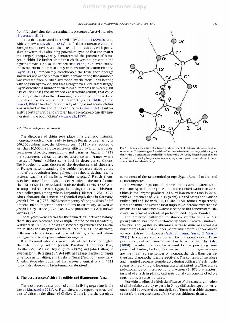
The most recent description of chitin in living organisms is theone by Muzzarelli (2011). As Fig. 1 shows, the repeating structuralunit of chitin is the dimer of GlcNAc. Chitin is the characteristic
Fig. 1. Chemical structure of a disaccharide segment of chitosan, showing positionnumbering. The two angles � and ̊ define the chain conformation, and the angle �define the O6 orientation. Dashed lines denote the O3–O5 hydrogen bonds that arecrucial for rigidity. Hydrogen bonds connecting various positions of adjacent chainsare omitted for sake of clarity.
component of the taxonomical groups Zygo-, Asco-, Basidio- andDeuteromycetes.
The worldwide production of mushrooms was updated by theFood and Agriculture Organization of the United Nations in 2009.China is the largest producer (>1.5 million metric tons in 2007,with an increment of 65% in 10 years). United States and Canadaranked 2nd and 3rd with 390,000 and 81,500 tonnes, respectively.Israel and India showed the most impressive increase over the saiddecade, due to consumer awareness of the health benefits of mush-rooms, in terms of contents of prebiotics and polysaccharides.
The preferred cultivated mushroom worldwide is A. bis-porus (button mushroom), followed by Lentinus edodes (shiitake),Pleurotus spp (oyster mushrooms), Auricula auricula (wood earmushroom), Flamulina velutipes (winter mushroom) and Volvariellavolvacea (straw mushroom) (Aida, Shuhaimi, Yazid, & Maaruf,2009). The chemical composition and the nutritional value of Euro-pean species of wild mushrooms has been reviewed by Kalac(2009): carbohydrates usually account for the prevailing com-ponent of fruiting bodies: glucose, mannitol and �,�-trehaloseare the main representatives of monosaccharides, their deriva-tives and oligosaccharides, respectively. The contents of trehaloseand mannitol decrease considerably during boiling of fresh mush-rooms, while drying and freezing results in limited loss. The reservepolysaccharide of mushrooms is glycogen (5–10% dry matter),instead of starch in plants. Anti-nutritional components of ediblemushrooms are also indicated.
Notwithstanding the high significance of the structural modelsof chitin elaborated by experts in X-ray diffraction spectrometry,one should be aware of the multiplicity of forms that chitin assumesto satisfy the requirements of the various chitinous tissues.
Author's personal copy
998 R.A.A. Muzzarelli et al. / Carbohydrate Polymers 87 (2012) 995– 1012
3.1. Chitin in edible fungi
The chitin content of pileus and stipes of fruit bodies of culti-vated Agaricus bisporus, Pleurotus ostreatus and Lentinula edodes isa stable characteristic of the species and there are no significantdifferences between the varieties. The chitin levels of pileus andstipes are not significantly different (for A. bisporus, 6.68 and 7.25)but they are for P. ostreatus and L. edodes, for which the pileus hashigher chitin content than the stipe. The saprotrophic mushroom A.bisporus has higher chitin level than both wood-rotting P. ostreatusand L. edodes. The chitin of cultivated mushrooms is an importantcomponent of their nutritional value (Vetter, 2007).
Evidence for covalent linkages between chitin and �-glucanin Schizophyllum commune (Basidiomycete) was provided bySietsma and Wessels (1979) among others. The isolation ofthe chitin + glucan complex from the biomass of Armillariellamellea and the yellow morel Morchella esculenta (classes Basid-iomycetes and Ascomycetes, respectively), has been developedby Ivshina, Artamonova, Ivshin, and Sharnina (2009). The foursteps were deproteinization, demineralization, depigmentation,and final removal of residual protein. The chitin in the isolated com-plexes was ca. 80% and 47%, respectively; the degree of acetylationwas 0.80 and 0.41, respectively, while the degree of crystallinitywas 61 for both; various analytical techniques demonstrated theidentity of fungal and arthropod chitin.
The solid-state fermentation of L. edodes was found to be anefficient approach to chitosan production, according to Crestiniand Giovannozzi-Sermanni (1996); in fact, the chitosan yield was6.18 g/kg and the acetylation degree was 12.5% at 12 days afterinoculation. These data are of particular significance: the solid-state fermentation gave yields 50 times higher than submergedfermentation, and the chitosan had smaller degree of acetylationthan crustacean commercial chitosans.
3.2. Chitin and chitosan in filamentous fungi
Yeasts and filamentous fungi contain glucan complexes withchitin or chitosan in their cell walls and septa (Muzzarelli, Tanfani,& Emanuelli, 1981; Smith & Berry, 1977; Trinci, 1978). San-blas andCarbonell (1974) published microphotographs of stained materialfrom the mycelia of Histoplasma farciminosum treated with chiti-nase and �-1,3 glucanase, showing the remaining thick bundles42 nm wide composed of 5-nm fibres made of �-1,3 glucan andchitin that escaped hydrolysis by said lytic enzymes thanks to theirstability.
In the order Mucorales many strains have interesting produc-tivity of chitosan, in particular Absidia spp as documented byKuhlmann et al. (2000): MW as high as 700 kDa and yields of1.0–1.3 g per liter of submerged culture medium were obtained.For example, the mycelia of cultured Absidia glauca var. paradoaxawere treated with hot 2% sodium hydroxide to isolate the alkali-insoluble polysaccharides, from which the extraction of chitosanwas carried out with 2% acetic acid at room temperature. Thechitosan extracted had degree of deacetylation 0.86, and viscos-ity 4.0 cP at 0.1% chitosan in 0.5% acetic acid that indicates lowmolecular weight (Rungsardthong, Wonputtanakul, Kongpien, &Chotiwaranon, 2006). Niederhofer and Muller (2004) extracted chi-tosan with average MW 45 kDa from Absidia coerulea.
Chitosan is the most abundant component of both filamentousand yeast-like forms of Mucor rouxii: the maximum chitosan lev-els are 32.7 and 27.9%, respectively, according to Bartnicki-Garciaand Nickerson (1962) while the degree of acetylation of chitosanis 0.19. Chitosan is also accompanied by minor amounts of chitinand glucan. The pathway of chitosan formation in M. rouxii, includ-ing the description of the role of chitin deacetylase (the enzymethat hydrolyses the acetamido groups of chitin) was reported by
Araki and Ito (1975). The biochemistry of Mucor was exhaustivelytreated by Sypherd, Borgia, and Paznokas (1978) and the glyoxylatecycle in Mucor was detailed by O’Connor and Paznokas (1980). Theproduction yield of chitosan from M. rouxii was greatly improvedwith the aid of plant growth hormones, viz., indoleacetic acid,indolebutyric acid, kinetin and gibberellic acid: the yield of chi-tosan increased 34–69%, gibberellic acid being the most potenthormone (Chatterjee, Adhya, Guha, & Chatterjee, 2005; Chatterjee,Chatterjee, Chatterjee, & Guha, 2009).
A. niger, instead, contains only chitin. Amounts of glucan inthe range 7.4–39.8% are associated with chitin and are elimi-nated by extraction. In the A. niger mycelium, Wu, Zivanovic,Draughon, and Sams (2004) and Wu, Zivanovic, Draughon, Conway,and Sams (2005) found that the maximum glucosamine level was11.10% d.w. The degrees of acetylation were determined to be0.76–0.50. In consideration of the large spent biomass generatedduring the production of citric acid, many authors characterizedthe chitin + glucan complex present in the cell walls of A. niger witha view at its recovery (Stagg & Feather, 1973) and determined therecoverable amount of chitin. In the earliest report on the isolatedchitosan + glucan from A. niger, the statistical evaluation of a largeset of preparative conditions indicated that a 4-h treatment with40% NaOH aqueous solution without nitrogen blanketing at 128 ◦Cprovided satisfactory yield (44%) and good chitosan content (>32%).These products were not fully soluble in 5% acetic acid, but disper-sions were obtained upon prolonged stirring. The infrared spectrataken on the fractions obtained after filtration indicated differentratios of chitosan and glucan. The collection percentages of 8 tran-sition metal ions were definitely higher than the correspondingvalues for animal chitosan despite the fact that the complex con-tains nearly one half chitosan by weight, this fact being justified bythe much bigger the surface area. The filamentous mycelia shapeswere still visible under the microscope in rather frail films obtainedthanks to the retained typical filmogenicity of chitosan (Muzzarelli,Tanfani, & Scarpini, 1980). Evidence of the covalent binding ofchitin with glucans was confirmed many years later for a variety offungi by Nwe, Stevens, Tokura, and Tamura (2008), Nwe, Furuike,and Tamura (2010), Vincendon and Desbrieres (2002) and Zamani,Edebo, Sjostrom, and Taherzadeh (2007) among others. The glu-cans have been determined quantitatively in edible mushrooms byNitschke et al. (2011).
The chitin + glucan from A. niger was later produced industrially:the poly(N-acetyl-d-glucosamine) and the ˇ(1,3)-d-glucan werepresent in the w/w ratio 35/65 as determined by 13C-solid stateNMR. Said material was studied in an animal model of atheroscle-rosis by Berecochea-Lopez et al. (2009): it strongly reduced thearea of aortic fatty streak deposition by 87–97%, cardiac produc-tion of superoxide anion by 25%; on the other hand, it enhancedliver superoxide dismutase activity by 7–45% and glutathione per-oxidase activity by 38–120%. These findings support the viewthat constant consumption of chitin + glucan has potential effectsagainst atherosclerosis, the underlying mechanism being relatedmainly to improving the antioxidant defenses. No undesirable ortoxic effects were detected, nor evidence of other clinical signs,thus at low doses chitin + glucan was deemed to be a safe nutraceu-tical supplement. Certain aspects of said work were confirmed byNeyrinck et al. (2009) who concluded that fungal chitosan coun-teracts some inflammatory disorders and metabolic alterationsoccurring in diet-induced obese mice since it decreases feed effi-ciency, adipocytokine secretion, fat mass and ectopic fat depositionin the liver and the muscle. The Absidia chitosans tested accordingto ASTM F813-83 and F619-79 with murine fibroblasts were foundto be biocompatible in terms of cytotoxicity and lactate dehydro-genase determinations in agreement with Muzzarelli, Ilari, Tarsi,Dubini, and Xia (1994), Muzzarelli, Ilari, Xia, Pinotti, and Tomasetti(1994) and Chung, Schmidt, Hamlyn, and Sagar (1994).
Author's personal copy
R.A.A. Muzzarelli et al. / Carbohydrate Polymers 87 (2012) 995– 1012 999
Potato peel was used as a substrate for chitosan productionfrom Rhizopus oryzae. The best extraction condition was using 46%sodium hydroxide at 45 ◦C for 13 h followed by 2% acetic acid at95 ◦C for 8 h: the chitosan yield obtained was 10.8 g/kg substrateafter 5 days of fermentation at pH 5. The R. oryzae chitosan had86–90% degree of deacetylation, molecular weight of 80–128 kDaand viscosity of 3.1–6.1 mPa s: this set of results qualify this sys-tem as a convenient one for fungal chitosan production (Kleekayai& Suntornsuk, 2011).
3.3. Filamentous fungi in traditional foods
While there is much interest in growing filamentous fungi inorder to produce fungal chitin and chitosan, it should be noted thatthe microbial cells (algae, bacteria, yeasts and fungi) have a placein world food supply from both standpoints of animal feedstuff andhuman food applications, and that this will continue in the future. Itshould not be expected, however, that the portion of edible proteinof microbial origin will be quite large.
Filamentous fungi in fact are present in traditional sourcesof food in many regions of the world. The Indonesian tempe(or tempeh), for example, consists of soybean fermented by thezygomycete Rhizopus oligosporus and contains a fraction of glu-cosamine that can reach important concentrations, from 51 to111 g/kg dry biomass. Values of glucosamine as high as 97 and93 g/kg of dry biomass for submerged cultures of Rhizopus japonicusand Mucor racemosus, respectively were reported. The glucosaminecontent of the mycelium can vary significantly, being strongly influ-enced by the growth medium; it increases in old mycelia where thechitin fraction is larger (Sasaki, Kodama, Uchida, & Yoshino, 1985;Sparringa & Owens, 1999).
Moreover Roubos-van den Hil, Rob Nout, van der Meulen, andGruppen (2010) demonstrated tempe to play a bioactive functionagainst toxigenic Escherichia coli. This effect of soybean tempe isobtained by inhibiting the adhesion of enterotoxigenic E. coli tointestinal brush border cells, thus preventing the most frequentcause of childhood diarrhea in developing countries, and of trav-eler’s diarrhea. Moreover, other foods such as barley, chick pea,cowpea, groundbean, horsebean, pea, oats, sorghum and wheat arealso good substrates for growing chitin-bearing fungi.
4. Human chitinases in nutrition and metabolism
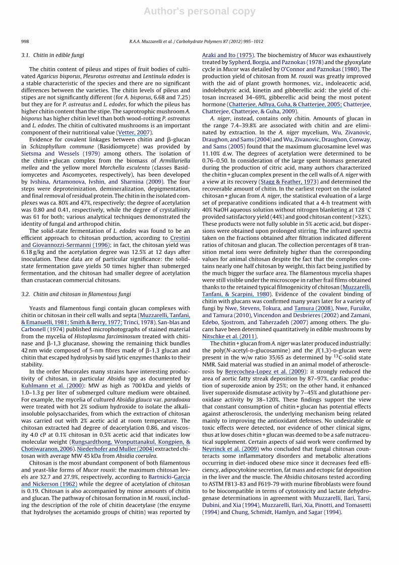
Chitinases hydrolyze the �1–4 bonds of the chitin chain downto the N-acetyl-d-glucosamine dimer (Jollès & Muzzarelli, 1999).This is an extraordinary performance because the substrate chitinin most instances is an insoluble and crystalline solid that assumesa large variety of forms depending on the role of the living tissue,an example being the one in Fig. 2.
These enzymes are highly preserved through species and king-doms (Boot et al., 2001; Gianfrancesco & Musumeci, 2004) andexert various functions in many living organisms. For example, inyeast and fungi they take part in morphogenesis (Cohen-Kupiecand Chet, 1998), and in plants they have an important role againstpathogens; they are also implicated in the processes of develop-ment and growth, i.e. during insect ecdysis (Kaspresewska, 2003).
Until 1994 the human organism was not thought to pro-duce chitinases, because of the absence of endogenous chitin inhuman tissues, which means no need for enzymatic turnover andremodeling of chitin structures. Therefore nutritionists have indi-cated chitin as a non-digestible polymer in the human organism(Bukkens, 2005; DeFoliart, 1992) although chitin-containing foodssuch as mushroom, crustaceans and insects are largely consumed,especially in tropical countries. This assertion, however, has beenrecently contradicted by reports describing up to nine mammalian
Fig. 2. Scanning electron micrographs of different regions within a single wingscaleof Callophrys rubi. The scale of the structure is commensurate with the wavelengthof visible light, with an edge of the conventional cubic unit cell of the parallel-Gyroid of approximately 310 nm. In the butterfly, the templating is achieved bythe lipid–protein membranes within the smooth endoplasmic reticulum (while itremains in the chrysalis), that likely form membranes folded according to the form ofthe Gyroid. The subsequent formation of the chiral rigid chitin framework is drivenby the gradual polymerization of the chitin precursors; the material is opticallyactive, that is, it rotates the polarisation of incoming light.
Source: From Schroder-Turk et al. (2011). Elsevier Science, San Diego.
chitinases and chitinase-like genes that are present in man andother mammals, all belonging to the glycosyl hydrolase 18 fam-ily (Bierbaum et al., 2005; Boot et al., 2001; Fusetti et al., 2002; Lee,Waalen, Crain, Smargon, & Beutler, 2007; Seibold et al., 2009).
The first human chitinase was detected by Hollak, van Weely,van Oers, and Aerts (1994) who found high chitinolytic activityin serum of patients affected by Gaucher disease. Because of itscapacity to hydrolyze chitotriose, it was called chitotriosidase. Thisprotein is specifically expressed by activated macrophages and hasan optimum pH of 6. Subsequent studies proved and quantifiedthe chitinolytic activity of chitotriosidase on colloidal chitin and onchitin of cell wall of Candida albicans. In the early investigations,a catalytic deficiency of chitotriosidase was described and namedAllele H. The complete sequencing of chitotriosidase gene was alsodescribed (Barone, Simpore, Malaguarnera, Pignatelli, & Musumeci,2003; Boot, Renkema, Strijland, van Zonneveld, & Aerts, 1995; Bootet al., 1998; Renkema, Boot, Muijsers, Donker-Koopman, & Aerts,1995).
In search of other chitinases that could compensate chitotriosi-dase deficiency, Bleau, Massicotte, Merlen, and Boisvert (1999)and Chang et al. (2001) identified and characterized 4 mammalianchitinase-like enzymes that, because of mutations in their highlyconserved catalytic sites, maintain chitin-binding capability but notthe catalytic activity. Boot et al. (2001) discovered another chitinaseshowing high catalytic activity at pH 2 and named it acidic mam-malian chitinase (AMCase); it is expressed in gastric epithelia, inlung macrophages and in the pulmonary epithelia during asthmaticinflammation (Zhu et al., 2004).
4.1. Role of human chitinases
Since the earliest discoveries, scientists hypothesized that mam-malian chitinases take part in the innate immune response toparasites, with the function of binding and digesting chitin struc-tures such as cell walls, egg shells and cysts produced by manyorganisms including fungi, protozoans and nematodes (Fusettiet al., 2002; Malaguarnera et al., 2003 among others).
Author's personal copy
1000 R.A.A. Muzzarelli et al. / Carbohydrate Polymers 87 (2012) 995– 1012
Barone et al. (2003) detected higher chitotriosidase activity inplasma of African children affected by acute malaria with respect toreference values obtained in age-matched African children. More-over, chitotriosidase activity was found to be higher in Africanchildren than in Caucasian children matched for age. Musumeciet al. (2005) obtained similar results by comparing chitotriosidaseconcentrations in colostrum in African and Caucasian populations.
Furthermore, AMCase is adapted to different tissue-specificfunctions and Boot et al. (2005) discussed the possibility that thischitinase might have a dual function in immune defense and in fooddigestion. Nature published the study of Reese et al. (2007), who,using a murine model of infection with the nematode Nippostrongy-lus brasiliensis, induced alternative macrophages activation andrecruitment of Th2 cells, eosinophils and basophils in different tis-sues. Moreover, in the same experiment they demonstrated thatwhen chitin was pre-treated with a recombinant AMCase, or intransgenic mice over-expressing active AMCase, the inflammatoryresponsiveness to chitin was reduced. Further data are in Lee et al.(2011).
4.2. Chitin digestion
The state of the art of human activity of collecting, cultivatingand consuming chitin-bearing food such as crustaceans, insects andmushrooms has been the object of several comprehensive ecolog-ical and ethnobiological investigations (DeFoliart, 1992; Paoletti &Bukkens, 1997; Paoletti, 2005). The importance of entomophagyhas been also remarked by FAO, which stressed the great diver-sity and accessibility of this class of alimentary resources, being asource of inorganics, proteins, vitamins and poly-unsaturated fattyacids (Bukkens, 2005; Fontaneto et al., 2011).
More than 2000 species of arthropods are mostly used as foodby many populations worldwide, as extensively described and dis-cussed by Posey (1984) for Amazonia, Ramos-Elorduy et al. (1997)for Central America, Malaisse (1997) for central Africa, Zhi-Yi (2005)for China, and Mitsihashi (2005) for Japan. For example, within theTukanoan population living in the Colombian Vaupes Amazon rainforest, arthropods represent a fundamental source of food, provid-ing 12–24% of total protein intake (Paoletti, Buscardo, & Dufour,2000).
Chitin represents 5–20% of total dry-weight in adult crus-taceans and insects. For this reason, Paoletti, Norberto, Damini, andMusumeci (2007) and Paoletti, Norberto, Cozzarini, and Musumeci(2009) suggested that a chitin-rich diet may influence chitinasevariability in human populations. Consequently they proposed thatthe reduction of chitin intake in western diet may have reducedselective pressure on the chitinase genes, probably resulting in theloss of catalytic efficiency.
Chitin digestion seems to depend on AMCase, the main chiti-nase synthesized by gastric tissues and, therefore, adapted toextremely acidic pH conditions (Boot et al., 2005). Human chiti-nases hydrolyze chitin to N-acetyl-glucosamine that can be easilymetabolized by N-acetylglucosamine kinase, an enzyme present inall human tissues including gastric mucosa and intestinal epithe-lium, as described by Gindzienski, Głowacka, and Zwierz (1974).Also the commensal human bacteria Clostridium paraputrificum wasisolated in human faeces and was found to have chitinolytic activity(Simunek, Kopecny, Hodrova, & Bartonova, 2002).
Genetic and histochemical analyses of mammalian chitinaseexpression, conducted by Suzuki et al. (2002) and Goto, Fujimoto,Nio, Iwanaga, and Kawasaki (2003), respectively, reported thepresence of AMCase in the human and murine gastric mucosae.Shortly afterwards, Boot et al. (2005) confirmed elevated expres-sion of mammalian chitinases in the glandular portion of thestomach, in chief cells at the bottom of the gastric glands. Interest-ingly, pyloric glands in the antrum of the stomach do not express
chitinase mRNA: likewise AMCase was not detected in the intestine.On the other hand, chitotriosidase is synthesized also in duodenumand other parts of the small intestine, in specialized secretory cellscalled Paneth cells, probably with a defensive role against parasites.Boot et al. (2005) discussed the possibility that gastrointestinalchitinases might have a dual function, in immune defense and infood digestion, thus supporting the observations by Dobson, Prager,and Wilson (1984).
AMCase-dependent chitinolytic activity was finally proved inhumans and quantified by Paoletti et al. (2007) who analyzedthe gastric juice of 25 patients submitted to gastroscopy. TheAMCase activity was tested in vitro on two artificial substrates,4-methylumbelliferyl-�-d-N,N′-diacetylchitobiose and/or fluores-cein isothiocyanate chitin. Out of the 25 patients as many as 20showed a detectable activity, ranging from 0.21 to 36.27 nmol/ml/hand 8881–1,254,782 fluorescence emission units, with the highestchitinolytic activity observed at pH 2, as expected. In subse-quent experiments realized on human gastric juices, Cozzariniet al. (2009) associated increased expression of chitotriosidase,but not of AMCase, to Helicobacter pylori and other gastricdiseases.
Subsequent studies were performed to test in vitro the abilityof human gastric juices to hydrolyze natural chitin model obtainedfrom the wings of Calliphora vomitoria. Fly wings were found tobe only moderately digested by gastric juices even though AMCaselevels were high, thus no significant results were obtained fromthese unpublished studies: it might be supposed however that inthe wings the chiral rigid chitin framework shown in Fig. 2 canwithstand the enzymatic attack better than other chitinous sub-strates.
The most complete study on AMCase genetic variability, stem-ming from an inter-institutional investigation on a large cohort ofLatin-American and Afro-American asthmatics and their relatives,has been published by Seibold et al. (2009). Interestingly, they iden-tified a particular haplotype codifying for an isoform that revealed,by biochemical characterization, an enhanced efficiency and adapt-ability at three different pH values, those characteristic of stomach,lung and lysosomes, thus confirming the hypothesis of a tissue-specific adaptation of AMCase. The consistent increase in enzymaticactivity at pH 2.2 (4-fold higher) and 7.0 (10-fold higher), typicalof stomach and lung, respectively, is extremely relevant becausethese organs are intensively exposed to chitin through ingestionand inhalation, respectively. This functional variant has a relevantfrequency in the whole sample, specifically: 24.1% in AfroAmerindi-ans, 12.5% in Mexicans and 8.4% in Puerto Ricans.
Thus the expression of AMCase in the human gastrointestinaltract can represent a reminiscence of a past nutritional function(Boot et al., 2005), and, therefore, AMCase expression in west-ern population now can be a redundant phenomenon due to thereduction of chitin in modern diet (Paoletti et al., 2007). To testthis hypothesis, the University of Padua, in collaboration with theUniversity of Trujillo (Peru), is currently performing a study onchitinase genetic variability in Amerindian populations living inthe Amazonian basin, where dependence on chitinous food maystill represent a strong selective factor for chitinase functionality.Two intriguing ethnobiological findings that expect to be assessedand confirmed, emerged recently from the first fieldwork in thoseregions: the traditional use, as an anti-asthmatic treatment, of asyrup produced from suri palm-worm (Rhynchophorus palmarum,Coleoptera) and the popular belief that states: people eating insectsdo not suffer from asthma.
4.3. Deficiency of chitotriosidase
A relevant question has been whether chitinases activity inhumans is just an evolutionary remnant or they still have an
Author's personal copy
R.A.A. Muzzarelli et al. / Carbohydrate Polymers 87 (2012) 995– 1012 1001
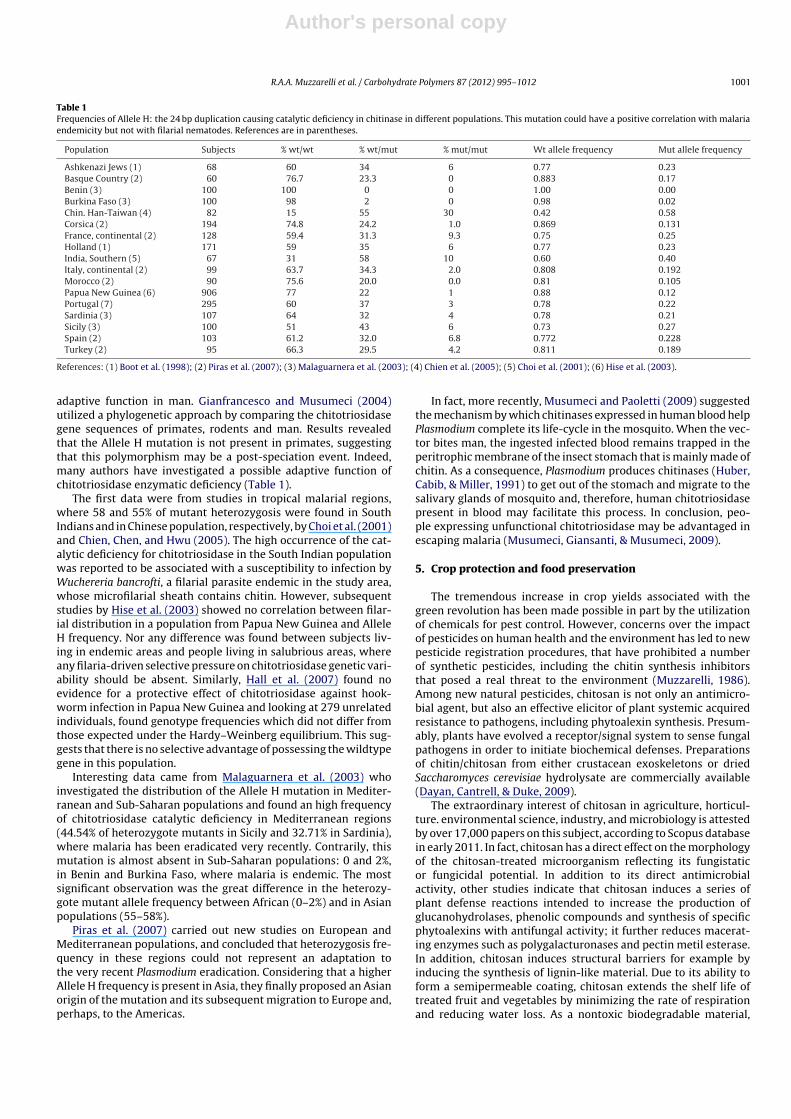
Table 1Frequencies of Allele H: the 24 bp duplication causing catalytic deficiency in chitinase in different populations. This mutation could have a positive correlation with malariaendemicity but not with filarial nematodes. References are in parentheses.
Population Subjects % wt/wt % wt/mut % mut/mut Wt allele frequency Mut allele frequency
Ashkenazi Jews (1) 68 60 34 6 0.77 0.23Basque Country (2) 60 76.7 23.3 0 0.883 0.17Benin (3) 100 100 0 0 1.00 0.00Burkina Faso (3) 100 98 2 0 0.98 0.02Chin. Han-Taiwan (4) 82 15 55 30 0.42 0.58Corsica (2) 194 74.8 24.2 1.0 0.869 0.131France, continental (2) 128 59.4 31.3 9.3 0.75 0.25Holland (1) 171 59 35 6 0.77 0.23India, Southern (5) 67 31 58 10 0.60 0.40Italy, continental (2) 99 63.7 34.3 2.0 0.808 0.192Morocco (2) 90 75.6 20.0 0.0 0.81 0.105Papua New Guinea (6) 906 77 22 1 0.88 0.12Portugal (7) 295 60 37 3 0.78 0.22Sardinia (3) 107 64 32 4 0.78 0.21Sicily (3) 100 51 43 6 0.73 0.27Spain (2) 103 61.2 32.0 6.8 0.772 0.228Turkey (2) 95 66.3 29.5 4.2 0.811 0.189
References: (1) Boot et al. (1998); (2) Piras et al. (2007); (3) Malaguarnera et al. (2003); (4) Chien et al. (2005); (5) Choi et al. (2001); (6) Hise et al. (2003).
adaptive function in man. Gianfrancesco and Musumeci (2004)utilized a phylogenetic approach by comparing the chitotriosidasegene sequences of primates, rodents and man. Results revealedthat the Allele H mutation is not present in primates, suggestingthat this polymorphism may be a post-speciation event. Indeed,many authors have investigated a possible adaptive function ofchitotriosidase enzymatic deficiency (Table 1).
The first data were from studies in tropical malarial regions,where 58 and 55% of mutant heterozygosis were found in SouthIndians and in Chinese population, respectively, by Choi et al. (2001)and Chien, Chen, and Hwu (2005). The high occurrence of the cat-alytic deficiency for chitotriosidase in the South Indian populationwas reported to be associated with a susceptibility to infection byWuchereria bancrofti, a filarial parasite endemic in the study area,whose microfilarial sheath contains chitin. However, subsequentstudies by Hise et al. (2003) showed no correlation between filar-ial distribution in a population from Papua New Guinea and AlleleH frequency. Nor any difference was found between subjects liv-ing in endemic areas and people living in salubrious areas, whereany filaria-driven selective pressure on chitotriosidase genetic vari-ability should be absent. Similarly, Hall et al. (2007) found noevidence for a protective effect of chitotriosidase against hook-worm infection in Papua New Guinea and looking at 279 unrelatedindividuals, found genotype frequencies which did not differ fromthose expected under the Hardy–Weinberg equilibrium. This sug-gests that there is no selective advantage of possessing the wildtypegene in this population.
Interesting data came from Malaguarnera et al. (2003) whoinvestigated the distribution of the Allele H mutation in Mediter-ranean and Sub-Saharan populations and found an high frequencyof chitotriosidase catalytic deficiency in Mediterranean regions(44.54% of heterozygote mutants in Sicily and 32.71% in Sardinia),where malaria has been eradicated very recently. Contrarily, thismutation is almost absent in Sub-Saharan populations: 0 and 2%,in Benin and Burkina Faso, where malaria is endemic. The mostsignificant observation was the great difference in the heterozy-gote mutant allele frequency between African (0–2%) and in Asianpopulations (55–58%).
Piras et al. (2007) carried out new studies on European andMediterranean populations, and concluded that heterozygosis fre-quency in these regions could not represent an adaptation tothe very recent Plasmodium eradication. Considering that a higherAllele H frequency is present in Asia, they finally proposed an Asianorigin of the mutation and its subsequent migration to Europe and,perhaps, to the Americas.
In fact, more recently, Musumeci and Paoletti (2009) suggestedthe mechanism by which chitinases expressed in human blood helpPlasmodium complete its life-cycle in the mosquito. When the vec-tor bites man, the ingested infected blood remains trapped in theperitrophic membrane of the insect stomach that is mainly made ofchitin. As a consequence, Plasmodium produces chitinases (Huber,Cabib, & Miller, 1991) to get out of the stomach and migrate to thesalivary glands of mosquito and, therefore, human chitotriosidasepresent in blood may facilitate this process. In conclusion, peo-ple expressing unfunctional chitotriosidase may be advantaged inescaping malaria (Musumeci, Giansanti, & Musumeci, 2009).
5. Crop protection and food preservation
The tremendous increase in crop yields associated with thegreen revolution has been made possible in part by the utilizationof chemicals for pest control. However, concerns over the impactof pesticides on human health and the environment has led to newpesticide registration procedures, that have prohibited a numberof synthetic pesticides, including the chitin synthesis inhibitorsthat posed a real threat to the environment (Muzzarelli, 1986).Among new natural pesticides, chitosan is not only an antimicro-bial agent, but also an effective elicitor of plant systemic acquiredresistance to pathogens, including phytoalexin synthesis. Presum-ably, plants have evolved a receptor/signal system to sense fungalpathogens in order to initiate biochemical defenses. Preparationsof chitin/chitosan from either crustacean exoskeletons or driedSaccharomyces cerevisiae hydrolysate are commercially available(Dayan, Cantrell, & Duke, 2009).
The extraordinary interest of chitosan in agriculture, horticul-ture. environmental science, industry, and microbiology is attestedby over 17,000 papers on this subject, according to Scopus databasein early 2011. In fact, chitosan has a direct effect on the morphologyof the chitosan-treated microorganism reflecting its fungistaticor fungicidal potential. In addition to its direct antimicrobialactivity, other studies indicate that chitosan induces a series ofplant defense reactions intended to increase the production ofglucanohydrolases, phenolic compounds and synthesis of specificphytoalexins with antifungal activity; it further reduces macerat-ing enzymes such as polygalacturonases and pectin metil esterase.In addition, chitosan induces structural barriers for example byinducing the synthesis of lignin-like material. Due to its ability toform a semipermeable coating, chitosan extends the shelf life oftreated fruit and vegetables by minimizing the rate of respirationand reducing water loss. As a nontoxic biodegradable material,
Author's personal copy
1002 R.A.A. Muzzarelli et al. / Carbohydrate Polymers 87 (2012) 995– 1012
as well as an elicitor, chitosan has the potential to become a newclass of plant protectant, assisting towards the goal of sustainableagriculture (Bautista-Banos et al., 2006).
5.1. Applications in crop protection
Attack by various fungi to certain seeds may result in decreasedgermination (Donald & Mirocha, 1977; Sharma, Fisher, & Webster,1977). Chitosan-based coatings exerting antifungal activity helppreserve the quality of the stored seeds. The treatment consistsin seed immersion in a chitosan suspension (up to 4%) followedby drying. Chitosan molecular weight, presence of a surfactant, pHvalue, and thickness (number of coating layers) are parameters act-ing on the seed germination, as well as on the fungal activity andvegetative growth. Generally, chitosan treatments reduce the num-ber of type of fungi and promote plant growth. Mazaro et al. (2009)treated tomato and beet seeds by immersion in a chitosan suspen-sion. Seeds were then sowed in trays with a substrate infected withRhizoctonia spp. and maintained in greenhouse for 14 days. Chi-tosan induced seedling resistance against Rhizoctonia and reduceddamping-off. It increased the phenylalanine ammonialyase activityand interfered with the total proteins and total and reduced sugarsrates in the leaves. The partially hydrolyzed chitosans lead to betterresults in terms of microbial contamination and germination.
Zeng and Shi (2009) obtained another safer, cheaper and moreenvironmentally friendly seed coating agent using chitosan com-bined with plant growth regulators and other additives. Such anovel seed coating agent significantly enhanced sprout growth inregard of the traditional agents. It stimulated the seedling growthof rice, advanced the growth of root, improved root activity andincreased the crop yield in the germination test and field trial. Com-pared with the traditional rice seed coating agent, the crop yield ofcoated seeds increased by 5%, and at 25% lower cost. The fungalinhibition test of this seed coating agent showed that it has an evi-dent fungal inhibitory effect and a higher safety index during usageand disposal.
As noted by El Hadrami, Adam, El Hadrami, and Daayf (2010),since chitin and chitosan fragments are known to have elicitingactivities leading to a variety of defensive responses in host plantsagainst microbial infections (e.g. accumulations of phytoalexins,pathogen-related proteins and proteinase inhibitors, lignin synthe-sis and callose formation) the use of chitin and chitosan derivativeshas increased, because they allow to increase host plant defenses inagricultural systems and to reduce the negative impact of diseaseson yield and quality of crops.
5.2. Applications in food technology
The main applications of chitosan in the food area are inregard of its flocculating and antibacterial activities (Vargas andGonzález-Martínez (2010)). In fact, chitosan and its derivatives arecharacterized by great versatility in flocculation and adsorptioncapacity mainly because of their cationicity and macromolecu-lar structure. Chitosan is used as a flocculant in various domainsand particularly in food technology (Zheng, Wu, & Xu, 2009), forexample for fruit juice clarification in eliminating pectin and car-bohydrates, as an aid for the separation of suspended particles frombeverages (Chatterjee, Chatterjee, Chatterjee, & Guha, 2004). Suchjuice clarifications have been successfully carried out for apple,carrot, grape, lemon, orange and pineapple juices (Rungsardthonget al., 2006).
Moreover since chitosan has a good affinity for polyphenoliccompounds such as catechins, proanthocyanidins, cinnamic acid,and their derivatives, it can be used to remove polyphenolic com-ponents which allows to reduce juice color (Oszmianski & Wojdylo,2007; Spagna et al., 1996) or to change the initial color of a
solution. With the aid of xanthan, the removal of haze precursorsbecomes feasible and has some advantages in terms of time savingin comparison to conventional fining methods (Fang, Zhang, Tao,Sun, & Sun, 2006). Chitosan treatment, can also clarify leave infu-sions (e.g. green tea), thus avoiding the formation of haze duringlow-temperature storage. In order to understand better the coag-ulation process, some studies have also been performed to analyzethe interaction between chitosan and proteins (Boeris, Micheletto,Lionzo, da Silveira, & Pico, 2011; Hiorth, Skoien, & Sande, 2010;Marudova, McDougall, & Ring, 2004).
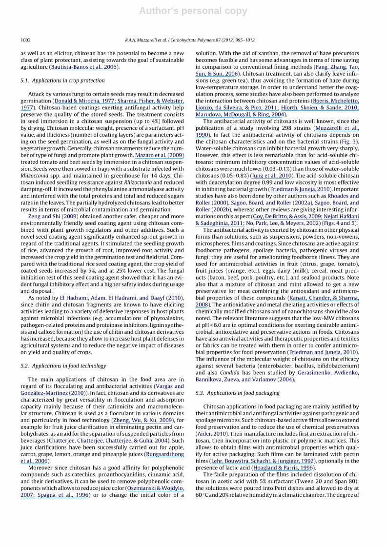
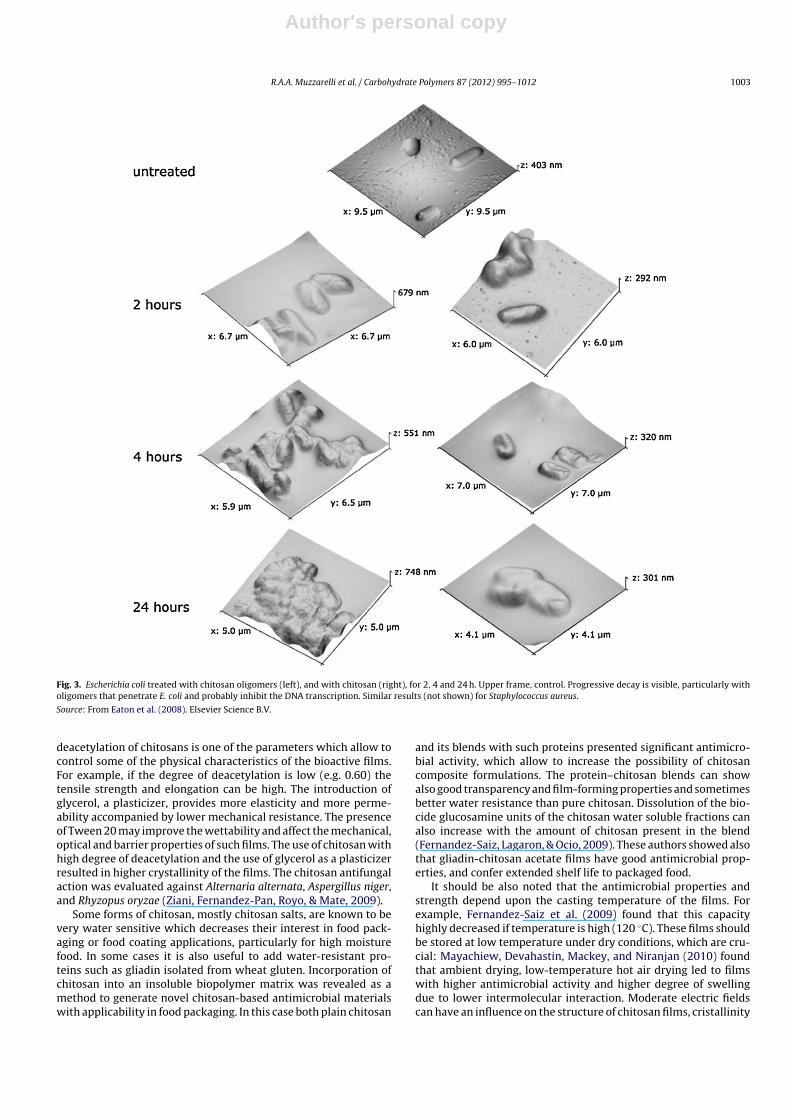
The antibacterial activity of chitosans is well known, since thepublication of a study involving 298 strains (Muzzarelli et al.,1990). In fact the antibacterial activity of chitosans depends onthe chitosan characteristics and on the bacterial strains (Fig. 3).Water-soluble chitosans can inhibit bacterial growth very sharply.However, this effect is less remarkable than for acid-soluble chi-tosans: minimum inhibitory concentration values of acid-solublechitosans were much lower (0.03–0.1%) than those of water-solublechitosans (0.05–0.8%) (Jung et al., 2010). The acid-soluble chitosanwith deacetylation degree 0.99 and low viscosity is most effectivein inhibiting bacterial growth (Friedman & Juneja, 2010). Importantstudies have also been done by other authors such as Rhoades andRoller (2000), Sagoo, Board, and Roller (2002a), Sagoo, Board, andRoller (2002b), whereas other reviews are giving interesting infor-mations on this aspect (Goy, De Britto, & Assis, 2009; Nejati Hafdani& Sadeghinia, 2011; No, Park, Lee, & Meyers, 2002) (Figs. 4 and 5).
The antibacterial activity is exerted by chitosan in other physicalforms than solutions, such as suspensions, powders, non-vowens,microspheres, films and coatings. Since chitosans are active againstfoodborne pathogens, spoilage bacteria, pathogenic viruses andfungi, they are useful for ameliorating foodborne illness. They areused for antimicrobial activities in fruit (citrus, grape, tomato),fruit juices (orange, etc.), eggs, dairy (milk), cereal, meat prod-ucts (bacon, beef, pork, poultry, etc.), and seafood products. Notealso that a mixture of chitosan and mint allowed to get a newpreservative for meat combining the antioxidant and antimicro-bial properties of these compounds (Kanatt, Chander, & Sharma,2008). The antioxidative and metal chelating activities or effects ofchemically modified chitosans and of nanochitosans should be alsonoted. The relevant literature suggests that the low-MW chitosansat pH < 6.0 are in optimal conditions for exerting desirable antimi-crobial, antioxidative and preservative actions in foods. Chitosanshave also antiviral activities and therapeutic properties and textilesor fabrics can be treated with them in order to confer antimicro-bial properties for food preservation (Friedman and Juneja, 2010).The influence of the molecular weight of chitosans on the efficacyagainst several bacteria (enterobacter, bacillus, bifidobacterium)and also Candida has been studied by Gerasimenko, Avdienko,Bannikova, Zueva, and Varlamov (2004).
5.3. Applications in food packaging
Chitosan applications in food packaging are mainly justified bytheir antimicrobial and antifungal activities against pathogenic andspoilage microbes. Such chitosan-based active films allow to extendfood preservation and to reduce the use of chemical preservatives(Aider, 2010). Their manufacture includes first an extraction of chi-tosan, then incorporation into plastic or polymeric matrices. Thisallows to obtain films with antimicrobial properties which qual-ify for active packaging. Such films can be laminated with pectinfilms (Lehr, Bouwstra, Schacht, & Jungiger, 1992), optionally in thepresence of lactic acid (Hoagland & Parris, 1996).
The facile preparation of the films included dissolution of chi-tosan in acetic acid with 5% surfactant (Tween 20 and Span 80):the solutions were poured into Petri dishes and allowed to dry at60 ◦C and 20% relative humidity in a climatic chamber. The degree of
Author's personal copy
R.A.A. Muzzarelli et al. / Carbohydrate Polymers 87 (2012) 995– 1012 1003
Fig. 3. Escherichia coli treated with chitosan oligomers (left), and with chitosan (right), for 2, 4 and 24 h. Upper frame, control. Progressive decay is visible, particularly witholigomers that penetrate E. coli and probably inhibit the DNA transcription. Similar results (not shown) for Staphylococcus aureus.
Source: From Eaton et al. (2008). Elsevier Science B.V.
deacetylation of chitosans is one of the parameters which allow tocontrol some of the physical characteristics of the bioactive films.For example, if the degree of deacetylation is low (e.g. 0.60) thetensile strength and elongation can be high. The introduction ofglycerol, a plasticizer, provides more elasticity and more perme-ability accompanied by lower mechanical resistance. The presenceof Tween 20 may improve the wettability and affect the mechanical,optical and barrier properties of such films. The use of chitosan withhigh degree of deacetylation and the use of glycerol as a plasticizerresulted in higher crystallinity of the films. The chitosan antifungalaction was evaluated against Alternaria alternata, Aspergillus niger,and Rhyzopus oryzae (Ziani, Fernandez-Pan, Royo, & Mate, 2009).
Some forms of chitosan, mostly chitosan salts, are known to bevery water sensitive which decreases their interest in food pack-aging or food coating applications, particularly for high moisturefood. In some cases it is also useful to add water-resistant pro-teins such as gliadin isolated from wheat gluten. Incorporation ofchitosan into an insoluble biopolymer matrix was revealed as amethod to generate novel chitosan-based antimicrobial materialswith applicability in food packaging. In this case both plain chitosan
and its blends with such proteins presented significant antimicro-bial activity, which allow to increase the possibility of chitosancomposite formulations. The protein–chitosan blends can showalso good transparency and film-forming properties and sometimesbetter water resistance than pure chitosan. Dissolution of the bio-cide glucosamine units of the chitosan water soluble fractions canalso increase with the amount of chitosan present in the blend(Fernandez-Saiz, Lagaron, & Ocio, 2009). These authors showed alsothat gliadin-chitosan acetate films have good antimicrobial prop-erties, and confer extended shelf life to packaged food.
It should be also noted that the antimicrobial properties andstrength depend upon the casting temperature of the films. Forexample, Fernandez-Saiz et al. (2009) found that this capacityhighly decreased if temperature is high (120 ◦C). These films shouldbe stored at low temperature under dry conditions, which are cru-cial: Mayachiew, Devahastin, Mackey, and Niranjan (2010) foundthat ambient drying, low-temperature hot air drying led to filmswith higher antimicrobial activity and higher degree of swellingdue to lower intermolecular interaction. Moderate electric fieldscan have an influence on the structure of chitosan films, cristallinity
Author's personal copy
1004 R.A.A. Muzzarelli et al. / Carbohydrate Polymers 87 (2012) 995– 1012
Fig. 4. FTIR spectra of the complexes of chitosan with: 1A, xanthan; 2A, hyaluronan; 3A, pectin. The remarkable band at 1745 cm−1 is assigned to the methyl ester groupstypical of pectin.
Source: From Muzzarelli et al. (2004), Elsevier Science Publ., Oxford.
index and morphology. For example Souza et al. (2010) found thatafter such applications the structure are more regular.
According to Elsabee, Abdou, Nagy, and Eweis (2008) polypropy-lene films treated with acidic solutions of chitosan and sohaving antifungal and antibacterial properties can adhere to car-boxymethyl films leading to carboxymethyl chitosan and chitinfilms. The antifungal and antibacterial properties of these deriva-tives were found to be even superior to the chitosan itself. Chitosanand pectin can interact at pH 5.6, however the gel behavior dependsupon the degree of esterification of the pectin. In fact the polyelec-trolyte complex formation requires ionized carboxylate groups ofpectin and protonated amino groups of chitosan (Lehr et al., 1992).Chitosan can also form complex compounds with pectin allowingto build up stable multilayered structures on these film surfaces
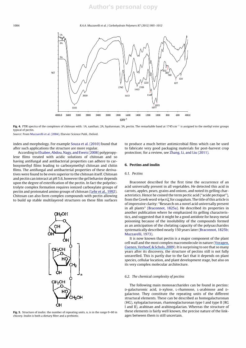
Fig. 5. Structure of inulin: the number of repeating units, n, is in the range 0–60 inchicory. Inulin is both a dietary fibre and a prebiotic.
to produce a much better antimicrobial films which can be usedto fabricate very good packaging materials for post-harvest cropprotection; for a review, see Zhang, Li, and Liu (2011).
6. Pectins and inulin
6.1. Pectins
Braconnot described for the first time the occurrence of anacid universally present in all vegetables. He detected this acid incarrots, apples, pears, grains and onions, and noted its gelling char-acteristics. Hence he coined the term pectic acid (“acide pectique”),from the Greek word � �́���́ for coagulum. The title of this article isof impressive clarity: “Research on a novel acid universally presentin all plants” (Braconnot, 1825a). He described its properties inanother publication where he emphasized its gelling characteris-tics, and suggested that it might be a good antidote for heavy metalpoisoning because of the insolubility of the compounds formedas an anticipation of the chelating capacity of the polysaccharidessystematically described nearly 150 years later (Braconnot, 1825b;Muzzarelli, 1973).
It is now known that pectin is a major component of the plantcell wall and the most complex macromolecule in nature (Voragen,Coenen, Verhoef, & Schols, 2009). It is surprising to see that so manyyears after its discovery, the structure of pectins still is not fullyunravelled. This is partly due to the fact that it depends on plantspecies, cellular location, and plant development stage, but also onits very complex molecular architecture.
6.2. The chemical complexity of pectins
The following main monosaccharides can be found in pectins:d-galacturonic acid, d-xylose, l-rhamnose, l-arabinose and d-galactose. They constitute the repeating units of the differentstructural elements. These can be described as homogalacturonan(HG), xylogalacturonan, rhamnoglacturonan type I and type II (RGI and II), arabinan and arabinogalactan. Whereas the structure ofthese elements is fairly well known, the precise nature of the link-ages between them is still uncertain.
Author's personal copy
R.A.A. Muzzarelli et al. / Carbohydrate Polymers 87 (2012) 995– 1012 1005
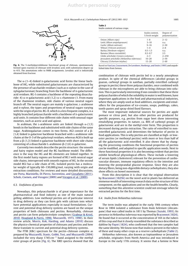
Fig. 6. The 5-methylpyrrolidinone functional group of chitosan, spontaneouslyformed upon reaction of chitosan with levulinic acid, with substitution degree upto 41%. The annotations refer to NMR assignments. Levulinic acid is industriallyobtained from fructose.
The �-(1–4)-linked d-galacturonic acid forms the linear back-bone of HG, while substituted galacturonans are characterized bythe presence of saccharide residues (such as d-xylose in the case ofxylogalacturonan) branching from the backbone of d-galacturonicacid residues. RG-I contains a backbone of the repeating disaccha-ride: 4)-�-d-galacturonic acid-(1,2)-�-l-rhamnose-(1. From manyof the rhamnose residues, side chains of various neutral sugarsbranch off. The neutral sugars are mainly d-galactose, l-arabinoseand d-xylose, the types and proportions of neutral sugars varyingwith the origin of pectin. RG-II, which is a less frequent complex, is ahighly branched polysaccharide with a backbone of d-galacturonicacid units. It contains four different side chains with unusual sugarmoieties, such as aceric acid and apiose.
In arabinan, the l-arabinose units are linked through �-(1,5)bonds in the backbone and substituted with side chains of the samesugar. Arabinogalactan comes in two forms. AGI consist of a �-(1,4) linked d-galactose backbone branched with l-arabinose sidechains at the O-3 of the galactosyl residues, whereas AGII is built upfrom a �-(1,3) linked d-galactose backbone with short side chainsconsisting of a disaccharide L-arabinose-�-(1,6)-d-galactose.
Currently two models describe the pectin structure: the smoothand hairy region model and the RG backbone model. The differ-ence is the way the HG and RG elements are linked together. Inthe first model hairy regions are formed of RG I with neutral sugarside chains, interspersed with smooth regions of HG. In the secondmodel RGI has a side chain of HG. Isolated pectin has a molecu-lar weight of typically 60–130,000 g/mol, varying with origin andextraction conditions. For reviews and more detailed discussions,see Porta, Mariniello, Di Pierro, Sorrentino, and Giosafatto (2011),Schols, Coenen, and Voragen (2009) and Voragen et al. (2009).
6.3. Usefulness of pectins
Nowadays, this polysaccharide is of great importance for thepharmaceutical and food industry as one of the main naturalgelling additives. Low methoxyl pectins are of particular interestin drug delivery as they can form gels with calcium ions whichhave potential applications especially in nasal formulations. Cur-rent and potential drug delivery systems are based on the uniqueproperties of both chitosans and pectins. Remarkably, chitosanand pectin can form polyelectrolyte complexes (Grabnar & Kristl,2010; Hoagland & Parris, 1996; Muzzarelli, 1973, 1990). In theirreview article, Morris, Kok, Harding, and Adams (2010) discussthe physico-chemical properties of chitosans and pectins and howthese translate to current and potential drug delivery systems.
The FTIR (KBr) spectrum for the pectin–chitosan complex asprepared by Muzzarelli, Stanic, Gobbi, Tosi, and Muzzarelli (2004)included the very sharp 1745 cm−1 band assigned to the methylester groups of pectin (Fig. 6). The XRD spectra showed that the
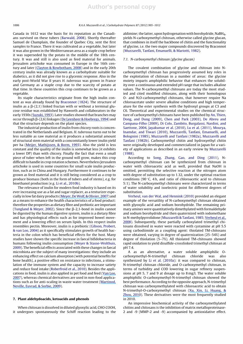
Table 2Inulin content of various crops.
Inulin content,% fresh weight
Degree ofpolymerization
Onion (Allium cepa) 1–8 2–12Leek (Allium ampeloprasum) 3–10 n.a.Garlic (Allium sativum) 9–16 2–50Wheat (Triticum aestivum) 1–4 2–8Barley (Hordeum vulgare) 0.5–1.5 n.a.Banana (Musa cavendishii) 0.3–0.7 2.5Globe artichoke (Cynara scolymus) 2–3 2–250Jerusalem artichoke (Helianthus tuberosus) 16–20 2–50Chicory (Cichorium intybus) 15–20 2–60
Based on Van Loo, Coussement, De Leenheer, Hoebregs, and Smits (1995) and unpub-lished data from Sensus.
combination of chitosan with pectin led to a nearly amorphousproduct. In spite of the chemical differences (alcohol groups inguaran, carboxyl groups in xanthan, partially esterified carboxylgroups in pectin) these three polysaccharides, once combined withchitosan in the microspheres are able to bring chitosan into solu-tion. This is particularly interesting if one considers that these threepolysaccharides of which the solubility in water is well known, haveimportant applications in the food and pharmaceutical industries,where they are amply used as food additives, excipients and emul-sifiers for the preparation of ice-creams, soups, puddings, cakes,tooth-pastes and spray-dried fixed flavours.
The most important industrial sources for pectins are applepomace or citrus peel, but also other pectins are produced forspecific purposes, e.g. pectins from sugar beet show interestingemulsifying properties. In nature, ca. 80% of carboxyl groups ofgalacturonic acid are in the methyl ester form. This proportion isdecreased during pectin extraction. The ratio of esterified to non-esterified galacturonic acid determines the behavior of pectin infood applications. This is why pectins are classified as high- or low-ester pectins (or methoxyl pectins) with more or less than half ofall the galacturonic acid esterified. It also shows that by chang-ing the processing conditions the functional properties of pectinscan be modified, and adapted to specific application needs. Next tothese functional properties it is also becoming evident that pectinsexhibit beneficial physiological effects. These include a loweringof serum lipids (cholesterol) relevant for the prevention of cardio-vascular diseases, immune regulatory effects in the intestine andlowering the postprandial glucose response. Since they are alsodietary fibres, being non-digestible dietary carbohydrates, they alsoshow effects on bowel movement.
From this description it is clear that the original observationby Braconnot (1825b) on the novel acid in plants has delivered animmense wealth of interesting research data on the structure of thiscomponent, on the applications and on the health benefits. Clearly,something that this attentive scientist could not envisage when heextracted pectins for the first time.
6.4. Inulin from Helianthus tuberosus
The term inulin was adopted in the early 19th century whenRose in 1804 isolated a substance from Inula helenium (elecam-pane) that was called inulin in 1811 by Thomas (Suzuki, 1993). Itspresence in Helianthus tuberosus was reported by Braconnot (1824).He found that it occurred at the concentration of 16% in the tubersof this crop and that it closely resembled the substance described asdahline by Payot (1824), who confirmed that the two samples hadthe same identity. We know now that inulin is present in the tubersof these and many other crops as a reserve carbohydrate (Table 2).
Helianthus tuberosus, also known as topinambour or Jerusalemartichoke, originates from North America and was introduced inEurope in the early 17th century. It seems that a famine in New
Author's personal copy
1006 R.A.A. Muzzarelli et al. / Carbohydrate Polymers 87 (2012) 995– 1012
Canada in 1612 was the basis for its reputation as the Canadi-ans survived on these tubers (Barwald, 2008). Shortly thereafterSamuel de Champlain, the founder of Quebec City, sent the firstsamples to France. There it was cultivated as a vegetable, but laterit was also grown in the Mediterranean area as a staple crop beforeit was superseded by the potato in the middle of the 18th cen-tury. It was and still is also used as feed material for animals.Jerusalem artichoke was consumed in Europe in the 16th cen-tury and later (Clayton & Rowbotham, 2008) and in the early 20thcentury inulin was already known as a carbohydrate suitable fordiabetics, as it did not give rise to a glycemic response. Also in theearly post-World War II years H. tuberosus was grown in Franceand Germany as a staple crop due to the scarcity of potato atthat time. In these countries this crop continues to be grown as avegetable.
Its staple characteristics originate from the high inulin con-tent as was already found by Braconnot (1824). The structure ofinulin as a �-(2,1) linked fructan with or without a terminal glu-cose residue was established by Haworth and collaborators in theearly 1930s (Suzuki, 1993). Later studies showed that branches mayoccur through �-(2,6) linkages (De Leenheer & Hoebregs, 1994) andthat the structure depends on the plant species.
The industrial production of inulin from chicory roots is concen-trated in the Netherlands and Belgium. H. tuberosus turns out to beless suitable as raw material as it produces a substantial amountof structural stem material with a concomitantly lower inulin yieldper ha (Meijer, Mathijssen, & Borm, 1993). Also the yield is lessconstant and the quality of the inulin is somewhat less (it exhibitsa lower DP) than with chicory. Finally the fact that even a smallpiece of tuber when left in the ground will grow, makes this cropdifficult to handle in crop rotation schemes. Nevertheless Jerusalemartichoke is used in some countries for small scale inulin produc-tion, such as in China and Hungary. Furthermore it continues to begrown as feed material and it is still being considered as a crop toproduce biomass (both in the form of tubers and of stems), e.g. forbioethanol production (e.g. Li et al., 2010b).
The relevance of inulin for modern food industry is based on itsever increasing use as a fat and sugar replacer, as a texturizer espe-cially in low fat dairy products (Meyer, De Wolf, & Olivier, 2007) andas a means to enhance the health characteristics of a food product:therefore the properties as dietary fibre and prebiotic are important(Tungland & Meyer, 2002). Since the �-2,1-bond in inulin cannotbe digested by the human digestive system, inulin is a dietary fibreand has physiological effects such as for improved bowel move-ment and a lowering effect on serum lipids. In this respect inulinresembles pectin. Moreover, inulin is a prebiotic (Gibson, Probert,& van Loo, 2004) as it specifically stimulates growth of health bac-teria in the colon which has beneficial effects for the host. Manystudies have shown the specific increase in faecal bifidobacteria inhumans following inulin consumption (Meyer & Stasse-Wolthuis,2009). The beneficial effects associated with these changes in faecalmicrobiota are the subject of many investigations: they include anenhancing effect on calcium absorption (with potential benefits forbone health), a positive effect on resistance to infections, a stimu-lation of the immune system and the capacity to increase satietyand reduce food intake (Roberfroid et al., 2010). Besides the appli-cations in food, inulin is also applied in pet food and feed (Van Loo,2007), whereas chemical derivatives are used in non-food applica-tions such as for anti-scaling in waste water treatment (Martinod,Neville, Euvrad, & Sorbie, 2009).
7. Plant aldehydoacids, ketoacids and phenols
When chitosan is dissolved in diluted glyoxylic acid, CHO·COOH,it undergoes spontaneously the Schiff reaction leading to the
aldimine; the latter, upon hydrogenation with borohydride, NaBH4,yields N-carboxymethyl chitosan, otherwise called glycine glucan,that combines in itself the backbone of chitin and the functionalityof glycine, i.e. the two major compounds discovered by Braconnot(Muzzarelli, Tanfani, Emanuelli, & Mariotti, 1982).
7.1. N-carboxymethyl chitosan (glycine glucan)
The covalent combination of glycine and chitosan into N-carboxymethyl chitosan has progressively assumed key roles inthe exploitation of chitosan in a number of areas: the glycinemoiety imparts amphiphilic behavior that enhances the solubil-ity over a continuous and extended pH range that includes alkalinevalues. The N-carboxymethyl chitosans are today the most stud-ied and cited modified chitosans, along with their homologousO- and N,O-carboxymethyl chitosans, that however require Nachloroacetate under severe alkaline conditions and high temper-ature for the ester synthesis with the hydroxyl groups at C3 andC6. Theoretical and experimental studies on the chemical struc-ture of carboxymethyl chitosans have been published by An, Thien,Dong, and Dung (2009), Chen and Park (2003), De Abreu andCampana-Filho (2009), Di Colo, Zambito, Burgalassi, Nardini, andSaettone, 2004, Jayakumar et al. (2010), Lu et al. (2011), Mourya,Inamdar, and Tiwari (2010), Muzzarelli, Tanfani, Emanuelli, andBolognini (1985), Muzzarelli and Tanfani (1982a), Muzzarelli andTanfani (1982b). Carboxymethyl chitin and hydroxyethyl chitinwere originally developed and commercialized in Japan for a vari-ety of applications as described in an early review by Muzzarelli(1988).
According to Song, Zhang, Gao, and Ding (2011), N-carboxymethyl chitosan can be synthesized from chitosan inwater with chloroacetic acid as well, provided that NaOH isomitted, permitting the selective reaction at the nitrogen atomwith degree of substitution up to 1.32, under the optimal reactionconditions (90 ◦C, 4 h, and ratio of chloroacetic acid to chitosan5:1). The N-carboxymethyl chitosans were characterized in termsof water solubility and isoelectric point for different degrees ofsubstitution.
Verheul, van der Wal, and Hennink (2010) provided an elegantexample of the versatility of N-carboxymethyl chitosan obtainedwith glyoxylic acid and sodium borohydride. The remaining pri-mary amines were quantitatively dimethylated with formaldehydeand sodium borohydride and then quaternized with iodomethanein N-methylpyrrolidone (Muzzarelli & Tanfani, 1985; Verheul et al.,2008). Subsequently, these partially carboxylated trimethyl chi-tosans dissolved in water were reacted with cystamine at pH 5.5using carbodiimide as a coupling agent: thiolated TM-chitosanswere obtained, varying in degree of quaternization (25–54%) anddegree of thiolation (5–7%). All thiolated TM-chitosans showedrapid oxidation to yield disulfide-crosslinked trimethyl chitosan atpH 7.4.
A as an alternative, a water soluble amphiphilic O-carboxymethyl-N-trimethyl chitosan chloride was alsosynthesized by Li et al. (2010a): it was compared to chitosan,N-trimethyl chitosan chloride, and O-carboxymethyl chitosan, interms of turbidity and COD lowering in sugar refinery suspen-sions at pH 5, 7 and 9 at dosage up to 8 mg/l. The water solubleamphiphilic O-carboxymethyl-N-trimethyl chitosan showed thebest performance. According to the opposite approach, N-trimethylchitosan was carboxymethylated with chloroacetic acid to obtainN-trimethyl-O-carboxymethyl chitosan (Xu, Xin, Li, Huang, &Zhou, 2010). These derivatives were the most frequently studiedin 2010.
An impressive biochemical activity of the carboxymethylatedchitins and chitosans is the inhibition of matrix metalloproteinase-2 and -9 (MMP-2 and -9) accompanied by antioxidative effect.
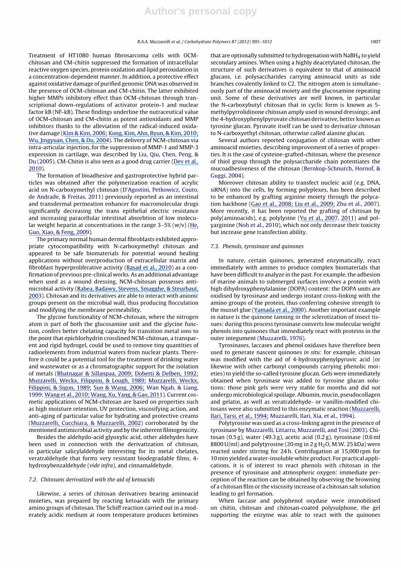
Author's personal copy
R.A.A. Muzzarelli et al. / Carbohydrate Polymers 87 (2012) 995– 1012 1007
Treatment of HT1080 human fibrosarcoma cells with OCM-chitosan and CM-chitin suppressed the formation of intracellularreactive oxygen species, protein oxidation and lipid peroxidation ina concentration-dependent manner. In addition, a protective effectagainst oxidative damage of purified genomic DNA was observed inthe presence of OCM-chitosan and CM-chitin. The latter exhibitedhigher MMPs inhibitory effect than OCM-chitosan through tran-scriptional down-regulations of activator protein-1 and nuclearfactor kB (NF-kB). These findings underline the nutraceutical valueof OCM-chitosan and CM-chitin as potent antioxidants and MMPinhibitors thanks to the alleviation of the radical-induced oxida-tive damage (Kim & Kim, 2006; Kong, Kim, Ahn, Byun, & Kim, 2010;Wu, Jingyuan, Chen, & Du, 2004). The delivery of NCM-chitosan viaintra-articular injection, for the suppression of MMP-1 and MMP-3expression in cartilage, was described by Liu, Qiu, Chen, Peng, &Du (2005). CM-Chitin is also seen as a good drug carrier (Dev et al.,2010).
The formation of bioadhesive and gastroprotective hybrid par-ticles was obtained after the polymerization reaction of acrylicacid on N-carboxymethyl chitosan (D’Agostini, Petkowicz, Couto,de Andrade, & Freitas, 2011) previously reported as an intestinaland transdermal permeation enhancer for macromolecular drugssignificantly decreasing the trans epithelial electric resistanceand increasing paracellular intestinal absorbtion of low molecu-lar weight heparin at concentrations in the range 3–5% (w/v) (He,Guo, Xiao, & Feng, 2009).
The primary normal human dermal fibroblasts exhibited appro-priate cytocompatibility with N-carboxymethyl chitosan andappeared to be safe biomaterials for potential wound healingapplications without overproduction of extracellular matrix andfibroblast hyperproliferative activity (Rasad et al., 2010) as a con-firmation of previous pre-clinical works. As an additional advantagewhen used as a wound dressing, NCM-chitosan possesses anti-microbial activity (Rabea, Badawy, Stevens, Smagghe, & Steurbaut,2003). Chitosan and its derivatives are able to interact with anionicgroups present on the microbial wall, thus producing flocculationand modifying the membrane permeability.
The glycine functionality of NCM-chitosan, where the nitrogenatom is part of both the glucosamine unit and the glycine func-tion, confers better chelating capacity for transition metal ions tothe point that epichlorhydrin crosslined NCM-chitosan, a transpar-ent and rigid hydrogel, could be used to remove tiny quantities ofradioelements from industrial waters from nuclear plants. There-fore it could be a potential tool for the treatment of drinking waterand wastewater or as a chromatographic support for the isolationof metals (Bhatnagar & Sillanpaa, 2009; Dobetti & Delben, 1992;Muzzarelli, Weckx, Filippini, & Lough, 1989; Muzzarelli, Weckx,Filippini, & Sigon, 1989; Sun & Wang, 2006; Wan Ngah, & Liang,1999; Wang et al., 2010; Wang, Xu, Yang, & Gao, 2011). Current cos-metic applications of NCM-chitosan are based on properties suchas high moisture retention, UV protection, viscosifying action, andanti-aging of particular value for hydrating and protective creams(Muzzarelli, Cucchiara, & Muzzarelli, 2002) corroborated by thementioned antimicrobial activity and by the inherent filmogenicity.
Besides the aldehydo-acid glyoxylic acid, other aldehydes havebeen used in connection with the derivatization of chitosan,in particular salicylaldehyde interesting for its metal chelates,veratraldehyde that forms very resistant biodegradable films, 4-hydroxybenzaldehyde (vide infra), and cinnamaldehyde.
7.2. Chitosans derivatized with the aid of ketoacids
Likewise, a series of chitosan derivatives bearing aminoacidmoieties, was prepared by reacting ketoacids with the primaryamino groups of chitosan. The Schiff reaction carried out in a mod-erately acidic medium at room temperature produces ketimines
that are optionally submitted to hydrogenation with NaBH4 to yieldsecondary amines. When using a highly deacetylated chitosan, thestructure of such derivatives is equivalent to that of aminoacidglucans, i.e. polysaccharides carrying aminoacid units as sidebranches covalently linked to C2. The nitrogen atom is simultane-ously part of the aminoacid moiety and the glucosamine repeatingunit. Some of these derivatives are well known, in particularthe N-carboxybutyl chitosan that in cyclic form is known as 5-methylpyrrolidinone chitosan amply used in wound dressings; andthe 4-hydroxyphenylpyruvate chitosan derivative, better known astyrosine glucan. Pyruvate itself can be used to derivatize chitosanto N-carboxyethyl chitosan, otherwise called alanine glucan.
Several authors reported conjugation of chitosan with otheraminoacid moieties, describing improvement of a series of proper-ties. It is the case of cysteine-grafted-chitosan, where the presenceof thiol group through the polysaccharide chain potentiates themucoadhesiveness of the chitosan (Bernkop-Schnurch, Hornof, &Guggi, 2004).
Moreover chitosan ability to transfect nucleic acid (e.g. DNA,siRNA) into the cells, by forming polyplexes, has been describedto be enhanced by grafting arginine moiety through the polyca-tion backbone (Gao et al., 2008; Liu et al., 2009; Zhu et al., 2007).More recently, it has been reported the grafting of chitosan bypoly(aminoacids), e.g. polylysine (Yu et al., 2007, 2011) and pol-yarginine (Noh et al., 2010), which not only decrease their toxicitybut increase gene transfection ability.
7.3. Phenols, tyrosinase and quinones
In nature, certain quinones, generated enzymatically, reactimmediately with amines to produce complex biomaterials thathave been difficult to analyze in the past. For example, the adhesionof marine animals to submerged surfaces involves a protein withhigh dihydroxyphenylalanine (DOPA) content: the DOPA units areoxidised by tyrosinase and undergo instant cross-linking with theamino groups of the protein, thus conferring cohesive strength tothe mussel glue (Yamada et al., 2000). Another important examplein nature is the quinone tanning in the sclerotization of insect tis-sues: during this process tyrosinase converts low molecular weightphenols into quinones that immediately react with proteins in theouter integument (Muzzarelli, 1976).
Tyrosinases, laccases and phenol oxidases have therefore beenused to generate nascent quinones in situ: for example, chitosanwas modified with the aid of 4-hydroxyphenylpyruvic acid (orlikewise with other carbonyl compounds carrying phenolic moi-eties) to yield the so-called tyrosine glucan. Gels were immediatelyobtained when tyrosinase was added to tyrosine glucan solu-tions: those pink gels were very stable for months and did notundergo microbiological spoilage. Albumin, mucin, pseudocollagenand gelatin, as well as veratraldehyde- or vanillin-modified chi-tosans were also submitted to this enzymatic reaction (Muzzarelli,Ilari, Tarsi, et al., 1994; Muzzarelli, Ilari, Xia, et al., 1994).
Polytyrosine was used as a cross-linking agent in the presence oftyrosinase by Muzzarelli, Littarru, Muzzarelli, and Tosi (2003). Chi-tosan (0.5 g), water (49.3 g), acetic acid (0.2 g), tyrosinase (0.6 ml8800 U/ml) and polytyrosine (20 mg in 2 g H2O, M.W. 25 kDa) werereacted under stirring for 24 h. Centrifugation at 15,000 rpm for10 min yielded a water-insoluble white product. For practical appli-cations, it is of interest to react phenols with chitosan in thepresence of tyrosinase and atmospheric oxygen: immediate per-ception of the reaction can be obtained by observing the browningof a chitosan film or the viscosity increase of a chitosan salt solutionleading to gel formation.
When laccase and polyphenol oxydase were immobilisedon chitin, chitosan and chitosan-coated polysulphone, the gelsupporting the enzyme was able to react with the quinones
Author's personal copy
1008 R.A.A. Muzzarelli et al. / Carbohydrate Polymers 87 (2012) 995– 1012
generated by the enzymatic action on phenols. Chitosan couldalso be coupled to polymeric substances in this way, in particularto proteins, gelatin and polyhydroxy styrene (Vazquez-Duhalt,Tinoco, D’Antonio, Topoleski, & Payne, 2001). In one decade,these studies had fantastic developments in the area of syn-ergy between microelectronics and biotechnology (Liu et al.,2010).
The phenols taken into consideration in the cited studies werevolatile phenols such as p-cresol, gallate esters such as dodecylgallate, phenol, hexyloxyphenol, chlorogenic acid, 1,2-benzenediol,tert-butylcathecol, p-hydroxyphenoxyacetate, olive-mill wastewa-ter phenolics, and polyhydroxystyrene: the attention was directedto chemical models of industrial importance. In addition severalcompound of recognized biomedical/biochemical importance werealso examined.
Vitamin K quinones and ubiquinone/ubiquinol actually arecompounds of major importance from the biochemical stand-point, being involved in modification of blood coagulation factorsand in the electron/proton transport, respectively. Menadione,2-methyl-1,4-naphthalenedione is a synthetic naphthoquinonederivative having the physiologic properties of vitamin K, thelatter being a collective name including phylloquinones K1,menaquinones K2 and menadione K3. Phylloquinone, 2-methyl-3-(3,7,11,15-tetramethyl-2-hexadecenyl)-1,4-naphthoquinone, isactually a photosynthetic electron carrier synthesized in greenplants, a major dietary source of protrombogenic vitamin K. Othercompounds of interest are plumbagin, 5-hydroxy-2-methyl-1,4-naphthoquinone, an antimicrobial very toxic agent from Plumbagoindica, and bupravacone, an antibiotic used in clinical experimentsagainst Cryptosporidium parum.
The derivatization of chitosan made by Muzzarelli, Weckx,Filippini, and Lough (1989) and Muzzarelli, Weckx, Filippini, andSigon (1989) with the aid of levulinic acid (4-oxo-pentanoic acid, i.e.a -ketoacid obtained from fructose that Broussignac might havehandled) deserves a mention here in consideration of the majorsignificance that it had in recent research in experimental surgery,wound management and healing, bone regeneration, repair oflesions of the meniscal cartilage, decubitus ulcer treatment, den-tistry, coated prostetic materials, urology, and cosmetology. Mostrecently the preparation protocol was refined as follows: pH4.5–5.0. Molar ratios of the primary amino groups, NaBH3CN andlevulinic acid 1:10:3, the substitution degree was close to 41% for5-methylpyrrolidinone chitosans, after 72 h of reaction at roomtemperature; no depolymerization occurred, and the NMR spectrawere cleaner (Kurita & Isogai, 2010).
8. Conclusion
Well, we are back from our journey to the past. We dreamed tohave met Henri Braconnot in his Botanical Garden for an excitingexchange of views, and to have been admitted to his laboratory,where he used to work alone, to the point that he had no disciples.Actually he did not even get married. Even though he attendedcourses of chemistry, natural history and pharmacy in Strasbourgand Paris whenever possible because of recurrent military obli-gations, he was never concerned about getting an academic title.The personal qualities that we appreciate in him as a scientistare the clarity of ideas, the scrupulous planning and execution ofthe experiments, the concise and very efficacious writing, and thetotal devotion to his duties. We see today that his work was highlyanticipatory in character, and opened up new routes in botany,pharmacy and chemistry as we demonstrate in this concise assay.After two centuries, the main subjects of his research work are stillpivotal and proficuous for envisaging sustainable green solutionsfor modern economies.
Acknowledgments
The preparation of this article was a spontaneous and indepen-dent initiative of the authors, and was not financially supported.Thanks are due to Prof. M. Boulangé, former President of the Uni-versity of Nancy, France, who made available copies of originaldocuments; to Dr. Marilena Falcone, Central Library, PolytechnicUniversity, Ancona, Italy, for assistance in handling the biblio-graphic information, to Mrs. Maria Weckx for assistance in thepreparation of the manuscript, and to Dr. Monica Glebocki for crit-ically reading parts of it.
References
Aida, F. M. N. A., Shuhaimi, M., Yazid, M. & Maaruf, A. G. (2009). Mushroom as apotential source of prebiotics: A review. Trends in Food Science & Technology, 20,567–575.
Aider, M. (2010). Chitosan application for active bio-based films production andpotential in the food industry: Review. LWT-Food Science and Technology, 43(6),837–842.
An, N. T., Thien, D. T., Dong, N. T. & Dung, P. L. (2009). Water-soluble N-carboxymethylchitosan derivatives: Preparation, characteristics and its application. Carbohy-drate Polymers, 75(3), 489–497.
Araki, Y. & Ito, E. (1975). A pathway of chitosan formation in Mucor rouxii. EuropeanJournal of Biochemistry, 55, 71–78.
Barone, R., Simpore, J., Malaguarnera, L., Pignatelli, S. & Musumeci, S. (2003). Plasmachitotriosidase activity in acute Plasmodium falciparum malaria. Clinica ChimicaActa, 331, 79–85.
Bartnicki-Garcia, S. & Nickerson, W. J. (1962). Isolation, composition and structureof cell walls of filamentous and yeast-like forms of Mucor rouxii. Biochimica etBiophysica Acta, 58, 102–119.
Barwald, G. (2008). Gesund abnehmen mit Topinambur. Stuttgart, Germany: TRIASVerlag.
Bautista-Banos, S., Hernandez-Lauzardo, A. N., Velazquez-del Valle, M. G.,Hernandez-Lopez, M., Ait Barka, E., Bosquez-Molina, E., et al. (2006). Chitosanas a potential natural compound to control pre and postharvest diseases ofhorticultural commodities. Crop Protection, 25, 108–118.
BeMiller, J. N. (1965). Chitin. Methods of Carbohydrate Chemistry, 5, 103–106.Berecochea-Lopez, A., Decorde, K., Ventura, E., Godard, M., Bornet, A., Teissedre, P.
L., et al. (2009). Fungal chitin–glucan from Aspergillus niger efficiently reducesaortic fatty streak accumulation in the high-fat fed hamster, and animal model ofnutritionally induced atherosclerosis. Journal of Agricultural and Food Chemistry,57(3), 1093–1098.
Bernkop-Schnurch, A., Hornof, M. & Guggi, D. (2004). Thiolated chitosans. EuropeanJournal of Pharmaceutics and Biopharmaceutics, 57(1), 9–17.
Bhatnagar, A. & Sillanpaa, M. (2009). Applications of chitin and chitosan derivativesfor the detoxification of water and wastewater: A short review. Advances inColloid and Interface Science, 152(1–2), 26–38.
Bierbaum, S., Nickel, R., Koch, A., Lau, S., Deichmann, K. A., Wahn, U., et al. (2005).Polymorphisms and haplotypes of acid mammalian chitinase are associatedwith bronchial asthma. American Journal of Respiratory and Critical Care Medicine,172, 1505–1509.
Bleau, G., Massicotte, F., Merlen, Y. & Boisvert, C. (1999). Mammalian chitinase-likeproteins. EXS, 87, 211–221.
Boeris, V., Micheletto, Y., Lionzo, M., da Silveira, N. P. & Pico, G. (2011). Interactionbehavior between chitosan and pepsin. Carbohydrate Polymers, 84, 459–464.
Boot, R. G., Blommaart, E. F., Swart, E., Ghauharali-van der Vlugt, K., Bijl, N., Moe, C.,et al. (2001). Identification of a novel acid mammalian chitinase distinct fromchitotriosidase. Journal of Biological Chemistry, 276, 6770–6778.
Boot, R. G., Bussink, A. P., Verhoek, M., de Boer, P. A., Moorman, A. F. & Aerts, J. M.(2005). Marked differences in tissue-specific expression of chitinases in mouseand man. Journal of Histochemistry and Cytochemistry, 53, 1283–1292.
Boot, R. G., Renkema, G. H., Strijland, A., van Zonneveld, A. J. & Aerts, J. M. (1995).Cloning of a cDNA encoding chitotriosidase, a human chitinase produced bymacrophages. Journal of Biological Chemistry, 270, 26252–26256.
Boot, R. G., Renkema, G. H., Verhoek, M., Strijland, A., Bliek, J., de Meulemeester, T.M., et al. (1998). The human chitotriosidase gene. Nature of inherited enzymedeficiency. Journal of Biological Chemistry, 273, 25680–25685.
Braconnot, H. (1811). Sur la nature des champignons. Annales de Chimie Physique,79, 265–304.
Braconnot, H. (1824). Analyse de tubercules de l’Helianthus tuberosus, et observa-tions sur le dahline. Annales de Chimie et de Physique, 25, 358–373.
Braconnot, H. (1825a). Recherches sur un nouvel acide universellement répandudans tous les végétaux. Annales de Chimie et de Physique, 28, 173–178.
Braconnot, H. (1825b). Nouvelles observations sur l’acide pectique. Annales de Chimieet de Physique, 30, 96–102.
Bukkens, S. G. F. (2005). Insects in the human diet: Nutritional aspects. In M. G.Paoletti (Ed.), Ecological implications of minilivestock. Potential of insects, rodents,frog and snails (pp. 545–577). Enfield: Science Publishers, Inc.
Chang, N. C., Hung, S. I., Hwa, K. Y., Kato, I., Chen, J. E., Liu, C. H., et al. (2001). Amacrophage protein, Ym1, transiently expressed during inflammation is a novelmammalian lectin. Journal of Biological Chemistry, 276, 17497–17506.
Author's personal copy
R.A.A. Muzzarelli et al. / Carbohydrate Polymers 87 (2012) 995– 1012 1009
Chatterjee, S., Adhya, M., Guha, A. K. & Chatterjee, B. P. (2005). Chitosan from Mucorrouxii: Production and physico-chemical characterization. Process Biochemistry,40(1), 395–400.
Chatterjee, S., Chatterjee, S., Chatterjee, B. P. & Guha, A. K. (2004). Clarification offruit juice with chitosan. Process Biochemistry, 39, 2229–2232.
Chatterjee, S., Chatterjee, S., Chatterjee, B. P. & Guha, A. K. (2009). Influence of plantgrowth hormones on the growth of Mucor rouxii and chitosan production. Micro-biological Research, 164(3), 347–351.
Chen, X. G. & Park, H. J. (2003). Chemical characteristics of O-carboxymethyl chi-tosans related to the preparation conditions. Carbohydrate Polymers, 53(4),355–359.
Chien, Y. H., Chen, J. H. & Hwu, W. L. (2005). Plasma chitotriosidase activity andmalaria. Clinica Chimica Acta, 353, 215.
Children, J. G. (1824). On the nature of mushrooms. Zoological Journal, 1, 101–111.Choi, E. H., Zimmerman, P. A., Foster, C. B., Zhu, S., Kumaraswami, V., Nutman, T.
B., et al. (2001). Genetic polymorphisms in molecules of innate immunity andsusceptibility to infection with Wuchereria bancrofti in South India. Genes andImmunity, 2, 248–253.
Chung, L. Y., Schmidt, R. J., Hamlyn, P. F. & Sagar, B. F. (1994). Biocompatibil-ity of potential wound management products: Fungal mycelia as a source ofchitin/chitosan and their effect on the proliferation of human FI000 fibroblastsin culture. Journal of Biomedical Material Research, 28, 463–469.
Clayton, P. & Rowbotham, J. (2008). An unsuitable and degraded diet? Part two:Realities of the mid-Victorian diet. Journal of the Royal Society of Medicine, 101,350–357.
Cohen-Kupiec, R. & Chet, I. (1998). The molecular biology of chitin digestion. CurrentOpinion in Biotechnology, 9, 270–277.
Conrad, J. (1964). Chitin. Encyclopedia of Polymer Science and Technology, 3, 695–704.
Cozzarini, E., Bellin, M., Norberto, L., Polese, L., Musumeci, S., Lanfranchi, G., et al.(2009). CHIT1 and AMCase expression in human gastric mucosa: Correlationwith inflammation and Helicobacter pylori infection. European Journal of Gas-troenterology & Hepatology, 21(10), 1119–1126.
Crestini, C. & Giovannozzi-Sermanni, G. (1996). Solid state fermentation of Lenti-nus edodes: A new and efficient approach to chitosan production. In R. A. A.Muzzarelli (Ed.), Chitin enzymology (pp. 595–600). Italy: Atec.
D’Agostini, O., Jr., Petkowicz, C. L., Couto, A. G., de Andrade, S. F. & Freitas, R. A. (2011).Simultaneous in situ monitoring of acrylic acid polymerization reaction on N-carboxymethyl chitosan using multidetectors: Formation of a new bioadhesiveand gastroprotective hybrid particle. Materials Science & Engineering, C-31(3),677–682.
Dayan, F. E., Cantrell, C. L. & Duke, S. O. (2009). Natural products in crop protection.Bioorganic & Medicinal Chemistry, 17, 4022–4034.
De Abreu, F. R. & Campana-Filho, S. P. (2009). Characteristics and properties ofcarboxymethylchitosan. Carbohydrate Polymers, 75(2), 214–221.
De Leenheer, L. & Hoebregs, H. (1994). Progress in the elucidation of the compositionof chicory inulin. Starch/die Stärke, 46, 193–196.
DeFoliart, G. (1992). Insects as human food. Crop Protection, 11, 395–399.Dev, A., Mohan, J. C., Sreeja, V., Tamura, H., Patzke, G. R., Hussain, F., et al. (2010).
Novel carboxymethyl chitin nanoparticles for cancer drug delivery applications.Carbohydrate Polymers, 79(4), 1073–1079.
Di Colo, G., Zambito, Y., Burgalassi, S., Nardini, I. & Saettone, M. F. (2004). Effectof chitosan and of N-carboxymethyl chitosan on intraocular penetration oftopically applied ofloxacin. International Journal of Pharmaceutics, 273(1–2),37–44.
Dobetti, L. & Delben, F. (1992). Binding of metal cations by N-carboxymethyl chi-tosans in water. Carbohydrate Polymers, 18(4), 273–282.
Dobson, D. E., Prager, E. M. & Wilson, A. C. (1984). Stomach lysozymes of rumi-nants. I. Distribution and catalytic properties. Journal of Biological Chemistry, 259,11607–11616.
Donald, W. W. & Mirocha, C. J. (1977). Chitin as a measure of fungal growth in storedcorn and soybean seed. Cereal Chemistry, 54, 466–474.
Eaton, P., Fernandes, J. C., Pereira, E., Pintado, M. E. & Malcata, F. X. (2008). Atomicforce microscopy study of the antibacterial effects of chitosans on Escherichiacoli and Staphylococcus aureus. Ultramicroscopy, 108, 1128–1134.
El Hadrami, A., Adam, L. R., El Hadrami, I. & Daayf, F. (2010). Chitosan in plantprotection. Marine Drugs, 8(4), 968–987.
Elsabee, M. Z., Abdou, E. S., Nagy, K. S. A. & Eweis, M. (2008). Surface modification ofpolypropylene films by chitosan and chitosan/pectin multilayer. CarbohydratePolymers, 71(2), 187–195.
Fang, Z. X., Zhang, M., Tao, G. J., Sun, Y. F. & Sun, J. C. (2006). Chemical composi-tion of clarified bayberry (Myrica rubra Sieb. et Zucc.) juice sediment. Journal ofAgricultural and Food Chemistry, 54, 7710–7716.
Fernandez-Saiz, P., Lagaron, J. M. & Ocio, M. J. (2009). Optimization of the film-forming and storage conditions of chitosan as an antimicrobial agent. Journalof Agricultural and Food Chemistry, 57, 3298–3307.
Fontaneto, D., Tommaseo-Ponzetta, M., Galli, C., Risè, P., Glew, R. H. &Paoletti, M. G. (2011). Differences in fatty acid composition between aquatic andterrestrial insects used as food in human nutrition. Ecology of Food and Nutrition,50, 351–367.
Friedman, M. & Juneja, V. K. (2010). Review of antimicrobial and antioxidative activ-ities of chitosans in food. Journal of Food Protection, 73(9), 1737–1761.
Fusetti, F., von Moeller, H., Houston, D., Rozeboom, H. J., Dijkstra, B. W.,Boot, R. G., et al. (2002). Structure of human chitotriosidase. Implications for spe-cific inhibitor design and function of mammalian chitinase-like lectins. Journalof Biological Chemistry, 277, 25537–25544.
Gao, Y., Xu, Z., Chen, S., Gu, W., Chen, L. & Li, Y. (2008). Arginine-chitosan/DNAself-assemble nanoparticles for gene delivery: In vitro characteristics and trans-fection efficiency. International Journal of Pharmaceutics, 359(1–2), 241–246.
Gerasimenko, D. V., Avdienko, I. D., Bannikova, G. E., Zueva, O. Yu. & Varlamov,V. P. (2004). Antibacterial effects of water-soluble low-molecular-weight chi-tosans on different microorganisms. Applied Biochemistry and Microbiology,40(3), 253–257.
Gianfrancesco, F. & Musumeci, S. (2004). The evolutionary conservation of thehuman chitotriosidase gene in rodents and primates. Cytogenetic and GenomeResearch, 105, 54–56.
Gibson, G. R., Probert, H. M. & van Loo, J. (2004). Dietary modulation of the colonicmicrobiota: Updating the concept of prebiotics. Nutrition Research Reviews, 17,259–275.
Gilson, E. (1894). Recherches chimiques sur la membrane cellulaire deschampignons. Bulletin de la Société Chimique Paris, 3, 1099–1102.
Gindzienski, A., Głowacka, D. & Zwierz, K. (1974). Purification and properties ofN-acetylglucosamine kinase from human gastric mucosa. European Journal ofBiochemistry, 43, 155–160.
Godron, D. A. (1872). Notice historique sur les jardins botaniques de Pont-à-Moussonet de Nancy. Sordoillet, Nancy.
Goto, M., Fujimoto, W., Nio, J., Iwanaga, T. & Kawasaki, T. (2003). Immunohistochem-ical demonstration of acidic mammalian chitinase in the mouse salivary glandand gastric mucosa. Archives of Oral Biology, 48, 701–707.
Goy, R. C., De Britto, D. & Assis, O. B. G. (2009). A review of the antimicrobial activityof chitosan. Polimeros, 19(3), 241–247.
Grabnar, P. A. & Kristl, J. (2010). Physicochemical characterization of protein-loadedpectin–chitosan nanoparticles prepared by polyelectrolyte complexation. Phar-mazie, 65, 851–852.
Hall, A. J., Quinnell, R. J., Raikoc, A., Lagog, M., Siba, P., Morroll, S., et al. (2007). Chi-totriosidase deficiency is not associated with human hookworm infection in aPapua New Guinean population. Infection, Genetics and Evolution, 7(6), 743–747.
He, W., Guo, X., Xiao, L. & Feng, M. (2009). Study on the mechanisms of chitosan andits derivatives used as transdermal penetration enhancers. International Journalof Pharmaceutics, 382(1–2), 234–243.
Hiorth, M., Skoien, T. & Sande, S. A. (2010). Immersion coating of pellet cores consist-ing of chitosan and calcium intended for colon drug delivery. European Journalof Pharmacy and Biopharmacy, 75, 245–253.
Hise, G. A., Hazlett, E. F., Bockarie, J. M., Zimmerman, A. P., Tisch, J. D. & Kazura,W. J. (2003). Polymorphisms of innate immunity genes and susceptibility tolymphatic filariasis. Genes and Immunity, 4, 524–527.
Hoagland, P. D. & Parris, N. (1996). Chitosan/pectin laminated films. Journal of Agri-cultural and Food Chemistry, 44(7), 1915–1919.
Hollak, C. E. M., van Weely, S., van Oers, M. H. J. & Aerts, J. M. (1994). Marked elevationof plasma chitotriosidase activity. A novel hallmark of Gaucher disease. Journalof Clinical Investigation, 93, 1288–1292.
Huber, M., Cabib, E. & Miller, L. H. (1991). Malaria parasite chitinase and penetrationof the mosquito peritrophic membrane. PNAS, 88, 2807–2810.
Ivshina, T. N., Artamonova, S. D., Ivshin, V. P. & Sharnina, F. F. (2009). Isolationof the chitin–glucan complex from the fruiting bodies of mycothallus. AppliedBiochemistry and Microbiology, 45(3), 313–318.
Jayakumar, R., Prabaharan, M., Nair, S. V., Tokura, S., Tamura, H. & Selvamurugan, N.(2010). Novel carboxymethyl derivatives of chitin and chitosan materials andtheir biomedical applications. Progress in Materials Science, 55(7), 675–709.
Jollès, P., & Muzzarelli, R. A. A. (Eds.). (1999). Chitin and chitinases. Birkhauser Verlag.Jung, E. A., Youn, D. A., Lee, S. A., No, H. K. A., Ha, J. B. & Prinyawiwatkul, W. C. (2010).
Antibacterial activity of chitosans with different degrees of deacetylation andviscosities. International Journal of Food Science and Technology, 45(4), 676–681.
Kalac, P. (2009). Chemical composition and nutritional value of European species ofwild growing mushrooms: A review. Food Chemistry, 113, 9–16.
Kanatt, R. K., Chander, R. & Sharma, A. (2008). Chitosan and mint mixture: A newpreservative for meat and meat product. Food Chemistry, 107, 845–852.
Kaspresewska, A. (2003). Plant chitinases—regulation and function. Cellular andMolecular Biology Letters, 8, 809–824.
Kim, M. M. & Kim, S. K. (2006). Chitooligosaccharides inhibit activation and expres-sion of matrix metalloproteinase-2 in human dermal fibroblasts. FEBS Letters,580(11), 2661–2666.
Kleekayai, T. & Suntornsuk, W. (2011). Production and characterization of chitosanobtained from Rhizopus oryzae grown on potato chip processing waste. WorldJournal of Microbiology & Biotechnology, 27(5), 1145–1154.
Kong, C. S., Kim, J. A., Ahn, B., Byun, H. G. & Kim, S. K. (2010). Carboxymethylationsof chitosan and chitin inhibit MMP expression and ROS scavenging in humanfibrosarcoma cells. Process Biochemistry, 45(2), 179–186.
Kuhlmann, K., Czupala, A., Haunhorst, J., Weiss, A., Prasch, T. & Schorken, U. (2000).Preparation and characterization of chitosan from Mucorales. In M. Peter, A.Domard, & R. A. A. Muzzarelli (Eds.), Advances in chitin science (pp. 7–14). Druck-haus Schmergow Germany.
Kurita, Y. & Isogai, A. (2010). Reductive N-alkylation of chitosan with acetone and lev-ulinic acid in aqueous media. International Journal of Biological Macromolecules,47(2), 184–189.
Labrude, P. & Becq, C. (2003). Le pharmacien et chimiste Henri Braconnot (Commercy1780–Nancy 1855). Revue d’Histoire de la Pharmacie, 91(337), 61–78.
Lassaigne, I. L. (1843). Sur les tissues tégumentaires des insectes de differents ordres.Comptes Rendus des Séances de l’Académie des Sciences, 16, 1087–1089.
Lee, C. G., Da Silva, C. A., DelaCruz, C. S., Ahangari, F., Ma, B., Kang, M. J., et al.(2011). Role of chitin and chitinase/chitinase-like proteins in inflammation, tis-sue remodeling, and injury: A review. Annual Review of Physiology, 73, 479–501.
Author's personal copy
1010 R.A.A. Muzzarelli et al. / Carbohydrate Polymers 87 (2012) 995– 1012
Lee, P., Waalen, J., Crain, K., Smargon, A. & Beutler, E. (2007). Human chitotriosi-dase polymorphisms g354r and a442v associated with reduced enzyme activity.Blood Cells, Molecules, and Diseases, 39(3), 353–360.
Lehr, C. M., Bouwstra, J. A., Schacht, E. H. & Jungiger, H. E. (1992). In vitro evalua-tion of mucoadhesive properties of chitosan and some other natural polymers.International Journal of Pharmaceutics, 78(1–3), 43–48.
Li, S. Q., Zhou, P. J., Yao, P. J., Wei, Y. A., Zhang, Y. H. & Yue, W. (2010). Prepa-ration of O-carboxymethyl-N-trimethyl chitosan chloride and flocculation ofthe wastewater in sugar refinery. Journal of Applied Polymer Science, 116(5),2742–2748.
Li, X., Hou, S., Su, M., Yang, M., Shen, S., Jiang, G., et al. (2010). Major energy plants andtheir potential for bioenergy development in China. Environmental Management,46, 579–589.
Liu, L., Bai, Y., Song, C., Zhu, D., Song, L., Zhang, H., et al. (2009). The impact of arginine-modified chitosan–DNA nanoparticles on the function of macrophages. Journalof Nanoparticle Research, 12(5), 1637–1644.
Liu, S. Q., Qiu, B., Chen, L. Y., Peng, H. & Du, Y. M. (2005). The effects of car-boxymethylated chitosan on metalloproteinase-1, -3 and tissue inhibitor ofmetalloproteinase-1 gene expression in cartilage of experimental osteoarthritis.Rheumatology International, 26(1), 52–57.
Liu, Y., Kim, E., Ghodssi, R., Rubloff, G. W., Culver, J. N., Bentley, W. E., et al. (2010).Biofabrication to build the biology–device interface. Biofabrication, 2(2), 19–39.
Lu, X., Xue, J. Q., Wang, Y. J., Mao, W. B., Wu, M. & Li, J. X. (2011). Theoretical stud-ies on the chemical structure of carboxymethyl chitosan. Materials Science andEngineering Applications, Pts 1–3, 160–162, 1822–1827.
Malaguarnera, L., Simpore, J., Prodi, D. A., Angius, A., Sassu, A., Persico, I., et al. (2003).A 24-bp duplication in exon 10 of human Chitotriosidase gene from the Sub-Saharan to the Mediterranean area: Role of parasitic diseases and environmentalconditions. Genes and Immunology, 4, 570–574.
Malaisse, F. (1997). Se nourrir en foret claire africaine. Approche écologique et nutri-tionnelle. Gembloux, Belgium: Presses Agronomiques de Gembloux.
Martinod, A., Neville, A., Euvrad, M. & Sorbie, K. (2009). Electrodeposition of acalcareous layer: Effects of green inhibitors. Chemical Engineering Science, 64,2413–2421.
Marudova, M., McDougall, A. J. & Ring, S. G. (2004). Pectin–chitosan interactions andgel formation. Carbohydrate Research, 339, 1933–1939.
Mayachiew, P., Devahastin, S., Mackey, B. M. & Niranjan, K. (2010). Effectof drying methods and conditions on antimicrobial activity of edible chi-tosan films enriched with galangal extract. Food Research International, 43,125–132.
Mazaro, S. M., Wagner Júnior, A. W., dos Santos, I., Citadin, I., Possenti, J. C. & DeGouvea, A. (2009). Control of beet and tomato damping-off by seed treatmentwith chitosan. Pesquisa Agropecuaria Brasileira, 44(11), 1424–1430.
Meijer, W. J. M., Mathijssen, E. W. J. M. & Borm, G. E. L. (1993). Crop characteristicsand inulin production of Jerusalem artichoke and chicory. In A. Fuchs (Ed.), Inulinand inulin-containing crops (pp. 29–38). Amsterdam: Elsevier Science Publishers.
Meyer, D. & Stasse-Wolthuis, M. (2009). The bifidogenic effect of inulin andoligofructose and its consequences for gut health. European Journal of ClinicalNutrition, 63, 1277–1289.
Meyer, P. D., De Wolf, J. & Olivier, P. (2007). Inulin und fructooligosaccharide. In K.Rosenplenter, & U. Nöhle (Eds.), Handbuch Süßungsmittel (pp. 155–193). Ham-burg: Behr’s Verlag.
Mitsihashi, J. (2005). Edible insects in Japan. In M. G. Paoletti (Ed.), Ecological implica-tions of mini-livestock. Potential of insects, rodents, frog and snails (pp. 251–261).Enfield: Science Publishers, Inc.
Morris, G. A., Kok, M. S., Harding, S. E. & Adams, G. G. (2010). Polysaccharide drugdelivery systems based on pectin and chitosan. Biotechnology and Genetic Engi-neering Reviews, 27, 257–283.
Mourya, V. K., Inamdar, N. N. & Tiwari, A. (2010). Carboxymethyl chitosan and itsapplications. Advanced Materials Letters, 1(1), 11–33.
Musumeci, M., Giansanti, A. & Musumeci, S. (2009). Chitinase in Plasmodium, Anophe-les and human interaction. In Binomium chitin–chitinase: Recent issues. New York:Nova Biomedical Books.
Musumeci, S., & Paoletti, M. G. (Eds.). (2009). Binomium chitin–chitinase: Recent issues.New York: Nova Biomedical Books.
Musumeci, M., Malaguarnera, L., Simpore, J., Barone, R., Whalen, M. & Musumeci, S.(2005). Chitotriosidase activity in colostrum from African and Caucasian women.Clinical Chemistry and Laboratory Medicine, 43, 198–201.
Muzzarelli, C., Stanic, V., Gobbi, L., Tosi, G. & Muzzarelli, R. A. A. (2004). Spray-dryingof solutions containing chitosan together with polyuronans, and characteriza-tion of the microspheres. Carbohydrate Polymers, 57, 73–82.
Muzzarelli, R. A. A. (1973). Natural chelating polymers. Oxford, UK: Pergamon Press.Muzzarelli, R. A. A. (1976). Biochemical modifications of chitin. In A. Hepburn (Ed.),
The insect integument. Amsterdam: Elsevier.Muzzarelli, R. A. A. (1977). Chitin. Oxford, UK: Pergamon Press.Muzzarelli, R. A. A. (2011). Chitin nanostructures in living organisms. In N. S. Gupta
(Ed.), Chitin formation and diagenesis. New York: Springer.Muzzarelli, R. A. A. (1986). Chitin synthesis inhibitors: Effects on insects and on non-
target organisms. CRC Critical Reviews in Environmental Control, 16, 141–146.Muzzarelli, R. A. A. (1988). Carboxymethylated chitins and chitosans. Carbohydrate
Polymers, 8(1), 1–21.Muzzarelli, R. A. A. (1990). Book review: Methods in enzymology, Vol. 161: Lignin,
pectin and chitin. Carbohydrate Polymers, 12, 242–243.Muzzarelli, R. A. A. & Tanfani, F. (1982a). N-(O-carboxybenzyl) chitosan,
N-carboxymethyl chitosan and dithiocarbamate chitosan: New chelatingderivatives of chitosan. Pure & Applied Chemistry, 54(11), 2141–2150.
Muzzarelli, R. A. A. & Tanfani, F. (1982b). The chelating ability of chitinous materialsfrom Aspergillus niger, Streptomyces, Mucor rouxii, Phycomyces blakesleeanus andChoanephora cucurbitarum. In S. Hirano, & S. Tokura (Eds.), Chitin and chitosan.Tottori, Japan: Jap. Soc. Chitin and Chitosan.
Muzzarelli, R. A. A. & Tanfani, F. (1985). The N-permethylation of chitosan andthe preparation of N-trimethyl chitosan iodide. Carbohydrate Polymers, 5,297–307.
Muzzarelli, R. A. A., Tanfani, F. & Emanuelli, M. (1981). The chelating ability ofchitinous materials from Streptomyces, Mucor rouxii, Phycomyces blakesleeanusand Choanephora cucurbitarum. Journal of Applied Biochemistry, 3, 322–327.
Muzzarelli, R. A. A., Cucchiara, M. & Muzzarelli, C. (2002). N-carboxymethyl chitosanin innovative cosmeceutical products. Journal of Applied Cosmetology, 20(3),201–208.
Muzzarelli, R. A. A., Ilari, P., Tarsi, R., Dubini, B. & Xia, W. S. (1994). Chitosan fromAbsidia coerulea. Carbohydrate Polymers, 25, 45–50.
Muzzarelli, R. A. A., Ilari, P., Xia, W., Pinotti, M. & Tomasetti, M. (1994). Tyrosinase-mediated quinone tanning of chitinous materials. Carbohydrate Polymers, 24,294–300.
Muzzarelli, R. A. A., Littarru, G. P., Muzzarelli, C. & Tosi, G. (2003). Selective reactivityof biochemically relevant quinones towards chitosans. Carbohydrate Polymers,53, 109–115.
Muzzarelli, R. A. A., Tanfani, F. & Scarpini, G. F. (1980). Chelating, film formingand coagulating ability of the chitosan–glucan complex from Aspergillus nigerindustrial wastes. Biotechnology and Bioengineering, 22, 885–896.
Muzzarelli, R. A. A., Tanfani, F., Emanuelli, M. & Bolognini, L. (1985). Aspartate glucan,glycine glucan, and serine glucan for the removal of cobalt and copper fromsolutions and brines. Biotechnology and Bioengineering, 27(8), 1115–1121.
Muzzarelli, R. A. A., Tanfani, F., Emanuelli, M. & Mariotti, S. (1982). N-(carboxymethylidene)chitosans and N-(carboxymethyl)chitosans: Novelchelating polyampholytes obtained from chitosan glyoxylate. CarbohydrateResearch, 107(2), 199–214.
Muzzarelli, R. A. A., Tarsi, R., Filippini, O., Giovanetti, E., Biagini, G. & Varaldo, P.E. (1990). Antimicrobial properties of N-carboxybutyl chitosan. AntimicrobialAgents and Chemotherapy, 34, 2019–2023.
Muzzarelli, R. A. A., Weckx, M., Filippini, O. & Lough, C. (1989). Characteristic prop-erties of N-carboxybutyl chitosan. Carbohydrate Polymers, 11, 307–320.
Muzzarelli, R. A. A., Weckx, M., Filippini, O. & Sigon, F. (1989). Removal of trace metalions from industrial waters, nuclear effluents and drinking water, with the aid ofcross-linked N-carboxymethyl chitosan. Carbohydrate Polymers, 11(4), 293–306.
Nejati Hafdani, F. & Sadeghinia, N. (2011). A review on application of chitosan asa natural antimicrobial. In Proceedings of World Academy of Science, Engineeringand Technology, 74 (pp. 257–261).
Neyrinck, A. M., Bindels, L. B., De Backer, F., Pachikian, B. D., Cani, P. D. & Delzenne,N. M. (2009). Dietary supplementation with chitosan derived from mushroomschanges adipocytokine profile in diet-induced obese mice, a phenomenon linkedto its lipid-lowering action. International Immunopharmacology, 9, 767–773.
Niederhofer, A. & Muller, B. W. (2004). A method for direct preparation of chitosanwith low molecular weight from fungi. European Journal of Pharmaceutics andBiopharmaceutics, 57(1), 101–105.
Nitschke, J., Modick, H., Busch, E., von Rekowski, R. W., Altenbach, H. J. & Molleken, H.(2011). A new colorimetric method to quantify beta-1,3-1,6-glucans in compar-ison with total beta-1,3-glucans in edible mushrooms. Food Chemistry, 127(2),791–796.
No, H. K., Park, N. Y., Lee, S. H. & Meyers, S. P. (2002). Antibacterial activity of chitosanand chotosan oligomers with different molecular weights. International Journalof Food Microbiology, 74, 65–72.
Noh, S. M., Park, M. O., Shim, G., Han, S. E., Lee, H. Y., Huh, J. H., et al. (2010). Pegylatedpoly-l-arginine derivatives of chitosan for effective delivery of siRNA. Journal ofControlled Release, 145(2), 159–164.
Nwe, N., Furuike, T. & Tamura, H. (2010). Chitin and chitosan from terrestrial organ-isms. In S. K. Kim (Ed.), Chitin, chitosan, oligosaccharides and their derivatives:Biological activities and applications (pp. 3–10). FL, USA: Taylor & Francis GroupLLC.
Nwe, N., Stevens, W. F., Tokura, S. & Tamura, H. (2008). Characterization of chitosanand chitosan–glucan complex extracted from the cell wall of fungus Gongronellabutleri USDB 0201 by enzymatic method. Enzyme and Microbial Technology,42(3), 242–251.
O’Connor, B. T. & Paznokas, J. L. (1980). Glyoxylate cycle in Mucor racemosus. Journalof Bacteriology, 143, 416–421.
Odier, A. (1823). Mémoir sur la composition chimique des parties cornées desinsectes. Mémoirs de la Societé d’Histoire Naturelle, 1, 29–42.
Oszmianski, J. & Wojdylo, A. (2007). Effects of various clarification treatments onphenolic compounds and color of apple juice. European Food Research and Tech-nology, 224, 755–762.
Paoletti, M. G., & Bukkens, S. (Eds.). (1997). Minilivestock. Ecology of Food and Nutri-tion, 36(2–4), 95–346.
Paoletti, M. G. (Ed.). (2005). Ecological implications of mini-livestock. Potential ofinsects, rodents, frog and snails. Enfield: Science Publishers, Inc.
Paoletti, M. G., Buscardo, E. & Dufour, D. L. (2000). Edible invertebrates amongamazonian Indians: A critical review of disappearing knowledge. Environment,Development and Sustainability, 2, 195–225.
Paoletti, M. G., Norberto, L., Cozzarini, E. & Musumeci, S. (2009). Role of chitinasesin human stomach for chitin digestion: AMCase in the gastric digestion of chitinand chitotriosidase in gastric pathologies. In S. Musumeci, & M. G. Paoletti (Eds.),Binomium chitin–chitinase: Recent issues. New York: Nova Biomedical Books.
Author's personal copy
R.A.A. Muzzarelli et al. / Carbohydrate Polymers 87 (2012) 995– 1012 1011
Paoletti, M. G., Norberto, L., Damini, R. & Musumeci, S. (2007). Human gastric juicecontains chitinase that can degrade chitin. Annals of Nutrition and Metabolism,51, 244–251.
Payen, A. (1843). Propriétés distinctives entre les membranes végétales et lesenveloppes des insectes et des crustacés. Comptes Rendus des Séances del’Académie des Sciences, 17, 227–231.
Payot, M. (1824). Observations sur l’analyse des tubercules de l’Helianthus tubero-sus. Annales de Chimie et de Physique, 26, 98–106.
Piras, I., Melis, A., Ghiani, M. E., Falchi, A., Luiselli, D., Moral, P., et al. (2007). HumanCHIT1 gene distribution: New data from Mediterranean and European popula-tions. Journal of Human Genetics, 52(2), 110–116.
Porta, R., Mariniello, L., Di Pierro, P., Sorrentino, A. & Giosafatto, C. V. L. (2011).Transglutaminase crosslinked pectin- and chitosan-based edible films: A review.Critical Reviews in Food Science and Nutrition, 51(3), 223–238.
Posey, D. A. (1984). A preliminary report on diversified management of tropicalforest by the Kayapo Indians of the Brazilian Amazon. Advances in EconomicBotany, 11, 112–126.
Prévost, M. & D’Amat, R. (1956). Braconnot. Dictionnaire de biographies francaisesParis: Letouzey & Ané., pp. 132–133.
Rabea, E. I., Badawy, M. E. T., Stevens, C. V., Smagghe, G. & Steurbaut, W. (2003).Chitosan as antimicrobial agent: Applications and mode of action. Biomacro-molecules, 4(6), 1457–1465.
Ramos-Elorduy, J., Pino Moreno, J. M., Escamilla Prado, E., Alvarado Perez, M.,Lagunez Otero, J. & Ladron de Guevara, O. (1997). Nutritional value of edi-ble insects from the State of Oaxaca, Mexico. Journal of Food Composition andAnalysis, 10, 142–157.
Rasad, M. S. B. A., Halim, A. S., Hashim, K., Rashid, A. H. A., Yusof, N. & Shamsud-din, S. (2010). In vitro evaluation of novel chitosan derivatives sheet and pastecytocompatibility on human dermal fibroblasts. Carbohydrate Polymers, 79(4),1094–1100.
Reese, T. A., Liang, H. E., Tager, A. M., Luster, A. D., Van Rooijen, N., Voehringer, D., et al.(2007). Chitin induces accumulation in tissue of innate immune cells associatedwith allergy. Nature, 447(7140), 92–96.
Renkema, G. H., Boot, R. G., Muijsers, A. O., Donker-Koopman, W. E. & Aerts, J.M. (1995). Purification and characterization of human chitotriosidase, a novelmember of the chitinase family of proteins. Journal of Biological Chemistry, 270,2198–2202.
Rhoades, J. & Roller, S. (2000). Antimicrobial actions of degraded and native chitosanagainst spoilage organisms in laboratory media and foods. Applied EnvironmentalMicrobiology, 66, 80–86.
Roberfroid, M., Gibson, G. R., Hoyles, L., McCartney, A. L., Rastall, R., Rowland, I. R.,et al. (2010). Prebiotic effects: Metabolic and health benefits. British Journal ofNutrition, 104, S1–S63.
Roubos-van den Hil, P. J., Rob Nout, M. J., van der Meulen, J. & Gruppen, H. (2010).Bioactivity of tempe by inhibiting adhesion of ETEC to intestinal cells, as influ-enced by fermentation substrates and starter pure cultures. Food Microbiology,27, 638–644.
Rungsardthong, V., Wonputtanakul, N., Kongpien, N. & Chotiwaranon, P. (2006).Application of fungal chitosan for clarification of apple juice. Process Biochem-istry, 41, 589–593.
Sagoo, S. K., Board, R. & Roller, S. (2002). Chitosan potentiates the antimicrobialaction of sodium benzoate on spoilage yeasts. Letters of Applied Microbiology,34(3), 168–172.
Sagoo, S., Board, R. & Roller, S. (2002). Chitosan inhibits growth of spoilage micro-organisms in chilled pork products. Food Microbiology, 19, 175–182.
San-blas, G. & Carbonell, L. M. (1974). Chemical and ultrastructural studies on the cellwalls of the yeastlike and mycelia forms of Histoplasma farciminosum. Journalof Bacteriology, 119, 602–611.
Sasaki, S., Kodama, K., Uchida, K. & Yoshino, H. (1985). Antitumor activity ofcell walls of microorganisms. Agricultural and Biological Chemistry, 49, 2807–2808.
Schols, H. A., Coenen, G. J. & Voragen, A. G. J. (2009). Revealing pectin’s structure. InH. A. Schols, R. G. F. Visser, & A. G. J. Voragen (Eds.), Pectins and pectinases (pp.19–34). Wageningen: Wageningen Academic Publishers.
Schroder-Turk, G. E., Wickham, S., Averdunk, H., Brink, F., Gerald, J. D. F., Poladian,L., et al. (2011). The chiral structure of porous chitin within the wing-scales ofCallophrys rubi. Journal of Structural Biology, 174(2), 290–295.
Seibold, M. A., Reese, T. A., Choudhry, S., Salam, M. T., Beckman, K., Eng, C., et al.(2009). Differential enzymatic activity of common haplotypic versions of thehuman acidic mammalian chitinase protein. Journal of Biological Chemistry,284(29), 19650–19658.
Sharma, P. D., Fisher, P. J. & Webster, J. (1977). Critique of the chitin assay techniquefor estimation of fungal biomass. Transactions of the British Mycological Society,69, 479–483.
Sietsma, J. H. & Wessels, J. G. H. (1979). Evidence of covalent kinkages between chitinand �-glucan in a fungal wall. Journal of General Microbiology, 114, 99–108.
Simunek, J., Kopecny, J., Hodrova, B. & Bartonova, H. (2002). Identification and char-acterization of Clostridium paraputrificum, a chitinolytic bacterium of humandigestive tract. Folia Microbiologica, 47(5), 559–564.
Smith, J. S. & Berry, D. R. (1977). The filamentous fungi London: Edward Arnold.Song, Q., Zhang, Z., Gao, J. & Ding, C. (2011). Synthesis and property studies of
N-carboxymethyl chitosan. Journal of Applied Polymer Science, 119(6), 3282–3285.
Souza, B. W. S., Cerqueira, M. A., Martins, J. T., Casariego, A., Teixeira, J. A. &Vicente, A. A. (2010). Influence of electric fields on the structure of chitosanedible coatings. Food Hydrocolloids, 24, 330–335.
Spagna, G., Pifferi, P. G., Rangoni, C., Mattivi, F., Nicolini, G. & Palmonari, R. (1996).The stabilization of white wines by adsorption of phenolic compounds on chitinand chitosan. Food Research International, 29, 241–248.
Sparringa, R. A. & Owens, J. D. (1999). Glucosamine content of tempe mould, Rhizopusoligosporus. International Journal of Food Microbiology, 47(1–2), 153–157.
Stagg, C. M. & Feather, M. S. (1973). The characterization of chitin-associated d-glucan from the cell walls of Aspergillus niger. Biochimica et Biophysica Acta, 320,64–72.
Sun, S. & Wang, A. (2006). Adsorption properties of carboxymethyl-chitosan andcross-linked carboxymethyl-chitosan resin with Cu(II) as template. Separationand Purification Technology, 49(3), 197–204.
Suzuki, M. (1993). History of fructan research: Rose to Edelman. In M. Suzuki, & N. J.Chatterton (Eds.), Science and technology of fructans (pp. 22–39). Boca Raton, FL:CRC Press.
Suzuki, M., Fujimoto, W., Goto, M., Morimatsu, M., Syuto, B. & Iwanaga, T. (2002).Cellular expression of gut chitinase mRNA in the gastrointestinal tract of miceand chickens. Journal of Histochemistry and Cytochemistry, 50, 1081–1089.
Sypherd, P. S., Borgia, P. T. & Paznokas, J. L. (1978). Biochemistry of dimorphism inthe fungus Mucor. Advances in Microbiology, 18, 67–104.
Trinci, A. P. J. (1978). Wall and hyphal growth Oxford: Science & Progress., pp. 75–99.Tungland, B. C. & Meyer, D. (2002). Nondigestible oligo- and poly-saccharides
(dietary fiber): Their physiology and role in human health and food. Compre-hensive Reviews in Food Science and Food Safety, 1, 73–92.
Van Loo, J. (2007). How chicory fructans contribute to zootechnical performanceand well-being in livestock and companion animals. Journal of Nutrition, 137,2594S–2597S.
Van Loo, J., Coussement, P., De Leenheer, L., Hoebregs, H. & Smits, G. (1995). On thepresence of inulin and oligofructose as natural ingredients in the Western diet.Critical Reviews in Food Science and Nutrition, 35, 525–552.
Vargas, M. & González-Martínez, C. (2010). Recent patents on food applica-tions of chitosan. Recent Patents on Food, Nutrition & Agriculture, 2(2), 121–128.
Vazquez-Duhalt, R., Tinoco, R., D’Antonio, P., Topoleski, L. D. T. & Payne, G. F. (2001).Enzyme conjugation to the polysaccharide chitosan: Smart biocatalysts and bio-catalytic hydrogels. Bioconjugate Chemistry, 12(2), 301–306.
Verheul, R. J., Amidi, M., van der Wal, S., van Riet, E., Jiskoot, W. & Hennink,W. E. (2008). Synthesis, characterization and in vitro biological propertiesof O-methyl free N,N,N-trimethylated chitosan. Biomaterials, 29(27), 3642–3649.
Verheul, R. J., van der Wal, S. & Hennink, W. E. (2010). Tailorable thiolated trimethylchitosans for covalently stabilized nanoparticles. Biomacromolecules, 11(8),1965–1971.
Vetter, J. (2007). Chitin content of cultivated mushrooms Agaricus bisporus, Pleurotusostreatus and Lentinula edodes. Food Chemistry, 102(1), 6–9.
Vincendon, M. & Desbrieres, J. (2002). Chitosan extraction from the edible mush-room Agaricus bisporus. In R. A. A. Muzzarelli, & C. Muzzarelli (Eds.), Chitosan inpharmacy and chemistry (pp. 511–518). Italy: Atec.
Voragen, A. G. J., Coenen, G. J., Verhoef, R. P. & Schols, H. A. (2009). Pectin, a ver-satile polysaccharide present in plant cell walls. Structural Chemistry, 20, 263–275.
Wan Ngah, W. S. & Liang, K. H. (1999). Adsorption of gold(III) ions onto chitosanand N-carboxymethyl chitosan: Equilibrium studies. Industrial & EngineeringChemistry Research, 38(4), 1411–1414.
Wang, J., Zhang, D., Liu, F., Yu, X., Zhao, C. & Wan, C. (2010). Structure and proper-ties of chitosan derivatives modified calcium polyphosphate scaffolds. PolymerDegradation and Stability, 95(7), 1205–1210.
Wang, S., Xu, X., Yang, J. & Gao, J. (2011). Effect of the carboxymethyl chitosan onremoval of nickel and vanadium from crude oil in the presence of microwaveirradiation. Fuel Processing Technology, 92(3), 486–492.
Wu, H. B., Jingyuan, D., Chen, L. Y. & Du, Y. M. (2004). Carboxymethylated chitinreduces MMP-1 expression in rabbit ACLT osteoarthritic cartilage. Annals of theRheumatic Diseases, 63(4), 369–372.
Wu, T., Zivanovic, S., Draughon, F. A., Conway, W. S. & Sams, C. E. (2005). Physic-ochemical properties and bioactivity of fungal chitin and chitosan. Journal ofAgricultural and Food Chemistry, 53(10), 3888–3894.
Wu, T., Zivanovic, S., Draughon, F. A. & Sams, C. E. (2004). Chitin and chitosan:Value-added products from mushroom waste. Journal of Agricultural and FoodChemistry, 52(26), 7905–7910.
Xu, T., Xin, M. H., Li, M. C., Huang, H. L. & Zhou, S. Q. (2010). Synthesis, characteristicand antibacterial activity of N,N,N-trimethyl chitosan and its carboxymethylderivatives. Carbohydrate Polymers, 81(4), 931–936.
Yamada, K., Chen, T. H., Kumar, G., Vesnovsky, O., Topoleski, L. D. T. &Payne, G. F. (2000). Chitosan based water-resistant adhesive. Analogy to musselglue. Biomacromolecules, 1(2), 252–258.
Yu, H., Chen, X., Lu, T., Sun, J., Tian, H., Hu, J., et al. (2007). Poly(l-lysine)-graft-chitosan copolymers: Synthesis, characterization, and gene transfection effect.Biomacromolecules, 8(5), 1425–1435.
Yu, H., Deng, C., Tian, H., Lu, T., Chen, X. & Jing, X. (2011). Chemo-physical and bio-logical evaluation of poly(l-lysine)-grafted chitosan copolymers used for highlyefficient gene delivery. Macromolecular Bioscience, 11(3), 352–361.
Zamani, A., Edebo, L., Sjostrom, B. & Taherzadeh, M. J. (2007). Extraction and precip-itation of chitosan from cell wall of zygomycetes fungi by dilute sulfuric acid.Biomacromolecules, 8(12), 3786–3790.
Zeng, D. F. & Shi, Y. F. (2009). Preparation and application of a novel environmentallyfriendly organic seed coating for rice. Journal of the Science of Food and Agriculture,89(13), 2181–2185.

Author's personal copy
1012 R.A.A. Muzzarelli et al. / Carbohydrate Polymers 87 (2012) 995– 1012
Zhang, H. Y., Li, R. P. & Liu, W. M. (2011). Effects of chitin and its derivative chitosan onpostharvest decay of fruits: A review. International Journal of Molecular Sciences,12(2), 917–934.
Zheng, H., Wu, B. & Xu, Y. B. (2009). Effect of water-soluble chitosan on floccula-bility of Huangqi water-extract solution. Journal of Clinical Rehabilitative TissueEngineering Research, 13(12), 2273–2277.
Zhi-Yi, L. (2005). Insects as traditional food in China. In M. G. Paoletti (Ed.), Ecologi-cal implications of mini-livestock. Potential of insects, rodents, frog and snails (pp.275–280). Enfield: Science Publishers, Inc.
Zhu, D. W., Zhang, H. L., Bai, J. G., Liu, W. G., Leng, X. G., Song, C. X., et al. (2007).Enhancement of transfection efficiency for HeLa cells via incorporating argininemoiety into chitosan. Chinese Science Bulletin, 52(23), 3207–3215.
Zhu, Z., Zheng, T., Homer, R. J., Kim, Y. K., Chen, N. Y., Cohn, L., et al. (2004). Acidicmammalian chitinase in asthmatic Th2 inflammation and IL-13 pathway acti-vation. Science, 304, 1678–1682.
Ziani, K., Fernandez-Pan, I., Royo, M. & Mate, J. I. (2009). Antifungal activity of filmsand solutions based on chitosan against typical seed fungi. Food Hydrocolloids,23, 2309–2314.
Related Documents