diagnostics Article Cerebrospinal Fluid Chitinases as Biomarkers for Amyotrophic Lateral Sclerosis Júlia Costa 1, * , Marta Gromicho 2,3 , Ana Pronto-Laborinho 2,3 , Conceição Almeida 4,5 , Ricardo A. Gomes 4,5 , Ana C. L. Guerreiro 4,5 , Abel Oliva 1 , Susana Pinto 2,3 and Mamede de Carvalho 2,3,6 Citation: Costa, J.; Gromicho, M.; Pronto-Laborinho, A.; Almeida, C.; Gomes, R.A.; Guerreiro, A.C.L.; Oliva, A.; Pinto, S.; de Carvalho, M. Cerebrospinal Fluid Chitinases as Biomarkers for Amyotrophic Lateral Sclerosis. Diagnostics 2021, 11, 1210. https://doi.org/10.3390/diagnostics 11071210 Academic Editor: José M. Millán Received: 3 June 2021 Accepted: 29 June 2021 Published: 5 July 2021 Publisher’s Note: MDPI stays neutral with regard to jurisdictional claims in published maps and institutional affil- iations. Copyright: © 2021 by the authors. Licensee MDPI, Basel, Switzerland. This article is an open access article distributed under the terms and conditions of the Creative Commons Attribution (CC BY) license (https:// creativecommons.org/licenses/by/ 4.0/). 1 Instituto de Tecnologia Química e Biológica António Xavier, Universidade Nova de Lisboa, Avenida da República, 2780-157 Oeiras, Portugal; [email protected] 2 Instituto de Fisiologia, Faculdade de Medicina, Universidade de Lisboa, 1649-028 Lisbon, Portugal; [email protected] (M.G.); [email protected] (A.P.-L.); [email protected] (S.P.); [email protected] (M.d.C.) 3 Instituto de Medicina Molecular, Faculdade de Medicina, Universidade de Lisboa, 1649-028 Lisbon, Portugal 4 UniMS—Mass Spectrometry Unit, iBET—Instituto de Biologia Experimental e Tecnologica, 2780-157 Oeiras, Portugal; [email protected] (C.A.); [email protected] (R.A.G.); [email protected] (A.C.L.G.) 5 UniMS—Mass Spectrometry Unit, ITQB—Instituto de Tecnologia Quimica e Biologica Antonio Xavier, Universidade Nova de Lisboa, 2780-157 Oeiras, Portugal 6 Department of Neurosciences and Mental Health, Hospital de Santa Maria-CHULN, 1649-035 Lisbon, Portugal * Correspondence: [email protected]; Tel.: +351-214469437 Abstract: Amyotrophic lateral sclerosis (ALS) is a neurodegenerative neuromuscular disease that affects motor neurons controlling voluntary muscles. Survival is usually 2–5 years after onset, and death occurs due to respiratory failure. The identification of biomarkers would be very useful to help in disease diagnosis and for patient stratification based on, e.g., progression rate, with implications in therapeutic trials. Neurofilaments constitute already-promising markers for ALS and, recently, chitinases have emerged as novel marker targets for the disease. Here, we investigated cerebrospinal fluid (CSF) chitinases as potential markers for ALS. Chitotriosidase (CHIT1), chitinase-3-like protein 1 (CHI3L1), chitinase-3-like protein 2 (CHI3L2) and the benchmark marker phosphoneurofilament heavy chain (pNFH) were quantified by an enzyme-linked immunosorbent assay (ELISA) from the CSF of 34 ALS patients and 24 control patients with other neurological diseases. CSF was also analyzed by UHPLC-mass spectrometry. All three chitinases, as well as pNFH, were found to correlate with disease progression rate. Furthermore, CHIT1 was elevated in ALS patients with high diagnostic performance, as was pNFH. On the other hand, CHIT1 correlated with forced vital capacity (FVC). The three chitinases correlated with pNFH, indicating a relation between degeneration and neuroinflammation. In conclusion, our results supported the value of CHIT1 as a diagnostic and progression rate biomarker, and its potential as respiratory function marker. The results opened novel perspectives to explore chitinases as biomarkers and their functional relevance in ALS. Keywords: amyotrophic lateral sclerosis; chitinases; cerebrospinal fluid; biomarkers 1. Introduction Amyotrophic lateral sclerosis (ALS) is a neurodegenerative disease that affects upper and lower motor neurons in the motor cortex, brainstem and spinal cord. Typical age of onset is between 50 and 70 years of age. It is an infrequent disease, with an incidence of 2–3/100,000; however, the lifetime risk is estimated at 1/350 in Europe [1]. Therapies include ventilatory and nutritional support. Riluzole and edaravone are the only licensed drugs, with only modest effects on survival and rate of progression, respectively. ALS is incurable and death generally occurs within 2–5 years after onset due to respiratory insufficiency and complications. Diagnostics 2021, 11, 1210. https://doi.org/10.3390/diagnostics11071210 https://www.mdpi.com/journal/diagnostics

Welcome message from author
This document is posted to help you gain knowledge. Please leave a comment to let me know what you think about it! Share it to your friends and learn new things together.
Transcript
diagnostics
Article
Cerebrospinal Fluid Chitinases as Biomarkers for AmyotrophicLateral Sclerosis
Júlia Costa 1,* , Marta Gromicho 2,3 , Ana Pronto-Laborinho 2,3, Conceição Almeida 4,5, Ricardo A. Gomes 4,5,Ana C. L. Guerreiro 4,5 , Abel Oliva 1 , Susana Pinto 2,3 and Mamede de Carvalho 2,3,6
�����������������
Citation: Costa, J.; Gromicho, M.;
Pronto-Laborinho, A.; Almeida, C.;
Gomes, R.A.; Guerreiro, A.C.L.; Oliva,
A.; Pinto, S.; de Carvalho, M.
Cerebrospinal Fluid Chitinases as
Biomarkers for Amyotrophic Lateral
Sclerosis. Diagnostics 2021, 11, 1210.
https://doi.org/10.3390/diagnostics
11071210
Academic Editor: José M. Millán
Received: 3 June 2021
Accepted: 29 June 2021
Published: 5 July 2021
Publisher’s Note: MDPI stays neutral
with regard to jurisdictional claims in
published maps and institutional affil-
iations.
Copyright: © 2021 by the authors.
Licensee MDPI, Basel, Switzerland.
This article is an open access article
distributed under the terms and
conditions of the Creative Commons
Attribution (CC BY) license (https://
creativecommons.org/licenses/by/
4.0/).
1 Instituto de Tecnologia Química e Biológica António Xavier, Universidade Nova de Lisboa,Avenida da República, 2780-157 Oeiras, Portugal; [email protected]
2 Instituto de Fisiologia, Faculdade de Medicina, Universidade de Lisboa, 1649-028 Lisbon, Portugal;[email protected] (M.G.); [email protected] (A.P.-L.);[email protected] (S.P.); [email protected] (M.d.C.)
3 Instituto de Medicina Molecular, Faculdade de Medicina, Universidade de Lisboa, 1649-028 Lisbon, Portugal4 UniMS—Mass Spectrometry Unit, iBET—Instituto de Biologia Experimental e Tecnologica, 2780-157 Oeiras,
Portugal; [email protected] (C.A.); [email protected] (R.A.G.); [email protected] (A.C.L.G.)5 UniMS—Mass Spectrometry Unit, ITQB—Instituto de Tecnologia Quimica e Biologica Antonio Xavier,
Universidade Nova de Lisboa, 2780-157 Oeiras, Portugal6 Department of Neurosciences and Mental Health, Hospital de Santa Maria-CHULN,
1649-035 Lisbon, Portugal* Correspondence: [email protected]; Tel.: +351-214469437
Abstract: Amyotrophic lateral sclerosis (ALS) is a neurodegenerative neuromuscular disease thataffects motor neurons controlling voluntary muscles. Survival is usually 2–5 years after onset, anddeath occurs due to respiratory failure. The identification of biomarkers would be very useful to helpin disease diagnosis and for patient stratification based on, e.g., progression rate, with implicationsin therapeutic trials. Neurofilaments constitute already-promising markers for ALS and, recently,chitinases have emerged as novel marker targets for the disease. Here, we investigated cerebrospinalfluid (CSF) chitinases as potential markers for ALS. Chitotriosidase (CHIT1), chitinase-3-like protein1 (CHI3L1), chitinase-3-like protein 2 (CHI3L2) and the benchmark marker phosphoneurofilamentheavy chain (pNFH) were quantified by an enzyme-linked immunosorbent assay (ELISA) fromthe CSF of 34 ALS patients and 24 control patients with other neurological diseases. CSF was alsoanalyzed by UHPLC-mass spectrometry. All three chitinases, as well as pNFH, were found tocorrelate with disease progression rate. Furthermore, CHIT1 was elevated in ALS patients with highdiagnostic performance, as was pNFH. On the other hand, CHIT1 correlated with forced vital capacity(FVC). The three chitinases correlated with pNFH, indicating a relation between degeneration andneuroinflammation. In conclusion, our results supported the value of CHIT1 as a diagnostic andprogression rate biomarker, and its potential as respiratory function marker. The results openednovel perspectives to explore chitinases as biomarkers and their functional relevance in ALS.
Keywords: amyotrophic lateral sclerosis; chitinases; cerebrospinal fluid; biomarkers
1. Introduction
Amyotrophic lateral sclerosis (ALS) is a neurodegenerative disease that affects upperand lower motor neurons in the motor cortex, brainstem and spinal cord. Typical age ofonset is between 50 and 70 years of age. It is an infrequent disease, with an incidenceof 2–3/100,000; however, the lifetime risk is estimated at 1/350 in Europe [1]. Therapiesinclude ventilatory and nutritional support. Riluzole and edaravone are the only licenseddrugs, with only modest effects on survival and rate of progression, respectively. ALSis incurable and death generally occurs within 2–5 years after onset due to respiratoryinsufficiency and complications.
Diagnostics 2021, 11, 1210. https://doi.org/10.3390/diagnostics11071210 https://www.mdpi.com/journal/diagnostics

Diagnostics 2021, 11, 1210 2 of 13
About 5–10% of ALS cases have a positive family history and more than 30 ALSgenes have been associated with the disease [2]. The GGGGCC hexanucleotide repeatupstream of the C9ORF72 coding region is the most common cause of familial ALS andfrontotemporal dementia. Other well-studied mutated genes are SOD1, TARDBP andFUS. Very recently, mutations in EGF domain-specific O-linked N-acetylglucosaminetransferase [3] and glycosyltransferase 8 domain-containing 1 [4] have also been found inALS. The remaining 90–95% of ALS cases are sporadic, for which mutations are also foundin some patients.
Pathological mechanisms in ALS include dysregulation of DNA and RNA metabolism,protein misfolding and aggregation, endoplasmic reticulum stress, proteasome inhibitionand autophagy, neurofilament accumulation and impaired axonal transport, mitochondriadamage and apoptosis, oxidative stress, excitotoxicity and neuroinflammation [5,6]. Deathof the motor neuron is non-cell autonomous and also depends on surrounding glia andpossibly other cell types [7,8]. Neuroinflammation plays an important role in familial andsporadic ALS pathogenesis, as in other neurodegenerative diseases. Neuroinflammation inALS is mostly characterized by the activation of microglia and astroglia, but peripheralimmune cells infiltrating the CNS, including lymphocytes, macrophages and naturalkiller cells, also play an important role. Microglia that, in healthy conditions, play asupportive role to maintain neuron homeostasis, with disease initiation due to differentfactors (e.g., ALS-associated mutations), acquire toxic properties, inducing neuron damage.Concomitantly, deregulation of secretory factors occurs, such as imbalanced pro- versusanti-inflammatory cytokine profiles and growth factors [8–11].
Although mammals do not contain endogenous chitin or chitin synthases genes,several cells in the human body, including activated immune cells, are capable of producingchitinases [12]. Chitinases in mammals belong to the glycoside hydrolase 18 family (CAZy,GH18). They include two true chitinases (chitotriosidase, CHIT1, and acidic mammalianchitinase, AMCase) that are catalytically active, as well as chitin-like proteins or chitolectinsthat still bind chitin but do not present hydrolytic activity. CHIT1 (EC 3.2.1.14) cleavesglycosidic linkages in chitin, which is a linear polymer of β1,4 linked N-acetylglucosamine(GlcNAc). CHIT1 was detected in microglia from the corticospinal tract [13] in macrophagesand neutrophils [12]. Chitin-like proteins include CHI3L1 (YKL-40) and CHI3L2 (YKL-39) [14]. CHI3L1 was found to bind heparin and play a role in cell signaling [15]. CHI3L1was detected in a subset of activated astrocytes in the white matter of the motor cortexand spinal cord of ALS patients [16], as well as in monocytes/macrophages, chondrocytes,synovial cells and osteoclasts [12]. CHI3L2 has been detected in macrophages, tumorcells [17] and cartilage chondrocytes [12].
The search for ALS biomarkers has been a field of intensive research, either byhypothesis-driven approaches related to disease pathology or by unbiased systematicanalyses (omics). Currently, neurofilaments (NF), which are increased in CSF and bloodof ALS patients as consequence of motor neuron damage [18], are largely accepted asbiomarkers for ALS; they constitute benchmark biomarkers and have been used in clin-ical trials [19]. This has been supported by numerous studies from different groups andmulticenter studies validating the phosphoneurofilament heavy chain (pNFH) and neuro-filament light chain (NFL) as ALS biomarkers [20–25]. Unfortunately, NF are also increasedin other neurological diseases, driving the need to find additional targets. The value of chiti-nases as biomarkers for amyotrophic lateral sclerosis has been supported by strong recentevidence [12,26,27]. CHIT1 has been advanced as an ALS biomarker in diagnosis and pro-gression [13,16,28–31], as has CHI3L1 [16,29,30]. Although less studied, CHI3L2 [29,30,32]also appeared promising. In this context, even if CSF CHIT1 is known to be active towardssynthetic substrates in vitro [30], its potential substrates in vivo have not been identifiedyet. Thus, the functional role of chitinases in ALS is still unknown [8,27].
Here, we investigated CSF levels of chitinases CHIT1, CHI3L1 and CHI3L2, testingtheir biomarker potential for ALS. In addition, we analyzed the CSF of ALS patients byUHPLC-mass spectrometry to investigate potential products of CHIT1 activity.

Diagnostics 2021, 11, 1210 3 of 13
2. Materials and Methods2.1. Patient Material
Cerebrospinal fluid was collected by lumbar puncture into polypropylene tubeswithout additives and immediately stored at −80 ◦C. In all included subjects, serology forBorrelia burgdoferi and Treponema pallidum (CSF) and retrovirus (blood) were negative.We included the following groups of subjects: 37 ALS patients; 24 controls with otherneurological diseases—polyneuropathy (15); sudden headache with normal diagnosticworkup (3); normal pressure hydrocephalus (1); ganglionopathy (1); myelitis (1); primaryprogressive multiple sclerosis (MS) (1); mitochondriopathy (1); brachial plexopathy (1)(Table 1). All ALS patients included were regularly followed at the Neuromuscular Unit ofour hospital. ALS patients presented probable or definite disease, according to the revisedEl Escorial criteria [33]. All patients had spinal onset, except for one who had bulbar onset.At the time of CSF sampling, the patients were observed and disease severity was scored byapplying ALSFRS (Amyotrophic Lateral Sclerosis Functional Rating Scale), and forced vitalcapacity (FVC) was registered. CSF was collected as part of the diagnostic workup. Patientswere above 18 years old and all signed a written informed consent before performing thelumbar puncture, which was performed as part of the diagnosis workup. Patients withother medical conditions, on gastrostomy, taking supplements other than vitamins or withsymptoms of respiratory distress or cognitive changes were excluded.
2.2. ELISA Quantifications
For ELISA assays, CSF was centrifuged at 2000× g for 10 min, RT, and the super-natant used for quantification. pNFH from the CSF was quantified using the ELISA kitfrom BioVendor Research and Diagnostic Products (RD191138300R, Brno, Czech Republic),as previously described [21]. CHIT1, CHI3L1 and CHI3L2 were quantified using a Cir-cuLex ELISA kit (MBL, CY-8074; Nagoya, Japan), Quantikine ELISA kit (R&D, DC3L10;Minneapolis, MN, USA) and CircuLex ELISA kit (MBL, CY-8087), respectively, followingthe supplier’s instructions. Calibration curves between 56.25 and 3600 pg/mL for CHIT1,between 62.5 and 4000 pg/mL for CHI3L1 and between 37.5 and 2400 pg/mL for CHI3L2were performed. Measurements were conducted in duplicate and CV was typically below10%. CHIT1, CHI3L1 and CHI3L2 concentrations were calculated from interpolation infour-parameter logistic (4PL) non-linear regression curves of log (concentration) versus(absorbance 450–absorbance 540) curve (GraphPad Prism 9) (r2 above 0.998, 0.999 and0.992, respectively). CSF dilution was adjusted to meet this criterion; typical dilutionsused were 1:15, 1:300 and 1:25 for CHIT1, CHI3L1 and CHI3L2, respectively. For 2 controlsand 2 patients, CHIT1 values were below the lower concentration of the calibration curveand were considered as zero for the calculations. These patients could correspond tohomozygous CHIT1 duplication mutation carriers [28].
2.3. Statistical Analysis
Normality was checked by the D’Agostino and Pearson omnibus normality test andsome sample distributions were not normal. Biomarker concentration was presentedas median and the interquartile range (IQR, 25–75% percentiles). Progression rate wascalculated as follows (40—ALSFRS)/disease duration). Statistical comparisons appliednonparametric Mann–Whitney test and Spearman’s non-parametric correlation analysis.Receptor operator characteristic (ROC) curve analysis was performed, and area under thecurve was (AUC) calculated. Values of p < 0.05 were considered significant. Kaplan–Meiersurvival curves and the log-rank (Mantel–Cox) test were used to compare survival inpatients with CSF levels of pNFH and each chitinase marker above and below mediumvalue. Onset age and disease duration at sampling were entered as covariables in the Coxmodel. Statistical analysis was conducted with GraphPad Prism 9 (GraphPad Software,San Diego, CA, USA) and MedCalc software (Ostend, Belgium).
Diagnostics 2021, 11, 1210 4 of 13
2.4. UHPLC-MS Analysis of CSF
Two CSF pools of 280 and 550 µL from distinct ALS patients tested in this work wereused for oligosaccharide isolation. CHIT1 activities of the pools were 14911–97,420 and28,195 pg/mL, respectively. CSF was centrifuged at 10,000× g for 10 min and appliedonto reverse–phase C18 cartridges (50 mg; Waters; Milford, MA, USA) pre-conditionedwith 60% acetonitrile (ACN), 0.5% trifluoroacetic acid (TFA), followed by 60% ACN, 0.5%TFA (two-fold 0.75 mL each) and water (three-fold 1 mL). Flow-through was re-appliedonce. Cartridges were washed with 0.1% TFA (two-fold 0.5 mL). The flow-through andthe two washes, which contained the oligosaccharides, were pooled, neutralized with 2.5%ammonia and dried in the Speed-Vac concentrator. This fraction was solubilized in 0.8 mLwater and applied onto Hypercarb cartridges (25 mg; ThermoFisher Scientific; Waltham,MA USA) pre-conditioned with 0.8 mL ACN and 0.1% TFA and water (three-fold 0.8 mL),as previously described [34]. The cartridges were washed with water (three-fold 0.8 mL)and the bound oligosaccharides were eluted with 0.8 mL 25% can, followed by 0.8 mL40% ACN, 0.1% TFA and finally 0.8 mL 80% can and 0.1% TFA. The Hypercarb cartridgesbound disaccharides and larger oligosaccharides but not monosaccharides. The eluateswere neutralized with 2.5% ammonia and dried in the Speed-Vac concentrator.
Chromatographic analysis of CSF oligosaccharides was performed on an UltiMate 3000UHPLC (Thermo Scientific, ThermoFisher Scientific; Waltham, MA, USA). The separationwas performed using a Thermo column Hypercarb (2.1 × 100 mm, 3 µm particle size, P/N36003-102130). The mobile phase A was water with 0.1% formic acid (v/v), and mobilephase B was acetonitrile with 0.1% formic acid (v/v) (Optima™ LC/MS Grade, FisherScientific, ThermoFisher Scientific; Waltham, MA USA). The gradient was as follows: 0 to 1min 99% A and 1% B; 1 to 13 min decrease to 1% A, which was maintained until 15 min; 15to 16 min gradient to 99% A, which was maintained until 20 min. The column temperaturewas 30 ◦C, and a flow rate of 400 µL/min was used.
The data were acquired on a Q Exactive Focus (Thermo Scientific) coupled to UHPLC,using Xcalibur software v.4.0.27.19 (Thermo Scientific). The method consisted the Full MSscan (R = 70,000) and ddMS2 (data-dependent MS2) in negative mode. External calibrationwas performed using LTQ ESI Negative Ion Calibration Solution (Thermo Scientific) andthe lock mass enabled internal calibration. The raw HRAM (high-resolution accurate-mass) data were analyzed using Compound Discoverer software v3.2 (Thermo Scientific).The data analysis workflow employed the mass list and mzVault nodes populated withdatabases containing the compounds of interest. The compound database search wasperformed with a 5 ppm mass tolerance. The mark background node was also employedto filter out background compounds identified in the blank samples.
Hypercarb bound fractions were screened for the presence of: Galβ1-4GlcNAc (Lac-NAc) and GalNAcβ1-4GlcNAc (LacdiNAc) potentially resulting from endogenous glycol-ipids or glycoproteins [35]; chitooligosaccharides (di- to hexasaccharides) that would resultfrom the hydrolysis of chitin, which is a homopolymer of GlcNAcβ1-4GlcNAc [36]; di- tohexasaccharides that would result from the hydrolysis of hyaluronan, which is a polymerof disaccharide repeating units GlcNAcβ1-4GlcAβ1-3. As external standard to validatethe analysis, we used monosaccharide N-glycolylneuraminic acid, which is absent fromhuman samples [34].
3. Results3.1. Chitinases and pNFH Analysis from the CSF
Chitinases and pNFH were quantified by ELISA in ALS patients and in disease controlpatients. Demographic data are presented in Table 1. Age was similar between groups(p > 0.05).
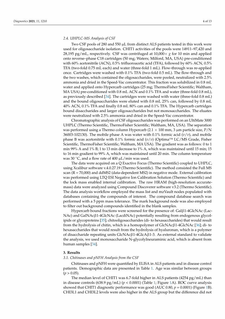
The median level of CHIT1 was 6.7-fold higher in ALS patients (4254 pg/mL) thanin disease controls (638.9 pg/mL) (p < 0.0001) (Table 1; Figure 1A). ROC curve analysisshowed that CHIT1 diagnostic performance was good (AUC 0.80, p = 0.0001) (Figure 1B).CHI3L1 and CHI3L2 levels were also higher in the ALS group but the difference did not
Diagnostics 2021, 11, 1210 5 of 13
reach statistical significance. Concerning the benchmark marker, pNFH, the median levelwas 5.2-fold higher in ALS patients (1751 pg/mL) than in controls (338.7 pg/mL), and itsdiagnostic performance was good (AUC = 0.84, p < 0.0001) (Figure 1A,B).
One control patient with multiple sclerosis displayed the highest level of CHIT1(16,456 pg/mL), CHI3L1 (357 ng/mL) and CHI3L2 (36 ng/mL), but not pNFH (355 pg/mL)(Figure 1A).
Diagnostics 2021, 11, x FOR PEER REVIEW 7 of 14
Figure 1. Levels of pNFH and CHIT1 from the CSF of ALS patients: (A) Comparison between pa-
tients with ALS and controls with other diseases. (B) ROC curve analysis of pNFH and CHIT1. AUC
represents area under the curve. ***, p ≤ 0.001.
CHIT1 had a reasonable correlation with progression rate (r = 0.56, p = 0.0007), as did
CHI3L2 (r = 0.54, p = 0.002) and CHI3L1 (r = 0.43, p = 0.015) (Figure 2A). As expected, pNFH
had a high correlation with disease progression rate (r = 0.72, p < 0.0001). Furthermore,
CHIT1 and CHI3L1 showed moderate but significant correlations with ALSFRS at base-
line (r = −0.37, p = 0.036 and r = −0.38, p = 0.034, respectively). Concerning limb onset, no
significant difference was found among left/right/lower/upper limbs. Among all markers,
CHI3L2 presented a significant correlation with disease duration (r = −0.47, p = 0.0095), as
did pNFH (r = −0.63, p < 0.0001) (Figure 2A).
Interestingly, we found a negative correlation between FVC and CHIT1 (r = −0.45, p
= 0.020), which was not observed for other markers (Figure 2A). This is in line with a neg-
ative correlation between FVC and progression rate (r = −0.47, p = 0.008).
Contr
olsALS
0
50,000
100,000
CH
IT1 (
pg
/ml)
Contr
olsALS
0
5,000
10,000p
NF
H (
pg
/ml)
A
B
CHIT1
AUC=0.80
p=0.0001
pNFH
AUC=0.84
p<0.0001
*** ***
Figure 1. Levels of pNFH and CHIT1 from the CSF of ALS patients: (A) Comparison betweenpatients with ALS and controls with other diseases. (B) ROC curve analysis of pNFH and CHIT1.AUC represents area under the curve. ***, p ≤ 0.001.
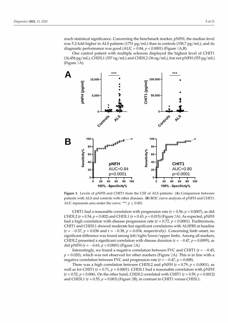
CHIT1 had a reasonable correlation with progression rate (r = 0.56, p = 0.0007), as didCHI3L2 (r = 0.54, p = 0.002) and CHI3L1 (r = 0.43, p = 0.015) (Figure 2A). As expected, pNFHhad a high correlation with disease progression rate (r = 0.72, p < 0.0001). Furthermore,CHIT1 and CHI3L1 showed moderate but significant correlations with ALSFRS at baseline(r = −0.37, p = 0.036 and r = −0.38, p = 0.034, respectively). Concerning limb onset, nosignificant difference was found among left/right/lower/upper limbs. Among all markers,CHI3L2 presented a significant correlation with disease duration (r = −0.47, p = 0.0095), asdid pNFH (r = −0.63, p < 0.0001) (Figure 2A).
Interestingly, we found a negative correlation between FVC and CHIT1 (r = −0.45,p = 0.020), which was not observed for other markers (Figure 2A). This is in line with anegative correlation between FVC and progression rate (r = −0.47, p = 0.008).
There was a high correlation between CHI3L2 and pNFH (r = 0.79, p < 0.0001), aswell as for CHIT1 (r = 0.71, p < 0.0001). CHI3L1 had a reasonable correlation with pNFH(r = 0.52, p = 0.006). On the other hand, CHI3L2 correlated with CHIT1 (r = 0.59, p = 0.0012)and CHI3L1 (r = 0.55, p = 0.003) (Figure 2B), in contrast to CHIT1 versus CHI3L1.

Diagnostics 2021, 11, 1210 6 of 13
Table 1. Demographic information and levels of pNFH and chitinases from the CSF of the patients included. Median and interquartile range are shown. Abbreviations are: C, controls; CC,concentration; M/F, male/female. Age is presented in years. ***, p ≤ 0.001.
pNFH CHIT1 CHI3L1 CHI3L2
M/F Age CC (pg/mL) M/F Age CC (pg/mL) M/F Age CC (ng/mL) M/F Age CC (ng/mL)
C 11/8 62.3 (52.3–67.0) 338.7 (114.9–605.7) 12/12 55.9 (43.1–65.5) 638.9 (273.7–1678) 9/9 56.0 (45.8–67.0) 111.7 (84.3–147.7) 8/9 57.2 (47.7–67.0) 8.66 (6.47–12.13)
ALS 26/10 56.0 (48.0–66.4) 1751 (604.1–3285) 25/9 56.7 (47.4–66.3) 4254 (1293–17074) 24/8 58.0 (49.7–66.4) 128 (91.4–173.3) 22/8 59.4 (49.8–66.6) 12.10 (8.17–22.27)
p - 0.420 <0.0001 *** - 0.826 <0.0001 *** - 0.909 0.515 - 0.991 0.099

Diagnostics 2021, 11, 1210 7 of 13Diagnostics 2021, 11, x FOR PEER REVIEW 8 of 14
Figure 2. Correlation of pNFH, CHIT1, CHI3L1 and CHI3L2 from the CSF of ALS patients with clinical parameters (A)
and with each other (B). Spearman’s r is presented. In (A), all values were considered for the calculations, whereas in (B),
only values from ALS patients for which pNFH, CHIT1, CHI3L1 and CHI3L2 were all concomitantly available (n = 27)
were considered. *, p ≤ 0.05; **, p ≤ 0.01; ***, p ≤ 0.001.
There was a high correlation between CHI3L2 and pNFH (r = 0.79, p < 0.0001), as well
as for CHIT1 (r = 0.71, p < 0.0001). CHI3L1 had a reasonable correlation with pNFH (r =
0.52, p = 0.006). On the other hand, CHI3L2 correlated with CHIT1 (r = 0.59, p = 0.0012) and
CHI3L1 (r = 0.55, p = 0.003) (Figure 2B), in contrast to CHIT1 versus CHI3L1.
Survival of patients stratified by different levels of pNFH or CHI3L2 was signifi-
cantly different (log-rank test: χ2 = 9.0999, p = 0.003; log-rank test: χ2 = 8.4278, p = 0.004,
respectively), but not for CHIT1 or CHI3L1 (Figure 3).
0 5000 10000
0
50,000
100,000
pNFH (pg/ml)
CH
IT1 (
pg
/ml)
0 50000 100000
0
100
200
300
400
500
CHIT1 (pg/ml)
CH
I3L
1 (
ng
/ml)
0 100 200 300 400 500
0
10
20
30
40
50
CHI3L1 (ng/ml)
CH
I3L
2 (
ng
/ml)
0 50000 100000
0
10
20
30
40
50
CHIT1 (pg/ml)
CH
I3L
2 (
ng
/ml)
0 5000 10000
0
10
20
30
40
50
pNFH (pg/ml)
CH
I3L
2 (
ng
/ml)
0 5000 10000
0
100
200
300
400
500
pNFH (pg/ml)
CH
I3L
1 (
ng
/ml)
r=0.52
p=0.006**
r=0.59
p=0.0012**
r=0.79
p<0.0001***B
r=0.55
p=0.003**
r=0.26
p=0.186
r=0.71
p<0.0001***
A
0.0 0.5 1.0 1.5 2.0
0
10
20
30
40
Progression rate
CH
I 3L
2(n
g/ m
l)
r=0.56
p=0.0007***
r=0.43
p=0.015*
r=0.54
p=0.002**
0 5 10 15 20
Disease duration (years)
r=-0.63
p<0.0001***
r=-0.32
p=0.067
r=-0.19
p=0.310
r=-0.47
p=0.0095**
r=-0.29
p=0.115
r=-0.09
p=0.685
r=-0.29
p=0.166
r=-0.45
p=0.020*
0 50 100 150
FVC
r=0.72
p<0.0001***
0
100
200
300
400
CH
I 3L
1(n
g/ m
l)
0
5,000
10,000
15,000
pN
FH
( pg
/ml)
0
50,000
100,000
CH
IT1
(pg
/ ml)
Figure 2. Correlation of pNFH, CHIT1, CHI3L1 and CHI3L2 from the CSF of ALS patients with clinical parameters (A) andwith each other (B). Spearman’s r is presented. In (A), all values were considered for the calculations, whereas in (B), onlyvalues from ALS patients for which pNFH, CHIT1, CHI3L1 and CHI3L2 were all concomitantly available (n = 27) wereconsidered. *, p ≤ 0.05; **, p ≤ 0.01; ***, p ≤ 0.001.
Survival of patients stratified by different levels of pNFH or CHI3L2 was signifi-cantly different (log-rank test: χ2 = 9.0999, p = 0.003; log-rank test: χ2 = 8.4278, p = 0.004,respectively), but not for CHIT1 or CHI3L1 (Figure 3).

Diagnostics 2021, 11, 1210 8 of 13Diagnostics 2021, 11, x FOR PEER REVIEW 9 of 14
Figure 3. Kaplan–Meier survival curves stratified by low and high chitinase protein level (above or
below or equal to the median level). Time in months since first symptoms were considered.
Cox model showed that low CHI3L2 level was an independent predictor for survival,
in addition to age and disease duration (CHI3L2; HR = 0.201, 95% CI = 0.0525 to 0.7705, p
= 0.019) (Table 2).
Table 2. Cox proportional hazards modelling of factors known to be associated with survival and individual chitinase
proteins separately. b—regression coefficient; SE—standard error, Wald statistic; HR—hazard ratio; CI—confidence inter-
val.
Covariate b SE Wald p HR 95% CI of HR
Age at onset 0.076 0.033 5.206 0.023 1.079 1.011 to 1.153
Disease duration at sampling −0.099 0.043 5.428 0.020 0.906 0.833 to 0.984
CHIT1 −0.777 0.541 2.060 0.151 0.460 0.159 to 1.328
Age at onset 0.049 0.032 2.291 0.130 1.050 0.986 to 1.119
Disease duration at sampling −0.096 0.042 5.301 0.021 0.908 0.837 to 0.986
CHI3L1 −0.398 0.531 0.562 0.453 0.672 0.237 to 1.902
Age at onset 0.115 0.055 4.454 0.035 1.122 1.008 to 1.249
Disease duration at sampling −0.116 0.045 6.666 0.010 0.890 0.815 to 0.972
CHI3L2 −1.604 0.685 5.478 0.019 0.201 0.053 to 0.771
Age at onset 0.065 0.032 4.078 0.043 1.068 1.002 to 1.137
Disease duration at sampling −0.059 0.033 3.264 0.071 0.942 0.884 to 1.005
pNFH −0.890 0.537 2.741 0.098 0.411 0.143 to 1.178
3.2. UHPLC-MS Analysis of CSF
In an attempt to identify potential products of CHIT1 activity in the CSF, we fraction-
ated CSF using reverse-phase C18 (to remove proteins and hydrophobic molecules) and
graphite Hypercarb (that binds oligosaccharides) cartridges to obtain an oligosaccharide
enriched fraction. Pools of CSF available from patients tested for CHIT1 were used. Hy-
percarb-bound fractions were analyzed by UHPLC-MS using a Hypercarb column. We
screened for putative CHIT1 products, which included chito-oligosaccharides as hydrol-
ysis products of chitin (linear polymer of β1-4 linked GlcNAc) [36], the disaccharides
Figure 3. Kaplan–Meier survival curves stratified by low and high chitinase protein level (above orbelow or equal to the median level). Time in months since first symptoms were considered.
Cox model showed that low CHI3L2 level was an independent predictor for survival,in addition to age and disease duration (CHI3L2; HR = 0.201, 95% CI = 0.0525 to 0.7705,p = 0.019) (Table 2).
Table 2. Cox proportional hazards modelling of factors known to be associated with survival and individual chitinaseproteins separately. b—regression coefficient; SE—standard error, Wald statistic; HR—hazard ratio; CI—confidence interval.
Covariate b SE Wald p HR 95% CI of HR
Age at onset 0.076 0.033 5.206 0.023 1.079 1.011 to 1.153
Disease duration at sampling −0.099 0.043 5.428 0.020 0.906 0.833 to 0.984
CHIT1 −0.777 0.541 2.060 0.151 0.460 0.159 to 1.328
Age at onset 0.049 0.032 2.291 0.130 1.050 0.986 to 1.119
Disease duration at sampling −0.096 0.042 5.301 0.021 0.908 0.837 to 0.986
CHI3L1 −0.398 0.531 0.562 0.453 0.672 0.237 to 1.902
Age at onset 0.115 0.055 4.454 0.035 1.122 1.008 to 1.249
Disease duration at sampling −0.116 0.045 6.666 0.010 0.890 0.815 to 0.972
CHI3L2 −1.604 0.685 5.478 0.019 0.201 0.053 to 0.771
Age at onset 0.065 0.032 4.078 0.043 1.068 1.002 to 1.137
Disease duration at sampling −0.059 0.033 3.264 0.071 0.942 0.884 to 1.005
pNFH −0.890 0.537 2.741 0.098 0.411 0.143 to 1.178
3.2. UHPLC-MS Analysis of CSF
In an attempt to identify potential products of CHIT1 activity in the CSF, we frac-tionated CSF using reverse-phase C18 (to remove proteins and hydrophobic molecules)and graphite Hypercarb (that binds oligosaccharides) cartridges to obtain an oligosaccha-ride enriched fraction. Pools of CSF available from patients tested for CHIT1 were used.Hypercarb-bound fractions were analyzed by UHPLC-MS using a Hypercarb column.

Diagnostics 2021, 11, 1210 9 of 13
We screened for putative CHIT1 products, which included chito-oligosaccharides as hy-drolysis products of chitin (linear polymer of β1-4 linked GlcNAc) [36], the disaccharidesGalβ1-4GlcNAc and GalNAcβ1-4GlcNAc (LacdiNAc) as potential products of mamalianglycoproteins or glycolipids [35] and oligosaccharides resulting from the hydrolysis ofhyaluronan (linear polymer of the repeating disaccharide GlcNAcβ1-4GlcAβ1-3) that ispresent in the CSF [37] and has some structural resemblance to chitin. From these, only asignal at m/z 469.166 [M+FA-H] compatible with HexNAc-HexNAc was detected (Supple-mentary Figure S1). This could correspond to di-N-acetylchitobiose (GlcNAcβ1-4GlcNAc)or LacdiNAc (GalNAcβ1-4GlcNAc). It was not possible to perform fragmentation toconfirm the structure due to the low intensity of the signal.
4. Discussion
In this study, we presented evidence supporting the relevance of chitinases as ALSbiomarkers. This comparative study highlighted the importance of different chitinases aspotentially useful markers for distinct disease characteristics.
CHIT1 levels were elevated in ALS compared to disease controls, in agreement withreports by other groups [13,16,28–30,38]. Furthermore, a high diagnostic performance ofCHIT1 was detected but it did not outperform pNFH. Most interestingly, we disclosed asignificant negative correlation between CHIT1 and the respiratory function index of forcedvital capacity (FVC). FVC measures potential respiratory impairment in ALS. Evidencefrom the literature has indicated that FVC value may constitute a predictor of survival anddisease progression [39], which is further corroborated by our findings here. In this context,it will be necessary to validate these results in a larger and independent cohort of patients.For CHI3L1, there was not a significant increase, which is in agreement with others [16].
For CHI3L2, increased levels have been reported in the literature [30,32] but, in ourstudy, although an increase was observed, it was not significant. On the other hand, wefound CHI3L2 to be a significant and independent predictor of survival using Kaplan–Meierestimator and Cox proportional hazards modelling, which contrasted to CHIT1 and CHI3L1.For CHIT1, existing reports favor a predictive value of survival [30,38]. Concerning CHI3L1,there is discrepancy of results in the literature [29,30,38]. The significant relevance ofCHI3L2 to predict survival in ALS observed in our patients differed from results of anothergroup [29,30]. Since the number of independent studies investigating CHI3L2 is low inthe literature, our results emphasize the need to further investigate this chitinase and itsbiomarker potential in larger independent cohorts of patients.
Although concentration values for all proteins were within the range described else-where [13,30], there were some differences between our study and others concerning thepotential of different chitinases as biomakers for ALS. This was probably due to the numberand subject characteristics of the control group with other diseases, since some of the con-trols may also exhibit chitinases induction. For example, in multiple sclerosis, an increasein CHIT1 has been reported in the literature [40], which is in agreement with our findings.Therefore, besides testing larger independent cohorts of ALS and control patients, it wouldalso be relevant to analyze additional control groups (e.g., healthy controls).
The three tested chitinases correlated with disease progression rate in ALS patients,and CHIT1 and CHI3L2 showed the strongest correlations. These results were in agreementwith other authors [13,16,29,30,38]. Therefore, these molecules constitute potentially usefulbiomarkers for progression rate, which is particularly useful in the context of precisionmedicine that has patient stratification for differentiated therapeutic trials in mind.
We found that all chitinases correlated with pNFH, with the correlation being highestfor CHIT1 and CHI3L2. Since pNFH is a marker of neuronal damage [41], most importantly,these results supported a connection between neuronal damage and neuroinflammation.In ALS, neuroinflammation is characterized predominantly by the activation of microgliaand astrocytes, and by the presence in the CNS of non-resident immune cells, includingT cells, monocyte-derived macrophages and natural killer cells [11]. Many studies haveshown a deregulation in the levels of cytokines in the CSF and blood of ALS patients,

Diagnostics 2021, 11, 1210 10 of 13
including interleukins, tumor necrosis factors and interferon gamma produced by differentimmune cells [10]. Glial cells that are normally supportive of neuron homeostasis maybecome neurotoxic during the initial stages of the disease; this process may be triggered,for example, by dysfunction and misfolding of mutant proteins associated with ALS(e.g., SOD1, TDP-43, dipeptide repeat proteins) and neuron damage. Activated glial cellsand peripheral immune cells release toxic molecules, including pro-inflammatory cytokines(e.g., IL-1β), that affect motor neuron integrity [10]. Therefore, a complex interplay betweenneuronal damage and immune cell activation occurs in ALS.
Chitinases are secreted by activated glial cells, as well as by other cells of the immunesystem [12,13,16]. CHIT1 was detected in microglia from the corticospinal tract [13] andin macrophages [12]. CHI3L1 was found in a subset of activated astrocytes [16], mono-cytes/macrophages [12]. CHI3L2 has been detected in macrophages [17]; curiously, ithas been reported in neuronal cells from the cerebral cortex (https://www.proteinatlas.org/ENSG00000064886-CHI3L2/tissue/cerebral+cortex#img), but its expression in ALStissues has not been described so far. As such, the origin of CSF CHI3L2 is currentlyunknown. Possible interplay between pro-inflammatory cytokines and chitinases has beendiscussed [12], with implications for their functional activity. In this context, chitinasesrecently appeared as promising therapeutic targets. For example, inhibition of CHI3L1 withthe compound K284-6111 prevented amyloid beta-induced neuroinflammation and impair-ment of recognition memory via inhibition of the NF-κB pathway in an Alzheimer’s diseasemouse model [42]. In line with this, CHI3L1 was found to be neurotoxic towards corticalneurons but not immune cells [43]. By contrast, human CHIT1 and CHI3L1 promotedoligodendrogenesis from neural stem cells [44]. Additionally, CHIT1 had a protective rolein an Alzheimer’s disease rat model and N9 microglia cells [45]. More studies need to beperformed to evaluate whether chitinases are just bystanders resulting from immune cellactivation, or whether they have a physiological relevant role.
CHIT1 has chitinolytic activity towards chitin and artificial substrates [36], as well asmammalian N-acetylglucosamine-containing glycoconjugates [35]. CHIT1 from humanCSF is active in vitro towards a synthetic substrate [30] but its endogenous target(s) is notknown. Our efforts aiming at identifying potential products of CHIT1 activity in the CSFof patients tested here indicated an m/z signal compatible with the disaccharide HexNAc-HexNAc, which could consist of di-N-acetylchitobiose (GlcNAcβ1-4GlcNAc, a hydrolysisproduct of chitin) or LacdiNAc (GalNAcβ1-4GlcNAc, found in some human glycoproteins),which are hydrolyzed by CHIT1 [35,36]. Humans do not produce chitin that is found infungi; however, there has been evidence of fungal infection in the CNS of ALS patients [46],which could explain the origin of di-N-acetylchitobiose. On the other hand, evidence forchitin-like polysaccharides has been reported in Alzheimer’s disease [47]. However, itshould also be considered that di-N-acetylchitobiose could originate from the degradationof other glycoconjugates, such as N-linked glycans from endogenous human glycoproteins.Since hyaluronan (polysaccharide of the repeating disaccharide of N-acetylglucosamineand D-glucuronic acid) has some structural resemblance with chitin and is present in theCSF [37], we also screened for potential hydrolysis products but found no signal compatiblewith those oligosaccharides. This was in agreement with in vitro data indicating that CHIT1did not cleave hyaluronan [48]. Limitations of this preliminary study include the following:low volumes for testing; only a duplicate, but from two independent pools, was performed;CSF pools were analyzed instead of individual samples; only ALS samples and not controlsfor comparison were investigated. However, a methodology for the study of CSF-freeglycans has been presented. Moreover, a novel perspective has been presented to furtherinvestigate whether the disaccharide HexNAc-HexNAc is found in independent ALSand control samples, as well as in the positive scenario, to unequivocally elucidate thecorresponding structure by MS/MS and/or liquid chromatography techniques.
In conclusion, our results supported the importance of chitinases as biomarker targetsin ALS. Particularly, CHIT1 was a promising biomarker for diagnosis, progression rateand respiratory function. These results provided novel perspectives to further explore the

Diagnostics 2021, 11, 1210 11 of 13
potential of chitinases as ALS biomarkers and their functional relevance, they and mayhave implications in other neurological diseases.
Supplementary Materials: The following are available online at https://www.mdpi.com/article/10.3390/diagnostics11071210/s1, Figure S1: Full MS spectra with isotope pattern fit of HexNAc-HexNAc identified using Compound Discoverer 3.2 (Thermo).
Author Contributions: Conceptualization, J.C. and M.d.C.; methodology, J.C., M.G., C.A., R.A.G.,A.C.L.G.; formal analysis, J.C., M.G., A.C.L.G., R.A.G.; investigation, J.C., M.G., A.P.-L., S.P., M.d.C.;resources, J.C., A.O., R.A.G., M.d.C.; writing—original draft preparation, J.C., R.A.G.; writing—review and editing, M.G., A.P-L., S.P., R.A.G., A.O., M.d.C.; visualization, J.C., M.G.; supervision, J.C.,M.d.C.; funding acquisition, J.C., M.d.C. All authors have read and agreed to the published versionof the manuscript.
Funding: We acknowledge iNOVA4Health – UIDB/04462/2020 and UIDP/04462/2020, a programfinancially supported by Fundação para a Ciência e Tecnologia/Ministério da Ciência, Tecnologia eEnsino Superior, through national funds.
Institutional Review Board Statement: The study was conducted according to the guidelines of theDeclaration of Helsinki, and approved by the Institutional Review Board of Centro Académico deMedicina da Universidade de Lisboa (protocol nº 94/19 from 2 April 2019).
Informed Consent Statement: Informed consent was obtained from all subjects involved in the study.
Data Availability Statement: The data presented in this study are available in this article andsupplementary material.
Conflicts of Interest: The authors declare no conflict of interest.
References1. Ryan, M.; Heverin, M.; McLaughlin, R.L.; Hardiman, O. Lifetime Risk and Heritability of Amyotrophic Lateral Sclerosis. JAMA
Neurol. 2019, 76, 1367–1374. [CrossRef]2. Volk, A.E.; Weishaupt, J.H.; Andersen, P.M.; Ludolph, A.C.; Kubisch, C. Current knowledge and recent insights into the genetic
basis of amyotrophic lateral sclerosis. Med. Genet. 2018, 30, 252–258. [CrossRef]3. Moll, T.; Shaw, P.J.; Cooper-Knock, J. Disrupted glycosylation of lipids and proteins is a cause of neurodegeneration. Brain 2020,
143, 1332–1340. [CrossRef]4. Cooper-Knock, J.; Moll, T.; Ramesh, T.; Castelli, L.; Beer, A.; Robins, H.; Fox, I.; Niedermoser, I.; Van Damme, P.; Moisse, M.; et al.
Mutations in the Glycosyltransferase Domain of GLT8D1 Are Associated with Familial Amyotrophic Lateral Sclerosis. Cell Rep.2019, 26, 2298–2306.e2295. [CrossRef] [PubMed]
5. Costa, J.; Gomes, C.; de Carvalho, M. Diagnosis, pathogenesis and therapeutic targets in amyotrophic lateral sclerosis. CNSNeurol. Disord. Drug Targets 2010, 9, 764–778. [CrossRef] [PubMed]
6. Taylor, J.P.; Brown, R.H., Jr.; Cleveland, D.W. Decoding ALS: From genes to mechanism. Nature 2016, 539, 197–206. [CrossRef]7. Clement, A.M.; Nguyen, M.D.; Roberts, E.A.; Garcia, M.L.; Boillee, S.; Rule, M.; McMahon, A.P.; Doucette, W.; Siwek, D.; Ferrante,
R.J.; et al. Wild-type nonneuronal cells extend survival of SOD1 mutant motor neurons in ALS mice. Science 2003, 302, 113–117.[CrossRef] [PubMed]
8. Vahsen, B.F.; Gray, E.; Thompson, A.G.; Ansorge, O.; Anthony, D.C.; Cowley, S.A.; Talbot, K.; Turner, M.R. Non-neuronal cells inamyotrophic lateral sclerosis—From pathogenesis to biomarkers. Nat. Rev. Neurol. 2021. [CrossRef]
9. Beers, D.R.; Appel, S.H. Immune dysregulation in amyotrophic lateral sclerosis: Mechanisms and emerging therapies. LancetNeurol. 2019, 18, 211–220. [CrossRef]
10. Moreno-Garcia, L.; Miana-Mena, F.J.; Moreno-Martinez, L.; de la Torre, M.; Lunetta, C.; Tarlarini, C.; Zaragoza, P.; Calvo, A.C.;Osta, R. Inflammasome in ALS Skeletal Muscle: NLRP3 as a Potential Biomarker. Int. J. Mol. Sci. 2021, 22, 2523. [CrossRef][PubMed]
11. McCauley, M.E.; Baloh, R.H. Inflammation in ALS/FTD pathogenesis. Acta Neuropathol. 2019, 137, 715–730. [CrossRef]12. Pinteac, R.; Montalban, X.; Comabella, M. Chitinases and chitinase-like proteins as biomarkers in neurologic disorders. Neurol.
Neuroimmunol. Neuroinflamm. 2021, 8. [CrossRef]13. Steinacker, P.; Verde, F.; Fang, L.; Feneberg, E.; Oeckl, P.; Roeber, S.; Anderl-Straub, S.; Danek, A.; Diehl-Schmid, J.; Fassbender,
K.; et al. Chitotriosidase (CHIT1) is increased in microglia and macrophages in spinal cord of amyotrophic lateral sclerosis andcerebrospinal fluid levels correlate with disease severity and progression. J. Neurol. Neurosurg. Psychiatry 2018, 89, 239–247.[CrossRef] [PubMed]

Diagnostics 2021, 11, 1210 12 of 13
14. Lee, C.G.; Da Silva, C.A.; Dela Cruz, C.S.; Ahangari, F.; Ma, B.; Kang, M.J.; He, C.H.; Takyar, S.; Elias, J.A. Role of chitin andchitinase/chitinase-like proteins in inflammation, tissue remodeling, and injury. Annu. Rev. Physiol. 2011, 73, 479–501. [CrossRef][PubMed]
15. Shao, R.; Hamel, K.; Petersen, L.; Cao, Q.J.; Arenas, R.B.; Bigelow, C.; Bentley, B.; Yan, W. YKL-40, a secreted glycoprotein,promotes tumor angiogenesis. Oncogene 2009, 28, 4456–4468. [CrossRef] [PubMed]
16. Vu, L.; An, J.; Kovalik, T.; Gendron, T.; Petrucelli, L.; Bowser, R. Cross-sectional and longitudinal measures of chitinase proteins inamyotrophic lateral sclerosis and expression of CHI3L1 in activated astrocytes. J. Neurol. Neurosurg. Psychiatry 2020, 91, 350–358.[CrossRef]
17. Liu, L.; Yang, Y.; Duan, H.; He, J.; Sun, L.; Hu, W.; Zeng, J. CHI3L2 Is a Novel Prognostic Biomarker and Correlated With ImmuneInfiltrates in Gliomas. Front. Oncol. 2021, 11, 611038. [CrossRef] [PubMed]
18. Gagliardi, D.; Meneri, M.; Saccomanno, D.; Bresolin, N.; Comi, G.P.; Corti, S. Diagnostic and Prognostic Role of Blood andCerebrospinal Fluid and Blood Neurofilaments in Amyotrophic Lateral Sclerosis: A Review of the Literature. Int. J. Mol. Sci.2019, 20, 4152. [CrossRef]
19. Chio, A.; Mazzini, L.; Mora, G. Disease-modifying therapies in amyotrophic lateral sclerosis. Neuropharmacology 2020, 167, 107986.[CrossRef]
20. Feneberg, E.; Oeckl, P.; Steinacker, P.; Verde, F.; Barro, C.; Van Damme, P.; Gray, E.; Grosskreutz, J.; Jardel, C.; Kuhle, J.; et al.Multicenter evaluation of neurofilaments in early symptom onset amyotrophic lateral sclerosis. Neurology 2018, 90, e22–e30.[CrossRef]
21. Goncalves, M.; Tillack, L.; de Carvalho, M.; Pinto, S.; Conradt, H.S.; Costa, J. Phosphoneurofilament heavy chain and N-glycomicsfrom the cerebrospinal fluid in amyotrophic lateral sclerosis. Clin. Chim. Acta 2015, 438, 342–349. [CrossRef]
22. Goncalves, M.; De Carvalho, M.; Peixoto, C.; Alves, P.; Barreto, C.; Oliva, A.; Pinto, S.; Laborinho-Pronto, A.; Gromicho, M.;Costa, J. Phosphoneurofilament heavy chain and vascular endothelial growth factor as cerebrospinal fluid biomarkers for ALS.Amyotroph. Lateral Scler. Frontotemporal. Degener. 2017, 18, 134–136. [CrossRef]
23. Lu, C.H.; Macdonald-Wallis, C.; Gray, E.; Pearce, N.; Petzold, A.; Norgren, N.; Giovannoni, G.; Fratta, P.; Sidle, K.; Fish, M.; et al.Neurofilament light chain: A prognostic biomarker in amyotrophic lateral sclerosis. Neurology 2015, 84, 2247–2257. [CrossRef][PubMed]
24. Oeckl, P.; Jardel, C.; Salachas, F.; Lamari, F.; Andersen, P.M.; Bowser, R.; de Carvalho, M.; Costa, J.; van Damme, P.; Gray, E.; et al.Multicenter validation of CSF neurofilaments as diagnostic biomarkers for ALS. Amyotroph. Lateral Scler. Frontotemporal. Degener.2016, 17, 404–413. [CrossRef] [PubMed]
25. Steinacker, P.; Feneberg, E.; Weishaupt, J.; Brettschneider, J.; Tumani, H.; Andersen, P.M.; von Arnim, C.A.; Bohm, S.; Kassubek,J.; Kubisch, C.; et al. Neurofilaments in the diagnosis of motoneuron diseases: A prospective study on 455 patients. J. Neurol.Neurosurg. Psychiatry 2016, 87, 12–20. [CrossRef] [PubMed]
26. Gaur, N.; Perner, C.; Witte, O.W.; Grosskreutz, J. The Chitinases as Biomarkers for Amyotrophic Lateral Sclerosis: Signals Fromthe CNS and Beyond. Front. Neurol. 2020, 11, 377. [CrossRef] [PubMed]
27. Swash, M. Chitinases, neuroinflammation and biomarkers in ALS. J. Neurol. Neurosurg. Psychiatry 2020, 91, 338. [CrossRef]28. Steinacker, P.; Feneberg, E.; Halbgebauer, S.; Witzel, S.; Verde, F.; Oeckl, P.; Van Damme, P.; Gaur, N.; Gray, E.; Grosskreutz, J.;
et al. Chitotriosidase as biomarker for early stage amyotrophic lateral sclerosis: A multicenter study. Amyotroph. Lateral Scler.Frontotemporal. Degener. 2021, 22, 276–286. [CrossRef]
29. Thompson, A.G.; Gray, E.; Thezenas, M.L.; Charles, P.D.; Evetts, S.; Hu, M.T.; Talbot, K.; Fischer, R.; Kessler, B.M.; Turner, M.R.Cerebrospinal fluid macrophage biomarkers in amyotrophic lateral sclerosis. Ann. Neurol. 2018, 83, 258–268. [CrossRef]
30. Thompson, A.G.; Gray, E.; Bampton, A.; Raciborska, D.; Talbot, K.; Turner, M.R. CSF chitinase proteins in amyotrophic lateralsclerosis. J. Neurol. Neurosurg. Psychiatry 2019, 90, 1215–1220. [CrossRef]
31. Varghese, A.M.; Sharma, A.; Mishra, P.; Vijayalakshmi, K.; Harsha, H.C.; Sathyaprabha, T.N.; Bharath, S.M.; Nalini, A.; Alladi,P.A.; Raju, T.R. Chitotriosidase—A putative biomarker for sporadic amyotrophic lateral sclerosis. Clin. Frontotemporal. 2013, 10,19. [CrossRef]
32. Barschke, P.; Oeckl, P.; Steinacker, P.; Al Shweiki, M.R.; Weishaupt, J.H.; Landwehrmeyer, G.B.; Anderl-Straub, S.; Weydt, P.;Diehl-Schmid, J.; Danek, A.; et al. Different CSF protein profiles in amyotrophic lateral sclerosis and frontotemporal dementiawith C9orf72 hexanucleotide repeat expansion. J. Neurol. Neurosurg. Psychiatry 2020, 91, 503–511. [CrossRef]
33. Brooks, B.R.; Miller, R.G.; Swash, M.; Munsat, T.L.; World Federation of Neurology Research Group on Motor Neuron Disease. ElEscorial revisited: Revised criteria for the diagnosis of amyotrophic lateral sclerosis. Amyotroph Lateral Scler Other Motor NeuronDisord 2000, 1, 293–299. [CrossRef]
34. Costa, J.; Gatermann, M.; Nimtz, M.; Kandzia, S.; Glatzel, M.; Conradt, H.S. N-Glycosylation of Extracellular Vesicles fromHEK-293 and Glioma Cell Lines. Anal Chem 2018, 90, 7871–7879. [CrossRef]
35. Larsen, T.; Yoshimura, Y.; Voldborg, B.G.; Cazzamali, G.; Bovin, N.V.; Westerlind, U.; Palcic, M.M.; Leisner, J.J. Humanchitotriosidase CHIT1 cross reacts with mammalian-like substrates. FEBS Lett 2014, 588, 746–751. [CrossRef]
36. Renkema, G.H.; Boot, R.G.; Muijsers, A.O.; Donker-Koopman, W.E.; Aerts, J.M. Purification and characterization of humanchitotriosidase, a novel member of the chitinase family of proteins. J. Biol. Chem. 1995, 270, 2198–2202. [CrossRef]
37. Yu, Y.; Zhang, F.; Colon, W.; Linhardt, R.J.; Xia, K. Glycosaminoglycans in human cerebrospinal fluid determined by LC-MS/MSMRM. Anal. Biochem. 2019, 567, 82–84. [CrossRef] [PubMed]
Diagnostics 2021, 11, 1210 13 of 13
38. Gille, B.; De Schaepdryver, M.; Dedeene, L.; Goossens, J.; Claeys, K.G.; Van Den Bosch, L.; Tournoy, J.; Van Damme, P.; Poesen, K.Inflammatory markers in cerebrospinal fluid: Independent prognostic biomarkers in amyotrophic lateral sclerosis? J. Neurol.Neurosurg. Psychiatry 2019, 90, 1338–1346. [CrossRef]
39. Czaplinski, A.; Yen, A.A.; Appel, S.H. Forced vital capacity (FVC) as an indicator of survival and disease progression in an ALSclinic population. J. Neurol. Neurosurg. Psychiatry 2006, 77, 390–392. [CrossRef]
40. Oldoni, E.; Smets, I.; Mallants, K.; Vandebergh, M.; Van Horebeek, L.; Poesen, K.; Dupont, P.; Dubois, B.; Goris, A. CHIT1 atDiagnosis Reflects Long-Term Multiple Sclerosis Disease Activity. Ann. Neurol. 2020, 87, 633–645. [CrossRef] [PubMed]
41. Khalil, M.; Teunissen, C.E.; Otto, M.; Piehl, F.; Sormani, M.P.; Gattringer, T.; Barro, C.; Kappos, L.; Comabella, M.; Fazekas, F.; et al.Neurofilaments as biomarkers in neurological disorders. Nat. Rev. Neurol. 2018, 14, 577–589. [CrossRef]
42. Choi, J.Y.; Yeo, I.J.; Kim, K.C.; Choi, W.R.; Jung, J.K.; Han, S.B.; Hong, J.T. K284-6111 prevents the amyloid beta-inducedneuroinflammation and impairment of recognition memory through inhibition of NF-kappaB-mediated CHI3L1 expression. JNeuroinflamm. 2018, 15, 224. [CrossRef]
43. Matute-Blanch, C.; Calvo-Barreiro, L.; Carballo-Carbajal, I.; Gonzalo, R.; Sanchez, A.; Vila, M.; Montalban, X.; Comabella, M.Chitinase 3-like 1 is neurotoxic in primary cultured neurons. Sci. Rep. 2020, 10, 7118. [CrossRef] [PubMed]
44. Starossom, S.C.; Campo Garcia, J.; Woelfle, T.; Romero-Suarez, S.; Olah, M.; Watanabe, F.; Cao, L.; Yeste, A.; Tukker, J.J.; Quintana,F.J.; et al. Chi3l3 induces oligodendrogenesis in an experimental model of autoimmune neuroinflammation. Nat. Commun. 2019,10, 217. [CrossRef]
45. Xiao, Q.; Yu, W.; Tian, Q.; Fu, X.; Wang, X.; Gu, M.; Lu, Y. Chitinase1 contributed to a potential protection via microglia polarizationand Abeta oligomer reduction in D-galactose and aluminum-induced rat model with cognitive impairments. Neuroscience 2017,355, 61–70. [CrossRef]
46. Alonso, R.; Pisa, D.; Fernandez-Fernandez, A.M.; Rabano, A.; Carrasco, L. Fungal infection in neural tissue of patients withamyotrophic lateral sclerosis. Neurobiol. Dis. 2017, 108, 249–260. [CrossRef]
47. Castellani, R.J.; Perry, G.; Smith, M.A. The role of novel chitin-like polysaccharides in Alzheimer disease. Neurotox. Res. 2007,12, 269–274. [CrossRef]
48. Danielson, B.; Chen, C.H.; Kaber, G.; Mochly-Rosen, D.; Grimes, K.; Stern, R.; Bollyky, P.L. Human Chitotriosidase Does NotCatabolize Hyaluronan. Int. J. Biol. Macromol. 2018, 109, 629–633. [CrossRef]
Related Documents