Microbiology (1996), 142, 1581-1 589 Printed in Great Britain Comparative studies of chitinases A and B from Serratia marcescens May B. Brurberg,'t2 Ingolf F. Nesl and Vincent G. H. Eijsinkl Author for correspondence: May B. Brurberg. Tel: +47 64949461. Fax: +47 64941465. e-mail : mbruberg@ bioslave.uio.no 1 Laboratory of Microbial Gene Technology, Department of Biotechnological Sciences, Agricultural University of Norw~y, PO Box 5051, 1432 As, Norway Research Institute, As, Norway 2 The Norwegian Crop Serratia marcescens produces several chit inol yt ic enzymes, including chit i nase A (ChiA) and chitinase B (ChiB). In this study, Chis was purified to homogeneity using a newly developed protocol based on hydrophobic interaction chromatography. Subsequently, characteristics of Chis and of the hitherto only partly characterized ChiA were determined and compared. Pure ChiA and Chis shared several characteristics such as a broad pH optimum around pH 5-0, and a temperature optimum between 50 and 60 OC. Both enzymes were fairly stable, with half-lives of more than 10 d at 37 "C, pH 61. Analyses of the degradation of various N-acetylglucosamine oligomers, f luorogenic substrates and colloidal chitin showed that both enzymes cleave chitobiose [(GlcNAc),] from (GlcNAc), and thus possess an exolN,N'- diacetylchitobiohydrolase activity. Both enzymes were also capable of producing monomers from longer (GlcNAc), substrates, indicating that they also have an endochitinase (ChiA) or exo-N,N',N''-triacetylchitotriohydrolase (ChiB) activity. Kinetic analyses with ~methylumbelliferyl-~D-N,N'- d iacet y Ich i to bioside, an analogue of (GlcN Ac), showed cooperative kinetics for ChiA, whereas for Chis normal hyperbolic kinetics were observed. ChiA had a higher specific activity towards chitin than Chis and synergistic effects on the chitin degradation rate were observed upon combining the two enzymes. These results, together with the results of sequence comparisons and previous studies of the cellular localization of the two chitinases in S. marcescens indicate possible roles for ChiA and ChiB in chitin breakdown. Keywords : chitinase, Serratia marcescens, chitin, chtobiosidase INTRODUCTION Several bacteria and fungi are capable of enzymic degra- dation of chitin, the (1 +4)-P-linked polymer of N-acetyl- P-D-glucosamine (GlcNAc), for autolytic, morphogenetic or nutritional purposes (Gooday, 1990). Since chitin is a major structural component of fungal cell walls, chitino- lytic enzymes could in principle be employed as natural anti-fungal agents and are therefore of biotechnological importance (Shapira et a/., 1989; Schickler et al., 1993; Lorito et al., 1994). The conversion of chitin to GlcNAc involves several hydrolases. Chitinases (EC 3.2.1 .14), also called endochitinases, cleave glycosidic linkages Abbreviations: ChiA, chitinase A from 5. marcescens; ChiB, chitinase B from 5. marcescens; GlcNAc, N-acetylglucosamine; 4-MU, 4- methylumbelliferyl; 4-MU-(GlcNAc), 4-methylumbelliferyl N-acetyl-8-o- glucosaminide; 4-MU-(GlcNAc),, 4-methylumbelliferyl p-o-N,N'- diacetylchitobioside; 4-MU-(GlcNAc),, 4-methylumbelliferyl B-D-N,N',N''- triacetylchitotriose; [S], substrate concentration. randomly along the chitin chain, eventually producing short oligomers of GlcNAc (Monreal & Reese, 1969; Robbins et al., 1988; Roberts & Selitrennikoff, 1988; De la Cruz et al., 1992). Exo-N,N'-diacetylchitobio- hydrolases, also called exochitinases or chitobiosidases, cleave off chitobiose (GlcNAc),, from the non-reducing end of (GlcNAc), (Davis & Eveleigh, 1984; Robbins et al., 1988; Tronsmo & Harman, 1993). Finally, N-acetyl- 8-glucosaminidases (EC 3.2.1 .52) cleave off GlcNAc monomers from the non-reducing ends of chitobiose and higher analogues (Monreal & Reese, 1969 ; Robbins et a/., 1988). Serratia marcescens, a Gram-negative bacterium belonging to the family Enterobacteriaceae, is one of the most efficient bacteria for degradation of chitin (Monreal & Reese, 1969; Schickler et al., 1993). It may produce up to five different chitinolytic enzymes upon induction with chitin (Fuchs et al., 1986). The best known of these is the secreted chitinase A (ChiA), the three-dimensional struc- 0002-0459 0 1996 SGM 1581

Welcome message from author
This document is posted to help you gain knowledge. Please leave a comment to let me know what you think about it! Share it to your friends and learn new things together.
Transcript
Microbiology (1996), 142, 1581-1 589 Printed in Great Britain
Comparative studies of chitinases A and B from Serratia marcescens
May B. Brurberg,'t2 Ingolf F. Nesl and Vincent G. H. Eijsinkl
Author for correspondence: May B. Brurberg. Tel: +47 64949461. Fax: +47 64941465. e-mail : mbruberg@ bioslave.uio.no
1 Laboratory of Microbial Gene Technology, Department of Biotechnological Sciences, Agricultural University of Norw~y, PO Box 5051, 1432 As, Norway
Research Institute, As, Norway
2 The Norwegian Crop
Serratia marcescens produces several chit inol y t ic enzymes, including chit i nase A (ChiA) and chitinase B (ChiB). In this study, Chis was purified to homogeneity using a newly developed protocol based on hydrophobic interaction chromatography. Subsequently, characteristics of Chis and of the hitherto only partly characterized ChiA were determined and compared. Pure ChiA and Chis shared several characteristics such as a broad pH optimum around pH 5-0, and a temperature optimum between 50 and 60 OC. Both enzymes were fairly stable, with half-lives of more than 10 d at 37 "C, pH 61. Analyses of the degradation of various N-acetylglucosamine oligomers, f luorogenic substrates and colloidal chitin showed that both enzymes cleave chitobiose [(GlcNAc),] from (GlcNAc), and thus possess an exolN,N'- diacetylchitobiohydrolase activity. Both enzymes were also capable of producing monomers from longer (GlcNAc), substrates, indicating that they also have an endochitinase (ChiA) or exo-N,N',N''-triacetylchitotriohydrolase (ChiB) activity. Kinetic analyses with ~methylumbelliferyl-~D-N,N'- d iacet y Ich i to bioside, an analogue of (GlcN Ac),, showed cooperative kinetics for ChiA, whereas for Chis normal hyperbolic kinetics were observed. ChiA had a higher specific activity towards chitin than Chis and synergistic effects on the chitin degradation rate were observed upon combining the two enzymes. These results, together with the results of sequence comparisons and previous studies of the cellular localization of the two chitinases in S. marcescens indicate possible roles for ChiA and ChiB in chitin breakdown.
Keywords : chitinase, Serratia marcescens, chitin, chtobiosidase
INTRODUCTION
Several bacteria and fungi are capable of enzymic degra- dation of chitin, the (1 +4)-P-linked polymer of N-acetyl- P-D-glucosamine (GlcNAc), for autolytic, morphogenetic or nutritional purposes (Gooday, 1990). Since chitin is a major structural component of fungal cell walls, chitino- lytic enzymes could in principle be employed as natural anti-fungal agents and are therefore of biotechnological importance (Shapira e t a/., 1989; Schickler e t al., 1993; Lorito e t al., 1994). The conversion of chitin to GlcNAc involves several hydrolases. Chitinases (EC 3.2.1 .14), also called endochitinases, cleave glycosidic linkages
Abbreviations: ChiA, chitinase A from 5. marcescens; ChiB, chitinase B from 5. marcescens; GlcNAc, N-acetylglucosamine; 4-MU, 4- methylumbelliferyl; 4-MU-(GlcNAc), 4-methylumbelliferyl N-acetyl-8-o- glucosaminide; 4-MU-(GlcNAc),, 4-methylumbelliferyl p-o-N,N'- diacetylchitobioside; 4-MU-(GlcNAc),, 4-methylumbelliferyl B-D-N,N',N''- triacetylchitotriose; [S], substrate concentration.
randomly along the chitin chain, eventually producing short oligomers of GlcNAc (Monreal & Reese, 1969; Robbins e t al., 1988; Roberts & Selitrennikoff, 1988; De la Cruz e t al., 1992). Exo-N,N'-diacetylchitobio- hydrolases, also called exochitinases or chitobiosidases, cleave off chitobiose (GlcNAc),, from the non-reducing end of (GlcNAc), (Davis & Eveleigh, 1984; Robbins e t al., 1988; Tronsmo & Harman, 1993). Finally, N-acetyl- 8-glucosaminidases (EC 3.2.1 .52) cleave off GlcNAc monomers from the non-reducing ends of chitobiose and higher analogues (Monreal & Reese, 1969 ; Robbins e t a/., 1988). Serratia marcescens, a Gram-negative bacterium belonging to the family Enterobacteriaceae, is one of the most efficient bacteria for degradation of chitin (Monreal & Reese, 1969; Schickler e t al., 1993). It may produce up to five different chitinolytic enzymes upon induction with chitin (Fuchs e t al., 1986). The best known of these is the secreted chitinase A (ChiA), the three-dimensional struc-
0002-0459 0 1996 SGM 1581
M. B. B R U R B E R G , I. F. N E S a n d V. G. H. EI JSINK
ture of which has been described (Perrakis e t al., 1994). However, knowledge of the enzymic properties of ChiA is very limited (Monreal & Reese, 1969; Roberts & Cabib, 1982; Roberts & Selitrennikoff, 1988; Vorgias e t al., 1993) and most of this knowledge is derived from studies with partly purified enzyme preparations con- taining an incompletely defined mixture of chitinases (Monreal & Reese, 1969; Roberts & Cabib, 1982). Genes encoding ChiA have been isolated from various strains of S. marcescens, and their nucleotide sequences have been determined (Jones e t al., 1986 ; Koo e t al., 1993 ; Brurberg e t al., 1994). Harpster & Dunsmuir (1 989) and Brurberg e t al. (1995) have described two almost identical S. marcescens genes encoding a periplasmic chitinolytic enzyme, called chitinase B (ChiB). Little is known about the properties of the latter enzyme and its purification has not been described. ChiA (M, 58514) and ChiB ( M , 55469) share 32 % sequence identity (Brurberg e t al., 1994,1995). Both enzymes are able to inhibit fungal growth under some conditions (Sundheim e t al., 1988; Shapira e t al., 1989; Schickler e t al., 1993).
Apart from a few characteristics of ChiA, little is known about the properties of the individual 3. marcescens chitinases. The present report describes a detailed study of ChiA and ChiB produced by S. marcescens BJL200 (Sundheim e t a/., 1988). This study was conducted to elucidate the individual roles of ChiA and ChiB in the degradatory pathway for chitin and, furthermore, to determine general enzyme characteristics, such as stability and catalytic properties, that could be of importance for the application of these enzymes. ChiA and ChiB were purified from recombinant Escbericbia coli strains over- expressing the previously cloned cbiA (Brurberg e t al., 1994) and cbiB (Brurberg e t al., 1995) genes. Since the purification of ChiB had not been described previously, an efficient protocol for the purification of this enzyme was developed.
METHODS
Purification of the chitinases. ChiA and ChiB were purified from periplasmic extracts of early stationary phase cultures of E. coli harbouring plasmid pMAY20-1 (Brurberg e t al., 1994) and E. coli harbouring plasmid pMAY2-10 (Brurberg e t al., 1995), respectively. The extracts were prepared by osmotic shock, as described by Manoil & Beckwith (1986). The volume of the periplasmic extracts was one-tenth of the volume of the original culture.
All chromatographic media, columns and equipment were supplied by Pharmacia-LKB. The purification of ChiA has been described elsewhere (Brurberg e t al., 1994). In the optimized purification procedure for ChiB, the periplasmic extract (in 0.65 mM MgCl,, 0.1 mM PMSF, 1 mM EDTA) was diluted l.4-fold and adjusted to 20 mM Tris/HCl, pH 8-0 and 0.4 M ammonium sulphate. From this dilution, 2 ml was loaded onto a Phenyl-Superose HR 5/5 column ( 5 ~ 5 0 m m ) in an FPLC system, equilibrated in buffer A (20 mM Tris/HCl, pH 8.0, 1 mM EDTA, 0.1 mM PMSF) containing 0.4 M ammonium sulphate. After loading the sample, the column was washed with the starting buffer followed by a 5 ml linear gradient of 0.4-0 M ammonium sulphate. Subsequently, a linear gradient of 0-6 Yo (v/v) 2-propanol was applied (see Fig. 1). The same purification
procedure could also be performed successfully using a volatile buffer system (25 mM ammonium bicarbonate, pH 8.0 ; no addition of PMSF and EDTA). Enzyme purity was verified by SDS-PAGE, essentially ac- cording to Laemmli (1970). The gels were stained with Coomassie brilliant blue G-250. The purified enzymes were identified by N-terminal amino acid sequencing, using an Applied Biosystems automatic sequenator, model 477A. Enzyme assays. Chitinolytic activity was determined using the (GlcNAc),, analogue 4-methylumbelliferyl-~-~-N,N'-diacetyl- chitobioside [4-MU-(GlcNAc), ; Sigma ; Kuranda & Robbins, 19871. Standard reactions mixtures contained 69 pM 4-MU- (GlcNAc),, 0.1 mg ml-' purified BSA (New England Biolabs) and the enzyme, in 50 mM citrate phosphate buffer, pH 6-1 (Stoll & Blanchard, 1990). The reaction mixture (total volume 100 pl) was incubated at 37 "C for 10 min, after which the reaction was stopped by adding 1.9 ml 0.2 M Na,CO,. The product formation was linear with time during the reaction. The 4-methylumbelliferone (4-MU) moiety is fluorescent when it becomes ionized above pH 8 and after excitation at 380 nm, 4- MU emits at a wavelength of 460 nm. The amount of 4-MU released was determined with a T K O 100 Mini Fluorimeter using a 4-MU solution as standard (Hoefer Scientific Instru- ments). The activities were determined as the mean of at least three independent measurements and expressed as the amount of 4-MU released min-l. Standard deviations were less than 3 YO of the means in all cases. Activity towards 4-MU-GlcNAc and 4-MU-(GlcNAc), was determined using the same conditions as for 4-MU-(GluNAc),. Cleavage of 4-MU-(GlcNAc), into (GlcNAc), and 4-MU- GlcNAc (undetectable) was monitored by measuring the amount of substrate remaining after the incubation. The substrate was measured by adding excess purified ChiA, which converts 4-MU-(GlcNAc), almost exclusively into 4-MU and GlcNAc mono- and oligomers (see Results). Variation in the pH in the assays was achieved by replacing the standard pH 6.1 buffer with the following buffers (50 mM): citrate/phosphate (pH 2-6-6*6), phosphate (pH 6.2-8-0) and Tris/HCl (pH 7.6-8-8) (Stoll & Blanchard, 1990). The standard pH 6.1 citrate/phosphate buffer was replaced by 50 mM MES, pH 6.1, in reaction mixtures containing metal ions. Kinetic properties were determined by initial rate measurements at various 4-MU-(GlcNAc), concentrations using the standard assay conditions described above, except for a larger reaction volume. Reactions were initated by the addition of the enzyme to a final concentration of 0.19 nM or 0-67 nM, for ChiA and ChiB, respectively. Product formation was monitored by measuring fluorescence in 100 pl samples that were transferred from the reaction mixture to 1.9 ml 0.2 M Na,CO, at different time points. Under these conditions product formation was linear with time for at least 10 min, at all substrate concen- trations used. Initial rates (v ) were determined using at least six time points within the first 10 min after adding the enzyme to the reaction mixture. All product formation curves obtained by linear regression had correlation coefficients over 0.999. Where appropriate, the data were fitted to the Michaelis-Menten model, using a program minimizing deviations in log v (cf. Cleland, 1963). Protein concentrations were determined by the method of Bradford (1976), using BSA as a standard. Analysis of hydrolysis products of GlcNAc oligomers and chitin. ChiA (15 pg ml-l) and ChiB (10 pg ml-l) were incubated for various times with 2-4 mg of a GlcNAc oligomer ml-' [(GlcNAc),-, ; Seikagaku Kogyo Co. ; Sigma] in 50 mM citratel
1582

Chitinolytic activity of S. marcemm
phosphate buffer (pH 6*1), at 37 O C . The hydrolysis products were separated by TLC using a silica gel 60 plate (Merck) and a solvent system of 1-butanol/ethanol/water (5 : 3 : 2, by vol.) (John e t al., 1993). Sugars were detected by spraying with a diphenylamine/aniline/orthophosphoric acid reagent (Walkley & Tillman, 1977).
Colloidal chitin was prepared according to Vessey & Pegg (1973) and used as a substrate for the chitinases. The enzymes were incubated at 37 "C with 0.19 mg chitin ml-', in 50 mM citrate/phosphate buffer (pH 6 ~ 1 ) ~ 0.1 mg BSA ml-'. The re- action was followed by determining the turbidity of the chitin suspensions at 410 nm. The release of reducing groups was determined using the method described by Imoto & Yagishita (1971).
RESULTS
Purification of ChiA and Chis
Several chromatographic methods were tested for the purification of ChiB from periplasmic extracts of E. coli harbouring pMAY2-10. Cation and anion exchange appeared not to be suitable, since no satisfactory binding
0.4
0.3
8 qN 0.2
0.1
0
I 1 I I I 4 0 ...........
0 5 10 15 Volume (ml)
Fig. 1. Purification of ChiB. Hydrophobic interaction chromatography was performed as described in Methods. At the point indicated by the arrow, the sensitivity of the UV detector was increased by a factor of 2.5. Chitinase activity was only detected in the peak a t 16 ml. -, A,,,; ....., ammonium sulphate concentration; ---, 2-propanol concentration.
Fig. 2. SDS-PAGE analysis of purified ChiB and ChiA. Lanes 1-3, ChiB; lanes 4 and 5, ChiA. Lane 1, undiluted periplasmic extract of E. coli harbouring pMAY2-10; lane 2, purified ChiB: lane 3, fraction corresponding to that in lane 2, but obtained after hydrophobic interaction chromatography using a 25 mM ammonium bicarbonate buffer, followed by concentration by freeze-drying; lane 4, undiluted periplasmic extract of E. coli harbouring pMAY2O-1; lane 5, purified ChiA. The approximate positions of marker proteins are indicated.
of chitinolytic activity to SP-Sepharose (pH tested 5.0-6.9) or Q-Sepharose (pH tested) 6-9-8-9) was obtained. ChiB adsorbed efficiently to Phenyl-Sepharose CL-4B at low concentrations of ammonium sulphate and therefore a hydrophobic interaction chromatography protocol was developed. Fig. 1 shows the elution profile obtained using the optimized conditions for hydrophobic interaction chromatography described in Methods. All chitinase activity adsorbed to the column and was eluted as a sharp peak at 16 ml (Fig. 1). A typical experiment yielded 70-90 pg chitinase, the recovery of enzyme activity being over 90 % (Table 1). SDS-PAGE analysis (Fig. 2) showed that the peak fractions contained highly pure protein, with an Mr in good agreement with that predicted from the sequence of the cbiB gene (55469; Brurberg e t al., 1995). Similar results were obtained when a volatile buffer system was used (Fig. 2, lane 3). ChiA was purified as described previously (Brurberg e t al., 1994) and the
Table 1. One-step purification of ChiB by hydrophobic interaction chromatography
Volume Total Total Recovery Specific (pl) protein activity* of activity activity
(pg) (nmol s-l) (%) (nmol s-l mg-')
Diluted periplasmic extract 2000 955 11.7 100 12-3 Peak fractions 620 7 8 11.2 96 144
* Chitinase activity is expressed as the amount of 4-MU released per unit of time; standard deviations in the activity measurements did not exceed 3% of the means (n = 3).
1583
M. B. B R U R B E R G , I. F. N E S a n d V. G. H. E I J S I N K
identities of purified ChiA and ChiB were confirmed by N-terminal amino acid sequencing.
Optimal conditions for enzyme action
At the standard assay temperature of 37 OC, both ChiA and ChiB were active towards 4-MU-(GlcNAc), in a broad pH range, with maximum activity occurring between pH 5.0 and pH 6.0. The enzymes retained 50% of their activity in a broad pH range (3-5-8.4 for ChiA and 3.2-8.2 for ChiB). At pH 6.1, maximum activity occurred at temperatures between 50 and 60 OC, the optima being 54 and 58 OC, for ChiA and ChiB, respectively. At least 50% of the maximum activity of ChiA and ChiB was obtained at 39-62 "C and 46-66 OC, respectively.
Under standard assay conditions (37 OC, pH 6.1) the activity of ChiA and ChiB was not affected by the presence of 10 mM divalent metal ions (MgCl,, CaCl,, CoCl,, CuSO, and MnC1, were tested), nor by the presence of 20 mM EDTA. The activity of both enzymes was reduced by more than 99 YO in the presence of 10 mM AgNO,. Varying the NaCl concentration in the reaction mixtures from 0 to 0.5 M did not result in changes in activity. ChiA activity was reduced to 30% in the presence of 1.2 mM (GlcNAc),. Under the same conditions, ChiB displayed 91 O/O activity.
Enzyme stability
Purified chitinases [SO-200 pg ml-', in elution buffer: 20 mM Tris/HCl, pH 8.0,1 mM EDTA, 0.1 mM PMSF, 2-4% (v/v) 2-propanol] could be stored at 4 "C for several months without significant loss of activity. After dilution to working concentrations (0.1-2 pg ml-', pH 6.1) a rapid loss of activity was observed, which could be prevented by the addition of 0.1 mg BSA ml-' to the dilution buffer. Therefore, all solutions used for assaying enzyme activity in this study contained 0.1 mg BSA ml-'.
Thermal inactivation experiments showed that ChiA and ChiB were quite stable at pH 6.1, but at temperatures over 50 "C stability was reduced considerably. ChiA was the more stable of the two chitinases, its half-life being 5-10- fold higher than that of ChiB at temperatures between 48 and 54 "C. At 37 OC, the half-lives of ChiA and ChiB were > 400 h and 280 h, respectively.
Degradation of GlcNAc oligomers, synthetic substrates and chitin
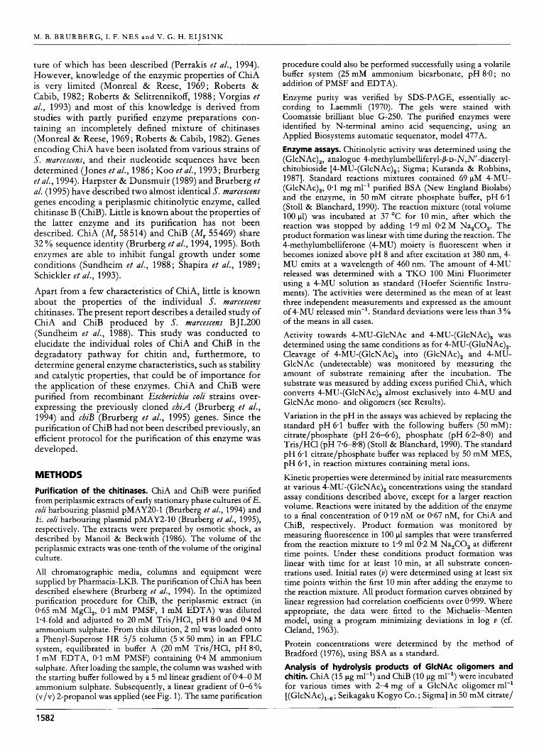
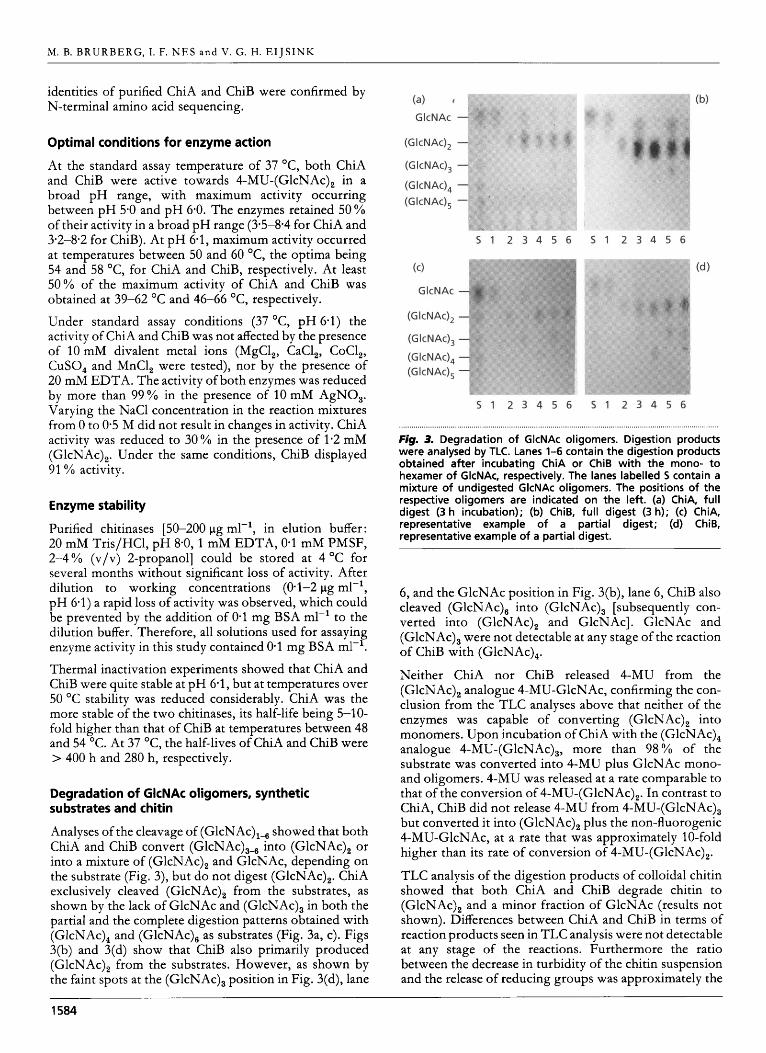
Analyses of the cleavage of (GlcNAc),-, showed that both ChiA and ChiB convert (GlcNAc),-, into (GlcNAc), or into a mixture of (GlcNAc), and GlcNAc, depending on the substrate (Fig. 3), but do not digest (GlcNAc),. ChiA exclusively cleaved (GlcNAc), from the substrates, as shown by the lack of GlcNAc and (GlcNAc), in both the partial and the complete digestion patterns obtained with (GlcNAc), and (GlcNAc), as substrates (Fig. 3a, c). Figs 3(b) and 3(d) show that ChiB also primarily produced (GlcNAc), from the substrates. However, as shown by the faint spots at the (GlcNAc), position in Fig. 3(d), lane
....................................... . ......... . ................. . ...................................................................................... Fig. 3. Degradation of GlcNAc oligomers. Digestion products were analysed by TLC. Lanes 1-6 contain the digestion products obtained after incubating ChiA or ChiB with the mono- to hexamer of GlcNAc, respectively. The lanes labelled 5 contain a mixture of undigested GlcNAc oligomers. The positions of the respective oligomers are indicated on the left. (a) ChiA, full digest (3 h incubation); (b) ChiB, full digest (3 h); (c) ChiA, representative example of a partial digest; (d) ChiB, representative example of a partial digest.
6, and the GlcNAc position in Fig. 3(b), lane 6, ChiB also cleaved (GlcNAc), into (GlcNAc), [subsequently con- verted into (GlcNAc), and GlcNAc]. GlcNAc and (GlcNAc), were not detectable at any stage of the reaction of ChiB with (GlcNAc),.
Neither ChiA nor ChiB released 4-MU from the (GlcNAc), analogue 4-MU-GlcNAc, confirming the con- clusion from the TLC analyses above that neither of the enzymes was capable of converting (GlcNAc), into monomers. Upon incubation of ChiA with the (GlcNAc), analogue 4-MU-(GlcNAc),, more than 98% of the substrate was converted into 4-MU plus GlcNAc mono- and oligomers. 4-MU was released at a rate comparable to that of the conversion of 4-MU-(GlcNAc),. In contrast to ChiA, ChiB did not release 4-MU from 4-MU-(GlcNAc), but converted it into (GlcNAc), plus the non-fluorogenic 4-MU-GlcNAc, at a rate that was approximately 10-fold higher than its rate of conversion of 4-MU-(GlcNAc),.
TLC analysis of the digestion products of colloidal chitin showed that both ChiA and ChiB degrade chitin to (GlcNAc), and a minor fraction of GlcNAc (results not shown). Differences between ChiA and ChiB in terms of reaction products seen in TLC analysis were not detectable at any stage of the reactions. Furthermore the ratio between the decrease in turbidity of the chitin suspension and the release of reducing groups was approximately the
1584

Chitinolytic activity of S. marcexens
Table 2. Degradation of chitin by ChiA and Chis
The indicated amounts of enzyme were incubated with colloidal chitin in a total volume of 1 ml for 24 h at 37 OC, as described in Methods. Chitin degradation is expressed as the percentage reduction in turbidity of the reaction mixture. Values are means k SD for three determinations.
...... . ....... . .......... . ........... ........... ..... . ..... . ................. . ............................................................................
Enzyme Degradation
(%)
ChiA (1.8 pg) ChiA (3.6 pg) ChiB (1.8 pg) ChiB (3.6 pg)
ChiA (1.8 pg) + ChiB (1 *8 pg)
26 f 2.2 36 f 2-3 16 f 3.6 21 f 1.9 65 f 3.7
same for both enzymes (differences in this ratio could indicate differences between the exo/endo character of the enzymes; Tronsmo & Harman, 1993). In contrast with this apparent similarity of the two enzymes, clear syn- ergistic effects on the rate of degradation of chitin were observed when ChiA and ChiB were combined (Table 2). Consequently, the two chitinases must differ in the way they contribute to the degradation of chitin, as already indicated by the differences observed with synthetic substrates and GlcNAc oligomers (see above). Another difference between the two enzymes is illustrated by the fact that ChiA has a higher specific activity towards chitin than has ChiB (Table 2).
Kinetic properties
The assay for chitinolytic activity with the (GlcNAc), analogue 4-MU-(GlcNAc), is sensitive and reliable, and permitted detailed kinetic analyses. As illustrated by Fig. 4, both chitinases were inhibited at high concentrations of the substrate. At the lower substrate concentrations, ChiA exhibited sigmoid kinetics, indicating cooperativity (Fig. 4a), whereas ChiB displayed a normal hyperbolic re- lationship between the rate of catalysis, v , and substrate concentration, [S] (Fig. 4b). Kinetic parameters for ChiB were approximated using eight points at low substrate concentration (1.5-8 pM), resulting in a K , of 34.1 f 1.4 pM and a kcat of 19.1 & 0.7 s-'.
The occurrence of substrate inhibition by an unknown mechanism prevented quantitative analysis of the sigmoid kinetics observed for ChiA. In an attempt to approximate kinetic parameters for ChiA, possible values for the apparent Hill coefficient (h) were tested by analysing the linearity and the distribution of residual errors in adapted Lineweaver-Burk plots (l/v versus 1/[SIn, for [S] < 12 pM; Segel, 1975). This approach indicated an h in the range 1.1-1.2. If h is known, [S],., ([S] at which v = 0.5 Vmax) and Vmax can be derived from the v and [S] values belonging to the maximum in the Eadie-Scatchard plot (Segel, 1975). The maximum in the Eadie-Scatchard plot (Fig. 4a) occurs at [S] = 26 pM, v = 13.59 s-l,
30 c 1
20
f 15
10
5
n
I F
- 20 40 60 80 100 120 140 160
10 1
8
c 6 'ul v
b 4
2
L
v (5-1)
20 40 60 80 100 120 151 (PM)
Fig- 4. Kinetic analysis. The main graph shows the dependence of initial velocity (v) on substrate concentration ([S]) in the reaction of ChiA (a) or Chis (b) with 4-MU-(GlcNAc),. Enzyme concentrations were 0.19 nM and 0-67 nM, for ChiA and ChiB, respectively. The inserts show Eadie-Scatchard plots derived from the v versus [S] curves; the lines in these plots were drawn for illustration purposes only and connect the points in order of increasing [S], starting at the points close to the v/[S] axis. Both enzymes are inhibited at high substrate concentrations. The curvature in the left part of the Eadie-Scatchard plot in (a) indicates cooperative kinetics for ChiA. The accuracy of these measurements is discussed in Results.
v / [ S ] = 523 s-' nM-', which, assuming an h of 1-15, results in [S],., = 135 pM and kcat = 104 s-'. The values for [S],., and kcat derived from the Eadie-Scatchard plot are an underestimation of the real values, since substrate inhibition lowers the v and [S] values at which the maximum in this plot occurs.
Sequence comparisons
Several authors have pointed out that prokaryotic chiti- nases are built up of regions (domains) that are recog- nizable by sequence similarities. Such regions are the signal sequence, the catalytic domain, the chitin-binding domain, and the so-called fibronectin type III-like domain (Bork & Doolittle, 1992; Watanabe e t al., 1993, 1994; Blaak e t al., 1993). The chitin-binding domain and the fibronectin type III-like domains are important for activity towards chitin (Watanabe e t al., 1994). Blaak e t al. (1993) concluded that ChiB consists of a catalytic domain and a C-terminal domain of approximately 60 residues that
1585
M. B. B R U R B E R G , I. F. N E S and V. G. H. E I J S I N K
(a 1 1 23 150 563
CAT ChiA,
S. rnarcescensl [SI I 1 430 499
CAT CH ChiB, S. rnarcescensz I
(b) ChiA 5. marcescens’ 409 AEAWKEDTAYTTVNGVNALLA 4 2 9 ChiB: 5. marcescen3 453 A-YVEGTTYAQGALVSYQGY 4 1 3
Chitinase D, B. circulane 31 AAQWQAGTAYKQGDLVTYLNK 51 Chitinase A l , B. circulang 653 VSAIQVNTAYTAGQLVTYNGX 6 7 3
Chitinase II, Aeromonas S P . ~ 494 CAAWAEGNTYTAGTCASYGGK 5 14
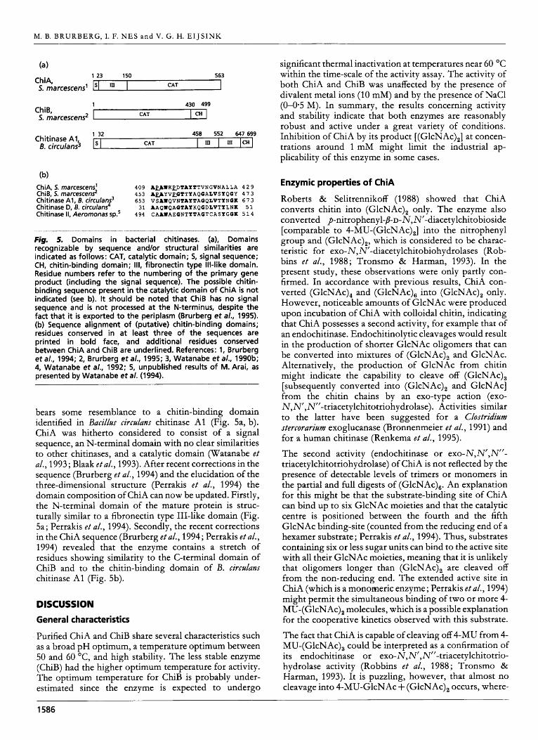
Fig. 5. Domains in bacterial chitinases. (a), Domains recognizable by sequence and/or structural similarities are indicated as follows: CAT, catalytic domain; 5, signal sequence; CHI chitin-binding domain; 111, fibronectin type Ill-like domain. Residue numbers refer to the numbering of the primary gene product (including the signal sequence). The possible chitin- binding sequence present in the catalytic domain of ChiA is not indicated (see b). It should be noted that ChiB has no signal sequence and is not processed at the N-terminus, despite the fact that it is exported to the periplasm (Brurberg et a/., 1995). (b) Sequence alignment of (putative) chitin-binding domains; residues conserved in at least three of the sequences are printed in bold face, and additional residues conserved between ChiA and ChiB are underlined. References: 1, Brurberg et a/., 1994; 2, Brurberg et a/., 1995; 3, Watanabe et a/., 1990b; 4, Watanabe et a/., 1992; 5, unpublished results of M. Arai, as presented by Watanabe eta / . (1994).
bears some resemblance to a chitin-binding domain identified in Bacillus circulans chitinase A1 (Fig. 5a, b). ChiA was hitherto considered to consist of a signal sequence, an N-terminal domain with no clear similarities to other chitinases, and a catalytic domain (Watanabe e t a/., 1993; Blaak e t al., 1993). After recent corrections in the sequence (Brurberg e t al., 1994) and the elucidation of the three-dimensional structure (Perrakis e t al., 1994) the domain composition of ChiA can now be updated. Firstly, the N-terminal domain of the mature protein is struc- turally similar to a fibronectin type 111-like domain (Fig. 5a; Perrakis e t al., 1994). Secondly, the recent corrections in the ChiA sequence (Brurberg e t al., 1994; Perrakis e t al., 1994) revealed that the enzyme contains a stretch of residues showing similarity to the C-terminal domain of ChiB and to the chitin-binding domain of B. circulans chitinase A1 (Fig. 5b).
DISCUSSION
General characteristics
Purified ChiA and ChiB share several characteristics such as a broad pH optimum, a temperature optimum between 50 and 60 OC, and high stability. The less stable enzyme (ChiB) had the higher optimum temperature for activity. The optimum temperature for ChiB is probably under- estimated since the enzyme is expected to undergo
significant thermal inactivation at temperatures near 60 “C within the time-scale of the activity assay. The activity of both ChiA and ChiB was unaffected by the presence of divalent metal ions (10 mM) and by the presence of NaCl (0-0-5 M). In summary, the results concerning activity and stability indicate that both enzymes are reasonably robust and active under a great variety of conditions. Inhibition of ChiA by its product [(GlcNAc),] at concen- trations around 1 mM might limit the industrial ap- plicability of this enzyme in some cases.
Enzymic properties of ChiA
Roberts & Selitrennikoff (1988) showed that ChiA converts chitin into (GlcNAc), only. The enzyme also converted p-nitrophenyl-P-D-N,N’-diacetylchitobioside [comparable to 4-MU-(GlcNAc),] into the nitrophenyl group and (GlcNAc),, which is considered to be charac- teristic for exo-N,N’-diacetylchitobiohydrolases (Rob- bins e t al., 1988; Tronsmo & Harman, 1993). In the present study, these observations were only partly con- firmed. In accordance with previous results, ChiA con- verted (GlcNAc), and (GlcNAc), into (GlcNAc), only. However, noticeable amounts of GlcNAc were produced upon incubation of ChiA with colloidal chitin, indicating that ChiA possesses a second activity, for example that of an endochitinase. Endochitinolytic cleavages would result in the production of shorter GlcNAc oligomers that can be converted into mixtures of (GlcNAc), and GlcNAc. Alternatively, the production of GlcNAc from chitin might indicate the capability to cleave off (GlcNAc), [subsequently converted into (GlcNAc), and GlcNAc] from the chitin chains by an exo-type action (exo- N,N’,N’’-triacetylchitotriohydrolase). Activities similar to the latter have been suggested for a Clostridium stercorarium exoglucanase (Bronnenmeier e t al., 1991) and for a human chitinase (Renkema e t al., 1995). The second activity (endochitinase or exo”,N’,N’’- triacetylchitotriohydrolase) of ChiA is not reflected by the presence of detectable levels of trimers or monomers in the partial and full digests of (GlcNAc),. An explanation for this might be that the substrate-binding site of ChiA can bind up to six GlcNAc moieties and that the catalytic centre is positioned between the fourth and the fifth GlcNAc binding-site (counted from the reducing end of a hexamer substrate ; Perrakis e t al., 1994). Thus, substrates containing six or less sugar units can bind to the active site with all their GlcNAc moieties, meaning that it is unlikely that oligomers longer than (GlcNAc), are cleaved off from the non-reducing end. The extended active site in ChiA (which is a monomeric enzyme ; Perrakis e t a/., 1994) might permit the simultaneous binding of two or more 4- MU-(GlcNAc), molecules, which is a possible explanation for the cooperative kinetics observed with this substrate. The fact that ChiA is capable of cleaving off 4-MU from 4- MU-(GlcNAc), could be interpreted as a confirmation of its endochitinase or exo-N,N’,N’’-triacetylchitotrio- hydrolase activity (Robbins e t al., 1988; Tronsmo & Harman, 1993). It is puzzling, however, that almost no cleavage into 4-MU-GlcNAc + (GlcNAc), occurs, where-
~
1586

Chitinolytic activity of S. marcescens
as the natural substrate (GlcNAc), is converted into (GlcNAc), only. Furthermore, 4-MU is produced at approximately the same rate as when 4-MU-(GlcNAc), is used as a substrate, which contrasts with the observation that ChiA primarily produces (GlcNAc), and not (GlcNAc),. These conflicting observations indicate that the cleavage rates and patterns obtained with the artificial 4-MU-chito-oligosaccharides do not (or only partly) reflect the real action of the chitinases on chitin and (GlcNAc), (see also Robbins e t al., 1988; Watanabe e t al., 1990a; Takayanagi e t al., 1991). Nevertheless, com- parisons of the cleavage rates of various 4-MU substrates are still used to characterize the catalytic activity of chitinases (Tronsmo & Harman, 1993; Renkema e t al., 1995).
The presence of an extended substrate binding site suggests that ChiA is optimized for the degradation of longer chito-oligosaccharides and chitin. This is sup- ported by the presence of the N-terminal fibronectin type III-like domain in ChiA that is known to have a positive effect on the activity of chitinases towards chitin (Watanabe e t al., 1994). Furthermore, ChiA possesses a region sharing some similarity with part of the chitin- binding domain identified in B. circtllans chitinase A1 (Watanabe e t al., 1994; Fig. 5).
Enzymic properties of Chis and comparison with ChiA
ChiB was found to be similar to ChiA in that its main product was (GlcNAc), for all natural substrates tested. In the case of ChiB, in contrast to ChiA, the endochitinase or exo-N,N’,N’’-triacetylchitotriohydrolase activity that causes the formation of monomers in the reaction with chitin was confirmed by the detection of (GlcNAc), and GlcNAc as products of the reaction with (GlcNAc),. It is tempting to speculate that the substrate-binding site of ChiB is designed to bind less than six GlcNAc moieties. This could explain why the endochitinase or exo- N,N’,N”-triacetylchitotriohydrolase activity of ChiB is detectable in the reaction with (GlcNAc),. Several other results support the notion that ChiB possesses a less extended substrate-binding site than ChiA and, conse- quently, that ChiB may be optimized for cleavage of relatively short chito-oligosaccharides. First, ChiB has a lower specific activity towards chitin than ChiA and it does not contain a fibronectin type III-like domain as does ChiA. Second, ChiB does not exhibit the cooperative kinetics that were observed for ChiA. Finally, ChiB seems to have a higher affinity for the (GlcNAc), analogue than ChiA: whereas ChiB has a K, of 34.1 pM for 4-MU- (GlcNAc),, the [S],., of ChiA for 4-MU-(GlcNAc), was about 100 pM. Vorgias e t al. (1993), determined the Km of ChiA for another (GlcNAc), analogue to be as high as 500pM. However, in the latter study, the problem of substrate inhibition (which is expected to occur: see above and Krishnan e t al., 1994) was not addressed.
Analysis of the breakdown products of chitin did not reveal differences between ChiA and ChiB. This is likely
to be due to the fact that only minor, undetectable amounts of products other than GlcNAc and (GlcNAc), accumulate during chitin degradation. Likewise, and possibly for the same reason, no differences in the ratio between reducing sugar formation and degradation rate were found. Furthermore, the reducing sugar assay may provide artificial results since the signal obtained per reducing group increases with the length of the chito- oligosaccharides (Imoto & Yagishita, 1971 ; M. B. Brurberg, unpublished observations). The observation that combining ChiA and ChiB results in synergistic effects on the rate of chitin degradation clearly shows that the enzymes not only differ in terms of their specific activity but also in terms of the mechanism by which they degrade the substrate. An obvious explanation for the synergy would be that one of the enzymes, by exerting an endochitinase activity, increases the substrate availability for the other enzyme, which has an exo-N,N’,N’’- triacetylchitotriohydrolase activity.
To permit a more detailed analysis of the differences between ChiA and ChiB, we are currently conducting X- ray crystallographic studies of ChiB prepared using the purification protocol described in this report. The avail- ability of the ChiB structure would permit a structural comparison which ChiA (Perrakis e t al., 1994) which might shed new light on the function of these enzymes and could provide a structural explanation for the differences between them [see Davies & Henrissat (1995) for a review of existing structures of glycosyl hydrolases].
The role of ChiA and Chis in chitin breakdown
The analyses described above show both similarities and differences in the enzymic properties of ChiA and ChiB. Both enzymes seem to have exo-N,N’- diace t ylchit o bio hydrolase activity , com bined with either an endochitinase or an exo-N,N’,N’’-triacetylchitotrio- hydrolase activity. In 5’. marcescens ChiA is located extracellularly (Brurberg e t al., 1994) and therefore is likely to be optimized for degrading chitin and the longer GlcNAc oligomers, as suggested above. Considering its location, the presence of domains known to be important for chitin degradation, its extended substrate-binding site, and its high specific activity towards chitin (as compared to ChiB), ChiA is likely to possess an endochitinase activity in addition to its exo-N,N’-diacetylchitobio- hydrolase activity. Consequently, ChiB’s second activity is expected to be that of an exo-N,N’,N”-triacetyl- chitotriohydrolase. ChiB has been found in the super- natant of late-stationary cultures of S. marcescens (Roberts & Cabib, 1982; Fuchs e t al., 1986), but analysis of the localization of ChiB in exponential and early-stationary cultures of S. marcescens has shown that this enzyme is almost exclusively directed towards the periplasm (with- out being processed; Brurberg e t al., 1995). Thus it would seem that the role of ChiB in vivo is to digest the shorter GlcNAc oligomers capable of entering the periplasm, in accordance with the suggestions described above. The deduction that ChiA and ChiB possess an endochitinase and an exo-N,N’,N”-triacetylchitotriohydrolase activity,
1587

M. B. B R U R B E R G , I. F. N E S and V. G. H. E I J S I N K
respectively, is supported by preliminary results of studies on the degradation of chitin by mixtures of ChiA or ChiB and an endochitinase isolated from the fungus Trichoderma haryianzrm (Tronsmo & Harman, 1993; M. B. Brurberg & A. Tronsmo, unpublished results). These studies showed no synergistic effects on chitin breakdown upon com- bining ChiA with the endochitinase, whereas clear syn- ergy was observed upon combining ChiB with the endochitinase.
A third chitinolytic enzyme of S. marcescens, an N-acetyl- P-glucosaminidase, has been described by Kless e t al. (1989) and Vorgias e t al. (1993). This enzyme is probably located in the periplasm (Kless e t al., 1989) where its obvious role would be to convert the chitobiose produced by ChiA and ChiB into GlcNAc monomers. From the present study it seems that an enzymic apparatus con- sisting of ChiA, ChiB and an N-acetyl-P-glucosaminidase could be enough for effective in vivo degradation of chitin.
ACKNOWLEDGEMENTS
We thank Anne-May Lsnneborg for technical assistance and Arne Tronsmo for supplying us with colloidal chitin. We are grateful to Per Nissen and Juke Lolkema for valuable discus- sions. This work was supported by a grant from the Norwegian Research Council.
REFERENCES
Blaak, H., Schnellmann, J., Walter, S., Henrissat, B. & Schrempf, H. (1 993). Characteristics of an exochitinase from Streptomyces olivaceo- viridis, its corresponding gene, putative domains and relationship to other chitinases. Eur J Biochem 214, 659-669. Bork, P. & Doolittle, R. F. (1992). Proposed acquisition of an animal protein domain by bacteria. Proc Natl Acad Sci U S A 89,8990-8994. Bronnenmeier, K., Rucknagel, K. P. & Staudenbauer, W. L. (1991). Purification and properties of a novel type of exo-l,4-~-glucanase (Avicellase 11) from the cellulolytic Clostridium stercurarium. Ear J Biochem 200, 379-385. Bradford, M. M. (1976). A rapid and sensitive method for the quantitation of protein utilizing the principle of protein-dye binding. Anal Biochem 72, 248-254. Brurberg, M. B., Eijsink, V. G. H. & Nes, 1. F. (1994). Characteri- zation of a chitinase gene (chiA) from Serratia marcescens strain BJL200 and one-step purification of the gene product. FEMS Microbiol Lett 124, 399-404. Brurberg, M. B., Eijsink, V. G. H., Haandrikman, A. J., Venema, G. & Nes, 1. F. (1995). Chitinase B from Serratia marcescens BJL200 is exported to the periplasm without processing. Micrubiulogy 141,
Cleland, W. W. (1963). Computer programmes for processing enzyme kinetic data. Nature 198, 463-465. Davies, G. & Henrissat, B. (1995). Structures and mechanisms of glycosyl hydrolases. Structure 3, 853-859. Davis, B. & Eveleigh, D. E. (1984). Chitosanases: occurrence, production and immobilization. In Chitin, Chitusan, and Related EnZymes, pp. 161-179. Edited by J. P. Zikakis. London: Academic Press. De la Cruz, J., Hidalgo-Gallego, A., Lora, J. M., Benitez, T., Pintor- Toro, J. & Llobell, A. (1992). Isolation and characterization of three chitinases from Trichoderma harqianum. Eur J Biochem 206, 859-867.
123-1 31,
Fuchs, R. L., McPherson, 5. A. & Drahos, D. J. (1986). Cloning of a Serratia marcescens gene encoding chitinase. Appl Environ Microbiol 51, 504-509. Gooday, G. W. (1990). Physiology of microbial degradation of chitin and chitosan. Biodegradation 1, 177-1 90. Harpster, M. H. & Dunsmuir, P. (1989). Nucleotide sequence of the chitinase B gene of S. marcescens QMB1466. Nucleic Acids Res 17, 5395. Imoto, T. & Yagishita, K. (1971). A simple activity measurement of lysozyme. Agr Bid Chem 35, 1 154-1 156. John, M., RUhrig, H., Schmidt, J., Wieneke, U. & Schell, J. (1993). Rhiqobium NodB protein involved in nodulation signal synthesis is a chitooligosaccharide deacetylase. Pruc Natl Acad Sci U S A 90,
Jones, J. D. G., Grady, K. L., Suslow,T. V. & Bedbrook, J. R. (1986). Isolation and characterization of genes encoding two chitinase enzymes from Serratia marcescens. EMBO J 5, 467473. Kless, H., Sitrit, Y., Chet, H. & Oppenheim, A. B. (1989). Cloning of the gene coding for the chitobiase of Serratia marcescens. Mol Gen Genet 217, 471-473. Koo, J. C., Lim, C. O., Choi, Y. J., Kim, C. Y., Bahk, J. D., Lee, 5. Y. & Cho, M. J. (1993). Expression of bacterial chitinase in tobacco and enhanced resistance against phytopathogenic fungi. EMBL Data- base, Accession number L01455. Krishnan, A., Nair, P. N. &Jones, D. (1994). Isolation, cloning and characterization of new chitinase stored in active form in chitin- lined venom reservoir. ] Bid Chem 269, 20971-20976. Kuranda, M. J. & Robbins, P. W. (1987). Cloning and heterologous expression of glycosidase genes from Saccbaromyces cereuisiae. Proc Natl Acad Sci U S A 84, 2585-2589. Laemmli, U. K. (1970). Cleavage of structural proteins during the assembly of the head of bacteriophage T4. Nature 227, 680-685. Lorito, M., Peterbauer, C., Hayes, C. K. & Harman, G. E. (1994). Synergistic interaction between fungal cell wall degrading enzymes and different antifungal compounds enhances inhibition of spore germination. Microbiology 140, 623629. Manoil, C. & Beckwith, J. (1986). A genetic approach to analyzing membrane protein topology. Science 233, 1403-1408. Monreal, J. & Reese, E. (1969). The chitinase of Serratia marcescens. Can J Microbiol 15, 689-696. Perrakis, A., Tews, I., Dauter, Z., Oppenheim, A. B., Chet, I., Wilson, K. 5. &Vorgias, C. E. (1994). Crystal structure of a bacterial chitinase at 2.3 A resolution. Structure 2, 1169-1 180. Renkema, G. H., Boot, R. G., Muijsers, A. O., Donker-Koopman, W. E. & Aerts, J. M. F. G. (1995). Purification and characterization of human chitotriosidase, a novel member of the chitinase family of proteins. J Biol Chem 270, 2198-2202. Roberts, R. L. & Cabib, E. (1982). Serratia marcescens chitinase : one- step purification and use for the determination of chitin. Anal Biuchem 127, 402-412. Roberts, W. K. & Selitrennikoff, C. P. (1988). Plant and bacterial chitinases differ in antifungal activity. J Gen Microbioll34,169-176. Robbins, P. W., Albright, C. & Benfield, B. (1988). Cloning and expression of a Streptomyces plicutus chitinase (chitinase-63) in Eschericbia coli. J Biol Chem 263, 443-447. Schickler, H., Haran, S., Oppenheim, A. B. & Chet, 1. (1993). Cloned chitinases and their role in biological control of plant pathogenic fungi. In: Chitin Envwzology, pp. 375-382. Edited by R. A. A. Muzzarelli. Ancona, Italy : European Chitin Society. Segel, 1. H. (1975). Envme kinetics: Behavior and Anahsis of Rapid Equilibrium and Stea&state Envme Systems. New York : John Wiley.
625-629.
1588

Chitinolytic activity of S. marcexcenx
Shapira, R., Ordentlich, A., Chet, 1. & Oppenheim, A. 6. (1989). Control of plant diseases by chitinase expressed from cloned DNA in Escbericbia cofi. Pbytopatbofogy 79, 1246-1 249.
Stoll, V. 5. & Blanchard, J. 5. (1 990). Buffers : principles and practice. Methods Enumof 182, 24-38.
Sundheim, L., Poplawsky, A. R. & Ellingboe, A. H. (1988). Mole- cular cloning of two chitinase genes from Serratia marcescens and their expression in Pseudomonas species. Pbys Mol Plant Patbol33, 483-491.
Takayanagi, T., Sjisaka, K., Takiguchi, Y. & Shimahara, K. (1991). Isolation and characterization of thermostable chitinases from Baciffus ficbenifrmis X-7u. Biocbim Biopbys Acta 1078, 404-410.
Tronsmo, A. & Harman, G. E. (1993). Detection and quantification of N-acetyl-p-D-glucosaminidase, chitobiase and endochitinase in solutions and on gels. Anal Biocbem 208, 74-79.
Vessey, J. C. & Pegg, G. F. (1973). Autolysis and chitinase production in cultures of Verticiffium afbo-atrum. Trans Br Mycof Soc 60, 133-143.
Vorgias, C. E., Tews, I., Perrakis, A., Wilson, K. 5. & Oppenheim, A. 6. (1993). Purification and characterization of the recombinant chitin degrading enzymes, chitinase A and chitobiase from Serratia marcescens. In : Chitin EnZymofogy, pp. 41 7-422. Edited by R. A. A. Muzzarelli. Ancona, Italy : European Chitin Society. Walkley, 1. W. & Tillman, J. (1977). A simple thin-layer chromato- graphic technique for the separation of mono- and oligosaccharides. J Cbromatogr 132, 172-174.
Watanabe, T., Oyanagi, W., Suzuki, K. & Tanaka, H. (1990a). Chitinase system of Bacillus circulans WL-12 and importance of chitinase A1 in chitin degradation. J Bacteriof 172, 401 7-4022.
Watanabe, T., Suzuki, K., Oyanagi, W., Ohnishi, K. &Tanaka, H. (1990b). Gene cloning of chitinase A1 from Bacillus circufans WL-12 revealed its evolutionary relationship to Serratia chitinase and to the type I11 homology units of fibronectin. J Biof Cbem 265,
Watanabe, T., Oyanagi, W., Suzuki, K., Ohnishi, K. & Tanaka, H. (1992). Structure of the gene encoding chitinase D of Bacillus circulans WL-12 and possible homology of the enzyme to other prokaryotic chitinases and class I11 plant chitinases. J Bacteriof 174, 408-414.
Watanabe,T., Kobori, K., Miyashita, K., Fujii, T., Sakai, H., Uchida, M. & Tanaka, H. (1993). Identification of glutamic acid 204 and aspartic acid 200 in chitinase A1 of Bacillus circulans WL-12 as essential residues for chitinase activity. J Biof Cbem 268,
Watanabe, T., Ito, Y., Yamada, T., Hashimoto, M., Sekine, 5. & Tanaka, H. (1994). The roles of the C-terminal domain and type I11 domains of chitinase A1 from Bacillw circulans WL-12 in chitin degradation. J Bacteriol 176, 4465-4472.
15659-15665.
18567-1 8572.
Received 19 October 1995; revised 1 February 1996; accepted 21 February 1996.
- 1589
Related Documents