Welcome message from author
This document is posted to help you gain knowledge. Please leave a comment to let me know what you think about it! Share it to your friends and learn new things together.
Transcript
lazar
0824741390_tn_std.jpg
TM
Marcel Dekker, Inc. New York • Basel
Handbook of Cosmetic Science
and Technology
edited by
André O. BarelFree University of BrusselsBrussels, Belgium
Marc PayeColgate-Palmolive Research and Development, Inc.Milmort, Belgium
Howard I. MaibachUniversity of California at San Francisco School of MedicineSan Francisco, California
Copyright © 2001 by Marcel Dekker, Inc. All Rights Reserved.

ISBN: 0-8247-0292-1
This book is printed on acid-free paper.
HeadquartersMarcel Dekker, Inc.270 Madison Avenue, New York, NY 10016tel: 212-696-9000; fax: 212-685-4540
Eastern Hemisphere DistributionMarcel Dekker AGHutgasse 4, Postfach 812, CH-4001 Basel, Switzerlandtel: 41-61-261-8482; fax: 41-61-261-8896
World Wide Webhttp:/ /www.dekker.com
The publisher offers discounts on this book when ordered in bulk quantities. For more information,write to Special Sales/Professional Marketing at the headquarters address above.
Copyright 2001 by Marcel Dekker, Inc. All Rights Reserved.
Neither this book nor any part may be reproduced or transmitted in any form or by any means,electronic or mechanical, including photocopying, microfilming, and recording, or by any informa-tion storage and retrieval system, without permission in writing from the publisher.
Current printing (last digit):10 9 8 7 6 5 4 3 2 1
PRINTED IN THE UNITED STATES OF AMERICA
Preface
Cosmetic composition and formulation are becoming increasingly complex, and cosmeticingredients more sophisticated and functional, while laws and regulations impose moreconstraints on the cosmetic scientist and manufacturer. The Handbook of Cosmetic Scienceand Technology reviews in a single volume the multiple facets of the cosmetic field andprovides the reader with an easy-to-access information source.
This handbook covers topics as varied as the physiology of the potential targetsof cosmetics, safety, legal and regulatory considerations throughout the world, cosmeticingredients, vehicles and finished products, and new delivery systems, as well as microbi-ology and safety and efficacy testing.
To achieve our goal, we, the editors, requested the contributions of expert scientistsfrom academic dermatology and dermato-cosmetics, the cosmetics industry, ingredientsand raw materials producers, and regulatory agencies. Because cosmetology is universal,while having some regional specificity, those authors were selected on a broad geographi-cal basis, with some coming from the United States, Europe, Japan, and Australia. Theyshare in their chapters not only their experience and knowledge but also new informationand their expert views regarding the future. We thank the authors for their high dedication,which permitted us to make this handbook a review of the state of the art in cosmetologyin the new millennium. The staff of Marcel Dekker, Inc., played a great role in the produc-tion of the handbook, ensuring on a day-to-day basis the contact between the editors andthe authors. Our thanks especially go to Sandra Beberman, Jane Roh, and Moraima Suarezfor their constant and excellent help.
Finally, we encourage our readership to send us their comments and suggestions onwhat should be modified or considered in future editions.
André O. BarelMarc Paye
Howard I. Maibach
iii

Contents
Preface iiiContributors xi
Part 1 INTRODUCTION
1. Introduction 1André O. Barel, Marc Paye, and Howard I. Maibach
2. Definition of Cosmetics 5Stanley R. Milstein, John E. Bailey, and Allen R. Halper
Part 2 TARGET ORGANS FOR COSMETIC PRODUCTS
3. The Microscopic Structure of the Epidermis and Its Derivatives 19Joel J. Elias
4. The Normal Nail 29Josette André
5. Hair 35Ghassan Shaker and Dominique Van Neste
Part 3 SAFETY CONSIDERATIONS
6. Safety Terminology 47Ai-Lean Chew and Howard I. Maibach
7. Principles and Practice of Percutaneous Absorption 53Ronald C. Wester and Howard I. Maibach
8. Principles and Mechanisms of Skin Irritation 67Sibylle Schliemann-Willers and Peter Elsner
9. Allergy and Hypoallergenic Products 77An E. Goossens
v
vi Contents
10. Dermatological Problems Linked to Perfumes 89Anton C. de Groot
11. In Vitro Tests for Skin Irritation 95Michael K. Robinson, Rosemarie Osborne, and Mary A. Perkins
12. In Vivo Irritation 107Saqib J. Bashir and Howard I. Maibach
13. Eye Irritation Testing 119Leon H. Bruner, Rodger D. Curren, John W. Harbell, RosemarieOsborne, and James K. Maurer
Part 4 VEHICLES OF COSMETIC PRODUCTS
14. Main Cosmetic Vehicles 145Stephan Buchmann
15. Encapsulation to Deliver Topical Actives 171Jocélia Jansen and Howard I. Maibach
16. Encapsulation Using Porous Microspheres 191Jorge Heller, Subhash J. Saxena, and John Barr
17. Liposomes 201Hans Lautenschläger
18. Topical Delivery by Iontophoresis 211Véronique Préat and Rita Vanbever
19. Mousses 221Albert Zorko Abram and Roderick Peter John Tomlinson
20. Cosmetic Patches 233Spiros A. Fotinos
Part 5 COSMETIC INGREDIENTS
21. Antibacterial Agents and Preservatives 245Françoise Siquet and Michel J. Devleeschouwer
22. General Concepts of Skin Irritancy and Anti-irritant Products 253André O. Barel
23. Anti-irritants for Surfactant-Based Products 271Marc Paye
24. The Case of Alpha-Bisabolol 277Klaus Stanzl and Jürgen Vollhardt
25. Anti-irritants for Sensory Irritation 285Gary S. Hahn
26. Antioxidants 299Stefan Udo Weber, John K. Lodge, Claude Saliou, and Lester Packer
Contents vii
27. Alpha Hydroxy Acids 311Enzo Berardesca
28. Colorants 317Gisbert Otterstätter
29. Hair Conditioners 331Charles Reich and Dean T. Su
30. Hydrating Substances 347Marie Lodén
31. Ceramides and Lipids 361Bozena B. Michniak and Philip W. Wertz
32. Natural Extracts 369Jürgen Vollhardt
33. Rheological Additives and Stabilizers 377Ekong A. Ekong, Mohand Melbouci, Kate Lusvardi, andPaquita E. Erazo-Majewicz
34. Silicones: A Key Ingredient in Cosmetic and Toiletry Formulations 389Janet M. Blakely
35. Skin-Feel Agents 399Germaine Zocchi
36. Surfactants 417Takamitsu Tamura and Mitsuteru Masuda
37. Classification of Surfactants 431Louis Oldenhove de Guertechin
38. UV Filters 451Stanley B. Levy
39. Vitamins 463Alois Kretz and Ulrich Moser
40. Ellagic Acid: A New Skin-Whitening Active Ingredient 473Yoshimasa Tanaka
Part 6 COSMETIC PRODUCTS
Skincare Products
41. Cosmetics and Interactions with Superficial Epidermis 479Jørgen Serup
42. Skin Cleansing Bars 485Joshua B. Ghaim and Elizabeth D. Volz
43. Skin Cleansing Liquids 499Daisuke Kaneko and Kazutami Sakamoto
viii Contents
44. Emulsion-Based Skincare Products: Formulating and Measuring TheirMoisturizing Benefits 511Howard Epstein and F. Anthony Simion
45. Anticellulite Products and Treatments 531André O. Barel
46. Antiwrinkle Products 543William J. Cunningham
47. Artificial Tanning Products 551Stanley B. Levy
48. Barrier Creams 557Cees Korstanje
49. Skin-Whitening Products 567Hongbo Zhai and Howard I. Maibach
Haircare Products
50. Interactions with Hair and Scalp 575Dominique Van Neste and Ghassan Shaker
51. Hair Cosmetics 581Leszek J. Wolfram
52. Ethnic Differences in Haircare Products 605Joerg Kahre
Other Cosmetic Products
53. Oral-Care Products 619Abdul Gaffar
54. Decorative Products 645Mitchell L. Schlossman
55. Cosmetics for Nails 685Douglas Schoon and Robert Baran
56. Antiperspirants 689Jörg Schreiber
57. Deodorants 703Jörg Schreiber
58. Baby Care 715Uwe Schönrock
59. Cosmetics for the Elderly 723Uwe Schönrock
Contents ix
Part 7 LEGISLATION AND REGULATIONS WORLDWIDE
60. EEC Cosmetic Directive and Legislation in Europe 729René Van Essche
61. Regulatory Requirements for the Marketing of Cosmetics in the UnitedStates 737Stanley R. Milstein, John E. Bailey, and Allen R. Halper
62. Legislation in Japan 761Mitsuteru Masuda
Part 8 TESTING COSMETIC PRODUCTS
63. Stability Testing of Cosmetic Products 769Perry Romanowski and Randy Schueller
64. Stability Control: Microbiological Tests 781Michel J. Devleeschouwer and Françoise Siquet
Part 9 COSMETIC CLAIMS
65. Introduction to the Proof of Claims 797Marc Paye and A. O. Barel
66. Tests for Sensitive Skin 807Alessandra Pelosi, Sabrina Lazzerini, Enzo Berardesca, andHoward I. Maibach
67. Tests for Skin Hydration 815Bernard Gabard
68. Tests for Skin Protection: Barrier Effect 823Hongbo Zhai and Howard I. Maibach
69. Objective Methods for Assessment of Human Facial Wrinkles 829Gary Grove and Mary Jo Grove
70. Acnegenicity and Comedogenicity Testing for Cosmetics 837F. Anthony Simion
71. Sensory Testing 845Linda P. Oddo and Kathy Shannon
Index 859

Contributors
Albert Zorko Abram, B.Sc. Soltec Research Pty Ltd., Rowville, Victoria, Australia
Josette André, M.D. Faculty of Medicine, Free University of Brussels, and Departmentof Dermatology, Hôpital Saint-Pierre, Brussels, Belgium
John E. Bailey, Ph.D. Office of Cosmetics and Colors, Center for Food Safety andApplied Nutrition (CFSAN), U.S. Food and Drug Administration, Washington, D.C.
Robert Baran, M.D. Nail Disease Center, Cannes, France
André O. Barel, Ph.D. Faculty of Physical Education and Physiotherapy, Free Univer-sity of Brussels, Brussels, Belgium
John Barr, Ph.D. Pharmaceutical Sciences, Advanced Polymer Systems, RedwoodCity, California
Saqib J. Bashir, B.Sc.(Hons), M.B., Ch.B. Department of Dermatology, University ofCalifornia at San Francisco School of Medicine, San Francisco, California
Enzo Berardesca, M.D. Department of Dermatology, University of Pavia, Pavia, Italy
Janet M. Blakely, B.Sc.(Hons) Life Sciences Group, Science and Technology, DowCorning S.A., Brussels, Belgium
Leon H. Bruner, D.V.M., Ph.D. Gillette Medical Evaluation Laboratory, The GilletteCompany, Needham, Massachusetts
Stephan Buchmann, Ph.D. Department of Pharmaceutical Technology, Spirig PharmaAG, Egerkingen, Switzerland
xi
xii Contributors
Ai-Lean Chew, M.B.Ch.B. Department of Dermatology, University of California atSan Francisco School of Medicine, San Francisco, California
William J. Cunningham, M.D. CU-TECH, Mountain Lakes, New Jersey
Rodger D. Curren, Ph.D. Institute for In Vitro Sciences, Inc., Gaithersburg, Maryland
Anton C. de Groot, M.D., Ph.D. Department of Dermatology, Carolus Hospital,‘s-Hertogenbosch, The Netherlands
Michel J. Devleeschouwer, Ph.D. Laboratory of Microbiology and Hygiene, Institute ofPharmacy and Biocontaminants Unit, School of Public Health, Free University of Brussels,Brussels, Belgium
Ekong A. Ekong, Ph.D. Technology Division, Hercules Incorporated, Wilmington,Delaware
Joel J. Elias, Ph.D. Department of Anatomy, University of California at San FranciscoSchool of Medicine, San Francisco, California
Peter Elsner, M.D. Department of Dermatology and Allergology, University of Jena,Jena, Germany
Howard Epstein, M.S. Product Development, The Andrew Jergens Company, Cincin-nati, Ohio
Paquita E. Erazo-Majewicz, Ph.D. Aqualon Division, Hercules Incorporated, Wil-mington, Delaware
Spiros A. Fotinos, B.Sc.(Pharm), B.Sc.(Chem) Corporate Research and Innovation,Lavipharm, Peania Attica, Greece
Bernard Gabard, Ph.D. Department of Biopharmacy, Spirig Pharma Ltd., Egerkingen,Switzerland
Abdul Gaffar, Ph.D. Advanced Technology, Corporate Technology, Department ofOral Care, Colgate-Palmolive Company, Piscataway, New Jersey
Joshua B. Ghaim, Ph.D. Product Development, Skin Care Global Technology, Colgate-Palmolive Company, Piscataway, New Jersey
An E. Goossens, B.Pharm., Ph.D. Department of Dermatology, University Hospital,Katholieke Universiteit Leuven, Leuven, Belgium
Gary Grove, Ph.D. Research and Development, KGL’s Skin Study Center, Broomall,and cyberDERM, inc., Media, Pennsylvania
Contributors xiii
Mary Jo Grove, M.S. KGL’s Skin Study Center, Broomall, and cyberDERM, inc.,Media, Pennsylvania
Gary S. Hahn, M.D. Department of Pediatrics, University of California at San DiegoSchool of Medicine, San Diego, and Board of Scientific Advisors, Cosmederm Technolo-gies, LLC, La Jolla, California
Allen R. Halper Office of Cosmetics and Colors, Center for Food Safety and AppliedNutrition (CFSAN), U.S. Food and Drug Administration, Washington, D.C.
John W. Harbell, Ph.D. Institute for In Vitro Sciences, Inc., Gaithersburg, Maryland
Jorge Heller, Ph.D. Advanced Polymer Systems, Redwood City, California
Jocélia Jansen, Ph.D. Department of Pharmaceutical Sciences, State University ofPonta Grossa, Ponta Grossa, Paraná, Brazil
Joerg Kahre, Ph.D. VTP Department, Henkel KGaA, Düsseldorf, Germany
Daisuke Kaneko, Ph.D. Department of Product Development, AminoScience Labora-tories, Ajinomoto Co., Inc., Kanagawa, Japan
Cees Korstanje, R.Ph., Ph.D. Biological Research Department, Yamanouchi EuropeB.V., Leiderdorp, The Netherlands
Alois Kretz, M.D. Cosmetics, Roche Vitamins Europe Ltd., Basel, Switzerland
Hans Lautenschläger, Ph.D. Development & Consulting, Pulheim, Germany
Sabrina Lazzerini, M.D. Department of Dermatology, University of Pavia, Pavia, Italy
Stanley B. Levy, M.D. Department of Dermatology, University of North CarolinaSchool of Medicine at Chapel Hill, Chapel Hill, North Carolina, and Medical Affairs,Revlon Research Center, Edison, New Jersey
Marie Lodén, Pharm.Sc., Dr.Med.Sc. Department of Dermatology, ACO HUD AB,Upplands Väsby, Sweden
John K. Lodge, Ph.D. School of Biological Sciences, University of Surrey, Guildford,Surrey, England
Kate Lusvardi, Ph.D. Aqualon Division, Hercules Incorporated, Wilmington, Delaware
Howard I. Maibach, M.D. Department of Dermatology, University of California at SanFrancisco School of Medicine, San Francisco, California
Mitsuteru Masuda, Ph.D. Life Science Research Center, Research and DevelopmentHeadquarters, Lion Corporation, Tokyo, Japan
xiv Contributors
James K. Maurer, D.V.M., Ph.D. Human and Environmental Safety Division, TheProcter & Gamble Company, Cincinnati, Ohio
Mohand Melbouci, Ph.D. Personal Care Department, Aqualon Division, Hercules In-corporated, Wilmington, Delaware
Bozena B. Michniak, Ph.D. College of Pharmacy, University of South Carolina, Co-lumbia, South Carolina
Stanley R. Milstein, Ph.D. Office of Cosmetics and Colors, Center for Food Safety andApplied Nutrition (CFSAN), U.S. Food and Drug Administration, Washington, D.C.
Ulrich Moser, Ph.D. Roche Vitamins Europe Ltd., Basel, Switzerland
Linda P. Oddo, B.S. Hill Top Research, Inc., Scottsdale, Arizona
Louis Oldenhove de Guertechin, Ph.D. Department of Advanced Technology, Col-gate-Palmolive Research and Development, Inc., Milmort, Belgium
Rosemarie Osborne, Ph.D. Human and Environmental Safety Division, The Procter &Gamble Company, Cincinnati, Ohio
Gisbert Otterstätter Color Department, DRAGOCO Gerberding & Co. AG, Holzmin-den, Germany
Lester Packer, Ph.D. Department of Molecular and Cellular Biology, University ofCalifornia at Berkeley, Berkeley, California
Marc Paye, Ph.D. Skin Research Division, Department of Advanced Technology,Colgate-Palmolive Research and Development, Inc., Milmort, Belgium
Alessandra Pelosi, M.D. Department of Dermatology, University of Pavia, Pavia, Italy
Mary A. Perkins, A.Sc. Human and Environmental Safety Division, The Procter &Gamble Company, Cincinnati, Ohio
Véronique Préat, Ph.D. Unité de Pharmacie Galénique, Université Catholique de Lou-vain, Brussels, Belgium
Charles Reich, Ph.D. Advanced Technology, Hair Care, Colgate-Palmolive Technol-ogy Center, Piscataway, New Jersey
Michael K. Robinson, Ph.D. Department of Human and Environmental Safety Divi-sion, The Procter & Gamble Company, Cincinnati, Ohio
Perry Romanowski, B.S., M.S. Research and Development, Alberto Culver Company,Melrose Park, Illinois
Contributors xv
Kazutami Sakamoto, Ph.D. Applied Research Department, AminoScience Labora-tories, Ajinomoto Co., Inc., Kanagawa, Japan
Claude Saliou, Pharm.D., Ph.D. Department of Molecular and Cell Biology, Univer-sity of California at Berkeley, Berkeley, California
Subhash J. Saxena, Ph.D. Research and Development, Advanced Polymer Systems,Redwood City, California
Sibylle Schliemann-Willers, M.D. Department of Dermatology and Allergology, Uni-versity of Jena, Jena, Germany
Mitchell L. Schlossman, B.A., F.A.I.C., F.S.C.C. Kobo Products, Inc., SouthPlainfield, New Jersey
Uwe Schönrock, Ph.D. Active Ingredient Research, Beiersdorf AG, Hamburg, Ger-many
Douglas Schoon, M.S. Research and Development, Creative Nail Design Inc., Vista,California
Jörg Schreiber, Ph.D. Research New Delivery Systems, Beiersdorf AG, Hamburg, Ger-many
Randy Schueller, B.S. Consumer Products Research and Development, Alberto CulverCompany, Melrose Park, Illinois
Jørgen Serup, M.D., D.M.Sc. Department of Dermatological Research, Leo Pharma-ceutical Products, Copenhagen, Denmark
Ghassan Shaker, M.B.Ch.B., D.Sc. Skinterface sprl, Tournai, Belgium
Kathy Shannon, B.S. Hill Top Research, Inc., Scottsdale, Arizona
F. Anthony Simion, Ph.D. Product Development, The Andrew Jergens Company, Cin-cinnati, Ohio
Françoise Siquet, Ph.D. Department of Microbiology, Colgate-Palmolive TechnologyCenter, Milmort, Belgium
Klaus Stanzl, Ph.D. DRAGOCO Gerberding & Co. AG, Holzminden, Germany
Dean T. Su, Ph.D. Personal Care, Colgate-Palmolive Technology Center, Piscataway,New Jersey
Takamitsu Tamura, Ph.D. Material Science Research Center, Lion Corporation,Tokyo, Japan
xvi Contributors
Yoshimasa Tanaka, Ph.D. Life Science Research Center, Lion Corporation, Tokyo,Japan
Roderick Peter John Tomlinson Soltec Research Pty Ltd., Rowville, Victoria, Aus-tralia
Rita Vanbever, Ph.D. Unité de Pharmacie Galénique, Université Catholique de Lou-vain, Brussels, Belgium
René Van Essche, D.V.M., M.B.A. Institute of Pharmacy, Free University of Brussels,Brussels, Belgium
Dominique Van Neste, M.D., Ph.D. Skinterface sprl, Tournai, Belgium
Jürgen Vollhardt, Ph.D. Research and Development, Cosmetic Division, DRAGOCOInc., Totowa, New Jersey
Elizabeth D. Volz, M.ChE. Research and Development, Colgate-Palmolive Company,Piscataway, New Jersey
Stefan Udo Weber, M.D. Department of Molecular and Cell Biology, University ofCalifornia at Berkeley, Berkeley, California
Philip W. Wertz, Ph.D. Dows Institute, University of Iowa, Iowa City, Iowa
Ronald C. Wester, Ph.D. Department of Dermatology, University of California at SanFrancisco School of Medicine, San Francisco, California
Leszek J. Wolfram, Ph.D. Independent Consultant, Stamford, Connecticut
Hongbo Zhai, M.D. Department of Dermatology, University of California at San Fran-cisco School of Medicine, San Francisco, California
Germaine Zocchi, Ph.D. Department of Advanced Technology, Colgate-Palmolive Re-search and Development, Inc., Milmort, Belgium
1
Introduction
André O. BarelFree University of Brussels, Brussels, Belgium
Marc PayeColgate-Palmolive Research and Development, Inc., Milmort, Belgium
Howard I. MaibachUniversity of California at San Francisco School of Medicine,San Francisco, California
Although cosmetics for the purposes of beautifying, perfuming, cleansing, or for ritualshave existed since the origin of civilization, only in the twentieth century has great progressbeen made in the diversification of products and functions, as well as in the safety andprotection of the consumer.
Before 1938, cosmetics were not regulated as drugs, and cosmetology could oftenbe considered a way to sell dreams rather than objective efficacy. Safety for consumerswas also precarious at times. Subsequently, the Food and Drug Administration (FDA),through the Federal Food, Drug and Cosmetic Act, regulated cosmetics that were requiredto be safe for the consumer.
With industrialization, many new ingredients from several industries (oleo- and pet-rochemical, food, etc.) were used in the preparation of cosmetics, often introducing newfunctions and forms. For better control of these ingredients, U.S. laws have required ingre-dient classification and product labeling since 1966.
The latest innovation in the field of cosmetics is the development of active cosmetics.Currently, cosmetics are not only intended for the improvement of the appearance or odorof the consumer, but are also intended for the benefit of their target, whether it is the skin,the hair, the mucous membrane, or the tooth. With this functional approach, productsbecame diversified and started to claim a multitude of actions on the body. Subsequently,the cosmetic market greatly expanded, becoming accessible to millions of consumersworldwide. The competitive environment also pushed manufacturers to promise more toconsumers and to develop cosmetic products of better quality and higher efficacy. Today,many cosmetic products aim at hydrating the skin, reducing or slowing the signs of agedskin, or protecting the skin against the multitude of daily aggressions that it encounters.In order for cosmetic products to support these activities, raw materials became more
1
2 Barel et al.
efficacious, safe, bioavailable, and innovative, while remaining affordable. With the con-tinuous improvement of the basic sciences and the development of new sciences (e.g.,molecular biology), new sources for pure raw material have been found. Raw materialsare not only produced from natural sources and highly purified, but can also be specificallysynthesized or even produced from genetically manipulated microorganisms. However,the availability and use of these sophisticated and active ingredients are not always suffi-cient for them to be optimally delivered to their targets and to sustain their activity. Thecosmetic vehicle is also crucial to obtain this effect, and the role of the formulator is tocombine the right ingredient with the most appropriate vehicle.
Additional sciences also developed parallel to active cosmetology and contributedsignificantly to its rise; this is the case for biometric techniques, which have been devel-oping for two decades now and allow a progressive and noninvasive investigation of manyskin properties. Instruments and methods are now available to objectively evaluate andmeasure cutaneous elasticity, topography, hydration, turn-over rate, or even to see directlyin vivo inside the skin through microscope evolution. The major innovations in the fieldare reported by the International Society of Bioengineering and the Skin. Guidelines forthe appropriate usage of instrumental techniques and for the accurate measurement of skinfunction and properties are regularly published by expert groups such as the Standardiza-tion Group of the European Society of Contact Dermatitis or the European Group forEfficacy Measurement of Cosmetics and Other Topical Products (EEMCO). Today, anyclaimed effect of a cosmetic on the skin should find appropriate techniques for a cleardemonstration.
For better protection of the consumer against misleading claims, national or federallaws prohibit false advertisement on cosmetic products. More recently, the Sixth Amend-ment of the European Directive on Cosmetic Products has required manufacturers to havea dossier with the proof of the claims made on their products readily available.
Finally, the recent evolution of cosmetic products and the constraints imposed onthe cosmetic manufacturer have led cosmetology to largely increase its credibility beforescientists, physicians, and consumers. Cosmetology has become a science based on a com-bination of various types of expertise, whether they are in chemistry, physics, biology,bioengineering, dermatology, microbiology, toxicology, or statistics, among others.
Because of this complexity in cosmetic science, it is not possible to cover in auseful manner all the aspects of cosmetology in only one book. Details of most of theaforementioned fields are covered in different volumes of the Cosmetic Science and Tech-nology series. With the Handbook of Cosmetic Science and Technology, we aim to producea useful guide and source of innovative ideas for the formulation of modern cosmetics.The esteemed contributors to the handbook review many of the major ingredients, majortechnologies, and up-to-date regulations throughout the world that the formulator needsto know. For more experienced scientists, recent innovations in ingredients and cosmeticvehicle forms are described, which should orient the type of products of tomorrow. Finally,the large overview of cosmetic formulations should serve the dermatologist who is facedwith patients requesting recommendations for the most appropriate product for their skintype or who have specific intolerance to an ingredient. This should help them to betterunderstand cosmetics.
For easier access to the information contained herewith, the handbook has beensubdivided into nine parts, such including several chapters written by different authors.It may seem to some an excessive number of contributors, but we intentionally chose thisformat to guarantee that each subject is described by recognized experts in the field who
Introduction 3
are well aware of the latest developments in their topic. In addition, authors were selectedworldwide. Indeed, cosmetology is universal, but there exists some regional specificitythat should be addressed.
The first three parts present the reader with a series of generalities going from defini-tions of cosmetics, to a description of the anatomy and physiology of the body targets forcosmetics, to safety terminology, and finally to a description of the principles and mecha-nism of unwanted interactions of cosmetics with their target.
Part 4 covers cosmetic vehicles with a special emphasis on a few types of recentlyintroduced delivery systems, such as cosmetic patches and iontophoresis. Part 5 describescosmetic ingredients. For some categories of ingredients, the most useful information isa list of the ingredients they comprise, with a critical analysis of the advantages and disad-vantages for each. For others, however, a good understanding is needed of the role ofan ingredient in a product, its limitations, its mechanism of action, and its regulatoryconstraints.
Part 6, the largest section, is the core of the handbook and provides guidance tothe formulation of skin cleansing products, skin care products, hair products, oral careproducts, and decorative products. Chapters 58 and 59 cover special cosmetics for infantand elderly consumers.
The last three parts of the handbook compare the cosmetic legislation in the UnitedStates, Europe, and Japan; briefly describe how to control the stability of cosmetic prod-ucts; and give an overview on the clinical tests often performed for proving efficacy,tolerance, or perception of the products. These latter chapters, however, remain quite gen-eral, being more extensively covered in other, more specialized volumes.
Given the number of contributions and the need to publish them while they are stillcurrent, it has been a formidable challenge to edit the handbook; if we have succeeded,it is attributable to the dedication of the authors and the continuous follow-up made withthe authors by Sandra Beberman and Jane Roh from Marcel Dekker, Inc. We thank allof them for making this enormous task easy, enjoyable, and mainly feasible.
In view of the evolution of cosmetology over these past years, and seeing wherewe are today, we would like to conclude this introduction with a question that came afterreading these outstanding contributions: How will cosmetology continue to evolve withoutreaching and overlapping the pharmaceutical field in the future? There is still a margin,but this margin is becoming increasingly thinner. Has the time arrived to describe, afterthe ‘‘functional’’ or ‘‘active’’ cosmetology, the cosmetology of regulators?

2
Definition of Cosmetics
Stanley R. Milstein, John E. Bailey, and Allen R. HalperOffice of Cosmetics and Colors, Center for Food Safety and Applied Nutrition (CFSAN),U.S. Food and Drug Administration, Washington, D.C.
INTRODUCTION
Cosmetics are a category of consumer products marketed worldwide, the purpose andfunctions of which are universal to people of all cultures. The 1998 global cosmetics andtoiletries market was valued at $125.7 billion [1], including skincare, fragrance, haircare,personal hygiene, and makeup products. In the United States alone there are over 1400domestic manufacturing and repacking establishments, which in the aggregate use morethan 10,500 different cosmetic ingredients [2] and a corresponding number of fragranceingredients to make over 25,000 product formulations [3]. Once considered luxuries byconsumers of modest economic means, cosmetics and toiletries are seen today as necessi-ties by growing numbers of consumers, regardless of their relative states of affluence [4].Indeed, cosmetics are regarded not as mere pampered indulgences, but as key aids tomaintaining and promoting better standards of personal hygiene and health. Yet, what arethese products that we call cosmetics?
COSMETICS IN HISTORY
The word ‘‘cosmetic’’ is derived from the Greek Kosm tikos, meaning ‘‘having the powerto arrange, skilled in decorating giving kosmein, ‘‘to adorn,’’ and kosmos, ‘‘order, har-mony’’ [5], but the true origin of cosmetics probably lies further still in antiquity, becauseearly cave paintings of 30,000 years ago depict the use of body adornment (rudimentarycosmetics) in the rituals of mating and hunting [5].
Throughout the recorded history of man, cosmetics have been used with essentiallythe same three goals in mind, namely (1) to enhance personal appeal through decorationof the body, (2) to camouflage flaws in the integument, and (3) to alter or improve uponnature (6). Consider several historical vignettes showing the role of cosmetics downthrough the ages (4–6). Vases of alabaster and obsidian for cosmetics discovered by Flin-ders Petrie in 1914 illustrate that the ancient Egyptians were well versed in the use of eyeand face paints, body oils, and ointments. Theophrastus (363–278 b.c.), a student of Aris-totle, demonstrated considerable knowledge of the compounding of perfumes, and theRoman physician, Galen of Pergamon (130–200 a.d.), is said to have innovated that time-honored toiletry: cold cream (Cera Alba). Other people throughout the Middle East as
5
6 Milstein et al.
well as the Orient were reported to have made extensive use of cosmetics. The Babylonianswere said by Herodotus (490–420 b.c.) to be well practiced in the use of depilatories andthe eye adornment, kohl, while Alexander the Great (356–323 b.c.) reported the use ofunguents, incense, and other cosmetics by the countries of the Indo-Sumerian civilization.In Tudor England of the 1500s, sycophants of the Virgin Queen, Elizabeth I, adoptedwhatever cosmetic artifice and whimsy she chose to champion, whether by powderingtheir faces with the toxic lead paint, ceruse, to simulate the Queen’s pale complexion,rouging their cheeks with red ochre, or dyeing their hair orange to simulate the Queen’sonce-abundant wavy red-gold hair, which she had inherited from her father, King HenryVIII. In the 17th century, the phrase ‘‘makeup’’ was first used to connote ‘‘cosmetics’’by the poet Richard Cranshaw (1612–1649), while author and playwright Ben Johnsonsatirized women who ‘‘put on their faces’’ upon rising each morning before facing theworld.
STATUTORY DEFINITION OF COSMETICS
Consumers possess a reasonable operational understanding of what a cosmetic does (i.e.,its so-called function). The average consumer envisions a cosmetic to be a product such aslipstick, cold cream, facial foundation powder, nail polish, and other so-called decorativepersonal-care items of makeup, which are all designed to enhance superficial appearanceand beautify the body. Frequently, the consumer will also equate the term ‘‘cosmetic’’with ‘‘toiletry,’’ at which point other topical preparations intended to cleanse and perfumethe body are also included in the layperson’s operational definition of the term.
Despite the increasingly systematic and objective science associated with the art,formulation, and manufacture of cosmetics, our operational understanding of costmeticshas to the present date failed to produce a corresponding harmonized international statutoryagreement concerning what a cosmetic is and what the legitimate functions of such aproduct ought to be before it ceases to be a bonafide cosmetic. In the United States, thestatutory definition of cosmetic enacted in the 1938 Federal Food, Drug, and CosmeticAct (hereinafter, the Act) is more far reaching than the lay definition and implicitly ad-dresses intended use as much as it does beauty-enhancing attributes of a ‘‘cosmetic’’ [7].
The term ‘‘cosmetic’’ is defined in Section 201 (i) of the 1938 Food, Drug, andCosmetic Act (FD&C Act) as:
. . . 1) articles intended to be rubbed, poured, sprinkled, or sprayed on, introduced into,or otherwise applied to the human body or any part thereof for cleansing, beautifying,promoting attractiveness, or altering the appearance, and 2) articles intended for use as acomponent of any such articles; except that such term shall not include soap . . .
The Act thus views cosmetics as articles intended to be applied to the human body forcleansing, beautifying, promoting attractiveness, or altering the appearance. No mentionis explicitly made in this denotation of whether achieving such improvements in beauty,attractiveness, or appearance can legitimately be accomplished by a cosmetic productthrough its efficacy in affecting the body’s structure or functions. The implications of suchefficacy are taken into account in the treatment of the term ‘‘drug’’ by the Statute (seethe following).
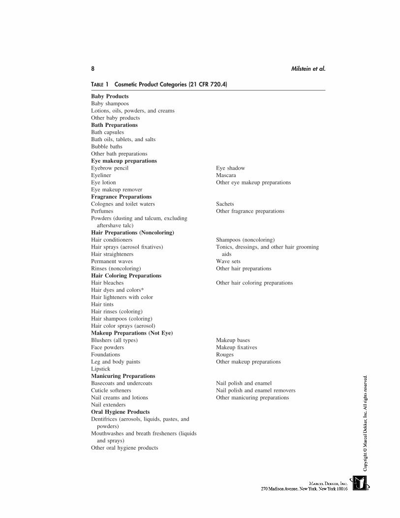
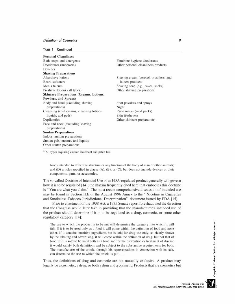
The 13 subdivided cosmetic product categories currently recognized by the U.S.Food & Drug Administration (FDA) for the voluntary filing of cosmetic product ingredientcomposition statements are enumerated in Title 21 of the Code of Federal Regulations
Definition of Cosmetics 7
(c.f., 21 CFR 720.4); these are presented in Table 1. Here one can find all of the productcategories that the consumer usually connotes with the terms ‘‘cosmetics & toiletries.’’Included in the definition of cosmetics are products intended to cleanse the body in thebath or shower, mask the various malodors of the oral, perigenital, and axillary regionsof the human anatomy, adorn the face, eyes, hair, and extremities in fashionable topical‘‘decorative’’ colors, alter the color and style of the scalp hair, and afford the integumentconditioning against losses of moisture caused by changes in environmental conditions(i.e., sun, wind, relative humidity) [8]. Note that the Act includes in the definition of‘‘cosmetic’’ any material intended for use as a component of a cosmetic product, so thatan ingredient intended to be used in a cosmetic is also considered to be a cosmetic.
Soap products, consisting primarily of an alkali metal salt of free fatty acids, makingno label claims other than cleansing of the human body, and labeled, sold, and representedonly as soap are not considered cosmetics under the law (c.f., 21 CFR 701.20). However,detergent-based ‘‘beauty or body bars,’’ so-called combination or combo-bars based onmixtures of soap and detergent(s), and those products containing other functional cosmeticingredients (i.e., emollients, moisturizers, or botanical ingredients) that make product per-formance claims other than cleansing of the human body, are considered ‘‘cosmetics.’’Additionally, soaps that contain antimicrobial active ingredients and that make antibacte-rial or germ-killing efficacy claims are regulated under the FD&C Act as ‘‘over-the-counter’’ (OTC) drug products. If they make cosmetic claims as well they may also beregulated as cosmetics [8] (see the following).
Other authoritative treatises in cosmetic science such as those of Jellinek [9],Poucher [5], deNavarre [10], Balsam and Sagarin [11], and Harry’s [12] discuss cosmeticproduct formulations in similar categories to those that have been adopted by regulationunder authority of the Act in the United States. Jackson [13] also presents an excellentand up-to-date tabulation of the product types that could reasonably be considered, whollyor in part, cosmetics. These include, as he correctly notes, some topical OTC drug productsamong his count of 77 product types, in addition to those products that the FDA wouldconsider bonafide cosmetics.
The Act also contains statutory provisions to regulate cosmetics in order to ensurethat only products deemed safe for their intended use and properly labeled are legallyoffered for sale in the United States. Thus, various prohibited actions are defined in Section301 of the Act that relate to the conditions under which cosmetics are deemed to be‘‘adulterated’’ (Section 601) or ‘‘misbranded’’ (Section 602) under the Act. These regula-tory provisions will be discussed in Chapter 62.
COSMETICS THAT ARE ALSO DRUGS: THE INTENDED USE DOCTRINE
All topical products are not necessarily cosmetics. Dermatologics, for example, are topicalproducts generally regulated as drug products based on the therapeutic or medicinal pur-pose for which the product is marketed as well as its formulation, which includes one ormore pharmacologically active ingredients. Section 201 (g)(1) of the FD&C Act definesthe term ‘‘drug’’ as:
. . . (A) articles recognized in the official United States Pharmacopoeia, official Homeo-pathic Pharmacopeia of the United States, or official National Formulary, or any supple-ment to any of them; and (B) articles intended for use in the diagnosis, cure, mitigation,treatment, or prevention of disease in man or other animals; and (C) articles (other than
8 Milstein et al.
TABLE 1 Cosmetic Product Categories (21 CFR 720.4)
Baby ProductsBaby shampoosLotions, oils, powders, and creamsOther baby productsBath PreparationsBath capsulesBath oils, tablets, and saltsBubble bathsOther bath preparationsEye makeup preparationsEyebrow pencil Eye shadowEyeliner MascaraEye lotion Other eye makeup preparationsEye makeup removerFragrance PreparationsColognes and toilet waters SachetsPerfumes Other fragrance preparationsPowders (dusting and talcum, excluding
aftershave talc)Hair Preparations (Noncoloring)Hair conditioners Shampoos (noncoloring)Hair sprays (aerosol fixatives) Tonics, dressings, and other hair groomingHair straighteners aidsPermanent waves Wave setsRinses (noncoloring) Other hair preparationsHair Coloring PreparationsHair bleaches Other hair coloring preparationsHair dyes and colors*Hair lighteners with colorHair tintsHair rinses (coloring)Hair shampoos (coloring)Hair color sprays (aerosol)Makeup Preparations (Not Eye)Blushers (all types) Makeup basesFace powders Makeup fixativesFoundations RougesLeg and body paints Other makeup preparationsLipstickManicuring PreparationsBasecoats and undercoats Nail polish and enamelCuticle softeners Nail polish and enamel removersNail creams and lotions Other manicuring preparationsNail extendersOral Hygiene ProductsDentifrices (aerosols, liquids, pastes, and
powders)Mouthwashes and breath fresheners (liquids
and sprays)Other oral hygiene products
Definition of Cosmetics 9
TABLE 1 Continued
Personal CleanlinessBath soaps and detergents Feminine hygiene deodorantsDeodorants (underarm) Other personal cleanliness productsDouchesShaving PreparationsAftershave lotions Shaving cream (aerosol, brushless, andBeard softeners lather) productsMen’s talcum Shaving soap (e.g., cakes, sticks)Preshave lotions (all types) Other shaving preparationsSkincare Preparations (Creams, Lotions,Powders, and Sprays)Body and hand (excluding shaving Foot powders and sprays
preparations) NightCleansing (cold creams, cleansing lotions, Paste masks (mud packs)
liquids, and pads) Skin freshenersDepilatories Other skincare preparationsFace and neck (excluding shaving
preparations)Suntan PreparationsIndoor tanning preparationsSuntan gels, creams, and liquidsOther suntan preparations
* All types requiring caution statement and patch test.
food) intended to affect the structure or any function of the body of man or other animals;and (D) articles specified in clause (A), (B), or (C); but does not include devices or theircomponents, parts, or accessories.
The so-called Doctrine of Intended Use of an FDA-regulated product generally will governhow it is to be regulated [14]; the maxim frequently cited here that embodies this doctrineis ‘‘You are what you claim.’’ The most recent comprehensive discussion of intended usemay be found in Section II.E of the August 1996 Annex to the ‘‘Nicotine in Cigarettesand Smokeless Tobacco Jurisdictional Determination’’ document issued by FDA [15].
Prior to enactment of the 1938 Act, a 1935 Senate report foreshadowed the directionthat the Congress would later take in providing that the manufacturer’s intended use ofthe product should determine if it is to be regulated as a drug, cosmetic, or some otherregulatory category [14]:
The use to which the product is to be put will determine the category into which it willfall. If it is to be used only as a food it will come within the definition of food and noneother. If it contains nutritive ingredients but is sold for drug use only, as clearly shownby the labeling and advertising, it will come within the definition of drug, but not that offood. If it is sold to be used both as a food and for the prevention or treatment of diseaseit would satisfy both definitions and be subject to the substantive requirements for both.The manufacturer of the article, through his representations in connection with its sale,can determine the use to which the article is put . . .
Thus, the definitions of drug and cosmetic are not mutually exclusive. A product maylegally be a cosmetic, a drug, or both a drug and a cosmetic. Products that are cosmetics but
10 Milstein et al.
are also intended to treat or prevent disease, or otherwise intended to affect the structure orany functions of the human body, are also considered drugs under the Act and must complywith both the drug and cosmetic provisions of the law [8].
Examples of products that are drugs as well as cosmetics are anticaries (fluoride)toothpastes, hormone creams, suntanning preparations containing a sunscreen active ingre-dient and either intended to protect against sunburn or make tanning claims [16], antiper-spirants and/or deodorants, antibacterial detergent bars or soaps, and antidandruff sham-poos. Most currently marketed cosmetics that are also drugs are OTC drugs. Several arenew drugs for which safety and effectiveness had to be proven to FDA (i.e., in a NewDrug Application or NDA) before they could be marketed [8]. A ‘‘new drug’’ is definedin Section 201 (p) of the Act as a drug that is not ‘‘generally recognized as safe andeffective’’ (GRAS/E) by experts under the conditions of intended use or that has becomeso recognized but has not been used to a material extent or for a material time under suchconditions.
It is relatively easy to market a cosmetic. Cosmetic products can be brought tomarket very quickly—a fact that is clearly reflected in the rapid pace with which innova-tions and changes occur in the cosmetic marketplace. No premarket approval (or manda-tory manufacturing establishment, product, or ingredient registration) is required. No de-lays are thereby incurred by the marketer while waiting for FDA approval. Nor does FDAhave a statutory mandate to monitor and regulate cosmetic performance advertising claims;the Agency’s oversight responsibility in this area extends only to ensure that cosmeticproduct package labeling is not violative with respect to ‘‘misbranding’’ (i.e., that theproduct performance claims are not false or misleading) [8]. More about U.S. cosmeticregulations will be said in Chapter 62.
The regulatory requirements for drugs (which are beyond the scope of this chapter)are more extensive than the requirements applicable to cosmetics. For example, the Actrequires that drug manufacturers register every year with the FDA and update their listsof all manufactured drugs twice annually (c.f., 21 CFR 207). Additionally, FDA druglabeling requirements and regulatory oversight of prescription drug advertising (FTC hasregulatory oversight for OTC drug advertising [17,18]) are more stringent than for cosmet-ics. Finally, drugs must be manufactured in accordance with Current Good ManufacturingPractice (CGMP) regulations (c.f., 21 CFR 210-211) [8].
THE COSMETIC/DRUG DISTINCTION: THE ROLE OF THE INTENDEDUSE DOCTRINE IN FDA ASSIGNMENT OF REGULATORY CATEGORYAND TRADE CORRESPONDENCE
The regulatory category occupied by a product clearly has a great impact on the marketingof that product. Because the drug approval process required by the Act (see previoussection) is rigorous, expensive, and time consuming, marketers of personal-care productswould rather market their products as cosmetics than as drugs. Some topical personal-care products are formulated in a nearly identical manner, and it is the manufacturer ofthe topical product that frequently determines what the intended use of the product is, andwhether it should be marketed as a cosmetic or as a drug by means of statements andother representations or performance claims made on product package labeling, collateralpromotional literature, and advertising. In other circumstances, whether this is done inten-tionally for marketing reasons or is otherwise unintentional, the manufacturer’s intended
Definition of Cosmetics 11
use may not be easy to discern, and it is not nearly as straightforward for FDA to determinethe most appropriate regulatory category for the product. How, then, is FDA to determinewhether such a product is a drug or a cosmetic?
It is the interpretation of what ‘‘intended use’’ means that has helped FDA to clarifyhow cosmetic products are distinguished from drugs. Needless to say it has also causeduncertainty, as topical cosmetic formulations have become more sophisticated and capableof delivering enhanced performance benefits to the consumer, or, viewed from the otherend of the drug–cosmetic continuum, as dermatological drug products have been formu-lated with ever increasing degrees of cosmetic elegance. FDA’s interpretation of cosmeticversus drug status for the various products that it regulates in the years since the enactmentof the 1938 Act has been guided by several sources of information.
Labeling
Intended use is determined principally, but not solely, by the claims that are made onproduct labeling (i.e., all labels and other written, printed, or graphic matter either on oraccompanying the product). ‘‘Puffery’’ claims [19] may draw upon the stylized artfulimagery and ‘‘hope in a bottle’’ that have traditionally sold cosmetics from the dawn ofthe cosmetic marketing era, when the formulation of cosmetics was more art than science,to the present day. ‘‘Subjective’’ and ‘‘objective’’ claims (20) are those that can andshould be substantiated, usually by focus-group panel interviews; home-placement tests,follow-up questionnaires, and phone interviews; or controlled-use medically supervisedclinical studies, with or without the use of accompanying bioengineering instrument as-sessments of various skin, hair, eye, or nail condition paramters. The Agency has even,on occasion, determined ‘‘intended use’’ of a product based, in part, on statements madeon behalf of the product by manufacturer sales associates at the point of sale, or on trainingand guidance provided to salespersons at the cosmetic counter.
Trade Correspondence
Early FDA guidance with respect to intended use commenced soon after passage of the1938 Act, when the Agency issued a series of informal opinions, known as Trade Corre-spondence (TC), that applied the statute to specific questions and situations; some of theTCs are still relied on as support for FDA regulatory policy [21]. Such TCs were the basisfor decisions setting Agency policy with respect to a cosmetic’s intended use. TC-10, forexample, notified marketers of cosmetic claims considered by the Agency to be ‘‘mis-brandings’’ in that they are ‘‘false and misleading’’ [22], while TC-229 stated that theword ‘‘healthful’’ contained in the labeling of a tooth powder would trigger the drugprovisions of the Act [23]. TC-26 held that a product’s mechanism of action could bethe basis of a cosmetic vs. drug intended-use determination, in that a deodorant powderinhibiting the normal physiological process of perspiration would be a drug (i.e., an anti-perspirant-deodorant), but the same product merely serving as a ‘‘reodorant-deodorant’’by absorbing the perspiration or masking the malodor would probably be a cosmetic[24]. TC-42 provided further clarification of the ‘‘affect the body’’ clause of Section 201(g) of the Act, in stating that a topical product containing emollient ingredients whoseclaims to efficacy were through such temporary improvements in skin condition param-eters as ‘‘softening’’ (or, by extrapolation, smoothing or moisturizing) would not neces-sarily be considered drugs [25]. TC-61, recently revoked in light of new science [16],
12 Milstein et al.
served for many years as the ‘‘line in the sand’’ for distinguishing between products thatreferred to sunburn protection as drugs and those represented exclusively for the produc-tion of an even tan as cosmetics [26]. Other TCs have established that ordinary facialtissue for wiping purposes is not a cosmetic [27], that other appliances used as adjunctsto, or in combination with, bonafide cosmetic products, such as manicuring instruments[28], razors and razor blades [28], shaving brushes [29], toothbrushes [29], and toiletbrushes [29] are not considered devices, and that cuticle removers [30] are cosmeticsrather than drugs.
FDA Case Law
The most direct guidance has been provided by Agency enforcement actions involvingcosmetics that were determined to be drugs. For example, case law from the 1960s estab-lished that promotional claims for the bovine serum albumin antiwrinkle products, SuddenChange (Hazel Bishop) and Line Away (Coty), taken in the overall context of productlabeling, caused these products to be classified as drugs [31,32]. The court held that adver-tising claims for these products, which included claims such as ‘‘[n]ot a face lift, not atreatment,’’ ‘‘[c]ontains . . . no hormones,’’ ‘‘[y]ou’ll feel a tingling sensation’’, ‘‘[n]our-ishes the skin,’’ ‘[t]ightens and goes to work on wrinkles’’; ‘‘made in a pharmaceuticallaboratory,’’ ‘‘packaged under biologically aseptic conditions,’’ ‘‘a face lift without sur-gery,’’ and ‘‘it lifts puffs under the eyes,’’ among others, established the respective ven-dor’s intent that the article had physiological and therapeutic effects. It is important tonote in these cases that, aside from the claims, there was no evidence that they exertedany real effects on the structure or function of the body. In a third court case in the early1970s, claims that the bovine serum albumin–containing products, Magic Secret (HeleneCurtis), is ‘‘pure protein’’ and ‘‘causes an astringent sensation’’ alone were consideredappropriate for a cosmetic [33].
1980s Regulatory Letters
The next actions taken by FDA that served to define labeling claims that may cause aproduct to be classified as a drug occurred in the late 1980s. In the spring of 1987, FDAsent 23 Regulatory Letters [34] to companies that were again marketing antiwrinkle andantiaging topical skincare products with aggressive marketing claims, which were deemedby the Agency to be ‘‘daring’’ [35]. These products made claims such as ‘‘revitalizes byaccelerating the rate of cellular renewal,’’ ‘‘revitalizes skin cells and promotes the skin’snatural repair process,’’ ‘‘helps stimulate the natural production of structural proteins,’’‘‘increases the proper uptake of oxygen and blood supply to the cells,’’ ‘‘reverses facialaging,’’ ‘‘restructures the deepest epidermal layers,’’ ‘‘increases collagen production,’’and ‘‘provides vital nourishing supplements,’’ among others. All of these claims, takenin the context of individual product labeling, were sufficient in the view of the Agencyto establish intended use as a drug; indeed, it would be very difficult to use these termsand not trigger the structure or function definition of a drug. Again, in all of the productscovered in this action, there was little expectation that they actually exerted an effect on thebody outside of that which normally occurs from topical application of any conventionalmoisturizer. The Regulatory Letters issued by the Agency served as useful precedents ofthe legal rationale regarding product classification, and also provided very clear guidance
Definition of Cosmetics 13
to the Industry, as had been requested in a Citizen Petition [36] concerning what labelclaims could get a product into regulatory difficulty.
OTC Drug Monographs: Cosmetics That Contain Active Ingredients
FDA has clearly stated that determination of intended use goes beyond direct label state-ments. The history of use of the ingredient, its functionality in the product, and the consum-er’s perception all play a role in product classification. This is the case with products thatcontain drug active ingredients in their formulations but do not make explicitly statedclaims about the drug effects of the active ingredient. Although there is no case law thataddresses product classification based on presence of active ingredients alone, this issuehas been addressed over the years in regulations for OTC drug products and other actionsby the Agency.
FDA acknowledged in the Tentative Final Monograph for First Aid Antiseptic DrugProducts, published August 16, 1991 (56 FR 33644), that antimicrobial soap productsmaking cosmetic claims only are not subject to regulation as OTC drugs and should notbe considered in a review of drug effectiveness. The Agency further established the policythat the presence of an antimicrobial ingredient does not, in and of itself, make a producta drug, provided that no drug claim (i.e., ‘‘kills germs,’’ ‘‘antibacterial’’) is made. How-ever, the level of antimicrobial ingredient in a cosmetic product, when such ingredient isintended only as part of a cosmetic preservative system, may not exceed the concentrationprovided for in the OTC Monograph. The Agency also noted in this rulemaking that the‘‘intended use’’ of a product may be inferred from labeling, promotional material, advertis-ing, and any other relevant factor, arguing that, based on case law, a manufacturers’ subjec-tive claims of intent may be pierced to find its actual intent on the basis of objectiveevidence.
Analogously, the Agency acknowledged in the Final Monograph for Topical AcneDrug Products, published in August, 1991 (56 FR 41008), that the final rule covers onlythe drug uses of the active ingredients and does not apply to the use of the same ingredientsfor non–drug effects in products intended solely as cosmetics.
FDA noted in the May 12, 1993 Tentative Final Monograph for OTC SunscreenDrug Products (58 FR 28194) that a product may contain a sunscreen ingredient and bea cosmetic if it is not intended to protect against the sun and no claims are made aboutthe ingredient. In these cases, the term sunscreen is not used, no SPF value is given, andthe sunscreen ingredient is only mentioned in the product’s labeling by its cosmetic namein the ingredient list in accordance with Agency regulations at 21 CFR 701.3. However,the presence of a sunscreen active ingredient in a product intended to protect from sunexposure makes the product a drug. Again, FDA noted that it is not bound by the manufac-turer’s subjective claims, but can find actual therapeutic intent on the basis of objectiveevidence. Such intent may be derived from labeling, promotional material, advertising,and any other relevant source, where ‘‘relevant source’’ can even include the consumer’sintent in using the product. The Agency reaffirmed these views in the May 21, 1999 FinalMonograph for OTC Sunscreen Drug Products (64 FR 27666) and codified them at 21CFR 700.35, adding only the caveat that when a cosmetic product contains a sunscreeningredient not intended to be used for therapeutic or physiological efficacy and uses theterm ‘‘sunscreen’’ or similar sun protection terminology anywhere in its labeling, the termmust be qualified by describing the cosmetic benefit provided by the sunscreen ingredient,
14 Milstein et al.
and this statement must appear prominently and conspicuously at least once in the labeling,contiguous with the term ‘‘sunscreen’’ or other similar sun-protection terminology usedin the labeling.
The Agency provided clear guidance in the February 3, 1994 Withdrawal of Ad-vance Notice of Proposed Rulemaking for OTC Vaginal Drug Products (59 FR 5226)that the mere presence of a pharmacologically active ingredient in therapeutically activeconcentrations could make a product a drug, even in the absence of explicit drug claims,if the intended use would be implied because of the known or recognized drug effects ofthe ingredient (i.e., fluoride in a dentrifrice or zinc pyrithione in a shampoo). Thus, al-though explicitly stated intended use is the primary factor in determining cosmetic vs.drug product category, the type and amount of ingredient(s) present in a product must beconsidered in determining its regulatory status, even if that product does not make explicitdrug claims.
Finally, FDA noted in a Notice of Proposed Rulemaking concerning Cosmetic Prod-ucts Containing Certain Hormone Ingredients that was published on September 9, 1993(58 FR 47611), along with a final rule on Topically Applied Hormone-Containing DrugProducts for Over-the-Counter Use (58 FR 47608), that ‘‘certain hormone-containingproducts not bearing drug claims could be cosmetics depending on the levels of hormonesused and whether that level of use affects the structure or any function of the body . . .’’.It was noted that only these hormone ingredients present at a level below that which exertsan effect on the structure or function of the body would be acceptable for use in productsmarketed as cosmetics. However, if the hormone ingredient was present at physiologicallyactive levels, then the product would be classified as a drug for regulatory purposes.
The Alpha Hydroxy Acid Situation
The alpha hydroxy acids (AHAs) have been hailed as the first examples of the new cosme-ceuticals since their first appearance in the marketplace several years ago [37]. Throughtheir promotional claims, AHAs promise skincare benefits that far exceed the humectantand moisturization attributes that were once associated with AHA salts such as sodiumlactate as components of the skin’s so-called natural moisturizing factor (NMF) in thecosmetics of the 1970s [38]. The scientific, clinical, and patent literature show that AHAs,as used today, probably function under at least certain conditions of formulation not onlyas traditional cosmetic moisturizers but as epidermal exfoliants and modulators of epider-mal and dermal structure and function [39–42]. They are promoted in mass-marketed andsalon-treatment products alike for treatment of a number of cosmetic (i.e., severe dry skin,tone/texture) and more significant dermatological (i.e., acneiform, photoaging, age spots)conditions [43, 44]. Manufacturers of these products have sought to market them directlyto consumers as cosmetics or through phsician offices, salons, and professional estheticians[37, 45–47]. Although most marketers have artfully avoided making direct and impactfulefficacy claims that might invite triggering the drug provisions of the Act [48], FDA isalso cognizant that the addition of chemical exfoliants to cosmetics on such a wide scaleis unprecedented [43], and 7 years of marketing history with such products may provean inadequate and unreliable predictor of future adverse impacts on public health. There-fore, despite prior evaluations of AHA safety by the Cosmetic Ingredient Review (CIR)[49] and some more recent evaluations conducted by FDA [50] as well, the Agency hasreserved its judgement concerning the appropriate regulatory category designation(s) forAHA skincare products and remains vigilant concerning the adequacy of the safety sub-
Definition of Cosmetics 15
stantiation for AHAs, particularly with respect to potential chronic effects of AHAs onthe sun sensitivity and photocarcinogenic responses of the skin [51].
SUMMARY: COSMECEUTICALS, COSMETIC THERAPEUTICS, ANDOTHER PROPOSED DEFINITIONS
Topical products marketed in the United States are regulated under the Act, variously, ascosmetics, drugs, or OTC drug-cosmetics. There is no intermediate category that corre-sponds, for example, to the ‘‘quasi-drugs,’’ defined under the Japanese PharmaceuticalAffairs Law [52]. Neither are there any provisions under the U.S. statute that would accom-odate classes of topical skincare products with levels of efficacy that exceed those oftraditional cosmetics but whose safety have not been as rigorously substantiated as tradi-tional drugs. Reed [53] and Kligman [54] proposed that such high performance cosmeticsbe classified as ‘‘cosmeceuticals,’’ despite the lack of legal standing of such a productcategory. Piacquadio [55] favors the term ‘‘cosmetic therapeutics’’ when referring to drugsand devices having known risk/benefit profiles and established efficacy for a cosmeticindication, pending or with FDA approval. Privat [56] suggested the categories ‘‘decora-tive and/or protective cosmetics’’ for those products that embellish by modifying (appear-ance, color, feel) or protecting the integument from external insults (i.e., UVR or bacteria),while reserving the term ‘‘remedial and/or active cosmetics’’ for those products that mod-ify or correct the physiological state of the integument [e.g., stratum corneum (SC), epider-mis, melanocytes, intercellular lipid layer, sudoral glands, hypodermis]. Morganti [57]coined the term ‘‘cosmetognosy’’ to denote the science that deals with the biologicaleffects of cosmetics. Although these proposals each have varying degrees of merit, they,too have no regulatory standing in the United States under provisions of the 1938FD&C Act.
ACKNOWLEDGMENT
We wish to acknowledge the assistance given by Ms. Beth Meyers, Technical Editor,Division of Programs and Policy Enforcement, Office of Cosmetics and Colors, FDA-CFSAN, in proofreading this manuscript and formatting Table 1.
DISCLAIMER
The views expressed herein are those of the authors and do not necessarily represent thoseof the FDA.
REFERENCES
1. Bucalo AJ. 1999 State of the Industry. Global Cosmet Ind, 1999; June: 32.2. Wenninger JA, R. Canterbery R, McEwen GA Jr, eds. CTFA International Cosmetic Ingredi-
ent Dictionary. 8th ed., 1999.3. FDA Compliance Program Guidance Manual 7329.001, pt. 1 at 1. August 1993.4. McDonaugh EG. Truth About Cosmetics. Drug Markets, Inc. 1937: vii.5. Butler H. Historical Background. In: Butler H, ed. Poucher’s Perfumes, Cosmetics and Soaps,
9th ed. London: Chapman & Hall, 1993: 639–692.6. Romm S. The Changing Face of Beauty. St. Louis: Mosby-Yearbook, Inc., 1992.
16 Milstein et al.
7. Yingling GL, Onel S. Cosmetic regulation revisited. In: Brady RP, Cooper RM, SilvermanRS, eds. Fundamentals of Law and Regulation. Vol. 1. Washington, DC: FDLI, 1997: 321.
8. FDA’s Cosmetics Handbook. Washington, D.C.: U.S. Government Printing Office, 1993:1–3.
9. Jellinek JS. Formulation and Function of Cosmetics. New York: Wiley-Interscience, 1970.10. deNavarre MG. The Chemistry and Manufacture of Cosmetics. 2nd ed. Vols. I–IV. Princeton:
D. Van Nostrand Company, Inc., 1969.11. Balsam MS, Sagarin E. Cosmetics: Science and Technology. Vols 1–3. New York: John Wiley
and Sons, Inc., 1972.12. Wilkinson JB, Moore RJ. Harry’s Cosmeticology. 7th ed. New York: Chemical Publishing
Co., Inc., 1982.13. Jackson EM. Consumer products: cosmetics and topical over-the-counter drug products. In:
Chengelis CP, Holson JF, Gad SC, eds. Regulatory Toxicology. New York: Raven Press, 1995:105–121.
14. Yingling GL, Swit MA. Cosmetic regulations. In: Cooper RM. Food and Drug Law. Washing-ton, D.C.: FDLI, 1991: 362.
15. The ‘Intended Use’ of a product is not determined only on the basis of promotional claims.In: Nicotine in Cigarettes and Smokeless Tobacco is a Drug and These Products Are NicotineDelivery Devices Under the Federal Food, Drug, and Cosmetic Act: Jurisdictional Determina-tion. U.S. Food & Drug Administration, Department of Health and Human Services, August1996, Annex, Section II.E.
16. Final Rule for Over-the-Counter (OTC) Sunscreen Products for Human Use. 64 FR 27666 @27668. May 21, 1999.
17. Hobbs CO. The FDA and the Federal Trade Commission. In: Cooper RM. Food and DrugLaw. Washington, D.C.: FDLI, 1991: 429–430, 452–456.
18. Memorandum of Understanding Between FTC and FDA. 36 FR 18539. 1971.19. (a) McNamara SH. FDA Regulation of Cosmeceuticals. Cosmet Toilet 1997; 112(3): 41–45.
(b) FTC Deception Policy Statement. Letter to the Honorable John D. Dingell, Chairman,Committee on Energy and Commerce, U.S. House of Representatives, @ n42. October 14,1983. (c) Feldman JP. Puffery in Advertising. Arent Fox Advertising Law (http:/ /www.arent-fox.com), June 1995. (d) Hobbs CO. Advertising for foods, veterinary products, and cosmetics.In: Brady RP, Cooper RM, Silverman RS, eds. Fundamentals of Law and Regulation. Vol.7. Washington, D.C., 1997: 350. (e) Legal aspects of promotion strategy: advertising. In: SternLW, Eovaldi TL. Legal Aspects of Marketing Strategy: Antitrust and Consumer ProtectionIssues. Englewood Cliffs: Prentice-Hall, Inc., 1984: 375–377.
20. (a) McNamara SH. Performance claims for skin care cosmetics. Drug Cosmet Ind 1985; Octo-ber: 34. (b) Weinstein S, Weinstein C, Drozdenko R. A current and comprehensive skin-evaluation program. Cosmet Technol, 1982; April: 36. (c) Grove GL. Noninvasive methodsfor assessing moisturizers. In: Waggoner WC, ed. Clinical Safety and Efficacy Testing of Cos-metics. New York: Marcel Dekker, 1990: 121–148. (d) Smithies RH. Substantiating pre-formance claims. Cosmet Toilet 1984; 99(3): 79–81, 84.
21. Kleinfeld VA, Dunn CW. Trade correspondence. In: Federal Food, Drug, and Cosmetic Act.Judicial and Administrative Record (1938–1949). New York: Commerce Clearing House,Inc., 1949: 561.
22. TC-10, (in Ref. 21) August 2, 1939: 566.23. TC-229, (in Ref. 21) April 11, 1940: 659.24. TC-26, (in Ref. 21) February 9, 1940: 581.25. TC-42, (in Ref. 21) February 12, 1940: 586.26. TC-61, (in Ref. 21) February 15, 1940: 593.27. TC-39, (in Ref. 21) February 9, 1940: 585.28. TC-112, (in Ref. 21) February 29, 1940: 613.29. TC-109, (in Ref. 21) February 29, 1940: 612.
Definition of Cosmetics 17
30. TC-245, (in Ref. 21) April 25, 1940: 665.31. United States v. An Article . . . Line Away, 284 F. Supp. 107 (D. Del. 1968); affirmed, 415
F. 2d 369 (3d Cir. 1969).32. United States v. An Article . . . Sudden Change, 288 F. Supp. 29 (E.D.N.Y. 1968); reviewed
409 F.2d 734 (2d Cir. 1969).33. United States v. An Article . . . Magic Secret, 331 F. Supp. 912 (D. MD 1971).34. FDA Regulatory Letters No. 87-HFN 312-08 to 87-HFN 312-29 (April 17, 1987 to June 23,
1987).35. McNamara SH. Performance claims for skin care cosmetics or how far may you go in claiming
to provide eternal youthfulness. Food Drug Law J 1986; 41:151–159.36. Citizen petition of McCutcheon, Doyle, Brown & Emerson. Bio Advance, FDA Docket No.
87P-0006, (January 6, 1987).37. (a) Godfrey-June J. The AHA phenomenon. Longevity 1993; Sept.: 36–39. (b) Jackson EM.
AHA-type products proliferate in 1993. Cosmet Dermatol 1993; 6(12):22, 24–26. (c) KintishL. AHAs: today’s fountain of youth? Soap/Cosmetics/Chemical Specialties 1994; Feb: 26–31.
38. (a) Harding CR, Bartolone J, Rawlings AV. Effects of Natural Moisturizing Factor and LacticAcid Isomers on Skin Function. In: Loden M, Maibach HI, eds. Dry Skin and Moisturizers:Chemistry and Function. Boca Raton: CRC Press, 2000:229–241. (b) Middleton JD, SodiumLactate as a Moisturizer. Cosmet Toilet 1978; 93:85–86.
39. (a) Leyden JJ, Lavker RM, Grove G, Kaidbey K. Alpha hydroxy acids are more than moisturiz-ers. J Geriatr Dermatol 1995 3 (suppl. A): 33A–37A. (b) Van Scott EJ, Yu RJ. Actions ofalpha hydroxy acids on skin compartments. J Geriatr Dermatol 1995; 3(suppl A): 19A–25A.
40. Smith WP. Hydroxy acids and skin aging. Soap/Cosmetics/Chemical Specialties 1993; 93(9):54, 56, 57–58, 76.
41. Smith WP. Hydroxy acids and skin aging. Cosmet Toilet 1994; 109: 41–48.42. Smith WP. Epidermal and dermal effects of topical lactic acid. 1996; J Am Acad Dermatol
35: 388–391.43. Kurtzweil P. Alpha hydroxy acids for skin care. FDA Consumer 1998; March-April: 30–35.44. Anonymous. Alpha hydroxy acids in cosmetics. FDA Backgrounder, BG 97-4, February 19,
1997.45. Brody HJ. Chemical Peeling and Resurfacing (2nd ed.), St. Louis: Mosby-Year Book, Inc.,
1997:90–100.46. Draelos ZD. New Developments in Cosmetics and Skin Care Products. In: Advances in Derma-
tology. Vol. 12. St. Louis: Mosby-Year Book, Inc., 1997; 3–17.47. (a) AHA ’95 Preview: New Developments in Alpha Hydroxy Acids. Symposium and Live
Patient Workshop, Jointly Sponsored by Cosmetic Peel Workshop and Medical EducationResources, Inc., Orlando, FL, December 3–4, 1994. (b) AHA ’96 Preview: New Advancesin AHAs and Skin Rejuvenation Techniques. Symposium and Live Patient Workshop, JointlySponsored by Medical Education Resources, Inc. and Herald Education & Research Founda-tion, San Diego, CA, December 2–3, 1995.
48. Yingling GL and Onel S. Cosmetic Regulation Revisited. In: RP Brady, RM Cooper, RSSilverman, eds, Fundamentals of Law and Regulation, Vol. 1, FDLI (Washington, DC), 1997:341–342.
49. (a) Cosmetic Ingredient Review. Final Report: Safety Assessment of Glycolic Acid; Ammo-nium, Calcium, Potassium and Sodium Glycolate; Methyl, Ethyl, Propyl, and Butyl Glycolate;Lactic Acid; Ammonium, Calcium, Potassium, Sodium, and TEA-Lactate; Methyl, Ethyl, Pro-pyl, and Butyl Lactate; and Lauryl, Myristyl, and Cetyl Lactate. Washington, D.C.: CosmeticIngredient Review, 1997. (b) Jackson, EM. CIR Expert Panel Releases AHA Report. CosmetDermatol 1997; 10(7):37–39
50. Effects of Alpha Hydroxy Acids on Skin. Report Submitted by KRA Corporation (SilverSpring, MD) to the Office of Cosmetics and Colors, CFSAN, FDA, DHHS under ContractNo. 223-94-2276. February 22, 1996.
18 Milstein et al.
51. (a) Kaidbey K. An Investigation of the Effects of Topical Treatment with an Alpha-HydroxyAcid (AHA) on the Sensitivity of Human Skin to UV-Induced Damage (FDA Sponsored Study# 1). Philadelphia: Ivy Laboratories (KGL, Inc.), 1999. (b) Kaidbey K. An Investigation of theEffects of Topical Treatment with Alpha-Hydroxy Acid (AHA) on UVB-Induced PyrimidineDimers in Human Skin (FDA Sponsored Study #2). Philadelphia: Ivy Laboratories (KGL,Inc.), 1999.
52. Santucci LG, Rempe JM. Legislation and Safety Regulations for Cosmetics in the UnitedStates, Europe, and Japan’’, Ref. 3, op. cit., Chapter 20; 556–571.
53. Reed RE. The definition of ‘cosmeceutical.’ J Soc Cosmet Chemists 1962; 13:103–106.54. (a) Skin: the hot topics. Vogue 1988; October:417. (b) HAPPI, 1996; May:61. (c) Kligman
AM. Why Cosmeceuticals? Cosmet Toilet 1993; 108(8):37–38. (d) Waleski M. Reed coined‘cosmecutical.’ Letter to the Editor. HAPPI 1996; August: 12.
55. Piacquadio D. Cosmetic therapeutic vs. cosmeceutical: which is it and why? AHA ‘95 Preview:New Developments in Alpha Hydroxy Acids. Symposium and Live Patient Workshop, JointlySponsored by Cosmetic Peel Workshop and Medical Education Resources, Inc., Orlando, FL,Dec. 3–4, 1994.
56. Privat Y. A new definition of cosmetology. In: Baran R, Maibach HI, eds. Cosmetic Dermatol-ogy. London: Martin Dunitz, Ltd., 1994: xiv–xv.
57. Morganti P-F. The cosmetic patch. A new frontier in cosmetic dermatology. Soap/Cosmetics/Chemical Specialties 1996; 96(2):48–50.
3
The Microscopic Structure of theEpidermis and Its Derivatives
Joel J. EliasUniversity of California at San Francisco School of Medicine,San Francisco, California
A general review of the microscopic structure of the epidermis and those epidermal deriva-tives that are distributed widely over the skin and, therefore, may be of interest in consider-ations of mechanisms of percutaneous absorption, will be presented here. Both light andelectron microscopic information will be discussed in order to give an integrated briefsummary of the basic morphological picture.
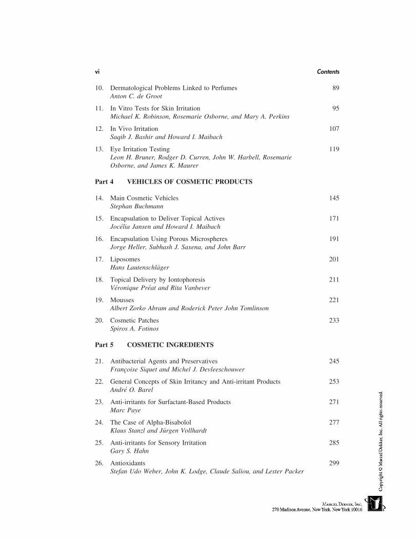
The epithelial component of the skin, the epidermis, is classified histologically as astratified squamous keratinizing epithelium. It is thickest on the palms and soles (Fig. 1)and thinner elsewhere on the body (Fig. 2). It lies on the connective tissue component ofthe skin, the dermis, in which are located the blood vessels and lymphatic vessels. Capil-lary loops in the dermis come to lie in close apposition to the underside of the epidermis.The epidermis, in common with other epithelia, is avascular. The living cells of the epider-mis receive their nutrients by diffusion of substances from the underlying dermal capillar-ies through the basement membrane and then into the epithelium. Metabolic products ofthe cells enter the circulation by diffusion in the opposite direction.
As in the case of other epithelia, the epidermis lies on a basement membrane (basallamina). This extracellular membrane, interposed between the basal cells of the epidermisand the connective tissue of the dermis, serves the important function of attaching thetwo tissues to each other. The point of contact of the epidermis with this structure is thebasal cell membrane of the basal cells. Along this surface the basal cells show manyhemidesmosomes, which increase the adherence of the basal cells (and therefore of theentire epidermis) to the basement membrane (and therefore to the dermis). In some loca-tions, such as the renal glomerulus, the basal lamina has been shown to also play a roleas a diffusion barrier to certain molecules.
The plane of contact between the epidermis and dermis is not straight but is anundulating surface, more so in some locations than others. Upward projections of connec-tive tissue, the dermal papillae, alternate with complementary downgrowths of the epider-
This chapter is reproduced with permission from Bronaugh RL, Maibach HI, eds. Percutaneous Absorption:Mechanisms—Methodology—Drug Delivery. 2nd ed. New York: Marcel Dekker, Inc., 1989.
19
20 Elias
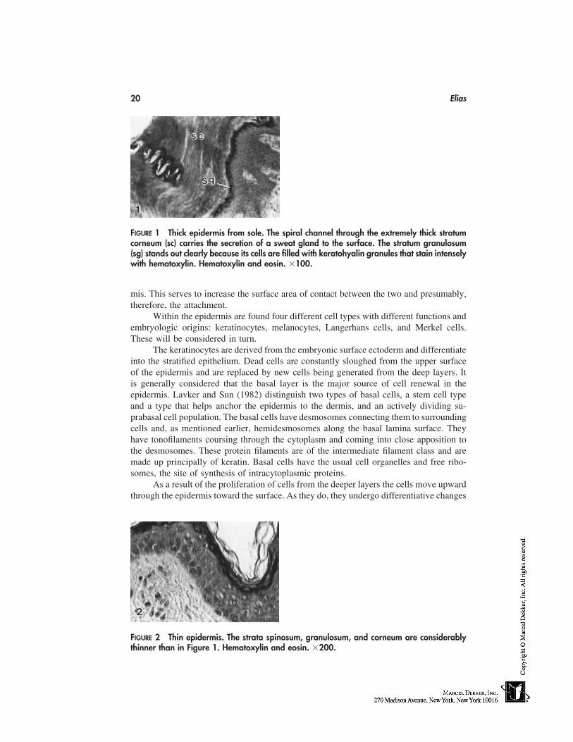
FIGURE 1 Thick epidermis from sole. The spiral channel through the extremely thick stratumcorneum (sc) carries the secretion of a sweat gland to the surface. The stratum granulosum(sg) stands out clearly because its cells are filled with keratohyalin granules that stain intenselywith hematoxylin. Hematoxylin and eosin. �100.
mis. This serves to increase the surface area of contact between the two and presumably,therefore, the attachment.
Within the epidermis are found four different cell types with different functions andembryologic origins: keratinocytes, melanocytes, Langerhans cells, and Merkel cells.These will be considered in turn.
The keratinocytes are derived from the embryonic surface ectoderm and differentiateinto the stratified epithelium. Dead cells are constantly sloughed from the upper surfaceof the epidermis and are replaced by new cells being generated from the deep layers. Itis generally considered that the basal layer is the major source of cell renewal in theepidermis. Lavker and Sun (1982) distinguish two types of basal cells, a stem cell typeand a type that helps anchor the epidermis to the dermis, and an actively dividing su-prabasal cell population. The basal cells have desmosomes connecting them to surroundingcells and, as mentioned earlier, hemidesmosomes along the basal lamina surface. Theyhave tonofilaments coursing through the cytoplasm and coming into close apposition tothe desmosomes. These protein filaments are of the intermediate filament class and aremade up principally of keratin. Basal cells have the usual cell organelles and free ribo-somes, the site of synthesis of intracytoplasmic proteins.
As a result of the proliferation of cells from the deeper layers the cells move upwardthrough the epidermis toward the surface. As they do, they undergo differentiative changes
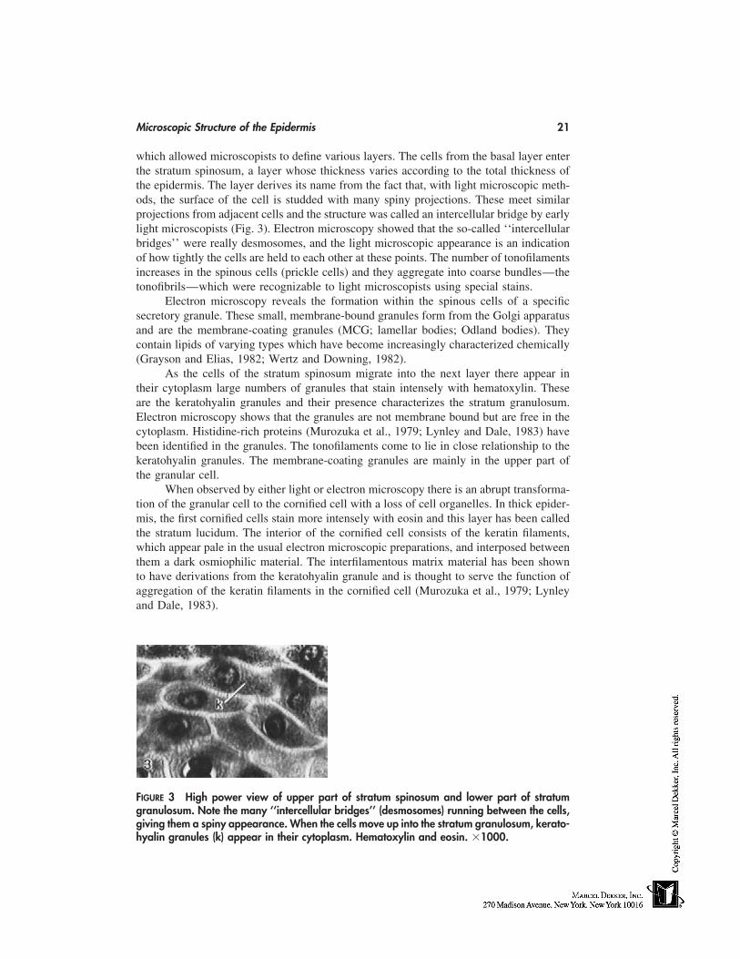
FIGURE 2 Thin epidermis. The strata spinosum, granulosum, and corneum are considerablythinner than in Figure 1. Hematoxylin and eosin. �200.
Microscopic Structure of the Epidermis 21
which allowed microscopists to define various layers. The cells from the basal layer enterthe stratum spinosum, a layer whose thickness varies according to the total thickness ofthe epidermis. The layer derives its name from the fact that, with light microscopic meth-ods, the surface of the cell is studded with many spiny projections. These meet similarprojections from adjacent cells and the structure was called an intercellular bridge by earlylight microscopists (Fig. 3). Electron microscopy showed that the so-called ‘‘intercellularbridges’’ were really desmosomes, and the light microscopic appearance is an indicationof how tightly the cells are held to each other at these points. The number of tonofilamentsincreases in the spinous cells (prickle cells) and they aggregate into coarse bundles—thetonofibrils—which were recognizable to light microscopists using special stains.
Electron microscopy reveals the formation within the spinous cells of a specificsecretory granule. These small, membrane-bound granules form from the Golgi apparatusand are the membrane-coating granules (MCG; lamellar bodies; Odland bodies). Theycontain lipids of varying types which have become increasingly characterized chemically(Grayson and Elias, 1982; Wertz and Downing, 1982).
As the cells of the stratum spinosum migrate into the next layer there appear intheir cytoplasm large numbers of granules that stain intensely with hematoxylin. Theseare the keratohyalin granules and their presence characterizes the stratum granulosum.Electron microscopy shows that the granules are not membrane bound but are free in thecytoplasm. Histidine-rich proteins (Murozuka et al., 1979; Lynley and Dale, 1983) havebeen identified in the granules. The tonofilaments come to lie in close relationship to thekeratohyalin granules. The membrane-coating granules are mainly in the upper part ofthe granular cell.
When observed by either light or electron microscopy there is an abrupt transforma-tion of the granular cell to the cornified cell with a loss of cell organelles. In thick epider-mis, the first cornified cells stain more intensely with eosin and this layer has been calledthe stratum lucidum. The interior of the cornified cell consists of the keratin filaments,which appear pale in the usual electron microscopic preparations, and interposed betweenthem a dark osmiophilic material. The interfilamentous matrix material has been shownto have derivations from the keratohyalin granule and is thought to serve the function ofaggregation of the keratin filaments in the cornified cell (Murozuka et al., 1979; Lynleyand Dale, 1983).
FIGURE 3 High power view of upper part of stratum spinosum and lower part of stratumgranulosum. Note the many ‘‘intercellular bridges’’ (desmosomes) running between the cells,giving them a spiny appearance. When the cells move up into the stratum granulosum, kerato-hyalin granules (k) appear in their cytoplasm. Hematoxylin and eosin. �1000.
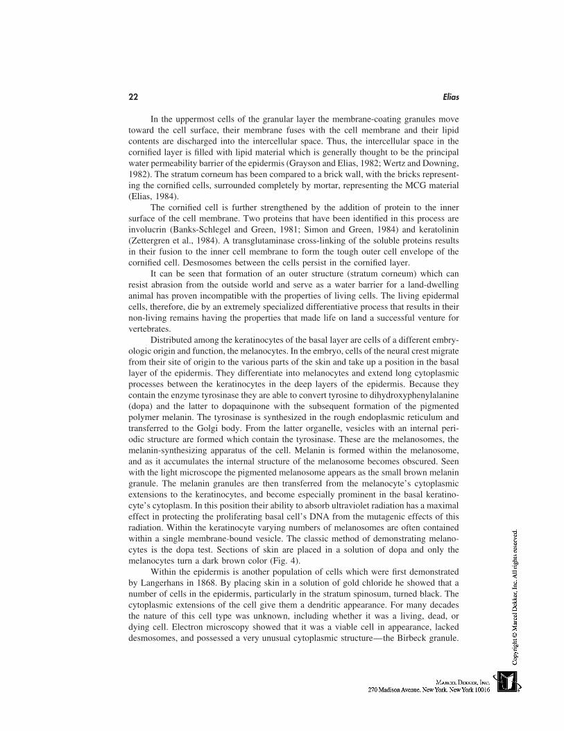
22 Elias
In the uppermost cells of the granular layer the membrane-coating granules movetoward the cell surface, their membrane fuses with the cell membrane and their lipidcontents are discharged into the intercellular space. Thus, the intercellular space in thecornified layer is filled with lipid material which is generally thought to be the principalwater permeability barrier of the epidermis (Grayson and Elias, 1982; Wertz and Downing,1982). The stratum corneum has been compared to a brick wall, with the bricks represent-ing the cornified cells, surrounded completely by mortar, representing the MCG material(Elias, 1984).
The cornified cell is further strengthened by the addition of protein to the innersurface of the cell membrane. Two proteins that have been identified in this process areinvolucrin (Banks-Schlegel and Green, 1981; Simon and Green, 1984) and keratolinin(Zettergren et al., 1984). A transglutaminase cross-linking of the soluble proteins resultsin their fusion to the inner cell membrane to form the tough outer cell envelope of thecornified cell. Desmosomes between the cells persist in the cornified layer.
It can be seen that formation of an outer structure (stratum corneum) which canresist abrasion from the outside world and serve as a water barrier for a land-dwellinganimal has proven incompatible with the properties of living cells. The living epidermalcells, therefore, die by an extremely specialized differentiative process that results in theirnon-living remains having the properties that made life on land a successful venture forvertebrates.
Distributed among the keratinocytes of the basal layer are cells of a different embry-ologic origin and function, the melanocytes. In the embryo, cells of the neural crest migratefrom their site of origin to the various parts of the skin and take up a position in the basallayer of the epidermis. They differentiate into melanocytes and extend long cytoplasmicprocesses between the keratinocytes in the deep layers of the epidermis. Because theycontain the enzyme tyrosinase they are able to convert tyrosine to dihydroxyphenylalanine(dopa) and the latter to dopaquinone with the subsequent formation of the pigmentedpolymer melanin. The tyrosinase is synthesized in the rough endoplasmic reticulum andtransferred to the Golgi body. From the latter organelle, vesicles with an internal peri-odic structure are formed which contain the tyrosinase. These are the melanosomes, themelanin-synthesizing apparatus of the cell. Melanin is formed within the melanosome,and as it accumulates the internal structure of the melanosome becomes obscured. Seenwith the light microscope the pigmented melanosome appears as the small brown melaningranule. The melanin granules are then transferred from the melanocyte’s cytoplasmicextensions to the keratinocytes, and become especially prominent in the basal keratino-cyte’s cytoplasm. In this position their ability to absorb ultraviolet radiation has a maximaleffect in protecting the proliferating basal cell’s DNA from the mutagenic effects of thisradiation. Within the keratinocyte varying numbers of melanosomes are often containedwithin a single membrane-bound vesicle. The classic method of demonstrating melano-cytes is the dopa test. Sections of skin are placed in a solution of dopa and only themelanocytes turn a dark brown color (Fig. 4).
Within the epidermis is another population of cells which were first demonstratedby Langerhans in 1868. By placing skin in a solution of gold chloride he showed that anumber of cells in the epidermis, particularly in the stratum spinosum, turned black. Thecytoplasmic extensions of the cell give them a dendritic appearance. For many decadesthe nature of this cell type was unknown, including whether it was a living, dead, ordying cell. Electron microscopy showed that it was a viable cell in appearance, lackeddesmosomes, and possessed a very unusual cytoplasmic structure—the Birbeck granule.
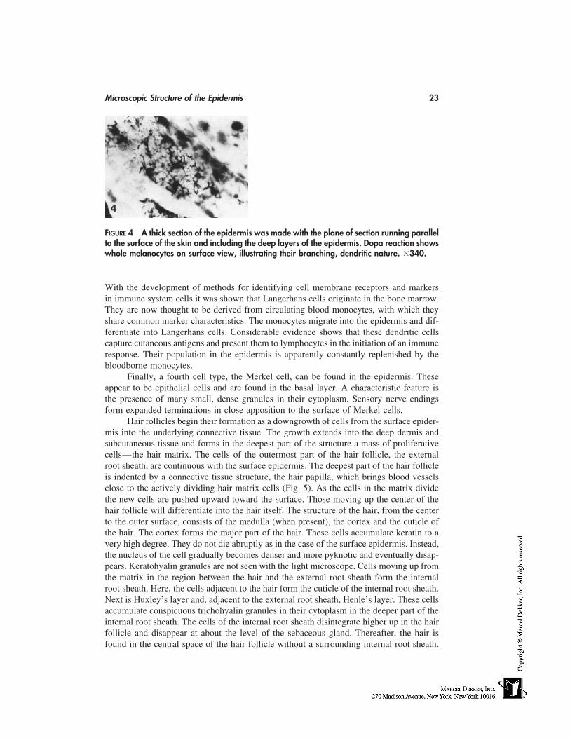
Microscopic Structure of the Epidermis 23
FIGURE 4 A thick section of the epidermis was made with the plane of section running parallelto the surface of the skin and including the deep layers of the epidermis. Dopa reaction showswhole melanocytes on surface view, illustrating their branching, dendritic nature. �340.
With the development of methods for identifying cell membrane receptors and markersin immune system cells it was shown that Langerhans cells originate in the bone marrow.They are now thought to be derived from circulating blood monocytes, with which theyshare common marker characteristics. The monocytes migrate into the epidermis and dif-ferentiate into Langerhans cells. Considerable evidence shows that these dendritic cellscapture cutaneous antigens and present them to lymphocytes in the initiation of an immuneresponse. Their population in the epidermis is apparently constantly replenished by thebloodborne monocytes.
Finally, a fourth cell type, the Merkel cell, can be found in the epidermis. Theseappear to be epithelial cells and are found in the basal layer. A characteristic feature isthe presence of many small, dense granules in their cytoplasm. Sensory nerve endingsform expanded terminations in close apposition to the surface of Merkel cells.
Hair follicles begin their formation as a downgrowth of cells from the surface epider-mis into the underlying connective tissue. The growth extends into the deep dermis andsubcutaneous tissue and forms in the deepest part of the structure a mass of proliferativecells—the hair matrix. The cells of the outermost part of the hair follicle, the externalroot sheath, are continuous with the surface epidermis. The deepest part of the hair follicleis indented by a connective tissue structure, the hair papilla, which brings blood vesselsclose to the actively dividing hair matrix cells (Fig. 5). As the cells in the matrix dividethe new cells are pushed upward toward the surface. Those moving up the center of thehair follicle will differentiate into the hair itself. The structure of the hair, from the centerto the outer surface, consists of the medulla (when present), the cortex and the cuticle ofthe hair. The cortex forms the major part of the hair. These cells accumulate keratin to avery high degree. They do not die abruptly as in the case of the surface epidermis. Instead,the nucleus of the cell gradually becomes denser and more pyknotic and eventually disap-pears. Keratohyalin granules are not seen with the light microscope. Cells moving up fromthe matrix in the region between the hair and the external root sheath form the internalroot sheath. Here, the cells adjacent to the hair form the cuticle of the internal root sheath.Next is Huxley’s layer and, adjacent to the external root sheath, Henle’s layer. These cellsaccumulate conspicuous trichohyalin granules in their cytoplasm in the deeper part of theinternal root sheath. The cells of the internal root sheath disintegrate higher up in the hairfollicle and disappear at about the level of the sebaceous gland. Thereafter, the hair isfound in the central space of the hair follicle without a surrounding internal root sheath.
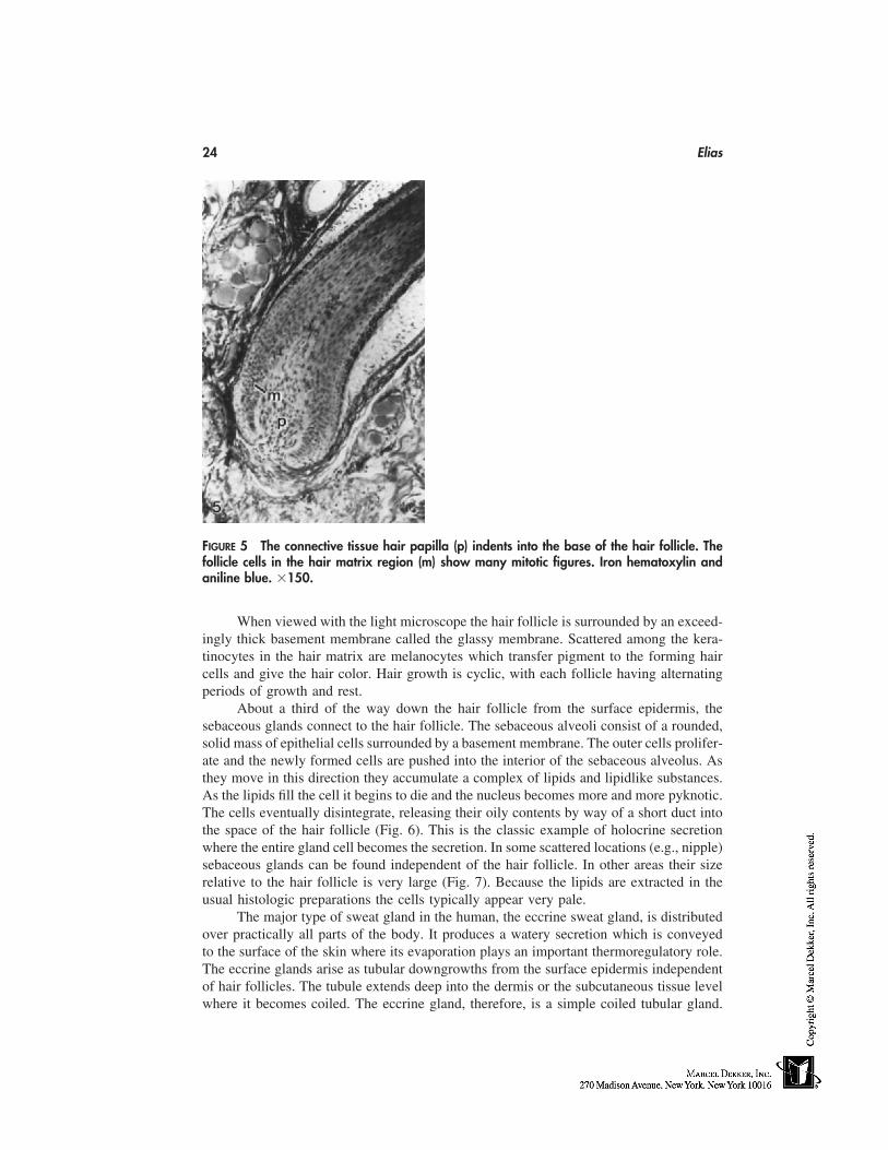
24 Elias
FIGURE 5 The connective tissue hair papilla (p) indents into the base of the hair follicle. Thefollicle cells in the hair matrix region (m) show many mitotic figures. Iron hematoxylin andaniline blue. �150.
When viewed with the light microscope the hair follicle is surrounded by an exceed-ingly thick basement membrane called the glassy membrane. Scattered among the kera-tinocytes in the hair matrix are melanocytes which transfer pigment to the forming haircells and give the hair color. Hair growth is cyclic, with each follicle having alternatingperiods of growth and rest.
About a third of the way down the hair follicle from the surface epidermis, thesebaceous glands connect to the hair follicle. The sebaceous alveoli consist of a rounded,solid mass of epithelial cells surrounded by a basement membrane. The outer cells prolifer-ate and the newly formed cells are pushed into the interior of the sebaceous alveolus. Asthey move in this direction they accumulate a complex of lipids and lipidlike substances.As the lipids fill the cell it begins to die and the nucleus becomes more and more pyknotic.The cells eventually disintegrate, releasing their oily contents by way of a short duct intothe space of the hair follicle (Fig. 6). This is the classic example of holocrine secretionwhere the entire gland cell becomes the secretion. In some scattered locations (e.g., nipple)sebaceous glands can be found independent of the hair follicle. In other areas their sizerelative to the hair follicle is very large (Fig. 7). Because the lipids are extracted in theusual histologic preparations the cells typically appear very pale.
The major type of sweat gland in the human, the eccrine sweat gland, is distributedover practically all parts of the body. It produces a watery secretion which is conveyedto the surface of the skin where its evaporation plays an important thermoregulatory role.The eccrine glands arise as tubular downgrowths from the surface epidermis independentof hair follicles. The tubule extends deep into the dermis or the subcutaneous tissue levelwhere it becomes coiled. The eccrine gland, therefore, is a simple coiled tubular gland.
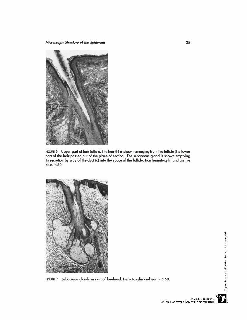
Microscopic Structure of the Epidermis 25
FIGURE 6 Upper part of hair follicle. The hair (h) is shown emerging from the follicle (the lowerpart of the hair passed out of the plane of section). The sebaceous gland is shown emptyingits secretion by way of the duct (d) into the space of the follicle. Iron hematoxylin and anilineblue. �50.
FIGURE 7 Sebaceous glands in skin of forehead. Hematoxylin and eosin. �50.
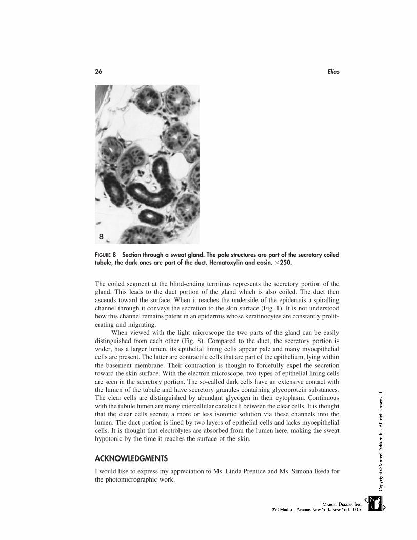
26 Elias
FIGURE 8 Section through a sweat gland. The pale structures are part of the secretory coiledtubule, the dark ones are part of the duct. Hematoxylin and eosin. �250.
The coiled segment at the blind-ending terminus represents the secretory portion of thegland. This leads to the duct portion of the gland which is also coiled. The duct thenascends toward the surface. When it reaches the underside of the epidermis a spirallingchannel through it conveys the secretion to the skin surface (Fig. 1). It is not understoodhow this channel remains patent in an epidermis whose keratinocytes are constantly prolif-erating and migrating.
When viewed with the light microscope the two parts of the gland can be easilydistinguished from each other (Fig. 8). Compared to the duct, the secretory portion iswider, has a larger lumen, its epithelial lining cells appear pale and many myoepithelialcells are present. The latter are contractile cells that are part of the epithelium, lying withinthe basement membrane. Their contraction is thought to forcefully expel the secretiontoward the skin surface. With the electron microscope, two types of epithelial lining cellsare seen in the secretory portion. The so-called dark cells have an extensive contact withthe lumen of the tubule and have secretory granules containing glycoprotein substances.The clear cells are distinguished by abundant glycogen in their cytoplasm. Continuouswith the tubule lumen are many intercellular canaliculi between the clear cells. It is thoughtthat the clear cells secrete a more or less isotonic solution via these channels into thelumen. The duct portion is lined by two layers of epithelial cells and lacks myoepithelialcells. It is thought that electrolytes are absorbed from the lumen here, making the sweathypotonic by the time it reaches the surface of the skin.
ACKNOWLEDGMENTS
I would like to express my appreciation to Ms. Linda Prentice and Ms. Simona Ikeda forthe photomicrographic work.
Microscopic Structure of the Epidermis 27
REFERENCES
1. S Banks-Schlegel, H Green. Involucrin synthesis and tissue assembly by keratinocytes in naturaland cultured human epithelia. J Cell Biol 90:732–737, 1981.
2. PM Elias. Stratum corneum lipids in health and disease. In: Progress in Diseases of the Skin,Vol. 2, R. Fleischmajer, ed. Grune and Stratton, San Diego, 1984, pp. 1–19.
3. S Grayson, PM Elias. Isolation and lipid biochemical characterization of stratum corneum mem-brane complexes: implications for the cutaneous permeability barrier. J Invest Dermatol 78:128–135, 1982.
4. RM Lavker, T Sun. Heterogeneity in epidermal basal keratinocytes: morphological and func-tional correlations. Science 215:1239–1241, 1982.
5. AM Lynley, BA Dale. The characterization of human epidermal filaggrin: a histidine-rich, kera-tin filament-aggregating protein. Biochim Biophys Acta 744:28–35, 1983.
6. T Murozuka, K Fukuyama, WL Epstein. Immunochemical comparison of histidine-rich proteinin keratohyalin granules and cornified cells. Biochim Biophys Acta 579:334–345, 1979.
7. M Simon, H Green. Participation of membrane-associated proteins in the formation of the cross-linked envelope of the keratinocyte. Cell 36:827–834, 1984.
8. PW Wertz, DT Downing. Glycolipids in mammalian epidermis: structure and function in thewater barrier. Science 217:1261–1262, 1982.
9. JG Zettergren, LL Peterson, KD Wuepper. Keratolinin: the soluble substrate of epidermal trans-glutaminase from human and bovine tissue. Proc Natl Acad Sci USA 81:238–242, 1984.

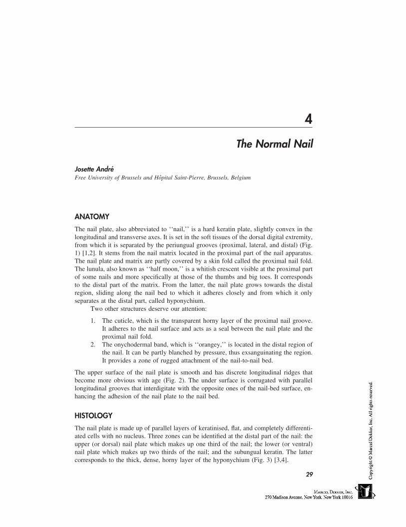
4
The Normal Nail
Josette AndréFree University of Brussels and Hôpital Saint-Pierre, Brussels, Belgium
ANATOMY
The nail plate, also abbreviated to ‘‘nail,’’ is a hard keratin plate, slightly convex in thelongitudinal and transverse axes. It is set in the soft tissues of the dorsal digital extremity,from which it is separated by the periungual grooves (proximal, lateral, and distal) (Fig.1) [1,2]. It stems from the nail matrix located in the proximal part of the nail apparatus.The nail plate and matrix are partly covered by a skin fold called the proximal nail fold.The lunula, also known as ‘‘half moon,’’ is a whitish crescent visible at the proximal partof some nails and more specifically at those of the thumbs and big toes. It correspondsto the distal part of the matrix. From the latter, the nail plate grows towards the distalregion, sliding along the nail bed to which it adheres closely and from which it onlyseparates at the distal part, called hyponychium.
Two other structures deserve our attention:
1. The cuticle, which is the transparent horny layer of the proximal nail groove.It adheres to the nail surface and acts as a seal between the nail plate and theproximal nail fold.
2. The onychodermal band, which is ‘‘orangey,’’ is located in the distal region ofthe nail. It can be partly blanched by pressure, thus exsanguinating the region.It provides a zone of rugged attachment of the nail-to-nail bed.
The upper surface of the nail plate is smooth and has discrete longitudinal ridges thatbecome more obvious with age (Fig. 2). The under surface is corrugated with parallellongitudinal grooves that interdigitate with the opposite ones of the nail-bed surface, en-hancing the adhesion of the nail plate to the nail bed.
HISTOLOGY
The nail plate is made up of parallel layers of keratinised, flat, and completely differenti-ated cells with no nucleus. Three zones can be identified at the distal part of the nail: theupper (or dorsal) nail plate which makes up one third of the nail; the lower (or ventral)nail plate which makes up two thirds of the nail; and the subungual keratin. The lattercorresponds to the thick, dense, horny layer of the hyponychium (Fig. 3) [3,4].
29
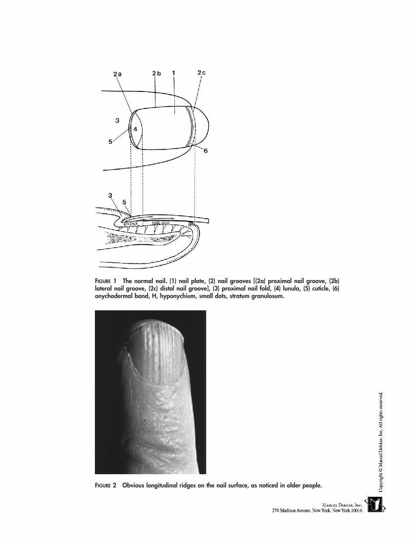
FIGURE 1 The normal nail. (1) nail plate, (2) nail grooves [(2a) proximal nail groove, (2b)lateral nail groove, (2c) distal nail groove], (3) proximal nail fold, (4) lunula, (5) cuticle, (6)onychodermal band, H, hyponychium, small dots, stratum granulosum.
FIGURE 2 Obvious longitudinal ridges on the nail surface, as noticed in older people.
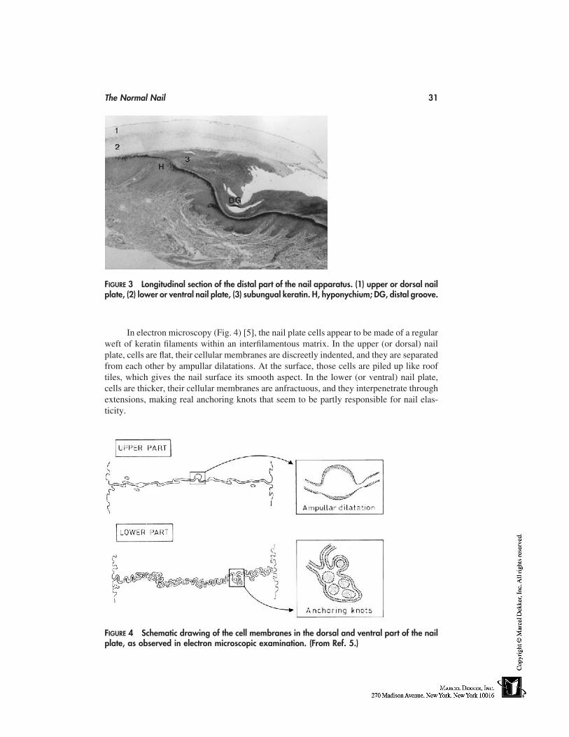
The Normal Nail 31
FIGURE 3 Longitudinal section of the distal part of the nail apparatus. (1) upper or dorsal nailplate, (2) lower or ventral nail plate, (3) subungual keratin. H, hyponychium; DG, distal groove.
In electron microscopy (Fig. 4) [5], the nail plate cells appear to be made of a regularweft of keratin filaments within an interfilamentous matrix. In the upper (or dorsal) nailplate, cells are flat, their cellular membranes are discreetly indented, and they are separatedfrom each other by ampullar dilatations. At the surface, those cells are piled up like rooftiles, which gives the nail surface its smooth aspect. In the lower (or ventral) nail plate,cells are thicker, their cellular membranes are anfractuous, and they interpenetrate throughextensions, making real anchoring knots that seem to be partly responsible for nail elas-ticity.
FIGURE 4 Schematic drawing of the cell membranes in the dorsal and ventral part of the nailplate, as observed in electron microscopic examination. (From Ref. 5.)
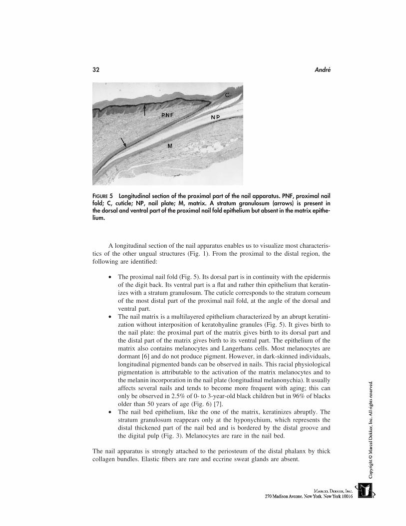
32 André
FIGURE 5 Longitudinal section of the proximal part of the nail apparatus. PNF, proximal nailfold; C, cuticle; NP, nail plate; M, matrix. A stratum granulosum (arrows) is present inthe dorsal and ventral part of the proximal nail fold epithelium but absent in the matrix epithe-lium.
A longitudinal section of the nail apparatus enables us to visualize most characteris-tics of the other ungual structures (Fig. 1). From the proximal to the distal region, thefollowing are identified:
• The proximal nail fold (Fig. 5). Its dorsal part is in continuity with the epidermisof the digit back. Its ventral part is a flat and rather thin epithelium that keratin-izes with a stratum granulosum. The cuticle corresponds to the stratum corneumof the most distal part of the proximal nail fold, at the angle of the dorsal andventral part.
• The nail matrix is a multilayered epithelium characterized by an abrupt keratini-zation without interposition of keratohyaline granules (Fig. 5). It gives birth tothe nail plate: the proximal part of the matrix gives birth to its dorsal part andthe distal part of the matrix gives birth to its ventral part. The epithelium of thematrix also contains melanocytes and Langerhans cells. Most melanocytes aredormant [6] and do not produce pigment. However, in dark-skinned individuals,longitudinal pigmented bands can be observed in nails. This racial physiologicalpigmentation is attributable to the activation of the matrix melanocytes and tothe melanin incorporation in the nail plate (longitudinal melanonychia). It usuallyaffects several nails and tends to become more frequent with aging; this canonly be observed in 2.5% of 0- to 3-year-old black children but in 96% of blacksolder than 50 years of age (Fig. 6) [7].
• The nail bed epithelium, like the one of the matrix, keratinizes abruptly. Thestratum granulosum reappears only at the hyponychium, which represents thedistal thickened part of the nail bed and is bordered by the distal groove andthe digital pulp (Fig. 3). Melanocytes are rare in the nail bed.
The nail apparatus is strongly attached to the periosteum of the distal phalanx by thickcollagen bundles. Elastic fibers are rare and eccrine sweat glands are absent.
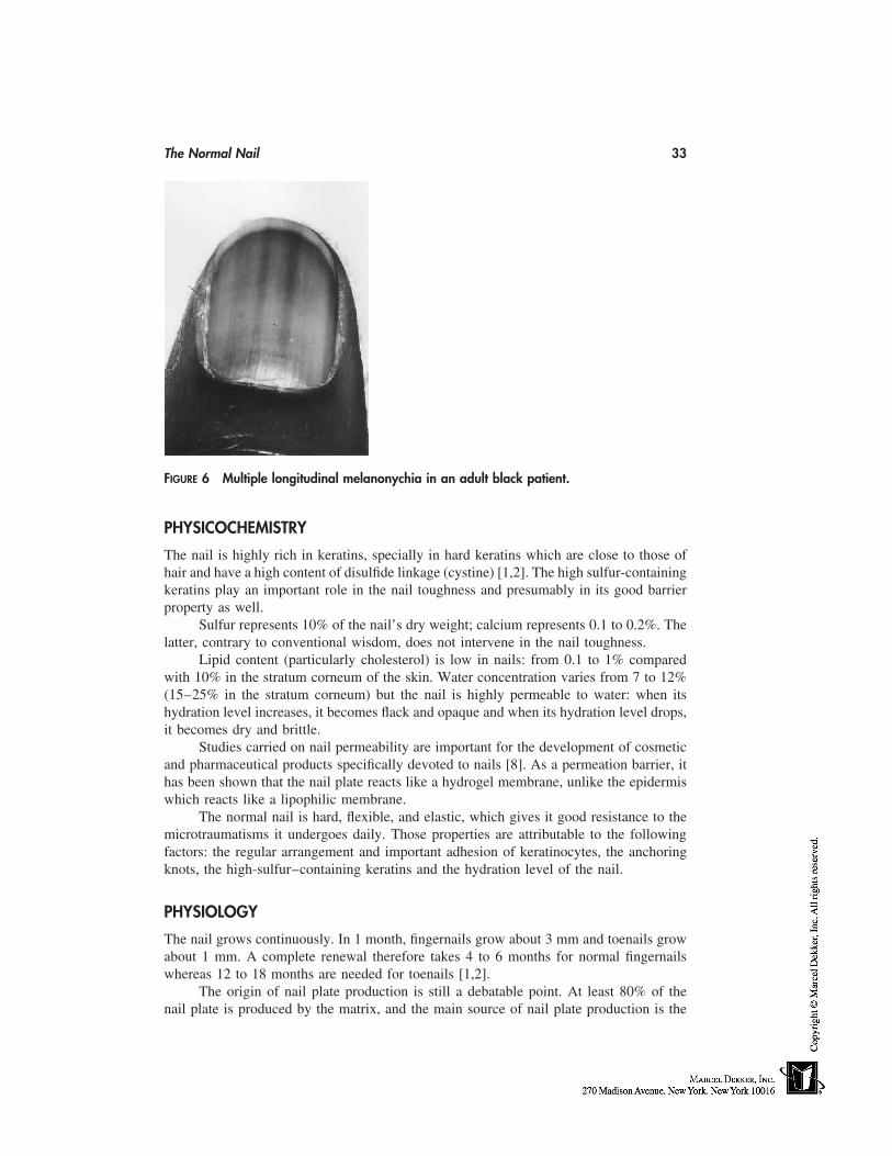
The Normal Nail 33
FIGURE 6 Multiple longitudinal melanonychia in an adult black patient.
PHYSICOCHEMISTRY
The nail is highly rich in keratins, specially in hard keratins which are close to those ofhair and have a high content of disulfide linkage (cystine) [1,2]. The high sulfur-containingkeratins play an important role in the nail toughness and presumably in its good barrierproperty as well.
Sulfur represents 10% of the nail’s dry weight; calcium represents 0.1 to 0.2%. Thelatter, contrary to conventional wisdom, does not intervene in the nail toughness.
Lipid content (particularly cholesterol) is low in nails: from 0.1 to 1% comparedwith 10% in the stratum corneum of the skin. Water concentration varies from 7 to 12%(15–25% in the stratum corneum) but the nail is highly permeable to water: when itshydration level increases, it becomes flack and opaque and when its hydration level drops,it becomes dry and brittle.
Studies carried on nail permeability are important for the development of cosmeticand pharmaceutical products specifically devoted to nails [8]. As a permeation barrier, ithas been shown that the nail plate reacts like a hydrogel membrane, unlike the epidermiswhich reacts like a lipophilic membrane.
The normal nail is hard, flexible, and elastic, which gives it good resistance to themicrotraumatisms it undergoes daily. Those properties are attributable to the followingfactors: the regular arrangement and important adhesion of keratinocytes, the anchoringknots, the high-sulfur–containing keratins and the hydration level of the nail.
PHYSIOLOGY
The nail grows continuously. In 1 month, fingernails grow about 3 mm and toenails growabout 1 mm. A complete renewal therefore takes 4 to 6 months for normal fingernailswhereas 12 to 18 months are needed for toenails [1,2].
The origin of nail plate production is still a debatable point. At least 80% of thenail plate is produced by the matrix, and the main source of nail plate production is the
34 André
proximal part of the matrix. This probably explains why distal matrix surgery or nail bedsurgery has a low potential for scarring compared with proximal matrix surgery [9]. Somestudies suggest that the nailbed produces 20% of the nail plate, whereas others suggestthat the nail bed hardly participates in the making of the nail plate [9,10].
The nail plays an important role in everyday life. It protects the distal phalanx fromtraumatisms it undergoes regularly. It plays a role in the sensitivity of the digital extremityand intervenes more specifically in the picking up of small objects such as needles. Thenail allows scratching in case of itching and can be used as a means of attack or defense.Finally, the aesthetic importance of the nail should not be neglected.
AESTHETICS
For centuries the nail has played an important aesthetic role. Having clean nails is essentialto looking well groomed and refined, and among women nails also need to be long andpainted.
A ‘‘good-looking’’ nail has a smooth and shiny surface. It is transparent and adheresto its bed. Regarding the proximal groove, the cuticle has to be intact and thin. The distaland the lateral grooves have to be clean and the periungual tissues must be without hang-nails and sores. The free border has to be smooth; its shape can be round, pointed, oval,or square. Women often wear long fingernails cut oval, which makes fingers look longerand thinner. Yet, square nails are in fashion. Too-long nails can look unpleasant and caneven be a nuisance.
Men wear short fingernails cut square. Both women and men have short toenailscut square. A normal nail structure and appropriate cosmetic care are necessary to obtainsuch ‘‘good-looking’’ nails.
REFERENCES
1. RPR Dawber, D de Berker, R Baran. Science of the nail apparatus. In: R Baran, RPR Dawber,eds. Diseases of the Nails and Their Management. 2d ed. Oxford: Blackwell Scientific Publica-tions, 1994, pp. 1–34.
2. D de Berker. The normal nail. In: J André, ed. CD-ROM: Illustrated Nail Pathology. Diagnosisand Management. Antwerpen: Lasion Europe, 1995.
3. G Achten, J André, M Laporte. Nails in light and electron microscopy. Semin Dermatol 10:54–64, 1991.
4. J André, M Laporte. Ungual histology in practice. In: J André, ed. CD-ROM: Illustrated NailPathology. Diagnosis and Management. Antwerpen: Lasion Europe, 1995.
5. D Parent, G Achten, F Stouffs-Vanhoof. Ultrastructure of the normal human nail. Am J Derma-topathol 7: 529–535, 1985.
6. Ch Perrin, JF Michiels, A Pisani, JP Ortonne. Anatomic distribution of melanocytes in normalnail unit. An immunohistochemical investigation. Am J Dermatopathol 19:462–467, 1997.
7. JJ Leyden, DA Spott, H Goldschmidt. Diffuse and banded melanin pigmentation in nails. ArchDermatol 105:548–550, 1972.
8. Y Sun, J-C Liu, JCT Wang, P De Doncker. Nail penetration. Focus on topical delivery ofantifungal drugs for onychomycosis treatment. In: RL Bronaugh, HI Maibach, eds. Percutane-ous Absorption. Drugs-Cosmetics-Mechanisms-Methodology, 3rd ed. New York: Marcel Dek-ker, 1999, pp. 759–778.
9. D de Berker, B Mawhinney, L Sviland. Quantification of regional matrix nail production. BrJ Dermatol 134:1083–1086, 1996.
10. M Johnson, S Shuster. Continuous formation of nail along the bed. Br J Dermatol 128:277–280, 1993.
5
Hair
Ghassan Shaker and Dominique Van NesteSkinterface sprl, Tournai, Belgium
INTRODUCTION
Hair is a symbol of good looks and beauty in some areas of the human body. So muchtime, effort, and money are spent in caring for it, especially in the case of scalp hair. Insome other areas, like the beard, daily care by shaving is necessary for the majority ofmales. In females, abundant scalp hair is very much welcomed, unlike leg hair, facialhair, and armpit (axillary) hair. Hair distribution in certain body regions is a secondarysex characteristic and starts to appear around puberty as the beard, moustache, and bodyhair in males, and pubic and axillary hair in both sexes.
The social meaning of hair is very important. So many old and present social and/or religious practices deal with hair. Enforced shaving of scalp hair has long been usedas a sign of punishment and in certain religious practices as a sign of obedience. TheRomans completely shaved the scalps of prisoners, adulterers, and traitors. Scalping thewarring enemies, which was long practiced by some primitive societies was meant toexpress victory and revenge [1].
Hair styling can serve as a form of expression. Rebellion of youth to the existingsocial order is often manifested as a change in appearance, and especially change of hairstyle, e.g., long hair on males, shaved hair (skinheads), and dyed hair (punks) [1].
Hair also plays a role as a distinguishing sign of one’s ethnicity, varying fromstraight to curly in form and from dark to blond in color. There is also a difference inthe amount of body hair between races. Hair is generally subject to so much interracialand interindividual variation that it can be said that, apart from the hair follicle, there isno organ in the human body that is morphologically so much variable as hair.
Although hair is not vital to human existence, it is greatly important to one’s psycho-logical equilibrium [2–4]. Psychological problems of hair loss occur in both sexes, andmore among women because of the relevance of physical attractiveness [5]. Hair is closelyrelated to physical attractiveness and the difference between male and female hair pat-terning provides a recognition phenomenon. In general, baldness leads to overestimationof age of affected males [1].
In addition to the aesthetic function of hair, it has more natural functions, whichare becoming less important because of the anthropological evolution and technical prog-
35
36 Shaker and Van Neste
ress of mankind. Scalp hair protects against certain environmental conditions like sun raysand cold. Body hair in man is very much reduced in comparison with other mammals,and many theories have been postulated to explain this fact; most are based on temperatureand thermal regulation of the human body all along the course of the evolution of mankind.Nasal hair protects against dust and acts as an air filter. Axillary and perineal hair reducethe friction during body movement and also serve for the wider or prolonged disseminationof apocrine gland odor. Pubic hair is said to have some excitatory functions during sexualintercourse.
Innumerable are the cosmetic products intended for use in hair care to remove sebumand dirt and to improve the look, shininess, uniformity, softness, color, odor, and ease ofcomb of the hair, as well as deposition of conditioning molecules and reduction of static‘‘fly-aways’’ (e.g., shampoos, conditioners, hair dyes, fixation sprays, gels, creams, etc.)There are also many products that have been marketed and used by people as anti–hairloss preparations and/or hair growth–promoting agents. Many have not stood the test oftime. Ancient medical literature is full of pharmaceutical prescriptions and formulas tobe used to treat hair loss or to promote hair growth. They are so diverse in source andnature that any attempt to categorize them seems useless.
In addition to scalp hair formulas, many other compounds are intended to removeor to assist the removal of hair from other parts of the body, e.g., preshave and aftershavepreparations, depilatories, and so on. Other products aim to decrease the contrast of hairwith the skin, making hair less visible, e.g., bleaching agents. Besides the variable efficacyof these products, consumers may develop many nonintended effects on the hair andskin such as hair damage, hair loss, skin irritation, and/or allergy and photoreactionsattributable to some active ingredients and/or their additives. In order to understand hairproduction, it is necessary to revisit the embryogenesis and to have an idea about thestructure and functional activity of the hair follicle. These aspects will now be brieflydescribed.
THE HAIR FOLLICLE
Embryology
In the early stages of hair follicle development in human fetal skin, a simultaneous differ-entiation of some epidermal and dermal cells takes place between the second and thirdmonths of intrauterine life in some areas such as the eyebrows and chin, followed by otherbody regions in the fourth month. Histologically, it begins as a crowding of cells in thebasal layer of the epidermis with a simultaneous aggregation of mesenchymal cells directlybeneath the developing epithelial component. Cells in the basal layer elongate to formthe hair peg, which grows obliquely downwards in an orientation characteristic for eachbody region. The broad tip of the hair peg will become slightly concave and carries beforeit the aggregated mesenchymal cells, which will become the dermal papilla. During thedownward course of the hair peg, two swellings appear at the posterior side of the follicle.The upper swelling will form the sebaceous gland, whereas the lower will become theinsertion site of the arrector pili muscle. In some body sites, such as the axilla, groin, skinof genitalia, and face, a third swelling is going to develop above the sebaceous gland budand this will form the apocrine gland [6–8].
Hair follicle development proceeds in a cephalocaudal direction and is completed
Hair 37
by the 22nd week of intrauterine life. These follicles progressively synthesize hair shafts(lanugo hair), which are visible at the cutaneous surface by the 28th week. The first haircoat of fine lanugo hair is shed in utero at about 1 month before birth at full term. Theshedding course follows a cephalo caudal direction, which means that frontal hair folliclesbegin their second hair cycle while occipital hair follicles are still in their first hair cycle.The second coat of lanugo hair is going to shed from all areas during the first 3 to 4months of life [6–8].
Histology
The hair follicle bulb is composed of a central dermal papilla and a surrounding hairmatrix. It undergoes many changes according to the cyclical activity of the hair folliclein health and disease. At the level of attachment of the arrector pili muscle to the follicleis the bulge zone of the root sheaths. This is considered to be the stem cell site fromwhich a new hair cycle is initiated. The hair shaft is enclosed in two sheaths, i.e., theinner root sheath and the outer root sheath. The inner root sheath consists of a cuticlelayer on the inside (next to the cuticle layer of the hair cortex), Huxley’s layer in themiddle, and Henle’s layer on the outside. The inner root sheath hardens before the pre-sumptive hair within it, and it is consequently thought to control the definitive shape ofthe hair shaft [6–8].
The outer root sheath cells have a characteristic vacuolated aspect. This sheath iscovered by the vitreous membrane. Next to this layer we can find the connective tissuesheath with its characteristic fibroblasts [6–8].
Cyclical Activity
Production of a hair segment by a hair follicle undergoes a cyclical rhythm. Activity(anagen) is followed by a relatively short transitional phase (catagen) and a resting phase(telogen) (Fig. 1). The duration of activity or anagen varies greatly with species, bodyregion, season, age, and the type of hair (i.e., terminal or vellus).
In adult humans the activity of each follicle is independent of its neighbors (asyn-chronous). However, during the development of the human embryo as well as the earlymonths of life, there is a more or less synchronous moult of scalp hairs. Each folliclegoes through the hair cycle a variable number of times in the course of a lifetime. Onaverage, at any one time about 13% of the scalp hair follicles are in telogen and only 1%or less are in catagen. Telogen ratio may count higher in certain stressful physical and/or mental conditions such as telogen effluvium and postpartum alopecia [6–8].
HAIR STRUCTURE
Postnatal hair may be divided into two broad categories: vellus hair, which is soft, unme-dullated, occasionally pigmented, and seldom exceeds 2 cm in length; and terminal, whichis longer, coarser, and often pigmented and medullated [8]. Before puberty, terminal hairis limited to the scalp, eyebrows, and eyelashes. After puberty, secondary sexual terminalhair is developed from vellus hair in response to androgens. The bulk of any hair segmentis formed mainly by the cortex, which is surrounded by a cuticle and may also have acontinuous or discontinuous core or medulla [8,9]. The medulla is usually found in thicker
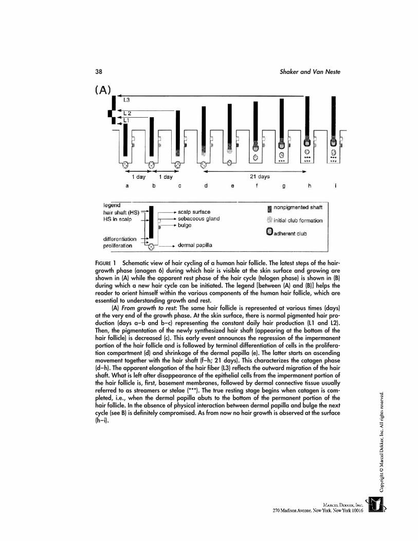
38 Shaker and Van Neste
FIGURE 1 Schematic view of hair cycling of a human hair follicle. The latest steps of the hair-growth phase (anagen 6) during which hair is visible at the skin surface and growing areshown in (A) while the apparent rest phase of the hair cycle (telogen phase) is shown in (B)during which a new hair cycle can be initiated. The legend [between (A) and (B)] helps thereader to orient himself within the various components of the human hair follicle, which areessential to understanding growth and rest.
(A) From growth to rest: The same hair follicle is represented at various times (days)at the very end of the growth phase. At the skin surface, there is normal pigmented hair pro-duction (days a–b and b–c) representing the constant daily hair production (L1 and L2).Then, the pigmentation of the newly synthesized hair shaft (appearing at the bottom of thehair follicle) is decreased (c). This early event announces the regression of the impermanentportion of the hair follicle and is followed by terminal differentiation of cells in the prolifera-tion compartment (d) and shrinkage of the dermal papilla (e). The latter starts an ascendingmovement together with the hair shaft (f–h; 21 days). This characterizes the catagen phase(d–h). The apparent elongation of the hair fiber (L3) reflects the outward migration of the hairshaft. What is left after disappearance of the epithelial cells from the impermanent portion ofthe hair follicle is, first, basement membranes, followed by dermal connective tissue usuallyreferred to as streamers or stelae (***). The true resting stage begins when catagen is com-pleted, i.e., when the dermal papilla abuts to the bottom of the permanent portion of thehair follicle. In the absence of physical interaction between dermal papilla and bulge the nextcycle (see B) is definitely compromised. As from now no hair growth is observed at the surface(h–i).
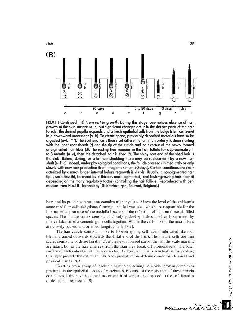
Hair 39
FIGURE 1 Continued (B) From rest to growth: During this stage, one notices absence of hairgrowth at the skin surface (a–g) but significant changes occur in the deeper parts of the hairfollicle. The dermal papilla expands and attracts epithelial cells from the bulge (stem cell zone)in a downward movement (a–b). To create space, previously deposited materials have to bedigested (a–b, ***). The epithelial cells then start differentiation in an orderly fashion startingwith the inner root sheath (c) and the tip of the cuticle and hair cortex of the newly formedunpigmented hair fiber (d). The resting hair remains in the hair follicle for approximately 1to 3 months (a–e), then the detached hair is shed (f ). The shiny root end of the shed hair isthe club. Before, during, or after hair shedding there may be replacement by a new hairshaft (e–f–g). Indeed, under physiological conditions, the follicle proceeds immediately or onlyslowly with new hair production (from f to g; maximum 90 days). Certain conditions are char-acterized by a much longer interval before regrowth is visible. Usually, a nonpigmented hairtip is seen first (h), followed by a thicker, more pigmented, and faster-growing hair fiber (i)depending on the many regulatory factors controlling the hair follicle. (Reproduced with per-mission from H.A.I.R. Technology [Skinterface sprl, Tournai, Belgium].)
hair, and its protein composition contains trichohyaline. Above the level of the epidermissome medullar cells dehydrate, forming air-filled vacuoles, which are responsible for theinterrupted appearance of the medulla because of the reflection of light on these air-filledspaces. The mature cortex consists of closely packed spindle-shaped cells separated byintercellular lamella cementing the cells together. Within the cells most of the microfibrilsare closely packed and oriented longitudinally [8,9].
The hair cuticle consists of five to 10 overlapping cell layers imbricated like rooftiles and aimed outwards (towards the distal end of the hair). The mature cells are thinscales consisting of dense keratin. Over the newly formed part of the hair the scale marginsare intact, but as the hair emerges from the skin they break off progressively. The outersurface of each cuticular cell has a very clear A-layer, which is rich in high-sulfur protein;this layer protects the cuticular cells from premature breakdown caused by chemical andphysical insults [8,9].
Keratins are a group of insoluble cystine-containing helicoidal protein complexesproduced in the epithelial tissues of vertebrates. Because of the resistance of these proteincomplexes, hairs have been said to contain hard keratins as opposed to the soft keratinsof desquamating tissues [9].
40 Shaker and Van Neste
CLINICAL HAIR-GROWTH–EVALUATION METHODS
Subjective evaluation and personal satisfaction of people using hair-growth modulatorsand/or cosmetics on a wide scale are the most important factors for the survival of theseproducts in the market. This evaluation will be based on whether they are perceived asefficacious, especially when the benefit is cosmetic in nature (acknowledging the massiveplacebo effect and the possible bias). Hence, before they reach the hands of consumers,safety and efficacy testing have to be performed according to the science, ethics, and rulesof good clinical practice and medical research in order to adequately support the claimsmade to the patient and the consumer.
For an evaluation method to be considered valuable, it should provide informationabout the following variables: hair density, which is the number of hairs per unit area(usually number/cm2); linear hair growth rate (LHGR) as millimeters per day; percentageof anagen growth phase (%A); hair diameter in micrometers; and time to hair regrowthafter completion of telogen phase [10]. For many evaluation techniques, the methodologydetails are lacking as well as information about sensitivity and reproducibility usuallyrequired for clinical investigative techniques [11]. Much effort is needed for the standard-ization of evaluation methods in order to make it possible to compare different methods,or different results from different centers using the same method. For classification pur-poses these methods can be categorized as invasive, semi-invasive, and noninvasive.
Invasive methods
Biopsy
In addition to the ordinary vertical sectioning of skin biopsies which permits the study oflongitudinal follicular sections, horizontal sectioning (parallel to the skin surface) of scalpbiopsies offers further diagnostic opportunities. First described by Headington [12], it hasbeen demonstrated that horizontal sectioning may provide a better diagnostic yield thanvertical sectioning [13,14]. Horizontal sectioning allows the study of larger number offollicular structures. Inflammatory infiltrates are more easily seen and their relationshipto the follicular structures is more obvious than in vertical sectioning. Fibrous tracts, whichare often difficult to visualise on vertical sectioning, become much more apparent onhorizontal sectioning. It is possible as well to distinguish vellus from terminal hairs, toidentify the stages of all hairs in one section and to classify them into anagen, telogen orcatagen follicles.
Semi-invasive Methods
Trichogram
The idea of estimating changes affecting hair growth by examining hair roots was firstsuggested by Van Scott et al. [15]. In order to examine hair root status necessary to diag-nose hair disorders, at least 50 hairs should be plucked in order to reduce sampling errors.The roots are examined under a low-power microscope. The root morphology is stableand hairs can be kept for many weeks in dry packaging before analysis. Due to the relativevalues generated telogen/anagen (T/A) ratio, this technique is a relatively poor indicatorof disease activity and/or disease severity in androgen-dependent alopecia in women [16].In our center this method has been abandoned because it generates only relative valuesas compared with the method described in the following section.
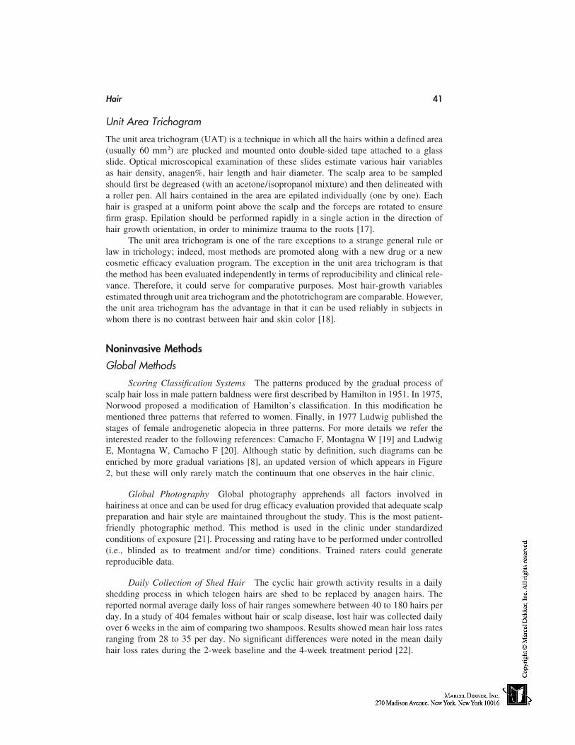
Hair 41
Unit Area Trichogram
The unit area trichogram (UAT) is a technique in which all the hairs within a defined area(usually 60 mm2) are plucked and mounted onto double-sided tape attached to a glassslide. Optical microscopical examination of these slides estimate various hair variablesas hair density, anagen%, hair length and hair diameter. The scalp area to be sampledshould first be degreased (with an acetone/isopropanol mixture) and then delineated witha roller pen. All hairs contained in the area are epilated individually (one by one). Eachhair is grasped at a uniform point above the scalp and the forceps are rotated to ensurefirm grasp. Epilation should be performed rapidly in a single action in the direction ofhair growth orientation, in order to minimize trauma to the roots [17].
The unit area trichogram is one of the rare exceptions to a strange general rule orlaw in trichology; indeed, most methods are promoted along with a new drug or a newcosmetic efficacy evaluation program. The exception in the unit area trichogram is thatthe method has been evaluated independently in terms of reproducibility and clinical rele-vance. Therefore, it could serve for comparative purposes. Most hair-growth variablesestimated through unit area trichogram and the phototrichogram are comparable. However,the unit area trichogram has the advantage in that it can be used reliably in subjects inwhom there is no contrast between hair and skin color [18].
Noninvasive Methods
Global Methods
Scoring Classification Systems The patterns produced by the gradual process ofscalp hair loss in male pattern baldness were first described by Hamilton in 1951. In 1975,Norwood proposed a modification of Hamilton’s classification. In this modification hementioned three patterns that referred to women. Finally, in 1977 Ludwig published thestages of female androgenetic alopecia in three patterns. For more details we refer theinterested reader to the following references: Camacho F, Montagna W [19] and LudwigE, Montagna W, Camacho F [20]. Although static by definition, such diagrams can beenriched by more gradual variations [8], an updated version of which appears in Figure2, but these will only rarely match the continuum that one observes in the hair clinic.
Global Photography Global photography apprehends all factors involved inhairiness at once and can be used for drug efficacy evaluation provided that adequate scalppreparation and hair style are maintained throughout the study. This is the most patient-friendly photographic method. This method is used in the clinic under standardizedconditions of exposure [21]. Processing and rating have to be performed under controlled(i.e., blinded as to treatment and/or time) conditions. Trained raters could generatereproducible data.
Daily Collection of Shed Hair The cyclic hair growth activity results in a dailyshedding process in which telogen hairs are shed to be replaced by anagen hairs. Thereported normal average daily loss of hair ranges somewhere between 40 to 180 hairs perday. In a study of 404 females without hair or scalp disease, lost hair was collected dailyover 6 weeks in the aim of comparing two shampoos. Results showed mean hair loss ratesranging from 28 to 35 per day. No significant differences were noted in the mean dailyhair loss rates during the 2-week baseline and the 4-week treatment period [22].
42 Shaker and Van Neste
FIGURE 2 Scoring of androgen-dependent alopecia (ADA) in men. The present classificationshows ADA patterns that affect the scalp of genetically susceptible male subjects after puberty.They are subdivided in six stages from mild to severe balding (1–6). The anterior pattern (A)indicates a backward progression of hair follicle miniaturization and deficient hair productionwith the ensuing bald appearance. The vertex type (V) indicates isolated regression occurringon the vertex but this is usually combined with the involvement of the frontal temporal areas.(Reproduced with permission from H.A.I.R. Technology [Skinterface sprl, Tournai, Belgium].)
Quantitating daily hair loss in women was assessed in another study of 234 womencomplaining of hair loss among which 89 had apparently normal hair density. They havefound that subjects with normally dense hair (although complaining about hair loss) shedless than 50 hairs a day [16]. So the magic number of 100 so often referred to in textbooksand found in the lay press should be seriously revisited. Less than 50 hairs can be signifi-cantly abnormal in a patient having lost 50% of his hair. Further standardization studiesare currently being run in our laboratory.
Hair Weight and Hair Count The efficacy of hair-growth–promoting agents canbe established by comparing the total hair mass (weight) and counts of grown hair in asmall, carefully maintained area of the scalp [23,24]. A plastic sheet with a 1.2 cm2 holewas placed over the selected site. All hairs within the square hole were pulled through itand hand clipped to 1 mm in length. The apparent advantage of this method is that itprovides a global measurement of growth on a small sample size for the detection of drugeffects and between treatment regimens (e.g., 2 vs. 5% minoxidil) [24]. One must be awareof the technical skills necessary to handle the samples in the proper way to avoid the lossof some hairs between the clinic and the laboratory. Again, as for many of these
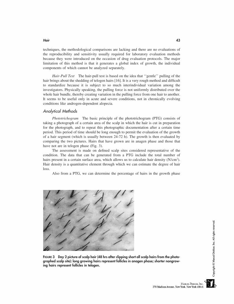
Hair 43
techniques, the methodological comparisons are lacking and there are no evaluations ofthe reproducibility and sensitivity usually required for laboratory evaluation methodsbecause they were introduced on the occasion of drug evaluation protocols. The majorlimitation of this method is that it generates a global index of growth, the individualcomponents of which cannot be analyzed separately.
Hair-Pull Test The hair-pull test is based on the idea that ‘‘gentle’’ pulling of thehair brings about the shedding of telogen hairs [16]. It is a very rough method and difficultto standardize because it is subject to so much interindividual variation among theinvestigators. Physically speaking, the pulling force is not uniformly distributed over thewhole hair bundle, thereby creating variation in the pulling force from one hair to another.It seems to be useful only in acute and severe conditions, not in chronically evolvingconditions like androgen-dependent alopecia.
Analytical Methods
Phototrichogram The basic principle of the phototrichogram (PTG) consists oftaking a photograph of a certain area of the scalp in which the hair is cut in preparationfor the photograph, and to repeat this photographic documentation after a certain timeperiod. This period of time should be long enough to permit the evaluation of the growthof a hair segment (which is usually between 24-72 h). The growth is then evaluated bycomparing the two pictures. Hairs that have grown are in anagen phase and those thathave not are in telogen phase (Fig. 3).
The assessment is made on defined scalp sites considered representative of thecondition. The data that can be generated from a PTG include the total number ofhairs present in a certain surface area, which allows us to calculate hair density (N/cm2).Hair density is a quantitative element through which we can estimate the degree of hairloss.
Also from a PTG, we can determine the percentage of hairs in the growth phase
FIGURE 3 Day 2 picture of scalp hair (48 hrs after clipping short all scalp hairs from the photo-graphed scalp site): long growing hairs represent follicles in anagen phase; shorter nongrow-ing hairs represent follicles in telogen.
44 Shaker and Van Neste
(anagen%) and can calculate the LHGR. Meanwhile, the reliability of the evaluation ofhair thickness has been the subject of detailed analysis. The most precise instrument usedfor hair-diameter evaluation remains the microscope.
One of the main advantages of the PTG is that first of all it is a patient-friendlymethod. Secondly it is a totally noninvasive method so it does not affect the natural processof hair growth/loss by itself. However, many patients are afraid of the idea of having theirhair cut at one or more given scalp surface sites (area � 1 cm2 in our protocol). Mostare reassured by the fact that this process cannot prevent them from enjoying a normalprivate and social life. Finally, PTG also permits the chronological follow-up of exactlythe same area under the study, and this has been shown to bring about a lot of valuableinformation [25]. Some technical improvements have been introduced during the courseof evolution of the PTG technique. For example, the application of a frontal window witha glass slide mounted on it has been considered a major improvement [26,27]. It reducesthe curvature of the scalp and permits a better image clarity.
Some technical photography problems have been identified during the course of theevolution of the PTG, and a series of detailed analysis performed at our laboratory andclinic have pinpointed a number of them, including the primary enlargement factor (PEF),which is one of the factors responsible for the ‘‘visibility’’ of hair on a photograph [28];the secondary enlargement factor (size of printouts); and the experience of technicians.A further improvement was the development of scalp immersion proxigraphy (SIP), whichis routinely used at our hair clinic and permits a better diffusion of light through a mediumof lower optic heterogeneity [29].
After comparison with UAT [18], weak points of the method have been consideredwith great care, and using photography in combination with hair-micrometry results in avalid method for global hair perception while allowing an analytical description of allvariables intervening in hair-quality evaluation.
Variants of PhototrichogramVideo PTG In this method, the photographic camera is replaced by a video camera
equipped with specific lenses. In fact, recent reports in which this method has been usedhave been on Asians. In these subjects the contrast between hair and scalp seems favorablefor the application of this method. Moreover, the reported low figures of hair density couldpossibly be racial in origin. However, we advise taking these factors into account in orderto keep the biological variation as low as possible [30]. The recent introduction of cheapCCD cameras will certainly contribute to further developments in this field.
Traction PTG This test is based on the fact that hairs that can be easily pulledfrom the scalp are in telogen and those resisting pull are in anagen [31]. This test hasbeen performed on a surface area of 0.25 cm2. Hairs present at this surface area are heldgently between the thumb and index fingers and pulled repeatedly. Hairs that can be eas-ily pulled are counted and their number is considered the number of telogen hairs.Those resisting pulling are clipped and counted, and their number represents the anagenhairs. Through this method, we can calculate the hair density per unit area as well as theanagen%.
It is necessary to evaluate this semi-invasive method more critically to define itsreproducibility through the standardization of the pulling technique. Other comparativestudies may be essential as well to estimate the sensitivity and specificity of this method,
Hair 45
which as it stands today would be rated as flawed with many weak points (e.g., smallsurface area, lack of control on traction forces etc.).
REFERENCES
1. Van der Donk A. Psychological aspects of androgenetic alopecia. Thesis, University of Rotter-dam, The Netherlands, 1992.
2. Passchier J. Quality of life issues in male pattern hair loss. Dermatology 1998; 197:217–218.3. Girman CJ, Rhodes T, Lilly FRW, Guo SS, Siervogel RM, Patrick DL, Chumlea WC. Effects
of self-perceived hair loss in a community sample of men. Dermatology 1998; 197:223–229.4. Cash TF. The psychological effects of androgenetic alopecia in men. J Am Acad Dermatol
1992; 26:926–931.5. Cash TF, Price VH, Savin RC. Psychological effects of androgenetic alopecia on women:
comparison with balding men and with female control subjects. J Am Acad Dermatol 1993;29:568–575.
6. Montagna W, Camacho F. The anatomy and development of hair, hair follicles, and the hairgrowth cycles. In: Camacho F, Montagna W, eds. Trichology: Diseases of the PilosebaceousFollicle. Madrid: Aula Medica Group, 1997:1–27.
7. Messenger AG, Dawber RPR. The physiology and embryology of hair growth. In: DawberR, ed. Diseases of the Hair and Scalp. 3rd ed. Oxford: Blackwell Science Ltd, 1997:1–22.
8. Dawber R, Van Neste D. Hair and Scalp Disorders. London: Martin Dunitz Ltd, 1995.9. Zviak C. The Science of Hair Care. New York: Marcel Dekker, 1986.
10. Van Neste D. Hair growth evaluation in clinical dermatology. Dermatology 1993; 187:233–234.
11. Trancik RJ. Physical methods for human hair evaluation. In: Van Neste D, Randall VA, eds.Hair Research for the Next Millenium. Amsterdam: Elsevier, 1996: 84–85.
12. Headington JT. Transverse microscopic anatomy of human scalp. Arch Dermatol 1984; 120:449–456.
13. Whiting DA. The value of horizontal sections of scalp biopsies. J Cut Aging Cosm Dermatol1990; 1:165–173.
14. Whiting DA. Diagnostic and predictive value of horizontal sections of scalp biopsy specimensin male pattern androgenetic alopecia. J Am Acad Dermatol 1993; 28:755–763.
15. Van Scott EJ, Reinerston RP, Steinmuller R. The growing hair roots of human scalp andmorphologic changes therein following amethopterin-therapy. J Invest Dermatol 1957; 29:197–204.
16. Guarrera M, Semino MT, Rebora A. Quantitating hair loss in women: a critical approach.Dermatology 1997; 194:12–16.
17. Rushton DH. Chemical and morphological properties of scalp hair in normal and abnormalstates. Ph. D. thesis, University of Wales, United Kingdom, 1988.
18. Rushton DH, de Brouwer B, De Coster W, Van Neste DJJ. Comparative evaluation of scalphair by phototrichogram and unit area trichogram analysis within the same subjects. ActaDermato Venereologica 1993; 73:150–153.
19. Camacho F, Montagna W. Current concept and classification. Male androgenetic alopecia. In:Camacho F, Montagna W, eds. Trichology. Madrid: Aula Medica Group, 1997: 325–342.
20. Ludwig E, Montagna W, Camacho F. Female androgenetic alopecia. In: Camacho F, MontagnaW, eds. Trichology. Madrid: Aula Medica Group, 1997: 343–355.
21. Canfield D. Photographic documentation of hair growth in androgenetic alopecia. DermatolClin 1996; 14:713–721.
22. Kullavanijaya P, Gritiyarangsan P, Bisalbutra P, Kulthanan R, Cardin CW. Absence of effectsof dimethicone and non-dimethicone containing shampoos on daily hair loss rates. J Soc Cos-met Chem 1992; 43:195–206.
46 Shaker and Van Neste
23. Price VH, Menefee E. Quantitative estimation of hair growth. 1. Androgenetic alopecia inwomen: effect of minoxidil. J Invest Dermatol 1990; 95:683–687.
24. Price VH, Menefee E. Quantitative estimation of hair growth: comparative changes in weightand hair count with 5% and 2% minoxidil, placebo and no treatment. In: Van Neste D, RandallVA, eds. Hair Research for the Next Millenium. Amsterdam: Elsevier, 1996: 67–71.
25. Courtois M. The phototrichogram. In: Baran R, Maibach HI, eds. Cosmetic Dermatology.London: Martin Dunitz, 1994:397–400.
26. Barth JH. Measurement of hair growth. Clin Exp Dermatol 1986; 11:127–138.27. Friedel J, Will F, Grosshans E. Le phototrichogramme. Adaptation, standardisation et applica-
tion. Ann Dermatol Vénéréol 1989; 116:629–636.28. Van Neste DJJ, de Brouwer B, De Coster W. The phototrichogram: analysis of some factors
of variation. Skin Pharmacology 1994; 7:67–72.29. Van Neste D, Dumortier M, de Brouwer B, De Coster W. Scalp immersion proxigraphy (SIP):
an improved imaging technique for phototrichogram analysis. J Eur Acad Dermatol Venereol1992; 1:187–191.
30. Hayashi S, Miayamoto I, Takeda K. Measurement of human hair growth by optical microscopyand image analysis. Brit J Dermatol 1991; 125:123–129.
31. Bouhanna P. Le tractiophototrichogramme, méthode d’appréciation objective d’une chute decheveux. Ann Dermatol Venereol 1988; 115:759–764.
6
Safety Terminology
Ai-Lean Chew and Howard I. MaibachUniversity of California at San Francisco School of Medicine,San Francisco, California
INTRODUCTION
One of the skin’s primary physiological functions is to act as the body’s first line ofdefense against exogenous agents. However, the skin should not be viewed as a flawlessphysicochemical barrier. Many low–molecular weight compounds are capable of penetrat-ing this barrier. When toxic agents (such as irritants or allergens in cosmetic products)permeate it, the resulting adverse effects may cause considerable discomfort to the con-sumer. Even minor disturbances of the skin surface can produce discomfort, especiallyin the facial area which has an extensive network of sensory nerves. Moreover, becausemost cosmetics are applied to the highly permeable facial skin, the majority of reportedcosmetic reactions occur in the face. Therefore, safety with regard to cosmetic productsis a vital issue.
This chapter provides a brief summary of the safety terminology pertaining to cos-metic reactions, as well as an overture to the succeeding chapters. The reader is directedtoward some in-depth reviews of each topic in the bibliography.
CONTACT DERMATITIS
This is a nonspecific term used to describe any inflammatory skin disease resulting fromcontact with an irritant or allergenic substance. Whatever the causative agent, the clinicalfeatures are similar: itching, redness, and skin lesions. It is also often used (inaccurately)as a synonym for allergic contact dermatitis (ACD).
IRRITANT CONTACT DERMATITIS (IRRITATION)
Irritant contact dermatitis (ICD) is a term given to a complex group of localized inflamma-tory reactions that follow nonimmunological damage to the skin. The inflammation maybe the result of an acute toxic (usually chemical) insult to the skin, or of repeated andcumulative damage from weaker irritants (chemical or physical). There is no definite labo-ratory test for ICD—diagnosis is by clinical morphology, of course, and appropriate nega-tive patch-test results.
47
48 Chew and Maibach
Irritant
An irritant is any agent, physical or chemical, that is capable of producing cell damageif applied for sufficient time and in sufficient concentration. Irritants can produce a reactionin anyone, although individual susceptibility varies. The clinical reaction produced byirritants varies considerably.
Acute Irritant Contact Dermatitis
Acute ICD is the result of a single overwhelming exposure to a strong irritant or a seriesof brief physical or chemical contacts, leading to acute inflammation of the skin. Theresultant clinical appearance is that of erythema, edema, pain, and sometimes vesiculationat the site of contact, usually associated with burning or stinging sensations.
Irritant Reaction
An irritant reaction is a transient noneczematous dermatitis characterized by erythema,chapping, or dryness, and resulting from exposure to less potent irritants. Repeated irritantreactions may lead to contact dermatitis.
Cumulative Irritant Contact Dermatitis
Cumulative irritant contact dermatitis or chronic ICD develops as a result of a series ofrepeated and damaging insults to the skin. The insults may be chemical or physical.
Delayed Acute Irritant Contact Dermatitis
Some chemicals produce acute irritation in a delayed manner so that the signs and symp-toms of acute irritant dermatitis appear 12 to 24 hours or more after the original insult.
Subjective (Sensory) Irritation
This refers to sensations of burning, stinging, and itching that are experienced by certainsusceptible individuals after contact with certain chemicals, although no visible inflamma-tory pathology can be seen. Examples of sensory irritants in cosmetics are lactic acid,salicylic acid, propylene glycol, and some benzoyl peroxide preparations.
ALLERGIC CONTACT DERMATITIS
ACD occurs when a substance comes into contact with skin that has undergone an acquiredspecific alteration in its reactivity as a result of prior exposure of the skin to the substanceeliciting the dermatitis. The skin response of ACD is delayed, immunologically mediated(Type IV), and consists of varying degrees of erythema, edema, papules, and papuloves-icles. Patch testing is the gold standard; it is imperative for proving ACD, determiningthe actual allergen, predictive testing, i.e., determining ‘‘safe’’ materials for the consumer,and exclusion of other diagnoses.
Allergen
Allergens are low–molecular-weight (�500–1000 Da) molecules capable of penetratingthe skin and binding to skin proteins to form a number of different antigens that may
Safety Terminology 49
stimulate an allergic response in an individual. Common allergens in cosmetic productsare fragrances (e.g., cinnamic aldehyde) and preservatives (e.g., formaldehyde and formal-dehyde donors).
PHOTOIRRITANT CONTACT DERMATITIS(PHOTOIRRITATION/PHOTOTOXICITY)
Photoirritant contact dermatitis (PICD) is a chemically induced nonimmunological skinirritation requiring light. This reaction will occur in all individuals exposed to the chemi-cal–light combination. The clinical picture is that of erythema, edema, or vesiculation insun-exposed areas, resembling an exaggerated sunburn. This may be followed by hyper-pigmentation, or if the exposure is repeated, scaling and lichenification may occur. Bergap-ten, a component of bergamot oil, which used to be a popular ingredient in perfume, isa potent photoirritant that causes berloque dermatitis.
PHOTOALLERGIC CONTACT DERMATITIS
Photoallergic contact dermatitis (PACD) is an immunological response to a substance thatrequires the presence of light. The substance in the skin absorbs photons and is convertedto a stable or unstable photoproduct, which binds to skin proteins to form an antigen,which then elicits a delayed hypersensitivity response. Examples of photoallergens presentin cosmetics are musk ambrette and 6-methylcoumarin, which are present in fragrances.Photopatch testing is the diagnostic procedure for photoallergy.
CONTACT URTICARIA SYNDROME
Contact urticaria syndrome (CUS) represents a heterogeneous group of inflammatory reac-tions that appear, usually within a few minutes to an hour, after contact with the elicitingsubstance. Clinically, erythematous wheal-and-flare reactions are seen, and sensations ofburning, stinging, or itching are experienced. These are transient, usually disappearingwithin a few hours. In its more severe forms, generalized urticaria or extracutaneous mani-festations, such as asthma, nausea, abdominal cramps, and even anaphylactic shock, mayoccur. Diagnosis may be achieved by a variety of skin tests—the open test is the simplestof these and is the ‘‘first-line’’ test.
CUS may be divided into two categories on the basis of pathophysiological mecha-nisms: nonimmunological and immunological. There are also urticariogens that act by anuncertain mechanism.
Nonimmunological Contact Urticaria
Nonimmunological contact urticaria (NICU), which occurs without prior sensitization, isthe most common class of CUS. The reaction usually remains localized. Examples ofcosmetic substances known to produce NICU are preservatives (e.g., benzoic acid andsorbic acid) and fragrances (e.g., cinnamic aldehyde).
Immunological Contact Urticaria
Immunological contact urticaria (ICU) are immediate (Type I) allergic reactions in peoplewho have previously been sensitized to the causative agent. ICU is IgE mediated and ismore common in atopic individuals. Food substances are common causes of ICU.
50 Chew and Maibach
ACNEGENICITY
This refers to the capacity of some agents to cause acne or aggravate existing acne lesions.This term may be subdivided to include comedogenicity and pustulogenicity.
Comedogenicity
This is the capability of an agent to cause hyperkeratinous impactions in the sebaceousfollicle, or the formation of microcomedones, usually in a relatively short period of time.
Pustulogenicity
This refers to the capability of an agent to cause inflammatory papules and pustules, usu-ally in a relatively short period of time.
SENSITIVE SKIN
This term is a neologism for consumers’ feelings about their intolerance to a variety oftopical agents, be it topical medicaments or cosmetics and toiletries. Individuals presentwith very similar complaints, such as burning, stinging or itching sensations, on contactwith certain cosmetic products that most people do not seem to react to, sometimes accom-panied by slight erythema or edema. They frequently complain of a ‘‘tight feeling’’ intheir skin, secondary to associated dry skin. Sensitive skin describes the phenotype notedby the consumer; mechanisms include sensory irritation, suberythematous irritation, acuteand cumulative irritation, contact urticaria, allergic contact dermatitis, as well as photoal-lergic and phototoxic contact dermatitis. Sensory irritation and suberythematous irritationare believed to be far more common than the remaining mechanisms.
Cosmetic Intolerance Syndrome
The term cosmetic intolerance syndrome (CIS) is applied to the multifactorial syndromein which certain susceptible individuals are intolerant of a wide range of cosmetic prod-ucts. CIS is thought to be caused by one or more underlying occult dermatological condi-tions, such as subjective irritation, objective irritation, allergic contact dermatitis, contacturticaria, or subtle manifestations of endogenous dermatological diseases, such as atopiceczema, psoriasis, and rosacea.
Status Cosmeticus
Status cosmeticus is a condition in which every cosmetic product applied to the faceproduces itching, burning or stinging, rendering the sufferer incapable of using any cos-metic product. The patient’s history usually includes ‘‘sensitivity’’ to a wide range ofproducts. This diagnosis is only declared after a full battery of tests have proved negative,and may be considered the extreme end of the spectrum of sensitive skin.
BIBLIOGRAPHY
Irritant Contact Dermatitis
Elsner P, Maibach HI, eds. Irritant Dermatitis: New Clinical and Experimental Aspects. CurrentProblems in Dermatology Series, Vol. 23, Basel; Karger, 1995.
Safety Terminology 51
Lammintausta K, Maibach HI. Irritant contact dermatitis. In: Moschella SL, Hurley HJ, eds. Derma-tology, 3rd edition. Philadelphia; W.B. Saunders Company, 1992:425–432.
Van Der Valk PGM, Maibach HI. The Irritant Contact Dermatitis Syndrome. Boca Raton: CRCPress, 1996.
Wilkinson JD, Rycroft RJG. Contact dermatitis. In: Champion RH, Burton JL, Ebling FJG, eds.Rook/Wilkinson/Ebling Textbook of Dermatology, 5th edition. Oxford; Blackwell ScientificPublications, 1992:611.
Allergic Contact Dermatitis
Cronin E. Contact Dermatitis. Edinburgh; Churchill Livingstone, 1980.Larsen WG, Maibach HI. Allergic contact dermatitis. In: Moschella SL, Hurley HJ, eds. Dermatol-
ogy, 3rd edition. Philadelphia; W.B. Saunders Company, 1992; 17:391–424.Rietschel RL, Fowler JF Jr, eds. Fisher’s Contact Dermatitis, 4th edition. Williams & Baltimore;
Williams and Wilkins, 1995.
Phototoxic/Photoallergic Contact Dermatitis
DeLeo VA, Maso MJ. In: Moschella SL, Hurley HJ, eds. Dermatology, 3rd edition. Philadelphia:W.B. Saunders Company, 1992:507.
Harber LC, Bickers DR, eds. In: Photosensitivity Diseases: Principles of Diagnosis and Treatment,2nd edition. Ontario; BC Decker Inc. 1989.
Marzulli FN, Maibach HI. Photoirritation (phototoxicity, phototoxic dermatitis). In: Dermatotoxicol-ogy, 5th edition. Washington, DC: Taylor & Francis, 1996; 231–237.
Contact Urticaria Syndrome
Amin S, Lahti A, Maibach HI. Contact Urticaria Syndrome. Boca Raton: CRC Press, 1997.Lahti A, Maibach HI. Contact Urticaria Syndrome. In: Moschella SL, Hurley HJ, eds. Dermatology,
3rd edition. Philadelphia; W.B. Saunders Company, 1992, 19:433.
Acnegenicity
Mills OH Jr, Berger RS. Defining the susceptibility of acne-prone and sensitive skin populationsto extrinsic factors. Dermatologic Clinics, 1991; 9(1):93–98.
Sensitive Skin
Amin S, Engasser P, Maibach HI. Sensitive skin: what is it? In: Baran R, Maibach HI. Textbookof Cosmetic Dermatology, 2nd edition. London; Martin Dunitz Ltd, 1998; 343–349.
Fisher AA. Cosmetic actions and reactions: Therapeutic, irritant and allergic. Cutis 1980; 26:22–29.
Maibach HI, Engasser P. Management of cosmetic intolerance syndrome. Clin Dermatol 1988; 6(3):102–107.

7
Principles and Practice of Percutaneous Absorption
Ronald C. Wester and Howard I. MaibachUniversity of California at San Francisco School of Medicine,San Francisco, California
INTRODUCTION
Percutaneous absorption is a complex biological process. The skin is a multilayered bio-membrane that has certain absorption characteristics. If the skin were a simple membrane,absorption parameters could easily be measured, and these would be fairly constant pro-vided there was no change in the chemistry of the membrane. However, skin is a dynamictissue and as such its absorption parameters are susceptible to constant change. Manyfactors and skin conditions can rapidly change the absorption parameters. Additionally,skin is a living tissue and it will change through its own growth patterns, and this changewill also be influenced by many factors. This chapter reviews some of the principles andtechnologies of percutaneous absorption for developers and users of cosmetics.
STEPS TO PERCUTANEOUS ABSORPTION
A cosmetic that comes in contact with human skin will be absorbed into and through theskin. The components of the cosmetic will respond to the chemical and physical lawsof nature, which direct the absorption process. Examples of this are solubility, partitioncoefficients, and molecular weight. The skin presents a barrier, both physical structureand chemical composition. A cosmetic component will transverse from a lipophilic stratumcorneum to a more progressively hydrophilic epidermis, dermis, and blood microcircula-tion.
Percutaneous absorption has been defined as a series of steps [1]. Table 1 lists ourcurrent knowledge of these steps. Step 1 is the vehicle containing the chemical(s) of inter-est. There is a partitioning of the chemical from the vehicle to the skin. This initiates aseries of absorption and excretion kinetics that are influenced by a variety of factors, suchas regional and individual variation. These factors moderate the absorption and excretionkinetics [2].
Once a chemical has been absorbed through the skin, it enters the systemic circula-tion of the body. Here, the pharmacokinetics of the chemical define body interactions.This is illustrated for [14C]hydroquinone in vivo in man, where plasma radioactivity wasmeasured ipsilaterally (next to the dose site) and contralaterally (in the opposite arm) aftera topical dose. Thirty minutes after the dose, the hydroquinone has been absorbed throughthe skin and has reached a near-peak plasma concentration (Fig. 1) [3]. Figure 2 shows
53
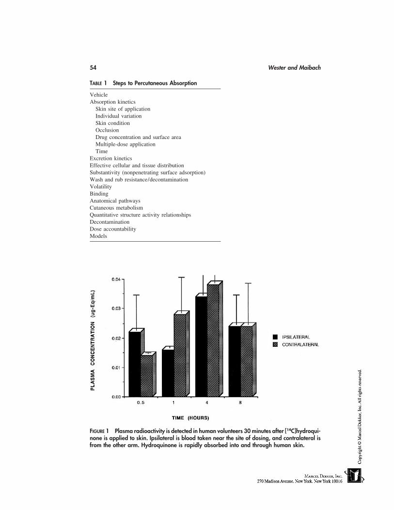
54 Wester and Maibach
TABLE 1 Steps to Percutaneous Absorption
VehicleAbsorption kinetics
Skin site of applicationIndividual variationSkin conditionOcclusionDrug concentration and surface areaMultiple-dose applicationTime
Excretion kineticsEffective cellular and tissue distributionSubstantivity (nonpenetrating surface adsorption)Wash and rub resistance/decontaminationVolatilityBindingAnatomical pathwaysCutaneous metabolismQuantitative structure activity relationshipsDecontaminationDose accountabilityModels
FIGURE 1 Plasma radioactivity is detected in human volunteers 30 minutes after [14C]hydroqui-none is applied to skin. Ipsilateral is blood taken near the site of dosing, and contralateral isfrom the other arm. Hydroquinone is rapidly absorbed into and through human skin.
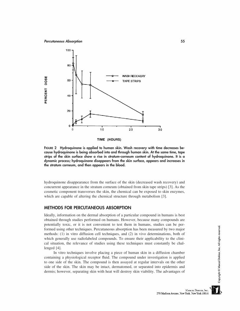
Percutaneous Absorption 55
FIGURE 2 Hydroquinone is applied to human skin. Wash recovery with time decreases be-cause hydroquinone is being absorbed into and through human skin. At the same time, tapestrips of the skin surface show a rise in stratum-corneum content of hydroquinone. It is adynamic process; hydroquinone disappears from the skin surface, appears and increases inthe stratum corneum, and then appears in the blood.
hydroquinone disappearance from the surface of the skin (decreased wash recovery) andconcurrent appearance in the stratum corneum (obtained from skin tape strips) [3]. As thecosmetic component transverses the skin, the chemical can be exposed to skin enzymes,which are capable of altering the chemical structure through metabolism [3].
METHODS FOR PERCUTANEOUS ABSORPTION
Ideally, information on the dermal absorption of a particular compound in humans is bestobtained through studies performed on humans. However, because many compounds arepotentially toxic, or it is not convenient to test them in humans, studies can be per-formed using other techniques. Percutaneous absorption has been measured by two majormethods: (1) in vitro diffusion cell techniques, and (2) in vivo determinations, both ofwhich generally use radiolabeled compounds. To ensure their applicability to the clini-cal situation, the relevance of studies using these techniques must constantly be chal-lenged [4].
In vitro techniques involve placing a piece of human skin in a diffusion chambercontaining a physiological receptor fluid. The compound under investigation is appliedto one side of the skin. The compound is then assayed at regular intervals on the otherside of the skin. The skin may be intact, dermatomed, or separated into epidermis anddermis; however, separating skin with heat will destroy skin viability. The advantages of
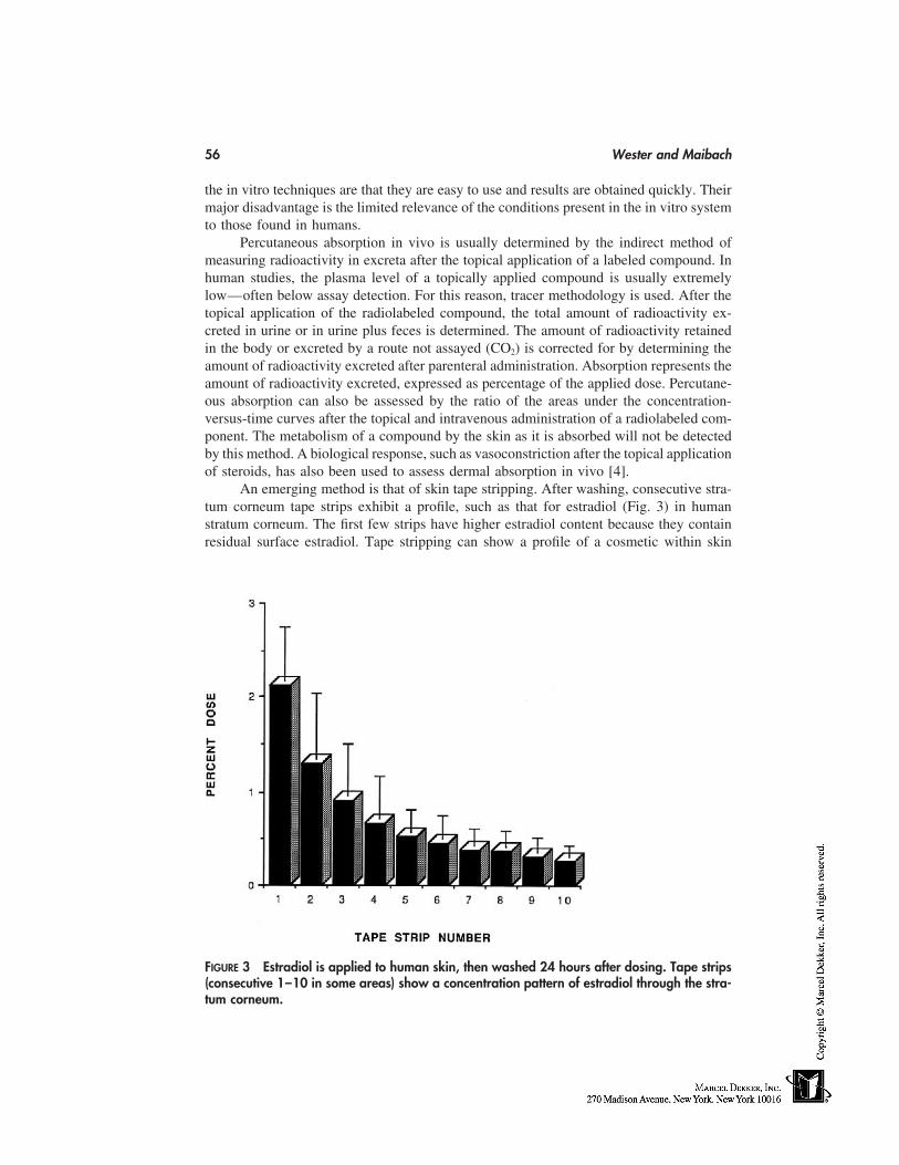
56 Wester and Maibach
the in vitro techniques are that they are easy to use and results are obtained quickly. Theirmajor disadvantage is the limited relevance of the conditions present in the in vitro systemto those found in humans.
Percutaneous absorption in vivo is usually determined by the indirect method ofmeasuring radioactivity in excreta after the topical application of a labeled compound. Inhuman studies, the plasma level of a topically applied compound is usually extremelylow—often below assay detection. For this reason, tracer methodology is used. After thetopical application of the radiolabeled compound, the total amount of radioactivity ex-creted in urine or in urine plus feces is determined. The amount of radioactivity retainedin the body or excreted by a route not assayed (CO2) is corrected for by determining theamount of radioactivity excreted after parenteral administration. Absorption represents theamount of radioactivity excreted, expressed as percentage of the applied dose. Percutane-ous absorption can also be assessed by the ratio of the areas under the concentration-versus-time curves after the topical and intravenous administration of a radiolabeled com-ponent. The metabolism of a compound by the skin as it is absorbed will not be detectedby this method. A biological response, such as vasoconstriction after the topical applicationof steroids, has also been used to assess dermal absorption in vivo [4].
An emerging method is that of skin tape stripping. After washing, consecutive stra-tum corneum tape strips exhibit a profile, such as that for estradiol (Fig. 3) in humanstratum corneum. The first few strips have higher estradiol content because they containresidual surface estradiol. Tape stripping can show a profile of a cosmetic within skin
FIGURE 3 Estradiol is applied to human skin, then washed 24 hours after dosing. Tape strips(consecutive 1–10 in some areas) show a concentration pattern of estradiol through the stra-tum corneum.
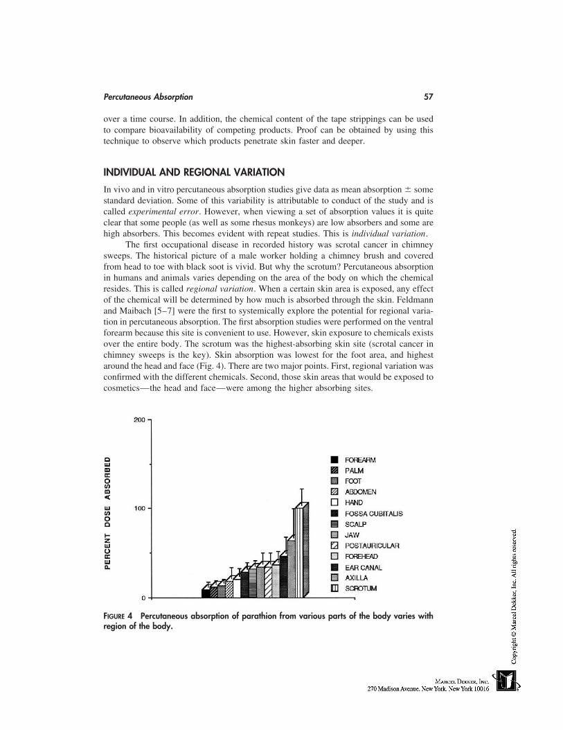
Percutaneous Absorption 57
over a time course. In addition, the chemical content of the tape strippings can be usedto compare bioavailability of competing products. Proof can be obtained by using thistechnique to observe which products penetrate skin faster and deeper.
INDIVIDUAL AND REGIONAL VARIATION
In vivo and in vitro percutaneous absorption studies give data as mean absorption � somestandard deviation. Some of this variability is attributable to conduct of the study and iscalled experimental error. However, when viewing a set of absorption values it is quiteclear that some people (as well as some rhesus monkeys) are low absorbers and some arehigh absorbers. This becomes evident with repeat studies. This is individual variation.
The first occupational disease in recorded history was scrotal cancer in chimneysweeps. The historical picture of a male worker holding a chimney brush and coveredfrom head to toe with black soot is vivid. But why the scrotum? Percutaneous absorptionin humans and animals varies depending on the area of the body on which the chemicalresides. This is called regional variation. When a certain skin area is exposed, any effectof the chemical will be determined by how much is absorbed through the skin. Feldmannand Maibach [5–7] were the first to systemically explore the potential for regional varia-tion in percutaneous absorption. The first absorption studies were performed on the ventralforearm because this site is convenient to use. However, skin exposure to chemicals existsover the entire body. The scrotum was the highest-absorbing skin site (scrotal cancer inchimney sweeps is the key). Skin absorption was lowest for the foot area, and highestaround the head and face (Fig. 4). There are two major points. First, regional variation wasconfirmed with the different chemicals. Second, those skin areas that would be exposed tocosmetics—the head and face—were among the higher absorbing sites.
FIGURE 4 Percutaneous absorption of parathion from various parts of the body varies withregion of the body.
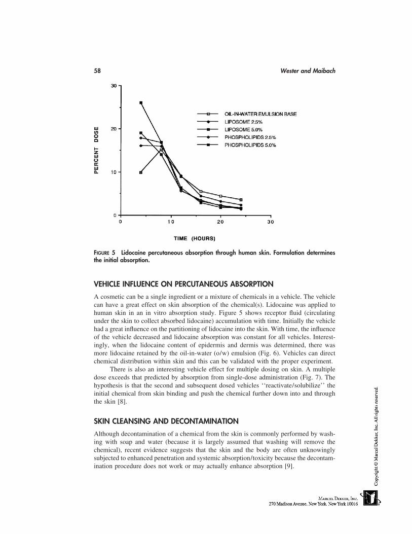
58 Wester and Maibach
FIGURE 5 Lidocaine percutaneous absorption through human skin. Formulation determinesthe initial absorption.
VEHICLE INFLUENCE ON PERCUTANEOUS ABSORPTION
A cosmetic can be a single ingredient or a mixture of chemicals in a vehicle. The vehiclecan have a great effect on skin absorption of the chemical(s). Lidocaine was applied tohuman skin in an in vitro absorption study. Figure 5 shows receptor fluid (circulatingunder the skin to collect absorbed lidocaine) accumulation with time. Initially the vehiclehad a great influence on the partitioning of lidocaine into the skin. With time, the influenceof the vehicle decreased and lidocaine absorption was constant for all vehicles. Interest-ingly, when the lidocaine content of epidermis and dermis was determined, there wasmore lidocaine retained by the oil-in-water (o/w) emulsion (Fig. 6). Vehicles can directchemical distribution within skin and this can be validated with the proper experiment.
There is also an interesting vehicle effect for multiple dosing on skin. A multipledose exceeds that predicted by absorption from single-dose administration (Fig. 7). Thehypothesis is that the second and subsequent dosed vehicles ‘‘reactivate/solubilize’’ theinitial chemical from skin binding and push the chemical further down into and throughthe skin [8].
SKIN CLEANSING AND DECONTAMINATION
Although decontamination of a chemical from the skin is commonly performed by wash-ing with soap and water (because it is largely assumed that washing will remove thechemical), recent evidence suggests that the skin and the body are often unknowinglysubjected to enhanced penetration and systemic absorption/toxicity because the decontam-ination procedure does not work or may actually enhance absorption [9].
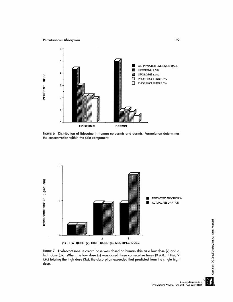
Percutaneous Absorption 59
FIGURE 6 Distribution of lidocaine in human epidermis and dermis. Formulation determinesthe concentration within the skin component.
FIGURE 7 Hydrocortisone in cream base was dosed on human skin as a low dose (x) and ahigh dose (3x). When the low dose (x) was dosed three consecutive times (9 A.M., 1 P.M., 9P.M.) totaling the high dose (3x), the absorption exceeded that predicted from the single highdose.
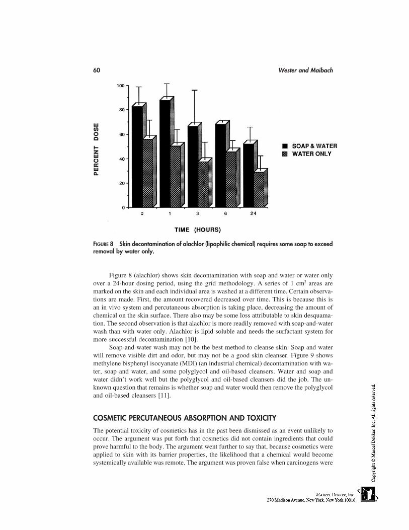
60 Wester and Maibach
FIGURE 8 Skin decontamination of alachlor (lipophilic chemical) requires some soap to exceedremoval by water only.
Figure 8 (alachlor) shows skin decontamination with soap and water or water onlyover a 24-hour dosing period, using the grid methodology. A series of 1 cm2 areas aremarked on the skin and each individual area is washed at a different time. Certain observa-tions are made. First, the amount recovered decreased over time. This is because this isan in vivo system and percutaneous absorption is taking place, decreasing the amount ofchemical on the skin surface. There also may be some loss attributable to skin desquama-tion. The second observation is that alachlor is more readily removed with soap-and-waterwash than with water only. Alachlor is lipid soluble and needs the surfactant system formore successful decontamination [10].
Soap-and-water wash may not be the best method to cleanse skin. Soap and waterwill remove visible dirt and odor, but may not be a good skin cleanser. Figure 9 showsmethylene bisphenyl isocyanate (MDI) (an industrial chemical) decontamination with wa-ter, soap and water, and some polyglycol and oil-based cleansers. Water and soap andwater didn’t work well but the polyglycol and oil-based cleansers did the job. The un-known question that remains is whether soap and water would then remove the polyglycoland oil-based cleansers [11].
COSMETIC PERCUTANEOUS ABSORPTION AND TOXICITY
The potential toxicity of cosmetics has in the past been dismissed as an event unlikely tooccur. The argument was put forth that cosmetics did not contain ingredients that couldprove harmful to the body. The argument went further to say that, because cosmetics wereapplied to skin with its barrier properties, the likelihood that a chemical would becomesystemically available was remote. The argument was proven false when carcinogens were
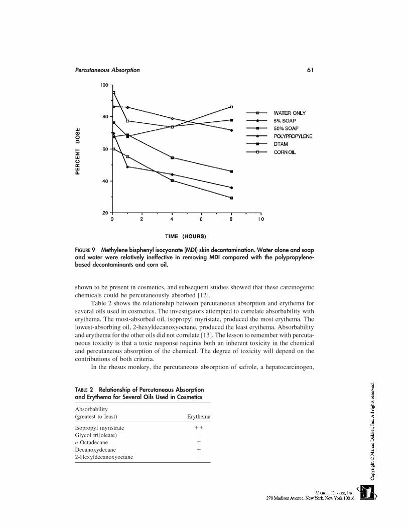
Percutaneous Absorption 61
FIGURE 9 Methylene bisphenyl isocyanate (MDI) skin decontamination. Water alone and soapand water were relatively ineffective in removing MDI compared with the polypropylene-based decontaminants and corn oil.
shown to be present in cosmetics, and subsequent studies showed that these carcinogenicchemicals could be percutaneously absorbed [12].
Table 2 shows the relationship between percutaneous absorption and erythema forseveral oils used in cosmetics. The investigators attempted to correlate absorbability witherythema. The most-absorbed oil, isopropyl myristate, produced the most erythema. Thelowest-absorbing oil, 2-hexyldecanoxyoctane, produced the least erythema. Absorbabilityand erythema for the other oils did not correlate [13]. The lesson to remember with percuta-neous toxicity is that a toxic response requires both an inherent toxicity in the chemicaland percutaneous absorption of the chemical. The degree of toxicity will depend on thecontributions of both criteria.
In the rhesus monkey, the percutaneous absorption of safrole, a hepatocarcinogen,
TABLE 2 Relationship of Percutaneous Absorptionand Erythema for Several Oils Used in Cosmetics
Absorbability(greatest to least) Erythema
Isopropyl myristrate ��Glycol tri(oleate) �n-Octadecane �Decanoxydecane �2-Hexyldecanoxyoctane �
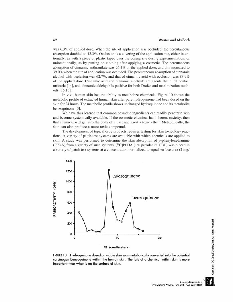
62 Wester and Maibach
was 6.3% of applied dose. When the site of application was occluded, the percutaneousabsorption doubled to 13.3%. Occlusion is a covering of the application site, either inten-tionally, as with a piece of plastic taped over the dosing site during experimentation, orunintentionally, as by putting on clothing after applying a cosmetic. The percutaneousabsorption of cinnamic anthranilate was 26.1% of the applied dose, and this increased to39.0% when the site of application was occluded. The percutaneous absorption of cinnamicalcohol with occlusion was 62.7%, and that of cinnamic acid with occlusion was 83.9%of the applied dose. Cinnamic acid and cinnamic aldehyde are agents that elicit contacturticaria [14], and cinnamic aldehyde is positive for both Draize and maximization meth-ods [15,16].
In vivo human skin has the ability to metabolize chemicals. Figure 10 shows themetabolic profile of extracted human skin after pure hydroquinone had been dosed on theskin for 24 hours. The metabolic profile shows unchanged hydroquinone and its metabolitebenzoquinone [3].
We have thus learned that common cosmetic ingredients can readily penetrate skinand become systemically available. If the cosmetic chemical has inherent toxicity, thenthat chemical will get into the body of a user and exert a toxic effect. Metabolically, theskin can also produce a more toxic compound.
The development of topical drug products requires testing for skin toxicology reac-tions. A variety of patch-test systems are available with which chemicals are applied toskin. A study was performed to determine the skin absorption of p-phenylenediamine(PPDA) from a variety of such systems. [14C]PPDA (1% petrolatum UDP) was placed ina variety of patch-test systems at a concentration normalized to equal surface area (2 mg/
FIGURE 10 Hydroquinone dosed on viable skin was metabolically converted into the potentialcarcinogen benzoquinone within the human skin. The fate of a chemical within skin is moreimportant than what is on the surface of skin.
Percutaneous Absorption 63
TABLE 3 Percutaneous Absorption of p-Phenylenediamine (PPDA) from Patch-Test Systems
Total load Concentration Absorptionin chamber in chamber
(mg) (mg/mm2) Percent* Total (mg)
Hill Top chamber 40 2 53.4 � 20.6 21.4Teflon (control) 16 2 48.6 � 9.3 7.8Small Finn chamber 16 2 29.8 � 9.0 4.8Large Finn chamber 24 2 23.1 � 7.3 5.5AL-test chamber 20 2 8.0 � 0.8 1.6Small Finn chamber
with paper disc insert 16 2 34.1 � 19.8 5.5
* Each value is the mean � standard deviation for three guinea pigs.
mm2). Skin absorption was determined in the guinea pig by urinary excretion of 14C. Therewas a sixfold difference in the range of skin absorption (p � 0.02). In decreasing order,the percentage skin absorption from the systems were 53.4 � 20.6 (Hill Top chamber),48.6 � 9.3 (Teflon control patch), 23.1 � 7.3 (small Finn chamber), and 8.0 � 0.8 (AL-test chamber). Thus, the choice of patch system could produce a false-negative error ifthe system inhibits skin absorption, with a subsequent toxicology reaction (Table 3) [17].
COSMECEUTICS
The early concept of cosmetics was one of inert ingredients used as coloring or coveragents to enhance visual appearance. There was no concern with systemic toxicity becauseskin had barrier properites and it was assumed nothing would permeate across the skin.The line between cosmetics and pharmaceutics has become a gray area as more activeagents are incorporated into cosmetics. These active agents are referred to as cosmeceutics.Hydroquinone when prescribed by a physician is a drug. Hydroquinone in a cosmetic asa lightening agent is not a drug. The only differentiation between the two preparations isthe hydroquinone concentration in the preparation. However, applied concentration doesnot matter; what matters is how much of the hydroquinone gets into and through theskin. For hydroquinone, percutaneous absorption is 45% of the applied dose for a 24-hourapplication to in vivo human skin [3]. That is a lot of drug—or is it cosmetic, or cosmeceu-tic? The important point is that for active chemicals the bioavailability needs to be knownto assess risk assessment.
Another example is α-tocopherol, or vitamin E [18]. The biological activities ofvitamin E in cosmetics are supported by several studies of its percutaneous absorption.In data obtained in vitro on rat skin 6 hours after application of a 5% vitamin E alcoholsolution, 38.6% of the applied dose was recovered in the viable epidermis and dermis.The amount detected in the horny layer was 7.12%, and the residual fraction persistingon the surface on the integument represented 54.3% of the applied dose. Both the alcoholand acetate forms of vitamin E are readily absorbed through the human scalp, and within6 to 24 hours after treatment they concentrate in the dermis. These results substantiatethe claim that vitamin E can be used as an active ingredient in cosmetology with thepossibility of efficacy in the deeper structures of the skin. Table 4 summarizes the invitro percutaneous absorption of vitamin E acetate into and through human skin. Each
64 Wester and Maibach
TABLE 4 In Vitro Percutaneous Absorption of Vitamin E Acetate Into and Through HumanSkin
Percent dose absorbed
Treatment Receptor fluid Skin content Surface wash
Formula ASkin source 1 0.34 0.55 74.9Skin source 2 0.39 0.66 75.6Skin source 3 0.47 4.08 89.1Skin source 4 1.30 0.96 110.0
Mean � SD 0.63 � 0.45* 1.56 � 1.69† 87.4 � 16.4
Formula BSkin source 1 0.24 0.38 —Skin source 2 0.40 0.64 107.1Skin source 3 0.41 4.80 98.1Skin source 4 2.09 1.16 106.2
Mean � SD 0.78 � 0.87* 1.74 � 2.06† 103.8 � 5.0
* p � 0.53 (nonsignificant; paired t-test).† p � 0.42 (nonsignificant; paired t-test).
formulation was tested in four different human skin sources. The percent dose absorbedfor a 24-hour dosing period is given for receptor-fluid accumulation (absorbed), skin con-tent, and surface wash (soap-and-water wash recovery after the 24-hour dosing period).
Table 4 also contains what is referred to as material balance. All of the applieddose is accounted for in the receptor fluid, skin content, and skin-surface wash. Totalabsorbed dose would be the sum of that in the receptor fluid plus that in the skin (content).This is an example of a complete in vitro percutaneous absorption study.
DISCUSSION
The concepts of cosmetics and of the skin have undergone changes in the last few decades.Cosmetics have evolved from being formulations of inert ingredients to containing ingredi-ents that have some biological activity directed to living skin. This is sometimes referredto as cosmeceutics. The concept of skin has evolved from an impenetrable barrier to onewhere percutaneous absorption does occur. Risk assessment requires a knowledge of per-cutaneous absorption so that health is not jeopardized. This applies to any topically appliedchemical, be it cosmetic, pharmaceutic, industrial, or environmental.
REFERENCES
1. Wester RC, Maibach HI. Cutaneous pharmacokinetics: 10 steps to percutaneous absorption.Drug Metab Rev 14:169–205, 1983.
2. Wester RC, Maibach HI. Percutaneous absorption of drugs. Clin Pharmacokin 23:253–266,1992.
3. Wester RC, Melendres J, Hui X, Wester RM, Serranzana S, Zhai H, Quan D, Maibach HI.Human in vivo and in vitro hydroquinone topical bioavailability. J Toxicol Environ Health54:301–317, 1998.
Percutaneous Absorption 65
4. Wester RC, Maibach HI. Toxicokinetics: dermal exposure and absorption of toxicants. In:Bond J, ed. Comparative Toxicology, Vol. 1, General Principles. New York: Elsevier Sciences,1997:99–114.
5. Feldmann RJ, Maibach HI. Percutaneous penetration of steroids in man. J Invest Dermatol542:89–94, 1969.
6. Feldmann RJ, Maibach HI. Absorption of some organic compounds through the skin in man.J Invest Dermatol 54:399–404, 1970.
7. Feldmann RJ, Maibach HI. Percutaneous penetration of some pesticides and herbicides inman. Toxicol Appl Pharmacol 28:126–132, 1974.
8. Wester RC, Melendres J, Logan F, Maibach HI. Triple therapy: multiple dosing enhanceshydrocortisone percutaneous absorption in vivo in humans. In: Smith E, Maibach HI, eds.Percutaneous Penetration Enhancers. Boca Raton: CRC Press, 1995:343–349.
9. Feldmann RJ, Maibach HI. Systemic absorption of pesticides through the skin of man. In:Occupational Exposure to Pesticides: Report to the Federal Working Group on Pest Manage-ment from the Task Group on Occupational Exposure to Pesticides. Appendix B, pp. 120–127.
10. Wester RC, Melendres J, Maibach HI. In vivo percutaneous absorption of alachlor in rhesusmonkey. J Toxicol Environ Health 36:1–12, 1992.
11. Wester RC, Hui X, Landry T, Maibach HI. In vivo skin decontamination of metheylene bisphe-nyl isocyanate (MDI): soap and water ineffective compared to polypropylene glycol, polygly-col-based cleanser, and corn oil. Toxicol Sci 48:1–4, 1999.
12. Wester RC, Maibach HI. Comparative percutaneous absorption. In: Maibach HI, Boisits EK,eds. Neonatal Skin: Structure and Function. New York: Marcel Dekker, 1982:137–147.
13. Suzuki M, Asaba K, Komatsu H, Mockizuki M. Autoradiographic study on percutaneous ab-sorption of several oils useful for cosmetics. J Soc Cosmet Chem 29:265–271, 1978.
14. von Krogh G, Maibach HI. The contact urticaria syndrome. In: Marzulli FN, Maibach HI,eds. Dermatotoxicology. Washington, D.C.: Hemisphere, 1983:301–322.
15. Marzulli FN, Maibach HI. Contact allergy: predictive testing in humans. In: Marzulli FN,Maibach HI, eds. Dermatotoxicology. Washington, D.C.: Hemisphere, 1983:279–299.
16. Marzulli FN, Maibach HI. Allergic contact dermatitis. In: Marzulli FN, Maibach HI, eds.Dermatotoxicology. Washington, D.C.: Taylor and Francis, 1996:143–146.
17. Kim HO, Wester RC, McMaster JA, Bucks DAW, Maibach HI. Skin absorption from patchtest systems. Contact Dermat 17:178–180, 1987.
18. Wester RC, Maibach HI. Cosmetic percutaneous absorption. In: Baran R, Maibach HI, eds.Textbook of Cosmetic Dermatology. London: Martin Dunitz, 1998:75–83.

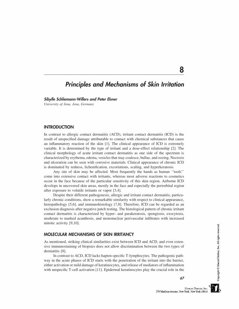
8
Principles and Mechanisms of Skin Irritation
Sibylle Schliemann-Willers and Peter ElsnerUniversity of Jena, Jena, Germany
INTRODUCTION
In contrast to allergic contact dermatitis (ACD), irritant contact dermatitis (ICD) is theresult of unspecified damage attributable to contact with chemical substances that causean inflammatory reaction of the skin [1]. The clinical appearance of ICD is extremelyvariable. It is determined by the type of irritant and a dose-effect relationship [2]. Theclinical morphology of acute irritant contact dermatitis as one side of the spectrum ischaracterized by erythema, edema, vesicles that may coalesce, bullae, and oozing. Necrosisand ulceration can be seen with corrosive materials. Clinical appearance of chronic ICDis dominated by redness, lichenification, excoriations, scaling, and hyperkeratosis.
Any site of skin may be affected. Most frequently the hands as human ‘‘tools’’come into extensive contact with irritants, whereas most adverse reactions to cosmeticsoccur in the face because of the particular sensitivity of this skin region. Airborne ICDdevelops in uncovered skin areas, mostly in the face and especially the periorbital regionafter exposure to volatile irritants or vapor [3,4].
Despite their different pathogenesis, allergic and irritant contact dermatitis, particu-larly chronic conditions, show a remarkable similarity with respect to clinical appearance,histopathology [5,6], and immunohistology [7,8]. Therefore, ICD can be regarded as anexclusion diagnosis after negative patch testing. The histological pattern of chronic irritantcontact dermatitis is characterized by hyper- and parakeratosis, spongiosis, exocytosis,moderate to marked acanthosis, and mononuclear perivascular infiltrates with increasedmitotic activity [9,10].
MOLECULAR MECHANISMS OF SKIN IRRITANCY
As mentioned, striking clinical similarities exist between ICD and ACD, and even exten-sive immunostaining of biopsies does not allow discrimination between the two types ofdermatitis [8].
In contrast to ACD, ICD lacks hapten-specific T-lymphocytes. The pathogenic path-way in the acute phases of ICD starts with the penetration of the irritant into the barrier,either activation or mild damage of keratinocytes, and release of mediators of inflammationwith unspecific T-cell activation [11]. Epidermal keratinocytes play the crucial role in the
67
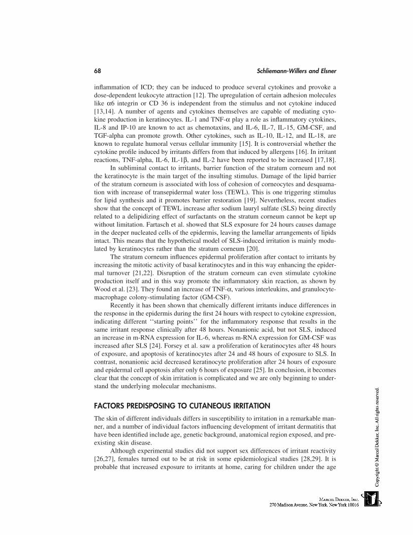
68 Schliemann-Willers and Elsner
inflammation of ICD; they can be induced to produce several cytokines and provoke adose-dependent leukocyte attraction [12]. The upregulation of certain adhesion moleculeslike α6 integrin or CD 36 is independent from the stimulus and not cytokine induced[13,14]. A number of agents and cytokines themselves are capable of mediating cyto-kine production in keratinocytes. IL-1 and TNF-α play a role as inflammatory cytokines,IL-8 and IP-10 are known to act as chemotaxins, and IL-6, IL-7, IL-15, GM-CSF, andTGF-alpha can promote growth. Other cytokines, such as IL-10, IL-12, and IL-18, areknown to regulate humoral versus cellular immunity [15]. It is controversial whether thecytokine profile induced by irritants differs from that induced by allergens [16]. In irritantreactions, TNF-alpha, IL-6, IL-1β, and IL-2 have been reported to be increased [17,18].
In subliminal contact to irritants, barrier function of the stratum corneum and notthe keratinocyte is the main target of the insulting stimulus. Damage of the lipid barrierof the stratum corneum is associated with loss of cohesion of corneocytes and desquama-tion with increase of transepidermal water loss (TEWL). This is one triggering stimulusfor lipid synthesis and it promotes barrier restoration [19]. Nevertheless, recent studiesshow that the concept of TEWL increase after sodium lauryl sulfate (SLS) being directlyrelated to a delipidizing effect of surfactants on the stratum corneum cannot be kept upwithout limitation. Fartasch et al. showed that SLS exposure for 24 hours causes damagein the deeper nucleated cells of the epidermis, leaving the lamellar arrangements of lipidsintact. This means that the hypothetical model of SLS-induced irritation is mainly modu-lated by keratinocytes rather than the stratum corneum [20].
The stratum corneum influences epidermal proliferation after contact to irritants byincreasing the mitotic activity of basal keratinocytes and in this way enhancing the epider-mal turnover [21,22]. Disruption of the stratum corneum can even stimulate cytokineproduction itself and in this way promote the inflammatory skin reaction, as shown byWood et al. [23]. They found an increase of TNF-α, various interleukins, and granulocyte-macrophage colony-stimulating factor (GM-CSF).
Recently it has been shown that chemically different irritants induce differences inthe response in the epidermis during the first 24 hours with respect to cytokine expression,indicating different ‘‘starting points’’ for the inflammatory response that results in thesame irritant response clinically after 48 hours. Nonanionic acid, but not SLS, inducedan increase in m-RNA expression for IL-6, whereas m-RNA expression for GM-CSF wasincreased after SLS [24]. Forsey et al. saw a proliferation of keratinocytes after 48 hoursof exposure, and apoptosis of keratinocytes after 24 and 48 hours of exposure to SLS. Incontrast, nonanionic acid decreased keratinocyte proliferation after 24 hours of exposureand epidermal cell apoptosis after only 6 hours of exposure [25]. In conclusion, it becomesclear that the concept of skin irritation is complicated and we are only beginning to under-stand the underlying molecular mechanisms.
FACTORS PREDISPOSING TO CUTANEOUS IRRITATION
The skin of different individuals differs in susceptibility to irritation in a remarkable man-ner, and a number of individual factors influencing development of irritant dermatitis thathave been identified include age, genetic background, anatomical region exposed, and pre-existing skin disease.
Although experimental studies did not support sex differences of irritant reactivity[26,27], females turned out to be at risk in some epidemiological studies [28,29]. It isprobable that increased exposure to irritants at home, caring for children under the age
Skin Irritation 69
of 4 years, lack of dishwashing machine [30], and preference for high-risk occupationscontribute to the higher incidence of ICD in females [27]. The most established individualrisk factor, out of several studies about occupational hand eczema, is probably atopicdermatitis [28,31–33]. On the other hand, experimental studies concerning the reactivityof atopics and nonatopics to standard irritants have given contradictory results [34,35]and, as shown in a Swedish study, about 25% of the atopics in extreme-risk occupations,such as hairdressers and nursing assistants, did not develop hand eczema [36]. Age is aswell related to irritant susceptibility insofar as irritant reactivity declines with increasingage. This is true not only for acute but also for cumulative irritant dermatitis [37,38]. Fairskin, especially skin type I, is supposed to be the most reactive to all types of irritants,and black skin is the most resistant [39,40].
Clinical manifestation of ICD is also influenced by type and concentration of irritant,solubility, vehicle, and length of exposure [41], as well as temperature and mechanicalstress. During the winter months, low humidity and low temperature decrease the watercontent of the stratum corneum and increase irritant reactivity [42,43].
EPIDEMIOLOGY
Population-based data on the incidence and prevalence of ICD are rare, but there is agree-ment that incidence of ICD is higher than that of ACD in general. The figures on theincidence of ICD vary considerably, depending on the study population. Most data stemfrom studies about occupational hand dermatoses, and in this an overview is given aboutthe important findings of these studies. In general, it can be assumed that nonoccupationalcontact dermatitis attributable to all causes is more frequent in comparison to occupationalcontact dermatitis [29].
Coenraads and Smit reviewed international prevalence studies for eczema attribut-able to all causes conducted with general populations in different countries (England, TheNetherlands, Norway, Sweden, the United States) and found point prevalence rates of 1.7to 6.3%, and 1- to 3-year period prevalence rates of 6.2 to 10.6% [44].
An extensive study of Meding on hand eczema in Gothenburg, Sweden, included20,000 individuals randomly selected from the population register [28]. She estimated a1-year period prevalence of hand eczema of 11% attributable to all causes, and a pointprevalence of 5.4%. ICD contributed to 35% of the cases, whereas 22% were diagnosedas atopic hand dermatitis and 19% as ACD. In a multicenter epidemiological study oncontact dermatitis in Italy by GIRDCA (Gruppo Italiano Ricerca Dermatiti da Contatto eAmbientali) 42,839 patients with contact dermatitis underwent patch testing. In accordancewith the findings of Meding, nonoccupational as well as occupational ICD affected womenin a higher percentage compared with males [28,29]. In Heidelberg, Germany, a retrospec-tive study of 190 cases of hand dermatitis revealed 27% as ICD, 15,8% as ACD, and themajority (40%) as being of atopic origin with 10% various other diseases [45].
Shenefelt studied the frequency of visits by university students to campus prepaid–health-plan dermatologists for irritant and allergic contact dermatitis compared with othertypes of dermatitis and skin problems. In contrast to other studies, he found slightly morecases of allergic (3.1% of all first visits) than irritant contact dermatitis (2.3%) [46].
Reports on adverse reactions to cosmetics, including those with only subjective per-ceptions without morphological signs, are more frequent than assumed. In a questionnairecarried out in Thuringia, eastern Germany, even 36% of 208 persons reported adversecutaneous reactions against cosmetics, 75% of them being female [47]. Nevertheless, it
70 Schliemann-Willers and Elsner
must be emphasized that this includes, in addition to allergic contact dermatitis, dermatosesas seborrheic dermatitis, perioral dermatitis, rosacea and psoriasis, which cannot be sepa-rated by the unexperienced. Higher incidence in females was confirmed by several studies[48]. Most untoward reactions caused by cosmetics occur on the face, including the perior-bital area [49].
In a study by Broeckx et al., 5.9% of a test population of 5202 patients with possiblecontact dermatitis had adverse reactions to cosmetics. Patch testing classified only 1.46%as irritant reactions whereas 3.0% could be classified as ACD. More than 50% of thecases of irritation were attributable to soaps and shampoos [50]. In Sweden, the top-rank-ing products causing adverse effects, as reported by the Swedish Medical ProductsAgency, were moisturizers, haircare products, and nail products [48].
In other studies, the incidence of cosmetic intolerance varied between 2 and 8.3%,depending on the test population [49,51,52]. In a large multicenter prospective study onreactions caused by cosmetics, Eiermann et al. found irritancy to account for only 16%of 487 cases of contact dermatitis caused by cosmetics. Of 8093 patients tested for contactdermatitis, 487 cases (6%) were diagnosed as contact dermatitis caused by cosmetics [53].Since most consumers just stop using cosmetics when experiencing mild irritant or adversereactions and seldom consult a physician, it can be assumed that mild irritant reactionsto cosmetic products are underestimated [54].
CLINICAL TYPES OF IRRITANT CONTACT DERMATITIS
According to the highly variable clinical picture, several different forms of ICD have beendefined. The following types of irritation have been described [55,56]:
• Acute ICD• Delayed acute ICD• Irritant reaction• Cumulative ICD• Traumiterative ICD• Exsiccation eczematid• Traumatic ICD• Pustular and acneiform ICD• Nonerythematous• Sensory irritation
Acute ICD
Acute ICD is caused by contact to a potent irritant. Substances that cause necrosis arecalled corrosive and include acids and alkaline solutions. Contact is often accidental atthe workplace. Cosmetics are unlikely to cause this type of ICD because they do notcontain primary irritants in sufficient concentrations.
Symptoms and clinical signs of acute ICD develop with a short delay of minutesto hours after exposure, depending on the type of irritant, concentration, and intensity ofcontact. Characteristically the reaction quickly reaches its peak and then starts to heal;this is called ‘‘decrescendo phenomenon.’’ Symptoms include burning rather than itching,
Skin Irritation 71
stinging, and soreness of the skin, and are accompanied by clinical signs such as erythema,edema, bullae, and even necrosis. Lesions are usually restricted to the area that came intocontact, and sharply demarcated borders are an important sign of acute ICD. Nevertheless,clinical appearance of acute ICD can be highly variable and sometimes may even beindistinguishable from the allergic type. In particular, combination of irritant and allergiccontact dermatitis can be troublesome. Prognosis of acute ICD is good if irritant contactis avoided.
Delayed Acute ICD
For some chemicals, such as anthralin, it is typical to produce a delayed acute ICD. Visibleinflammation is not seen until 8 to 24 hours or more after exposure [57]. Clinical pictureand symptoms are similar to acute ICD. Other substances that cause delayed acute ICDinclude dithranol, tretinoin, and benzalkonium chloride. Irritation to tretinoin can developafter a few days and results in a mild to fiery redness followed by desquamation, or largeflakes of stratum corneum accompanied by burning rather than itching. Irritant patch-testreactions to benzalkonium chloride may be papular and increase with time, thus resem-bling allergic patch-test reactions [58]. Tetraethylene glycol diacrylate caused delayedskin irritation after 12 to 36 hours in several workers in a plant manufacturing acrylatedchemicals [59].
Irritant Reaction
Irritants may produce cutaneous reactions that do not meet the clinical definition of a‘‘dermatitis.’’ Irritant reaction is therefore a subclinical form of irritant dermatitis and ischaracterized by a monomorphic rather than polymorphic picture. This may include oneor more of the following clinical signs: dryness, scaling, redness, vesicles, pustules, anderosions [60]. Irritant reactions often occur after intense water contact and in individualsexposed to wet work, such as hairdressers or metal workers, particularly during their firstmonths of training. It often starts under rings worn on the finger or in the interdigital area,and may spread over the dorsum of the fingers and to the hands and forearms. Frequently,the condition heals spontaneously, resulting in hardening of the skin, but it can progressto cumulative ICD in some cases.
Cumulative ICD
Cumulative ICD is the most common type of ICD [55]. In contrast to acute ICD that canbe caused by single contact to a potent irritant, cumulative ICD is the result of multiplesubthreshold damage to the skin when time is too short for restoration of skin-barrierfunction [61]. Clinical symptoms develop after the damage has exceeded a certain manifes-tation threshold, which is individually determined and can vary within one individual atdifferent times. Typically, cumulative ICD is linked to exposure of several weak irritantsand water contact rather than to repeated exposure to a single potent irritant. Because thelink between exposure and disease is often not obvious to the patient, diagnosis may beconsiderably delayed, and it is important to rule out an allergic cause. Symptoms includeitching and pain caused by cracking of the hyperkeratotic skin. The clinical picture isdominated by dryness, erythema, lichenification, hyperkeratosis, and chapping. Xeroticdermatitis is the most frequent type of cumulative toxic dermatitis [62]. Vesicles are less
72 Schliemann-Willers and Elsner
frequent in comparison to allergic and atopic types [28]; however, diagnosis is often com-plicated by the combination of irritation and atopy, irritation and allergy, or even all three.Lesions are less sharply demarcated in contrast to acute ICD.
Prognosis of chronic cumulative ICD is rather doubtful [63,64]. Some investigatorssuggest that the repair capacity of the skin may enter a self-perpetuating cycle [61].
Traumiterative ICD
This term is often used similarly to cumulative ICD [55,60]. Clinically, the two types arevery similar as well. According to Malten and den Arend, traumiterative ICD is a resultof too-early repetition of just one type of load, whereas cumulative ICD results from too-early repetition of different types of exposures [2].
Exsiccation Eczematid
Exsiccation eczematid is a subtype of ICD that mainly develops on the extremities. It isoften attributable to frequent bathing and showering as well as extensive use of soaps andcleansing products. It often affects elderly people with low sebum levels of the stratumcorneum. Low humidity during the winter months and failure to remoisturize the skincontribute to the condition. The clinical picture is typical, with dryness, ichthyosiformscaling, and fissuring. Patients often suffer from intense itching.
Traumatic ICD
Traumatic ICD may develop after acute skin traumas such as burns, lacerations, and acuteICD. The skin does not heal as expected, but ICD with erythema, vesicles and/or papu-lovesicles, and scaling appears. The clinical course resembles that of nummular dermatitis[55].
Pustular and Acneiform ICD
Pustular and acneiform ICD may result from contact to irritants such as mineral oils, tars,greases, some metals, croton oil, and naphthalenes. Pustules are sterile and transient. Thesyndrome must be considered in conditions in which acneiform lesions develop outsidetypical acne age. Patients with seborrhoea, macroporous skin, and prior acne vulgaris arepredisposed along with atopics.
Nonerythematous ICD
Nonerythematous ICD is an early stage of skin irritation that lacks visible inflammation butis characterized by changes in the function of the stratum corneum that can be measured bynoninvasive bioengineering techniques [55,65].
Sensory Irritation
Sensory irritation is characterized by subjective symptoms without morphologicalchanges. Predisposed individuals complain of stinging, burning, tightness, itching, or evenpainful sensations that occur immediately or after contact. Those individuals with hyperir-ritable skin often report adverse reactions to cosmetic products with most reactions oc-curring on the face. Fisher defined the term ‘‘status cosmeticus,’’ which describes a condi-tion in patients who try a lot of cosmetics and complain of being unable to tolerate any
Skin Irritation 73
of them [66]. Lactic acid serves as a model irritant for diagnosis of so called ‘‘stingers’’when it is applied in a 5% aqueous solution on the nasolabial fold after induction ofsweating in a sauna [67]. Other chemicals that cause immediate-type stinging after secondsor minutes include chloroform and methanol (1:1) and 95% ethanol. A number of sub-stances that have been systematically studied by Frosch and Kligman may also causedelayed-type stinging [67,68]. Several investigators tried to determine parameters thatcharacterize those individuals with sensitive skin, a term that still lacks a unique definition[69,70]. It could be shown that individuals who were identified as having sensitive skinby their own assessment have altered baseline biophysical parameters, showing decreasedcapacitance values, increased transepidermal water loss, and higher pH values accompa-nied by lower sebum levels [70]. Possible explanations for hyperirritability (other thandiminished barrier function) that have been discussed are heightened neurosensory inputattributable to altered nerve endings, more neurotransmitter release, unique central infor-mation processing or slower neurotransmitter removal, and enhanced immune respon-siveness [69,71]. It is not clear whether having sensitive skin is an acquired or inheritedcondition; most probably it can be both. As in other forms of ICD, seasonal variabilityin stinging with a tendency to more intense responses during winter has been observed[72]. Detailed recommendations for formulation of skincare products for sensitive skinhave been given by Draelos [69].
REFERENCES
1. Mathias CGT, Maibach HI. Dermatotoxicology monographs I. cutaneous irritation: factorsinfluencing the response to irritants. Clin Toxicol 1978; 13:333–346.
2. Malten KE, den Arend JA. Irritant contact dermatitis. Traumiterative and cumulative impair-ment by cosmetics, climate, and other daily loads. Derm Beruf Umwelt 1985; 4:125–132.
3. Dooms-Goossens AE, Debusschere KM, Gevers DM, Dupre KM, Degref HJ, Loncke JP,Snauwaert JE. Contact dermatitis caused by airborne agents. A review and case reports. J AmAcad Dermatol 1986; 15:1–10.
4. Lachapelle JM. Industrial airborne irritant or allergic contact dermatitis. Contact Dermatitis1986; 14:137–145.
5. Brand CU, Hunziker T, Braathen LR. Studies on human skin lymph containing Langerhanscells from sodium lauryl sulphate contact dermatitis. J Invest Dermatol 1992; 5:109s–110s.
6. Brand CU, Hunziker T, Limat A, et al. Large increase of Langerhans cells in human skinlymph derived from irritant contact dermatitis. Br J Dermatol 1993; 2:184–188.
7. Medenica M, Rostenberg A Jr. A comparative light and electron microscopic study of primaryirritant contact dermatitis and allergic contact dermatitis. J Invest Dermatol 1971; 4:259–271.
8. Brasch J, Burgard J, Sterry W. Common pathogenetic pathways in allergic and irritant contactdermatitis. J Invest Dermatol 1992; 2:166–170.
9. Cohen LM, Skopicki DK, Harrist DJ, Clark WH. Noninfectious vesiculobullous and vesiculo-pustular diseases. In: Elder D, Elenitsas R, Jaworsky C, Johnson B, eds. Lever’s Histopathol-ogy of the Skin. (8th ed.) Philadelphia: Lippincott-Raven, 1997:209–252.
10. Le TK, Schalkwijk J, van de Kerkhof PC, van Haelst U, van der Valk PG. A histological andimmunhistochemical study on chronic irritant contact dermatitis. Am J Contact Dermat 1998;9:23–28.
11. Berardesca E, Distante F. Mechanisms of skin irritation. In: Elsner P, Maibach HI, eds. IrritantDermatitis: New Clinical and Experimental Aspects. Current Problems in Dermatology. Basel:Karger, 1995: 1–8.
12. Nickoloff BJ, Naidu Y. Perturbation of epidermal barrier function correlates with initiationof cytokine cascade in human skin. J Am Acad Dermatol 1994; 30:535–546.
74 Schliemann-Willers and Elsner
13. Willis CM, Stephens CJ, Wilkinson JD. Epidermal damage induced by irritants in man: a lightand electron microscopic study. J Invest Dermatol 1989; 93:695–699.
14. Jung K, Imhof BA, Linse R, Wollina U, Neumann C. Adhesion molecules in atopic dermatitis:upregulation of α6 integrin expression in spontaneous lesional skin as well as in atopen,antigen and irritative induced patch test reactions. Int Arch Allergy Immunol 1997; 113:495–504.
15. Corsini E, Galli CL. Cytokines and irritant contact dermatitis. Toxicol Lett 1998; 28:277–282.
16. Kalish RS. T cells and other leukocytes as mediators of irritant contact dermatitis. In: BeltraniVS, ed. Immunology and Allergy Clinics of North America. Contact Dermatitis. Irritant andAllergic. Philadelphia: W.B. Saunders Company 1997:407–415.
17. Larsen CG, Ternowitz T, Larsen FG, Zachariae CO, Thestrup-Pedersen K. ETAF/interleukin-1 and epidermal lymphocyte chemotactic factor in epidermis overlying an irritant patch test.Contact Dermatitis 1989; 20:335–340.
18. Hunziker T, Brand CU, Kapp A, Waelti ER, Braathen LR. Increased levels of inflammatorycytokines in human skin lymph derived from sodium lauryl sulphate-induced contact derma-titis. Br J Dermatol 1992; 127:254–257.
19. Grubauer G, Elias PM, Feingold KR: Transepidermal water loss: the signal for recovery ofbarrier structure and function. J Lipid Res 1989; 30:323–333.
20. Fartasch M, Schnetz E, Diepgen TL. Characterization of detergent-induced barrier alter-ations—effect of barrier cream on irritation. J Invest Dermatol Symp Proceed 1998; 3:121–127.
21. Fisher LB, Maibach HI. Effects of some irritants on human epidermal mitosis. Contact Derma-titis 1975; 1:273–276.
22. Wilhelm KP, Saunders JC, Maibach HI. Increased stratum corneum turnover induced by sub-clinical irritant dermatitis. Br J Dermatol 1990; 122:793–798.
23. Wood LC, Jackson SM, Elias PM, Grunfeld C, Feingold KR. Cutaneous barrier perturbationstimulates cytokine production in the epidermis of mice. J Clin Invest 1992; 90:482–487.
24. Grängsjö A, Leijon-Kuligowski A, Törmä H, Roomans GM, Lindberg M. Different pathwaysin irritant contact eczema? Early differences in the epidermal elemental content and expressionof cytokines after application of 2 different irritants. Contact Dermatitis 1996; 35:355–360.
25. Forsey RJ, Shahidullah H, Sands C, McVittie E, Aldridge RD, Hunter JA, Howie SE. Epider-mal Langerhans cell apoptosis is induced in vivo by nonanionic acid but not by sodium laurylsulphate. Br J Dermatol 1998; 139:453–461.
26. Bjornberg A. Skin reactions to primary irritants in men and women. Acta Derm Venereol(Stockh) 1975; 55:191–194.
27. Hogan DJ, Dannaker CJ, Maibach HI. The prognosis of contact dermatitis. J Am Acad Derma-tol 1990; 23:300–307.
28. Meding B. Epidemiology of hand eczema in an industrial city. Acta Derm Venereol Suppl(Stockh) 1990; 153:1–43.
29. Sertoli A, Francalanci S, Acciai MC, Gola M. Epidemiological survey of contact dermatitisin Italy (1984–1993) by GIRDCA (Gruppo Italiano Ricera Dermatiti da Contatto e Ambi-entali). Am J Contact Dermat 1999; 10:18–30.
30. Nilsson E. Individual and environmental risk factors for hand eczema in hospital workers.Acta Derm Venereol (Stockh) (Suppl) 1986; 128:1–63.
31. Wilhelm KP, Maibach HI. Factors prediposing to cutaneous irritation. Dermatol Clin 1990;8:17–22.
32. Coenraads PJ, Diepgen TL. Risk for hand eczema in employees with past or present atopicdermatitis. Int Arch Occup Environ Health 1998; 71:7–13.
33. Berndt U, Hinnen U, Iliev D, Elsner P. Role of the atopy score and of single atopic featuresas risk factors for development of hand eczema in trainee metal workers. Br J Dermatol 1999;140:922–924.
Skin Irritation 75
34. Gallacher G, Maibach HI. Is atopic dermatitis a predisposing factor for experimental acuteirritant contact dermatitis? Contact Dermatitis 1998; 38:1–4.
35. Basketter DA, Miettinen J, Lahti A. Acute irritant reactivity to sodium lauryl sulfate in atopicsand non-atopics. Contact Dermatitis 1998; 38:253–257.
36. Rysted I. Work-related hand eczema in atopics. Contact Dermatitis 1985; 12:164–171.37. Suter-Widmer J, Elsner P. Age and irritation. In: van der Valk PGM, Maibach HI, eds. The
Irritant Contact Dermatitis Syndrome. Boca Raton: CRC Press, 1994: 257–261.38. Schwindt DA, Wilhelm KP, Miller DL, Maibach HI. Cumulative irritation in older and
younger skin: a comparison. Acta Derm Venereol 1998; 78:279–283.39. Lammintausta K, Maibach HI, Wilson D. Susceptibility to cumulative and acute contact der-
matitis. Contact Dermatitis 1988; 19:84–90.40. Maibach HI, Berardesca E. Racial and skin color differences in skin sensitivity: implications
for skin care products. Cosmet Toilet 1990; 105:35–36.41. Dahl MV. Chronic, irritant contact dermatitis: mechanisms, variables, and differentiation from
other forms of contact dermatitis. Adv Dermatol 1988; 3:261–275.42. Mozzanica N. Pathogenetic aspects of allergic and irritant contact dermatitis. Clin Dermatol
1992; 10:115–121.43. Uter W, Gefeller O, Schwanitz HJ. An epidemiological study of the influence of season (cold
and dry air) on the occurrence of irritant skin changes of the hands. Br J Dermatol 1998; 138:266–272.
44. Coenraads PJ, Smit J. Epidemiology. In: Rycroft RJG, Menné T, Frosch PJ, eds. Textbookof Contact Dermatitis. (2nd ed.) Berlin: Springer, 1995:133–150.
45. Kühner-Piplack B. Klinik und Differentialdiagnose des Handekzems. Eine retrospektive Stu-die am Krankengut der Universitäts-Hautklinik Heidelberg 1982–1985. Thesis, Ruprecht-Karls-University, Heidelberg, Germany.
46. Shenefelt PD. Descriptive epidemiology of contact dermatitis in a university student popula-tion. Am J Contact Dermat 1996; 7:88–93.
47. Röpcke F. Auswertung zur Umfrage ‘‘Epidemiologie von Kosmetika-Unverträglichkeiten—eine bevölkerungsbasierte Studie.’’ 1999, unpublished data.
48. Berne B, Bostrom A, Grahnen AF, Tammela M. Adverse effects of cosmetics and toiletriesreported to the Swedish Medical Products Agency 1989–1994. Contact Dermatitis 1996; 34:359–362.
49. Adams RM, Maibach HI. A 5-year study of cosmetic reactions. J Am Acad Dermatol 1985;13:1062–1069.
50. Broeckx W, Blondeel A, Dooms-Goossens A, Achten G. Cosmetic intolerance. Contact Der-matitis 1987; 16:189–194.
51. Skog E. Incidence of cosmetic dermatitis. Contact Dermatitis 1980; 6:449–451.52. Romaguera C, Camarasa JMG, Alomar A, Grimalt F. Patch tests with allergens related to
cosmetics. Contact Dermatitis 1983; 9:167–168.53. Eiermann HJ, Larsen W, Maibach HI, Taylor JS. Prospective study of cosmetic reactions:
1977–1980. J Am Acad Dermatol 1982; 6:909–917.54. Amin S, Engasser PG, Maibach HI. Adverse cosmetic reactions. In: Baran R, Maibach HI,
eds. Textbook of Cosmetic Dermatology. 2nd ed. London: Martin Dunitz Ltd., 1998:709–746.
55. Lammintausta K, Maibach HI. Contact dermatitis due to irritation: General principles, etiol-ogy, and histology. In: Adams RM, ed. Occupational skin disease. Philadelphia: WB SaundersCompany, 1990:1–15.
56. Berardesca E, Distante F. Mechanisms of skin irritation. In: Elsner P, Maibach HI, eds. Irritantdermatitis. New clinical and experimental aspects. Basel: Karger 1995:1–8.
57. Malten KE, den Arend JA, Wiggers RE. Delayed iritation: hexanediol diacrylate and butaned-iol diacrylate. Contact Dermatitis 1979; 3:178–184.
58. Bruynzeel DP, van Ketel WG, Scheper RJ, von Blomberg-van der Flier BME. Delayed time
76 Schliemann-Willers and Elsner
course of irritation by sodium lauryl sulfate: observations on threshold reactions. Contact Der-matitis 1982; 8:236–239.
59. Nethercott JR, Gupta S, Rosen C, Enders LJ, Pilger CW. Tetraethylene glycol diacrylate. Acause of delayed cutaneous irritant reaction and allergic contact dermatitis. J Occup Med 1984;26:513–516.
60. Frosch PJ. Cutaneous irritation. In: Rycroft RJG, Menné T, Frosch PJ, eds. Textbook of Con-tact Dermatitis. 2nd ed. Berlin: Springer, 1995:28–61.
61. Malten KE. Thoughts on irritant contact dermatitis. Contact Dermatitis 1981; 7:238–247.62. Eichmann A, Amgwerd D. Toxische Kontaktdermatitis. Schweiz Rundsch Med Prax 1992;
19:615–617.63. Keczkes K, Bhate SM, Wyatt EH. The outcome of primary irritant hand dermatitis. Br J Der-
matol 1983; 109:665–668.64. Elsner P, Baxmann F, Liehr HM. Metal working fluid dermatitis: A comparative follow-up
study in patients with irritant and non-irritant dermatitis. In: Elsner P, Maibach HI, eds. IrritantDermatitis: New Clinical and Experimental Aspects. Basel: Karger, 1995:77–86.
65. Van der Valk PGM, Maibach HI. Do topical corticosteroids modulate skin irritation in humanbeings? Assessment by transepidermal water loss and visual scoring. J Am Acad Dermatol1989; 21:519–522.
66. Fisher AA. Cosmetic actions and reactions: therapeutic, irritant and allergic. Cutis 1980; 26:22–29.
67. Frosch PJ, Kligman AM. A method for appraising the stinging capacity of topically appliedsubstances. J Soc Cosm Chem 1977; 28:197–209.
68. Parrish JA, Pathak MA, Fitzpatrick TB. Facial irritation due to sunscreen products. Letter tothe editor. Arch Dermatol 1975; 111:525.
69. Draelos ZD. Sensitive skin: perceptions, evaluation, and treatment. Am J Contact Dermat1997; 8:67–78.
70. Seidenari S, Francomano M, Mantovani L. Baseline biophysical parameters in subjects withsensitive skin. Contact Dermatitis 1999; 38:311–315.
71. Muizzudin N, Marenus KD, Maes DH. Factors defining sensitive skin and its treatment. AmJ Contact Dermat 1998; 9:170–175.
72. Leyden JJ. Risk assessment of products used on skin. Am J Contact Dermat 1993; 4:158–162.
9
Allergy and Hypoallergenic Products
An E. GoossensUniversity Hospital, Katholieke Universiteit Leuven, Leuven, Belgium
INTRODUCTION
The assessment and detection of the number of contact allergic reactions to cosmeticsare not simple. Generally, a consumer who has a problem with cosmetics will consult adoctor only if he or she does not recognize the cause to be a particular cosmetic productor if the dermatitis persists when the suspect product has been replaced by another, de-termined by trial and error. Consequently, only a small proportion of the populationwith cosmetic intolerance problems is ever seen by a dermatologist. Moreover, cosmeticreactions may present in unusual clinical forms, which may evoke an erroneous diagnosis[1–3].
In general, adverse effects are underreported [4], certainly to the cosmetics industrywhich obtains its most reliable information in this regard mainly from the relatively fewdermatologists who concentrate on cosmetic-intolerance problems and from reports in theliterature which are, almost by definition, out of date. Sometimes beauticians and consum-ers report adverse reactions, but in most cases this kind of information is difficult to objec-tify unless it is verified by a dermatologist.
Application of cosmetic products to the skin may cause irritant, phototoxic, contact,and photocontact allergic reactions as well as contact urticaria. It is generally agreed thatmost skin-adverse reactions to cosmetic products are irritant in nature and that people with‘‘sensitive skin,’’ as indicated by conditions like atopic dermatitis, rosacea, or seborrheicdermatitis, are particularly liable to develop such reactions. However, contact allergicreactions attract much more attention and thus tend to be overestimated [4]. Indeed, theidentification of the cosmetic allergen is by no means a simple task. It demands specialskills and interest on the part of the dermatologist, even though the labeling of all cosmeticingredients, which is now obligatory also in Europe, is facilitating that task. Moreover,there are many factors involved in the sensitization to a specific cosmetic product, all ofwhich have to be taken into account when one seeks an allergen [1,2] (see the followingsection).
77
78 Goossens
FACTORS CONTRIBUTING TO CONTACT ALLERGIC REACTIONS TO ACOSMETIC PRODUCT
Frequency of Use
One may expect frequently used products to cause more skin reactions than more exclusiveproducts simply because more people are exposed to them. This alone does not implyanything about the quality of these products (the same thing may be said about individualcosmetic ingredients).
Composition
The complexity of a formula can be either positive or negative as far as its allergenicityis concerned. One of the principles of creating ‘‘hypoallergenic’’ cosmetics and perfumesis simplicity of formula. The fewer the constituents, the easier it is to identify the offendingsubstance should difficulties arise, and the less danger there is of synergism. The moreingredients there are, the more chance there is of sensitization by one of them. However,some investigators recommend placing upper limits on concentrations rather than advisingagainst the use of any particular ingredient. They may also suggest more complex for-mulas [5].
Preservatives are needed in water-based or other easily contaminated products andare common cosmetic allergens. It seems that it is very difficult to combine potent antimi-crobial and antifungal properties with low allergenicity. Indeed, it is very difficult to re-strict the biological activity of a substance to a single domain.
Concentration of Ingredients
Although the use of low concentrations does not assure complete safety, the incidence ofsensitization induction is, indeed, a function of the concentration of the allergen, at leastto some extent. Cases of allergy to the preservative agent (chloro)methylisothiazolinoneillustrate this problem very well. At first, when a 50 ppm concentration of this agentwas allowed for use in cosmetic products in the European Community and when thisconcentration was actually used in some products, there were ‘‘epidemics’’ of contactallergic reactions to it [6]. Of late, the frequency of positive reactions has been diminishingconsiderably, not only because its use is declining and primarily limited to ‘‘rinse-off’’products [3] but also because its usage concentration has been reduced to 15 to 7.5 ppm(as the manufacturers recommended). Of course, once a patient has become sensitized,even low concentrations can trigger a reaction.
Purity of Ingredients
It is impossible to refine raw materials to absolute purity. More or less strict quality controlof raw materials and finished products has long been general practice in modern cosmeticmanufacturing. However, one can never rule out the sensitizing potential of impurities inthese materials [5].
The Common Use of Cosmetic Ingredients in Pharmaceuticals
Patients easily become sensitized to topical pharmaceutical products which, unlike cosmet-ics, are most often used on diseased skin. Once sensitization has occurred, however, theymay react to cosmetics containing the same ingredients [5].
Allergy and Hypoallergenic Products 79
The Role of Cross-Sensitivity
Chemically related substances are likely to induce cross-reactions and contact eczematouslesions may be maintained in this way. This is especially the case with perfume ingredi-ents, which often cross-react with each other, but applies to all other cosmetic ingredientsas well.
Penetration-Enhancing Substances
The chemical environment can substantially affect the sensitizing potential of individualchemicals. For example, emulsifiers and solvents enhance skin penetration and therebycontact sensitization. Penetration-enhancing agents can also be the root of false-negativepatch-test reactions; the cosmetic product itself may be clearly allergenic (or irritant) al-though the individual ingredients, abstracted from the environment of the product andtested separately, may not cause a reaction.
Application Site
Some areas of the skin, like the eyelids, are particularly prone to contact dermatitis reac-tions. A cream applied to the entire face such as a facecare product, along with hair prod-ucts may cause an allergic reaction only on the eyelids. Moreover, ‘‘ectopic dermatitis’’[caused by the transfer of the allergen by the hand, as often occurs with tosylamide/formaldehyde (� para-toluenesulfonamideformaldehyde) resin, the allergen in nail pol-ish], ‘‘airborne’’ contact dermatitis (e.g., caused by perfumes) [7], as well as ‘‘connubial’’dermatitis (caused by products used by the partner) [8] often occur only on ‘‘sensitive’’skin areas such as the eyelids, the lips, and the neck.
Moreover, the penetration potential of cosmetics is heightened in certain ‘‘oc-cluded’’ areas, such as the body folds (axillary, inguinal) and the anogenital region, whichalso increases the risk of contact sensitization. In the body folds, the allergenic reactionstend to persist for weeks after the initial contact with the allergen. This may be partlyattributable to residual contamination of clothing as well as the increased penetration ofthe allergen, which is certainly assisted by occlusion and friction [9]. Indeed, a reservoirmay be formed from which the allergen is subsequently released.
Condition of the Skin
Application on damaged skin, where the skin barrier is impaired, enhances the penetrationof substances and thus increases the risk of an allergic reaction. This is the case withbodycare products used to alleviate dry, atopic skin and with barrier creams for protectingthe hands, which often suffer from irritancy problems (e.g., dryness, cracking). Sometimes,the allergic reaction may be limited to certain areas of the skin (areas already affectedreact more readily to another application of the same allergen) and may even present anunusual clinical picture that does not immediately suggest contact dermatitis. Indeed, con-tact allergic reactions to preservative agents on the face may present as a lymphocyticinfiltrate or even have a lupus erythematous–like picture [3,10].
Contact Time
In the world of cosmetics, a distinction is now being made between leave-on products,which remain on the skin for several hours (e.g., face- and bodycare products and makeup),and rinse-off products, which are removed almost immediately.
80 Goossens
The division between these two kinds of products is not always relevant to thesensitization process because a thin film can remain on the skin and be sufficient to allowingredients to penetrate. This occurs, for example, with moist toilet paper (with mainlypreservatives as the allergens) and makeup removers.
Frequency of Application and Cumulative Effects
Daily use or use several times a day of cosmetics may cause ingredients to accumulatein the skin and thus increase the risk of adverse reactions. In fact, the concentration ofan ingredient may be too low to induce sensitivity in a single product but may reachcritical levels in the skin if several products containing it are used consecutively. Thismay be the case for people who are loyal to the same brand of, e.g., day and night creams,foundations, and cleansing products, because a manufacturer will often use the same pre-servative system for all of its products. This should be taken into consideration by compa-nies that use biologically active ingredients such as preservative agents, emulsifiers, anti-oxidants, and perfumes, because it might well account for many of the adverse reactionsto these particular substances. In our experience, intense users of cosmetics are more proneto cosmetic dermatitis than others.
CORRELATIONS WITH THE LOCATION OF THE LESIONS
Like many other contact allergens, cosmetics can reach the skin in several different ways[1,2]: by direct application; by airborne exposure to vapors, droplets, or particles that arereleased into the atmosphere and then settle on the skin [7]; by contact with people (part-ners, friends, coworkers) who transmit allergens to cause ‘‘connubial’’ or ‘‘consort’’ der-matitis [8]; by transfer from other sites on the body, often the hands, to more sensitiveareas such as the mouth or the eyelids (ectopic dermatitis); and by exposure to the sunwith photoallergens.
The most common sources of cosmetic allergens applied directly to the body arelisted in Table 1.
THE NATURE OF COSMETIC ALLERGENS
Fragrance Ingredients
Fragrance ingredients are the most frequent culprits in cosmetic allergies [11–15]. Katsararet al., who investigated the results of patch testing over a 12-year period, found an increas-ing trend in sensitivity to fragrance compounds, which reflects the effectiveness of theadvertising of perfumed products [16]. Common features of a fragrance contact dermatitisare localization in the axillae, localization on the face (including the eyelids) and neck,and well-circumscribed patches in areas of dabbing-on perfumes (wrists, behind the ears)and hand eczema or its aggravation. Airborne or connubial contact dermatitis should beconsidered as well.
Other less frequent adverse reactions to fragrances are photocontact dermatitis, con-tact urticaria, irritation, and pigmentation disorders [17].
Sensitization is most often induced by highly perfumed products, such as toilet wa-ters, aftershave lotions, and deodorants, the last of which have recently been shown tocontain well-known allergens such as cinnamic aldehyde and iso-eugenol [18].
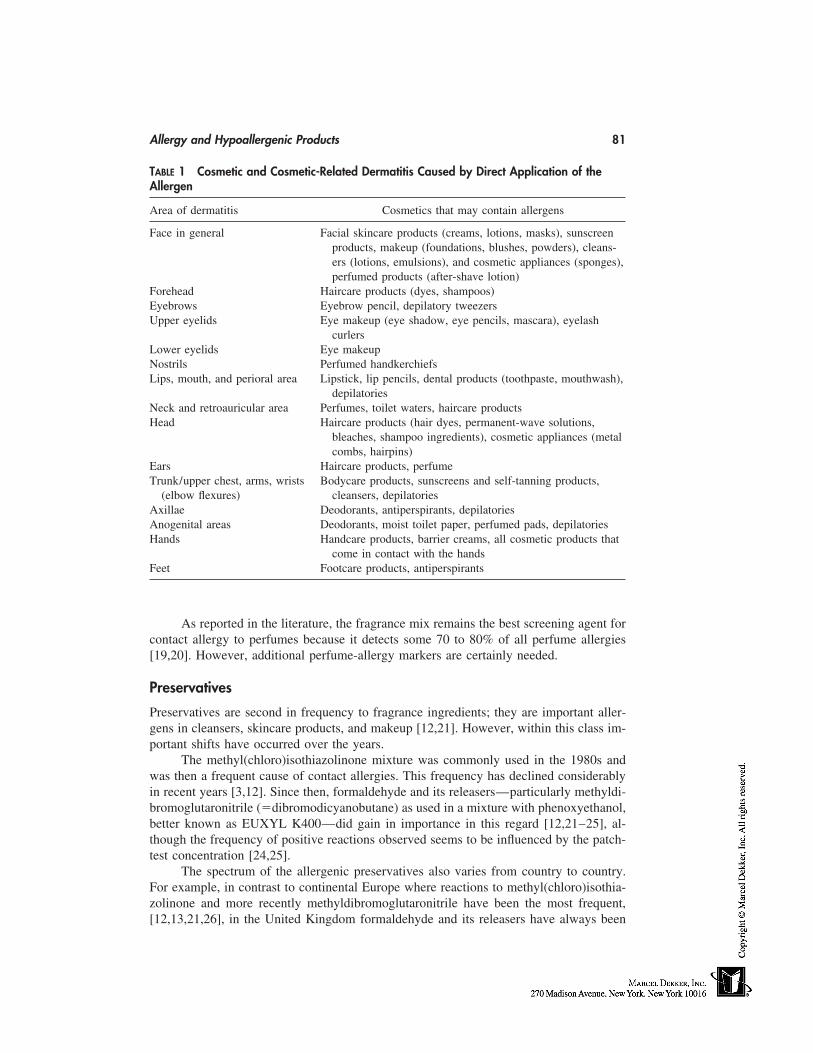
Allergy and Hypoallergenic Products 81
TABLE 1 Cosmetic and Cosmetic-Related Dermatitis Caused by Direct Application of theAllergen
Area of dermatitis Cosmetics that may contain allergens
Face in general Facial skincare products (creams, lotions, masks), sunscreenproducts, makeup (foundations, blushes, powders), cleans-ers (lotions, emulsions), and cosmetic appliances (sponges),perfumed products (after-shave lotion)
Forehead Haircare products (dyes, shampoos)Eyebrows Eyebrow pencil, depilatory tweezersUpper eyelids Eye makeup (eye shadow, eye pencils, mascara), eyelash
curlersLower eyelids Eye makeupNostrils Perfumed handkerchiefsLips, mouth, and perioral area Lipstick, lip pencils, dental products (toothpaste, mouthwash),
depilatoriesNeck and retroauricular area Perfumes, toilet waters, haircare productsHead Haircare products (hair dyes, permanent-wave solutions,
bleaches, shampoo ingredients), cosmetic appliances (metalcombs, hairpins)
Ears Haircare products, perfumeTrunk/upper chest, arms, wrists Bodycare products, sunscreens and self-tanning products,
(elbow flexures) cleansers, depilatoriesAxillae Deodorants, antiperspirants, depilatoriesAnogenital areas Deodorants, moist toilet paper, perfumed pads, depilatoriesHands Handcare products, barrier creams, all cosmetic products that
come in contact with the handsFeet Footcare products, antiperspirants
As reported in the literature, the fragrance mix remains the best screening agent forcontact allergy to perfumes because it detects some 70 to 80% of all perfume allergies[19,20]. However, additional perfume-allergy markers are certainly needed.
Preservatives
Preservatives are second in frequency to fragrance ingredients; they are important aller-gens in cleansers, skincare products, and makeup [12,21]. However, within this class im-portant shifts have occurred over the years.
The methyl(chloro)isothiazolinone mixture was commonly used in the 1980s andwas then a frequent cause of contact allergies. This frequency has declined considerablyin recent years [3,12]. Since then, formaldehyde and its releasers—particularly methyldi-bromoglutaronitrile (�dibromodicyanobutane) as used in a mixture with phenoxyethanol,better known as EUXYL K400—did gain in importance in this regard [12,21–25], al-though the frequency of positive reactions observed seems to be influenced by the patch-test concentration [24,25].
The spectrum of the allergenic preservatives also varies from country to country.For example, in contrast to continental Europe where reactions to methyl(chloro)isothia-zolinone and more recently methyldibromoglutaronitrile have been the most frequent,[12,13,21,26], in the United Kingdom formaldehyde and its releasers have always been
82 Goossens
much more important, particularly as concerns quaternium-15 [21] although its incidenceseems to have recently slightly decreased [27]. Parabens are rare causes of cosmetic derma-titis. When a paraben allergy does occur, the sensitization source is most often a topicalpharmaceutical product, although its presence in other products can be sensitizing as well[28]. Recently, we observed such a case (data on file): a young lady, after having previ-ously been sensitized to mefenesin in a rubefacient, presented with an acute contact derma-titis on the face at the first application of a new cosmetic cream containing chlorphenesin,which was used as a preservative agent. Apparently it is a potential sensitizing agent [29]and probably cross-reacts with mefenesin, which is used in pharmaceuticals.
Antioxidants
Antioxidants form only a minor group of cosmetic allergens. Examples are propyl gallate,which may cross-react with other gallates and are also used as food additives, and t-butylhydroquinone, a well-known allergen in the United Kingdom but not in continental Europe[21].
‘‘Active’’ or Category-Specific Ingredients
With regard to ‘‘active’’ or category-specific ingredients, in contrast to de Groot [3] wefound an increase of the number of reactions to oxidative hair dyes (PPD and relatedcompounds) during the period 1991–1996 compared with the period 1985–1990 [12,13].According to one cosmetic manufacturer (personal communication, L’Oréal, 1997), theuse of such hair dyes has more than doubled in recent years. However, the replacementsince 1987 of PPD-hydrochloride by PPD-base—a more appropriate screening agent forPPD-allergy—may also have influenced the incidence [30]. They are important causesof professional dermatitis in hairdressers, who also often react to allergens in bleaches(persulfates, also causes of contact urticaria), permanent-wave solutions (primarily glycer-ylmonothioglycolate, which may provoke cross-sensitivity to ammoniumthioglycolate),and sometimes shampoos (e.g., cocamidopropylbetaine and formaldehyde) [31,32]. So-dium pyrosulfite (or metabisulfite), present in oxidative hair dyes (data on file), was re-cently also found to be a professional allergen.
Tosylamide/formaldehyde (�toluenesulfonamide formaldehyde) resin is consideredan important allergen [4] and is the cause of ‘‘ectopic’’ dermatitis attributable to naillacquer, which may also contain epoxy and (meth)acrylate compounds [3]. It often givesrise to confusing clinical pictures and may mimic professional dermatitis [33].
(Meth)acrylates are also causes of reactions to artificial nail preparations, more re-cently to gel formulations, in both manicurists and their clients [34].
Moreover, some more recently introduced ‘‘natural’’ ingredients may induce con-tact-allergic reactions. Some examples are butcher broom (Ruscus aculateus), which isalso a potential allergen in topical pharmaceutical products [35], hydrocotyl (asiaticoside)[36], and dexpanthenol [37]. Farnesol, a well-known perfume ingredient and cross-re-acting agent to balsam of Peru, has become a potential allergen in deodorants in whichit is used for its bacteriostatic properties [38].
Some sunscreen agents such as benzophenone-3, which may also cause contact urti-caria, and dibenzoylmethane derivatives have been recognized in the past as being impor-tant allergens [3,21,39–41]. Indeed, isopropyldibenzoylmethane was even withdrawn forthis reason [3]. Methylbenzylidene camphor, cinnamates, and phenylbenzimidazole sul-
Allergy and Hypoallergenic Products 83
fonic acid are only occasional, sometimes even rare, causes of cosmetic reactions. Theuse of para-aminobenzoic acid (PABA) and its derivatives has decreased considerably.Contact allergic reactions to them were generally related to their chemical relationship topara-amino compounds [42], although they were also important photosensitizers [39].
In our experience [12,13,21], the contribution of sunscreens to cosmetic allergy isrelatively small despite the increase in their use because of media attention being givento the carcinogenic and accelerated skin-aging effects of sunlight. The low rate of allergicreactions observed may well be because a contact allergy or a photoallergy to sunscreenproducts is often not recognized, since a differential diagnosis with a primary sun intoler-ance is not always obvious. Furthermore, the patch-test concentrations generally usedmight be too low [43], in part because of the risk of irritancy.
Excipients and Emulsifiers
Many excipients and emulsifiers are common ingredients to topical pharmaceutical andcosmetic products, the former being likely to induce sensitization. Typical examples arewool alcohols, fatty alcohols (e.g., cetyl alcohol), and propylene glycol [13]. They mayalso be sensitizing in cosmetics, as is the case with maleated soybean oil [44]. Emulsifiersin particular have long been regarded as irritants, but their sensitization capacities shouldnot be overlooked. It is imperative, of course, that patch testing be properly performedto avoid irritancy and that the relevance of the positive reactions be determined. This iscertainly the case for cocamidopropylbetaine, an amphoteric tenside mainly present inhair-and skin-cleansing products. Whether the compound itself or cocamidopropyl dimeth-ylamine, an amido-amine, or dimethylaminopropylamine (both intermediates from thesynthesis) are the actual sensitizers is still a matter of discussion [45,46]. It is also not clearwhether cocamidopropyl-PG-dimonium chloride phosphate (phospholipid PTC) [47], anew allergen in skincare products, can cross-react with cocamidopropylbetaine.
Coloring Agents
Coloring agents other than hair dyes have rarely been reported as cosmetic allergens.However, with the increased use of cosmetic tattoos (e.g., eye and lip makeup), moretreatment-resistant skin lesions might develop in the future [48].
DIAGNOSING COSMETIC ALLERGY
Taking the history of the patient and noting the clinical symptoms and localization of thelesions are critical. Allergen identification for a patient with a possible contact allergy tocosmetics is performed by means of patch testing with the standard series, specific cos-metic-test series, the product itself, and all its ingredients. We can only find the allergenswe look for. For skin tests with cosmetic products the patients supply themselves, thereare several guidelines [49]. Not only patch and photopatch tests but also semiopen tests,usage tests, or repeated open application tests (ROATs) may need to be performed toobtain a correct diagnosis.
HYPOALLERGENIC PRODUCTS
Most of the cosmetic industry is making a great effort to commercialize products that arethe safest possible. Some manufacturers market cosmetics containing raw materials having
84 Goossens
a ‘‘low’’ sensitization index or a high degree of purity, or from which certain componentshave been eliminated [5,50] (generally perfume ingredients). Sometimes ‘‘active’’ preser-vative agents are also omitted, and immunologically inert physical agents are being usedmore often in sunscreens rather than chemical ultraviolet (UV) absorbants.
Statements such as ‘‘recommended by dermatologists,’’ ‘‘allergy-tested,’’ or ‘‘hy-poallergenic’’ have been put on packaging by manufacturers to distinguish their productsfrom those of their competitors. Although there are several ways to reduce allergenicity[3], there are no governmentally mandated standards or industry requirements [51].
The latest trend is target marketing to people with hypersensitive skin—an often-used term for the shadowy zone between normal and pathological skin. These would bepeople with increased neurosensitivity (e.g., atopics), heightened immune responsiveness(e.g., atopic and contact allergic individuals), or a defective skin barrier, i.e., people withirritable skin such as atopics or those suffering from seborrheic dermatitis [52] or rosacea.This means that part of the cosmetic industry is moving more into the area of pathologicalskin and that certain products are in fact becoming drugs, often called cosmeceuticals.This has caused a great deal of regulatory concern [53,54] both in the United States andthe European Union because it suggests some middle category between cosmetics anddrugs that does not yet legally exist. In Japan, however, these products fall in the categoryof ‘‘quasidrugs.’’
The meaning of most such claims used nowadays is unclear both for the dermatolo-gist [50–52] and the consumer, the latter being convinced that hypersensitive skin is aller-gic skin. It is the dermatologist’s task to diagnose the skin condition and to provide specificadvice about the products that can safely be used. All such problems must be approachedindividually, not at least the contact allergic types because people sensitive to specificingredients must avoid products containing them. Therefore, ingredient labeling, whichis also now required in Europe, can be of tremendous help. Providing the allergic patientwith a limited list of cosmetics that can be used is practical and effective [55].
CONCLUSION
The identification of cosmetic allergens is challenging because of the extreme complexityof the problem. This applies not only for the dermatologist who is trying to identify theculprit and advise his patient but also certainly for cosmetic manufacturers, who are ex-tremely concerned about assuring the innocuousness of their products. Precise, current,and rapid information about adverse reactions to cosmetic products is critical in productdesign. Apparently, premarketing studies are unable to identify all the pitfalls. Therefore,the fruitful communication that is developing between dermatologists and cosmetic manu-facturers must be encouraged. Sensitivity to cosmetics can never be totally avoided, butits incidence can be substantially reduced.
REFERENCES
1. Dooms-Goossens A. Contact allergy to cosmetics. Cosmetics & Toiletries 1993; 108:43–46.2. Dooms-Goossens A. Cosmetics as causes of allergic contact dermatitis. Cutis 1993; 52:316–
320.3. de Groot AC. Fatal attractiveness: the shady side of cosmetics. Clin Dermatol 1998; 16:167–
179.4. Berne B, Boström A, Grahnén AF, Tammela M. Adverse effects of cosmetics and toiletries
Allergy and Hypoallergenic Products 85
reported to the Swedish medical products agency 1989–1994. Contact Dermatitis 1996; 34:359–362.
5. Dooms-Goossens A. Reducing sensitizing potential by pharmaceutical and cosmetic design.J Am Acad Dermatol 1984; 10:547–553.
6. Pasche E, Hunziker N. Sensitization to Kathon CG in Geneva and Switzerland. Contact Der-matitis 1989; 20:115–119.
7. Dooms-Goossens AE, Debusschere KM, Gevers DM, Dupré KM, Degreef H, Loncke JP,Snauwaert JE. Contact dermatitis caused by airborne agents. J Am Acad Dermatol 1989; 15:1–10.
8. Morren M-A, Rodrigues R, Dooms-Goossens A, Degreef H. Connubial contact dermatitis.Eur J Dermatol 1992; 2:219–223.
9. Dooms-Goossens A, Dupré K, Borghijs A, Swinnen C, Dooms M, Degreef H. Zinc ricinoleate:sensitizer in deodorants. Contact Dermatitis 1987; 16:292–293.
10. Morren M-A, Dooms-Goossens A, Delabie J, Dewolf-Peeters C, Mariën K, Degreef H. Contactallergy to isothiazolinone derivatives. Dermatologica 1992; 198:260–264.
11. Adams RM, Maibach HI. A five-year study of cosmetic reactions. J Am Acad Dermatol 1985;13:1062–1069.
12. Goossens A, Merckx L. l’Allergie de contact aux cosmétiques. Allergie et Immunologie 1997;29:300–303.
13. Dooms-Goossens A, Kerre S, Drieghe J, Bossuyt L, Degreef H. Cosmetic products and theirallergens. Eur J Dermatol 1992; 2:465–468.
14. Berne B, Lundin A, Enander Malmros P. Side effects of cosmetics and toiletries in relationto use: a retrospective study in a Swedish population. Eur J Dermatol 1994; 4:189–193.
15. de Groot AC, Nater JP, van der Lende R, Rijcken B. Adverse effects of cosmetics: a retrospec-tive study in the general population. Int J Cosm Science 1987; 9:255–259.
16. Katsarar A, Kalogeromitros D, Armenaka M, Koufou V, Davou E, Koumantaki E. Trends inthe results of patch testing to standard allergens over the period 1984–1995. Contact Dermatitis1997; 37:245–246.
17. de Groot AC, Frosch PJ. Adverse reaction to fragrances. Contact Dermatitis 1997; 36:57–86.
18. Rastogi SC, Johansen JD, Frosch P, Menné T, Bruze M, Lepoittevin J-P, Dreier B, AndersenKE, White IR. Deodorants on the European market: quantitative chemical analysis of 21 fra-grances. Contact Dermatitis 1998; 38:29–35.
19. Johansen JD, Menné T. The fragrance mix and its constituents: a 14-year material. ContactDermatitis 1995; 32:18–23.
20. Frosch PJ, Pilz B, Andersen KE, Burrows D, Camarasa JG, Dooms-Goossens A, DucombsG, Fuchs T, Hannuksela M, Lachapelle J-M, Lahti A, Maibach HI, Menné T, Rycroft RJG,Shaw S, Wahlberg JE, White IR, Wilkinson JD. Patch testing with fragrances: results of amulticenter study of the European Environmental and Contact Dermatitis Research Groupwith 48 frequently used constituents of perfumes. Contact Dermatitis 1995; 33:333–342.
21. Goossens A, Beck M, Haneke E, McFadden J, Nolting S, Durupt G, Ries G. Cutaneous reac-tions to cosmetic allergens. Contact Dermatitis 1999; 40:112–113.
22. de Groot AC, de Cock PAJJM, Coenraads PJ, van Ginkel CJW, Jagtman BA, van Joost T,van der Kley AMJ, Meinardi MMHM, Smeenk G, van der Valk PGM, van der Walle HB,Weyland JW. Methyldibromoglutaronitrile is an important contact allergen in the Netherlands.Contact Dermatitis 1996; 34:118–120.
23. Okkerse A, Geursen-Reitsma AM, Van Joost T. Contact allergy to methyldibromoglutaroni-trile and certain other preservatives. Contact Dermatitis 1996; 34:151–152.
24. Corazza M, Mantovani L, Roveggio C, Virgili A. Frequency of sensitization to Euxyl K400in 889 cases. Contact Dermatitis 1993; 28:298–299.
25. Tosti A, Vincenzi C, Trevisi P, Guerra L, Euxyl K400: incidence of sensitization, patch testconcentration and vehicle. Contact Dermatitis 1995; 33:193–195.
86 Goossens
26. Perrenoud D, Birchner A, Hunziker T, Suter H, Bruckner-Tuderman L, Stäger J, ThürlimannW, Schmid P, Suard A, Hunziker N. Frequency of sensitization to 13 common preservativesin Switzerland. Contact Dermatitis 1994; 30:276–279.
27. Jacobs M-C, White IR, Rycroft RJG, Taub N. Patch testing with preservatives at St. John’sfrom 1982–1993. Contact Dermatitis 1995; 33:247–254.
28. Verhaeghe I, Dooms-Goossens A. Multiple sources of allergic contact dermatitis from para-bens. Contact Dermatitis 1997; 36:269–270.
29. Wakelin SH, White IR. Dermatitis from Chlorphenesin in a facial cosmetic. Contact Dermatitis1997; 37:138–139.
30. Dooms-Goossens A, Scheper RJ, Andersen KE, Burrows D, Camarasa JG, Frosch PJ, LahtiA, Wilkinson J. Comparative patch testing with PPD-base and PPD-dihydrochloride: humanand animal data compiled by the European Environmental Contact Dermatitis Research Group.In: Frosch PJ, Dooms-Goossens A, Lachapelle J-M, Rycroft RJG, eds. Current Topics in Con-tact Dermatitis. Berlin, Heidelberg: Springer-Verlag, 1989:281–285.
31. Frosch PJ, Burrows D, Camarasa JG, Dooms-Goossens A, Ducombs G, Lahti A, Menné T,Rycroft RJG, Shaw S, White IR, Wilkinson JD. Allergic reactions to a hairdressers’ series:results from 9 European centers. Contact Dermatitis 1993; 28:180–183.
32. Holness DL, Nethercott JR. Epicutaneous testing results in hairdressers. Am J Contact Derma-titis 1990; 1:224–234.
33. Liden C, Berg M, Färm G, Wrangsjö K. Nail varnish allergy with far-reaching consequences.Br J Derm 1993; 128:57–62.
34. Kanerva L, Lauerma A, Estlander T, Alanko K, Henriks-Eckerman ML, Jolanki R. Occupa-tional allergic contact dermatitis caused by photobonded sculptured nail and a review of(meth)acrylates in nail cosmetics. Am J Contact Dermatitis 1996; 7:109–115.
35. Landa N, Aguirre A, Goday J, Ratón JA, Díaz-Pérez JL. Allergic contact dermatitis from avasoconstrictor cream. Contact Dermatitis 1990; 22:290–291.
36. Santucci B, Picardo M, Cristando A. Contact dermatitis to Centelase. Contact Dermatitis1985; 13:39.
37. Stables GI, Wilkinson SM. Allergic contact dermatitis to panthenol. Contact Dermatitis 1998;38:236–237.
38. Goossens A, Merckx L. Allergic contact dermatitis from farnesol in a deodorant. ContactDermatitis 1997; 37:179–180.
39. Gonçalo M, Ruas E, Figueiredo A, Gonçalo S. Contact and photocontact sensitivity to sun-screens. Contact Dermatitis 1995; 33:278–280.
40. Berne B, Ros A-M. 7 years experience of photopatch testing with sunscreen allergens in Swe-den. Contact Dermatitis 1998; 38:61–64.
41. Schauder S, Ippen H. Photoallergische and allergisches Kontaktekzem durch dibenzoylmeth-anverbindungen und andere lichtschutzfilter. Hautarzt 1988; 39:435–440.
42. Theeuwes M, Degreef H, Dooms-Goossens A. Para-aminobenzoic acid (PABA) and sunscreenallergy. Am J Contact Dermatitis 1992; 3:206–207.
43. Ricci C, Vaccari S, Cavalli M, Vincenzi C. Contact sensitization to sunscreens. Am J ContactDermatitis 1997; 8:165–166.
44. Dooms-Goossens A, Buyse L, Stals H. Maleated soybean oil, a new cosmetic allergen. ContactDermatitis 1995; 32:49–51.
45. Pigatto PD, Bigardi AS, Cusano F. Contact dermatitis to cocamidopropyl betaine is caused byresidual amines: relevance, clinical characteristics and review of the literature. Am J ContactDermatitis 1995; 6:13–16.
46. Fowler JF, Fowler LM, Hunter JE. Allergy to cocamidopropyl betaine may be due amido-amine: a patch and product use test study. Contact Dermatitis 1997; 37:276–281.
47. Lorenzi S, Placucci F, Vincenzi C, Tosti A. Contact sensitisation to cocamido-propyl-PG-dimonium chloride phosphate in a cosmetic cream; Contact Dermatitis 1996; 34:149–150.
Allergy and Hypoallergenic Products 87
48. Duke D, Urioste SS, Dover JS, Andersen RR. A reaction to a red lip cosmetic tattoo. J AmAcad Dermatol 1998; 39:488–490.
49. Dooms-Goossens A. Testing without a kit. In: Guin JD, ed. Handbook of Contact Dermatitis.New-York: McGraw-Hill, 1995:63–74.
50. Dooms-Goossens A. Hypo-allergenic products. J Appl Cosmetol 1985; 3:153–172.51. Draelos ZD, Rietschel RL. Hypoallergenicity and the dermatologist’s perception. J Am Acad
Dermatol 1996; 35:248–251.52. Draelos ZD. Sensitive skin: perceptions, evaluation, and treatment. Am J Contact Dermatitis
1997; 8:67–78.53. Barker MO. Cosmetic industry. If the regulators don’t get you, your competitors will. Am J
Contact Dermatitis 1997; 8:49–51.54. Jackson EM. Science of cosmetics. Lawyers, regulations, and cosmetic claims. Am J Contact
Dermatitis 1997; 8:243–246.55. Goossens A, Drieghe J. Computer applications in contact allergy. Contact Dermatitis 1998;
38:51–52.

10
Dermatological Problems Linked to Perfumes
Anton C. de GrootCarolus Hospital, ’s-Hertogenbosch, The Netherlands
INTRODUCTION
Perfumes are so much a part of our culture that we take them for granted. However, ifthey were suddenly taken from us, society would suffer immeasurably. We do pay a pricefor their service, and part of that concerns dermatological and other medical reactions.Adverse reactions to fragrances in perfumes and in fragranced cosmetic products includeallergic contact dermatitis, irritant contact dermatitis, photosensitivity, immediate contactreactions (contact urticaria), pigmented contact dermatitis [1] and (worsening of) respira-tory problems [2]. In this chapter, the issue of allergic contact reactions is discussed. (Fora full review of side effects of fragrances [and essential oils] see Ref. 3.) A recent bookon beneficial and adverse reactions to fragrances also provides valuable information [4].The history of fragrances has been well described [5,6].
ALLERGIC CONTACT DERMATITIS FROM FRAGRANCES
Epidemiology
Considering the extensive use of fragrances, the frequency of contact allergy to them isrelatively small. In absolute numbers, however, fragrance allergy is common. In a groupof 90 student nurses, 12 (13%) were shown to be fragrance allergic [7]. In a group of1609 adult subjects, 196 (12%) reported cosmetic reactions in the preceding 5 years. Sixty-nine of these (35% of the reactors and 4.3% of the total population) attributed their reac-tions to products primarily used for their smell (deodorants, aftershaves, perfumes) [8].In 567 unselected individuals aged 15 to 69 years, 6 (1.1%) were shown to be allergic tofragrances as evidenced by a positive patch test reaction to the fragrance mix (videinfra) [9].
In dermatitis patients seen by dermatologists, the prevalence of contact allergy tofragrances is between 6 and 14%; only nickel allergy occurs more frequently. When testedwith 10 popular perfumes, 6.9% of female eczema patients proved to be allergic to them[10] and 3.2 to 4.2% were allergic to fragrances from perfumes present in various cosmeticproducts [11]. In cosmetics causing contact allergic reactions, perfumes account for upto 18% and deodorants/antiperspirants for up to 17% of all cases. When patients with
89
90 de Groot
suspected allergic cosmetic dermatitis are investigated, fragrances are identified as themost frequent allergens, not only in perfumes, aftershaves, and deodorants, but also inother cosmetic products not primarily used for their smell [12–15].
Patients allergic to fragrances are usually adult individuals of either sex. They mainlybecome allergic by the use of cosmetics and personal-care products; occupational contactwith fragrances is seldom important, not even in workers in the cosmetics industry [3].
Clinical Picture of Contact Allergy to Fragrances
Contact allergy to fragrances usually causes dermatitis of the hands, face, and/or armpits[16–18], the latter site being explained by contact allergy to deodorants and fragrancedantiperspirants. In the face, the skin behind the ears and neck is exposed to high concentra-tions of fragrances in perfumes and aftershaves. Microtraumata from shaving facilitates(photo)contact allergy to aftershave fragrances. The sensitive skin of the eyelids is particu-larly susceptible to developing allergic contact dermatitis to fragrances in skincare prod-ucts, decorative cosmetics, and cleansing preparations, as well as from fragrances spreadthrough the air (airborne contact dermatitis) [19]. Most reactions are mild and are charac-terized by erythema (redness) only with some swelling of the eyelids. More acute lesionswith papules, vesicles, and oozing may sometimes be observed. Dermatitis attributable toperfumes or toilet water tends to be ‘‘streaky.’’ In some cases, the eruption resemblesother skin diseases such as nummular eczema, seborrhoeic dermatitis, sycosis barbae, orlupus erythematosus [20]. Lesions in the skin folds may be mistaken for atopic dermatitis.Psoriasis of the face may be induced or worsened by allergic contact dermatitis fromfragrances. Hand eczema is also common in fragrance-sensitive patients [17,18]. However,fragrances are rarely the sole cause of hand eczema. Usually, patients first have irritantdermatitis or atopic dermatitis, which is later complicated by contact allergy to productsused for treatment (fragranced topical drugs) or prevention (hand creams and lotions)of hand dermatitis, or to other perfumed products in the household, recreation, or workenvironment.
The Causative Products
Patients appear to become sensitized to fragrances especially by the use of deodorantsprays and/or perfumes, and to a lesser degree by cleansing agents, deodorant sticks, orhand lotions [21]. Thereafter, new rashes may appear or are worsened by contact withother fragranced products: cosmetics, toiletries, oral hygiene products, household prod-ucts, industrial contacts (e.g., cutting fluids, electroplating fluids, paints, rubber, plastics,additives in air-conditioning water), paper and paper products, laundered fabrics andclothes, topical drugs, and fragrances used as spices in foods and drinks [22]. By theirubiquitous use, virtually everyone is in daily contact with fragrance materials, which arevery hard to completely avoid [3].
The Fragrance Allergens
Over 100 fragrances have been identified as allergens [3]. Most reactions are caused bythe eight fragrances in the perfume mix (vide infra), and of these oak moss, isoeugenol,and cinnamic aldehyde (cinnamal) are the main sensitizers. Other fragrances (and essential
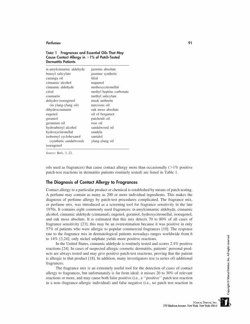
Perfumes 91
TABLE 1 Fragrances and Essential Oils That MayCause Contact Allergy in �1% of Patch-TestedDermatitis Patients
α-amylcinnamic aldehyde jasmine absolutebenzyl salicylate jasmine syntheticcananga oil lilialcinnamic alcohol majantolcinnamic aldehyde methoxycitronellalcitral methyl heptine carbonatecoumarin methyl salicylatedehydro-isoeugenol musk ambrette
(in ylang-ylang oil)dihydrocoumarin
narcissus oil
eugenoloak moss absolute
geranioloil of bergamot
geranium oilpatchouli oil
hydroabietyl alcoholrose oil
hydroxycitronellalsandalwood oil
isobornyl cyclohexanolsandela
(synthetic sandalwood)santalol
isoeugenolylang-ylang oil
Source: Refs. 3, 22.
oils used as fragrances) that cause contact allergy more than occasionally (�1% positivepatch-test reactions in dermatitis patients routinely tested) are listed in Table 1.
The Diagnosis of Contact Allergy to Fragrances
Contact allergy to a particular product or chemical is established by means of patch testing.A perfume may contain as many as 200 or more individual ingredients. This makes thediagnosis of perfume allergy by patch-test procedures complicated. The fragrance mix,or perfume mix, was introduced as a screening tool for fragrance sensitivity in the late1970s. It contains eight commonly used fragrances: α-amylcinnamic aldehyde, cinnamicalcohol, cinnamic aldehyde (cinnamal), eugenol, geraniol, hydroxycitronellal, isoeugenol,and oak moss absolute. It is estimated that this mix detects 70 to 80% of all cases offragrance sensitivity [23]; this may be an overestimation because it was positive in only57% of patients who were allergic to popular commercial fragrances [10]. The responserate to the fragrance mix in dermatological patients nowadays ranges worldwide from 6to 14% [3,24]; only nickel sulphate yields more positive reactions.
In the United States, cinnamic aldehyde is routinely tested and scores 2.4% positivereactions [24]. In cases of suspected allergic cosmetic dermatitis, patients’ personal prod-ucts are always tested and may give positive patch-test reactions, proving that the patientis allergic to that product [18]. In addition, many investigators test (a series of) additionalfragrances.
The fragrance mix is an extremely useful tool for the detection of cases of contactallergy to fragrances, but unfortunately is far from ideal: it misses 20 to 30% of relevantreactions or more, and may cause both false positive (i.e., a ‘‘positive’’ patch test reactionin a non–fragrance-allergic individual) and false negative (i.e., no patch test reaction in
92 de Groot
an individual who is actually allergic to one or more of the ingredients of the mix) reac-tions [25].
Another useful test in cases of doubt (e.g., with weakly positive patch-test reactionsthat are difficult to interpret) is the repeated open application test (ROAT). The suspectedallergen, which may be both an individual fragrance or scented product, is applied to theelbow flexure twice daily for a maximum of 14 days. A positive reaction confirms theexistence of contact allergy and makes relevance of the reaction (vide infra) more likely.
The Relevance of Positive Patch Test Reactions to the Fragrance Mix
The finding of a positive reaction to the fragrance mix should be followed by a searchfor its relevance, i.e., if fragrance allergy is the cause of the patient’s current or previouscomplaints or if it at least contributes to it. Often, however, correlation with the clinicalpicture is lacking and many patients can tolerate perfumes and fragranced products withoutproblem [11]. This sometimes may be explained by irritant (false positive) patch-test reac-tions to the mix. Alternative explanations include the absence of relevant allergens in thoseproducts or a concentration too low to elicit clinically visible allergic contact reactions.
It is assumed that between 50 and 65% of all positive patch-test reactions to themix are relevant, although this is sometimes hard to prove [24,26]. Nevertheless, there isa highly significant association between the occurrence of self-reported visible skin symp-toms to scented products earlier in life and a positive patch test to the fragrance mix, andmost fragrance-sensitive patients are aware that the use of scented products may causeskin problems [27].
In perfume-mix–allergic patients with concomitant positive reactions to perfumesor scented products used by them, interpretation of the reaction as relevant is highly likely.In such patients the incriminated cosmetics very often contain fragrances present in themix, and thus the fragrance mix appears to be a good reflection of actual exposure [18].Indeed, one or more of the ingredients of the mix are present in nearly all deodorants[28], popular prestige perfumes [10], perfumes used in the formulation of other cosmeticproducts [11], and natural-ingredient–based cosmetics [29], often in levels high enoughto cause allergic reactions [30,31]. Thus, fragrance allergens are ubiquitous and virtuallyimpossible to avoid if perfumed cosmetics are used.
CONCLUSIONS
Contact allergy to fragrance materials is common in both eczema patients and in the gen-eral population. Allergic contact dermatitis caused by perfumes and scented cosmetics isusually located in the face (including the eyelids), on the hands, and in the axillae. Patientsappear to become sensitized to fragrances especially by the use of deodorant sprays and/or perfumes, and to a lesser degree by cleansing agents, deodorant sticks, or hand lotions.Thereafter, new rashes may appear or be worsened by contact with other fragranced prod-ucts: cosmetics, toiletries, oral-hygiene products, household products, industrial contacts,paper and paper products, laundered fabrics and clothes, topical drugs, and fragrancesused as flavors in foods and drinks.
Over 100 fragrances have been identified as allergens. The diagnosis of fragranceallergy is established by positive patch-test reactions to the fragrance mix (a mixture ofeight commonly used fragrances) and/or to the patients’ personal perfumes or scentedproducts. Most reactions to the mix are relevant, i.e., fragrance allergy is the cause of the
Perfumes 93
patient’s current or previous complaints, and most fragrance-sensitive patients are awarethat the use of scented products may cause skin problems. One or more of the ingredientsof the mix are present in nearly all deodorants, perfumes, and scented cosmetics, oftenin levels high enough to cause allergic reactions. Industry is advised to pay special atten-tion to the safety evaluation of fragrance materials, notably those used in perfumes anddeodorants.
REFERENCES
1. Ebihara T, Nakayama H. Pigmented contact dermatitis. Clin Dermatol 1997; 15:593–599.2. De Groot AC, Frosch PJ. Fragrances as a cause of contact dermatitis in cosmetics: clinical
aspects and epidemiological data. In: Frosch PJ, Johansen JD, White IR, eds. Fragrances.Beneficial and Adverse Effects. Berlin: Springer-Verlag, 1998:66–75.
3. De Groot AC, Frosch PJ. Adverse reactions to fragrances. A clinical review. Contact Derma-titis 1997; 36:57–86.
4. Frosch PJ, Johansen JD, White IR, eds. Fragrances. Beneficial and Adverse Effects. Berlin:Springer-Verlag, 1998.
5. Guin JD. History, manufacture, and cutaneous reactions to perfumes. In: Frost P, HorwitzSW, eds. Principles of Cosmetics for the Dermatologist. St Louis: The CV Mosby Company,1982: 111–129.
6. Scheinman PL. Allergic contact dermatitis to fragrance: a review. Am J Contact Dermatitis1996; 7:65–76.
7. Guin JD, Berry VK. Perfume sensitivity in adult females. A study of contact sensitivity to aperfume mix in two groups of student nurses. J Am Acad Dermatol 1980; 3:299–302.
8. De Groot AC, Nater JP, van der Lende R, Rijcken B. Adverse effects of cosmetics: a retrospec-tive study in the general population. Int J Cosm Science 1987; 9:255–259.
9. Nielsen NH, Menné T. Allergic contact sensitization in an unselected Danish population. ActaDerm Venereol (Stockh) 1992; 72:456–460.
10. Johansen JD, Rastogi SC, Menné T. Contact allergy to popular perfumes; assessed by patchtest, use test and chemical analysis. Br J Dermatol 1996; 135:419–422.
11. Johansen JD, Rastogi SC, Andersen KE, Menné T. Content and reactivity to product perfumesin fragrance mix positive and negative eczema patients. A study of perfumes used in toiletriesand skin-care products. Contact Dermatitis 1997; 36:291–296.
12. Adams RM, Maibach HI. A five-year study of cosmetic reactions. J Am Acad Dermatol 1985;13:1062–1069.
13. De Groot AC, Bruynzeel DP, Bos JD, van Joost Th, Jagtman BA, Weyland JW. The allergensin cosmetics. Arch Dermatol 1988; 124:1525–1529.
14. Berne B, Boström Å, Grahnén AF, Tammela M. Adverse effects of cosmetics and toiletriesreported to the Swedish Medical Product Agency 1989–1994. Contact Dermatitis 1996; 34:359–362.
15. Dooms-Goossens A, Kerre S, Drieghe J, Bossuyt L, Degreef H. Cosmetic products and theirallergens. Eur J Dermatol 1992; 2:465–468.
16. Larsen W, Nakayama H, Lindberg M, Fisher T, Elsner P, Burrows D, Jordan W, Shaw S,Wilkinson J, Marks J Jr, Sugawara M, Nethercott J. Fragrance contact dermatitis. A worldwidemulticenter investigation (Part I). Am J Contact Dermatitis 1996; 7:77–83.
17. Santucci B, Cristaudo A, Cannistraci C, Picardo M. Contact dermatitis to fragrances. ContactDermatitis 1987; 16:93–95.
18. Johansen JD, Rastogi SC, Menné T. Exposure to selected fragrance materials. A case studyof fragrance-mix-positive eczema patients. Contact Dermatitis 1996; 34:106–110.
19. Dooms-Goossens A. Cosmetics as causes of allergic contact dermatitis. Cutis 1993; 52:316–320.
94 de Groot
20. Meynadier J-M, Raison-Peyron N, Meunier L, Meynadier J. Allergie aux parfums. Rev frAllergol 1997; 37:641–650.
21. Johansen JD, Andersen TF, Kjøller M, Veien N, Avnstorp C, Andersen KE, Menné T. Identi-fication of risk products for fragrance contact allergy: a case-referent study based on patients’histories. Am J Contact Dermatitis 1998; 9:80–87.
22. Larsen WG, Nethercott JR. Fragrances. Clin Dermatol 1997; 15:499–504.23. Larsen WG. Perfume dermatitis. J Am Acad Dermatol 1985; 12:1–9.24. Marks JG Jr, Belsito DV, DeLeo VA, Fowler JF Jr, Fransway AF, Maibach HI, Mathias
CGT, Nethercott JR, Rietschel RL, Sheretz EF, Storrs FJ, Taylor JS. North American ContactDermatitis Group patch test results for the detection of delayed-type hypersensitivity to topicalallergens. J Am Acad Dermatol 1998; 38:911–918.
25. De Groot AC, van der Kley AMJ, Bruynzeel DP, Meinardi MMHM, Smeenk G, van JoostTh, Pavel S. Frequency of false-negative reactions to the fragrance mix. Contact Dermatitis1993; 28:139–140.
26. Frosch PJ, Pilz B, Burrows D, Camarasa JG, Lachapelle J-M, Lahti A, Menné T, WilkinsonJD. Testing with the fragrance mix—is the addition of sorbitan sesquioleate to the constituentsuseful? Contact Dermatitis 1995; 32:266–272.
27. Johansen JD, Andersen TF, Veien N, Avnstorp C, Andersen KE, Menné T. Patch testing withmarkers of fragrance contact allergy. Do clinical tests correspond to patients’ self-reportedproblems? Acta Derm Venereol (Stockh) 1997; 77:149–153.
28. Rastogi SC, Johansen JD, Frosch PJ, Menné T, Bruze M, Lepoittevin JP, Dreier B, AndersenKE, White IR. Deodorants on the European market: quantitative chemical analysis of 21 fra-grances. Contact Dermatitis 1998; 38:29–35.
29. Rastogi S, Johansen JD, Menné T. Natural ingredients based cosmetics. Content of selectedfragrance sensitizers. Contact Dermatitis 1996; 34:423–426.
30. Johansen JD, Andersen KE, Menné T. Quantitative aspects of isoeugenol contact allergy as-sessed by use and patch tests. Contact Dermatitis 1996; 34:414–418.
31. Johansen JD, Andersen KE, Rastogi SC, Menné T. Threshold responses in cinnamic-aldehyde-sensitive subjects: results and methodological aspects. Contact Dermatitis 1996; 34:165–171.
11
In Vitro Tests for Skin Irritation
Michael K. Robinson, Rosemarie Osborne, and Mary A. PerkinsThe Procter & Gamble Company, Cincinnati, Ohio
INTRODUCTION
The manufacture, transport, and marketing of chemicals and finished products requiresthe prior toxicological evaluation and assessment of skin corrosivity and skin irritationthat might result from intended or accidental skin exposure. Traditionally, animal testingprocedures have provided the data needed to assess the more severe forms of skin toxicity,an assessment requiring extrapolation of the data from the animal species to humans [1].Current regulations may require animal test data before permission is granted for the manu-facture, transport, or marketing of chemicals [2], as well as for the formulations that con-tain them [3].
In recent years, animal testing for dermatotoxic effects has come under increasingscrutiny and criticism from animal-rights activists for being inhumane and unnecessary.Legislation is pending that would restrict the marketing of products containing ingredientsthat have been tested on animals [4]. The often conflicting needs to protect worker andconsumer safety, comply with regulatory statutes, and reduce animal testing procedureshas led to a significant effort within industry, government, and academia to develop alter-native testing methods for assessing the skin corrosion and irritation hazard of chemicalsand product formulations without reliance on animal test procedures [5].
A recent example for which regulatory requirements have been coupled to the press-ing need for alternative methods development is in the evaluation of skin corrosion. UnitedStates and international regulations require that chemicals be properly classified, labeled,packaged, and transported on the basis of their potential to damage or destroy tissue,including the speed with which such tissue-destructive reactions occur [2,6]. The mostcommon animal testing methods used over the years for the evaluation of chemical corro-sion potential are all based on the original method by Draize [7]. We, as well as otherlaboratories, have been active in the development of alternative procedures for skin-corro-sion testing [8–11]. Recently, several test methods have been evaluated in an internationalvalidation program [12]. Certain of these methods should provide short-term and cost-effective alternatives to the Draize procedure, at the same time providing experimentalsystems for developing a better mechanistic understanding of the process of skin corrosion[8].
95
96 Robinson et al.
Skin irritation, by definition, is a less severe response than corrosion, but can span arange of responses from near corrosive at one extreme to weak cumulative or neurosensoryresponses at the other. The development of alternatives for skin irritation testing has laggedbehind that of skin corrosion testing, likely because of the greater urgency of developingalternatives for the more severe skin responses and because of the range of responsesencompassed within the ‘‘skin irritation’’ umbrella. Currently, the irritation hazard poten-tial of chemicals is often determined through use of the same Draize procedure used forcorrosion testing, the difference being mainly in the length of chemical exposure, withresults used to determine labeling requirements for chemicals and products according toEuropean Commission (EC) directives [2,3]. For noncorrosive chemicals, there has beena recent effort to develop and promote the use of clinical patch testing methods for a morerelevant assessment of chemical skin irritation potential than that provided by the rabbittest [13–16]. This approach has not yet been extended to the testing of product formula-tions, although the European Cosmetic, Toiletry and Perfumery Association (COLIPA)has recently issued guidelines for skin-compatibility testing of cosmetic formulations inman [17]. The major problem of human testing for skin irritation or compatibility is theextended duration and relatively high cost of this clinical testing. In vitro skin irritationtest methods could be used to rank chemicals or formulations for skin irritation potential,even at the low end of the irritation spectrum [18,19]. These methods (and others underdevelopment elsewhere) might provide for short-term, cost-effective approaches forscreening chemicals and product formulations of interest, so that only those with satisfac-tory skin irritation profiles would undergo longer and more costly clinical evaluations.
This chapter will provide a brief summary of the developmental status of in vitroskin irritation test methods. It includes a brief description and update on the current valida-tion status of skin corrosion tests. Then, it summarizes ongoing efforts in our laboratory,and the work of others, towards development of a battery of skin irritation tests that mightpredict varying degrees of skin irritation potential of chemicals and formulations, includingmany with relatively mild clinical skin irritation properties.
SKIN CORROSION TESTING
Assay Systems
Screening of chemicals for skin corrosion properties in vitro has followed three generalformats. These include 1) changes in electrical conductance across intact skin (rat or hu-man), 2) breaching of noncellular biobarriers, and 3) cellular cytotoxicity in skin or epider-mal equivalent cell culture systems. Each of these systems has been subject to intra- andinterlaboratory development, evaluation, and validation.
Skin corrosivity has been distinguished from skin irritation in two important ways.First, corrosive skin reactions generally occur soon after chemical exposure and are irre-versible. Second, it is thought that the major processes leading to chemical corrosivityare more commonly physicochemical in nature rather than the result of inflammatorybiological events [11], although inflammation is a common consequence of skin corrosion.
Initial efforts to develop a screening test for skin corrosivity examined the effectsof chemical exposure on barrier function of skin through assessment of changes in theresistance of the exposed skin to transmission of electric current [20]. This test method,called transcutaneous electrical resistance (TER), was based on early studies of the electri-
In Vitro Tests for Skin Irritation 97
cal resistance properties of skin [21] and has been developed as a corrosivity assay overthe past 15 years using either rat or human skin [9,11,20,22–26]. In the TER assay, full-thickness skin is stretched over a hollow tube opening with the stratum corneum sideexposed to the lumen. Test materials are applied to the skin surface for varying periodsof time while the skin is immersed in buffer. After chemical exposure, the electrical resis-tance of the skin is measured. TER values empirically established as corrosion thresholdshave been set at 4 K ohms for rat skin and 11 K ohms for human skin [9,11]. The currentvalidation status of this assay is described in the following section.
The biobarrier destruction assay approach for corrosivity testing is exemplified bythe commercial Corrositex assay system manufactured by In Vitro International (Irvine,CA). Like the TER assay, the premise here is physicochemical destruction of a barrierby direct chemical action of a test material. Instead of intact stratum corneum, the Corrosi-tex assay relies on a macromolecular protein matrix as the barrier. Chemicals that breachthis barrier come into contact with an underlying chemical detection system (CDS). Acolor change indicates penetration of the test material into the CDS. The speed with whichthe color change occurs after application of the chemical to the biobarrier is proportionalto the severity of corrosive action. A summary of results on 75 chemicals and detergent-based formulations has been published [10], as well as a recent study on the corrosivityof organosilicon compounds [27]. An update of the current validation status of this assayis provided in the following section.
A variety of cell-based biological assay systems have been developed over the past10 years to investigate the dermatotoxic effects of chemicals and product formulations onthe skin. These have included simple submerged cell cultures, submerged cell coculturesincorporating more than a single cell type, and, more recently, the development of full-thickness skin and epidermal equivalent systems. The latter are characterized by stratifiedepidermal cell layers and a multilayered stratum corneum. The full-thickness culture sys-tems also have different types of cellular and macromolecular matrices serving as a dermalelement. These systems have undergone extensive development and evaluation in variousacademic and commercial laboratories [28–38]. We have recently reviewed features ofmany of the submerged and skin/epidermal equivalent cell systems [39,40]. A few ofthese systems have been used to develop skin corrosion screening assays [8,27]. A reviewof the current validation status of those assays is presented in the following section.
Validation Status
In the early 1990s a program was initiated under the auspices of the European Center forthe Validation of Alternative Methods (ECVAM) to develop and validate alternative meth-ods for the assessment of skin corrosion. This program focused on three assay systems,the TER, Corrositex, and Skin2 systems. The Skin2 system was a commercial ‘‘skinequivalent’’ culture system, manufactured by Advanced Tissue Sciences (La Jolla, CA)and comprising human neonatal foreskin–derived dermal fibroblasts in a collagen matrixgrown on nylon mesh and seeded with human neonatal foreskin–derived epidermal kera-tinocytes to form a stratified and cornified epidermal component. A prevalidation studywas completed with these three assay systems in seven different laboratories to assessintralaboratory and interlaboratory consistency as well as overall sensitivity and specificityof the assays in identifying known corrosive and noncorrosive chemicals. The results ofthe prevalidation study were published in 1995 [41]. All three tests performed well, and
98 Robinson et al.
no firm conclusions could be drawn as to the superiority or inferiority of one test versus theothers. Individual tests had specific problems that warranted further study. These problemsincluded relatively low specificity (TER), a high number of incompatible chemicals (Cor-rositex), and an inferior interlaboratory consistency profile (Skin2). It was recommendedthat effort be made to address these individual deficiencies and that each assay be furtherevaluated in a future validation study.
The formal ECVAM-sponsored skin corrosivity validation study began in early 1995and was completed in October 1997 with the submission of the study findings [12]. Inaddition to the assays included in the prevalidation work (TER, Corrositex, and Skin2),the validation study included a second commercially available skin equivalent cultureconstruct, Episkin (Chaponost, France). Each assay was evaluated by three independenttest laboratories, and each laboratory evaluated only one of the four assays. Hence, 12laboratories participated in the validation study. A total of 60 corrosive and noncorrosivechemicals from a variety of chemical classes (including organic and inorganic acids andbases, neutral organics, phenols, inorganic salts, electrophiles, and soaps/surfactants) weretested [42].
All four assay systems showed acceptable intralaboratory and interlaboratory repro-ducibility, and all but Corrositex were applicable to the testing of all the selected chemi-cals. Two of the assays, TER and Episkin met the first of two major objectives of thevalidation study. They were capable of distinguishing corrosive from noncorrosive chemi-cals with acceptable rates of under- or overprediction. Only the Episkin assay system metthe second major objective of the study, the ability to distinguish between known R35(United Nations packing group I) and R34 (UN packing group II/III) chemicals acrossall of the chemical classes. Only 60% of the test chemicals could be adequately evaluatedby the Corrositex assay. For this reason, it did not meet the criteria for a validated replace-ment test, although it might be valid for certain chemical classes. The Skin2 assay systemshowed high specificity (100% of noncorrosive chemicals were properly identified) butlow sensitivity (only 43% of corrosive chemicals were correctly identified). It also per-formed poorly with respect to distinguishing known R35 and R34 chemicals. Only 35%of the assays conducted on these chemicals resulted in proper classification. Previously,both the Skin2 and Corrositex assays had received exemptions from the U.S. Departmentof Transportation as valid alternatives to assess skin corrosivity based on more limitedevaluation. It is not certain what effect the recent ECVAM-sponsored study will have onthe exemption status of these assays, although for the Skin2 assay it is a moot point giventhat this culture system is no longer commercially available.
SKIN IRRITATION TESTING
Our Experience
Introduction
As previously indicated, development of in vitro methods to assess skin irritation is com-plicated by the fact that skin irritation encompasses a range of clinical responses fromnear corrosive at one extreme to very mild (perhaps sensory only) skin responses at theother. Hence, we believe that test methods and prediction models will need to be optimizedfor different categories of test materials or formulations and for anticipated ranges ofirritation severity. That is the approach we have taken in developing in vitro skin irritationtest methods for several chemical and product categories [32,39,40].
In Vitro Tests for Skin Irritation 99
Methods
Cell Cultures. The culture system used in our studies was a stratified epidermalculture with a stratum corneum obtained from MatTek Corp. (EpiDerm No. EPI-100;Ashland, MA). These cultures were composed of a multilayered and differentiatedepidermis and multilayered stratum corneum seeded onto a permeable transwell filter. Onarrival, the cultures were placed at 4°C until used for experiments (within 24 h). Beforetreatment, the cultures were aseptically transferred to 6-well culture plates containing assaymedium.
Treatments. Test materials were reagent grade chemicals from Sigma ChemicalCo. (St. Louis, MO), Aldrich Chemical Co. (Milwaukee, WI), or The Procter & GambleCo. (Cincinnati, OH). Test-product formulations were obtained from The Procter &Gamble Co. Application of test materials to skin-equivalent cultures was as previouslydescribed [32].
MTT Viability Assay. The MTT assay is a colorimetric method of determining cellviability based on reduction of the yellow tetrazolium salt 3-[4,5-dimethylthiazol-2-yl]2,5-diphenyl tetrazolium bromide (Sigma Chemical Co., St. Louis, MO) to a purpleformazan dye by mitochondrial succinate dehydrogenase in viable cells [43]. This assaywas performed as previously described [8].
Enzyme-Release Assay. At the end of the test material and control treatmentexposures, the assay medium from under each treated or control skin culture was collectedin plastic vials and immediately analyzed for lactate dehydrogenase (LDH) and aspartate-aminotransferase (AST) enzymes. The enzymes, LDH and AST, were analyzed using acolorimetric method performed with a Hitachi 717 autoanalyser with commercial test kits(Boehringer Mannheim Corp., Indianapolis, IN).
Interleukin-1α Assay. Assay medium was recovered from treated and control skincultures (EPI-100) and stored at �20°C until analyzed. Interleukin-1α (IL-1α) wasassayed with a specific enzyme-linked immunoassay kit (Quantikine; R&D Systems, Inc.,Minneapolis, MN).
Results
In vitro methods for screening product formulations for mild to moderate irritation poten-tial can aid selection of formulations for further clinical evaluation. Our approach hasbeen to directly compare in vitro assay endpoints to in vivo human skin responses usinghistoric or concurrent skin-response data for products and ingredients including surfac-tants, cosmetics, antiperspirants, and deodorants. For the in vitro studies we evaluated thecornified human epidermal skin cultures (EpiDerm, MatTek, EPI-100) dosing neat or di-luted test substances to the stratum corneum surface of the skin cultures. The in vitroendpoints included the MTT metabolism assay of cell viability, enzyme release (lactatedehydrogenase and aspartate aminotransferase), and inflammatory cytokine (IL-1α) re-lease.
We have been able to rank order chemicals (surfactants), product formulations andcontrol materials in the in vitro and clinical studies to determine the value of the EpiDermassay system in providing a clinically relevant ordering of irritancy potential. Whereas
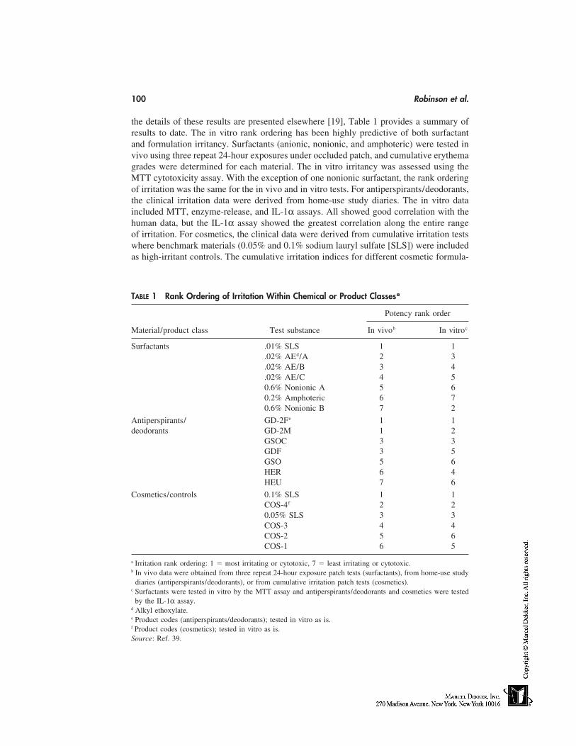
100 Robinson et al.
the details of these results are presented elsewhere [19], Table 1 provides a summary ofresults to date. The in vitro rank ordering has been highly predictive of both surfactantand formulation irritancy. Surfactants (anionic, nonionic, and amphoteric) were tested invivo using three repeat 24-hour exposures under occluded patch, and cumulative erythemagrades were determined for each material. The in vitro irritancy was assessed using theMTT cytotoxicity assay. With the exception of one nonionic surfactant, the rank orderingof irritation was the same for the in vivo and in vitro tests. For antiperspirants/deodorants,the clinical irritation data were derived from home-use study diaries. The in vitro dataincluded MTT, enzyme-release, and IL-1α assays. All showed good correlation with thehuman data, but the IL-1α assay showed the greatest correlation along the entire rangeof irritation. For cosmetics, the clinical data were derived from cumulative irritation testswhere benchmark materials (0.05% and 0.1% sodium lauryl sulfate [SLS]) were includedas high-irritant controls. The cumulative irritation indices for different cosmetic formula-
TABLE 1 Rank Ordering of Irritation Within Chemical or Product Classesa
Potency rank order
Material/product class Test substance In vivob In vitroc
Surfactants .01% SLS 1 1.02% AEd/A 2 3.02% AE/B 3 4.02% AE/C 4 50.6% Nonionic A 5 60.2% Amphoteric 6 70.6% Nonionic B 7 2
Antiperspirants/ GD-2Fe 1 1deodorants GD-2M 1 2
GSOC 3 3GDF 3 5GSO 5 6HER 6 4HEU 7 6
Cosmetics/controls 0.1% SLS 1 1COS-4f 2 20.05% SLS 3 3COS-3 4 4COS-2 5 6COS-1 6 5
a Irritation rank ordering: 1 � most irritating or cytotoxic, 7 � least irritating or cytotoxic.b In vivo data were obtained from three repeat 24-hour exposure patch tests (surfactants), from home-use study
diaries (antiperspirants/deodorants), or from cumulative irritation patch tests (cosmetics).c Surfactants were tested in vitro by the MTT assay and antiperspirants/deodorants and cosmetics were tested
by the IL-1α assay.d Alkyl ethoxylate.e Product codes (antiperspirants/deodorants); tested in vitro as is.f Product codes (cosmetics); tested in vitro as is.Source: Ref. 39.
In Vitro Tests for Skin Irritation 101
tions were compared with the in vitro test data. Again, the IL-1α assays provide the bestcorrelation with the human data across the entire range of clinical irritation responses.
Other Literature
A number of other laboratories have used various constructs of skin cultures to examine thein vitro irritation potential of chemicals and formulations. The developers of the EpiDermcultures examined dose-response profiles to surfactants and surfactant-containing formula-tions, and found a good correlation between residual cell viability measures and clinicalirritation profiles [44]. Later testing of chemical irritants and allergens showed a compara-ble irritant response profile regardless of whether cytotoxicity or cytokine release wasmeasured. However, cytokine release in response to contact allergens occurred at noncyto-toxic doses and was thought to provide additional mechanistic and perhaps a predictiveapplication for these cultures [45]. Recently, the EpiDerm system has been used by agroup from Unilever (Sharnbrook, U.K.) to examine the cytotoxicity patterns of mixedsurfactants [46]. They found that, in vitro as in vivo, mixtures of surfactants produce lessirritation than expected based on the irritation properties of the individual components ofthe mixture, a phenomenon known as antagonism.
A group from Leiden University (Leiden, The Netherlands) has been developingand applying their own unique skin-culture system to the assessment of skin irritationresponses. They have used a system comprising epidermal keratinocytes seeded on de-epidermized dermis (RE-DED) and have tested various skin irritants [34,36]. This groupconfirmed the ability of the RE-DED system to effectively assess skin irritation potentialof the anionic surfactant sodium lauryl sulfate [36]. They also showed that in vitro skinirritation patterns for oleic acid were different in submerged keratinocyte cultures versusthe RE-DED system [34]. In the latter, higher doses were required because of the require-ment for the chemical to penetrate the barrier. Of course, the irritation potential of acidsand bases can also be underestimated in submerged cultures because of the bufferingeffects of the culture media [32,39,40].
Quite recently, another group of researchers (Lyon, France) have used skin-equiva-lent culture systems to examine the irritation potential of cosmetic product formulations.Testing cosmetic formulations of various types (creams, lotions, oils, mascaras), they ob-served a good correlation between in vitro indices of irritation and previously knownDraize irritation indices [47,48]. Like our group, they have used viability, enzyme release,and IL-1α release to profile in vitro skin irritation. All of the above results point to theutility of skin-equivalent culture systems to detect skin irritation responses in vitro in amanner consistent with the clinical skin irritation properties of the chemicals. They offeropportunities for the further development of valid alternative test methods.
DISCUSSION
It has been important to validate the relevance of in vitro skin irritation endpoints to invivo toxicity by confirming the presence of these endpoints in skin models representingvarious levels of skin organization, from intact skin to isolated cell cultures. The initialresponse of human cells to chemical irritants is cell damage, ranging from subtle perturba-tions or biochemical changes to cell death. As a response to damage, skin cells releaseinflammatory mediators and cytokines to initiate a local inflammation response, resulting
102 Robinson et al.
in the visual hallmark of erythema and edema attributable to increased blood flow andleakage of plasma from blood vessels [32,49,50].
The isolated keratinocyte culture represents the simplest of the test systems for eval-uating skin irritancy in vitro. For test materials compatible with the aqueous culture me-dium, there has been an excellent correlation shown between human irritation potentialand in vitro cytotoxicity over several orders of magnitude [32]. However, many types ofchemicals (particularly acids, alkalis, and oxidants) are incompatible with the assay sys-tem. For acids and alkalis, the buffering capacity of the medium will interfere with theirevaluation if pH is a key factor in their in vivo irritancy. Formulations are also difficultto test in vitro because, from a pharmacokinetic standpoint, conditions of exposure ofviable keratinocytes to key irritant components of the formulation may be quite differentin the culture system versus intact skin. Lastly, skin irritation can sometimes be overpre-dicted in these submerged cultures because they bypass the need for chemicals to penetratea stratum corneum barrier [34].
In the late 1980s, cultured human-skin models were developed to provide a hopefultherapeutic approach to skin transplantation. An offshoot of this technology was to provideskin-equivalent culture systems for dermatotoxicity testing. Although clearly not the sameas intact skin, these cultures provided a three-dimensional model of skin with the majorstructural components intact. The availability of cornified versions of these culture systemshas provided for a major advance in development and validation of in vitro skin corrosionand irritation test methods. Although still lacking key cellular elements, these culturesystems have very similar structural features as intact skin, including many of the samestructural proteins, although they are generally more permeable than intact skin. The majoradvantage of these cultures is the ability to test anything that can be applied to and testedon intact human skin, including highly toxic materials. Validation testing has verified theability of at least certain constructs to predict the corrosive potential of chemicals of differ-ent classes [12].
Use of these cultures for testing milder materials (e.g., cosmetics) provides a toolfor early screening of new product formulations in a time- and cost-effective manner priorto more costly clinical evaluations. They also provide a means to investigate mechanismsof skin irritation. Our early efforts using cornified culture systems to screen and rank orderthe mild to moderate skin irritation potential of product ingredients and formulations havebeen highly successful [18,19]. It is well known that the irritation potential of any materialin vivo is a function of both concentration and time of exposure. The in vitro testing ofmaterials that are relatively mild after acute testing, and produce clinical irritation onlyafter chronic or repeated exposure, is complicated by the limited duration of exposurepossible in vitro. In the development of more sensitive in vitro methods, we are lookingto extend the duration of exposure as much as the cultures will allow and/or use noncorni-fied culture systems. Clearly, any increase in permeability of the culture systems versusintact skin (often viewed as a negative property for many applications) can be a benefitfor the skin irritation assessment of relatively mild chemicals or product formulations. Inaddition, skin irritation responses in epidermal skin equivalents, with and without dermalcomponents, are being investigated.
Although the development of one skin-equivalent culture system and the TER assayhave achieved validation status under the recent ECVAM recommendation, the same isnot true for skin irritation assessment. An ECVAM task force recently summarized thestatus of alternative methods for skin irritation testing [51]. A major recommendation wasto continue development of reconstituted human-skin models and preliminary prediction
In Vitro Tests for Skin Irritation 103
models for their use in predictive skin irritation testing. In addition, it was noted thatethical human-skin testing procedures are being developed for skin irritation hazard assess-ment [13–16,52] and deserve consideration in the hierarchical scheme of skin irritationtesting [51].
Many issues remain unanswered in the future development of cell-based in vitroassays for skin toxicity. Continued interlaboratory validation is needed to enhance accep-tance into the regulatory evaluation and approval process. Further refinement and develop-ment of irritation testing methods will enhance the utility of the models for screeningpurposes. Included is the development of ‘‘flanker’’ models that contain additional epider-mal cell types such as melanocytes or Langerhans cells. For example, MatTek (Ashland,MA) has developed a melanocyte containing epidermal model (MelanoDerm) and isinvestigating its use in UVB-protection studies [53]. Finally, the increased reliance onthese models for toxicity testing and irritation screening has also created concerns overtheir long-term commercial supply. Increased use of high-quality culture systems and con-tinued efforts to validate methods using these cultures may help in this process and thusensure future access to this important technology.
NOTE ADDED IN PROOF
In the months since the submission of this chapter, several advances have occurred in thefield of in vitro skin corrosion and irritation testing. In addition to the TER and Episkinassays, a second skin construct, EpiDerm, has now completed successful ‘catch-up’ valida-tion [54,55] and has been endorsed by ECVAM as an alternative skin corrosivity test[56]. Also, the noncellular corrosion assay, Corrositex, was cited by the U.S. InteragencyCoordinating Committee on the Validation of Alternative Methods (ICCVAM) as equiva-lent to the Draize test for predicting corrosivity and noncorrosivity for specified chemicalclasses (acids and bases) [57]. In the European Union, a new test method on skin corrosion(including the rat skin TER and human skin model assays) has just been incorporated intoAnnex V of Directive 67/548/EEC [58], and a draft guideline on in vitro tests for skincorrosion is under consideration by the Organization for Economic Cooperation and De-velopment (OECD) member countries. In regard to in vitro skin irritation test methods,efforts are currently underway to identify potential in vitro acute skin irritation test meth-ods and evaluate them through rigorous prevalidation and validation studies [59].
REFERENCES
1. OECD guideline for testing of chemicals. Guideline No. 404. Acute dermal irritation/corrosion1992.
2. EEC. Annex I to Commission Directive 91/325/EEC of 1st March 1991 adapting to technicalprogress for the twelfth time Council Directive 67/548/EEC on the approximation of the laws,regulations and administrative provision relating to the classification, packaging and labelingof dangerous substances. Off J Eur Comm 1991; L180:34.
3. EEC. Council Directive of 7 June 1988 on the approximation of the laws, regulations andadministrative provisions of the Member States relating to the classification, packaging andlabeling of dangerous preparations. Off J Eur Comm 1988; L18:14.
4. EEC. Council Directive 93/35/EEC of 14 June 1993 amending for the 6th time Directive 76/768/EEC on the approximation of the laws of the Member States relating to cosmetic products.Off J Eur Comm 1993; L15:32.
104 Robinson et al.
5. Rougier A, Goldberg AM, Maibach HI, eds. In Vitro Skin Toxicology. New York: Mary AnnLiebert, Inc., 1994.
6. Department of Transportation. Method of testing corrosion to the skin. 1991; Title 49, Appen-dix A: Code of Federal Regulations.
7. Draize JH, Woodard G, Calvery HO. Methods for the study of irritation and toxicity of sub-stances applied topically to the skin and mucous membranes. J Pharm Exp Therap 1944; 82:377–390.
8. Perkins MA, Osborne R, Johnson GR. Development of an in vitro method for skin corrosiontesting. Fundam Appl Toxicol 1996; 31:9–18.
9. Whittle E, Barratt MD, Carter JA, Basketter DA, Chamberlain M. Skin corrosivity potentialof fatty acids: in vitro rat and human skin testing and QSAR studies. Toxicol In Vitro 1996;10:95–100.
10. Gordon VC, Harvell JD, Maibach HI. Dermal corrosion, the CORROSITEX system: a DOTaccepted method to predict corrosivity potential of test materials. In: Rougier A, GoldbergAM, Maibach HI, eds. In Vitro Skin Toxicology. New York: Mary Ann Liebert, 1994:37–45.
11. Lewis RW, Botham PA. Measurement of transcutaneous electrical resistance to assess theskin corrosivity potential of chemicals. In: Rougier A, Goldberg AM, Maibach HI, eds. InVitro Skin Toxicology. New York: Mary Ann Liebert, 1994:161–169.
12. Fentem JH, Archer GEB, Balls M, Botham PA, Curren RD, Earl LK, Esdaile DJ, HolzhütterHG, Liebsch M. The ECVAM international validation study on in vitro tests for skin corro-sivity. 2. Results and evaluation by the management team. Toxicol In Vitro 1998; 12:483–524.
13. Basketter DA, Whittle E, Griffiths HA, York M. The identification and classification of skinirritation hazard by a human patch test. Food Chem Toxicol 1994; 32:769–775.
14. York M, Griffiths HA, Whittle E, Basketter DA. Evaluation of a human patch test for theidentification and classification of skin irritation potential. Contact Dermatitis 1996; 34:204–212.
15. Griffiths HA, Wilhelm KP, Robinson MK, Wang XM, McFadden J, York M, Basketter DA.Interlaboratory evaluation of a human patch test for the identification of skin irritationpotential/hazard. Food Chem Toxicol 1997; 35:255–260.
16. Robinson MK, Perkins MA, Basketter DA. Application of a 4-h human patch test method forcomparative and investigative assessment of skin irritation. Contact Dermatitis 1998; 38:194–202.
17. Walker AP, Basketter DA, Baverel M, Diembeck W, Matthies W, Mougin D, Paye M, Roth-lisberger R, Dupuis J. Test guidelines for assessment of skin compatibility of cosmetic finishedproducts in man. Food Chem Toxicol 1996; 34:651–660.
18. Perkins MA, Osborne R, Robinson MK, Rana F, Ghassemi A, Hall B. Comparison of in vitroand in vivo human skin responses to consumer products and ingredients with a range of irri-tancy potential. Fundam Appl Toxicol 1996; 30(abstr):168–169.
19. Perkins MA, Osborne R, Rana F, Ghassemi A, Robinson MK. Comparison of in vitro and invivo human skin responses to consumer products and ingredients with a range of irritancypotential. Toxicological Sciences 1999; 48:218–229.
20. Oliver GJ, Pemberton MA, Rhodes C. An in vitro skin corrosivity test—modifications andvalidation. Food Chem Toxicol 1986; 24:507–512.
21. Blank IH, Finesinger JE. Electrical resistance of the skin. Arch Neurol Psychiat 1964; 56:544–557.
22. Oliver GJ, Pemberton MA. An in vitro epidermal slice technique for identifying chemicalswith potential for severe cutaneous effects. Food Chem Toxicol 1985; 23:229–232.
23. Oliver GJA, Pemberton MA, Rhodes C. An in vitro model for identifying skin-corrosive chem-icals: I. Initial validation. Toxicol In Vitro 1988; 2:7–18.
24. Barlow A, Hirst R, Pemberton MA, Rigden A, Hall TJ, Oliver G-JA, Botham PA. Refinement
In Vitro Tests for Skin Irritation 105
of an in vitro test for the identification of skin corrosive chemicals. Toxicol Methods 1991;1:106–115.
25. Botham PA, Hall TJ, Dennett R, McCall JC, Basketter DA, Whittle E, Cheeseman M, EsdaileDJ, Gardner J. The skin corrosivity test in vitro: results of an interlaboratory trial. Toxicol InVitro 1992; 6:191–194.
26. Basketter DA, Whittle E, Chamberlain M. Identification of irritation and corrosion hazards toskin: an alternative strategy to animal testing. Food Chem Toxicol 1994; 32:539–542.
27. Cassidy SL, Stanton ES. In vitro skin irritation and corrosivity studies on organosilicon com-pounds. J Toxicol Cutan Ocul Toxicol 1996; 15:355–367.
28. Harvell J, Bason MM, Maibach HI. In vitro skin irritation assays: relevance to human skin.J Toxicol Clin Toxicol 1992; 30:359–369.
29. Harvell J, Maibach HI. In vitro dermal toxicity tests: validation aspects. Cosmet Toiletries1992; 107:31–34.
30. Harvell JD, Maibach HI. Validation of in vitro skin irritation assays using human in vivo data.In Vitro Toxicol 1992; 5:235–239.
31. Harvell JD, Tsai YC, Maibach HI, Gay R, Gordon VC, Miller K, Munn GC. An in vivocorrelation with three in vitro assays to assess skin irritation potential. J Toxicol-Cutan OculToxicol 1994; 13:171–183.
32. Osborne R, Perkins MA. An approach for development of alternative test methods based onmechanisms of skin irritation. Food Chem Toxicol 1994; 32:133–142.
33. Rheins LA, Edwards SM, Miao O, Donnelly TA. Skin(2TM): an in vitro model to assess cutane-ous immunotoxicity. Toxicol In Vitro 1994; 8:1007–1014.
34. Boelsma E, Tanojo H, Bodde HE, Ponec M. Assessment of the potential irritancy of oleicacid on human skin: evaluation in vitro and in vivo. Toxicol In Vitro 1996; 10:729–742.
35. Ponec M. The use of in vitro skin recombinants to evaluate cutaneous toxicity. In: RougierA, Goldberg AM, Maibach HI, eds. In Vitro Skin Toxicology. New York: Mary Ann Liebert,Inc., 1994:107–116.
36. Ponec M, Kempenaar J. Use of human skin recombinants as an in vitro model for testing theirritation potential of cutaneous irritants. Skin Pharmacol 1995; 8:49–59.
37. Lawrence JN. Application of in vitro human skin models to dermal irritancy: a brief overviewand future prospects. Toxicol In Vitro 1997; 11:305–312.
38. Rosdy M, Bertino B, Butet V, Gibbs S, Ponec M, Darmon M. Retinoic acid inhibits epidermaldifferentiation when applied topically on the stratum corneum of epidermis formed in vitroby human keratinocytes grown on defined medium. In Vitro Toxicol 1997; 10:39–47.
39. Robinson MK, Perkins MA, Osborne R. Comparative studies on cultured human skin modelsfor irritation testing. In: van Zutphen LFM, Balls M, eds. Animal Alternatives, Welfare andEthics. Amsterdam: Elsevier, 1997:1123–1134.
40. Perkins MA, Robinson MK, Osborne R. Alternative methods in dermatotoxicology. In: Mar-zulli FN, Maibach HI, eds. Dermatotoxicology Methods. Washington, DC: Taylor & Francis,1998:319–336.
41. Botham PA, Chamberlain M, Barratt MD, Curren RD, Esdaile DJ, Gardner JR, Gordon VC,Hildebrand B, Lewis RW, Liebsch M, Logemann P, Osborne R, Ponec M, Regnier JF, Stei-ling W, Walker AP, Balls M. A prevalidation study on in vitro skin corrosivity testing. Thereport and recommendations of ECVAM workshop 6. ATLA-Altern Lab Anim 1995; 23:219–255.
42. Barratt MD, Brantom PG, Fentem JH, Gerner I, Walker AP, Worth AP. The ECVAM interna-tional validation study on in vitro tests for skin corrosivity. 1. Selection and distribution ofthe test chemicals. Toxicol In Vitro 1998; 12:471–482.
43. Mossman T. Rapid colorimetric assay for cellular growth and survival: applications to prolifer-ation and cytotoxicity assays. J Immunol Methods 1983; 65:55–63.
44. Cannon CL, Neal PJ, Southee JA, Kubilus J, Klausner M. New epidermal model for dermalirritancy testing. Toxicol In Vitro 1994; 8:889–891.
106 Robinson et al.
45. Kubilus J, Cannon C, Neal P, Sennott H, Klausner M. Response of the EpiDerm skin modelto topically applied irritants and allergens. In Vitro Toxicol 1996; 9:157–166.
46. Holland G, Earl LK, Hall-Manning TJ. Assessment of the skin irritation effect of mixed surfac-tants using the 4 hour human patch test and EpiDerm EPI-100 in vitro skin model. Proceedingsof 38th International Detergency Conference 1998:81–85.
47. Augustin C, Collombel C, Damour O. Use of dermal equivalent and skin equivalent modelsfor identifying phototoxic compounds in vitro. Photodermatol Photoimmunol Photomedicine1997; 13:27–36.
48. Augustin C, Collombel C, Damour O. Use of dermal equivalent and skin equivalent modelsfor in vitro cutaneous irritation testing of cosmetic products: comparison with in vivo humandata. J Toxicol Cutan Ocul Toxicol 1998; 17:5–17.
49. Willis CM. The histopathology of irritant contact dermatitis. In: van der Valk PGM, MaibachHI, eds. The Irritant Contact Dermatitis Syndrome. Boca Raton: CRC Press, 1996:291–303.
50. Thestrup-Pedersen K, Halkier-Sorensen L. Mechanisms of irritant contact dermatitis. In: vander Valk PGM, Maibach HI, eds. The Irritant Contact Dermatitis Syndrome. Boca Raton: CRCPress, 1996:305–309.
51. Botham PA, Earl LK, Fentem JH, Roguet R, Johannes JM. Alternative methods for skin irrita-tion testing: the current state. ATLA Altern Lab Anim 1998; 26:195–211.
52. Basketter DA, Chamberlain M, Griffiths HA, Rowson M, Whittle E, York M. The classifica-tion of skin irritants by human patch test. Food Chem Toxicol 1997; 35:845–852.
53. Kubilus J, Neal PJ, Klausner M. Initial characterization of an epidermal model containingfunctional melanocytes. J Invest Dermatol 1995; 104(abstr):616.
54. Balls M, Fentem JH. The validation and acceptance of alternatives to animal testing. Toxicol-ogy In Vitro 1999; 13:837–846.
55. Liebsch M, Traue D, Barrabas C, Spielmann H, Uphill P, Wilkins S, Wiemann C, KaufmannT, Remmele M, Holzhütter HG. The ECVAM prevalidation study on the use of EpiDerm forskin corrosivity testing. ATLA Altern Lab Anim 2000; 28:371–401.
56. ECVAM. Statement on the application of the Epiderm human skin model for skin corrosivitytesting. ATLA-Altern Lab Anim 2000; 28:365–366.
57. Scala R, Fentem JH, Chen J, Derelanko MJ, Green S, Harbell J, Kohrman KA, Sauder DN,Stegeman J. Corrositex: An in vitro test method for assessing dermal corrosivity potentialof chemicals. 1999; URL:http://iccvam.niehs.nih.gov/corprep.htm.
58. EEC. Annex I to Commission Directive 2000/33/EC adapting to technical progress for the27th time Council Directive 67/548/EEC on the approximation of laws, regulations and ad-ministrative provisions relating to the classification, packaging and labeling of dangerous sub-stances. Official Journal of the European Communities 2000; L136:91–97.
59. Fentem JH, Botham PA, Earl LK, Roguet R, van de Sandt JJM. Prevalidation of in vitro testsfor acute skin irritation. In: Clark DG, Lisansky SG, Macmillan R, eds. Alternatives to AnimalTesting. II. Proceedings of the Second International Scientific Conference Organised by theEuropean Cosmetic Industry. Newbury, U.K.: CPL Press, 1999:228–231.
12
In Vivo Irritation
Saqib J. Bashir and Howard I. MaibachUniversity of California at San Francisco School of Medicine,San Francisco, California
INTRODUCTION
Irritant Dermatitis
Skin irritation is a localized nonimmunologically mediated inflammatory process. It maymanifest objectively with skin changes such as erythema, edema, and vesiculation, orsubjectively with the complaints of burning, stinging, or itching, with no detectable visibleor microscopic changes. Several forms of objective irritation exist (see Table 1). Acuteirritant dermatitis may follow a single, usually accidental, exposure to a potent irritantand generally heals soon after exposure. An irritant reaction may be seen in individualssuch as hairdressers and wet-work performing employees, who are more extensively andregularly exposed to irritants. Repeated irritant reactions may develop into a contact der-matitis, which generally has a good prognosis. Other forms of irritant dermatitis includedelayed acute irritant contact dermatitis, which occurs when there is a delay betweenexposure and inflammation, and cumulative irritant dermatitis, which is the most commonform of irritant contact dermatitis. After exposure, an acute irritant dermatitis is not seenbut invisible skin changes occur, which eventually lead to an irritant dermatitis whenexposure reaches a threshold point. This may follow days, weeks, or years of exposure[1]. These various forms require specialized models to predict their occurrence after expo-sure to specific products.
Need for Models
Prevention of skin irritation is important for both the consumer who will suffer from itand for the industry, which needs a licensable and marketable product. Accurate predictionof the irritation potential of industrial, pharmaceutical, and cosmetic materials is thereforenecessary for the consumer health and safety and for product development. Presently,animal models fulfill licensing criteria for regulatory bodies. In the European Union, ani-mal testing for cosmetics was to be banned in 1998; however, the deadline was extended toJune 30, 2000 because scientifically validated models were not available. Until alternativemodels can be substituted, in vivo models provide a means by which a cosmetic can be
107
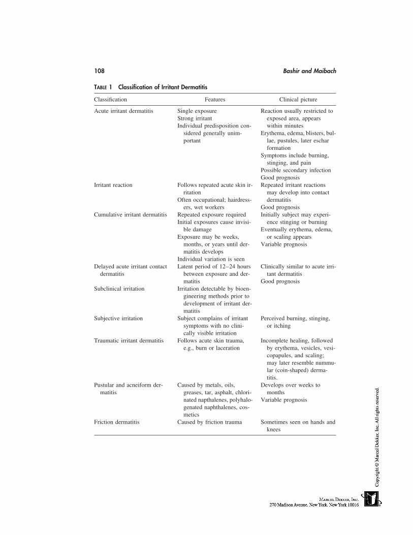
108 Bashir and Maibach
TABLE 1 Classification of Irritant Dermatitis
Classification Features Clinical picture
Acute irritant dermatitis Single exposure Reaction usually restricted toStrong irritant exposed area, appearsIndividual predisposition con- within minutes
sidered generally unim- Erythema, edema, blisters, bul-portant lae, pustules, later eschar
formationSymptoms include burning,
stinging, and painPossible secondary infectionGood prognosis
Irritant reaction Follows repeated acute skin ir- Repeated irritant reactionsritation may develop into contact
Often occupational; hairdress- dermatitisers, wet workers Good prognosis
Cumulative irritant dermatitis Repeated exposure required Initially subject may experi-Initial exposures cause invisi- ence stinging or burning
ble damage Eventually erythema, edema,Exposure may be weeks, or scaling appears
months, or years until der- Variable prognosismatitis develops
Individual variation is seenDelayed acute irritant contact Latent period of 12–24 hours Clinically similar to acute irri-
dermatitis between exposure and der- tant dermatitismatitis Good prognosis
Subclinical irritation Irritation detectable by bioen-gineering methods prior todevelopment of irritant der-matitis
Subjective irritation Subject complains of irritant Perceived burning, stinging,symptoms with no clini- or itchingcally visible irritation
Traumatic irritant dermatitis Follows acute skin trauma, Incomplete healing, followede.g., burn or laceration by erythema, vesicles, vesi-
copapules, and scaling;may later resemble nummu-lar (coin-shaped) derma-titis.
Pustular and acneiform der- Caused by metals, oils, Develops over weeks tomatitis greases, tar, asphalt, chlori- months
nated napthalenes, polyhalo- Variable prognosisgenated naphthalenes, cos-metics
Friction dermatitis Caused by friction trauma Sometimes seen on hands andknees
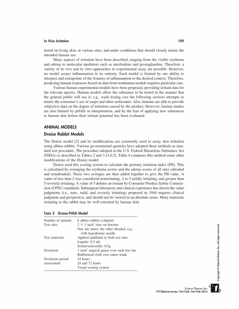
In Vivo Irritation 109
tested on living skin, at various sites, and under conditions that should closely mimic theintended human use.
Many aspects of irritation have been described, ranging from the visible erythemaand edema to molecular mediators such as interleukins and prostaglandins. Therefore, avariety of in vivo and in vitro approaches to experimental assay are possible. However,no model assays inflammation in its entirety. Each model is limited by our ability tointerpret and extrapolate of the features of inflammation to the desired context. Therefore,predicting human responses based on data from nonhuman models requires particular care.
Various human experimental models have been proposed, providing irritant data forthe relevant species. Human models allow the substance to be tested in the manner thatthe general public will use it; e.g., wash testing (see the following section) attempts tomimic the consumer’s use of soaps and other surfactants. Also, humans are able to providesubjective data on the degree of irritation caused by the product. However, human studiesare also limited by pitfalls in interpretation, and by the fear of applying new substancesto human skin before their irritant potential has been evaluated.
ANIMAL MODELS
Draize Rabbit Models
The Draize model [2] and its modifications are commonly used to assay skin irritationusing albino rabbits. Various governmental agencies have adopted these methods as stan-dard test procedure. The procedure adopted in the U.S. Federal Hazardous Substance Act(FHSA) is described in Tables 2 and 3 [3,4,5]. Table 4 compares this method some othermodifications of the Draize model.
Draize used this scoring system to calculate the primary irritation index (PII). Thisis calculated by averaging the erythema scores and the edema scores of all sites (abradedand nonabraded). These two averages are then added together to give the PII value. Avalue of less than 2 was considered nonirritating, 2 to 5 mildly irritating, and greater than5 severely irritating. A value of 5 defines an irritant by Consumer Product Safety Commis-sion (CPSC) standards. Subsequent laboratory and clinical experience has shown the valuejudgments (i.e., non-, mild, and severely irritating) proposed in 1944 requires clinicaljudgment and perspective, and should not be viewed in an absolute sense. Many materialsirritating to the rabbit may be well tolerated by human skin.
TABLE 2 Draize-FHSA Model
Number of animals 6 albino rabbits (clipped)Test sites 2 � 1 inch2 sites on dorsum
One site intact, the other abraded, e.g.,with hypodermic needle
Test materials Applied undiluted to both test sitesLiquids: 0.5 mLSolids/semisolids: 0.5g
Occlusion 1 inch2 surgical gauze over each test siteRubberized cloth over entire trunk
Occlusion period 24 hoursAssessment 24 and 72 hours
Visual scoring system
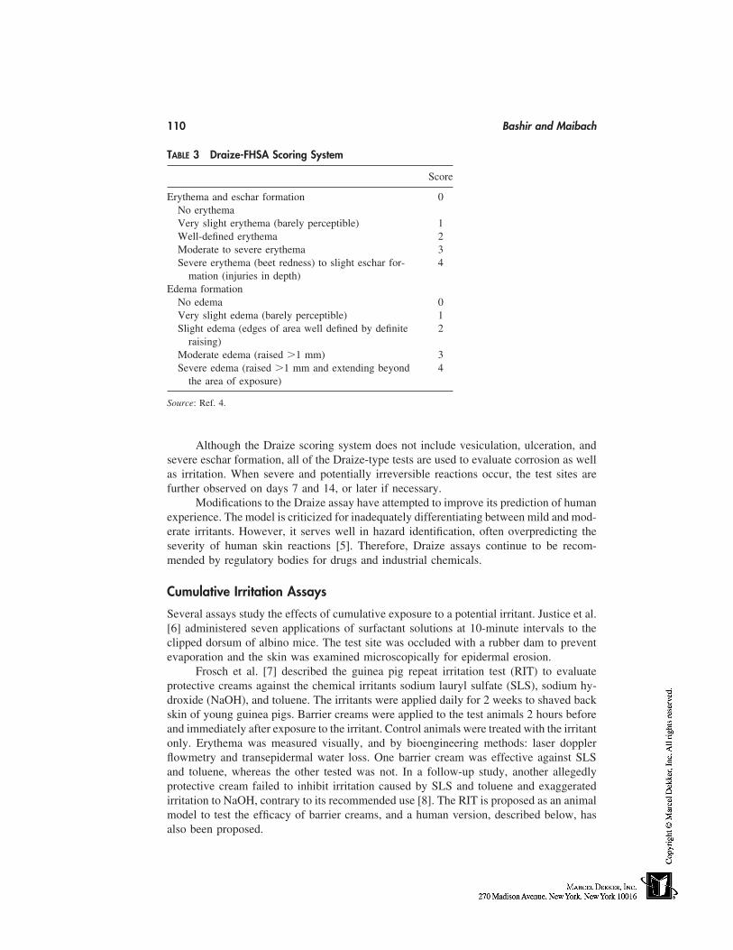
110 Bashir and Maibach
TABLE 3 Draize-FHSA Scoring System
Score
Erythema and eschar formation 0No erythemaVery slight erythema (barely perceptible) 1Well-defined erythema 2Moderate to severe erythema 3Severe erythema (beet redness) to slight eschar for- 4
mation (injuries in depth)Edema formation
No edema 0Very slight edema (barely perceptible) 1Slight edema (edges of area well defined by definite 2
raising)Moderate edema (raised �1 mm) 3Severe edema (raised �1 mm and extending beyond 4
the area of exposure)
Source: Ref. 4.
Although the Draize scoring system does not include vesiculation, ulceration, andsevere eschar formation, all of the Draize-type tests are used to evaluate corrosion as wellas irritation. When severe and potentially irreversible reactions occur, the test sites arefurther observed on days 7 and 14, or later if necessary.
Modifications to the Draize assay have attempted to improve its prediction of humanexperience. The model is criticized for inadequately differentiating between mild and mod-erate irritants. However, it serves well in hazard identification, often overpredicting theseverity of human skin reactions [5]. Therefore, Draize assays continue to be recom-mended by regulatory bodies for drugs and industrial chemicals.
Cumulative Irritation Assays
Several assays study the effects of cumulative exposure to a potential irritant. Justice et al.[6] administered seven applications of surfactant solutions at 10-minute intervals to theclipped dorsum of albino mice. The test site was occluded with a rubber dam to preventevaporation and the skin was examined microscopically for epidermal erosion.
Frosch et al. [7] described the guinea pig repeat irritation test (RIT) to evaluateprotective creams against the chemical irritants sodium lauryl sulfate (SLS), sodium hy-droxide (NaOH), and toluene. The irritants were applied daily for 2 weeks to shaved backskin of young guinea pigs. Barrier creams were applied to the test animals 2 hours beforeand immediately after exposure to the irritant. Control animals were treated with the irritantonly. Erythema was measured visually, and by bioengineering methods: laser dopplerflowmetry and transepidermal water loss. One barrier cream was effective against SLSand toluene, whereas the other tested was not. In a follow-up study, another allegedlyprotective cream failed to inhibit irritation caused by SLS and toluene and exaggeratedirritation to NaOH, contrary to its recommended use [8]. The RIT is proposed as an animalmodel to test the efficacy of barrier creams, and a human version, described below, hasalso been proposed.
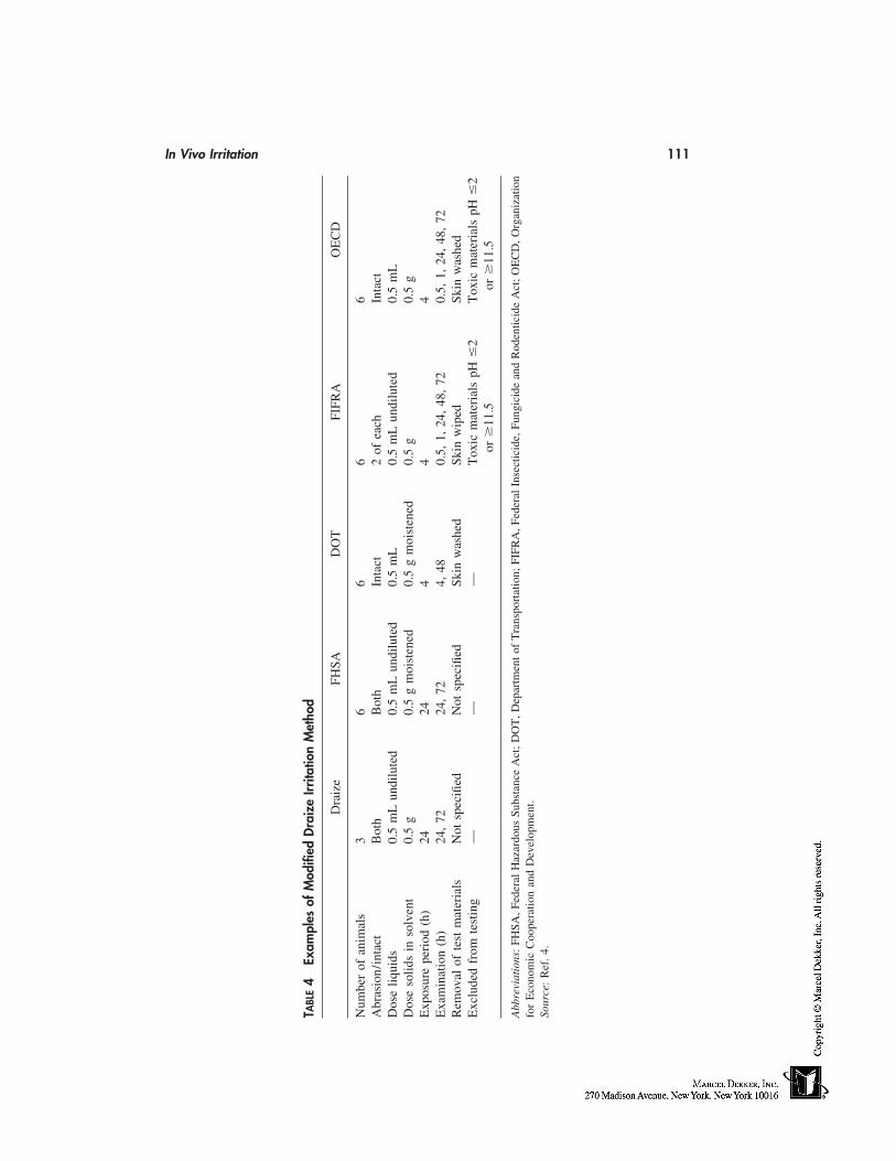
In Vivo Irritation 111
TABL
E4
Exam
ples
ofM
odifi
edD
raiz
eIrr
itatio
nM
etho
d
Dra
ize
FHSA
DO
TFI
FRA
OE
CD
Num
ber
ofan
imal
s3
66
66
Abr
asio
n/in
tact
Bot
hB
oth
Inta
ct2
ofea
chIn
tact
Dos
eliq
uids
0.5
mL
undi
lute
d0.
5m
Lun
dilu
ted
0.5
mL
0.5
mL
undi
lute
d0.
5m
LD
ose
solid
sin
solv
ent
0.5
g0.
5g
moi
sten
ed0.
5g
moi
sten
ed0.
5g
0.5
gE
xpos
ure
peri
od(h
)24
244
44
Exa
min
atio
n(h
)24
,72
24,
724,
480.
5,1,
24,
48,
720.
5,1,
24,
48,
72R
emov
alof
test
mat
eria
lsN
otsp
ecifi
edN
otsp
ecifi
edSk
inw
ashe
dSk
inw
iped
Skin
was
hed
Exc
lude
dfr
omte
stin
g—
——
Tox
icm
ater
ials
pH�
2T
oxic
mat
eria
lspH
�2
or
11.5
or
11.5
Abb
revi
atio
ns:
FHSA
,Fed
eral
Haz
ardo
usSu
bsta
nce
Act
;D
OT
,Dep
artm
ent
ofT
rans
port
atio
n;FI
FRA
,Fed
eral
Inse
ctic
ide,
Fung
icid
ean
dR
oden
ticid
eA
ct;
OE
CD
,Org
aniz
atio
nfo
rE
cono
mic
Coo
pera
tion
and
Dev
elop
men
t.So
urce
:R
ef.
4.
112 Bashir and Maibach
Repeat application patch tests have been developed to rank the irritant potential ofproducts. Putative irritants are applied to the same site for 3 to 21 days, under occlusion.The degree of occlusion influences percutaneous penetration, which may in turn influencethe sensitivity of the test. Patches used vary from Draize-type gauze dressings to metalchambers. Therefore, a reference irritant material is often included in the test to facilitateinterpretation of the results. Various animal species have also been used, such as the guineapig and the rabbit [9,10]. Wahlberg measured skinfold thickness with Harpenden calipersto assess the edema-producing capacity of chemicals in guinea pigs. This model showedclear dose-response relationships and discriminating power, except for acids and alkaliswhere no change in skinfold thickness was found.
Open application assays are also used for repeat irritation testing. Marzulli and Mai-bach [11] described a cumulative irritation assay in rabbits that uses open applicationsand control reference compounds. The test substances are applied 16 times over a 3-weekperiod and the results are measured with a visual score for erythema and skin thicknessmeasurements. These two parameters correlated highly. A significant correlation was alsoshown between the scores of 60 test substances in the rabbit and in man, suggesting thatthe rabbit assay is a powerful predictive model.
Anderson et al. [12] used an open application procedure in guinea pigs to rank weakirritants. A baseline response to SLS solution was obtained after 3 applications per dayfor 3 days to a 1 cm2 test area. This baseline is used to compare other irritants, of whichtrichloroethane was the most irritant, similar to 2% SLS. Histology showed a mononucleardermal inflammatory response.
Immersion Assay
The guinea pig immersion assay was developed to assess the irritant potential of aqueoussurfactant–based solutions, but might be extended to other occupational settings such asaqueous cutting fluids. Restrained guinea pigs are immersed in the test solution whilemaintaining their head above water. The possibility of systemic absorption of a lethal doserestricts the study to products of limited toxic potential. Therefore, the test concentrationis usually limited to 10%.
Ten guinea pigs are placed immersed in a 40°C solution for 4 hours daily for threedays. A comparison group is immersed in a reference solution. Twenty-four hours afterthe final immersion, the animals’ flanks are shaved and evaluated for erythema, edema,and fissures [13,14,15,16]. Gupta et al. [17] concomitantly tested the dermatotoxic effectsof detergents in guinea pigs and humans, using the immersion test and the patch test,respectively. Epidermal erosion and a 40 to 60% increase in the histamine content of theguinea pig skin was found, in addition to a positive patch test reaction in seven of eightsubjects.
Mouse Ear Model
Uttley and Van Abbe [18] applied undiluted shampoos to one ear of mice daily for fourdays, visually quantifying the degree of inflammation as vessel dilatation, erythema, andedema. Patrick and Maibach [19] measured ear thickness to quantify the inflammatoryresponse to surfactant–based products and other chemicals. This allowed quantificationof dose-response relationships and comparison of chemicals. Inoue et al. [20] used thismodel to compare the mechanism of mustard oil–induced skin inflammation to the mecha-nism of capsaicin-induced inflammation. Mice were pretreated with various receptor an-
In Vivo Irritation 113
tagonists, such as 5-HT2, H1, and tachykinin antagonists, showing that the tachykinin NK1receptor was an important mediator of inflammation induced by mustard oil. The mousemodels provide simplicity and objective measurements. Relevance for man requires eluci-dation.
Other Methods
Several other assays of skin irritation have been suggested. Humphrey [21] quantified theamount of Evans blue dye recovered from rat skin after exposure to skin irritants. Trushet al. [22] used myeloperoxidase in polymorphonuclear leukocytes as a biomarker forcutaneous inflammation.
HUMAN MODELS
Human models for skin irritation testing are species relevant, thereby eliminating the pre-carious extrapolation of animal and in vitro data to the human setting. As the requiredtest area is small, several products or concentrations can be tested simultaneously andcompared. Inclusion of a reference irritant substance facilitates interpretation of the irritantpotential of the test substances. Prior animal or in vitro studies, depending on model rele-vance and regulatory issue, can be used to exclude particularly toxic substances or concen-trations before human exposure.
Single-Application Patch Testing
The National Academy of Sciences (NAS) [23] outlined a single-application patch testprocedure determining skin irritation in humans. Occlusive patches may be applied to theintrascapular region of the back or the volar surface of the forearms, using a relativelynonocclusive tape for new or volatile materials. More occlusive tapes or chambers gener-ally increase the severity of the responses. A reference material is included in each batteryof patches.
The exposure time may vary to suit the study. NAS suggests a 4-hour exposureperiod, although it may be desirable to test new or volatile materials for 30 minutes to 1hour. Studies longer than 24 hours have been performed. Skin responses are evaluated30 minutes to 1 hour after removal of the patch, using the animal Draize scale (Table 2)or similar. Kligman and Wooding [24] described statistical analysis on test data to calcu-late the IT50 (time to produce imitation in 50% of the subjects) and the ID50 (dose requiredto produce irritation in 50% of the subjects after a 24-hour exposure).
Robinson et al. [25] suggested a 4-hour patch test as an alternative to animal testing.Assessing erythema by visual scoring, they tested a variety of irritants on Caucasians andAsians. A relative ranking of irritancy was obtained using 20% SLS as a benchmark.Taking this model further, McFadden et al. [26] investigated the threshold of skin irritationin the six different skin types. Again using SLS as a benchmark, they defined the skinirritant threshold as the lowest concentration of SLS that would produce skin irritationunder the 4-hour occluded patch conditions. They found no significant difference in irrita-tion between the skin types.
Cumulative Irritation Testing
Lanman et al. [27] and Phillips et al. [9] described a cumulative irritation assay, whichhas become known as the ‘‘21-day’’ cumulative irritation assay. The purpose of the test
114 Bashir and Maibach
was to screen new formulas before marketing. A 1 inch square of Webril was saturatedwith liquid of 0.5 g of viscous substances and applied to the surface of the pad to beapplied to the skin. The patch was applied to the upper back and sealed with occlusivetape. The patch is removed after 24 hours, and then reapplied after examination of thetest site. This is repeated for 21 days and the IT50 can then be calculated. Note that theinterpretation of the data is best done by comparing the data to an internal standard forwhich human clinical experience exists.
Modifications have been made to this method. The chamber scarification test (seethe following) was developed to predict the effect of repeated applications of a potentialirritant to damaged skin, rather than healthy skin. The cumulative patch test describedabove had failed to predict adverse reactions to skin damaged by acne or shaving, orsensitive areas such as the face [28].
Wigger-Alberti et al. [29] compared two cumulative models by testing skin reactionto metalworking fluids (MWF). Irritation was assessed by visual scoring, transepidermalwater loss, and chromametry. In the first method, MWF were applied with Finn Chamberson the volunteers’ midback, removed after 1 day of exposure, and reapplied for a further2 days. In the second method, cumulative irritant contact dermatitis was induced using arepetitive irritation test for 2 weeks (omitting weekends) for 6 hours per day. The 3-daymodel was preferred because of its shorter duration and better discrimination of irritancy.For low-irritancy materials in which discrimination is not defined with visual and palpatoryscores, bioengineering methods (i.e., transepidermal water loss) may be helpful.
The Chamber Scarification Test
This test was developed [30,31] to test the irritant potential of products on damaged skin.Six to eight 1 mm sites on the volar forearm were scratched eight times with a 30-gaugeneedle without causing bleeding. Four scratches were parallel and the other four are per-pendicular to these. Duhring chambers, containing 0.1 g of test material (ointments,creams, or powders), were then placed over the test sites. For liquids, a fitted pad saturated(0.1 mL) may be used. Chambers containing fresh materials are reapplied daily for 3 days.the sites are evaluated by visual scoring 30 minutes after removal of the final set of cham-bers. A scarification index may be calculated if both normal and scarified skin are testedto reflect the relative degree of irritation between compromised and intact skin; this is thescore of scarified sites divided by the score of intact sites. However, the relationship ofthis assay to routine use of substances on damaged skin remains to be established. Anothercompromised skin model, the arm immersion model of compromised skin, is describedin the following immersion tests section.
The Soap Chamber Test
Frosch & Kligman [32] proposed a model to compare the potential of bar soaps to cause‘‘chapping.’’ Standard patch testing was able to predict erythema, but unable to predictthe dryness, flaking, and fissuring seen clinically. In this method, Duhring chambers fittedwith Webril pads were used to apply 0.1 mL of an 8% soap solution to the human forearm.The chambers were secured with porous tape, and applied for 24 hours on day 1. On days2 to 5, fresh patches were applied for 6 hours. The skin is examined daily before patchapplication and on day 8, the final study day. No patches are applied after day 5. Applica-
In Vivo Irritation 115
tions were discontinued if severe erythema was noted at any point. Reactions were scoredon a visual scale of erythema, scaling, and fissures. This test correlated well with skin-washing procedures, but tended to overpredict the irritancy of some substances [33].
Immersion Tests
These tests of soaps and detergents were developed in order to improve irritancy predictionby mimicking consumer use. Kooyman & Snyder [34] describe a method in which soapsolutions of up to 3% are prepared in troughs. The temperature was maintained at 105°Fwhile subjects immersed one hand and forearm in each trough, comparing different prod-ucts (or concentrations). The exposure period ranged from 10 to 15 minutes, three timeseach day for 5 days, or until irritation was observed in both arms. The antecubital fossawas the first site to show irritation, followed by the hands [6,34]. Therefore, antecubitalwash tests (see the following) and hand immersion assays were developed [5].
Clarys et al. [35] used a 30-minute/4-day immersion protocol to investigate theeffects of temperature as well as anionic character on the degree of irritation caused bydetergents. The irritation was quantified by assessment of the stratum corneum barrierfunction (transepidermal water loss), skin redness (a* color parameter), and skin dryness(capacitance method). Although both detergents tested significantly affected the integrityof the skin, higher anionic content and temperature, respectively, increased the irritantresponse.
Allenby et al. [36] describe the arm immersion model of compromised skin, whichis designed to test the irritant or allergic potential of substances on damaged skin. Suchskin may show an increased response, which may be negligible or undetectable in normalskin. The test subject immersed one forearm in a solution of 0.5% sodium dodecyl sulfatefor 10 minutes, twice daily until the degree of erythema reached 1 to 1� on visual scale.This degree of damage corresponded to a morning’s wet domestic work. Patch tests ofvarious irritants were applied to the dorsal and volar aspects of both the pretreated anduntreated forearms, and also to the back. Each irritant produced a greater degree of reactionon the compromised skin.
Wash Tests
Hannuksela and Hannuksela [37] compared the irritant effects of a detergent in use testingand patch testing. In this study of atopic and nonatopic medical students, each subjectwashed the outer aspect of the one forearm with liquid detergent for 1 minute, twice dailyfor 1 week. Concurrently, a 48-hour chamber patch test of five concentrations of the samedetergent was performed on the upper back. The irritant response was quantified by bioen-gineering techniques: transepidermal water loss, electrical capacitance, and skin bloodflow. In the wash test, atopics and nonatopics developed irritant contact dermatitis equally,whereas atopics reacted more readily to the detergent in chamber tests. The disadvantageof the chamber test is that, under occlusion, the detergent can cause stronger irritationthan it would in normal use [38]. Although the wash test simulates normal use of theproduct being tested, its drawback is a lack of standard guidelines for performing the test.Charbonnier et al. [39] included squamometry in their analysis of a hand-washing modelof subclinical irritant dermatitis with SLS solutions. Squamometry showed a significant
116 Bashir and Maibach
difference between 0.1 and 0.75% SLS solutions whereas visual, subjective, capacitance,transepidermal water loss, and chromametry methods were unable to make the distinction.Charbonnier suggests squamometry as an adjunct to the other bioengineering methods.
Frosch [33] describes an antecubital washing test to evaluate toilet soaps, using twowashing procedures per day. Simple visual scoring of the reaction (erythema and edema)allows products to be compared. This comparison can be in terms of average score, ornumber of washes required to produce an effect.
Assessing Protective Barriers
Zhai et al. [40] proposed a model to evaluate skin protective materials. Ten subjects wereexposed to the irritants SLS and ammonium hydroxide (in urea), and Rhus allergen. Theoccluded test sites were on each forearm, with one control site on each. The irritantresponse was assessed visually using a 10-point scale, which included vesiculation andmaceration unlike standard Draize scales. The scores were statistically analyzed for non-parametric data. Of the barrier creams studied, paraffin wax in cetyl alcohol was foundto be the most effective in preventing irritation.
Wigger-Alberti and Elsner [41] investigated the potential of petrolatum to preventepidermal barrier disruption induced by various irritants in a repetitive irritation test. Whitepetrolatum was applied to the backs of 20 human subjects who were exposed to SLS,NaOH, toluene, and lactic acid. Irritation was assessed by transepidermal water loss andcolorimetry in addition to visual scoring. It was concluded that petrolatum was an effectivebarrier cream against SLS, NaOH, and lactic acid, and moderately effective against tol-uene.
Frosch et al. [42] adapted the guinea pig RIT previously described for use in humans.Two barrier creams were evaluated for their ability to prevent irritation to SLS. In thisrepetitive model, the irritant was applied to the ventral forearm, using a glass cup, for 30minutes daily for 2 weeks. One arm of each subject was pretreated with a barrier cream.As in the animal model, erythema was assessed by visual scoring, laser doppler flow, andtransepidermal water loss. Skin color was also measured by colorimetry (La* value). Thebarrier cream decreased skin irritation to SLS, the most differentiating parameter beingtransepidermal water loss and the least differentiating being colorimetry.
Bioengineering Methods in Model Development
Many of the models previously described do not use the modern bioengineering techniquesavailable, and therefore data based on these models may be imprecise. Despite the investi-gations skill, subjective assessment of erythema, edema, and other visual parameters maylead to confounding by inter and intraobserver variation. Although the eye may be moresensitive than current spectroscopy and chromametric techniques, the reproducibility andincreased statistical power of such data may provide greater benefit. A combination oftechniques, such as transepidermal water loss, capacitance, ultrasound, laser dopplerflowmetry, spectroscopy, and chromametric analysis, in addition to skilled observationmay increase the precision of the test. Andersen and Maibach [43] compared variousbioengineering techniques, finding that clinically indistinguishable reactions induced sig-nificantly different changes in barrier function and vascular status. An outline of manyof these techniques is provided by Patil et al. [5].
In Vivo Irritation 117
REFERENCES
1. Weltfriend S, Bason M, Lammintausta K, Maibach HI. Irritant dermatitis (irritation). In: Mar-zulli FN, Maibach HI, eds. Dermatotoxicology. 5th ed. Washington, D.C.: Taylor Francis,1996.
2. Draize TH, Woodland G, Calvery HO. Methods for the study of irritation and toxicity ofsubstances applied to the skin and mucous membranes. J Pharmacol Exp Ther 1944; 82:377–390.
3. Code of Federal Regulations. Office of the Federal Registrar, National Archive of Records.General Services Administration, 1985, title 16, parts 1500.40–1500.42.
4. Patrick E, Maibach HI. Comparison of the time course, dose response and mediators of chemi-cally induced skin irritation in three species. In: Frosch PJ et al., eds. Current Topics in ContactDermatitis. New York: Springer-Verlag, 1989:399–402.
5. Patil SM, Patrick E, Maibach HI. Animal, human and in vitro test methods for predicting skinirritation. In: Marzulli FN, Maibach HI, eds. Dermatotoxicology Methods: The LaboratoryWorker’s Vade Mecum. Washington, D.C.: Taylor & Francis, 1998:89–104.
6. Justice JD, Travers JJ, Vinson LJ. The correlation between animal tests and human tests inassessing product mildness. Proc Scientific Section Toilet Goods Assoc 1961; 35:12–17.
7. Frosch PJ, Schulze-Dirks A, Hoffmann M, Axthelm I, Kurte A. Efficacy of skin barrier creams(I). The repetitive irritation test (RIT) in the guinea pig. Contact Derm 1993a; 28(2):94–100.
8. Frosch PJ, Schulze-Dirks A, Hoffmann M, Axthelm I. Efficacy of skin barrier creams (II).Ineffectiveness of a popular ‘‘skin protector’’ against various irritants in the repetitive irritationtest in the guinea pig. Contact Derm 1993; 29(2):74–77.
9. Phillips L, Steinberg M, Maibach HI, Akers WA. A comparison of rabbit and human skinresponses to certain irritants. Toxicol Appl Pharmacol 1972; 21:369–382.
10. Wahlberg JE. Measurement of skin fold thickness in the guinea pig. Assessment of edema-inducing capacity of cutting fluids acids, alkalis, formalin and dimethyl sulfoxide. ContactDerm, 1993; 28:141–145.
11. Marzulli FN, Maibach HI. The rabbit as a model for evaluating skin irritants: a comparisonof results obtained on animals and man using repeated skin exposure. Food Cosmet Toxicol1975; 13:533–540.
12. Anderson C, Sundberg K, Groth O. Animal model for assessment of skin irritancy. ContactDerm 1986; 15:143–151.
13. Opdyke DL, Burnett CM. Practical problems in the evaluation of the safety of cosmetics. ProcScientific Section Toilet Goods Assoc 1965; 44:3–4.
14. Calandra J. Comments on the guinea pig immersion test. CTFA Cosmet J 1971; 3(3):47.15. Opdyke DL. The guinea pig immersion test—a 20 year appraisal. CTFA Cosmet J 1971; 3(3):
46–47.16. MacMillan FSK, Ram RR, Elvers WB. A comparison of the skin irritation produced by cos-
metic ingredients and formulations in the rabbit, guinea pig, beagle dog to that observed inthe human. In: Maibach HI, ed. Animal Models in Dermatology. Edinburgh: Churchill Living-stone, 1975:12–22.
17. Gupta BN, Mathur AK, Srivastava AK, Singh S, Singh A, Chandra SV. Dermal exposure todetergents. Veterinary Human Toxicol 1992; 34(5):405–407.
18. Uttley M, Van Abbe NJ. Primary irritation of the skin: mouse ear test and human patch testprocedures. J Soc Cosmet Chem 1973; 24:217–227.
19. Patrick E, Maibach HI. A novel predictive assay in mice. Toxicologist 1987; 7:84.20. Inoue H, Asaka T, Nagata N, Koshihara Y. Mechanism of mustard oil–induced skin inflam-
mation in mice. Eur J Pharmacol 1997; 333(2,3):231–240.21. Humphrey DM. Measurement of cutaneous microvascular exudates using Evans blue. Biotech-
nic Histochem 1993; 68(6):342–349.

118 Bashir and Maibach
22. Trush MA, Egner PA, Kensler TW. Myeloperoxidase as a biomarker of skin irritation andinflammation. Food Chem Toxicol 1994; 32(2):143–147.
23. National Academy of Sciences. Committee for the Revision of NAS Publication 1138. Princi-ples and Procedures for Evaluating the Toxicity of Household Substances. Washington, D.C.:National Academy of Sciences, 1977:23–59.
24. Kligman AM, Wooding WM. A method for the measurement and evaluation of irritants onhuman skin. J Invest Dermatol 1967; 49:78–94.
25. Robinson MK, Perkins MA, Basketter DA. Application of a 4-h human patch test method forcomparative and investigative assessment of skin irritation. Contact Derm 1998; 38(4):194–202.
26. McFadden JP, Wakelin SH, Basketter DA. Acute irritation thresholds in subjects with typeI–type VI skin. Contact Derm 1998; 38(3):147–149.
27. Lanman BM, Elvers WB, Howard CS. The role of human patch testing in a product develop-ment program. In: Proc. Joint Conference on Cosmetic Sciences. Washington, D.C.: ToiletGoods Association, 1968:135–145.
28. Battista CW, Rieger MM. Some problems of predictive testing. J Soc Cosmet Chem 1971;22:349–359.
29. Wigger-Alberti W, Hinnen U, Elsner P. Predictive testing of metalworking fluids: a compari-son of 2 cumulative human irritation models and correlation with epidemiological data. Con-tact Derm 1997; 36(1):14–20.
30. Frosch PJ, Kligman AM. The chamber scarification test for irritancy. Contact Derm 1976; 2:314–324.
31. Frosch PJ, Kligman AM. The chamber scarification test for testing the irritancy of topicallyapplied substances. In: Drill VA, Lazar P, eds. Cutaneous Toxicity. New York: AcademicPress, 1977:150.
32. Frosch PJ, Kligman AM. The soap chamber test. A new method for assessing the irritancyof soaps. J Am Acad Dermatol 1979; 1(1):35–41.
33. Frosch PJ. The irritancy of soap and detergent bars. In: Frost P, Howitz SN, eds. Principlesof Cosmetics for the Dermatologist. St. Louis: C. V. Mosby, 1982:5–12.
34. Kooyman DJ, Snyder FH. The test for mildness of soaps. Arch Dermatol Syphilol 1942; 46:846–855.
35. Clarys P, Manou I, Barel AO. Influence of temperature on irritation in the hand/forearm im-mersion test. Contact Derm 1997; 36(5):240–243.
36. Allenby CF, Basketter DA, Dickens A, Barnes EG, Brough HC. An arm immersion modelof compromised skin (I). Influence on irritation reactions. Contact Derm 1993; 28(2):84–88.
37. Hannuksela A, Hannuksela M. Irritant effects of a detergent in wash, chamber and repeatedopen application tests. Contact Derm 1996; 34(2):134–137.
38. Van der Valk PG, Maibach HI. Post-application occlusion substantially increases the irritantresponse of the skin to repeated short-term sodium lauryl sulfate (SLS) exposure. ContactDerm 1989; 21(5):335–338.
39. Charbonnier V, Morrison Jr BM, Paye M, Maibach HI. Open application assay in investigationof subclinical dermatitis induced by sodium lauryl sulfate (SLS) in man: advantage of squa-mometry. Skin Res Technol 1998; 4:244–250.
40. Zhai H, Willard P, Maibach HI. Evaluating skin-protective materials against contact irritantsand allergens. An in vivo screening human model. Contact Derm 1998; 38(3):155–158.
41. Wigger-Alberti W, Elsner P. Petrolatum prevents irritation in a human cumulative exposuremodel in vivo. Dermatology 1997; 194(3):247–250.
42. Frosch PJ, Schulze-Dirks A, Hoffmann M, Axthelm I, Kurte A. Efficacy of skin barrier creams(I). The repetitive irritation test (RIT) in the guinea pig. Contact Derm 1993; 28(2):94–100.
43. Andersen PH, Maibach HI. Skin irritation in man: a comparative bioengineering study usingimproved reflectance spectroscopy. Contact Derm 1995; 33(5):315–322.
13
Eye Irritation Testing
Leon H. BrunerGillette Medical Evaluation Laboratory, The Gillette Company, Needham,Massachusetts
Rodger D. Curren and John W. HarbellInstitute for In Vitro Sciences, Inc., Gaithersburg, Maryland
Rosemarie Osborne and James K. MaurerThe Procter & Gamble Company, Cincinnati, Ohio
INTRODUCTION
The eye is the sensory organ that captures visible light energy and converts it into neuralimpulses that give rise to vision, our most important sense. Because of its external location,the eye is constantly exposed. It can be damaged by drying, natural environmental contam-inants, and micro-organisms. It is also vulnerable to injury induced by a variety of trau-matic insults, including chemical exposure.
Accidental eye exposure to chemicals or consumer products occurs at home and inthe workplace. Therefore, developers of consumer goods and chemicals must performocular safety assessments in order to prevent dangerous products from reaching the marketand to correctly advise consumers and workers on the safety of the materials they use[1–3].
Data from animal tests have been used to make eye safety assessments since the1940s. These tests use the albino rabbit as the animal model and a systematic numericalscoring system for quantifying the irritation response [4]. Although the in vivo eye irrita-tion tests provide important and useful information, they are not without faults. Thus,there is great interest in developing alternative methods that will allow toxicologists tomake accurate ocular safety assessments without using animals. Accomplishing such agoal is a great challenge.
This chapter will review the state of the art in developing nonanimal methods forthe Draize eye irritation test. It will describe the anatomical and physiological features ofthe anterior eye relevant to ocular safety testing and development of alternative assaysystems. The work that has been done to develop alternative methods will be reviewed.The chapter closes with a discussion of how alternative methods may be used in the safetyassessment process and the areas where additional research is needed in order to providemore reliable tests for the future.
119
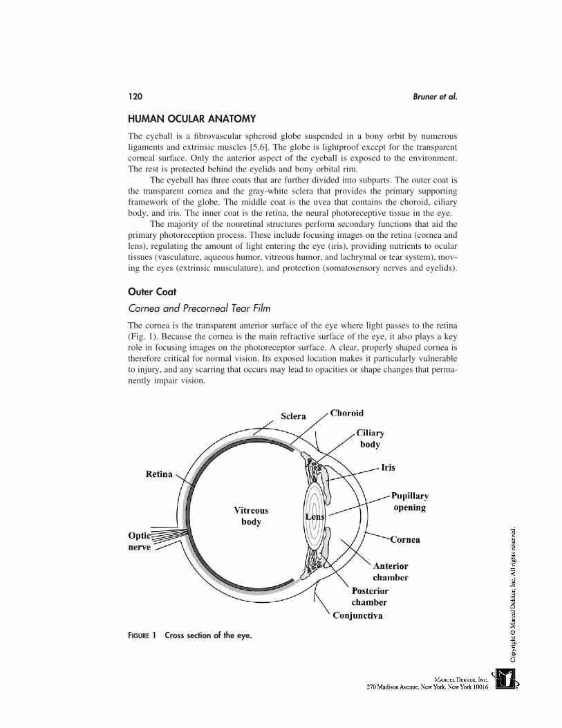
120 Bruner et al.
HUMAN OCULAR ANATOMY
The eyeball is a fibrovascular spheroid globe suspended in a bony orbit by numerousligaments and extrinsic muscles [5,6]. The globe is lightproof except for the transparentcorneal surface. Only the anterior aspect of the eyeball is exposed to the environment.The rest is protected behind the eyelids and bony orbital rim.
The eyeball has three coats that are further divided into subparts. The outer coat isthe transparent cornea and the gray-white sclera that provides the primary supportingframework of the globe. The middle coat is the uvea that contains the choroid, ciliarybody, and iris. The inner coat is the retina, the neural photoreceptive tissue in the eye.
The majority of the nonretinal structures perform secondary functions that aid theprimary photoreception process. These include focusing images on the retina (cornea andlens), regulating the amount of light entering the eye (iris), providing nutrients to oculartissues (vasculature, aqueous humor, vitreous humor, and lachrymal or tear system), mov-ing the eyes (extrinsic musculature), and protection (somatosensory nerves and eyelids).
Outer Coat
Cornea and Precorneal Tear Film
The cornea is the transparent anterior surface of the eye where light passes to the retina(Fig. 1). Because the cornea is the main refractive surface of the eye, it also plays a keyrole in focusing images on the photoreceptor surface. A clear, properly shaped cornea istherefore critical for normal vision. Its exposed location makes it particularly vulnerableto injury, and any scarring that occurs may lead to opacities or shape changes that perma-nently impair vision.
FIGURE 1 Cross section of the eye.
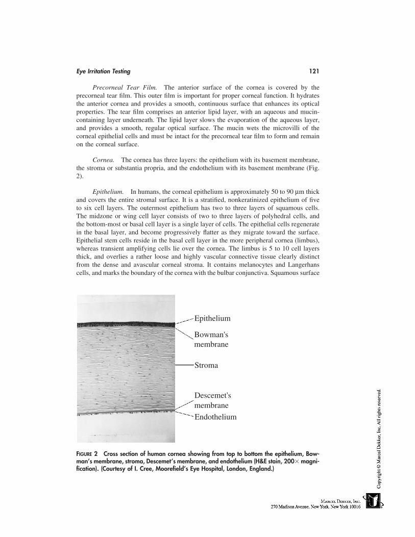
Eye Irritation Testing 121
Precorneal Tear Film. The anterior surface of the cornea is covered by theprecorneal tear film. This outer film is important for proper corneal function. It hydratesthe anterior cornea and provides a smooth, continuous surface that enhances its opticalproperties. The tear film comprises an anterior lipid layer, with an aqueous and mucin-containing layer underneath. The lipid layer slows the evaporation of the aqueous layer,and provides a smooth, regular optical surface. The mucin wets the microvilli of thecorneal epithelial cells and must be intact for the precorneal tear film to form and remainon the corneal surface.
Cornea. The cornea has three layers: the epithelium with its basement membrane,the stroma or substantia propria, and the endothelium with its basement membrane (Fig.2).
Epithelium. In humans, the corneal epithelium is approximately 50 to 90 µm thickand covers the entire stromal surface. It is a stratified, nonkeratinized epithelium of fiveto six cell layers. The outermost epithelium has two to three layers of squamous cells.The midzone or wing cell layer consists of two to three layers of polyhedral cells, andthe bottom-most or basal cell layer is a single layer of cells. The epithelial cells regeneratein the basal layer, and become progressively flatter as they migrate toward the surface.Epithelial stem cells reside in the basal cell layer in the more peripheral cornea (limbus),whereas transient amplifying cells lie over the cornea. The limbus is 5 to 10 cell layersthick, and overlies a rather loose and highly vascular connective tissue clearly distinctfrom the dense and avascular corneal stroma. It contains melanocytes and Langerhanscells, and marks the boundary of the cornea with the bulbar conjunctiva. Squamous surface
m
mmembrane
membraneDescemet's
Endothelium
Bowman's
Epithelium
Stroma
FIGURE 2 Cross section of human cornea showing from top to bottom the epithelium, Bow-man’s membrane, stroma, Descemet’s membrane, and endothelium (H&E stain, 200� magni-fication). (Courtesy of I. Cree, Moorefield’s Eye Hospital, London, England.)
122 Bruner et al.
cells are shed from the surface of the cornea after approximately 7 days. Directly belowthe basal cell layer is the basement membrane.
Stroma. The stroma constitutes approximately 90% of the corneal thickness. Itsanterior portion, Bowman’s layer, is an acellular region lying just under the epithelialbasement membrane. It is more resistant to deformation, trauma, passage of foreign bodies,or infecting organisms than the other layers. Once damaged, its architecture may not berestored, leading to abnormalities in corneal thickness and optical properties that couldresult in permanent vision deficit. The remainder of the stroma is composed of collagenfibrils gathered together in lamellae that run in parallel with the corneal surface. The fibrilswithin a lamella are highly organized and are surrounded by a glycosaminoglycan matrix.Corneal glycosaminoglycans are 60% keratin sulfate, and 40% chondroitin sulfates. Theseact as anions and bind cations and water. The posterior surface of the stroma is lined withthe loosely attached Descemet’s layer that is the basement membrane for the endothelialcells. Scattered throughout the lamellae are long, flat fibroblast-like cells calledkeratocytes. These cells have long processes that extend to adjacent cells. There are alsoa few neutrophils and macrophages that migrate through the stroma. Branches of theophthalmic branch of the fifth (trigeminal) cranial nerve, which are primarily sensory, runthrough the anterior third of the corneal stroma and associate with the epithelium.
Endothelium. The endothelium is a single layer resting on Descemet’s layer. Theendothelium originates from the neural crest and therefore is not a true endothelium. Theapical surface is in contact with the aqueous humor of the anterior chamber. The cells aretightly bound to each other with desmosomes. The endothelium serves the importantfunction of maintaining the dehydration (deturgescence) that is also required to maintaincorneal clarity (see the following section).
Sclera
The sclera is a dense, fibrous, collagenous structure that makes up the gray-white part ofthe globe. Like the cornea, it has three layers. The outermost layer is the episclera. Theepisclera is a vascularized connective tissue that merges with the scleral stroma and ex-tends connective tissue bundles into the fascia surrounding the globe. The major layer ofthe sclera is the stroma. The stroma lies in the middle and is composed of irregularlyarranged bundles of collagen fibrils. The irregular size and arrangement of these fibrilsleads to the white color of the majority of the eyeball. The inner surface of the sclera isthe lamina fuscia, which lies interior to the scleral stroma. It contains fine collagen fibersthat form the connection between the choroid and sclera. The anterior external scleralsurface of the stroma is covered by the conjunctiva. The conjunctiva is a transparent mu-cous membrane that covers the externally exposed scleral surface (bulbar conjunctiva) aswell as the inner surface of the eyelids (palpebral conjunctiva). The conjunctival epithe-lium is continuous with the corneal epithelium and the lachrymal drainage system. Theconjunctiva contains many blood vessels, nerves, conjunctival glands, and inflammatorycells. Small blood vessels are present throughout. They are usually not visible, but dilateand become leaky during inflammation. The nerves transmit pain responses and mediateneurogenic vasodilatation and tearing. The conjunctival glands provide moisture and se-crete the constituents of the precorneal tear film.
Anterior Chamber, Posterior Chamber, and Aqueous Humor
Between the rear surface of the cornea and the front surface of the lens capsule is a fluid-filled chamber (Fig. 1). This chamber is divided into anterior and posterior regions by the
Eye Irritation Testing 123
iris. These chambers are connected through the pupillary opening. The anterior chamberlies in front of the iris and the posterior chamber lies behind the iris and in front of thelens capsule.
The Middle Coat. The middle coat of the eye is the uvea. It consists of the choroid,the ciliary body, and the iris (Fig. 1). The choroid is a blood vessel–rich layer that providesblood to the retinal pigmented epithelium and outer half of the adjacent sensory retina.The ciliary body secretes the aqueous humor that fills the anterior and posterior chambersand contains the smooth muscle that alters the lens shape as needed for near and far vision.The iris is a diaphragm that lies in front of the lens and ciliary body. Contraction of iriscircular or radial muscles leads to closing or opening of the pupil, respectively, whichregulates the amount of light entering the eye.
The Inner Coat. The inner coat of the eye is the retina. This layer contains theneurosensory cells that transmit light-induced signals to the brain for visual interpretation.The two major parts of the retina are the inner sensory layer and the outer pigmentedepithelium. The sensory layer lies between the pigmented epithelium on the outside and thevitreous humor on the inside. It is stratified into several sublayers containing the differentphotoreceptor and accessory cells involved with sensing and processing the light projectedonto the retinal surface. The pigmented epithelium is only one layer thick and lies betweenthe sensory epithelium and choroid. Readers interested in more details on ocular anatomy,physiology, and biochemistry should consult recent texts on the subject [7–11].
ROUTINE IN VIVO OCULAR IRRITATION TESTING
The need for ocular safety testing became clear early in the 1930s when an untestedeyelash product containing p-phenylene diamine was marketed in the United States. Useof this and similar products led to sensitization of the external ocular structures, cornealulceration, vision loss, and at least one fatality [12]. These events resulted in passage inthe United States of the Food, Drug and Cosmetic Act of 1938, which required that materi-als sold to consumers be safe.
In response to the need for test methods to assess ocular safety, in vivo assays weredeveloped and put into use. One of the earliest reported experimental animal procedureswas devised by Friedenwald to assess the effects of acids and bases on the eye [13]. Thiswas the first time the effects of test materials on the cornea, conjunctiva, and iris wereseparately recorded. Subsequently, Carpenter and Smyth [14] studied many materials andprimarily recorded their effects on the cornea. Draize et al. [4] improved the test by stan-dardizing Friedenwald’s method and simplifying the scoring system. Subsequently, theDraize procedure and modifications of it have become the standard for assessing the irri-tancy potential of test materials for more than 50 years. The data are also used by toxicolo-gists to assure that chemicals and consumer products (1) can be made safely in factories,(2) are safe for their intended use and any foreseeable misuse, (3) are appropriately labeled,and (4) meet regulatory safety testing requirements [15].
The Draize Eye-Irritation Test
The standard Draize eye-irritation test uses either three or six albino rabbits. Statisticalstudies conducted to determine the effect of reducing the number of animals used in asingle study from six to three showed that a three-animal test provides eye-irritation classi-fication similar to that obtained by using six rabbits [16,17]. Standard Draize eye-irritation
124 Bruner et al.
test protocols normally require that 100 µL of a test material is placed in the lower cul-de-sac of one eye, and the eyelids are held shut for a brief period of time. The untreatedcontralateral eye is used as the control. The eyes are sometimes rinsed after treatment todetermine the effect of irrigation on the extent of irritation or to remove test substancestrapped within the cul-de-sac.
Generally the eyes are examined using a pen light and graded by a technician forirritation on days 1, 2, 3, 4, and 7 after dosing and weekly thereafter. However, times atwhich the eyes are examined for irritation after dosing may vary because of differencesin government regulations and preferences of different toxicologists. In some cases, theeyes are examined at time points earlier than day 1 (e.g., 1h, 3h). Similarly the maximumperiod allowed to determine recovery may vary (e.g., 3–5 weeks). Eyes are generally notexamined once they have returned to normal. Examinations are sometimes augmented byfluorescein staining and slit-lamp examinations to better assess corneal changes. A gradingscale has been proposed based on examinations with a slit lamp [18].
The Draize test uses a systematic numeric grading system to quantify the eye irrita-tion response (Table 1). Changes associated with the cornea, conjunctiva, and iris areassessed by using a pen light. Scores are assigned for the various changes. The scores forthe cornea, conjunctiva, and iris are weighted such that changes associated with the corneaare given the most weight, with the maximum score for the cornea being 80 out of a totalpossible score of 110. A test substance’s potential to cause ocular irritation is then deter-mined by assessing the individual animal scores, the maximum average score (highestmean group score during the study), and days to recovery. In general, innocuous or slightlyirritating materials tend to affect only the conjunctiva, and the eye recovers in 1 to 2 days;mildly to moderately irritating materials affect the conjunctiva and cornea, and the eyerecovers in days to weeks; and moderately to severely irritating materials affect the cornea,iris, and conjunctiva, and the eye recovers in weeks or not at all. These results are oftenfurther classified according to various regulatory classification schemes in use around theworld. The interested reader should consult Chan and Hayes [19] for a summary of regula-tory considerations.
Although the Draize eye-irritation test and slight variations of it have remained thestandard procedure for determining ocular-irritation responses, the use of this test has notcontinued without significant criticism. The sensitivity and relevance of the Draize testhave been questioned because the dose given is greater than the volume of the conjunctivalcul-de-sac of the rabbit eye (30 µl) [20], thereby considerably exceeding the dose receivedin human accidental eye exposure [21,22]. Additionally, the in vivo tests have been criti-cized for their subjectivity [23], lack of repeatability [24,25], overprediction of humanresponses [26–28], and by animal welfare advocates because they require the use of ani-mals [29]. Therefore, efforts have been made to develop and validate significantly modifiedin vivo test protocols as well as develop in vitro tests to reduce and perhaps ultimatelyeliminate the use of animals in ocular-irritation testing.
Modifications of the In Vivo Eye-Irritation Test
The Low-Volume Eye Test
In the early 1980s, modifications made in the amount of test material dosed and site ofapplication resulted in a refined version of the classical Draize test, called the low-volumeeye test (LVET). The LVET has been reported to be less stressful to rabbits and morepredictive of human ocular irritancy potential than the standard Draize procedure
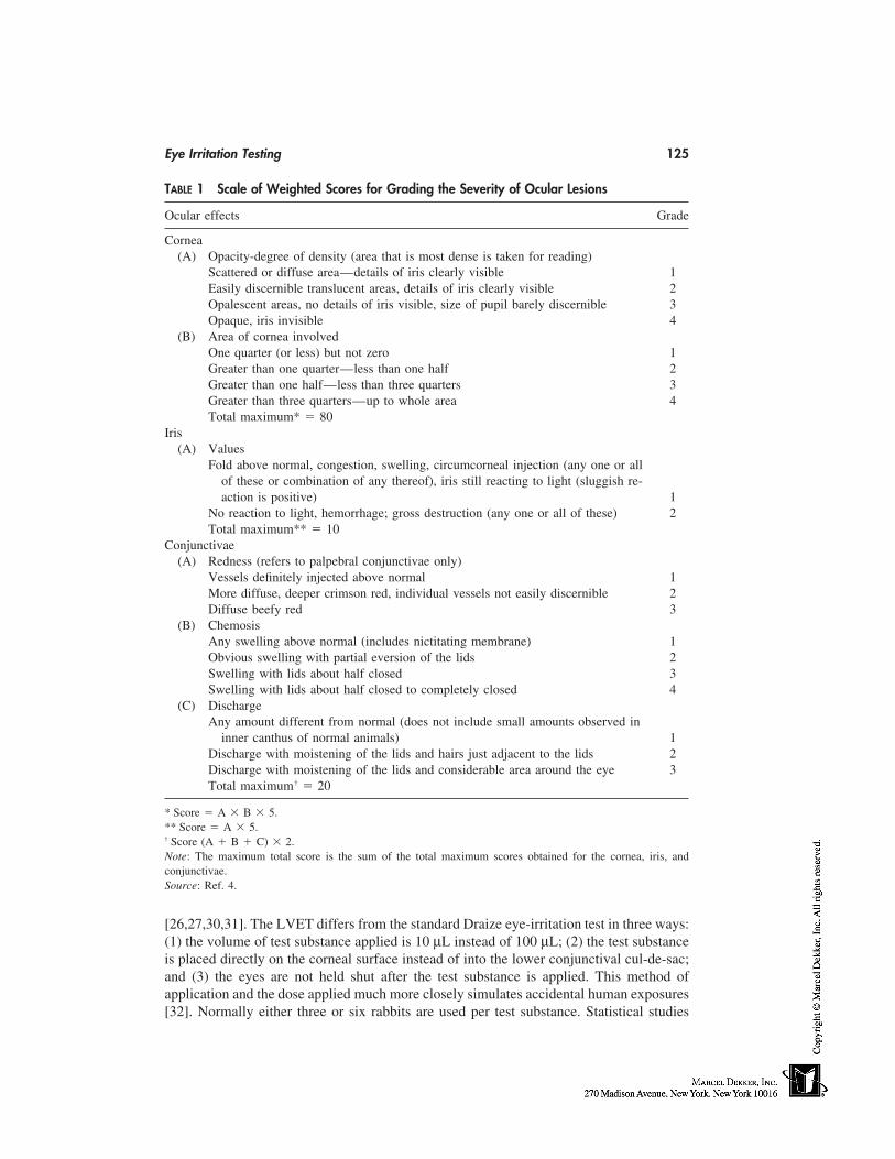
Eye Irritation Testing 125
TABLE 1 Scale of Weighted Scores for Grading the Severity of Ocular Lesions
Ocular effects Grade
Cornea(A) Opacity-degree of density (area that is most dense is taken for reading)
Scattered or diffuse area—details of iris clearly visible 1Easily discernible translucent areas, details of iris clearly visible 2Opalescent areas, no details of iris visible, size of pupil barely discernible 3Opaque, iris invisible 4
(B) Area of cornea involvedOne quarter (or less) but not zero 1Greater than one quarter—less than one half 2Greater than one half—less than three quarters 3Greater than three quarters—up to whole area 4Total maximum* � 80
Iris(A) Values
Fold above normal, congestion, swelling, circumcorneal injection (any one or allof these or combination of any thereof), iris still reacting to light (sluggish re-action is positive) 1
No reaction to light, hemorrhage; gross destruction (any one or all of these) 2Total maximum** � 10
Conjunctivae(A) Redness (refers to palpebral conjunctivae only)
Vessels definitely injected above normal 1More diffuse, deeper crimson red, individual vessels not easily discernible 2Diffuse beefy red 3
(B) ChemosisAny swelling above normal (includes nictitating membrane) 1Obvious swelling with partial eversion of the lids 2Swelling with lids about half closed 3Swelling with lids about half closed to completely closed 4
(C) DischargeAny amount different from normal (does not include small amounts observed in
inner canthus of normal animals) 1Discharge with moistening of the lids and hairs just adjacent to the lids 2Discharge with moistening of the lids and considerable area around the eye 3Total maximum† � 20
* Score � A � B � 5.** Score � A � 5.† Score (A � B � C) � 2.Note: The maximum total score is the sum of the total maximum scores obtained for the cornea, iris, andconjunctivae.Source: Ref. 4.
[26,27,30,31]. The LVET differs from the standard Draize eye-irritation test in three ways:(1) the volume of test substance applied is 10 µL instead of 100 µL; (2) the test substanceis placed directly on the corneal surface instead of into the lower conjunctival cul-de-sac;and (3) the eyes are not held shut after the test substance is applied. This method ofapplication and the dose applied much more closely simulates accidental human exposures[32]. Normally either three or six rabbits are used per test substance. Statistical studies
126 Bruner et al.
similar to those conducted for the Draize test indicate that results from three rabbits pro-vide eye-irritation classification similar to that obtained from studies using six rabbits, sothat animal use in this test can be minimized [33].
Objective Measurements of Eye Injury
In addition to the LVET, other modifications have been made to the in vivo test. Mostof these changes have been made in an attempt to minimize variability. Because the subjec-tive nature of the grading is thought to be a major source of variability, work has beendone to eliminate as much as possible the subjective components of the test. Some of themethods evaluated include assessing corneal thickness [34–36], water content [36,37],permeability [38–40], and surface area damaged using fluorescein, wound healing, andexfoliative cytology [41]. Objective measurements of conjunctivitis have included assess-ments of capillary permeability [36,37], redness, and exfoliative cytology [41]. Othershave attempted to assess the utility of measuring intraocular pressure [42] and proteincontent of the aqueous humor [36,37]. None of these methods is in routine use.
REPLACING THE ANIMAL TEST WITH IN VITRO METHODS
Introduction
There are strong social, political, ethical, and scientific arguments for the developmentand use of nonanimal methods as alternatives to the Draize eye-irritation test. Alternativemethods currently under investigation use a diverse set of human and animal cells, tissues,and biochemical reagents, and measure a diverse set of endpoints thought to be associatedwith eye-irritation responses in vivo. Few of these tests, however, attempt to model theentire eye. Instead, they usually model subparts of the larger, more complex eye-irritationresponse. Figure 3 shows this reductionist relationship across the spectrum of available
FIGURE 3 A diagram illustrating how in vitro assays have been developed to model differentparts of the eye-irritation response. In the development of in vitro tests, the eye is in effectreduced to component parts. The tests developed model different parts of the eye-irritationresponse and allows studies on mechanisms of action. The first reduction step from the intactanimal uses isolated whole eyes obtained from the abattoir. Examples include the chickenenucleated eye test and the isolated rabbit eye test. The next level of reduction is representedby tests that use isolated corneas and 3-dimensional tissue constructs. Examples include thebovine cornea opacity and permeability test (BCOP) and the topical application assays (TEA),respectively. The final level of reduction represents tests based on cell cultures containing singlecell types. Examples of tests in this category include the fluorescein leakage test and othercytotoxicity tests.
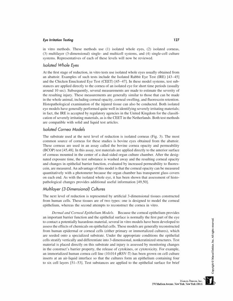
Eye Irritation Testing 127
in vitro methods. These methods use (1) isolated whole eyes, (2) isolated corneas,(3) multilayer (3-dimensional) single- and multicell systems, and (4) single-cell culturesystems. Representatives of each of these levels will now be reviewed.
Isolated Whole Eyes
At the first stage of reduction, in vitro tests use isolated whole eyes usually obtained froman abattoir. Examples of such tests include the Isolated Rabbit Eye Test (IRE) [43–45]and the Chicken Enucleated Eye Test (CEET) [45–47]. In these model systems, test sub-stances are applied directly to the cornea of an isolated eye for short time periods (usuallyaround 10 sec). Subsequently, several measurements are made to estimate the severity ofthe resulting injury. These measurements are generally similar to those that can be madein the whole animal, including corneal opacity, corneal swelling, and fluorescein retention.Histopathological examination of the injured tissue can also be conducted. Both isolatedeye models have generally performed quite well in identifying severely irritating materials;in fact, the IRE is accepted by regulatory agencies in the United Kingdom for the classifi-cation of severely irritating materials, as is the CEET in the Netherlands. Both test methodsare compatible with solid and liquid test articles.
Isolated Cornea Models
The substrate used at the next level of reduction is isolated corneas (Fig. 3). The mostcommon source of corneas for these studies is bovine eyes obtained from the abattoir.These corneas are used in an assay called the bovine cornea opacity and permeability(BCOP) test [45,48]. In this assay, test materials are applied directly to the anterior surfaceof corneas mounted in the center of a dual-sided organ culture chamber. After the desig-nated exposure time, the test substance is washed away and the resulting corneal opacityand changes in epithelial barrier function, evaluated by increased permeability to fluores-cein, are measured. An advantage of this model is that the corneal opacity can be measuredquantitatively with a photometer because the organ chamber has transparent glass coverson each end. As with the isolated whole eye, it has been shown that assessment of histo-pathological changes provides additional useful information [49,50].
Multilayer (3-Dimensional) Cultures
The next level of reduction is represented by artificial 3-dimensional tissues constructedfrom human cells. These tissues are of two types: one is designed to model the cornealepithelium, whereas the second attempts to reconstruct the cornea in vitro.
Dermal and Corneal Epithelium Models. Because the corneal epithelium providesan important barrier function and the epithelial surface is normally the first part of the eyeto contact a potentially hazardous material, several in vitro models have been developed toassess the effects of chemicals on epithelial cells. These models are generally reconstructedfrom human epidermal or corneal cells (either primary or immortalized cultures), whichare seeded onto a specialized substrate. Under the appropriate conditions the epithelialcells stratify vertically and differentiate into 3-dimensional, nonkeratinized structures. Testmaterial is placed directly on this substrate and injury is assessed by monitoring changesin the construct’s barrier property, the release of cytokines, or cytotoxicity. For example,an immortalized human cornea cell line (10.014 pRSV-T) has been grown on cell cultureinserts at an air-liquid interface so that the cultures form an epithelium containing fourto six cell layers [51–53]. Test substances are applied to the epithelial surface for brief
128 Bruner et al.
periods (up to 5 min) in dose-response experiments. Endpoints measured include thebarrier function of the epithelium using transepithelial permeability to fluorescein andelectrical resistance, along with cell viability [54]. Results from this model, called HCE-T, correlate with historical rabbit-eye data for water-soluble ingredients and surfactant-based personal care products [52]. Others have reported that early (1 h) release of thecytokine interleukin 1-α is a predictive marker for surfactant responses in another humancorneal epithelial cell line, CEPI 17 c1.4 [55]. Interleukin 8 appears to be a late (24 h)marker of response, although the bulk of the IL-8 response appears secondary to the releaseof IL-1α. Taken together, this work shows the potential utility of human cornea epithelialcells to assess effects of test substances on epithelial barrier function, viability, andinflammation, as well as to evaluate specific biochemical and molecular mechanisms ofthese responses.
Other models have been constructed using primary human epidermal cells ratherthan immortalized cell lines. Several tissues of this type are available commercially. Cur-rently available substrates include EpiOcular [56] (MatTek Corporation, Ashland, MA)and SkinEthic cultures [57,58] (SkinEthic, Nice, France). In these assays, test substancesare applied to the surface of the cultures for a specified period of time. Then, the testsubstance is washed away and viability of the cells is measured by using one of severalvital dyes [57–61]. The release of various cytokines is also measured. These models havebeen shown capable of differentiating degrees of irritancy between mild test substances.Another advantage of these systems is that they have proven useful for assessing bothwater-soluble and water-insoluble consumer products, cosmetics, and ingredients [56,59,62].
Human Cornea Models. The development of human corneal cultures analogousto 3-dimensional human skin cultures that are used to evaluate skin irritation [63,64] isnow an active area of research. Martin et al. [65] have reported on trilaminar substratesdeveloped from early passage human corneal epithelial, stroma, and endothelial cells.Endpoints evaluated in this model include barrier function, cytotoxicity, and release ofthe inflammatory mediators PGE2 and LTB4. Development of immortalized human corneacell lines and their incorporation into trilaminar corneal models have also been reportedby Griffith and coworkers [66,67]. Functional and biochemical analysis of these culturesindicate the presence of differentiation markers and other properties similar to those foundin intact human corneas. In initial characterization, cultures treated with model surfactantselicit responses similar to those observed in vivo.
Single-Cell Culture Systems, Isolated Single Cells
At the last step in the reductionist scheme are assays that use monolayer cell culturesderived from epithelial cells of eyes or other organs such as the skin. The study of interac-tions between test substances and single cells and monolayer cultures of various typeswas one of the earliest approaches evaluated for eye-irritation tests in vitro. The mostcommonly used endpoint is assessment of direct cytotoxicity after a short-term exposureto test articles. Examples of methods in this category include the neutral red uptake test[62,68–71], the neutral red release test [72,73], and the red blood cell lysis test[62,70,74,75]. In addition, the real-time effects of a test material on the metabolic rate ofcultured cells can be assessed by using the Cytosensor microphysiometer (Molecular De-vices Corp., Menlo Park, CA) [62,69,70,76–78]. The Fluorescein Leakage Test is anothercytotoxicity assay that measures the capacity of a test substance to damage the barrierfunction normally associated with epithelial cells. With this assay, confluent monolayer
Eye Irritation Testing 129
cultures of renal epithelial cells are treated with test material. After exposure the changein the capacity of the epithelial cells to block fluorescein passage is measured [62,70,79–81]. Additional information on these tests may be found in an extensive review of assaysbased on single-cell cultures published by U.S. Interagency Regulatory Alternatives Group(IRAG) [82,83].
Other Test Systems
There are several in vitro tests that have been evaluated extensively as alternatives foreye-irritation testing that do not fit entirely within the reductionist scheme just described.The most significant tests in this category are the chorioallantoic membrane (CAM) assays.Use of the CAM of the chicken egg as a substrate for in vitro testing was first described byLuepke et al. [84], who reasoned that the highly vascularized CAM might be an acceptablesurrogate for conjunctival tissue. To this end, they developed a model called the hen’segg test–CAM (HET–CAM). In this procedure, test substances are placed directly on theCAM exposed directly underneath the air cell. The resulting hemorrhage, coagulation,and lysis appearing on the CAM are measured at defined timepoints after the test articleis applied. Results from this test are accepted by regulatory agencies in Germany as ade-quate for identifying severe irritants. A complementary test called the CAM vascular assay(CAMVA) has also been developed [85,86]. The CAMVA differs from the HET–CAMin several ways, including the site of the egg shell that is opened (side of the egg insteadof the air cell), the endpoint measured (changes in characteristics of the CAM vasculature),and the dosing scheme (serially diluted test substances instead of a single test concentra-tion). Both the HET–CAM and the CAMVA assay are reviewed in detail in the U.S.IRAG evaluations [87]. Results from evaluation of this test in several international valida-tion studies have been reported [62,88–90].
Practical Use of In Vitro Tests for Eye-Irritation Testing
The effort to develop and validate nonanimal test methods has significantly increased theuse of these tests for assessing eye safety. Experience gained from this work has shownthat the methods provide information useful for safety assessments, but the conduct andinterpretation of results from in vitro tests are more complex than for standard in vivotesting. Therefore, considerable care and planning need to be undertaken before beginninga study in order to obtain reliable results. Given the increased complexity associated within vitro testing, we have found that the use of the new methods is greatly facilitated bythe establishment of a standard framework that contains four elements. These include1) a well-defined process that specifies the steps to follow during the conduct of an eyesafety assessment of a test article, 2) protocols and standard operating procedures (SOPs)that define all the tests used within the eye safety assessment process, 3) prediction modelsthat guide the interpretation of results obtained from in vitro and other test methods, and4) a summary document that provides practical guidance to toxicologists on how to con-duct the overall process. The important aspects of each of these elements will now bereviewed.
Process for the Assessment of Test Materials in Nonanimal Methods
A clearly defined testing process is the central element in a nonanimal testing framework.These processes usually take the form of flow charts showing the key decision points,data-gathering procedures, and test methods that may be conducted during a safety assess-
130 Bruner et al.
ment. An example suitable for eye-irritation testing is shown in Figure 4. The processbegins with the entry of a test substance into a safety assessment program. The first stepin the process involves gathering as much previously existing information as possibleabout the material. The information obtained should include all available toxicity data onthe test article, such as in vivo and in vitro data, human clinical data, supplier information,results from quantitative structure activity relationship (QSAR) analyses, physical-chemi-cal data, marketplace experiences, and data on consumer habits and practices. In the caseof completely new chemicals or formulations, information on similar materials should begathered. Once these data are obtained, they must be assessed to determine if it is possibleto complete the safety assessment without further testing. At this point, three decisionsare possible: 1) market the product because the pre-existing data are considered adequateto support the product safety without further testing, 2) terminate or reformulate becausethe pre-existing data indicate the article is not safe for intended use, or 3) conduct addi-tional testing because more data are necessary in order to complete the assessment. Whenthe third decision is made, the next step in the process is to evaluate the article in anappropriate in vitro test(s). When the testing is complete, the results are passed throughthe algorithms of the prediction model so that a toxicity prediction can be obtained. Thetoxicity prediction is then considered along with the previously existing results. At thispoint it is again necessary to ask whether the test article is considered safe for intendeduse. If the answer is no, then reformulation or termination are the available options. Ifthe answer is yes, it is necessary to decide whether human tolerance testing is necessary.Such studies may be needed, for example, to develop data for marketing claims support.If there is no need for human tolerance testing, then the safety assessment is completed.
Protocols and SOPs
Each safety assessment process contains several different tests. In order to facilitate thegeneration of reliable data from these tests, it is essential that all factors important to theirconduct are clearly documented. It is therefore important that protocols and SOPs beprovided for each test used in the safety assessment process. Adequate protocols and SOPswill contain at least four key elements. First, each SOP must have a detailed step-by-stepdescription of how to conduct a test. Enough details need to be provided such that anyappropriately trained and competent laboratory technician need use only this documentas the guide to conduct the assay. Secondly, the SOP must indicate the steps used todefine the final endpoint of the assay and the number of replicates necessary. Any datatransformation or algorithms applied to the data should be clearly documented and consis-tently applied. Thirdly, the protocol should specify the positive and negative controls tobe performed concurrently with each assay and the acceptable ranges for the resultingresponses. Assays where the positive or negative controls values fall outside of thosespecified ranges would be considered invalid and should be repeated. Finally, the protocolmust specifically describe the prediction model used to guide the interpretation of results.
Prediction Models
In order to use an in vitro test method in the safety assessment process, it must be possibleto convert the in vitro results into a meaningful prediction of toxicity. The tool that is usedto make this conversion is the prediction model [25,91]. A prediction model is consideredadequate when it defines four elements. These elements include 1) a definition of thespecific purpose(s) for which the alternative method is to be used, 2) a definition of allthe possible results that may be obtained from an alternative method (inputs), 3) an algo-
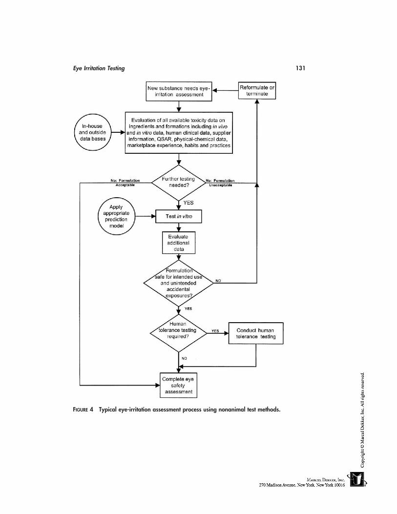
Eye Irritation Testing 131
FIGURE 4 Typical eye-irritation assessment process using nonanimal test methods.
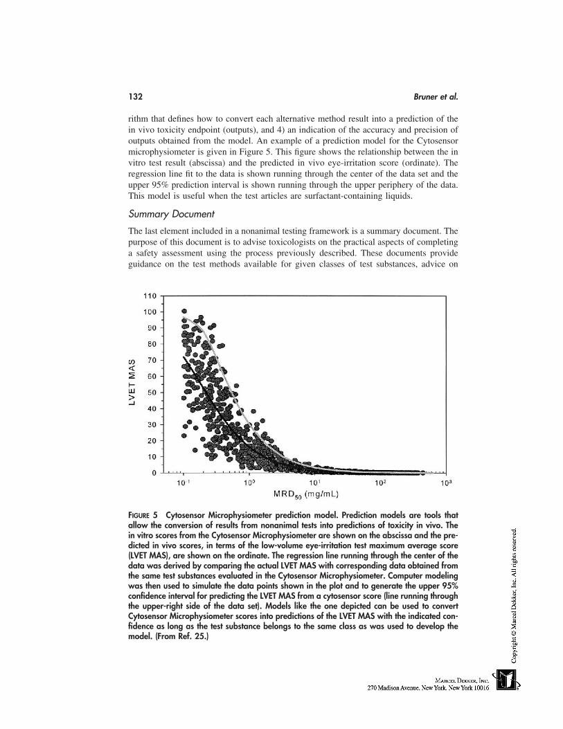
132 Bruner et al.
rithm that defines how to convert each alternative method result into a prediction of thein vivo toxicity endpoint (outputs), and 4) an indication of the accuracy and precision ofoutputs obtained from the model. An example of a prediction model for the Cytosensormicrophysiometer is given in Figure 5. This figure shows the relationship between the invitro test result (abscissa) and the predicted in vivo eye-irritation score (ordinate). Theregression line fit to the data is shown running through the center of the data set and theupper 95% prediction interval is shown running through the upper periphery of the data.This model is useful when the test articles are surfactant-containing liquids.
Summary Document
The last element included in a nonanimal testing framework is a summary document. Thepurpose of this document is to advise toxicologists on the practical aspects of completinga safety assessment using the process previously described. These documents provideguidance on the test methods available for given classes of test substances, advice on
FIGURE 5 Cytosensor Microphysiometer prediction model. Prediction models are tools thatallow the conversion of results from nonanimal tests into predictions of toxicity in vivo. Thein vitro scores from the Cytosensor Microphysiometer are shown on the abscissa and the pre-dicted in vivo scores, in terms of the low-volume eye-irritation test maximum average score(LVET MAS), are shown on the ordinate. The regression line running through the center of thedata was derived by comparing the actual LVET MAS with corresponding data obtained fromthe same test substances evaluated in the Cytosensor Microphysiometer. Computer modelingwas then used to simulate the data points shown in the plot and to generate the upper 95%confidence interval for predicting the LVET MAS from a cytosensor score (line running throughthe upper-right side of the data set). Models like the one depicted can be used to convertCytosensor Microphysiometer scores into predictions of the LVET MAS with the indicated con-fidence as long as the test substance belongs to the same class as was used to develop themodel. (From Ref. 25.)
Eye Irritation Testing 133
which test should be used with different types of test substances, and an indication of themost appropriate prediction model to use with the test substance(s) being evaluated. Fi-nally, these documents provide a wide range of relevant information on the technicalaspects of the safety testing process (see the following) and names of individuals withinthe organization who can provide advice. All of this information is placed in a readilyavailable location so that toxicologists can use the reference material easily.
Practical Considerations in the Conduct of Eye-Safety AssessmentsWithout Animal Testing
In addition to establishing a framework for the practical conduct of eye-safety programs,it is also important to address several important technical issues that need to be consideredwhen conducting in vitro tests. These matters have considerable influence on the choiceof nonanimal tests to be used and the interpretation of the results. The matters that needto be considered include (1) the physical characteristics of the test article, (2) the expectedtoxicity of the test article, (3) the level of resolution required from the testing, and (4) re-sources available for a safety program.
Physical Characteristics of the Test Article. One of the most important consid-erations in the conduct of an in vitro test is the compatibility of the test article with thein vitro test being conducted. There are two general forms of in vitro tests: dilution-basedtests where the target cells are completely immersed in growth medium, and topicalapplication tests where the target cell surface is available for direct application of the testmaterial (Table 2). For in vitro tests of the first type, it is necessary to serially dilute the testsubstance into a water-based cell culture medium and then apply the diluted test articles tothe target cells. Dilution-based tests are particularly well suited for screening largenumbers of water-soluble test substances quickly at a relatively low cost. The dilution-based tests also appear to have an increased capacity to distinguish between differentdegrees of mildness compared with the topical application tests [92].
Despite these advantages, the dilution of test articles in cell culture media resultsin technical problems that need to be considered before the procedure is used. First, be-cause water-insoluble test substances cannot be diluted easily in aqueous cell culture me-dia, it is generally unwise to evaluate water-insoluble substances in dilution-based tests.Second, when diluting test substances it is important to note that the dilution process can
TABLE 2 Dilution-Based and Topical Application–Based Assays: Examples of Dilution-Basedand Topical Application–Based Assays Are Shown. Dilution-Based Tests Are More Suitedfor Test Substances That Are Water Soluble. Topical Application–Based Tests Have theAdvantage That Dilution of Test Substance Is Not Required, Which Alleviates TechnicalProblems That Can Arise After Dilution. See Text for Details.
Dilution-based tests Topical application–based tests
Cytosensor microphysiometer Bovine corneal opacity and permeability assayFluorescein leakage test Chicken enucleated eye testNeutral red release test Corneal and dermal 3-dimensional culture-based testsNeutral red uptake test Hen’s egg test-chorioallantoic membrane (HET-CAM)Red blood cell lysis test Isolated rabbit eye testChorioallantoic membrane vascular
assay (CAMVA)
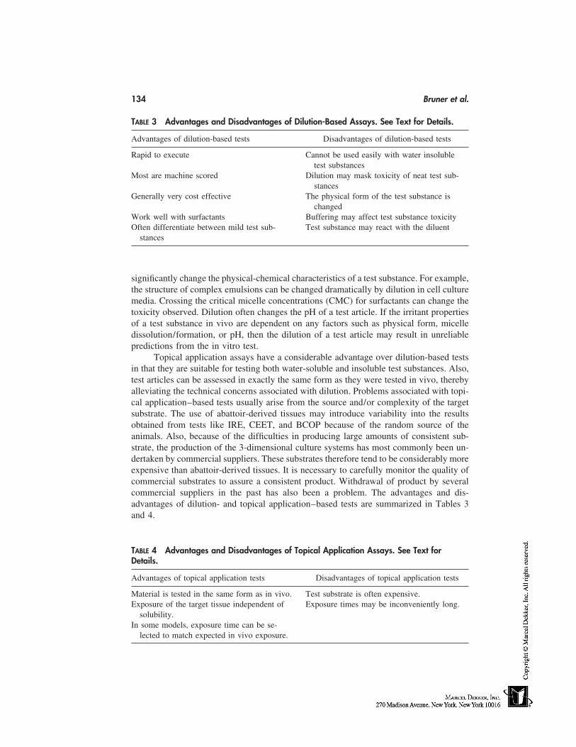
134 Bruner et al.
TABLE 3 Advantages and Disadvantages of Dilution-Based Assays. See Text for Details.
Advantages of dilution-based tests Disadvantages of dilution-based tests
Rapid to execute Cannot be used easily with water insolubletest substances
Most are machine scored Dilution may mask toxicity of neat test sub-stances
Generally very cost effective The physical form of the test substance ischanged
Work well with surfactants Buffering may affect test substance toxicityOften differentiate between mild test sub- Test substance may react with the diluent
stances
significantly change the physical-chemical characteristics of a test substance. For example,the structure of complex emulsions can be changed dramatically by dilution in cell culturemedia. Crossing the critical micelle concentrations (CMC) for surfactants can change thetoxicity observed. Dilution often changes the pH of a test article. If the irritant propertiesof a test substance in vivo are dependent on any factors such as physical form, micelledissolution/formation, or pH, then the dilution of a test article may result in unreliablepredictions from the in vitro test.
Topical application assays have a considerable advantage over dilution-based testsin that they are suitable for testing both water-soluble and insoluble test substances. Also,test articles can be assessed in exactly the same form as they were tested in vivo, therebyalleviating the technical concerns associated with dilution. Problems associated with topi-cal application–based tests usually arise from the source and/or complexity of the targetsubstrate. The use of abattoir-derived tissues may introduce variability into the resultsobtained from tests like IRE, CEET, and BCOP because of the random source of theanimals. Also, because of the difficulties in producing large amounts of consistent sub-strate, the production of the 3-dimensional culture systems has most commonly been un-dertaken by commercial suppliers. These substrates therefore tend to be considerably moreexpensive than abattoir-derived tissues. It is necessary to carefully monitor the quality ofcommercial substrates to assure a consistent product. Withdrawal of product by severalcommercial suppliers in the past has also been a problem. The advantages and dis-advantages of dilution- and topical application–based tests are summarized in Tables 3and 4.
TABLE 4 Advantages and Disadvantages of Topical Application Assays. See Text forDetails.
Advantages of topical application tests Disadvantages of topical application tests
Material is tested in the same form as in vivo. Test substrate is often expensive.Exposure of the target tissue independent of Exposure times may be inconveniently long.
solubility.In some models, exposure time can be se-
lected to match expected in vivo exposure.
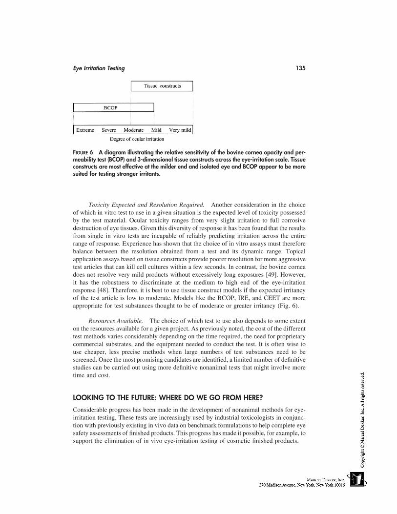
Eye Irritation Testing 135
FIGURE 6 A diagram illustrating the relative sensitivity of the bovine cornea opacity and per-meability test (BCOP) and 3-dimensional tissue constructs across the eye-irritation scale. Tissueconstructs are most effective at the milder end and isolated eye and BCOP appear to be moresuited for testing stronger irritants.
Toxicity Expected and Resolution Required. Another consideration in the choiceof which in vitro test to use in a given situation is the expected level of toxicity possessedby the test material. Ocular toxicity ranges from very slight irritation to full corrosivedestruction of eye tissues. Given this diversity of response it has been found that the resultsfrom single in vitro tests are incapable of reliably predicting irritation across the entirerange of response. Experience has shown that the choice of in vitro assays must thereforebalance between the resolution obtained from a test and its dynamic range. Topicalapplication assays based on tissue constructs provide poorer resolution for more aggressivetest articles that can kill cell cultures within a few seconds. In contrast, the bovine corneadoes not resolve very mild products without excessively long exposures [49]. However,it has the robustness to discriminate at the medium to high end of the eye-irritationresponse [48]. Therefore, it is best to use tissue construct models if the expected irritancyof the test article is low to moderate. Models like the BCOP, IRE, and CEET are moreappropriate for test substances thought to be of moderate or greater irritancy (Fig. 6).
Resources Available. The choice of which test to use also depends to some extenton the resources available for a given project. As previously noted, the cost of the differenttest methods varies considerably depending on the time required, the need for proprietarycommercial substrates, and the equipment needed to conduct the test. It is often wise touse cheaper, less precise methods when large numbers of test substances need to bescreened. Once the most promising candidates are identified, a limited number of definitivestudies can be carried out using more definitive nonanimal tests that might involve moretime and cost.
LOOKING TO THE FUTURE: WHERE DO WE GO FROM HERE?
Considerable progress has been made in the development of nonanimal methods for eye-irritation testing. These tests are increasingly used by industrial toxicologists in conjunc-tion with previously existing in vivo data on benchmark formulations to help complete eyesafety assessments of finished products. This progress has made it possible, for example, tosupport the elimination of in vivo eye-irritation testing of cosmetic finished products.
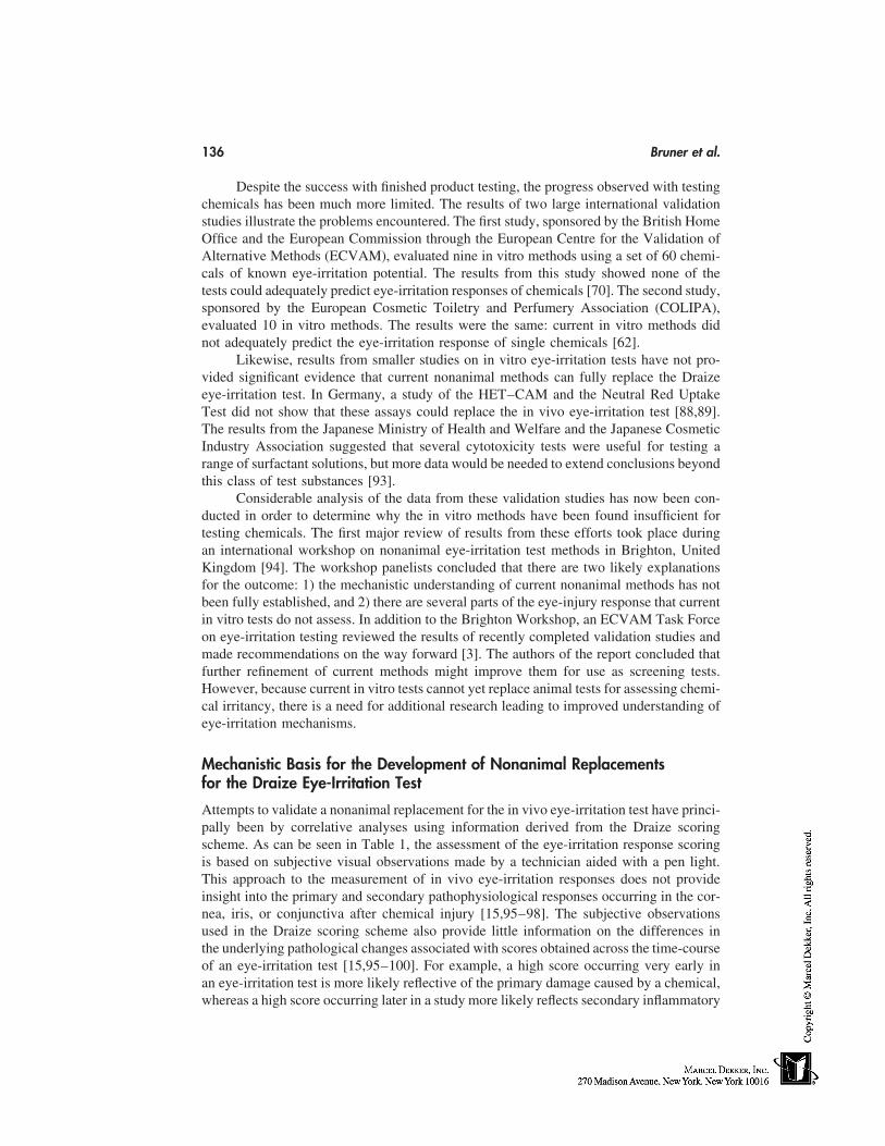
136 Bruner et al.
Despite the success with finished product testing, the progress observed with testingchemicals has been much more limited. The results of two large international validationstudies illustrate the problems encountered. The first study, sponsored by the British HomeOffice and the European Commission through the European Centre for the Validation ofAlternative Methods (ECVAM), evaluated nine in vitro methods using a set of 60 chemi-cals of known eye-irritation potential. The results from this study showed none of thetests could adequately predict eye-irritation responses of chemicals [70]. The second study,sponsored by the European Cosmetic Toiletry and Perfumery Association (COLIPA),evaluated 10 in vitro methods. The results were the same: current in vitro methods didnot adequately predict the eye-irritation response of single chemicals [62].
Likewise, results from smaller studies on in vitro eye-irritation tests have not pro-vided significant evidence that current nonanimal methods can fully replace the Draizeeye-irritation test. In Germany, a study of the HET–CAM and the Neutral Red UptakeTest did not show that these assays could replace the in vivo eye-irritation test [88,89].The results from the Japanese Ministry of Health and Welfare and the Japanese CosmeticIndustry Association suggested that several cytotoxicity tests were useful for testing arange of surfactant solutions, but more data would be needed to extend conclusions beyondthis class of test substances [93].
Considerable analysis of the data from these validation studies has now been con-ducted in order to determine why the in vitro methods have been found insufficient fortesting chemicals. The first major review of results from these efforts took place duringan international workshop on nonanimal eye-irritation test methods in Brighton, UnitedKingdom [94]. The workshop panelists concluded that there are two likely explanationsfor the outcome: 1) the mechanistic understanding of current nonanimal methods has notbeen fully established, and 2) there are several parts of the eye-injury response that currentin vitro tests do not assess. In addition to the Brighton Workshop, an ECVAM Task Forceon eye-irritation testing reviewed the results of recently completed validation studies andmade recommendations on the way forward [3]. The authors of the report concluded thatfurther refinement of current methods might improve them for use as screening tests.However, because current in vitro tests cannot yet replace animal tests for assessing chemi-cal irritancy, there is a need for additional research leading to improved understanding ofeye-irritation mechanisms.
Mechanistic Basis for the Development of Nonanimal Replacementsfor the Draize Eye-Irritation Test
Attempts to validate a nonanimal replacement for the in vivo eye-irritation test have princi-pally been by correlative analyses using information derived from the Draize scoringscheme. As can be seen in Table 1, the assessment of the eye-irritation response scoringis based on subjective visual observations made by a technician aided with a pen light.This approach to the measurement of in vivo eye-irritation responses does not provideinsight into the primary and secondary pathophysiological responses occurring in the cor-nea, iris, or conjunctiva after chemical injury [15,95–98]. The subjective observationsused in the Draize scoring scheme also provide little information on the differences inthe underlying pathological changes associated with scores obtained across the time-courseof an eye-irritation test [15,95–100]. For example, a high score occurring very early inan eye-irritation test is more likely reflective of the primary damage caused by a chemical,whereas a high score occurring later in a study more likely reflects secondary inflammatory
Eye Irritation Testing 137
responses developing in response to the primary injury. Overall, these observations suggestthat the scoring system used in the current in vivo eye-irritation test may not provideenough information about the critical cellular and molecular changes involved in ocularinjury and repair to be used as the basis for developing adequately predictive nonanimaltests.
In order to address this shortcoming it has been proposed that more data must beobtained on the pathophysiological processes underlying chemical-induced eye-irritationresponses [99]. The new information needs to be derived from additional in vivo testingof a panel of test substances covering relevant chemical classes and the appropriate rangeof eye-irritation response. Where possible, these studies should include test substancesfor which there is human eye-irritation data so that alternative methods can be developedto predict human responses [101]. The information derived from the new in vivo studieswould characterize the key cellular and molecular events and extent of variability oc-curring with the ocular irritation response and serve as the basis for the developmentmechanism–based replacement tests [15,95–100]. Preliminary work suggests that two ar-eas of research are likely to be most beneficial. These include studies to (1) characterizethe pathological changes associated with the initial eye injury caused by chemical, and(2) characterize changes in the expression of cytokines and other extracellular factorsassociated with inflammation and corneal repair.
Characterizing the Pathological Changes Associated withInitial Eye Injury
Differences in extent of the initial tissue injury after chemical exposure has been hypothe-sized to be one of the primary factors that determine the responses and ultimately the finaloutcome of an ocular irritation response [15,96–100]. Results from studies of a broadsampling of surfactants support this premise [15,95–100,102–104]. Light microscopy[15,97,99] and in vivo confocal microscopy [95,96,102] studies in rats and rabbits showthere are differences in the extent of ocular injury induced by surfactants of known irri-tancy occurring as early as 3 hours after treatment. Collectively, these studies indicatethat slight irritants affect only the superficial corneal epithelium, mild and moderate irri-tants affect the epithelium and superficial stroma, and severe irritants affect the epithelium,deep stroma, and at times the endothelium. Additional work suggests that the extent ofsurfactant-induced injury correlates with cell death [98] and that the extent of the primaryinjury correlates with subsequent responses and the eventual outcome in rats [97] andrabbits [100].
Overall, these results suggest that prediction models for mechanism-based in vitrotests could be developed based on measurements of the extent of injury and, perhapsmore specifically, on measures of cell death in the cornea after chemical treatment in vivo[98,100]. Such an approach would require that replacement tests assess the area and depthof injury in multilayered in vitro substrates that contain at least a stratified epithelium andkeratocyte-laden stroma [98]. Examples of appropriate substrates for such studies includeisolated whole eye and isolated cornea models, or perhaps 3-dimensional corneal modelslike those previously described [98].
Characterizing Changes in Expression of Cytokines andOther Extracellular Factors
Changes in the expression and/or levels of biomarkers, cytokines, and other extracellularfactors associated with the different stages of chemically induced eye injury have also
138 Bruner et al.
been proposed as possible endpoints for mechanism-based replacement [15,96,97,105].For example, Sotozono et al. [106] have observed that the production of IL-1α and IL-6reflect the severity of alkali burns on the cornea. Shams et al. [107] have shown that levelsof corneal IL-1 correlate with severity of inflammation. Planck et al. [108] have proposedthat cytokine signatures characterized by varying patterns of expression of biological fac-tors occur with different types of corneal injury. In this regard, their studies in rats haveindicated IL-6 induction occurs with alkali burns and incisional trauma of the cornea,whereas IL-1β induction occurs with alkali burns but not incisional trauma. Further, differ-ences in mRNA expression for different chemokines were observed in mouse corneasinfected with HSV-1 versus traumatic injury [109]. Finally, a more recent study has indi-cated that differences in expression of corneal IL-1α, IL-1β, and IL-6 levels are observedafter surfactant-induced injury in rats with the magnitude of the differences reflecting theextent of injury observed [105].
Other Endpoints Worthy of Consideration
In addition to studies of pathology and inflammatory mediator release associated withchemical injury, there are other areas of research that may be of interest. First, it may beuseful to examine other early events occurring after exposure of the eye to chemicals.Studies could explore the interaction of chemicals with cell membranes that lead to acutedamage of the eye tissue and activation of ocular nerves. Approaches that may be usefulfor such work include quantitative structure-activity relationship approaches and neuro-physiological models of the eye [110]. After the initial chemical trauma, various physio-logical responses in addition to inflammatory mediator release take place in the intermedi-ate stages of the response depending on the extent of the initial damage and the modulatinginfluence of nerve activation. Therefore studies on the physiological effects of chemicalson isolated eyes may prove useful. In the later stages of the reaction, the inflammationsubsides and the eye returns to a quiet state. Of critical importance is whether or not theeye returns to the normal pre-exposure state or whether there is scarring of the corneathat can lead to vision deficit or, in the worst case, loss of sight. Therefore, the biologicalresponses related to recovery need to be studied. As these areas are evaluated in ongoingresearch programs sponsored by industry and relevant governmental agencies, the newknowledge gained may be directly applied to the development of mechanism-based assaysthat may be validated by interested parties.
CONCLUSION
Nonanimal test methods are now routinely used by industrial toxicologists to assess thesafety of certain test articles [111]. These tests are most useful when conducted as partof a larger process that uses significant amounts of other supporting information. No singletest or battery of tests can yet completely replace the need for animals in ocular safetytesting. If complete elimination of animal use in eye safety assessment is to be achieved,a better understanding of the mechanisms by which chemicals cause eye irritation will beneeded. The areas of research needed have been outlined in considerable detail and propos-als have been made for the conduct of the research. The application of recent progress intissue-culture techniques, cellular and molecular biology, and analytical cytometric tech-niques will greatly facilitate the conduct of this research and lead us closer to our ultimategoal of eliminating the need for animals in ocular safety testing.
Eye Irritation Testing 139
ACKNOWLEDGEMENT
We wish to acknowledge Janet Smith for her assistance in preparation of this manuscript.
REFERENCES
1. Grant WM. Toxicology of the Eye. Effects on the eyes and visual system from chemicals,drugs, metals and minerals, plants, toxins and venoms; also systemic side effects from eyemedications. 3rd ed. Springfield: Charles C. Thomas, 1986.
2. Bruner LH. Ocular irritation. In: Frazier JM, ed. In Vitro Toxicity Testing. Applications toSafety Evaluation. New York: Marcel Dekker, Inc.:1992:149–190.
3. Balls M, Berg N, Curren RD, deSilva O, Earl LK, Esdaile DJ, Fentem JH, Liebsch M, OhnoY, Prinsen MK, Spielmann H, Worth AP. Eye irritation testing: the way forward. Reportand recommendations of ECVAM Workshop 32. Alternatives to Laboratory Animals 1999;27:53–77.
4. Draize JH, Woodard G, Calvery HO. Methods for the study of irritation and toxicity ofsubstances applied topically to the skin and mucous membranes. J Pharmacol Exp Ther 1944;82:377–390.
5. Prince JH, Diesem CD, Eglitis, I, Ruskell GL. Anatomy and Histology of the Eye and Orbitin Domestic Animals. Springfield: Charles C. Thomas, 1960.
6. Copenhaver WM, Kelly DE, Wood RL. The organs of special senses. In: Bailey FR, ed.Bailey’s Textbook of Histology. 17th ed. Baltimore: Williams & Wilkins, 1983: 731–765.
7. Newell FW. Ophthalmology Principles and Concepts, 6th ed. St. Louis: Mosby, 1986.8. Wikehart DR. Biochemistry of the Eye. Boston: Butterworth-Heinemann, 1994.9. Krachmer JH, Mannis MJ, Holland EJ. Cornea. St. Louis: Mosby, 1997.
10. Kaufman HE, Barron BA, McDonald MB. The Cornea. 2d ed. Boston: Butterworth-Heine-mann, 1998.
11. Nishida T. Corneal Healing Responses to Injuries and Refractive Surgeries. Hague: KuglerPublications, 1998.
12. McCally AW, Farmer AG, Loomis, EC. Corneal ulceration following use of lash lure. JAMA1933; 101:1560–1561.
13. Friedenwald JS, Hughes WF, Herrmann H. Acid-base tolerance of the cornea. Arch Ophthal-mol 1944; 31:279–283.
14. Carpenter CP, Smyth HF. Chemical burns of the rabbit cornea. Am J Ophthalmol 1946; 29:1363–1372.
15. Maurer JK, Parker RD. Light microscopic comparison of surfactant-induced eye irritationin rabbits and rats at 3 hours and recovery/day 35. Toxicol Pathol 1996; 24:403–411.
16. DeSousa DJ, Rouse AA, Smolon WJ. Statistical consequences of reducing the number ofrabbits utilised in eye irritation testing: data on 67 petrochemicals. Toxicol Appl Pharmacol1984; 76:234–242.
17. Talsma DM, Leach CL, Hatoum NS, Gibbons RD, Roger J-C, Garvin PJ. Reducing thenumber of rabbits in the Draize eye irritancy test: a statistical analysis of 155 studies con-ducted over 6 years. Fund Appl Toxicol 1988; 10:146–153.
18. Hackett RB, McDonald TO. Eye irritation. In: Marzulli FN, Maibach HI, eds. Dermatotoxi-cology, Fourth Edition. New York: Hemisphere Pub. Corp., 1991:749–815.
19. Chan PK, Hayes AW. Acute toxicity and eye Irritancy. In: Hayes AW, ed. Principles andMethods of Toxicology. New York: Raven Press, 1994:579–648.
20. Edelhauser HF, Ubels JL. Models and methods for testing toxicity with tear fluid, cornea,conjunctiva. In: Hockwin O, ed. Manual of Oculotoxicity Testing of Drugs. New York:Gustav Fischer Verlag, 1992:195–218.
21. Swanston DW. Assessment of the validity of animal techniques in eye irritation testing. FoodChem Toxicol 1985; 23:169–173.
140 Bruner et al.
22. Daston GP, Freeberg FE. Ocular irritation testing. In: Hobson DW, ed. Dermal and OcularToxicology. Fundamentals and Methods. Ann Arbor: CRC Press, 1991:509–540.
23. Heywood R, James RW. Towards objectivity in the assessment of eye irritation. J Soc CosmetChem 1978; 29:25–29.
24. Weil CS, Scala RA. Study of intra- and interlaboratory variability in the results of rabbiteye and skin irritation tests. Toxicol Appl Pharmacol 1971; 19:276–360.
25. Bruner LH, Carr GJ, Chamberlain M, Curren RD. Validation of alternative methods fortoxicity testing. Toxicol In Vitro 1996; 10:479–501.
26. Freeberg FE, Hooker DT, Griffith JF. Correlation of animal eye data with human experiencefor household products. J Toxicol Cut Ocular Toxicol 1986; 5:115–123.
27. Freeberg FE, Nixon GA, Reer PJ, Weaver JE, Bruce RD, Griffith JF, Sanders III LW. Humanand rabbit eye responses to chemical insult. Fund Appl Toxicol 1986; 7:626–634.
28. Marzulli FN, Simon ME. Eye irritation from topically applied drugs and cosmetics: pre-clinical studies. Am J Optom Arch Am Acad Optom 1971; 48:61–79.
29. Rowan AN. The future of animals in research and training. The search for alternatives. Fun-dam Appl Toxicol 1984; 4:508–516.
30. Cormier EM, Hunter JE, Billhimer W, May J, Farage MA. Correlation of animal eye datawith human experience for household products. J Toxicol Cut Ocular Toxicol 1995; 14:197–205.
31. Freeberg FE, Griffith JF, Bruce RD, Bay PHS. Correlation of animal test methods with humanexperience for household products. J Toxicol Cut Ocular Toxicol 1984; 3:53–64.
32. Griffith JF, Nixon GA, Bruce RD, Reer PJ, Bannan EA. Dose-response studies with chemicalirritants in the albino rabbit eye as a basis for selecting optimum testing conditions for pre-dicting hazard to the human eye. Toxicol Appl Pharmacol 1980; 55:501–513.
33. Bruner LH, Parker RD, Bruce RD. Reducing the number of rabbits in the low-volume eyetest. Fundam Appl Toxicol 1992; 19:330–335.
34. Burton ABD. A method for the objective assessment of eye irritation. Food Cosmet Toxicol1972; 10:209–217.
35. Kennah HE, Hignet S, Laux PE, Dorko JD, Barrow CS. An objective procedure for quantitat-ing eye irritation based upon changes of corneal thickness. Fund Appl Toxicol 1989; 12:258–268.
36. Conquet P, Durand G, Laillier J, Plazonnet B. Evaluation of ocular irritation in the rabbit:objective versus subjective assessment. Toxicol Appl Pharmacol 1977; 39:129–139.
37. Laillier J, Plazonnet B, LeDourarec JC. Evaluation of ocular irritation in the rabbit: develop-ment of an objective method of studying eye irritation. Proc Eur Soc Toxicol 1975; 17:336–350.
38. Green K, Tonjum A. Influence of various agents on corneal permeability. Am J Ophthalmol1971; 72:897–905.
39. Etter JC, Wildhaber A. Biopharmaceutical test of ocular irritation in the mouse. Food ChemToxicol 1985; 23:321–323.
40. Maurice D, Singh T. A permeability test for acute corneal toxicity. Toxicol Lett 1986; 31:125–130.
41. Walberg J. Exfoliative cytology as a refinement of the Draize eye irritancy test. Toxicol Lett1983; 18:49–55.
42. Walton RM, Heywood R. Applanation tonometry in the assessment of eye irritation. J SocCosmet Chem 1978; 29:365–368.
43. Burton ABD, York M, Lawrence RS. The in vitro assessment of severe eye irritants. FoodCosmetics Toxicol 1981; 19:471–480.
44. Whittle E, Basketter D, York M. Findings of an inter-laboratory trial of the enucleated eyemethod as an alternative eye irritation test. Toxic Meth 1992; (2):30–41.
45. Chamberlain M, Gad SC, Gautheron P, Prinsen MK. IRAG Working Group 1: organotypic
Eye Irritation Testing 141
models for the assessment/prediction of ocular irritation. Food Chem Toxicol 1997; 35:23–37.
46. Prinsen MK, Koëter HBWM. Justification of the enucleated eye test with eyes from slaughter-house animals as an alternative to the Draize eye irritation test with rabbits. Food ChemToxicol 1993; 31:69–76.
47. Prinsen MK. The chicken enucleated eye test (CEET): a practical (pre)screen for the assess-ment of eye irritation/corrosion potential of test materials. Food Chem Toxicol 1996; 34:291–296.
48. Gautheron P, Dukic M, Alix D, Sina JF. Bovine corneal opacity and permeability test: anin vitro assay of ocular irritancy. Fund Appl Toxicol 1992; 18:442–449.
49. Bruner LH, Evans MG, McPherson JP, Southee JA, Williamson PS. Investigation of ingredi-ent interactions in cosmetic formulations using isolated bovine corneas. Toxic In Vitro 1998;12:669–690.
50. Harbell JW, Raabe HA, Evans MG, Curren RD. Histopathology associated with opacity andpermeability changes in bovine corneas in vitro. Toxicol Sci 1999; 48(1-S):336–337.
51. Kruszewski FH, Walker TL, Ward SL, Dipasquale LC. Progress in the use of human oculartissues for in vitro alternative methods. Comments Toxicol 1995; 5:203–224.
52. Kruszewski FH, Walker TL, DiPasquale LC. Evaluation of a human corneal epithelial cellline as an in vitro model for assessing ocular irritation. Fund Appl Toxicol 1997; 36:130–140.
53. Kruszewski FH. New directions in in vitro eye irritation testing. Proceedings of the Toxicol-ogy Forum Annual Winter Meeting, February 2–5, 1998. The Toxicology Forum, Washing-ton, D.C., 1998.
54. Ward SL, Walker TL, Dimitrijevich SL. Evaluation of chemically induced toxicity using anin vitro model of human corneal epithelium. Toxicol In Vitro 1997; 11:121–139.
55. Faquet B, Buiatti-Tcheng M, Tromvoukis Y, Offord EA, Leclaire L. Cytokines productionafter treatment with surfactants in a human corneal epithelial cell line. Invest OphthalmolVis Sci 1997; 38:S866.
56. Stern M, Klausner M, Alvarado R, Renskers K, Dickens M. Evaluation of the EpiOculartissue model as an alternative to the Draize eye irritation test. Toxicol In Vitro 1998; 12:455–461.
57. Doucet O, Lanvin M, Zastrow L. Comparison of three in vitro methods for the assessmentof the eye irritation potential of formulated products. In Vitro & Molec Toxicol 1999; (12):63–76.
58. Doucet O, Lanvin M, Zastrow L. A new in vitro human epithelial model for assessing theeye irritation potential of formulated cosmetic products. In Vitro & Molec Toxicol 1998;(11):273–283.
59. Osborne R, Perkins MA, Roberts DA. Development and intralaboratory evaluation of an invitro human cell-based test to aid ocular irritancy assessments. Fund Appl Toxicol 1995; 28:139–153.
60. Southee JA, McPherson JP, Osborne R, Carr GJ, Rasmussen E. The performance of thetissue equivalent assay using the skin2 ZK1200 model in the COLIPA international validationstudy on alternatives to the Draize eye irritation test. Toxicol In Vitro 1999; 13:355–373.
61. Curren RD, Sina JF, Feder P, Kruszewski FH, Osborne RM, Regnier J-F. Interagency regula-tory alternatives group (IRAG) working group 5: other assays. Food Chem Toxicol 1997;35:127–158.
62. Brantom PG, Bruner LH, Chamberlain M, de Silva O, Dupuis J, Earl LK, Lovell DP, PapeWJW, Uttley M, Bagley DM, Baker FW, Bracher M, Courtellemont P, Declercq L, FreemanS, Steiling W, Walker AP, Carr GJ, Dami N, Thomas G, Harbell J, Jones PA, PfannenbeckerU, Southee JA, Tcheng M, Argembeaux H, Castelli D, Clothier R, Esdaile DJ, Itigaki H,Jung K, Kasai Y, Kojima H, Kristen U, Larnicol M, Lewis RW, Marenus K, Moreno O,
142 Bruner et al.
Peterson A, Rasmussen ES, Robles C, Stern M. A summary report of the COLIPA interna-tional validation study on alternatives to the Draize rabbit eye irritation test. Toxicol In Vitro1997; 11:141–179.
63. Botham PA, Earl LK, Fentem JH, Roguet R, van de Sandt JJM. Alternative methods forskin irritation testing: the current status. ATLA 1998; 26:195–211.
64. Robinson MK, Osborne R, Perkins MA. Chapter 11 of this volume.65. Martin KM, Bernhofer LP, Stott CW. Preliminary evaluation of a three dimensional corneal
construct as an in vitro model for ocular irritation. In Vitro Toxicol 1993; 7:164.66. Griffith CM. Future directions in in vitro testing: human corneal cell lines and reconstructed
corneal equivalents. Proceedings of the Toxicology Forum Annual Winter Meeting, February2–5, 1998. The Toxicology Forum, Washington, D.C., 1998.
67. Griffith M, Osborne R, Munger Xiong X, Doillon CJ, Laycock NLC, Hakim M, Song Y,Watsley MA. Functional human corneal equivalents constructed from all lines. Science 1999;286:2169–2172.
68. Borenfreund E, Puerner JA. Toxicity determined in vitro by morphological alterations andneutral red absorption. Toxicol Lett 1985; 24:119–124.
69. Bruner LH, Kain JD, Roberts D, Parker RD. Evaluation of seven in vitro alternatives forocular safety testing. Fund Appl Toxicol 1991; 17:136–149.
70. Balls M, Botham PA, Bruner LH, Spielmann H. The EC/HO international validation studyon alternatives to the Draize eye irritation test. Toxicol In Vitro 1995; 9:871–929.
71. Jones PA, Bracher M, Marenus K, Kojima H. Performance of the neutral red uptake assayin the COLIPA validation study on alternatives to the rabbit eye irritation test. Toxicol InVitro 1999; 13:335–342.
72. Reader SJ, Blackwell V, O’Hara R, Clothier RH, Griffin G, Balls M. A vital dye releasemethod for assessing the short term cytotoxic effects of chemicals and formulations. ATLA1989; 17:28–37.
73. Courtellemont P, Hebert P, Biesse JP, Castelli D, Friteau L, Serrano J, Robles C. Relevanceand reliability of the predisafe assay in the COLIPA eye irritation validation programme(phase 1). Toxicol In Vitro 1999; 13:305–312.
74. Pape WJW, Pfannenbecker U, Hoppe U. Validation of the red blood cell test system as anin vitro assay for the rapid screening of irritation potential of surfactants. Mol Toxicol 1987;1:525–536.
75. Pape WJW, Pfannenbecker U, Argembeaux H, Bracher M, Esdaile DJ, Hagino S, Kasai Y,Lewis RW. COLIPA validation project on in vitro eye irritation tests for cosmetic ingredientsand finished products (phase 1): the red blood cell test for the estimation of acute eye irritationpotentials. Present status. Toxicol In Vitro 1999; 13:343–354.
76. Bruner LH, Kercso KM, Owicki JC, Parce JW, Muir VC. Testing ocular irritancy in vitrowith a cellular biosensor. Toxicol In Vitro 1991; 5:277–284.
77. Cartoux P, Rougier A, Dossou KG, Cottin M. The silicon microphysiometer for testing oculartoxicity in vitro. Toxicol In Vitro 1993; 7:465–469.
78. Harbell JW, Osborne R, Carr GJ, Peterson A. Assessment of the cytosensor microphysi-ometer assay in the COLIPA in vitro eye irritation validation study. Toxicol In Vitro 1999;13:313–323.
79. Tchao R. Trans-epithelial permeability of fluorescein in vitro as an assay to determine eyeirritants. In: Goldberg AM, ed. Alternative Methods in Toxicology. Vol 6. New York: MaryAnn Liebert, 1988:271–283.
80. Shaw AJ, Clothier RH, Balls M. Loss of trans-epithelial impermeability of a confluent mono-layer of Madin-Darby canine kidney (MDCK) cells as a determinant of ocular irritancy poten-tial. Alternatives to Laboratory Animals 1990; 18:145–151.
81. Zanvit A, Meunier P-A, Clothier R, Ward R, Buiatti-Tcheng M. Ocular toxicity assessmentof cosmetics formulations and ingredients: fluorescein leakage test. Toxicol In Vitro 1999;13:385–391.
Eye Irritation Testing 143
82. Botham P, Osborne R, Atkinson K, Carr G, Cottin M, van Buskirk RG. Cell function-basedassays (for eye irritation testing). Interagency regulatory alternatives group (IRAG) workinggroup 3. Food Chemi Toxicol 1997; 35:67–77.
83. Harbell JW, Koontz SW, Lewis RW, Lovell D, Acosta D. Interagency regulatory alternativesgroup (IRAG) working group 4: cell cytotoxicity assays. Food Chem Toxicol 1997; 35:79–126.
84. Luepke NP. Hen’s egg chorioallantoic membrane test for irritation potential. Food ChemToxicol 1985; 23:287–291.
85. Bagley DM, Waters D, Kong BM. Development of a 10-day chorioallantoic membrane vas-cular assay as an alternative to the Draize rabbit eye irritation test. Food Chem Toxicol 1994;32:1155–1160.
86. Bagley DM, Cerven D, Harbell J. Assessment of the chorioallantoic membrane vascularassay (CAMVA) in the COLIPA in vitro eye irritation validation study. Toxicol In Vitro1999; 13:285–293.
87. Spielmann H, Liebsch M, Moldenhauer F, Holzhutter H-G, Bagley DM, Lipman JM, PapeWJW, Miltenburger H, deSilva O, Hofer H, Steiling W. Interagency regulatory alternativesgroup (IRAG) working group 4: cam-based assays. Food Chem Toxicol 1997; 35:39–66.
88. Spielmann H, Kalweit S, Liebsch M, Wirnsberger T, Gerner I, Bertram-Neis E, Kranser K,Kreiling R, Miltenberger HG, Pape WS, Steiling W. Validation study of alternatives to theDraize eye irritation test in Germany: cytotoxicity testing and HET-CAM test with 136 indus-trial chemicals. Toxicology in Vitro 1993; 7:505–510.
89. Spielmann H, Liebsch M, Kalweit S. Results of a validation study in Germany on two invitro alternatives to the Draize eye irritation test, the HET-CAM test and the 3T3-NRU cyto-toxicity test. Alternatives to Laboratory Animals 1996; 24:741–858.
90. Steiling W, Bracher M, Courtellemont P, de Silva O. The HET-CAM, a useful in vitro assayfor assessing the eye irritation properties of cosmetic formulations and ingredients. ToxicolIn Vitro 1999; 13:375–384.
91. Bruner LH, Carr G, Chamberlain M, Curren R. No prediction model, no validation study.ATLA 1996; 24:139–142.
92. Grabarz R, Sharma R, Dressler W, Harbell J, Raabe H, Curren R. Successful use of a batteryof in vitro tests to screen for ocular safety before human trials. COLIPA Symposium onAlternatives to Animal Testing, In: Alternatives to Animal Testing II. Clark DG, LisanskySG, Macmillan R, eds. CPL Press, Newbury, Berkshire, UK, 1999:192.
93. Ohno Y, Kaneko T, Kobayashi T. First-phase interlaboratory validation of the in vitro eyeirritation tests for cosmetic ingredients. I. Overview, organization and results of the validationstudy. Alternatives to Animals Testing and Experimentation. 1995; 3:123–136.
94. Bruner LH, de Silva O, Earl LK, Easty DL, Pape W, Spielmann H. Report on the COLIPAWorkshop on Mechanisms of Eye Irritation. Alternatives to Laboratory Animals 1998; 26:811–820.
95. Jester JV, Maurer JK, Petroll WM, Wilkie DA, Parker RD, Cavanagh HD. Application ofin vivo confocal microscopy to the understanding of surfactant-induced ocular irritation. Tox-icol Pathol 1996; 24:412–428.
96. Maurer JK, Li HF, Petroll WM, Parker RD, Cavanagh HD, Jester JV. Confocal microscopiccharacterization of initial corneal changes of surfactant-induced eye irritation in the rabbit.Toxicol Appl Pharmacol 1997; 143:291–300.
97. Maurer JK, Parker RD, Carr GJ. Ocular irritation: microscopic changes occurring over timein the rat with surfactants of known irritancy. Toxicol Pathol 1998; 26:217–225.
98. Jester JV, Li HF, Petroll WM, Parker RD, Cavanagh HD, Carr GJ, Smith B, Maurer JK.Area and depth of surfactant induced corneal injury correlates with cell death. Invest Ophthal-mol Vis Sci 1998; 39:922–936.
99. Maurer JK, Parker RD, Carr GJ. Ocular irritation: pathological changes occurring in the ratwith surfactants of unknown irritancy. Toxicol Pathol 1998; 26:226–233.
144 Bruner et al.
100. Jester JV, Petroll WM, Bean J, Parker RD, Carr GJ, Cavanagh HD, Maurer JK. Area anddepth of surfactant-induced corneal injury predicts extent of subsequent ocular responses.Invest Ophthalmol Vis Sci 1998; 39(13):2610–2625.
101. National Institute of Environmental Health Sciences. Validation of test materials. In: Valida-tion and Regulatory Acceptance of Toxicological Test Methods. A Report of the ad hocInteragency Coordinating Committee on the Validation of Alternative Methods, NIH Publica-tion No. 97-398 1, NIEHS, Research Triangle Park, NC, 1997, pp. 9–25.
102. Maurer JK, Parker RD, Petroll WM, Carr GJ, Cavanagh HD, Jester JV. Quantitative measure-ment of acute corneal injury occurring in rabbits with surfactants of different type and irri-tancy. Toxicol Appl Pharmacol 1999; 158:61–70.
103. Maurer JK. Pathobiology of surfactant-induced eye irritation. In: The Toxicology Forum,Washington, D.C., February 1998, pp. 222–234.
104. Maurer JK, Jester JV. Use of in vivo confocal microscopy to understand the pathology ofaccidental ocular irritation. Toxicol Pathol 1999; 27:44–47.
105. Maurer JK, Parker RD, Carr GJ. Differences in corneal cytokine levels with surfactant-in-duced ocular irritation in rats. J Toxicol Cut Ocul Toxicol 2000; 19:3–20.
106. Sotozono C, He J, Matsumoto Y, Masakazu K, Imanishi J, Kinoshita S. Cytokine expressionin alkali-burned cornea. Curr Eye Res 1997; 16:670–676.
107. Shams NBK, Reddy CV, Watanabe K, Elgebaly SA, Hanninen LA, Kenyon KR. Increasedinterleukin-1 activity in the injured vitamin-A-deficient cornea. Cornea 1994; 13:156–166.
108. Planck SR, Rich LF, Ansel JC, Huang XN, Rosenbaum JT. Trauma and alkali burns inducedistinct patterns of cytokine gene expression in the rat cornea. Ocul Immunol Inflamm 1997;5:95–100.
109. Su Y-H, Yan X-T, Oakes JE, Lausch RN. Protective antibody therapy is associated withreduced chemokine transcripts in herpes simplex virus type I corneal infection. J Virol 1997;70:1277–1281.
110. Belmonte C, Garcia-Hirschfeld J, Gallar J. Neurobiology of Ocular Pain. Progress in Retinaland Eye Research 1997; 16(1):117–156.
111. Anonymous. Guidelines for the Safety Assessment of a Cosmetic Product. Brussels: TheEuropean Cosmetic Toiletry and Perfumery Association, 1997.
14
Main Cosmetic Vehicles
Stephan BuchmannSpirig Pharma AG, Egerkingen, Switzerland
INTRODUCTION
The aim of this chapter is to treat the topic of cosmetic vehicles in a conceptual way. Itis not the purpose to present a lot of formulations or types of vehicles that are used forall the different cosmetic products and sites of application. Neither will the topic be pre-sented in a comprehensive way, because of its complexity. There are many good examplesof formulation compositions described in cosmetic literature and brochures of companiesoffering cosmetic excipients. In this chapter an overview of various selected aspects isgiven that should be taken into account when cosmetic preparations are to be formulated.The critical issues for formulation development will be pointed out.
FUNCTION OF VEHICLES
Direct Intrinsic Effect
The term vehicle is used in pharmaceutics as well as in cosmetics in the area of formula-tion. In general, this term implies differentiation between active and inactive principles.The active principle is embedded into a matrix, the vehicle. With the aid of the vehiclethe active principle is delivered to the application site or to the target organ, respectively,where the desired effect is achieved. As a matter of fact, however, when dermatologicaland cosmetical preparations are applied, sharp differentiation between active and inactiveprinciple is generally not possible because of the so-called vehicle effect.
The aim of application of both a pharmaceutical preparation as well as a cosmetictopical care product is to achieve a desired effect. Pharmaceutical preparations are effec-tive because of a pharmacologically active compound delivered with the aid of a vehicle,whereas cosmetic formulations are not allowed to contain such compounds. Nevertheless,an effect is also achieved by a cosmetic preparation—not any systemic or central or cura-tive effect—but a caring or preventing effect mainly on skin, hair, or nails. This effectmay be achieved either by cosmetically active ingredients or by the vehicle itself on thesite of application, i.e., on the skin in most cases. In contrast to pharmaceutics, in cosmeticsthe vehicle is of greater importance.
145
146 Buchmann
Depending on the composition, a vehicle is used to exert mainly five types of effectson the skin, briefly described in following sections.
Cleansing
The most common and probably oldest use of cosmetic preparations is to clean the humanbody. In our modern time and society, not just soap but a variety of sophisticated cosmeticcleansing products are available.
Decoration
Decoration serves to produce a pleasing appearance by minimizing facial defects of coloror shape and unobtrusively enhancing and directing attention toward better points [1].Decorative cosmetic preparations are not the main object of this chapter on vehicles, al-though similar principles have to be considered for decorative cosmetic preparations.
Care
Probably more cosmetic preparations are applied to care for the outermost organs of thebody, i.e., skin, hair, and nails, than to decorate these organs. Care of skin, hair, and nailsand improvement of their state is an important function of an applied cosmetic product.Application of an appropriate vehicle may be fully sufficient for care of the body.
Hydration
The state of dry skin may be treated by applying a cosmetic product. In this case the skinis hydrated by application of an appropriate vehicle containing specific components thatare able to reduce the transepidermal water loss. This results in an increase in water inthe stratum corneum and a smoother surface of the skin.
Protection
A further important function of cosmetic vehicles is to build up a protective layer againstexternal potentially damaging factors that could come into contact with the body. Espe-cially in recent years the protective and preventive function of vehicles has become in-creasingly important, because of an increase of various external harmful factors or at leasthigher awareness about them (e.g., air pollution, UV radiation).
Delivery of Actives
From a stringent medicinal and legal point of view, a cosmetic preparation must not con-tain any (pharmacologically) active substance or ingredient that treats or prevents diseaseor alters the structure or function of the human body [2]. That means just the vehicle iseffective directly at the site of application. This is in contrast to pharmaceutical vehicles,which in principle should serve as pure vehicles delivering active substances to the targetorgan and showing no effect on the body. However, in reality there are no such distinctbut floating boundaries. Therefore, cosmetic vehicles can also be considered as meanscontaining cosmetic actives that are applied to the outermost layer of the body. Further-more, many cosmetically used substances are bifunctional: first they constitute the vehiclestructure and second they show a positive effect on the skin status when applied.
Carrying Actives to Target (Targeting)
Going even one step further, cosmetic vehicles can also be considered and used as carriersfor cosmetic actives which, after application, are carried and delivered to the specified
Main Cosmetic Vehicles 147
target sites, i.e., to legally allowed targets in deeper regions of skin. However, this is onlyallowed if no systemic, physiological, or pharmacological effect is achieved and the prod-uct has shown to be safe.
Delivering active substances to these targets requires the right concentration of ac-tives in the formulation to achieve the optimal release rate and desired distribution ofactive substances between the vehicle and the target site. That means the vehicle shouldpenetrate (superficially) into the stratum corneum and release the active substance at theoptimal rate (immediate or sustained for depot effect) at the target site where the desiredeffect is achieved.
CLASSIFICATION SYSTEMS OF VEHICLES
There are many types of classification systems based on various principles described inthe literature. But one has to be aware that cosmetic preparations are rather complex sys-tems. Most of the various classification systems are unsatisfactory and it is difficult to setup a comprehensive system. In most cases, it is problematic to make clear distinctionsfor classifying the vehicles in a proper and unambigous way. This is because of variouspossible points of view and characterization criteria used. The state of matter, e.g., dependson temperature, and therefore a lipid-based vehicle might exist either in liquid or semisolidform.
A few systems are discussed in this chapter. For modern formulation developmentthe physicochemically based systems have been found to be the most useful and practicalfor understanding and explanation of formulation issues.
Appearance
The most obvious and simple classification may be performed according to the appearanceof the preparations or vehicles, respectively. Based on the macroscopic physical state ofmatter, three types of preparations are distinguished: liquid, semisolid, and solid forms.This classification is not of great interest for rational formulation design and development.However, for many practical issues it is quite useful, e.g., for manufacturing, packaging,and application on the body.
A further classification system is based on state of matter and optical discrimination,be it macroscopically or microscopically. That means vehicles can be classified into mono-phasic, isotropic systems on one hand and into anisotropic heterophasic systems on theother. For example, the term ‘‘solution’’ is commonly used to describe a liquid form withisotropic appearance. However, solutions also occur in solid form, so-called solid solu-tions. With regard to macroscopic appearance, colloidal systems (e.g., mixed micellarsolutions, microemulsions) are also isotropic, whereas e.g., coarse dispersions belong tothe anisotropic systems. Unlike solutions, most cosmetic vehicles are anisotropic, hetero-phasic systems (mixtures). Thus, a more sophisticated system is needed to describe andclassify the heterogeneity of possible vehicle forms in a satisfactory way (see Table 1).
Application, Use
Classification of vehicles may also be performed as a function of their use and applicationsite, i.e., preparations used for the following:
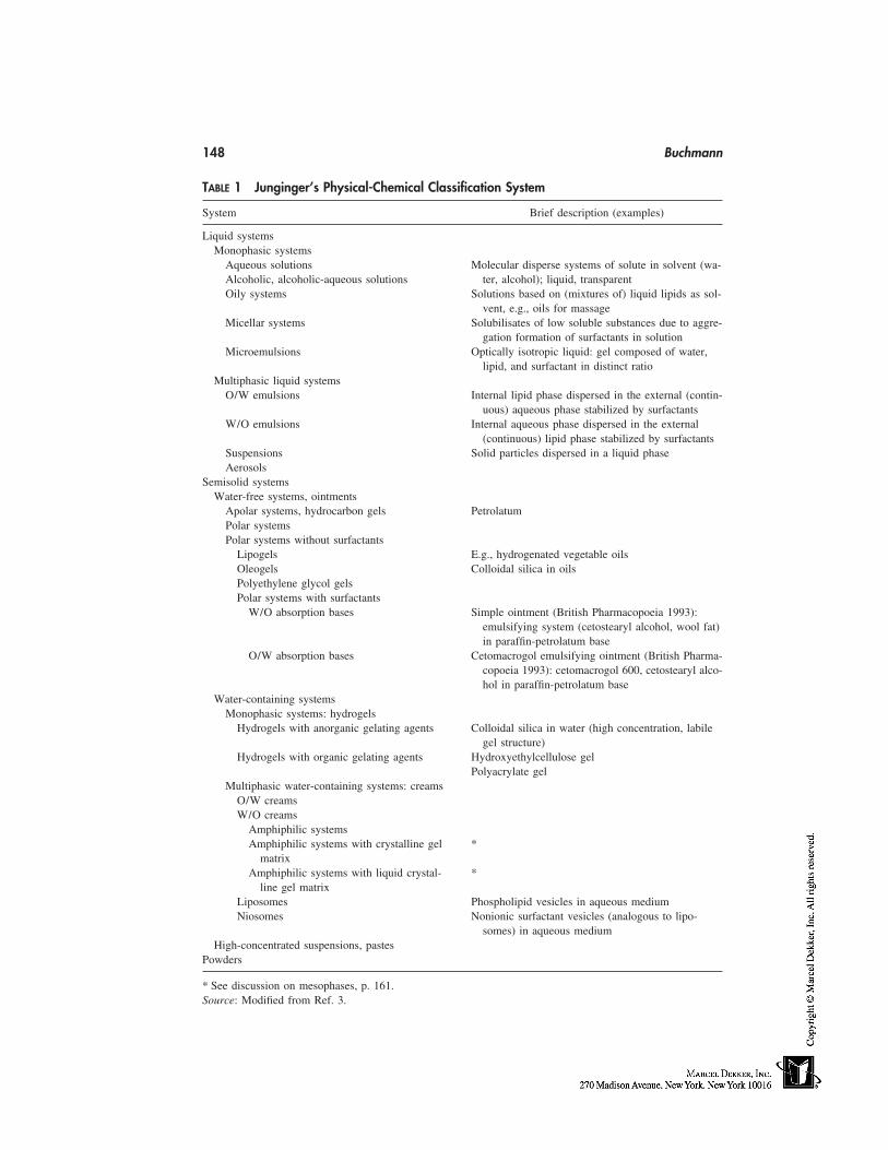
148 Buchmann
TABLE 1 Junginger’s Physical-Chemical Classification System
System Brief description (examples)
Liquid systemsMonophasic systems
Aqueous solutions Molecular disperse systems of solute in solvent (wa-Alcoholic, alcoholic-aqueous solutions ter, alcohol); liquid, transparentOily systems Solutions based on (mixtures of) liquid lipids as sol-
vent, e.g., oils for massageMicellar systems Solubilisates of low soluble substances due to aggre-
gation formation of surfactants in solutionMicroemulsions Optically isotropic liquid: gel composed of water,
lipid, and surfactant in distinct ratioMultiphasic liquid systems
O/W emulsions Internal lipid phase dispersed in the external (contin-uous) aqueous phase stabilized by surfactants
W/O emulsions Internal aqueous phase dispersed in the external(continuous) lipid phase stabilized by surfactants
Suspensions Solid particles dispersed in a liquid phaseAerosols
Semisolid systemsWater-free systems, ointments
Apolar systems, hydrocarbon gels PetrolatumPolar systemsPolar systems without surfactants
Lipogels E.g., hydrogenated vegetable oilsOleogels Colloidal silica in oilsPolyethylene glycol gelsPolar systems with surfactants
W/O absorption bases Simple ointment (British Pharmacopoeia 1993):emulsifying system (cetostearyl alcohol, wool fat)in paraffin-petrolatum base
O/W absorption bases Cetomacrogol emulsifying ointment (British Pharma-copoeia 1993): cetomacrogol 600, cetostearyl alco-hol in paraffin-petrolatum base
Water-containing systemsMonophasic systems: hydrogels
Hydrogels with anorganic gelating agents Colloidal silica in water (high concentration, labilegel structure)
Hydrogels with organic gelating agents Hydroxyethylcellulose gelPolyacrylate gel
Multiphasic water-containing systems: creamsO/W creamsW/O creams
Amphiphilic systemsAmphiphilic systems with crystalline gel *
matrixAmphiphilic systems with liquid crystal- *
line gel matrixLiposomes Phospholipid vesicles in aqueous mediumNiosomes Nonionic surfactant vesicles (analogous to lipo-
somes) in aqueous mediumHigh-concentrated suspensions, pastes
Powders
* See discussion on mesophases, p. 161.Source: Modified from Ref. 3.
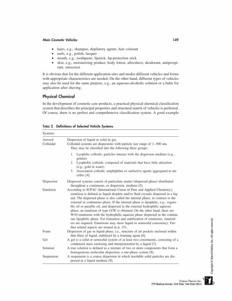
Main Cosmetic Vehicles 149
• hairs, e.g., shampoo, depilatory agents, hair colorant• nails, e.g., polish, lacquer• mouth, e.g., toothpaste, lipstick, lip-protection stick• skin, e.g., moisturizing product, body lotion, aftershave, deodorant, antiperspi-
rant, sunscreen
It is obvious that for the different application sites and modes different vehicles and formswith appropriate characteristics are needed. On the other hand, different types of vehiclesmay also be used for the same purpose, e.g., an aqueous-alcoholic solution or a balm forapplication after shaving.
Physical Chemical
In the development of cosmetic care products, a practical physical-chemical classificationsystem that describes the principal properties and structural matrix of vehicles is preferred.Of course, there is no perfect and comprehensive classification system. A good example
TABLE 2 Definitions of Selected Vehicle Systems
Systems
Aerosol Dispersion of liquid or solid in gas.Colloidal Colloidal systems are dispersions with particle size range of 1–500 nm.
They may be classified into the following three groups:
1. Lyophilic colloids: particles interact with the dispersion medium (e.g.,gelatin)
2. Lyophobic colloids: composed of materials that have little attraction(e.g., gold in water)
3. Association colloids: amphiphiles or surfactive agents aggregated to mi-celles [4].
Dispersion Dispersed systems consist of particulate matter (dispersed phase) distributedthroughout a continuous, or dispersion, medium [5].
Emulsion According to IUPAC (International Union of Pure and Applied Chemistry),emulsion is defined as liquid droplets and/or fluid crystals dispersed in a liq-uid. The dispersed phase is also called the internal phase, in contrast to theexternal or continuous phase. If the internal phase is lipophilic, e.g., vegeta-ble oil or paraffin oil, and dispersed in the external hydrophilic aqueousphase, an emulsion of type O/W is obtained. On the other hand, there areW/O emulsions with the hydrophilic aqueous phase dispersed in the continu-ous lipophilic phase. For formation and stabilization of emulsions, emulsifi-ers are required. Emulsions may show liquid or semisolid consistency. Fur-ther related aspects are treated in p. 151.
Foam Dispersion of gas in liquid phase, i.e., structure of air pockets enclosed withinthin films of liquid, stabilized by a foaming agent [6].
Gel A gel is a solid or semisolid system of at least two constituents, consisting of acondensed mass enclosing and interpenetrated by a liquid [7].
Solution A true solution is defined as a mixture of two or more components that form ahomogeneous molecular dispersion, a one-phase system [8].
Suspension A suspension is a coarse dispersion in which insoluble solid particles are dis-persed in a liquid medium [9].
150 Buchmann
of a physical chemical system is described by Junginger [3] and slightly modified in Table1. Although not comprehensive, such a system is a useful tool for rational formulationdesign and development, in particular when controlled and targeted delivery of activeprinciples has to be achieved. Such a vehicle classification system is also a practical basisfor production, use, and understanding of cosmetic vehicles. However, the boundariesbetween the different classes are flexible, and changing with the state of art and science.More important than pure classification of a cosmetic vehicle is its exact characterization,based on physical, chemical, and biological principles that may eventually lead to a varietyof classification possibilities.
In a physical chemical classification system, various characterization criteria areused for classification of the vehicles:
• Polarity: hydrophilicity, lipophilicity• State of matter: solid, semisolid, liquid, gaseous• Size/dimensions of particulates dispersed in the mixtures (dispersions)
true solution, molecular dispersion: particle size �1 nmcolloidal dispersion: particle size 1 nm–500 nmcoarse dispersion: particle size �500 nm
• Solubility characteristics• Rheology, viscosity• Composition: physical chemical characteristics of the main vehicle components
waterfree, oilyaqueoushydrophilic, nonaqueous solvents
For clarification of the terminology, a selection of definitions or descriptions of the majorsystems is given in Table 2. (See also Refs. 4–9.)
DESCRIPTION AND DEFINITION OF MAIN VEHICLES
Solutions
The term ‘‘solution’’ may be used in a narrow sense, describing true solutions (moleculardispersions; see Table 2), or in a broader sense, also comprising colloidal solutions, i.e.,more or less transparent liquids, e.g., micellar solutions and vesicular systems (mediacontaining liposomes, niosomes).
In general, true solutions used in cosmetics are either based on aqueous, or aqueous-alcoholic, media or on inert oily vehicles. Most organic solvents cannot be used becauseof their local or systemic toxicity, which causes skin irritation or permeation across theskin barrier into the body, respectively. Although good solvents for lipophilic substances,oils may not be used in every case because of their grassy characteristic, low acceptance,and exclusion for hairy application sites. However, for special applications oils are pre-ferred, e.g., for massage. ‘‘Massage oils’’ contain essential oils and fragrances, compoundsthat are easily dissolved in the oily vehicle because of their lipophilic properties.
Prerequisite for solution formulation is a sufficiently high solubility of the solute inthe solvent. Classical examples for solutions used in cosmetics are ‘‘eau de parfums’’ and‘‘eau de toilettes.’’ In order to enable solubilization of the lipophilic fragrances, alcoholor aqueous-alcoholic solutions are prepared. The addition of alcohol to water, or othersuitable hydrophilic but less polar solvents (e.g., glycerol, polyethylene glycol), decreases
Main Cosmetic Vehicles 151
the polarity of the solvent and thus increases the solubility of the lipophilic solutes. Fre-quently, a solute is more soluble in a mixture of solvents than in one solvent alone. Thisphenomenon is known as cosolvency, and the solvents that in combination increase thesolubility of the solute are called cosolvents [10].
Another classical example is preparations for mouthwashes. They usually containessential oils or liquid plant extracts like peppermint or myrrh extract, which are kept insolution by the added ethanol (ca. 70%). When used for application, these concentratesare diluted with water. Then turbidity occurs because of overstepping saturation solubility.In order to prevent turbidity, solubilizing agents (surfactants, e.g., PEG-40 hydrogenatedcastor oil) may be added. The solubilization effect is attributed to aggregation formationof surfactants when in solution. In aqueous solutions surfactants form micelles, smallaggregates, when the concentration of the surfactant exceeds the critical micelle concentra-tion (CMC) [11]. With the aid of those micelles, the solubility of low soluble, apolarcompounds may be increased because of an association or incorporation of the apolarcompounds with the apolar region of the micelle. Thus, solubilization or formation ofmicelles is a favorable means for formulation of solutions.
Finally, salt formation or adjustment of pH also results in improved solubility oforiginally low soluble, ionizable solutes. Thus, e.g., addition of sodium hydroxide maybe used to improve the solubility of hyaluronic acid or preservatives such as sorbic orbenzoic acid. Accordingly, appropriate acids, e.g., lactic acid and citric acid, may be addedwhen solubility of a basic substance must be increased. Although not the main type offormulation used in cosmetics, solutions have the following advantages:
1. They remain physically stable (if true solution and not oversaturated),2. Are easily prepared: simple mixing, under heating if necessary,3. Are transparent, clear, and have a ‘‘clean’’ appearance, and4. Are especially suitable for rinsing and cleaning body surfaces.
However, it must be kept in mind that many compounds are chemically less stable whenin a dissolved state.
In summary, whenever a solution has to be formulated, the optimal solvent mustbe selected, that (1) guarantees sufficient solubility and stability for the solute(s), and(2) is acceptable and safe for application to the body. Solubility may be improved by(1) adaptation of the solvent’s polarity with regard to the solute, (2) salt formation/pHadjustment (ionizable compounds), (3) using mixtures of suitable solvents and cosolvents,and (4) solubilization with the aid of surfactants.
Emulsions: Lotions and Creams
Out of the range of cosmetic care products, the emulsion is the form that is probably themost used. For reasons of skin feeling, consumer appeal, and ease of application, emul-sions are preferred to waterless oils and lipids along with gels. The main components ofemulsions are lipids (lipophilic compounds) and water (and/or hydrophilic compounds).These two immiscible phases are allowed to remain in a metastable mixed state by anamphiphilic component, an emulsifier. This biphasic system may be regarded in analogyto the skin or even to the skin cells, which, simply put, consist of lipophilic and hydrophiliccomponents. Emulsions can either be of the water-in-oil (w/o) or oil-in-water (o/w) types.Showing very similar structural principles, both lotions and creams are discussed in thischapter. If emulsions are liquid, they are generally called lotions. Creams are emulsions
152 Buchmann
occurring in semisolid form. Under gravitation, creams do not flow out through the orificeof reversed containers because of the heavier consistency in comparison with lotions.
Emulsions are prepared by dispersion of the internal in the external phase. For thisenergy-consuming process, emulsifiers that decrease the interfacial tension between thetwo immiscible phases are required. Emulsifiers are not only used for formation but alsofor stabilizing emulsions. Emulsions are metastable systems and the two phases tend toseparate because of coalescence, i.e., when the dispersed droplets fuse. This process maybe slowed by the addition of appropriate emulsifiers, which are ionic or anionic surfactants.The emulsifiers are thought to be located at the interfaces between the two phases, thehydrophilic part of the molecule in contact with the water phase and the lipophilic domainof the emulsifier contacting/touching the lipid phase. Large molecules may even dig intothe lyophilic phase and serve as stabilizing anchors. Being adsorbed at the interfaces, theemulsifying substances form a film—monomolecular or multimolecular, depending onthe substances’ structures—that stabilizes the emulsion [12]. The addition of viscosity-increasing substances further results in an improved consistency and consequently morestable emulsions.
Except for the emulsifiers, the following types of ingredients are usually added tocosmetic emulsions:
• Emollients: They improve the sensory properties of the emulsions. Addition ofan emollient results in better spreading when the emulsion is applied to the skin.Examples: isopropyl myristate, silicon oils.
• Moisturizers and humectants: They increase and control the hydration state ofthe skin. Examples: glycerol, urea.
• Viscosity-increasing agents are added to increase the viscosity of the externalphase, if desired. Examples: xanthan gum, cellulose esters.
• Active substances such as UV sunscreens and vitamins.• Preservatives to prevent microbial growth, particularly in o/w emulsions.• Perfumes and coloring agents for aesthetic purposes.
Oil-in-Water Emulsions
The high acceptance of o/w emulsions is based on the following reasons:
• They feel light and not greasy when applied.• They show good skin spreadability and penetration and an active hydration effect
by the external water phase.• They cause a cooling effect because of the evaporation of the external aqueous
phase.
However, o/w emulsions show a lower effect in preventing dry skin in comparison withw/o emulsions. A typical o/w emulsion is composed as follows:
1. Lipid(s) � lipophilic thickening agent (optional, e.g., microcrystal-line wax) 10–40%
2. Emulsifier system with optimal HLB-value (approx. 9–10 [13]) 5%3. Co-emulsifier (e.g., cetostearyl alcohol, behenyl alcohol) 2%4. Preservatives (antimicrobial, antioxidants) q.s.5. Water � hydrophilic thickening agent (optional, e.g., carbomer) ad 100%
Main Cosmetic Vehicles 153
Depending on the desired product effect, different types of lipids may be used for formula-tion. Addition of nonpolar, occluding lipids (e.g., paraffin oil) improves retention of mois-ture in the skin but lowers spreading on the skin. A good spreading effect is achieved byformation of a low-viscosity emulsion containing polar oils that show a high spreadingcoefficient (e.g., macadamia nut oil, wheat germ oil, isostearyl neopentanoate) [14].
Selection of the lipophilic ingredients and the excipients of the water phase deter-mine the emulsifier system to be used and additional adjuvants, e.g., viscosity-increasingthickening agents. There is no universal emulsifier system, and a huge variety of combina-tions might be used. Today, complex emulgator systems that consist of one or more surfac-tants and a cosurfactant are commonly used. That means at least two surfactants withdifferent HLB-values are combined. For example, steareth-21 (HLB � 15.5) may be com-bined with PEG-5-glyceryl stearate (HLB � 8.7). The latter emulsifier is especially suit-able when nonpolar oils are to be incorporated. In recent years selected polymeric excipi-ents have been used for emulsion stabilization, e.g., crosslinked and linear polyacrylates,polyacrylamides, and derivates of cellulose.
In selecting a co-emulsifier, the following general guidelines apply:
• For the same fatty residue, the viscosity decreases if the degree of ethoxylationincreases.
• For the same degree of ethoxylation, the viscosity increases if the fatty carbonchain length increases [14].
The degree of viscosity (consistency) of o/w emulsions depends on various factors [15]:
• Volume ratio of internal to external phase: increasing lipid percentage results inhigher viscosity, but not necessarily in a semisolid cream.
• Type of lipid: incorporation of high melting lipophilic compounds, e.g., solidparaffin and petrolatum, may result in soft semisolid o/w creams.
• Presence of thickening agents in the lipid phase: addition of cetostearyl alcoholgenerally results in (‘‘hard’’) semisolid creams.
• Presence of thickening agents in the external aqueous phase: the ultimate meanto increase the consistency of a thin o/w emulsion. Addition of hydrocolloids,e.g., carbomers or hydropropyl guar (Jaguar 8600, Rhodia Inc., Cranbury, NJ),is the most efficient method to increase the viscosity of o/w emulsions. However,depending on the properties of the added polymer, the skin feeling of the emul-sion may become negatively influenced because of the stickiness.
An interesting phenomenon is the occurrance of liquid crystal structures (mesophases) inemulsions under certain conditions. This has been investigated and has become of interestmore and more during the last 10 to 20 years. This subject is treated on p. 161.
Water-in-Oil Emulsions
Water-in-oil (w/o) emulsions may still be regarded as heavy, greasy, and sticky althoughduring recent years great progress has been achieved in the preparation of pleasant w/oemulsions. Therefore, the w/o emulsion type is not only the basis for water-resistant sunprotection, baby creams, or night creams, but also for protective day creams. This is be-cause during recent years better excipients have become available. The advantages ofw/o emulsions are:
154 Buchmann
• Close resemblance to the natural protective lipid layer in the stratum corneum• Efficient skin protection attributable to formation of a continuous layer of lipids
on skin after application• Sustained moisturization because on skin a continuous semiocclusive barrier is
formed that reduces evaporation of skin water and that in addition actively re-leases the incorporated water from the internal phase, generally several timesmore efficient than o/w emulsions
• Improved penetration into the lipophilic stratum corneum coupled with improvedcarrier function of lipophilic active substances, and even of hydrophilic sub-stances incorporated in the internal aqueous phase
• Lowered risk of microbial growth• Liquid at very low temperatures (beneficial for winter sport products)
A typical w/o emulsion is composed as follows:
1. Lipid component 20%2. Lipophilic thickening agent (e.g., wax, optional) 1%3. Emulsifier system with optimal HLB-value (3–8) 7–10%4. Preservatives (antimicrobial, antioxidants) q.s.5. MgSO4 ⋅ 7H2 O 0.5%6. Water (� hydrophilic thickening agent, optional) ad 100%
In order to avoid the heavy feel of w/o emulsions, appropriate excipients must be selectedto get products with well-accepted sensory properties. This heavy feel of w/o emulsionsis directly related to the spreading characteristics of the external oil phase. Therefore,polar oils with a high spreading coefficient [16] are preferably used, e.g., macadamia nutoil, isopropyl isostearate, isostearyl neopentanoate. Addition of low-viscosity silicone flu-ids or volatile cyclomethicone also improves the spreading effect. The physicochemicalnature of the lipid components not only determines the spreading on the skin, the degree ofocclusivity, and skin protection, but also influences the selection of the emulsifier system.Therefore, choosing an optimal emulsifier system is crucial. For example, glyceryl sorbitanunsaturated fatty acid ester (Arlacel 481) and glyceryl sorbitan saturated fatty acid ester(Arlacel 986) are better suited to emulsify apolar lipids, whereas more hydrophilic emulsi-fyers like the analogous ethoxylated sorbitan fatty acid esters (Arlacel 581, saturated, andArlacel 582, unsaturated) or fatty acid esters of polyols (Arlacel 1689, saturated, and 1690,unsaturated) are designed for more polar lipids. A combination of PEG-7-hydrated castoroil and polyglyceryl-3-diisostearate may also be used. Skin feel may be improved bycausing thixotropic behavior of the product, which is achieved by addition of a thixotropicagent or by reduction of the emulsifier content.
Multiple Emulsions
Multiple emulsions are triphasic systems or emulsions of emulsions. That means there isa primary emulsion dispersed in an external phase, e.g., water-in-oil-in-water (w/o/w).The dispersed phase in the resulting system contains smaller droplets having the samecomposition as the external phase [17]. The inner aqueous phase is separated from theouter aqueous phase by the oil phase, and therefore the composition of the two aqueousphases may be different, at least after preparation and for a certain storage time. Prepara-tion and stabilization of multiple emulsions is a challenging task. They may either beprepared by a two-step method or by the relatively new one-step process ‘‘Partial Phase
Main Cosmetic Vehicles 155
Solu-Inversion Technology PPSIT’’ [18]. The two-step method includes preparation ofthe primary emulsion, which thereafter is dispersed in the external phase. In the PPSITthe lipid and electrolyte-containing water phase are heated and mixed above the phaseinversion temperature (PIT), where the hydrophilic emulsifier forms w/o emulsions. Bycooling down, a w/o/w system occurs at the PIT for a short time period. Then the systemis immediately fixed by salting out and forming a lamellar matrix structure based on theemulsifier [19]. The advantage of w/o/w emulsions is that they comprise both the lightfeeling and positive sensory characteristics of o/w-emulsions and the skin hydration effectof w/o-emulsions.
Gels
Gels are dispersed systems, originally liquids (solutions) that have a certain consistencyuseful and practical for topical application. In contrast to emulsions, gels generally donot comprise two immiscible phases of opposite lyophilicity. Therefore, the polarity andsolubility characteristics of the incorporated substances are either hydrophilic—in hy-drogels—or lipophilic—in lipogels (or oleogels). The consistency of gels is caused bygelling (thickening) agents, usually polymers, building a three-dimensional network. Inter-molecular forces bind the solvent molecules to the polymeric network, and thus the re-duced mobility of these molecules results in a structured system with increased viscosity.Pure gels are transparent and clear or at least opalescent. Transparency is only achievedif all ingredients are dissolved or occur at least in colloidal form, i.e., the size of particlesis in the submicron range. Transparency in particular is an attractive property of gels. Gelproducts have positive aesthetic characteristics and are thus becoming more and morepopular in cosmetic care products today. Gels can also serve as the basis for more complexformulations:
• Solid particles can be incorporated, resulting in stabilized suspensions• Incorporation of oily lipids results in so-called hydrolipid dispersions or quasi-
emulsions (see p. 156).
Hydrogels
Hydrogels are hydrophilic, consisting mainly (85–95%) of water or an aqueous-alcoholicmixture and the gelling agent. The latter is usually an organic polymeric compound suchas polyacrylic acid (Carbopol), sodium carboxy methylcellulose, or nonionic cellulose-ethers. Hydrogels have to be preserved against microbial growth.
After application, hydrogels show a cooling effect caused by evaporation of thesolvent. They are easily applicable and humidify instantaneously, but if applied over along time they desiccate the skin. For that reason, humectants such as glycerol may beadded. After evaporation, the polymer residue may cause a sticky or ‘‘tearing’’ feel onthe skin if inappropriate thickening agents have been used. Careful selection and testingof the needed adjuvants is therefore recommended.
Hydrophobic Gels
Lipogels or oleogels are obtained by adding a suitable thickening agent to an oil or liquidlipid. For example, colloidal silica may be used for that reason. A special type of hydropho-bic gels is silicone-based systems.
156 Buchmann
Hydrolipid Dispersions
Hydrolipid dispersions are a special type of emulsion and are therefore treated separatelyin this chapter. They are disperse systems with a hydrophilic continuous phase and alipophilic internal phase. The concentration of lipids lies between 2 and 20%. In principle,such a system is thermodynamically unstable. For stabilization, suitable large polymersare added, which are hydrated lyophilic colloids in the aqueous medium. Because of theirmolecular structure these polymeric emulsifiers are able to form mono- to multilamellarfilms at the interfaces and hence stabilize the emulsion. Typical examples are acrylates/C10-30alkyl acrylate crosspolymers. These polymers must have a sufficient surface activ-ity that enables them to interact between the two different phases, resulting in a ‘‘quasi-emulsion,’’ alternatively called balm, costabilized by hydroxypropyl methylcellulose orpolyacrylate. The dispersed oil droplets may show a relatively large size of 20 to 50 µm,but such a quasiemulsion remains stable [20]. The great advantage of hydrolipid-disper-sions is their lack of conventional emulsifiers, surfactants with skin irritation potential.
Microemulsions
According to the definition of Danielsson and Lindman [21], a microemulsion is definedas a system of water, oil, and amphiphile, which is a single optically isotropic and thermo-dynamically stable liquid solution. ‘‘This definition should be widened, however, to in-clude metastable states, spontaneous emulsions of long-lived kinetic stability [22].’’ Theterm microemulsion may be a misnomer, because microemulsions consist of large or‘‘swollen’’ micelles containing the internal phase, much like that found in a solubilizedsolution [23]. Microemulsions contain oil droplets in a water phase or water droplets inoil with diameters of about 10 to 200 nm. Therefore they appear as isotropic, opticallyclear liquid or gel-like systems. Unlike micellar solubilized systems, microemulsions maynot be thermodynamically stable; nevertheless, they are more stable than ordinary emul-sions. They are a type of ternary system composed from water, lipid, and surfactant mix-ture in a distinct ratio. The latter is usually a surfactant, such as Brij 96 [polyoxyethylene(10) oleyl ether] combined with a cosurfactant such as propylene glycol or ethylene glycol.Microemulsions may be used to incorporate or dissolve active substances and have beenfound to improve skin penetration and permeation [24].
The disadvantage of microemulsions is their rather high concentration of surfactants,which is a risk for increased skin irritation and sensitization. Nevertheless, modern micro-emulsion formulation is based on alkyl polyglycosides which are regarded to be milderthan conventional nonionic surfactants with polyoxyethylene chains.
Nanoemulsions and Nanoparticles
During the last years, special dispersion formulations have been developed and describedthat contain ultra small particles used as carriers for active substances. The particles havea size in the range of 10 to a few hundred nanometers. This group of formulations showsa large heterogeneity and very often various terms or trade names have been created nam-ing the same or similar systems. Generally the particles are dispersed in an aqueous me-dium.
For example, solid lipid nanoparticles possess a solid matrix composed of physiolog-ical lipids or lipoids with a mean diameter in the range of approximately 50 to 1000 nm
Main Cosmetic Vehicles 157
[25]. Active substances may be incorporated into these lipid nanoparticles serving as carri-ers, provided that the active substances are released after application on the skin.
Alternatively, the core of nanoparticles may either be a liquid lipid functioning ascarrier or a lipophilic agent being directly effective, e.g., an emollient or occlusive agent.For stabilization, a monolayer of surfactants surrounding/covering the lipid droplet is used,e.g., phospholipids combined with a selected cosurfactant in a defined ratio [26,27]. In-stead of a lipid, lipophilic active substances may be incorporated, e.g., vitamin A or E,UV filters, fragrances, etc. This type of nanoparticle is thought to be relatively insensitivetoward the presence of additional surfactants in contrast to liposomes; therefore they canbe mixed with conventional emulsions and the size of the nanoparticles remains in thesubmicron range.
Suspensions
Strictly considered, suspensions are not just vehicles but products consisting of particles,generally actives or functional excipients, that are dispersed in a liquid or semisolid me-dium that functions as a vehicle. Nevertheless, a suspension is also a type of formulationthat may be used for application on the skin and to deliver substances to a target. In thisway, a suspension can be regarded as a vehicle entity affecting the application site. Exam-ples are sun-protection products or pearlescent nail lacquers containing pigments.
In suspension, sedimentation of unsoluble particles may happen because of differ-ence in density. In order to guarantee a homogeneous product when applied, the particlesmust be redispersible by shaking before use. Alternatively, sedimentation must be hinderedor at least reduced during storage. This is achieved by reduction of particle size and/orby increasing the viscosity of the vehicle, ideally creating a thixotropic system. The vehicleeffect of the suspension on the skin is primarily caused by the liquid or semisolid phaseof the vehicle comparable to solutions and emulsions.
Sticks
A stick is a solid delivery vehicle cast in an elongated form. By rubbing a stick onto skin,a variety of cosmetic ingredients can be delivered, such as fragrances, coloring agents,and emollients. In particular, sticks are ideally suited to deliver insoluble substances, e.g.,pigments. The most popular cosmetic sticks are lipsticks and antiperspirant/deodorantsticks.
There are mainly three basic vehicle types of sticks:
1. Mixture of waxes (e.g., beeswax, carnauba) and oils (e.g., mineral, castor oil)that are cast into solid form, containing dissolved or undissolved active ingredi-ents
2. Hydrophilic or aqueous sticks: solutions based on aqueous, propylene glycol,alcohol mixtures, solidified usually by sodium stearate, containing, e.g., alumin-ium chlorohydrate as antiperspirant
3. Matrix consisting of a high-boiling volatile silicone (e.g., cyclomethicone)gelled by fatty alcohol (e.g., stearyl alcohol)
In recent years, clear sticks have become popular. As a gelling agent, dibenzylidene sorbi-tol is used in propylene glycol or other related polyols [28].
158 Buchmann
FUNCTIONAL DESIGN, COMPOSITION, AND RESULTING EFFECT
There is no universal cosmetic vehicle available that can simply be mixed with an activecosmetic substance to get the cosmetic care product of choice, nor is there a generalprinciple that could be observed to perform development of such a product. But a cosmeticcare product has to be developed and whenever this is the case, various issues and aspectshave to be considered and many problems must be solved step-by-step. Although formula-tion (galenical development) of cosmetic products is still rather empirical today, a rationalapproach is suggested. This section discusses the main issues that are to be consideredwhen a functionally designed cosmetic product is being developed.
Target Profile
First, a clear target profile of the product must be defined. This includes the following:
1. Site of application. Depending on the site, certain forms may not be adequate,e.g., a w/o cream is not at all suitable for application on hair.
2. Area of application. A sticky, greasy cream cannot be applied on the wholebody surface.
3. Target site. For example, the uppermost layer of stratum corneum or viableepidermis.
4. Sensory properties. For example, foaming shampoo or a light, smooth, low-viscosity cream.
5. Optical aspect. Clear, transparent, or milky, mono- or multiphasic.6. State of matter. Liquid, semisolid, or solid.7. Basic type of form. Solution or emulsion.8. Active substances. Selected vegetable oil, vitamins, UV screen.9. Storage stability and conditions.
10. Packaging.11. Comparable, competitor products.
Selection of Vehicle Type
The type of vehicle may already be determined by the product target profile. If varioustypes are possible, the most suitable should be selected. The following selection criteriaare important: function or desired effect of the vehicle on the skin, ease of formulationfeasibility, and physical and chemical stability. Furthermore, solubility, polarity, saturationsolubility, vehicle interactions, and formation of mesophases are subjects to be consideredwhen dealing with development and selection of vehicles. These topics are discussed later.
True Solution Versus Disperse System
Whenever the target of an active substance lies in deeper regions of the skin or even inskin cells, the substance must be present in molecular form for successful and efficientdelivery, i.e., it must be dissolved in the vehicle or it must be able to dissolve, at least,after application. In other words, dissolution of a substance is a prerequisite for its de-livery to a biological viable target (e.g., cell, enzyme). It is only in the dissolved statethat fast and efficient penetration and transport into the deeper skin layers and cells ispossible.
Main Cosmetic Vehicles 159
Thus, the first goal in formulation development is to dissolve the active substancein the vehicle. Therefore, the vehicle should be an ideal solvent for the active substance. Ifa substance cannot be dissolved in the vehicle—this may happen because of low solubilityproperties or stability reasons—then the substance has to be incorporated in particulateform; the smaller the size, the better. Fine particles in the order of 1 µm can be deliveredonto or even into the uppermost layers of the skin, as close as possible to the target site.There they may dissolve, faster or slower, depending on their solubility in the skin. Invehicle systems containing particulate matter, homogeneous distribution of the undis-solved substances must be guaranteed.
In summary, if the first goal—dissolution of active substance in the vehicle—is notachieved, the first alternative in formulation development must be targeted: the substanceto be delivered must occur in particulate form as fine as possible. This is the prerequisitefor fast and efficient delivery of unsoluble matter into the skin close to the target site.
Polarity
In order to achieve dissolution of a substance (solute), the adequate vehicle (solvent) hasto be selected. The solubility of a substance is attributable in large measure to the polarityof the solvent, and it generally depends on chemical, electrical, and structural effects thatlead to mutual interactions between the solute and solvent [29]. Polar solvents dissolveionic solutes and other polar substances, whereas nonpolar substances are dissolved innonpolar, lipophilic solvents. Solubility properties determine the selection of the appro-priate vehicle for both, for solid as well as for liquid substances. Only nonpolar liquidsare mutually completely miscible and thus can be used to make a nonpolar liquid vehicle.Accordingly, the same is true for polar liquids (e.g., water and alcohol).
Solubility characteristics of a compound used in formulation is one of the mostimportant factors to be considered. Solubility data can be found in the literature; veryoften they are delivered by suppliers of the substances or they must be determined experi-mentally. In formulation the solubility parameter δ, according to Hildebrand and Scott[30], is a useful tool for selection of appropriate solvents. The more alike the δ-values ofthe compounds, the greater is their mutual solubility. A list of solubility parameters ofcosmetic ingredients is given in Ref. 31. Very apolar substances have a low δ-value, andwater has the highest value [23]. A rule of thumb states that mutual solubility is given ifthe difference between the two specific δ-values is at maximum 2 units (cal/cm3)�2.
Particularly in cosmetic formulation, where oils and lipids play a dominating role,polarity of oils is a factor to be considered. According to ICI Surfactants [16], the polaritymay also be expressed by the polarity index based on the surface tension between the oil andwater. Another interesting and simple characterization method is based on the bathochromiceffect of a suitable dye dissolved in oils. The absorption maximum in the visible light—andtherefore the color—of a nil-red-oil solution depends on the polarity of the oil; the higherthe absorption maximum, the more polar is the oil or oil mixture [32].
In conclusion, if a monophasic system has to be formulated, only substances withmutual solubility can be combined. In contrast, if multiphasic systems such as emulsionsand suspensions are made, the phase-forming components must be mutually insoluble.Nevertheless, preparation and solubilization of multiphasic systems require the additionof amphiphilic substances (emulsifiers in emulsions, surfactants for wetting and repulsingthe particles in suspensions). In emulsions, polar as well as nonpolar substances can bedissolved in the hydrophilic or lipophilic phase, respectively. This is one reason for thepopularity of emulsions.
160 Buchmann
Saturation, Supersaturation
Theoretically, a solute can be dissolved in a solvent up to the saturation solubility. Beyondthis concentration, precipitation of the solute or phase separation usually occurs. Somesubstances are able to remain transiently in solution above saturation solubility. This phe-nomenon is known as supersaturation, a metastable condition. Supersaturated solutionscan be caused to return to saturation equilibrium by triggers such as agitation, scratchingthe wall of containers, or addition of seeding crystals.
The driving force for delivery of substances, i.e., release from vehicle and penetra-tion into skin, is thermodynamic activity, which is maximal at saturation concentration[33]. Consequently, in order to achieve maximal penetration rate into the skin, a substancemust be dissolved in a vehicle at saturation concentration. Moreover, saturated or supersat-urated systems are necessary, but not the only prerequisites for optimal topical delivery.For example, the skin—vehicle partition coefficient of the solute also plays a role. Thepartition coefficient may be raised because of the vehicle—skin interaction yielding inincreased skin penetration. In conclusion, achieving the highest possible concentration inthe dissolved state is the second goal to be aimed for in formulation development if deliv-ery into the skin is targeted.
Vehicle Interactions
Sun-protection products are a good example of showing interactions between vehicle,active substance, and the skin. The absorption of UV radiation not only depends on themolecular structure and concentration of the protecting agent, but on the solvent as well.Also, water resistancy may be influenced by selection and composition of the vehicle.
Vehicle components may penetrate into the stratum corneum and interact with thestratum corneum lipids. This may result in disturbance of their lamellar structures andincreased and faster penetration of compounds in the stratum corneum. Alternatively, pres-ence of vehicle components in the stratum corneum may cause a depot effect for certaincompounds.
Substantivity
The term substantivity describes adherence properties of materials to keratinous substratesin the upper skin layers, in particular regarding deposition and retention capacity when incontact with water, which could deplete the material [34]. High substantivity is especiallyimportant for sun protection products. It is primarily a function of the physicochemicalproperties of the active molecules but may also be influenced by the vehicle. For example,addition of film-forming, skin-adherent polymeric substances to the vehicle may increaseretention of sunscreens in the skin and thus result in an improved water-resistant product.Another means is creating formulations that contain phospholipids, enabling the formationof vesicular, liposomal structures in the vehicle or in the upper layers of stratum corneumand thus yielding in a depot effect.
An interesting model to assess substantivity has been presented by Ref. 34. Theinvestigators used human callus to simulate and quantify solute sorption to human skin,which was found to be more suitable than octanol or animal keratin. However, waterresistancy still has to be determined in vivo to know the true quality of the product.
Main Cosmetic Vehicles 161
Mesophases
Not only the type of vehicle, e.g., solution or o/w emulsion, but also occurrence and typeof mesophases (liquid crystal structures) determine the properties and behavior of a vehi-cle. At certain concentrations and combinations of specific emulsifying agents in liquids,associations may be formed, resulting in liquid crystal structures, also called mesomorphicstate or mesophase. The mesophase shows anisotropy and is thermodynamically stable.Different types of mesophases have been described: middle phase (hexagonal), cubicphase, and neat phase (lamellar).
Fatty amphiphiles (e.g., long chain alcohols, acids, monoglycerides) that are dis-persed in water in the presence of a high hydrophilic-lipophilic balance (HLB) surfactantform lamellar phases. They are able to swell at an elevated temperature close to the meltingpoint of the hydrocarbon chain. These swollen lamellar liquid crystalline phases can incor-porate significant quantities of water. The hydrocarbon chains are liquid-like, i.e., disor-dered. If the temperature decreases, the lamellar liquid crystalline phases of fatty amphi-philes are transformed to so-called lamellar crystalline gel network phases, which buildcomplex gel networks. Such networks not only stabilize creams and lotions, but also con-trol their consistency because of their viscoelastic properties. Such mesophases providethe following advantages to emulsions:
1. Increased stability2. Prolonged hydration properties3. Controlled release of active ingredient4. Easy to formulate5. Well-liked skin feel [35]
Metamorphosis of Vehicles
Most vehicles undergo considerable changes during and after application to the skin be-cause of mechanical stress when spread over the surface and/or evaporation of volatileingredients. Mechanical stress and skin temperature may influence the viscosity of thevehicle and consequently the release rate of active ingredients. Uptake of water from theskin may alter the composition of the vehicle. All these factors may also cause phaseinversion or phase separation. And last but not least, as a consequence of these alterationsthe thermodynamic activity of an active ingredient within its vehicle will change as well.Thus, by controlling or changing the thermodynamic activity, release of a substance fromthe vehicle and penetration into the skin can be modulated. For example, if after applica-tion the volatile component of the vehicle, being an excellent solvent of the active sub-stance, evaporates, saturation concentration of the active in the remaining vehicle or evensupersaturation may be achieved. This results either in improved release and delivery aspreviously mentioned (see Section 5.2.3) or in precipitation and deposition of the activesubstance. Another interesting example is given by an optimally composed sun-protectingo/w-emulsion; after application the emulsion has transformed to the w/o type because ofwater evaporation and the mechanical stress caused by spreading. The remaining lipophilicprotective film yields in improved water resistancy.
In conclusion, the optimally designed and developed vehicle not only demonstratesexcellent properties after manufacturing and storage, but also after application and meta-morphosis at the application site.
162 Buchmann
Rheology
The term rheology describes the flow characteristics of liquids and the deformation ofsolids. Viscosity is an expression of the resistance of a fluid to flow. Rheological propertiesare crucial for liquid and semiliquid cosmetic formulations because they determine theproduct’s properties meaningful in mixing and flow when produced, filled into containersand removed before use, as well as sensory properties when applied, such as consistency,spreadability, and smoothness. Furthermore, the rheology of a product may also affectthe physical stability and the biological availability of the product [36].
Regarding rheological characteristics, there are two main types of systems: Newto-nian and non-Newtonian. The former show constant viscosity when stressed, i.e., the rateof shear (flow velocity) is directly proportional to the shearing stress, e.g., water, mineraloil, etc. In non-Newtonian systems (most cosmetic products), however, viscosity changeswith varying stress, i.e., viscosity depends on the degree of shearing stress, resulting eitherin plastic, pseudoplastic, or dilatant flow or in thixothropy, characteristics that are notdiscussed in depth here although they are of practical significance. An ideal topical prod-uct, e.g., shows optimal thixotropic properties; it does not flow out of a tube’s orificeunless slightly pressed, and when on the skin it does not immediately flow and drop offunless easily spread over the application area, where under a certain stress it becomesmore fluid because of the thixotropy. The rheological properties of semisolid products aredetermined first for general characterization in the development phase and second forquality-control reasons after manufacturing. There are various instrumental methods usedto measure rheology or viscosity. Today, apparatus based on rotation or oscillation arecommonly used for non-Newtonian systems.
In order to adjust the rheology of products, various means and excipients are avail-able. If the viscosity has to be increased, addition of viscosity increasing agents is needed.Addition or increase in concentration of electrolytes may influence viscosity. Many sys-tems, e.g., polyacrylates, are sensitive to the presence of ions and the viscosity is reduced.
In particular, emulsions are susceptible to rheological issues. Various factors deter-mine the rheological properties of emulsions, such as viscosity of internal and externalphases, phase volume ratio, particle size distribution, type and concentration of emulsify-ing system, and viscosity-modifying agents. However, this topic is too complex to betreated comprehensively in this context. It is further discussed in a review by Sherman[37]. It is important to realize that small changes in concentrations or ratio of certainingredients may result in drastic changes of the rheological characteristics. Emulsifiedproducts may undergo a wide variety of shear stresses during either preparation or use.Thus, an emulsion formulation should be robust enough to resist external factors that couldmodify its rheological properties or the product should be designed so that change inrheology results in a desired effect.
Preservation
Antimicrobials
Most cosmetic care products must be protected against microbial growth. Not only forthe protection of consumers against infection but also for stability reasons. Growth ofmicroorganisms might result in degradation of ingredients and consequently in deteriora-tion of physical and chemical stability. In general, presence of water in the vehicle as wellas other ingredients susceptible to microbial metabolism require adequate preservation.
Main Cosmetic Vehicles 163
There are various ways to protect a product against microbial growth:
1. Addition of an antimicrobial agent, which is common practice2. Sterile or aseptic production and filling into packaging material, preventing mi-
crobial contamination during storage and usage3. Reduced water activity, i.e., controlling growth of spoilage microorganisms by
reducing the available amount of water in cosmetic preparations [38]
It is not only mandatory to add antimicrobials but also to test their efficacy aftermanufacturing and after storage until the expiration date. Nowadays performance of thepreservative efficacy test (PET), also known as the challenge test, is state of the art [39].Today more and more in-use tests are performed to simulate the usage by the consumerand to show efficacious protection against microbial growth after contamination.
Addition of preservatives to complex, multiphasic systems, in particular, is a criticalformulation issue for the following reasons:
1. Many preservatives interact with other components of the vehicle, e.g., withemulsifyers, resulting in change of viscosity or in phase separation in the worstcase.
2. Depending on the physicochemical characteristics, preservatives are distributedbetween the different phases which might result in too-low effective concentra-tion in the aqueous phase.
3. Adsorption of the preservatives to polymers in the formulation and/or packagingmaterial; complexation or micellization might also result in too-low antimicro-bial activity.
In conclusion, it is not sufficient to add a preservative at recommended concentra-tion. To protect the vehicle sufficiently, a properly designed preservative system is requiredthat must be tested in the formulation regarding efficacy and safety. It is a great formulationchallenge to achieve sufficient protection against microbial growth in the product, espe-cially as many antimicrobials are discredited because of their irritation and sensitizationpotential.
Antioxidants
Protection against oxidation may also be a formulation issue although not so relevant asantimicrobial efficacy. It is achieved by addition of antioxidants or by manufacturing andstoring in an inert atmosphere. In particular, modern formulations containing oxidation-sensitive compounds, such as certain vitamins and vegetable oils with unsaturated fattyacid derivatives, must be sufficiently protected against oxygen.
Development Strategy and Rationale
Having considered the aforementioned issues, formulation development is preferably con-ducted according to a suitable, rational procedure. The complex formulation developmentprocess may be represented symbolically by the ‘‘magic formulation triangle’’ (Fig. 1),showing the mutual interaction and dependency of the following:
1. Feasibility of preparation or formulation of the active substance(s) in the vehicle2. Stability (chemical and physical) of the product, and3. Effectivity or activity of the product when applied.
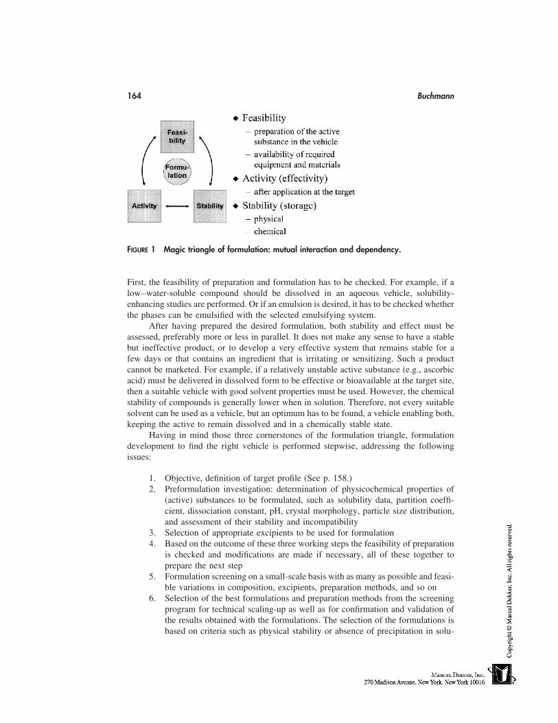
164 Buchmann
FIGURE 1 Magic triangle of formulation: mutual interaction and dependency.
First, the feasibility of preparation and formulation has to be checked. For example, if alow–water-soluble compound should be dissolved in an aqueous vehicle, solubility-enhancing studies are performed. Or if an emulsion is desired, it has to be checked whetherthe phases can be emulsified with the selected emulsifying system.
After having prepared the desired formulation, both stability and effect must beassessed, preferably more or less in parallel. It does not make any sense to have a stablebut ineffective product, or to develop a very effective system that remains stable for afew days or that contains an ingredient that is irritating or sensitizing. Such a productcannot be marketed. For example, if a relatively unstable active substance (e.g., ascorbicacid) must be delivered in dissolved form to be effective or bioavailable at the target site,then a suitable vehicle with good solvent properties must be used. However, the chemicalstability of compounds is generally lower when in solution. Therefore, not every suitablesolvent can be used as a vehicle, but an optimum has to be found, a vehicle enabling both,keeping the active to remain dissolved and in a chemically stable state.
Having in mind those three cornerstones of the formulation triangle, formulationdevelopment to find the right vehicle is performed stepwise, addressing the followingissues:
1. Objective, definition of target profile (See p. 158.)2. Preformulation investigation: determination of physicochemical properties of
(active) substances to be formulated, such as solubility data, partition coeffi-cient, dissociation constant, pH, crystal morphology, particle size distribution,and assessment of their stability and incompatibility
3. Selection of appropriate excipients to be used for formulation4. Based on the outcome of these three working steps the feasibility of preparation
is checked and modifications are made if necessary, all of these together toprepare the next step
5. Formulation screening on a small-scale basis with as many as possible and feasi-ble variations in composition, excipients, preparation methods, and so on
6. Selection of the best formulations and preparation methods from the screeningprogram for technical scaling-up as well as for confirmation and validation ofthe results obtained with the formulations. The selection of the formulations isbased on criteria such as physical stability or absence of precipitation in solu-
Main Cosmetic Vehicles 165
tion, no sedimentation or phase separation or recrystallization in multiphasicsystems; chemical stability or degradation, respectively; preservative efficacytest (PET); biological assessment, e.g., skin-hydration effect, sun-protecting ef-fect, and antioxidant or radical scavenger effect in cells; and
7. Safety evaluation in human beings with formulation chosen for introductioninto market.
PREPARATION METHODS
It is not the intention to present a review on preparation methods and equipment for themanufacturing of cosmetic vehicles and products in this chapter. But it is common sensethat the preparation method may influence a product’s quality. Thus, not only the composi-tion but also the way of preparation should be in the scope of development and preparationwork. There are many types and variations of mixing, dispersion, emulsification, and size-reduction equipment that can be used to prepare vehicles that are used in cosmetics. Forexample, size reduction of the internal phase droplets in an emulsion depends on themechanical principle of the used equipment, and best results are achieved with a valvehomogenizer. In every case the goal is to get a homogeneous product of specified andreproducible quality. Only with a product of specified and constant quality a reproducibleeffect can be achieved when applied. Standard, basic operations are dissolution, blendingand mixing, dispersion and homogenization, and size reduction, which may all be associ-ated by energy transfer involving cooling or heating.
It is of paramount importance that in early development phases preparation is per-formed under well-defined and known conditions, otherwise scaling-up and reproducibilityof product quality becomes a risky task. Closely related with the preparation method istesting and characterization of the product. This is treated in the following section.
CHARACTERIZATION
Physical Characterization
Appearance
Assessment and description of appearance is one of the easiest, most practical, and never-theless powerful tests. It may be performed macroscopically, describing color, clearness,transparency, turbidity, and state of matter. In addition, microscopic investigation is rec-ommended; taking microphotographs is useful for documentation.
Rheology
Rheological properties (viscosity, consistency) are important characteristics of most typesof cosmetic care products because they have an impact on preparation, packaging, storage,application, and delivery of actives. Thus these properties should be assessed for character-ization and quality control of the product.
Most disperse systems and thus cosmetic care products show Non-Newtonian flowbehavior, namely pseudoplastic, plastic, or dilatant behavior. A wide variety of techniquesand methods have been developed to measure viscosity properties. These procedures canbe classified as either absolute or relative. The absolute either directly or indirectly mea-sures specific components of shear stress and shear rate to define an appropriate rheologicalfunction. Methods used for absolute viscosity measurements are flow through a tube, rota-
166 Buchmann
tional methods, or surface viscosity methods. Methods used for relative viscosity measure-ment are those using orifice viscometers, falling balls, or plungers. Such instruments,although they do not measure stress or shear rate, offer valuable quality-control tests forrelative comparison between different materials [40]. Apparatus based on rotational oreven oscillating principles to assess viscoelastic properties is state of the art.
pH
Measurement of pH value (concentration of hydrogen ions) in aqueous vehicles (solutions,suspensions, o/w emulsions, gels) is a valuable control mean. First of all, if possible, apH value in the physiological range is generally targeted, ideally similar to that of theskin or the specific application site, in order to prevent irritation. Many reactions andprocesses depend on pH, e.g., efficacy of antimicrobial preservatives, stability and degra-dation of substances, and solubility. Thus, pH measurement is a ‘‘must’’ and it is easilyperformed with the available measurement systems.
Homogeneity
In many cases, at a first step homogeneity may be assessed visibly; precipitation in asolution or distinct phase separation in an emulsion is easily detected. Nontransparent,multiphasic systems are more difficult to check. In these cases, microscopic investigationof representative samples is suggested along with quantitative assays regarding activeingredients (uniformity of content).
Droplet or Particle Size and Distribution
The physical stability of colloidal systems as well as emulsions or suspensions partiallydepends on the particle size. In particular, preparations containing small particles withidentical electrical charge are more resistant to flocculation and sedimentation than sys-tems containing larger or uncharged entities. Similarly, reduced particle size is an indicatorof improved kinetic stability of emulsions or suspensions. For that reason, determinationof particle size and size distribution is an important characterization method. Variousoptical methods are available; A minireview is given in Ref. 41 and a selection is listedas follows:
1. Perhaps the most commonly used method today is based on laser diffraction,suitable to measure solid particles and also dispersed droplets under specialconditions, size range 1 to 600 µm.
2. Dynamic light scattering (DLS), also known as photon correlation spectroscopy(PCS), is used for measuring micelles, liposomes, and submicron suspensions(size range 0.003 to 3 µm).
3. Optical or electron microscopy are further methods of choice.
Chemical Characterization
Besides physical characterization, chemically based investigations are indispensable toassess the quality of a product. It is well known that the quality and composition of avehicle can influence the chemical stability of ingredients. Many reactions, such as esterhydrolysis or other degradations, may be enhanced or sustained by change in pH, presenceof catalytic or stabilizing agents, respectively. Thus, development and optimal selectionof the best vehicle is supported by chemical stability investigations.
Main Cosmetic Vehicles 167
Biological Characterization
Further important assessment methods are based on biological tests. This is to evaluateand validate the desired targeted effects in vivo after application of the product. Examplesinclude hydration of the skin, protection against sun radiation, and protection against skinirritating substances during work. This subject is treated in other chapters of this textbook.
Sensory Assessment
The sensory assessment is a useful tool for product and concept development and forquality control in the cosmetic industry. Although a very subjective and liable method,valuable data is obtained if sensory assessment is conducted in a systematic way. Termslike pick up, consistency, peaking, cushion, absorption, smoothness, stickiness, tackiness,oiliness, and greasy are used. An interesting paper on that subject has been published byBusch and Gassenmeier [42].
Barry and coworkers carried out sensory testing on topical preparations and estab-lished rheological methods for use as control procedures to maintain uniform skin feel andspreadability [43]. The consistency of a material can be assessed by using three attributes:smoothness, thinness, and warmth [44].
REFERENCES
1. Wilkinson JB, Moore RJ, eds. Harry’s Cosmeticology. New York: Chemical Publishing, 1982.2. Rieger MM. Cosmetics and their relation to drugs. In: Swarbrick J, Boylan JC, eds. Encyclope-
dia of Pharmaceutical Technology, Vol. 3. New York: Marcel Dekker, 1990:361–373.3. Junginger HE. Systematik der dermatika—kolloidchemischer aufbau. In: Niedner R, Ziegen-
meyer J, eds. Dermatika. Stuttgart: Wissenschaftliche Verlagsgesellschaft mbH, 1992:476.4. Martin A, Bustamante P, Chun AHC. Physical Pharmacy. Philadelphia: Lea & Febiger, 1993:
393–396.5. Martin A, Bustamante P, Chun AHC. Physical Pharmacy. Philadelphia: Lea & Febiger, 1993:
393.6. Martin A, Bustamante P, Chun AHC. Physical Pharmacy. Philadelphia: Lea & Febiger, 1993:
386.7. Martin A, Bustamante P, Chun AHC. Physical Pharmacy. Philadelphia: Lea & Febiger, 1993:
496.8. Martin A, Bustamante P, Chun AHC. Physical Pharmacy. Philadelphia: Lea & Febiger, 1993:
101.9. Martin A, Bustamante P, Chun AHC. Physical Pharmacy. Philadelphia: Lea & Febiger, 1993:
477.10. Martin A, Bustamante P, Chun AHC. Physical Pharmacy. Philadelphia: Lea & Febiger, 1993:
234.11. Martin A, Bustamante P, Chun AHC. Physical Pharmacy. Philadelphia: Lea & Febiger, 1993:
396.12. Martin A, Bustamante P, Chun AHC. Physical Pharmacy. Philadelphia: Lea & Febiger, 1993:
488.13. Martin A, Bustamante P, Chun AHC. Physical Pharmacy. Philadelphia: Lea & Febiger, 1993:
490.14. ICI Surfactants, brochure 41-1E. Personal Care. Middlesbrough, Cleveland, United Kingdom,
1996.15. Herzog B, Marquart D, Müller S, Pedrussio R, Sucker H. Einfluss von zusammensetzung und
phasenverhältnis auf die konsistenz von cremes. Pharm Ind 1998; 60:713–721.
168 Buchmann
16. ICI Surfactants, brochure 42-4E. Personal Care, emulsifiers for water in oil emulsions. Mid-dlesbrough, Cleveland, United Kingdom, 1996:5.
17. Rosoff M. Specialized pharmaceutical emulsions. In: Liebermann HA, Rieger MM, BankerGS, eds. Pharmaceutical Dosage Forms: Disperse Systems, Vol. 3. New York: Marcel Dekker,1998:11.
18. Gohla SH, Nielsen J. Partial phase solu-inversion technology (PPSIT). Seifen Oele FetteWachse J 1995; 121:707–713.
19. Kutz G, Friess S. Moderne Verfahren zur Herstellung von halbfesten und flüssigen Emulsio-nen—eine aktuelle Uebersicht. Seifen Oele Fette Wachse J 1998; 124:308–313.
20. Daniels R. Neue anwendungsformen bei sonnenschutzmitteln. Apotheken Journal. 1997;19(5):22–28.
21. Danielsson L, Lindman B. Colloids Surfaces 1981; 3:391.22. Rosoff M. Specialized pharmaceutical emulsions. In: Liebermann HA, Rieger MM, Banker
GS, eds. Pharmaceutical Dosage Forms: Disperse Systems, Vol. 3. New York: Marcel Dekker,1998:20.
23. Martin A, Bustamante P, Chun AHC. Physical Pharmacy. Philadelphia: Lea & Febiger, 1993:495.
24. Martin A, Bustamante P, Chun AHC. Physical Pharmacy. Philadelphia: Lea & Febiger, 1993:496.
25. Müller RH, Weyhers H, zur Mühlen A, Dingler A, Mehnert W. Solid lipid nanoparticles—ein neuartiger Wirkstoff-carrier für Kosmetika und Pharmazeutika. Pharm Ind 1997; 59:423–427.
26. Zülli F, Suter F. Preparation and properties of small nanoparticles for skin and hair care. SeifenOele Fette Wachse J 1997; 123:880–885.
27. Herzog B, Sommer K, Baschong W, Röding J. Nanotopes: a surfactant resistant carriersystem. Seifen Oele Fette Wachse J 1998; 124:614–623.
28. Schueller R, Romanowsky P. Gels and sticks. Cosmet Toilet Mag 1998; 113:43–46.29. Martin A, Bustamante P, Chun AHC. Physical Pharmacy. Philadelphia: Lea & Febiger, 1993:
215.30. Hildebrand JR, Scott RL. Solubility of Nonelectrolytes. New York: Dover, 1964; (Chap. 23).31. Vaughan CD. Using solubility parameters in cosmetics formulation. J Soc Cosmet Chem 1985;
36:319–333.32. Dietz Th. Solvatochromie von Nilrot. Parfümerie und Kosmetik 1999; 80:44–49.33. Flynn GL, Weiner ND. Topical and transdermal delivery—provinces of realism. In: Gurny
R, Teubner A, eds. Dermal and Transdermal Drug Delivery. Stuttgart: Wissenschaftliche Ver-lagsgesellschaft mbH, 1993:44.
34. Hagedorn-Leweke U, Lippold BC. Accumulation of sunscreens and other compounds in kera-tinous substrates. Eur J Pharmaceutics Biopharmaceutics 1998; 46:215–221.
35. Loll P. Liquid crystals in cosmetic emulsions. Reprint RP 94-93E. ICI Europe Limited, Ever-berg, B, 1993.
36. Martin A, Bustamante P, Chun AHC. Physical Pharmacy. Philadelphia: Lea & Febiger, 1993:457.
37. Sherman P. Rheology of Emulsions. Oxford: Pergamon Press, 1963.38. Enigl DC, Sorrells KM. Water activity and self-preserving formulas. In: Kabara JJ, Orth DS,
eds. Preservative-Free and Self-Preserving Cosmetics and Drugs. New York: Marcel Dekker,1997:45.
39. Sabourin JR. A Perspective on Preservation for the New Millennium, Cosmetics and ToiletriesManufacture Worldwide. Hemel Hempstead, United Kingdom: Aston Publishing Group, 1999:50–59.
40. Hanna SA. Quality assurance. In: Liebermann HA, Rieger MM, Banker GS, eds. Pharmaceuti-cal Dosage Forms: Disperse Systems, Vol. 3. New York: Marcel Dekker, 1998:460.
Main Cosmetic Vehicles 169
41. Haskell RJ. Characterization of submicron systems via optical methods. J Pharm Sci 1998;87:125–129.
42. Busch P, Gassenmeier Th. Sensory assessment in the cosmetic field. Parfümerie und Kosmetik1997; 7/8:16–21.
43a. Barry BW, Grace AJ. J Pharm Sci 1971; 60:1198, J Pharm Sci 1972; 61:335.43b. Barry BW, Meyer MC. J Pharm Sci 1973; 62:1349.44. Martin A, Bustamante P, Chun AHC. Physical Pharmacy. Philadelphia: Lea & Febiger, 1993:
471.

15
Encapsulation to Deliver Topical Actives
Jocélia JansenState University of Ponta Grossa, Ponta Grossa,Paraná, Brazil
Howard I. MaibachUniversity of California at San Francisco School of Medicine,San Francisco, California
INTRODUCTION
Cosmetic technology is constantly developing raw materials and formulation with activeingredients. The new surfactant molecules, the search for original active substances andefficient combinations, and the design of novel vehicles or carriers has led to the imple-mentation of new cosmetic systems in contrast to the classic forms such as creams orgels.
The achievements of recent extensive research has resulted in the development ofcontrolled delivery systems. Some of these systems have been extensively investigatedfor their therapeutic potential while simultaneously being examined for their possible cos-metic uses. One objective in the design of novel drug delivery systems is controlled deliv-ery of the active to its site of action at an appropriate rate. Novel polymers and surfactantsin different forms, sizes, and shapes can aid in this goal. Encapsulation techniques areused in pharmaceuticals, cosmetics, veterinary application, food, copying systems, laundryproducts, agricultural uses, pigments, and other less well-known uses to control the deliv-ery of encapsulated agents as well as to protect those agents from environmental degrada-tion.
DESIGN ASPECTS OF A VECTOR
Microparticles
Microencapsulation is a process by which very thin coatings of inert natural or syntheticpolymeric materials are deposited around microsized particles of solids or droplets ofliquids. Products thus formed are known as microparticles, covering two types of forms:microcapsules, micrometric reservoir systems, and microspheres, micrometric matrix sys-tems (Fig. 1).
171
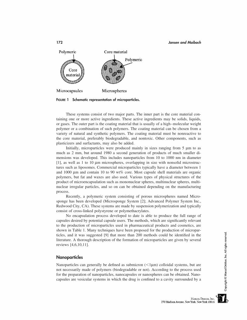
172 Jansen and Maibach
FIGURE 1 Schematic representation of microparticles.
These systems consist of two major parts. The inner part is the core material con-taining one or more active ingredients. These active ingredients may be solids, liquids,or gases. The outer part is the coating material that is usually of a high–molecular weightpolymer or a combination of such polymers. The coating material can be chosen from avariety of natural and synthetic polymers. The coating material must be nonreactive tothe core material, preferably biodegradable, and nontoxic. Other components, such asplasticizers and surfactants, may also be added.
Initially, microparticles were produced mainly in sizes ranging from 5 µm to asmuch as 2 mm, but around 1980 a second generation of products of much smaller di-mensions was developed. This includes nanoparticles from 10 to 1000 nm in diameter[1], as well as 1 to 10 µm microspheres, overlapping in size with nonsolid microstruc-tures such as liposomes. Commercial microparticles typically have a diameter between 1and 1000 µm and contain 10 to 90 wt% core. Most capsule shell materials are organicpolymers, but fat and waxes are also used. Various types of physical structures of theproduct of microencapsulation such as mononuclear spheres, multinuclear spheres, multi-nuclear irregular particles, and so on can be obtained depending on the manufacturingprocess.
Recently, a polymeric system consisting of porous microspheres named Micro-sponge has been developed (Microsponge System [2]; Advanced Polymer System Inc.,Redwood City, CA). These systems are made by suspension polymerization and typicallyconsist of cross-linked polystyrene or polymethacrylates.
No encapsulation process developed to date is able to produce the full range ofcapsules desired by potential capsule users. The methods, which are significantly relevantto the production of microparticles used in pharmaceutical products and cosmetics, areshown in Table 1. Many techniques have been proposed for the production of micropar-ticles, and it was suggested [9] that more than 200 methods could be identified in theliterature. A thorough description of the formation of microparticles are given by severalreviews [4,6,10,11].
Nanoparticles
Nanoparticles can generally be defined as submicron (�1µm) colloidal systems, but arenot necessarily made of polymers (biodegradable or not). According to the process usedfor the preparation of nanoparticles, nanocapsules or nanospheres can be obtained. Nano-capsules are vesicular systems in which the drug is confined to a cavity surrounded by a
Encapsulation to Deliver Topical Actives 173
TABLE 1 Microencapsulation Methods
Type Reference
Coacervation-phase separation procedures using 3aqueous vehicles
Coacervation-phase separation procedures using 4nonaqueous vehicles
Interfacial polymerization 5In situ polymerization 6Polymer-polymer incompatibility 3Spray drying, spray congealing, spray embedding, 4
and spray polymerizationDroplet extrusion 7
8
unique polymeric membrane; nanospheres are matrix systems in which the drug is dis-persed throughout the particles.
Several methods have been developed for preparing nanoparticles. They can be clas-sified in two main categories according to whether the formation of nanoparticles requiresa polymerization reaction (Table 2) or whether it is achieved from a macromolecule or apreformed polymer (Table 3). De Vringer and Ronde [25] proposed a water-in-oil (w/o)cream containing nanoparticles of solid paraffin to obtain a topical dermatological productwith a high degree of occlusivity combined with attractive cosmetic properties. Kim etal. [26] reported the encapsulation of fat vitamin series in nanospheres prepared withsoybean lecithin coated with a nonionic surfactant. Müller [27,28] believes that the solidlipid nanoparticles (SLN) appear as an attractive carrier system for cosmetic ingredients—unloaded and loaded. In the case of unloaded particles, the SLN themselves represent theactive ingredient, e.g., when made from skin-carrying lipids. Alternatively, the SLN canbe blended with special lipids, e.g., ceramides. Finally, good reviews with methods ofpreparation for nanoparticles can be found in the literature, such those by Kreuter [12]and Couvreur et al. [29].
Multiple Emulsions
Multiple emulsions are emulsions in which the dispersion phase contains another disper-sion phase. Thus, a water-in-oil-in-water (w/o/w) emulsion is a system in which the glob-ules of water are dispersed in globules of oil, and the oil globules are themselves dispersed
TABLE 2 Nanoparticles Obtained by Polymerization of aMonomer
Type Reference
NanospheresPoly(methylmethacrylate) and Polyalkyl- 12
cyanoacrilate nanoparticlesPolyalkylcyanoacrylate nanospheres 13
NanocapsulesPolyalkylcyanoacrylate nanocapsules 14, 15
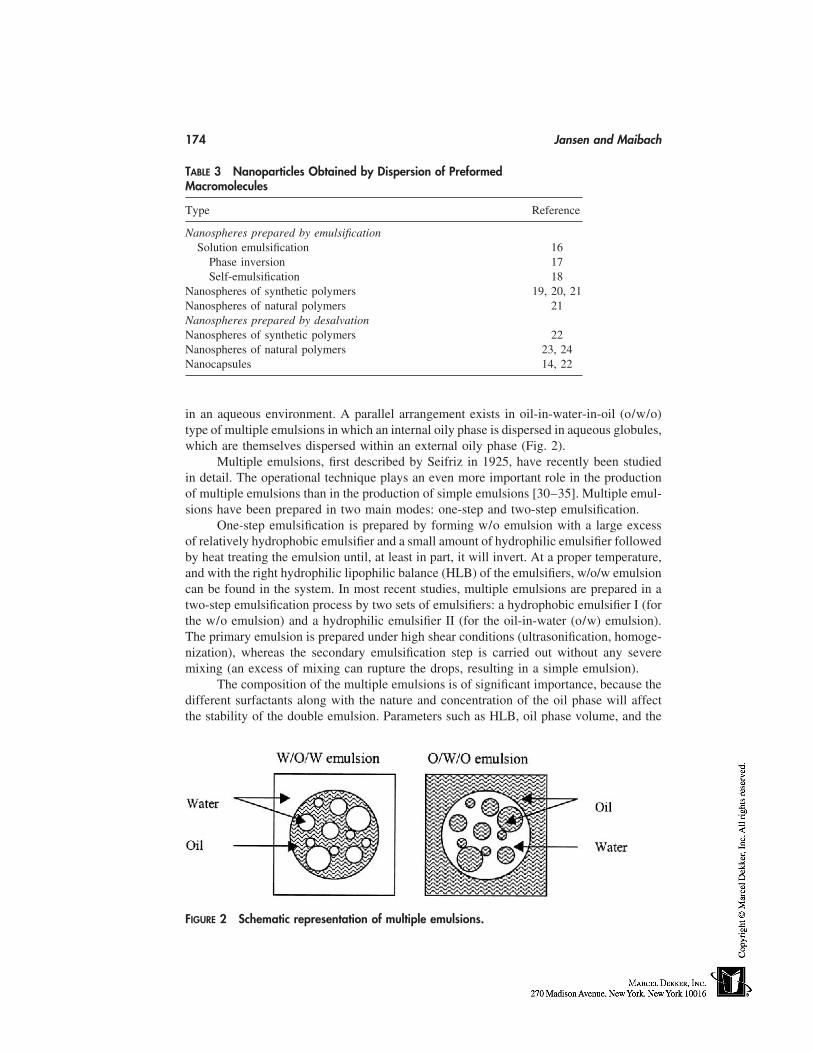
174 Jansen and Maibach
TABLE 3 Nanoparticles Obtained by Dispersion of PreformedMacromolecules
Type Reference
Nanospheres prepared by emulsificationSolution emulsification 16
Phase inversion 17Self-emulsification 18
Nanospheres of synthetic polymers 19, 20, 21Nanospheres of natural polymers 21Nanospheres prepared by desalvationNanospheres of synthetic polymers 22Nanospheres of natural polymers 23, 24Nanocapsules 14, 22
in an aqueous environment. A parallel arrangement exists in oil-in-water-in-oil (o/w/o)type of multiple emulsions in which an internal oily phase is dispersed in aqueous globules,which are themselves dispersed within an external oily phase (Fig. 2).
Multiple emulsions, first described by Seifriz in 1925, have recently been studiedin detail. The operational technique plays an even more important role in the productionof multiple emulsions than in the production of simple emulsions [30–35]. Multiple emul-sions have been prepared in two main modes: one-step and two-step emulsification.
One-step emulsification is prepared by forming w/o emulsion with a large excessof relatively hydrophobic emulsifier and a small amount of hydrophilic emulsifier followedby heat treating the emulsion until, at least in part, it will invert. At a proper temperature,and with the right hydrophilic lipophilic balance (HLB) of the emulsifiers, w/o/w emulsioncan be found in the system. In most recent studies, multiple emulsions are prepared in atwo-step emulsification process by two sets of emulsifiers: a hydrophobic emulsifier I (forthe w/o emulsion) and a hydrophilic emulsifier II (for the oil-in-water (o/w) emulsion).The primary emulsion is prepared under high shear conditions (ultrasonification, homoge-nization), whereas the secondary emulsification step is carried out without any severemixing (an excess of mixing can rupture the drops, resulting in a simple emulsion).
The composition of the multiple emulsions is of significant importance, because thedifferent surfactants along with the nature and concentration of the oil phase will affectthe stability of the double emulsion. Parameters such as HLB, oil phase volume, and the
FIGURE 2 Schematic representation of multiple emulsions.
Encapsulation to Deliver Topical Actives 175
nature of the entrapped materials have been discussed and optimized. Several reviews andstudies include Florence and Whitehill [36–38], Matsumoto et al. [39,40] and Frenkel[41–43].
Microemulsions
Miocroemulsions are stable dispersions in the form of spherical droplets whose diameteris in the range of 10 to 100 nm. They are composed of oil, water, and usually surfactantand cosurfactant. These systems show structural similarity to micelles and inverse mi-celles, resulting in o/w or w/o microemulsions, respectively. They are highly dynamicsystems showing fluctuating surfaces caused by forming and deforming processes.
The main characteristics of microemulsions are the low viscosity associated with aNewtonian-type flow, a transparent or translucid appearance, and isotropic and thermody-namic stability within a specific temperature setting. Certain microemulsions may thus beobtained without heating, simply by mixing the components as long as they are in a liquidstate. One of the conditions for microemulsion formation is a very small, rather than atransient negative, interfacial tension (44). This is rarely achieved by the use of a singlesurfactant, usually necessitating the addition of a cosurfactant. The presence of a shortchain alcohol, e.g., can reduce the interfacial tension from about 10 mN/m to a value lessthan 10�2mN/m. Exceptions to this rule are provided by nonionic surfactants which, attheir phase inversion temperature, also exhibit very low interfacial tensions.
A microemulsion is usually created by the establishment of pseudoternary diagramfor which a ratio of surfactant/cosurfactant is fixed, representing a sole constituent. Theestablishment of a ternary diagram is generally accomplished for locating the microemul-sion or the microemulsion zones by titration. Using a specific ratio of surfactant/cosurfac-tant, various combinations of oil and surfactant/cosurfactant are produced. The water isadded drop by drop. After the addition of each drop, the mixture is stirred and examinedthrough a crossed polarized filter. The appearance (transparence, opalescence, isotropy)is recorded, along with a number of phases. In this way, an approximate delineation ofthe boundaries can be obtained in which it is possible to refine through the production ofcompositions point by point beginning with the four basic components.
Nanoemulsions (Submicron Emulsions)
Emulsions are heterogeneous systems in which one immiscible liquid is dispersed as drop-lets in another liquid. Such a system is thermodynamically unstable and is kineticallystabilized by the addition of one further component or mixture of components that exhibitsemulsifying properties. Depending on the nature of the diverse components of the emulsi-fying agents, various types of emulsions can result from the mixture of immiscible liquids.The main characteristic of nanoemulsions or submicron emulsions is the droplet size,which must be inferior to 1µm.
Emulsions prepared by use of conventional apparatus, e.g., electric mixers and me-chanical stirrers, show large droplet sizes and wide particle distribution. The techniquesusually used to prepare submicron emulsions involve the use of ultrasound, evaporationof solvent (45), two-stage homogenizer [46,47], and the microfluidizer [48,49]. The nano-emulsion preparation process involves the following steps:
1. Three approaches can be used to incorporate the drug and/or the emulsifiers inthe aqueous or oil phase. The most common is to dissolve the water-soluble
176 Jansen and Maibach
ingredients in the aqueous phase and the oil-soluble ingredients in the oil phase.The second approach, which is used in fat emulsion preparations [46], involves thedissolution of an aqueous-insoluble emulsifier in alcohol, the dispersion of thealcohol solution in water, and the evaporation and total removal of the alcoholuntil a fine dispersion of the alcohol solution of the emulsifer in the aqueousphase is reached. The third, which is mainly used for amphotericin B incorpora-tion into an emulsion, involves the preparation of a liposome-like dispersion.The drugs and phospholipids are first dissolved in methanol, dichloromethane,chloroform, or a combination of these organic solvents, and then filtered intoa round-bottom flask. The drug-phospholipid complex is deposited into a thinfilm by evaporation of the organic solvent under reduced pressure. After sonica-tion with the aqueous phase, a liposome-like dispersion is formed in the aqueousphase. The filtered oil phase and the aqueous phase are heated separately to70°C and then combined by magnetic stirring.
2. The oil and aqueous phases are emulsified with a high-shear mixer at 70 to80°C.
3. The resulting coarse emulsion (1–5µm) is then rapidly cooled and homogenizedinto a fine monodispersed emulsion.
Vesicles
Bangham [50] clearly shows that the dispersion of natural phospholipids in aqueous solu-tions leads to the formation of ‘‘closed vesicles structures,’’ which morphologically resem-ble cells. Since 1975 [51], vesicles have been prepared from surfactants. In 1986, the firstcommercial product incorporating liposomes identical to those described by Banghamappeared on the market (Capture). At the same time, a synthetic one made by nonionicsurfactants [52] was also launched (Niosomes). Several different compositions, for scien-tific, economic and business reasons, prevailed in cosmetic vesicles. None of them reallyresembles the liposomes we have seen in medical applications. These main groups include:(1) liposomes made from soya phospholipids; (2) sphingosomes, i.e., liposomes madefrom sphingolipids, and (3) nonionic surfactant vesicles (niosomes) which are a proprietaryproduct of L’Óréal and other synthetic amphiphiles. In the 1990s, transfersomes, i.e., lipidvesicles containing large fractions of fatty acids, were introduced. Transfersomes [53–55] consist of a mixture of a lipidic agent with a surfactant. Consequently, their bilayersare much more elastic than those of most liposomes.
This chapter focuses on nonionic surfactant vesicles and transfersomes. Nonionicsurfactant vesicles (NSVs or niosomes) consist of one or more nonionic surfactant bilayersenclosing an aqueous space. NSVs consisting of one bilayer are designed as small unila-
TABLE 4 Vesicles Preparation Methods
Method Reference
Sonication 56, 58, 60Ether injection 56Handshaking 56Reversed phase evaporation 61Method as described by Handjani-Vila 52
Encapsulation to Deliver Topical Actives 177
mellar vesicles (SUVs) or large unilamellar vesicles (LUVs). Vesicles with more bilayersare called multilamellar vesicles.
Niosomes can be prepared from various classes of nonionic surfactants, e.g., poly-glycerol alkyl ethers [52,56], glucosyl dialkyl ethers [57], crown ethers, and polyoxyeth-ylene alkyl ethers and esters [58]. The preparation methods used should be chosen ac-cording to the use of niosomes, because the preparation methods influence the number ofbilayers, size, size distribution, entrapment efficiency of the aqueous phase, and membranepermeability of the vesicles [56,59]. NSVs can be formed using the same methods thatare used for the preparation of liposomes (Table 4).
PROPERTIES OF A VECTOR
Microparticles
Microencapsulation has been applied to solve problems in the development of pharmaceu-tical dosage forms as well as in cosmetics for several purposes. These include the conver-sion of liquids to solids, separation of incompatible components in dosage form, tastemasking, reduction of gastrointestinal irritation, protection of the core materials againstatmospheric deterioration, and enhancement of stability and controlled-release of activeingredients.
For drug follicular targeting, microspheres were envisaged mainly as site-specificdrug delivery systems because they present several advantages: 1) good stability of themicrospheres when applied on the skin, 2) easy preparation of microspheres with a definedsize in a narrow size distribution, 3) protection of the active incorporated, 4) controlledrelease of the active in the hair follicles from the microspheres, and 5) the possibility ofincorporating either lipophilic or hydrophilic actives into the microspheres [62]. Concern-ing the microsponge system, each microsphere is composed of thousands of small beadswrapped together to form a microscopic sphere capable of binding, suspending, or en-trapping a range of substances. The outer surface is porous, allowing the controlled flow.Microsponges can be incorporated into gels, creams, liquids, powders, or other formula-tions, and can release ingredients depending on their temperature, moisture, friction, vola-tility of the entrapped ingredient, or time.
Nanoparticles
Nanoparticles are attractive delivery systems. In most cases the advantages are 1) the solidmatrix gives flexibility to modify the drug release profile, 2) the relatively slow degrada-tion allows long release times, and 3) the protection of incorporated compounds againstchemical degradation. Drug release from colloidal carriers is dependent on both the typeof carrier and the loading mechanisms involved.
Nanospheres
Release from nanospheres may be different according to the drug-entrapment mechanisminvolved. When the drug is superficially adsorbed, the release mechanism can be describedas a partitioning process (rapid and total release if sink conditions are met). When thedrug is entrapped within the matrix, diffusion plus bioerosion will be involved with abiodegradable carrier, whereas diffusion will be the only mechanism if the carrier is notbiodegradable. From this, it can be inferred that entrapment within the matrix of nano-
178 Jansen and Maibach
spheres may lead to sustained release, the rate of which may be related to the rate ofbiodegradation of the polymer.
Nanocapsules
Release from nanocapsules is related to partitioning processes within immiscible phases.The equilibrium between the carrier (loaded drug) and the dispersing aqueous medium(free drug) is dependent both on the partition coefficient of the molecule between the oilyand the aqueous phases and on the volume ratio of these two phases. This means that theamount released is directly related to the dilution of the carrier and that the release ispractically instantaneous when sink conditions exist. Diffusion of the drug through thepolymeric wall of nanocapsules does not seem to be a rate-limiting step [63]. Coating thepolymeric wall with an outer layer of phospholipids can advantageously reduce drug leak-age from nanocapsules.
Multiple Emulsions
Double emulsions are an excellent and exciting potential system for slow or controlledrelease of active entrapped compounds. The fact that the inner w/o emulsion serves as alarge confined reservoir of water is a very attractive property for dissolving it in significantamounts of water-soluble drugs. The oil membrane seems to serve as good transport barrierfor the confined ionized and/or nonionized water-soluble drugs. The two amphiphilic inter-faces are yet an additional barrier. The possibility to manipulate transport and releasecharacteristics of the formulations seems to be feasible. However, despite 20 years ofresearch, no pharmaceutical preparation using the multiple emulsion technology exists inthe marketplace. It seems that the main reasons are the droplet instability and the uncon-trolled release.
Although the release of the encapsulated active substance is complicated, becauseof the existence of different mechanisms, the multiple emulsion’s behavior after applica-tion to the skin appears to be relatively simple because it is similar to the behavior observedwith simple emulsions.
Microemulsions
Miocroemulsions are effective vehicle systems for dermal as well as for transdermal drugdelivery because of their high drug-loading capacity of their colloidal structure. Further-more, thermodynamic stability and simple preparation process favor them to be consideredas vehicles for skin applications.
Several workers have reported studies in which the lipophilicity of the drug hasbeen increased to enhance its solubility in the dispersed oil droplets. In this way, a reservoirof the drug is produced and a sustained-release effect is achieved as the drug continuouslytransfers from the oil droplets to the continuous phase to replace drug release from themicroemulsion.
Nanoemulsions
Nanoemulsions have been gaining more and more attention in the last few years, mainlyas vehicles for the intravenous administration of lipophilic drugs. In the skin, the patentsclaimed that these systems could penetrate through the skin to a greater extent comparedwith usual topical compositions. Nanoemulsions are so strongly compressed that they
Encapsulation to Deliver Topical Actives 179
become ultralight and, like vesicular systems constitute a new form that could prove ex-tremely fruitful for the release of substances.
Vesicles
Vesicles appear to be promising transdermal drug-delivery systems. The major advantagesof topical vesicle drug formulations are:
• hydrophilic, lipophilic, as well as amphiphilic substances can be encapsulatedin the vesicles
• for the lipophilic and amphiphilic drugs the liposomes serve as ‘‘organic’’ sol-vent and as a result, higher local drug concentrations can be applied
• the vesicles can act as depot, releasing their drug content slowly and controlled• systemic effect of a dermal active compound can be reduced and the systemic
effect of a transdermal drug can be increased depending on the vesicle composi-tion
• the vesicles may serve as penetration enhancer• the vesicles can interact with the skin because of the amphiphilic character of
the bilayer• liposomes are biocompatible and biodegradable and have a low toxicity and lack
antigenicity status as well• vesicle formulations are cosmetically accepted
There are also some disadvantages of vesicles as drug carriers:
• low encapsulation efficiencies for lipophilic or amphiphilic drugs• no drug release from the vesicle• low–molecular weight drugs can leak out of the vesicle• instability of vesicles during shelf life• sterilization of liposome formulations
DERMATOLOGICAL AND COSMETIC USES OF ENCAPSULATION
Microparticles
In recent years, numerous vectors have been proposed and used in topical formulationsas drug-carrier vehicles. It has been claimed that these drug vehicles can improve andcontrol the drug release from conventional topical formulations. Although the applicationof these colloidal particles in dermatology is of great interest, there are few articles aboutthe characteristics of these vehicles for topical formulations and most of the backgroundis based on different patents.
Miocroparticles can serve as a drug reservoir in skin products. Rolland et al. [62]investigated in vitro and in vivo the role of 50:50 poly (dl-lactic-co-glycolic acid) micro-spheres as particulate carriers to improve the therapeutic index of adaptalene. The percuta-neous penetration pathway of the microspheres was shown to be dependent on their meandiameter. Thus, after topical application onto hairless rat or human skin, adaptalene-loadedmicrospheres (5 µm diameter) were specifically targeted to the follicular ducts and didnot penetrate via the stratum corneum. A reduction of either the applied dose (0.01%) orthe frequency of administration (every day) was shown to give pharmacological results in
180 Jansen and Maibach
the animal model comparable to a daily administration of 0.1% free adaptalene-containingaqueous gel.
Egg albumin microspheres of size 222 � 25 µm, containing a vitamin A (15.7� 0.8%), were used to prepare o/w creams. The in vitro and in vivo drug release of amicroencapsulated vitamin A cream was studied and compared with a nonmicroencapsu-lated vitamin A cream. The in vitro study showed that, during the first 3 hours, the micro-spheres could remain on the surface of the skin, and as a consequence, were able to prolongthe release of vitamin A. The relative bioavailability of the microencapsulated formulationwas 78.2 � 7.3% [64].
Mizushima [65] reported that lipid microspheres containing prostaglandin E1
(PGE1), delivered preferentially to specific lesion sites, increased local action and pre-vented systemic side effects. Sakakibara et al. [66] evaluated the potential of topical appli-cation of lipid microspheres containing PGE1 to treat ischemic ulcers. Nine of the 10patients responded to the treatment, and at the sixth month of follow-up six patients hadhealed ulcers and recurrence was noted in three patients.
Skin absorption of benzoyl peroxide from a topical lotion containing freely disperseddrug was compared with that from the same lotion in which the drug was entrapped in acontrolled-release styrene-divinylbenzene polymer system (Microsponge). The studiesdone by Wester et al. [67] showed the following: 1) in vivo, less benzoyl peroxide wasabsorbed through rhesus monkey skin from the polymeric system, 2) reduced skin irritationin cumulative irritancy studies on rabbits and human, and 3) when the experimental formu-lations were evaluated for antimicrobial activity in vivo, their efficiency was in line withthat of conventional products.
A formulation containing 0.1% tretinoin was tested on 360 patients during 12 weeksfor antiacne efficacy in a multicenter, double-blind, placebo-controlled study. Comparedwith placebo, statistically significant greater reductions in inflammatory, noninflammatory,and the total number of lesions were obtained with the entrapped retinoic acid formulation[68]. Encapsulation of deet in liposphere microdispersion resulted in improved efficacyand reduced dermal absorption. Deet-containing lipospheres (10%) were effective againstmosquitoes for at least 3.5 hours. The deet absorption through skin from these formulationswas a third of that from alcoholic solution for the same concentration [69].
Nanoparticles
Although cosmetic applications of nanoparticles proliferate (numerous patents have beengranted), publications, studies, or reports on the skin after topical application have beenrare. The incorporation of active substances in the nanospheres attempt to modulate therelease of the substances in the skin. When nanocapsules are concerned, the active sub-stances are usually of lipophilic nature, and they can be composed of an oily compoundor dispersion. Here again the objective is to control the release of the actives because themolecule is protected. The release profile of the actives depends on the nature of theconstituents.
Recently, Lancôme launched a cosmetic product containing nanocapsules of vitaminE (Primordiale). They claim that the vitamin is widely distributed throughout the outerlayers of the skin in the form of a gradient. The effectiveness of vitamin E protectionwhen it is incorporated into nanoparticles has been shown in vivo. Dingler et al. [70]reported that the incorporation of vitamin E into solid lipid nanoparticles enhances thestability. The ultrafine particles possess an adhesive effect. This leads to a formation of
Encapsulation to Deliver Topical Actives 181
fine adhesive film on the skin leading to occlusion and subsequent hydration. Hydrationof the skin promotes penetration of actives and enhances their cosmetic efficiency. Inanother publication of the same research group [71], drug release of encapsulated materialas well as nonencapsulated material was measured by tape stripping assay. The drug(RMAD 95) was released into the skin at approximately 53%, whereas the control (RMAD95/isopropanol) was at 31%.
Immobilization of nanoparticles (polyamide) on the skin for prolonged periods oftime has been proved feasible [72]. It has been shown to be dependent on formulationbecause particle retention was increased from 40% up to 98% when embedding the parti-cles into a emulsion. Particle size, surface charge, and payload determine the propertiesof the nanoparticles and their application. Zülli et al. [73] encapsulated Uvinil T 150 (UV-B filter) into lipid nanoparticles. They observed an almost one-hundredfold higher affinityof Uvinil T to hair from positively charged particles compared with negatively chargedparticles. The same group also showed the application of a gel containing nanoparticlesloaded with vitamin A and E derivatives enhances the skin humidity compared with con-trols.
In a 1997 patent, De Vringer [74] showed that the size of particles can change theocclusion factor. Lipoid microparticles are greatly inferior to solid lipoid nanoparticles intheir occlusive effect, and the addition of solid lipoid microparticles in a cream lowersthe cream’s occlusivity, whereas the addition of solid lipoid nanoparticles in a cream raisesthe cream’s occlusivity. Nanospheres containing beta carotene and a blend of UV-A andUV-B sun filters were prepared by Olivier-Terras [75]. The results clearly show the syner-gistic effect resulting from the combination of nanospheres and filters. They obtained withthis formulation better bioavailability, better efficacy, and lastly a synergy that possessesan inhibitory effect on tyrosinase as a result of the cinnamic nature of the UV-B screeningagents.
The effect of poly (methylmethacrylate) and poly (butylcyanoacrylate) nanoparticleson the permeation of methanol and octanol through hairless mouse skin was reported byCappel and Kreuter [76]. Nanoparticles increase the permeability of methanol throughhairless mouse skin and the permeability of lipophilic octanol is either unaffected by nano-particles or decreases as a function of nanoparticle concentration depending on the lipo-philicity of the polymer material. The potential use of nanoparticles as an ophthalmicdrug-delivery system has been shown in numerous studies for either hydrophobic or hydro-philic drugs [77–79]. Despite the promising in vivo results, many issues must be resolvedbefore an ophthalmic product can be developed using this technology.
Tobio et al. [80] encapsulated a model protein antigen, tetanus toxoid, into PLA-PEG nanoparticles and evaluated the potential of these colloidal carriers for the transportof proteins through the nasal mucous. The results showed that PLA-PEG nanoparticleshave a great potential for delivery of proteins, either to the lymphatic system or to the bloodcirculation, after nasal administration. Regarding the mode of action of nanoparticles, onemight hypothesize that they are associated with the skin surface, facilitating drug transportby changing the vehicle/stratum corneum partition coefficient.
Multiple Emulsions
The first commercial use of a w/o/w type multiple emulsion is Unique Moisturizing byLancaster, which was marketed in 1991. Cosmetic application of multiple emulsions havebeen reported in the patents issued for their composition. One example of an application
182 Jansen and Maibach
is perfume encapsulated in the internal phase; very small amounts of it are released overa long period of time. The patents show that multiple emulsions are recommended for allkinds of cosmetic applications: sunscreens, makeup removers, cleansers, and nutritive,hydrating, and cooling products. Kamperman and Sallis [81] show that a highly chargedsmall water-soluble molecule such as phosphocitrate can be presented in the form of aliposome or multiple emulsion and be capable of exerting a positive action against dystro-phic calcification. In a rat calcergy model, both vehicles effectively reduced the formationof induced subcutaneous calcified plaques at doses for which the phosphocitrate salt alonewas inactive. Three emulsions type (w/o/w, o/w, and w/o) containing a water-solublemolecule (glucose) were obtained with the same formula [82,83]. The release of glucosefrom the o/w emulsion was the fastest, and the w/o emulsion was the slowest, whereasthe release obtained from the w/o/w emulsion was intermediate. The w/o/w emulsionshowed some tendency toward steady state during the first 3 to 12 hours and the flux wasfound to be 1.7 times greater than that from the w/o emulsion.
In vivo release of 2.5% lidocaine hydrochloride from simple and multiple emulsionsystems was compared with that from aqueous and micellar solution, and anesthetic effectssuch as duration of action and tolerability were also compared. The double emulsionsshowed a longer duration of action, less eye irritation, and improved efficacy comparedwith aqueous solutions [44].
Microemulsions
Over the last 15 years, many studies have been performed with the percutaneous absorp-tion of various actives carried by microemulsions. There are numerous cosmetic productsin the form of microemulsions. These products range from body care to facial and hairtreatments. They include bath oils, body-thinning products, fixatives for hair, hardenersfor nails, hydrating products, antiwrinkle products, seborrhea preventive products, andantiaging serums marketed principally in Europe, the United States, and Japan. In biophar-maceutics, microemulsions were used to solubilize drugs and to improve systemic andtopical drug availability.
Gasco et al. [84] ascertained concentrations of timolol in aqueous humor after multi-ple instillation in rabbit eyes. The microemulsion, a solution of the ion-pair, and a solutionof timolol alone was used. The bioavailability of timolol from the microemulsion and theion-pair solution was higher than that obtained from timolol alone. Transport of glucoseacross human cadaver skin was shown [85] using microemulsions containing up to 68%water. A thirtyfold enhancement of the glucose transport was achieved. The enhancingeffect for drugs contained in microemulsions in comparison to a cream gel formulationconsisting of the same components was shown by Ziegnmeyer and Führer [86]. The invitro permeation across skin membranes as well as the in vivo penetration of tetracyclinehydrochloride was higher from a microemulsion than from conventional systems. Thusis can be shown that in addition to the composition, the structure of each of the typicallyapplied vehicles may play a dominant role in the process of penetration.
Février [87] has reported in vitro experiments designed to simulate the percutaneouspenetration of tyrosine when administered using an o/w microemulsion composed of abetaine derivative as surfactant, benzyl alcohol, hexadecane, and water. The release ofradiolabeled tyrosine from this vehicle was compared with that from a liquid-crystal sys-tem and an emulsion using a diffusion cell equipped with rat skin. Both the microemulsionand liquid-crystal formulation enhanced the penetration of tyrosine through the epidermis
Encapsulation to Deliver Topical Actives 183
when compared with the emulsion. However, cutaneous irritation studies showed astrongly irritant effect from the liquid-crystal formulation but none from the microemul-sion.
The penetration of the hydrophilic diphenhydramine hydrochloride from a w/o mi-croemulsion into human skin under ex vivo conditions was studied by Schmalfuß et al.[88]. Modifications of the vehicle components clarified the extent to which it is possibleto control the penetration of a hydrophilic drug incorporated in a microemulsion system.A standard microemulsion showed an accumulation of penetrated drug in the dermis,indicating a potential after high absorption rate. Incorporation of cholesterol into the sys-tem leads to an even higher penetration rate and a shifting of the concentration profilefurther towards the epidermis. The addition of oleic acid had no effect.
Wallin et al. [89] showed that high concentrations of lidocaine base included in amicroemulsion produced peripheral nerve block of long duration, compared with solutionsas a consequence of slow release of lidocaine. The effect of polysorbate 80 concentrationon the permeation of propanolol incorporated into micelles of polysorbate 80 in water,o/w microemulsions of isopropyl myristate-polysorbate 80-sorbitol water, and o/w emul-sions of isopropyl myristate-polysorbate 80-sorbitan monooleate-water has been investi-gated by use of an artificial double-layer membrane, composed of a barrier foil and a lipidbarrier, in Franz-type diffusion cells [90]. For each system, the apparent permeabilitycoefficient of propanolol decreased with increasing polysorbate 80 concentration. More-over, for a given polysorbate 80 concentration, the apparent permeability coefficient ofpropanolol increased when the system was changed from emulsion to a microemulsionand then to a solubilized system because of the increasing interfacial area of total dispersephase.
Microemulsions may exert irritative effects, often by their high content of surfac-tants. It is possible to overcome this problem by the use of physiologically compatiblenonionic and polymeric surfactants. The irritation potential of the formulation dependsstrongly on its structure. Because of an equilibrium between microemulsions and liquidcrystals, when brought into contact microemulsions may dissolve skin structures that areorganized in liquid crystalline form. Thus, an irritation is produced. Deduced from this,the nature of the system formed during the penetration process and the residue remainingon the skin surface are of importance in this regard.
Acute and cumulative tests were performed on human subjects in vivo with lecithinmicroemulsion gels using as comparison a unilamellar soybean lecithin liposome prepara-tion and the solvent isopropyl palmitate [91]. The study showed a very low acute and alow cumulative irritancy potential for the soybean lecithin microemulsion gel. In general,microemulsions undergo structural changes after an application to the skin because of thepenetration and/or evaporation of constituents and under occlusion by the uptake of waterfrom the skin surface. The formed substances and their penetration behavior finally influ-ences the effectiveness of the systems for dermal drug transport.
Nanoemulsions
Many formulations of nanoemulsion are available in patents. Recently, Lancôme launcheda nanoemulsion rich in ceramides, Re-source. The scientific studies, however, are orien-tated mainly in the parenteral use of these formulations. Amselem and Friedman [92]indicated that the actives incorporated in submicron emulsions (diameter between 100–300 nm) can penetrate through the skin to a greater extent compared with the usual topical
184 Jansen and Maibach
compositions. Improved efficacy of different steroidal and nonsteroidal anti-inflammatorydrugs and local anesthetics has been observed.
Anselem and Zwoznik [93] determined drug penetration through the skin, local tis-sue (muscle and joint), and plasma levels of ketoprofen and diclofenac after topical admin-istration in submicron emulsion (SME) creams compared with peroral administration.Compared with peroral drugs, SME-diclofenac and SME-ketoprofen showed sixty- toeightyfold more drug in muscle tissue, about ninefold more drug in joints, andfour- to sixfold less drug in plasma. The improved skin penetrative properties of the sol-vent-free SME delivery makes this topical carrier very promising to achieve increasedtranscutaneous penetration of lipophilic drugs and site specificity.
Diazepam was formulated in various regular topical creams and SMEs of differentcomposition [94]. The different formulations were applied topically on mice. The efficacyof diazepam applied topically in emulsions strongly depends on the oil droplet size and,to a lesser degree, on the formulation and oil type. The SMEs as vehicles for transdermaldelivery of diazepam generate significant systemic activity of the drug as compared withregular creams or ointments. Transdermal delivery of diazepam via SME is effective, andthe activity may reach the range of parenteral delivery. A single application of diazepamin SME cream to mice skin provides pronounced transdermal drug delivery and prolongedprotective activity up to 6 hours.
Using a nanoemulsion composed of lanolin, polyethylene glycol ether of lanolin’salcohol and water [95], the investigators showed the transdermal delivery of a numberof pharmaceutically active ingredients (testosterone, ibuprofen, 5-fluorouracil, verapamilhydrochloride, metronidazole, vincristine sulphate, fentanyl citrate) across isolated stratumcorneum. The studies indicated that nanoemulsions derived from lanolin and its derivativesare capable of being developed into useful drug-delivery systems.
Vesicles
The effectiveness of vesicles has been investigated by several research groups (Table 5).Liposomes in particular have received considerable attention [103]. In several studies thediffusion of a drug was facilitated or achieved certain selectivity into human and nonhu-man skin by vesicle encapsulation. Other studies show that the influence of vesicles ondrug transport is negligible. The conflicting results can be understood in terms of vesiclecharacteristics or in terms of protocol of investigation. Special surface characteristics ofvesicle hydration and electrostatic forces, in addition to Van der Waals, can govern theshort and long range of repulsive or attractive forces between vesicles and biologicalmedia.
The particle sizes, the physical state (liquid or gel) of the bilayers, the number ofbilayers, the electrostatic nature of drugs and vesicles, and the stability of the vesiclesface to face with biofluids in different ranges of pHs, temperatures, and degrees of dehy-dration can also play an important role in the phenomenon. An important contribution tothe understanding of the interactions between vesicles and human skin was made by Jun-ginger and his group [100,104]. They used freeze fracture electron microscopy and small-angle radiograph scattering to study the effects that vesicle formulations have on the stra-tum corneum. They identified two types of liposome-skin interactions: 1) adsorption andfusion of loaded vesicles on the surface of the skin leading to increased thermodynamicactivity and enhanced penetration of lipophilic drugs, and 2) interaction of the vesicleswithin the deeper layers of the stratum corneum promoting impaired barrier function of
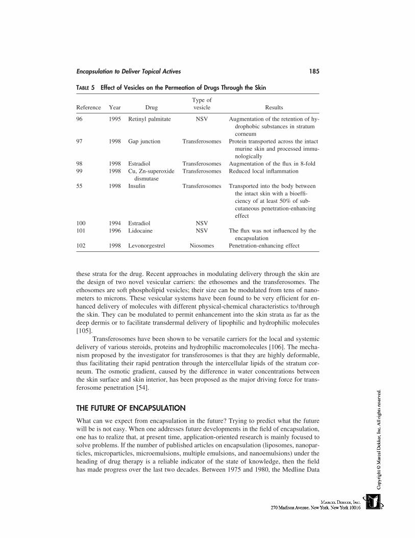
Encapsulation to Deliver Topical Actives 185
TABLE 5 Effect of Vesicles on the Permeation of Drugs Through the Skin
Type ofReference Year Drug vesicle Results
96 1995 Retinyl palmitate NSV Augmentation of the retention of hy-drophobic substances in stratumcorneum
97 1998 Gap junction Transferosomes Protein transported across the intactmurine skin and processed immu-nologically
98 1998 Estradiol Transferosomes Augmentation of the flux in 8-fold99 1998 Cu, Zn-superoxide Transferosomes Reduced local inflammation
dismutase55 1998 Insulin Transferosomes Transported into the body between
the intact skin with a bioeffi-ciency of at least 50% of sub-cutaneous penetration-enhancingeffect
100 1994 Estradiol NSV101 1996 Lidocaine NSV The flux was not influenced by the
encapsulation102 1998 Levonorgestrel Niosomes Penetration-enhancing effect
these strata for the drug. Recent approaches in modulating delivery through the skin arethe design of two novel vesicular carriers: the ethosomes and the transferosomes. Theethosomes are soft phospholipid vesicles; their size can be modulated from tens of nano-meters to microns. These vesicular systems have been found to be very efficient for en-hanced delivery of molecules with different physical-chemical characteristics to/throughthe skin. They can be modulated to permit enhancement into the skin strata as far as thedeep dermis or to facilitate transdermal delivery of lipophilic and hydrophilic molecules[105].
Transferosomes have been shown to be versatile carriers for the local and systemicdelivery of various steroids, proteins and hydrophilic macromolecules [106]. The mecha-nism proposed by the investigator for transferosomes is that they are highly deformable,thus facilitating their rapid pentration through the intercellular lipids of the stratum cor-neum. The osmotic gradient, caused by the difference in water concentrations betweenthe skin surface and skin interior, has been proposed as the major driving force for trans-ferosome penetration [54].
THE FUTURE OF ENCAPSULATION
What can we expect from encapsulation in the future? Trying to predict what the futurewill be is not easy. When one addresses future developments in the field of encapsulation,one has to realize that, at present time, application-oriented research is mainly focused tosolve problems. If the number of published articles on encapsulation (liposomes, nanopar-ticles, microparticles, microemulsions, multiple emulsions, and nanoemulsions) under theheading of drug therapy is a reliable indicator of the state of knowledge, then the fieldhas made progress over the last two decades. Between 1975 and 1980, the Medline Data
186 Jansen and Maibach
Base registered about 20 articles per year with the term ‘‘liposomes’’ in their title in thedomain of drug therapy. This number has grown to over 100 per year. Because many ofthese publications dealt directly with new experimental data, we must conclude that ourexperience has expanded dramatically.
The skin has been ‘‘in the picture’’ since Mezei and his collaborators reportedaround 1980 on their early work on the liposomal delivery of drugs. Through the effortsof the cosmetic industry, liposomal formulations and nanoparticle formulations on theskin have definitively been an economic success. However, many unanswered questionsremain. Molecular biology has provided us with tools to identify and build genetic materi-als that can be used for the treatment of hereditary diseases. Developing a carrier for genetherapy is one of the main challenges that the encapsulation field faces today. With respectto gene therapy for the skin, both molecular biology and encapsulation technology are intheir debut, and much progress may and should be made in the coming years.
Again, what will the future bring us? We have already indicated where, on the basisof our present knowledge, encapsulation in many vectors offer a rational advantage asactive carrier systems to the skin. Therefore, efforts should be made to obtain a betterunderstanding concerning the mechanisms of formulations of these systems at the molecu-lar and supramolecular level. This could lead to new formulation processes and couldopen new prospects in the area of active delivery by means of encapsulated systems. Thefield will develop in a more useful fashion when appropriate well-controlled biologicaland percutaneous penetration studies accompany the advances in chemistry.
REFERENCES
1. Kreuter J. Evaluation of nanoparticles as drug-delivery systems. I. Preparation methods.Pharm Acta Helv 1983; 58:196–201.
2. Won R. U.S. Patent 4,690,825. 1987.3. Bakan J. Microencapsulation using coacervation/phase separation techniques. In: Controlled
Release Technologies: Methods, Theory and Applications, Vol. 2. Boca Raton: CRC press,1980:83–105.
4. Deasy P. Microencapsulation and Related Drug Processes. New York: Marcel Dekker, 1984.5. Chang TMS. Artificial Kidney, Artificial Liver and Artificial Cells. New York: Plenum Press,
1978.6. Thies C. A survey of microencapsulation processes. In: Benita S, ed. Microencapsulation,
Methods and Industrial Applications. New York: Marcel Dekker, 1996:1–9.7. Lim F, Moss RD. Microencapsulation of living cells and tissues. J Pharm Sci 1981; 70:351–
356.8. Matsumoto S, Kabayashi H, Takashima Y. Production of monodispersed capsules. J Mi-
croencaps 1986; 3:25–31.9. Finch CA. Ullman’s Encyclopedia of Industrial Chemistry. Vol. A 16. 5th ed. New York:
VCH Publishers, 1990:575–588.10. Kondo A. Microcapsule Processing and Technology. New York: Marcel Dekker, 1979.11. Jacobs IC, Mason NS: Polymeric delivery systems. In: Elnolkaly MA, Piatt DM, Charpentier
BA, eds. ACS Symposium Series 520. Washington, D.C.: American Chemical Society, 1993:1–17.
12. Kreuter J. Nanoparticles—preparation and applications. In: Donbrow M, ed. Microcapsulesand Nanoparticles in Medicine and Pharmacy. Boca Raton: CRC Press, 1992:125–148.
13. Couvreur P, Kante B, Rolland M. Polycyanoacrylate nanocapsules as potential lysosomotriccarriers: preparation morphological and sorptive properties. J Pharm Pharmacol 1979; 31:331–338.
Encapsulation to Deliver Topical Actives 187
14. Al Khoury FN, Roblot-/Treupel L, Fessi H. Development of new process for the manufactureof poly-isobutylcyanoacrylate nanocapsules. Int J Pharm 1986; 28:125–132.
15. Rollot JM, Couvreur P, Roblot-Treupel L, Puisieux F. Physicochemical and morphologicalcharacterization of polyisobutyl cyanoacrylate nanocapsules. J Pharm Sci 1986; 75(4):361.
16. Aleony D, Wittcoff H. U.S. Patent 2, 899, 397, 1959.17. Cooper W. U.S. Patent 3, 009, 891, 1961.18. Judd P. Brit. Patent 1, 142, 375, 1969.19. Gurny R, Peppas NA, Harrington DD, Banker GS. Development of biodegradable lactices
for controlled release of potent drugs. Drug Dev Ind Pharm 1981; 7:1–12.20. Rhone-Poulenc Rorer. Fr Patent 2, 660, 556, 1990.21. Kramer PA. Albumin microspheres as vehicles for achieving specificity in drug delivery. J
Pharm Sci 1974; 63:1646–1652.22. Fessi H, Devissaguet JP, Puisieux F, Thies C. Fr Patent 8, 618, 446, 1986.23. Marty JJ, Oppenheim RC, Speiser PP. Nanoparticles—a new colloidal drug delivery system.
Pharm Acta Helv 1978; 53:17–24.24. Stainmesse S, Fessi H, Devissaguet JP, Puisieux F. 1st add to Fr Patent 8, 618, 446, 1988.25. De Vringer T, de Ronde HAG. Preparation and structure of a water-in-oil cream containing
lipid nanoparticles. J Pharm Sci 1995; 84(4):466–472.26. Kim SY, Lee YM, Lee SI. Preparation and evaluation of in vitro stability of lipid nanospheres
containing vitamin A and vitamin E for cosmetic application. Proc Intl Symp Cont Rel BioactMater 24. 1997:483–484.
27. Müller RH. Particulate systems for the controlled delivery of active compounds in pharma-ceutics and cosmetics. In: Diederichs JE, Müller RH, eds. Future strategies for drug deliverywith particulate systems. Stuttgart: CRC Press, 1998:73–90.
28. Müller RH, Mehnert W, Dingler A, Runge SA, zur Mühlen A, Freitas C. Solid lipid nanopar-ticles (SLN, Lipopearls). Proc Intl Symp Cont Rel Bioact Mater 24, 1997; 923–924.
29. Couvreur P, Coarraze G, Devissaguet JP, Puisieux F. Nanoparticles: preparation and charac-terization. In: Benita S, ed. Microencapsulation, Methods and Industrial Applications. NewYork: Marcel Dekker, 1996:183–211.
30. Matsumoto S, Kita Y, Yonezava D. An attempt at preparing water-in-oil-in-water multiplephase emulsion. J Colloid Interf Sci 1976; 57:353–361.
31. Matsumoto S, Sherman P. A preliminary study of w/o/w emulsions with a view to possiblefood applications. J Texture Studies 1981; 12:243–257.
32. Matsumoto S. Development of w/o/w type dispersion during phase inversion of concentratedw/o emulsions. J Colloid Interf Sci 1983; 94:362–368.
33. Kavaliunas DR, Franck SG. Liquid crystal stabilization of multiple emulsion. J Colloid InterfSci 1978; 66:586–588.
34. Magdassi S, Frenkel M, Garti N. On the factors affecting the yield of preparation and stabilityof multiple emulsions. J Dispersion Sci Technol 1984; 5:49–59.
35. De Luca M. Les emulsions multiples H/L/H. Obtention, validation, et liberation. These del’Université de Paris XI, Paris, 1991.
36. Florence AT, Whitehill D. Some features of breakdown in w/o/w multiple emulsions. J Col-loid Interf Sci 1981; 79:243–256.
37. Florence AT, Whitehill D. The formulation and stability of multiple emulsions. Int J Pharm1982; 11:277–308.
38. Florence AT, Whitehill D. Stability and stabilization of w/o/w multiple emulsions. In: ShahDO, ed. Macro and micro emulsions, theory and applications. Washington, D.C.: AmericanChemical Society, 1985:359–380.
39. Matsumoto S, Inoue T, Khoda M, Ikurak K. Water permeability of oil layers in w/o/w emul-sion under osmotic pressure gradients. J Colloid Interf Sci 1980; 77:555–563.
40. Matsumoto S, Koh J, Michura A. Preparation of w/o/w emulsions in edible form on the basisof phase inversion technique. J Dispos Sci Technol 1985; 6:507–521.
188 Jansen and Maibach
41. Frenkel M, Schwartz R, Garti N. Multiple emulsions. I. Stability inversion, apparent andweighed HLB. J. Colloid Interf Sci 1983; 94:174–178.
42. Csóka I, Erõs I. Stability of multiple emulsions. I. Determination of factors influencing multi-ple drop breakdown. Int J Pharm 1997; 156:119–123.
43. Opawale FO, Burgess DJ. Influence of interfacial rheological properties of mixed emulsifierfilms on the stability of w/o/w emulsions. J Pharm Pharmacol 1998; 50:965–973.
44. Garti N, Aserin A. Pharmaceutical emulsions, double emulsions and microemulsions. In:Benita S, ed. Microencapsulation, Methods and Industrial Applications. New York: MarcelDekker, 1996:412–534.
45. Yu W, Tabosa do Egito ES, Barrat G, Fessi H, Devissaguet JP, Puisieux F. A novel approachto the preparation of injectable emulsions by a spontaneous emulsification process. Int JPharm 1993; 89:139–146.
46. Hansrani PK, Davis SS, Groves MJ. The preparation and properties of sterile intravenousemulsions. J Parenter Sci Technol 1983; 37:145–150.
47. Yalabik-Kas HS, Erylmaz S, Hincal AA. Formation, stability and toxicity studies of intrave-nous fat emulsions. STP Pharm 1985; 1:12–19.
48. Washington C, Davis SS. The production of parenteral feeding emulsions by microfluidizer.Int J Pharm 1988; 169–176.
49. Lidgate DM, Fu RC, Fleitman JS. Using a microfluidizer to manufacture parenteral emul-sions. Pharm Technol 1990; 14:30–33.
50. Bangham AD, Standish MM, Watkins JC. Diffusion of univalent ions across the lamellaeof swollen phospholipids. J Mol Biol 1965; 13:238–252.
51. Gebicki JM, Hicks M. Preparation and properties of vesicle enclosed by fatty acid mem-branes. Chem Phys Lipids 1975; 16:142–160.
52. Handjani-Vila RM, Ribier A, Rondot B, Valenberghe G. Dispersions of lamellar phases ofnon-ionic lipids in cosmetic products. Int J Cosmet Sci 1979; 1:303–314.
53. Planas ME, Gonzalez P, Rodriguez L. Non invasive percutaneous induction of topical analge-sia by a new type of drug carriers and prolongation of the local pain-insensitivity by analgesicliposomes. Anesth Analg 1992; 95:614–621.
54. Cevc G, Glume G. Lipid vesicles penetrate into the skin owing to the transdermal osmoticgradients and hydration force. Bioch Biophys Acta 1992; 1104:226–232.
55. Cerc G, Gebauer D, Stieber J, Schätzlein A, Blume G. Ultraflexible vesicles, transfersomes,have an extremely low pore penetration resistance and transport therapeutic amounts of insu-lin across the intact mammalian skin. Bioch Biophys Acta 1998; 1368:201–215.
56. Baillie AJ, Florence AT, Hume LR, Muirhead GT, Rogerson A. The preparation and proper-ties of niosome non-ionic surfactant vesicles. J Pharm Pharmacol 1985; 37:863–868.
57. Van Hal DA, Bowstra JA, Junginger HE. Preparation and characterization of new dermaldosage form for antipsoriatic drug, dithranol, based on non ionic surfactant vesicles. Eur JPharm Biopharm 1992; 38:47.
58. Hofland HEJ, Bowstra JA, Ponec M, Boddé HE, Spies F, Verhoef JC, Junginger HE. Interac-tions of non-ionic surfactant vesicles with cultured keratinocytes and human skin in vitro.J Control Rel 1991; 16:155–168.
59. Hofland HEJ, Bowstra JA, Verhoef JC, Buckton G, Chowdry BZ, Ponec M, Junginger HE.Safety aspects of non-ionic surfactant vesicles. A toxicity study related to the physiochemicalcharacteristics of non ionic surfactants. J Pharmacol 1992; 44:287–294.
60. Carafa M, Al Haique F, Coviello T, Murtas E, Riccieri FM, Lucania G, Torrisi MR. Prepara-tion and properties of new unilamellar non-ionic/ ionic surfactant vesicles. Int J Pharm 1998;160:51–59.
61. Kiwada H, Nimura H, Fujisali Y, Yamada S, Kato Y. Application of synthetic alkyl glycosidevesicles as drug carriers. (1) Preparation and physical properties. Chem Pharm Bull 1985;33:753–759.
62. Rolland A, Wagner N, Chatelus A, Shroot B, Schaefer H. Site-specific drug delivery to
Encapsulation to Deliver Topical Actives 189
pilosebaceous structures using polymeric microspheres. Pharm Res 1993; 10(12):1738–1744.
63. Ammoury N, Dubrasquet M, Fessi H. Indomethacin-loaded poly (d,l-lactide) nanocapsules:protection from gastrointestinal ulcerations and anti-inflammatory activity evaluation in rats.Clin Mat 1993; 13:121–127.
64. Torrado S, Torrado JJ, Cadorniga R. Topical application of albumin microspheres containingvitamin A. Drug release and availability. Int J Pharm 1992;86: 147–152.
65. Mizushima Y. Lipid microspheres as novel drug carriers. Drug Exp Clin Res 1985; 11:595–600.
66. Sakakibara Y, Jikuya T, Mitsui T. Application of lipid microspheres containing prostaglandinE1 ointment to peripheral ischemic ulcers. Dermatology 1997; 195:252–257.
67. Wester RC, Rajesh P, Nacht S, Leyden J, Melendres J, Maibach HI. Controlled release ofbenzoyl peroxide from a porous microsphere polymeric system can reduce topical irritancy.J Am Acad Dermat 1991; 24(5):720–726.
68. Embil K, Natch S. The Microsponge delivery system (MDS): a topical delivery system withreduced irritancy incorporating multiple triggering mechanisms for the release of actives. JMicroencaps 1996; 13(5):575–588.
69. Domb AJ, Marlinsky A, Maniar M, Teomim L. Insect repellent formulations of n,n-diethyl-m-toluamide (deet) in a liposphere system: efficacy of skin uptake. J Am Mosquito ControlAss 1995; 11(1):29–34.
70. Dingler A, Hildebrand G, Niehus H, Müller RH. Cosmetic anti-aging formulation based onvitamin E–loaded solid lipid nanoparticles. Proc Intl Symp Cont Rel Bioact Mater 25. 1998:433–434.
71. Müller RH, Dingler A, Hildebrand G, Gohla S. Development of cosmetic products based onsolid lipid nanoparticles (SLN). Proc Intl Symp Cont Rel Bioact Mater 25. 1998:238–239.
72. Deniau N, Ponchel G, Bonze F, Meybeck A, Duchene D. Immobilization of particulate sys-tems on the skin by the mean of emulsions. Dru Dev Ind Pharm 1993; 19(13):1521–1540.
73. Zülli F, Suter F, Birman M. Cationic nanoparticles: a new system for the delivery of lipophilicUV-filters to hair. Drug Cosmet Ind 1996; 4:46–48.
74. De Vringer T. U.S. Patent 5, 667, 800, 1997.75. Olivier-Terras J. U.S Patent 5, 554, 374, 1996.76. Cappel MJ, Kreuter J. Effect of nanoparticles on transdermal drug delivery. J Microencaps
1991; 8(3):369–374.77. Calvo P, Vila-Jato JL, Alonso MJ. Comparative in vitro evaluation of several colloidal sys-
tems, nanoparticles, nanocapsules, and nanoemulsions, as ocular drug carriers. J Pharm Sci1996; 85(5):530–536.
78. Calvo P, Alonso MJ, Vila-Jato JL, Robinson JR. Improved ocular bioavailability of indo-methacin by novel ocular drug carriers. J Pharm Pharmacol 1996; 48:1147–1152.
79. Heussler LM, Sirbart D, Hoffman M. Maincent P. Poly (- caprolactone) nanocapsules incarteolol ophthalmic delivery. Pharm Res 1993; 10(3):386–390.
80. Tobio M, Greef R, Sánchez A, Langer R, Alonso MJ. Stealth PLA-PEG nanoparticles asprotein carriers for nasal administration. Pharm Res 1998; 15(2):270–275.
81. Kamperman H, Sallis JD. Liposome and multiple emulsion formulations augment the antical-cifying efficacy of phosphocitrate in a cutaneous calcergy model. J Pharm Pharmacol 1995;47:802–807.
82. Ferreira LAM, Seiller M, Grossiord JL, Marty JP, Wepierre J. Vehicle influence on in vitrorelease of glucose: w/o, w/o/w and o/w systems compared. J Cont Rel 1995; 33:349–356.
83. Ferreira LAM, Doucet J, Seiller M, Grossiord JL, Marty JP, Wepierre J. In vitro percutaneousabsorption of metronidazole and glucose: comparison of o/w, w/o/w and o/w systems. IntJ Pharm 1995; 121: 169–179.
84. Gasco MR, Gallarate M, Trotta M, Bauchiero L, Gremmo E, Chiappero O. Microemulsions
190 Jansen and Maibach
as topical delivery vehicles: ocular administration of timolol. J Pharm Biom Anal 1989; 7(4):433–434.
85. Osborne DW, Ward AJI, O’Neill KJ. Microemulsions as topical drug delivery vehicles: in-vitro transdermal studies of a hydrophilic model drug. J Pharm Pharmacol 1991; 43:451–455.
86. Ziegnmeyer J, Führer C. Mikroemulsionen als topishe arzneiform. Acta Pharm Technol 1980;26(4):273–275.
87. Février F. Formulation de microemulsion cosmetiques. Nouv Dermatol 1991; 10:84–87.88. Schmalfuß U, Neubert R, Wohlrab W. Modification of drug penetration into human skin
using microemulsions. J Cont Rel 1997; 46:279–285.89. Wallin R, Dyhre H, Björkman S, Fyge A, Engström S, Renck H. Prolongation of lidocaine
induced regional anaesthesia by a slow release microemulsion formulation. Proc Intl SympCont Rel Bioact Mater 24. 1997:555–556.
90. Kristis G, Niopas I. A study on the in vitro percutaneous absorption of propanolol fromdispersed systems. J Pharm Pharmacol 1998; 50:413–418.
91. Dreher F, Walde P, Luisi PL, Elsner P. Human skin irritation studies of a lecithin microemul-sion gel and of lecithin liposomes. Skin Pharmacol 1996; 9:124–129.
92. Amselem S, Friedman D. U.S. Patent 5, 662, 932, 1997.93. Amselem S, Zwoznik E. Enhanced skin penetration and site specificity of ketoprofen and
diclorofenac formulated in submicron emulsion topical creams. Pharm Sci, 1998;(suppl):65.94. Schwarz JS, Weisspapir MR, Friedman DL. Enhanced transdermal delivery of diazepam by
submicrom emulsion (SME) creams. Pharm Res 1995; 12(5):687–692.95. Flockart IR, Steel I, Kitchen G. Nanoemulsions derived from lanolin show promising drug
delivery properties. J Pharm Pharmacol 1998; 50(suppl):141.96. Guénin EP, Zatz J. Skin permeation of retinyl palmitate from vesicles. J Soc Cosmet Chem
1995; 46:261–270.97. Paul A, Cevc G, Bachawat BK. Transdermal immunisation with an integral membrane com-
ponent, gap junction protein, by means of ultradeformable drug carriers, transfersomes. Vac-cine 1998; 16(2/3):188–195.
98. El Maghraby GMM, Williams AC, Barry BW. Optimization of deformable vesicles for epi-dermal delivery of oestradiol. J Pharmacol 1998; 50 (suppl):146.
99. Simões SI, Marins MBF, Cruz MEM, Cevc G. Anti-inflammatory effects of Cu, Zn-superox-ide dismutase in liposomes, transfersomes or micelles in the acute murine ear edema model.Perspec Percutan Penetration 1997; 5b:50.
100. Hofland HEJ, Van der Geest R, Bodde HE, Junginger HE, Bowstra JA. Estradiol permeationfrom non-ionic surfactant vesicles through human stratum corneum in vitro. Pharm Res 1994;11(5):659–664.
101. Van Hal DA, Jeremiasse E, de Vringer T, Junginger HE, Bowstra JA. Encapsulation oflidocaine base and hydrochloride into non-ionic surfactant vesicles (NSVs) and diffusionthrough stratum corneum in vitro. Eur J Pharm Sci 1996; 4:147–157.
102. Vora B, Khopade AJ, Jain NK. Proniosome based transdermal delivery of levanorgestrel foreffective contraception. J Cont Rel 1998; 54:149–165.
103. Bowstra JA, Junginger HE. Non-ionic surfactant vesicles (niosomes) for oral and transdermaladministration of drugs. In: Puisieux F, Couvreur P, Dellatre J, Devissaguet JP, eds. Lip-somes, New Systems and New Trends in Their Applications. 1995:101–121.
104. Hofland HEJ, Bowstra JA, Bodde HE, Spies F, Junginger HE. Interactions between liposomesand human stratum corneum in vitro: freeze fracture electron microscopic visualization andsmall angle x-ray scattering studies. Br J Dermatol 1995; 132:853–866.
105. Touitou E, Alkabes M, Dayan N, Eliaz N. Ethosomes: novel vesicular carriers for enhancedskin delivery. Pharm Res 1997; 14(11):(Suppl):305.
106. Cevc G. Material transport across permeability barriers by means of lipid vesicles. In: PowskyRL ed. Handbook of Physics of Biological Systems, vol. I, Elsevier Science. Ch. 9, 1995:441–466.
16
Encapsulation Using Porous Microspheres
Jorge Heller, Subhash J. Saxena, and John BarrAdvanced Polymer Systems, Redwood City, California
INTRODUCTION
Encapsulation can be broadly defined as the formation of small, spherical particles thatincorporate an active agent. The first commercial application of encapsulation was by theNational Cash Register Company, who developed an improved copying paper using twodyes that were coated with a clay. When these capsules were ruptured by the applicationof pressure, a colored imprint was produced. This successful application triggered otheruses in agriculture, pharmaceuticals, oil industries, food industries, and consumer prod-ucts [1].
Because such spherical particles are very small, usually in the range of several toabout 20 microns, the process of forming such particles is referred to as microencapsula-tion. However, we need to distinguish between microcapsules and microspheres. Micro-capsules have a core containing the active agent surrounded by a membrane, whereasmicrospheres are solid particles that contain an active agent homogeneously dispersedwithin the solid matrix. Microspheres can be either solid or porous. These three types areshown schematically in Figure 1.
Release of agents incorporated into microcapsules can occur either abruptly, as inthe National Cash Register Company product, or the ‘‘scratch and sniff’’ product manufac-tured by the 3M Company, where the outer membrane is ruptured by the application ofpressure or can occur in a controlled manner by diffusion of the active agent from thecore through the outer rate-limiting membrane. In the latter case, if the thermodynamicactivity of the drug in the core reemains constant and the drug is removed rapidly fromthe aqueous environment surrounding the microcapsule, constant release kinetics, referredto as zero order, are obtained. No such products have been applied to the cosmetics andcosmeceutical field, but have been extensively investigated in controlled-release applica-tions, particularly in contraception [2] and narcotic addiction [3].
Agents incorporated into microspheres are released by kinetics that are typical ofmatrix systems and follow t1/2 kinetics as predicted by the Higuchi equation [4]. Thus,initial release rate is rapid and then declines as the thickness of the drug-depleted layerincreases. Studies of release kinetics from biodegradable porous microspheres indicatethat release kinetics similar to that noted for matrix-type microspheres are obtained [5].
191
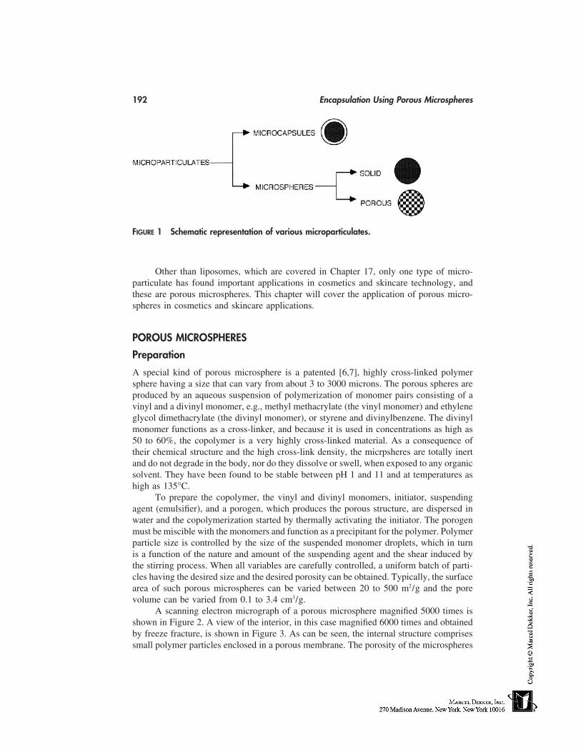
192 Encapsulation Using Porous Microspheres
FIGURE 1 Schematic representation of various microparticulates.
Other than liposomes, which are covered in Chapter 17, only one type of micro-particulate has found important applications in cosmetics and skincare technology, andthese are porous microspheres. This chapter will cover the application of porous micro-spheres in cosmetics and skincare applications.
POROUS MICROSPHERES
Preparation
A special kind of porous microsphere is a patented [6,7], highly cross-linked polymersphere having a size that can vary from about 3 to 3000 microns. The porous spheres areproduced by an aqueous suspension of polymerization of monomer pairs consisting of avinyl and a divinyl monomer, e.g., methyl methacrylate (the vinyl monomer) and ethyleneglycol dimethacrylate (the divinyl monomer), or styrene and divinylbenzene. The divinylmonomer functions as a cross-linker, and because it is used in concentrations as high as50 to 60%, the copolymer is a very highly cross-linked material. As a consequence oftheir chemical structure and the high cross-link density, the micrpsheres are totally inertand do not degrade in the body, nor do they dissolve or swell, when exposed to any organicsolvent. They have been found to be stable between pH 1 and 11 and at temperatures ashigh as 135°C.
To prepare the copolymer, the vinyl and divinyl monomers, initiator, suspendingagent (emulsifier), and a porogen, which produces the porous structure, are dispersed inwater and the copolymerization started by thermally activating the initiator. The porogenmust be miscible with the monomers and function as a precipitant for the polymer. Polymerparticle size is controlled by the size of the suspended monomer droplets, which in turnis a function of the nature and amount of the suspending agent and the shear induced bythe stirring process. When all variables are carefully controlled, a uniform batch of parti-cles having the desired size and the desired porosity can be obtained. Typically, the surfacearea of such porous microspheres can be varied between 20 to 500 m2/g and the porevolume can be varied from 0.1 to 3.4 cm3/g.
A scanning electron micrograph of a porous microsphere magnified 5000 times isshown in Figure 2. A view of the interior, in this case magnified 6000 times and obtainedby freeze fracture, is shown in Figure 3. As can be seen, the internal structure comprisessmall polymer particles enclosed in a porous membrane. The porosity of the microspheres
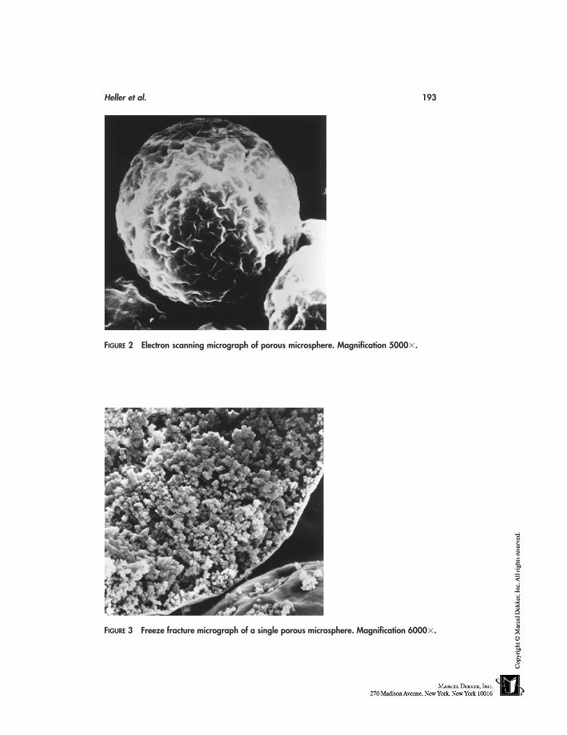
Heller et al. 193
FIGURE 2 Electron scanning micrograph of porous microsphere. Magnification 5000�.
FIGURE 3 Freeze fracture micrograph of a single porous microsphere. Magnification 6000�.
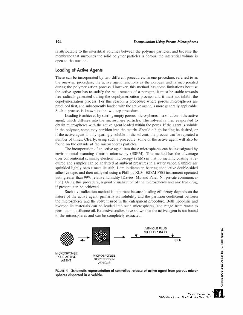
194 Encapsulation Using Porous Microspheres
is attributable to the interstitial volumes between the polymer particles, and because themembrane that surrounds the solid polymer particles is porous, the interstitial volume isopen to the outside.
Loading of Active Agents
These can be incorporated by two different procedures. In one procedure, referred to asthe one-step procedure, the active agent functions as the porogen and is incorporatedduring the polymerization process. However, this method has some limitations becausethe active agent has to satisfy the requirements of a porogen, it must be stable towardsfree radicals generated during the copolymerization process, and it must not inhibit thecopolymerization process. For this reason, a procedure where porous microspheres areproduced first, and subsequently loaded with the active agent, is more generally applicable.Such a process is known as the two-step procedure.
Loading is achieved by stirring empty porous microspheres in a solution of the activeagent, which diffuses into the microsphere particles. The solvent is then evaporated toobtain microspheres with the active agent loaded within the pores. If the agent is solublein the polymer, some may partition into the matrix. Should a high loading be desired, orif the active agent is only sparingly soluble in the solvent, the process can be repeated anumber of times. Clearly, using such a procedure, some of the active agent will also befound on the outside of the microspheres particles.
The incorporation of an active agent into these microspheres can be investigated byenvironmental scanning electron microscopy (ESEM). This method has the advantageover conventional scanning electron microscopy (SEM) in that no metallic coating is re-quired and samples can be analyzed at ambient pressures in a water vapor. Samples aresprinkled lightly onto a metallic stub, 1 cm in diameter, bearing conductive double-sidedadhesive tape, and then analyzed using a Phillips XL30 ESEM FEG instrument operatedwith greater than 99% relative humidity [Davies, M., and Patel, N., private communica-tion]. Using this procedure, a good visualization of the microspheres and any free drug,if present, can be achieved.
Such a visualization method is important because loading efficiency depends on thenature of the active agent, primarily its solubility and the partition coefficient betweenthe microspheres and the solvent used in the entrapment procedure. Both lipophilic andhydrophilic materials can be loaded into such microspheres, and range from water topetrolatum to silicone oil. Extensive studies have shown that the active agent is not boundto the microspheres and can be completely extracted.
FIGURE 4 Schematic representation of controlled release of active agent from porous micro-spheres dispersed in a vehicle.
Heller et al. 195
Release of Active Agents
Although porous microspheres can function in a limited way as a sustained-release deliv-ery vehicle, they are best viewed as a reservoir. However, the combination of microsphereswith incorporated active agents dispersed in a vehicle can function as a controlled-releasedevice if a vehicle in which the drug is only poorly soluble is chosen. When such aformulation is applied to the skin, only that amount of the drug dissolved in the vehicleis presented to the skin. Then, as the drug diffuses from the vehicle into the skin, thesaturation concentration of the drug in the vehicle is maintained by diffusion of drug fromthe microspheres into the vehicle. This process is shown schematically in Figure 4.
APPLICATIONS
Porous microspheres have been used in two major applications. One application takesadvantage of the high porosity of the microspheres to entrap liquid materials, such assilicone oil, to convert a liquid into a free-flowing powder. This allows significant formula-tion flexibility, and a babywipe product has been developed where silicone in porousmicrospheres has been formulated in an aqueous medium.
In the other application, microspheres with incorporated active agents are dispersedin a suitable vehicle for topical applications. As already discussed, when active agentsthat are normally skin irritants are used and a vehicle in which the active agent is onlypoorly soluble is chosen, a significant reduction of irritation, when compared with ordinaryformulation, is noted. Such a reduction in irritancy will be illustrated with two products,one incorporating benzoyl peroxide and the other incorporating trans-retinoic acid (RA).
Benzoyl Peroxide
Benzoyl peroxide (BPO) is clinically effective in acne, primarily because of its bactericidalactivity against Proprionibacterium acnes and possibly also through its mild keratolyticeffects [8–10]. The main site of pharmacological action is the pilosebaceous canal [11].BPO penetrates through the follicular opening, probably by dissolving into sebaceouslipids, and then exerts its antimicrobial activity [12]. Skin irritation is a common sideeffect and a dose relation seems to exist between efficacy and irritation [13]. Thus, acontrolled-release formulation would clearly be advantageous.
In vitro release kinetics were determined by applying formulations to silastic mem-branes mounted in static diffusion cells, and by using excised human skin. Release ofBPO from two formualtions applied to a silastic membrane, one incorporating free BPOand one incorporating BPO entrapped in porous microspheres is shown in Figure 5. Initialrelease of BPO dispersed in the vehicle shows good linearity, but with further releasewould decline, as expected for t1/2 kinetics. The calculated flux for the initial release is0.09 mg/cm2/h. The release of BPO entrapped in the porous microspheres shows a discon-tinuity. Initial flux is about 0.1 mg/cm2/h, very close to the release from BPO dispersedin the vehicle, followed by a slower release with a flux of 0.04 mg/cm2/h. These dataindicate that not all BPO has been entrapped in the porous microspheres, and that theformulation contains some free BPO. Initial release is attributable to release of the freeBPO, followed by the release of entrapped BPO.
The topical irritancy of a BPO controlled-release formulation has been determinedin rabbits, in rhesus monkeys, and in human volunteers [14] using formulations with BPOdispersed in a vehicle and BPO entrapped in porous microspheres dispersed in a vehicle.
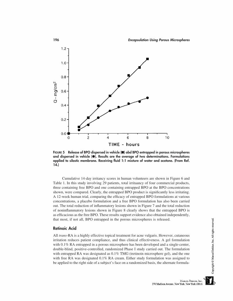
196 Encapsulation Using Porous Microspheres
FIGURE 5 Release of BPO dispersed in vehicle (■) abd BPO entrapped in porous microspheresand dispersed in vehicle (�). Results are the average of two determinations. Formulationsapplied to silastic membrane. Receiving fluid 1:1 mixture of water and acetone. (From Ref.14.)
Cumulative 14-day irritancy scores in human volunteers are shown in Figure 6 andTable 1. In this study involving 29 patients, total irritaancy of four commercial products,three containing free BPO and one containing entrapped BPO at the BPO concentrationsshown, were compared. Clearly, the entrapped BPO product is significantly less irritating.A 12-week human trial, comparing the efficacy of entrapped BPO formulations at variousconcentrations, a placebo formulation and a free BPO formulation has also been carriedout. The total reduction of inflammatory lesions shown in Figure 7 and the total reductionof noninflammatory lesions shown in Figure 8 clearly shows that the entrapped BPO isas efficacious as the free BPO. These results support evidence also obtained independently,that most, if not all, BPO entrapped in the porous microspheres is released.
Retinoic Acid
All trans-RA is a highly effective topical treatment for acne vulgaris. However, cutaneousirritation reduces patient compliance, and thus clinical effectiveness. A gel formulationwith 0.1% RA entrapped in a porous microsphere has been developed and a single-center,double-blind, positive-controlled, randomized Phase I study carried out. The formulationwith entrapped RA was designated as 0.1% TMG (tretinoin microsphere gel), and the onewith free RA was designated 0.1% RA cream. Either study formulation was assigned tobe applied to the right side of a subject’s face on a randomized basis, the alternate formula-
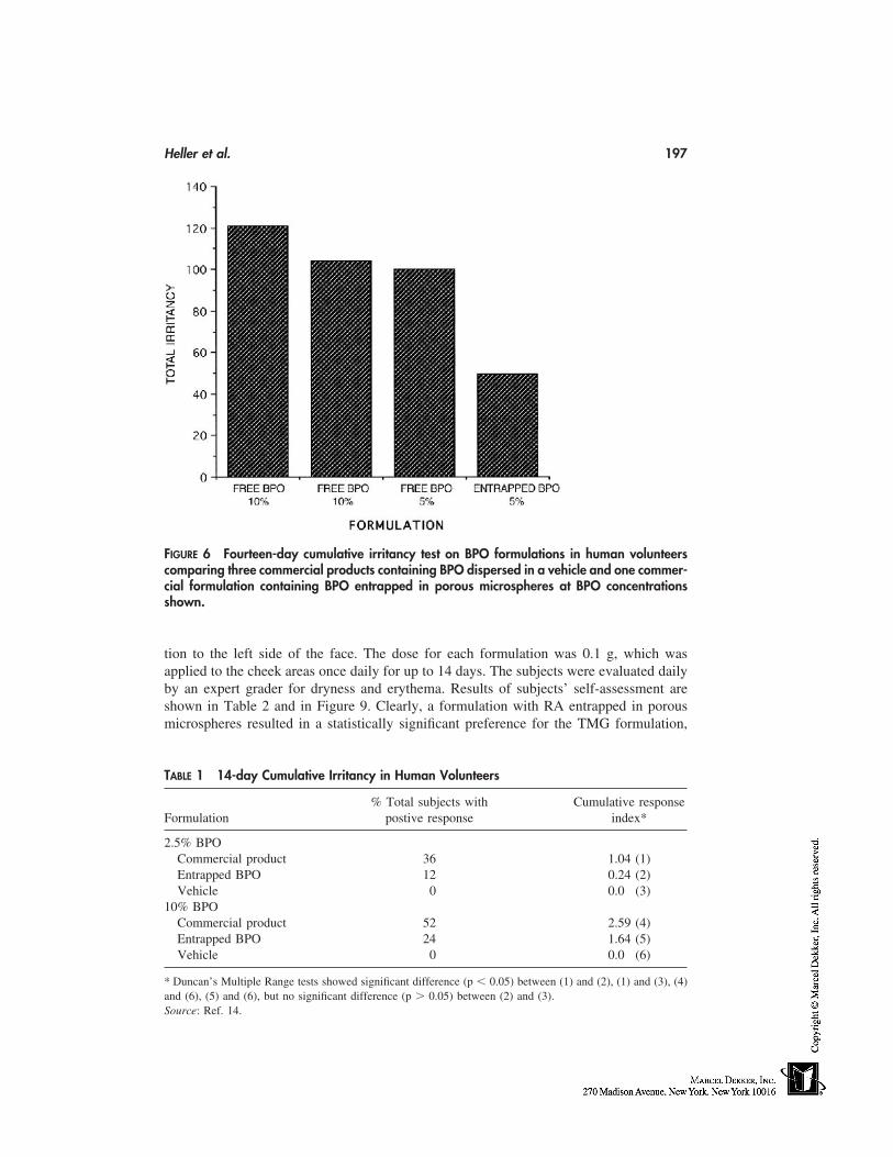
Heller et al. 197
FIGURE 6 Fourteen-day cumulative irritancy test on BPO formulations in human volunteerscomparing three commercial products containing BPO dispersed in a vehicle and one commer-cial formulation containing BPO entrapped in porous microspheres at BPO concentrationsshown.
tion to the left side of the face. The dose for each formulation was 0.1 g, which wasapplied to the cheek areas once daily for up to 14 days. The subjects were evaluated dailyby an expert grader for dryness and erythema. Results of subjects’ self-assessment areshown in Table 2 and in Figure 9. Clearly, a formulation with RA entrapped in porousmicrospheres resulted in a statistically significant preference for the TMG formulation,
TABLE 1 14-day Cumulative Irritancy in Human Volunteers
% Total subjects with Cumulative responseFormulation postive response index*
2.5% BPOCommercial product 36 1.04 (1)Entrapped BPO 12 0.24 (2)Vehicle 0 0.0 (3)
10% BPOCommercial product 52 2.59 (4)Entrapped BPO 24 1.64 (5)Vehicle 0 0.0 (6)
* Duncan’s Multiple Range tests showed significant difference (p � 0.05) between (1) and (2), (1) and (3), (4)and (6), (5) and (6), but no significant difference (p � 0.05) between (2) and (3).Source: Ref. 14.
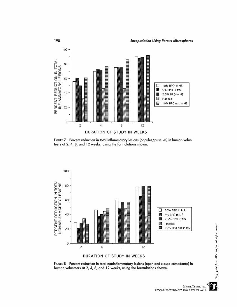
198 Encapsulation Using Porous Microspheres
FIGURE 7 Percent reduction in total inflammatory lesions (papules/pustules) in human volun-teers at 2, 4, 8, and 12 weeks, using the formulations shown.
FIGURE 8 Percent reduction in total noninflammatory lesions (open and closed comedones) inhuman volunteers at 2, 4, 8, and 12 weeks, using the formulations shown.
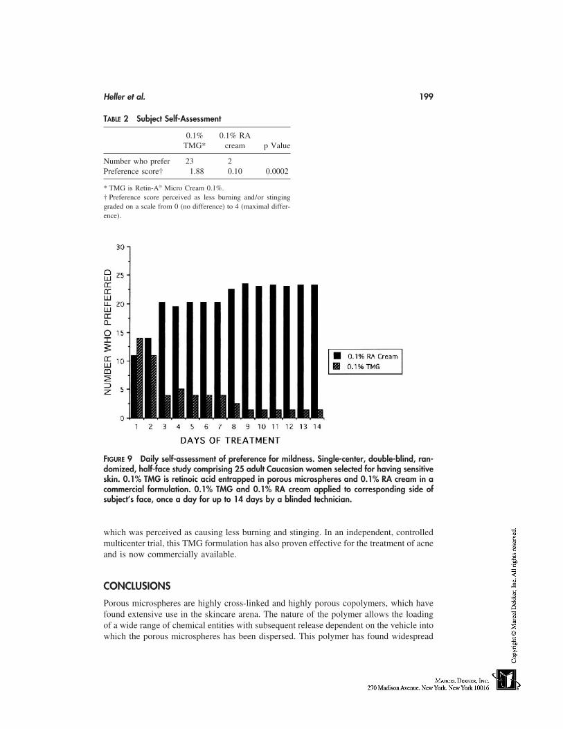
Heller et al. 199
TABLE 2 Subject Self-Assessment
0.1% 0.1% RATMG* cream p Value
Number who prefer 23 2Preference score† 1.88 0.10 0.0002
* TMG is Retin-A Micro Cream 0.1%.† Preference score perceived as less burning and/or stinginggraded on a scale from 0 (no difference) to 4 (maximal differ-ence).
FIGURE 9 Daily self-assessment of preference for mildness. Single-center, double-blind, ran-domized, half-face study comprising 25 adult Caucasian women selected for having sensitiveskin. 0.1% TMG is retinoic acid entrapped in porous microspheres and 0.1% RA cream in acommercial formulation. 0.1% TMG and 0.1% RA cream applied to corresponding side ofsubject’s face, once a day for up to 14 days by a blinded technician.
which was perceived as causing less burning and stinging. In an independent, controlledmulticenter trial, this TMG formulation has also proven effective for the treatment of acneand is now commercially available.
CONCLUSIONS
Porous microspheres are highly cross-linked and highly porous copolymers, which havefound extensive use in the skincare arena. The nature of the polymer allows the loadingof a wide range of chemical entities with subsequent release dependent on the vehicle intowhich the porous microspheres has been dispersed. This polymer has found widespread
200 Encapsulation Using Porous Microspheres
acceptance as a means of reducing irritation without decreasing efficacy when used appro-priately.
REFERENCES
1. Luzzi, L. A. (1970). Microencapsulation. J. Pharm. Sci. 59:1367–1376.2. Beck, L. R., and Tice, T. R. (1983). Poly(lactic) and poly(lactic acid-co-glycolic acid) contra-
ceptive delivery systems. In Mishell, D. R. (ed.), Long-Acting Steroid Contraception. NewYork: Raven Press, 175–199.
3. Nuwayser, E. S., Gay, M. H., DeRoo, D. J., and Blaskovich, P. D. (1988). Sustained releaseinjectable naltrexone microcapsules. Proc. Intern. Symp. Control Rel. Bioact. Mater. 15:201–202.
4. Higuchi, T. (1961). Rates of release of medicamenets from ointment bases containing drugsin suspension. J. Pharm. Sci. 50:874–875.
5. Sato, T., Kanke, M., Schroeder, H. G., and DeLuca, P. (1988). Porous biodegradable micro-spheres for controlled drug delivery. I. Assessssment of processing conditions and solventremoval techniques. Pharm. Res. 5:21–30.
6. Won, R. Method for delivering an active ingredient by controlled time release utilizing a noveldelivery vehicle which can be prepared by process utilizing the active ingredient as a porogen.U.S. Patent 4,690,825. September 1, 1987.
7. Won, R. Two step method for preparation of controlled release formulations. U.S. Patent5,145,675, September 8, 1992.
8. Nacht, S. (1983). Comparative activity of benzoyl peroxide and hexachlorophene. In vivostudies against Proprionibacterium acnes in humans. Arch. Dermatol. 119:577–579.
9. Fulton, J. E., and Bradley, S. (1976). The choice of vitamin A, erythromycin and benzoylperoxide for the topical treatment of acne. Cutis 17:560–564.
10. Kligman, A. M., Leyden, J. J., and Stewart, R. (1977). New uses of benzoyl peroxide: a broadspectrum antimicrobial agent. Int. J. Dermatol. 16:413–417.
11. Nacht, S. (1981). Methods to assess the transepidermal and intrafollicular penetration of anti-acne agents. In: Proceedings of the 1980 Research and Scientific Development Conference,New York, pp. 88–91.
12. Leyden, J. J. Topical antibiotics and topical antimcrobial agents in acne therapy. In: Julin,L. A., Rossman, H., and Strauss, H. (eds.), Symposium in Lund, Uppsala, Sweden: UpplandGrafisker AB. 1980:151–164.
13. Fulton, J. E., and Bradley, S. (1974). Studies on the mechanism of action of topical benzoylperoxide in acne vulgaris. J. Cuta. Pathol. 1:191–194.
14. Wester, R. C., Patel, R., Nacht, S., Leyden, J., Melendres, J., and Maibach, H. (1991). Con-trolled release of benzoyl peroxide from a porous microsphere polymeric system can reducetopical irritancy. J. Am. Acad. Dermatol. 24:720–726.
17
Liposomes
Hans LautenschlägerDevelopment & Consulting, Pulheim, Germany
INTRODUCTION
Publications about and patents on liposomes, along with their different chemical compo-nents, preparation, and use in skincare products have often been reviewed [1–4]. Thereviews do not need any additional comments. Of interest are general questions, such aswhy liposomes should be used in cosmetics, which functionalities are expected from them,and which advantages they do provide compared with alternative formulations.
The properties of the widely used main component of liposomes, phosphatidylcho-line, play a key role for answering these questions. Other compounds such as niotensidesand ceramides, which are naturally predestinated for the preparation of liposomes, are lessimportant today. Niotensides do not offer superior claims, and ceramides are not availablein sufficient quantities and qualities at convenient prices.
PHOSPHATIDYLCHOLINE
Looking at the horny layer, which is the barrier against external materials, phospholipidsand phosphatidylcholine in particular play a minor role. The lipid bilayers contain onlytraces of phospholipids, and the main components are free fatty acids, cholesterol, tri-glycerides, hydrocarbons, and ceramides. But looking deeper into the living part of theepidermis, phosphatidylcholine is usually found as the most important constituent of allbiological membranes, especially of plasma cell membranes. Over and above that phos-phatidylcholine is the source of phosphocholine to transform ceramides to sphingomyelins.In this context, phosphatidylcholine stands for living tissues whereas the increase of cera-mides in the cells means that their death by apoptosis is soon ahead (Fig. 1).
Human phosphatidylcholine and phosphatidylcholine of vegetable origin show afatty acid composition, which is dominated by unsaturated fatty acids. The fatty acid con-tent of soy phosphatidylcholine, which is readily available and mostly used in cosmeticformulas, is characterized by a ratio of linoleic acid up to 70% of the total fatty acids.Consequently, soy phosphatidylcholine has a very low phase-transition temperature ofbelow 0°C in water-containing systems. This may be the reason for its ability to fluidizethe lipid bilayers of the horny layer, which can be measured by an increase of the transepi-dermal water loss (TEWL) after application for a short while. The slight increase of TEWL
201
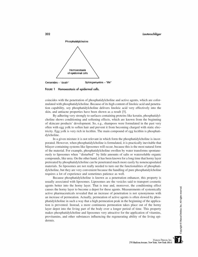
202 Lautenschläger
FIGURE 1 Homoeostasis of epidermal cells.
coincides with the penetration of phosphatidylcholine and active agents, which are cofor-mulated with phosphatidylcholine. Because of its high content of linoleic acid and penetra-tion capability, soy phosphatidylcholine delivers linoleic acid very effectively into theskin, and antiacne properties have been shown as a result [5].
By adhering very strongly to surfaces containing proteins like keratin, phosphatidyl-choline shows conditioning and softening effects, which are known from the beginningof skincare products’ development. So, e.g., shampoos were formulated in the past veryoften with egg yolk to soften hair and prevent it from becoming charged with static elec-tricity. Egg yolk is very rich in lecithin. The main compound of egg lecithin is phosphati-dylcholine.
In a given mixture it is not relevant in which form the phosphatidylcholine is incor-porated. However, when phosphatidylcholine is formulated, it is practically inevitable thatbilayer-containing systems like liposomes will occur, because this is the most natural formof the material. For example, phosphatidylcholine swollen by water transforms spontane-ously to liposomes when ‘‘disturbed’’ by little amounts of salts or watersoluble organiccompounds, like urea. On the other hand, it has been known for a long time that horny layerpretreated by phosphatidylcholine can be penetrated much more easily by nonencapsulatedmaterials. So liposomes are not really needed to turn out the functionalities of phosphati-dylcholine, but they are very convenient because the handling of pure phosphatidylcholinerequires a lot of experience and sometimes patience as well.
Because phosphatidylcholine is known as a penetration enhancer, this property isusually associated with liposomes. Liposomes are the vesicles said to transport cosmeticagents better into the horny layer. That is true and, moreover, the conditioning effectcauses the horny layer to become a depot for these agents. Measurements of systemicallyactive pharmaceuticals revealed that an increase of penetration is not synonymous withan increase of permeation. Actually, permeation of active agents is often slowed by phos-phatidylcholine in such a way that a high permeation peak in the beginning of the applica-tion is prevented. Instead, a more continuous permeation takes place out of the hornylayer depot into the living part of the body over a longer period of time. This propertymakes phosphatidylcholine and liposomes very attractive for the application of vitamins,provitamins, and other substances influencing the regenerating ability of the living epi-dermis.
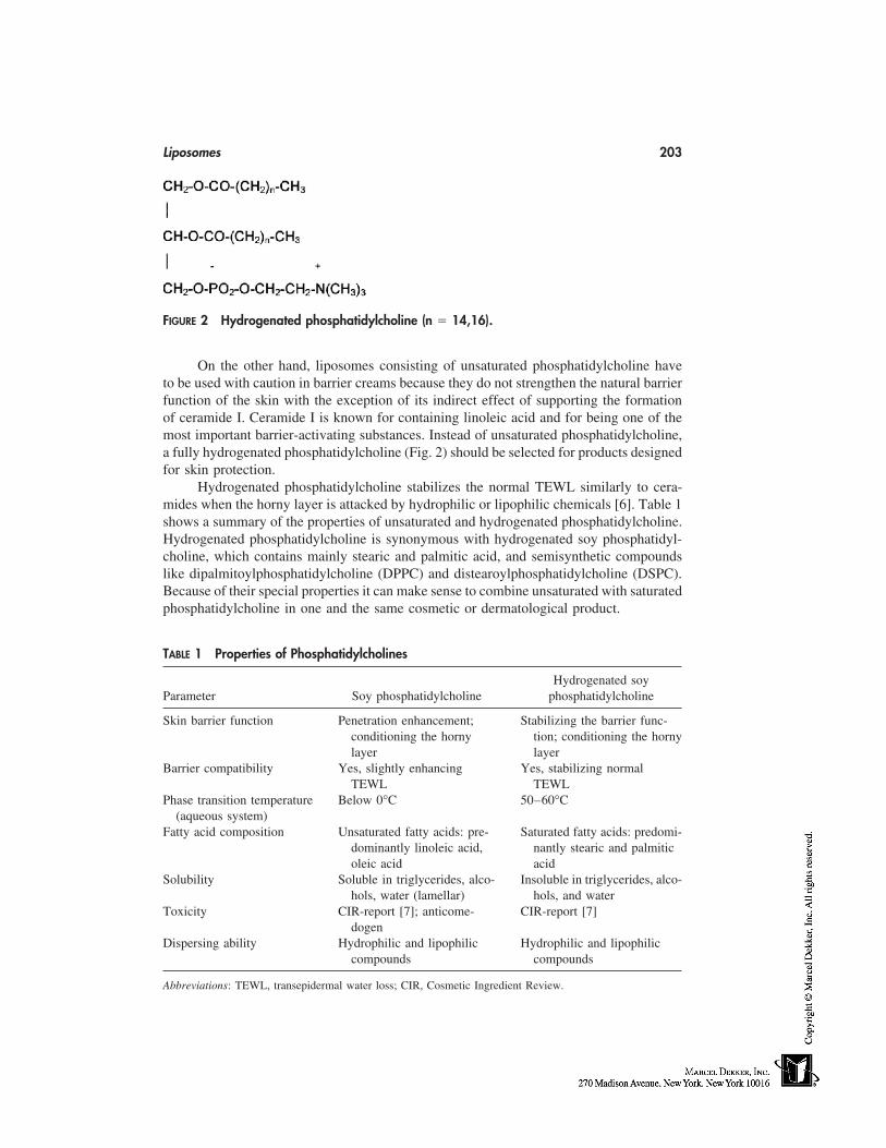
Liposomes 203
FIGURE 2 Hydrogenated phosphatidylcholine (n � 14,16).
On the other hand, liposomes consisting of unsaturated phosphatidylcholine haveto be used with caution in barrier creams because they do not strengthen the natural barrierfunction of the skin with the exception of its indirect effect of supporting the formationof ceramide I. Ceramide I is known for containing linoleic acid and for being one of themost important barrier-activating substances. Instead of unsaturated phosphatidylcholine,a fully hydrogenated phosphatidylcholine (Fig. 2) should be selected for products designedfor skin protection.
Hydrogenated phosphatidylcholine stabilizes the normal TEWL similarly to cera-mides when the horny layer is attacked by hydrophilic or lipophilic chemicals [6]. Table 1shows a summary of the properties of unsaturated and hydrogenated phosphatidylcholine.Hydrogenated phosphatidylcholine is synonymous with hydrogenated soy phosphatidyl-choline, which contains mainly stearic and palmitic acid, and semisynthetic compoundslike dipalmitoylphosphatidylcholine (DPPC) and distearoylphosphatidylcholine (DSPC).Because of their special properties it can make sense to combine unsaturated with saturatedphosphatidylcholine in one and the same cosmetic or dermatological product.
TABLE 1 Properties of Phosphatidylcholines
Hydrogenated soyParameter Soy phosphatidylcholine phosphatidylcholine
Skin barrier function Penetration enhancement; Stabilizing the barrier func-conditioning the horny tion; conditioning the hornylayer layer
Barrier compatibility Yes, slightly enhancing Yes, stabilizing normalTEWL TEWL
Phase transition temperature Below 0°C 50–60°C(aqueous system)
Fatty acid composition Unsaturated fatty acids: pre- Saturated fatty acids: predomi-dominantly linoleic acid, nantly stearic and palmiticoleic acid acid
Solubility Soluble in triglycerides, alco- Insoluble in triglycerides, alco-hols, water (lamellar) hols, and water
Toxicity CIR-report [7]; anticome- CIR-report [7]dogen
Dispersing ability Hydrophilic and lipophilic Hydrophilic and lipophiliccompounds compounds
Abbreviations: TEWL, transepidermal water loss; CIR, Cosmetic Ingredient Review.
204 Lautenschläger
LIPOSOMES
Liposomes are spherical vesicles whose membranes consist of one (unilamellar) or more(oligolamellar, multilamellar) bilayers of phosphatidylcholine. Sometimes, especially inpatents, reference is made not about liposomes but about ‘‘vesicles with an internal aque-ous phase.’’ The vesicles can differ in size (diameter about 15–3500 nm) and shape (singleand fused particles). At a given chemical composition, these parameters strongly dependon the process of preparation. Very often the preparations are metastable. That means thestate of free enthalpy is not in an equilibrium with the environment. As a result the vesicleschange their lamellarity, size, size distribution, and shape with time. For example, smallvesicles tend to form larger ones and large vesicles smaller ones. Fortunately this is mostlynot critical for quality because the properties of the phosphatidylcholine, which the vesi-cles are based on, remain unchanged as a rule. Nevertheless the stability seems to be bestin a range of about 100 to 300 nm. That is the case of pure aqueous dispersions of highlyenriched (80–100%) soy phosphatidylcholine.
In a complete formulation together with further ingredients, other influences likecompatibility, concentration of salts, amphiphilics, and lipophilics play an important role.Therefore, it is often very difficult to prove the existence of liposomes, e.g., in a gelphase or a creamy matrix. However, this is more a marketing problem than a problem ofeffectiveness of the formulation. Today we can assume that the effectiveness of phosphati-dylcholine is based more on the total chemical composition of the cosmetic product andless on the existence or nonexistence of the added liposomes. This may seem curious, butis in fact the reality.
Of course, formulations are very effective in particular when consisting of pureliposomal dispersions bearing lipophilic additives in the membrane spheres and/or hydro-philics in the internal and external aqueous phases within the range of their bearing capac-ity. In this respect, there has been an intensive search to increase the encapsulation capacityof liposomes for lipids because consumers are used to applying lipid-rich creams. Effortswere made to add emulsifier to the liposomal dispersions to stabilize higher amounts oflipids. Formulators now know that the compatibility of liposomes with regard to emulsifi-ers is generally limited, more or less. On the other hand, additional emulsifiers have aweakening effect on the barrier affinity of phosphatidylcholine. They cause the phosphati-dylcholine and the lipids to be more easily removed from the skin while washing. In thisrespect there is only one rational consideration: to make use of nanoemulsions consistingof phosphatidylcholine and lipids instead of liposomes. Nanoemulsions are a consequenceof the observation that oil droplets can fuse with liposomes when the capacity of bilayersfor lipids is exhausted [8]. Further increasing the lipid/phosphatidylcholine ratio and usinghigh-pressure homogenizers lead to nanoemulsions. Nanoemulsions consist of emulsion-like oil droplets surrounded by a monolayer of phosphatidylcholine. The advantage ofnanoemulsions is that they allow formulations to tolerate more lipids and remain stable.Also, additional emulsifiers are not needed.
Liposomal dispersions based on unsaturated phosphatidylcholine are lacking in sta-bility against oxidation. Like linoleic esters and linoleic glycerides, these dispersions haveto be stabilized by antioxidants. Thinking naturally, a complex of Vitamin C and E (respec-tively, their derivatives like acetates and palmitates) can be used with success. In somecases, phosphatidylcholine and urea seem to stabilize each other [9,10]. Moreover, agentsthat are able to mask traces of radical-forming ions of heavy metals, like iron, can be
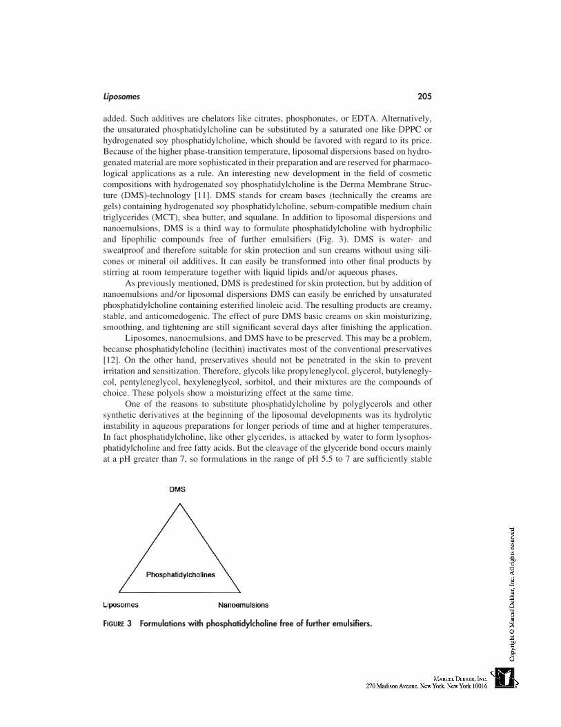
Liposomes 205
added. Such additives are chelators like citrates, phosphonates, or EDTA. Alternatively,the unsaturated phosphatidylcholine can be substituted by a saturated one like DPPC orhydrogenated soy phosphatidylcholine, which should be favored with regard to its price.Because of the higher phase-transition temperature, liposomal dispersions based on hydro-genated material are more sophisticated in their preparation and are reserved for pharmaco-logical applications as a rule. An interesting new development in the field of cosmeticcompositions with hydrogenated soy phosphatidylcholine is the Derma Membrane Struc-ture (DMS)-technology [11]. DMS stands for cream bases (technically the creams aregels) containing hydrogenated soy phosphatidylcholine, sebum-compatible medium chaintriglycerides (MCT), shea butter, and squalane. In addition to liposomal dispersions andnanoemulsions, DMS is a third way to formulate phosphatidylcholine with hydrophilicand lipophilic compounds free of further emulsifiers (Fig. 3). DMS is water- andsweatproof and therefore suitable for skin protection and sun creams without using sili-cones or mineral oil additives. It can easily be transformed into other final products bystirring at room temperature together with liquid lipids and/or aqueous phases.
As previously mentioned, DMS is predestined for skin protection, but by addition ofnanoemulsions and/or liposomal dispersions DMS can easily be enriched by unsaturatedphosphatidylcholine containing esterified linoleic acid. The resulting products are creamy,stable, and anticomedogenic. The effect of pure DMS basic creams on skin moisturizing,smoothing, and tightening are still significant several days after finishing the application.
Liposomes, nanoemulsions, and DMS have to be preserved. This may be a problem,because phosphatidylcholine (lecithin) inactivates most of the conventional preservatives[12]. On the other hand, preservatives should not be penetrated in the skin to preventirritation and sensitization. Therefore, glycols like propyleneglycol, glycerol, butylenegly-col, pentyleneglycol, hexyleneglycol, sorbitol, and their mixtures are the compounds ofchoice. These polyols show a moisturizing effect at the same time.
One of the reasons to substitute phosphatidylcholine by polyglycerols and othersynthetic derivatives at the beginning of the liposomal developments was its hydrolyticinstability in aqueous preparations for longer periods of time and at higher temperatures.In fact phosphatidylcholine, like other glycerides, is attacked by water to form lysophos-phatidylcholine and free fatty acids. But the cleavage of the glyceride bond occurs mainlyat a pH greater than 7, so formulations in the range of pH 5.5 to 7 are sufficiently stable
FIGURE 3 Formulations with phosphatidylcholine free of further emulsifiers.
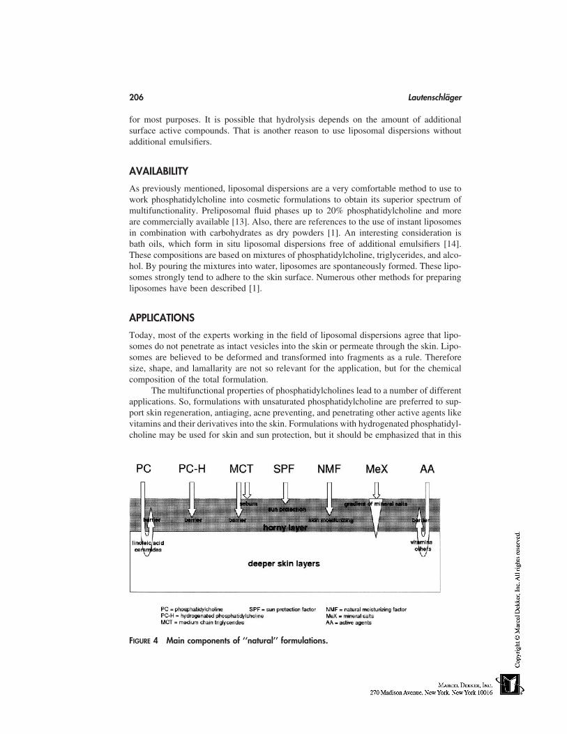
206 Lautenschläger
for most purposes. It is possible that hydrolysis depends on the amount of additionalsurface active compounds. That is another reason to use liposomal dispersions withoutadditional emulsifiers.
AVAILABILITY
As previously mentioned, liposomal dispersions are a very comfortable method to use towork phosphatidylcholine into cosmetic formulations to obtain its superior spectrum ofmultifunctionality. Preliposomal fluid phases up to 20% phosphatidylcholine and moreare commercially available [13]. Also, there are references to the use of instant liposomesin combination with carbohydrates as dry powders [1]. An interesting consideration isbath oils, which form in situ liposomal dispersions free of additional emulsifiers [14].These compositions are based on mixtures of phosphatidylcholine, triglycerides, and alco-hol. By pouring the mixtures into water, liposomes are spontaneously formed. These lipo-somes strongly tend to adhere to the skin surface. Numerous other methods for preparingliposomes have been described [1].
APPLICATIONS
Today, most of the experts working in the field of liposomal dispersions agree that lipo-somes do not penetrate as intact vesicles into the skin or permeate through the skin. Lipo-somes are believed to be deformed and transformed into fragments as a rule. Thereforesize, shape, and lamallarity are not so relevant for the application, but for the chemicalcomposition of the total formulation.
The multifunctional properties of phosphatidylcholines lead to a number of differentapplications. So, formulations with unsaturated phosphatidylcholine are preferred to sup-port skin regeneration, antiaging, acne preventing, and penetrating other active agents likevitamins and their derivatives into the skin. Formulations with hydrogenated phosphatidyl-choline may be used for skin and sun protection, but it should be emphasized that in this
FIGURE 4 Main components of ‘‘natural’’ formulations.
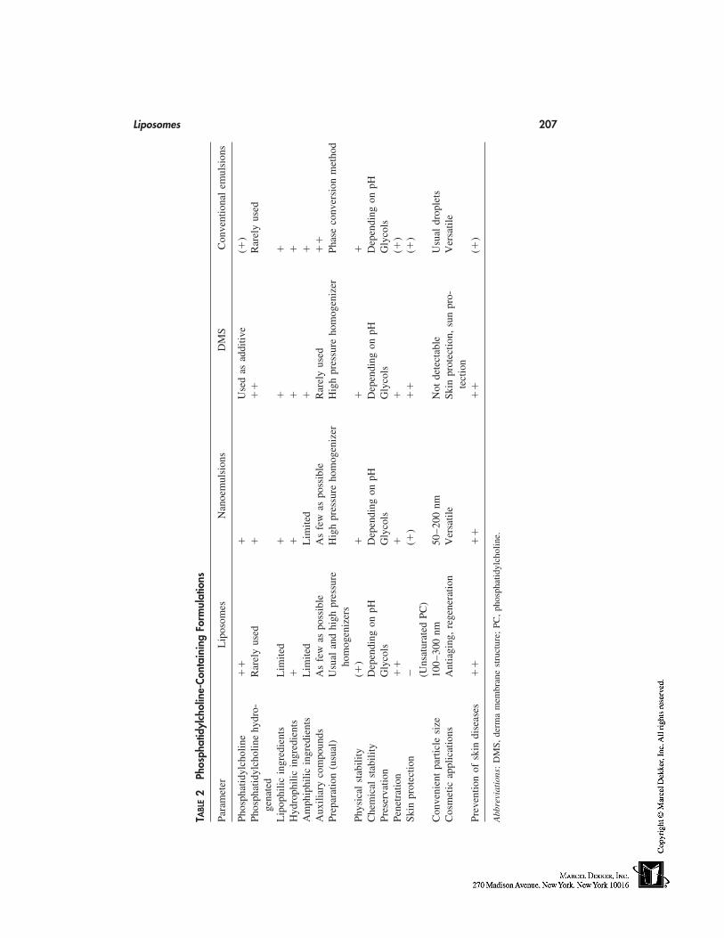
Liposomes 207
TABL
E2
Phos
phat
idyl
chol
ine-
Cont
aini
ngFo
rmul
atio
ns
Para
met
erL
ipos
omes
Nan
oem
ulsi
ons
DM
SC
onve
ntio
nal
emul
sion
s
Phos
phat
idyl
chol
ine
��
�U
sed
asad
ditiv
e(�
)Ph
osph
atid
ylch
olin
ehy
dro-
Rar
ely
used
��
�R
arel
yus
edge
nate
dL
ipop
hilic
ingr
edie
nts
Lim
ited
��
�H
ydro
phili
cin
gred
ient
s�
��
�A
mph
iphi
licin
gred
ient
sL
imite
dL
imite
d�
�A
uxili
ary
com
poun
dsA
sfe
was
poss
ible
As
few
aspo
ssib
leR
arel
yus
ed�
�Pr
epar
atio
n(u
sual
)U
sual
and
high
pres
sure
Hig
hpr
essu
reho
mog
eniz
erH
igh
pres
sure
hom
ogen
izer
Phas
eco
nver
sion
met
hod
hom
ogen
izer
sPh
ysic
alst
abili
ty(�
)�
��
Che
mic
alst
abili
tyD
epen
ding
onpH
Dep
endi
ngon
pHD
epen
ding
onpH
Dep
endi
ngon
pHPr
eser
vatio
nG
lyco
lsG
lyco
lsG
lyco
lsG
lyco
lsPe
netr
atio
n�
��
�(�
)Sk
inpr
otec
tion
�(�
)�
�(�
)(U
nsat
urat
edPC
)C
onve
nien
tpa
rtic
lesi
ze10
0–30
0nm
50–2
00nm
Not
dete
ctab
leU
sual
drop
lets
Cos
met
icap
plic
atio
nsA
ntia
ging
,re
gene
ratio
nV
ersa
tile
Skin
prot
ectio
n,su
npr
o-V
ersa
tile
tect
ion
Prev
entio
nof
skin
dise
ases
��
��
��
(�)
Abb
revi
atio
ns:
DM
S,de
rma
mem
bran
est
ruct
ure;
PC,
phos
phat
idyl
chol
ine.
208 Lautenschläger
respect nanoemulsions and DMS are still more convenient. The main components ofchoice to prepare ‘‘natural’’ formulations, which are compatible with horny layer, sebumconstituents, and their functions are illustrated in Figure 4. About the role of mineral saltssee Ref. 15.
THE FUTURE OF LIPOSOMAL PREPARATIONS
Liposomal dispersions have proved not only to be innovative and effective cosmetic ingre-dients, but also to be a very convenient form to work with phosphatidylcholine. In derma-tology, they will be used with success for preventing and treating several skin diseases.Complementary formulations are established where liposomal dispersions come up againstlimiting factors. Table 2 shows liposomal and complementary formulations in a directcomparison.
Generally, liposomes, nanoemulsions, and DMS are more compatible with the skinstructure than conventional emulsions usually applied. Compatible means that formula-tions do not disturb the integrity of the skin lipid bilayers and are not washed out whilecleaning the skin. In the sense of modern strategies of cosmetics, these formulations getby with a minimum of auxiliary compounds, which put only a strain on the skin. Moreover,compatibility means embedding lipids and hydrophilic agents in the horny layer and beingin accordance with the natural situation.
Remarkably, phosphatidylcholine need not be applied in high concentrations be-cause the experience shows that formulations are stable at lower amounts. Also, there isa cumulative effect in the horny layer with repeated application of phosphatidylcholine.In many cases, liposomes, nanoemulsions, and DMS are compatible with each other in asense that they can be used as a sort of construction kit. So these formulations are believedto still have a great future in cosmetic science. How far new findings about the importanceof the choline moiety of phosphatidylcholine [16] will impact skincare research and devel-opment cannot be estimated today.
REFERENCES
1. Lasic DD. Liposomes and niosomes. In: Rieger MM, Rhein LD, eds. Surfactants in Cosmetics.2d ed. New York: Marcel Dekker, 1997:263–283.
2. Wendel A. Lecithins, phospholipids, liposomes in cosmetics, dermatology and in washing andcleansing preparations. Augsburg: Verlag fuer chemische Industrie, 1994.
3. Wendel A. Lecithins, phospholipids, liposomes in cosmetics, dermatology and in washing andcleansing preparations. Part II. Augsburg: Verlag fuer chemische Industrie, 1997.
4. Braun-Falco O, Korting HC, Maibach HI, eds. Liposome Dermatics. Berlin: Springer-Verlag,1992.
5. Ghyczy M, Nissen H-P, Biltz H. The treatment of acne vulgaris by phosphatidylcholine fromsoybeans, with a high content of linoleic acid. J Appl Cosmetol 1996; 14:137–145.
6. Lautenschlaeger H. Kuehlschmierstoffe und Hautschutz—neue Perspektiven. Mineraloeltech-nik 1998; (5):1–16.
7. Cosmetic Ingredient Review. Lecithin and Hydrogenated Lecithin. Washington: The Cos-metic, Toiletry, and Fragrance Association, 1996.
8. Lautenschlaeger H. Liposomes in dermatological preparations. Part II. Cosmet Toilet 1990;105(7):63–72.
9. Japanese patent 199104364104. Nippon Surfactant Kogyo KK, 1992.10. German patent 4021082. Lautenschlaeger, 1990.
Liposomes 209
11. Kutz G. Galenische Charakterisierung ausgewaehlter Hautpflegeprodukte. PharmazeutischeZeitung 1997; 142(45):4015–4019.
12. Wallhaeusser KH. Praxis der Sterilisation, Desinfektion—Konservierung. 5th ed. Stuttgart:Georg Thieme Verlag, 1995:43, 394.
13. Roeding J. Properties and Characterisation of Pre-Liposome Systems. In: Braun-Falco O,Korting HC, Maibach HI, eds. Liposome Dermatics. Berlin: Springer-Verlag, 1992:110–117.
14. German patent 4021083. Lautenschlaeger, 1990.15. Feingold KR. Permeability barrier homeostasis: its biochemical basis and regulation. Cosmet
Toilet 1997; 112(7):49–59.16. Blusztajn JK. Choline, a vital amine. Science 1998; 281:794–795.

18
Topical Delivery by Iontophoresis
Véronique Préat and Rita VanbeverUniversité Catholique de Louvain, Brussels, Belgium
INTRODUCTION
Passive permeation of drugs across the skin is limited by the low permeability of thestratum corneum. Transdermal and topical delivery of drugs are presently applicable toonly a few drugs with appropriate balance hydro/lipophilicity, small size, no charge, andrelatively high potency [1,2].
Strategies have been developed to increase transdermal and topical delivery acrossor into the skin. They consist of increasing the permeability of the skin or providing adriving force acting on the drug. Chemicals methods (e.g., penetration enhancers) or physi-cal methods (e.g., iontophoresis, sonophoresis, or electroporation) have been shown tosignificantly enhance transdermal transport [2–4].
Iontophoresis is a noninvasive technique that uses a mild electric current to facilitatetransdermal delivery of drugs for both systemic and local effects. Iontophoretic transportof drugs has been extensively studied [5–8]. It has the potential to overcome many of thebarriers to topical drug absorption [6–13]. This chapter will focus on local delivery byiontophoresis as an aid to penetration of topically applied drugs. The mechanisms and theparameters affecting iontophoretic transport will be reviewed. The role of iontophoresisin clinical practice and cosmetics will be discussed.
IONTOPHORESIS
Iontophoresis may be defined as the administration of molecules through the skin by theapplication of an electric current [5–8].
An iontophoretic system has three basic components: 1) the source of electric cur-rent, 2) an active reservoir containing the active and an electrode as well as a counterelectrode in a return reservoir, and 3) a control unit. The current used for iontophoreticdelivery is applied for minutes or hours with current density ranging from 0.1 to 0.5mA/cm2. Miniaturized systems of approximately 10 cm2 including a battery have beendeveloped for transdermal drug delivery. For the topical delivery of actives, the currentsource can be an external power supply and a larger area can be treated by the current.
The principle of iontophoresis is mainly based on electrorepulsion: the electric fielddrives the molecules into the skin. Positive ions will be repelled from the positive elec-
211
212 Préat and Vanbever
trode, called the anode, and attracted to the cathode, or the negative electrode. Negativelycharged compounds will be repelled from the cathode. Neutral compounds can also bedelivered by electro-osmosis [3].
Iontophoresis has been widely studied for transdermal drug delivery. It has beenused to achieve systemic concentration sufficient for a desired therapeutic effect. Ionto-phoresis has also been successfully used in clinical medicine to achieve topical deliveryof drugs for several decades. It has found widespread use in physical therapy and dermatol-ogy. Large quantities of a medication are targeted to a localized treatment region, mini-mizing the systemic level of the medication. The literature supports the concept thationtophoresis is a method of choice for drug application in the therapy of surface tis-sue [9–13].
The rationales for topical drug delivery by iontophoresis are as follows: 1) to delivera locally high concentration of an active—the delivery of the drug is enhanced by ionto-phoresis by one to three orders of magnitude as compared with passive diffu-sion; 2) to control delivery of the active by current application—inter- and intraindivid-ual variations can be reduced; 3) to extend transdermal transport to low and medium(�5000) molecular weight hydrophilic compounds [5–8,14,15].
MECHANISMS OF IONTOPHORETIC TRANSPORT
Theoretical Mechanisms of Iontophoretic Transport
The electrically induced transport of an ion across a membrane results from three mecha-nisms: (1) diffusion related to a chemical potential gradient, 2) electrical mobility attribut-able to an electric potential gradient, and 3) solute transfer attributable to a convectivesolvent flow, i.e., electro-osmosis [5–8,15,16].
JT � JP � JE � JO JT � total fluxJP � passive diffusion fluxJE � electrical fluxJO � electro-osmotic flux
J � �D dC/dx � Dzc F/RT ⋅ dε/dx D � diffusion coefficientc � concentrationz � valenceF � Faraday’s constantR � gaz constantT � absolute temperatureε � electrical potentialX � distance
For ionic species, the contribution of passive diffusion is neglible. The major mechanismof active transport by iontophoresis is the electromigration or electrostatic repulsion. How-ever, the contribution of electro-osmotic flow has been reported to be significant for neutralmolecules and macromolecules. Because of its negative charges, the skin is permselectiveto cations, inducing a net convective solvent flow from the anode to the cathode. Hence,neutral molecules can be delivered into or extracted from the skin by cathodal ionto-phoresis [16–18].
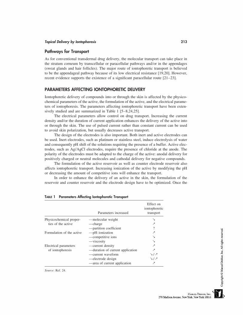
Topical Delivery by Iontophoresis 213
Pathways for Transport
As for conventional transdermal drug delivery, the molecular transport can take place inthe stratum corneum by transcellular or paracellular pathways and/or in the appendages(sweat glands and hair follicles). The major route of iontophoretic transport is believedto be the appendageal pathway because of its low electrical resistance [19,20]. However,recent evidence supports the existence of a significant paracellular route [21–23].
PARAMETERS AFFECTING IONTOPHORETIC DELIVERY
Iontophoretic delivery of compounds into or through the skin is affected by the physico-chemical parameters of the active, the formulation of the active, and the electrical parame-ters of iontophoresis. The parameters affecting iontophoretic transport have been exten-sively studied and are summarized in Table 1 [5–8,24,25].
The electrical parameters allow control on drug transport. Increasing the currentdensity and/or the duration of current application enhances the delivery of the active intoor through the skin. The use of pulsed current rather than constant current can be usedto avoid skin polarization, but usually decreases active transport.
The design of the electrodes is also important. Both inert and active electrodes canbe used. Inert electrodes, such as platinum or stainless steel, induce electrolysis of waterand consequently pH shift of the solutions requiring the presence of a buffer. Active elec-trodes, such as Ag/AgCl electrodes, require the presence of chloride at the anode. Thepolarity of the electrodes must be adapted to the charge of the active: anodal delivery forpositively charged or neutral molecules and cathodal delivery for negative compounds.
The formulation of the active reservoir as well as counter electrode reservoir alsoaffects iontophoretic transport. Increasing ionization of the active by modifying the pHor decreasing the amount of competitive ions will enhance the transport.
In order to enhance the delivery of an active in the skin, the formulation of thereservoir and counter reservoir and the electrode design have to be optimized. Once the
TABLE 1 Parameters Affecting Iontophoretic Transport
Effect oniontophoretic
Parameters increased transport
Physicochemical proper- —molecular weight 'ties of the active —charge ;
—partition coefficient ?Formulation of the active —pH:ionization ;
—competitive ions '—viscosity '
Electrical parameters —current density ;of iontophoresis —duration of current application ;
—current waveform '/;—electrode design '/;—area of current application ;
Source: Ref. 24.
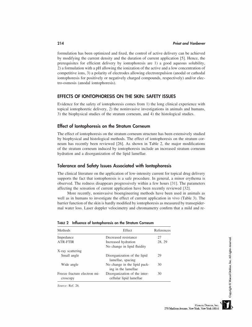
214 Préat and Vanbever
formulation has been optimized and fixed, the control of active delivery can be achievedby modifying the current density and the duration of current application [5]. Hence, theprerequisites for efficient delivery by iontophoresis are 1) a good aqueous solubility,2) a formulation with a pH allowing the ionization of the active and a low concentration ofcompetitive ions, 3) a polarity of electrodes allowing electrorepulsion (anodal or cathodaliontophoresis for positively or negatively charged compounds, respectively) and/or elec-tro-osmosis (anodal iontophoresis).
EFFECTS OF IONTOPHORESIS ON THE SKIN: SAFETY ISSUES
Evidence for the safety of iontophoresis comes from 1) the long clinical experience withtopical iontophoretic delivery, 2) the noninvasive investigations in animals and humans,3) the biophysical studies of the stratum corneum, and 4) the histological studies.
Effect of Iontophoresis on the Stratum Corneum
The effect of iontophoresis on the stratum corneum structure has been extensively studiedby biophysical and histological methods. The effect of iontophoresis on the stratum cor-neum has recently been reviewed [26]. As shown in Table 2, the major modificationsof the stratum corneum induced by iontophoresis include an increased stratum corneumhydration and a disorganization of the lipid lamellae.
Tolerance and Safety Issues Associated with Iontophoresis
The clinical literature on the application of low-intensity current for topical drug deliverysupports the fact that iontophoresis is a safe procedure. In general, a minor erythema isobserved. The redness disappears progressively within a few hours [31]. The parametersaffecting the sensation of current application have been recently reviewed [32].
More recently, noninvasive bioengineering methods have been used in animals aswell as in humans to investigate the effect of current application in vivo (Table 3). Thebarrier function of the skin is hardly modified by iontophoresis as measured by transepider-mal water loss. Laser doppler velocimetry and chromametry confirm that a mild and re-
TABLE 2 Influence of Iontophoresis on the Stratum Corneum
Methods Effect References
Impedance Decreased resistance 27ATR-FTIR Increased hydration 28, 29
No change in lipid fluidityX-ray scattering
Small angle Disorganization of the lipid 29lamellae, spacing
Wide angle No change in the lipid pack- 30ing in the lamellae
Freeze fracture electron mi- Disorganization of the inter- 30croscopy cellular lipid lamellae
Source: Ref. 26.
Topical Delivery by Iontophoresis 215
TABLE 3 Bioengineering Investigations of the Effect of Iontophoresison the Skin
Methods Effect References
Transepidermal water loss Transient increase (due to an 27, 28, 33–35increased hydration)
Laser Doppler velocimetry Transient increase 28, 33–35Chromametry Transient increase in redness 33, 35
Source: Ref. 26.
versible erythema is induced by current application. The higher the density or the durationof current application, the higher the erythema [28].
In conclusion, the clinical use as well as experimental studies attest to the overallsafety of iontophoresis and the absence of long-term side effects. Nevertheless, it shouldbe pointed out that iontophoresis is not without potential injury if not used correctly. Themajor danger in all iontophoretic treatments is the occurrence of skin irritation and burns.Pain sensation can be relied on as a criterion for the prevention of skin burns as a conse-quence of excessive densities (�0.5 mA/cm2). If the electrode metal touches the skin,burns can be caused by excessive current at the site of contact. The solute and the excipi-ents in the solution being delivered can also influence the reaction of the skin [32].
TOPICAL DELIVERY OF DRUGS AND COSMETICS BY IONTOPHORESIS
Topical Iontophoretic Delivery
The main rationale for using iontophoresis for topical delivery is to achieve a higherconcentration of the active in the skin. It has been shown that iontophoresis enhances theamount of permeant such as fentanyl, TRH, acyclovir, Ara-AMP, or lidocaine in the stra-tum corneum, epidermis, and dermis [36–39]. Confocal laser microscopy also shows thationtophoresis enhances the local concentration of fluorescent dye, oligonucleotides, ormacromolecules [19,22,23].
Clinical Applications of Topical Iontophoretic Transport
Iontophoresis has been successfully used in medicine to achieve topical delivery of drugsand actives. Most of the clinical applications of iontophoresis were developed in physicaltherapy and dermatology. The key areas include local anesthesia, hyperhidrosis, and localtreatment of inflammation. Efficacy has been shown in clinical studies. In some cases,notably for the delivery of cosmetics, the ability of the medication to penetrate the targettissue in sufficient quantities to produce a clinical effect was not studied in controlledclinical trials.
Tap-water iontophoresis has been widely used for the treatment of hyperhidrosis.It is effective in the management of hyperhidrosis for the axillae, palms, and soles byreducing sweat production with only mild and temporary side effects. The exact mecha-nism of action remains unknown [40,41]. Current is typically applied in a 10 to 20 minsession, which needs to be repeated two or three times per week and followed by a mainte-nance program [9]. Commercial devices have been marketed. Iontophoresis of activessuch as anticholinergic agent and aluminium chloride can increase the average remission.
216 Préat and Vanbever
The successful use of iontophoretic delivery of lidocaine for local anesthesia of theskin has been reported in a variety of situations, including painless venipuncture, painlessdermatological procedures such as pulsed-dye ablation of port wine stains, and lacerationrepairs. The advantages of iontophoresis-induced anesthesia include the painless proce-dure, the adequate local and low systemic concentration, and the quick onset of action ascompared with anesthesia using a eutectic mixture of local anesthetics (10 vs. 60 min) [42–46]. The first drug-iontophoresis device combination approved by the FDA is Iontocaine.
Iontophoresis can also facilitate the penetration of active molecules in the deep tissueunderlying the skin. Iontophoresis of dexamethasone sodium phosphate has been reportedto be effective for the treatment of patients with musculoskeletal inflammation such astendinitis, arthritis, or carpal tunnel syndrome [47,48]. Iontophoretic delivery of pilocar-pine is extensively used for the diagnosis of cystic fibrosis. It enhances sweat secretion,allowing the measure of chloride concentration in the sweat [49]. Cystic fibrosis indicatorsare commercially available.
Antiviral drugs such as idoxuridine, acyclovir, or vidarabin can be delivered topi-cally by iontophoresis [10,11,39]. Iontophoresis of Ara-AMP or idoxuridine is efficientin treating HSV1 and HSV2 in mice and orolabial HSV in humans [10,11]. Antiviral-drug iontophoresis could also be useful for the treatment of active zoster lesions andpostherpetic neuralgia.
Other applications for topical iontophoresis include the treatment of warts with so-dium salicylate [50], calcium deposit with acetic acid [51], improvement of peripheralmicrocirculation by PGE1 [52,53], treatment of acne scars [54], hypertrophic scars [56,57],or photodynamic therapy with 5 aminolevulinic acid [58].
CONCLUSIONS
Iontophoresis has gained a great deal of attention during the last two decades for bothsystemic and topical delivery. It offers a convenient and safe means to enhance the topicalconcentration of drug in the skin and even in deeper underlying tissue as compared withpassive diffusion or systemic delivery. Its use to treat local conditions is well known. Itis particularly attractive for the delivery of low molecular weight (�1000) hydrophilicsolutes at the site of action. Iontophoresis enables precise control of topical delivery byvarying electrical current.
The rationales for using iontophoresis to deliver actives in cosmetics and the technol-ogy for optimized and controlled iontophoretic transport are well established. However,further double-blind clinical studies are needed to confirm the interest of iontophoresis inspecific cosmetic uses.
REFERENCES
1. Hadgraft J, Guy R, eds. Transdermal Drug Delivery. New York: Marcel Dekker, 1989.2. Guy R. Current status and future prospects for transdermal drug delivery. Pharm Res 1996;
13:1765–1769.3. Walters K, Hadgraft J, eds. Pharmaceutical Skin Permeation Enhancement. New York: Marcel
Dekker, 1993.4. Barry B, Williams A. Permeation enhancement through skin. In: Swarbick J, Boylan J, eds.
Encyclopedia of Pharmaceutical Technology, Vol. 11. 1995:449–493.5. Sage B. Iontophoresis. In: Swarbick J, Boylan J, eds. Encyclopedia of Pharmaceutical Technol-
ogy, Vol. 8. 1993:217–247.
Topical Delivery by Iontophoresis 217
6. Singh P, Maibach H. Iontophoresis in drug delivery: basic principles and applications. CritRev Therap Drug Carrier Syst 1994; 11:161–213.
7. Singh P, Maibach H. Iontophoresis: an alternative to the use of carriers in cutaneous drugdelivery. Adv Drug Del Rev 1996; 18:379–394.
8. Roberts M, Lai M, Cross S, Yoshida N. Solute transport as a determinant of iontophoretictransport. In: Potts R, Guy R, eds. Mechanisms of Transdermal Drug Delivery. New York:Marcel Dekker, 1997:291–349.
9. Banga A. Clinical applications of iontophoresis devices for topical dermatological delivery.In: Banga A, ed. Electrically Enhanced Transdermal Drug Delivery. Francis & Taylor, 1998:57–74.
10. Gargarosa L, Ozawa A, Ohkido M, Shimomura Y, Hill J. Iontophoresis for enhancing penetra-tion of dermatologic and antiviral drugs. J Dermatol 1995; 22:865–875.
11. Gargarosa L, Hill M. Modern iontophoresis for local drug delivery. Int J Pharm 1995; 123:159–171.
12. Singh J, Bhatia K. Topical iontophoretic drug delivery: pathways, principles, factors and skinirritation. Med Res Rev 1996; 16:285–296.
13. Costello C, Jeshe A. Iontophoresis: applications in transdermal medication delivery. Phys Ther1995; 75:554–563.
14. Green P. Iontophoretic delivery of peptides drug. J Control Release 1996; 41:33–48.15. Phipps JB, Gyory J. Transdermal ion migration. Adv Drug Del Rev 1992; 9:137–176.16. Pikal M. The role of electroosmotic flow in transdermal iontophoresis. Adv Drug Del Rev
1992; 9:201–237.17. Hirvonen Y, Guy R. Transdermal iontophoresis: modulation of electroosmosis by polypep-
tides. J Control Release 1998; 50:283–289.18. Rao G, Guy R, Glikfeld P, LaCourse W, Leung L, Tamada J, Potts R, Azimi N. Reverse
iontophoresis: non invasive glucose monitoring in vivo in humans. Pharm Res 1995; 12:1869–1873.
19. Cullander C. What are the pathways of iontophoretic current flow through mammalian skin?Adv Drug Del Rev 1992; 9:119–135.
20. Scott E, Laplazza A, White H, Phipps B. Transport of ionic species in skin: contribution ofpores to the overall skin conductance. Pharm Res 1993; 10:1699–1709.
21. Monteiro-Riviere N. Identification of the pathways of transdermal iontophoretic drug delivery:light and ultrastructural studies using mercuric chloride in pigs. Pharm Res 1994; 11:251–256.
22. Turner N, Ferry L, Price M, Cullander C, Guy R. Iontophoresis of poly-L-lysines: the roleof molecular weight? Pharm Res 1997; 14:1322–1331.
23. Regnier V, Préat V. Localization of a FITC-labeled phosphorothioate oligodeoxynucleotidein the skin after topical delivery by iontophoresis and electroporation. Pharm Res 1998; 15:1596–1602.
24. Préat V, Vanbever R, Jadoul A, Regnier V. Electrically enhanced transdermal drug delivery:iontophoresis vs electroporation. In: Couvreur P, Duchêne D, Green P, Junginger H, eds.Transdermal administration, a case study, Iontophoresis. Paris: Editions de la santé. 1997:58–67.
25. Jadoul A, Mesens J, de Beukelaer F, Crabbé R, Préat V. Transdermal permeation of alnitidanby iontophoresis: in vitro optimization and human pharmacokinetic data. Pharm Res 1996;13:1347–1352.
26. Jadoul A, Bouwstra J, Préat V. Effects of iontophoresis and electroporation on the stratumcorneum. Review of the biophysical studies. Adv Drug Del Rev 1999; 35:89–106.
27. Kalia Y, Nomato LD, Guy R. The effect of iontophoresis on skin barrier integrity: non invasiveinvestigation by impedance spectroscopy and transepidermal water loss. Pharm Res 1996; 13:957–961.
28. Thysman S, Van Neste D, Préat V. Non invasive investigation of human skin after in vivoiontophoresis. Skin Pharmacol 1995; 8:229–236.
218 Préat and Vanbever
29. Jadoul A, Doucet J, Durand D, Préat V. Modifications induced on stratum corneum by ionto-phoresis: ATR-FTIR and x-ray scattering studies. J Control Release 1996; 42:165–173.
30. Craane-vanHinsberg W, Verhoef J, Spies F, Bouwstra J, Gooris G, Junginger H, Boddé H.Electroperturbation on the human skin barrier in vitro (II) effects on the stratum corneum lipidordering and ultrastructure. Micros Res Tech 1997; 37:200–213.
31. Ledger P. Skin biological in electrically enhanced transdermal delivery. Adv Drug Del Rev1992; 9:289–307.
32. Prausnitz M. The effects of electric current applied to skin: a review for transdermal drugdelivery. Adv Drug Del Rev 1996; 18:395–425.
33. Fouchard D, Hueber F, Teillaud E, Marty JP. Effect of iontophoretic current flow on hairlessrat skin in vivo. J Control Release 1997; 49:89–99.
34. Vandergeest R, Elshove D, Danhof M, Lavrijsen A, Boddé H. Non-invasive assessment ofskin barrier integrity and skin irritation following iontophoretic current application in humans.J Control Release 1996; 41:205–213.
35. Vanbever R, Fouchard D, Jadoul A, De Morre N, Préat V, Marty J-P. In vivo non-invasiveevaluation of hairless rat skin after high-voltage pulse exposure. Skin Pharmacol Appl SkinPhysiol 1998; 11:23–34.
36. Park N, Gangorasa C, Hill J. Iontophoretic application of Ara-AMP (9b-D-arabinofuranoyl-adenine-5-monophosphate) into adult mouse skin. Proc Soc Exp Biol Med 1977; 156:326–329.
37. Singh P, Roberts M. Iontophoretic transdermal delivery of salicylic acid and lidocaine to localsubcutaneous structures. J Pharm Sci 1993; 82:127–131.
38. Jadoul A, Hanchard C, Thysman S, Préat V. Quantification and localization of fentanyl andTRH delivered by iontophoresis in the skin. Int J Pharm 1995; 120:221–228.
39. Volpato N, Nicoli S, Laureri C, Colombo P, Santi P. In vitro acyclovir distribution in humanskin layers after transdermal iontophoresis. J Control Release 1998; 50:291–296.
40. Hill A, Baker G, Jansen G. Mechanism of action of iontophoresis in the treatment of palmarhyperhidrosis. Cutis 1981; 28:69–72.
41. Holzle E, Alberti N. Long-term efficacy and side effects of tap water iontophoresis of pal-moplantar hyperhidrosis. The usefulness of home therapy. Dermatologica 1987; 175:126.
42. Lener EV, Bucalo B, Kist D, Moy R. Topical anesthetic agents in dermatologic surgery: areview. Dermatologic Surg 1997; 23:673–683.
43. Ashburn M, Gauthier M, Love G, Basta S, Gaylord B, Kessler K. Iontophoretic administrationof 2% lidocaine HCl and 1:100,000 epinephrine in humans. Clin J Pain 1997; 13:22–26.
44. Zempsky W, Arand K, Sullivan K, Fraser D, Wana K. Lidocaine iontophoresis for topicalanesthesia before intravenous line placement in children. J Pediatr 1998; 132:1061–1063.
45. Greenbaum SS, Bernstein EF. Comparison of iontophoresis of lidocaine with a eutectic mix-ture of lidocaine and prilocaine (EMLA) for topically administered local-anesthesia. J Derma-tolog Surg Oncology 1994; 20:579–583.
46. Irsfeld S, Klement W, Lipfert P. Dermal anaesthesia: comparison of EMLA cream with ionto-phoretic local anaesthesia. Br J Anaesth 1993; 71:375.
47. Hasson S, Daniels J, Schieb D. Exercise training and dexamethasone iontophoresis in rheuma-toid arthritis. Physiotherapy Canada 1991; 43:11–14.
48. Bertolucci L. Introduction of antiinflammatory drugs by iontophoresis: double blind study. JOrthop Sports Phys Ther 1982; 4:103–108.
49. Gibson L, Cooke R. A test for concentration of electrolytes in sweat in cystic fibrosis of thepancreas utilizing pilocarpine by iontophoresis. Pediatrics 1959; 23:545.
50. Gordon A, Weinstein M. Sodium salicylate iontophoresis in the treatment of plantar warts.Phys Ther 1968; 49:869.
51. Kahn J. Acetic acid iontophoresis for calcium deposit. Phys Ther 1977; 57:658.52. Asai J, Fukuta K, Torii S. Topical administration of prostaglandin E1 with iontophoresis for
skin flap viability. Ann Plast Surg 1997; 38:514–517.
Topical Delivery by Iontophoresis 219
53. Saeki S, Yamamura K, Matsushita M, Niishikimi N, Sakurai T, Nimura Y. Iontophoretic appli-cation of prostaglandin E1 for improvement in peripheral microcirculation. Int J Clin PharmacTher 1998; 36:525–529.
54. Schmidt JB, Binder M, Macheines UV, Bieglmager C. New treatment of atrophic acne scarsby iontophoresis with estriol and tretinoin. Int J Dermatol 1995; 34:53–57.
55. Tannenbaum M. Iodine iontophoresis in reducing scar tissue. Phys Ther 1980; 60:792.56. Shigeni S, Murakami T, Yata N, Ikuta Y. Treatment of keloid and hypertrophic scars by
iontophoretic transdermal delivery of tranilast. Scand J Plast Reconstr Surg Hand Surg 1997;31:151–158.
57. Zhao L, Hung L, Choy T. Delivery of medication by iontophoresis to treat post-burn hypertro-phic scars: investigation of a new electronic technique. Burns 1997; 23(suppl 1):S27–S29.
58. Rhodes L, Tsoukas M, Anderson R, Kollias N. Iontophoretic delivery of ALA provides aquantitative model for ALA pharmacokinetics and PpIX phototoxicity in human skin. J InvestDermatol 1997; 108:87–91.

19
Mousses
Albert Zorko Abram and Roderick Peter John TomlinsonSoltec Research Pty Ltd, Rowville, Victoria, Australia
INTRODUCTION
The term ‘‘mousse’’ originated as the French word for foam, and this in fact is a goodbasic description of an aerosol mousse. A foam is defined as a two-phase system whereina gas is dispersed in either a liquid or solid phase to form a foam structure. For the purposeof this chapter all aerosol foams will be considered mousses, although the emphasis willbe placed on the more recent applications.
Aerosol mousses have wider applications and can suffer greater formulating prob-lems than are generally recognized. One of the first questions to address is: When shouldconsideration be given to formulating a product as a mousse? Possible reasons could in-clude cosmetic attributes, minimization of inhalable particles, ease of dosing, ease of appli-cation, and/or ease of spreading. All of these characteristics differentiate a mousse froma lotion, cream, or spray.
MOUSSE ATTRIBUTES
The primary cosmetic attributes of a mousse are its low density and attractive, pure, whiteappearance. Specific densities of mousses can vary considerably depending on the typeand level of propellant(s) and surfactant(s) used. For oil-in-water emulsions this variablegives an ability to produce a wide range of mousse types for any basic emulsion formula.Product characteristics can be fine-tuned from a slowly expanding, dense foam to a rapidlyexpanding, light, dry foam. By using a low-pressure hydrocarbon such as butane, theformer is produced, whereas if propane were used the latter observation would be seen.
A further variable in tuning the cosmetic attributes of a mousse is the nature of theformulation itself. Factors that affect the nature of the emulsion or dispersion will alsoimpact on the visual characteristics of the foam. In shave and moisturizing mousses, whichare generally based on alkali or amine neutralized fatty acids, the nature and blend of thefatty acids can markedly vary the appearance of the foam. The use of mixed fatty acidsas opposed to single fatty acids can produce denser, creamier foams than would otherwisebe the case. Addition of foam boosters such as coconut diethanolamide will also impacton the cosmetic characteristics. Use of humectants such as glycerine and propylene glycolwill also tend to produce denser, creamier foams.
221
222 Abram and Tomlinson
In a world that will inevitably become more safety conscious, the minimization ofingestion or inhalation of consumer chemicals is becoming increasingly important. Duringthe late 1960s almost all aerosol hairstyling products were hair sprays. Recognition ofozone depletion led to a reduction in the propellant content of many aerosols. This, inconjunction with a need for marketing innovation, led to the hair mousse. Sales of moussehair treatments grew over the years to take up 50% of the market in some countries, andit became apparent that many users preferred the mousse variant because it was no longernecessary to hold one’s breath while styling the hair.
Quite clearly, many aspects of hairstyling could be achieved with a mousse whileeliminating the inhalation of solvents, resins, plasticizers, etc. Recently, we have seenconcerns expressed for aerosol spray head-lice products as a result of fears that significantquantities of insecticide could be inhaled. We solved this problem by incorporating theinsecticide, synergists, and solvents into an aerosol mousse form, which, in addition tocapturing the number-one market position, eliminated the inhalation risk.
Although it is not easy to use metering valves with mousses, the user can achievereasonable dose control by estimating the volume of mousse dispensed. Consumersquickly learn how to dispense the ‘‘right’’ amount of product for daily tasks such asmoisturizing the hands, styling the hair or covering the lower face for shaving. Moussesdo, however, present difficulties in terms of metering valves for several reasons. First,metering valves tend to have a small capacity, anywhere between 25 µL to 5 mL. Thelarger-chamber metering valves are available, particularly in Europe, but are also relativelyexpensive. When one uses metering valves with a capacity above 1 mL, there is a tendencyfor residual product in the metering chamber to expand and slowly emerge from the actua-tor, leading to dripping and mess.
One of the great pleasures of using a mousse product is the ease of application.Even viscous lotions feel lighter and easier to apply as a result of the gas cells producinga ‘‘thin film’’ liquid structure, which collapses with pressure and heat. Variables thatcontribute to the application characteristics include propellant nature, pressure and quan-tity, emulsion or vehicle viscosity, and the nature of the formulation excipients and actives.Account must be taken for the required mousse characteristics which will be dependanton the nature of the product. For example, a hairstyling mousse needs to collapse quicklyduring application because the user wants the resin solution to wet the hair and dry in acontrolled but rapid manner. In contrast to this, a shave-mousse user needs a long-lastingfoam that will easily spread over the area to be shaved and remain stable during the processof shaving.
A further advantage of mousse systems is the ease with which the mousse can bespread over a large surface area. Again, formulation variables include the propellant type,pressure, and concentration, but, more critically, the viscosity of the product and the natureof the excipients will play an important role. Even with high viscosity, high drag oilphases, the presence of the propellant in the oil phase of oil-in-water emulsions (such asmoisturizers and shave foams) tends to reduce the oil phase viscosity during rub-in. Inaddition to this useful attribute, the thin film nature of the expanded emulsion allows muchgreater spreadability when compared with an ungassed emulsion.
It is possible to use a portion of low-pressure propellent to keep some waxes andsolid fatty acids and alcohols in a liquid state during rub-in. Subsequent loss of propellantleaves these materials free to recrystallize and deliver their cosmetic attributes as waxes.Incorporation of slip agents such as silicones will also help with rub-in and can also
Mousses 223
be used to allow conventional moisturizing foams to break more rapidly and further aidspreading.
MOUSSE TECHNOLOGY
The mousse product can be defined as a colloidal dispersion of gas in liquid or gas insolid. Mousse products are typically dispensed from pressurized aerosol containers thatcontain a liquefied propellant (or a suitable compressed gas) that is soluble or misciblewith the base formulation. Depending on the propellant concentration, a mousse productcan be further classified as either dilute or concentrated. The former exists as sphericalbubbles separated by thick, viscous films, whereas the latter is mostly gas phase—con-sisting of polyhedral gas cells separated by thin liquid films.
Further distinctions of mousse products, with respect to thermodynamic stability,are ‘‘unstable’’ mousses, where solutions of short chain fatty acids or alcohols (whichare mildly surface active) drain rapidly from the liquid films surrounding the bubbles,resulting in film rupture and collapse of the foam structure. The other category is ‘‘metasta-ble’’ mousses, where solutions of soaps, synthetic detergents, proteins, and the like forma film that achieves a minimum thickness below which no drainage of the liquid filmoccurs.
Most cosmetic/therapeutic mousse products contain a significant amount of water(anywhere from 5 to 95% by weight of the overall formulation) that exists in the followingforms; (1) a solution with a suitable organic solvent, emulsifier, or solvent/emulsifier com-bination; or (2) the continuous phase of an oil-in-water emulsion. Mousses can also beprepared without water where a suitably volatile propellant is in solution with a viscousnonvolatile material; as the solution is dispensed from the pressurized container the dis-solved propellant has sufficient energy to diffuse from the viscous material. This causesa rapid expansion of the nonvolatile material which then sets because of its inherent vis-cosity.
Although it is relatively simple to form a foamy product using a combination ofwater, surfactant, and propellant, there are a number of considerations that must be ad-dressed when formulating a mousse for commercial purposes. One of the most importantaspects of formulating such a product is the physical quality of the finished product. Itmust be consistent throughout the life of the product to ensure consumer satisfaction. Thatis, the color should not change, the bubble size should not significantly vary, the pHshould not change, and there should be no packaging interaction. Although this seemsfundamental to the process of product development, there are still large numbers of prod-ucts that find their way to the market and inevitably disappoint consumers or are recalledbecause of a lack of thorough testing. The consequences of this can include interruptionsto marketing, product launch cancellations, bad press, and even lawsuits.
LIQUID-LIQUID AND LIQUID-GAS INTERFACES
One fundamental necessity of the aerosol mousse is that it must be in the liquid statewithin the container. This allows the product to flow within the container and be dispensedthrough the valve. In the case of single-phase products, low viscosity assists with solvationof the propellant within the base formulation. Multiple-phase products, such as emulsionsor suspensions, must be formulated to ensure the contents remain homogenous during
224 Abram and Tomlinson
both manufacture and product application. Reproducible dosing from multiple-phase prod-ucts is taken into consideration at the commencement of a product development program.A dispersion within a pressurized container is likely to show some signs of sedimentationor creaming during standing so it is important that the contents can be redispersed withgentle agitation. Through the observation of subtle physical changes in the formulationduring product development, a homogeneous product that delivers a foam of consistentquality can be prepared.
Surfactants play a very important role in maintaining product uniformity. A simplerepresentation of a surfactant is a molecule that has a hydrophilic head and a hydrophobictail. The hydrophilic head is typically a hydroxyl group, ethylene oxide chain, or otherwater-soluble functional group. The hydrophobic tail can be thought of as a saturatedhydrocarbon chain that may have additional oil-soluble functional groups attached to thechain. In the case of oil-in-water emulsions, surfactants are dispersed throughout the liquidmedium with the hydrophilic heads aligned with the water phase and the hydrophobictails extending into the surface of the oil droplets. A surfactant layer effectively coverseach oil droplet with the hydrophilic heads protruding outward. Sufficient surfactant mustbe present at the water/oil interface to inhibit coalescence of the oil droplets as they collide.
The surfactant can be a single excipient or a combination of several. The use ofmore than one surfactant provides a means of establishing the required hydrophile-lipo-phile balance (HLB) and concentration of the surfactants necessary to form an emulsionwith a particular oil phase. Surfactants and oil-phase ingredients have HLB values assignedto them from an empirical scale. Those surfactants that have a HLB value of greater than10 are generally referred to as hydrophilic whereas those with HLB values below 10 areconsidered lipophilic. The HLB value for the oil-phase ingredients represents the optimumvalue a surfactant or combination of surfactants must have to produce an emulsion withthe oil and water phases. The HLB for the oil phase can be experimentally determinedthrough monitoring the separation rates of emulsions prepared using different ratios of aset pair of hydrophilic and lipophilic surfactants. The oil-phase HLB value is determinedfor the surfactant ratio that produces the most stable emulsion. It is calculated by multi-plying the fraction of each surfactant present by its respective HLB value and adding thetwo new values together.
The types of surfactants necessary to produce a spontaneous emulsion are generallyselected on a ‘‘like dissolves like’’ principle, where it can be assumed that a match infunctional groups between the primary oil-phase ingredient and the hydrophobic tail oc-curs. Surfactant selection can be simplified somewhat when developing pharmaceuticalproducts through the use of materials conforming to a pharmacopoeial monograph. Therange of surfactants for cosmetic products, on the other hand, is quite extensive, so muchso that it is possible to select materials from any particular origin (physical or geographi-cal). Industry journals, cosmetic ingredient dictionaries, and literature from raw-materialsuppliers all represent useful sources of information to assist in narrowing down the taskof surfactant selection.
Surfactants are also very important in generating and maintaining the foam structure.As the mousse product is ejected from the aerosol container, it is immediately exposedto a lower-pressure atmosphere. The propellant that has been dissolved or dispersed inthe formulation rapidly dissipates and is encapsulated by thin films of the liquid phase.As a result, the foam structure expands away from the surface of the liquid. The presenceof surfactant lowers the interfacial tension between the propellant vapor and the liquid
Mousses 225
phase, enabling thin films to flex and form a matrix of polyhedra. The liquid film comprisesthe primary liquid phase or continuous phase of an emulsion depending on the characteris-tics of the base formulation.
Surfactant molecules in the foam structure are aligned with hydrophobic tails point-ing away from the surface of the foam and into the center of the individual bubbles thatmake up the foam structure. By increasing the surfactant concentration of the formulation,a more stable foam structure with finer bubbles can be produced. Lowering the surfactantconcentration enables a formulator to prepare a product that liquefies under low shear ora change in temperature. The foam’s resistance to flow and subsequent film rupture aredirectly related to the surfactant concentration, whereas the thickness of the film is relatedto the cohesive forces that exist within the liquid.
CORROSION AND AEROSOL MOUSSES
Surprisingly, corrosion is not limited to the inside of the aerosol container. There are manyinstances where the environment in which an aerosol product is stored has led to thepackaging’s demise. For example, when an exposed tinplate container is stored in a wetor humid area it is not uncommon for the outer surface to rust. Shaving foams are probablythe most susceptible mousse product to suffer external corrosion problems because theyare often left in the bathroom and are exposed to moisture during handling and storage.Other causes could be from warehousing or transporting products from humid or tropicalareas both before and after manufacturing.
Corrosion within the aerosol container can be controlled with an informed selectionof packaging and excipients. As a general rule of thumb, if a product is to be formulatedbetween an alkaline and neutral pH, use a lined or unlined tinplate container. If the productis between neutral and acidic pH use a lined aluminium container. Some products do notfollow this rule, primarily because they rely entirely on the lining or corrosion passivatingingredients to minimize corrosion. This is typical of hairstyling mousses that use amine-neutralized resins to control hold and high-humidity curl retention and yet are quite alka-line in character.
Water quality is an important issue to address in minimizing the potential for aerosol-can corrosion. Unless you have a foolproof method for controlling corrosion in watercontaining mousse products, always use deionized or purified water! Trace amounts ofchloride ions can wreak havoc on an aqueous aerosol product, ensuring corrosion andleakage of the pressurized containers within months after production. There are manyelectronegative ions that can have a similar effect, the most common being anionic surfac-tants. Salt is sometimes added, or formed as a by-product during the preparation andisolation of surfactants and other formulation excipients. If there is an element of doubtas to the existence of chloride ions in a raw material for your mousse formulation, checkwith the manufacturer; it could save you a lot of time and headaches!
Because of their single-piece construction and simple elegance, aluminium contain-ers with internal coatings are typically used for many mousse products, although thereare exceptions. Disinfectant mousses and shave foams have been packaged in lined andunlined tinplate for years. The most common linings for aluminium containers are epoxyphenolic, organosol, and polyamide imide, although other linings including polyethyleneare available. Each lining has specific characteristics, and this determines which applica-tions are suitable. Epoxy phenolic and organosol linings are the most common and are
226 Abram and Tomlinson
approved by most regulatory bodies for food, personal care, and pharmaceutical productcontact. Polyamide imide linings are relatively new to aluminium aerosol cans and maynot be fully approved for these purposes but, unlike the epoxy phenolic and organosollinings, they are quite resilient to degradation in acidic solutions.
Tinplate aerosol cans are commonly manufactured as three separate components,the base, the dome, and the can wall. A gasket compound seals the base and dome wherethey join with the can wall. The only negative aspects to this type of can are that a lotof work is done to the individual components before and during the assembly, and somedamage can occur in the tinplate and can lining during this process. Also, there are tinypockets or crevices between the can rims and seam that can inhibit diffusion of productwithin the container. The consequence of this is that the pockets can act as centers foraccelerated corrosion. This issue has been minimized recently, with a new two-piece steelcan entering the international market. The potential for liquid phase crevice corrosion hasbeen ameliorated, but it is still quite possible for crevice corrosion to occur in the vaporphase of the can. With the advent of new processing techniques, there is speculation thata single piece or monobloc steel can is not too far away. The two main advantages oftinplate cans are that they are cheaper than aluminium cans and are also magnetic. Thislatter feature enables tinplate cans to be transported through leak-testing baths on a magne-tized conveyor rather than magnetic pucks having to be individually fitted.
TYPES OF MOUSSES
Emulsion Mousses
The use of an oil-in-water emulsion is a convenient starting point for the development ofan aqueous aerosol mousse. An important consideration that must be addressed in thedevelopment of such a product is the ease with which the product’s uniformity can bemaintained before dispensing.
During the storage of an aerosol emulsion it is almost inevitable that some separationof the emulsion will occur. In some cases the separated layers will be low in viscosityand hence will be easily redispersed with minimal agitation. However, if the formulationcontains excipients that are normally solids at room temperature, there is the possibilitythat these may crystallize during cold-temperature storage of the finished product. Theconsequences of this phenomenon are that the oil phase (or possibly water phase) couldincrease in viscosity to the extent that it is no longer possible to redisperse the separatephases with simple agitation. Crystals may also appear in either phase which, after redis-persion is achieved, could potentially block the valve mechanism so that no product canbe ejected, or alternatively, product is continually ejected after one actuation (i.e., valvedoes not cut off).
Shaving foams are a unique example of emulsion mousses in that they contain avery low level of nonvolatile components, yet possess remarkable stability and lubricatingproperties. A simple shaving foam composition can be prepared with only 5% by weightfatty acid salt, 5% by weight propellant, and the remaining 90% by weight of water. Thelubricity of the foam can be enhanced by the addition of emollient oils, polymers, andhumectants. In some instances, these can also improve the stability of the foam structure.The density of the foam can also be improved with the incorporation of additional fatty
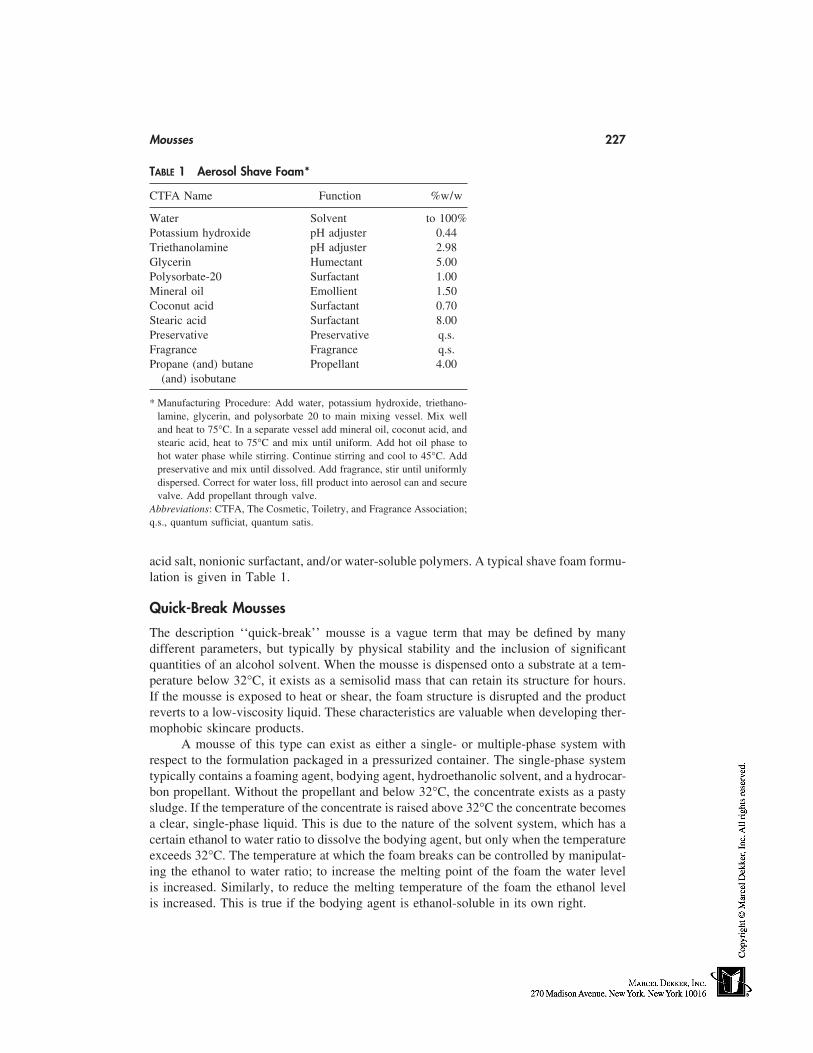
Mousses 227
TABLE 1 Aerosol Shave Foam*
CTFA Name Function %w/w
Water Solvent to 100%Potassium hydroxide pH adjuster 0.44Triethanolamine pH adjuster 2.98Glycerin Humectant 5.00Polysorbate-20 Surfactant 1.00Mineral oil Emollient 1.50Coconut acid Surfactant 0.70Stearic acid Surfactant 8.00Preservative Preservative q.s.Fragrance Fragrance q.s.Propane (and) butane Propellant 4.00
(and) isobutane
* Manufacturing Procedure: Add water, potassium hydroxide, triethano-lamine, glycerin, and polysorbate 20 to main mixing vessel. Mix welland heat to 75°C. In a separate vessel add mineral oil, coconut acid, andstearic acid, heat to 75°C and mix until uniform. Add hot oil phase tohot water phase while stirring. Continue stirring and cool to 45°C. Addpreservative and mix until dissolved. Add fragrance, stir until uniformlydispersed. Correct for water loss, fill product into aerosol can and securevalve. Add propellant through valve.
Abbreviations: CTFA, The Cosmetic, Toiletry, and Fragrance Association;q.s., quantum sufficiat, quantum satis.
acid salt, nonionic surfactant, and/or water-soluble polymers. A typical shave foam formu-lation is given in Table 1.
Quick-Break Mousses
The description ‘‘quick-break’’ mousse is a vague term that may be defined by manydifferent parameters, but typically by physical stability and the inclusion of significantquantities of an alcohol solvent. When the mousse is dispensed onto a substrate at a tem-perature below 32°C, it exists as a semisolid mass that can retain its structure for hours.If the mousse is exposed to heat or shear, the foam structure is disrupted and the productreverts to a low-viscosity liquid. These characteristics are valuable when developing ther-mophobic skincare products.
A mousse of this type can exist as either a single- or multiple-phase system withrespect to the formulation packaged in a pressurized container. The single-phase systemtypically contains a foaming agent, bodying agent, hydroethanolic solvent, and a hydrocar-bon propellant. Without the propellant and below 32°C, the concentrate exists as a pastysludge. If the temperature of the concentrate is raised above 32°C the concentrate becomesa clear, single-phase liquid. This is due to the nature of the solvent system, which has acertain ethanol to water ratio to dissolve the bodying agent, but only when the temperatureexceeds 32°C. The temperature at which the foam breaks can be controlled by manipulat-ing the ethanol to water ratio; to increase the melting point of the foam the water levelis increased. Similarly, to reduce the melting temperature of the foam the ethanol levelis increased. This is true if the bodying agent is ethanol-soluble in its own right.
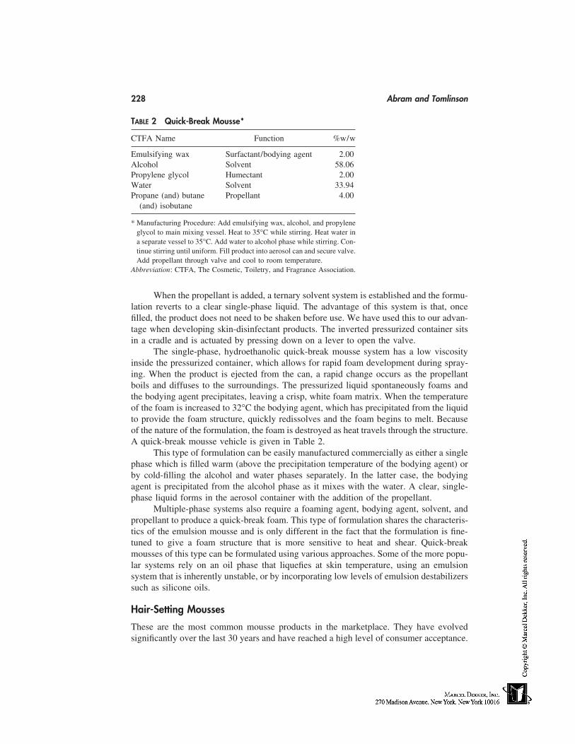
228 Abram and Tomlinson
TABLE 2 Quick-Break Mousse*
CTFA Name Function %w/w
Emulsifying wax Surfactant/bodying agent 2.00Alcohol Solvent 58.06Propylene glycol Humectant 2.00Water Solvent 33.94Propane (and) butane Propellant 4.00
(and) isobutane
* Manufacturing Procedure: Add emulsifying wax, alcohol, and propyleneglycol to main mixing vessel. Heat to 35°C while stirring. Heat water ina separate vessel to 35°C. Add water to alcohol phase while stirring. Con-tinue stirring until uniform. Fill product into aerosol can and secure valve.Add propellant through valve and cool to room temperature.
Abbreviation: CTFA, The Cosmetic, Toiletry, and Fragrance Association.
When the propellant is added, a ternary solvent system is established and the formu-lation reverts to a clear single-phase liquid. The advantage of this system is that, oncefilled, the product does not need to be shaken before use. We have used this to our advan-tage when developing skin-disinfectant products. The inverted pressurized container sitsin a cradle and is actuated by pressing down on a lever to open the valve.
The single-phase, hydroethanolic quick-break mousse system has a low viscosityinside the pressurized container, which allows for rapid foam development during spray-ing. When the product is ejected from the can, a rapid change occurs as the propellantboils and diffuses to the surroundings. The pressurized liquid spontaneously foams andthe bodying agent precipitates, leaving a crisp, white foam matrix. When the temperatureof the foam is increased to 32°C the bodying agent, which has precipitated from the liquidto provide the foam structure, quickly redissolves and the foam begins to melt. Becauseof the nature of the formulation, the foam is destroyed as heat travels through the structure.A quick-break mousse vehicle is given in Table 2.
This type of formulation can be easily manufactured commercially as either a singlephase which is filled warm (above the precipitation temperature of the bodying agent) orby cold-filling the alcohol and water phases separately. In the latter case, the bodyingagent is precipitated from the alcohol phase as it mixes with the water. A clear, single-phase liquid forms in the aerosol container with the addition of the propellant.
Multiple-phase systems also require a foaming agent, bodying agent, solvent, andpropellant to produce a quick-break foam. This type of formulation shares the characteris-tics of the emulsion mousse and is only different in the fact that the formulation is fine-tuned to give a foam structure that is more sensitive to heat and shear. Quick-breakmousses of this type can be formulated using various approaches. Some of the more popu-lar systems rely on an oil phase that liquefies at skin temperature, using an emulsionsystem that is inherently unstable, or by incorporating low levels of emulsion destabilizerssuch as silicone oils.
Hair-Setting Mousses
These are the most common mousse products in the marketplace. They have evolvedsignificantly over the last 30 years and have reached a high level of consumer acceptance.
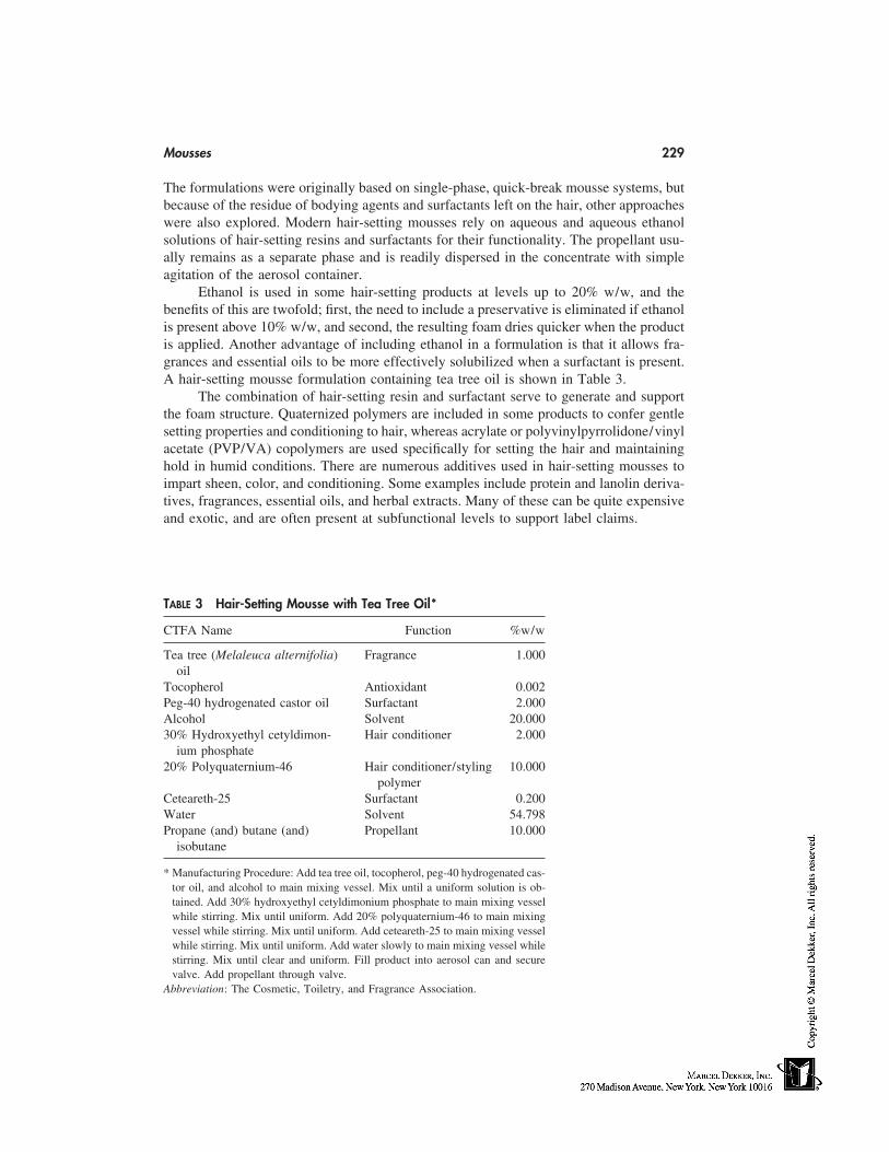
Mousses 229
The formulations were originally based on single-phase, quick-break mousse systems, butbecause of the residue of bodying agents and surfactants left on the hair, other approacheswere also explored. Modern hair-setting mousses rely on aqueous and aqueous ethanolsolutions of hair-setting resins and surfactants for their functionality. The propellant usu-ally remains as a separate phase and is readily dispersed in the concentrate with simpleagitation of the aerosol container.
Ethanol is used in some hair-setting products at levels up to 20% w/w, and thebenefits of this are twofold; first, the need to include a preservative is eliminated if ethanolis present above 10% w/w, and second, the resulting foam dries quicker when the productis applied. Another advantage of including ethanol in a formulation is that it allows fra-grances and essential oils to be more effectively solubilized when a surfactant is present.A hair-setting mousse formulation containing tea tree oil is shown in Table 3.
The combination of hair-setting resin and surfactant serve to generate and supportthe foam structure. Quaternized polymers are included in some products to confer gentlesetting properties and conditioning to hair, whereas acrylate or polyvinylpyrrolidone/vinylacetate (PVP/VA) copolymers are used specifically for setting the hair and maintaininghold in humid conditions. There are numerous additives used in hair-setting mousses toimpart sheen, color, and conditioning. Some examples include protein and lanolin deriva-tives, fragrances, essential oils, and herbal extracts. Many of these can be quite expensiveand exotic, and are often present at subfunctional levels to support label claims.
TABLE 3 Hair-Setting Mousse with Tea Tree Oil*
CTFA Name Function %w/w
Tea tree (Melaleuca alternifolia) Fragrance 1.000oil
Tocopherol Antioxidant 0.002Peg-40 hydrogenated castor oil Surfactant 2.000Alcohol Solvent 20.00030% Hydroxyethyl cetyldimon- Hair conditioner 2.000
ium phosphate20% Polyquaternium-46 Hair conditioner/styling 10.000
polymerCeteareth-25 Surfactant 0.200Water Solvent 54.798Propane (and) butane (and) Propellant 10.000
isobutane
* Manufacturing Procedure: Add tea tree oil, tocopherol, peg-40 hydrogenated cas-tor oil, and alcohol to main mixing vessel. Mix until a uniform solution is ob-tained. Add 30% hydroxyethyl cetyldimonium phosphate to main mixing vesselwhile stirring. Mix until uniform. Add 20% polyquaternium-46 to main mixingvessel while stirring. Mix until uniform. Add ceteareth-25 to main mixing vesselwhile stirring. Mix until uniform. Add water slowly to main mixing vessel whilestirring. Mix until clear and uniform. Fill product into aerosol can and securevalve. Add propellant through valve.
Abbreviation: The Cosmetic, Toiletry, and Fragrance Association.
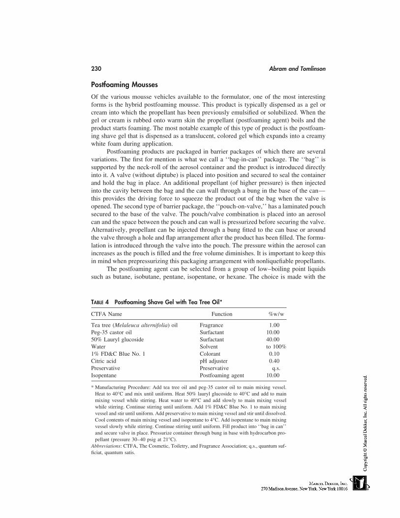
230 Abram and Tomlinson
Postfoaming Mousses
Of the various mousse vehicles available to the formulator, one of the most interestingforms is the hybrid postfoaming mousse. This product is typically dispensed as a gel orcream into which the propellant has been previously emulsified or solubilized. When thegel or cream is rubbed onto warm skin the propellant (postfoaming agent) boils and theproduct starts foaming. The most notable example of this type of product is the postfoam-ing shave gel that is dispensed as a translucent, colored gel which expands into a creamywhite foam during application.
Postfoaming products are packaged in barrier packages of which there are severalvariations. The first for mention is what we call a ‘‘bag-in-can’’ package. The ‘‘bag’’ issupported by the neck-roll of the aerosol container and the product is introduced directlyinto it. A valve (without diptube) is placed into position and secured to seal the containerand hold the bag in place. An additional propellant (of higher pressure) is then injectedinto the cavity between the bag and the can wall through a bung in the base of the can—this provides the driving force to squeeze the product out of the bag when the valve isopened. The second type of barrier package, the ‘‘pouch-on-valve,’’ has a laminated pouchsecured to the base of the valve. The pouch/valve combination is placed into an aerosolcan and the space between the pouch and can wall is pressurized before securing the valve.Alternatively, propellant can be injected through a bung fitted to the can base or aroundthe valve through a hole and flap arrangement after the product has been filled. The formu-lation is introduced through the valve into the pouch. The pressure within the aerosol canincreases as the pouch is filled and the free volume diminishes. It is important to keep thisin mind when prepressurizing this packaging arrangement with nonliquefiable propellants.
The postfoaming agent can be selected from a group of low–boiling point liquidssuch as butane, isobutane, pentane, isopentane, or hexane. The choice is made with the
TABLE 4 Postfoaming Shave Gel with Tea Tree Oil*
CTFA Name Function %w/w
Tea tree (Melaleuca alternifolia) oil Fragrance 1.00Peg-35 castor oil Surfactant 10.0050% Lauryl glucoside Surfactant 40.00Water Solvent to 100%1% FD&C Blue No. 1 Colorant 0.10Citric acid pH adjuster 0.40Preservative Preservative q.s.Isopentane Postfoaming agent 10.00
* Manufacturing Procedure: Add tea tree oil and peg-35 castor oil to main mixing vessel.Heat to 40°C and mix until uniform. Heat 50% lauryl glucoside to 40°C and add to mainmixing vessel while stirring. Heat water to 40°C and add slowly to main mixing vesselwhile stirring. Continue stirring until uniform. Add 1% FD&C Blue No. 1 to main mixingvessel and stir until uniform. Add preservative to main mixing vessel and stir until dissolved.Cool contents of main mixing vessel and isopentane to 4°C. Add isopentane to main mixingvessel slowly while stirring. Continue stirring until uniform. Fill product into ‘‘bag in can’’and secure valve in place. Pressurize container through bung in base with hydrocarbon pro-pellant (pressure 30–40 psig at 21°C).
Abbreviations: CTFA, The Cosmetic, Toiletry, and Fragrance Association; q.s., quantum suf-ficiat, quantum satis.
Mousses 231
product’s intended use and its physical characteristics in mind. For a low-viscosity orthixotropic liquid, either the pentane(s) or hexane could be used, whereas for a high-viscosity cream or gel it may be necessary to use the butane(s) to get satisfactory expansionof the foam.
Because of the density difference between the postfoaming agent and the bulk aque-ous phase, it is likely that some separation of the two phases will occur. This can becontrolled with the use of thixotropic, water-soluble polymers (such as the carbomers orxanthan gum) alone, or in combination with suitable surfactants. Although most of thepostfoaming shave products marketed today are based on neutralized fatty acids, it ispossible to formulate totally nonionic products. An example of such a product containingTea tree oil is shown in Table 4.
THE FUTURE OF MOUSSES
Although it is easy to describe the various characteristics and attributes of mousse prod-ucts, it is difficult to entirely separate this technology from that of other product forms.There are obvious overlaps between mousse technology and the technologies pertinent tosolutions, emulsions, and suspensions. The mousse product evolved from a combinationof these overlaps as well as an appropriate type of packaging being available. The propel-lant can be considered simply as a low–boiling point excipient in the formulation. Aftergrasping the ‘‘contents under pressure’’ concept, anyone competent in physical chemistrycan successfully formulate a mousse product, although there is considerable ‘‘art’’ informulating a product that is commercially successful.
The full potential of aerosol-mousse technology is only beginning to be exploited.Once only a form of presentation novelty, mousse formulations, where direct comparisonswith ‘‘conventional’’ products have been made, are now showing important, relevant dif-ferentiators. Mousse products have in some instances shown to have better efficacy andconsumer acceptance than nonaerosol formulations. Clinical studies [5,6] conducted on ascalp psoriasis-treatment mousse have shown greater clinical efficacy and patient accept-ability than comparator products. The mousse product is also more likely to be used be-cause it is effective, easy to apply, and well tolerated, thereby further increasing compli-ance and therapeutic efficacy. Furthermore, it has been shown [7] that an alcohol-basedhead-lice treatment mousse was able to exert ‘‘a high level of direct ovicidal activity,making it effective with a single application.’’ The mousse vehicle was shown to generatesynergized pediculicide droplets that were small enough to penetrate the breathing poresof the louse egg shell cap and achieve a greater louse egg mortality than a commercialrinse product.
The various mousse categories previously described are by no means absolute. Thereare many new product forms in development that are unique in their own right. Microemul-sion mousses are one such a vehicle that will offer the advantage of a single-phase systemwithout the need for high levels of volatile organic compounds. Nonaerosol mousses havea presence in the marketplace and can be described simply as aqueous surfactant solutions.Solutions become aerated as liquid passes through a vented pump (or valve) mechanismof the dispenser and a foam with a shampoo-like consistency is formed. Facial cleansingand baby-wash products are suited to this type of mousse technology because of the wetnature of the foam.
Specialized mousse products continue to be developed for cosmetic and pharmaceu-tical markets, showing a willingness by consumers to try new and effective products. We
232 Abram and Tomlinson
are currently exploring new ways of using mousse technology to deliver active compoundsto the skin for local and systemic use. Therapeutic mousse products for topical and trans-dermal administration of active compounds are already in the marketplace, and new vehi-cles are actively being developed. As more approaches to formulating mousse productsare explored, greater possibilities are being realized. Products that are cosmetically elegantand efficacious will continue to evolve as more companies explore the possibilities andopportunities of mousse technology.
REFERENCES
1. Johnson Montfort A. The Aerosol Handbook. 2nd ed., Mendham, New Jersey: Wayne DorlandCompany, 1982.
2. Balsam MS, Sagarin E, Gershon SD, Rieger MM, Strianse SJ. Cosmetics: Science and Technol-ogy. Vol. 1 and 2. 2nd ed., New York: Wiley-Interscience, 1972.
3. DeNavarre Maison G. The Chemistry and Manufacture of Cosmetics. Vol. 3 and 4. 2nd ed.Wheaton, Illinois: Allured Publishing, 1993.
4. Shaw Duncan J. Introduction to Colloid and Surface Chemistry. 4th ed. Oxford: Butterworth-Heinemann Ltd, 1992.
5. Evans Medical Limited, Regent Park, Leatherhead, U.K. Bettamousse Product Monograph,April, 1996.
6. Connetics Corporation. Press Release: Connetics Announces Positive Phase III Data For NovelFormulation of Scalp Psoriasis Treatment. August, 1997.
7. Burgess IF, Brown CM, Burgess NA. Synergised pyrethrin mousse, a new approach to headlice eradication: efficacy in field and laboratory studies. Clin Therapeutics 1994; 16(1):57–64.
20
Cosmetic Patches
Spiros A. FotinosLavipharm, Peania Attica, Greece
GENERAL
The cosmetic patch is a new ‘‘cosmetic form’’ that is the result of the natural evolutionof this technology in the pharmaceutical field. It appeared in the market just a few yearsago, and although its applications are not too many for the time being, they have beenalready established as the new weapon to fight against the natural imperfections of our skinor to prevent the adverse reaction caused by environmental or other external influences. Abroad spectrum of companies, including the major players, distribute at least one cosmetic-patch system. L’Oreal, Estee Lauder, Beiersdorf, Cheseborough-Ponds, Neutrogena, Lavi-pharm, as well as smaller manufacturers, participate in this special market.
HISTORY AND EVOLUTION
There is a close relation between topical pharmaceutical and cosmetic preparations. Thisrelationship has its origin in the ancient years. Not only the forms (creams, ointments,solutions, liposomes, microemulsions), but also technologies and their production condi-tions are very close to each other. Under this rationale, the research and development ofcosmetic patches started a few years ago. The influence of the pharmaceutical technologyis apparent in the case of the cosmetic patches not as simple cosmetic forms but as cosmeticdelivery systems. It is not the first time that such a thing has happened. Liposomes andmicroparticles, for example, had been transferred from other application fields to the phar-maceutical and later to the cosmetic technology fields with successful results. In Figures1 and 2 we can see the similarities of these two categories regarding the Conventionalforms as well as their delivery systems.
Cosmetic patches today, although at the beginning of their evolution and havingweaknesses in some cases, represent a convenient, simple, easy, safe, and effective way forcosmetic applications, using one of the most acceptable, modern, and successful deliverytechnology.
BORDERS BETWEEN PHARMACEUTICAL AND COSMETIC PATCHES
By definition, cosmetic products cannot be used or claimed for the therapy of diseases.Sometimes the companies use claims exceeding the borders between pharmaceutical and
233
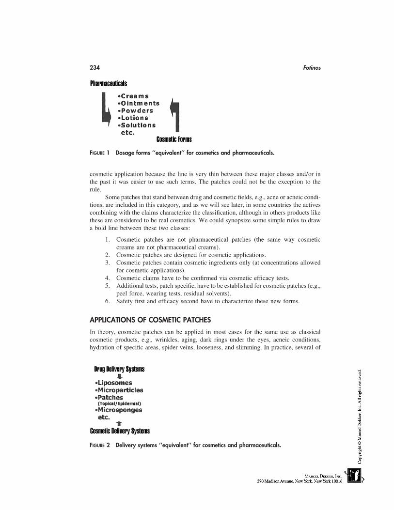
234 Fotinos
FIGURE 1 Dosage forms ‘‘equivalent’’ for cosmetics and pharmaceuticals.
cosmetic application because the line is very thin between these major classes and/or inthe past it was easier to use such terms. The patches could not be the exception to therule.
Some patches that stand between drug and cosmetic fields, e.g., acne or acneic condi-tions, are included in this category, and as we will see later, in some countries the activescombining with the claims characterize the classification, although in others products likethese are considered to be real cosmetics. We could synopsize some simple rules to drawa bold line between these two classes:
1. Cosmetic patches are not pharmaceutical patches (the same way cosmeticcreams are not pharmaceutical creams).
2. Cosmetic patches are designed for cosmetic applications.3. Cosmetic patches contain cosmetic ingredients only (at concentrations allowed
for cosmetic applications).4. Cosmetic claims have to be confirmed via cosmetic efficacy tests.5. Additional tests, patch specific, have to be established for cosmetic patches (e.g.,
peel force, wearing tests, residual solvents).6. Safety first and efficacy second have to characterize these new forms.
APPLICATIONS OF COSMETIC PATCHES
In theory, cosmetic patches can be applied in most cases for the same use as classicalcosmetic products, e.g., wrinkles, aging, dark rings under the eyes, acneic conditions,hydration of specific areas, spider veins, looseness, and slimming. In practice, several of
FIGURE 2 Delivery systems ‘‘equivalent’’ for cosmetics and pharmaceuticals.
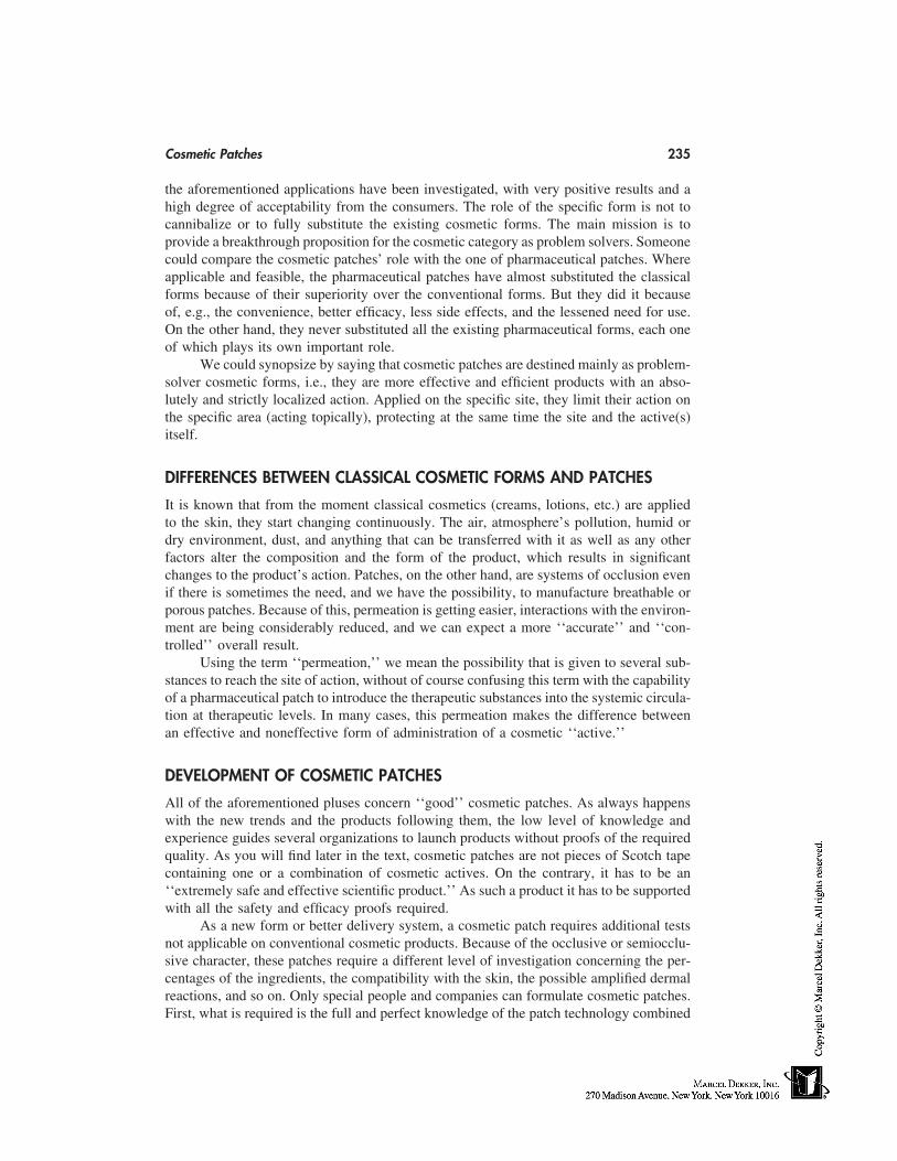
Cosmetic Patches 235
the aforementioned applications have been investigated, with very positive results and ahigh degree of acceptability from the consumers. The role of the specific form is not tocannibalize or to fully substitute the existing cosmetic forms. The main mission is toprovide a breakthrough proposition for the cosmetic category as problem solvers. Someonecould compare the cosmetic patches’ role with the one of pharmaceutical patches. Whereapplicable and feasible, the pharmaceutical patches have almost substituted the classicalforms because of their superiority over the conventional forms. But they did it becauseof, e.g., the convenience, better efficacy, less side effects, and the lessened need for use.On the other hand, they never substituted all the existing pharmaceutical forms, each oneof which plays its own important role.
We could synopsize by saying that cosmetic patches are destined mainly as problem-solver cosmetic forms, i.e., they are more effective and efficient products with an abso-lutely and strictly localized action. Applied on the specific site, they limit their action onthe specific area (acting topically), protecting at the same time the site and the active(s)itself.
DIFFERENCES BETWEEN CLASSICAL COSMETIC FORMS AND PATCHES
It is known that from the moment classical cosmetics (creams, lotions, etc.) are appliedto the skin, they start changing continuously. The air, atmosphere’s pollution, humid ordry environment, dust, and anything that can be transferred with it as well as any otherfactors alter the composition and the form of the product, which results in significantchanges to the product’s action. Patches, on the other hand, are systems of occlusion evenif there is sometimes the need, and we have the possibility, to manufacture breathable orporous patches. Because of this, permeation is getting easier, interactions with the environ-ment are being considerably reduced, and we can expect a more ‘‘accurate’’ and ‘‘con-trolled’’ overall result.
Using the term ‘‘permeation,’’ we mean the possibility that is given to several sub-stances to reach the site of action, without of course confusing this term with the capabilityof a pharmaceutical patch to introduce the therapeutic substances into the systemic circula-tion at therapeutic levels. In many cases, this permeation makes the difference betweenan effective and noneffective form of administration of a cosmetic ‘‘active.’’
DEVELOPMENT OF COSMETIC PATCHES
All of the aforementioned pluses concern ‘‘good’’ cosmetic patches. As always happenswith the new trends and the products following them, the low level of knowledge andexperience guides several organizations to launch products without proofs of the requiredquality. As you will find later in the text, cosmetic patches are not pieces of Scotch tapecontaining one or a combination of cosmetic actives. On the contrary, it has to be an‘‘extremely safe and effective scientific product.’’ As such a product it has to be supportedwith all the safety and efficacy proofs required.
As a new form or better delivery system, a cosmetic patch requires additional testsnot applicable on conventional cosmetic products. Because of the occlusive or semiocclu-sive character, these patches require a different level of investigation concerning the per-centages of the ingredients, the compatibility with the skin, the possible amplified dermalreactions, and so on. Only special people and companies can formulate cosmetic patches.First, what is required is the full and perfect knowledge of the patch technology combined
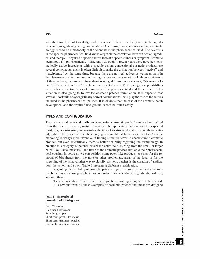
236 Fotinos
with the same level of knowledge and experience of the cosmetically acceptable ingredi-ents and synergistically acting combinations. Until now, the experience on the patch tech-nology used to be a monopoly of the scientists in the pharmaceutical field. The scientistsin the specific pharmaceutical field know very well the correlation between active ingredi-ent and therapy. They used a specific active to treat a specific illness or symptom. Cosmetictechnology is ‘‘philosophically’’ different. Although in recent years there have been cos-metically active ingredients with a specific action, conventional cosmetic products useseveral components, and it is often difficult to make the distinction between ‘‘active’’ and‘‘excipients.’’ At the same time, because there are not real actives as we mean them inthe pharmaceutical terminology or the regulations and we cannot use high concentrationsof these actives, the cosmetic formulator is obliged to use, in most cases, ‘‘its own cock-tail’’ of ‘‘cosmetic actives’’ to achieve the expected result. This is a big conceptual differ-ence between the two types of formulators; the pharmaceutical and the cosmetic. Thissituation is also going to follow the cosmetic patches formulation. It is expected thatseveral ‘‘cocktails of synergistically correct combinations’’ will play the role of the activesincluded in the pharmaceutical patches. It is obvious that the case of the cosmetic patchdevelopment and the required background cannot be found easily.
TYPES AND CONFIGURATION
There are several ways to describe and categorize a cosmetic patch. It can be characterizedfrom the patch form (e.g., matrix, reservoir), the application purpose and the expectedresult (e.g., moisturising, anti-wrinkle), the type of its structural materials (synthetic, natu-ral, hybrid), the duration of application (e.g., overnight patch, half-hour patch). Cosmeticmarketing is always more inventive in finding attractive terms to characterize a cosmeticproduct, but even scientifically there is better flexibility regarding the terminology. Inpractice this category of patches covers the entire field, starting from the small or largerpatch-like ‘‘facial masques’’ and finish to the cosmetic patches similar to their pharmaceu-tical cousins. In between, we can position some patch-like products, or strips for the re-moval of blackheads from the nose or other problematic areas of the face, or for thestretching of the skin. Another way to classify cosmetic patches is the duration of applica-tion, the action, and so on. Table 1 presents a different classification:
Regarding the flexibility of cosmetic patches, Figure 3 shows several and numerouscombinations concerning applications as problem solvers, shape, ingredients, and site,among others.
Table 2 presents a ‘‘map’’ of cosmetic patches, covering a big part of their world.It is obvious from all these examples of cosmetic patches that most are designed
TABLE 1 Examples ofCosmetic Patch Categories
Pore CleansersBlackhead removersStretching stripesShort-term patch-like masksShort-term treatment patchesOvernight treatment patches
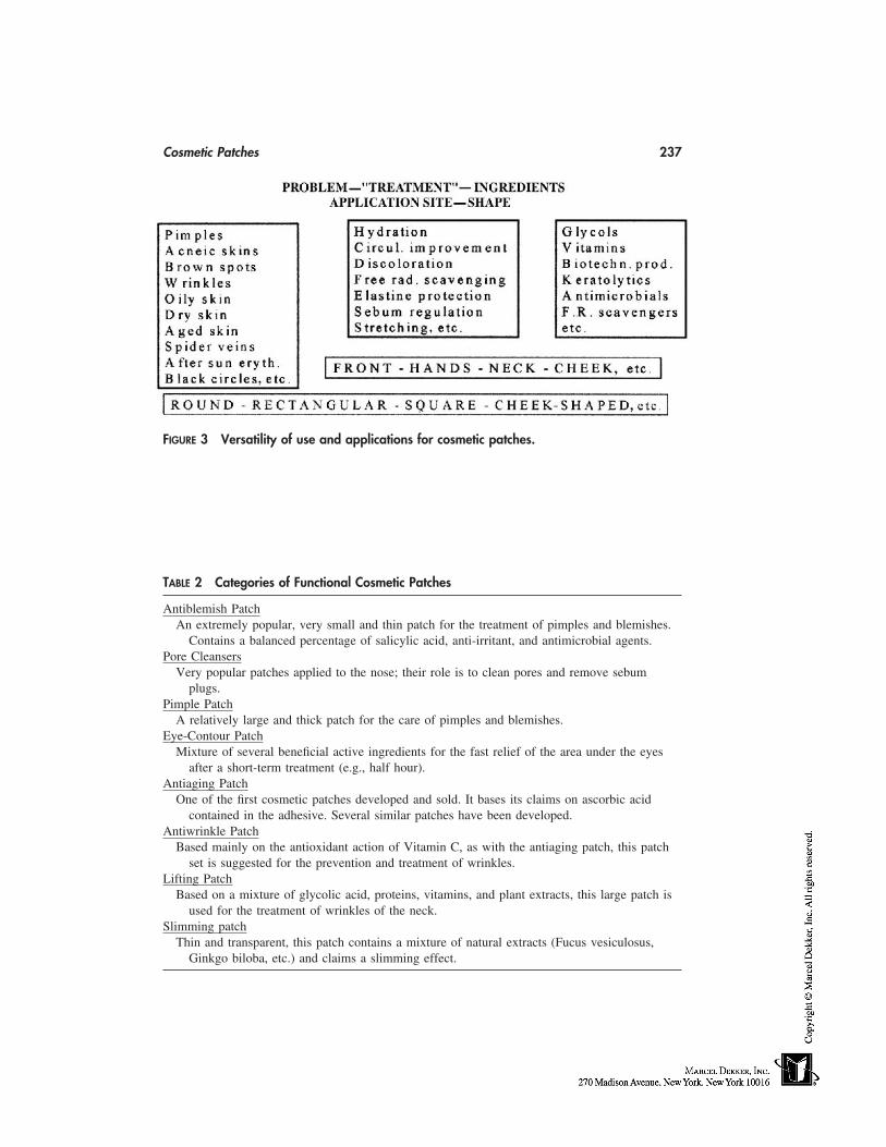
Cosmetic Patches 237
FIGURE 3 Versatility of use and applications for cosmetic patches.
TABLE 2 Categories of Functional Cosmetic Patches
Antiblemish PatchAn extremely popular, very small and thin patch for the treatment of pimples and blemishes.
Contains a balanced percentage of salicylic acid, anti-irritant, and antimicrobial agents.Pore Cleansers
Very popular patches applied to the nose; their role is to clean pores and remove sebumplugs.
Pimple PatchA relatively large and thick patch for the care of pimples and blemishes.
Eye-Contour PatchMixture of several beneficial active ingredients for the fast relief of the area under the eyes
after a short-term treatment (e.g., half hour).Antiaging Patch
One of the first cosmetic patches developed and sold. It bases its claims on ascorbic acidcontained in the adhesive. Several similar patches have been developed.
Antiwrinkle PatchBased mainly on the antioxidant action of Vitamin C, as with the antiaging patch, this patch
set is suggested for the prevention and treatment of wrinkles.Lifting Patch
Based on a mixture of glycolic acid, proteins, vitamins, and plant extracts, this large patch isused for the treatment of wrinkles of the neck.
Slimming patchThin and transparent, this patch contains a mixture of natural extracts (Fucus vesiculosus,
Ginkgo biloba, etc.) and claims a slimming effect.
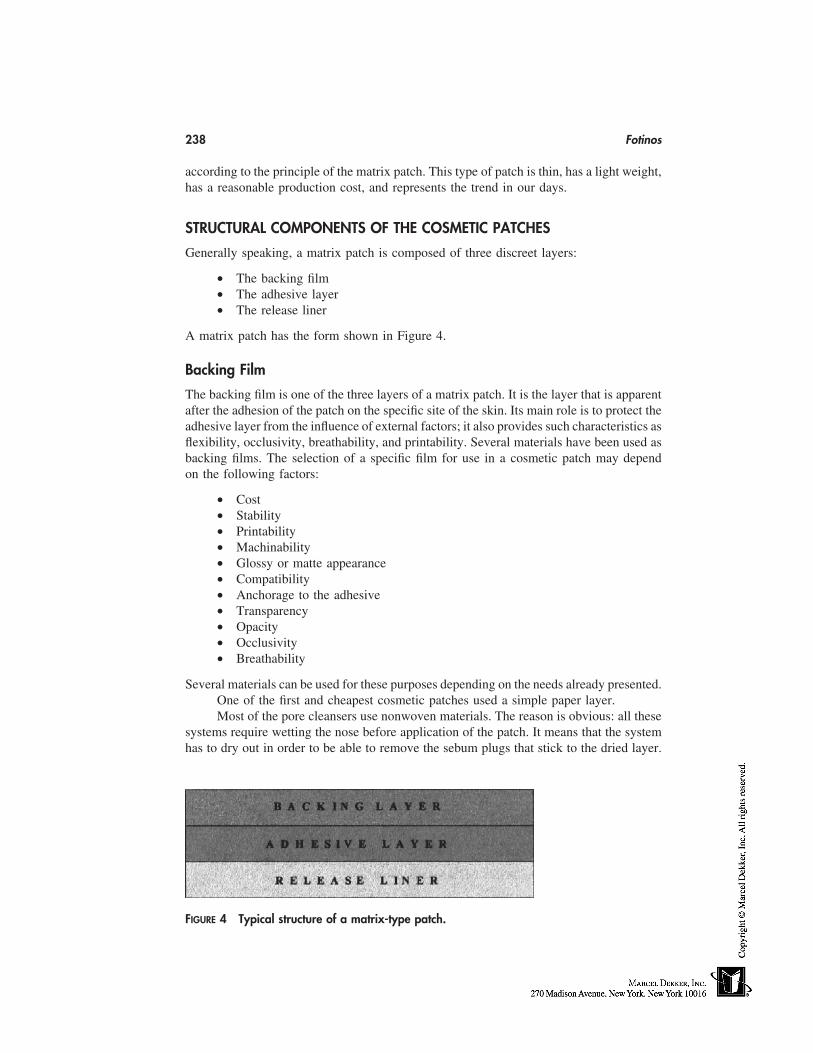
238 Fotinos
according to the principle of the matrix patch. This type of patch is thin, has a light weight,has a reasonable production cost, and represents the trend in our days.
STRUCTURAL COMPONENTS OF THE COSMETIC PATCHES
Generally speaking, a matrix patch is composed of three discreet layers:
• The backing film• The adhesive layer• The release liner
A matrix patch has the form shown in Figure 4.
Backing Film
The backing film is one of the three layers of a matrix patch. It is the layer that is apparentafter the adhesion of the patch on the specific site of the skin. Its main role is to protect theadhesive layer from the influence of external factors; it also provides such characteristics asflexibility, occlusivity, breathability, and printability. Several materials have been used asbacking films. The selection of a specific film for use in a cosmetic patch may dependon the following factors:
• Cost• Stability• Printability• Machinability• Glossy or matte appearance• Compatibility• Anchorage to the adhesive• Transparency• Opacity• Occlusivity• Breathability
Several materials can be used for these purposes depending on the needs already presented.One of the first and cheapest cosmetic patches used a simple paper layer.Most of the pore cleansers use nonwoven materials. The reason is obvious: all these
systems require wetting the nose before application of the patch. It means that the systemhas to dry out in order to be able to remove the sebum plugs that stick to the dried layer.
FIGURE 4 Typical structure of a matrix-type patch.
Cosmetic Patches 239
Polyethylene or polyester films are used also in most systems. They do not need todry out after the application. Sometimes the film used is nontransparent. A white, foamymaterial is the backing layer of the pimple patch.
In some cases, other more expensive materials have also been tested, such as poly-urethane, chlorinated polyethylenes, nylon, and saran. It is very important that the materi-als used as backing films for cosmetic patches have the same quality specifications withthe similar films used for pharmaceutical patches to avoid any adverse reactions of theskin.
Release Liners
The main role of this layer is to protect the product, especially the adhesive layer, beforethe use of the product. The pharmaceutical patch development has provided a long listof release liners that can be useful for cosmetic patches as well. There are three mainclasses of release liners according to their composition:
1. Paper based: Glassine paper, densified Kraft super-calendered paper, clay-coated paper, polyolefine coated paper, etc.
2. Plastic based: Polystyrene, polyester (plain, metallicized), polyethylene (lowand high density), cast polypropylene, polyvinyl chloride, etc.
3. Composite material based on the combination of several films
All these materials have a common characteristic: one release layer coated on one or bothsides depending on the needs of the product and the system itself. This coating is, generallyspeaking, silicon or polyfluorocarbon. The grade, thickness, coating, and curing methodsvary according to the materials and the satisfaction of specific needs.
As mentioned for backing films, this layer has to be compatible with the componentsof the adhesive layer and should satisfy the specific needs of the product. Sometimes thislayer has to be, e.g., printed, scored, perforated, or tinted. The selection of the materialand the grade are dictated from similar factors to the ones influencing the selection of thebacking layer.
Adhesive Layer
This is the most important layer of a matrix cosmetic patch. The adhesive layer containsnot only the adhesive that makes the patch stick to the skin, but in most of cases thecosmetic active ingredients and the additives required for correct formulation of a cosmeticproduct. Starting with the adhesive itself, the majority of adhesives used in cosmeticpatches are taken from the general category of pressure-sensitive adhesives (PSAs). Thisis a class of adhesives used in several applications, and in all pharmaceutical patches. Asits name shows, PSAs are adhesives which, in their solvent free form, remain permanentlytacky and stick to the skin with the application of very slight pressure. There are threegroups of PSAs: 1) acrylics, 2) silicones, and 3) rubbers. There are numerous membersin the three main families of PSAs, but only few can be used for the formulation ofcosmetic patches. The reason is that as also happens with pharmaceutical patches, thereare so many restrictions on the selection of an adhesive that the useful members are rela-tively few. The limitations are governed by the mechanical and biomedical properties ofthe adhesive, as well as the characteristics of compatibility, reactivity, and stability.
The components of the adhesive are also governed by such properties as, solvents,monomers, cross-linkers, and emulsifiers.
240 Fotinos
There is also another category of cosmetic patches with similar structure, but formu-lated with a dry-adhesive system other than PSA. In this class we can bring the exampleof pore cleansers. Here the adhesive layer is created in situ, by wetting the dry adhesivelayer with water the same way we stick a stamp on a letter. The components included inthe composition of dry adhesives can be found in the classes of synthetic or natural deriva-tives, e.g., polyvinyl derivatives, starches, celluloses, and sugars.
Pouching Materials
Although this material is not a component of cosmetic patches, its importance for theintegrity of the product during its shelf life makes us examine it just after the basic patchcomponents. Almost all cosmetic patches as happens with the pharmaceutical ones, arepouched in pouches. For pharmaceutical patches, the rule is to package one patch in onepouch. With the cosmetic analogues, and in an effort to reduce cost, sometimes patchescan be found in the same pouch for more than one application. In this case, it is recom-mended that the product has stability information for the time interval between the openingof the pouch and the use of the last patch, as well as to foresee some kind of resealablepouch. The materials used for the two categories are similar or the same. One of thedifferences is the number of packaged patches in one pouch. The protection of the productis the main mission of this packaging material, the role of which is critical for long-termstability of the product.
The pouching material, as has been mentioned, influences a lot of the stability ofsome sensitive molecules. Sometimes the phenomena of adsorption are noticed becauseof the affinity of some ingredients with the internal, sealable layer of the pouching lami-nate. In this case, e.g., AHAs can escape from the adhesive layer and, passing the edge,can be absorbed from the ionomers plastic film of the pouching material. Another protec-tion the pouching material provides is protection from UV radiation by using at least oneopaque layer in case of light-sensitive materials, along with protection from oxygen.
Production
The production of cosmetic patches depends on the type of patch, the component character-istics, and the overall configuration of the final product. Because most cosmetic patchesare matrix patches, it is useful to follow the general steps of typical production concerningthis type of patch. Practically, production starts from the weighing of raw materials andother components, and ends with packaging of the product in the final carton. It is notwithin the scope of this chapter to go into details in this field, but we can mention thebasic steps of the production sequence. Some information is required regarding the criticalsteps of production, or better the steps that could influence the quality of the product itself.The mixing of cosmetic ingredients and adhesives has to take place under a very slightnitrogen atmosphere (pressure) to avoid oxidation of the ingredients during this phase,but not too high (to avoid inclusion of nitrogen in the mass of the mixture and bubbleformation during the drying cycle). Drying is also a critical step because, during thisprocess, the temperature of the coating goes up and the ingredients have to be stable atthese conditions. During drying, some of the ingredients are evaporated and/or sublimated.An accurate validated process has to be defined to finally take the patch as it had beendesigned. The exposure to light has to be limited as well, and the web has to be protectedand kept in the predefined conditions before packaging. Of course, all the technology for
Cosmetic Patches 241
production of pharmaceutical patches is applicable, but found outside the scope of thischapter.
PRODUCTION STEPS
Production of Casting Solution
This involves the mixing of active ingredient(s), additive(s), and other adjuvants, in themass of the adhesive in the appropriate size and design production vessel and in the appro-priate space.
The bulk could be a solvent or waterborn system, and the basic steps are as follows:
• Weighing• Mixing• Deaeration• Release• Filtration and transfer to pressure vessel• Final bulk release
Coating—Drying—Lamination
The casting solution is prepared, released, coated, dried, laminated, and formed to thefinal rolls according to the specific standard operating procedures (SOPs), and the produc-tion records as follows:
• Feeding of the dosing pump, and through this the coating station• Casting on the release film• Drying of the coated solution passing through the drying tunnel• Continuous thickness control and recording• Lamination with the backing material• Winding in rolls• Splitting of the rolls• Quarantine• Final control• Release
Packaging
The process involved in packaging is described as follows:
• Roll feeding• Punching• Pouching• Cartoning• Boxing
REGULATORY ISSUES
As always happens with new forms, there is some confusion regarding the regulatorystatus of cosmetic patches. The main reason is that cosmetic patches are not included, forthe time being, in the approved forms of cosmetic preparations. Considering the Directive76/768/EEC, August 1993, which is the official regulation of cosmetic products in the
242 Fotinos
European Union, a cosmetic product ‘‘shall mean any substance or preparation intendedto be placed in contact with the various external parts of the human body (epidermis, hairsystem, nails, lips and external genital organs) or with the teeth and the mucous membranesof the oral cavity with a view exclusively or mainly to cleaning them, perfuming them,changing their appearance and/or correcting body odours or/and protecting them or keep-ing them in good condition.’’ According to this definition, cosmetic patches, acting simi-larly to conventional cosmetics, are included with cosmetic products. The confusion startsfrom paragraph 2 of the same article, stating that; ‘‘The products to be considered ascosmetic products within the meaning of this definition are listed in Annex I.’’ In AnnexI are included all the conventional forms, but not patches because, at the time of issuing,patches did not exist. So, because cosmetic patches conceptually, according to the cosmeticdefinition, comply with it, and because cosmetic patches are reality in our days, AnnexI has to be revised with the addition of this new category.
Another reason for this confusion is the common origin of patches and transdermalsystems. As previously mentioned before, all transdermals are not patches and all patchesare not transdermals.
It is true that the first patches were dedicated to transdermal delivery of actives. Atthe same time, it is true and correct that not all transdermal systems are patches and thatnot all patches are by definition transdermals. We have the case of Nitro-Bid ointmentfor the transdermal delivery of nitroglycerin, but at the same time we have ‘‘patches’’stuck to the skin for diagnostic purposes or for delivering the active to the opposite direc-tion, e.g., to the air to repel mosquitoes or for the topical treatment of pain.
To achieve transdermal delivery and effectiveness, several other factors are required:
• the intrinsic properties of the molecule,• its concentration in the system,• the appropriate permeation enhancers,• the application site,• the surface area;
and other factors play a very significant role in
• the rate and extent of absorption,• the ability of the specific active to reach the blood stream, and• its efficacy and toxicity.
Without forgetting the peculiarity of cosmetic patches as cosmetic delivery systems orforms, we could propose that this new system not be encountered with scepticism and tofollow the rules governing other cosmetic preparation. It means that the composition ofthe formula qualitatively and quantitatively has to follow existing cosmetic regulations,followed by specific tests and controls required especially for patches (e.g., residual sol-vents, adhesion on the steel, wearability), as well as tests regarding the safety parametersof an occlusive or semiocclusive system.
FUTURE TRENDS
The evolution of cosmetic patches is something expected after the warm acceptance ofnew cosmetic delivery systems from consumers. There are three axes for their expansion:
Cosmetic Patches 243
1. The technological field. It is expected that any new progress on patches, gen-erally speaking, will strongly influence cosmetic patches as well. Even nonpas-sive cosmetic patches, like the iontophoretic ones, will find in the future severalapplications for the administration of more sophisticated cosmetic ingredientsand actives.
2. The applications. For the time being, the applications of cosmetic patchescover a small part of the overall cosmetic applications. It is expected in thefuture to have a coverage of almost the whole spectrum of cosmetic applications.
3. The ingredients. The cosmetic patches, as previously explained, need to pres-ent a more potent solution for the cosmetic treatment of skin problems. For thisreason, there is the need for the use of very potent ingredients or extracts, thatare probably especially designed for the patches in order to achieve a very fastand effective action.

21
Antibacterial Agents and Preservatives
Françoise SiquetColgate-Palmolive Technology Center, Milmort, Belgium
Michel J. DevleeschouwerFree University of Brussels, Brussels, Belgium
INTRODUCTION
The term ‘‘antibacterial agent’’ is largely used to qualify chemical agents that are includedin cosmetics or household products to provide them either with a specific bactericidal orbacteriostatic activity during usage. The second function of antibacterial chemicals is toprotect the product during its life by providing a preservative efficacy against microbialinsults. A particular chemical agent can be used as an active ingredient in antibacterialproduct or as a preservative to protect the formula from microbial contamination. Takinginto account that not only bacteria but also fungi or yeast can be concerned, to cover allgerms simultaneously the word ‘‘antimicrobial’’ will be used.
Historically, the first antibacterial products developed were skinwash products suchas soap bars, derived from deodorant soap bars. The purpose was not only to clean theskin but also to reduce its microbial flora [1]. During the last 20 years, many differentantibacterial or antimicrobial products were marketed. They include toothpastes andmouthwashes, liquid antibacterial soaps, deodorants, and even antibacterial products fordishwashing.
The first part of this chapter will review the different kinds of antibacterial productsand the methods to show their efficacy.
The purpose of preservation is to protect all aspects of a product against microbialattack before and during consumer use. Integrity of products in terms of efficacy, fra-grance, appearance, and stability must be maintained. The second part of this chapter willreview the preservative systems and how to build a well-preserved formula. The test meth-ods for preservative efficacy can be found in Chapter 64 of this book.
ANTIBACTERIAL PRODUCTS
Topical Antimicrobial Products
Most antibacterial soap bars contain triclocarban (TCC) as the active ingredient. In thepast, antibacterial soap bars were also formulated with formaldehyde. These were very
245
246 Siquet and Devleeschouwer
effective for hospital use, but skin toxicity and irritation were very high. Currently, liquidsoaps are formulated with triclosan up to 1% maximum. Safety of the regular use oftriclocarban and triclosan in hand-washing products was extensively discussed by the Foodand Drug Administration (FDA) [1]. The agency prepared a tentative final monograph in1994 in which topical antimicrobial products were classified in the following categories:1) antiseptic handwash or healthcare personnel handwash, 2) patient presurgical skin prep-aration, and 3) surgical hand scrub. But this meant that products intended to be used inhomecare would have to meet the requirements of products for healthcare. In response,two industrial associations, The Cosmetics, Toiletry, and Fragrance Association (CTFA)and the Soap and Detergent Association (SDA), proposed another classification, based ona healthcare continuum model (HCCM) in which the antimicrobial products were relatedto six categories; two to be used by the general population (antimicrobial handwashes andbodywashes), three for use by healthcare professionals (presurgical preparation, surgicalscrubs, and healthcare personnel handwashes), and one category for food handlers. Sincethen, industry has submitted data to the FDA showing the efficacy of active ingredientsused in the six categories; among these ingredients are triclosan, triclocarban, chloroxy-lenol (PCMX), povidone-iodine, surfactant iodophor, alcohol, and quaternary ammoniumcompounds [2].
Extensive studies have also been carried out with essential oils as antibacterial agentsin soaps. Unfortunately, the data showed that the minimal inhibitory concentration (MIC)for antimicrobial soaps formulated with different essential oils were more than 100 timeshigher that the MIC obtained on TCC-based soaps when tested against Staphylococcusaureus [3].
Deodorants and Antiperspirants
The first antiperspirants appeared on the market at the beginning of the 20th century. Theywere based on aluminium chloride, which induced skin irritation and fabric damage be-cause of the low pH of the solutions [4]. Several years later, Shelley and colleagues showedthat underarm odor was provoked by the growth of the axillae bacterial flora which de-graded the apocrine secretions [5]. These bacteria are mainly staphylococci (S. epider-midis) and diphteroids from the Corynebacteriaceae family. Antiperspirants can preventthe growth of these degrading bacteria by reducing the available moisture of the axillariesamong other mechanisms (see Chap. 56). Some products used the hexaclorophene as anactive but its use was discontinued because of its neurotoxic properties [6]. Currently,many contain aluminium salts, or zirconium-aluminium combinations such as Al-Zi-Tri-/tetra-chlorydrex glycinate as active ingredients. Their low pH (4.0) also helps the antibac-terial activity. Antiperspirants are deodorants because they suppress the odor source byreducing perspiration and bacterial growth. Deodorants may or may not have an antimicro-bial action; either they are masking products—in this case they contain perfumes or essen-tial oils that hide the odor—or they can contain antibacterial agents which are mainlyalcohols and triclosan [6].
Oral Care Products
These are mainly toothpastes and mouthrinses. In general, dental creams serve to cleanthe teeth, to remove dental stains, and most recently to reduce and/or to prevent gingivitisand to kill the germs responsible for bad mouth odor. Mouthrinses, whether their recom-mended use is before or after brushing, are also claimed to sanitize the mouth.
Antibacterial Agents and Preservatives 247
Active ingredients used in dental cream are mainly triclosan and chlorexhidine.Other ingredients such as the natural sanguinarine extract also claim a sanitizing effecton the oral flora. The same ingredients can be used in mouthrinses, but most also containalcohol to ensure a good antiseptic effect of the product. It is interesting to observe thatfluorinated dental creams without any specific active ingredient also exhibit antimicrobialactivity [7]. This could be related to their fluoride content which, in association with thesurfactant system in the formula, release antibacterial active cationic systems.
Dishwashing Products
Among the antibacterial household products that have recently appeared on the market,antibacterial hand dishwashing liquids have become increasingly popular. Even if theseproducts are not true cosmetics, during the dishwashing, they are in direct contact withthe skin for a certain time. From a safety point of view, they can be considered as rinse-off cosmetics.
Furthermore, some products on the market have a double claim: ‘‘dishwashing liquidand antibacterial liquid soap.’’ They are classical dish liquids based on anionic and non-ionic surfactants, to which one or more antibacterial agents have been introduced. Someof these formula have been optimized to maintain their cleaning/degreasing performanceon dishes and to fight bacteria on the hands, in the washing solution, and on washingimplements. Ingredients used can be Triclosan, essential oils, or others. The use levelsare chosen to ensure a good balance between a maximum efficacy, a low skin toxicity,and keeping good cleaning performances.
Methods to Show Antimicrobial Product Efficacy
In vitro and in vivo tests can be used to show the efficacy of antimicrobial products. Onlythe in vitro tests will be considered here because they are applicable to all antibacterialproducts. A detailed review of the in vivo tests, useful for topical antibacterials, can befound in Ref. 1.
—The minimal inhibitory concentration (MIC) test principle is to determine theMIC of the test product by performing serial dilutions of the latter in growth medium andinoculating each dilution with the test strain. Products are generally tested at twofold serialdilutions. After suitable incubation, the first tube not exhibiting bacterial growth gives theMIC level, generally expressed in ppm (part per million) of product. The test can becarried out using either 2 mL of broth in tubes or 0.5 to 0.1 mL, in microtiter plates [8]or on agar plates. Control samples without any antimicrobials must be included in thetest. This test is very useful to compare activities of different products, products from thesame category (e.g., soaps) with different actives, or the active ingredients themselves.However, MIC data obtained on formulated products are very subjective and should beinterpreted carefully. Usually, test organisms are Staphylococcus aureus, Staphylococcusepidermidis, and Escherichia coli, for topical antimicrobial. Pseudomonas aeruginosa andSalmonella typhimurium are added for the dishwashing products; for specific claims inthe kitchen, Aspergillus niger and Candida albicans can be used as test strains. To testoral care products, the chosen organisms are Actynomyces viscosus, Streptococcus mutans,and Streptococcus sanguis, representatives of the oral flora [7].
—The zone inhibition test method is largely used to test the resistance of bacteriato antibiotics [9]. Antibacterial agents or products at different concentrations are applied
248 Siquet and Devleeschouwer
to a substrate, a paper disk, or directly to the surface of an agar plate previously seededwith the test bacteria. During the incubation, the test product will diffuse into the agarlayer and produce a zone of growth inhibition of the micro-organism. The larger the inhibi-tion zone, the higher the efficacy of the product. However, the data are influenced by thediffusion capacity of the product or the active into the agar; oily products will not diffuseat the same rate as aqueous-based products. It is thus very important to use negative andpositive controls. The data will be expressed in millimeters of inhibition zone around thedisk. The strains used for this test are usually the same that those used for the MIC test.These two methods give a good idea of the bacteriostatic concentrations of the testedproduct or ingredient.
The requirements from the FDA monograph of 1994 [10] are the MIC test on theactive ingredient, the vehicle, and the final formula, associated with a time-kill test method-ology to be carried out at several time points over a period of 30 minutes.
—The time-kill test determines both the killing kinetics and the activity spectrumof antibacterial formulations. This test is generally performed in suspension. The principleis to place in contact a dilution of the product or the antibacterial agent and a specifiedbacterial inoculum during a defined period of time. At the end of the contact time, theantibacterial in the mixture is inactivated by dilution into neutralizing broth. Serial dilu-tions in appropriate broth are performed and the number of survival bacteria enumeratedon solid culture media. This method can use different concentrations of test agents andbacterial inocula, and different contact times. In general, the concentrations are chosenso that the final organism/test solution concentration is representative of the use concentra-tion of the product.
In the United States, there is no detailed standardized time-kill test, even if the U.S.Food and Drug Administration (FDA) requested a standard procedure [10]. In response,the American Society For Testing and Materials (ASTM) subcommittee of antimicrobialagents has prepared a draft to standardize the organism inocula, media, neutralizers, andcontact times [11].
In Europe, the situation is different: to test the antimicrobial efficacy of productsand/or agents, standards exist since more than 20 years in France [12], Holland, Germany,and the United Kingdom. Recently, the Council of Europe has installed a Commissionfor the Normalization of European Norms [13], which is writing and publishing the Euro-pean Norms (EN) for testing disinfectants and antiseptics. The requirements for disinfec-tion are 99.99% to 99.999% of killing (4 to 5 log reductions) of the initial inoculum,depending on the test.
These norms are also used by the industry to prove the efficacy of their antibacterialproducts, but the requirements are less strict: 99 to 99.9% killing (2–3 log reduction).Detailed review of the ENs can be found in Ref. 14.
PRESERVATION AND PRESERVATIVE SYSTEMS
Concept of Active Preservation and Self-Preserving Formula
To ensure effective preservation, the method of choice is to add one or more active antimi-crobial ingredients to the product. These ingredients must be compatible with the otheringredients of the formula and must retain efficacy for an extended period of time. Theyalso have to be nontoxic for the consumer.
Antibacterial Agents and Preservatives 249
To choose an active antimicrobial molecule as preservative is not so easy; this mole-cule must have a good oil-water partition coefficient because the contaminating microbesare living in the aqueous phase of the formula. It must not be inactivated by externalfactors such as the pH and the manufacturing process [15]. Other factors also have to beconsidered; such as the packaging, which could affect the preservative activity, the adsorp-tion rate on some components of the formula, the solubility of the preservative moleculeand its volatility [15].
Furthermore, the inactivation of the micro-organisms by the preservative should besufficiently fast to prevent any adaptation or resistance to the preservative system [16].So, the ideal preservative system must be selected for each formula, taking into accountthe possible inactivating ingredients or the potentiation capacity of other ingredients.Among these, ethylenediaminetetra-acetic acid (EDTA) is well known to act in synergywith many other chemical preservatives. This potentiation is delivered through the perme-ation of the cell membrane of gram-negative bacteria. EDTA is a chelating agent anddisrupts the outer lipid layer where stability is calcium and magnesium ion dependent.As such, it increases the penetration of the other antimicrobial chemical into the bacterialcell [17,18]. In general, liquid- and emulsion-based cosmetic products are the most suscep-tible to the development of micro-organisms. Powdered products, such as talc, are alsosusceptible to contamination and need to be preserved [19].
Another way to preserve a product is to build a ‘‘self-preserved’’ formula by usingraw materials that are not supporting germ growing and optimizing their relative content.The use of humectants such as glycerin or sorbitol at a sufficient level increases the formularesistance. In a dental cream, a mixture of sorbitol and glycerine, at respective levels of10% and 12%, is often enough to protect the formula. This is linked to the decrease ofthe water activity in the formula because of the presence of these humectants [20]. Otheringredients, such as alcohols, cationic detergents, fragrance components, and lipophilicacids (lauric and myristic acids) used as emulsifiers, which have intrinsically antibacterialproperties, can contribute to the self-preservation of a cosmetic. This is also true for essen-tial oils like tea tree oil or geraniol or eucalyptol, often used as cosmetic ingredients.
Some physical factors, such as the pH and the formula water activity, can also con-tribute to build a self-preserved product. Micro-organisms are essentially living at pH ofaround 5 to 8, and any pH outside this range induces difficult life conditions for bacteria.The water activity or availability is an important factor as the water is a necessary ingredi-ent for bacterial growth. The water availability concept is detailed in Chapter 64 of thisbook.
Most Commonly Used Preservatives
Table 1 lists the most commonly used chemicals to preserve cosmetic products. Attentionmust be paid to the regulations; in Europe, the Annex VI of the Cosmetic Directive 79/768 lists the chemicals, permanently and provisionally allowed to be used as preservativesin the cosmetic products. For each of them, there is an upper concentration use limit,and for several of them, restrictions are mentioned [21]. In the United States, the use ofpreservative molecules is regulated by the FDA. The chemical preservatives are too nu-merous to be listed here; details on preservatives can be found in Ref. 22. These moleculescan be used in synergistic mixtures to improve the activity spectrum. For example, theparabens can be used with the imidazolydinil urea, the formaldehyde can be used with
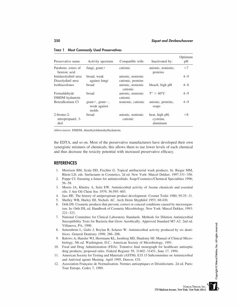
250 Siquet and Devleeschouwer
TABLE 1 Most Commonly Used Preservatives
OptimumPreservative name Activity spectrum Compatible with: Inactivated by: pH
Parabens: esters of fungi, gram� cationic anionic, nonionic, �7benzoic acid proteins
Imidazolydinil urea broad, weak anionic, nonionic 4–9Diazolydinil urea against fungi cationic, proteinsIsothiazolones broad anionic, nonionic bleach, high pH 4–8
cationicFormaldehyde broad anionic, nonionic T° � 60°C 4–9DMDM hydantoin cationicBenzalkonium Cl gram�, gram�, nonionic, cationic anionic, proteins, 4–9
weak against soapsmolds
2-bromo-2- broad anionic, nonionic heat, high pH, �6nitropropanel, 3- cationic cysteine,diol aluminum
Abbreviation: DMDM, dimethyloldimethylhydantoin.
the EDTA, and so on. Most of the preservative manufacturers have developed their ownsynergistic mixtures of chemicals; this allows them to use lower levels of each chemicaland thus decrease the toxicity potential with increased preservative efficacy.
REFERENCES
1. Morrison BM, Scala DD, Fischler G. Topical antibacterial wash products. In: Rieger MM,Rhein LD, eds. Surfactants in Cosmetics, 2d ed. New York: Marcel Dekker, 1997:331–356.
2. Poppe CJ. Ensuring a future for antimicrobials. Soap/Cosmetics/Chemical Specialities 1996;56–58.
3. Morris JA, Khettry A, Seitz EW. Antimicrobial activity of Arome chemicals and essentialoils. J Am Oil Chem Soc 1979; 56:595–603.
4. Jass HE. The history of antiperspirant product development. Cosmet Toilet 1980; 95:25–31.5. Shelley WB, Hurley HJ, Nichols AC. Arch Derm Shyphilol 1953; 68:430.6. Orth DS. Cosmetic products that prevent, correct or conceal conditions caused by microorgan-
ism. In: Orth DS, ed. Handbook of Cosmetic Microbiology. New York: Marcel Dekker, 1993:221–323.
7. National Committee for Clinical Laboratory Standards. Methods for Dilution AntimicrobialSusceptibility Tests for Bacteria that Grow Aerobically. Approved Standard M7-A2. 2nd ed.Villanova, PA, 1990.
8. Settembrini L, Gultz J, Boylan R, Scherer W. Antimicrobial activity produced by six denti-frices. General Dentistry 1998; 286–288.
9. Balows A, Hausler WJ, Herrmann KL, Isenberg HD, Shadomy HJ. Manual of Clinical Micro-biology, 5th ed. Washington, D.C.: American Society of Microbiology, 1991.
10. Food and Drug Administration (FDA). Tentative final monograph for healthcare antisepticdrug products; proposed rules. Federal Register 59, 31402–31451, June 17, 1994.
11. American Society for Testing and Materials (ASTM). E35.15 Subcommittee on Antimicrobialand Antiviral agents Meeting. April 1995, Denver, CO.
12. Association Française de Normalisation. Normes antiseptiques et Désinfectants. 2d ed. Paris:Tour Europe, Cedex 7, 1989.
Antibacterial Agents and Preservatives 251
13. European Committee for Standardization. Brussels, Belgium: CEN 216, 1998.14. Siquet F. Disinfection and preservation in detergents. In: Stubenrauch J, Broze G, eds. Hand-
book of Detergents. Vol. 1. New York: Marcel Dekker, 1999.15. McCarthy TJ. Formulated factors affecting the activity of preservatives. In: Kabara JJ, ed.
Cosmetic and Drug Preservation, Principles and Practices. New York: Marcel Dekker, 1984:359–387.
16. Orth DS, Lutes CM. Adaptation of bacteria to cosmetic preservatives. Cosmet Toilet 1985;100:57–59.
17. Kabara JJ. Food grade chemicals in a system approach to cosmetic evaluation. In: Kabara JJ,ed. Cosmetic and Drug preservation, Principles and Practices. New York: Marcel Dekker,1984: 339–356.
18. Denyer SP, Hugo WB, Harding VD. Synergy in preservative combinations. Internat J Pharm1985; 25:245–253.
19. Selleri R, Caldini O, Orzalesi G, Facchini S. La conservation du produit cosmétique. BiolChim Farm 1974; 113:617–627.
20. Orth DS, ed. Handbook of Cosmetic Microbiology. New York: Marcel Dekker, 1993:75–99.21. European cosmetic directive 76/768EEC.22. Wallhäuser KH. Antimicrobial preservatives used by the cosmetic industry. In: Kabara JJ, ed.
Cosmetic and Drug Preservation, Principles and Practices. New York: Marcel Dekker, 1984:605–745.

22
General Concepts of Skin Irritancy andAnti-irritant Products
André O. BarelFree University of Brussels, Brussels, Belgium
INTRODUCTION
In the past, some hazardous materials were used in cosmetics such as lead carbonate,bismuth, and mercurials. Serious adverse reactions to cosmetic ingredients and prepara-tions are actually infrequent. However, side effects do occur and are by no means rare.The unwanted effects of cosmetics can be classified in the following categories [1–4]:
1. Irritation and contact urticaria2. Contact allergy3. Photosensitive reaction (photoallergy and photoirritation)4. Acnegenesis and comedogenesis5. Color changes of the skin and appendages6. Systemic side effects7. Other local side effects
When considering skin-irritation symptoms, we are dealing with nonimmunological medi-ated inflammation of the skin induced by external agents. Chemical irritants are the majorcause, but mechanical, thermal, climatic, and UV and IR light are also important factorsor cofactors of irritancy [5]. This nonimmunological skin irritancy reaction comprises twoforms: the acute irritant reaction with a monofactorial cause (detergent, acid, oxidant, etc.)and the chronic multifactorial form. The symptoms of skin irritation are well known:erythema, dryness, scaling, itching, burning, and tingling. The clinical symptoms are de-scribed by some investigators as objective irritation [1–4]. Because these symptoms areclearly perceptible, in vivo testing in humans can easily and reliably detect strong andmoderate irritants for cosmetic ingredients and eliminate these potential hazards. However,most cosmetic-use ingredients do not produce acute irritation from a single exposure be-cause they are mild or very mild and consequently difficult to detect. However, they mayproduce inflammation after repeated application on the same area of the skin, which isreferred to as cumulative irritation.
Application of a cosmetic causing symptoms of burning, stinging, or itching withoutdetectable visible or microscopic changes is designated as a subjective irritation or subclin-
253
254 Barel
ical irritation [2–4]. This reaction is common in certain susceptible individuals, occurringmost frequently on the face. These persons are identified as ‘‘stingers.’’ Some of theingredients that cause this reaction are not generally considered as typical irritants, andwill not cause abnormal responses in nonsusceptible individuals. Typically about 10 to20% of the subjects exposed to a 5% aqueous lactic acid develop a stinging response whenapplied to the face. Generally, all stingers have reported a history of adverse reactions tofacial cosmetics, soaps, and similar products. Prior skin damage caused by UV sunburn,pretreatment with surfactants, and tape stripping increase the intensity of the response in‘‘stingers.’’ Attempts to identify reactive subjects by association with other skin problemssuch as atopy or with phototype or skin dryness have not been very fruitful [6].
Among the potential adverse reactions of cosmetic ingredients and products suchas irritant contact dermatitis, immediate contact reaction (urticaria), allergic contact derma-titis, and acnegenesis and comedogenesis, we will consider particularly adverse reactionsof irritancy. It is the purpose of this chapter to 1) describe shortly the different symptomsof irritancy and how to evaluate skin irritants by clinical visual and tactile assessments,by noninvasive bioengineering measurements and by self-perception of skin irritation;2) to give a short overview of the different chemical ingredients, which are potentialcosmetic and occupational skin irritants; 3) to give a description of the different in vivotests for measuring skin irritation and to test the efficiency of specific anti-irritant productsand ingredients, and 4) to give an overview of the different possibilities to conceive anti-irritant cosmetics and treatments.
IRRITANCY AND SKIN IRRITANT EVALUATION AND SYMPTOMS
Methods to evaluate skin alterations induced by topical products can be classified in threecategories [7]:
1. Clinical visual and tactile assessments2. Instrumental noninvasive bioengineering measurements3. Self-perception by the subjects themselves
Clinical Visual and Tactile Assessments
Several skin modifications induced by irritants can be easily evaluated visually and tac-tilely, e.g., by skin redness (erythema), skin dryness with increased desquamation, scali-ness, and flakiness, and skin roughness or edema. Moderate to very intense signs of skinredness/erythema are the visual manifestations of a skin inflammatory process with vasodi-latation of the capillary system and increase of the blood flow. After contact with anirritant (particularly with soaps and detergents), symptoms of skin dryness appear after acertain time with a whitish appearance, flakiness, scaliness, and roughness. In the mostsevere cases of irritation, fissuring, and cracking can also appear. Edema is the result ofan accumulation of fluid from the blood vessels in the upper dermis. It appears only in verysevere cases of irritancy, which happens very rarely unless in experimental conditions. Thevisual and tactile assessments of irritancy are made by dermatologists or trained evaluators.These observations always remain subjective in nature even with trained observers, withwell-standardized clinical and experimental protocols and with well-established scoringgrades. However, the clinical assessments are precise and very reproducible.
Skin Irritancy and Anti-irritant Products 255
Instrumental Noninvasive Bioengineering Measurements
Many changes in skin properties induced by irritant cosmetic ingredients can be evaluatedquantitatively in a noninvasive manner by instrumental techniques. In this section thefollowing techniques will be described: 1) skin redness by reflectance skin colorimetryand by Laser Doppler flowmetry; 2) alterations in the integrity of the barrier function bytransepidermal water loss; 3) skin hydration measurements using electrical impedance andskin surface alterations using squamometry; and 4) other bioengineering methods such aselasticity and microrelief.
Skin Redness/Erythema by Measuring Skin Color
Most color measurements of the skin surface are based on reflectance colorimetry instru-ments, such as tristumulus color analysis, Chromameter Minolta, erythema index,Erythemameter Diastron, Mexameter Courage-Khazaka, and Dermaspectrometer Cortex[8–10].
The Minolta chromameter CR-200, considered by many investigators as a sort ofreference instrument, quantifies skin surface color using the three-dimensional CIE colorrepresentation with the L*a*b* system. Skin redness is readily evaluated by means of thea* values; erythema is always characterized by an increase of the a* skin color parameter.Different, more simple, reflectance meters (Erythemameter Diastron, Mexameter Courage-Khazaka, and Dermaspectrometer Cortex) are also used [9,11]. These instruments arebased on the same optical principle, namely, measurements of light absorption and reflec-tion of respectively the melanin and hemoglobin components of the skin. The specificabsorption of melanin and hemoglobin in the visible (green and red) and in the near infra-red is determined and these instruments quantify redness by a relative erythema index.The erythema index is proportional to the hemoglobin content of the upper layers of thedermis.
Excellent correlations have been shown between visual clinical scoring and ery-thema and Chromameter measurements of the a* color parameter [12]. Furthermore, rea-sonably good correlations were noticed between the a* Chromameter parameter and theerythema index of the simple reflectance meters (Mexameter Courage-Khazaka and Der-maspectrometer Cortex) [9,13].
Measurement of Superficial Blood Flux by Laser Doppler Flowmetry
The hemoglobin of the red blood cells of the upper dermis microcirculation system par-tially absorbs the light of a helium laser beam. The laser Doppler method measures theshift in frequency of the reflected light of this laser beam. This small frequency shift isproportional to the number and the speed of red blood cells present in the superficial bloodmicrocirculation system. An inflammatory reaction with vasodilatation of the capillarieswill produce a marked increase in blood flow [14]. There two types of laser Dopplerinstruments: the first generation flowmeters, which measure the blood flux of a small spotarea of the skin (2–3 mm2) (Servomed, Sweden, Lisca, Sweden and Moor, United King-dom), and more recently the development of laser Doppler imaging instruments, whichhas enabled the two dimensional quantitative measurement of blood microcirculation ofa much larger skin area (maximum 10 cm2) [15]. Good correlations were found betweenclinical assessments of irritancy and noninvasive bioengineering methods, such as skincolor and laser Doppler flowmetry, respectively [16].
256 Barel
Alterations in the Integrity of the Barrier Function
When some irritant cosmetic ingredient comes in contact with the skin, the earliest modi-fications in the skin structure is an alteration of the lipidic barrier structure of the stratumcorneum [17]. The physiological function of this barrier is to protect the skin from thepenetration of irritants and to ensure low insensible perspiration of the skin [transepidermalwater loss (TEWL)]. When the barrier function of the skin is altered by an irritant, theamount of water vapor passing through the stratum corneum is increased, which is charac-terized by an increase in TEWL. The two most widely used TEWL instruments are theEvaporimeter (ServoMed, Sweden) and the Tewameter (Courage-Khazaka, Germany).Both TEWL instruments are very sensitive, and the slightest alterations of the barrierfunction can be measured with this technique (‘‘nonvisible’’ subclinical irritation). Thishappens mostly when extremely mild cosmetic ingredients are tested or when normal-useapplication protocols are considered [18].
Alterations in the Skin Surface Hydration
The assessment of the hydration status of the superficial layers of the epidermis is animportant parameter with which to characterize the skin. The hydration level of the stratumcorneum remains more or less constant, taking in consideration the following mechanisms:1) hydration coming from the deeper layers of the fully hydrated viable epidermis andretarded from evaporation in the stratum corneum by the lipids from the hydrolipidicbarrier, 2) hydration due to equilibrium with the external ambient humidity, and 3) thepresence of entrapped water bound to the natural moisturing factors (NMF) present in thelayers of the stratum corneum. When an irritant cosmetic ingredient, such as a surfactant,interacts with the skin surface, it partially or completely removes the lipidic film coatingthe surface of these and extracts some NMF components altering the equilibrium mecha-nism of the hydration of the skin surface. Such a dehydration of the horny layer willhave many different consequences, such as 1) increase of the desquamation rate of thecorneocytes giving the skin a scaly aspect, 2) a modification of the relief of the skin witha rough and wrinkled appearance, and 3) modifications in the viscoelastic properties ofthe stratum corneum. The modifications in the hydration level of the stratum corneumhave been extensively investigated using bioengineering methods based on the electricalimpedance to the skin to an alternating current [19]. Many commercial instruments mea-sure the electrical properties of the skin, such as capacitance, impedance, and conductancemethods. The measured electrical properties of the superficial layers of the epidermis(impedance units or arbitrary electrical units) are indirectly related to the amount of waterpresent in the horny layer. When used under standard conditions and in thermostatizedexperimental rooms, all the instruments are able to provide highly accurate and reproduc-ible hydration values. Excellent correlations were obtained between the visual scoring ofskin dryness induced by surfactants in a soap chamber test and instrumental readings [20].
Skin-Surface Stripping Tests
The investigation of skin-surface alterations has made great progress by the developmentand use of skin-surface stripping systems. The superficial layers of the stratum corneumcan be easily collected, and without any damage for the viable epidermis, simply by press-ing a sticky tape on the skin (D-Squames). When removing the sticky tape after a fewseconds, several layers of corneocytes are collected and can be analyzed. The level ofdesquamation can be quantified by squamometry, which is the staining of the corneocytes
Skin Irritancy and Anti-irritant Products 257
and measuring the amount of color [21]. The degree of cohesion between the corneocytescan be measured by visual scoring under the microscope and by image analysis. Withsome surfactants, no clinical irritation could be observed; however, they induce significantchanges at the surface of the stratum corneum as shown by an increase of the amount ofcorneocytes and a deorganization/loss of the intercorneocyte cohesion [21,22].
Other Noninvasive Bioengineering Methods
Other methods are available to measure some symptoms of skin irritancy, but will not bedescribed in this chapter. Skin dryness and roughness as induced by some irritants canbe evaluated by the following techniques: (1) measurement of the viscoelastic propertiesof the upper layers of the epidermis [23], and (2) skin surface microrelief [24–26].
Self-Perception of Skin Irritations
Generally when a finished cosmetic product comes into contact with the skin of potentialconsumers, it is very unlikely that observable signs of irritation are noticed in normal use.However, the overall perception of the finished product by the consumer is an importantcriterion for accepting its cosmetic use. In this global perception many different parametersmay play a role, some independant of the potential irritancy of ingredients, such as feelingof aesthetic nature, ease of spreading on the skin, viscosity, perfume, and color. However,the subjective perception of skin feel is closely related to the composition of the cosmeticproduct. Skin feel attributes, such as self-perception of dryness (feels tight, rough, anddry), or irritation (itching and burning), softness, and smoothness are easily perceived bythe subjects. In most cases, the subjects are able to perceive very early on the effects ofsome cosmetics on the skin well before they become clinically observable or measurableby bioengineering techniques. The assessment of the self-perception of the interactionbetween some cosmetic ingredients with the stratum corneum is performed by means ofquestionnaires where several skin attributes are evaluated. Some questionnaires are de-signed to receive an answer Yes or No to each of the attributes, or the subject will haveto rate each of the attributes on a 0 to 10 point scale.
FACTORS THAT INFLUENCE SKIN RESPONSIVENESS TO IRRITANTS
Many factors can influence the responsiveness of a consumer’s skin to a potential irritant.Some factors are intrinsic, inherent to the subjects themselves (e.g., sensitive skin, atopicskin), the body site, and previous traumas to the considered skin area. Other factors areexternal, such as composition of product, conditions of exposure, occupation of the subject,and climatic factors [4,5,7]. The reason why these factors are covered in this chapter areevident. Some cosmetics with anti-irritant ingredients are designed for some specific skinsites, such as the face, or considered as seasonal products, such as cosmetics against winterdryness of the skin.
Factors inherent to the constitution of the skin of the subjects that may influenceskin responsiveness are numerous. A marked interindividual variability in response toirritants have been reported and ascribed to host-related factors. Considering the interindi-vidual variability of subjects to skin irritants, one must mention here the concept of ‘‘sensi-tive skin.’’ The term sensitive skin clearly has a different meaning for consumers thanfor cosmetic scientists and dermatologists [4,6]. Consumers use the term sensitive skinto indicate that their skin readily experiences adverse reactions or unwanted changes to
258 Barel
external factors, such as the use of personal care products. Subjects with sensitive skintend to more readily develop skin reactions to cosmetics and other topical drugs than donormal persons. Many attempts have been made by cosmetic scientists and dermatologiststo describe and demonstrate in a scientific way what sensitive skin is. Visible effects, suchas erythema and skin dryness, are noticed. However, half of adverse reactions are purelysensory perceptions, subjective symptoms of stinging, itching, burning, and feelings ofdryness with or without visible effects.
Regional Differences in the Sensitivity of Normal Skin
It has been clearly demonstrated that when measuring the potential irritancy of cosmeticingredients, great regional differences in the sensitivity of normal skin are observed[27,28]. Several factors must be considered in order to explain the observed regional differ-ences in skin sensitivity, such as differences in total skin thickness, skin permeability,the amount and composition of epidermal and sebaceous lipids, blood microcirculation,hydration level of the horny layer, thickness of the horny layer, and desquamation rateand local daily exposure to irritant products. Most skin-irritation phenomena are noticedin the face.
Influence of Gender, Age, and Ethnic Group
Contradictory data are presented in the scientific literature about the influence of ethnicgroup on skin sensitivity [29]. It has been demonstrated that the irritant response may behigher in babies and children and decrease with age [30]. Concerning skin sensibility toirritants related to gender, many studies show that women are more reactive than men[31,32]. However, this difference could be attributable to the fact that women are moreexposed to household chemicals and more frequently use face care cosmetics, rather thanrelated to real physiological differences. Other factors are external to the subject, such ascomposition of their usual products, conditions of exposure, occupation of the subject,and climatic factors.
Mode of Exposure of the Product on the Skin
Acute skin exposures of a very irritant chemical cosmetic ingredient are very rare andattributable to accidents, inadequate use, or problems in the manufacturing of the cosmeticproduct. The list of very irritant products are known and must be totally avoided or usedat very low concentrations; we will be dealing mostly with subacute and chronic exposureof the skin. Subacute exposure will provoke an immediate impairment of the skin barrier.Repeated exposures to certain cosmetic products with very limited impairment of the skinbarrier can induce, after a certain time, significant cutaneous reactions.
Climatic Factors
There is clearly a seasonal or climatic effect on the amplitude of the skin irritation reaction.Generally, much higher irritation reactions are observed in winter than in summer. Thisdifference is related to a dehydration factor: a situation of dryness of the horny layerprovoked by ambient air with very low relative humidity. This situation is particularypresent on the lower legs and more frequent in older subjects; typical symptoms includewinter xerosis, extreme dryness, scaling, and rough skin surface. Furthermore, in the win-
Skin Irritancy and Anti-irritant Products 259
ter the epidermis is more aggressed by extreme temperature changes between the insideand outside world. In the summer period, the upper layers of the epidermis are well hy-drated, and the skin is smooth unless excessively exposed to sun damage. Actinic agingof the skin is characterized by various clinical symptoms, including dryness of the skin.
COSMETIC AND OCCUPATIONAL SKIN IRRITANTS
Occupational Skin Irritants
A broad definition of occupational contact irritant dermatitis is contact dermatitis causedwholly or partially by the occupation of the subject. Occupational irritants may cause anacute response that may take from 1 hour to 1 day to appear, and is usually traceable toa single factor. Chronic irritant contact dermatitis may take months or years to appear andis often multifactorial [33]. Hands are involved in 80 to 90% of all cases of occupationalcontact dermatitis, and in the minority of cases the wrist, forearm, lower leg, or face isthe primary site.
The clinical features are described as follows. Many cases of occupational irritantcontact dermatitis start as erythema and scaling on the back of joints and adjacent partsof the back of the fingers, as well as in the web spaces between the fingers. A generalized,rather shiny, superficially fissured, scaly fingertip dermatitis is also characteristic of certainforms of irritancy. Exclusive or more severe involvement of the thumb, index finger, and/or middle finger of the dominant hand (or of the nails) is generally an indication of possibleoccupational causation [33]. The principal occupational irritants are listed in Table 1.
Cosmetic Skin Irritants
Cosmetics are complex mixtures of chemical compounds. The abundance of commerciallyavailable ingredients has created endless variety in cosmetic formulation. The cosmeticsubstances used in cosmetic products may be arbitrarily divided in great categories ofproduct and/or function. The principal categories of cosmetic irritants are listed in Table 2.
Intolerance to some ingredients is related to symptoms of contact dermatitis andallergic dermatitis. There is not always a clear distinction between these problems. Somecosmetic ingredients present both an irritant character with the additional possibility ofallergic reaction (e.g., cinnamic acid derivates). An overview of cosmetic categories caus-ing irritant side effects in descending importance has been given by A. C. de Groot andcoworkers [1–3] and are summarized briefly in Table 3. It has clearly been shown thatcertain categories of cosmetics, taking into account their composition, frequency of use,mode of application on the skin, and skin area to be treated, are more specific candidatesfor causing symptoms of skin irritation.
A short overview will be given of the potential irritant character of each categoryof cosmetic ingredients. Some chemicals are used in industry (occupational irritants) aswell as in the cosmetic world (cosmetic irritants). Chapter 37 describes the irritancy ofthe most frequent emulgators and detergents used primarily in cleansing products.
Preservatives/antimicrobials, antioxidants, fragrances, colors, and UV filters are po-tentially irritant components. However, these components are often present in cosmeticpreparations at low concentrations and are consequently not affecting the overall irritationpotential of the final product. These substances are more often incriminated for their aller-gic reactions.
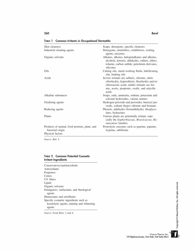
260 Barel
TABLE 1 Common Irritants in Occupational Dermatitis
Skin cleansers Soaps, detergents, specific cleansersIndustrial cleaning agents Detergents, emulsifiers, solubilizers, wetting
agents, enzymesOrganic solvents Alkanes, alkenes, halogenalkanes and alkenes,
alcohols, ketones, aldehydes, esthers, ethers,toluene, carbon sulfide, petroleum derivates,silicones
Oils Cutting oils, metal working fluids, lubrificatingoils, braking oils
Acids Severe irritants are sulfuric, chromic, nitricchlorhydric, hyperchloric, fluorhydric and tri-chloroacetic acids; milder irritants are for-mic, acetic, propionic, oxalic, and salycilicacids
Alkaline substances Soaps, soda, ammonia, sodium, potassium andcalcium hydroxides, various amines
Oxidizing agents Hydrogen peroxide and peroxides, benzoyl per-oxide, sodium (hypo) chlorate and bromate
Reducing agents Phenols, aldehydes (formaldehyde), thioglyco-lates, hydrazines
Plants Various plants are potentially irritant, espe-cially the Euphorbiaceae, Brassicaceae, Ra-nunculeae families
Products of animal, food proteins, plant, and Proteolytic enzymes such as pepsine, papaine,bacterial origin trypsine, subtilisine
Physical factors
Source: Ref. 5.
TABLE 2 Common Potential CosmeticIrritant Ingredients
Conservatives/antimicrobialsAntioxidantsFragranceColorsUV filtersLipidsOrganic solventsEmulgators, surfactants, and rheological
agentsHumectants and emollientsSpecific cosmetic ingredients such as
keratolytic agents, tanning and whiteningagents
Source: From Refs. 2 and 4.
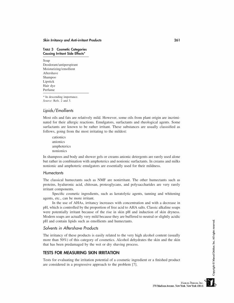
Skin Irritancy and Anti-irritant Products 261
TABLE 3 Cosmetic CategoriesCausing Irritant Side Effects*
SoapDeodorant/antiperspirantMoisturizing/emollientAftershaveShampooLipstickHair dyePerfume
* In descending importance.Source: Refs. 2 and 3.
Lipids/Emollients
Most oils and fats are relatively mild. However, some oils from plant origin are incrimi-nated for their allergic reactions. Emulgators, surfactants and rheological agents. Somesurfactants are known to be rather irritant. These substances are usually classsified asfollows, going from the most irritating to the mildest:
cationicsanionicsamphotericsnonionics
In shampoos and body and shower gels or creams anionic detergents are rarely used alonebut rather in combination with amphoterics and nonionic surfactants. In creams and milksnonionic and amphoteric emulgators are essentially used for their mildness.
Humectants
The classical humectants such as NMF are nonirritant. The other humectants such asproteins, hyaluronic acid, chitosan, proteoglycans, and polysaccharides are very rarelyirritant components.
Specific cosmetic ingredients, such as keratolytic agents, tanning and whiteningagents, etc., can be more irritant.
In the use of AHAs, irritancy increases with concentration and with a decrease inpH, which is controlled by the proportion of free acid to AHA salts. Classic alkaline soapswere potentially irritant because of the rise in skin pH and induction of skin dryness.Modern soaps are actually very mild because they are buffered to neutral or slightly acidicpH and contain lipids such as emollients and humectants.
Solvents in Aftershave Products
The irritancy of these products is easily related to the very high alcohol content (usuallymore than 50%) of this category of cosmetics. Alcohol dehydrates the skin and the skinthat has been predamaged by the wet or dry shaving process.
TESTS FOR MEASURING SKIN IRRITATION
Tests for evaluating the irritation potential of a cosmetic ingredient or a finished productare considered in a progressive approach to the problem [7].
262 Barel
First, a minimum of toxicological information must be obtained from the generalavailable scientific literature and from information derived from in vitro testing and testingon animals. Starting with the premises that the considered ingredient or product is nottoxic or very irritant, testing on humans will be envisaged. A short overview of the differ-ent published test method will be given in this chapter. Supplementary information con-cerning the test methods can be found in Chapter 12 and in a recent review article byPaye [7].
Open Epicutaneous Applications
In a second phase of testing, single and eventual repetitive open application tests arenormally used for studying new chemicals with a safety purpose in order to determine ifthis ingredient is likely to cause serious skin irritation [34].
Occlusive Patch Testing
If the product is not irritant in such open epicutaneous applications, it can be consideredto use occlusive patch tests in a further phase. The objective of the clinical study is tocompare the mildness or irritation potential of a certain cosmetic ingredient with othersimilar products. For this purpose some level of cutaneous irritation has to be induced.Generally we are dealing with very mild cosmetic products and it is necessary to includein the comparative testing some more irritating products as a positive reference. By usingocclusive conditions one induces a better percutaneous diffusion of the test solutionthrough the horny layer. Occlusion increases the hydration of this layer (increase in percu-taneous penetration) and slight increase of skin temperature under the occlusive dressing.
Many variants of occlusive patch tests have been described in the literature [7],some of the most used tests are:
• The single 24-hour occlusive test [35,36]• Successive occlusive applications, such as the Frosch-Kligman soap chamber
test [37], the modified soap chamber test [18]• The 21-day cumulative irritation test [37]• The 4-hour occlusive test [38]
Skin irritation is evaluated clinically (visual and tactile) for erythema, dryness, scaling,roughness, and edema, and/or by bioengineering methods.
The Exaggerated Use Tests
The occlusive patch tests were developed as a rapid screening test for evaluating therelative irritation potential of cosmetic products and ingredients. However, these condi-tions do not simulate the normal usage of the test materials, and other test procedureswere developed to be closer to realistic use conditions of the product by the consumer[39]. These exaggerated use tests combine the application of the product to its normalway but still in an exaggerated way: the number of applications per day and the totalduration and temperature of application is exaggerated in order to induce more skin irrita-tion reactions than expected in normal use. Several protocols have been published, dif-fering in terms of sort of application, number of applications, skin sites, and so on [7].Most of these exaggerated in-use tests are concerned with soaps and detergents, but can,
Skin Irritancy and Anti-irritant Products 263
with the necessary experimental adaptations, be used for other cosmetic preparations, suchas the following:
• The forearm wash test [39]• The flex wash test [40]• The hand/forearm immersion test [41]
One advantage of these testing methods is the fact that they are carried out on a relativelysmall number of subjects [12–25].
Home-Use Testing
Even if the exaggerated in-use tests predict with good confidence the skin tolerance of acertain ingredient or product, it is necessary and safer for the manufacturer to run anextended study with a large number of subjects using the product in normal way and intheir usual environment, so it is called a ‘‘home-use test’’ or ‘‘in-use test.’’
The panel will be selected among the population of potential users, e.g., for thetarget group and for the type of treatment. The duration of the testing is generally for amuch longer period (weeks and sometimes months). Any unwanted effects of the producton the skin are recorded, such as visible signs of intolerance (redness, dryness, roughness,)as well as nonvisible perceptions such as itching, burning and tightness. Evaluations ofthese signs are made very regularly (most cases daily) by the subjects themselves and oncea week by an expert evaluator. Usually clinical ratings by visual and tactile assessmentare made using numerical grades. They can be completed by instrumental noninvasivebioengineering measurements.
STRATEGY OF MAKING ANTI-IRRITANT COSMETICS
Strictly by definition, an anti-irritant is an agent which, by its presence, minimizes theirritating effect of a cosmetic preparation on the skin. The anti-irritant could reflect allmechanisms that have an opposed effect to an irritant insult. Hence, the term could reflectactions such as skin calming, soothing, and healing, and assisting in the recovery of theskin from an irritation provoked by, e.g., contact with soaps and household cleaning prod-ucts. As has been demonstrated earlier, very often irritant reactions are associated withinflammation, the so-called anti-irritant effect could eventually also mean alleviation fromthe inflammatory symptoms that arise shortly after the impairment of the skin barrier. Theconcept of anti-irritant activity also includes skin protection with barrier creams, whichdecrease irritant potential of some harmful substances encountered in occupational derma-titis [33]. Despite the numerous claims of skincare products for anti-irritant or protectiveactivity, some lack of scientific data is present to substantiate these claims. There is alsoa lack of suitable standardized clinical protocols to quantify these anti-irritant properties.
The basic principle of development of general anti-irritant cosmetics or cosmeticsfor sensitive skin is to avoid as much as possible any risk of irritation [4, 42]. The safestway is to use well-tolerated, chemical compounds for the vehicle and active ingredientswithout history of ‘‘skin problems.’’ Allergic reactions and skin irritancy are generallyprovoked by known specific ingredients, mostly fragrances, colors, and preservatives. Theeasy task is to remove fragrances and coloring agents; hypoallergenic cosmetics minimize
264 Barel
the use of or do not contain these ingredients. Actually, a modern trend in cosmetics isto develop specific cosmetics without preservatives. This challenge can be partially an-swered in cosmetic preparations with none or low water content: oils, fats, water/oil emul-sions, and lipogels using some synthetic lipids and/or essential oils with bactericidal prop-erties as preservatives. With aqueous solutions, hydrogels, and oil/water emulsions, thisgoal is very difficult to achieve and presently not realized; consequently, these types ofcosmetics still contain preservatives.
In order to elaborate an anti-irritant cosmetic preparation or a cosmetic preparationfor sensitive skin, we have a choice from the following possibilities:
1. The vehicle must respect the natural, slightly acidic pH of the skin (pH around5.3) or be neutral, avoiding alkaline preparations.
2. Strengthen or restore the hydrolipidic barrier function of the skin. As describedearlier in this chapter, irritancy reactions are often accompanied by modifica-tions of the structure of the intercellular lipids and water binding capacity re-sulting in an increase of TEWL and consequently higher penetration rate ofirritants. Therefore, anti-irritant preparations should restore the disturbed barrierfunction by providing the appropriate lipids to the lipidic film. Modern skincare products contain endogeneous components of epidermal lipids such as cera-mides and gamma linoleic acid. In a general way, lipids are emollients withsoothing capacities.
3. Soothing effect by filmogen compounds. The skin surface is anionic in charac-ter. Quaternized derivatives of plant proteins or emollients that are positivelycharged will smooth the skin surface by a filmogen effect.
4. Irritated skin is very often partially dehydrated skin. In order to alleviate thesymptoms of dehydration, water is brought back to the horny layer by humec-tants (NMF) or by occlusive effect of water/oil emulsions, lipogels, or siliconeoils.
5. Use of very mild surfactants and emulgators in cosmetic preparations. Generaluse of amphoteric and nonionic emulgators in creams/milks and cleansing prod-ucts. In the preparation of shampoos and shower gels, use of anionic emulsifierswith an adequate carbon chain length and sufficient degree of ethoxylation inorder to reduce irritancy. Another possibility is to use an adequate mixture ofseveral surfactants. A strong antagonism effect occurs when combining the po-tential irritant anionic surfactants with amphoterics, nonionic, or even other an-ionic surfactants with resultant decreased skin irritation [7].
6. Use of specific anti-irritant ingredients. There are a lot of soothing ingredients indermatological treatments mainly from plant origin, such as hamamelis, algae,chamomile, and aloe vera. Polysaccharides, proteoglycans, and glycoproteinswith filmogen and hydrating properties can provide a feel of less or nonirritatedskin. Polymers, when used at high concentrations, have also been demonstratedas reducing the irritation potential of anionic surfactants, essentially by en-trapping high quantities of surfactants into micelles in solution (see Chap. 23).
7. Sun exposure without UV filters can induce or increase irritant reactions of theskin and accelerate actinic aging. The cosmetic industry has developed suncareproducts with very high sun protection factors that are waterproof and withreasonably good cosmetic acceptance. There are sun protection products with
Skin Irritancy and Anti-irritant Products 265
active UV filters with the lowest allergenic potential, especially developed forsensitive skin with a minimum amount of emulgators and are fragrance free.
IN VIVO STUDIES OF THE ANTI-IRRITATION PROPERTIES OF SOMECOSMETIC INGREDIENTS
In vivo evaluation of the anti-irritant and/or anti-inflammatory effect of dermatocosmeticformulations on human skin is usually based on the quantification of the inhibition pre-sented by these products against an artificially induced contact dermatitis [42]. The modelirritant for this purpose can be selected out of a wide range of skin-aggravating factors.Irritation of the skin can be provoked after topical application of Peru balsam [43], solu-tions of anionic surfactants [44,45], nicotinates [46,47], after exposure to UV-B radiation[48,49], skin abrasion [50], or tape stripping [51,52]. There is clearly a difficulty in identi-fying the conditions under which these various irritants can be used for inducing a ‘‘suit-able’’ irritation. The induced irritation should be great enough to be measurable with goodreproducibility and to allow quantification of its inhibition by the tested products. Theanionic surfactant sodium lauryl sulphate (SLS) has lately become the model irritant ofchoice, used widely for inducing experimental contact dermatitis in anti-irritation proto-cols [45,53–55] or as a reference irritant in safety tests ranking the skin irritation potentialof soaps and detergents [56–58]. The irritant character of SLS is attributable to the follow-ing factors:
1. Modification of the protein and lipid structure of the stratum corneum. Impair-ment of the highly ordered bilayers and changes in the fluidity of the lipids[59]. Swelling of the horny layer occurs because of protein denaturation andexposure of new water-binding sites of the keratins [54].
2. Alterations in skin permeability [60]. This surfactant is often used as a pretreat-ment in order to enhance the penetration of topically applied products [45].
3. SLS causes a vascular inflammatory response [61–62]. SLS is not a sensitizeror carcinogenic agent; it causes no systemic toxicity or permanent cosmeticinconvenience to the skin [45]. The great sensitivity of TEWL parameter inquantifying the impairment of the barrier caused by SLS [63] and the propertyas a primary irritant have led to the large use of this surfactant in studies ofexperimental irritant contact dermatitis. However, as for other irritants, the in-duced cutaneous irritation is not completely reproducible. A marked interindi-vidual variability in response has been reported for this irritant and is ascribed toseveral host-related factors [42, 45, 64]. Furthermore, intraindividual variabilitywithin anatomical regions of skin site have been reported [65]. In the experimen-tal study of the anti-irritant properties of a cosmetic ingredient, three differenttypes of clinical protocol are generally used: postirritation treatment protocols,pretreatment protocols, and treatment with the combined introduction of theanti-irritant into the irritant product.
In the postirritation treatment protocol, the considered skin regions are irritated by treat-ment with SLS during a certain time and with a certain frequency. After the SLS irritationchallenge the skin areas are treated with the anti-irritant ingredient or finished productduring a certain time and frequency. One irritated area remains untreated and serves as
266 Barel
a control and the irritated areas are respectively treated with the vehicle alone and with thevehicle containing the active anti-irritant ingredient. This last site should heal significantlyquicker than the vehicle-treated site. In the pretreatment protocol, the considered skinareas are pretreated during a certain time and frequency with either the vehicle alone orthe vehicle with the anti-irritant component. A nonpretreated skin area serves as a control.Following this pretreatment the different skin areas are irritated with a SLS solution.
The typical clinical signs of skin irritancy (redness and dryness) are visually assessedby trained evaluators. Furthermore, redness is quantified by skin color (reflectance color-imetry) and microcirculation of the bloof flux by Laser Doppler flowmetry. Alterationsin the barrier function are measured by TEWL and hydration is measured by electricalimpedance of the skin. In order to obtain a significant measurable irritancy, the SLS chal-lenge is carried under occlusive dressing. It can also be treated by repetitive open applica-tions with the SLS solution. Different anti-irritant experimental protocols are describedin the scientific literature [42].
As found in the literature, these studies are often concerned with the anti-irritantproperties of plant extracts. Here follows a short overview of the anti-inflammatory/anti-irritant studies described in the literature:
• Anti-inflammatory properties of the active ingredients α-bisabolol and azuleneof chamomile oil [66–69]
• Anti-inflammatory and healing effect of a cream containing glycolic extract ofsix plants (calendula, Roman and German chamomile, linden, cornflower, andmillepertuis) [70]
• Anti-inflammatory effect of the active ingredient namely esculoside extractedfrom horse chestnut [71]
• Anti-inflammatory properties of the active ingredient, namely ursolic acid ex-tracted from rosemary [72]
• Anti-irritant properties of a preparation containing licorice and chamomileagainst a wide range of daily life skin irritations (aftershave, depilation, solarerythema, and insect stings) [73]
All these studies differ with respect to the irritation challenge and with respect to the anti-irritant treatment. In both type of protocols, namely postirritation treatment and pretreat-ment with the anti-irritant cosmetic ingredients, significant anti-irritant effects were ob-served between the treated skin sites and the untreated skin sites used as a reference. Withmore discriminative protocols (double-blind vehicle-controlled), where the anti-irritancyefficiency of an anti-irritant ingredient solubilized or dispersed in suitable vehicles (water/oil or oil/water) is compared with the efficiency of the vehicle alone, one generally expectsthat the specific effect of the anti-irritant alone will be very small and not very oftensignificantly different from that of the vehicle alone. To illustrate this statement, we referto recent work on plant anti-irritants [42]. Manou [42] has studied, in a double-blindvehicle-controlled way, the potential anti-irritant properties of essential oils and glycolicextracts obtained from different plants such as chamomile, sage, clary sage, peppermint,and hyssop. The essential oils were solubilized at a concentration of 3 to 5% in oil/waterand water/oil vehicles. The anti-irritant properties were examined according to the postirri-tation treatment protocols and pretreatment protocols using visual clinical assessments ofredness and dryness and bioengineering methods (skin color, laser Doppler flowmetry,TEWL, and hydration). The results do not support the existence of a significant anti-irritant effect of the essential oils tested under these very strict conditions. In general, the
Skin Irritancy and Anti-irritant Products 267
treated skin was found to have benefited from the treatment with the vehicle with orwithout the essential oils, compared with the irritated but untreated skin. These resultscould be explained taking in account the following points. First, the concentration rangeof the active anti-irritant ingredients used in these experiments is rather low (3–5%), andare concentrations that can be found in commercial cosmetic preparations. Probably athigher concentrations (5–10%) a significant specific anti-irritant effect will be observed,but because of the problems of high cost of these plant extracts and the possibility ofincreasing the risk for allergic contact dermatitis, these higher concentrations are rarelyused in commercial cosmetic preparations. Secondly, there is always a significant anti-irritant, anti-inflammatory effect on the skin of the lipids and emollients present in thevehicle.
REFERENCES
1. Cosmetics: introduction. In: De Groot AC, Weyland JW, Nater JP, eds. Unwanted Effects ofCosmetics and Drugs Used in Dermatology. Amsterdam: Elsevier, 1994:422.
2. The spectrum of side effects of cosmetics. In: De Groot AC, Weyland JW, Nater JP, eds. Un-wanted Effects of Cosmetics and Drugs Used in Dermatology. Amsterdam: Elsevier, 1994:437.
3. The frequency of adverse reactions to cosmetics and the products involved. In: De Groot AC,Weyland JW, Nater JP, eds. Unwanted Effects of Cosmetics and Drugs Used in Dermatology.Amsterdam: Elsevier, 1994:442.
4. Simion FA, Rau AH. Sensitive skin: what it is and how to formulate for it. Cosmet Toilet1994; 109:43.
5. Frosch PJ. Cutaneous irritation. In: Rycroft RJG, Menné T, Frosch PJ, eds. Textbook of Con-tact Dermatitis. Berlin:Springer-Verlag, 1995:28.
6. Amin S, Engasser PG, Maibach HI. Adverse cosmetic reactions. In: Textbook of CosmeticDermatology, Second Edition. Baran R, Maibach HI, eds. London, United Kingdom: MartinDunitz, 1998:709.
7. Paye M. Models for studying surfactant interactions with the skin. In: Broze G, ed. Handbookof Detergent Properties. Part A: Properties. Surf Sci Series, vol. 82. New York: Marcel Dekker,1999:469–509.
8. Bjerring P. Spectrophotometric characterization of skin pigments and skin color. In: Serup J,Jemec CBE, eds. Handbook of Non-Invasive Methods and the Skin. Boca Raton: CRC Press,1995:373–376.
9. Takiwaki H, Serup, J. Measurement of erythema and melanin indices. In: Serup J, Jemec CBE,eds. Handbook of Non-Invasive Methods and the Skin. Boca Raton: CRC Press, 1995:373.
10. Westerhof W. CIE Colorimeter. In: Serup J, Jemec CBE, eds. Handbook of Non-InvasiveMethods and the Skin. Boca Raton: CRC Press, 1995:385.
11. Diffey BL, Oliver RJ, Farr PM. A portable instrument for quantifying erythema induced byultraviolet radiation. Br J Dermatol 1984; 111:663.
12. Babulak SE, Rhein LD, Scala DD, Simion FA, Grove GG. Quantification of erythema in asoap chamber test using the Minolta Chroma (reflectance) Meter: comparison of instrumentalresults with visual assessments. J Cosmet Chem 1986; 37:475.
13. Clarys P. Alewaeters K, Barel AO. Comparative study of skin colour using different bioengin-eering methods. Abstract, 6th Congress of the International Society for Skin Imaging, London,United Kingdom, 1999.
14. Oberg PA, Tenland T, Nilsson GE. Laser Doppler flowmetry: a non invasive and continuousmethod for blood flow evaluation in microvascular studies. Acta Med Scand Suppl 1984; 687:17.
15. Wärdell K, Nilsson G. Laser Doppler imaging of skin. In: Serup J, Jemec CBE, eds. Handbookof Non-Invasive Methods and the Skin. Boca Raton: CRC Press, 1995:421.
268 Barel
16. Anderson PH, Abrams K, Bjerring P, Maibach H. A time correlation study of ultraviolet B-induced erythema measured by reflectance spectroscopy and Laser Doppler flowmetry. Photo-dermatol Photoimmunol Photomed 1991: 8:123.
17. Imokawa G. In vitro and in vivo models. In: Elsner P, Maibach HI, eds. Bioengineering ofthe Skin: Water and the Stratum Corneum. Boca Raton: CRC Press, 1994:23.
18. Simion FA, Rhein LD, Grove GG, Wojtkowski JM, Cagan RH, Scala DS. Sequential orderof skin responses to surfactants during a soap chamber test. Contact Dermatitis 1991; 27:174.
19. Barel AO, Clarys P, Gabard B. In vivo evaluation of the hydration state of the skin. In: ElsnerP, Merck HF, Maibach HI, eds. Cosmetics Controlled Efficacy Studies and Regulation. Berlin:Springer, 1999:57.
20. Paye M, Van de Gaer D, Morrison Jr BM. Corneometry measurements to evaluate skin drynessin the modified soap chamber test. Skin Res Technol 1995;1:123.
21. Piérard GE, Piérard-Franchimont C, Saint Leger D, Kligman AM. Squamometry: the assess-ment of xerosis by colorimetry of D-Squame adhesive discs. J Cosmet Chem 1992; 47:297.
22. Paye M, Goffin V, Cartiaux Y, Morrison Jr BM, Piérard GE. D-Squame strippings in theassessment of intercorneocyte cohesion. Allergologie 1995; 18:462.
23. Barel AO, Lambrecht R, Clarys P. Mechanical function of the skin: state of the art. In: ElsnerP, Barel AO, Berardesca E, Gabard B, Serup J, eds. Skin Bioengineering: Techniques andApplications in Dermatology and Cosmetology. Basel: Karger 1998:69.
24. Gasmüller J, Keckes A, Jahn P. Stylus method for skin surface contour measurements. In:Serup J, Jemec CBE, eds. Handbook of Non-Invasive Methods and the Skin. Boca Raton:CRC Press, 1995:83.
25. Corcuff P, Lévêque JL. Skin surface replica image analysis of furrows and wrinkles. In: SerupJ, Jemec CBE, eds. Handbook of Non-Invasive Methods and the Skin. Boca Raton: CRCPress, 1995:89.
26. Efsen J, Hansen HN, Christiansen S, Keiding J. Laser profilometry. In: Serup J, Jemec CBE,eds. Handbook of Non-Invasive Methods and the Skin. Boca Raton: CRC Press, 1995:97.
27. Hannuksela M. Sensitivity of various skin sites in the repeated open application test. Am JContact Derm 1991; 2:102.
28. Van der Valk PGM, Maibach HI. Potential for irritation increases from the wrist to the cubitalfossa. Br J Dermatol 1989; 121:709.
29. Berardesca E, Maibach HI. Racial differences in sodium lauryl sulphate induced cutaneousirritation: black and white. Contact Derm 1988; 18:65.
30. Coenraads PJ, Bleumink E. Nater JP. Susceptibility to primary irritants. Contact Derm 1975;1:377.
31. Rystedt I. Factors influencing the occurrence of hand eczema in adults with a history of atopicdermatitis in childhood. Contact Derm 1985; 12:247.
32. Lantinga H, Nater JP, Coenraads PJ. Prevalence, incidence and course of eczema on the handand forearm in a sample of the general population. Contact Derm 1984; 10:135.
33. Rycroft RJG. Occupational contact dermatitis. In: Rycroft RJG, Menné T, Frosch PJ, eds.Textbook of Contact Dermatitis. Berlin: Springer-Verlag, 1995; 343.
34. Hannuksela M. Salo H. The repeated open application test (ROAT). Contact Derm 1986: 14:221.
35. Tronnier H, Heinrich U. Prüfung der hautvertraglichkeit am menschen zur sicherheitsbewer-tung von kosmetika. Parf Kosmet 1995; 76:314.
36. Tausch I, Bielfeldt S, Hildebrand A, Gasmüller J. Validation of a modified Duhring ChamberTest (DCT) as a repeated patch test for the assessment of the irritant potential of topical prepa-rations. Parf Kosmet 1996; 76:28.
37. Frosch PJ, Kligman AM. The soap chamber test: a new method for assessing the irritancy ofsoaps. J Am Acad Dermatol 1979; 1:35.
38. York M, Griffiths HA, White E, Basketter DA. Evaluation of human patch test for the identifi-cation and classification of skin irritation potential. Contact 1996; 34:204.
Skin Irritancy and Anti-irritant Products 269
39. Lukakovic MF, Dunlap FE, Michaels SE, Visscher MO, Watson DD. Forearm wash test toevaluate the clinical mildness of cleansing products. J Cosmet Chem 1988; 39:355.
40. Strubbe DD, Koontz SE, Murahata RI, Theiler RF. The flex wash test: a method for evaluatingthe mildness of personal washing products. J Cosmet Chem 1989; 40:297.
41. Clarys P, Van de Straat R, Boon A, Barel AO. The use of the hand/forearm test for evaluatingskin irritation by various detergent solutions. Proc Eur Soc Contact Derm, 1992, Brussels,Belgium p. 130.
42. Manou I. Evaluation of the dermatocosmetic properties of essential oils from aromatic plantsby means of skin bioengineering methods. Ph.D. thesis, Free University of Brussels (VUB),Brussels, Belgium, 1998.
43. Muizzudin N, Marenus K, Maes D, Smith WS. Use of a chromameter in assessing the efficacyof anti-irritants and tanning accelerators. J Soc Cosmet Chem 1990; 41:369.
44. Mahmoud G, Lachapelle JM, Van Neste D. Histological assessments of skin damage by irri-tants: its possible use in the evaluation of barrier cream. Contact Derm 1984: 11:179.
45. Lee CH, Maibach HI. The sodium lauryl sulfate model: an overview. Contact Derm 1995;33:1.
46. Poelman MC, Piot B, Guyon F, Deroni M, Lévêque JL. Assessment of topical non-steroidalanti-inflammatory drugs. J Pharm Pharmacol 1989; 41:720.
47. Smith WP, Maes D, Marenus K, Calvo L. Natural cosmetic ingredients: enhanced function.Cosmet Toilet 1991; 106:65.
48. Bjerring P. Inhibition of UV-B induced inflammation monitored by laser Doppler bloodflowmetry. Skin Pharmacol 1993; 6:187.
49. Woodbury RA, Klingman LH, Woodbury MJ, Kligman AM. Rapid assay of the inflammatoryactivity of topical corticosteroids by inhibition of UV-A induced neutrophil infiltration in hair-less mouse skin. I. The assay and its sensitivity. Acta Derm Venereol (Stockholm) 1994; 74:15.
50. Fleischner AM. Plant extracts: to accelerate healing and reduce inflammation. Cosmet Tioilet1985, 100:45.
51. Albring M, Albrecht H, Alcorn G, Lücker PW. The measuring of the anti-inflammatory effectof a compound on the skin of volunteers. Meth Find Exp Clin Pharmacol 1983; 5:575.
52. Mao-Quang M, Brown B, Wu-Pong S, Feingold KR, Elias PM. Exogenous nonphysiologicversus physiologic lipids. Divergent mechanism for correction of permeability barrier dysfunc-tion. Arch Dermatol 1995; 131:809.
53. Frosch PJ. Pilz B. Irritant patch test techniques. In: Serup J, Jemec CBE, eds. Handbook ofNon-Invasive Methods and the Skin. Boca Raton: CRC Press, 1995:587.
54. Effendy I, Maibach HI. Surfactants and experimental irritant contact dermatitis. Contact Derm1995; 33:217.
55. Gabard B, Elsner P, Treffel P. Barrier function of the skin in a repetitive irritation model andinfluence of 2 different treatments. Skin Res Technol 1996; 2:78.
56. Berardesca E, Fideli D, Gabba P, Cespa M, Rabiosi G, Maibach HI. Ranking of surfactantskin irritancy in vivo in man using the plastic occlusion stress test. Contact Derm. 1990; 3:1.
57. DA Basketter, E White, HA Griffith, York M. The identification and classification of skinirritation hazard by human patch test. Second International Symposium on Irritant ContactDermatitis, Zurich, Switzerland. Allergologie 1994; 17:131.
58. Morrison Jr BM, Paye M. A comparison of three in vitro screening tests with an in vivoclinical test to evaluate the irritation potential of antibacterial soaps. J Soc Cosmet Chem 1995;46:291.
59. Forslind B. A domain mosaic model of the skin barrier. Acta Derm Venereol (Stockholm)1994; 74:1.
60. Di Nardo A, Sugino K, Wertz P, Adenola J, Maibach HI. Sodium lauryl sulfate induced irritantcontact dermatitis: a correlation study between ceramides and in vivo parameters of irritation.Contact Derm 1996; 35:86.
270 Barel
61. Bruynzeel DP, Van Ketel WG, Scheper RJ, Blomberg Van Der Flier BME. Delayed timecourse of irritation by sodium lauryl sulfate: observation on threshold reactions. Contact Derm1982; 8:236.
62. Novak E, Francom SF. Inflammatory response to sodium lauryl sulfate in aqueous solutionsapplied to the skin of normal human volunteers. Contact Derm 1984; 10:101.
63. Van Der Valk PGM, Kruis-DeVries MH, Nater JP, Bleumink E, De Jong MC. Eczematous(irritant and allergic) reactions of the skin and barrier function as determined by water vapourloss. Clin Exp Dermatol 1985; 10:185.
64. Judge MR, Griffiths HA, Basketter DA, White IR, Rycroft RJG, McFadden JP. Variations inresponse of human skin to irritant challenge. Contact Dermatitis 1996; 34:115.
65. Van Der Valk PGM, Maibach HI. Potential for irritation increases from the wrist to the cubitalfossa. Br J Dermatol 1989; 121:709.
66. Isaac O. Pharmacological investigations with compounds of chamomile: on the pharmacologyof alpha-bisabolol and bisabolol oxides. Planta Med 1979; 35:118.
67. Jellinek S. Alpha-bisabolol un agent anti-inflammatoire pour produits cosmétiques. ParfumsCosmétique Arômes 1984; 57:55.
68. Jakovlev V, Isacc O, Flaskamp E. Pharmacological investigations with compounds of chamo-mile. Investigation of the anti-phlohistic effects of chamazulene and matricine. Planta Med1983; 48:67.
69. Mann C, Staba EJ. The chemistry, pharmacology and commercial formulations of chamomile.In: Cracker L, Simon JE, eds. Herbs, Spices and Medicinal Plants, Vol 1. Phoenix: Oryx Press,1986:235.
70. Fleischner AM. Plant extracts: to accelerate healing and reduce inflammation. Cosmet Toilet1985; 100:45.
71. Esculoside, Veinotonic molecule, treatment of the red blotches of the skin and rosacea. Techni-cal information. Laboratoires Phybiotex, France, 1997.
72. Ursolic acid, a multifunctional anti-inflammatory principle. Technical information. Labora-toires Phybiotex, France, 1997.
73. Cher S. Botanical: Myth and reality. Cosmet Toilet 1991; 106:65.
23
Anti-irritants for Surfactant-Based Products
Marc PayeColgate-Palmolive Research and Development, Inc., Milmort, Belgium
In the scientific literature, sodium lauryl sulfate (SLS) is regularly used as the ‘‘gold’’model to induce skin irritation [1]. This is for several reasons:
1. SLS is classified as a skin irritant, Xi-R38 [2],2. SLS can be obtained in a very pure form, which allows different laboratories
to work on the same material,3. SLS can be easily formulated in various vehicles,4. Although a few cases were reported [3], allergic reactions to SLS are not fre-
quent, and5. The level of induced irritation can be more or less controlled by adjusting the
concentration [4,5], and any skin damage is rapidly reversible.
However, SLS is not the only surfactant to be an irritant to the skin, and even if somesurfactants are not classified as such by the Dangerous Substances Directive [2], in certainconditions and concentrations all surfactants can be regarded as potential irritants to differ-ent degrees. This paragraph will, however, mainly focus on anionic surfactants, as theyare mostly used in toiletries and require the most attention in order to optimize their skincompatibility in finished products.
Fortunately, nowadays many systems have been developed to minimize the risks ofintolerance in hygiene cosmetics or surfactant-based products. This is extremely importantbecause hygiene habits have strongly evolved over the years. Not so long ago, peoplecame into contact with surfactants only once a day maximum with the only objectivebeing to clean themselves; today it is not unusual to see people having several showersa day not only for cleaning themselves but also for pleasure and relaxation. So far, toiletproducts must be as mild as possible for the skin. Not only are the mildest ingredientsused, but finished hygiene products also have to contain one or more of the followinganti-irritant systems.
ANTI-IRRITATION BY AN APPROPRIATE COMBINATIONOF SURFACTANTS
Although rarely described as an anti-irritation system, this approach, in my view, shouldbe regarded as the most potent one to get a very mild surfactant-based product. The best
271
272 Paye
counterirritants for surfactants are other surfactants. Several investigators have clearlyshown such a positive interaction between various surfactants both in vitro [6,7] and invivo [8–10], as well as with diluted [6–8] and highly concentrated solutions [9,10]. Am-photeric surfactants are probably best known to decrease the irritation potential of anionicones [11], but nonionic surfactant can have the same effect as well when used at a suffi-ciently high concentration. More suprisingly, a well-selected anionic surfactant can alsoreduce the irritation potential of another anionic surfactant, instead of cumulating theireffects [9].
The suspected mechanism occurring in this system is linked to the formation oflarger and mainly more stable micelles of surfactants when several surfactants are presentin the same solution. It has been described in Chapter 36 [12] that surfactants in aqueoussolutions tend to assemble by their hydrophobic tail and form micelles. The totality ofsurfactants is, however, not entrapped into the micelles and the micelles are not staticstructures. They form and dissociate constantly at a rate depending on the type of surfac-tants entering into their composition. Importantly, even if micelles are capable of permea-bilizing the skin barrier by interacting with the lipids [10], they do not irritate skin bythemselves; only the monomers of surfactant can directly interact with the skin proteinsand cause irritation. Forming larger and more stable micelles by an appropriate combina-tion of surfactants can thus decrease the relative amount of monomers available to irritatethe skin. Such a mechanism is well acccepted, but it would be too simplistic to considerthat it is the only one. For instance, the addition of a secondary surfactant milder thanthe primary one could decrease the binding to skin surface of this latter by occupying andcompeting for the same binding site. Although such a mechanism has not been clearlyshown yet as being a cause for anti-irritation, it looks quite realistic and possible whenusing two anionic surfactants in view of surfactant binding studies showing that variousanionic surfactants saturate the skin surface from a very similar concentration (personaldata). Furthermore, a decrease of binding of anionic surfactants to skin surface has beenshown by attenuated total reflectance—Fourier transformed infrared spectroscopy(ATR—FTIR) in presence of a secondary surfactant of any type (personal data). However,this could be the consequence of the bulk effect previously described and not a directcause of anti-irritation.
ANTI-IRRITATION BY POLYMERS OR PROTEINS/PEPTIDES
The counterirritant capability of polymers or proteins on surfactants has been known fromliterature data for a long time [13–16]. The mechanism by which they function is similarto the one previously described above for surfactant mixtures, being incorporated into themicelles to decrease the relative amount of free monomers into the solutions. Their usualskin substantivity can also involve some hiding of binding site at the surface of the skinfor the surfactants.
All polymers are not equally effective to be incorporated into the micelles or tointeract with the skin surface; when selecting a polymer/protein, the following parametersshould be considered:
1. A better interaction with the micelles is obtained when the hydrophobicity in-creases [13]
2. A better substantivity with the skin is obtained when the hydrophobicity in-creases, such as when the polymer is quaternized or cationic or when the netcharge or the size of the polymer/protein increases [14–16]
Anti-irritants for Surfactant-Based Products 273
In view of these properties, more hydrophobic and/or larger polymers/proteins are muchmore effective to depress the skin irritation potential of surfactants. However, in the litera-ture the anti-irritant effect of proteins/polymers onto surfactants has usually been shownin a single surfactant solution, and at a high polymer-surfactant ratio that is often incompat-ible with a finished product for stickiness, formulation, foaming, or cost reasons. Frommy experience, many polymers or proteins, described as depressors of irritation, do notbring any additional benefit on the clinical mildness of the product when they are formu-lated into a finished product that has already been optimized for skin compatibility throughan appropriate combination of surfactants. In some cases, however, those polymers havebeen shown to reduce the penetration of the surfactants into the stratum corneum in condi-tions where nonexaggerated application tests are run, but not in occlusive patch tests thatwould enforce such a penetration whether in the presence or absence of a polymer (per-sonal data).
ANTI-IRRITATION BY REFATTENING AGENTS
One of the effects of surfactants on skin is the alteration of its permeability barrier, whichcan be easily assessed by measuring the transepidermal water loss [17,18]. Using refat-tening ingredients or skin barrier repairing ingredients in the surfactant-based product canlead to a reduction of irritation if appropriately delivered to the skin surface. Such ingredi-ents are often the basis for barrier cream effect when topically applied before or aftercontact with an irritant. Some of these ingredients can, however, be formulated into asurfactant system to act directly as anti-irritants in the mixture. The occlusive effect theybring at the surface of the skin delays the water loss and maintains the skin in a lessdehydrated state. Furthermore, they can introduce a barrier that can protect the skin againstsurfactants when running repetitive applications. Several types of refattening ingredientsare available and can be formulated in surfactant systems, such as ethoxylated mono-, di-,and triglycerides, fatty alcohols and ethoxylated fatty alcohols, fatty acid esters, lanolinderivatives, or silicone derivatives. A few products containing a high percentage of oilalso exist on the market and can possibly play such a role.
ANTI-INFLAMMATORY EFFECT
Ingredients with an anti-inflammatory effect are not specific for surfactants and are de-scribed in the other sections of this chapter. Such ingredients act directly at the skin leveland it is obvious that they have no anti-inflammatory effect in solution. In order to beeffective, they must be delivered to the skin in a bioavailable form and in sufficient amount.
ANTISENSORY IRRITATION
Although much less discussed than the clinical irritation that is characterized by observableor functional alterations, subjective irritation also exists. It does not have great interestfor the dermatologists, but for cosmetologists it can be the reason for the success or rejec-tion of their product. Two types of sensory irritation can be observed by the consumer:itching, stinging, or burning sensations, and unpleasant rough, dry tight sensations. Anti-irritant systems for the former sensations are described in Chapter 25 [19]. Regarding thelatter sensations, the irritation perception can be addressed in two ways: by reformulatingthe surfactant system or by introducing ‘‘good’’ skin feel additives. Each surfactant pro-
274 Paye
vides in itself a specific perception on the skin of the consumer, going from smooth (per-ception of nonirritated skin) to dry/tight (perception of irritated skin) skin feel. Adaptinga combination of surfactants can allow formulators to provide the expected feel. However,if constraints in the choice of surfactants does not allow moving away from an ‘‘irritated’’feel, it is still possible to add skin feel additives into the product in order for the productto be perceived as smoothing or hydrating the surface of the skin. Skin feel additives havebeen reviewed in Chapter 35 [20]. In the consumer view, this will often be considered asa milder product.
MAGNESIUM AND DIVALENT CATIONS ARE NOT ANTI-IRRITANTSFOR SURFACTANTS
Magnesium is frequently described as a depressor of skin irritation [21]. Such a false ideais essentially arising from in vitro data based on protein denaturation tests. In those tests,the more a surfactant solution denatures a protein, the more it is predicted to be an irritantfor the skin, and magnesium clearly depresses surfactant-induced protein denaturation invitro [22]. However, when well-controlled in vivo tests are performed to investigate theeffect of magnesium directly on human volunteers, it comes out unambiguously that mag-nesium does not decrease the skin irritation potential of surfactants or surfactant-basedproducts [21]. The in vivo studies included both acute irritation by occlusive patch testsand chronic irritation by repetitive short-term applications of the products. The study com-pared sodium and magnesium salts of surfactants (e.g., magnesium and sodium laurylsulfate) in single solutions or incorporated into finished products, and investigated theeffect of adding magnesium sulfate to a solution of surfactant. Some preliminary studieswith calcium showed a similar behavior as magnesium (personal data) both in vitro andin vivo.
CONCLUSION
This chapter briefly reviews several systems by which it is now possible to control theskin irritation potential of surfactant-based products. This can be done
1. Through a modification of their behavior in solution,2. Through a modification of their interaction with the surface of the skin,3. Through a protection of the skin surface via the solution, and4. Through an action onto the inflammatory process.
This last mechanism is, however, not specific at all to surfactant systems and has beenreviewed in other parts of this chapter.
These anti-irritant systems, combined with a selection of mild surfactants, allow thecosmetic formulator to design very mild hygiene products. In the synthesis or chemicaltransformation of surfactants, it is also possible to modify the surfactant molecule to makeit less irritating for the skin. This can be done by modifying the carbon chain length, bygrafting fatty chains to the surfactant, or by increasing the ethoxylation level of the surfac-tant. Such modifications are, however, not directly considered anti-irritant systems, evenif their goal and consequence is usually a decrease of the overall irritation potential.
Anti-irritants for Surfactant-Based Products 275
REFERENCES
1. Lee CH, Maibach HI. The sodium lauryl sulfate model: an overview. Contact Dermatitis 1995;33:1–7.
2. EC Directive 67/548/EEC.3. Prater E, Goring HD, Schubert H. Sodium lauryl sulfate—a contact allergen. Contact Derma-
titis 1978; 4:242–243.4. Dillarstone A, Paye M. Classification of surfactant-containing products as ‘‘skin irritants.’’
Contact Dermatitis 1994; 30:314–315.5. Agner T, Serup J. Sodium lauryl sulphate for irritant patch testing—a dose-response study
using bioengineering methods for determination of skin irritation. J Invest Dermatol 1990;95:543–547.
6. Rhein LD, Simion FA. Surfactant interactions with skin. Surf Sci Ser 1991; 32:33–49.7. Rhein LD, Robbins CR, Fernee K, et al. Surfactant structure effects on swelling of isolated
human stratum corneum. J Soc Cosmet Chem 1986; 37:125–139.8. Lee CH, Kawasaki Y, Maibach HI. Effect of surfactant mixtures on irritant contact dermatitis
potential in man: sodium lauryl glutamate and sodium lauryl sulphate. Contact Dermatitis1994; 30:205–209.
9. Dillarstone A, Paye M. Antagonsim in concentrated surfactant systems. Contact Dermatitis1993; 28:198.
10. Hall-Manning TJ, Holland GH, Rennie G, et al. Skin irritation potential of mixed surfactantsystems. Food Chem Toxicol 1998; 36:233–238.
11. Dominguez JG, Balaguer F, Parra JL, Pelejero CM. The inhibitory effect of some amphotericsurfactants on the irritation potential of alkylsulphates. Intl J Cosmet Sci 1981; 3:57–68.
12. Tamura T, Masuda M. Surfactants. In: Contact Dermatitis, Chapter 36:417–443.13. Teglia A, Secchi G. New protein ingredients for skin detergency: native wheat protein-surfac-
tant complexes. Intl J Cosmet Sci 1994; 16:235–246.14. Teglia A, Mazzola G, Secchi G. Relationships between chemical characteristics and cosmetic
properties of protein hydrolysates. Cosmet Toilet 1993; 108:56–65.15. Goddard ED, Leung PS. Protection of skin by cationic cellulosics: in-vitro testing methods.
Cosmet Toilet 1982; 97:55–69.16. Pugliese P, Hines G, Wielenga W. Skin protective properties of a cationic guar derivative.
Cosmet Toilet 1990; 105:105–111.17. Van der Valk PGM, Nater JP, Bleumink E. Skin irritancy of surfactants as assessed by water
vapor loss measurements. J Invest Dermatol 1984; 82:291–293.18. Kawasaki Y, Quan D, Sakamoto D, et al. Influence of surfactant mixtures on intercellular
lipid fluidity and skin barrier function. Skin Res Technol 1999; 5:96–101.19. Hahn GS. Antisensory anti-irritants. In: Contact Dermatitis, Chapter 25:285–288.20. Zocchi G. Skin-feel agents. In: Contact Dermatitis, Chapter 35:388–415.21. Paye M, Zocchi G, Broze G. Magnesium as skin irritation depressor: fact or artifact? Proceed-
ings of the XXVII Jornadas Anuales del CED, Barcelona, Spain, June 1998, 449–456.22. Goffin V, Paye M, Piérard GE. Comparison of in vitro predictive tests for irritation induced
by anionic surfactants. Contact Dermatitis 1995; 33:38–41.

24
The Case of Alpha-Bisabolol
Klaus StanzlDRAGOCO Gerberding & Co. AG, Holzminden, Germany
Jürgen VollhardtDRAGOCO Inc., Totowa, New Jersey
INTRODUCTION
In the inflammatory process, monocytes leave the blood and enter the tissue at the siteof inflammation as part of the cellular infiltrate. The tissue endothelial cells in inflamma-tion express adhesion molecules to which monocytes adhere, then they penetrate throughthe endothelium into the tissue along a gradient of inflammation signals. The metabolitesof the arachidonic acid cascade (Fig. 1), like leukotriene, prostaglandin, as well as oxygenradicals, play an important role.
Chamomile is one of the most popular plants in medicine as well as in cosmetics.Its active ingredients are essential oils with a blue color coming from chamazulen—yellowflavonoids as well as some coumarins and mucilage among others.
The essential oil has an excellent anti-inflammatory effect according to its chamazu-lene, (�)-α-bisabolol, -oxides, and enindicycloether content [1]. This is the reason whywe have chosen chamomile ingredients, and especially Bisabolol, as an example of anti-irritants and how these ingredients actually work.
The major constituents of chamomile are: Matricin, (�)-α-bisabolol, bisabololoxidesA and B, flavonoids (apigenin, apigenin-7-glucosides), and cis-trans-en-in-dicycloether.Chamazulen is formed from matricin. Matricin will be transferred by steam distillationinto chamazulencarbonacid and further to chamazulen during extraction of the essentialoil (Fig. 2).
Alpha-bisabolol is a sesquiterpene component (Fig. 3), which was detected by Isaacet al. [2] The antiphlogistic property was demonstrated in several animal tests [3–5]. Inan in vitro study, Ammon et al. [6] described the mechanism of the activity of chamomileingredients. (�)-α-Bisabolol works by inhibiting 5-lipoxygenase and cyclooxygenase.There is no inhibition of the 12-lipoxygenase and (�)-α-bisabolol does not have anyantioxidant properties. The author found that bisabolol is effective at a concentration levelof about 30 to 80 micromoles to inhibit 50% of the enzyme activity.
In 1983, Guillot et al. [7] compared the anti-irritant properties of various ingredientsused in cosmetic products (Table 1). In this study, he made an emulsion irritating by the
277
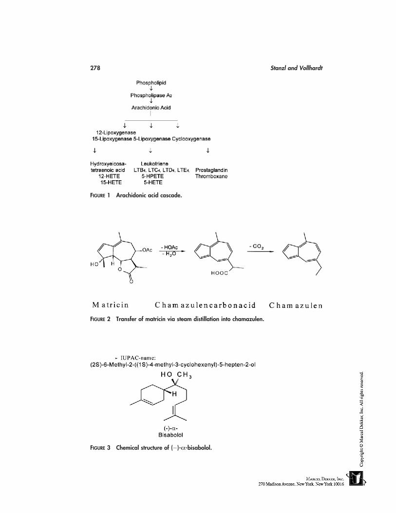
278 Stanzl and Vollhardt
FIGURE 1 Arachidonic acid cascade.
FIGURE 2 Transfer of matricin via steam distillation into chamazulen.
FIGURE 3 Chemical structure of (�)-α-bisabolol.
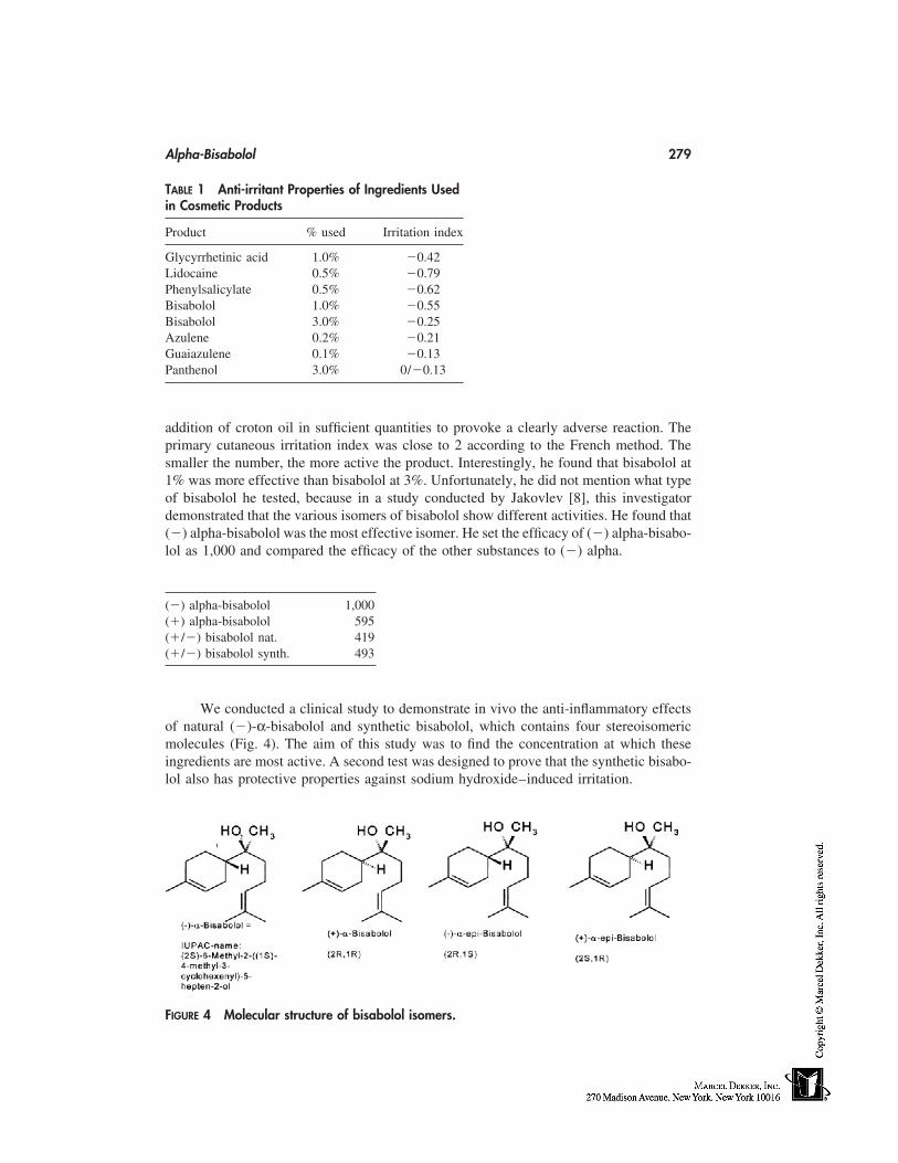
Alpha-Bisabolol 279
TABLE 1 Anti-irritant Properties of Ingredients Usedin Cosmetic Products
Product % used Irritation index
Glycyrrhetinic acid 1.0% �0.42Lidocaine 0.5% �0.79Phenylsalicylate 0.5% �0.62Bisabolol 1.0% �0.55Bisabolol 3.0% �0.25Azulene 0.2% �0.21Guaiazulene 0.1% �0.13Panthenol 3.0% 0/�0.13
addition of croton oil in sufficient quantities to provoke a clearly adverse reaction. Theprimary cutaneous irritation index was close to 2 according to the French method. Thesmaller the number, the more active the product. Interestingly, he found that bisabolol at1% was more effective than bisabolol at 3%. Unfortunately, he did not mention what typeof bisabolol he tested, because in a study conducted by Jakovlev [8], this investigatordemonstrated that the various isomers of bisabolol show different activities. He found that(�) alpha-bisabolol was the most effective isomer. He set the efficacy of (�) alpha-bisabo-lol as 1,000 and compared the efficacy of the other substances to (�) alpha.
(�) alpha-bisabolol 1,000(�) alpha-bisabolol 595(�/�) bisabolol nat. 419(�/�) bisabolol synth. 493
We conducted a clinical study to demonstrate in vivo the anti-inflammatory effectsof natural (�)-α-bisabolol and synthetic bisabolol, which contains four stereoisomericmolecules (Fig. 4). The aim of this study was to find the concentration at which theseingredients are most active. A second test was designed to prove that the synthetic bisabo-lol also has protective properties against sodium hydroxide–induced irritation.
FIGURE 4 Molecular structure of bisabolol isomers.
280 Stanzl and Vollhardt
STUDY OF THE EFFECTIVENESS OF FIVE PRODUCTS CONTAININGBISABOLOL OR SYNTHETIC BISABOLOL ON SLS-INDUCED SKINIRRITATION: TEST METHOD
Thirty female volunteers at the age of 18 to 63 years with healthy skin were included inthe test. The participants were briefed on the study procedures and each gave writteninformed consent. Measurements were carried out at a temperature of 22 � 1°C andrelative humidity of 60 � 10%. The test was carried out on the volar forearms. Skinirritation was induced in the test sites by applying sodium lauryl sulphate (SLS) 2% indistilled water under aluminum chamber occlusion. After 24 hours, occlusion was re-moved, and 2 hours later skin redness and TEWL were recorded. After the initial measure-ment the five test products were applied, and one area remained untreated. The dose ofapplication was about 2 mg/cm2. In the following 5 days, the subjects applied the testsamples in the morning and in the evening. Measurements were done during the treatmentperiod on days 1, 3, and 5, 2 hours after the last daily application. No use of other cosmeticswas allowed on the test sites during the whole test.
EVALUATION OF THE PROTECTIVE EFFICACY OF SYNTHETICBISABOLOL AGAINST SODIUM HYDROXIDE–INDUCED IRRITATION
Fifteen volunteers between the age of 25 and 44 years with healthy skin were enteredinto the study. The participants were briefed on the study procedures and gave writteninformed consent. Measurements were carried out at a temperature of 22 � 1°C andrelative humidity of 60 � 10%. The test was carried out on the volar forearms. The doseof application was about 2 mg/cm2. Two products were tested. One contained 0.56%synthetic bisabolol in mineral oil, the other pure mineral oil. Two hours after the applica-tion, 50 µL 0.1 M sodium hydroxide (NaOH) was applied to the volar forearms withocclusive aluminum chambers for 12 hours. At the end of exposure, the skin was wipedwith a soft paper towel to remove remaining solution, rinsed with distilled water andgently dried with a soft paper towel. Measurements were performed after 15 minutes.
Chromametry
Skin color was assessed with the Minolta Chromameter CR 300 (Minolta, Japan) in com-pliance with the Commission International de I’Eclairage (CIE) system. A color is ex-pressed in a three-dimensional coordinate system with greed-red (a*), yellow-blue (b*),and L* axes (brightness). In inflamed skin, a positive change on the a* axis is observed.Each value was the average of three recordings.
TEWL
Measurements of TEWL were performed with the Tewameter TM 210 (Courage & Kha-zaka, Cologne, Germany). Each value was the average of three recordings.
Statistics
Summary statistics procedure was used to determine the center, spread, and shape of thedata. Statistical analysis was performed using Wilcoxon matched pairs signed rank test.A p-value of less than 0.05 was taken to indicate a significant difference.
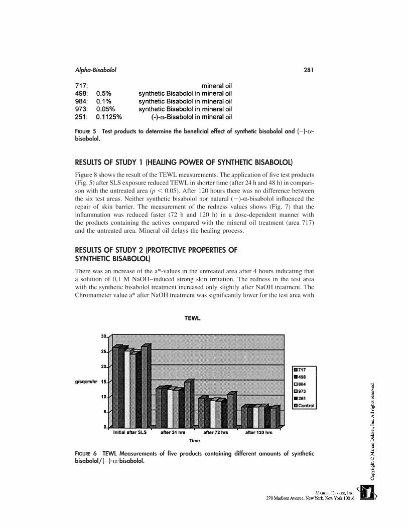
Alpha-Bisabolol 281
FIGURE 5 Test products to determine the beneficial effect of synthetic bisabolol and (�)-α-bisabolol.
RESULTS OF STUDY 1 (HEALING POWER OF SYNTHETIC BISABOLOL)
Figure 8 shows the result of the TEWL measurements. The application of five test products(Fig. 5) after SLS exposure reduced TEWL in shorter time (after 24 h and 48 h) in compari-son with the untreated area (p � 0.05). After 120 hours there was no difference betweenthe six test areas. Neither synthetic bisabolol nor natural (�)-α-bisabolol influenced therepair of skin barrier. The measurement of the redness values shows (Fig. 7) that theinflammation was reduced faster (72 h and 120 h) in a dose-dependent manner withthe products containing the actives compared with the mineral oil treatment (area 717)and the untreated area. Mineral oil delays the healing process.
RESULTS OF STUDY 2 (PROTECTIVE PROPERTIES OFSYNTHETIC BISABOLOL)
There was an increase of the a*-values in the untreated area after 4 hours indicating thata solution of 0,1 M NaOH–induced strong skin irritation. The redness in the test areawith the synthetic bisabolol treatment increased only slightly after NaOH treatment. TheChromameter value a* after NaOH treatment was significantly lower for the test area with
FIGURE 6 TEWL Measurements of five products containing different amounts of syntheticbisabolol/(�)-α-bisabolol.
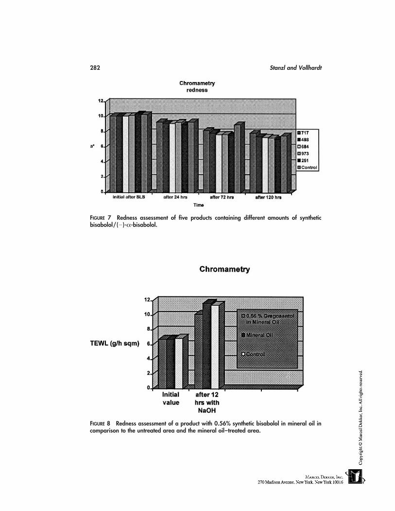
282 Stanzl and Vollhardt
FIGURE 7 Redness assessment of five products containing different amounts of syntheticbisabolol/(�)-α-bisabolol.
FIGURE 8 Redness assessment of a product with 0.56% synthetic bisabolol in mineral oil incomparison to the untreated area and the mineral oil–treated area.
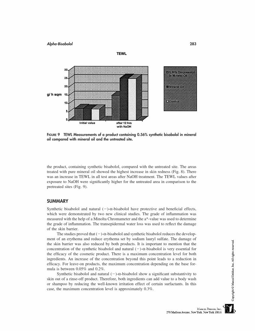
Alpha-Bisabolol 283
FIGURE 9 TEWL Measurements of a product containing 0.56% synthetic bisabolol in mineraloil compared with mineral oil and the untreated site.
the product, containing synthetic bisabolol, compared with the untreated site. The areastreated with pure mineral oil showed the highest increase in skin redness (Fig. 8). Therewas an increase in TEWL in all test areas after NaOH treatment. The TEWL values afterexposure to NaOH were significantly higher for the untreated area in comparison to thepretreated sites (Fig. 9).
SUMMARY
Synthetic bisabolol and natural (�)-α-bisabolol have protective and beneficial effects,which were demonstrated by two new clinical studies. The grade of inflammation wasmeasured with the help of a Minolta Chromameter and the a*-value was used to determinethe grade of inflammation. The transepidermal water loss was used to reflect the damageof the skin barrier.
The studies proved that (�)-α-bisabolol and synthetic bisabolol reduces the develop-ment of an erythema and reduce erythema set by sodium lauryl sulfate. The damage ofthe skin barrier was also reduced by both products. It is important to mention that theconcentration of the synthetic bisabolol and natural (�)-α-bisabolol is very essential forthe efficacy of the cosmetic product. There is a maximum concentration level for bothingredients. An increase of the concentration beyond this point leads to a reduction inefficacy. For leave-on products, the maximum concentration depending on the base for-mula is between 0.05% and 0.2%.
Synthetic bisabolol and natural (�)-α-bisabolol show a significant substantivity toskin out of a rinse-off product. Therefore, both ingredients can add value to a body washor shampoo by reducing the well-known irritation effect of certain surfactants. In thiscase, the maximum concentration level is approximately 0.3%.
284 Stanzl and Vollhardt
REFERENCES
1. Ammon HPT, Kaul R. Pharmakologie der Kamille und ihrer Inhaltsstoffe. Dtsch, Apoth, Ztg,1992; 132:1–26.
2. Isaac O. Fortschritte der Kamillenforschung. Struktur und Wirkung des (�) Bizabolols. Praep-erative Pharmazie 1986; 5:189–199.
3. Wichtl M. Teedrogen, 2. Auflage Wissenschaftliche Verlagsgesellschaft mbH, Stuttgart.4. Wagner H. Pharmazeutische Biologie, 5. Stuttgart, New York: Auflage Gustav Fischer Verlag.5. Issac O. Die Kamillentherapie–Erfahrung und Bestätigung Deutsche Apotheker Zeitung, 120
Jahrg., 13. 567.6. Ammon HPT, Sabieraj J, Kaul R. Kamille–Mechanismus der antiphlogistischen Wirkung von
Kamillenextrakten und -inhaltsstoffen. Dtsch. Apoth. Ztg 1996; 136:1821.7. Guillot et al. Intern J Cosm Sci 1983; 5:255.8. Jakovlev et al. Planta Medica 1979; 35:125.
25
Anti-irritants for Sensory Irritation
Gary S. HahnUniversity of California at San Diego School of Medicine, San Diego, andCosmederm Technologies, LLC, La Jolla, California
INTRODUCTION
Many chemicals found in cosmetics, personal-care products, pharmaceuticals, and in in-dustrial processes can irritate the skin and mucous membranes of the eye and the respira-tory and gastrointestinal tracts. Perhaps the most effective early-warning system that re-sponds to these chemicals is sensory irritation—the rapid-onset stinging, burning, anditching sensations that alert an organism to their exposure to foreign, and potentially injuri-ous, substances. These sensations, even when intense, may occur in the absence of visiblesigns of irritation or skin damage or, alternatively, may be accompanied by erythema and/or edema [1].
Sensory irritation occurs when thin, unmyelinated, chemically sensitive type-C noci-ceptors (from the Latin nocere, to injure) are activated and transmit a depolarizing signalvia the dorsal root ganglia (DRG) in the spinal cord to the brain where stinging, burning,itching, and poorly localized burning pain is appreciated [2]. These sensations are neuro-logically distinct from the highly localized sharp pain caused by cutting or puncturing theskin that is transmitted by the thinly myelinated A-delta class of nerve fibers [3]. Type-C nociceptors are present throughout the dermis and extend to the outermost layer of theviable epidermis, thus acting as one of the skin’s earliest warning systems [4]. Whenthe intensity of the irritant stimulus is sufficiently high, interneurons in the DRG and/ordepolarizing signals within the terminal aborization of a single nerve fiber trigger retro-grade depolarization down the activated fiber, resulting in the exocytosis of inflammatorymediators at the site of the irritant stimulus [5,6]. The principal mediators in humansinclude substance P, calcitonin gene-related peptide (CGRP), and neurokinin-A, a memberof the substance P family. These mediators, coupled with the neurogenically mediatedvasodilatory erythematous ‘‘flare’’ surrounding the irritated site, produce erythema,edema, and activation of immune cells, including mast cells, that contribute to the clinicalresponse of neurogenic inflammation.
285
286 Hahn
THE IDEAL ANTISENSORY ANTI-IRRITANT
The idea antisensory anti-irritant would effectively inhibit stinging, burning, and itchingcaused by a broad range of acidic, neutral, and basic chemical irritants by reducing thesensitivity of type-C nociceptors. In contrast, it would not inhibit the warning symptomof pain mediated by A-delta nerves, nor would it affect other nerve sensors that mediatetactile, temperature, or vibratory sensations. Since most cosmetic-induced sensory irrita-tion occurs within several minutes after application, the ideal anti-irritant should workwithin seconds when formulated with the irritant. For broad product use, it should alsowork when applied as a pretreatment before the irritating formulation and it should workwhen applied after irritation has occurred. Because cosmetics use a wide range of chemi-cals, the anti-irritant should be stable in many chemical environments and inexpensiveenough to be used in low-cost products. With repeated daily use, the ideal anti-irritantshould provide the same effective level of anti-irritant protection (no tachyphylaxis) and,most importantly, it must be safe for broad, unsupervised use.
With the exception of local anesthetics that are regulated as drugs in most countriesand may have undesirable side effects and safety concerns, no compounds have beendescribed that are able to broadly inhibit sensory irritation from cosmetics and pharmaceu-ticals. Because a safe compound capable of blocking sensory irritation and inflammationwould provide considerable benefit, I sought to identify compounds that could effectivelyblock sensory irritant reactions. Simple water-soluble strontium salts have proved to bepotent and selective inhibitors of chemically induced sensory irritation and neurogenicinflammation in humans and do not produce numbness or loss of other tactile sensations[7–10].
THE FIRST EFFECTIVE ANTISENSORY ANTI-IRRITANTS:STRONTIUM SALTS
Clinical Evaluation of Sensory Irritation
A variety of chemical irritants used in cosmetics were used to induce sensory irritation.All clinical studies were conducted according to double-blind, vehicle-controlled, randomtreatment assignment protocols in which each subject served as her own control. Testsubjects were healthy women, aged 18 to 65, who self-reported a history of sensitive skinand were sensitive to lactic acid facial challenge. Treated skin sites were first washed withIvory bar soap, followed by sequential application of test materials and sensory irritationevaluation. Statistical analysis of the mean sensory irritation differences between vehicleand strontium-treated groups was conducted using the Wilcoxon Signed Ranks Test forpaired comparisons. All subjects provided informed consent and all protocols were re-viewed by a safety committee.
Sensory Irritation Scale
Each minute for 10 to 60 minutes, depending on the study, subjects reported the mag-nitude of sensory irritation (stinging, burning, and itching) according to the followingscale:
Anti-irritants for Sensory Irritation 287
0 � none1 � slight Transient, barely perceptible irritation
Does not bother them2 � mild Definite and continuous irritation
Bothers them3 � moderate Distinctly uncomfortable irritation
Bothers them and interferes with concentration4 � severe Continuous, intensely uncomfortable irritation
Intolerable and would interfere with daily routine
ACIDIC IRRITANTS
Lactic Acid (7.5%, pH � 1.9) Sensory Irritation on the Face
Alpha-hydroxyacids (AHAs) including lactic and glycolic acids are used in cosmetics andin professionally applied chemical peels to reduce the visible signs of skin aging. Tomaximize AHA efficacy, the formulation must be acidic, which increases the active ‘‘freeacid’’ form of the AHA molecule and, unfortunately, directly contributes to their irritationpotential [11,12]. To evaluate the ability of strontium salts to reduce lactic acid sensoryirritation, either lactic acid alone (7.5% in 10% ethanol/water vehicle, pH � 1.9), or anidentical vehicle at the same pH containing various concentrations of strontium nitrate orstrontium chloride was applied (0.1 g) to cheek sites using cotton swabs (6 swipes) ex-tending from the nasolabial fold to the outer cheek. Test materials were applied to theright or left side of subjects’ faces sequentially followed by sensory irritation assessmenton each side for 10 minutes. A typical time-response curve for lactic acid (7.5%, pH �1.9) on the face is presented in Figure 1. When the areas under both irritation curves arecompared, strontium nitrate inhibited sensory irritation by 68% (p �0.01). Both strontiumnitrate and strontium chloride produced dose-dependent inhibition of sensory irritationwhen mixed with lactic acid (Table 1) [7]. In separate studies, the local anesthetic lidocaine(4%) was used as a positive control. When applied at the same time as the lactic acid,lidocaine did not produce significant inhibition (�10%), presumably because it requirestime to be absorbed. When lidocaine (4%) was applied 5 minutes before the lactic acid,lidocaine inhibited by 51% (p �0.05, n � 10).
Strontium Pretreatment on the Face
Many cosmetics such as toners and skin conditioners, are applied immediately beforeapplication of potentially irritating products. Incorporation of strontium salts into such apretreatment product from 1 minute to 15 minutes before the same lactic acid facial chal-lenge produced a substantial level of sensory irritation inhibition (Table 1). In other stud-ies, substantial anti-irritancy was also observed when strontium nitrate was applied severalminutes after lactic acid was applied.
When the same lactic acid challenge was used in conjunction with ‘‘conventional’’anti-irritants used in cosmetics such as green tea (3%), alpha-bisabolol (1%), and glycyr-rhizic acid (1%), no significant inhibition was observed (�10% difference from vehiclecontrol).
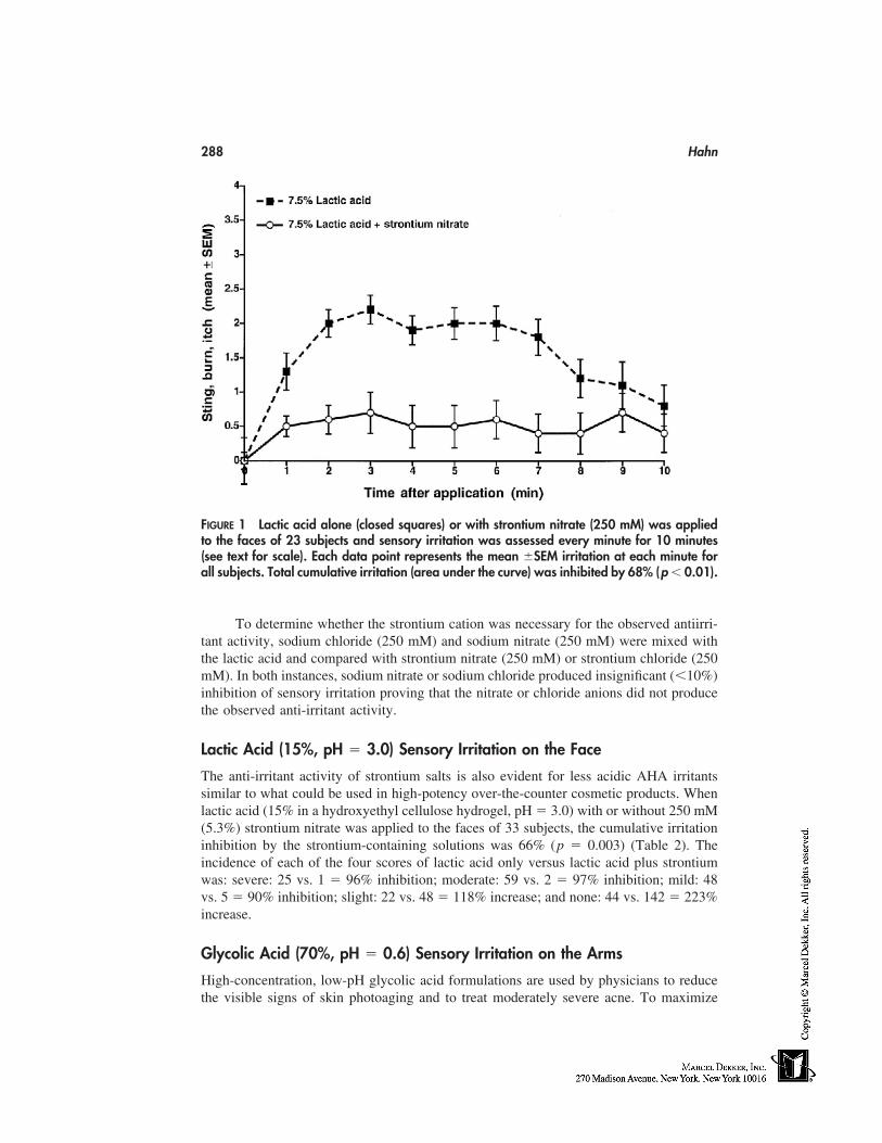
288 Hahn
FIGURE 1 Lactic acid alone (closed squares) or with strontium nitrate (250 mM) was appliedto the faces of 23 subjects and sensory irritation was assessed every minute for 10 minutes(see text for scale). Each data point represents the mean �SEM irritation at each minute forall subjects. Total cumulative irritation (area under the curve) was inhibited by 68% (p � 0.01).
To determine whether the strontium cation was necessary for the observed antiirri-tant activity, sodium chloride (250 mM) and sodium nitrate (250 mM) were mixed withthe lactic acid and compared with strontium nitrate (250 mM) or strontium chloride (250mM). In both instances, sodium nitrate or sodium chloride produced insignificant (�10%)inhibition of sensory irritation proving that the nitrate or chloride anions did not producethe observed anti-irritant activity.
Lactic Acid (15%, pH � 3.0) Sensory Irritation on the Face
The anti-irritant activity of strontium salts is also evident for less acidic AHA irritantssimilar to what could be used in high-potency over-the-counter cosmetic products. Whenlactic acid (15% in a hydroxyethyl cellulose hydrogel, pH � 3.0) with or without 250 mM(5.3%) strontium nitrate was applied to the faces of 33 subjects, the cumulative irritationinhibition by the strontium-containing solutions was 66% (p � 0.003) (Table 2). Theincidence of each of the four scores of lactic acid only versus lactic acid plus strontiumwas: severe: 25 vs. 1 � 96% inhibition; moderate: 59 vs. 2 � 97% inhibition; mild: 48vs. 5 � 90% inhibition; slight: 22 vs. 48 � 118% increase; and none: 44 vs. 142 � 223%increase.
Glycolic Acid (70%, pH � 0.6) Sensory Irritation on the Arms
High-concentration, low-pH glycolic acid formulations are used by physicians to reducethe visible signs of skin photoaging and to treat moderately severe acne. To maximize
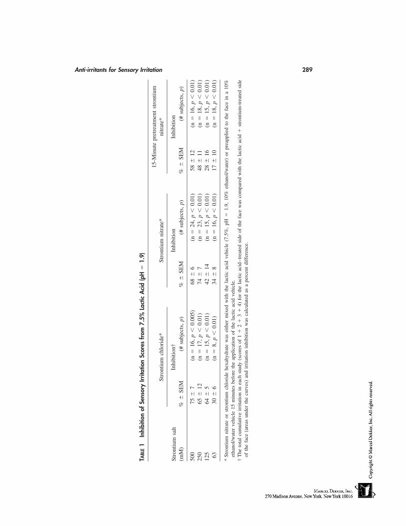
Anti-irritants for Sensory Irritation 289
TABL
E1
Inhi
bitio
nof
Sens
ory
Irrita
tion
Scor
esfr
om7.
5%La
ctic
Aci
d(p
H�
1.9)
15-M
inut
epr
etre
atm
ent
stro
ntiu
mSt
ront
ium
chlo
ride
*St
ront
ium
nitr
ate*
nitr
ate*
Inhi
bitio
n†In
hibi
tion
Inhi
bitio
nSt
ront
ium
salt
(mM
)%
�SE
M(#
subj
ects
,p)
%�
SEM
(#su
bjec
ts,
p)%
�SE
M(#
subj
ects
,p)
500
75�
7(n
�16
,p
�0.
005)
68�
6(n
�24
,p
�0.
01)
58�
12(n
�16
,p
�0.
01)
250
65�
12(n
�17
,p
�0.
01)
74�
7(n
�23
,p
�0.
01)
48�
11(n
�18
,p
�0.
01)
125
64�
5(n
�15
,p
�0.
01)
42�
14(n
�15
,p
�0.
01)
28�
16(n
�15
,p
�0.
01)
6330
�6
(n�
8,p
�0.
01)
34�
8(n
�16
,p
�0.
01)
17�
10(n
�18
,p
�0.
01)
*St
ront
ium
nitr
ate
orst
ront
ium
chlo
ride
hexa
hydr
ate
was
eith
erm
ixed
with
the
lact
icac
idve
hicl
e(7
.5%
,pH
�1.
9,10
%et
hano
l/wat
er)
orpr
eapp
lied
toth
efa
cein
a10
%et
hano
l/wat
erve
hicl
e15
min
utes
befo
reth
eap
plic
atio
nof
the
lact
icac
idve
hicl
e.†
The
tota
lcu
mul
ativ
eir
rita
tion
inea
chst
udy
(sco
res
of1
�2
�3
�4)
for
the
lact
icac
id–t
reat
edsi
deof
the
face
was
com
pare
dw
ithth
ela
ctic
acid
�st
ront
ium
-tre
ated
side
ofth
efa
ce(a
reas
unde
rth
ecu
rves
)an
dir
rita
tion
inhi
bitio
nw
asca
lcul
ated
asa
perc
ent
diff
eren
ce.
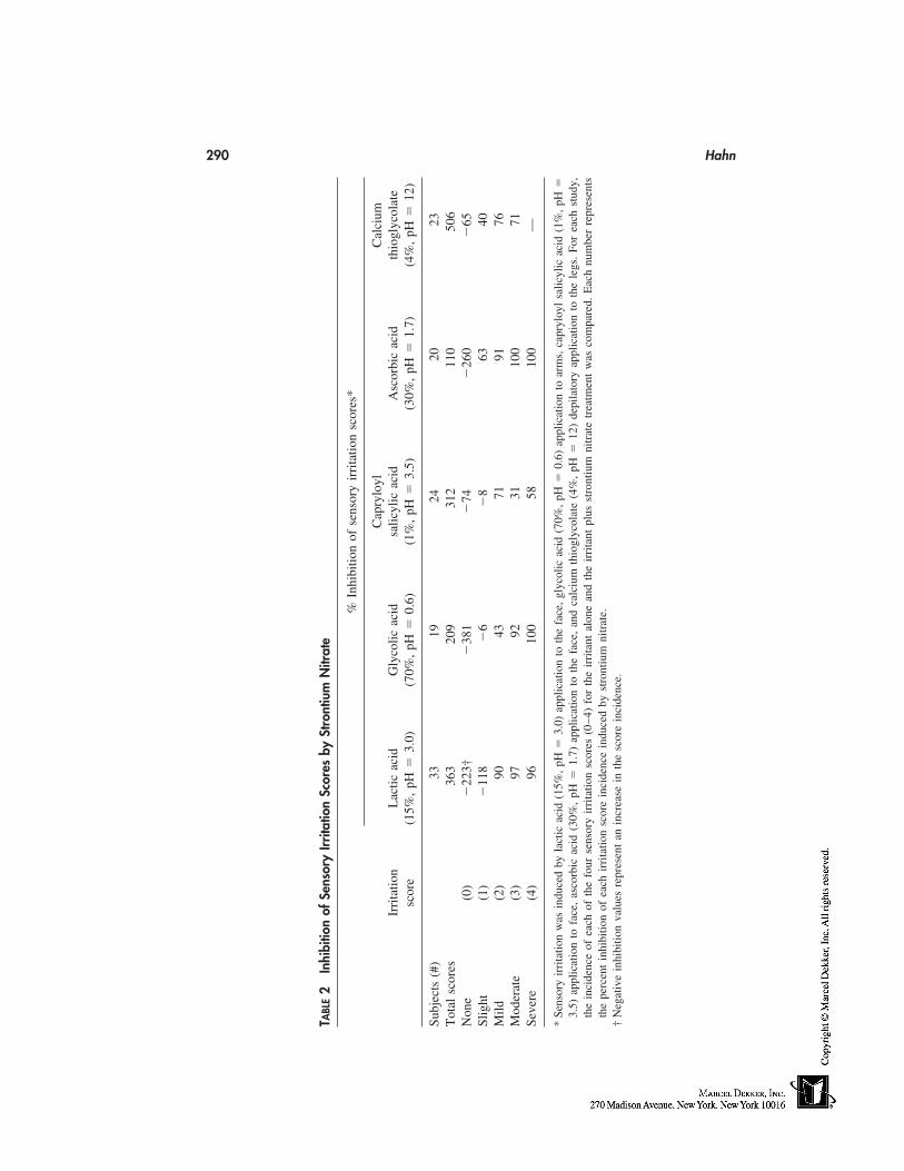
290 Hahn
TABL
E2
Inhi
bitio
nof
Sens
ory
Irrita
tion
Scor
esby
Stro
ntiu
mN
itrat
e
%In
hibi
tion
ofse
nsor
yir
rita
tion
scor
es*
Cap
rylo
ylC
alci
umIr
rita
tion
Lac
ticac
idG
lyco
licac
idsa
licyl
icac
idA
scor
bic
acid
thio
glyc
olat
esc
ore
(15%
,pH
�3.
0)(7
0%,
pH�
0.6)
(1%
,pH
�3.
5)(3
0%,
pH�
1.7)
(4%
,pH
�12
)
Subj
ects
(#)
3319
2420
23T
otal
scor
es36
320
931
211
050
6N
one
(0)
�22
3†�
381
�74
�26
0�
65Sl
ight
(1)
�11
8�
6�
863
40M
ild(2
)90
4371
9176
Mod
erat
e(3
)97
9231
100
71Se
vere
(4)
9610
058
100
—
*Se
nsor
yir
rita
tion
was
indu
ced
byla
ctic
acid
(15%
,pH
�3.
0)ap
plic
atio
nto
the
face
,gl
ycol
icac
id(7
0%,
pH�
0.6)
appl
icat
ion
toar
ms,
capr
yloy
lsa
licyl
icac
id(1
%,
pH�
3.5)
appl
icat
ion
tofa
ce,
asco
rbic
acid
(30%
,pH
�1.
7)ap
plic
atio
nto
the
face
,an
dca
lciu
mth
iogl
ycol
ate
(4%
,pH
�12
)de
pila
tory
appl
icat
ion
toth
ele
gs.
For
each
stud
y,th
ein
cide
nce
ofea
chof
the
four
sens
ory
irri
tatio
nsc
ores
(0–4
)fo
rth
eir
rita
ntal
one
and
the
irri
tant
plus
stro
ntiu
mni
trat
etr
eatm
ent
was
com
pare
d.E
ach
num
ber
repr
esen
tsth
epe
rcen
tin
hibi
tion
ofea
chir
rita
tion
scor
ein
cide
nce
indu
ced
byst
ront
ium
nitr
ate.
†N
egat
ive
inhi
bitio
nva
lues
repr
esen
tan
incr
ease
inth
esc
ore
inci
denc
e.
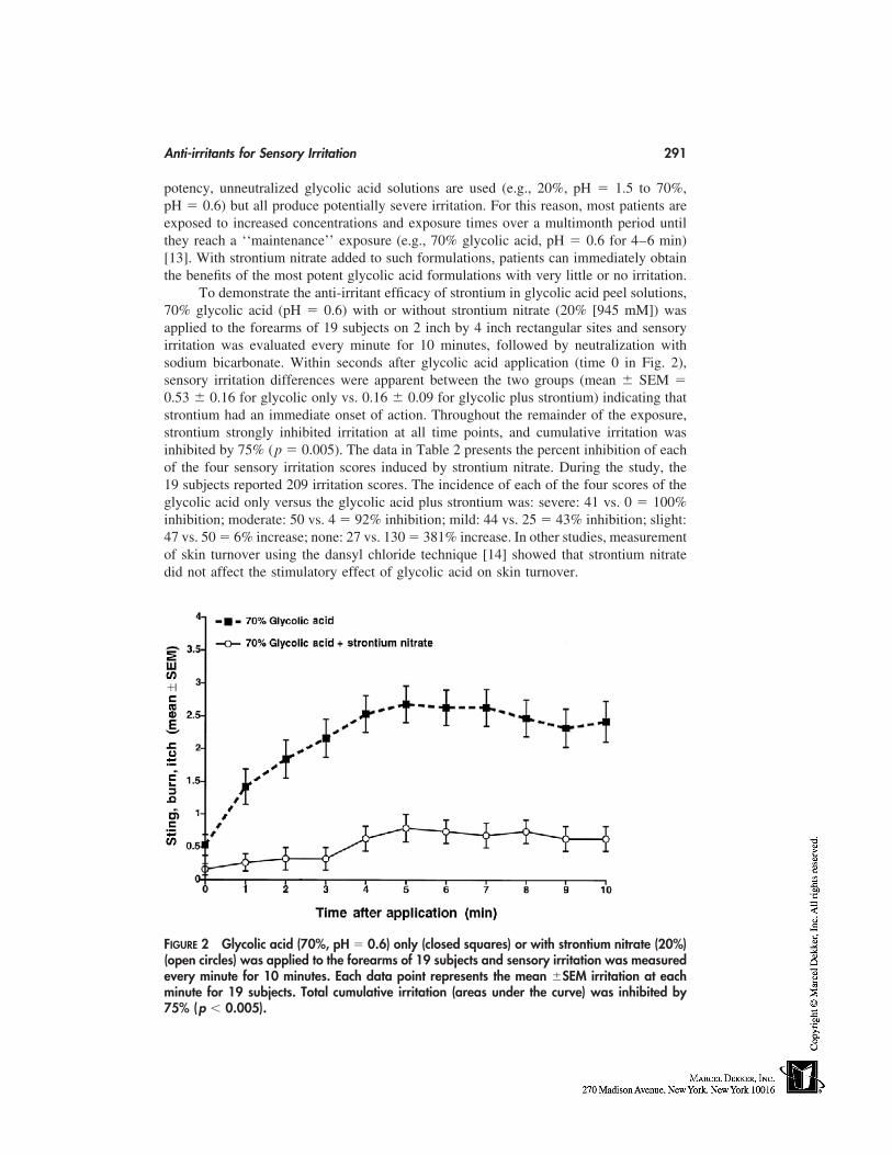
Anti-irritants for Sensory Irritation 291
potency, unneutralized glycolic acid solutions are used (e.g., 20%, pH � 1.5 to 70%,pH � 0.6) but all produce potentially severe irritation. For this reason, most patients areexposed to increased concentrations and exposure times over a multimonth period untilthey reach a ‘‘maintenance’’ exposure (e.g., 70% glycolic acid, pH � 0.6 for 4–6 min)[13]. With strontium nitrate added to such formulations, patients can immediately obtainthe benefits of the most potent glycolic acid formulations with very little or no irritation.
To demonstrate the anti-irritant efficacy of strontium in glycolic acid peel solutions,70% glycolic acid (pH � 0.6) with or without strontium nitrate (20% [945 mM]) wasapplied to the forearms of 19 subjects on 2 inch by 4 inch rectangular sites and sensoryirritation was evaluated every minute for 10 minutes, followed by neutralization withsodium bicarbonate. Within seconds after glycolic acid application (time 0 in Fig. 2),sensory irritation differences were apparent between the two groups (mean � SEM �0.53 � 0.16 for glycolic only vs. 0.16 � 0.09 for glycolic plus strontium) indicating thatstrontium had an immediate onset of action. Throughout the remainder of the exposure,strontium strongly inhibited irritation at all time points, and cumulative irritation wasinhibited by 75% (p � 0.005). The data in Table 2 presents the percent inhibition of eachof the four sensory irritation scores induced by strontium nitrate. During the study, the19 subjects reported 209 irritation scores. The incidence of each of the four scores of theglycolic acid only versus the glycolic acid plus strontium was: severe: 41 vs. 0 � 100%inhibition; moderate: 50 vs. 4 � 92% inhibition; mild: 44 vs. 25 � 43% inhibition; slight:47 vs. 50 � 6% increase; none: 27 vs. 130 � 381% increase. In other studies, measurementof skin turnover using the dansyl chloride technique [14] showed that strontium nitratedid not affect the stimulatory effect of glycolic acid on skin turnover.
FIGURE 2 Glycolic acid (70%, pH � 0.6) only (closed squares) or with strontium nitrate (20%)(open circles) was applied to the forearms of 19 subjects and sensory irritation was measuredevery minute for 10 minutes. Each data point represents the mean �SEM irritation at eachminute for 19 subjects. Total cumulative irritation (areas under the curve) was inhibited by75% (p � 0.005).
292 Hahn
Clinical studies of a 70% glycolic acid (pH � 0.6) chemical peel solution withstrontium nitrate applied to the whole face in over 150 human subjects demonstrated sub-stantially reduced sensory irritation and erythema without reducing the expected benefitsof the peel as judged by clinical response [15,16]. Histological analysis of punch biopsiesfrom skin exposed to AHA formulations containing strontium nitrate (70% glycolic acid,pH � 0.6) every 2 weeks for 8 weeks and 15% lactic acid lotion (pH � 3.2) twice dailyat the same facial sites) demonstrated that there was slightly less inflammation in the AHAand strontium-treated sites compared with untreated skin in the same individuals [16],thus demonstrating that strontium not only reduced irritation symptoms, but also protectedthe skin from cryptic damage.
Capryloyl Salicylic Acid–Induced Sensory Irritation
Capryloyl salicylic acid is a covalently modified derivative of salicylic acid with enhancedlipophylicity attributable to the 8 carbon caprylic acid moiety. It is used as a cosmeticexfoliant and is reported to have utility as an acne therapeutic [17]. A cream emulsionbase containing capryloyl salicylic acid (1%) with or without strontium nitrate (500 mM)was applied to cheek sites 2 inches by 4 inches extending from the nasolabial fold to theouter cheek of 24 female subjects and sensory irritation was evaluated every 5 minutesfor 60 minutes. The data in Table 2 presents the percent inhibition of each of the foursensory irritation scores induced by strontium nitrate. During the entire study, subjectsreported 312 sensory irritation scores. The incidence of each of the four scores of thecapryloyl salicylic acid versus the capryloyl plus strontium was: severe: 19 vs. 8; moder-ate: 13 vs. 9; mild: 35 vs. 10; slight: 39 vs. 42; and none: 50 vs. 87. The mean sensoryirritation score of the capryloyl salicylic acid reached approximately 0.8 5 minutes afterapplication, peaked at approximately 1.0 from 20 minutes to 35 minutes, and remainedat approximately 0.8 until 45 minutes, after which it declined to 0.4 at 60 minutes. Totalirritation, calculated as the percent difference of the areas under the 60-minute irritationcurves, was inhibited by 46% (p � 0.002).
Ascorbic Acid (30%, pH � 1.7) Sensory Irritation on the Face
Ascorbic acid (Vitamin C) is used in many cosmetic products because it is a potent water-soluble antioxidant and can protect the skin against damage from ultraviolet radiation [18].In vitro studies also show that ascorbic acid can also stimulate collagen synthesis [19].Because ascorbic acid is most stable and bioavailable in aqueous formulations at a highlyacidic pH (e.g., pH � 3) a 30% aqueous solution of ascorbic acid (pH � 1.7) was evaluatedfor sensory irritation with or without strontium nitrate (250 mM). After application to theface of 20 subjects, the cumulative irritation inhibition by the strontium-containing solu-tions was 84% (p � 0.005) (Table 2). The incidence of each of the four scores of theascorbic acid only versus the ascorbic acid plus strontium was: severe: 1 vs. 0 � 100%inhibition; moderate: 13 vs. 0 � 100% inhibition; mild: 23 vs. 2 � 91% inhibition; slight:48 vs. 18 � 63% inhibition; plus none: 25 vs. 90 � 260% increase).
Aluminum Chloride Antiperspirant Application to Axilla
Antiperspirants use aluminum salts alone or in combination with other agents to reduceperspiration. In the moist environment of the axilla, aluminum salts can cause sensoryirritation and inflammation [20]. The axilla of 16 subjects was pretreated with 1.0 mL of
Anti-irritants for Sensory Irritation 293
a strontium nitrate solution (500 mM, pH � 7.3 in 50% ethanol/water vehicle) followed2 minutes later by a 1 mL application of the aluminum chloride (20%) antiperspirantsolution. Sensory irritation was evaluated every 2 minutes for 20 minutes. The incidenceof each of the four scores of the aluminum chloride versus the aluminum chloride plusstrontium was: severe: 12 vs. 2; moderate: 22 vs. 9; mild: 30 vs. 13; slight: 60 vs. 41;and none: 52 vs. 111. Upon application, sensory irritation reached a mean score of 1within the first minute and a plateau at approximately 1.5 from minutes 6 to 10, thengradually declined to a score of approximately 1 at 20 minutes. During the study, the 16subjects reported 352 irritation scores. Total irritation caused by the aluminum chloridecalculated as the percent difference of the areas under the 20-minute irritation curves wasreduced by 56% when the areas under the irritation curves were compared (p � 0.005).
Aluminum/Zirconium Salt Erythema on the Arms
Aluminum salts, with or without zirconium salts, are FDA-approved antiperspirant ingre-dients and frequently cause both sensory irritation and inflammation [20]. Aluminum/zirconium salt solution (25%) with or without strontium nitrate (500 mM) or strontiumchloride (500 mM) was applied to the arms of 29 subjects using occluded patches for 21days and the magnitude of visible inflammation was evaluated every day. Inflammationwas visually measured according to the following scale:
0 � No evidence of erythema1 � Minimal erythema2 � Definite erythema3 � Erythema and papules
Both stronium nitrate (500 mM) or strontium chloride (500 mM) caused nearly completeinhibition of erythema development during the first week and substantially inhibited ery-thema during the second and third weeks (Fig. 3). Total erythema caused by the aluminum/zirconium salts, calculated as the percent difference of the areas under the 21 day irritationcurves, was reduced by 64% (p � 0.0001) by strontium nitrate and by 66% (p � 0.0001)by strontium chloride.
BASIC IRRITANTS
Calcium Thioglycolate Sensory Irritation on the Legs
Chemical depilatories typically use calcium thioglycolate formulated at a basic pH (e.g.,9–12) to dissolve hair keratin [21]. Twenty-three subjects shaved their legs with a safetyrazor to enhance irritation, then strontium nitrate pretreatment solution (10% w/v, in 10%ethanol/water) or vehicle was applied to 2 inch by 4 inch sites on the lateral portions ofthe legs. After 2 minutes, 5 grams of depilatory lotion was applied to each leg followedby irritation evaluation every minute for 10 minutes. During the study, the 23 subjectsreported 506 irritation scores (Table 2). The incidence of each of the four scores of thedepilatory versus the depilatory plus strontium was: severe: 0 vs. 0; moderate: 7 vs. 2 �71% inhibition; mild: 45 vs. 11 � 76% inhibition; slight: 88 vs. 53 � 40% inhibition;and none: 113 vs. 187 � 65% increase. Total irritation caused by the depilatory, calculated
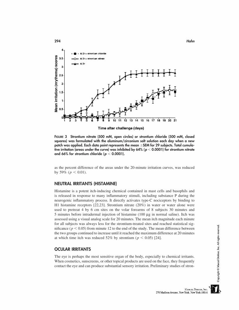
294 Hahn
FIGURE 3 Strontium nitrate (500 mM, open circles) or strontium chloride (500 mM, closedsquares) was formulated with the aluminum/zirconium salt solution each day when a newpatch was applied. Each data point represents the mean �SEM for 29 subjects. Total cumula-tive irritation (areas under the curve) was inhibited by 64% (p � 0.0001) for strontium nitrateand 66% for strontium chloride (p � 0.0001).
as the percent difference of the areas under the 20-minute irritation curves, was reducedby 59% (p � 0.01).
NEUTRAL IRRITANTS (HISTAMINE)
Histamine is a potent itch-inducing chemical contained in mast cells and basophils andis released in response to many inflammatory stimuli, including substance P during theneurogenic inflammatory process. It directly activates type-C nociceptors by binding toH1 histamine receptors [22,23]. Strontium nitrate (20%) in water or water alone wereused to pretreat 4 by 6 cm sites on the volar forearms of 8 subjects 30 minutes and5 minutes before intradermal injection of histamine (100 µg in normal saline). Itch wasassessed using a visual analog scale for 20 minutes. The mean itch magnitude each minutefor all subjects was always less for the strontium-treated sites and reached statistical sig-nificance (p � 0.05) from minute 12 to the end of the study. The mean difference betweenthe two groups continued to increase until it reached the maximum difference at 20 minutesat which time itch was reduced 52% by strontium (p � 0.05) [24].
OCULAR IRRITANTS
The eye is perhaps the most sensitive organ of the body, especially to chemical irritants.When cosmetics, sunscreens, or other topical products are used on the face, they frequentlycontact the eye and can produce substantial sensory irritation. Preliminary studies of stron-
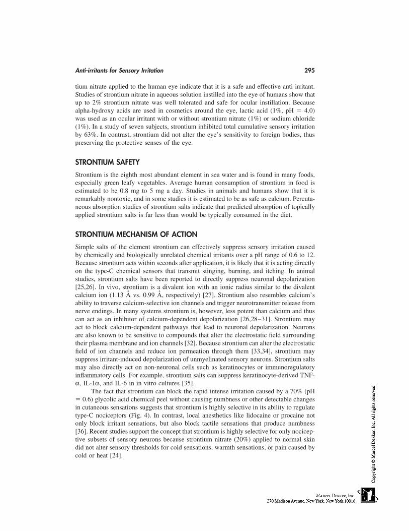
Anti-irritants for Sensory Irritation 295
tium nitrate applied to the human eye indicate that it is a safe and effective anti-irritant.Studies of strontium nitrate in aqueous solution instilled into the eye of humans show thatup to 2% strontium nitrate was well tolerated and safe for ocular instillation. Becausealpha-hydroxy acids are used in cosmetics around the eye, lactic acid (1%, pH � 4.0)was used as an ocular irritant with or without strontium nitrate (1%) or sodium chloride(1%). In a study of seven subjects, strontium inhibited total cumulative sensory irritationby 63%. In contrast, strontium did not alter the eye’s sensitivity to foreign bodies, thuspreserving the protective senses of the eye.
STRONTIUM SAFETY
Strontium is the eighth most abundant element in sea water and is found in many foods,especially green leafy vegetables. Average human consumption of strontium in food isestimated to be 0.8 mg to 5 mg a day. Studies in animals and humans show that it isremarkably nontoxic, and in some studies it is estimated to be as safe as calcium. Percuta-neous absorption studies of strontium salts indicate that predicted absorption of topicallyapplied strontium salts is far less than would be typically consumed in the diet.
STRONTIUM MECHANISM OF ACTION
Simple salts of the element strontium can effectively suppress sensory irritation causedby chemically and biologically unrelated chemical irritants over a pH range of 0.6 to 12.Because strontium acts within seconds after application, it is likely that it is acting directlyon the type-C chemical sensors that transmit stinging, burning, and itching. In animalstudies, strontium salts have been reported to directly suppress neuronal depolarization[25,26]. In vivo, strontium is a divalent ion with an ionic radius similar to the divalentcalcium ion (1.13 Å vs. 0.99 Å, respectively) [27]. Strontium also resembles calcium’sability to traverse calcium-selective ion channels and trigger neurotransmitter release fromnerve endings. In many systems strontium is, however, less potent than calcium and thuscan act as an inhibitor of calcium-dependent depolarization [26,28–31]. Strontium mayact to block calcium-dependent pathways that lead to neuronal depolarization. Neuronsare also known to be sensitive to compounds that alter the electrostatic field surroundingtheir plasma membrane and ion channels [32]. Because strontium can alter the electrostaticfield of ion channels and reduce ion permeation through them [33,34], strontium maysuppress irritant-induced depolarization of unmyelinated sensory neurons. Strontium saltsmay also directly act on non-neuronal cells such as keratinocytes or immunoregulatoryinflammatory cells. For example, strontium salts can suppress keratinocyte-derived TNF-α, IL-1α, and IL-6 in in vitro cultures [35].
The fact that strontium can block the rapid intense irritation caused by a 70% (pH� 0.6) glycolic acid chemical peel without causing numbness or other detectable changesin cutaneous sensations suggests that strontium is highly selective in its ability to regulatetype-C nociceptors (Fig. 4). In contrast, local anesthetics like lidocaine or procaine notonly block irritant sensations, but also block tactile sensations that produce numbness[36]. Recent studies support the concept that strontium is highly selective for only nocicep-tive subsets of sensory neurons because strontium nitrate (20%) applied to normal skindid not alter sensory thresholds for cold sensations, warmth sensations, or pain caused bycold or heat [24].
296 Hahn
FIGURE 4 Chemical irritants activate unmyelinated type-C nociceptors and trigger their depo-larization. Type-C nociceptors then synapse in the dorsal root ganglia (DRG) of the spinal cordand the signal travels to the brain where it is sensed as sting, burn, or itch. If the stimulationis of sufficient magnitude, interneurons in the DRG send a retrograde signal down the sametype-C fibers, which triggers the release of inflammatory substances including substance P,neurokinin A, calcitonin gene-related peptide (CGRP), and other mediators. These substancestrigger vasodilation, vascular permeability, and activate inflammatory cells, including mastcells that, in turn, release another set of inflammatory mediators, including histamine, whichfurther activate nociceptive sensory signals and inflammation. Strontium reduces the sensitivityof type-C nociceptors to chemical irritants while not affecting the A-delta nerves that transmitthe ability to detect pain.
PRODUCT APPLICATIONS
Burning, stinging, and especially itching sensations are among the most common con-sumer complaints from cosmetics and topical drugs. The rapid-onset and high-level anti-irritant potency of strontium salts suggest that they will have broad applications in topicalproducts. Throughout the world, cosmetic products are used daily to cleanse and beautifythe skin. With the discovery of new, potent, biologically active ingredients, formulatorscan provide consumers with increased benefit that may resemble that obtained from phar-maceutical products. Unfortunately, irritation frequently accompanies the use of higherconcentrations of active ingredients or more potent skin-delivery systems. For people withsensitive skin attributable to inherently dry skin or other causes, the problem is furthercompounded. In addition to products intentionally applied to the skin, many workers areexposed to chemical irritants in the workplace that can result in considerable occupationaldisability [37–39].
Strontium salts, particularly strontium nitrate, has proven to be highly effective inreducing irritation, erythema, and inflammation from many irritating ingredients used intopical products and found in the workplace. The first strontium-containing cosmetic prod-ucts were introduced in the United States, and made available internationally in October
Anti-irritants for Sensory Irritation 297
1999. The safety of strontium salts, coupled with their ability to inhibit both sensoryirritation and neurogenic inflammation, suggests that they may have therapeutic utility inthe treatment of many dermatological conditions. Because the neurogenic inflammationsyndrome is believed to be pathogenically important in many other conditions, includingallergic contact dermatitis, psoriasis, atopic dermatitis, ocular irritation and inflammation,allergic rhinitis, asthma, rheumatoid arthritis, inflammatory bowel disease and other gas-trointestinal disorders [40], strontium salts may have additional therapeutic utility. Stron-tium salts represent a new class of selective inhibitors of sensory irritation and irritantcontact dermatitis without local anesthetic side effects.
REFERENCES
1. Tausk F, Christian E, Johansson O, Milgram S. Neurobiology of the skin. In: Fitzpatrick TB,Eisen AZ, Wolff K, Freedberg IM, Austen KF, ed. Dermatology in General Medicine. Vol.1. 4th ed. New York: McGraw-Hill, 1993:396–403.
2. Martin JH, Jessell TM. Modality coding in the somatic sensory system. In: Kandel ER,Schwartz JH, Jessell TM, eds. Principles of Neural Science. 3rd ed. New York: Elsevier, 1991:341.
3. Meyer RA, Campbell JN, Raja SN. Peripheral neural mechanisms of nociception. In: WallPD, Melzack R, eds. Textbook of Pain. 3rd ed. London: Churchill Livingstone, 1994:13–44.
4. Kennedy WR, Wendelschafer-Crabb G. The innervation of the human epidermis. J NeurolSci (Netherlands) 1993; 115:184–190.
5. Baluk P. Neurogenic inflammation in skin and airways. J Invest Derm 1997; 2:76–81.6. Szolcsanyi J. Neurogenic inflammation: reevaluation of axon reflex theory. In: Geppetti P,
Holzer P, eds. Neurogenic Inflammation. New York: 1996:33–42.7. Hahn GS. Strontium is a potent and selective inhibitor of sensory irritation. Derm Surg 1999;
25:1–6.8. Hahn GS. Modulation of neurogenic inflammation by strontium. In: Kydonieus AF, Willie
JJ, eds. Biochemical Modulation of Skin Reactions: Transdermals, Topicals, Cosmetics. NewYork: CRC Press, 1999:261–272.
9. Hahn GS, Thueson DO. Cosmederm Technologies, Inc., assignee. Formulations and Methodsfor Reducing Skin Irritation. U.S. patent 5,716,625. Feb. 10, 1998.
10. Hahn GS, Thueson DO, Quick TW. Cosmoderm Technologies, Inc., assignee. Topical ProductFormulations Containing Strontium for Reducing Skin Irritation. U.S. patent 5,804,203. Sept.8, 1998.
11. Thueson DO, Chan EK, Oechsli LM, Hahn GS. The roles of pH and concentration in lacticacid-induced stimulation of epidermal turnover. Dermatol Surg 1998; 24:641–645.
12. Stiller MJ, Bartolone J, Stern R, Kollias N, Gillies R, Drake LA. Topical 8% glycolic acidand 8% L-lactic acid creams for the treatment of photodamaged skin. A double-blind vehicle-controlled clinical trial. Arch Dermatol 1996; 132:631–636.
13. Brody HJ. Chemical Peeling and Resurfacing. 2nd ed. St. Louis, MO: Mosby, 1997:73–108.14. Jansen LH, Hojyo-Tomoko MT, Kligman AM. Improved fluorescence staining technique for
estimating turnover of human stratum corneum. Br J Dermatol 1973; 90:9–12.15. Rubin MG, Harper RA, Hahn GS. Strontium nitrate in 70% free glycolic acid peels signifi-
cantly reduces erythema and sensory irritation (Abstr). Poster at American Academy of Derma-tology 1999. (Manuscript to be submitted.)
16. Greenway HT, Peterson C, Plis J, Cornell R, Hahn GS, Harper RA. Efficacy of a 70% glycolicacid peel product regimen containing the anti-irritant strontium nitrate (Abst). Poster at Ameri-can Academy of Dermatology 1999. (Manuscript to be submitted.)
17. Leveque JL, Raincy L, inventors; L’Oreal assignee. Use of salicylic acid derivatives for thetreatment of skin aging. US patent 5,262,407. 1993 Nov 16.
298 Hahn
18. Gabard DF. Topical melatonin in combination with vitamins E and C protects skin from ultra-violet-induced erythema: a human study in vivo. Br J Derm 1998; 139:332–339.
19. Colven RM, Pinnell SR. Topical vitamin C in aging. Clin Dermatol 1996; 14:227–234.20. Mueller WH, Quatrale RP. Antiperspirants and deodarants. In: deVavarre MG, ed. The Chem-
istry and Manufacture of Cosmetics. Vol. 3. 2d ed. Wheaton, IL: Allured Publishing, 1993:205–228.
21. Rieger MM, Brechner S. Depilatories. In: deVavarre MG, ed. The Chemistry and Manufactureof Cosmetics. Vol. 4. 2d ed. Wheaton, IL: Allured Publishing, 1993:1229–1273.
22. White MV, Kaliner MA. Histamine. In: Gallin JI, Goldstein IM, Snyderman R, eds. Inflamma-tion. New York, 1988:169–193.
23. Schmelz M, Schmidt R, Bickel A, Handwerker HO, Torebjörk HE. J Neuroscience 1997; 17:8003–8008.
24. Zhai H, Hannon W, Harper RA, Hahn GS, Alessandra P, Maibach HI. Strontium nitrate de-creased itch magnitude and duration without effecting thermal pain or sensation in experimen-tally induced pruritis in man. Contact Dermatitis 2000; 42:98–100.
25. Gutentag H. The effect of strontium chloride on peripheral nerve in comparison to the actionof ‘‘stabilizer’’ and ‘‘labilizer’’ compounds. Penn Dent J 1965; 68:37–43.
26. Silinsky EM, Mellow AM. The relationship between strontium and other divalent cations inthe process of transmitter release from cholinergic nerve endings. In: Skoryna SC, ed. Hand-book of Stable Strontium. New York: Plenum Press, 1981:263–285.
27. Pauling L. Nature of the Chemical Bond and Structure of Molecules and Crystals. 3d ed.Ithica: Cornell University Press, 1960:644.
28. Miledi R. Strontium as a substitute for calcium in the process of transmitter release at theneuromuscular junction. Nature 1966; 212:1233–1234.
29. Meiri U, Rahamimoff R. Activation of transmitter release by strontium and calcium ions atthe neuromuscular junction. J Physiol 1971; 215:709–726.
30. Nakazato Y, Onoda Y. Barium and strontium can substitute for calcium in nonadrenalineoutput induced by excess potassium in the guinea pig. J Physiol 1980; 305:59–71.
31. Mellow AM, Perry BD, Silinsky EM. Effects of calcium and strontium in the process of acetyl-choline release from motor nerve endings. J Physiol 1982; 328:547–562.
32. Hille B. Ionic Channels of Excitable Membranes. 2d ed. Sunderland, MA: Sinauer Associates,1992:445–471.
33. Elinder F, Medeja M, Arhem P. Surface charges of K�: effects of strontium on five clonedchannels expressed on Xenopus oocytes. J Gen Physiol 1996; 108:325–332.
34. Reuveny E, Jan YN, Jan YL. Contributions of a negatively charged residue in the hydrophobicdomain of the IRK1 inwardly rectifying K� channel to K�-selective permeation. Biophys J1996; 70:754–761.
35. Celerier P, Richard A, Litoux P, Dreno B. Modulatory effects of selenium and strontium saltson keratinocyte-derived inflammatory cytokines. Arch Dermatol Res 1995; 287:680–682.
36. Ritchie JM, Greene NM. Local anesthetics. In: Gilman AG, Rall TW, Nies AS, Taylor P, eds.The Pharmacological Basis of Therapeutics, 8th ed. New York: McGraw-Hill, 1993:311–331.
37. Björnberg A. Irritant dermatitis. In: Maibach HI, ed. Occupational and Industrial Dermatology.2d ed. Chicago: Year Book Medical Publishers, 1987:15–21.
38. Lammintausta K, Maibach HI, Wilson D. Mechanisms of subjective (sensory) irritation: pro-pensity to non-immunologic contact urticaria and objective irritation in stingers. Dermatosen1988; 36:45–49.
39. Weltfriend SI, Bason M, Lammintausta K, Maibach HI. Irritant dermatitis (irritation). In: Mar-zulli FN, Maibach HI, eds. Dermatotoxicology. 5th ed. Washington, D.C.: Taylor & Francis1996:87–118.
40. Geppetti P, Holzer P. Neurogenic Inflammation. Boca Raton: CRC Press, 1996:1–324.
26
Antioxidants
Stefan Udo Weber, Claude Saliou, and Lester PackerUniversity of California at Berkeley, Berkeley, California
John K. LodgeUniversity of Surrey, Guildford, Surrey, England
INTRODUCTION
In the field of dermatology, antioxidants are widely used and innovative ingredients intopical applications. This chapter is intended to provide an overview of the current stateof research on the use of antioxidants in cosmeceutical applications. The most importantantioxidants, vitamin E, vitamin C, thiols, and flavonoids will be introduced and theirintriguing cooperation as well as their role in signal transduction events will be discussed.
The body is continuously exposed to oxidants. Endogenous sources arise as a conse-quence of normal metabolic pathways. For example, mitochondrial respiration producessuperoxide and hydrogen peroxide, whilst enzymes such as lipoxygenases, xanthine oxi-dase, and NADPH oxidase produce hydroperoxides and superoxide respectively. Exoge-nous oxidants arise from environmental pollutants such as smoke, smog, UV radiation,and the diet. In response to these oxidants, a number of systemic antioxidants are availablewhose functions are to scavenge reactive oxygen species preventing damage to macromol-ecules such as lipids, DNA, and proteins. Antioxidant protection arises from moleculessynthesized as part of metabolism, e.g., GSH and uric acid; essential vitamins which mustbe taken in from the diet, e.g., vitamin E and C; and enzymes which decompose reactiveoxygen species, e.g., superoxide dismutases, catalase, and the glutathione peroxidases.These systems provide protection in various intra- and intercellular compartments. Usuallythere is a tight balance between oxidants produced and antioxidant scavenging, howeverunder certain conditions the balance can be tipped in favor of the oxidants, a conditioncalled oxidative stress. Potentially oxidative stress can be caused either by an increase inthe number of oxidants, for example as a result of cigarette smoking or UV irradiation,or by a deficiency in antioxidants. This is of major concern since oxidative stress hasbeen implicated in a number of conditions including atherosclerosis, skin cancer, andphotoaging.
VITAMIN E
Vitamin is the major lipophilic antioxidant in skin, and it is the most commonly usednatural antioxidant in topical formulations. It is found in all parts of the skin, the dermis,
299
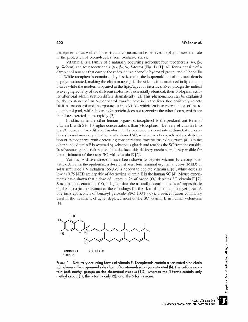
300 Weber et al.
and epidermis, as well as in the stratum corneum, and is believed to play an essential rolein the protection of biomolecules from oxidative stress.
Vitamin E is a family of 8 naturally occurring isoforms: four tocopherols (α-, β-,γ-, δ-form) and four tocotrienols (α-, β-, γ-, δ-form) (Fig. 1) [1]. All forms consist of achromanol nucleus that carries the redox-active phenolic hydroxyl group, and a lipophilictail. While tocopherols contain a phytil side chain, the isoprenoid tail of the tocotrienolsis polyunsaturated, making the chain more rigid. The side chain is anchored in lipid mem-branes while the nucleus is located at the lipid/aqueous interface. Even though the radicalscavenging activity of the different isoforms is essentially identical, their biological activ-ity after oral administration differs dramatically [2]. This phenomenon can be explainedby the existence of an α-tocopherol transfer protein in the liver that positively selectsRRR-α-tocopherol and incorporates it into VLDL which leads to recirculation of the α-tocopherol pool, while this transfer protein does not recognize the other forms, which aretherefore excreted more rapidly [3].
In skin, as in the other human organs, α-tocopherol is the predominant form ofvitamin E with 5 to 10 higher concentrations than γ-tocopherol. Delivery of vitamin E tothe SC occurs in two different modes. On the one hand it stored into differentiating kera-tinocytes and moves up into the newly formed SC, which leads to a gradient-type distribu-tion of α-tocopherol with decreasing concentrations towards the skin surface [4]. On theother hand, vitamin E is secreted by sebaceous glands and reaches the SC from the outside.In sebaceous gland–rich regions like the face, this delivery mechanism is responsible forthe enrichment of the outer SC with vitamin E [5].
Various oxidative stressors have been shown to deplete vitamin E, among otherantioxidants. In the epidermis, a dose of at least four minimal erythemal doses (MED) ofsolar simulated UV radiation (SSUV) is needed to deplete vitamin E [6], while doses aslow as 0.75 MED are capable of destroying vitamin E in the human SC [4]. Mouse experi-ments have shown that a dose of 1 ppm � 2h of ozone (O3) depletes SC vitamin E [7].Since this concentration of O3 is higher than the naturally occuring levels of troposphericO3 the biological relevance of these findings for the skin of humans is not yet clear. Aone time application of benzoyl peroxide BPO (10% w/v), a concentration commonlyused in the treatment of acne, depleted most of the SC vitamin E in human volunteers[8].
FIGURE 1 Naturally occurring forms of vitamin E. Tocopherols contain a saturated side chain(a), whereas the isoprenoid side chain of tocotrienols is polyunsaturated (b). The α-forms con-tain both methyl groups on the chromanol nucleus (1,2), whereas the β-forms contain onlymethyl group (1), the γ-forms only (2), and the δ-forms none.
Antioxidants 301
α-Tocopherol is widely used as an active ingredient in topical formulations. Aftertopical application, it penetrates readily into skin [9]. Since the free form of vitamin E isquite unstable and light-sensitive (it absorbs in the UV-B range), the active hydroxyl groupis usually protected by esterification with acetate. This increases the stability but rendersthe compound redox inactive. When administered orally, vitamin E-acetate is hydrolyzedquantitatively in the intestines [10]. There is some controversy however as to whether α-tocopherol acetate can by hydrolyzed in human skin. Chronic application of α-tocopherolacetate leads to an increase in free vitamin E in both the rat [11] and the mouse [12],where it was recently shown that UV-B increases the hydrolysis of α-tocopherol acetateby induction of nonspecific esterases up to 10 to 30 fold [13]. While one study suggestedthat bioconversion of α-tocopherol acetate does not occur in human skin [14], significanthydrolysis was demonstrated in recent studies using a human epidermis–tissue culturemodel [15].
The availability of the free form of vitamin E needs to be considered when analyzingpossible health benefits. The majority of studies have been carried out in animal models,while only limited data exists for human studies. Lipid peroxidation is inhibited aftertopical application of α-tocopherol [16]. Several studies indicate that topically appliedα-tocopherol inhibits UVB-induced photodamage of DNA in a mouse model [17] andkeratinocyte cultures (trolox) [18]. Protection against Langerhans cell depletion by UVlight was observed after topical application of α-tocopherol in a mouse model [19]. α-Tocopherol and its sorbate ester were studied in a mouse model of skin aging. Both antioxi-dants were found to be effective, sorbate even more so than α-tocopherol [20]. Systemicadministration of vitamin E in humans (only in combination with vitamin C) increasedthe MED and reduced changes in skin blood flow after UV-irradiation [21,22]. Yet severalstudies indicate that α-tocopherol acetate is not as effective as free vitamin E when appliedtopically. Inhibition of DNA mutation in mice was 5 to 10 times less effective [18]. Also,in a mouse model, unlike free vitamin E, the acetate form seemed to be ineffective [23].In summary, even though some health benefits of vitamin E supplementation have beenshown, there is still a need for controlled studies in humans under physiological conditions.
Recently, the tocotrienol forms of vitamin E have become a focus of interest, sincethey have been found to be more efficient antioxidants in some model systems than tocoph-erols [24]. Even if they are not bioavailable after oral supplementation, topical applicationcircumvents the exclusion by α-TTP in the liver. In fact, free tocotrienols readily penetrateinto mouse skin [9], and tocotrienyl acetate is hydrolyzed in skin homogenates and inmurine skin in vivo [25]. Topical application of a tocotrienol-rich fraction has been demon-strated to protect mouse skin from UV- and O3-induced oxidative stress [26,27]. In conclu-sion, tocotrienols bear a potential that yet remains to be explored.
VITAMIN C
Ascorbic acid or vitamin C is one of the most important water soluble antioxidants andpresent in high amounts in the skin. While most species are able to produce ascorbic acid,humans lack the enzymes necessary for its synthesis. Deficiency in ascorbic acid causesscurvy, a disease already described in the ancient writings of the Greeks [28]. Apart fromthe pure antioxidant function ascorbic acid is an essential co-factor for different enzymes.The antioxidant capacity of vitamin C is related to its unique structure (Fig. 2). Due toits pKa1 of 4.25 it is present as a monoanion at physiological pH, which can undergo a
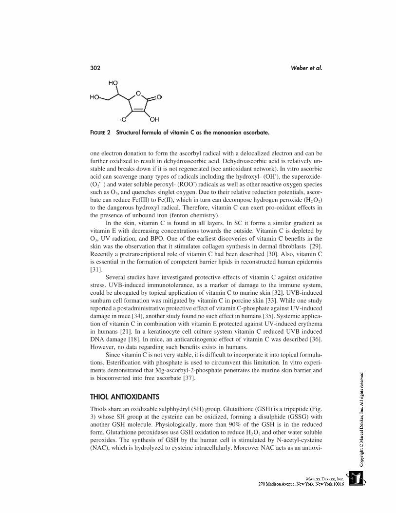
302 Weber et al.
FIGURE 2 Structural formula of vitamin C as the monoanion ascorbate.
one electron donation to form the ascorbyl radical with a delocalized electron and can befurther oxidized to result in dehydroascorbic acid. Dehydroascorbic acid is relatively un-stable and breaks down if it is not regenerated (see antioxidant network). In vitro ascorbicacid can scavenge many types of radicals including the hydroxyl- (OH•), the superoxide-(O2
•�) and water soluble peroxyl- (ROO•) radicals as well as other reactive oxygen speciessuch as O3, and quenches singlet oxygen. Due to their relative reduction potentials, ascor-bate can reduce Fe(III) to Fe(II), which in turn can decompose hydrogen peroxide (H2 O2)to the dangerous hydroxyl radical. Therefore, vitamin C can exert pro-oxidant effects inthe presence of unbound iron (fenton chemistry).
In the skin, vitamin C is found in all layers. In SC it forms a similar gradient asvitamin E with decreasing concentrations towards the outside. Vitamin C is depleted byO3, UV radiation, and BPO. One of the earliest discoveries of vitamin C benefits in theskin was the observation that it stimulates collagen synthesis in dermal fibroblasts [29].Recently a pretranscriptional role of vitamin C had been described [30]. Also, vitamin Cis essential in the formation of competent barrier lipids in reconstructed human epidermis[31].
Several studies have investigated protective effects of vitamin C against oxidativestress. UVB-induced immunotolerance, as a marker of damage to the immune system,could be abrogated by topical application of vitamin C to murine skin [32]. UVB-inducedsunburn cell formation was mitigated by vitamin C in porcine skin [33]. While one studyreported a postadministrative protective effect of vitamin C-phosphate against UV-induceddamage in mice [34], another study found no such effect in humans [35]. Systemic applica-tion of vitamin C in combination with vitamin E protected against UV-induced erythemain humans [21]. In a keratinocyte cell culture system vitamin C reduced UVB-inducedDNA damage [18]. In mice, an anticarcinogenic effect of vitamin C was described [36].However, no data regarding such benefits exists in humans.
Since vitamin C is not very stable, it is difficult to incorporate it into topical formula-tions. Esterification with phosphate is used to circumvent this limitation. In vitro experi-ments demonstrated that Mg-ascorbyl-2-phosphate penetrates the murine skin barrier andis bioconverted into free ascorbate [37].
THIOL ANTIOXIDANTS
Thiols share an oxidizable sulphhydryl (SH) group. Glutathione (GSH) is a tripeptide (Fig.3) whose SH group at the cysteine can be oxidized, forming a disulphide (GSSG) withanother GSH molecule. Physiologically, more than 90% of the GSH is in the reducedform. Glutathione peroxidases use GSH oxidation to reduce H2 O2 and other water solubleperoxides. The synthesis of GSH by the human cell is stimulated by N-acetyl-cysteine(NAC), which is hydrolyzed to cysteine intracellularly. Moreover NAC acts as an antioxi-
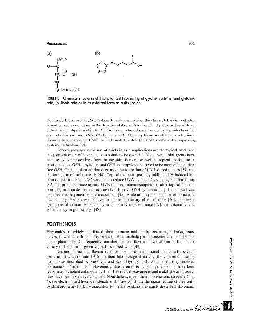
Antioxidants 303
FIGURE 3 Chemical structures of thiols: (a) GSH consisting of glycine, cysteine, and glutamicacid; (b) lipoic acid as in its oxidized form as a disulphide.
dant itself. Lipoic acid (1,2-dithiolane-3-pentanonic acid or thioctic acid, LA) is a cofactorof multienzyme complexes in the decarboxylation of α-keto acids. Applied as the oxidizeddithiol dehydrolipoic acid (DHLA) it is taken up by cells and is reduced by mitochondrialand cytosolic enzymes (NAD(P)H dependent). It thereby forms an efficient cycle, sinceit can in turn regenerate GSSG to GSH and stimulate the GSH synthesis by improvingcysteine utilization [38].
General provisos in the use of thiols in skin applications are the typical smell andthe poor solubility of LA in aqueous solutions below pH 7. Yet, several thiol agents havebeen tested for protective effects in the skin. For oral as well as topical application inmouse models, GSH-ethylesters and GSH-isopropylesters proved to be more efficient thanfree GSH. Oral supplementation decreased the formation of UV-induced tumors [39] andthe formation of sunburn cells [40]. Topical treatment partially inhibited UV-induced im-munosupression [41]. NAC was able to reduce UVA-induced DNA damage in fibroblasts[42] and protected mice against UVB-induced immunosuppression after topical applica-tion [43] in a mode that did not involve de novo GSH synthesis [44]. Lipoic acid wasdemonstrated to penetrate into mouse skin [45], while oral supplementation of lipoic acidhas actually been shown to have an anti-inflammatory effect in mice [46], to preventsymptoms of vitamin E deficiency in vitamin E–deficient mice [47], and vitamin C andE deficiency in guinea pigs [48].
POLYPHENOLS
Flavonoids are widely distributed plant pigments and tannins occurring in barks, roots,leaves, flowers, and fruits. Their roles in plants include photoprotection and contributingto the plant color. Consequently, our diet contains flavonoids which can be found in avariety of foods from green vegetables to red wine [49].
Despite the fact that flavonoids have been used in traditional medicine for severalcenturies, it was not until 1936 that their first biological activity, the vitamin C–sparingaction, was described by Rusznyak and Szent-Györgyi [50]. As a result, they receivedthe name of ‘‘vitamin P.’’ Flavonoids, also referred to as plant polyphenols, have beenrecognized as potent antioxidants. Their free radical-scavenging and metal-chelating activ-ities have been extensively studied. Nonetheless, given their polyphenolic structure (Fig.4), the electron- and hydrogen-donating abilities constitute the major feature of their anti-oxidant properties [51]. By opposition to the antioxidants previously described, flavonoids
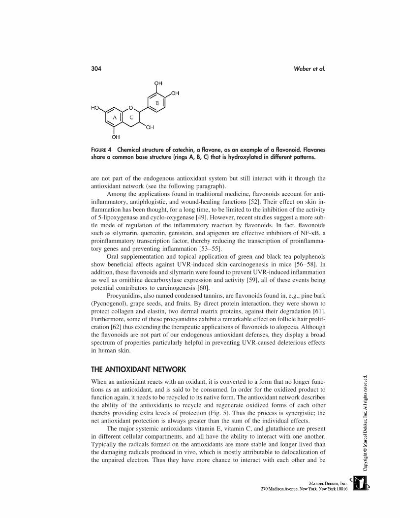
304 Weber et al.
FIGURE 4 Chemical structure of catechin, a flavane, as an example of a flavonoid. Flavanesshare a common base structure (rings A, B, C) that is hydroxylated in different patterns.
are not part of the endogenous antioxidant system but still interact with it through theantioxidant network (see the following paragraph).
Among the applications found in traditional medicine, flavonoids account for anti-inflammatory, antiphlogistic, and wound-healing functions [52]. Their effect on skin in-flammation has been thought, for a long time, to be limited to the inhibition of the activityof 5-lipoxygenase and cyclo-oxygenase [49]. However, recent studies suggest a more sub-tle mode of regulation of the inflammatory reaction by flavonoids. In fact, flavonoidssuch as silymarin, quercetin, genistein, and apigenin are effective inhibitors of NF-κB, aproinflammatory transcription factor, thereby reducing the transcription of proinflamma-tory genes and preventing inflammation [53–55].
Oral supplementation and topical application of green and black tea polyphenolsshow beneficial effects against UVR-induced skin carcinogenesis in mice [56–58]. Inaddition, these flavonoids and silymarin were found to prevent UVR-induced inflammationas well as ornithine decarboxylase expression and activity [59], all of these events beingpotential contributors to carcinogenesis [60].
Procyanidins, also named condensed tannins, are flavonoids found in, e.g., pine bark(Pycnogenol), grape seeds, and fruits. By direct protein interaction, they were shown toprotect collagen and elastin, two dermal matrix proteins, against their degradation [61].Furthermore, some of these procyanidins exhibit a remarkable effect on follicle hair prolif-eration [62] thus extending the therapeutic applications of flavonoids to alopecia. Althoughthe flavonoids are not part of our endogenous antioxidant defenses, they display a broadspectrum of properties particularly helpful in preventing UVR-caused deleterious effectsin human skin.
THE ANTIOXIDANT NETWORK
When an antioxidant reacts with an oxidant, it is converted to a form that no longer func-tions as an antioxidant, and is said to be consumed. In order for the oxidized product tofunction again, it needs to be recycled to its native form. The antioxidant network describesthe ability of the antioxidants to recycle and regenerate oxidized forms of each otherthereby providing extra levels of protection (Fig. 5). Thus the process is synergistic; thenet antioxidant protection is always greater than the sum of the individual effects.
The major systemic antioxidants vitamin E, vitamin C, and glutathione are presentin different cellular compartments, and all have the ability to interact with one another.Typically the radicals formed on the antioxidants are more stable and longer lived thanthe damaging radicals produced in vivo, which is mostly attributable to delocalization ofthe unpaired electron. Thus they have more chance to interact with each other and be
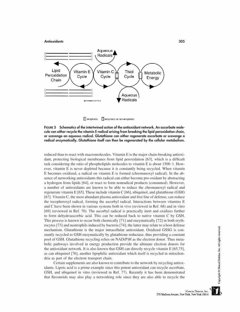
Antioxidants 305
FIGURE 5 Schematics of the intertwined action of the antioxidant network. An ascorbate mole-cule can either recycle the vitamin E radical arising from breaking the lipid peroxidation chain,or scavenge an aqueous radical. Glutathione can either regenerate ascorbate or scavenge aradical enzymatically. Glutathione itself can then be regenerated by the cellular metabolism.
reduced than to react with macromolecules. Vitamin E is the major chain-breaking antioxi-dant, protecting biological membranes from lipid peroxidation [63], which is a difficulttask considering the ratio of phospholipids molecules to vitamin E is about 1500:1. How-ever, vitamin E is never depleted because it is constantly being recycled. When vitaminE becomes oxidized, a radical on vitamin E is formed (chromanoxyl radical). In the ab-sence of networking antioxidants this radical can either become pro-oxidant by abstractinga hydrogen from lipids [64], or react to form nonradical products (consumed). However,a number of antioxidants are known to be able to reduce the chromanoxyl radical andregenerate vitamin E [65]. These include vitamin C [66], ubiquinol, and glutathione (GSH)[67]. Vitamin C, the most abundant plasma antioxidant and first line of defense, can reducethe tocopheroxyl radical, forming the ascorbyl radical. Interactions between vitamins Eand C have been shown in various systems both in vivo (reviewed in Ref. 68) and in vitro[69] (reviewed in Ref. 70). The ascorbyl radical is practically inert and oxidizes furtherto form dehydroascorbic acid. This can be reduced back to native vitamin C by GSH.This process is known to occur both chemically [71] and enzymatically [72] in both eryth-rocytes [73] and neutrophils induced by bacteria [74]; the latter may relate to a host defensemechanism. Glutathione is the major intracellular antioxidant. Oxidized GSSG is con-stantly recycled to GSH enzymatically by glutathione reductase, thus providing a constantpool of GSH. Glutathione recycling relies on NAD(P)H as the electron donor. Thus meta-bolic pathways involved in energy production provide the ultimate electron donors forthe antioxidant network. It is also known that GSH can directly recycle vitamin E [65,75],as can ubiquinol [76], another lipophilic antioxidant which itself is recycled in mitochon-dria as part of the electron transport chain.
Certain supplements are also known to contribute to the network by recycling antiox-idants. Lipoic acid is a prime example since this potent antioxidant can recycle ascorbate,GSH, and ubiquinol in vitro (reviewed in Ref. 77). Recently it has been demonstratedthat flavonoids may also play a networking role since they are also able to recycle the
306 Weber et al.
ascorbyl radical [78]. Thus there exists a very organized defense system against free radicalattack, which ultimately serves to protect and recycle antioxidants in various cellular com-partments.
REGULATION OF GENE TRANSCRIPTION BY ANTIOXIDANTS
The skin is the largest human organ and permanently exposed to a variety of stresses.Among those, oxidative insults such as ultraviolet radiation and ozone exposure accountfor the cause of many skin disorders. However, oxidative damage are not responsible forall biological effects engendered by these stressors in the skin. Indeed, ultraviolet radiation(UVR) causes changes in the expression of genes encoding, e.g., proinflammatory cyto-kines, growth factors, stress response proteins, oncoproteins, and matrix metalloprotei-nases [79]. Although the immediate target(s) of UVR is/are still unknown, certain kinasesand transcription factors can be activated by UVR thereby increasing gene transcription[80]. One transcription factor, NF-κB, appears of particular interest for the skin since thelack of its inhibitory protein, IκBα, is associated with the development of a widespreaddermititis in knockout mice [81,82]. Furthermore, reactive oxygen species, such as theones produced after UVR, are suspected to play an important role in the activation of NF-κB [83]. Consequently, antioxidants have been found to be among the most potent NF-κB inhibitors. However, clinical studies are required in order to assess the effectivenessof these antioxidants, including the flavonoid silymarin, α-lipoic acid and the glutathioneprecursor N-acetyl-L-cysteine, on skin inflammatory disorders. Using high-throughputprocedures such as the cDNA arrays, for instance [84], the evaluation of the antioxidantson the whole genome is henceforth possible. These studies will only confirm the hypothesisthat antioxidants are responsible for a much broader action spectrum than their antioxidantfunctions per se and extend their role on more subtle regulatory mechanisms of the geneexpression.
PERSPECTIVES
The general role of antioxidants in the protection against oxidative stress is well estab-lished. In skin applications antioxidants are a promising tool to mitigate oxidative injury.Even though a growing amount of literature deals with skin protection by antioxidants,there is still a need for investigation. In particular, clinical human studies need to be carriedout to show the efficacy of antioxidants in topical formulations.
ACKNOWLEDGMENT
We would like to thank Nancy Han for editing the manuscript.
REFERENCES
1. Brigelius-Flohe R, Traber MG. Vitamin E: function and metabolism. FASEB J 1999; 13:1145–1155.
2. Traber MG, Rader D, Acuff RV, Ramakrishnan R, Brewer HB, Kayden HJ. Vitamin E dose-response studies in humans with use of deuterated RRR-alpha-tocopherol. Am J Clin Nutr1998; 68:847–853.
3. Traber MG, Ramakrishnan RR, Kayden HJ. Human plasma vitamin E kinetics demonstrate
Antioxidants 307
rapid recycling of plasma RRR-alpha-tocopherol. Proc Natl Acad Sci USA 1994; 91:10005–10008.
4. Thiele JJ, Traber MG, Packer L. Depletion of human stratum corneum vitamin E: an earlyand sensitive in vivo marker of UV induced photo-oxidation. J Invest Dermatol 1998; 110:756–761.
5. Thiele JJ, Weber SU, Packer L. Sebaceous gland secretion is a major physiological route ofvitamin E delivery to the skin. J Invest Dermatol 1999; 113:1006–1010.
6. Shindo Y, Witt E, Han D, Packer L. Dose-response effects of acute ultraviolet irradiation onantioxidants and molecular markers of oxidation in murine epidermis and dermis. J InvestDermatol 1994; 102:470–475.
7. Thiele JJ, Traber MG, Polefka TG, Cross CE, Packer L. Ozone-exposure depletes vitamin Eand induces lipid peroxidation in murine stratum corneum. J Invest Dermatol 1997; 108:753–757.
8. Thiele JJ, Rallis M, Izquierdo-Pullido M, et al. Benzoyl peroxide depletes human stratumcorneum antioxidants. J Invest Dermatol 1998; 110:674.
9. Traber MG, Rallis M, Podda M, Weber C, Maibach HI, Packer L. Penetration and distributionof alpha-tocopherol, alpha- or gamma-tocotrienols applied individually onto murine skin. Lip-ids 1998; 33:87–91.
10. Traber MG, Serbinova EA, Packer L. Biological activities of tocotrienols and tocopherols. In:Packer L, Hiramatsu M, Yoshikawa T, eds. Antioxidant Food Supplements in Human Health.New York: Academic Press, 1999.
11. Norkus EP, Bryce GF, Bhagavan HN. Uptake and bioconversion of alpha-tocopheryl acetateto alpha-tocopherol in skin of hairless mice. Photochem Photobiol 1993; 57:613–615.
12. Beijersbergen van Henegouwen GM, Junginger HE, de Vries H. Hydrolysis of RRR-alpha-tocopheryl acetate (vitamin E acetate) in the skin and its UV protecting activity (an in vivostudy with the rat). J Photochem Photobiol B 1995; 29:45–51.
13. Kramer-Stickland K, Liebler DC. Effect of UVB on hydrolysis of alpha-tocopherol acetateto alpha-tocopherol in mouse skin. J Invest Dermatol 1998; 111:302–307.
14. Alberts DS, Goldman R, Xu MJ, et al. Disposition and metabolism of topically administeredalpha-tocopherol acetate: a common ingredient of commercially available sunscreens and cos-metics. Nutr Cancer 1996; 26:193–201.
15. Nabi Z, Tavakkol A, Soliman N, Polefka TG. Bioconversion of tocopheryl acetate to tocoph-erol in human skin: use of human skin organ culture models. J Dermatol Sci 1998; 16:S207.
16. Lopez-Torres M, Thiele JJ, Shindo Y, Han D, Packer L. Topical application of alpha-tocoph-erol modulates the antioxidant network and diminishes ultraviolet-induced oxidative damagein murine skin. Br J Dermatol 1998; 138:207–215.
17. McVean M, Liebler DC. Inhibition of UVB induced DNA photodamage in mouse epidermisby topically applied alpha-tocopherol. Carcinogenesis 1997; 18:1617–1622.
18. Stewart MS, Cameron GS, Pence BC. Antioxidant nutrients protect against UVB-induced oxi-dative damage to DNA of mouse keratinocytes in culture. J Invest Dermatol 1996; 106:1086–1089.
19. Halliday GM, Bestak R, Yuen KS, Cavanagh LL, Barnetson RS. UVA-induced immunosup-pression. Mutat Res 1998; 422:139–145.
20. Jurkiewicz BA, Bisset DL, Buettner GR. Effect of topically applied tocopherol on ultravioletradiation-mediated free radical damage in skin. J Invest Dermatol 1995; 104:484–488.
21. Eberlein-Konig B, Placzek M, Przybilla B. Protective effect against sunburn of combinedsystemic ascorbic acid (vitamin C) and d-alpha-tocopherol (vitamin E). J Am Acad Dermatol1998; 38:45–48.
22. Fuchs J, Kern H. Modulation of UV-light-induced skin inflammation by D-alpha-tocopheroland L-ascorbic acid: a clinical study using solar simulated radiation. Free Radic Biol Med1998; 25:1006–1012.
23. Yuen KS, Halliday GM. Alpha-Tocopherol, an inhibitor of epidermal lipid peroxidation, pre-
308 Weber et al.
vents ultraviolet radiation from suppressing the skin immune system. Photochem Photobiol1997; 65:587–592.
24. Serbinova EA, Packer L. Antioxidant properties of alpha-tocopherol and alpha-tocotrienol.Methods Enzymol 1994; 234:354–366.
25. Weber SU, Luu C, Traber MG, Packer L. Tocotrienol acetate penetrates into murine skin andis hydrolyzed in vivo. Oxygen Club of California, Book of Abstracts 1999:13.
26. Weber C, Podda M, Rallis M, Thiele JJ, Traber MG, Packer L. Efficacy of topically appliedtocopherols and tocotrienols in protection of murine skin from oxidative damage induced byUV-irradiation. Free Radic Biol Med 1997; 22:761–769.
27. Thiele JJ, Traber MG, Podda M, Tsang K, Cross CE, Packer L. Oxone depletes tocopherolsand tocotrienols topically applied to murine skin. FEBS Lett 1997; 401:167–170.
28. Sauberlich HE. Pharmacology of vitamin C. Ann Rev Nutr 1994; 14:371–391.29. Murad S, Grove D, Lindberg KA, Reynolds G, Sivarajah A, Pinnel SR. Regulation of collagen
synthesis by ascorbic acid. Proc Natl Acad Sci USA 1981; 78:2879–2882.30. Davidson JM, LuValle PA, Zoia O, Quaglino D Jr, Giro M. Ascorbate differentially regulates
elastin and collagen biosynthesis in vascular smooth muscle cells and skin fibroblasts by pre-translational mechanisms. J Biol Chem 1997; 272:345–352.
31. Ponec M, Weerheim A, Kempenaar J, et al. The formulation of competent barrier lipids inreconstructed human epidermis requires the presence of vitamin C. J Invest Dermatol 1997;109:348–355.
32. Nakamura T, Pinnel SR, Darr D, et al. Vitamin C abrogates the deleterious effects of UVBradiation on cutaneous immunity by a mechanism that does not depend on TNF-alpha. J InvestDermatol 1997; 109:20–24.
33. Darr D, Combs S, Dunston S, Manning T, Pinnell S. Topical vitamin C protects porcine skinfrom ultraviolet radiation-induced damage. Br J Dermatol 1992; 127:247–253.
34. Kobayashi S, Takehana M, Kanke M, Itoh S, Ogata E. Postadministration protective effectof magnesium-L-ascorbyl-phosphate on the development of UVB-induced cutaneous damagein mice. Photochem Photobiol 1998; 67:669–675.
35. Dreher F, Denig N, Gabard B, Schwindt DA, Maibach HI. Effect of topical antioxidants onUV-induced erythema formation when administered after exposure. Dermatology 1999; 198:52–55.
36. Pauling L. Effect of ascorbic acid on incidence of spontaneous mammary tumors and UV-light–induced skin tumors in mice. Am J Clin Nutr 1991; 54:1252S–1255S.
37. Kobayashi S, Takehana M, Itoh S, Ogata E. Protective effect of magnesium-L-ascorbyl-2phosphate against skin damage induced by UVB irradiation. Photochem Photobiol 1996; 64:224–228.
38. Han D, Handelman G, Marcocci L, et al. Lipoic acid increases de novo synthesis of cellularglutathione by improving cytine utilization. Biofactors 1997; 6:321–338.
39. Kobayashi S, Takehana M, Tohyama C. Glutathione isopropyl ester reduces UVB-inducedskin damage in hairless mice. Photochem Photobiol 1996; 63:106–110.
40. Hanada K, Sawamura D, Tamai K, Hashimoto I, Kobayashi S. Photoprotective effect of esteri-fied glutathione against ultraviolet B–induced sunburn cell formation in the hairless mice. JInvest Dermatol 1997; 108:727–730.
41. Steenvoorden DP, Beijersbergen van Henegouwen G. Glutathione ethylester protects againstlocal and systemic suppression of contact hypersensitivity induced by ultraviolet B radiationin mice. Radiat Res 1998; 150:292–297.
42. Emonet-Piccardi N, Richard MJ, Ravanat JL, Signorini N, Cadet J, Beani JC. Protective effectsof antioxidants against UVA-induced DNA damage in human skin fibroblasts in culture. FreeRadic Res 1998; 29:307–313.
43. Steenvoorden DP, Beijersburgen van Henegouwen GM. Glutathione synthesis is not involvedin protection by N-acetylcysteine against UVB-induced systemic immunosuppression in mice.Photochem Photobiol 1998; 68:97–100.
Antioxidants 309
44. Steenvoorden DP, Hasselbaink DM, Beijersbergen van Henegouwen GM. Protection againstUV-induced reactive intermediates in human cells and mouse skin by glutathione precursors:a comparison of N-acetylcysteine and glutathione ethylester. Photochem Photobiol 1998; 67:651–656.
45. Podda M, Rallis M, Traber MG, Packer L, Maibach HI. Kinetic study of cutaneous and subcu-taneous distribution following topical application of [7,8–14C]rac-alpha-lipoic acid onto hair-less mice. Biochem Pharmacol 1996; 52:627–633.
46. Fuchs J, Milbradt R. Antioxidant inhibition of skin inflammation induced by reactive oxidants:evaluation of the redox couple dihydrolipoate/lipoate. Skin Pharmacol 1994; 7:278–284.
47. Podda M, Tritschler HJ, Ulrich H, Packer L. Alpha-lipoic acid supplementation prevents symp-toms of vitamin E deficiency. Biochem Biophys Res Commun 1994; 204:98–104.
48. Rosenberg HR, Culik R. Effect of α-lipoic acid on vitamin C and vitamin E deficiencies. ArchBiochem Biophys 1959; 80:86–93.
49. Pietta P. Flavonoids in medicinal plants. In: Packer L, Rice-Evans C, eds. Flavonoids in Healthand Disease. New York: Marcel Dekker, 1998:61–110.
50. Rusznyak SP, Szent-Györgyi A. Vitamin P: flavonols as vitamins. Nature 1936; 138:27.51. Rice-Evans CA, Miller NJ, Paganga G. Structure-antioxidant activity relationships of flavo-
noids and phenolic acids. Free Radic Biol Med 1996; 20:933–956.52. Middleton E Jr, Kandaswami C. The impact of plant flavonoids on mammalian biology: impli-
cations for immunity, inflammation and cancer. In: Harborne JB, ed. The Flavonoids: Ad-vances in Research Since 1986. London: Chapman & Hall, 1993.
53. Saliou C, Kitazawa M, McLaughlin L, et al. Antioxidants modulate acute solar ultravioletradiation-induced NF-kappa-B activation in a human keratinocyte cell line. Free Radic Med1999; 26:174–183.
54. Gerritsen ME, Carley WW, Ranges GE, et al. Flavonoids inhibit cytokine-induced endothelialcell adhesion protein gene expression. Am J Pathol 1995; 147:278–292.
55. Natarajan K, Manna SK, Chaturvedi MM, Aggarwal BB. Protein tyrosine kinase inhibitorsblock tumor necrosis factor-induced activation of nuclear factor-κB, degradation of IκBα,nuclear translocation of p65, and subsequent gene expression. Arch Biochem Biophys 1998;352:59–70.
56. Gensler HL, Timmermann BN, Valcic S, et al. Prevention of photocarcinogenesis by topicaladministration of pure epigallocatechin gallate isolated from green tea. 1996; 26:325–335.
57. Wang ZY, Huang MT, Lou YR, et al. Inhibitory effects of black tea, green tea, decaffein-ated black tea, and decaffeinated green tea on ultraviolet B light–induced skin carcino-genesis in 7,12-dimethylbenz[a]anthracene-initiated SKH-1 mice. Cancer Res 1994; 54:3428–3435.
58. Javed S, Mehrotra NK, Sukla Y. Chemopreventive effects of black tea polyphenols in mouseskin model of carcinogenesis. Biomed Environ Sci 1998; 11:307–313.
59. Katiyar SK, Korman NJ, Mukhtar H, Agarwal R. Protective effects of silymarin against photo-carcinogenesis in a mouse skin model. J Natl Cancer Inst 1997; 89:556–566.
60. Agarwal R, Mukhtar H. Chemoprevention of photocarcinogenesis. Photochem Photobiol 1996;63:440–444.
61. Tixier JM, Godeau G, Robert AM, Hornebeck W. Evidence by in vivo and in vitro studiesthat binding of pycnogenols to elastin affects its rate of degradation by elastases. BiochemPharmacol 1984; 33:3933–3939.
62. Takahashi T, Kamiya T, Hasegawa A, Yokoo Y. Procyanidin oligomers selectively and inten-sively promote proliferation of mouse hair epithelial cells in vitro and activate hair folliclegrowth in vivo. J Invest Dermatol 1999; 112:310–316.
63. Burton GW, Ingold KU. Autoxidation of biological molecules. I. The antioxidant activity ofvitamin E and related chain-breaking phenolic antioxidants in vitro. J Am Chem Soc 1981;103:6472–6477.
64. Bowry VW, Stocker R. Tocopherol-mediated peroxidation—the prooxidant effect of vitamin
310 Weber et al.
E on the radical initiated oxidation of human low density lipoprotein. J Am Chem Soc 1993;115:6029–6044.
65. Sies H. Strategies of antioxidant defense. Eur J Biochem 1993; 215:213–219.66. Packer JE, Slater TF, Willson RL. Direct observation of a free radical interaction between
vitamin E and vitamin C. Nature 1979; 278:737–738.67. Wefers H, Sies H. The protection by ascorbate and glutathione against microsomal lipid perox-
idation is dependent on vitamin E. Eur J Biochem 1988; 174:353–357.68. Gey KF. Vitamins E plus C and interacting conutrients required for optimal health. Biofactors
1998; 7:113–174.69. Kagan VE, Witt E, Goldman R, Scita G, Packer L. Ultraviolet light-induced generation of
vitamin E radicals and their recycling. A possible photosensitizing effect of vitamin E in skin.Free Radic Res Commun 1992; 16:51–64.
70. Kamal-Eldin A, Appelqvist L-A. The chemistry and antioxidant properties of tocopherols andtocotrienols. Lipids 1996; 31:671–701.
71. Winkler BS. Unequivocal evidence in support of the nonenzymatic redox coupling betweenglutathione/glutathione disulfide and ascorbic acid/dehydroascorbic acid. Biochem BiophysActa 1992; 1117:287–290.
72. Wells WW, Xu DP, Yang YF, Rocque PA. Mammalian thioltransferase (glutaredoxin) andprotein disulfide isomerase have dehydroascorbate reductase activity. J Biol Chem 1990; 265:15361–15364.
73. May JM, Qu ZC, Whitesell RR, Cobb CE. Ascorbate recycling in human erythrocytes: roleof GSH in reducing dehydroascorbate. Free Rad Biol Med 1996; 20:543–551.
74. Wang Y, Russo TA, Kwon O, Chanock S, Rumsey SC, Levine M. Ascorbate recycling inhuman neutrophils: induction by bacteria. Proc Natl Acad Sci USA 1997; 94:13816–13819.
75. Bast A, Haenen GRMM. Regulation of lipid peroxidation of glutathione and lipoic acid:involvement of liver microsomal vitamin E free radical reductase. In: Emerit I, Packer L,Auclair C, eds. Antioxidant in Therapy in Preventive Medicine. New York: Plenum Press,1990:111–116.
76. Kagan V, Serbinova E, Packer L. Antioxidant effects of ubiquinones in microsomes and mito-chondria are mediated by tocopherol recycling. Biochem Biophys Res Commun 1990; 169:851–857.
77. Packer L, Witt E, Tritschler HJ. α-Lipoic acid as a biological antioxidant. Free Rad Biol Med1995; 19:227–250.
78. Cossins E, Lee R, Packer L. ESR studies of vitamin C regeneration, order of reactivity ofnatural source phytochemical preparations. Biochem Mol Biol Int 1998; 45:583–597.
79. Tyrrell RM. UV activation of mammalian stress protein. In: Feige U, Morimoto RI, YaharaI, Polla B, eds. Stress-Inducible Cellular Responses. Basel (Switzerland): Birkhaüser Verlag,1996:255–271.
80. Herrlich P, Blattner C, Knebel A, Bender K, Rahmsdorf HJ. Nuclear and non-nuclear targetsof genotoxic agents in the induction of gene expression: shared principles in yeast, rodents,man and plants. Biol Chem 1997; 378:1217–1229.
81. Beg AA, Sha WC, Bronson RT, Baltimore D. Constitutive NF-kappa-B activation, enhancedgranulopoiesis, and neonatal lethality in I-kappa-B-alpha deficient mice. Genes Dev 1995; 9:2736–2746.
82. Klement JF, Rice NR, Car BD, et al. I-kappa-B-alpha deficiency results in a sustained NF-kappa-B response and severe widespread dermatitis in mice. Mol Cell Biol 1996; 16:2341–2349.
83. Flohé L, Brigelius-Flohé R, Saliou C, Traber M, Packer L. Redox regulation of NF-κB activa-tion. Free Radic Biol Med 1997; 22:1115–1126.
84. Schena M, Heller RA, Theriault TP, Konrad K, Lachenmeier E, Davis RW. Microarrays:biotechnology’s discovery platform for functional genomics. Trends Biotechnol 1998; 16:301–306.
27
Alpha Hydroxy Acids
Enzo BerardescaUniversity of Pavia, Pavia, Italy
Alpha hydroxy acids (AHAs) constitute a class of compounds that exert specific andunique effects on skin structures. The therapeutic utility of these acids continues to expand;when applied to the skin in higher concentrations they cause detachment of keratinocytesand epidermolysis while application in lower concentration reduces intercorneocyte cohe-sion and visible stratum corneum desquamation.
The smallest AHA is glycolic acid, which is constituted by two carbons (H2 C(OH)-COOH); lactic acid contains three carbons and converts to its keto form, pyruvic acid,and vice versa. Malic acid and tartaric acid consists of four carbon chains, while citricand gluconic acid have six carbon chains [1].
AHAs are found in nature in a variety of species including foods and plants (citric,malic, tartaric, glycolic), animals (cells and body fluids), and microorganisms such asbacteria, fungi, viruses, and algae. AHA are involved in many metabolic processes andparticipate in essential cellular pathways such as Krebs cycle, glycolysis, and serine bio-synthesis. Furthermore, they promote collagen maturation and formation of glucosaminog-lycans. Their mechanism of action can be hypothesized via multiple effects [2]:
1. On stratum corneum: low concentration of AHAs diminish corneocyte cohesion.The effect occurs at the lower levels of the stratum corneum and may involve a dynamicprocess, operative at a particular step of keratinization, like the modification of ionic bond-ing. The effect is clinically evident as a sheetlike separation of the stratum corneum [3].Indeed, intercorneocyte bonds are mostly noncovalent. In noncovalent bonds, the bondingforce may be ionic or nonionic. AHAs reduce corneocyte cohesion by influencing ionicbonds via three mechanisms: (a) the distance between charges, (b) the number of charges,and (c) the medium between charges. When the stratum corneum becomes hydrated, thedistance between corneocytes is increased and therefore cohesion is decreased. Anothermechanism involved is the enzymatic inhibition, induced by AHAs, of the reactions ofsulphate transferase, phosphototransferase, and kinases which leads to fewer electronega-tive sulphate and phosphate groups on the outer wall of corneocytes resulting in diminish-ment of cohesion forces. On the contrary, retinoids reduce intercorneocyte cohesion bybreaking down already formed sulphate and phosphate bonds via induction or activationof sulphatase or phosphatase.
311
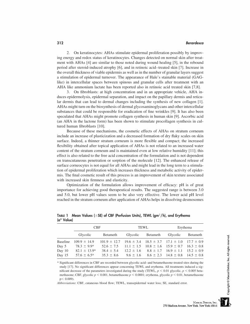
312 Berardesca
2. On keratinocytes: AHAs stimulate epidermal proliferation possibly by improv-ing energy and redox status of keratinocytes. Changes detected on normal skin after treat-ment with AHAs [4] are similar to those noted during wound healing [5], in the reboundperiod after steroid-induced atrophy [6], and in retinoic acid–treated skin [7]. Increase inthe overall thickness of viable epidermis as well as in the number of granular layers suggesta stimulation of epidermal turnover. The appearance of Hale’s stainable material (GAG-like) in intercellular spaces between spinous and granular cells after treatment with anAHA like ammonium lactate has been reported also in retinoic acid treated skin [7,8].
3. On fibroblasts: at high concentration and in an appropriate vehicle, AHA in-duces epidermolysis, epidermal separation, and impact on the papillary dermis and reticu-lar dermis that can lead to dermal changes including the synthesis of new collagen [1].AHAs might turn on the biosynthesis of dermal glycosaminoglycans and other intercellularsubstances that could be responsible for eradication of fine wrinkles [9]. It has also beenspeculated that AHAs might promote collagen synthesis in human skin [9]. Ascorbic acid(an AHA in the lactone form) has been shown to stimulate procollagen synthesis in cul-tured human fibroblasts [10].
Because of these mechanisms, the cosmetic effects of AHAs on stratum corneuminclude an increase of plasticization and a decreased formation of dry flaky scales on skinsurface. Indeed, a thinner stratum corneum is more flexible and compact; the increasedflexibility obtained after topical application of AHAs is not related to an increased watercontent of the stratum corneum and is maintained even at low relative humidity [11]; thiseffect is also related to the free acid concentration of the formulation and is not dependenton transcutaneous penetration or sorption of the molecule [12]. The enhanced release ofsurface corneocytes is not equal for all AHAs and might lead in the long term to a stimula-tion of epidermal proliferation which increases thickness and metabolic activity of epider-mis. The final cosmetic result of this process is an improvement of skin texture associatedwith increased skin firmness and elasticity.
Optimization of the formulation allows improvement of efficacy: pH is of greatimportance for achieving good therapeutical results. The suggested range is between 3.0and 5.0, but lower pH values seem to be also very effective. The lower acid pH levelreached in the stratum corneum after application of AHAs helps in dissolving desmosomes
TABLE 1 Mean Values (�SE) of CBF (Perfusion Units), TEWL (gm2/h), and Erythema(a* Value)
CBF TEWL Erythema
Glycolic Betameth Glycolic Betameth Glycolic Betameth
Baseline 109.9 � 14.9 101.9 � 12.7 19.6 � 3.4 18.5 � 3.7 17.1 � 1.0 17.7 � 0.9Day 5 78.3 � 9.9* 52.6 � 7.5 11.1 � 1.5 10.8 � 1.6 15.9 � 0.7 16.3 � 0.8Day 10 82.1 � 13.9* 38.4 � 5.4 12.2 � 1.6 8.8 � 1.7 16.9 � 1.1 15.2 � 0.9Day 15 57.6 � 6.5* 35.3 � 8.6 9.6 � 1.6 8.6 � 2.3 14.8 � 0.8 14.5 � 0.8
* Significant differences in CBF are recorded between glycolic acid–and betamethasone-treated sites during thestudy [17]. No significant differences appear concerning TEWL and erythema. All treatments induced a sig-nificant decrease of the parameters investigated during the study (TEWL, p � 0.01 glycolic, p � 0.005 beta-methasone; CBF, glycolic p � 0.001, betamethasone p � 0.0001; erythema, glycolic p � 0.01, betamethasonep� 0.009).
Abbreviations: CBF, cutaneous blood flow; TEWL, transepidermal water loss; SE, standard error.
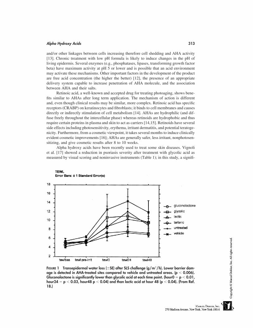
Alpha Hydroxy Acids 313
and/or other linkages between cells increasing therefore cell shedding and AHA activity[13]. Chronic treatment with low pH formula is likely to induce changes in the pH ofliving epidermis. Several enzymes (e.g., phosphatases, lipases, transforming growth factorbeta) have maximum activity at pH 5 or lower and is possible that an acid environmentmay activate these mechanisms. Other important factors in the development of the productare free acid concentration (the higher the better) [12], the presence of an appropriatedelivery system capable to increase penetration of AHA molecule, and the associationbetween AHA and their salts.
Retinoic acid, a well-known and accepted drug for treating photoaging, shows bene-fits similar to AHAs after long term application. The mechanism of action is differentand, even though clinical results may be similar, more complex. Retinoic acid has specificreceptors (CRABP) on keratinocytes and fibroblasts; it binds to cell membranes and causesdirectly or indirectly stimulation of cell metabolism [14]. AHAs are hydrophilic (and dif-fuse freely throughout the intercellular phase) whereas retinoids are hydrophobic and thusrequire certain proteins in plasma and skin to act as carriers [14,15]. Retinoids have severalside effects including photosensitivity, erythema, irritant dermatitis, and potential teratoge-nicity. Furthermore, from a cosmetic viewpoint, it takes several months to induce clinicallyevident cosmetic improvements [16]; AHAs are generally safer, less irritant, nonphotosen-sitizing, and give cosmetic results after 8 to 10 weeks.
Alpha hydroxy acids have been recently used to treat some skin diseases. Vignoliet al. [17] showed a reduction in psoriasis severity after treatment with glycolic acid asmeasured by visual scoring and noninvasive instruments (Table 1); in this study, a signifi-
FIGURE 1 Transepidermal water loss (�SE) after SLS challenge (g/m2/h). Lower barrier dam-age is detected in AHA-treated sites compared to vehicle and untreated areas. (p � 0.006).Gluconolactone is significantly lower than glycolic acid at each time point. (hour0 � p � 0.01,hour24 � p � 0.03, hour48 p � 0.04) and than lactic acid at hour 48 (p � 0.04). (From Ref.18.)
314 Berardesca
cant improvement of transepidermal water loss (TEWL), erythema (a* value), and cutane-ous blood flow after treatment with either 15% glycolic acid or betamethasone 0.05%.No significant differences appear in TEWL and erythema between glycolic acid and beta-methasone; on the other hand, a significantly decreased CBF is recorded in the sites treatedwith betamethasone confirming the higher effect of corticosteroid in terms of vasoconstric-tion and reduction of inflammation.
Prolonged treatment with AHAs can also lead to stratum corneum barrier fortifica-tion and increased resistance to chemical irritation; sodium lauryl sulphate (SLS) irritationhas been shown to be reduced in AHA-treated sites; a recent study [18] shows that AHAscan modulate stratum corneum barrier function and prevent skin irritation; and the effect isnot equal for all AHAs, being more marked for the molecules characterized by antioxidantproperties (Fig. 1). This effect has been shown by other keratolytic compounds such asurea [19] and can be related to the increased production of stratum corneum lipids suchas ceramides induced by the treatment [20].
Over the years a number of cosmetic or dermatological compounds have gainedattention for the capability to treat skin disorders and in particularly skin aging. AHAsare certainly the most intriguing class of compounds that are beginning to be incorporatedinto the new generation of cosmetic products. Even though many mechanisms are stillfar from being completely understood and much work remains to be done, the future ispromising for these simple molecules.
REFERENCES
1. Van Scott E, Yu RJ. Alpha hydroxyacids: therapeutic potentials. Canadian Dermatol 1989;1:108–112.
2. Van Scott E, Yu RJ. Hyperkeratinization, corneocyte cohesion and alpha hydroxy acids. J AmAcad Dermatol 1984; 11:867–879.
3. Van Scott E, Yr RJ. Substances that modify the stratum corneum by modulating its formation.In Frost P, Horwitz SN, eds. Principles of Cosmetic for the Dermatologist. St. Louis: Mosby,1982:70–74.
4. Lavker RM, et al. Effects of topical ammonium lactate on cutaneous atrophy from a potenttopical corticosteriod. J Am Acad Dermatol 1992; 26:535–544.
5. Pinkus H. Examination of the epidermis by strip method. J Invest Dermatol 1952; 19:431–447.
6. Zheng P, et al. Morphologic investigations on the rebound phenomenon after corticoid-inducedatrophy in human skin. J Invest Dermatol 1984; 82:345–352.
7. Elias PM, Williams ML. Retinoids, cancer and the skin. Arch Dermatol 1981; 117:160–180.8. Weiss JS, et al. Topical tretinoin improves photoaged skin: a double blind, vehicle controlled
study. JAMA 1988; 259:527–532.9. Van Scott E, Yu RJ. Alpha hydroxy acids: procedures for use in clinical practice. Cutis 1989;
43:222–228.10. Pinnel SR, et al. Induction of collagen synthesis by ascorbic acid. A possible mechanism.
Arch Dermatol 1987; 123:1684–1686.11. Takahashi M, Machida Y. The influence of hydroxyacids on the rheological properties of the
stratum corneum. J Soc Cosmet Chem 1985; 36:177–187.12. Hall KJ, Hill JC. The skin plasticization effect of 2-hydroxyoctanoic acid. I. The use of potenti-
ators. J Soc Cosmet Chem 1986; 37:397–407.13. Smith WP. Hydroxy acids and skin aging. Soap/Cosm/Chem Specialties, 54–58, Sept 1993.14. Puhvel SM, Sakamoto M. Cellular retinoic acid binding proteins in human epidermis and
sebaceous follicles. J Invest Dermatol 1984; 82:79–84.
Alpha Hydroxy Acids 315
15. Siegenthaler G, Saurat JH. Plasma and skin carriers for natural and synthetic retinoids. ArchDermatol 1987; 123:1690.
16. Hermitte R. Aged skin, retinoids and alpha hydroxy acids. Cosme Toilet 1992; 107:63–67.17. Vignoli GP, Distante F, Rona C, Berardesca E. Effects of glycolic acid on psoriasis. Clin Exp
Dermatol 1998; 23:190–191.18. Berardesca E, Distante F, Vignoli GP, Oresajo C, Green B. Alpha hydroxyacids modulate
stratum corneum barrier function. Br J Dermatol 1997; 137:934–938.19. Loden M. Urea-containing moisturizers influence barrier properties of normal skin. Arch Der-
matol Res. 1996; 288:103–107.20. Rawlings AV, Davies A, Carlomusto M, Pillai S, Zhang K, Kosturko R, Verdejo P, Feinberg
C, Nguyen L, Chandar P. Effect of lactic acid isomers on keratinocyte ceramide synthesis,stratum corneum lipid levels and stratum corneum barrier function. Arch Dermatol Res 1996;288:382–390.

28
Colorants
Gisbert OtterstätterDRAGOCO Gerberding & Co. AG, Holzminden, Germany
The use of coloring agents for decorative purposes is one of the earliest cultural accom-plishments of humankind. Even in prehistoric times, colorants could be found not onlyfor art—the famous cave paintings in southern Europe, for example—but also especiallyfor body painting, tattooing, or, to use the modern phrase, for decorative cosmetics. Al-though there were several historical periods in which those who wore cosmetics werescorned or condemned, its use has nevertheless remained a constant among culturesthroughout history. In more recent times, decorative cosmetics have been joined by othercosmetic products whose colors are not intended to conceal or change the appearance ofsomething; instead, these colorants must conform to the statement that a given productmakes about itself. While it is true that many first-time purchases are heavily influencedby the way the consumer feels about the color of the product and the attractiveness of itspackaging, we nevertheless have some very definite associations between certain productsand the colors they should have. Blue would certainly be inappropriate for a soap perfumedwith sandalwood; the only color that would do for a pine-scented bubble bath is green;and it is logical to give citrus scents psychological reinforcement by coloring them yellowor yellow-green.
Although the use of colorants* has a long history, a great deal of time passed beforetheir role in cosmetics was legally established. This happened in Germany in 1887 withthe enactment of the so-called Color Law, which banned the use of hazardous colorants.The issue of concern that led to this law was primarily pigments containing heavy metals;products of the then-developing color industry were not a genuine consideration. In 1906a color law was passed in Austria that included various purity specifications and madethe use of some coal-tar dyes illegal. In 1907 the use of the first certified food colorantswere legalized in the United States, and at the same time purity specifications were also
* Colorants: general term for all materials that can be used to color. There are three kinds: (1) colorants thatare soluble in the medium being colored (in the case of cosmetics, usually water- or oil-soluble), (2) pigmentsand color lakes that are not soluble in the medium being colored (the latter are usually aluminum hydroxidelakes of water-soluble colorants), and (3) water-dispersible pigments (pigments that yield stable dispersions inwater when excipients are added; they can then be processed like soluble colorants).
317
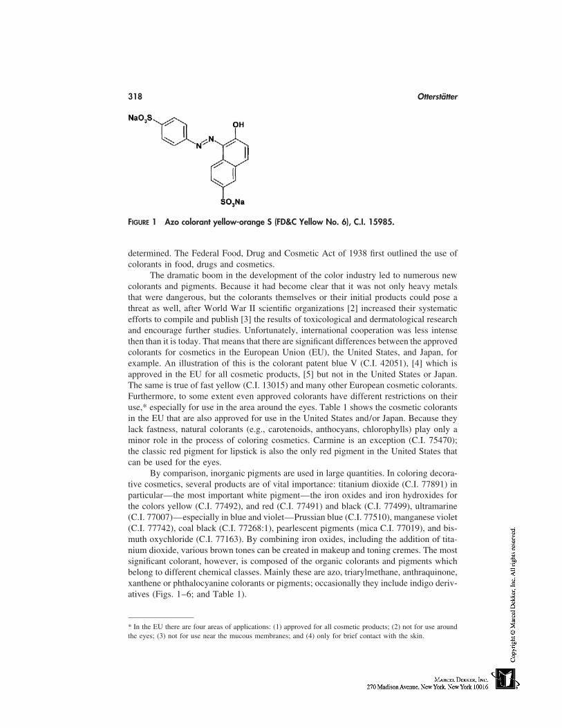
318 Otterstätter
FIGURE 1 Azo colorant yellow-orange S (FD&C Yellow No. 6), C.I. 15985.
determined. The Federal Food, Drug and Cosmetic Act of 1938 first outlined the use ofcolorants in food, drugs and cosmetics.
The dramatic boom in the development of the color industry led to numerous newcolorants and pigments. Because it had become clear that it was not only heavy metalsthat were dangerous, but the colorants themselves or their initial products could pose athreat as well, after World War II scientific organizations [2] increased their systematicefforts to compile and publish [3] the results of toxicological and dermatological researchand encourage further studies. Unfortunately, international cooperation was less intensethen than it is today. That means that there are significant differences between the approvedcolorants for cosmetics in the European Union (EU), the United States, and Japan, forexample. An illustration of this is the colorant patent blue V (C.I. 42051), [4] which isapproved in the EU for all cosmetic products, [5] but not in the United States or Japan.The same is true of fast yellow (C.I. 13015) and many other European cosmetic colorants.Furthermore, to some extent even approved colorants have different restrictions on theiruse,* especially for use in the area around the eyes. Table 1 shows the cosmetic colorantsin the EU that are also approved for use in the United States and/or Japan. Because theylack fastness, natural colorants (e.g., carotenoids, anthocyans, chlorophylls) play only aminor role in the process of coloring cosmetics. Carmine is an exception (C.I. 75470);the classic red pigment for lipstick is also the only red pigment in the United States thatcan be used for the eyes.
By comparison, inorganic pigments are used in large quantities. In coloring decora-tive cosmetics, several products are of vital importance: titanium dioxide (C.I. 77891) inparticular—the most important white pigment—the iron oxides and iron hydroxides forthe colors yellow (C.I. 77492), and red (C.I. 77491) and black (C.I. 77499), ultramarine(C.I. 77007)—especially in blue and violet—Prussian blue (C.I. 77510), manganese violet(C.I. 77742), coal black (C.I. 77268:1), pearlescent pigments (mica C.I. 77019), and bis-muth oxychloride (C.I. 77163). By combining iron oxides, including the addition of tita-nium dioxide, various brown tones can be created in makeup and toning cremes. The mostsignificant colorant, however, is composed of the organic colorants and pigments whichbelong to different chemical classes. Mainly these are azo, triarylmethane, anthraquinone,xanthene or phthalocyanine colorants or pigments; occasionally they include indigo deriv-atives (Figs. 1–6; and Table 1).
* In the EU there are four areas of applications: (1) approved for all cosmetic products; (2) not for use aroundthe eyes; (3) not for use near the mucous membranes; and (4) only for brief contact with the skin.
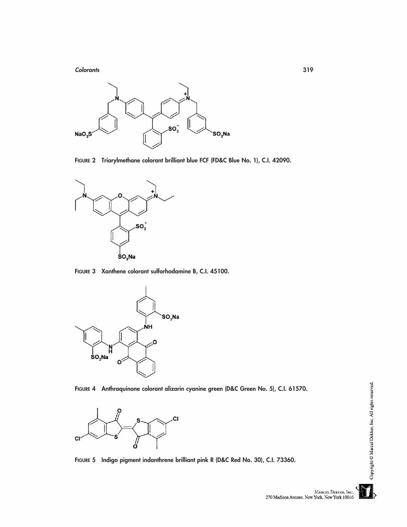
Colorants 319
FIGURE 2 Triarylmethane colorant brilliant blue FCF (FD&C Blue No. 1), C.I. 42090.
FIGURE 3 Xanthene colorant sulforhodamine B, C.I. 45100.
FIGURE 4 Anthraquinone colorant alizarin cyanine green (D&C Green No. 5), C.I. 61570.
FIGURE 5 Indigo pigment indanthrene brilliant pink R (D&C Red No. 30), C.I. 73360.
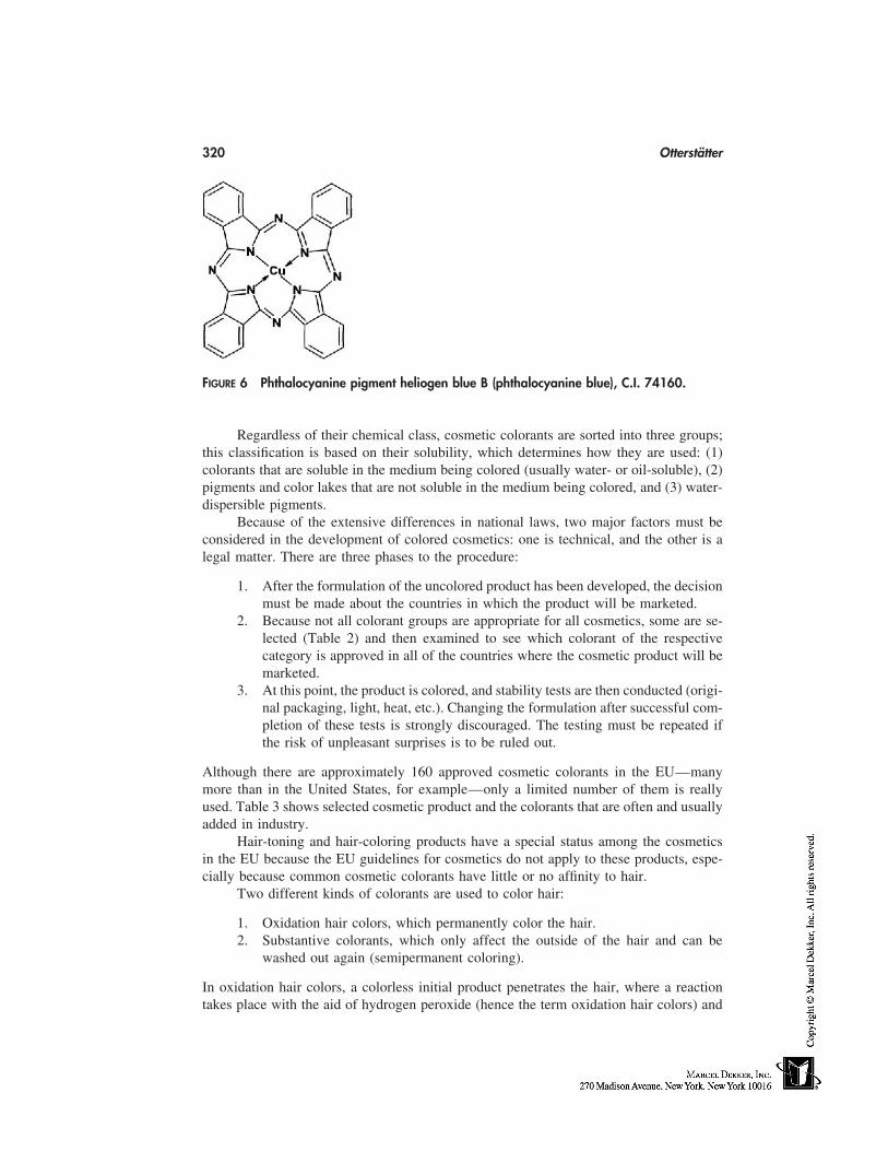
320 Otterstätter
FIGURE 6 Phthalocyanine pigment heliogen blue B (phthalocyanine blue), C.I. 74160.
Regardless of their chemical class, cosmetic colorants are sorted into three groups;this classification is based on their solubility, which determines how they are used: (1)colorants that are soluble in the medium being colored (usually water- or oil-soluble), (2)pigments and color lakes that are not soluble in the medium being colored, and (3) water-dispersible pigments.
Because of the extensive differences in national laws, two major factors must beconsidered in the development of colored cosmetics: one is technical, and the other is alegal matter. There are three phases to the procedure:
1. After the formulation of the uncolored product has been developed, the decisionmust be made about the countries in which the product will be marketed.
2. Because not all colorant groups are appropriate for all cosmetics, some are se-lected (Table 2) and then examined to see which colorant of the respectivecategory is approved in all of the countries where the cosmetic product will bemarketed.
3. At this point, the product is colored, and stability tests are then conducted (origi-nal packaging, light, heat, etc.). Changing the formulation after successful com-pletion of these tests is strongly discouraged. The testing must be repeated ifthe risk of unpleasant surprises is to be ruled out.
Although there are approximately 160 approved cosmetic colorants in the EU—manymore than in the United States, for example—only a limited number of them is reallyused. Table 3 shows selected cosmetic product and the colorants that are often and usuallyadded in industry.
Hair-toning and hair-coloring products have a special status among the cosmeticsin the EU because the EU guidelines for cosmetics do not apply to these products, espe-cially because common cosmetic colorants have little or no affinity to hair.
Two different kinds of colorants are used to color hair:
1. Oxidation hair colors, which permanently color the hair.2. Substantive colorants, which only affect the outside of the hair and can be
washed out again (semipermanent coloring).
In oxidation hair colors, a colorless initial product penetrates the hair, where a reactiontakes place with the aid of hydrogen peroxide (hence the term oxidation hair colors) and
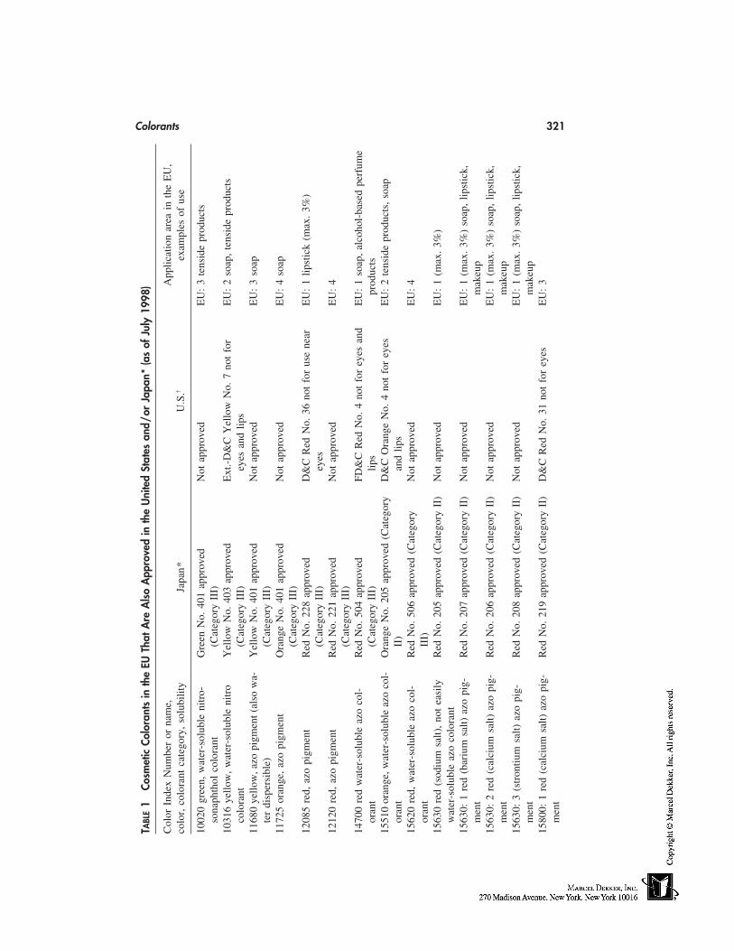
Colorants 321
TABL
E1
Cosm
etic
Colo
rant
sin
the
EUTh
atA
reA
lso
App
rove
din
the
Uni
ted
Stat
esan
d/or
Japa
n*(a
sof
July
1998
)
Col
orIn
dex
Num
ber
orna
me,
App
licat
ion
area
inth
eE
U,
colo
r,co
lora
ntca
tego
ry,
solu
bilit
yJa
pan*
U.S
.†ex
ampl
esof
use
1002
0gr
een,
wat
er-s
olub
leni
tro-
Gre
enN
o.40
1ap
prov
edN
otap
prov
edE
U:
3te
nsid
epr
oduc
tsso
naph
thol
colo
rant
(Cat
egor
yII
I)10
316
yello
w,
wat
er-s
olub
leni
tro
Yel
low
No.
403
appr
oved
Ext
.-D
&C
Yel
low
No.
7no
tfo
rE
U:
2so
ap,
tens
ide
prod
ucts
colo
rant
(Cat
egor
yII
I)ey
esan
dlip
s11
680
yello
w,a
zopi
gmen
t(al
sow
a-Y
ello
wN
o.40
1ap
prov
edN
otap
prov
edE
U:
3so
apte
rdi
sper
sibl
e)(C
ateg
ory
III)
1172
5or
ange
,az
opi
gmen
tO
rang
eN
o.40
1ap
prov
edN
otap
prov
edE
U:
4so
ap(C
ateg
ory
III)
1208
5re
d,az
opi
gmen
tR
edN
o.22
8ap
prov
edD
&C
Red
No.
36no
tfo
rus
ene
arE
U:
1lip
stic
k(m
ax.
3%)
(Cat
egor
yII
I)ey
es12
120
red,
azo
pigm
ent
Red
No.
221
appr
oved
Not
appr
oved
EU
:4
(Cat
egor
yII
I)14
700
red
wat
er-s
olub
leaz
oco
l-R
edN
o.50
4ap
prov
edFD
&C
Red
No.
4no
tfo
rey
esan
dE
U:
1so
ap,
alco
hol-
base
dpe
rfum
eor
ant
(Cat
egor
yII
I)lip
spr
oduc
ts15
510
oran
ge,w
ater
-sol
uble
azo
col-
Ora
nge
No.
205
appr
oved
(Cat
egor
yD
&C
Ora
nge
No.
4no
tfo
rey
esE
U:
2te
nsid
epr
oduc
ts,
soap
oran
tII
)an
dlip
s15
620
red,
wat
er-s
olub
leaz
oco
l-R
edN
o.50
6ap
prov
ed(C
ateg
ory
Not
appr
oved
EU
:4
oran
tII
I)15
630
red
(sod
ium
salt)
,no
tea
sily
Red
No.
205
appr
oved
(Cat
egor
yII
)N
otap
prov
edE
U:
1(m
ax.
3%)
wat
er-s
olub
leaz
oco
lora
nt15
630:
1re
d(b
ariu
msa
lt)az
opi
g-R
edN
o.20
7ap
prov
ed(C
ateg
ory
II)
Not
appr
oved
EU
:1
(max
.3%
)so
ap,
lipst
ick,
men
tm
akeu
p15
630:
2re
d(c
alci
umsa
lt)az
opi
g-R
edN
o.20
6ap
prov
ed(C
ateg
ory
II)
Not
appr
oved
EU
:1
(max
.3%
)so
ap,
lipst
ick,
men
tm
akeu
p15
630:
3(s
tron
tium
salt)
azo
pig-
Red
No.
208
appr
oved
(Cat
egor
yII
)N
otap
prov
edE
U:
1(m
ax.
3%)
soap
,lip
stic
k,m
ent
mak
eup
1580
0:1
red
(cal
cium
salt)
azo
pig-
Red
No.
219
appr
oved
(Cat
egor
yII
)D
&C
Red
No.
31no
tfo
rey
esE
U:
3m
ent
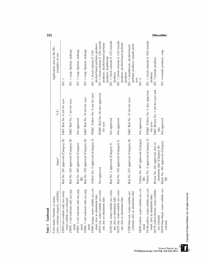
322 Otterstätter
TABL
E1
Cont
inue
d
Col
orIn
dex
Num
ber
orna
me,
App
licat
ion
area
inth
eE
U,
colo
r,co
lora
ntca
tego
ry,
solu
bilit
yJa
pan*
U.S
.†ex
ampl
esof
use
1585
0re
d(s
odiu
msa
lt)no
tea
sily
Red
No.
201
appr
oved
(Cat
egor
yII
)D
&C
Red
No.
6no
tfo
rey
esE
U:
1w
ater
-sol
uble
azo
colo
rant
1585
0:1
red
(cal
cium
salt)
azo
pig-
Red
No.
202
appr
oved
(Cat
egor
yII
)D
&C
Red
No.
7no
tfo
rey
esE
U:
1so
ap,
lipst
ick,
mak
eup
men
t15
865:
2re
d(c
alci
umsa
lt)az
opi
g-R
edN
o.40
5ap
prov
ed(C
ateg
ory
Not
appr
oved
EU
:1
soap
,lip
stic
k,m
akeu
pm
ent
III)
1588
0:1
red
(cal
cium
salt)
azo
pig-
Red
No.
220
appr
oved
(Cat
egor
yII
)D
&C
Red
No.
34no
tfo
rey
esE
U:
1so
ap,
lipst
ick,
mak
eup
men
t15
985
oran
ge,w
ater
-sol
uble
azo
col-
Yel
low
No.
5ap
prov
ed(C
ateg
ory
I)FD
&C
Yel
low
No.
6no
tfo
rey
esE
U:
1(f
ood
colo
rant
E11
0)or
ant,
also
asal
umin
umla
keal
coho
l-ba
sed
perf
ume
prod
ucts
1603
5re
d,w
ater
-sol
uble
azo
colo
r-N
otap
prov
edFD
&C
Red
No.
40al
soap
prov
edE
U:
1(f
ood
colo
rant
E12
9)te
nsid
ean
t,al
soas
alum
inum
lake
for
eyes
prod
ucts
,al
coho
l-ba
sed
perf
ume
prod
ucts
,m
outh
was
h16
185
red,
wat
er-s
olub
leaz
oco
lor-
Red
No.
2ap
prov
ed(C
ateg
ory
I)N
otap
prov
edE
U:
1(f
ood
colo
rant
E12
3)te
nsid
ean
t,al
soas
alum
inum
lake
prod
ucts
1625
5re
d,w
ater
-sol
uble
azo
colo
r-R
edN
o.10
2ap
prov
ed(C
ateg
ory
I)N
otap
prov
edE
U:
1(f
ood
colo
rant
E12
4)te
nsid
ean
t,al
soas
alum
inum
lake
prod
ucts
,al
coho
l-ba
sed
perf
ume
prod
ucts
1720
0bl
ue-r
ed,
wat
er-s
olub
leaz
oR
edN
o.22
7ap
prov
ed(C
ateg
ory
II)
D&
CR
edN
o.33
not
for
eyes
EU
:1
mou
thw
ash,
alco
hol-
base
dco
lora
nt,
also
asal
umin
umla
kepe
rfum
epr
oduc
ts,
tens
ide
prod
-uc
ts18
820
yello
w,w
ater
-sol
uble
azo
col-
Yel
low
No.
407
appr
oved
(Cat
egor
yN
otap
prov
edE
U:
4or
ant
III)
1914
0ye
llow
,wat
er-s
olub
leaz
oco
l-Y
ello
wN
o.4
appr
oved
(Cat
egor
yI)
FD&
CY
ello
wN
o.5
also
appr
oved
EU
:1
(foo
dco
lora
ntE
102)
tens
ide
oran
t,al
soas
alum
inum
lake
for
eyes
prod
ucts
2017
0ye
llow
-bro
wn,
wat
er-s
olub
leB
row
nN
o.20
1ap
prov
ed,
also
asD
&C
Bro
wn
No.
1no
tfo
rey
esan
dE
U:
3te
nsid
epr
oduc
tsaz
oco
lora
ntal
umin
umla
ke(C
ateg
ory
II)
lips
2047
0bl
ue-b
lack
,w
ater
-sol
uble
azo
Bla
ckN
o.40
1ap
prov
ed(C
ateg
ory
Not
appr
oved
EU
:4
tens
ide
prod
ucts
,so
apco
lora
ntII
I)
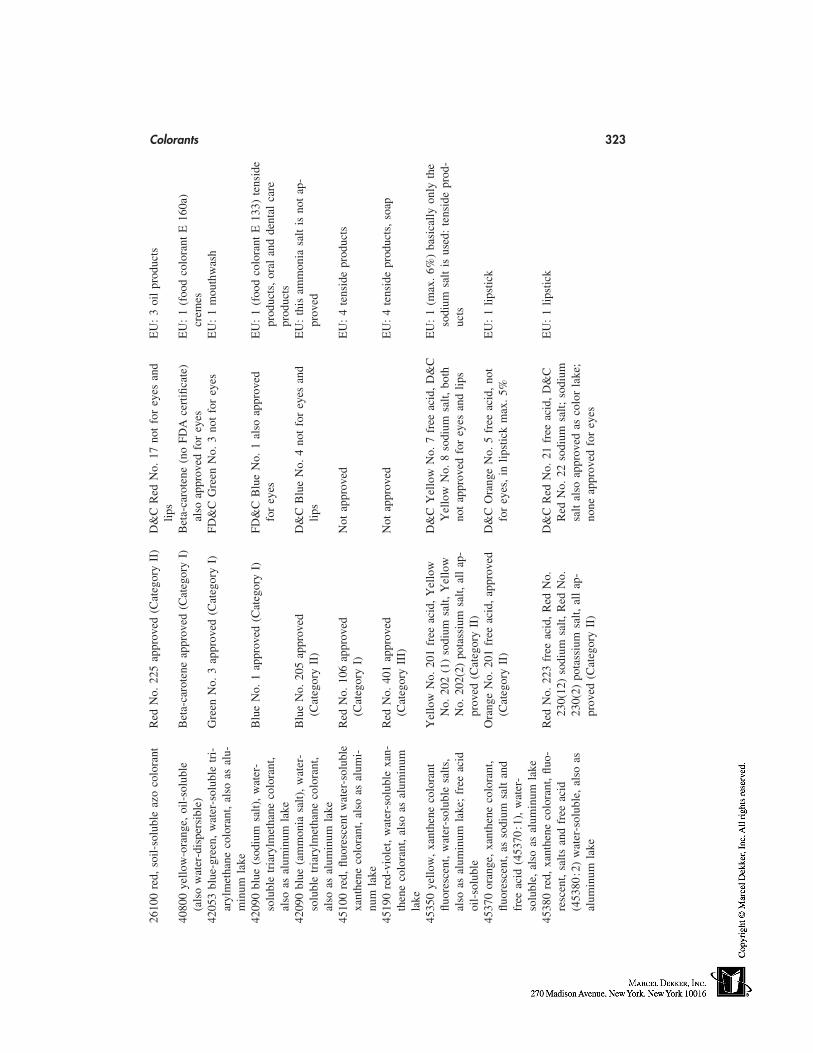
Colorants 32326
100
red,
soil-
solu
ble
azo
colo
rant
Red
No.
225
appr
oved
(Cat
egor
yII
)D
&C
Red
No.
17no
tfo
rey
esan
dE
U:
3oi
lpr
oduc
tslip
s40
800
yello
w-o
rang
e,oi
l-so
lubl
eB
eta-
caro
tene
appr
oved
(Cat
egor
yI)
Bet
a-ca
rote
ne(n
oFD
Ace
rtifi
cate
)E
U:
1(f
ood
colo
rant
E16
0a)
(als
ow
ater
-dis
pers
ible
)al
soap
prov
edfo
rey
escr
emes
4205
3bl
ue-g
reen
,w
ater
-sol
uble
tri-
Gre
enN
o.3
appr
oved
(Cat
egor
yI)
FD&
CG
reen
No.
3no
tfo
rey
esE
U:
1m
outh
was
har
ylm
etha
neco
lora
nt,
also
asal
u-m
inum
lake
4209
0bl
ue(s
odiu
msa
lt),
wat
er-
Blu
eN
o.1
appr
oved
(Cat
egor
yI)
FD&
CB
lue
No.
1al
soap
prov
edE
U:
1(f
ood
colo
rant
E13
3)te
nsid
eso
lubl
etr
iary
lmet
hane
colo
rant
,fo
rey
espr
oduc
ts,
oral
and
dent
alca
real
soas
alum
inum
lake
prod
ucts
4209
0bl
ue(a
mm
onia
salt)
,w
ater
-B
lue
No.
205
appr
oved
D&
CB
lue
No.
4no
tfo
rey
esan
dE
U:
this
amm
onia
salt
isno
tap
-so
lubl
etr
iary
lmet
hane
colo
rant
,(C
ateg
ory
II)
lips
prov
edal
soas
alum
inum
lake
4510
0re
d,flu
ores
cent
wat
er-s
olub
leR
edN
o.10
6ap
prov
edN
otap
prov
edE
U:
4te
nsid
epr
oduc
tsxa
nthe
neco
lora
nt,
also
asal
umi-
(Cat
egor
yI)
num
lake
4519
0re
d-vi
olet
,w
ater
-sol
uble
xan-
Red
No.
401
appr
oved
Not
appr
oved
EU
:4
tens
ide
prod
ucts
,so
apth
ene
colo
rant
,al
soas
alum
inum
(Cat
egor
yII
I)la
ke45
350
yello
w,
xant
hene
colo
rant
Yel
low
No.
201
free
acid
,Y
ello
wD
&C
Yel
low
No.
7fr
eeac
id,
D&
CE
U:
1(m
ax.
6%)
basi
cally
only
the
fluor
esce
nt,
wat
er-s
olub
lesa
lts,
No.
202
(1)
sodi
umsa
lt,Y
ello
wY
ello
wN
o.8
sodi
umsa
lt,bo
thso
dium
salt
isus
ed:
tens
ide
prod
-al
soas
alum
inum
lake
;fr
eeac
idN
o.20
2(2)
pota
ssiu
msa
lt,al
lap
-no
tap
prov
edfo
rey
esan
dlip
suc
tsoi
l-so
lubl
epr
oved
(Cat
egor
yII
)45
370
oran
ge,
xant
hene
colo
rant
,O
rang
eN
o.20
1fr
eeac
id,
appr
oved
D&
CO
rang
eN
o.5
free
acid
,no
tE
U:
1lip
stic
kflu
ores
cent
,as
sodi
umsa
ltan
d(C
ateg
ory
II)
for
eyes
,in
lipst
ick
max
.5%
free
acid
(453
70:1
),w
ater
-so
lubl
e,al
soas
alum
inum
lake
4538
0re
d,xa
nthe
neco
lora
nt,
fluo-
Red
No.
223
free
acid
,R
edN
o.D
&C
Red
No.
21fr
eeac
id,
D&
CE
U:
1lip
stic
kre
scen
t,sa
ltsan
dfr
eeac
id23
0(12
)so
dium
salt,
Red
No.
Red
No.
22so
dium
salt;
sodi
um(4
5380
:2)
wat
er-s
olub
le,
also
as23
0(2)
pota
ssiu
msa
lt,al
lap
-sa
ltal
soap
prov
edas
colo
rla
ke;
alum
inum
lake
prov
ed(C
ateg
ory
II)
none
appr
oved
for
eyes
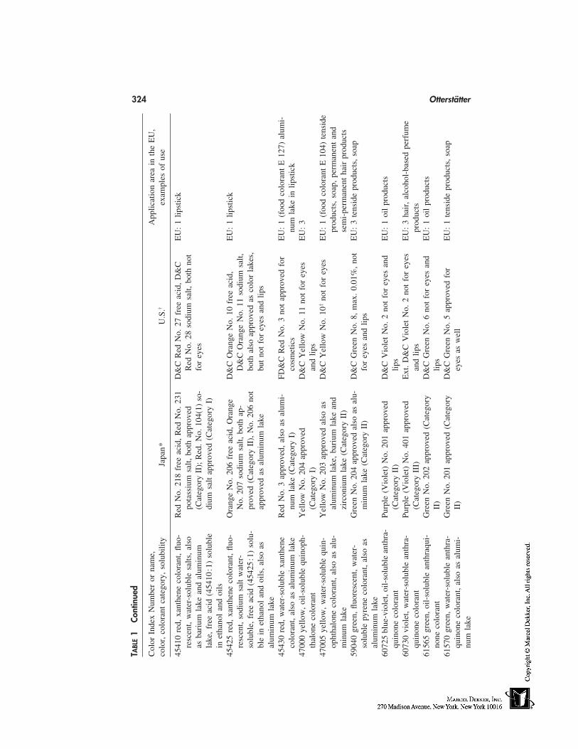
324 Otterstätter
TABL
E1
Cont
inue
d
Col
orIn
dex
Num
ber
orna
me,
App
licat
ion
area
inth
eE
U,
colo
r,co
lora
ntca
tego
ry,
solu
bilit
yJa
pan*
U.S
.†ex
ampl
esof
use
4541
0re
d,xa
nthe
neco
lora
nt,
fluo-
Red
No.
218
free
acid
,R
edN
o.23
1D
&C
Red
No.
27fr
eeac
id,
D&
CE
U:
1lip
stic
kre
scen
t,w
ater
-sol
uble
salts
,al
sopo
tass
ium
salt,
both
appr
oved
Red
No.
28so
dium
salt,
both
not
asba
rium
lake
and
alum
inum
(Cat
egor
yII
);R
ed.N
o.10
4(1)
so-
for
eyes
lake
,fr
eeac
id(4
5410
:1)
solu
ble
dium
salt
appr
oved
(Cat
egor
yI)
inet
hano
lan
doi
ls45
425
red,
xant
hene
colo
rant
,flu
o-O
rang
eN
o.20
6fr
eeac
id,
Ora
nge
D&
CO
rang
eN
o.10
free
acid
,E
U:
1lip
stic
kre
scen
t,so
dium
salt
wat
er-
No.
207
sodi
umsa
lt,bo
thap
-D
&C
Ora
nge
No.
11so
dium
salt,
solu
ble,
free
acid
(454
25:1
)so
lu-
prov
ed(C
ateg
ory
II),
No.
206
not
both
also
appr
oved
asco
lor
lake
s,bl
ein
etha
nol
and
oils
,al
soas
appr
oved
asal
umin
umla
kebu
tno
tfo
rey
esan
dlip
sal
umin
umla
ke45
430
red,
wat
er-s
olub
lexa
nthe
neR
edN
o.3
appr
oved
,al
soas
alum
i-FD
&C
Red
No.
3no
tap
prov
edfo
rE
U:
1(f
ood
colo
rant
E12
7)al
umi-
colo
rant
,al
soas
alum
inum
lake
num
lake
(Cat
egor
yI)
cosm
etic
snu
mla
kein
lipst
ick
4700
0ye
llow
,oi
l-so
lubl
equ
inop
h-Y
ello
wN
o.20
4ap
prov
edD
&C
Yel
low
No.
11no
tfo
rey
esE
U:
3th
alon
eco
lora
nt(C
ateg
ory
I)an
dlip
s47
005
yello
w,
wat
er-s
olub
lequ
in-
Yel
low
No.
203
appr
oved
also
asD
&C
Yel
low
No.
10‡
not
for
eyes
EU
:1
(foo
dco
lora
ntE
104)
tens
ide
opht
halo
neco
lora
nt,
also
asal
u-al
umin
umla
ke,
bari
umla
kean
dpr
oduc
ts,
soap
,pe
rman
ent
and
min
umla
kezi
rcon
ium
lake
(Cat
egor
yII
)se
mi-
perm
anen
tha
irpr
oduc
ts59
040
gree
n,flu
ores
cent
,w
ater
-G
reen
No.
204
appr
oved
also
asal
u-D
&C
Gre
enN
o.8,
max
.0.
01%
,no
tE
U:
3te
nsid
epr
oduc
ts,
soap
solu
ble
pyre
neco
lora
nt,
also
asm
inum
lake
(Cat
egor
yII
)fo
rey
esan
dlip
sal
umin
umla
ke60
725
blue
-vio
let,
oil-
solu
ble
anth
ra-
Purp
le(V
iole
t)N
o.20
1ap
prov
edD
&C
Vio
let
No.
2no
tfo
rey
esan
dE
U:
1oi
lpr
oduc
tsqu
inon
eco
lora
nt(C
ateg
ory
II)
lips
6073
0vi
olet
,w
ater
-sol
uble
anth
ra-
Purp
le(V
iole
t)N
o.40
1ap
prov
edE
xt.
D&
CV
iole
tN
o.2
not
for
eyes
EU
:3
hair
,al
coho
l-ba
sed
perf
ume
quin
one
colo
rant
(Cat
egor
yII
I)an
dlip
spr
oduc
ts61
565
gree
n,oi
l-so
lubl
ean
thra
qui-
Gre
enN
o.20
2ap
prov
ed(C
ateg
ory
D&
CG
reen
No.
6no
tfo
rey
esan
dE
U:
1oi
lpr
oduc
tsno
neco
lora
ntII
)lip
s61
570
gree
n,w
ater
-sol
uble
anth
ra-
Gre
enN
o.20
1ap
prov
ed(C
ateg
ory
D&
CG
reen
No.
5ap
prov
edfo
rE
U:
1te
nsid
epr
oduc
ts,
soap
quin
one
colo
rant
,al
soas
alum
i-II
)ey
esas
wel
lnu
mla
ke
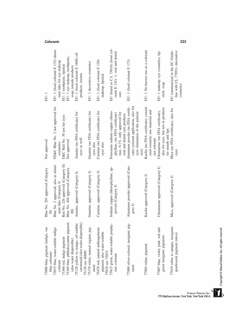
Colorants 32573
000
blue
,pi
gmen
t(i
ndig
o,va
t-B
lue
No.
201
appr
oved
(Cat
egor
yN
otap
prov
edE
U:
1bl
ueco
lora
nt)
II)
7301
5bl
ue,
wat
er-s
olub
lein
digo
Blu
eN
o.2
appr
oved
,al
soas
alum
i-FD
&C
Blu
eN
o.2
not
appr
oved
for
EU
:1
(foo
dco
lora
ntE
132)
alum
i-co
lora
ntnu
mla
ke(C
ateg
ory
I)co
smet
ics
num
lake
for
eye
mak
eup
7336
0re
d,in
digo
pigm
ent
Red
No.
226
appr
oved
(Cat
egor
yII
)D
&C
Red
No.
30no
tfo
rey
esE
U:
1to
othp
aste
,lip
stic
k74
160
blue
,ph
thal
ocya
nine
pigm
ent
Blu
eN
o.40
4ap
prov
ed(C
ateg
ory
Not
appr
oved
EU
:1
eye
mak
eup,
toot
hpas
te,
(als
ow
ater
disp
ersi
ble)
III)
soap
,te
nsid
epr
oduc
ts75
120
yello
wto
oran
ge,
oil-
solu
ble
Ann
atto
,ap
prov
ed(C
ateg
ory
I)A
nnat
to(n
oFD
Ace
rtifi
cate
)fo
rE
U:
1(f
ood
colo
rant
E16
0b)
oil
caro
teno
id(a
lso
wat
er-d
ispe
rsib
le)
eyes
asw
ell
prod
ucts
,cr
eam
s75
130
see
4080
075
170
whi
te,
natu
ral
orga
nic
pig-
Gua
nine
,ap
prov
ed(C
ateg
ory
I)G
uani
ne(n
oFD
Ace
rtifi
cate
)fo
rE
U:
1de
cora
tive
cosm
etic
sm
ent
eyes
also
7547
0re
d,na
tura
lan
thra
quin
one
Car
min
e,ap
prov
ed(C
ateg
ory
I)C
arm
ine
(no
FDA
cert
ifica
te)
for
EU
:1
(foo
dco
lora
ntE
120)
pigm
ent,
also
wat
er-s
olub
leey
esal
som
akeu
p,lip
stic
k75
810
see
7581
575
815
gree
n,w
ater
-sol
uble
porp
hy-
Sodi
umco
pper
chlo
roph
yllin
e,ap
-Po
tass
ium
sodi
umco
pper
chlo
ro-
EU
(lis
ted
asC
.I.
7581
0)(f
ood
col-
rine
colo
rant
prov
ed(C
ateg
ory
I)ph
yllin
e,(n
oFD
Ace
rtifi
cate
)or
ant
E14
1):
1,or
alan
dde
ntal
max
.0.
1%,
only
appr
oved
for
care
oral
and
dent
alca
repr
oduc
ts77
000
silv
er-c
olor
ed,
inor
gani
cpi
g-A
lum
inum
pow
der
appr
oved
(Cat
e-A
lum
inum
pow
der
(no
FDA
cert
ifi-
EU
:1
(foo
dco
lora
ntE
173)
men
tgo
ryI)
cate
)ex
tern
alap
plic
atio
n,al
sofo
rey
es(l
imita
tion
ofth
epa
rtic
lesi
ze)
7700
4w
hite
,pi
gmen
tK
aolin
appr
oved
(Cat
egor
yI)
Kao
lin(n
oFD
Ace
rtifi
cate
),co
nsid
-E
U:
1N
okn
own
use
asa
colo
rant
ered
cosm
etic
raw
mat
eria
lan
dno
tco
lora
nt77
007
blue
,vi
olet
,pi
nk,
red
and
Ultr
amar
ine
appr
oved
(Cat
egor
yI)
Ultr
amar
ine
(no
FDA
cert
ifica
te),
EU
:1
mak
eup,
eye
cosm
etic
s,lip
-gr
een
inor
gani
cpi
gmen
tsal
sofo
rey
es,
but
not
inpr
oduc
tsst
ick,
soap
for
mou
than
dlip
s77
019
whi
teto
opaq
ue,
inor
gani
cM
ica,
appr
oved
(Cat
egor
yI)
Mic
a(n
oFD
Ace
rtifi
cate
),al
sofo
rE
U(s
umm
ariz
edin
the
EC
Gui
de-
pear
lesc
ent
pigm
ent
(mic
a)ey
eslin
ew
ithC
L77
891)
:de
cora
tive
cosm
etic
s
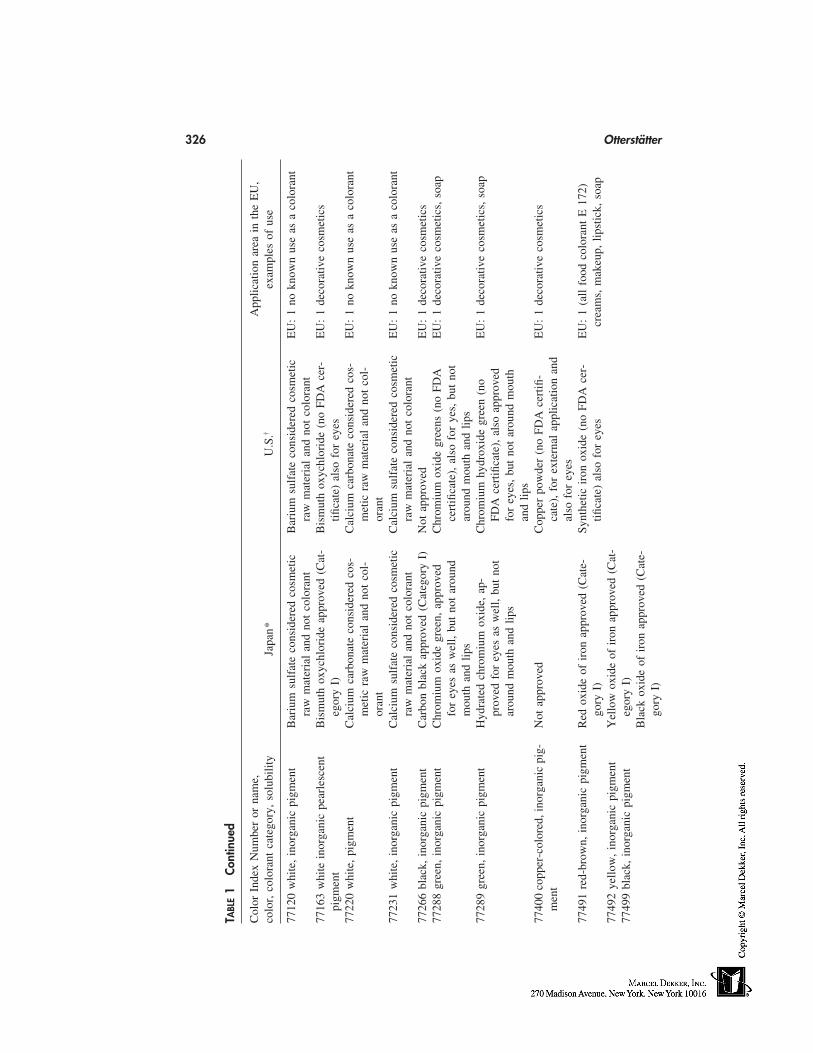
326 Otterstätter
TABL
E1
Cont
inue
d
Col
orIn
dex
Num
ber
orna
me,
App
licat
ion
area
inth
eE
U,
colo
r,co
lora
ntca
tego
ry,
solu
bilit
yJa
pan*
U.S
.†ex
ampl
esof
use
7712
0w
hite
,in
orga
nic
pigm
ent
Bar
ium
sulf
ate
cons
ider
edco
smet
icB
ariu
msu
lfat
eco
nsid
ered
cosm
etic
EU
:1
nokn
own
use
asa
colo
rant
raw
mat
eria
lan
dno
tco
lora
ntra
wm
ater
ial
and
not
colo
rant
7716
3w
hite
inor
gani
cpe
arle
scen
tB
ism
uth
oxyc
hlor
ide
appr
oved
(Cat
-B
ism
uth
oxyc
hlor
ide
(no
FDA
cer-
EU
:1
deco
rativ
eco
smet
ics
pigm
ent
egor
yI)
tifica
te)
also
for
eyes
7722
0w
hite
,pi
gmen
tC
alci
umca
rbon
ate
cons
ider
edco
s-C
alci
umca
rbon
ate
cons
ider
edco
s-E
U:
1no
know
nus
eas
aco
lora
ntm
etic
raw
mat
eria
lan
dno
tco
l-m
etic
raw
mat
eria
lan
dno
tco
l-or
ant
oran
t77
231
whi
te,
inor
gani
cpi
gmen
tC
alci
umsu
lfat
eco
nsid
ered
cosm
etic
Cal
cium
sulf
ate
cons
ider
edco
smet
icE
U:
1no
know
nus
eas
aco
lora
ntra
wm
ater
ial
and
not
colo
rant
raw
mat
eria
lan
dno
tco
lora
nt77
266
blac
k,in
orga
nic
pigm
ent
Car
bon
blac
kap
prov
ed(C
ateg
ory
I)N
otap
prov
edE
U:
1de
cora
tive
cosm
etic
s77
288
gree
n,in
orga
nic
pigm
ent
Chr
omiu
mox
ide
gree
n,ap
prov
edC
hrom
ium
oxid
egr
eens
(no
FDA
EU
:1
deco
rativ
eco
smet
ics,
soap
for
eyes
asw
ell,
but
not
arou
ndce
rtifi
cate
),al
sofo
rye
s,bu
tno
tm
outh
and
lips
arou
ndm
outh
and
lips
7728
9gr
een,
inor
gani
cpi
gmen
tH
ydra
ted
chro
miu
mox
ide,
ap-
Chr
omiu
mhy
drox
ide
gree
n(n
oE
U:
1de
cora
tive
cosm
etic
s,so
appr
oved
for
eyes
asw
ell,
but
not
FDA
cert
ifica
te),
also
appr
oved
arou
ndm
outh
and
lips
for
eyes
,bu
tno
tar
ound
mou
than
dlip
s77
400
copp
er-c
olor
ed,i
norg
anic
pig-
Not
appr
oved
Cop
per
pow
der
(no
FDA
cert
ifi-
EU
:1
deco
rativ
eco
smet
ics
men
tca
te),
for
exte
rnal
appl
icat
ion
and
also
for
eyes
7749
1re
d-br
own,
inor
gani
cpi
gmen
tR
edox
ide
ofir
onap
prov
ed(C
ate-
Synt
hetic
iron
oxid
e(n
oFD
Ace
r-E
U:
1(a
llfo
odco
lora
ntE
172)
gory
I)tifi
cate
)al
sofo
rey
escr
eam
s,m
akeu
p,lip
stic
k,so
ap77
492
yello
w,
inor
gani
cpi
gmen
tY
ello
wox
ide
ofir
onap
prov
ed(C
at-
7749
9bl
ack,
inor
gani
cpi
gmen
teg
ory
I)B
lack
oxid
eof
iron
appr
oved
(Cat
e-go
ryI)
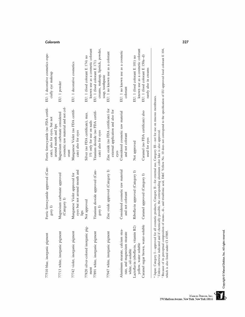
Colorants 32777
510
blue
,in
orga
nic
pigm
ent
Ferr
icfe
rroc
yani
deap
prov
ed(C
ate-
Ferr
icfe
rroc
yani
de(n
oFD
Ace
rtifi
-E
U:
1de
cora
tive
cosm
etic
ses
pe-
gory
I)ca
te),
also
for
eyes
,bu
tno
tci
ally
eye
mak
eup
arou
ndm
outh
and
lips
7771
3w
hite
,in
orga
nic
pigm
ent
Mag
nesi
umca
rbon
ate
appr
oved
Mag
nesi
umca
rbon
ate
cons
ider
edE
U:
1po
wde
r(C
ateg
ory
I)co
smet
icra
wm
ater
iala
ndno
tcol
-or
ant
7774
2vi
olet
,in
orga
nic
pigm
ent
Man
gane
seV
iole
tap
prov
edfo
rM
anga
nese
Vio
let
(no
FDA
cert
ifi-
EU
:1
deco
rativ
eco
smet
ics
eyes
but
not
arou
ndm
outh
and
cate
)al
sofo
rey
eslip
s77
820
silv
er-c
olor
edin
orga
nic
pig-
Not
appr
oved
Silv
er(n
oFD
Ace
rtifi
cate
),m
ax.
EU
:1
(foo
dco
lora
ntE
174)
nom
ent
1%on
lyfo
rus
eon
nails
know
nus
eas
aco
smet
icco
lora
nt77
891
whi
te,
inor
gani
cpi
gmen
tT
itani
umdi
oxid
eap
prov
ed(C
ate-
Tita
nium
diox
ide
(no
FDA
cert
ifi-
EU
:1
(foo
dco
lora
ntE
171)
gory
I)ca
te)
also
for
eyes
crea
ms,
mak
eup,
lipst
ick,
pow
der,
soap
,to
othp
aste
7794
7w
hite
,in
orga
nic
pigm
ent
Zin
cox
ide
appr
oved
(Cat
egor
yI)
Zin
cox
ide
(no
FDA
cert
ifica
te)
for
EU
:1
nokn
own
use
asa
colo
rant
exte
rnal
appl
icat
ion
and
also
for
eyes
Alu
min
umst
eara
te,
calc
ium
stea
-C
onsi
dere
dco
smet
icra
wm
ater
ial
Con
side
red
cosm
etic
raw
mat
eria
lE
U:
1no
know
nus
eas
aco
smet
icra
te,
and
mag
nesi
umst
eara
tean
dno
tco
lora
ntan
dno
tco
lora
ntco
lora
ntw
hite
,oi
l-so
lubl
eL
acto
flavi
n(r
ibofl
avin
,vi
tam
inB
2)R
ibofl
avin
appr
oved
(Cat
egor
yI)
Not
appr
oved
EU
:1
(foo
dco
lora
ntE
101)
noye
llow
,w
ater
solu
ble
know
nus
eas
aco
smet
icco
lora
ntC
aram
elsu
gar
brow
n,w
ater
-sol
uble
Car
amel
appr
oved
(Cat
egor
yI)
Car
amel
(no
FDA
cert
ifica
te)
also
EU
:1
(foo
dco
lora
ntE
150a
–d)
used
for
eyes
rare
lyal
soin
crea
ms
*Ja
pan:
Cat
egor
yI—
appr
oved
for
all
cosm
etic
prod
ucts
,C
ateg
ory
II—
for
exte
rnal
use,
Cat
egor
yII
I—no
tfo
rus
eon
muc
ous
mem
bran
es.
†U
nles
sot
herw
ise
indi
cate
dan
dif
chem
ical
lypo
ssib
le,
the
corr
espo
ndin
gal
umin
umco
lor
lake
isal
soap
prov
ed.
‡B
ecau
seof
itspe
rcep
tual
com
posi
tion
ofm
ono-
,di
-,an
dtr
isul
foni
cac
id,
D&
CY
ello
wN
o.10
does
not
corr
espo
ndto
the
spec
ifica
tion
ofE
U-a
ppro
ved
food
colo
rant
E10
4,w
hich
isal
solis
ted
unde
rC
I47
005.
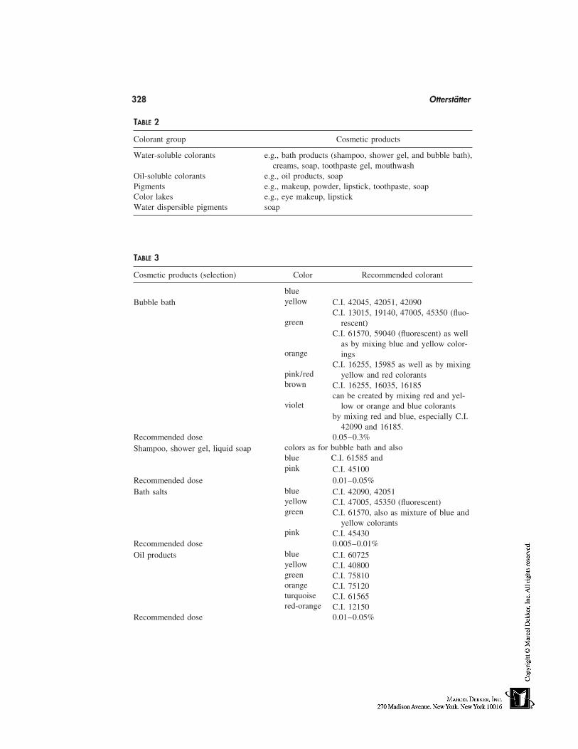
328 Otterstätter
TABLE 2
Colorant group Cosmetic products
Water-soluble colorants e.g., bath products (shampoo, shower gel, and bubble bath),creams, soap, toothpaste gel, mouthwash
Oil-soluble colorants e.g., oil products, soapPigments e.g., makeup, powder, lipstick, toothpaste, soapColor lakes e.g., eye makeup, lipstickWater dispersible pigments soap
TABLE 3
Cosmetic products (selection) Color Recommended colorant
blueyellowBubble bath C.I. 42045, 42051, 42090
C.I. 13015, 19140, 47005, 45350 (fluo-green rescent)
C.I. 61570, 59040 (fluorescent) as wellas by mixing blue and yellow color-
orange ingsC.I. 16255, 15985 as well as by mixing
pink/red yellow and red colorantsbrown C.I. 16255, 16035, 16185
can be created by mixing red and yel-violet low or orange and blue colorants
by mixing red and blue, especially C.I.42090 and 16185.
Recommended dose 0.05–0.3%colors as for bubble bath and alsoShampoo, shower gel, liquid soapblue C.I. 61585 andpink C.I. 45100
Recommended dose 0.01–0.05%blueBath salts C.I. 42090, 42051yellow C.I. 47005, 45350 (fluorescent)green C.I. 61570, also as mixture of blue and
yellow colorantspink C.I. 45430
Recommended dose 0.005–0.01%blueOil products C.I. 60725yellow C.I. 40800green C.I. 75810orange C.I. 75120turquoise C.I. 61565red-orange C.I. 12150
Recommended dose 0.01–0.05%
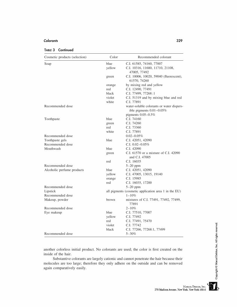
Colorants 329
TABLE 3 Continued
Cosmetic products (selection) Color Recommended colorant
Soap blue C.I. 61585, 74160, 77007yellow C.I. 10316, 11680, 11710, 21108,
47005, 77492green C.I. 10006, 10020, 59040 (fluorescent),
61570, 74260orange by mixing red and yellowred C.I. 12490, 77491black C.I. 77499, 77268:1violet C.I. 51319 and by mixing blue and redwhite C.I. 77891
Recommended dose water-soluble colorants or water dispers-ible pigments 0.01–0.05%
pigments 0.05–0.5%Toothpaste blue C.I. 74160
green C.I. 74260red C.I. 73360white C.I. 77891
Recommended dose 0.02–0.05%Toothpaste gels blue C.I. 42051, 42090Recommended dose C.I. 0.02–0.05%Mouthwash blue C.I. 42090
green C.I. 61570 or a mixture of C.I. 42090and C.I. 47005
red C.I. 16035Recommended dose 5–20 ppmAlcoholic perfume products blue C.I. 42051, 42090
yellow C.I. 47005, 13015, 19140orange C.I. 15985red C.I. 16035, 17200
Recommended dose 5–20 ppmLipstick all pigments (cosmetic application area 1 in the EU)Recommended dose 1–10%Makeup, powder brown mixtures of C.I. 77491, 77492, 77499,
77891Recommended dose 2–10%Eye makeup blue C.I. 77510, 77007
yellow C.I. 77492red C.I. 77491, 75470violet C.I. 77742black C.I. 77266, 77268:1, 77499
Recommended dose 5–30%
another colorless initial product. No colorants are used; the color is first created on theinside of the hair.
Substantive colorants are largely cationic and cannot penetrate the hair because theirmolecules are too large; therefore they only adhere on the outside and can be removedagain comparatively easily.
330 Otterstätter
BIBLIOGRAPHY
Colour Index: Third Edition, Vols. 1–4 (1971), Revised Third Edition, Vol. 5–6 (1975); The Societyof Dyers and Colourists, P.O. Box 244, Perkin House 82, Grattan Road, Bradford West York-shire BD1 2JB/England.
DFG-Farbstoff-Kommission (DFG Dyestuffs Commission), Cosmetic Colorants, 3rd completely re-vised edition, VCH Weinheim 1991.
Hendry, GAF and Houghton, JD: Natural Food Colorants; Blackie, Glasgow and London 1992.Lehmann, G, et al.: Identifizierung von Farbstoffen in Hautcremes (Identifying Colorants in Skin
Creams); Seifen-Ole-Fette-Wachse, Nr. (Soaps-Oils-Fats-Waxes No.) 16/1986, 565.Lehmann, G, Binkle, B: Identifizierung von Farbpigmenten in kosmetischen Erzeugnissen (Identi-
fying Color Pigments in Cosmetic Products); Seifen-Ole-Fette-Wachse, Nr. (Soaps-Oils-Fats-Waxes No.) 5/1984, 125.
Loscher, M: Farben—visualisierte Gefühle (Colors—Visualized Feelings); DRAGOCO Report4/5—1981.
Marmion, DM: Handbook of U.S. Colorants for Foods, Drugs and Cosmetics, Second Edition 1984,ISBN 0–471–09312–2.
Moschl, G, et al.: Perlglanzpigmente für Kosmetika (Pearlescent Pigments for Cosmetics); Seifen-Ole-Fette-Wachse, Nr. (Soaps-Oils-Fats-Waxes No.) 8/1980, 207.
Otterstätter, G: Die Färbung von Lebensmitteln, Arzneimitteln, Kosmetika (Coloring Foods, Drugs,Cosmetics); 2nd revised edition, Behr’s Verlag, Hamburg 1995.
Schweppe, H: Handbuch der Naturfarbstoffe—Vorkommen, Verwendung, Nachweis. (Handbookof Natural Colorants—Their Presence, Use and Verification), Landsberg/Lech: Ecomed 1992.
29
Hair Conditioners
Charles Reich and Dean T. SuColgate-Palmolive Technology Center, Piscataway, New Jersey
INTRODUCTION
Despite myriad claimed benefits, the primary purpose of a hair conditioner is to reducethe magnitude of the forces associated with combing or brushing hair [1], especially whenwet [2,3]. This is generally accomplished by the deposition of conditioning agents thatlubricate the hair fiber, diminishing surface friction and, therefore, combing forces [4].
In general, deposition of a conditioning agent also causes the hair to feel softer andmore moisturized. Another secondary benefit is the reduction or prevention of flyawayhair [5], especially by cationic conditioners [6]. Increasing ease of combing also makesthe hair more manageable, while improving the ability to align the hair fibers in a moreparallel configuration can increase hair shine, even if the shine of individual fibers is notincreased [7].
A number of other benefits have sometimes been claimed or implied for conditionersincluding, e.g., repair of damaged hair, strengthening of hair, repair of split ends, andvitamin therapy. Most of these are marketing hype or are based on laboratory conditionsor concentrations not found under actual usage conditions. In this chapter, we will confineourselves to a discussion of only the observable conditioner benefits presented above. Thechapter will begin with a discussion of the relationship between hair damage, conditioningand the state of the hair surface. This will be followed by a discussion of the major classesof conditioning agents currently in use. Finally, we will end with a brief discussion of theauxiliary ingredients necessary for the production of a commercial conditioning product.
CONDITIONING AND THE HAIR FIBER SURFACE
Hair Damage
In previous chapters, it has been shown that hair fibers consist of a central cortex thatcomprises the major portion of the fiber, surrounded by 8 to 10 layers of overlapping cellstermed the cuticle. The cortex is responsible for the tensile properties of the hair [8,9],while the state of the cuticle affects a variety of consumer perceivable properties including,e.g., hair feel, shine, and combability.
331
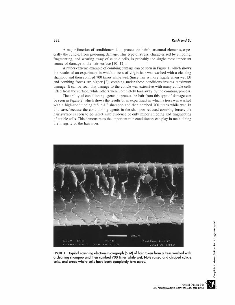
332 Reich and Su
A major function of conditioners is to protect the hair’s structural elements, espe-cially the cuticle, from grooming damage. This type of stress, characterized by chipping,fragmenting, and wearing away of cuticle cells, is probably the single most importantsource of damage to the hair surface [10–12].
A rather extreme example of combing damage can be seen in Figure 1, which showsthe results of an experiment in which a tress of virgin hair was washed with a cleaningshampoo and then combed 700 times while wet. Since hair is more fragile when wet [3]and combing forces are higher [2], combing under these conditions insures maximumdamage. It can be seen that damage to the cuticle was extensive with many cuticle cellslifted from the surface, while others were completely torn away by the combing process.
The ability of conditioning agents to protect the hair from this type of damage canbe seen in Figure 2, which shows the results of an experiment in which a tress was washedwith a high-conditioning ‘‘2-in-1’’ shampoo and then combed 700 times while wet. Inthis case, because the conditioning agents in the shampoo reduced combing forces, thehair surface is seen to be intact with evidence of only minor chipping and fragmentingof cuticle cells. This demonstrates the important role conditioners can play in maintainingthe integrity of the hair fiber.
FIGURE 1 Typical scanning electron micrograph (SEM) of hair taken from a tress washed witha cleaning shampoo and then combed 700 times while wet. Note raised and chipped cuticlecells, and areas where cells have been completely torn away.
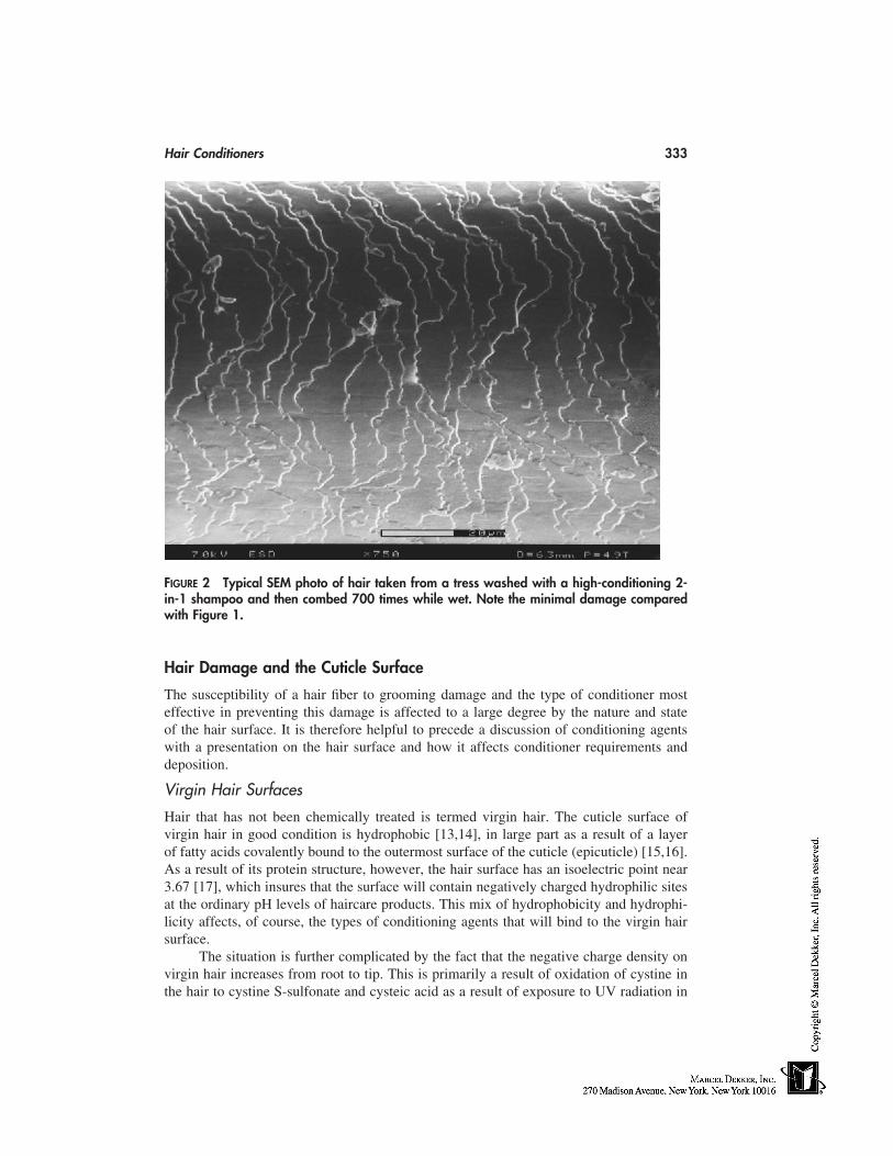
Hair Conditioners 333
FIGURE 2 Typical SEM photo of hair taken from a tress washed with a high-conditioning 2-in-1 shampoo and then combed 700 times while wet. Note the minimal damage comparedwith Figure 1.
Hair Damage and the Cuticle Surface
The susceptibility of a hair fiber to grooming damage and the type of conditioner mosteffective in preventing this damage is affected to a large degree by the nature and stateof the hair surface. It is therefore helpful to precede a discussion of conditioning agentswith a presentation on the hair surface and how it affects conditioner requirements anddeposition.
Virgin Hair Surfaces
Hair that has not been chemically treated is termed virgin hair. The cuticle surface ofvirgin hair in good condition is hydrophobic [13,14], in large part as a result of a layerof fatty acids covalently bound to the outermost surface of the cuticle (epicuticle) [15,16].As a result of its protein structure, however, the hair surface has an isoelectric point near3.67 [17], which insures that the surface will contain negatively charged hydrophilic sitesat the ordinary pH levels of haircare products. This mix of hydrophobicity and hydrophi-licity affects, of course, the types of conditioning agents that will bind to the virgin hairsurface.
The situation is further complicated by the fact that the negative charge density onvirgin hair increases from root to tip. This is primarily a result of oxidation of cystine inthe hair to cystine S-sulfonate and cysteic acid as a result of exposure to UV radiation in
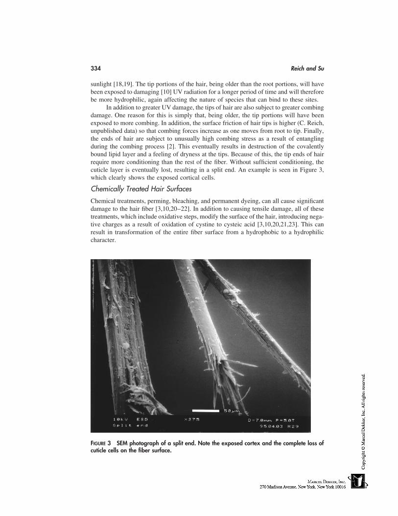
334 Reich and Su
sunlight [18,19]. The tip portions of the hair, being older than the root portions, will havebeen exposed to damaging [10] UV radiation for a longer period of time and will thereforebe more hydrophilic, again affecting the nature of species that can bind to these sites.
In addition to greater UV damage, the tips of hair are also subject to greater combingdamage. One reason for this is simply that, being older, the tip portions will have beenexposed to more combing. In addition, the surface friction of hair tips is higher (C. Reich,unpublished data) so that combing forces increase as one moves from root to tip. Finally,the ends of hair are subject to unusually high combing stress as a result of entanglingduring the combing process [2]. This eventually results in destruction of the covalentlybound lipid layer and a feeling of dryness at the tips. Because of this, the tip ends of hairrequire more conditioning than the rest of the fiber. Without sufficient conditioning, thecuticle layer is eventually lost, resulting in a split end. An example is seen in Figure 3,which clearly shows the exposed cortical cells.
Chemically Treated Hair Surfaces
Chemical treatments, perming, bleaching, and permanent dyeing, can all cause significantdamage to the hair fiber [3,10,20–22]. In addition to causing tensile damage, all of thesetreatments, which include oxidative steps, modify the surface of the hair, introducing nega-tive charges as a result of oxidation of cystine to cysteic acid [3,10,20,21,23]. This canresult in transformation of the entire fiber surface from a hydrophobic to a hydrophiliccharacter.
FIGURE 3 SEM photograph of a split end. Note the exposed cortex and the complete loss ofcuticle cells on the fiber surface.
Hair Conditioners 335
All of these treatments also increase surface friction considerably [3,4,24,25] re-sulting in a significant increase in combing forces. The result is hair that feels rough anddry and is subject to extensive grooming damage. Because of this, treated hair generallyrequires significantly more conditioning than does virgin hair.
COMMERCIAL CONDITIONERS
The commercial hair conditioners produced to deal with the aforementioned problemshave appeared in almost every conceivable form, including thick vaseline pomades; thick,clear, water-soluble gels; spray mists of volatile substances; mousses; lotions; and creams.Conditioners have been marketed as leave-in or rinse-off products. They have also beenpositioned as pre-shampoo or post-shampoo formulations.
Despite the wide variety of forms available, most commercial conditioners are oil-in-water emulsions in lotion form, having viscosities somewhere between 3000 and 12,000centipoise. The great majority of these products are of the rinse-off type. In addition,despite different forms and positionings, most commercial conditioners contain the samegeneral classes of conditioning agents with differences mainly in concentrations, numbersof different agents, and the particular members of a conditioning class employed.
The major classes of conditioning agents used in commercial products are surveyedin the following sections.
Cationic Surfactants
Cationic surfactants, in the form of quaternary ammonium compounds, are the most widelyused conditioning agents in commercial products [26–28]. Among the reasons for thisare their effectiveness, versatility, availability, and low cost.
Important examples of these quats include stearalkonium chloride, cetrimoniumchloride, and dicetyldimonium chloride.
Because of the positive charge on quaternary ammonium compounds such as theabove, they are substantive to hair, binding to negative sites on the hair surface. Treatment
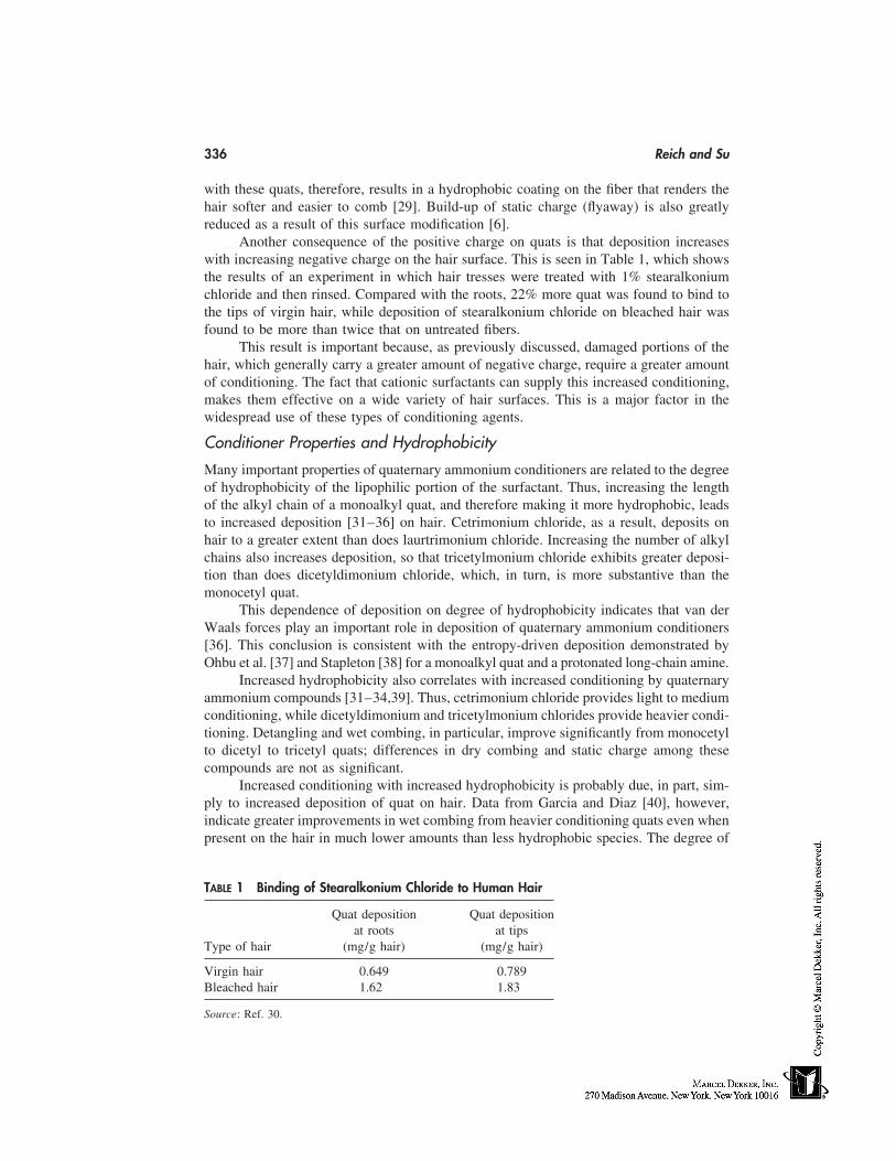
336 Reich and Su
with these quats, therefore, results in a hydrophobic coating on the fiber that renders thehair softer and easier to comb [29]. Build-up of static charge (flyaway) is also greatlyreduced as a result of this surface modification [6].
Another consequence of the positive charge on quats is that deposition increaseswith increasing negative charge on the hair surface. This is seen in Table 1, which showsthe results of an experiment in which hair tresses were treated with 1% stearalkoniumchloride and then rinsed. Compared with the roots, 22% more quat was found to bind tothe tips of virgin hair, while deposition of stearalkonium chloride on bleached hair wasfound to be more than twice that on untreated fibers.
This result is important because, as previously discussed, damaged portions of thehair, which generally carry a greater amount of negative charge, require a greater amountof conditioning. The fact that cationic surfactants can supply this increased conditioning,makes them effective on a wide variety of hair surfaces. This is a major factor in thewidespread use of these types of conditioning agents.
Conditioner Properties and Hydrophobicity
Many important properties of quaternary ammonium conditioners are related to the degreeof hydrophobicity of the lipophilic portion of the surfactant. Thus, increasing the lengthof the alkyl chain of a monoalkyl quat, and therefore making it more hydrophobic, leadsto increased deposition [31–36] on hair. Cetrimonium chloride, as a result, deposits onhair to a greater extent than does laurtrimonium chloride. Increasing the number of alkylchains also increases deposition, so that tricetylmonium chloride exhibits greater deposi-tion than does dicetyldimonium chloride, which, in turn, is more substantive than themonocetyl quat.
This dependence of deposition on degree of hydrophobicity indicates that van derWaals forces play an important role in deposition of quaternary ammonium conditioners[36]. This conclusion is consistent with the entropy-driven deposition demonstrated byOhbu et al. [37] and Stapleton [38] for a monoalkyl quat and a protonated long-chain amine.
Increased hydrophobicity also correlates with increased conditioning by quaternaryammonium compounds [31–34,39]. Thus, cetrimonium chloride provides light to mediumconditioning, while dicetyldimonium and tricetylmonium chlorides provide heavier condi-tioning. Detangling and wet combing, in particular, improve significantly from monocetylto dicetyl to tricetyl quats; differences in dry combing and static charge among thesecompounds are not as significant.
Increased conditioning with increased hydrophobicity is probably due, in part, sim-ply to increased deposition of quat on hair. Data from Garcia and Diaz [40], however,indicate greater improvements in wet combing from heavier conditioning quats even whenpresent on the hair in much lower amounts than less hydrophobic species. The degree of
TABLE 1 Binding of Stearalkonium Chloride to Human Hair
Quat deposition Quat depositionat roots at tips
Type of hair (mg/g hair) (mg/g hair)
Virgin hair 0.649 0.789Bleached hair 1.62 1.83
Source: Ref. 30.
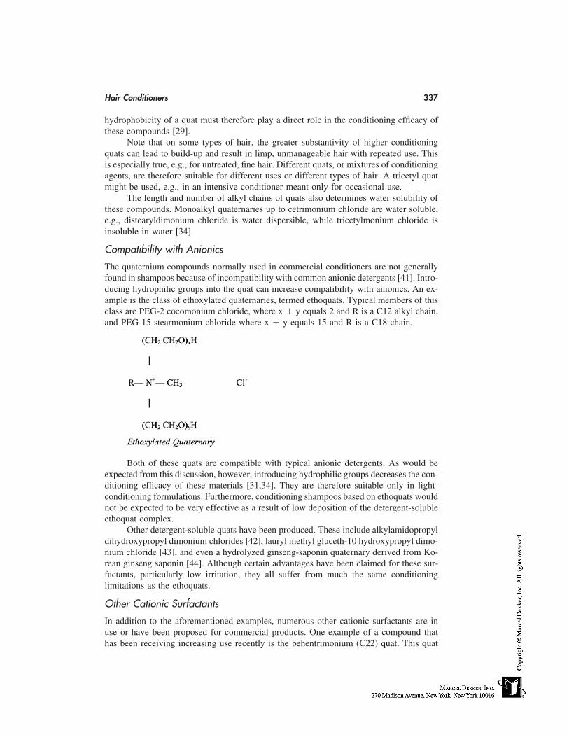
Hair Conditioners 337
hydrophobicity of a quat must therefore play a direct role in the conditioning efficacy ofthese compounds [29].
Note that on some types of hair, the greater substantivity of higher conditioningquats can lead to build-up and result in limp, unmanageable hair with repeated use. Thisis especially true, e.g., for untreated, fine hair. Different quats, or mixtures of conditioningagents, are therefore suitable for different uses or different types of hair. A tricetyl quatmight be used, e.g., in an intensive conditioner meant only for occasional use.
The length and number of alkyl chains of quats also determines water solubility ofthese compounds. Monoalkyl quaternaries up to cetrimonium chloride are water soluble,e.g., distearyldimonium chloride is water dispersible, while tricetylmonium chloride isinsoluble in water [34].
Compatibility with Anionics
The quaternium compounds normally used in commercial conditioners are not generallyfound in shampoos because of incompatibility with common anionic detergents [41]. Intro-ducing hydrophilic groups into the quat can increase compatibility with anionics. An ex-ample is the class of ethoxylated quaternaries, termed ethoquats. Typical members of thisclass are PEG-2 cocomonium chloride, where x � y equals 2 and R is a C12 alkyl chain,and PEG-15 stearmonium chloride where x � y equals 15 and R is a C18 chain.
Both of these quats are compatible with typical anionic detergents. As would beexpected from this discussion, however, introducing hydrophilic groups decreases the con-ditioning efficacy of these materials [31,34]. They are therefore suitable only in light-conditioning formulations. Furthermore, conditioning shampoos based on ethoquats wouldnot be expected to be very effective as a result of low deposition of the detergent-solubleethoquat complex.
Other detergent-soluble quats have been produced. These include alkylamidopropyldihydroxypropyl dimonium chlorides [42], lauryl methyl gluceth-10 hydroxypropyl dimo-nium chloride [43], and even a hydrolyzed ginseng-saponin quaternary derived from Ko-rean ginseng saponin [44]. Although certain advantages have been claimed for these sur-factants, particularly low irritation, they all suffer from much the same conditioninglimitations as the ethoquats.
Other Cationic Surfactants
In addition to the aforementioned examples, numerous other cationic surfactants are inuse or have been proposed for commercial products. One example of a compound thathas been receiving increasing use recently is the behentrimonium (C22) quat. This quat

338 Reich and Su
exhibits significantly reduced eye and skin irritation compared with the correspondingC18 conditioner. In addition, superior conditioning and thickening properties have beenclaimed [45].
Another interesting example is hydrogenated tallow octyl dimonium chloride [46].This material is quite substantive and provides high conditioning as a result of its twohydrophobic chains. Unlike conventional dialkyl quats, however, this particular condi-tioner is soluble in water as a result of branching (2-ethylhexyl) in the octyl moiety. Thismakes the compound much easier to formulate into a commercial product.
Stearamidopropyl dimethylamine is another conditioning agent that is found in manycommercial conditioners. This material is cationic at the pHs normally used in condition-ing products and therefore acts as a cationic emulsifier and, also, as a secondary condition-ing agent.
Concern for the environment has led to the synthesis of ester quats that exhibitincreased biodegradability and environmental safety. One such example is dipalmitoyl-ethyl hydroxyethylmonium methosulfate, an ester quat based on a partially hydrogenatedpalm radical [47].
Other cationic surfactants used in conditioners include quats derived from Guerbetalcohols [39] (low to high conditioning depending on length of the main and side alkylchains), distearyldimonium chloride (high conditioning), and the quaternized ammoniumcompounds of hydrolyzed milk protein, soy and wheat protein, and hydrolyzed keratin(varying conditioning efficacy depending on alkyl chain length).
Lipophilic Conditioners
Quaternary ammonium surfactants in commercial products are almost never used alone.Instead they are used in combination with long-chain fatty conditioners, especially cetyland stearyl alcohols [28]. These fatty materials are added to boost the conditioning effectsof the quaternary compounds [43]. In one study, e.g., addition of cetyl alcohol to cetrimo-nium bromide nearly doubled the observed reduction in wet combing forces on hair [48].In another study, using a novel hydrodynamic technique, Fukuchi et al. [49] found that theaddition of cetyl alcohol to a behentrimonium chloride formulation resulted in significantlyreduced surface friction.
Several investigators have studied combinations of cationic surfactants and fattyalcohols. Under the right conditions, these mixtures have been found to form liquid crystalmesophases and gel networks [50–54] that can greatly increase viscosity and, at the sametime, confer stability upon emulsions. As a result of reduced repulsion between cationichead groups when long chain alcohols are interposed, liquid crystal formation has beenobserved even at low concentrations [53,54]. The ready formation of these extended struc-tures between quats and cetyl and stearyl alcohols, along with the low cost, stability, andcompatibility with cosmetic ingredients of the latter are important reasons why these alco-hols are so ubiquitous in conditioning formulations.
Other lipids found in commercial products include, e.g., glycol distearate, triglycer-ides, fatty esters, waxes of triglycerides, and liquid paraffin.
Cationic Polymers
There are numerous cationic polymers that provide conditioning benefits, especially im-proved wet combing and reduced static charge. Important examples of these polymers arePolyquaternium-10, a quaternized hydroxyethylcellulose polymer; Polyquaternium-7, a
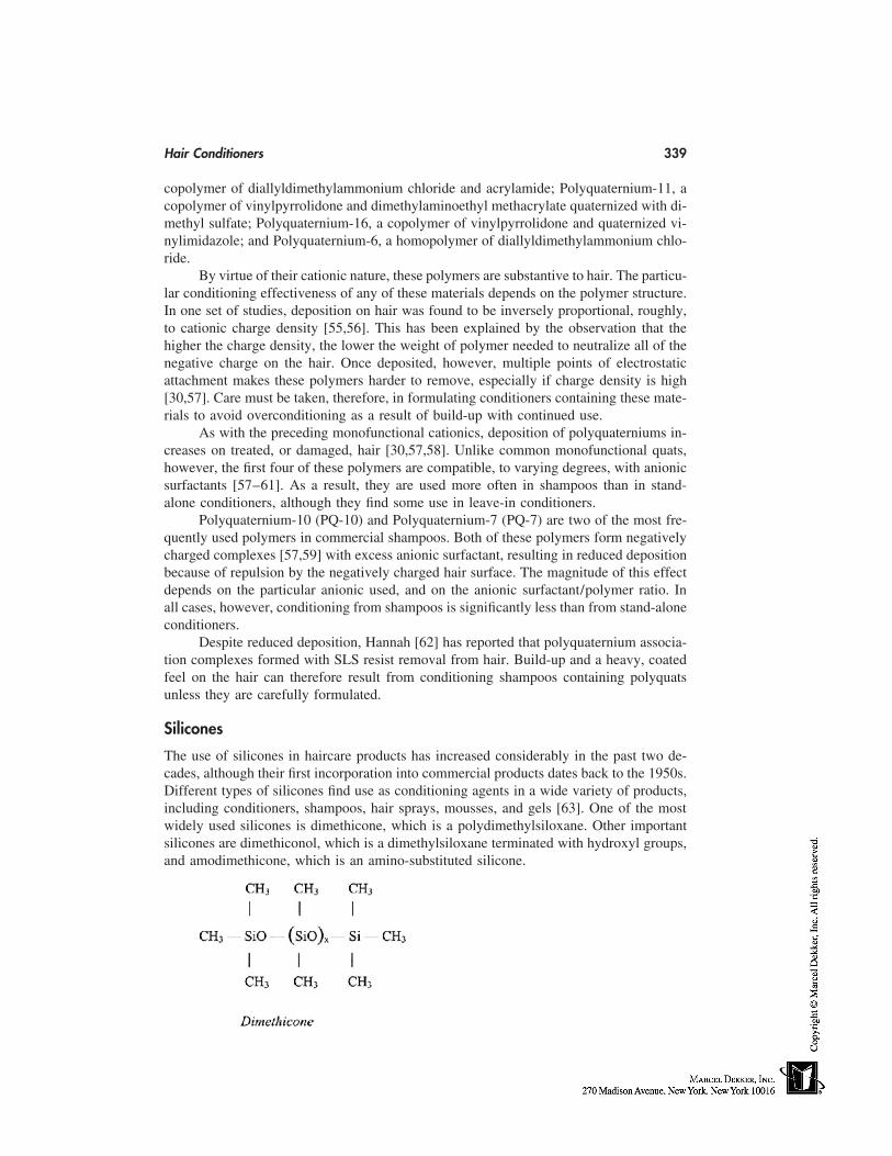
Hair Conditioners 339
copolymer of diallyldimethylammonium chloride and acrylamide; Polyquaternium-11, acopolymer of vinylpyrrolidone and dimethylaminoethyl methacrylate quaternized with di-methyl sulfate; Polyquaternium-16, a copolymer of vinylpyrrolidone and quaternized vi-nylimidazole; and Polyquaternium-6, a homopolymer of diallyldimethylammonium chlo-ride.
By virtue of their cationic nature, these polymers are substantive to hair. The particu-lar conditioning effectiveness of any of these materials depends on the polymer structure.In one set of studies, deposition on hair was found to be inversely proportional, roughly,to cationic charge density [55,56]. This has been explained by the observation that thehigher the charge density, the lower the weight of polymer needed to neutralize all of thenegative charge on the hair. Once deposited, however, multiple points of electrostaticattachment makes these polymers harder to remove, especially if charge density is high[30,57]. Care must be taken, therefore, in formulating conditioners containing these mate-rials to avoid overconditioning as a result of build-up with continued use.
As with the preceding monofunctional cationics, deposition of polyquaterniums in-creases on treated, or damaged, hair [30,57,58]. Unlike common monofunctional quats,however, the first four of these polymers are compatible, to varying degrees, with anionicsurfactants [57–61]. As a result, they are used more often in shampoos than in stand-alone conditioners, although they find some use in leave-in conditioners.
Polyquaternium-10 (PQ-10) and Polyquaternium-7 (PQ-7) are two of the most fre-quently used polymers in commercial shampoos. Both of these polymers form negativelycharged complexes [57,59] with excess anionic surfactant, resulting in reduced depositionbecause of repulsion by the negatively charged hair surface. The magnitude of this effectdepends on the particular anionic used, and on the anionic surfactant/polymer ratio. Inall cases, however, conditioning from shampoos is significantly less than from stand-aloneconditioners.
Despite reduced deposition, Hannah [62] has reported that polyquaternium associa-tion complexes formed with SLS resist removal from hair. Build-up and a heavy, coatedfeel on the hair can therefore result from conditioning shampoos containing polyquatsunless they are carefully formulated.
Silicones
The use of silicones in haircare products has increased considerably in the past two de-cades, although their first incorporation into commercial products dates back to the 1950s.Different types of silicones find use as conditioning agents in a wide variety of products,including conditioners, shampoos, hair sprays, mousses, and gels [63]. One of the mostwidely used silicones is dimethicone, which is a polydimethylsiloxane. Other importantsilicones are dimethiconol, which is a dimethylsiloxane terminated with hydroxyl groups,and amodimethicone, which is an amino-substituted silicone.
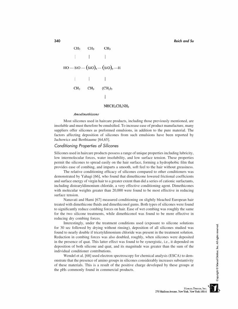
340 Reich and Su
Most silicones used in haircare products, including those previously mentioned, areinsoluble and must therefore be emulsified. To increase ease of product manufacture, manysuppliers offer silicones as preformed emulsions, in addition to the pure material. Thefactors affecting deposition of silicones from such emulsions have been reported byJachowicz and Berthiaume [64,65].
Conditioning Properties of Silicones
Silicones used in haircare products possess a range of unique properties including lubricity,low intermolecular forces, water insolubility, and low surface tension. These propertiespermit the silicones to spread easily on the hair surface, forming a hydrophobic film thatprovides ease of combing, and imparts a smooth, soft feel to the hair without greasiness.
The relative conditioning efficacy of silicones compared to other conditioners wasdemonstrated by Yahagi [66], who found that dimethicone lowered frictional coefficientsand surface energy of virgin hair to a greater extent than did a series of cationic surfactants,including distearyldimonium chloride, a very effective conditioning agent. Dimethiconeswith molecular weights greater than 20,000 were found to be most effective in reducingsurface tension.
Nanavati and Hami [67] measured conditioning on slightly bleached European hairtreated with dimethicone fluids and dimethiconol gums. Both types of silicones were foundto significantly reduce combing forces on hair. Ease of wet combing was roughly the samefor the two silicone treatments, while dimethiconol was found to be more effective inreducing dry combing forces.
Interestingly, under the treatment conditions used (exposure to silicone solutionsfor 30 sec followed by drying without rinsing), deposition of all silicones studied wasfound to nearly double if tricetyldimonium chloride was present in the treatment solution.Reduction in combing forces was also doubled, roughly, when silicones were depositedin the presence of quat. This latter effect was found to be synergistic, i.e., it depended ondeposition of both silicone and quat, and its magnitude was greater than the sum of theindividual conditioner contributions.
Wendel et al. [68] used electron spectroscopy for chemical analysis (ESCA) to dem-onstrate that the presence of amino groups in silicones considerably increases substantivityof these materials. This is a result of the positive charge developed by these groups atthe pHs commonly found in commercial products.
Hair Conditioners 341
Comparison of conditioning effects of a series of silicone emulsions on bleachedand virgin hair was carried out by Hoag et al. [69]. Most of the silicones were dimethiconesor amodimethicones, while emulsions were anionic, neutral, or cationic in nature. Dilutedemulsions were applied directly to the hair and combing forces measured both before andafter rinsing. Prior to rinsing, reduction of combing forces by most emulsions was greaterthan 80%. This number was decreased after rinsing as a result of partial removal of depos-ited silicone. Unsurprisingly, the least change in ease of combing was found for cationicemulsions, especially those containing amodimethicone. Combing forces on virgin hairincreased less than on bleached hair after rinsing, indicating that the silicones were moresubstantive to this type of hair. This is also unsurprising considering the hydrophobicnature of these conditioning agents.
Further effects of amodimethicones can be seen in work reported by Berthiaumeet al. [70], who studied a series of amodimethicone emulsions in a prototype conditionerformulation. Deposition on hair from the conditioner was found to increase with increasingamine content in the silicone. This increased deposition was found, in half-head tests, tocorrelate with conditioning efficacy, including wet and dry combing, softness, and detan-gling. A microemulsion in the test series that provided high conditioning was also shownto significantly reduce the color fading caused by shampooing of temporarily dyed hair.
Other Silicones
Two important silicones not covered in the preceding section are dimethicone copolyol,which is a dimethylsiloxane containing polyoxyethylene and/or propylene side chains,and cyclomethicone, which refers to a class of cyclic dimethyl polysiloxanes ranging fromtrimer to hexamer. The most commonly used variant is the pentamer.
Most commercial dimethicone copolyols are soluble in water and are therefore notvery effective in rinse-off products. These silicones find important application, however,in leave-on products, including hair sprays, styling mousses, and gels.
Cyclomethicone is volatile and would not remain on dry hair, especially after blow-drying. It helps other conditioning agents disperse, however, and form films on hair. Italso helps improve wet combing and provides transient shine.
342 Reich and Su
2-in-1 Shampoos
Silicones find important application as the primary conditioning agents in 2-in-1 condition-ing shampoos. These shampoos, upon their introduction in the latter part of the 1980s,represented a major advance in haircare technology, providing a significantly higher de-gree of conditioning than was then the norm for conditioning shampoos and, at the sametime, leaving a desirable, soft, smooth feel on the hair.
Conditioning from 2-in-1 shampoos is expected to occur primarily at the rinsingstage during which time the shampoo emulsion breaks, releasing the silicone for depositionon hair. This separation of cleaning and conditioning stages permits the shampoo to per-form both functions efficiently.
The conditioning agent used most frequently in 2-in-1 shampoos is dimethicone.This silicone can provide good performance in shampoo formulations without building-up excessively on the hair [71]. The level of conditioning from these types of shampoosis lower than that from stand-alone conditioners. This is especially true for treated hairbecause the greater the degree of negative charge on the hair surface, the lower the substan-tivity of a hydrophobic material like dimethicone. Many 2-in-1s contain polyquats, whichmight be expected to increase conditioning on damaged hair. In shampoos with high levelsof anionic detergent, however, polyquat performance on treated hair may be no betterthan dimethicone as a result of formation of the negatively charged polymer complexesdiscussed in the section on cationic polymers (see p. 338).
Yahagi [66] studied the performance of dimethicone, amodimethicone, and dimethi-cone copolyols in 2-in-1 shampoos. Ease of combing was found to be similar on hairtreated with shampoos containing dimethicone or amodimethicone. Unsurprisingly, solu-ble dimethicone copolyols did not perform well; insolubility, or at least dispersibility, wasrequired for adequate silicone deposition. In the latter case, dimethicone copolyols werefound to provide a somewhat lower level of conditioning than the other two siliconesstudied, especially once blowdrying was begun. Yahagi also studied silicone effects onfoam volume. In these studies dimethicone was found to significantly reduce foam volumein a model shampoo formulation, while amodimethicone and dimethicone copolyol hada minimal effect on foam.
Auxiliary Ingredients
A number of ingredients besides conditioning actives are added to commercial condition-ers for functional, aesthetic, and marketing purposes [72]. These include fragrances, dyes,preservatives, thickeners, emulsifying agents, pearlizers, herbal extracts, humectants, andvitamins. Some of these are discussed in the following sections; the literature also containsmany examples [28,73–77].
Preservatives
Preservatives are necessary to insure the microbiological integrity of a conditioning prod-uct. If the product contains high concentrations of ethyl alcohol (generally 20% or above),additional preservatives are not needed and the product is described as self-preserving.
For other products, a wide variety of preservatives are available; in general, combi-nations of different preservatives provide the broadest possible protection. Every commer-cial product that is not self-preserving must be carefully tested over time for adequacy ofpreservation. Most of the preservatives used in personal-care products are described inthe Cosmetic Preservatives Encyclopedia [75].
Hair Conditioners 343
Thickeners
The section on lipophilic conditioners described thickening as a result of liquid crystalformation in those products containing common quaternary ammonium compounds andfatty alcohols. Cationic conditioning polymers (see p. 338) can also act as thickeners.Many formulations may require additional thickening agents. Hydroxyethylcellulose, anonionic cellulose ether compatible with cationic surfactants and stable over a wide pHrange, is the most common thickening agent added to conditioning products [28]. In addi-tion to providing increased viscosity, this material stabilizes viscosity over time.
Polyamides may also be used to thicken formulations. A commercial product, Sepi-gel, which contains polyamide, laureth-7, and isoparaffin, can be used to emulsify andthicken lotion or cream conditioners. Other thickeners are described in Ref. 76.
Humectants
Many conditioners contain humectants, which are used to attract moisture. Examples arepropylene glycol, glycerine, honey, chitosan, and hyaluronic acid. These materials are notexpected to be very effective in rinse-off products.
Emulsifiers
As previously discussed, the fatty alcohol, quat combinations found in common condition-ers confer stability on product emulsions. If necessary, other emulsifiers may be added toimprove stability. Information on emulsions and emulsifiers may be found in the literature[77,78], as well as from manufacturers’ technical bulletins. Most emulsifiers used in condi-tioners are nonionic, including ethoxylated fatty alcohols, ethoxylated fatty esters, andethoxylated sorbitan fatty esters.
CONCLUSION
The foregoing sections have surveyed the action and properties of a diverse assortmentof commercially available conditioning agents. The availability of a large selection ofconditioning materials enables the formulator to tailor products for a wide variety of peoplehaving differing conditioning needs and preferences. Thus, a person having short, straighthair in good condition might desire a conditioner primarily to control fly-away. Such aneed could be satisfied by one of the ethoquats, which provide light-conditioning benefitstogether with very good static control. A person having long, heavily bleached hair, onthe other hand, would require improved hair feel, ease-of-combing, and manageability.These benefits could best be provided by a trialkyl quat.
Those people sensitive to the feel of their hair might prefer a product containing asilicone as a secondary conditioner. Other people might prefer the convenience of a 2-in-1 shampoo. In many cases, both 2-in-1 shampoos and stand-alone conditioners are usedto condition the hair.
There are a number of ways in which one might satisfy the conditioning needs ofa target population. It is anticipated that the information in this chapter will help theformulator to quickly choose the best conditioning system for a given purpose. It is alsohoped that the material in this chapter will help the formulator to effectively evaluate newconditioning agents and even to work with synthetic chemists as well as suppliers to designnew conditioning compounds to solve particular problems.
344 Reich and Su
REFERENCES
1. Robbins CR. Chemical and Physical Behavior of Human Hair. 3d ed. New York: Springer-Verlag, 1994:343.
2. Kamath YK, Weigmann HD. Measurement of combing forces. J Soc Cosmet Chem 1986; 37:111–124.
3. Jachowicz J. Hair damage and attempts to its repair. J Soc Cosmet Chem 1987; 38:263–286.4. Scott GV, Robbins CR. Effects of surfactant solutions on hair fiber friction. J Soc Cosmet
Chem 1980; 31:179–200.5. Lunn AC, Evans RE. The electrostatic properties of human hair. J Soc Cosmet Chem 1977;
28:549–569.6. Jachowicz J, Wis-Surel G, Garcia ML. Relationship between triboelectric charging and surface
modifications of human hair. J Soc Cosmet Chem 1985; 36:189–212.7. Reich C, Robbins CR. Interactions of cationic and anionic surfactants on hair surfaces: light-
scattering and radiotracer studies. J Soc Cosmet Chem 1993; 44:263–278.8. Robbins CR, Crawford RJ. Cuticle damage and the tensile properties of human hair. J Soc
Cosmet Chem 1991; 42:59.9. Robbins CR. Chemical and Physical Behavior of Human Hair. 3d ed. New York: Springer-
Verlag, 1994:301.10. Tate ML, Kamath YK, Ruetsch SB, Weigmann HD. Quantification and prevention of hair
damage. J Soc Cosmet Chem 1993; 44:347–371.11. Garcia ML, Epps JA, Yare RS. Normal cuticle-wear pattern in human hair. J Soc Cosmet
Chem 1978; 29:155–175.12. Kelley S, Robinson VNE. The effect of grooming on the hair surface. J Soc Cosmet Chem
1982; 33:203–215.13. Kamath YK, Danziger CJ, Weigmann HD. Surface wettability of human hair. I. Effect of
deposition of polymers and surfactants. J Appl Polym Sci 1984; 29:1011–1026.14. Wolfram LJ, Lindemann MKO. Some observations on the hair cuticle. J Soc Cosmet Chem
1971; 22:839–850.15. Negri AP, Cornell HJ, Rivett DE. A model for the surface of keratin fibers. Text Res J 1993;
63:109–115.16. Shao J, Jones DC, Mitchell R, Vickerman JC, Carr CM. Time-of-flight secondary-ion-mass
spectrometric (ToF-SIMS) and x-ray photoelectron spectroscopic (XPS) analyses of the sur-face lipids of wool. J Text Inst 1997; 88:317–324.
17. Wilkerson VJ. The chemistry of human epidermis. II. The isoelectric points of the stratumcorneum, hair, and nails as determined by electrophoresis. J Biol Chem 1935–1936; 112:329–335.
18. Robbins CR, Bahl MK. Analysis of hair by electron spectroscopy for chemical analysis. JSoc Cosmet Chem 1984; 35:379–390.
19. Stranick MA. Determination of negative binding sites on hair surfaces using XPS and Ba2�
labeling. Surface Interface Anal 1996; 24:522–528.20. Horiuchi T. Nature of damaged hair. Cosmet Toilet 1978; 93:65–77.21. Kaplin IJ, Schwann A, Zahn H. Effects of cosmetic treatments on the ultrastructure of hair.
Cosmet Toilet 1982; 97:22–26.22. Sandhu SS, Ramachandran R, Robbins CR. A simple and sensitive method using protein loss
measurements to evaluate damage to human hair during combing. J Soc Cosmet Chem 1995;46:39–52.
23. Robbins CR. Chemical and Physical Behavior of Human Hair. 3d ed. New York: Springer-Verlag, 1994:120–126, 234–249.
24. Schwartz A, Knowles D. Frictional effects in human hair. J Soc Cosmet Chem 1963; 14:455–463.
25. Robbins CR. Chemical and Physical Behavior of Human Hair. 3rd ed. New York: Springer-Verlag, 1994:341.
Hair Conditioners 345
26. Quack JM. Quaternary ammonium compounds in cosmetics. Cosmet Toilet 1976; 91(2):35–52.
27. Gerstein T. An introduction to quaternary ammonium compounds. Cosmet Toilet 1979;94(11):32–41.
28. Hunting ALL. Encyclopedia of Conditioning Rinse Ingredients. Cranford, NJ: Micelle Press,1987.
29. Foerster T, Schwuger MJ. Correlation between adsorption and the effects of surfactants andpolymers on hair. Progr Colloid Polym Sci 1990; 83:104–109.
30. Reich C. Hair cleansers. In: Rieger MM, Rhein LD, eds. Surfactants in Cosmetics. 2d ed.Surfactant Science Series, Vol. 68. New York: Marcel Dekker, 1997:373.
31. Jurczyk MF, Berger DR, Damaso GR. Quaternary ammonium salt. Applications in hair condi-tioners. Cosmet Toilet 1991; 106:63–68.
32. Finkelstein P, Laden K. The mechanism of conditioning of hair with alkyl quaternary ammo-nium compounds. Appl Poly Symp 1971; 18:673–680.
33. Jachowicz J. Fingerprinting of cosmetic formulations by dynamic electrokinetic and perme-ability analysis. II. Hair conditioners. J Soc Cosmet Chem 1995; 46:100–116.
34. Spiess E. The influence of chemical structure on performance in hair care preparations. Par-fumerie and Kosmetik 1991; 72(6):370–374.
35. Scott GV, Robbins CR, Barnhurst JD. Sorption of quaternary ammonium surfactants by humanhair. J Soc Cosmet Chem 1969; 20:135–152.
36. Robbins CR, Reich C, Patel A. Adsorption to keratin surfaces: a continuum between a charge-driven and a hydrophobically driven process. J Soc Cosmet Chem 1994; 45:85–94.
37. Ohbu K, Tamura T, Mizushima N, Fukuda M. Binding characteristics of ionic surfactantswith human hair. Colloid Polym Sci 1986; 264:798–802.
38. Stapleton IW. The adsorption of long chain amines and diamines or keratin fibers. J Soc Cos-met Chem 1983; 34:285–300.
39. Yahagi K, Hoshino N, Hirota H. Solution behavior of new cationic surfactants derived fromGuerbet alcohols and their use in hair conditioners. Int J Cosmet Sci 1991; 13:221–234.
40. Garcia ML, Diaz J. Combability measurements on human hair. J Soc Cosmet Chem 1976;27:379–398.
41. Fox C. An introduction to the formulation of shampoos. Cosmet Toilet 1988; 103(3):25–58.42. Smith L, Gesslein BW. Multi-functional cationics for hair and skin applications. Cosmet Toilet
1989; 104:41–47.43. Polovsky SB. An alkoxylated methyl glucoside quaternary. Cosmet Toilet 1991; 106:59–65.44. Kim YD, Kim CK, Lee CN, Ha BJ. Hydrolysed ginseng-saponin quaternary: a novel condition-
ing agent for hair care products. Int J Cosmet Chem 1989; 11:203–220.45. Gallagher KF. Superior conditioning and thickening from long-chain surfactants. Cosmet Toi-
let 1994; 109:67–74.46. Jurczyk MF. A new quaternary conditioner for damaged hair. Cosmet Toilet 1991; 106:91–95.47. Shapiro I, Sajic B, Bezdicek R. Environmentally friendly ester quats. Cosmet Toilet 1994;
109:77–80.48. Hunting All. Encyclopedia of Conditioning Rinse Ingredients. Cranford, NJ: Micelle Press,
1987:147.49. Fukuchi Y, Okoshi M, Murotani I. Estimation of shampoo and rinse effects on the resistance
to flow over human hair and hair softness using a newly developed hydrodynamic technique.J Soc Cosmet Chem 1989; 40:251–263.
50. Eccleston GM, Florence AT. Application of emulsion theory to complex and real systems.Int J Cosmet Chem 1985; 7:195–212.
51. Eccleston GM. The structure and rheology of pharmaceutical and cosmetic creams. Cetrimidecreams: the influence of alcohol chain length and homolog composition. J Colloid Int Sci1976; 57:66–74.
52. Barry BW, Saunders GM. Kinetics of structure build-up in self-bodied emulsions stabilizedby mixed emulsifiers. J Colloid Int Sci 1972; 41:331–342.
346 Reich and Su
53. Barry BW, Saunders GM. The self-bodying action of the mixed emulsifier cetrimide/ceto-stearyl alcohol. J Colloid Int Sci 1970; 34:300–315.
54. Barry BW, Saunders GM. The influence of temperature on the rheology of systems containingalkyltrimethylammonium bromide/cetostearyl alcohol: variation with quaternary chain length.J Colloid Int Sci 1971; 36:130–138.
55. Hossel P, Pfrommer E. Test methods for hair conditioning polymers. In: In-Cosmet. Exhib.Conf. Conf. Proc. Augsburg, Germany: Verlag fuer Chemische Industrie. H. Ziolkowsky,1994:133–148.
56. Pfau A, Hossel P, Vogt S, Sander R, Schrepp W. The interaction of cationic polymers withhuman hair. Macromol Symp 1997; 126:241–252.
57. Sykes AR, Hammes PA. The use of Merquat polymers is cosmetics. Drug Cosmet Ind 1980;February: 62–66.
58. Amerchol Corporation Technical Bulletin. Ucare polymers: conditioners for all conditions.59. Faucher JA, Goddard ED. Influence of surfactants on the sorption of a cationic polymer by
keratinous substrates. J Colloid Int Sci 1976; 55(2):313–319.60. Goddard ED, Faucher JA, Scott RJ, Turney ME. Adsorption of polymer JR on keratinous
surfaces—Part II. J Soc Cosmet Chem 1975; 26:539–550.61. Caelles J, Cornelles F, Leal JS, Parra JL, Anguera S. Anionic and cationic compounds in
mixed systems. Cosmet Toilet 1991; 106(4):49–54.62. Hannah RB, Goddard ED, Faucher JA. Desorption of a cationic polymer from human hair:
surfactant and salt effects. Text R J 1978; 48:57.63. Luoma A, Kara R. Silicones and the perm question. Society of Cosmetic Chemists 1988 Spring
Conference on Hair Care, London, UK, April 21–23, 1998.64. Jachowicz J, Berthiaume MD. Heterocoagulation of silicon emulsions on keratin fibers. J Col-
loid Int Sci 1989; 133:118–134.65. Berthiaume MD, Jachowicz J. The effect of emulsifiers on deposition of nonionic silicone oils
from oil-in-water emulsions onto keratin fibers. J Colloid Int Sci 1991; 141:299–315.66. Yahagi K. Silicones as conditioning agents in shampoos. J Soc Cosmet Chem 1992; 43:275–
284.67. Nanavati S, Hami A. A preliminary investigation of the interaction of a quat with silicones
and its conditioning benefits on hair. J Soc Cosmet Chem 1994; 43:135–148.68. Wendel SR, Disapio AJ. Organofunctional silicones for personal care applications. Cosmet
Toilet 1983; 98:103–106.69. Hoag CA, Rizwan BM, Quackenbush KM. Evaluating silicone emulsions for global hair care
applications. Global Cosmet Ind 1999; April:44–55.70. Berthiaume MD, Merrifield JH, Riccio DA. Effects of silicone pretreatment on oxidative hair
damage. J Soc Cosmet Chem 1995; 46:231–245.71. Rushton H, Gummer CL, Flasch H. 2-in-1 shampoo technology: state of the art shampoo and
conditioner in one. Skin Pharmacol 1994; 7:78.72. Hoshowski MA. Conditioning of hair. In: Johnson DH, ed. Hair and Hair Care. Cosmetic
Science and Technology Series, Vol. 17. New York: Marcel Dekker, 1997:65–104.73. Wenninger JA, McEwen GN, eds. CTFA Cosmetic Ingredients Handbook. 3d ed. Washington,
DC: Cosmetic, Toiletry and Fragrance Association, 1995.74. Leung AY. Encyclopedia of Common Natural Ingredients Used in Food, Drugs, and Cosmet-
ics. New York: John Wiley & Sons, 1980.75. Cosmetic Preservatives Encyclopedia-Antimicrobials. Cosmet Toilet 1990; 105(3):49–63.76. Lochhead R. Encyclopedia of polymers and thickeners for cosmetics. Cosmet Toilet 1988;
103(12):99–129.77. McCutcheon’s Vol. 1: Emulsifiers and Detergents, North American Edition. Glen Rock, NJ:
MC Publishing Co., 1991.78. Becher P, ed. Encyclopedia of Emulsion Technology. New York: Marcel Dekker, 1985.
30
Hydrating Substances
Marie LodénACO HUD AB, Upplands Väsby, Sweden
INTRODUCTION
Hydrating substances are used in cosmetic products to retard moisture loss from the prod-uct during use and to increase the moisture content in material in contact with the product.This function is generally performed by hygroscopic substances, or humectants. In theInternational Cosmetic Ingredient Dictionary 66 substances are listed as humectants and76 hygroscopic materials are used to increase the water content of the skin [1]. The re-sulting effect of the substances depends on their inherent hygroscopicity at different hu-midity, as well as their volatility and penetration characteristics. Some factors to considerduring product development are highlighted in Table 1.
Target body areas for treatment with humectants are dry hair and dry skin. Some-times mucous membranes also benefit from application of humectants. Dry hair is brittle,rough, has a tendency to tangle, and has hardly any luster. Humidity of the atmosphereis the only source of moisture to hair, except shampooing, and addition of humectants tothe hair will therefore facilitate its retention of water. The same is true for the skin, al-though it is constantly supplied with water from inside of the body. In the stratum corneuma special blend of humectants can be found, which is called natural moisturizing factor(NMF) [2]. NMF can make up about 10% of the dry weight of the stratum corneum cells[2]. Substances belonging to this group are amino acids, pyrrolidone carboxylic acid(PCA), lactates, and urea (Table 2) [2]. NMF is formed from the protein filaggrin andthis formation is regulated by the moisture content in the stratum corneum (3). The waterheld by the hygroscopic substances in the stratum corneum is a controlling factor in main-taining skin flexibility and desquamation (Table 3) [3,4]. This chapter will provide basicinformation about some commonly used humectants, primarily used for treatment of theskin. Moreover, some safety information will be given.
BUTYLENE GLYCOL
Description
Butylene glycol is a viscous, colorless liquid with a sweet flavor and bitter aftertaste [5,6].It is soluble in water, acetone, and castor oil, but practically insoluble in aliphatic hydrocar-bon [5].
347
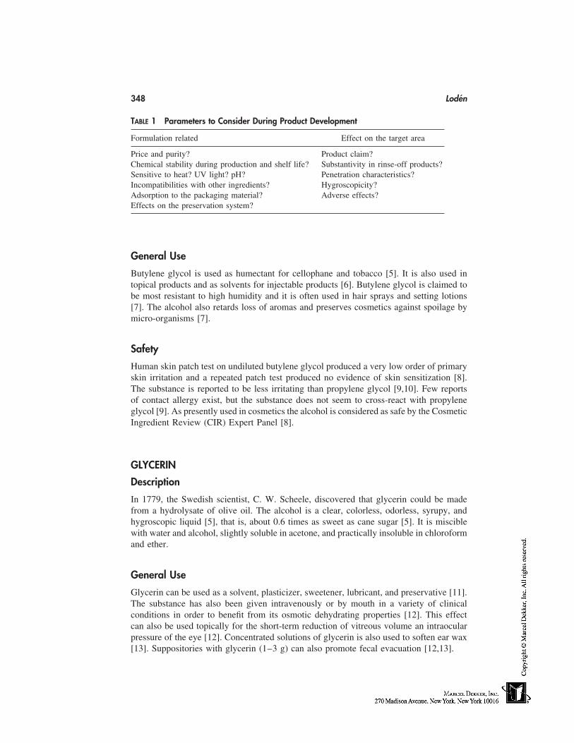
348 Lodén
TABLE 1 Parameters to Consider During Product Development
Formulation related Effect on the target area
Price and purity? Product claim?Chemical stability during production and shelf life? Substantivity in rinse-off products?Sensitive to heat? UV light? pH? Penetration characteristics?Incompatibilities with other ingredients? Hygroscopicity?Adsorption to the packaging material? Adverse effects?Effects on the preservation system?
General Use
Butylene glycol is used as humectant for cellophane and tobacco [5]. It is also used intopical products and as solvents for injectable products [6]. Butylene glycol is claimed tobe most resistant to high humidity and it is often used in hair sprays and setting lotions[7]. The alcohol also retards loss of aromas and preserves cosmetics against spoilage bymicro-organisms [7].
Safety
Human skin patch test on undiluted butylene glycol produced a very low order of primaryskin irritation and a repeated patch test produced no evidence of skin sensitization [8].The substance is reported to be less irritating than propylene glycol [9,10]. Few reportsof contact allergy exist, but the substance does not seem to cross-react with propyleneglycol [9]. As presently used in cosmetics the alcohol is considered as safe by the CosmeticIngredient Review (CIR) Expert Panel [8].
GLYCERIN
Description
In 1779, the Swedish scientist, C. W. Scheele, discovered that glycerin could be madefrom a hydrolysate of olive oil. The alcohol is a clear, colorless, odorless, syrupy, andhygroscopic liquid [5], that is, about 0.6 times as sweet as cane sugar [5]. It is misciblewith water and alcohol, slightly soluble in acetone, and practically insoluble in chloroformand ether.
General Use
Glycerin can be used as a solvent, plasticizer, sweetener, lubricant, and preservative [11].The substance has also been given intravenously or by mouth in a variety of clinicalconditions in order to benefit from its osmotic dehydrating properties [12]. This effectcan also be used topically for the short-term reduction of vitreous volume an intraocularpressure of the eye [12]. Concentrated solutions of glycerin is also used to soften ear wax[13]. Suppositories with glycerin (1–3 g) can also promote fecal evacuation [12,13].
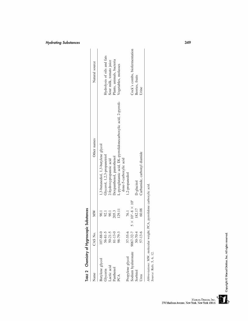
Hydrating Substances 349
TABL
E2
Chem
istr
yof
Hyg
rosc
opic
Subs
tanc
es
Nam
eC
AS
No.
MW
Oth
erna
mes
Nat
ural
sour
ce
But
ylen
egl
ycol
107-
88-0
90.1
1,3-
buta
nedi
ol,
1,3-
buty
lene
glyc
olG
lyce
rin
56-8
1-5
92.1
Gly
cero
l,1,
2,3-
prop
anet
riol
Hyd
roly
sis
ofoi
lsan
dfa
tsL
actic
acid
50-2
1-5
90.1
2-hy
drox
ypro
pano
icac
idSo
urm
ilk,
tom
ato
juic
ePa
nthe
nol
81-1
3-0
205.
3D
expa
nthe
nol,
pant
othe
nol
Plan
ts,
anim
als,
bact
eria
PCA
98-7
9-3
129.
11L
-pyr
oglu
tam
icac
id,
DL
-pyr
rolid
onec
arbo
xylic
acid
,2-
pyrr
oli-
Veg
etab
les,
mol
asse
sdo
ne-5
-car
boxy
licac
idPr
opyl
ene
glyc
ol57
-55-
676
.11,
2-pr
opan
edio
lSo
dium
hyal
uron
ate
9067
-32-
75
�10
4 –8
�10
6C
ock’
sco
mbs
,bi
ofer
men
tatio
nSo
rbito
l50
-70-
418
2.17
D-g
luci
tol
Ber
ries
,fr
uits
Ure
a57
-13-
660
.08
Car
bam
ide,
carb
onyl
diam
ide
Uri
ne
Abb
revi
atio
ns:
MW
,m
olec
ular
wei
ght;
PCA
,py
rrol
idon
eca
rbox
ylic
acid
.So
urce
:R
efs.
5,6,
12.
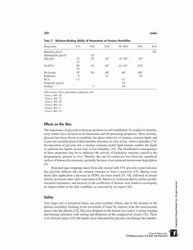
350 Lodén
TABLE 3 Moisture-Binding Ability of Humectants at Various Humidities
Humectant 31% 50% 52% 58–60% 76% 81%
Butylene glycol 38e
Dipropylene glycol 12a
Glycerin 13c 25a 26b 35–38c,f 67b
11b
Na-PCA 20c 44a 45b 61–63c,f 210b
17b
Na-lactate 19b 56a 40b 66f 104b
Panthenol 3d 11d 33d
PCA �1c �1c
Propylene glycol 32f
Sorbitol 1a 10f
Abbreviation: PCA, pyrrolidone carboxylic acid.a Source: Ref. 28.b Source: Ref. 72.c Source: Ref. 40.d Source: Ref. 35.e Source: Ref. 5.f Source: Ref. 73.
Effects on the Skin
The importance of glycerin in skincare products is well established. To explain its benefits,early studies have focused on its humectant and the protecting properties. More recently,glycerin has been shown to modulate the phase behavior of stratum corneum lipids andto prevent crystallization of their lamellar structures in vitro at low, relative humidity [14].Incorporation of glycerin into a stratum corneum model lipid mixture enables the lipidsto maintain the liquid crystal state at low humidity [14]. The biochemical consequencesof these properties may be to influence the activity of hydrolytic enzymes crucial to thedesquamatory process in vivo. Thereby, the rate of corneocyte loss from the superficialsurface of human skin increases, probably because of an enhanced desmosome degradation[3].
Repeated tape strippings taken from skin treated with 15% glycerin cream indicatesthat glycerin diffuses into the stratum corneum to form a reservoir [15]. During somehours after application a decrease in TEWL has been noted [15–18], followed in animalskin by increased values after some hours [18]. Moreover, in human skin its surface profile,electrical impedance, and increase in the coefficient of friction were found to accompanyan improvement in the skin condition, as assessed by an expert [16].
Safety
Very large oral or parenteral doses can exert systemic effects, due to the increase in theplasma osmolality resulting in the movement of water by osmosis from the extravascularspaces into the plasma [12]. Glycerin dropped on the human eye causes a strong stingingand burning sensation, with tearing and dilatation on the conjunctival vessels [19]. Thereis no obvious injury [19], but studies have indicated that glycerin can damage the endothe-
Hydrating Substances 351
lial cells of the cornea [12]. Before application of glycerin to the cornea, a local anestheticmay be administered to reduce the likelihood of a painful response [12].
HYALURONIC ACID
Description
Hyaluronic acid is a member of the class of amino sugar containing polysaccharides knownas the glycosaminoglycans widely distributed in body tissues. Molecular weight is withinthe range of 50,000 to 8 � 106 depending on source, methods of preparation, and determi-nation [5]. Hyaluronic acid binds water and functions as a lubricant between the collagenand elastic fiber networks in dermis during skin movement. Sodium hyaluronate is a whiteodorless powder, which forms viscous solutions in water [6]. A 2% aqueous solution ofpure hyaluronic acid holds the remaining 98% water so tightly that it can be picked upas though it were a gel [20].
During manufacturing, the large, unbranched, non–cross-linked, water-containingmolecule is easily broken by shear forces [20]. The carbohydrate chain is also very sensi-tive to breakdown by free radicals, UV radiation, and oxidative agents [20]. The manufac-turers state that solutions of sodium hyaluronate for injection are stable for 3 years whenstored in refrigerator and for 4 weeks when stored at room temperature [12].
General Use
A viscous solution of the sodium salt is used during surgical procedures on the eye andintra-articular injections have been tried in the treatment of osteoarthritis [12]. Topicalapplication of 0.1% solution in patients with dry eye increased tear-film stability andalleviated symptoms of burning and grittiness [12].
Effects on the Skin
High–molecular weight hyaluronic acid solutions form hydrated viscoelastic films on theskin [20]. The larger the molecular size, the greater the aggregation and entanglement ofthe molecules, and hence, the more substantial and functional the viscoelastic film associ-ated with the skin surface [20]. Because of the high molecular weight, hyaluronic acidwill not penetrate deeper than the crevices between the desquamating cells.
Safety
Sodium hyaluronate is essentially nontoxic [6]. When the substance is used as an ophthal-mic surgical aid, transient inflammatory ocular response has been described [19].
LACTIC ACID
Description
Lactic acid is colorless to yellowish crystals or syrupy liquid, miscible with water, alcohol,glycerol, but insoluble in chloroform [5,6]. Lactic acid is an α-hydroxy acid (AHA), i.e.,an organic carboxylic acid in which there is a hydroxy group at the two, or alpha (α),position of the carbon chain. Lactic acid can exist in a DL, D, or L form. The L and theD forms are enantiomorphic isomers (mirror images). Lactic acid is miscible with water,
352 Lodén
alcohol, and ether and practically insoluble in chloroform [12]. Lactate is also a componentof the natural hygroscopic material of the stratum corneum and constitutes about 12% ofthis material [2]. Formulations containing lactic acid have an acidic pH in the absence ofany inorganic alkali or organic base. pH is increased in several formulations by partialneutralization.
General Use
Lactic acid has been used in topical preparations for several decades because of its buff-ering properties and water binding capacity [21]. Lactic acid and its salts have been usedfor douching and to help maintain the normal, acidic atmosphere of the vagina. Lacticacid has also been used for correction of disorders associated with hyperplasia and/orretention of the stratum corneum, such as dandruff, callus, keratosis, and verrucae (viralwarts) [12]. It has also been suggested that lactic acid may be effective for adjuvant therapyof mild acne [22]. Also, ethyl lactate has been suggested to be effective in the treatmentof acne, because of its penetration into the sebaceous follicle ducts with subsequent low-ering of pH and decrease in the formation of fatty acids [23].
Investigators have also reported increases in the thickness of viable epidermis[24,25] as well as improvement in photoaging changes [24,26]. Lactic acid in combinationwith other peeling agents is used to produce a controlled partial-thickness injury to theskin which is believed to improve the clinical appearance of the skin [27].
Effects on the Skin
In guinea pig footpad corneum, it has been shown that both lactic acid and sodium lactateincrease the water holding capacity and skin extensibility [21]. When the pH increases,the adsorption of lactic acid decreases, because of the ionization of the acid [21]. In anotherstudy on strips of stratum corneum from human abdominal skin, the uptake of water bysodium lactate was greater than that by lactic acid, but the stratum corneum was plasticizedmarkedly by lactic acid and not by sodium lactate [28].
The concentrations used for treatment of ichthyosis and dry skin have ranged up to12% [29]. One formulation of 12% ammonium lactate has been approved by the Foodand Drug Administration (FDA, 1988) for treatment of ichthyosis vulgaris and dry, scalyskin (xerosis) and for the temporary relief of itching associated with these conditions.
Safety
Lactic acid is caustic to the skin, eyes, and mucous membranes in concentrated form [19].Compared with other acids, lactic acid has no unusual capacity to penetrate the cornea,so its injurious effect is presumably attributable to its acidity [19].
Immediately after application of an AHA, stinging and smarting may be noticed;this is closely related to the pH of the preparations and the substances in themselves [30–32]. In normal skin, irritation and scaling may be induced when the acids are applied inhigh concentrations and at low pH [30,33].
PANTHENOL
Description
D-panthenol is a clear, almost colorless, odorless, viscous hygroscopic liquid that maycrystallize on prolonged storage [12]. Panthenol is an alcohol that is rapidly converted to
Hydrating Substances 353
D-pantothenic acid in the body. Panthothenic acid is a water-soluble vitamin, subsequentlycalled vitamin B5. The substance can be isolated from various living creatures, which gavethe reason for its name (panthoten is Greek for ‘‘every-where’’) (Table 2) [34]. Panthenolis very soluble in water; freely soluble in alcohol and glycerol, but insoluble in fats andoils [35]. The substance is fairly stable to air and light if protected from humidity, but itis sensitive to acids and bases and also to heat [35]. The rate of hydrolysis is lowest atpH 4 to 6 [35].
General Use
Panthenol is widely used in the pharmaceutical and cosmetic industry for its moisturizing,soothing, and sedative properties [36]. It is also found in topical treatments for rhinitis,conjunctivitis, sunburn, and for wound healing (ulcers, burns, bed sores, and excoriations)[36]. Usually 2% is used [12]. It can further be used to prevent crystallization at the spraynozzles of aerosols [35].
Effects on the Skin and Hair
Topically applied panthenol is reported to penetrate the skin and hairs and to be trans-formed into panthothenic acid [35,37]. Pantothenic acid can be found in normal hair [35].Soaking of hair in 2% aqueous solution of panthenol has been reported to increase thehair diameter up to 10% [38].
Safety
Panthenol has very low toxicity. Panthenol and products containing panthenol (0.5–2%)administered to rabbits caused reactions ranging from no skin irritation to moderate-to-severe erythema and well-defined edema [39]. Low concentrations have also been testedon humans, and those formulations did not induce sensitization or significant skin irrita-tion. Contact sensitization to panthenol present in cosmetics, sunscreens, and hair lotionhas been reported, although allergy to panthenol among patients attending for patch testingis uncommon [34,36].
PCA AND SALTS OF PCA
Description
PCA is the cosmetic ingredient term used for the cyclic organic compound known as 2-pyrrolidone-5-carboxylic acid (Table 2). The sodium salt is a naturally occurring humec-tant in the stratum corneum at levels about 12% of the NMF [2] corresponding to about2% by weight in the stratum corneum [40]. The sodium salts of PCA are among the mostpowerful humectants (Table 3). PCA is also combined with a variety of other substances,like arginine, lysine, chitosan, and triethanolamine [1].
Effects on the Skin
The ‘‘L’’ form is a naturally occurring component of mammalian tissue and absorptionfrom cosmetics is in addition to PCA already present in the skin (41). A significant rela-tionship has been found between the moisture-binding ability and the PCA content ofsamples of stratum corneum [40]. Treatment of solvent-damaged guinea pig footpad cor-
354 Lodén
neum with humectant solutions shows that the water held by the corneum decreases inthe following order: sodium PCA � sodium lactate � glycerin � sorbitol [21]. Treatmentwith a cream containing 5% sodium-PCA also increased the water-holding capacity ofisolated corneum compared with the cream base [42]. The same cream was also moreeffective than a control product containing no humectant, and equally effective as a similarestablished product with urea as humectant, in reducing the skin dryness and flakiness[42].
Safety
In animal studies, no irritation to the eye and skin was noted at concentrations up to50% and no evidence of phototoxicity, sensitization, or comedogenicity was found [41].Minimal, transient ocular irritation has been produced by 50% PCA [41]. Immediate visi-ble contact reactions in back skin have also been noted after application of 6.25% to 50%aqueous solutions of sodium PCA [43]. The response appeared within 5 minutes anddisappeared within 30 minutes after application. CIR states that the ingredient should notbe used in cosmetic products in which N-nitroso compounds could be formed [41].
PROPYLENE GLYCOL
Description
Propylene glycol is a clear, colorless, viscous, and practically odorless liquid having asweet, slightly acrid taste resembling glycerol [11]. Under ordinary conditions it is stablein well-closed containers and it is also chemically stable when mixed with glycerin, water,or alcohol [5,11].
General Use
Propylene glycol is widely used in cosmetic and pharmaceutical manufacturing as a sol-vent and vehicle especially for substances unstable or insoluble in water [12,44]. It is alsooften used in foods as antifreeze and emulsifier [5,12]. Propylene glycol is also used asinhibitor of fermentation and mold growth [5].
Effects on the Skin
Propylene glycol has been tried in the treatment of a number of skin disorders, includingichthyosis [45,46], tinea versicolor [47], and seborrheic dermatitis [48], because of itshumectant, keratolytic, antibacterial, and antifungal properties [12,44].
Safety
The estimated acceptable daily intake of propylene glycol is up to 25 mg/kg body weight(WHO) [12]. It is considered a harmless ingredient for pharmaceutical products [11] andsafe for use in cosmetic products at concentrations up to 50% [49]. However, clinical datahave showed skin irritation and sensitization reactions to propylene glycol in normal sub-jects at concentrations as low as 10% under occlusive conditions and dermatitis patientsas low as 2% [10,49]. The nature of the cutaneous response remains obscure and, therefore,the skin reactions have been classified into four mechanisms: (1) irritant contact dermatitis,(2) allergic contact dermatitis, (3) nonimmunological contact urticaria, and (4) subjective
Hydrating Substances 355
or sensory irritation [50]. This concept allows a partial explanation of effects observedby different investigators [50].
PROTEINS
Description
Proteins and amino acids for cosmetics are based on a variety of natural sources. Collagenis the traditional protein used in cosmetics. Collagen has a complex triple helical structure,which is responsible for its high–moisture-retention properties. Vegetable-based proteinshave, in recent years, grown in importance as an alternative to using animal by-products.Suitable sources include wheat, rice, soybean, and oat.
In cosmetics native proteins can be used, but perhaps the most widely used proteintypes are hydrolyzed proteins of intermediate molecular weight with higher solubility. Anincreased substantivity is obtained by binding fatty alkyl quarternary groups to the protein.Improved film-forming properties can be obtained by combining the protein and polyvinyl-pyrrolidone into a copolymer. Such modifications may increase the moisture absorptioncompared with the parent compound. Potential problems with proteins are their odor andchange in color with time. Furthermore, as they are nutrients their inclusion in cosmeticsmay require stronger preservatives.
Efficacy and Safety
Amino acids belong to the NMF and account for 40% of its dry weight [2]. Because oftheir relatively low molecular weight, they are capable of penetrating the skin and cuticleof the hair more effectively than the higher–molecular-weight protein hydrolysates.
Salts of the condensation product of coconut acid and hydrolyzed animal protein[51] and wheat flour and wheat starch [52] are considered safe as cosmetic ingredientsby CIR. The most frequent clinical presentation of protein contact dermatitis is a chronicor recurrent dermatitis [53]. Sometimes an urticarial or vesicular exacerbation has beennoted a few minutes after contact with the causative substance [53,54]. Hair conditionerscontaining quaternary hydrolyzed protein or hydrolyzed bovine collagen have inducedcontact urticaria and respiratory symptoms [54]. Atopic constitution seems to be a predis-posing factor in the development of protein contact dermatitis [53].
SORBITOL
Description
Sorbitol is a hexahydric alcohol appearing as a white crystalline powder, odorless and offresh and sweet taste [11,12]. Sorbitol is most commonly available as 70% aqueous solu-tion, which is clear, colorless, and viscous. It occurs naturally in fruits and is easily dis-solved in water, but not so well in alcohol. It is practically insoluble in organic solvents.
Sorbitol is relatively chemically inert and compatible with most excipients, but itmay react with iron oxide and become discolored [11].
General Use
Sorbitol is used in pharmaceutical tablets and in candies when noncariogenic propertiesare desired. It is also used as sweetener in diabetic foods and in toothpastes. Sorbitol is
356 Lodén
also used as a laxative intrarectally and believed to produce less troublesome side effectsthan glycerin [13]. Its hygroscopic properties are reported to be inferior to that of glycerin(Table 3) [21,55].
Safety
When ingested in large amounts (30 g/day) it produces a laxative effect and according toWHO the acceptable daily intake in humans should not exceed 9 grams/day [11].
UREA
Description
Urea is colorless, transparent, slightly hygroscopic, odorless or almost odorless, prismaticcrystals, or white crystalline powder or pellets. Urea is freely soluble in water, slightlysoluble in alcohol, and practically insoluble in ether [12]. The extraction of pure urea fromurine was first accomplished by Proust in 1821 and pure urea was first synthesized byWöhler in 1828 [56]. Urea in solution hydrolyzes slowly to ammonia and carbon dioxide[12].
General Use
Urea is used as a 10% cream for the treatment of ichthyosis and hyperkeratotic skin disor-ders [12,56], and in lower concentrations for the treatment of dry skin. In the treatmentof onychomycosis, urea is added to a medicinal formulation at 40% as a keratoplasticagent to increase the bioavailability of the drug [57].
Effects on the Skin
An increased water-holding capacity of scales from psoriatic and ichthyotic patients hasbeen observed after treatment with urea-containing creams [58,59].
Concern has been expressed about the use of urea in moisturizers, with referenceto the risk of reducing the chemical barrier function of the skin to toxic substances [60].That urea can increase skin permeability has been shown in several studies, where it hasbeen found to be an efficient accelerant for the penetration of different substances [61–63]. Not all studies, however, support the belief that urea is an effective penetration pro-moter [64,65], and treatment of normal skin with moisturizers containing 5% to 10% ureahas been found to reduce transepidermal water loss (TEWL) and also to diminish theirritative response to the surfactant sodium lauryl sulphate [66,67].
Safety
Urea is a naturally occurring substance in the body, as the main nitrogen containing degra-dation product of protein metabolism [68]. Urea is an osmotic diuretic and has been usedin the past for treatment of acute increase in intracranial pressure due to cerebral edema[12]. No evidence of acute or cumulative irritation has been noted in previous studies onurea-containing moisturizers, but several patients [12–22%] have reported stinging aftertreatment with 10% urea creams [69,70]. Urea has also shown to give burning reactionson lesioned forearm skin at concentrations used in moisturizers [71].
Hydrating Substances 357
CONCLUSIONS
A number of interesting humectants are available as cosmetic ingredients. Most of themhave a long and safe history of use, and several are also accepted as food additives. Apotential drawback of the low–molecular weight substances are their stinging potential,since they may be absorbed into the skin. The high–molecular weight substances usuallydo not penetrate the skin; instead they are suggested to reduce the irritation potential ofsurfactants. However, case reports of urticarial reactions have been reported after exposureto modified proteins [54].
The advantage with the larger and chemically modified materials are that they havean increased substantivity to target areas, whereas it is apparent that small amounts ofseveral low–molecular-weight hygroscopic substances have a questionable contributionto the water content of hair and stratum corneum in rinse-off products. Another issue tobear in mind is whether the obtained humectancy is the only mode of action. Some humec-tants may modify the surface properties and increase the extensibility of stratum corneumwithout influencing the water content. Furthermore, humectants may also affect specificmetabolic process in the skin. One should also keep in mind that humectants can improvethe cosmetic properties of the formulation and some of them also facilitate marketing ofthe product just because of their names.
REFERENCES
1. Wenninger JA, McEwen GN. International Cosmetic Ingredient Dictionary and Handbook.Washington, DC: The Cosmetic, Toiletry, and Fragrance Association, 1997.
2. Jacobi O. Moisture regulation of the skin. Drug Cosmet Ind 1959; 84:732–812.3. Rawlings AV, Scott IR, Harding CR, Bowser PA. Stratum corneum moisturization at the
molecular level. J Invest Dermatol 1995; 103:731–740.4. Blank IH. Factors which influence the water content of stratum corneum. J Invest Dermatol
1952; 18:433–440.5. Budavari S. The Merck Index. Rahway: Merck & Co., Inc., 1989.6. Ash M, Ash I. Handbook of Pharmaceutical Additives. Hampshire: Gower Publishing Limited,
1995.7. Rietschel RL, Fowler, JF. Fisher’s contact dermatitis. Baltimore: Williams & Wilkins, 1995.8. The Cosmetic Ingredient Review Expert Panel. Final assessment of the safety assessment of
butylene glycol, hexylene glycol, ethoxydiglycol, and dipropylene glycol. J Am Coll Toxicol1985; 2:223–248.
9. Sugiura M, Hayakawa R. Contact dermatitis due to 1,3-butylene glycol. Contact Derm 1997;37:90.
10. Fan W, Kinnunen T, Niinimäke A, Hannuksela M. Skin reactions to glycols used in dermato-logical and cosmetic vehicles. Am J Contact Derm 1991; 2:181–183.
11. American Pharmaceutical Association and The Pharmaceutical Society of Great Britain. Hand-book of Pharmaceutical Excipients. Baltimore: The Pharmaceutical Press, 1986.
12. Reynolds JEF. Martindale: The Extra Pharmacopoeia. London: The Pharmaceutical Press,1993.
13. Zimmerman DR. The Essential Guide to Nonprescription Drugs. New York: Harper & Row,1983.
14. Froebe CL, Simion A, Ohlmeyer H, et al. Prevention of stratum corneum lipid phase transitionsin vitro by glycerol—an alternative mechanism for skin moisturization. J Soc Cosmet Chem1990; 41:51–65.
15. Batt MD, Fairhurst E. Hydration of the stratum corneum. Int J Cosmet Sci 1986; 8:253–264.
358 Lodén
16. Batt MD, Davis WB, Fairhurst E, Gerrard WA, Ridge BD. Changes in the physical propertiesof the stratum corneum following treatment with glycerol. J Soc Cosmet Chem 1988; 39:367–381.
17. Wilson E, Berardesca E, Maibach H. In vivo transepidermal water loss and skin surface hydra-tion in assessment of moisturization and soap effects. Int J Cosmet Sci 1988; 10:201–211.
18. Lieb LM, Nash RA, Matias JR, Orentreich N. A new in vitro method for transepidermal waterloss: a possible method for moisturizer evaluation. J Soc Cosmet Chem 1988; 39:107–119.
19. Grant WM. Toxicology of the Eye. Springfield: Charles C. Thomas, 1986.20. Balazs EA, Band P. Hyaluronic acid: its structure and use. Cosmet Toilet 1984; 99:65–72.21. Middleton JD. Development of a skin cream designed to reduce dry and flaky skin. J Soc
Cosmet Chem 1974; 25:519–534.22. Berson DS, Shalita AR. The treatment of acne: the role of combination therapies. J Am Acad
Dermatol 1995; 32:S31–41.23. Prottey C, George D, Leech RW, Black JG, Howes D, Vickers CFH. The mode of action of
ethyl lactate as a treatment for acne. Br J Dermatol 1984; 110:475–485.24. Ditre CM, Griffin TD, Murphy GF, et al. Effects of alpha-hydroxy acids on photaged skin:
a pilot clinical, histologic, and ultrastructural study. J Am Acad Dermatol 1996; 34:187–195.
25. Lavker RM, Kaidbey K, Leyden JJ. Effects of topical ammonium lactate on cutaneous atrophyfrom a potent topical corticosteroid. J Am Acad Dermatol 1992; 26:535–544.
26. Stiller MJ, Bartolone J, Stern R, et al. Topical 8% glycolic acid and 8% L-lactic acid creamsfor the treatment of photodamaged skin. A double-blind vehicle-controlled clinical trial. ArchDermatol 1996; 132:631–636.
27. Glogau RG, Matarasso SL. Chemical face peeling: patient and peeling agent selection. FacialPlast Surg 1995; 11:1–8.
28. Takahashi M, Yamada M, Machida Y. A new method to evaluate the softening effect of cos-metic ingredients on the skin. J Soc Cosmet Chem 1984; 35:171–181.
29. Wehr R, Krochmal L, Bagatell F, Ragsdale W. A controlled two-center study of lactate 12%lotion and a petrolatum-based creme in patients with xerosis. Cutis 1986; 37:205–209.
30. Smith WP. Hydroxy acids and skin aging. Cosmet Toilet 1994; 109:41–48.31. Smith WP. Comparative effectiveness of alpha-hydroxy acids on skin properties. Int J Cosmet
Sci 1996; 18:75–83.32. Frosch PJ, Kligman AM. A method for appraising the stinging capacity of topically applied
substances. J Soc Cosmet Chem 1977; 28:197–209.33. Effendy I, Kwangsukstith C, Lee LY, Maibach HI. Functional changes in human stratum
corneum induced by topical glycolic acid: comparison with all-trans retinoic acid. Acta DermVenereol (Stockh) 1995; 75:455–458.
34. Schmid-Grendelmeier P, Wyss M, Elsner P. Contact allergy to dexpanthenol. A report of sevencases and review of the literature. Dermatosen 1995; 43:175–178.
35. Huni JES. Basel: Roche, 1981.36. Stables GI, Wilkinson SM. Allergic contact dermatitis due to panthenol. Contact Derm 1998;
38:236–237.37. Stuttgen G, Krause H. Panthenol. Arch Klin Exp Dermatol 1960; 209:578–582.38. Driscoll WR. Panthenol in hair products. D&CI 1975; 116:45–49.39. The Cosmetic Ingredient Review Expert Panel. Final report on the safety assessment of pan-
thenol and pantothenic acid. J Am Coll Toxicol 1987; 6:139–163.40. Laden K, Spitzer R. Identification of a natural moisturizing agent in skin. J Soc Cosmet Chem
1967; 18:351–360.41. 1997 CIR Compendium. PCA and Sodium PCA. Washington, D.C.: Cosmetic Ingredient Re-
view, 1997:106–107.42. Middleton JD, Roberts ME. Effect of a skin cream containing the sodium salt of pyrrolidone
carboxylic acid on dry and flaky skin. J Soc Cosmet Chem 1978; 29:201–205.
Hydrating Substances 359
43. Larmi E, Lahti A, Hannuksela M. Immediate contact reactions to benzoic acid and the sodiumsalt of pyrrolidone carboxylic acid. Contact Derm 1989; 20:38–40.
44. Cantazaro JM, Smith JG. Propylene glycol dermatitis. J Am Acad Dermatol 1991; 24:90–95.45. Goldsmith LA, Baden HP. Propylene glycol with occlusion for treatment of ichthyosis. JAMA
1972; 220:579–580.46. Gånemo A, Vahlquist A. Lamellar ichthyosis is markedly improved by a novel combination
of emollients. Br J Dermatol 1997; 137:1011–1031.47. Faergemann J, Fredriksson T. Propylene glycol in the treatment of tinea versicolor. Acta Derm
Venereol (Stockh) 1980; 60:92–93.48. Faergemann J. Propylene glycol in the treatment of seborrheic dermatitis of the scalp: a double-
blind study. Cutis 1988; 42:69–71.49. Final report of the safety assessment of propylene glycol and polypropylene glycols (PPG-
9,-12,-15,-17,-20,-26,-30, and 34). J Am Coll Toxicol 1996; 13:6.50. Funk JO, Maibach HI. Propylene glycol dermatitis: re-evaluation of an old problem. Contact
Derm 1994; 31:236–241.51. The Cosmetic Ingredient Review Expert Panel. Final report on the safety assessment of potas-
sium-coco-hydrolyzed animal protein and triethanolamine-coco-hydrolyzed animal protein. JAm Coll Toxicol 1983; 2:75–86.
52. The Cosmetic Ingredient Review Expert Panel. Final report on the safety assessment of wheatflour and wheat starch. J Environ Pathol Toxicol 1980; 4:19–32.
53. Janssens V, Morren M, Dooms-Goossens A, Degreef H. Protein contact dermatitis: myth orreality? Br J Dermatol 1995; 132:1–6.
54. Freeman S, Lee M-S. Contact urticaria to hair conditioner. Contact Derm 1996; 35:195–196.55. Rovesti P, Ricciardi D. New experiments on the use of sorbitol in the field of cosmetics. P &
EOR 1959; 771–774.56. Rosten M. The treatment of ichthyosis and hyperkeratotic conditions with urea. Aust J Derma-
tol 1970; 11:142–144.57. Fritsch H, Stettendorf S, Hegemann L. Ultrastructural changes in onchomycosis during the
treatment with bifonazole/urea ointment. Dermatology 1992;185:32–36.58. Swanbeck G. A new treatment of ichthyosis and other hyperkeratotic conditions. Acta Derm
Venereol (Stockh) 1968; 48:123–127.59. Grice K, Sattar H, Baker H. Urea and retinoic acid in ichthyosis and their effect on transepider-
mal water loss and water holding capacity of stratum corneum. Acta Derm Venereol (Stockh)1973; 53:114–118.
60. Hellgren L, Larsson K. On the effect of urea on human epidermis. Dermatologica 1974; 149:289–293.
61. Wohlrab W. The influence of urea on the penetration kinetics of vitamin-A acid into humanskin. Z Hautkr 1990; 65:803–805.
62. Kim CK, Kim JJ, Chi SC, Shim CK. Effect of fatty acids and urea on the penetration ofketoprofen through rat skin. Int J Pharm 1993; 99:109–118.
63. Beastall J, Guy RH, Hadgraft J, Wilding I. The influence of urea on percutaneous absorption.Pharm Res 1986; 3:294–297.
64. Lippold BC, Hackemuller D. The influence of skin moisturizers on drug penetration in vivo.Int J Pharm 1990; 61:205–211.
65. Wahlberg JE, Swanbeck G. The effect of urea and lactic acid on the percutaneous absorptionof hydrocortisone. Acta Derm Venereol (Stockh) 1973; 53:207–210.
66. Lodén M. Urea-containing moisturizers influence barrier properties of normal skin. Arch Der-matol Res 1996; 288:103–107.
67. Lodén M. Barrier recovery and influence of irritant stimuli in skin treated with a moisturizingcream. Contact Derm 1997; 36:256–260.
68. Swanbeck G. Urea in the treatment of dry skin. Acta Derm Venereol (Stockh) 1992; 177(suppl):7–8.
360 Lodén
69. Serup J. A double-blind comparison of two creams containing urea as the active ingredient.Assessment of efficacy and side-effects by non-invasive techniques and a clinical scoringscheme. Acta Derm Venereol (Stockh) 1992; 177(suppl):34–38.
70. Fredriksson T, Gip L. Urea creams in the treatment of dry skin and hand dermatitis. Int JDermatol 1975; 32:442–444.
71. Gabard B, Nook T, Muller KH. Tolerance of the lesioned skin to dermatological formulations.J Appl Cosmetol 1991; 9:25–30.
72. Rieger MM, Deem DE. Skin moisturizers. II. The effects of cosmetic ingredients on humanstratum corneum. J Soc Cosmet Chem 1974; 25:253–262.
73. Huttinger R. Restoring hydrophilic properties to the stratum corneum—a new humectant. Cos-met Toilet 1978; 93:61–62.
31
Ceramides and Lipids
Bozena B. MichniakUniversity of South Carolina, Columbia, South Carolina
Philip W. WertzUniversity of Iowa, Iowa City, Iowa
HISTORICAL PERSPECTIVES
Many published accounts of the composition of lipids from human stratum corneum havebeen complicated by the almost inevitable presence of sebaceous lipids as well as exoge-nous contaminants. When stratum corneum samples are obtained from excised skin, thereis almost always massive contamination with subcutaneous triglycerides as well as fattyacids derived from the subcutaneous fat. In addition, precautions must be taken to avoidcontamination with environmental contaminants such as alkanes and cosmetic compo-nents. As a result of these complications, much work has been done with pig skin as amodel [1–6].
Young pigs, if properly housed and tended, can be kept clean, and the sebaceousglands are not active. By direct heat separation of epidermis from an intact carcass, it ispossible to avoid subcutaneous fat. In terms of general structure, composition, and perme-ability barrier function, the pig appears to provide a good model for the human. An alterna-tive approach is to use the contents of epidermal cysts [7,8]. This material representsexfoliated stratum corneum lipid that is free of sebaceous and environmental contaminants.If the contents are carefully expressed from the capsule, a contaminant-free sample ofstratum corneum lipid can be obtained. Cholesterol sulfate is partially hydrolyzed duringthe desquamation process; however, this is only a minor stratum corneum component. Ineither the pig or cyst model, the major lipid components are ceramides, cholesterol, andfatty acids, which represent approximately 45, 27, and 12% of the total lipid, respectively[9]. Other minor components include cholesterol sulfate and cholesterol esters. The fattyacids in either model are predominantly straight-chain saturated species ranging from ei-ther 14 (cyst) or 16 (pig) carbons through 28 carbons in length with the 22 and 24 carbonspecies being the most abundant. The main focus in the rest of this chapter will be onthe stratum corneum ceramides.
The first analysis of stratum corneum lipids was performed in 1932 by Kooyman[10], who showed a dramatic reduction in the proportion of phospholipid in stratum cor-neum compared with the inner portion of the epidermis. Subsequently, Long [11], using
361
362 Michniak and Wertz
the very thick epidermis from cow snout as a model, analyzed lipids from horizontal slicesof epithelial tissue. He observed a gradual accumulation of cholesterol and fatty acids inprogressing from the basal region toward the surface. Phospholipids initially accumulated,but were degraded as the stratum corneum was approached. In 1965, Nicolaides [12]identified ceramides as a polar lipid component of stratum corneum. This fact was includedin a footnote and was largely ignored until the pioneering work of Gray and Yardley inthe mid to late 1970s [1,2,13,14]. Among other things, these investigators showed that theceramides are structurally heterogeneous and contain normal fatty acids, α-hydroxyacids,sphingosines, and phytosphingosines as components. However, individual ceramide typeswere not well resolved and no definitive structures could be proposed. The first attemptto isolate individual ceramide types and to determine the identities of the individual fattyacid and long-chain base components was conducted in 1979 using neonatal mouse epider-mis as a source of lipids [15]. Eight putative ceramide fractions were isolated, and six ofthese were analyzed. The remaining two were too minor for any analysis. Unfortunately,only normal fatty acids, sphingosines, and dihydrosphingosines were reported for eachfraction analyzed. This suggests extensive cross-contamination sufficient to preclude rec-ognition of the actual structural diversity. In 1983, the detailed structures of the ceramidesfrom porcine epidermis were published [3]. Six structurally different types of ceramideswere identified, and these included sphingosines, dihydrosphingosines, and phytosphin-gosines as the base components; normal, α-hydroxyacids, and ω-hydroxyacids as the am-ide-linked fatty acids; and one ceramide type included an ester-linked fatty acid. Subse-quently, it was shown that the same ceramide structural types are present in human stratumcorneum, although the proportions are somewhat different [8,15]. More recently it hasbeen shown that in addition to the standard phytosphingosine present in porcine ceramides,the human ceramides also include a variant phytosphingosine, 6-hydroxysphingosine [16].
In 1987 it was discovered that porcine epidermal stratum corneum contains signifi-cant levels of covalently bound lipid, the major component of which is an ω-hydroxycera-mide [4]. Small amounts of saturated fatty acid and ω-hydroxyacid are also present. Asimilar situation was shown for human stratum corneum; however, in this case there wasa second hydroxyceramide that was shown to contain a variant phytosphingosine [17].This subsequently proved to be 6-hydroxysphingosine [16]. The free and covalently boundceramides are discussed in detail in the following section.
CERAMIDES FROM EPIDERMIS
As previously noted, the first comprehensive study of epidermal ceramide structures wasdirected at the porcine ceramides, which were separated into six chromatographically dis-tinct fractions [3]. Each fraction was analyzed by a combination of chemical, chromato-graphic and spectroscopic methods, and representative structures are included in Figure 1.
The least polar of the porcine ceramides, ceramide fraction 1, consists of 30- through34-carbon ω-hydroxyacids amide-linked to a mixture of sphingosines and dihydrosphingo-sines. The long-chain base component of this ceramide ranges from 16 through 22 carbonsin length with 18:1, 20:1, and 22:1 being the most abundant. There is also a fatty acidester-linked to the ω-hydroxyl group, 75% of which consists of linoleic acid. This specieshas often been referred to as ceramide 1 or acylceramide, but in the more systematicnomenclature system proposed by Motta et al. [18] this becomes Cer[OSE]. (In this sys-tem, the amide-linked fatty acid is designated as N, A, or O to indicate normal, α-hydroxy,or ω-hydroxy, respectively. The base component is designated S or P for sphingosine or
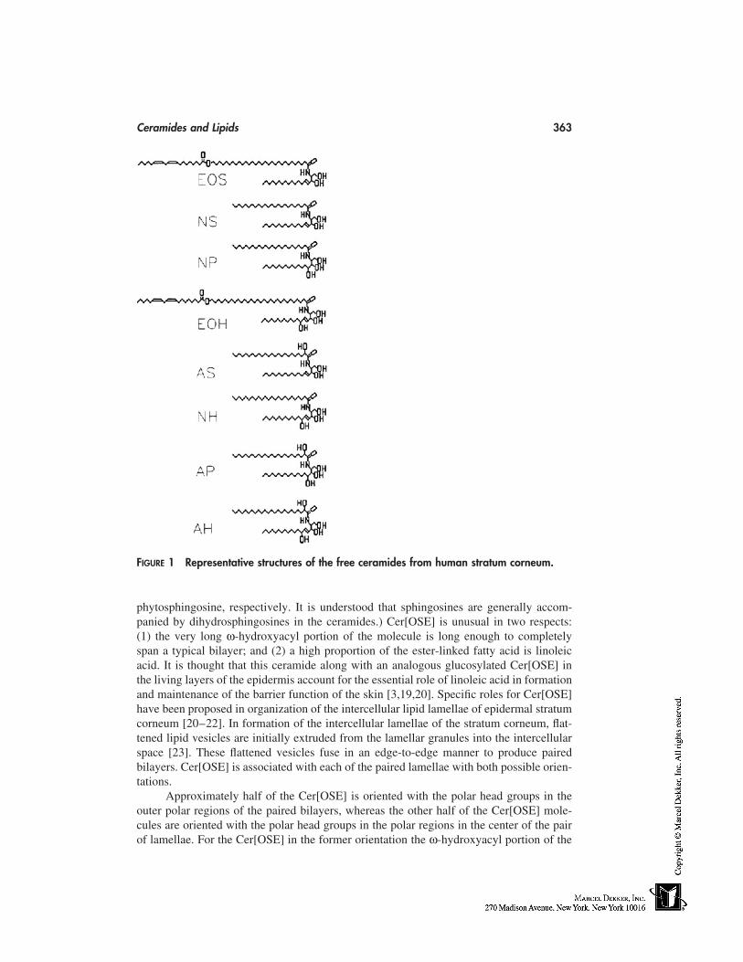
Ceramides and Lipids 363
FIGURE 1 Representative structures of the free ceramides from human stratum corneum.
phytosphingosine, respectively. It is understood that sphingosines are generally accom-panied by dihydrosphingosines in the ceramides.) Cer[OSE] is unusual in two respects:(1) the very long ω-hydroxyacyl portion of the molecule is long enough to completelyspan a typical bilayer; and (2) a high proportion of the ester-linked fatty acid is linoleicacid. It is thought that this ceramide along with an analogous glucosylated Cer[OSE] inthe living layers of the epidermis account for the essential role of linoleic acid in formationand maintenance of the barrier function of the skin [3,19,20]. Specific roles for Cer[OSE]have been proposed in organization of the intercellular lipid lamellae of epidermal stratumcorneum [20–22]. In formation of the intercellular lamellae of the stratum corneum, flat-tened lipid vesicles are initially extruded from the lamellar granules into the intercellularspace [23]. These flattened vesicles fuse in an edge-to-edge manner to produce pairedbilayers. Cer[OSE] is associated with each of the paired lamellae with both possible orien-tations.
Approximately half of the Cer[OSE] is oriented with the polar head groups in theouter polar regions of the paired bilayers, whereas the other half of the Cer[OSE] mole-cules are oriented with the polar head groups in the polar regions in the center of the pairof lamellae. For the Cer[OSE] in the former orientation the ω-hydroxyacyl portion of the
364 Michniak and Wertz
molecule will span the bilayer while the linoleate inserts into the other bilayer, thus linkingthe pair of bilayers together. For Cer[OSE] in the second orientation the linoleate tail isthought to participate in the formation of narrow interdigitated layers that intervene be-tween the paired bilayers. This action of the Cer[OSE] results in the formation of broad-narrow-broad lamellar patterns that are seen in transmission electron micrographs whenruthenium tetroxide is used as a postfixative and which give rise to a 13 nm repeat unitin radiograph diffraction studies [5,6,22].
Porcine ceramide fraction 2 has proven to be Cer[NS]. The fatty acid componentis saturated and straight-chained and ranges from 16- through 32-carbons in length.C20:0, C22:0, C24:0, C26:0, and C28:0 are the most abundant, constituting from 9%to 19% of the total fatty acid mass each. The long-chain bases again consist of a mixtureof sphingosines and dihydrosphingosines ranging from 16- through 22-carbons in length.The most abundant bases are 18:0, 18:1, 20:0, and 20:1.
Porcine ceramide fraction 3, Cer[NP], contains the same range of fatty acids foundin Cer[NS], but the long-chain base component is now a phytosphingosine with no doublebond and a third hydroxyl group on carbon 4. The phytosphingosines found here rangefrom 16- through 24-carbons long, and the most abundant are 20:0 and 22:0.
Porcine ceramide fractions 4 and 5 both proved to be Cer[AS], but they differed interms of the chain length distributions of the α-hydroxyacid component. The chromato-graphically more mobile fraction 4 contained 24- through 28-carbon α-hydroxyacids am-ide-linked to sphingosines and dihydrosphingosines, whereas ceramide fraction 5 containsα-hydroxypalmitic acid amide-linked to sphingosines and dihydrosphingosines. Ceramidefraction 4 also contains somewhat longer bases with major amounts of 20:0 and 20:1,whereas ceramide fraction 5 contains mainly 16- through 18-carbon bases. This differencein carbon content results in chromatographic separation into two fractions, even thoughthe basic structural type is the same in each.
Finally, the most polar of the pig ceramide fractions consists of α-hydroxyacidsamide-linked to phytosphingosine, Cer[AP]. The α-hydroxyacids present in Cer[AP] rangefrom 16- through 28-carbons in length, but the 24- and 26-carbon entities account forapproximately 70% of the total fatty acid mass. The phytosphingosines have a chain-length distribution similar to that already described for Cer[NP].
Subsequently, the human stratum corneum ceramides were investigated and wereshown to produce a similar, though not identical, pattern on thin-layer chromatograms[15]. Notably, the human fraction most closely matching porcine ceramide fraction 3 issomewhat broader and less symmetrical. The material most closely matching porcine cera-mide fractions 4 and 5 merged into one broad peak, and was designated ceramide 4/5.This was shown to reflect a more continuous chain-length distribution among the α-hydroxyacid component of Cer[AS] as opposed to the bipolar distribution found in thepig. The most polar human fraction similar to porcine ceramide fraction 6 appeared as anincompletely resolved doublet. These two components were designated ceramides 6I and6II. Subsequently it has been shown the ceramide fraction 6II contains the variant phyto-sphingosine—6-hydroxysphingosine [16]. The Motta system of nomenclature has beenextended to include this new long-chain base as H [16]. So ceramide 6I is Cer[AP], andceramide 6II becomes Cer[AH]. Human ceramide fraction 3 has been shown to containa minor amount of a 6-hydroxysphingosine-containing acylceramide, Cer[OHE] [16], inaddition to Cer[NH]. Likewise, ceramide fraction 4/5 contains Cer[NH] [24] in additionto Cer[AS] [15]. These additional ceramides containing 6-hydroxysphingosine can be re-solved on thin-layer chromatography by use of multiple development regimens.
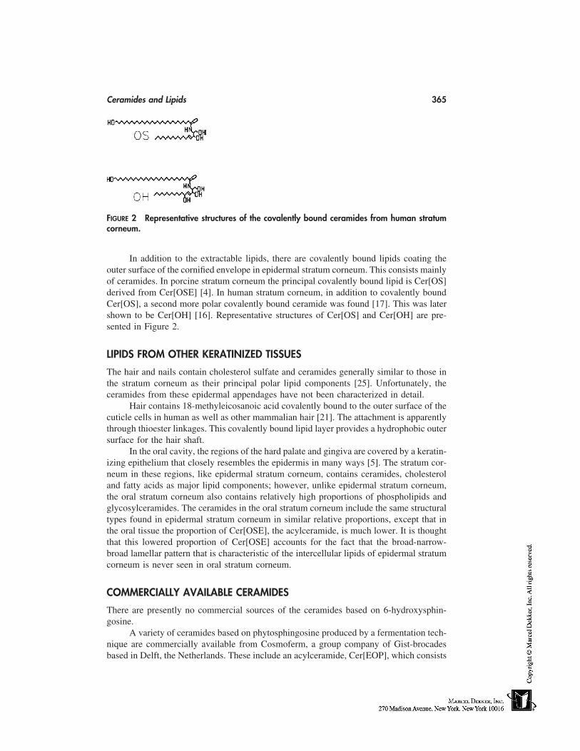
Ceramides and Lipids 365
FIGURE 2 Representative structures of the covalently bound ceramides from human stratumcorneum.
In addition to the extractable lipids, there are covalently bound lipids coating theouter surface of the cornified envelope in epidermal stratum corneum. This consists mainlyof ceramides. In porcine stratum corneum the principal covalently bound lipid is Cer[OS]derived from Cer[OSE] [4]. In human stratum corneum, in addition to covalently boundCer[OS], a second more polar covalently bound ceramide was found [17]. This was latershown to be Cer[OH] [16]. Representative structures of Cer[OS] and Cer[OH] are pre-sented in Figure 2.
LIPIDS FROM OTHER KERATINIZED TISSUES
The hair and nails contain cholesterol sulfate and ceramides generally similar to those inthe stratum corneum as their principal polar lipid components [25]. Unfortunately, theceramides from these epidermal appendages have not been characterized in detail.
Hair contains 18-methyleicosanoic acid covalently bound to the outer surface of thecuticle cells in human as well as other mammalian hair [21]. The attachment is apparentlythrough thioester linkages. This covalently bound lipid layer provides a hydrophobic outersurface for the hair shaft.
In the oral cavity, the regions of the hard palate and gingiva are covered by a keratin-izing epithelium that closely resembles the epidermis in many ways [5]. The stratum cor-neum in these regions, like epidermal stratum corneum, contains ceramides, cholesteroland fatty acids as major lipid components; however, unlike epidermal stratum corneum,the oral stratum corneum also contains relatively high proportions of phospholipids andglycosylceramides. The ceramides in the oral stratum corneum include the same structuraltypes found in epidermal stratum corneum in similar relative proportions, except that inthe oral tissue the proportion of Cer[OSE], the acylceramide, is much lower. It is thoughtthat this lowered proportion of Cer[OSE] accounts for the fact that the broad-narrow-broad lamellar pattern that is characteristic of the intercellular lipids of epidermal stratumcorneum is never seen in oral stratum corneum.
COMMERCIALLY AVAILABLE CERAMIDES
There are presently no commercial sources of the ceramides based on 6-hydroxysphin-gosine.
A variety of ceramides based on phytosphingosine produced by a fermentation tech-nique are commercially available from Cosmoferm, a group company of Gist-brocadesbased in Delft, the Netherlands. These include an acylceramide, Cer[EOP], which consists
366 Michniak and Wertz
of a 27-carbon ω-hydroxyacid amide-linked to phytosphingosine and bearing ester-linkedstearic acid on the ω-hydroxyl group. There are also two ceramides of the type Cer[NP].One of these contains stearic acid and the other oleic acid amide-linked to phytosphingo-sine. Finally, this supplier also produces N-2-hydroxystearoyl-phytosphingosine, Cer[AP].These specific ceramides are routinely available; however, it is also possible to customizeany of these general structural types to include different fatty acids.
There are several commercial sources of ceramides or ceramide analogues similarto the ceramide type Cer[NS]. For example, SEDERMA of Parsippany, New Jersey pro-duces a synthetic ceramide consisting of N-stearoyl-dihydrosphingosine and sold as cera-mide 2. This synthetic ceramide is partially racemic at carbon-3 of the base component;however, the stereochemical configuration at this carbon is at least 70% R, which is theconfiguration in natural dihydrosphingosine.
FUTURE DIRECTIONS
Presently, ceramides are being used in skin moisturizers and at least one line of hair careproducts. It has been documented that ceramides are important in the permeability barrierof the skin and the water-holding properties of the stratum corneum [26,27]. It seemslikely that the interest in ceramides for incorporation into cosmetic products will resultin the introduction of additional, novel ceramide formulations for use in skin and haircare. In addition, it can be anticipated that ceramides will eventually be incorporated intoother personal care products, such as stick deodorants, or cosmetic products, such as lip-stick. This will likely lead to commercial availability of additional ceramide structuralvariants that more closely resemble all of the ceramide types that have been identified inhuman stratum corneum.
REFERENCES
1. Gray GM, Yardley HJ. Different populations of pig epidermal cells: isolation and lipid compo-sition. J Lipid Res 1975; 16:441–447.
2. Yardley HJ, Summerly R. Lipid composition and metabolism in normal and diseased epider-mis. Pharmacology & Therapeutics 1981; 13:357–383.
3. Wertz PW, Downing DT. Ceramides of pig epidermis: structure determination. J Lipid Res1983; 24:759–765.
4. Wertz PW, Downing DT. Covalently bound ω-hydroxyceramide in the stratum corneum. Bio-chim Biophys Acta 1987; 917:108–111.
5. Law S, Wertz PW, Swartzendruber DC, Squier CA. Regional variation in content, compositionand organization of porcine epithelial barrier lipids revealed by thin-layer chromatographyand transmission electron microscopy. Arch Oral Biol 1995; 40:1085–1091.
6. Bouwstra JA, Cheng K, Gooris GS, Weerheim A, Ponec M. The role of ceramides 1 and 2in the stratum corneum lipid organization. Biochim Biophys Acta 1996; 1300:177–186.
7. Nicolaides N, Levan NE, Fu WC. The lipid pattern of the wen (keratinous cyst of the skin).J Invest Dermatol 1968; 50:189–194.
8. Wertz PW, Swartzendruber DC, Madison KC, Downing DT. The composition and morphologyof epidermal cyst lipids. J Invest Dermatol 1987; 89:419–425.
9. Wertz PW, Downing DT. Stratum corneum: biological and biochemical considerations. In:Hadgraft J, Guy RH, eds. Transdermal Delivery Systems. New York: Marcel Dekker, 1988:1–22.
10. Kooyman DJ. Lipids of the skin. Some changes in the lipids of epidermis during the processof keratinization. Arch Dermatol Syphilol 1932; 25:444–450.
Ceramides and Lipids 367
11. Long VJW. Variations in lipid composition of different depths of the cow snout epidermis.J Invest Dermatol 1970; 55:269–273.
12. Nicolaides N. Skin lipids. II. Class composition of samples from various species and anatomicsites. J Am Oil Chem Soc 1965; 42:691–702.
13. Gray GM, Yardley HJ. Lipid compositions of cells isolated from pig, human, and rat epidermis.J Lipid Res 1975; 16:434–440.
14. Gray GM, White RJ. Glycosphingolipids and ceramides in human and pig epidermis. J InvestDermatol 1977; 70:336–341.
15. Wertz PW, Miethke MC, Long SA, Strauss JS, Downing DT. The composition of the cera-mides from human stratum corneum and from comedones. J Invest Dermatol 1985; 84:410–412.
16. Robson KJ, Stewart ME, Michelsen S, Lazo ND, Downing DT. 6-Hydroxy-4-sphengenine inhuman epidermal ceramides. J Lipid Res 1994; 35:2060–2068.
17. Wertz PW, Madison KC, Downing DT. Covalently bound lipids of human stratum corneum.J Invest Dermatol 1989; 91:109–111.
18. Motta SM, Monti M, Sesana S, Caputo R, Carelli S, Ghidoni R. Ceramide composition ofthe psoriatic scale. Biochim Biophys Acta 1993; 1182:147–151.
19. Wertz PW, Downing DT. Glycolipids in mammalian epidermis: structure and function in thewater barrier. Science 1982; 217:1261–1262.
20. Kuempel D, Swartzendruber DC, Squier CA, Wertz PW. In vitro reconstitution of stratumcorneum lipid lamellae. Biochim Biophys Acta 1998; 1372:135–140.
21. Wertz PW. Integral lipids of hair and stratum corneum. In: Zahn H, Jolles P, eds. Hair: Biologyand Structure. Basel: Birkhauser, 1996:227–237.
22. Bouwstra JA, Gooris GS, Dubbelaar FE, Weerheim AM, Ijzerman AP, Ponec M. Role ofceramide 1 in the molecular organization of the stratum corneum lipids. J Lipid Res 1998;39:186–196.
23. Landmann L. The epidermal permeability barrier. Anat Embryol 1988; 178:1–13.24. Stewart ME, Downing DT. A new 6-hydroxy-4-5-sphingemine-containing ceramide in human
skin. J Lipid Res 1999; 40:1434–1439.25. Wix MA, Wertz PW, Downing DT. Polar lipid composition of mammalian hair. Comp Bio-
chem Biophys 1987; 86B:671–673.26. Lintner K, Mondon P, Girard F, Gibaud C. The effect of a synthetic ceramide-2 on transepider-
mal water loss after stripping or sodium lauryl sulfate treatment: an in vivo study. Int J CosmetSci 1997; 19:15–25.
27. Imokawa G, Akasaki S, Minematsu Y, Kawai M. Importance of intercellular lipids in water-retention properties of the stratum corneum: induction and recovery study of surfactant dryskin. Arch Dermatol Res 1989; 281:45–51.

32
Natural Extracts
Jürgen VollhardtDRAGOCO Inc., Totowa, New Jersey
INTRODUCTION
Natural extracts have played an important role since ancient times. Egyptian hieroglyphsover 3000 years old [1] offer formulas showing how treatment products were preparedwith extracts from plants. For quite some time natural materials were the only possibleraw materials available in cosmetic formulations. The very prosperous development ofsynthetic chemistry and manufacturing, which started around the beginning of the century,has led to a dramatic increase in materials of synthetic origin and with highly targetedfunctionality.
Up until the late 1960s, cosmetic formulators and consumers still did not perceivethe benefits of traditional, plant-based therapies. This all changed starting in the early1970s when consumers quickly returned ‘‘back to nature.’’ The dramatic changes in con-sumer perceptions that started some 30 years ago are still strong as ever. This evolutionarychange in the society is reflected in a strong interest by the consumer in cosmetic careformulations having a ‘‘natural’’ benefit. The trend toward ‘‘nature’’ is paralleled by afast-growing scientific knowledge about plant constituents, human molecular biology, andcell physiology.
Identification and commercialization of new efficacious materials are nowadaysstronger than ever, and guided by looking at active principles found in native plant species.Using natural plant extracts in a cosmetic product offers the potential for improved productperformance in addition to an appealing marketing story. The success of many mass andprestige products based largely on plant materials is testimony to this fact.
It is important to note however, that not everything that originates from nature canautomatically be considered beneficial or safe. Expertise is needed in the selection andapplication of natural extracts.
DEFINITION
By definition, an extract is the product of a purification procedure that is able to be isolatedfrom a given matrix. One might think of using the entire plant, e.g., dried leaves or groundplant material, for a given cosmetic application. The disadvantages of doing so mightbe poor application properties attributable to solid particles in the formulation, potential
369
370 Vollhardt
microbiological problems, and/or the requirement of a significantly elevated level of plantmaterial to deliver the same active constituents as an extract would. Using purified extractsfor cosmetic formulations is therefore much more convenient and safe.
CATEGORIZING EXTRACTS
There are several ways to categorize plant extracts. Some of the most common methodsof differentiation are as follows:
1. Application area (e.g., anti-inflammatory, antimicrobial, moisturizing)2. Botanical name and family together with origin3. Extraction method used (e.g., infusion, percolation, maceration, solvents used,
steam distillation)
KEY STEPS IN PRODUCTION AND INFLUENCES ON QUALITY
A number of sophisticated approaches exist on how to isolate a specific extract from herbs.However, there are some common steps.
Plant Cultivation, Harvesting, and Collecting
The process begins simply with the growing plant itself. This happens at random out inthe fields or by cultivation. Although ‘‘wild crafting’’ is still a method used for somespecies, cultivated plants can be harvested much more easily. Wild crafting additionallyleaves the risk of collecting the wrong species and getting impure plant material for extrac-tion. By using analytical methods, one is able to identify the purity of the herb. To mini-mize contamination, the collectors should be well educated about their work. One of themajor factors that must always be considered is the concern of overharvesting. The majorconcerns of farmers as well as commercial customers is the continued availability of rawmaterial. Proper cultivation of plants in a controlled environment offers greater securitythat plant species can be made available. Today many plants are also available that are‘‘organically grown.’’ Because no pesticides, chemical fertilizers, or chemical growingaids are used, there is a greater assurance that a minimum of such residues will be foundin the extract. However, because analytical methods have become increasingly sensitive(concentration in the parts per trillion range can now routinely be detected), even sometraces might be detected in those qualities as well.
Both of these methods, wild crafting and cultivating, are sensitive to seasonalchanges and may produce different levels of active constituents depending on the time ofyear as well as the quality of the soil conditions. However, one of the most importantpoints is the time of harvest. It should be at the peak of the activity level of the plant.Analytical techniques are available today that offer the farmer accurate information onwhen the right time for harvesting is.
Drying
In most cases plants are dried before extraction. The drying process results in a loss ofbetween 60% to 80% of its weight as moisture and the plant actives are being concentratedby up to three to four times based on weight. Generally, mild conditions are used, usuallybetween 100 and 140°F (38–60°C). In some cases, fresh plant material might be required
Natural Extracts 371
for extraction (usually whenever sensitive constituents could be totally damaged by dry-ing). Specific examples are extracts with a special sensory profile for perfumery or flavorcompositions, as well as extracts showing enzymatic activity. In those cases, fresh plantmaterial has to be used. This requires a well-organized infrastructure for a ‘‘just in time’’processing in order to avoid breakdown by micro-organisms. Generally, fresh plant extrac-tion takes place during the harvest period of the plant and results in unused extractioncapacity during the rest of the year, which might increase production costs.
Drug Preparation for Extraction
Whenever an active is found in only one part of a plant, garbling has to be performed torid those parts of the plant that should not enter the extraction. For a high-yield extraction,most often the dried drug particles have to be prechopped or minced and then put into agrinding system. Thermal stress during the process should be avoided. Therefore, somemills use liquid nitrogen.
Extraction
Water Extraction
Usually the process is performed with cold (maceration) or hot (infusion) water on driedand broken plant material. This method delivers polar, water-soluble molecules from thesource. Hot-water extraction offers the advantage of sterilization, as well as a potentialdisadvantage of heat-accelerated chemical reactions, which can induce breakdown ortransformation of active constituents.
Solvent Extraction
A variety of solvents can be used, e.g., ethanol, isopropanol, acetone, or hexane. Generally,less polar components are extracted than with water. Hexane is particularly well suitedto dissolve unpolar components like oils and waxes. The extraction of polar constituentswith alcohols or mixtures of alcohol and water is often more selective than the extractionwith pure water. However, in some cases a certain degree of oils and waxes are alsoextracted, which leads to difficult application properties. Solvent residues are a concernto be checked in these types of extracts. A special case to consider is the extraction usingsupercritical CO2. Its polarity could be varied via changing temperature and pressure be-tween hexane and ether. Because it is a gas it does not leave solvent residues. Unpolarsubstrates and smaller molecules, e.g., essential oils, could be extracted very selectivelyand commonly show only a minimum of color. The high-pressure jacketed extractionvessels and longer process times usually make this technology more expensive.
Steam Distillation
This method easily separates volatile compounds from all others. It has been used sinceancient times to gain essential oils from plants. The process is started with the plant beingplaced into boiling water; steam is feted directly into the flask and the condensate withall the volatiles is collected. The process usually takes several hours and requires heatingup to 100°C. This might lead to chemical changes as, e.g., seen for the steam distillationof chamomile by the formation of the blue azulene from a colorless precursor (matricin).
372 Vollhardt
Extract Concentration and Drying of Extracts
If necessary, solvents could be removed from the extract by, e.g., thin-layer distillationor spray drying. Both technologies place only minimal thermal stress on the extract. Thin-layer distillation even allows the removal of traces of organic solvents from a liquid formu-lation to yield the required level of extract. Liquid extracts with a suitable water activityusually require the addition of a preservative to prevent bacterial growth. Chemical reac-tions, e.g., polycondensation, may occur in the liquid phase. Solid-spray dried extractsoffer good microbiological and chemical stability compared with liquid extracts, but insome cases there are difficulties in incorporating these powders into clear cosmetic sys-tems. If there are no stability concerns, liquid extracts might offer a cost benefit becausethey spare the process step of concentration.
ANALYTICAL TECHNIQUES
Analytical techniques, such as high-performance liquid chromatography (HPLC) and tosome extent gas chromatography (GC), provide a very useful tool to check quality as wellas to make certain the presence of a plant constituent in a consumer product. HPLC isusually the preferred method, because it is very sensitive and provides reliable and quanti-tative data on the content of a certain compound. Thin-layer chromatography (TLC orHPTLC) is sometimes suitable as well if the mixture is not too complex and only a roughsemiquantative identity of the active is needed. A very powerful analytical device is thecombination of HPLC with mass spectrometry (HPLC-MS). This approach allows a fast,highly selective detection and quantification. This system is also able to separate compli-cated molecular structures in complex mixtures.
CONSTITUENTS TO AVOID
Undesired constituents, which might have entered the process chain at some stage, canbe pesticides, fungicides (agrochemical treatment), polycondensated aromatic compounds(flame drying), heavy metals, aflatoxins (microbiological, carcinogenic metabolites), andspecific plant constituents with known toxic side effects. The manufacturer of an extractshould specify the absence of such impurities, respectively, toxic compounds, and guaran-tee certain legal limits for them.
STANDARDIZATION
If the beneficial constituents of an extract are known, establishing specific quality stan-dards is not difficult. It has to be assured by a suitable analytical technique that the extractcontains a certain level of these active constituents. By way of an example, the anti-inflammatory constituents of oat extract (Avena sativa) have recently been discovered [2].This was a difficult task because the chemistry of oat is quite complex. The active in oatbelongs to a group of compounds called avenanthramides [3]. Only a few parts per million(ppm) are necessary to achieve a significant redness reduction of a UV-induced erythema[2]. Knowing now the active principle, it is possible to drive the extraction process in away of receiving the highest quality extract in regard to its activity level as well as givinga minimum guarantee on the amount of active in the extract. Standardization provides thecosmetic formulator a better guarantee of consistent raw material and ultimately productperformance.
Natural Extracts 373
Sometimes there is more than one active in an extract. Chamomile, e.g., containsseveral anti-inflammatory constituents [4]. The two major compounds are bisabolol andapigenin-7-glucoside. Standardization of a chamomile product could include either allcompounds that have a similar level of activity or only the most active.
Not all discussions regarding standardization are as clear as with the previous exam-ples. In some cases, the cause for activity (as well as the active constituent) has yet to beproven. One example of a substance that continues to cause controversy concerns a com-mon plant: Aloe barbadensis. Its efficacy is well documented in various in vivo studies,whereas the active principle has not yet been fully elucidated [5]. Standardization andanalysis are therefore less easy to perform [6], and a well-defined production process isimportant here to always receive the same quality.
In some cases where the active is currently unknown, it might be possible to stan-dardize on another constituent. This substance could be typical for the particular speciesbut not necessarily responsible for the plant extract’s activity. The logic behind that is ifthere are variations regarding the level for the typical standard, the unknown active mayvary with the same magnitude. This might not always be correct but is a reasonable work-ing basis. At least this gives a rough tool to check the amount of plant used to producethe extract by comparing the level in the crude drug with that of the extract. It has to be inline with the drug/extract ratio, which should be specified by the manufacturing company.Moreover, it is one very helpful criterion to assure detection of a plant in a consumerproduct as well. To give an example, willowherb (Epilobium angustifolium [7]) is knownfor its anti-inflammatory efficacy, although the exact reason for its activity is uncertain[8,8a]. However, the plant contains a very typical constituent, called oenothein B [9]. Thisspecific tannin could serve as a monitor for the quality of the extract as well as an identifi-cation tool.
There also exists a group of plant extracts for which it is hard to find characteristicconstituents suitable for analysis. An example is cucumber extract. It contains mucilagi-nous polysaccharides, which are difficult to analyze. The minimum requirements for docu-mentation of consistency of extracts with unknown active or lead compound(s) shouldinclude the botanical name of the species extracted, location of growth, process used forextraction, drug/extract ratio, stability data, data on impurities, safety data, and legal status.
EFFICACY TESTING
With the large variety of natural extracts it is possible to cover the full spectrum of cos-metic benefits. For details on claim substantiation see Chapter 65 of this book. An insightof possible claims can be offered only by in vivo studies. However, with the help of invitro test data, prediction of in vivo activities could be made. Therefore, in vitro testingis of particular interest for research on previously undiscovered activities in plant extractsas well as for substantiation of previously described ones. Many physiological processesin human skin could meanwhile be modeled and monitored. Examples include the in-flammation cascade by messenger molecules (IL-1α, PGE2, LTB4), collagen/elastin pro-duction from fibroblasts (ELISA for procollagen), collagen matrix degradation (MMPactivity), antioxidant potential, or melanin formation.
INFORMATION SOURCES
Quite informative sources for cosmetic chemists are the sections in pharmacopoeial ormedicinal plant handbooks [10] dealing with plant extracts and preparations. Botanical
374 Vollhardt
handbooks might give useful general information about the plant used for the extract.Another useful source is the Internet, either in searching for information on a particularspecies with the available search engines or by using free databases [11].*
FINAL REMARKS
Considering natural extracts for consumer products requires an inspiring relationship withnature and science. Specific know-how is required to ensure the safe incorporation of asubstance in a cosmetic formulation. Natural extract suppliers should be called on to offerguidance of proper concentrations as well as regulatory status of the material they offer.The ultimate goal of the use of a natural extract is to provide the basis for a better cosmeticproduct that can benefit the consumer.
REFERENCES
1. Ebbell B. trans. rhe Papyrus Ebers. Copenhagen: Levin & Munksgaard, 1937. Bryan, C.P.,tr. 1931. rhe Papyrus Ebers. New York: D. Appleton & Co.
2. Vollhardt J, Redmond M, Fielder D. Proceedings of the 21st IFSCC Congress 2000, Verlagfür chemische Industrie. Augsburg, Germany: H. Ziolkowsky, GmbH.
3. Collins FW. Oat phenolics: avenanthramides, novel substituted n-cinnamoylanthanilate alka-loids form oat groats and hulls. J Agric Food Chem 1989; 37:60–66.
4. Ammon HPT, Kaul R. Pharmakologie der kamille und ihrer inhaltsstoffe. Deutsche ApothZtg 1992; 132:1–26.
5. (a) Reynolds T. The compounds on aloe leaf exudates: a review. Observations on the phyto-chemistry of the aloe leaf-exudate compounds. Bot J Linnean Soc 1985; 90:157–199. (b)Joshi SP. Chemical constituents and biological activity of Aloe barbadensis: a review. J MedAromatic Plant Sci 1998; 20:768–773. (c) Yagi A. Bioactive components of aloe vera.Aromatopia 1997; 24:50–52. (d) Park MK, Park JH, Shin YG, Lee SK. Chemical constituentsof aloe species. Seoul Univ J of Pharma Sci 1996; 21:43–63.
6. Ross SA, Elsohly MA, Wilkins SP. Quantitative analysis of aloe vera mucilaginous polysac-charide in commercial aloe vera products. J AOAC Intl 1997; 80:455–457.
7. Hetherington M, Steck W. Natural Chemicals from Northern Prairie. Saskatoon, Canada: Fyto-kem Products Inc., 1997.
8. Hetherington M, Dudka G, Steck W. New functional substances form Canadian Willowherb.Pasadena: SCC Meeting, Sept. 28, 1997.
8a. Juan H, Sametz W, Hiermann A. Agents and Actions 1988; 23:106–107.9. Ducrey B, Marston A, Gohring S, Hartmann RW, Hostettmann K. Inhibition of 5α reductase
and aromatase by the ellagitannins oenothein A and oenothein B form Epilobium spec. PlantaMedica 1997; 63:111–114.
10. (a) Bradley PR. British Herbal Compendium: A Handbook of Scientific Information on WidelyUsed Plant Drugs. Dorset, UK: British Herbal Medicine Association, 1992. (b) Wichtl M,Grainger Bisset N, eds. Herbal Drugs and Phytopharmaceuticals. Boca Raton: CRC Press,1994. (c) Hocking GM. A dictionary of natural products. Terms in the field of pharmacognosyrelating to natural medicinal and pharmaceutical materials and the plants, animals and mineralsfrom which they are derived. Medford, NJ: Plexus Publishing, Inc., 1997. (d) Harborne JB,Baxter H. Dictionary of Plant Toxins. Chichester: John Wiley & Sons, 1996. [See also Phyto-
* General information about herbs: http:/ /www.herbnet.com, http:/ /www.herb-encyclopedia.com, http:/ /www.herbalgram.org/directory.html, http:/ /www.botanical.com.
Natural Extracts 375
chemical Dictionary of Harborne and Baxter. 1993.] (e) Duke JA. Handbook of PhytochemicalConstituents of GRAS Herbs and Other Economic Plants. Boca Raton: CRC Press, 1992. (f)McIntyre A. Complete Guide to Medicinal Plants for the Health and Beauty of Today’sWoman. Barcelona, Spain: Planeta. (g) Graves G. Medicinal Plants: An Illustrated Guide toMore Than 180 Herbal Plants. London: Bracken Books, 1996. (h) Millspaugh CF. MedicinalPlants: An Illustrated and Descriptive Guide to Plants Indigenous to and Naturalized in theUnited States which are used in medicine, Vol. 2. Philadelphia: J. C. Yorston & Co., 1892.(i) Still CC. Botany and healing. Medicinal plants of New Jersey and the region. New Bruns-wick: Rutgers University Press, 1998. ( j) Karnick CR. Pharmacopoeial standards of herbalplants, Vols. 1 and 2. Delhi, India: Sri Satguru Publications, 1994.
11. (a) Economic and Medicinal Botany. University of Maryland. http:/ /www.inform.umd.edu/PBIO/FindIT/ecmd.html. (b) Missouri Botanical Garden: Research: Databases (Large litera-ture collection). http:/ /www.mobot.org/MOBOT/database.html. (c) Botanical DermatologyDatabase: Richard J. Schmidt: 1994–1999. http:/ /bodd.cf.ac.uk/. (d) Poisonous plant data-bases: Cornell University, Poisonous Plants Home Page. http:/ /www.ansci.cornell.edu/plants/plants.html. (e) http:/ /www.scs.leeds.ac.uk/pfaf/D_other.html. (f) Botanical Museum, FinnishMuseum of Natural History. http:/ /www.helsinki.fi/kmus/botecon.html.

33
Rheological Additives and Stabilizers
Ekong A. Ekong, Mohand Melbouci, Kate Lusvardi,and Paquita E. Erazo-MajewiczHercules Incorporated, Wilmington, Delaware
INTRODUCTION
The use of rheological additives such as clays, plant exudates, and natural polymers, toformulate personal-care products dates back to ancient times. These rheological additivesare used to thicken the fluid, suspend dispersions of additives in the fluid, and improvethe stability of the ensuing dispersion or emulsion as a function of temperature and shearhistory. An attempt will be made in this chapter to classify the wide array of rheologicaladditives with respect to the actual function they serve in the final product.
THICKENERS
Water and oils form the base fluids in which most personal-care and cosmetic productsare formulated. These base fluids are generally classed as viscous or Newtonian fluidsin that they possess a characteristic viscosity that is independent of the imposed rate ofdeformation. Newtonian fluids are also viewed as ideal fluids, in that they flow readilywhen subjected to very low deformations.
Non-Newtonian fluids on the other hand possess viscosities that are dependent onthe rate of deformation and may exhibit other properties such as elasticity, yield stress,and thixotropy not seen in Newtonian fluids.
Newtonian Fluids
A schematic of the viscosity profiles of Newtonian and non-Newtonian fluids is shownin Figure 1. Fluid (a) represents a typical viscosity of the base fluid, which might be water,oils or other low molecular weight solvents. The viscosity of these fluids can be modifiedby addition of particulates that may strictly change the viscosity index as illustrated bythe higher viscosity for fluid (b). When non-interacting buoyant particles are used in thesefluids, the viscosity of the dispersion can be predicted using the Einstein relation [1].
µ � µo (1 � 2.5φ � . . .) (1)
where µ and µo are viscosities of the dispersion and medium respectively and φ is thevolume fraction of the particles. Examples of such rheology-modifying substances include
377
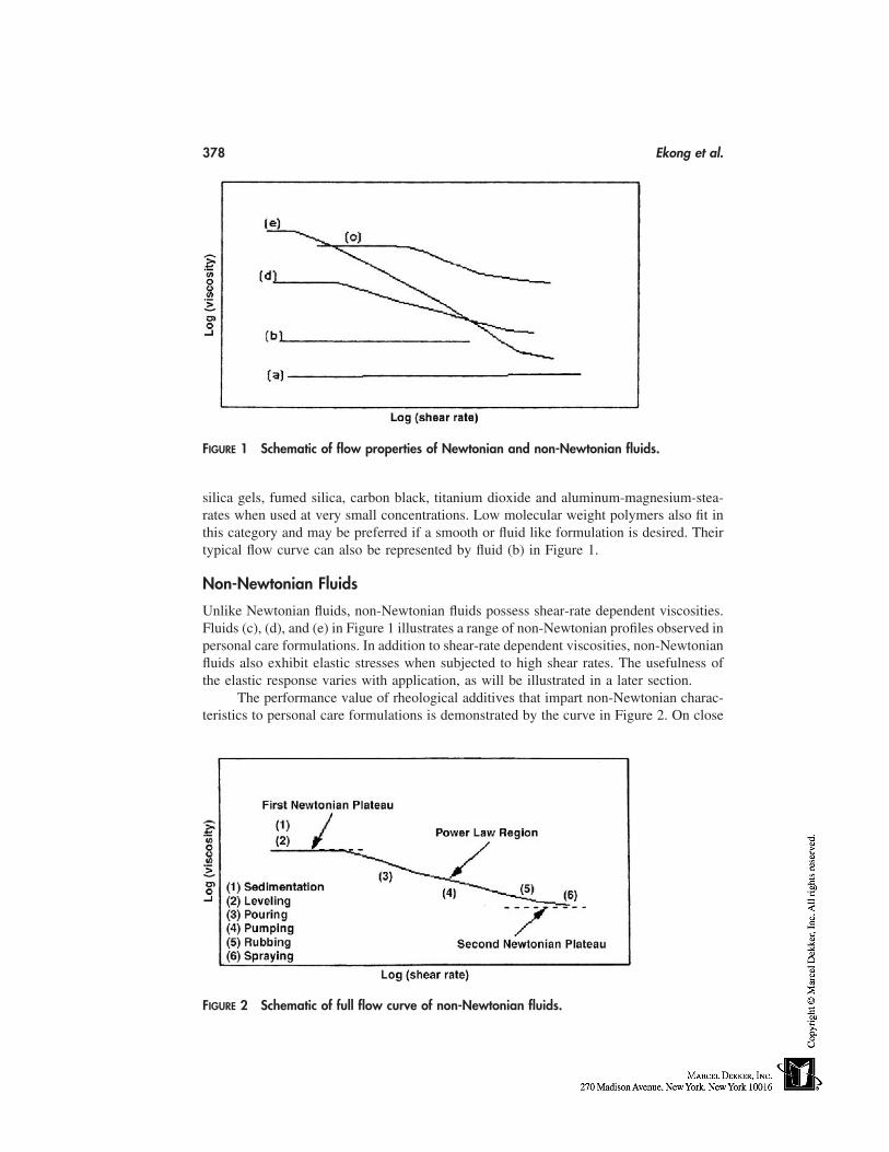
378 Ekong et al.
FIGURE 1 Schematic of flow properties of Newtonian and non-Newtonian fluids.
silica gels, fumed silica, carbon black, titanium dioxide and aluminum-magnesium-stea-rates when used at very small concentrations. Low molecular weight polymers also fit inthis category and may be preferred if a smooth or fluid like formulation is desired. Theirtypical flow curve can also be represented by fluid (b) in Figure 1.
Non-Newtonian Fluids
Unlike Newtonian fluids, non-Newtonian fluids possess shear-rate dependent viscosities.Fluids (c), (d), and (e) in Figure 1 illustrates a range of non-Newtonian profiles observed inpersonal care formulations. In addition to shear-rate dependent viscosities, non-Newtonianfluids also exhibit elastic stresses when subjected to high shear rates. The usefulness ofthe elastic response varies with application, as will be illustrated in a later section.
The performance value of rheological additives that impart non-Newtonian charac-teristics to personal care formulations is demonstrated by the curve in Figure 2. On close
FIGURE 2 Schematic of full flow curve of non-Newtonian fluids.
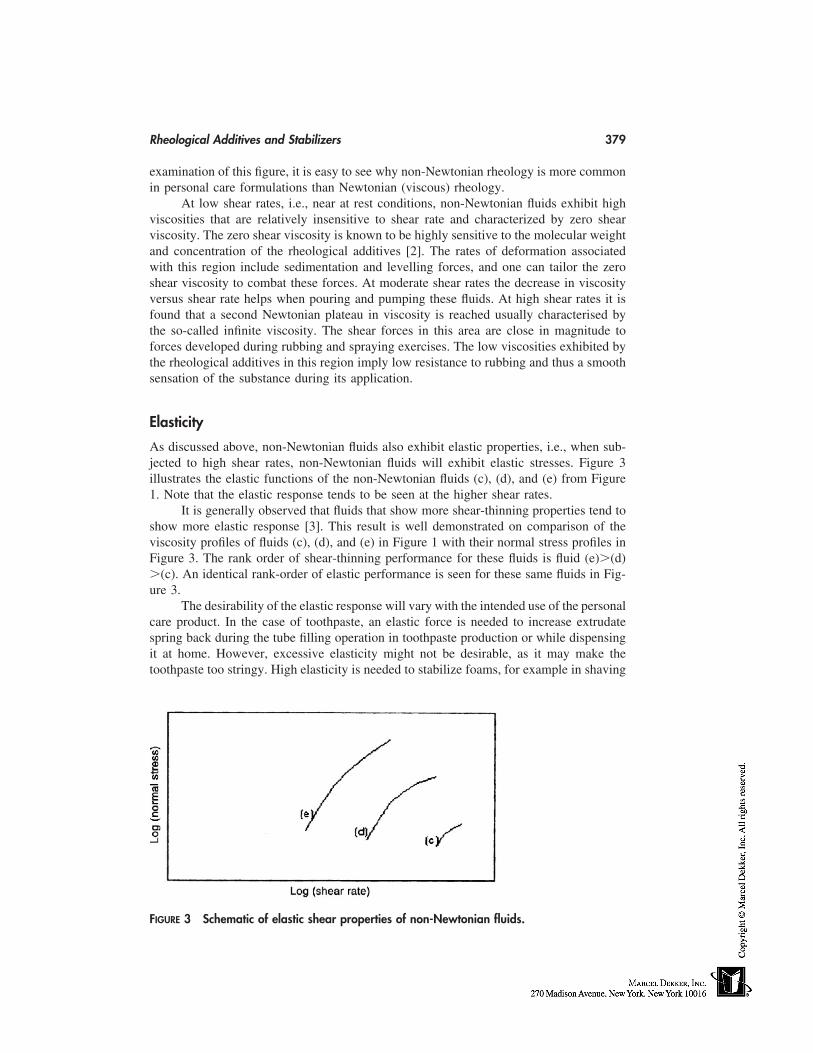
Rheological Additives and Stabilizers 379
examination of this figure, it is easy to see why non-Newtonian rheology is more commonin personal care formulations than Newtonian (viscous) rheology.
At low shear rates, i.e., near at rest conditions, non-Newtonian fluids exhibit highviscosities that are relatively insensitive to shear rate and characterized by zero shearviscosity. The zero shear viscosity is known to be highly sensitive to the molecular weightand concentration of the rheological additives [2]. The rates of deformation associatedwith this region include sedimentation and levelling forces, and one can tailor the zeroshear viscosity to combat these forces. At moderate shear rates the decrease in viscosityversus shear rate helps when pouring and pumping these fluids. At high shear rates it isfound that a second Newtonian plateau in viscosity is reached usually characterised bythe so-called infinite viscosity. The shear forces in this area are close in magnitude toforces developed during rubbing and spraying exercises. The low viscosities exhibited bythe rheological additives in this region imply low resistance to rubbing and thus a smoothsensation of the substance during its application.
Elasticity
As discussed above, non-Newtonian fluids also exhibit elastic properties, i.e., when sub-jected to high shear rates, non-Newtonian fluids will exhibit elastic stresses. Figure 3illustrates the elastic functions of the non-Newtonian fluids (c), (d), and (e) from Figure1. Note that the elastic response tends to be seen at the higher shear rates.
It is generally observed that fluids that show more shear-thinning properties tend toshow more elastic response [3]. This result is well demonstrated on comparison of theviscosity profiles of fluids (c), (d), and (e) in Figure 1 with their normal stress profiles inFigure 3. The rank order of shear-thinning performance for these fluids is fluid (e)�(d)�(c). An identical rank-order of elastic performance is seen for these same fluids in Fig-ure 3.
The desirability of the elastic response will vary with the intended use of the personalcare product. In the case of toothpaste, an elastic force is needed to increase extrudatespring back during the tube filling operation in toothpaste production or while dispensingit at home. However, excessive elasticity might not be desirable, as it may make thetoothpaste too stringy. High elasticity is needed to stabilize foams, for example in shaving
FIGURE 3 Schematic of elastic shear properties of non-Newtonian fluids.
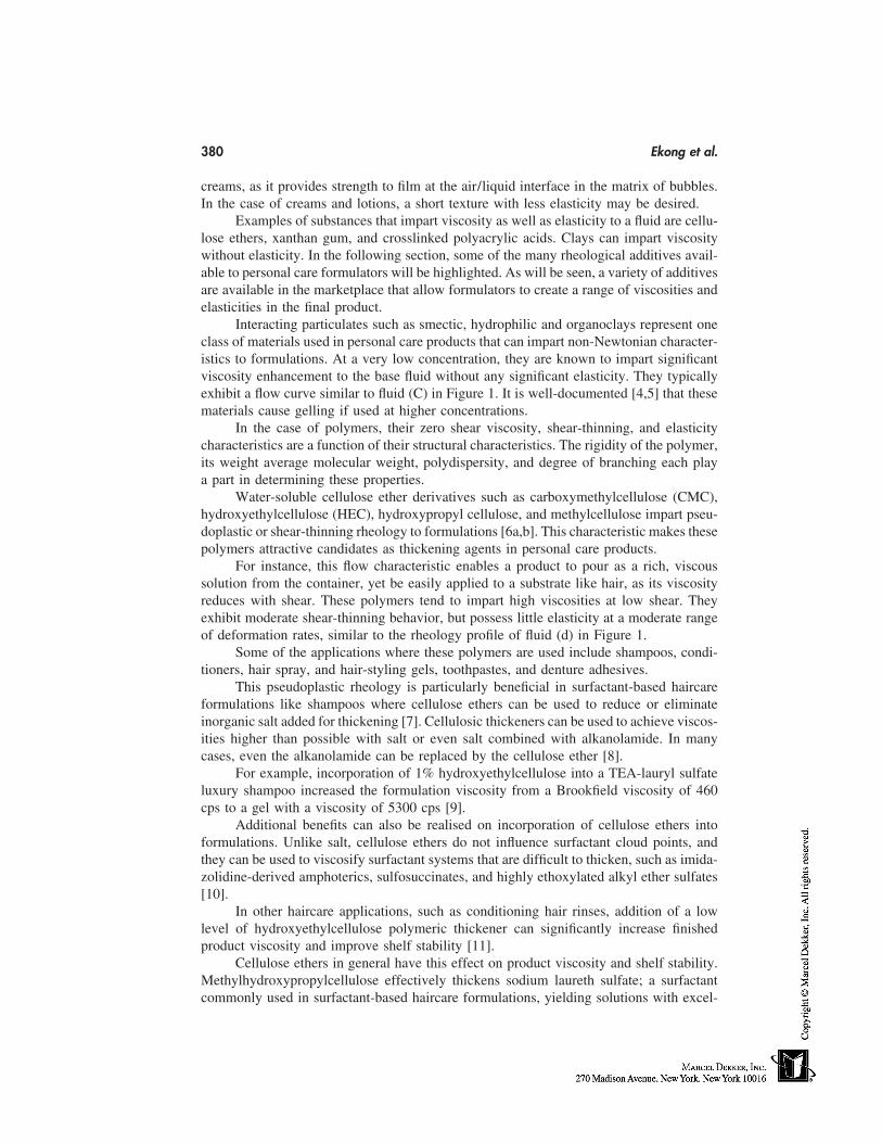
380 Ekong et al.
creams, as it provides strength to film at the air/liquid interface in the matrix of bubbles.In the case of creams and lotions, a short texture with less elasticity may be desired.
Examples of substances that impart viscosity as well as elasticity to a fluid are cellu-lose ethers, xanthan gum, and crosslinked polyacrylic acids. Clays can impart viscositywithout elasticity. In the following section, some of the many rheological additives avail-able to personal care formulators will be highlighted. As will be seen, a variety of additivesare available in the marketplace that allow formulators to create a range of viscosities andelasticities in the final product.
Interacting particulates such as smectic, hydrophilic and organoclays represent oneclass of materials used in personal care products that can impart non-Newtonian character-istics to formulations. At a very low concentration, they are known to impart significantviscosity enhancement to the base fluid without any significant elasticity. They typicallyexhibit a flow curve similar to fluid (C) in Figure 1. It is well-documented [4,5] that thesematerials cause gelling if used at higher concentrations.
In the case of polymers, their zero shear viscosity, shear-thinning, and elasticitycharacteristics are a function of their structural characteristics. The rigidity of the polymer,its weight average molecular weight, polydispersity, and degree of branching each playa part in determining these properties.
Water-soluble cellulose ether derivatives such as carboxymethylcellulose (CMC),hydroxyethylcellulose (HEC), hydroxypropyl cellulose, and methylcellulose impart pseu-doplastic or shear-thinning rheology to formulations [6a,b]. This characteristic makes thesepolymers attractive candidates as thickening agents in personal care products.
For instance, this flow characteristic enables a product to pour as a rich, viscoussolution from the container, yet be easily applied to a substrate like hair, as its viscosityreduces with shear. These polymers tend to impart high viscosities at low shear. Theyexhibit moderate shear-thinning behavior, but possess little elasticity at a moderate rangeof deformation rates, similar to the rheology profile of fluid (d) in Figure 1.
Some of the applications where these polymers are used include shampoos, condi-tioners, hair spray, and hair-styling gels, toothpastes, and denture adhesives.
This pseudoplastic rheology is particularly beneficial in surfactant-based haircareformulations like shampoos where cellulose ethers can be used to reduce or eliminateinorganic salt added for thickening [7]. Cellulosic thickeners can be used to achieve viscos-ities higher than possible with salt or even salt combined with alkanolamide. In manycases, even the alkanolamide can be replaced by the cellulose ether [8].
For example, incorporation of 1% hydroxyethylcellulose into a TEA-lauryl sulfateluxury shampoo increased the formulation viscosity from a Brookfield viscosity of 460cps to a gel with a viscosity of 5300 cps [9].
Additional benefits can also be realised on incorporation of cellulose ethers intoformulations. Unlike salt, cellulose ethers do not influence surfactant cloud points, andthey can be used to viscosify surfactant systems that are difficult to thicken, such as imida-zolidine-derived amphoterics, sulfosuccinates, and highly ethoxylated alkyl ether sulfates[10].
In other haircare applications, such as conditioning hair rinses, addition of a lowlevel of hydroxyethylcellulose polymeric thickener can significantly increase finishedproduct viscosity and improve shelf stability [11].
Cellulose ethers in general have this effect on product viscosity and shelf stability.Methylhydroxypropylcellulose effectively thickens sodium laureth sulfate; a surfactantcommonly used in surfactant-based haircare formulations, yielding solutions with excel-
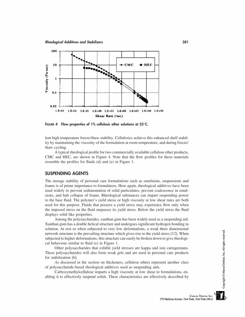
Rheological Additives and Stabilizers 381
FIGURE 4 Flow properties of 1% cellulosic ether solutions at 25°C.
lent high temperature freeze/thaw stability. Cellulosics achieve this enhanced shelf stabil-ity by maintaining the viscosity of the formulation at room temperature, and during freeze/thaw cycling.
A typical rheological profile for two commercially available cellulose ether products,CMC and HEC, are shown in Figure 4. Note that the flow profiles for these materialsresemble the profiles for fluids (d) and (e) in Figure 1.
SUSPENDING AGENTS
The storage stability of personal care formulations such as emulsions, suspensions andfoams is of prime importance to formulators. Here again, rheological additives have beenused widely to prevent sedimentation of solid particulates, prevent coalescence in emul-sions, and halt collapse of foams. Rheological substances can impart suspending powerto the base fluid. The polymer’s yield stress or high viscosity at low shear rates are bothused for this purpose. Fluids that possess a yield stress may experience flow only whenthe imposed stress on the fluid surpasses its yield stress. Below the yield stress the fluiddisplays solid like properties.
Among the polysaccharides, xanthan gum has been widely used as a suspending aid.Xanthan gum has a double helical structure and undergoes significant hydrogen bonding insolution. At rest or when subjected to very low deformations, a weak three dimensionalnetwork structure is the prevailing structure which gives rise to the yield stress [12]. Whensubjected to higher deformations, this structure can easily be broken down to give rheologi-cal behaviour similar to fluid (e) in Figure 1.
Other polysaccharides that exhibit yield stresses are kappa and iota carrageenans.These polysaccharides will also form weak gels and are used in personal care productsfor stabilization [6].
As discussed in the section on thickeners, cellulose ethers represent another classof polysaccharide-based rheological additives used as suspending aids.
Carboxymethylcellulose imparts a high viscosity at low shear to formulations, en-abling it to effectively suspend solids. These characteristics are effectively described by
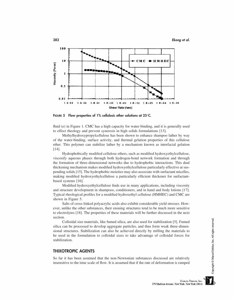
382 Ekong et al.
FIGURE 5 Flow properties of 1% cellulosic ether solutions at 25°C.
fluid (e) in Figure 1. CMC has a high capacity for water-binding, and it is generally usedto effect rheology and prevent syneresis in high solids formulations [13].
Methylhydroxypropylcellulose has been shown to enhance shampoo lather by wayof the water-binding, surface activity, and thermal gelation properties of this celluloseether. This polymer can stabilize lather by a mechanism known as interfacial gelation[14].
Hydrophobically modified cellulose ethers, such as modified hydroxyethylcellulose,viscosify aqueous phases through both hydrogen-bond network formation and throughthe formation of three-dimensional networks due to hydrophobic interactions. This dualthickening mechanism makes modified hydroxyethylcellulose particularly effective at sus-pending solids [15]. The hydrophobic moieties may also associate with surfactant micelles,making modified hydroxyethylcellulose a particularly efficient thickener for surfactant-based systems [16].
Modified hydroxyethylcellulose finds use in many applications, including viscosityand structure development in shampoos, conditioners, and in hand and body lotions [17].Typical rheological profiles for a modified hydroxethyl cellulose (HMHEC) and CMC areshown in Figure 5.
Salts of cross-linked polyacrylic acids also exhibit considerable yield stresses. How-ever, unlike the other substances, their ensuing structures tend to be much more sensitiveto electrolytes [18]. The properties of these materials will be further discussed in the nextsection.
Colloidal size materials, like fumed silica, are also used for stabilization [5]. Fumedsilica can be processed to develop aggregate particles, and thus form weak three-dimen-sional structures. Stabilization can also be achieved directly by milling the materials tobe used in the formulation to colloidal sizes to take advantage of colloidal forces forstabilization.
THIXOTROPIC AGENTS
So far it has been assumed that the non-Newtonian substances discussed are relativelyinsensitive to the time scale of flow. It is assumed that if the rate of deformation is ramped
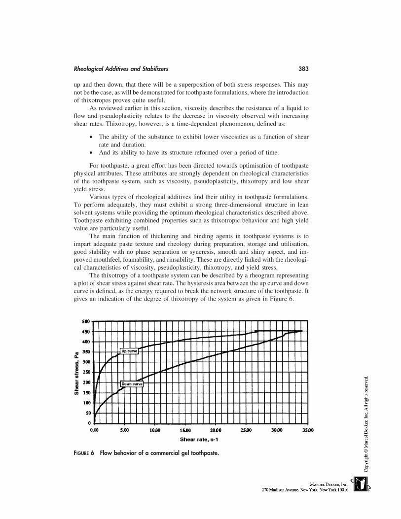
Rheological Additives and Stabilizers 383
up and then down, that there will be a superposition of both stress responses. This maynot be the case, as will be demonstrated for toothpaste formulations, where the introductionof thixotropes proves quite useful.
As reviewed earlier in this section, viscosity describes the resistance of a liquid toflow and pseudoplasticity relates to the decrease in viscosity observed with increasingshear rates. Thixotropy, however, is a time-dependent phenomenon, defined as:
• The ability of the substance to exhibit lower viscosities as a function of shearrate and duration.
• And its ability to have its structure reformed over a period of time.
For toothpaste, a great effort has been directed towards optimisation of toothpastephysical attributes. These attributes are strongly dependent on rheological characteristicsof the toothpaste system, such as viscosity, pseudoplasticity, thixotropy and low shearyield stress.
Various types of rheological additives find their utility in toothpaste formulations.To perform adequately, they must exhibit a strong three-dimensional structure in leansolvent systems while providing the optimum rheological characteristics described above.Toothpaste exhibiting combined properties such as thixotropic behaviour and high yieldvalue are particularly useful.
The main function of thickening and binding agents in toothpaste systems is toimpart adequate paste texture and rheology during preparation, storage and utilisation,good stability with no phase separation or syneresis, smooth and shiny aspect, and im-proved mouthfeel, foamability, and rinsability. These are directly linked with the rheologi-cal characteristics of viscosity, pseudoplasticity, thixotropy, and yield stress.
The thixotropy of a toothpaste system can be described by a rheogram representinga plot of shear stress against shear rate. The hysteresis area between the up curve and downcurve is defined, as the energy required to break the network structure of the toothpaste. Itgives an indication of the degree of thixotropy of the system as given in Figure 6.
FIGURE 6 Flow behavior of a commercial gel toothpaste.
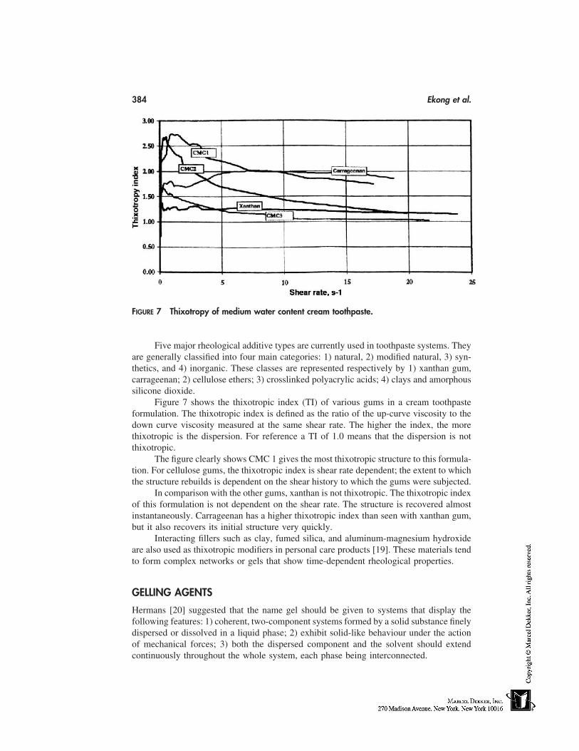
384 Ekong et al.
FIGURE 7 Thixotropy of medium water content cream toothpaste.
Five major rheological additive types are currently used in toothpaste systems. Theyare generally classified into four main categories: 1) natural, 2) modified natural, 3) syn-thetics, and 4) inorganic. These classes are represented respectively by 1) xanthan gum,carrageenan; 2) cellulose ethers; 3) crosslinked polyacrylic acids; 4) clays and amorphoussilicone dioxide.
Figure 7 shows the thixotropic index (TI) of various gums in a cream toothpasteformulation. The thixotropic index is defined as the ratio of the up-curve viscosity to thedown curve viscosity measured at the same shear rate. The higher the index, the morethixotropic is the dispersion. For reference a TI of 1.0 means that the dispersion is notthixotropic.
The figure clearly shows CMC 1 gives the most thixotropic structure to this formula-tion. For cellulose gums, the thixotropic index is shear rate dependent; the extent to whichthe structure rebuilds is dependent on the shear history to which the gums were subjected.
In comparison with the other gums, xanthan is not thixotropic. The thixotropic indexof this formulation is not dependent on the shear rate. The structure is recovered almostinstantaneously. Carrageenan has a higher thixotropic index than seen with xanthan gum,but it also recovers its initial structure very quickly.
Interacting fillers such as clay, fumed silica, and aluminum-magnesium hydroxideare also used as thixotropic modifiers in personal care products [19]. These materials tendto form complex networks or gels that show time-dependent rheological properties.
GELLING AGENTS
Hermans [20] suggested that the name gel should be given to systems that display thefollowing features: 1) coherent, two-component systems formed by a solid substance finelydispersed or dissolved in a liquid phase; 2) exhibit solid-like behaviour under the actionof mechanical forces; 3) both the dispersed component and the solvent should extendcontinuously throughout the whole system, each phase being interconnected.
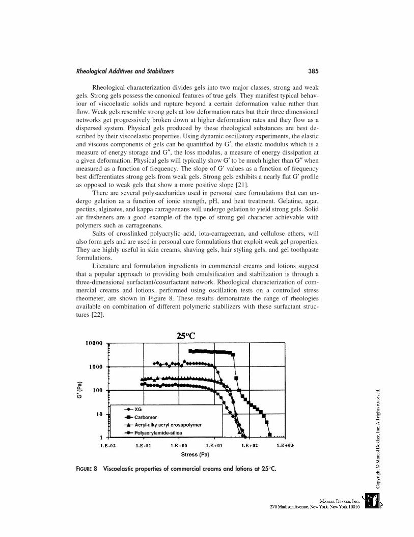
Rheological Additives and Stabilizers 385
Rheological characterization divides gels into two major classes, strong and weakgels. Strong gels possess the canonical features of true gels. They manifest typical behav-iour of viscoelastic solids and rupture beyond a certain deformation value rather thanflow. Weak gels resemble strong gels at low deformation rates but their three dimensionalnetworks get progressively broken down at higher deformation rates and they flow as adispersed system. Physical gels produced by these rheological substances are best de-scribed by their viscoelastic properties. Using dynamic oscillatory experiments, the elasticand viscous components of gels can be quantified by G′, the elastic modulus which is ameasure of energy storage and G″, the loss modulus, a measure of energy dissipation ata given deformation. Physical gels will typically show G′ to be much higher than G″ whenmeasured as a function of frequency. The slope of G′ values as a function of frequencybest differentiates strong gels from weak gels. Strong gels exhibits a nearly flat G′ profileas opposed to weak gels that show a more positive slope [21].
There are several polysaccharides used in personal care formulations that can un-dergo gelation as a function of ionic strength, pH, and heat treatment. Gelatine, agar,pectins, alginates, and kappa carrageenans will undergo gelation to yield strong gels. Solidair fresheners are a good example of the type of strong gel character achievable withpolymers such as carrageenans.
Salts of crosslinked polyacrylic acid, iota-carrageenan, and cellulose ethers, willalso form gels and are used in personal care formulations that exploit weak gel properties.They are highly useful in skin creams, shaving gels, hair styling gels, and gel toothpasteformulations.
Literature and formulation ingredients in commercial creams and lotions suggestthat a popular approach to providing both emulsification and stabilization is through athree-dimensional surfactant/cosurfactant network. Rheological characterization of com-mercial creams and lotions, performed using oscillation tests on a controlled stressrheometer, are shown in Figure 8. These results demonstrate the range of rheologiesavailable on combination of different polymeric stabilizers with these surfactant struc-tures [22].
FIGURE 8 Viscoelastic properties of commercial creams and lotions at 25°C.
386 Ekong et al.
The surfactant-gel network system provides a yield stress, a high degree of elasticity,shear-thinning behavior, and time-dependent structure build-up (thixotropy). These rheo-logical attributes are very important in the consumer’s perception of skin feel during lotionapplication and rub-in.
The primary component of these liquid crystalline gel network systems is a co-surfactant. Cosurfactants are water-insoluble fatty amphiphiles that are too lipophilic topromote o/w emulsions. Cosurfactants combined with a small fraction of a water-solublesurfactant having a high hydrophilic-lipophilic balance (HLB), produce swollen lamellar-gel networks after thermal processing and cooling.
A physical gel forming rheological additive, such as a cellulose ether, cross-linkedpolyacrylate, clay, or xanthan gum is added to improve temperature stability and modifythe rheology of these systems.
A plot of elastic modulus, G′ as a function of imposed stress for commercial creamscontaining xanthan gum (XG), crosslinked sodium polyacrylate (carbomer), Acrylate/C10-30 alkyl acrylate crosspolymer and polyacrylamide/silica is presented in Figure 8.The elastic modulus, G′, at low stresses is a measure of the gel rigidity of the sample.These results serve to distinguish the more solid-like creams, with G′ values � 1000 Paat low shear, from the more liquid-like lotions, with G′ values � 1000 Pa at low shear.
Other materials that can form weak gels when given the appropriate mechanicaltreatment are silica gels and fumed silica. These materials are sometimes used in combina-tion with other polymers to yield weak gels. They are used in toothpaste where it servesa dual role as an abrasive and a rheology modifier. The thickening silicas are the onlyinorganic products used extensively to structure toothpaste. They provide a good thick-ening effect and high thixotropic behaviour, but they lack the ability to bind water in thelean solvent slurry. As a result, they are unsuitable for syneresis control. Therefore, awater-soluble organic binder is necessary to modify the toothpaste rheology and to preventwater separation. Carboxymethylcellulose and carrageenans are often combined with silicafor this purpose.
Due to the broad performance criteria that personal care products have to meet, mostformulators find it necessary to use a mixture of rheological additives to achieve desiredproperties in final formulations. Mixtures of materials can bring significant synergy indesired properties. In conclusion, rheological additives significantly influence the mechani-cal, textural, stability, and ultimately the quality of personal care products.
REFERENCES
1. Einstein A. Ann Phys 1906; 19:289–306.2. Berry GC, Fox TG. Adv Poly Sci 1968; 5:261.3. Graessley WW. Adv Poly Sci 1974; 16:1.4. The Benefits of Hectorite Clay, NL Chemical Technical Lit. Hightown, NJ: NL Industries,
PB 149. June 1988.5. Ca-bo-sil Fumed Silica Properties and Functions, Technical Literature. Tuscola, IL: Cabot
Corporation, February 1990.6a. Desmarais AJ, Wint RF. Hydroxyalkyl ethyl ethers of cellulose. In: Industrial Gums. 3d ed.
Whistler RL, BeMiller JN, eds. 1992:505.6b. Feddersen RL, Thorp SN. Sodium carboxymethyl cellulose. In: Industrial Gums. 3d ed.
Whistler RL, BeMiller JN, eds. 3d ed. 1992:537.7. Reng AK, Skryzpak W. Ways to regulate the viscosity of cosmetic preparations. Cosmet Toilet
1979; 94:29–36.
Rheological Additives and Stabilizers 387
8. Schoenberg T. Formulating without diethanolamides. Household and Personal Products Indus-try 1998; 35(7):76–79.
9. Performance of Aqualon Water-Soluble Polymers in Shampoos, Aqualon Bulletin VC-526B.September 1989.
10. Hunting ALL. Shampoo thickeners. Cosmet Toilet 1982; 97:53–63.11. Performance of Natrosol Hydroxyethylcellulose in Hair-Conditioning Products, Aqualon
Bulletin VC-525. October 1987.12. Davidson RL, ed. Handbook of Water-Soluble Gums and Resins. NY: McGraw-Hill, Inc.,
1980:24–27.13. Aqualon cellulose ethers and their role in toothpaste. Aqualon technical literature 87.507-E2.
March 1998.14. Conklin J, McKnight SM. Cellulose ethers. Household and Personal Products Industry 1988;
25:80–84.15. Goodwin JW, et al. Polymers in aqueous media: performance through association. Glass JE,
ed. Advances in Chemistry Series, 223. Washington, D.C.: American Chemical Society, 1989:365.
16. AC Sau, Landoll LM. Glass JE, ed. Advances in Chemistry Series, 223. Washington, D.C.:American Chemical Society, 1989:343.
17. Natrosol Plus CS, Grade 330 Modified Hydroxyethylcellulose, Thickener for Hand and BodyLotions, Aqualon Bulletin VC-562A. Wilmington, Delaware: Hercules Inc., October 1992.
18. Laba D, ed. Rheological Properties of Cosmetics and Toiletries. Vol. 13. New York: MarcelDekker, Inc., 1993.
19. Reference 18, p. 123–145.20. Hermans PH. Colloid Science. Vol. II. NY: Elsevier Science Publishers, 1949, p. 194.21. Doublier JL, Choplin L. Carbohydrate Research. A Rheological Description of Amylose
Gelation. 1989; 193:215.22. Barnum PE. Presentation at SCC Mid Atlantic Chapter, Raw Material Symposium ‘‘Gums,
Polymers & Thickeners for Rheology Modification of Personal Care Products,’’ March 9,1999.

34
Silicones: A Key Ingredient in Cosmeticand Toiletry Formulations
Janet M. BlakelyDow Corning S.A., Brussels, Belgium
UNIQUE MATERIALS
Silicone is a generic name for many classes of organo-silicone polymer which consist ofan inorganic siloxane (SiEO) backbone with pendant organic groups (usually methyl)(Fig. 1). It is this structure that gives silicones their unique combination of properties and,in particular, their surface properties.
Siloxane Backbone
The prime role of the siloxane backbone is to present the available methyl groups to theirbest advantage and it does this by virtue of its unique flexibility. In most hydrocarbons,the bond angles are very fixed and steric packing considerations often prevent the availablemethyls from adopting lowest surface energy orientations. In silicones, the SiEO bondlength is significantly longer and the SiEOESi bond angle flatter than comparable CECand CEO bonds resulting in a very low barrier to rotation and making the polymer chainsvery flexible. This flexibility makes many orientations possible and provides ‘‘free space’’to accommodate different sized substituents or to allow easy diffusion of gaseous mole-cules; a property useful in the formation of ‘‘breathable’’ films. Coupled with the lowintermolecular forces between methyl groups, this flexibility also has a profound effecton the bulk as well as the surface properties of silicones. This is seen in the small variationof physical parameters with temperature and molecular weight, the low freezing and pourpoints of fluids, the low boiling points, the high compressibility and the retention of liquidnature to unusually high molecular weights. It also makes a number of structural andcompositional variations possible, resulting in many families of silicones, including linearand cyclic structures, a wide range of molecular weights and varying degrees of branchingor cross linking. Additionally, the siloxane bond is exceptionally strong providing thepolymer with a high degree of thermal and oxidative stability and ensuring stability whenformulated [1–3].
389
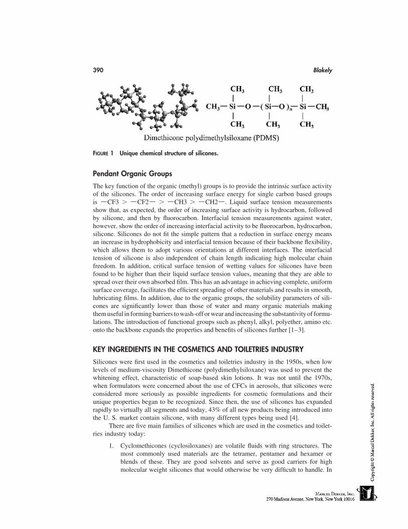
390 Blakely
FIGURE 1 Unique chemical structure of silicones.
Pendant Organic Groups
The key function of the organic (methyl) groups is to provide the intrinsic surface activityof the silicones. The order of increasing surface energy for single carbon based groupsis ECF3 � ECF2E � ECH3 � ECH2E. Liquid surface tension measurementsshow that, as expected, the order of increasing surface activity is hydrocarbon, followedby silicone, and then by fluorocarbon. Interfacial tension measurements against water,however, show the order of increasing interfacial activity to be fluorocarbon, hydrocarbon,silicone. Silicones do not fit the simple pattern that a reduction in surface energy meansan increase in hydrophobicity and interfacial tension because of their backbone flexibility,which allows them to adopt various orientations at different interfaces. The interfacialtension of silicone is also independent of chain length indicating high molecular chainfreedom. In addition, critical surface tension of wetting values for silicones have beenfound to be higher than their liquid surface tension values, meaning that they are able tospread over their own absorbed film. This has an advantage in achieving complete, uniformsurface coverage, facilitates the efficient spreading of other materials and results in smooth,lubricating films. In addition, due to the organic groups, the solubility parameters of sili-cones are significantly lower than those of water and many organic materials makingthem useful in forming barriers to wash-off or wear and increasing the substantivity of formu-lations. The introduction of functional groups such as phenyl, alkyl, polyether, amino etc.onto the backbone expands the properties and benefits of silicones further [1–3].
KEY INGREDIENTS IN THE COSMETICS AND TOILETRIES INDUSTRY
Silicones were first used in the cosmetics and toiletries industry in the 1950s, when lowlevels of medium-viscosity Dimethicone (polydimethylsiloxane) was used to prevent thewhitening effect, characteristic of soap-based skin lotions. It was not until the 1970s,when formulators were concerned about the use of CFCs in aerosols, that silicones wereconsidered more seriously as possible ingredients for cosmetic formulations and theirunique properties began to be recognized. Since then, the use of silicones has expandedrapidly to virtually all segments and today, 43% of all new products being introduced intothe U. S. market contain silicone, with many different types being used [4].
There are five main families of silicones which are used in the cosmetics and toilet-ries industry today:
1. Cyclomethicones (cyclosiloxanes) are volatile fluids with ring structures. Themost commonly used materials are the tetramer, pentamer and hexamer orblends of these. They are good solvents and serve as good carriers for highmolecular weight silicones that would otherwise be very difficult to handle. In
Silicones 391
addition, they have very low heats of vaporisation compared to water or ethanolgiving them a non-cooling feel when drying. Cyclomethicones are classified asnon-VOC (volatile organic compounds) in the USA.
2. Dimethicones (polydimethylsiloxanes-PDMS) are linear structures rangingfrom volatile to non-volatile with increasing molecular weight. Volatile Dimeth-icones exist as fluids with viscosities of 0.65–2 mm2/s. Non-volatile Dimeth-icones exist as fluids with viscosities of 5.0 mm2/s up to gums. Dimethiconeemulsions make handling of the higher molecular weight fluids easier.
3. Silicone blends consist of Dimethiconol or Dimethicone gums or Trimethylsi-loxysilicates (highly crosslinked resins) dispersed in lower molecular weightDimethicones or Cyclomethicones. They have been developed to improve easeof formulation and compatibility of high molecular gums or resins; used fortheir substantivity.
4. Dimethicone and Vinyldimethicone Crosspolymers or blends are silicone elasto-mers. They exist in powder form or as elastomeric silicone gels that are swollenwith solvent (usually Cyclomethicone). The introduction of different functionali-ties into such products is also possible. They are used as rheology modifiers inskincareandantiperspirantproducts,providingadry,powderyfeel to formulations.
5. Functional Silicones:(a) Dimethicone Copolyols (silicone polyethers) are fluids or waxes where
some of the methyl groups along the siloxane backbone have been replacedwith polyoxyethylene or polyoxypropylene groups. The addition of polyox-yethylene substituents increases the hydrophilicity of silicones. Polyoxypro-pylene substituents are used to balance out this hydrophilicity by increasingthe hydrophobic characteristics of the copolymer (16).
(b) Phenyl Trimethicones are fluids where some of the methyl groups havebeen replaced by phenyl groups. The phenyl groups increase the refractiveindex and improve compatibility with organic materials.
(c) Amodimethicones are fluids where some of the methyl groups have beenreplaced by secondary and primary amine groups. The polar amine groupshave a profound effect on the deposition properties of the silicone, givingit an affinity for negatively charged surfaces, such as the proteinaceous sur-face of the hair. Emulsions of these fluids are commonly used.
(d) Alkyl Dimethicones are fluids or waxes where some of the methyl groupshave been replaced by alkyl groups. This results in a family of silicone-hydrocarbon hybrids with possibilities for variations in viscosities, soften-ing temperatures and rheological characteristics. They have increased com-patibility with organic materials.
(e) Cyclomethicone (and) Dimethicone Copolyol or Laurylmethicone Copolyolare silicone emulsifiers. They show ampiphilic behaviour and have beendesigned to emulsify aqueous phases into silicones; usually Cyclomethiconeor low-medium polarity organic oils.
SKINCARE, SUNCARE, AND DECORATIVE PRODUCTS
Skin Feel/Emolliency
The main reason that silicones are used in all types of skin care product is because oftheir sensory properties. Studies on the emollient properties of various materials have
392 Blakely
shown that silicones deliver greater emolliency values than many commonly used cosmeticingredients both, during and after application. They are described as smooth, velvety andnon-greasy or oily and are able to impart this feel to cosmetic and toiletry formulations,improving the negative feel associated with other ingredients [5].
Cyclomethicones are used for transient effects giving slight lubricity, a light texture,fast spreading and good distribution of the product on application, whilst leaving no resid-ual effects. They are often included in formulations to remove the greasy or oily feel ofhydrocarbon-based emollients and are the basis for ‘‘oil-free’’ type claims [6]. They areused in light products for daily use such as facial cleansers, day creams or liquid founda-tions. Higher molecular weight silicones such as Dimethicone (and) Dimethiconol areused to give a more lubricious, longer lasting effect in richer, more nourishing skin treat-ment products such as night creams or after-sun products [7]. Silicone elastomers areused to give a dry, powdery feel to skincare formulations [8]. Silicones are also non-comedogenic/non-acnegenic unlike many occlusive, lipophilic fatty emollients which canpromote comedone/acne formation on the skin [9].
Substantivity (Long-Lasting/Durability)
High molecular weight Dimethicones or Cyclomethicone (and) Dimethiconols form water-resistant films on the skin which can help prolong the effects of skin care, sun care ordecorative products. This substantivity can be improved further by using Alkyl Dimethi-cones such as Cetyl Dimethicone or C30-45 Alkyl Methicone [7] (see Figure 2). The useof the substantivity of silicones to improve the substantivity of other ingredients in cos-metic and toiletry formulations has been demonstrated in sun care products. The additionof 2.5 wt% Cetyl Dimethicone to an oil-in-water sunscreen formulation shows excellentin vivo resistance to wash-off. The formulation has an in vivo SPF of 21.1 before immer-sion which reduces to 19.2 only, after immersion for 80 minutes [7] [10].
Cyclomethicones are the basis for long-lasting/non-transfer decorative products, es-pecially lipsticks. They are used to disperse waxes and pigments, improve applicationand impart a pleasant skin feel, often replacing non-volatile hydrocarbon oils. When theyevaporate, a uniform film of waxes and pigments remains which is resistant to transferand wear [11].
Permeability/Controlled Moisturization/ProtectionAgainst Dehydration
Due to the flexibility of the SiEOESi backbone, the majority of silicones are permeableto water vapour, producing ‘‘breathable’’ films. This is an important parameter for cleans-ing products or colour cosmetics to avoid clogging pores. The presence of an alkyl groupin the chain, however, reduces this permeability, resulting in silicones which can givecontrolled moisturization, e.g., Stearyl Dimethicone or moisturization (occlusivity) similarto petrolatum e.g. C30-45 Alkyl Methicone [7] [12].
Enhanced Efficacy
Apart from improving the feel and long-lasting benefits of skincare products, siliconescan also enhance the efficacy of other ingredients in the formulation. Studies carried outon suncare products have shown that the Alkylmethicones can enhance the in vitro SPFof products containing either organic or inorganic sunscreens. For inorganic sunscreens,
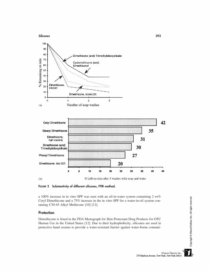
Silicones 393
FIGURE 2 Substantivity of different silicones, FTIR method.
a 100% increase in in vitro SPF was seen with an oil-in-water system containing 2 wt%Cetyl Dimethicone and a 75% increase in the in vitro SPF for a water-in-oil system con-taining C30-45 Alkyl Methicone [10] [12].
Protection
Dimethicone is listed in the FDA Monograph for Skin Protectant Drug Products for OTCHuman Use in the United States [12]. Due to their hydrophobicity, silicones are used inprotective hand creams to provide a water-resistant barrier against water-borne contami-
394 Blakely
nants. Recent studies indicate that Cyclomethicone and Dimethicone may also preventirritation caused by sunscreen agents [13].
Cleansing
The excellent spreading characteristics, dry non-greasy/oily feel, and good solvency ofCyclomethicones make them ideal for use in skin cleansers to help lift and remove dirtwithout stinging. They can be used alone or in combination with ingredients such asmineral oil. Silicone emulsifiers allow Cyclomethicone to be present in the continuousphase as well as allowing the incorporation of polar ingredients such as water, glycerineetc. This makes the formulation of rinsible foaming facial washes possible [14].
Water-soluble and water-dispersible Dimethicone Copolyols have shown benefitsin foaming facial washes. They provide a creamy, more dense foam as well as improvingthe foam volume. In liquid body cleansing products such as foam baths, shower gelsand liquid soaps, they can improve foaming and foam stabilization. They have also beenrecognized as additives that reduce eye and skin irritation from anionic surfactants [14,15].
Rheology Modification/Structural Integrity (Sticks)
As well as improving the aesthetics of formulations, silicones can also act as rheologymodifiers. This is particularly applicable to water-in-oil or water-in silicone-type systems.One such silicone rheology modifier is the C30-45 Alkyl Methicone where 149% and93% increases in emulsion viscosity have been observed for water-in-silicone and water-in-oil emulsions respectively with 2 wt% of the wax [7]. Rheology modification using 2–4 wt% Stearyl Dimethicone is believed to be part of the reason for the success of thisproduct in enhancing the SPF of sun care products containing organic sunscreens [10].These waxes are also used to maintain the structural integrity of stick or soft solid products,improving their feel and application. Silicone elastomers can also be used to modify therheology of skin care and antiperspirant formulations. Such elastomers have the capacityto absorb large amounts of solvents such as Cyclomethicone or low-viscosity Dimethiconewithout exhibiting any syneresis. It is this property which allows them to successfullythicken formulations. The ability of elastomers to significantly modify the rheology of aformulation combined with their unique powdery feel has led to their use in antiperspirantproducts.
Formulating Flexibility
Silicones can be used in all types of skin care products ranging from simple oil-in-watergels or emulsions to water-in-silicone and water-in-oil emulsions, from crystal clear towhite in colour. Silicone emulsifiers increase this flexibility further. They allow siliconesto be present in the continuous phase as well as allowing the incorporation of polar ingredi-ents such as water, glycerine etc. Matching the refractive index of the water phase withthe oil phase in such emulsions makes the formulation of clear gels possible and adjustingthe phase ratio determines the product form from lotions to gels. This technology is thebasis for the clear antiperspirant gels seen on the market today. It is also possible to makenon-aqueous emulsions using silicones to deliver hydrophilic ingredients or those that aresensitive to hydrolysis.
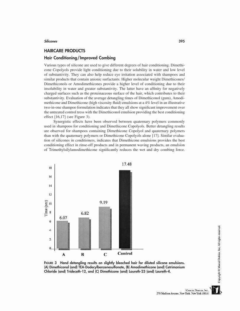
Silicones 395
HAIRCARE PRODUCTS
Hair Conditioning/Improved Combing
Various types of silicone are used to give different degrees of hair conditioning. Dimethi-cone Copolyols provide light conditioning due to their solubility in water and low levelof substantivity. They can also help reduce eye irritation associated with shampoos andsimilar products that contain anionic surfactants. Higher molecular weight Dimethicones/Dimethiconols or Amodimethicones provide a higher level of conditioning due to theirinsolubility in water and greater substantivity. The latter have an affinity for negativelycharged surfaces such as the proteinaceous surface of the hair, which contributes to theirsubstantivity. Evaluation of the average detangling times of Dimethiconol (gum), Amodi-methicone and Dimethicone (high viscosity fluid) emulsions at a 4% level in an illustrativetwo-in-one shampoo formulation indicates that they all show significant improvement overthe untreated control tress with the Dimethiconol emulsion providing the best conditioningeffect [16,17] (see Figure 3).
Synergistic effects have been observed between quaternary polymers commonlyused in shampoos for conditioning and Dimethicone Copolyols. Better detangling resultsare observed for shampoos containing Dimethicone Copolyol and quaternary polymersthan with the quaternary polymers or Dimethicone Copolyols alone [17]. Similar evalua-tion of silicones in conditioners, indicates that Dimethicone emulsions provides the bestconditioning effect in rinse-off products and in permanent waving products, an emulsionof Trimethylsilylamodimethicone significantly reduces the wet and dry combing force.
FIGURE 3 Hand detangling results on slightly bleached hair for diluted silicone emulsions.(A) Dimethiconol (and) TEA-Dodecylbenzenesulfonate, (B) Amodimethicone (and) CetrimoniumChloride (and) Trideceth-12, and (C) Dimethicone (and) Laureth-23 (and) Laureth-4.
396 Blakely
Combinations of silicones such as Cyclomethicone, silicone blends and Phenyl Trimethi-cone are the basis for anhydrous leave-in conditioners, sometimes referred to as ‘‘cuticlecoat’’ products [16].
Sensory Enhancement
As in skincare, silicones impart a soft smooth feel to the hair. Sensory evaluations ofcuticle coat formulations consisting entirely of blends of silicone showed that, in additionto ease of combing, they improve spreadability, silkiness and softness, gloss and perceivedrepair of split ends compared to the control [16,18].
Silicones as Drying Aids
Silicones such as Amodimethicone can help hair dry more quickly in comparison to dry-ing aids such as Stearalkonium Chloride, preventing damage due to the use of hair dryersetc. [16].
Foam Boosting
Dimethicone Copolyols can be used to boost the foaming properties of shampoos as wellas provide a light conditioning effect [16].
Reduced Flyaway
Tests comparing shampoo formulations containing quaternary polymers to those with qua-ternary polymers and Dimethicone Copolyols show an improvement in static control withthe addition of the silicone. Sensory evaluation has also shown a reduction of flyawaywith Dimethicone emulsions [16,18].
Improved Shine
Silicones, in particular Phenyl Trimethicone, are recognized for their ability to enhancehair shine and gloss along with adding softness, manageability, and smoothness to theabraded hair cuticle [16,19].
Natural-Look Fixatives
Because of their low surface tension, silicones spread easily to help fixative productsdistribute evenly on the surface of hair and improving their effectiveness. They are alsoused in conjunction with or as a replacement for organic plasticizers. Organic materialstend to be hydrophilic, which diminishes the holding power of a resin. In contrast, thehydrophobic nature of silicones helps repel water so there is less opportunity to reducethe resin’s holding properties. The use of Dimethicone Copolyol as a resin plasticizer canalso help give hair a more natural look [16].
Longer Lasting Permanent Wave and Coloring Products
Silicones, such as Amodimethicone, can be used to provide a more durable conditioningeffect and a longer lasting permanent wave. Pretreatments containing silicone blends helpprevent hair damage during the harsh perming process. In hair color products, blends ofvolatile and non-volatile silicone (Cyclomethicone and Amodimethicone) can be used to
Silicones 397
seal in the hair cuticle and hold color. The volatile silicone evaporates, leaving behind asmooth, uniform film on the surface of the hair [16,20].
ANTIPERSPIRANT AND DEODORANT PRODUCTS
In addition to the benefits which silicones bring to skin care products such as improvedfeel, delivery of actives, low residue, formulating flexibility, etc., the following advantagesare seen in antiperspirant and deodorant formulations [21].
Anti-whitening
Dimethicones, Phenyl Trimethicone or Alkyl Dimethicones have been shown to reduce/mask the whitening effect caused by antiperspirant salts by matching the refractiveindex [22].
Improved Spray Characteristics
Low levels of Cyclomethicone (and) Dimethiconol have been demonstrated to reduce thespray width, height, and particle size of antiperspirant pump spray and aerosol formula-tions, leading to a more directional spray with low mistiness and dustiness [21,23]. Thesilicone blend may also contribute to the substantivity of the antiperspirant active andlubricate the spray valve to prevent clogging.
Noncooling
The heat of vapourisation of volatile silicones such as Cyclomethicone is much lowerthan that of water or ethanol meaning that much less energy is required for them to evapo-rate. This leads to a noncooling effect in formulation [21].
The multifunctional benefits of silicones make them invaluable ingredients in to-day’s cosmetic and toiletry formulations and with the introduction of more and more newsilicones, this is a trend which is expected to continue well into the next millenium.
REFERENCES
1. Owen MJ. The surface activity of silicones: a short review. Ind Eng Chem Prod Res Dev1980; 19:97–103.
2. Owen MJ. Why silicones behave funny. Chemtech May 1981; 11:288–292.3. DiSapio A. Silicones in Personal Care: An Ingredient Revolution. Brussels: Dow Corning
Publication 22-1547.01, 1994.4. Cosmetic Research, USA News 1997.5. Goldemberg RL, Pela Rosa CP. Int Soc Cosm Chem 1971; 22:635–654.6. De Backer G, Ghirardhi D. Goodbye to Grease. Soap, Perfumery and Cosmetic; June 1993.7. Blakely J, Van Reeth I, Vagts A. The silicone difference in skincare. Inside Cosmetics
October/November, 1998; 14–17.8. Van Reeth I, Dahman F, Lau A, Starch M. Novel Silicone Thickening Technologies: Deliv-
ering the Appropriate Rheology Profile to Optimize Formulation Performance. Dow CorningPublication 22-1786-01. Brussels, Belgium, 1999.
9. Lanzet M. Comedogenic effects of cosmetic raw materials. Cosmet Toiletr 1986; 101:63–72.10. Van Reeth I, Dahman F, Hannington J. Alkymethylsiloxanes as SPF Enhancers. Relationship
398 Blakely
Between Effects and Physico-Chemical Properties. International Federation of Societies ofCosmetic Chemists 19th Congress Poster 1996, Sydney.
11. Abrutyn E. Translating Silicone Chemistry to Color Cosmetics. Dow Corning Publication 25-888-97, 1997, Midland, Michigan.
12. Van Reeth I, Marchioretto S, Dahman F, DeSmedt A, Dupont A. Silicones: Enhanced Protec-tion Across Personal Care Applications. IFSCC Poster 1998, Cannes.
13. Nichols K, Desai N, Lebwohl M. Effective sunscreen ingredients and cutaneous irritation inpatients with rosacea. Cutis 1998; 61:344–346.
14. Blakely J. The Benefits of Silicones in Facial and Body Cleansing Products. Dow CorningPublication 22-1549-01, Brussels, 1994.
15. Disapio AJ, Fridd P. Dimethicone Copolyols for Cosmetic and Toiletry Applications. IFSCCPaper 1988, London.
16. Marchioretto S. Optimising the Use of Silicones in Haircare Products. Dow Corning Publica-tion 22-1720-01, Brussels, 1998.
17. Marchioretto S, Blakely J. Substantiated synergy between silicone and quats for clear andmild conditioning shampoos. SÖFW October 2, 1997.
18. Thomson B, Vincent J, Halloran D. Anhydrous hair conditioners: silicone-in-silicone deliverysystems. Soap Cosmet Chem Specialties 1992; 68:25–28.
19. Reimer BM, Oldinski RL, Glover DA. An Objective Method for Evaluating Hair Shine. SoapCosmet Chem Specialties (October 1995).
20. Fridd PF, Taylor RM. GB Patents GB2186889 and GB2186890.21. Abrutyn ES, Bahr BC, Fuson SM. Overview of the Antiperspirant Market: Technology and
Trends. Dow Corning Publication 22-1555-01, Brussels, 1994.22. Abrutyn ES, Bahr BC, Legrow GE, Schulz WJ. US Patent 5, 225, 188; 1993.23. Spitzer J. US Patent 4, 152, 416; 1979.
35
Skin-Feel Agents
Germaine ZocchiColgate-Palmolive Research and Development, Inc., Milmort, Belgium
INTRODUCTION
Skin-feel additives are substances conferring sensorial properties to a skincare product,triggering pleasant perception during application to the skin and after use. Effectivenessof sensory triggers is governed by their substantivity to the skin which occurs either byhydrophobic interaction, charge attraction, or a combination of these two factors. A largevariety of cosmetic ingredients function as skin-feel/conditioning additives, comprisinglipophilic materials, silicones, water-soluble polymeric substances (including proteins) andtheir cationic derivatives, and humectants, among others. The Cosmetic, Toiletry and Fra-grance Association (CTFA) divides skin conditioning agents into various groups: emol-lients, occlusive materials, and miscellaneous substances including among others, cationicmacromolecules and several surfactants.
This chapter will focus on skin-feel agents for rinse-off products, and more particu-larly for surfactant-based skin-cleansing products, such as facial cleansers, soap and syndetbars, shower gels and body washes, foam baths (or bubble baths), and bath oils. Showergels, bars, and facial cleansers first contact the skin, even if only briefly, then are rinsedduring the cleaning process; the substantivity of the conditioning agents is crucial to ensuresensory performances, otherwise they will be washed off and the end skin benefit willnot be perceived by the user. For bath products intended to be heavily diluted for use, itis difficult to believe that skin-feel agents could function effectively, except perhaps incase of bath oils. Indeed, when bath oils are diluted in water they either float to the surfaceor lead to a coarse unstable o/w emulsion; when the body emerges from the bath, oilsspontaneously stick to the skin because they are incompatible with water and are excludedfrom the ‘‘bathing liquor.’’
The advent of emollients in body-cleansing liquids occurred with the emergence inthe early 1990s of the ‘‘body washes’’ referring to ‘‘2 in 1’’ foaming emulsions; beforethe development of this new product niche, cationic polymeric materials were the most-used skin-conditioning agents.
Sensorial performance profile of a body-cleansing product comes in a variety ofsignal attributes:
399
400 Zocchi
• Feeling on the skin during use: spreading of a liquid (also related to productrheology), feel of a bar (slipperiness or roughness), foam feel related to foamquality (creaminess, density)
• Skin feel during rinsing, e.g., slipperiness, roughness of the skin, ‘‘clean feel’’(squeaky feel) left by soaps
• Feel while drying the skin with a towel and feel on damp skin: softness,roughness, stickiness
• After feel and longevity of skin sensations: smoothness, softness, moisturization
All these product attributes are governed at first by e.g., the surfactant nature (amphoterics,nonionics, anionics), their total and relative concentrations, and the clinical mildness forthe skin of the surfactant mixture, and can be further influenced or improved by judiciouslychosen skin-conditioning agents.
Besides physical, clinical, and organoleptic characteristics of the body-cleansingproduct, several other imponderable parameters can act on the skin-feel performance andperception, such as usage habits, water hardness, skin condition of the user, and pilosity.Also, consumer expectations in terms of sensorial profile of a product depend on climatic(relative humidity, temperature) and sociodemographic parameters (e.g., sex, occupation,lifestyle, running-water availability), skin type and concerned body part (e.g., face, wholebody), and product positioning (e.g., sport, moisturizing, nourishing, others).
Criteria of selection and constraints to be taken into account when choosing skin-feel agents are as follows:
• Solubility and compatibility with the surfactant system• Sensitivity to electrolytes and pH• Product physical form: bar, liquid• Processability (bars) and ease of formulation• Sensitivity to temperature• Impact on finished product performance profile:
on the foam: foam feel, volume and stability, creaminess, bubble structureon the product rinsabilityInduction of undesirable and unexpected secondary effects on skin feel when
skin is damp (e.g., stickiness)• Impact on finished product aesthetic:
on fragrance perception and stabilityon product clarity when relevanton viscosity, rheological profileon color
• Origin: animal or vegetal, natural or synthetic• Risk of skin sensitization• Cost
EMOLLIENTS AND REFATTENERS
Introduction
The CTFA dictionary defines emollients as: ‘‘cosmetic ingredients which help to maintainthe soft, smooth and pliable appearance of the skin; emollients function by their abilityto remain on the skin surface or in stratum corneum to act as lubricant, to reduce flaking,
Skin-Feel Agents 401
and to improve the skin’s appearance.’’ Emollients are also described as refatting additivesor refatteners in the case of bath products. The word refattener refers to substances im-proving the lipid content of the upper layers of the skin; they prevent defatting and dryingout of the skin. Several emollients showing strong lipophilic character are identified asocclusive ingredients; they are fatty/oily materials that remain on the skin surface andreduce transepidermal water loss. The CTFA dictionary defines occlusives as: ‘‘cosmeticingredients which retard the evaporation of water from the skin surface; by blocking theevaporative loss of water, occlusive materials increase the water content of the skin.’’
Overall, emollients and refatteners are oils and fats derived from natural origins orobtained by chemical synthesis; they are classified in nonpolar (paraffins and isoparaffins)and polar substances (esters and triglycerides); their chemical structure influences the in-teraction with the skin surface and affects their sensorial properties. As a class, they com-prise lipids, oils and their derivatives, fatty acid esters, lanolin derivatives, and siliconesand their organofunctional derivatives. Originally, emollients were developed for use inleave-on skin care products; formulation technology can aid the deposition of refattingadditives on the skin from wash-off products and avoid that they rinse off with the surfac-tants; nevertheless, the large dilution factor in both products remains a significant hurdlefor skin end benefit perception (except in bath oils).
Emollients and refatteners will provide after feel, but will also influence skin feelduring usage, foam feel, and most of the time foam quantity and quality. The more hy-drophobic the refattening additive, the more negative its impact on flash foam generation,foam quantity, and stability. In other respects, the more lipidic the material, the better itsskin substantivity, and the easier the efficacy documentation; proof and substantiation ofclaims is of more and more importance in the frame of the Sixth Amendment of Europeanlegislation for cosmetics and toiletries.
Lipophilic Emollients and Occlusives
Occlusive materials comprise vegetable oils, triglycerides, mineral oil, natural or syntheticwaxes, fatty acid esters, lanolin oil and its derivatives, and polydimethylsiloxanes, amongothers (Table 1). They form an occlusive layer on the skin, keeping water inside upperstratum corneum layers and consequently acting as moisturizers.
Mineral oil and vegetable oils as well as waxes generally produce heavy and greasyfeeling on the skin. Hydrophobic fatty acid esters are an almost unlimited source of syn-thetic emollients and refatteners; they provide lighter and more pleasant skin feel thanoils and waxes. Any fatty acid can be esterified by either ethylene glycol, or propyleneglycol, or glycerin polymers, or isopropyl alcohol, or any longer chain alcohol. The feelthey impart and their impact on foam is related to the fatty acid chain length; short chains(e.g., isopropyl myristate, octyl octanoate, and cocoate) deliver dryer feel and have lesserimpact on foam than longer ones (e.g., stearates and isostearates), which are greasier anddetrimental to foam quantity and stability [1].
Hydrophobic emollients are efficacious skin refatteners but not easily formulated insurfactant mixtures commonly used in liquid skin-cleansing product without proceedingto an emulsification step, which most of the time necessitates hot process. Highly hy-drophobic refattening additives are not meant for foaming preparations but rather for bathoils. They have a detrimental impact on foam speed, quantity, and stability. Manufacturerscircumvent this weakness of lathering capacity by providing a mechanical foaming devicewith the product: a puff or massage flower [2].
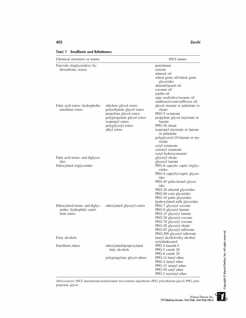
402 Zocchi
TABLE 1 Emollients and Refatteners
Chemical structures or nature INCI names
Fats/oils (triglycerides); hy- petrolatumdrocarbons; waxes ceresin
mineral oilwheat germ oil/wheat germ
glyceridesalmond/peach oilcoconut oiljojoba oilrape seed/olive/sesame oilsunflower/corn/safflower oil
Fatty acid esters: hydrophobic ethylene glycol esters glycol stearate or palmitate oremollient esters polyethylene glycol esters oleate
propylene glycol esters PEG-5 octanoatepolypropylene glycol esters propylene glycol myristate orisopropyl esters lauratepolyglyceryl esters PPG-36 oleatealkyl esters isopropyl myristate or laurate
or palmitatepolyglyceryl-10-laurate or my-
ristateoctyl octanoatecetearyl octanoateoctyl hydroxystearate
Fatty acid mono- and diglycer- glyceryl oleateides glyceryl laurate
Ethoxylated triglycerides PEG-6 caprylic capric triglyc-erides
PEG-4 caprylic/capric glycer-ides
PEG-45 palm kernel glycer-ides
PEG-20 almond glyceridesPEG-60 corn glyceridesPEG-18 palm glycerideshydroxylated milk glycerides
Ethoxylated mono- and diglyc- ethoxylated glyceryl esters PEG-7 glyceryl cocoateerides: hydrophilic emol- PEG-8 glyceryl lauratelient esters PEG-15 glyceryl laurate
PEG-30 glyceryl cocoatePEG-78 glyceryl cocoatePEG-20 glyceryl oleatePEG-82 glyceryl tallowatePEG-200 glyceryl tallowate
Fatty alcohols lauryl alcohol/oley alcoholoctyldodecanol
Emollient ethers ethoxylated/propoxylated PPG-5-laureth-5fatty alcohols PPG-5 ceteth 20
PPG-8 ceteth 20polypropylene glycol ethers PPG-14 butyl ether
PEG-4 lauryl etherPPG-15 stearyl etherPPG-50 cetyl etherPPG-3 myristyl ether
Abbreviations: INCI, international nomenclature for cosmetic ingredients; PEG, polyethylene glycol; PPG, poly-propylene glycol.
Skin-Feel Agents 403
Soaps and syndet bars can easily accommodate waxes and oils without impairingtheir basic foaming and cleaning functions. Besides beeswax, petrolatum or ceresin, lano-lin and jojoba oil, cocoa butter, and mineral oil, are other examples of skin conditionerscommonly used in bars. Paraffin wax is often used in soaps and syndets not only for thesmooth feel they impart to the finished bar, the mildness they bring to the formulation,but also for the role of plasticizer they play, adding firmness to the bar. Vegetal oils areincluded as skin-nourishing/refattening agents (e.g., almond, wheat germ, olive oils).
Fatty acid mono- and diglycerides [1,3] are prepared either by transesterification oftriglycerides with glycerin or treatment of alkanoate with glycerin. Lipophilic characterremains predominant in these esters; depending on chain length, they are soluble in surfac-tant solutions or they must be emulsified. Besides the improved skin feel they induce,they also reduce defatting of the skin possibly caused by surfactant-based cleansers. Mono-glycerides of stearic, lauric, and palmitic acids (glyceryl monostearate, laurate, and palmi-tate) intervene in the composition of natural lipids of the skin. They adsorb and can bedetected on skin after application through a skin-cleaning product [4].
Several mixtures of monoglycerides and mild foaming surfactants are commerciallyavailable; they claim improved foam qualities (bubble size, creaminess, and stability) anddocumentable skin-refattening properties [5,6]. On top of the skin feel improvement theybring, they will also reduce the degreasing effect of cleansers thanks to their lipophiliccharacter and improve the compatibility of the surfactants with the skin [1]. An exampleof improvement in the skin barrier function and in skin tactile sensations has been shownfor glyceryl oleate in a model shower-gel composition [5].
Hydrophilic Lipids
Hydrophilic lipids (Table 1) [1] are preferred for foaming skin-cleansing preparations.Ethoxylation and propoxylation make lipids more compatible with water and more easilysoluble in aqueous surfactants solutions. One has to find the right balance between ethoxy-lation and skin substantivity; the more the lipids are ethoxylated, the more they are soluble,the less the impact on foam and skin substantivity, and the weaker their refattening prop-erties.
Ethoxylated glycerides are obtained either by reaction of natural triglycerides withethylene oxide (a complex end mixture is then obtained) or by ethoxylation of monoglycer-ides. They are often referred to as ‘‘water-soluble vegetable oils’’; their solubility in waterwill depend on the carbon chain length of starting glycerides and on the degree of ethoxyla-tion.
Low ethoxylated triglycerides are still lipophilic enough to provide good refatteningproperties, leading to very pleasant skin feel, perceivable at quite high use levels. Ethoxy-lated mono- and diglycerides generally associate various properties beneficial to the skin.They are more or less refattening the skin, depending on chain length and ethoxylationratio and act as anti-irritant or mildness additives; they confer slipperiness to the foam.Depending on chain length and ethoxylation degree they are either water dispersible orsoluble. Among the low ethoxylates, PEG-7 glyceryl cocoate is one of the mostly used.This emollient depresses irritation of anionic surfactants and shows minimum impact onlathering profile. Higher ethoxylates of longer C chain length (PEG-200 glyceryl tallowate)are still substantive to the skin because of their high molecular weight, as well as providea smooth feel, but because of their stronger hydrophilic character their refatting propertiesare less obvious to evidence [7].
404 Zocchi
Ethoxylated/propoxylated fatty alcohols are useful light emollients: through an ap-propriate selection of optimum combination between parent alcohol chain length, propoxy-lation, and ethoxylation degree, these emollients can be formulated up to 2 to 3% insurfactant solutions with minimum impact on foam value.
Lanolin
Lanolin (Table 2) [8,9] is extracted from sheep wool grease; it is a complex mixture ofesters of high molecular weight lanolin alcohols (aliphatic alcohols, sterols, and trimethylsterols) and of lanolin acids; free lanolin alcohols, acids, and lanolin hydrocarbons areminors. Lanolin alcohols and lanolin oil are recommended as superfatting agents in soaps.Ethoxylation of the hydroxyl groups of lanolin or of its derivatives leads to hydrophilic,water-soluble lanolin compounds, offering a broad range of useful emollients to the formu-lator. Some moderately to highly ethoxylated derivatives, recommended for their goodemolliency and moisturization properties, are processable in liquid skin cleansers withlimited impact on foam profile; as an example, the 75 mol ethoxylated lanolin does notdepress foam and is recommended as skin conditioner in soaps, liquid body-cleansingproducts, and bubble baths. Medium ethoxylates lanolin alcohols have limited impact onfoam performances of body cleansing liquids; lower ethoxylates can be formulated in bars.Propoxylated lanolin alcohols are lipophilic emollients used in soap bars and in othercleansers based on synthetic surfactants.
Alkoxylated lanolin derivatives are obtained by reaction with mixtures of propyleneand ethylene oxides in various ratios; they are more soluble than ethoxylated lanolin. Theyserve as refattening and foam stabilizing agents. Esterification of lanolin fatty acid withisopropyl alcohol provides a range of esters of various molecular weights. Medium molec-ular weight esters are used as superfatting agents in soaps.
TABLE 2 Emollients and Refatteners
INCI names
Lecithin propylene glycol (and)lecithin (and) sodium laurylsulfate (and) disodiumsulfosuccinate (and)cocamidopropylhydroxysultaine (and)isopropyl alcohol
Lanolin and its derivatives lanolin oillanolin alcohol
ethoxylated lanolin PEG-75 lanolin
ethoxylated lanolin alcohols laneth-16laneth-25
propoxylated lanolin alcohols PPG-30 lanolin alcohol ether
alkoxylated lanolin PPG-12 PEG-50 lanolinPPG-40 PEG-60 lanolin oil
Abbreviations: PEG, polyethylene glycol; PPG, polypropylene glycol.
Skin-Feel Agents 405
Lecithin
Lecithin (Table 2) is a natural mixture of polar and neutral lipids; the word lecithin is alsoused as the trivial name of a particular phospholipid: phosphatidylcholine. Main vegetablesources of lecithin used in personal-care products are soybean and maize, egg yolk ispractically the only animal source of lecithin used in cosmetics and toiletries. The percent-age of polar lipids and their fatty acid pattern are characteristic of the lecithin source.
Bare lecithin, a secondary product of Soya oil extraction, typically contains 60 to70% polar lipids (mainly phospholipids, namely phosphatidylcholine, and glycolipids) anda remaining 25 to 35% Soy oil. This raw lecithin is further fractionated, purified, andchemically modified to allow easier processing and formulation in toiletry products. Emol-lient, refattening, and moisturizing properties of lecithin are guided by its content in phos-pholipids.
Lecithin softens, nourishes, and refattens the skin; it provides a nongreasy, long-lasting skin feel and improves foam feel and quality (creaminess, slipperiness, richness).Ready-to-use mixtures of phospholipids in surfactant solutions, free of residual Soya oil,are commercially available for an easy incorporation in liquids or bars; some of thesecompounds allow formulation of clear products.
Silicone Derivatives
For detailed information about silicones, lecturer will refer to Chapter 34. Only majormaterials used in body cleansing products will be briefly discussed here [10,11]. Predomi-nant silicones used overall in personal-care products are polydimethyl siloxane, alsonamed dimethicones. They are not soluble in water or in surfactant solutions; their incorpo-ration into liquid cleansers requires an emulsification process. The length of dimethylsilox-ane polymer chain dictates its molecular weight and hence its viscosity. Most commonlyused materials have viscosities ranging from about 100 to several thousands centistokes.High– to medium–molecular weight dimethicones are occlusive, skin-protective emol-lients; lower molecular weights are dryer emollients, generally preferred for use in skincleansers. Dimethicones have a detrimental effect on foam profile but are good film-form-ing agents, lubricants, imparting a nongreasy, nontacky silky feel as compared with ‘‘heav-ier’’ mineral or vegetable oils. They are used in soap bars, where they also aid moldrelease, and in 2-in-1 shower gels (body washes). Polymethylcyclosiloxanes or cyclometh-icones are tetrameric or pentameric oligomers of the same backbone as polydimethylsilox-ane, and show the same chemical and physical properties; they are low-viscosity fluidswith relatively high volatility because of their low molecular weight and the weak intermo-lecular attractivity. Because they are not substantive, cyclomethicones are often identifiedas dry emollients; they deliver light, transient, and dry skin feel during product use.
Formulation of these nonpolar insoluble silicones requests hot emulsification process(nonionic emulsifiers) and proper emulsion stabilization.
Dimethicones are modifed or functionalized with other organic groups to modulatetheir solubility in water or in surfactant solutions (and consequently make them easier toformulate) and their skin substantivity properties. By adjusting the type and proportionof hydrophilic substituents, the resulting copolymer is soluble or dispersible in aqueouscosmetic products. The combination of the dimethicone structure with polyoxyalkylatedsubstituents (ethylene or propylene oxide) yields dimethicone copolyols, which are copol-ymers more soluble in water with surface activity. They are foam boosters and stabilizers;
406 Zocchi
even if they are less film-forming than parent polydimethylsiloxanes, they significantlyadd to skin sensations during application (use) and provide excellent smooth and silkyafter feel [12]. They can be used to formulate clear, aqueous products. Blends of polydi-methylsiloxanes with volatile and/or water-soluble derivatives are used to design a senso-rial profile adapted to the finished product and its end use.
HUMECTANTS
The CTFA dictionary defines humectants as ‘‘cosmetic ingredients intended to increase thewater content of top layers of the skin’’ (Table 3). Humectants are hygroscopic substancesgenerally soluble in water. These ‘‘moisture attractants’’ maintain an aqueous film at theskin surface. The primary used humectant in personal-care products is glycerin; it tendsto provide heavy and tacky feel which can be overcome by using it in combination withother humectants such as sorbitol.
Less expensive than glycerin, propylene glycol is the second most widely used hu-mectant in cosmetic and toiletry products; it reduces viscosity of surfactant solutions andtends to depress the foam.
Low–molecular weight polyethylene glycol (PEGs from about 10 to 200 PEG units),amino acids and other constituents of skin natural moisturizing factors like sodium PCAand sodium lactate are also applicable for use in surfactant-based skin-cleansing products.
Humectants are not substantive to the skin and are easily rinsed-off after cleaning.Consequently, skin-feel improvement is not obvious to perceive and their efficacy in termsof skin moisturization is difficult to document. Glycerin, propylene glycol, 1,3-butyleneglycol, or sorbitol are typically used in body washes, bubble baths, shower gels, or soapsto prevent the dessication of the product itself and the formation of a dry layer at thesurface. They also ensure stability and clarity of liquid cleansers at cold temperatures.
Few substantive humectants can be mentioned. They are cationic in nature, whichmakes them absorbing to the negatively charged skin surface. In the quaternized polyal-
TABLE 3 Humectants
Chemical nature or structure INCI names
glyceringlycereth-26 and glycereth-7propylene glycol1,3 butylene glycolfrom PEG-8 to about PEG-200sorbitolsorbeth-6 to sorbeth-40xylitol
Ethoxylated methyl glucose methyl gluceth-10/methyl gluceth-20amino acidslactic acid/sodium lactatesodium PCA
Substantive conditioning humectants steardimonium panthenollauryl methyl gluceth-10 hydroxypropyl dimonium chloridechitosan-PCA
Abbreviations: PEG, polyethylene glycol; PCA, pyrrolidone carboxylic acid.
Skin-Feel Agents 407
koxylated methyl glucose derivative (lauryl methyl gluceth-10 hydroxypropyldimoniumchloride), the hydrophilic moiety delivers humectant properties; the hydrophobic chain atthe cationic end of the molecule ensures both substantivity and skin conditioning.
Chitosan-PCA is another example. Chitosan is a polycationic (at acidic pH) high–molecular weight polymer produced by deacetylation of chitin, the major constituent ofinvertebrate exoskeletons. Combining chitosan with pyrrolidone carboxylic acid (PCA)leads to a highly substantive, film-forming humectant material.
POLYMERS
Polymeric materials can interact both with protein of the skin surface and with skin lipids.Parameters influencing the interaction between skin surface and the polymers are as follows:
1. The positive charge density: the more cationic the character of the polymer, thebetter the polymer interaction with negatively charged skin surface.
2. The hydrophobicity of polymer: grafting of hydrophobic moieties on the poly-mer backbone favor van der Waals interactions with hydrophobic areas of thekeratin.
3. The molecular weight of the polymer: the higher the polymer size, the more itssubstantivity to the skin (film-forming properties). However, very low–molecu-lar weight polymers can easily penetrate the skin surface chinks and as suchadsorb into the superficial stratum disjonctum.
4. The nature of surfactants neighboring the polymer in the finished product: thepolymer can interact with surfactants either through their charges or throughhydrophobic interactions; also, competition between polymer and surfactantsfor skin anchoring sites can occur. In both cases, deposition and adsorption ofpolymer onto the skin surface is weakened.
Natural Polymers and Their Chemically Modified Derivatives
Proteins
Proteins differ by (1) the source; (2) the molecular weight, (3) the amino acid (AA) compo-sition, AA side groups, and electrical charge (more of cationic or of anionic AA); and(4) the chemically attached moieties (quats, fatty chains, silicone, etc.) on the peptidebackbone (Table 4) [13–15].
Proteins can be from vegetable or animal origin. The most widely used animal pro-tein is collagen from pork or beef; ‘‘marine collagen’’ (fish) is now an alternative sourceof collagen to traditional bovine-derived materials. Milk proteins, keratin, and elastin arealso considered in cosmetics and toiletries. The shift away from animal-derived ingredientshas resulted in an increased interest in plant-derived materials and increasing use of pro-teins from vegetable sources.
Vegetable/plant proteins are mostly associated with significant amounts of solubleand insoluble carbohydrates because of the extraction process; soluble carbohydratesconfer dark color and strong odor to the raw material, and in some commercial gradescarbohydrates have been removed. The combination of hydrolyzed vegetable proteins andoligosaccharides produces conditioning additives with synergistic moisturizing action andfilm-forming properties. Major vegetal starting materials are wheat gluten, almond meal,rice, oat, soya, and maize.
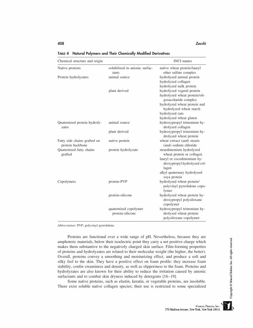
408 Zocchi
TABLE 4 Natural Polymers and Their Chemically Modified Derivatives
Chemical structure and origin INCI names
Native proteins solubilized in anionic surfac- native wheat protein/lauryltants ether sulfate complex
Protein hydrolyzates animal source hydrolyzed animal proteinhydrolyzed collagenhydrolyzed milk protein
plant derived hydrolyzed vegetal proteinhydrolyzed wheat protein/oli-
gosaccharide complexhydrolyzed wheat protein and
hydrolyzed wheat starchhydrolyzed oatshydrolyzed wheat gluten
Quaternized protein hydroly- animal source hydroxypropyl trimonium hy-zates drolyzed collagen
plant derived hydroxypropyl trimonium hy-drolyzed wheat protein
Fatty side chains grafted on native protein wheat extract (and) stearicprotein backbone (and) sodium chloride
Quaternized fatty chains protein hydrolyzate steardimonium hydrolyzedgrafted wheat protein or collagen
lauryl or cocodimonium hy-droxypropyl hydrolyzed col-lagen
alkyl quaternary hydrolyzedsoya protein
Copolymers protein-PVP hydrolyzed wheat protein/polyvinyl pyrrolidone copo-lymer
protein-silicone hydrolyzed wheat protein hy-droxypropyl polysiloxanecopolymer
quaternized copolymer hydroxypropyl trimonium hy-protein-silicone drolyzed wheat protein
polysiloxane copolymer
Abbreviation: PVP, polyvinyl pyrrolidone.
Proteins are functional over a wide range of pH. Nevertheless, because they areamphoteric materials, below their isoelectric point they carry a net positive charge whichmakes them substantive to the negatively charged skin surface. Film-forming propertiesof proteins and hydrolyzates are related to their molecular weight (the higher, the better).Overall, proteins convey a smoothing and moisturizing effect, and produce a soft andsilky feel to the skin. They have a positive effect on foam profile: they increase foamstability, confer creaminess and density, as well as slipperiness to the foam. Proteins andhydrolyzates are also known for their ability to reduce the irritation caused by anionicsurfactants and to combat skin dryness induced by detergents [16–19].
Some native proteins, such as elastin, keratin, or vegetable proteins, are insoluble.There exist soluble native collagen species; their use is restricted to some specialized
Skin-Feel Agents 409
applications. In order to make native proteins suitable for a wide range of applications,they are converted into soluble hydrolyzates by chemical or enzymatic degradation. Thesizes of resulting peptides depend on the hydrolysis process used: chemical processes giverise to broader molecular weight distributions and enzymatic digestion to narrower ones.Besides that, native proteins solubilized in various anionic surfactants (by formation of aprotein-surfactant complex) are commercially available, allowing easy formulation ofthese film-forming, moisturizing, mildness additives. A wide range of protein hydrolyzatesmolecular weights is available, ranging from 500,000 down to 1000 d. Protein hydroly-zates of intermediate molecular weight (average 3000 to 5000 d) are the most widelyused; they are less substantive than high–molecular weight proteins but provide smoothskin feel, slippery feel during use, and sensation of skin hydration.
Hydrolyzates are readily soluble and compatible with all classes of surfactants. Mostof the commercially available proteins and derivatives have a characteristic odor and color.Furthermore, products formulated with proteins or hydrolyzates should be adequately pre-served.
Chemically Modified Protein Derivatives
In order to increase interaction of proteinic material with skin surface, proteins or hydroly-zates are functionalized or chemically modified. Proteins possess reactive side chain aminoand carboxyl groups, which are sites for further modification of their intrinsic properties(Table 4).
Hydrophobic interactions with the skin surface are favored and reinforced by graft-ing fatty carbon chains, and ionic interactions are maximized by grafting cationic moietiesonto the protein backbone. Hydrolyzed protein copolymers combine substantivity andfilm-forming properties of parent proteins with characteristic sensorial properties of com-panion conditioning agents. These macromolecular protein complexes offer greater mois-turizing and conditioning potential as compared with the individual components (20).
Native Proteins Coupled with Fatty Acids. These lead to macromolecular entitywith dual hydrophilic/hydrophobic characteristics and physicochemical properties. Skinsubstantivity is guided both by the size of the starting protein and by the chain length(the hydrophobicity) of the fatty acid. The macromolecules are surface active and can beformulated in bars or liquids; they produce smooth, long-lasting skin feel. Long-chainfatty acid derivatives tend to decrease foam volume but confer creaminess, richness, andslipperiness to the lather.
Copolymers of Silicone and Proteins. These are obtained by covalent bonding oflow–molecular weight polydimethylsiloxanes on amino groups of (vegetable) proteinhydrolyzate. They combine beneficial properties of proteins (anti-irritant effect,substantivity, film-forming, soft afterfeel) with lubricity of silicone [21,22]. Quaternizedprotein-silicone copolymers are now commercially available.
PVP-Protein Copolymers. Proteinic component imparts substantivity andpolyvinyl pyrrolidone (PVP) modifies the moisture retention and film-forming propertiesof the resulting copolymer. PVP maximizes film-forming and hydration properties of theprotein. The PVP/protein ratio will modulate the profile of performance on the skin andthe influence on lathering characteristics of surfactant-based skin cleanser.
Quarternized Protein Hydrolyzates. Cationic protein hydrolyzates are obtained byreacting the primary amine sites on the protein backbone with a tertiary amine, i.e.,hydroxypropyl, propyl trimethyl ammonium, or alkyl trimethyl ammonium [23]. Covalent
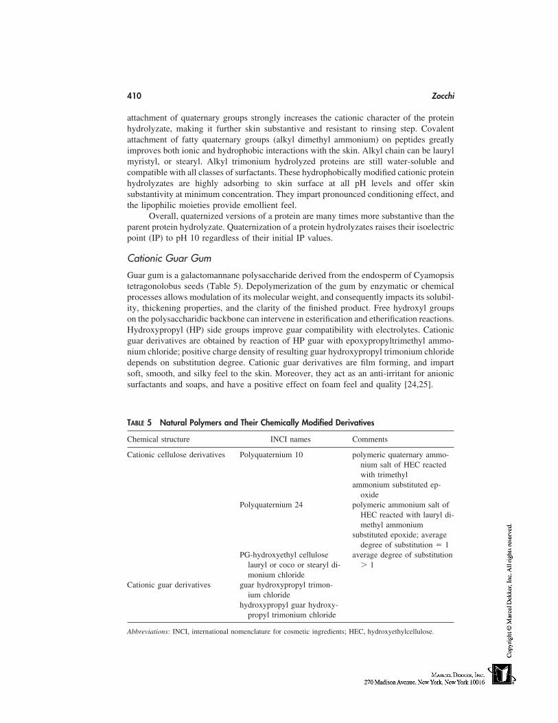
410 Zocchi
attachment of quaternary groups strongly increases the cationic character of the proteinhydrolyzate, making it further skin substantive and resistant to rinsing step. Covalentattachment of fatty quaternary groups (alkyl dimethyl ammonium) on peptides greatlyimproves both ionic and hydrophobic interactions with the skin. Alkyl chain can be laurylmyristyl, or stearyl. Alkyl trimonium hydrolyzed proteins are still water-soluble andcompatible with all classes of surfactants. These hydrophobically modified cationic proteinhydrolyzates are highly adsorbing to skin surface at all pH levels and offer skinsubstantivity at minimum concentration. They impart pronounced conditioning effect, andthe lipophilic moieties provide emollient feel.
Overall, quaternized versions of a protein are many times more substantive than theparent protein hydrolyzate. Quaternization of a protein hydrolyzates raises their isoelectricpoint (IP) to pH 10 regardless of their initial IP values.
Cationic Guar Gum
Guar gum is a galactomannane polysaccharide derived from the endosperm of Cyamopsistetragonolobus seeds (Table 5). Depolymerization of the gum by enzymatic or chemicalprocesses allows modulation of its molecular weight, and consequently impacts its solubil-ity, thickening properties, and the clarity of the finished product. Free hydroxyl groupson the polysaccharidic backbone can intervene in esterification and etherification reactions.Hydroxypropyl (HP) side groups improve guar compatibility with electrolytes. Cationicguar derivatives are obtained by reaction of HP guar with epoxypropyltrimethyl ammo-nium chloride; positive charge density of resulting guar hydroxypropyl trimonium chloridedepends on substitution degree. Cationic guar derivatives are film forming, and impartsoft, smooth, and silky feel to the skin. Moreover, they act as an anti-irritant for anionicsurfactants and soaps, and have a positive effect on foam feel and quality [24,25].
TABLE 5 Natural Polymers and Their Chemically Modified Derivatives
Chemical structure INCI names Comments
Cationic cellulose derivatives Polyquaternium 10 polymeric quaternary ammo-nium salt of HEC reactedwith trimethyl
ammonium substituted ep-oxide
Polyquaternium 24 polymeric ammonium salt ofHEC reacted with lauryl di-methyl ammonium
substituted epoxide; averagedegree of substitution � 1
PG-hydroxyethyl cellulose average degree of substitutionlauryl or coco or stearyl di- � 1monium chloride
Cationic guar derivatives guar hydroxypropyl trimon-ium chloride
hydroxypropyl guar hydroxy-propyl trimonium chloride
Abbreviations: INCI, international nomenclature for cosmetic ingredients; HEC, hydroxyethylcellulose.
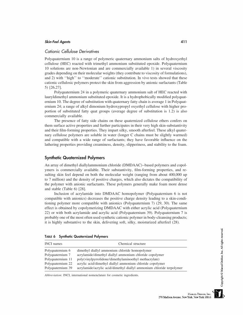
Skin-Feel Agents 411
Cationic Cellulose Derivatives
Polyquaternium 10 is a range of polymeric quaternary ammonium salts of hydroxyethylcellulose (HEC) reacted with trimethyl ammonium substituted epoxide. Polyquaternium10 solutions are non-Newtonian and are commercially available 1) in several viscositygrades depending on their molecular weights (they contribute to viscosity of formulations),and 2) with ‘‘high’’ to ‘‘moderate’’ cationic substitution. In vivo tests showed that thesecationic cellulosic polymers protect the skin from aggression by anionic surfactants (Table5) [26,27].
Polyquaternium 24 in a polymeric quaternary ammonium salt of HEC reacted withlauryldimethyl ammonium substituted epoxide. It is a hydrophobically modified polyquat-ernium 10. The degree of substitution with quaternary fatty chain is average 1 in Polyquat-ernium 24; a range of alkyl dimonium hydroxypropyl oxyethyl cellulose with higher pro-portion of substituted fatty quat groups (average degree of substitution is 1.2) is alsocommercially available.
The presence of fatty side chains on these quaternized cellulose ethers confers onthem surface active properties and further participates in their very high skin-substantivityand their film-forming properties. They impart silky, smooth afterfeel. These alkyl quater-nary cellulose polymers are soluble in water (longer C chains must be slightly warmed)and compatible with a wide range of surfactants; they have favorable influence on thelathering properties providing creaminess, density, slipperiness, and stability to the foam.
Synthetic Quaternized Polymers
An array of dimethyl diallylammonium chloride (DMDAAC)–based polymers and copol-ymers is commercially available. Their substantivity, film-forming properties, and re-sulting skin feel depend on both the molecular weight (ranging from about 400,000 upto 7 million) and the density of positive charges, which also dictates the compatibility ofthe polymer with anionic surfactants. These polymers generally make foam more denseand stable (Table 6) [28].
Inclusion of acrylamide into DMDAAC homopolymer (Polyquaternium 6 is notcompatible with anionics) decreases the positive charge density leading to a skin-condi-tioning polymer more compatible with anionics (Polyquaternium 7) (29, 30). The sameeffect is obtained by copolymerizing DMDAAC with either acrylic acid (Polyquaternium22) or with both acrylamide and acrylic acid (Polyquaternium 39). Polyquaternium 7 isprobably one of the most often used synthetic cationic polymer in body-cleansing products;it is highly substantive to the skin, delivering soft, silky, moisturized afterfeel (28).
TABLE 6 Synthetic Quaternized Polymers
INCI names Chemical structure
Polyquaternium 6 dimethyl diallyl ammonium chloride homopolymerPolyquaternium 7 acrylamide/dimethyl diallyl ammonium chloride copolymerPolyquaternium 11 poly(vinylpyrrolidone/dimethylaminoethyl methacrylate)Polyquaternium 22 acrylic acid/dimethyl diallyl ammonium chloride copolymerPolyquaternium 39 acrylamide/acrylic acid/dimethyl diallyl ammonium chloride terpolymer
Abbreviation: INCI, international nomenclature for cosmetic ingredients.
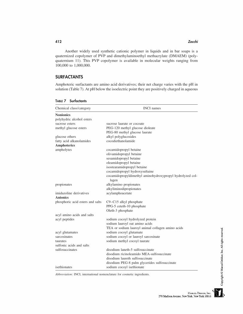
412 Zocchi
Another widely used synthetic cationic polymer in liquids and in bar soaps is aquaternized copolymer of PVP and dimethylaminoethyl methacrylate (DMAEM) (poly-quaternium 11). This PVP copolymer is available in molecular weights ranging from100,000 to 1,000,000.
SURFACTANTS
Amphoteric surfactants are amino acid derivatives; their net charge varies with the pH insolution (Table 7). At pH below the isoelectric point they are positively charged in aqueous
TABLE 7 Surfactants
Chemical class/category INCI names
Nonionicspolyhydric alcohol esterssucrose esters sucrose laurate or cocoatemethyl glucose esters PEG-120 methyl glucose dioleate
PEG-80 methyl glucose laurateglucose ethers alkyl polyglucosidesfatty acid alkanolamides cocodiethanolamideAmphotericsampholytes cocamidopropyl betaine
olivamidopropyl betainesesamidopropyl betaineoleamidopropyl betaineisostearamidopropyl betainecocamidopropyl hydroxysultainecocamidopropyldimethyl aminohydroxypropyl hydrolyzed col-
lagenpropionates alkylamino propionates
alkyliminodipropionatesimidazoline derivatives acylamphoacetateAnionicsphosphoric acid esters and salts C9–C15 alkyl phosphate
PPG-5 ceteth-10 phosphateOleth-3 phosphate
acyl amino acids and saltsacyl peptides sodium cocoyl hydrolyzed protein
sodium lauroyl oat amino acidsTEA or sodium lauroyl animal collagen amino acids
acyl glutamates sodium cocoyl glutamatesarcosinates sodium cocoyl or lauroyl sarcosinatetaurates sodium methyl cocoyl tauratesulfonic acids and saltssulfosuccinates disodium laneth-5 sulfosuccinate
disodium ricinoleamido MEA-sulfosuccinatedisodium laureth sulfosuccinatedisodium PEG-8 palm glycerides sulfosuccinate
isethionates sodium cocoyl isethionate
Abbreviation: INCI, international nomenclature for cosmetic ingredients.
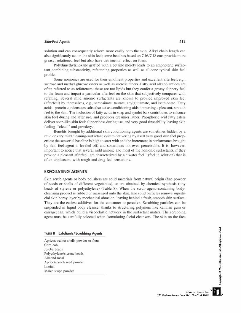
Skin-Feel Agents 413
solution and can consequently adsorb more easily onto the skin. Alkyl chain length canalso significantly act on the skin feel; some betaines based on C16/C18 cuts provide moregreasy, refattened feel but also have detrimental effect on foam.
Polydimethylsiloxane grafted with a betaine moiety leads to an amphoteric surfac-tant combining substantivity, refattening properties as well as silicone typical skin feelprofile.
Some nonionics are used for their emollient properties and excellent afterfeel; e.g.,sucrose and methyl glucose esters as well as sucrose ethers. Fatty acid alkanolamides areoften referred to as refatteners; these are not lipids but they confer a greasy slippery feelto the foam and impart a particular afterfeel on the skin that subjectively compares withrefatting. Several mild anionic surfactants are known to provide improved skin feel(afterfeel) by themselves, e.g., sarcosinate, taurate, acylglutamate, and isethionate. Fattyacids–protein condensates salts also act as conditioning aids, imparting a pleasant, smoothfeel to the skin. The inclusion of fatty acids in soap and syndet bars contributes to enhanceskin feel during and after use, and produces creamier lather. Phosphoric acid fatty estersdeliver soap-like skin feel: slipperiness during use, and very good rinseability leaving skinfeeling ‘‘clean’’ and powdery.
Benefits brought by additional skin conditioning agents are sometimes hidden by amild or very mild cleaning-surfactant system delivering by itself very good skin feel prop-erties; the sensorial baseline is high to start with and the increment in performance broughtby skin feel agent is leveled off, and sometimes not even perceivable. It is, however,important to notice that several mild anionic and most of the nonionic surfactants, if theyprovide a pleasant afterfeel, are characterized by a ‘‘water feel’’ (feel in solution) that isoften unpleasant, with rough and drag feel sensations.
EXFOLIATING AGENTS
Skin scrub agents or body polishers are solid materials from natural origin (fine powderof seeds or shells of different vegetables), or are obtained by chemical synthesis (tinybeads of styrene or polyethylene) (Table 8). When the scrub agent–containing body-cleansing product is rubbed or massaged onto the skin, fine solid particles remove superfi-cial skin horny layer by mechanical abrasion, leaving behind a fresh, smooth skin surface.They are the easiest additives for the consumer to perceive. Scrubbing particles can besuspended in liquid body cleanser thanks to structuring polymers like xanthan gum orcarrageenan, which build a viscoelastic network in the surfactant matrix. The scrubbingagent must be carefully selected when formulating facial cleansers. The skin on the face
TABLE 8 Exfoliants/Scrubbing Agents
Apricot/walnut shells powder or flourCorn cobJojoba beadsPolyethylene/styrene beadsAlmond mealApricot/peach seed powderLoofahMaize scape powder
414 Zocchi
is more sensitive or delicate than that of the rest of the body. For facial application, theformulator should orientate his choices towards, e.g., soft clays or melting jojoba beads.
CONCLUSIONS
The overall skin-feel profile provided by a skin-cleansing product is conditioned by thehuge variety of composition constituents. Many have been described in this chapter, butnot exhaustively. Other factors can influence the sensations perceived by the consumer,like the presence of electrolytes or of thickening polymers in the product, as well as thewater hardness in the user dwelling. It will be the responsibility of the formulator toconsider all the potential synergisms or antagonisms in the finished product, in order todeliver the desired skin feel.
REFERENCES
1. Domsch A. Modern bath and shower preparations under dermatological aspects. Seife ÖleFette Wachse 1991; 15:573–576.
2. Gordon G, Schoenberg CO, Winder LC. U.S. patent 5,804,539 (1998). Personal cleansingsystem comprising a polymeric diamond-mesh sponge and a liquid cleanser with moisturizer.Assigned to the Proctor and Gamble Company.
3. Herbe JF. Produits d’hygiene: les tendances. Parfums Cosmétiques Arômes 1993; 18(113):37–41.
4. Domsch A. Rückfettung in bade-und-duschpräparaten. Seifen Öle Fette Wache 1986; 112:163–167.
5. Gassenmeier T, Busch P, Hensen H, Seipel W. Some aspects of refatting the skin; effectsoriented to skin lipids for improving skin properties. Cosmet Toilet 1998; 113(9):89–92.
6. Both W, Gassenmeier T, Hensen H, Hörner V, Seipel W, Le Hen Ferrenbach C, Robbe TomineL. Agents relifidants dans les produits de soin: une nécessité. Parfum Cosmetiques Actualités1998; 23(142):63–65.
7. Fuller JG. Ethoxylated Mono and Diglycerides in Skin and Hair Care Applications, 15thIFSCC International Congress, London, 1988, Vol. A, paper A5: 43–55.
8. Barnett G. Lanolin and derivatives. Cosmet Toilet 1986; 101(3):23–44.9. Whalley GR. Take a closer look at lanolin. Household and Personal Products Industry 1998;
36(5):115–118.10. Wendel SR. Utilisation des silicones dans les cosmétiques et produits de toilette. Parfums,
Cosmétiques, Arômes 1984;9(59):67–68.11. Alexander P. Oils in water. Manufacturing Chemist 1989; 60(3):33–35.12. Wendel SR, DiSapio AJ. Organofunctional silicones for personal care applications. Cosmet
Toilet 1983; 98(5):103–106.13. Gallagher KF. Hydrolyzed vegetable proteins: a formulator’s guide (part 1). Drug Cosmet Ind
1991; 151(8):34–66.14. Gallagher KF, Jones RT. Hydrolyzed vegetable proteins: a formulator’s guide (part 2). Drug
Cosmet Ind 1992; 152(12):26–36.15. Chvapil M, Eckmayer Z. Role of proteins in cosmetics. Int J Cosmet Sci 1985; 7:41–49.16. Teglia A, Secchi G. New protein ingredient for skin detergency: native wheat protein-surfac-
tant complexes. Int J Cosmet Sci 1994;16:235–246.17. Tavss EA, Eigen E, Temnikow V, Kligman AM. Effect of protein cationicity on inhibition
of in vitro epidermal curling by alkylbenzene sulfonate. J Am Oil Chem Soc 1986; 63(4):574–579.
18. Eigen E, Weiss S. U.S. Patent 3,548,056 (1970). Skin protecting composition containing awater-soluble partially degraded protein. Assigned to Colgate Palmolive Company.
Skin-Feel Agents 415
19. Marsh RA, Mackie GJ, Hale P. U.S. Patent 4,195,077 (1980). Detergent composition compris-ing modified proteins. Assigned to The Procter and Gamble Company.
20. Gallagher KF, Jones RT. Emerging technology in protein copolymerization. Cosmet Toilet1993; 108(3):97–104.
21. Jones R. Protein potential. Soap Perfumery Cosmet 1992; 65(4):33–34.22. Jones R. Dérivés de protéines greffés aux silicones. Parfums Cosmétiques Arômes 1993;
18(109):69–71.23. Stern ES, Johnsen VL. Cosmetic proteins: a new generation. Cosmet Toilet 1983; 98(5):76–
84.24. Marti ME. Phyto-active cosmetics. Drug Cosmet Ind 1992; 152(2):36–46.25. Pugliese P, Hines G, Wielinga W. Skin protective properties of a cationic guar derivative.
Cosmet Toilet 1990; 105(5):105–111.26. Faucher JA, Goddard ED, Hannan RB, Kligman AM. Protection of the skin by a cationic
cellulose polymer. Cosmet Toilet 1977; 92(6):39–44.27. Goddard ED. Cationic cellulosic derivatives. Kennedy JF, Phillips GO, Williams PA, eds.
Cellulosic Chemical Biochemical and Material Aspects. London: Horwood, 1993:331–336.28. Alexander P. Cationic polymers for skin & hair conditioning. Manufacturing Chemist 1987;
58(7):24–29.29. Jack S. The use of Merquat in hair and skin care. Soap Perfumery Cosmet 1985: 58(11):633–
636.30. Sykes R, Hammes PA. The use of Merquat polymers in cosmetics. Drug Cosmet Ind 1980;
126(2):62–136.

36
Surfactants
Takamitsu Tamura and Mitsuteru MasudaLion Corporation, Tokyo, Japan
SOLUTION PROPERTIES OF SURFACTANTS
Surfactants for cosmetic use may be grouped into the following six categories: cleaningagents, emulsifying agents, foam boosters, hydrotropes, solubilizing agents, and sus-pending agents [1]. Most cosmetic products are formulated through the use of these surfac-tants as main ingredients. This section briefly surveys major surfactants for shampoos andrinses presently on the Japanese market. Basic solution properties of surfactants are thendiscussed.
Anionic Surfactants
Soaps for detergent have been in use since 3000 bc. Primary detergents in early shampoosbefore the 1950s were mainly potassium or ammonium salts of fatty acids. These soapshave good foaming performance in pure water, although only slightly so in hard waterbecause of the formation of insoluble metal soaps [2]. Various synthetic surfactants havebeen developed during the past 50 years. They have come to replace soaps and are solubleeven in hard water. The most common synthetic surfactants are alkyl sulfate (AS) andalkyl ether sulfate (AES). These initially appeared on the U.S. market more than 50 yearsago, and liquid shampoos subsequently came to be used throughout the country in the1960s. Ammonium or ethanolamine salts of AS and sodium or ammonium salts of AESwere used on a particularly large scale for the preparation of many products. Through theuse of ethylene oxide (EO) groups, AS increases solubility and reduces precipitate of Casalt and foam volume. Increase in solution viscosity is essential for enhancing shampooappeal to customers. Alkanol amides of fatty acids are effective for viscosity and foamenhancement.
Alpha–olefin sulfonate (AOS) is commonly used as an anionic surfactant in sham-poos [3]. A surfactant is a mixture of hydroxyalkane and alkene sulfonates whose struc-tures are shown in Figure 1. AOS exhibits excellent stability at low pH compared withAS or AES and is more soluble in hard water than AS. Increase in solution viscosityhas been shown possible through the use of alkanol amides and anionic surfactants incombination.
Various surfactants as supporting ingredients are used in the absence of complete
417
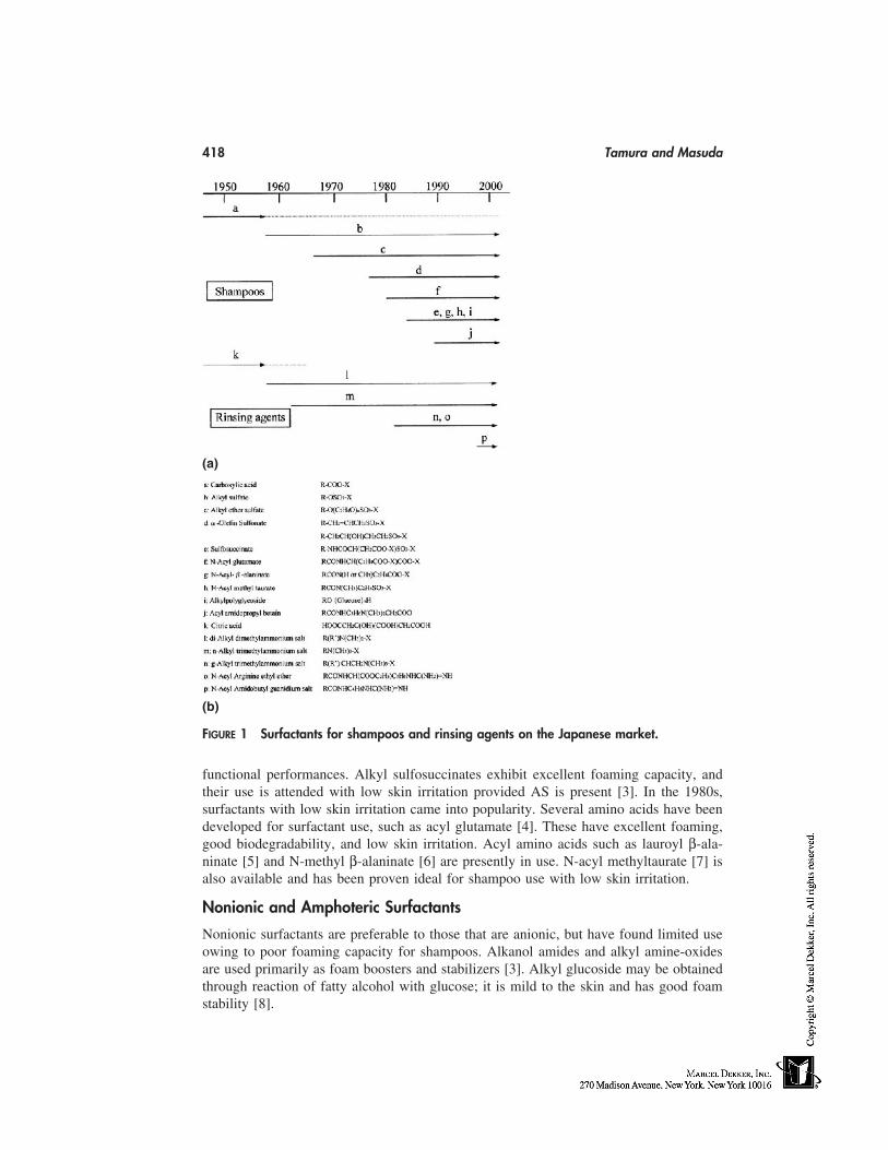
418 Tamura and Masuda
(a)
(b)
FIGURE 1 Surfactants for shampoos and rinsing agents on the Japanese market.
functional performances. Alkyl sulfosuccinates exhibit excellent foaming capacity, andtheir use is attended with low skin irritation provided AS is present [3]. In the 1980s,surfactants with low skin irritation came into popularity. Several amino acids have beendeveloped for surfactant use, such as acyl glutamate [4]. These have excellent foaming,good biodegradability, and low skin irritation. Acyl amino acids such as lauroyl β-ala-ninate [5] and N-methyl β-alaninate [6] are presently in use. N-acyl methyltaurate [7] isalso available and has been proven ideal for shampoo use with low skin irritation.
Nonionic and Amphoteric Surfactants
Nonionic surfactants are preferable to those that are anionic, but have found limited useowing to poor foaming capacity for shampoos. Alkanol amides and alkyl amine-oxidesare used primarily as foam boosters and stabilizers [3]. Alkyl glucoside may be obtainedthrough reaction of fatty alcohol with glucose; it is mild to the skin and has good foamstability [8].
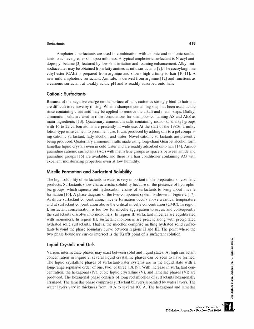
Surfactants 419
Amphoteric surfactants are used in combination with anionic and nonionic surfac-tants to achieve greater shampoo mildness. A typical amphoteric surfactant is N-acyl ami-dopropyl betaine [3] featured by low skin irritation and foaming enhancement. Alkyl imi-nodiacetates may be obtained from fatty amines as mild surfactants [9]. The cocoylarginineethyl ester (CAE) is prepared from arginine and shows high affinity to hair [10,11]. Anew mild amphoteric surfactant, Amisafe, is derived from arginine [12] and functions asa cationic surfactant at weakly acidic pH and is readily adsorbed onto hair.
Cationic Surfactants
Because of the negative charge on the surface of hair, cationics strongly bind to hair andare difficult to remove by rinsing. When a shampoo containing soap has been used, acidicrinse containing citric acid may be applied to remove the alkali and metal soaps. Dialkylammonium salts are used in rinse formulations for shampoos containing AS and AES asmain ingredients [13]. Quaternary ammonium salts containing mono- or dialkyl groupswith 16 to 22 carbon atoms are presently in wide use. At the start of the 1980s, a milkylotion-type rinse came into prominent use. It was produced by adding oils to a gel compris-ing cationic surfactant, fatty alcohol, and water. Novel cationic surfactants are presentlybeing produced. Quaternary ammonium salts made using long-chain Guarbet alcohol formlamellae liquid crystals even in cold water and are readily adsorbed onto hair [14]. Amidoguanidine cationic surfactants (AG) with methylene groups as spacers between amide andguanidino groups [15] are available, and there is a hair conditioner containing AG withexcellent moisturizing properties even at low humidity.
Micelle Formation and Surfactant Solubility
The high solubility of surfactants in water is very important in the preparation of cosmeticproducts. Surfactants show characteristic solubility because of the presence of hydropho-bic groups, which squeeze out hydrocarbon chains of surfactants to bring about micelleformation [16]. A phase diagram of the two-component system is shown in Figure 2 [17].At dilute surfactant concentration, micelle formation occurs above a critical temperatureand at surfactant concentration above the critical micelle concentration (CMC). In regionI, surfactant concentration is too low for micelle aggregation to occur, and consequentlythe surfactants dissolve into monomers. In region II, surfactant micelles are equilibratedwith monomers. In region III, surfactant monomers are present along with precipitatedhydrated solid surfactants. That is, the micelles comprise melting hydrated solid surfac-tants beyond the phase boundary curve between regions II and III. The point where thetwo phase boundary curves intersect is the Krafft point of a surfactant solution.
Liquid Crystals and Gels
Various intermediate phases may exist between solid and liquid states. At high surfactantconcentration in Figure 2, several liquid crystalline phases can be seen to have formed.The liquid crystalline phases of surfactant-water systems are in the liquid state with along-range repulsive order of one, two, or three [18,19]. With increase in surfactant con-centration, the hexagonal (IV), cubic liquid crystalline (V), and lamellae phases (VI) areproduced. The hexagonal phase consists of long rod micelles of surfactants hexagonallyarranged. The lamellae phase comprises surfactant bilayers separated by water layers. Thewater layers vary in thickness from 10 Å to several 100 Å. The hexagonal and lamellae
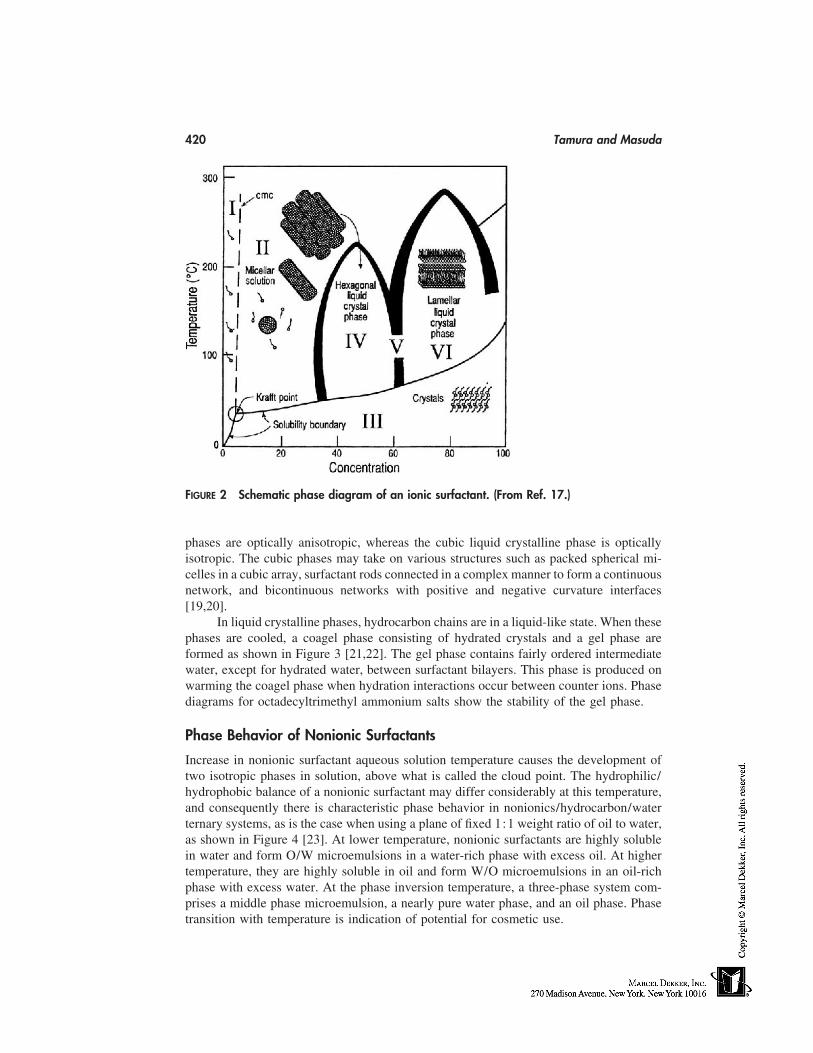
420 Tamura and Masuda
FIGURE 2 Schematic phase diagram of an ionic surfactant. (From Ref. 17.)
phases are optically anisotropic, whereas the cubic liquid crystalline phase is opticallyisotropic. The cubic phases may take on various structures such as packed spherical mi-celles in a cubic array, surfactant rods connected in a complex manner to form a continuousnetwork, and bicontinuous networks with positive and negative curvature interfaces[19,20].
In liquid crystalline phases, hydrocarbon chains are in a liquid-like state. When thesephases are cooled, a coagel phase consisting of hydrated crystals and a gel phase areformed as shown in Figure 3 [21,22]. The gel phase contains fairly ordered intermediatewater, except for hydrated water, between surfactant bilayers. This phase is produced onwarming the coagel phase when hydration interactions occur between counter ions. Phasediagrams for octadecyltrimethyl ammonium salts show the stability of the gel phase.
Phase Behavior of Nonionic Surfactants
Increase in nonionic surfactant aqueous solution temperature causes the development oftwo isotropic phases in solution, above what is called the cloud point. The hydrophilic/hydrophobic balance of a nonionic surfactant may differ considerably at this temperature,and consequently there is characteristic phase behavior in nonionics/hydrocarbon/waterternary systems, as is the case when using a plane of fixed 1:1 weight ratio of oil to water,as shown in Figure 4 [23]. At lower temperature, nonionic surfactants are highly solublein water and form O/W microemulsions in a water-rich phase with excess oil. At highertemperature, they are highly soluble in oil and form W/O microemulsions in an oil-richphase with excess water. At the phase inversion temperature, a three-phase system com-prises a middle phase microemulsion, a nearly pure water phase, and an oil phase. Phasetransition with temperature is indication of potential for cosmetic use.
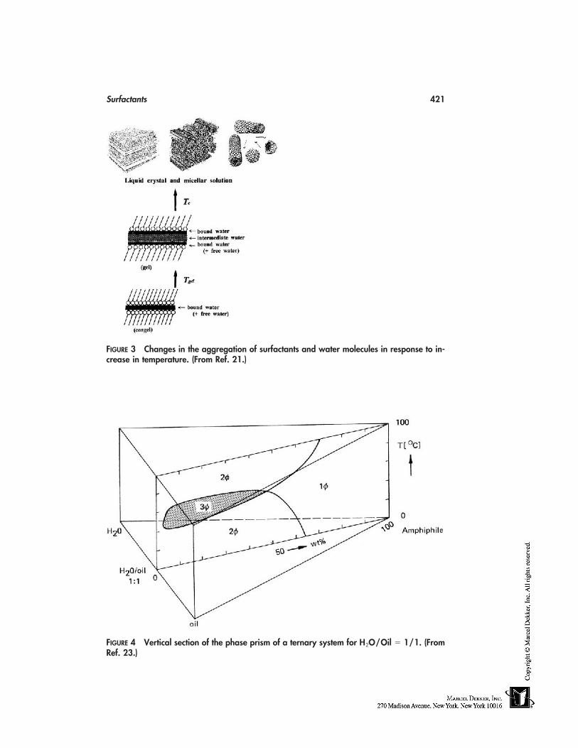
Surfactants 421
FIGURE 3 Changes in the aggregation of surfactants and water molecules in response to in-crease in temperature. (From Ref. 21.)
FIGURE 4 Vertical section of the phase prism of a ternary system for H2O/Oil � 1/1. (FromRef. 23.)
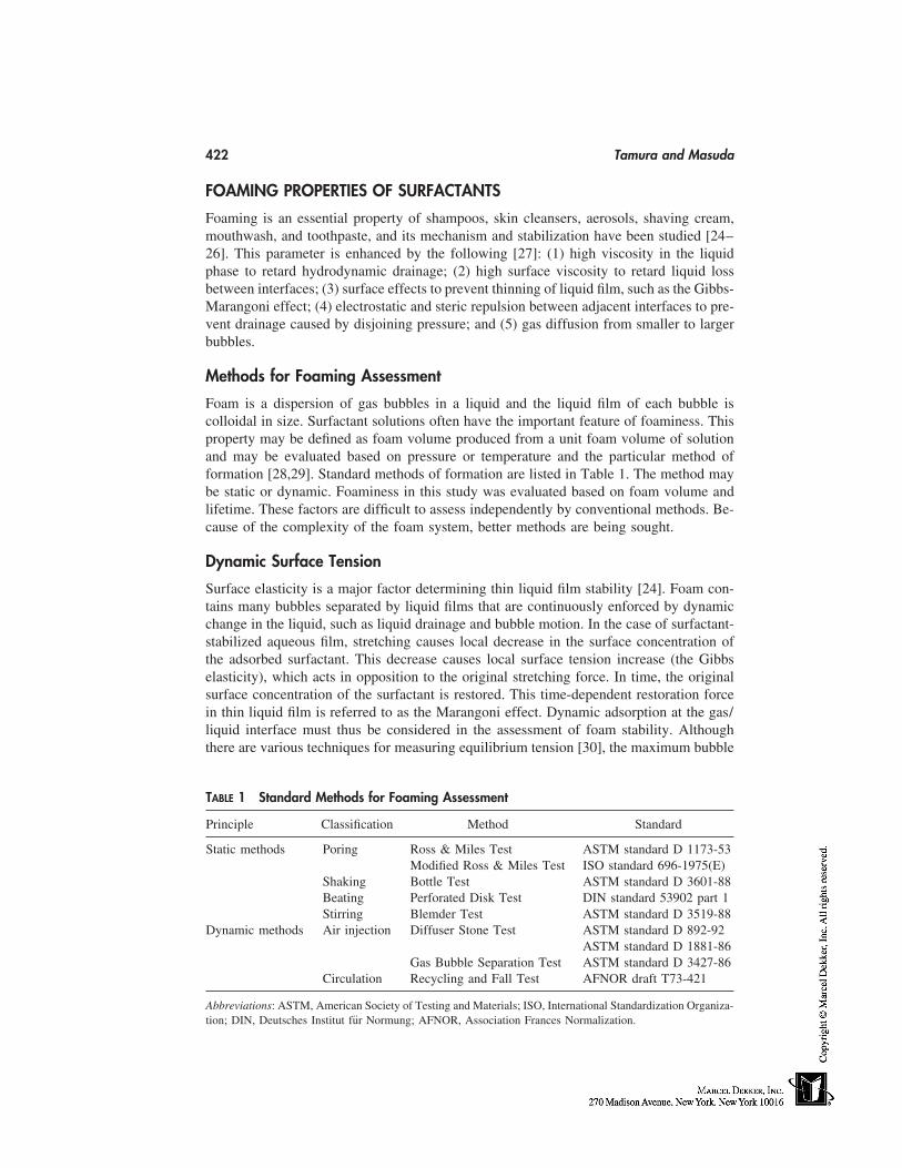
422 Tamura and Masuda
FOAMING PROPERTIES OF SURFACTANTS
Foaming is an essential property of shampoos, skin cleansers, aerosols, shaving cream,mouthwash, and toothpaste, and its mechanism and stabilization have been studied [24–26]. This parameter is enhanced by the following [27]: (1) high viscosity in the liquidphase to retard hydrodynamic drainage; (2) high surface viscosity to retard liquid lossbetween interfaces; (3) surface effects to prevent thinning of liquid film, such as the Gibbs-Marangoni effect; (4) electrostatic and steric repulsion between adjacent interfaces to pre-vent drainage caused by disjoining pressure; and (5) gas diffusion from smaller to largerbubbles.
Methods for Foaming Assessment
Foam is a dispersion of gas bubbles in a liquid and the liquid film of each bubble iscolloidal in size. Surfactant solutions often have the important feature of foaminess. Thisproperty may be defined as foam volume produced from a unit foam volume of solutionand may be evaluated based on pressure or temperature and the particular method offormation [28,29]. Standard methods of formation are listed in Table 1. The method maybe static or dynamic. Foaminess in this study was evaluated based on foam volume andlifetime. These factors are difficult to assess independently by conventional methods. Be-cause of the complexity of the foam system, better methods are being sought.
Dynamic Surface Tension
Surface elasticity is a major factor determining thin liquid film stability [24]. Foam con-tains many bubbles separated by liquid films that are continuously enforced by dynamicchange in the liquid, such as liquid drainage and bubble motion. In the case of surfactant-stabilized aqueous film, stretching causes local decrease in the surface concentration ofthe adsorbed surfactant. This decrease causes local surface tension increase (the Gibbselasticity), which acts in opposition to the original stretching force. In time, the originalsurface concentration of the surfactant is restored. This time-dependent restoration forcein thin liquid film is referred to as the Marangoni effect. Dynamic adsorption at the gas/liquid interface must thus be considered in the assessment of foam stability. Althoughthere are various techniques for measuring equilibrium tension [30], the maximum bubble
TABLE 1 Standard Methods for Foaming Assessment
Principle Classification Method Standard
Static methods Poring Ross & Miles Test ASTM standard D 1173-53Modified Ross & Miles Test ISO standard 696-1975(E)
Shaking Bottle Test ASTM standard D 3601-88Beating Perforated Disk Test DIN standard 53902 part 1Stirring Blemder Test ASTM standard D 3519-88
Dynamic methods Air injection Diffuser Stone Test ASTM standard D 892-92ASTM standard D 1881-86
Gas Bubble Separation Test ASTM standard D 3427-86Circulation Recycling and Fall Test AFNOR draft T73-421
Abbreviations: ASTM, American Society of Testing and Materials; ISO, International Standardization Organiza-tion; DIN, Deutsches Institut für Normung; AFNOR, Association Frances Normalization.
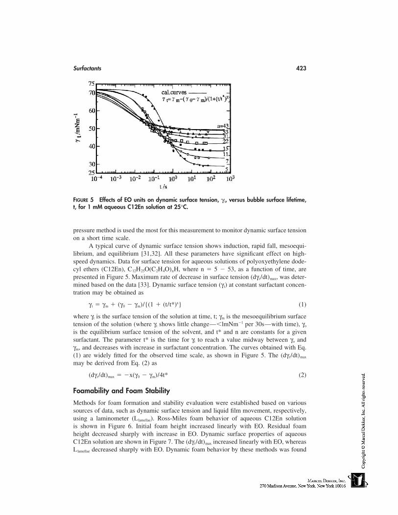
Surfactants 423
FIGURE 5 Effects of EO units on dynamic surface tension, γ t, versus bubble surface lifetime,t, for 1 mM aqueous C12En solution at 25°C.
pressure method is used the most for this measurement to monitor dynamic surface tensionon a short time scale.
A typical curve of dynamic surface tension shows induction, rapid fall, mesoequi-librium, and equilibrium [31,32]. All these parameters have significant effect on high-speed dynamics. Data for surface tension for aqueous solutions of polyoxyethylene dode-cyl ethers (C12En), C12H25O(C2H4O)nH, where n � 5 � 53, as a function of time, arepresented in Figure 5. Maximum rate of decrease in surface tension (dγ t/dt)max, was deter-mined based on the data [33]. Dynamic surface tension (γ t) at constant surfactant concen-tration may be obtained as
γ t � γm � (γ0 � γm)/{(1 � (t/t*)x} (1)
where γt is the surface tension of the solution at time, t; γm is the mesoequilibrium surfacetension of the solution (where γt shows little change—�lmNm�1 per 30s—with time), γo
is the equilibrium surface tension of the solvent, and t* and n are constants for a givensurfactant. The parameter t* is the time for γt to reach a value midway between γo andγm, and decreases with increase in surfactant concentration. The curves obtained with Eq.(1) are widely fitted for the observed time scale, as shown in Figure 5. The (dγ t/dt)max
may be derived from Eq. (2) as
(dγ t/dt)max � �x(γ0 � γm)/4t* (2)
Foamability and Foam Stability
Methods for foam formation and stability evaluation were established based on varioussources of data, such as dynamic surface tension and liquid film movement, respectively,using a laminometer (Llamellae). Ross-Miles foam behavior of aqueous C12En solutionis shown in Figure 6. Initial foam height increased linearly with EO. Residual foamheight decreased sharply with increase in EO. Dynamic surface properties of aqueousC12En solution are shown in Figure 7. The (dγ t/dt)max increased linearly with EO, whereasLlamellae decreased sharply with EO. Dynamic foam behavior by these methods was found
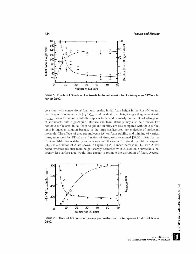
424 Tamura and Masuda
FIGURE 6 Effects of EO units on the Ross-Miles foam behavior for 1 mM aqueous C12En solu-tion at 26°C.
consistent with conventional foam test results. Initial foam height in the Ross-Miles testwas in good agreement with (dγt/dt)max, and residual foam height in good agreement withLlamellae. Foam formation would thus appear to depend primarily on the rate of adsorptionof surfactants onto a gas/liquid interface and foam stability may also be a factor. Fornonionic surfactants, initial foam height and stability are less compared with ionic surfac-tants in aqueous solution because of the large surface area per molecule of surfactantmolecule. The effects of area per molecule (A) on foam stability and thinning of verticalfilms, monitored by FT-IR as a function of time, were examined [34,35]. Data for theRoss and Miles foam stability and aqueous core thickness of vertical foam film at rupture(Drup) as a function of A are shown in Figure 8 [35]. Linear increase in Drup with A wasnoted, whereas residual foam height sharply decreased with A. Nonionic surfactants thatoccupy less surface area would thus appear to promote the disruption of foam. Accord-
FIGURE 7 Effects of EO units on dynamic parameters for 1 mM aqueous C12En solution at26°C.
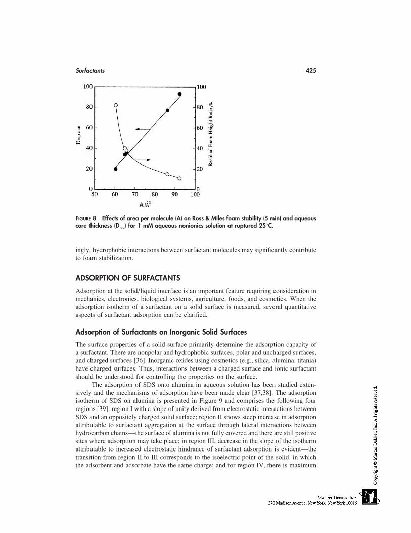
Surfactants 425
FIGURE 8 Effects of area per molecule (A) on Ross & Miles foam stability (5 min) and aqueouscore thickness (Drup) for 1 mM aqueous nonionics solution at ruptured 25°C.
ingly, hydrophobic interactions between surfactant molecules may significantly contributeto foam stabilization.
ADSORPTION OF SURFACTANTS
Adsorption at the solid/liquid interface is an important feature requiring consideration inmechanics, electronics, biological systems, agriculture, foods, and cosmetics. When theadsorption isotherm of a surfactant on a solid surface is measured, several quantitativeaspects of surfactant adsorption can be clarified.
Adsorption of Surfactants on Inorganic Solid Surfaces
The surface properties of a solid surface primarily determine the adsorption capacity ofa surfactant. There are nonpolar and hydrophobic surfaces, polar and uncharged surfaces,and charged surfaces [36]. Inorganic oxides using cosmetics (e.g., silica, alumina, titania)have charged surfaces. Thus, interactions between a charged surface and ionic surfactantshould be understood for controlling the properties on the surface.
The adsorption of SDS onto alumina in aqueous solution has been studied exten-sively and the mechanisms of adsorption have been made clear [37,38]. The adsorptionisotherm of SDS on alumina is presented in Figure 9 and comprises the following fourregions [39]: region I with a slope of unity derived from electrostatic interactions betweenSDS and an oppositely charged solid surface; region II shows steep increase in adsorptionattributable to surfactant aggregation at the surface through lateral interactions betweenhydrocarbon chains—the surface of alumina is not fully covered and there are still positivesites where adsorption may take place; in region III, decrease in the slope of the isothermattributable to increased electrostatic hindrance of surfactant adsorption is evident—thetransition from region II to III corresponds to the isoelectric point of the solid, in whichthe adsorbent and adsorbate have the same charge; and for region IV, there is maximum
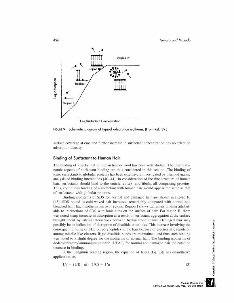
426 Tamura and Masuda
FIGURE 9 Schematic diagram of typical adsorption isotherm. (From Ref. 39.)
surface coverage at cmc and further increase in surfactant concentration has no effect onadsorption density.
Binding of Surfactant to Human Hair
The binding of a surfactant to human hair or wool has been well studied. The thermody-namic aspects of surfactant binding are thus considered in this section. The binding ofionic surfactants to globular proteins has been extensively investigated by thermodynamicanalysis of binding interactions [40–44]. In consideration of the fine structure of humanhair, surfactants should bind to the cuticle, cortex, and fibrils, all comprising proteins.Thus, continuous binding of a surfactant with human hair would appear the same as thatof surfactants with globular proteins.
Binding isotherms of SDS for normal and damaged hair are shown in Figure 10[45]. SDS bound to cold-waved hair increased remarkably compared with normal andbleached hair. Each isotherm has two regions. Region I shows Langmuir binding attribut-able to interactions of SDS with ionic sites on the surface of hair. For region II, therewas noted sharp increase in adsorption as a result of surfactant aggregation at the surfacebrought about by lateral interactions between hydrocarbon chains. Damaged hair maypossibly be an indication of disruption of disulfide crosslinks. This increase involving theconsequent binding of SDS on polypeptides in the hair because of electrostatic repulsionamong micelle-like clusters. Rigid disulfide bonds are maintained, and thus such bindingwas noted to a slight degree for the isotherms of normal hair. The binding isotherms ofdodecyltrimethylammonium chloride (DTAC) for normal and damaged hair indicated noincrease in binding.
In the Langmuir binding region, the equation of Klotz [Eq. (3)] has quantitativeapplication, as
1/γ � (1/K ⋅ n) ⋅ (1/C) � 1/n (3)
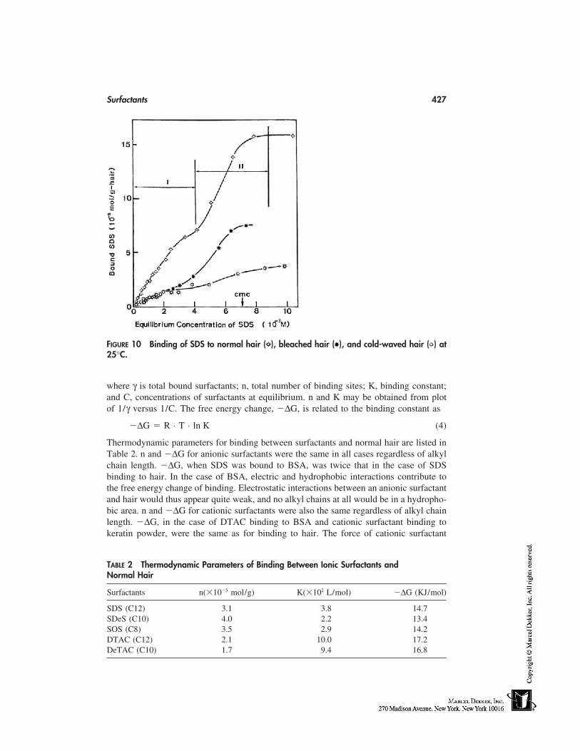
Surfactants 427
FIGURE 10 Binding of SDS to normal hair ( ), bleached hair ( ), and cold-waved hair ( ) at25°C.
where γ is total bound surfactants; n, total number of binding sites; K, binding constant;and C, concentrations of surfactants at equilibrium. n and K may be obtained from plotof 1/γ versus 1/C. The free energy change, �∆G, is related to the binding constant as
�∆G � R ⋅ T ⋅ ln K (4)
Thermodynamic parameters for binding between surfactants and normal hair are listed inTable 2. n and �∆G for anionic surfactants were the same in all cases regardless of alkylchain length. �∆G, when SDS was bound to BSA, was twice that in the case of SDSbinding to hair. In the case of BSA, electric and hydrophobic interactions contribute tothe free energy change of binding. Electrostatic interactions between an anionic surfactantand hair would thus appear quite weak, and no alkyl chains at all would be in a hydropho-bic area. n and �∆G for cationic surfactants were also the same regardless of alkyl chainlength. �∆G, in the case of DTAC binding to BSA and cationic surfactant binding tokeratin powder, were the same as for binding to hair. The force of cationic surfactant
TABLE 2 Thermodynamic Parameters of Binding Between Ionic Surfactants andNormal Hair
Surfactants n(�10�5 mol/g) K(�102 L/mol) �∆G (KJ/mol)
SDS (C12) 3.1 3.8 14.7SDeS (C10) 4.0 2.2 13.4SOS (C8) 3.5 2.9 14.2DTAC (C12) 2.1 10.0 17.2DeTAC (C10) 1.7 9.4 16.8
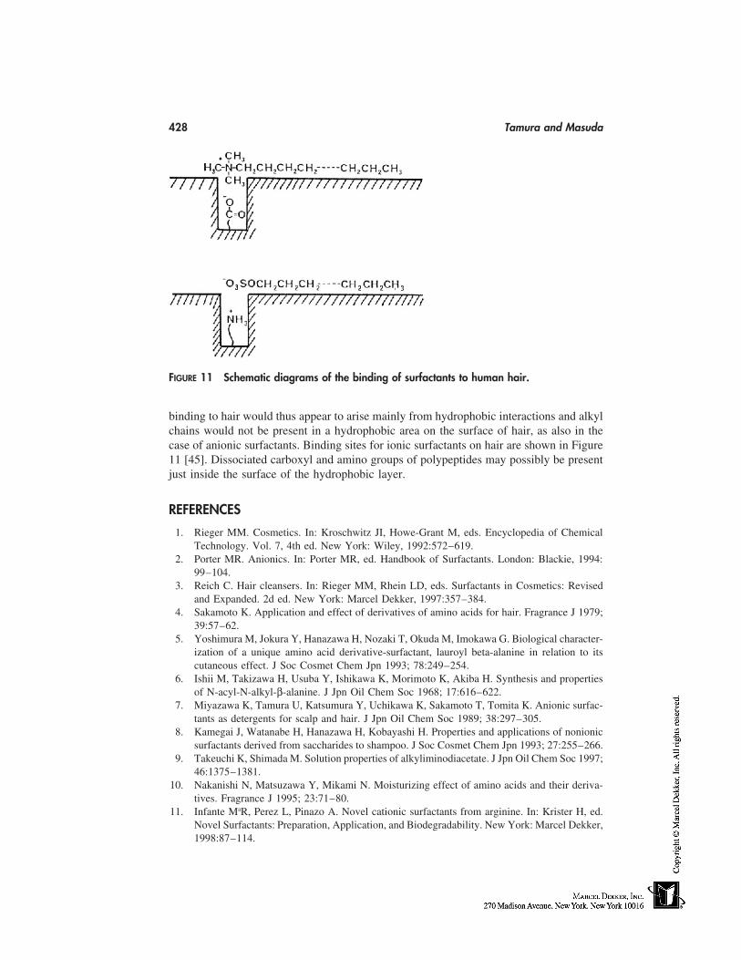
428 Tamura and Masuda
FIGURE 11 Schematic diagrams of the binding of surfactants to human hair.
binding to hair would thus appear to arise mainly from hydrophobic interactions and alkylchains would not be present in a hydrophobic area on the surface of hair, as also in thecase of anionic surfactants. Binding sites for ionic surfactants on hair are shown in Figure11 [45]. Dissociated carboxyl and amino groups of polypeptides may possibly be presentjust inside the surface of the hydrophobic layer.
REFERENCES
1. Rieger MM. Cosmetics. In: Kroschwitz JI, Howe-Grant M, eds. Encyclopedia of ChemicalTechnology. Vol. 7, 4th ed. New York: Wiley, 1992:572–619.
2. Porter MR. Anionics. In: Porter MR, ed. Handbook of Surfactants. London: Blackie, 1994:99–104.
3. Reich C. Hair cleansers. In: Rieger MM, Rhein LD, eds. Surfactants in Cosmetics: Revisedand Expanded. 2d ed. New York: Marcel Dekker, 1997:357–384.
4. Sakamoto K. Application and effect of derivatives of amino acids for hair. Fragrance J 1979;39:57–62.
5. Yoshimura M, Jokura Y, Hanazawa H, Nozaki T, Okuda M, Imokawa G. Biological character-ization of a unique amino acid derivative-surfactant, lauroyl beta-alanine in relation to itscutaneous effect. J Soc Cosmet Chem Jpn 1993; 78:249–254.
6. Ishii M, Takizawa H, Usuba Y, Ishikawa K, Morimoto K, Akiba H. Synthesis and propertiesof N-acyl-N-alkyl-β-alanine. J Jpn Oil Chem Soc 1968; 17:616–622.
7. Miyazawa K, Tamura U, Katsumura Y, Uchikawa K, Sakamoto T, Tomita K. Anionic surfac-tants as detergents for scalp and hair. J Jpn Oil Chem Soc 1989; 38:297–305.
8. Kamegai J, Watanabe H, Hanazawa H, Kobayashi H. Properties and applications of nonionicsurfactants derived from saccharides to shampoo. J Soc Cosmet Chem Jpn 1993; 27:255–266.
9. Takeuchi K, Shimada M. Solution properties of alkyliminodiacetate. J Jpn Oil Chem Soc 1997;46:1375–1381.
10. Nakanishi N, Matsuzawa Y, Mikami N. Moisturizing effect of amino acids and their deriva-tives. Fragrance J 1995; 23:71–80.
11. Infante MaR, Perez L, Pinazo A. Novel cationic surfactants from arginine. In: Krister H, ed.Novel Surfactants: Preparation, Application, and Biodegradability. New York: Marcel Dekker,1998:87–114.
Surfactants 429
12. Tabohashi T, Ninomiya R, Imori Y. A novel amino acid derivative for hair care products.Fragrance J 1998; 26:58–63.
13. Scott GV, Robbins CR, Barnhurst JD. Sorption of quaternary ammonium. J Soc Cosmet Chem1969; 20:135–152.
14. Yahagi K, Hoshino N, Hirota H. Solution behavior of new cationic surfactants synthesizedby using long-chain guerbet alcohols in water and their application to hair conditioners. J SocCosmet Chem Jpn 1990; 23:301–309.
15. Mitamura J, Suzuki N, Onuma K, Miyake M, Nakamura T, Kiyomiya A. Development ofnew cationic surfactant ‘‘AG’’ and application for hair conditioners. J Soc Cosmet Chem Jpn1996; 30:84–93.
16. Degiorgio V. Introduction. In: Degiorgio V, Corn M, eds. Physics of Amphiphiles: Micelles,Vesicles, and Microemulsions. Amsterdam: North-Holland, 1985:1–6.
17. Raney KH. Surfactant requirements for compact powder detergents. In: Showell MS, ed. Pow-der Detergents. New York: Marcel Dekker, 1998:241–284.
18. Lindman B. Amphiphilic systems. Some basic aspects. In: Degiorgio V, Corn M, eds. Physicsof Amphiphiles: Micelles, Vesicles, and Microemulsions. Amsterdam: North-Holland, 1985:7–23.
19. Laughlin RG. The structures and properties of surfactant phases. In: The Aqueous Phase Be-havior of Surfactants. London: Academic Press, 1994:181–237.
20. Fontell K. Cubic phases in surfactant and surfactant-like lipid systems. Colloid Polym Sci1990; 268:264–285.
21. Kodama M, Seki S. Hyomen Coagel-Ctel-Liquid crystal phase transition and heat propertiesof amphiphiles. 1990; 22:61–67.
22. Kodama M, Seki S. Thermodynamical investigations on phase transitions of surfactant-watersystems: thermodynamic stability of gel and coagel phases and the role of water moleculesin their appearance. Adv Colloid Interface Sci 1991; 35:1–30.
23. Kahlweit M, Strey R. The phase behavior of H2O-Oil-nonionic amphiphile ternary systems.In: Rosano HL, Clausse M, eds. Microemulsion Systems. New York: Marcel Dekker, 1987:1–13.
24. Malhotra AK, Wasan DT. Interfacial rheological properties of absorbed surfactant films withapplications to emulsion and foam stability. In: Ivanov IB, ed. Thin Liquid Films: Fundamen-tals and Applications. New York: Marcel Dekker, 1988:829–890.
25. Pugh RJ. Foaming, foam films, antifoaming and defoaming. Adv Colloid Interface Sci 1996;64:67–142.
26. Aveyard R, Clint JH. Foam and thin film breakdown processes. Curr Opin Colloid InterfaceSci 1996; 1:764–770.
27. Myers D. Foams. In: Myers D, ed. Surfaces, interfaces, and colloids: principles and applica-tions. New York: VCH, 1991:251–270.
28. Domingo X, Fiquet L, Meijer H. Foam ability/stability of surfactants. Tenside Surf Deter1992; 29:16–22.
29. Tamura T. The test methods for measuring foaming and antifoaming properties of liquid. JJpn Oil Chem Soc 1993; 42:737–745.
30. Dukhin SS, Kretzschmar G, Miller R. Experimental technique to study adsorption kinetics.In: Möbius D, Miller R, eds. Dynamics of Adsorption at Liquid Interfaces. Amsterdam: Elsev-ier, 1995:140–201.
31. Hua H, Rosen M. Dynamic surface tension of aqueous surfactant solutions: I. Basic parame-ters. J Colloid Interface Sci 1988; 124:652–659.
32. Tamura T, Kaneko Y, Ohyama M. Dynamic surface tension and foaming properties of aqueouspolyoxyethylene n-dodecyl ether solutions. J Colloid Interface Sci 1995; 173:493–499.
33. Rosen MJ, Hua XY, Zhu ZH. Dynamic surface tension of aqueous surfactant solutions: IV.Relationship to foaming. In: Mittal KL, Shah DO, eds. Surfactants in Solution. Vol. 11. NewYork: Plenum, 1991:315–327.
430 Tamura and Masuda
34. Tamura T, Kaneko Y, Nikaido M. Stability factors of foam film in contrast to fluctuationinduced by humidity reduction. J Colloid Interface Sci 1997; 190:61–70.
35. Tamura T, Takeuchi Y, Kaneko Y. Influence of surfactant structure on the drainage of nonionicsurfactant foam films. J Colloid Interface Sci 1998; 206:112–121.
36. Myers D. Physical properties of surfactants used in cosmetics. In: Rieger MM, Rhein LD,eds. Surfactants in Cosmetics. New York: Marcel Dekker, 1997:29–81.
37. Somasundaram P, Fuerstenau DW. Mechanisms of alkyl sulfonate adsorption at the alumina-water interface. J Phys Chem 1960; 70:90–96.
38. Somasundaram P, Chandar P, Turro NJ. Fluorescence probe studies on the structure of theadsorbed layer of dodecyl sulfate at the alumina-water interface. J Colloid Interface Sci 1987;117:31–46.
39. O’Haver JH, Harwell H. Adsorption: some expected and unexpected results. In: Sharma R,ed. Surfactant Adsorption and Surface Solubilization. ACS Symposium Ser. 615. Washington,D.C.: ACS, 1995:51–66.
40. Schwuger MJ, Bartnik G. Interaction of anionic surfactants with proteins, enzymes, and mem-branes. In: Gloxhuber C, ed. Anionic Surfactants: Biochemistry, Toxicology, Dermatology.New York: Marcel Dekker, 1980:1–49.
41. Reynolds JA, Gallagher JP, Steinhardt J. Effect of pH on the binding of N-Alkyl sulfates tobovine serum albumine. Biochemistry 1970; 9:1232–1238.
42. Nozaki Y, Reynolds JA, Tanford C. The interaction of a cationic detergent with bovine serumalbumin and other proteins. J Biol Chem 1974; 249:4452–4459.
43. Takagi T, Tsujii K, Shirahama K. Binding isotherms of sodium dodecyl sulfate to proteinpolypeptides with special performance to SDS-polyacrylamide gel electrophoresis. J Biochem1975; 77:939–947.
44. Hiramatsu K, Ueda C, Iwata K, Aoki K. The interaction of bovine plasma albumin with cat-ionic detergent. Studies by binding isotherm, optical rotation and difference spectrum. BullChem Soc Jpn 1977; 50:368–372.
45. Ohbu K, Tamura T, Mizushima N, Fukuda M. Binding characteristics of ionic surfactant withhair. Colloid Polym Sci 1986; 264:798–802.
37
Classification of Surfactants
Louis Oldenhove de GuertechinColgate-Palmolive Research and Development, Inc., Milmort, Belgium
INTRODUCTION
The term ‘‘surfactant’’ applies to a group of molecules having both a hydrophilic partand a hydrophobic (or lipophilic) part. Surfactants modify the interfacial properties of theliquids in which they are incorporated; this property stems from their tendency to concen-trate at the interfaces separating immiscible phases. Depending on the nature of the hydro-philic moiety ensuring the water-affinity of the molecule, major surfactants can be dividedinto anionic, cationic, amphoteric, and nonionic classes.
Regarding the hydrophobic moiety of the molecule, it is a hydrocarbon chain in mostcommon surfactants; however, in some more specialized surfactants, this hydrophobic partcan be a nonhydrocarbon chain such as a polydimethylsiloxane or a perfluorocarbon. Theselection of a surfactant for the development of cosmetic products should be carefullyperformed, taking into account numerous factors. Among others, one should consider thosedirectly related to functions to be fulfilled (detergency, emulsification, foam quality, rins-ability, mildness for skin, skin feel, etc.), and also those related to cost, toxicity, andbiodegradability. The aim of this chapter is to provide a classification of various commer-cially available surfactants. Various textbooks [1–4] or general articles [5,6] may usefullycomplete this survey.
IONIC SURFACTANTS
Anionic Surfactants
In aqueous solution, anionic surfactants form a negatively charged ion provided the com-position pH is neutral to alkaline. The ionized moiety can be a carboxylate, sulfate, sulfo-nate or phosphate. Among most frequently used surfactants in skin care products, the alkylsulfates and alkyl ethoxylated sulfates can be mentioned for their high foaming capacity.Anionics are generally used in association with other surfactants (nonionics or amphoter-ics), which bring improvements in the skin tolerance, in the foam quality, or in the productviscosity.
Other anionics are also used in personal products, as secondary surfactants, oftenfor their milder profile and their low foaming properties (isethionates, sulfosuccinates,taurates, sarcosinates, phosphoric acid esters, acylglutamates, etc.).
431
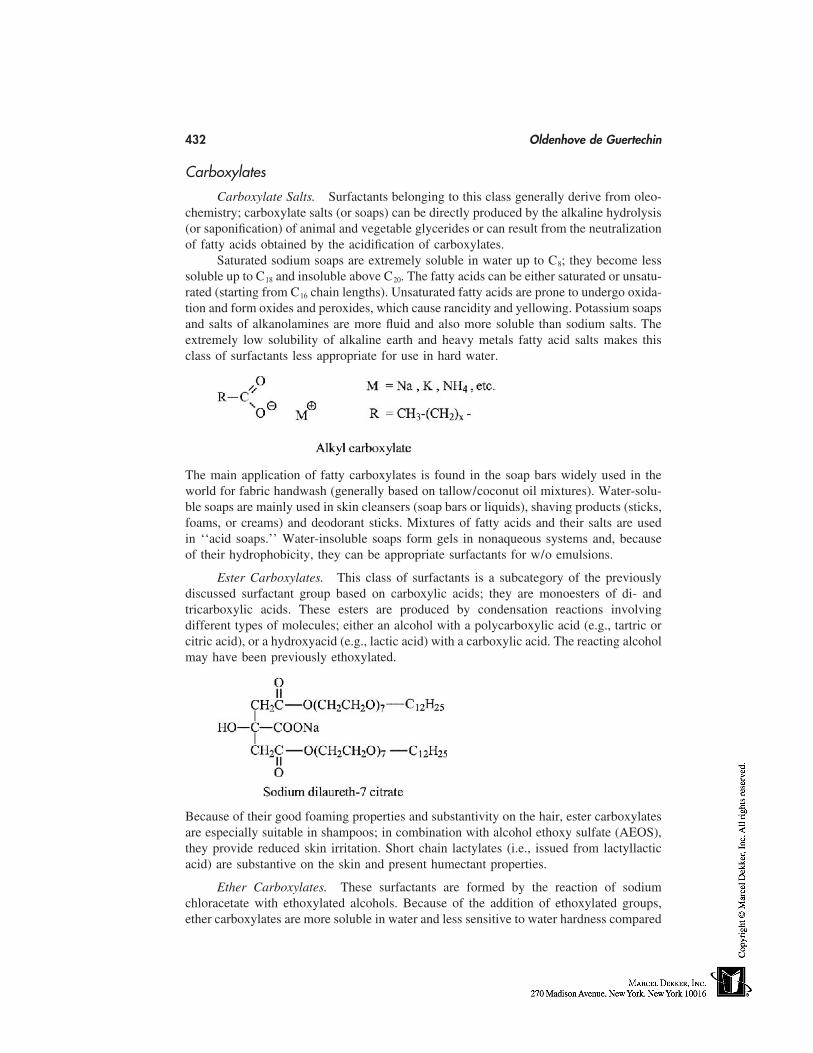
432 Oldenhove de Guertechin
Carboxylates
Carboxylate Salts. Surfactants belonging to this class generally derive from oleo-chemistry; carboxylate salts (or soaps) can be directly produced by the alkaline hydrolysis(or saponification) of animal and vegetable glycerides or can result from the neutralizationof fatty acids obtained by the acidification of carboxylates.
Saturated sodium soaps are extremely soluble in water up to C8; they become lesssoluble up to C18 and insoluble above C20. The fatty acids can be either saturated or unsatu-rated (starting from C16 chain lengths). Unsaturated fatty acids are prone to undergo oxida-tion and form oxides and peroxides, which cause rancidity and yellowing. Potassium soapsand salts of alkanolamines are more fluid and also more soluble than sodium salts. Theextremely low solubility of alkaline earth and heavy metals fatty acid salts makes thisclass of surfactants less appropriate for use in hard water.
The main application of fatty carboxylates is found in the soap bars widely used in theworld for fabric handwash (generally based on tallow/coconut oil mixtures). Water-solu-ble soaps are mainly used in skin cleansers (soap bars or liquids), shaving products (sticks,foams, or creams) and deodorant sticks. Mixtures of fatty acids and their salts are usedin ‘‘acid soaps.’’ Water-insoluble soaps form gels in nonaqueous systems and, becauseof their hydrophobicity, they can be appropriate surfactants for w/o emulsions.
Ester Carboxylates. This class of surfactants is a subcategory of the previouslydiscussed surfactant group based on carboxylic acids; they are monoesters of di- andtricarboxylic acids. These esters are produced by condensation reactions involvingdifferent types of molecules; either an alcohol with a polycarboxylic acid (e.g., tartric orcitric acid), or a hydroxyacid (e.g., lactic acid) with a carboxylic acid. The reacting alcoholmay have been previously ethoxylated.
Because of their good foaming properties and substantivity on the hair, ester carboxylatesare especially suitable in shampoos; in combination with alcohol ethoxy sulfate (AEOS),they provide reduced skin irritation. Short chain lactylates (i.e., issued from lactyllacticacid) are substantive on the skin and present humectant properties.
Ether Carboxylates. These surfactants are formed by the reaction of sodiumchloracetate with ethoxylated alcohols. Because of the addition of ethoxylated groups,ether carboxylates are more soluble in water and less sensitive to water hardness compared
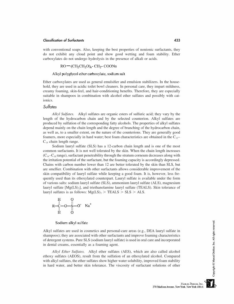
Classification of Surfactants 433
with conventional soaps. Also, keeping the best properties of nonionic surfactants, theydo not exhibit any cloud point and show good wetting and foam stability. Ethercarboxylates do not undergo hydrolysis in the presence of alkali or acids.
Ether carboxylates are used as general emulsifier and emulsion stabilizers. In the house-hold, they are used in acidic toilet bowl cleaners. In personal care, they impart mildness,creamy foaming, skin-feel, and hair-conditioning benefits. Therefore, they are especiallysuitable in shampoos in combination with alcohol ether sulfates and possibly with cat-ionics.
Sulfates
Alkyl Sulfates. Alkyl sulfates are organic esters of sulfuric acid; they vary by thelength of the hydrocarbon chain and by the selected counterion. Alkyl sulfates areproduced by sulfation of the corresponding fatty alcohols. The properties of alkyl sulfatesdepend mainly on the chain length and the degree of branching of the hydrocarbon chain,as well as, to a smaller extent, on the nature of the counterions. They are generally goodfoamers, more especially in hard water; best foam characteristics are obtained in the C12–C14 chain length range.
Sodium lauryl sulfate (SLS) has a 12-carbon chain length and is one of the mostcommon surfactants. It is not well tolerated by the skin. When the chain length increases(C14–C18 range), surfactant penetrability through the stratum corneum decreases along withthe irritation potential of the surfactant, but the foaming capacity is accordingly depressed.Chains with carbon number lower than 12 are better tolerated by the skin than SLS, butare smellier. Combination with other surfactants allows considerable improvement of theskin compatibility of lauryl sulfate while keeping a good foam. It is, however, less fre-quently used than its ethoxylated counterpart. Lauryl sulfate is available under the formof various salts: sodium lauryl sulfate (SLS), ammonium lauryl sulfate (ALS), magnesiumlauryl sulfate [Mg(LS)2], and triethanolamine lauryl sulfate (TEALS). Skin tolerance oflauryl sulfates is as follows: Mg(LS)2 � TEALS � SLS � ALS.
Alkyl sulfates are used in cosmetics and personal-care areas (e.g., DEA lauryl sulfate inshampoos); they are associated with other surfactants and improve foaming characteristicsof detergent systems. Pure SLS (sodium lauryl sulfate) is used in oral care and incorporatedin dental creams, essentially as a foaming agent.
Alkyl Ether Sulfates. Alkyl ether sulfates (AES), which are also called alcoholethoxy sulfates (AEOS), result from the sulfation of an ethoxylated alcohol. Comparedwith alkyl sulfates, the ether sulfates show higher water solubility, improved foam stabilityin hard water, and better skin tolerance. The viscosity of surfactant solutions of ether
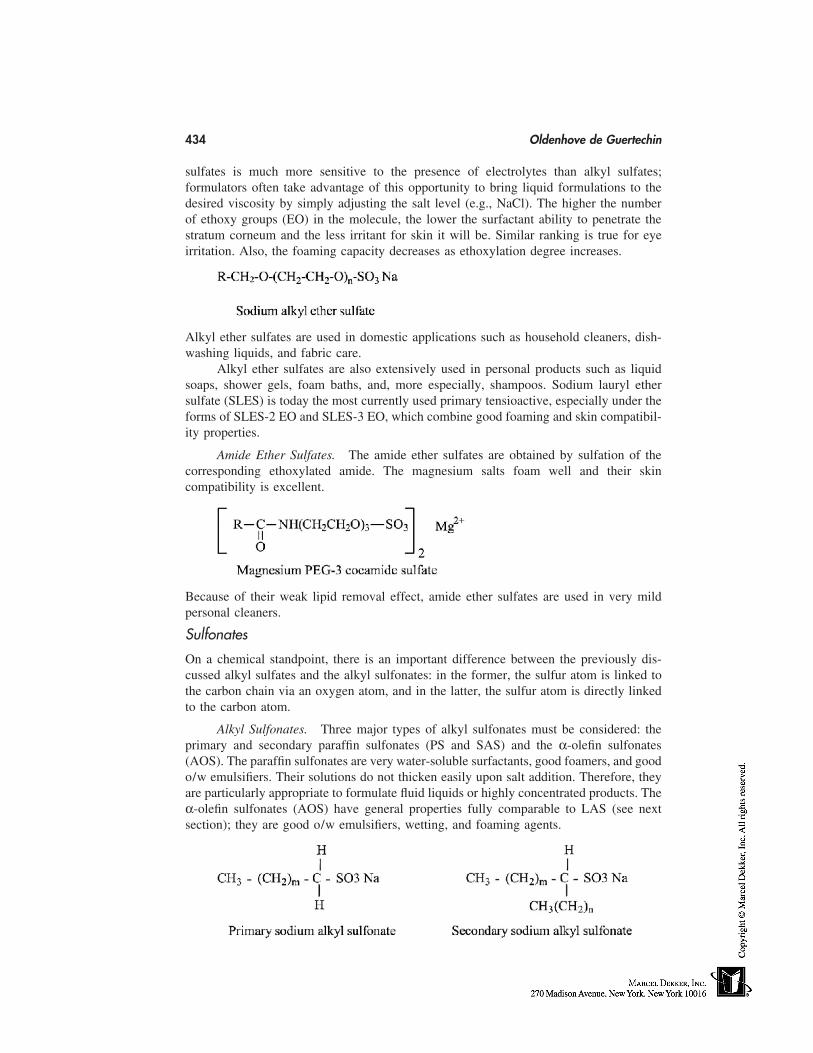
434 Oldenhove de Guertechin
sulfates is much more sensitive to the presence of electrolytes than alkyl sulfates;formulators often take advantage of this opportunity to bring liquid formulations to thedesired viscosity by simply adjusting the salt level (e.g., NaCl). The higher the numberof ethoxy groups (EO) in the molecule, the lower the surfactant ability to penetrate thestratum corneum and the less irritant for skin it will be. Similar ranking is true for eyeirritation. Also, the foaming capacity decreases as ethoxylation degree increases.
Alkyl ether sulfates are used in domestic applications such as household cleaners, dish-washing liquids, and fabric care.
Alkyl ether sulfates are also extensively used in personal products such as liquidsoaps, shower gels, foam baths, and, more especially, shampoos. Sodium lauryl ethersulfate (SLES) is today the most currently used primary tensioactive, especially under theforms of SLES-2 EO and SLES-3 EO, which combine good foaming and skin compatibil-ity properties.
Amide Ether Sulfates. The amide ether sulfates are obtained by sulfation of thecorresponding ethoxylated amide. The magnesium salts foam well and their skincompatibility is excellent.
Because of their weak lipid removal effect, amide ether sulfates are used in very mildpersonal cleaners.
Sulfonates
On a chemical standpoint, there is an important difference between the previously dis-cussed alkyl sulfates and the alkyl sulfonates: in the former, the sulfur atom is linked tothe carbon chain via an oxygen atom, and in the latter, the sulfur atom is directly linkedto the carbon atom.
Alkyl Sulfonates. Three major types of alkyl sulfonates must be considered: theprimary and secondary paraffin sulfonates (PS and SAS) and the α-olefin sulfonates(AOS). The paraffin sulfonates are very water-soluble surfactants, good foamers, and goodo/w emulsifiers. Their solutions do not thicken easily upon salt addition. Therefore, theyare particularly appropriate to formulate fluid liquids or highly concentrated products. Theα-olefin sulfonates (AOS) have general properties fully comparable to LAS (see nextsection); they are good o/w emulsifiers, wetting, and foaming agents.
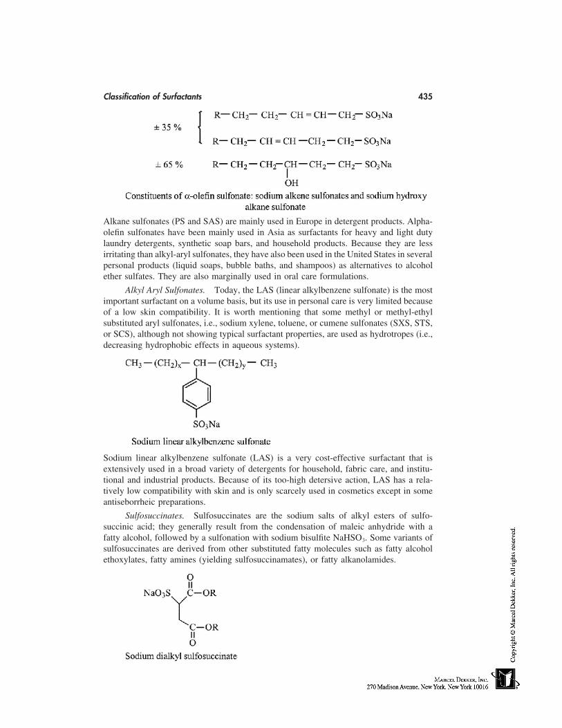
Classification of Surfactants 435
Alkane sulfonates (PS and SAS) are mainly used in Europe in detergent products. Alpha-olefin sulfonates have been mainly used in Asia as surfactants for heavy and light dutylaundry detergents, synthetic soap bars, and household products. Because they are lessirritating than alkyl-aryl sulfonates, they have also been used in the United States in severalpersonal products (liquid soaps, bubble baths, and shampoos) as alternatives to alcoholether sulfates. They are also marginally used in oral care formulations.
Alkyl Aryl Sulfonates. Today, the LAS (linear alkylbenzene sulfonate) is the mostimportant surfactant on a volume basis, but its use in personal care is very limited becauseof a low skin compatibility. It is worth mentioning that some methyl or methyl-ethylsubstituted aryl sulfonates, i.e., sodium xylene, toluene, or cumene sulfonates (SXS, STS,or SCS), although not showing typical surfactant properties, are used as hydrotropes (i.e.,decreasing hydrophobic effects in aqueous systems).
Sodium linear alkylbenzene sulfonate (LAS) is a very cost-effective surfactant that isextensively used in a broad variety of detergents for household, fabric care, and institu-tional and industrial products. Because of its too-high detersive action, LAS has a rela-tively low compatibility with skin and is only scarcely used in cosmetics except in someantiseborrheic preparations.
Sulfosuccinates. Sulfosuccinates are the sodium salts of alkyl esters of sulfo-succinic acid; they generally result from the condensation of maleic anhydride with afatty alcohol, followed by a sulfonation with sodium bisulfite NaHSO3. Some variants ofsulfosuccinates are derived from other substituted fatty molecules such as fatty alcoholethoxylates, fatty amines (yielding sulfosuccinamates), or fatty alkanolamides.
436 Oldenhove de Guertechin
Monoesters disodium salts are the most common sulfosuccinates used in cosmeticapplications. Monoesters of alkanolamines (sulfosuccinamates) are milder than monoes-ters of fatty alcohols (sulfosuccinates). Monoesters derived from ethoxylated alcohols oralkanolamides are extensively used in personal products and especially in shampoos; theyare known for their mildness and skin-irritation reduction when used in association withother anionic surfactants.
Sulfo Fatty Acid Esters. These surfactants are sometimes known under their ab-breviated names: FES for fatty ester sulfonate, MES for methyl ester sulfonate, or ASMEfor alpha sulfo methyl ester. Most of α-sulfo fatty acid esters derive from fatty acid methylesters. In general, alkyl esters of α-sulfo fatty acid have excellent detergency (i.e., oildispersing and emulsifying properties) when the molecule is dissymmetric (as in the caseof the α-sulfo methyl esters). On the other hand, the α-sulfo esters, in which the sulfonategroup is in the middle of the molecule (as in the case of long-chain alcohol esters), delivergood wetting but poor detergency.
Alpha-sulfo methyl ester surfactants deriving from C16–C18 fatty acid (e.g., ASMT, thetallowate) are appropriate for use in laundry detergents. ASME is also used in the formula-tion of syndet bars (laundry bars based on synthetic surfactants).
Fatty Acid Isethionates and Taurides. Fatty acid isethionates are usually preparedby reaction of a fatty acid chloride with sodium isethionate (HO-CH2-CH2-SO3-Na), itselfresulting from the addition of sodium bisulfite to ethylene oxide. These surfactants areinsensitive to water hardness and show good wetting, foaming, and emulsifying properties.In addition, they are very mild and have excellent compatibility with the skin. Taurides(or taurates) are acylamino alkane sulfonates that have chemical structures close toisethionates. They can be used in association with other surfactants to increase theviscosity.
Acyl isethionates have been used in shampoos and personal cleansers. They are also incor-porated in syndet bars together with various soaps. The most currently used is the cocoylisethionate.
Taurides (or taurates), which have the same expected properties as soaps (exceptthe sensitivity to water hardness), had been extensively used in shampoos but have beenreplaced by AEOS. Today they are limitedly used in cosmetics mainly in foam baths andtoilet bars. Taurides are also used in soap bars especially designed for laundering withseawater, in agriculture, and textile dying.
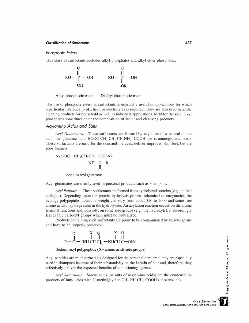
Classification of Surfactants 437
Phosphate Esters
This class of surfactants includes alkyl phosphates and alkyl ether phosphates.
The use of phosphate esters as surfactants is especially useful in applications for whicha particular tolerance to pH, heat, or electrolytes is required. They are also used in acidiccleaning products for household as well as industrial applications. Mild for the skin, alkylphosphates sometimes enter the composition of facial and cleansing products.
Acylamino Acids and Salts
Acyl Glutamates. These surfactants are formed by acylation of a natural aminoacid, the glutamic acid HOOC-CH2-CH2-CH(NH2)-COOH (or α-aminoglutaric acid).These surfactants are mild for the skin and the eyes, deliver improved skin feel, but arepoor foamers.
Acyl glutamates are mainly used in personal products such as shampoos.
Acyl Peptides. These surfactants are formed from hydrolyzed proteins (e.g., animalcollagen). Depending upon the protein hydrolysis process (chemical or enzymatic), theaverage polypeptide molecular weight can vary from about 350 to 2000 and some freeamino acids may be present in the hydrolysate. An acylation reaction occurs on the amineterminal functions and, possibly, on some side groups (e.g., the hydroxyls); it accordinglyleaves free carboxyl groups which must be neutralized.
Products containing such surfactants are prone to be contaminated by various germsand have to be properly preserved.
Acyl peptides are mild surfactants designed for the personal-care area; they are especiallyused in shampoos because of their substantivity on the keratin of hair and, therefore, theyeffectively deliver the expected benefits of conditioning agents.
Acyl Sarcosides. Sarcosinates (or salts of acylamino acids) are the condensationproducts of fatty acids with N-methylglycine CH3-NH-CH2-COOH (or sarcosine).
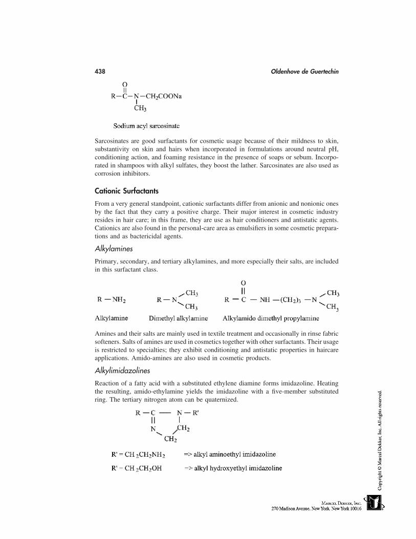
438 Oldenhove de Guertechin
Sarcosinates are good surfactants for cosmetic usage because of their mildness to skin,substantivity on skin and hairs when incorporated in formulations around neutral pH,conditioning action, and foaming resistance in the presence of soaps or sebum. Incorpo-rated in shampoos with alkyl sulfates, they boost the lather. Sarcosinates are also used ascorrosion inhibitors.
Cationic Surfactants
From a very general standpoint, cationic surfactants differ from anionic and nonionic onesby the fact that they carry a positive charge. Their major interest in cosmetic industryresides in hair care; in this frame, they are use as hair conditioners and antistatic agents.Cationics are also found in the personal-care area as emulsifiers in some cosmetic prepara-tions and as bactericidal agents.
Alkylamines
Primary, secondary, and tertiary alkylamines, and more especially their salts, are includedin this surfactant class.
Amines and their salts are mainly used in textile treatment and occasionally in rinse fabricsofteners. Salts of amines are used in cosmetics together with other surfactants. Their usageis restricted to specialties; they exhibit conditioning and antistatic properties in haircareapplications. Amido-amines are also used in cosmetic products.
Alkylimidazolines
Reaction of a fatty acid with a substituted ethylene diamine forms imidazoline. Heatingthe resulting, amido-ethylamine yields the imidazoline with a five-member substitutedring. The tertiary nitrogen atom can be quaternized.
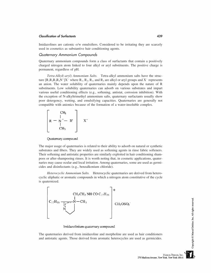
Classification of Surfactants 439
Imidazolines are cationic o/w emulsifiers. Considered to be irritating they are scarcelyused in cosmetics as substantive hair conditioning agents.
Quaternary Ammonium Compounds
Quaternary ammonium compounds form a class of surfactants that contain a positivelycharged nitrogen atom linked to four alkyl or aryl substituents. The positive charge ispermanent, regardless of pH.
Tetra-Alkyl(-aryl) Ammonium Salts. Tetra-alkyl ammonium salts have the struc-ture [R1R2R3R4N�]X� where R1, R2, R3, and R4 are alkyl or aryl groups and X� representsan anion. The water solubility of quaternaries mainly depends upon the nature of Rsubstituents. Low solubility quaternaries can adsorb on various substrates and impartvarious useful conditioning effects (e.g., softening, antistat, corrosion inhibition). Withthe exception of N-alkyltrimethyl ammonium salts, quaternary surfactants usually showpoor detergency, wetting, and emulsifying capacities. Quaternaries are generally notcompatible with anionics because of the formation of a water-insoluble complex.
The major usage of quaternaries is related to their ability to adsorb on natural or syntheticsubstrates and fibers. They are widely used as softening agents in rinse fabric softeners.Their softening and antistatic properties are similarly exploited in hair conditioning sham-poos or after-shampooing rinses. It is worth noting that, in cosmetic applications, quater-naries may cause ocular and local irritation. Among quaternaries, some are used as germi-cides and disinfectants (e.g., benzalkonium chloride).
Heterocyclic Ammonium Salts. Heterocyclic quaternaries are derived from hetero-cyclic aliphatic or aromatic compounds in which a nitrogen atom constitutive of the cycleis quaternized.
The quaternaries derived from imidazoline and morpholine are used as hair conditionersand antistatic agents. Those derived from aromatic heterocycles are used as germicides.
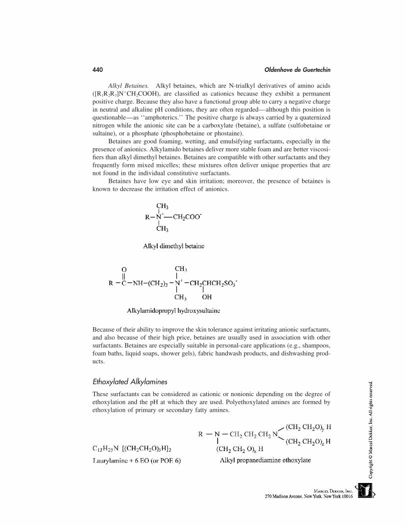
440 Oldenhove de Guertechin
Alkyl Betaines. Alkyl betaines, which are N-trialkyl derivatives of amino acids([R1R2R3]N�CH2COOH), are classified as cationics because they exhibit a permanentpositive charge. Because they also have a functional group able to carry a negative chargein neutral and alkaline pH conditions, they are often regarded—although this position isquestionable—as ‘‘amphoterics.’’ The positive charge is always carried by a quaternizednitrogen while the anionic site can be a carboxylate (betaine), a sulfate (sulfobetaine orsultaine), or a phosphate (phosphobetaine or phostaine).
Betaines are good foaming, wetting, and emulsifying surfactants, especially in thepresence of anionics. Alkylamido betaines deliver more stable foam and are better viscosi-fiers than alkyl dimethyl betaines. Betaines are compatible with other surfactants and theyfrequently form mixed micelles; these mixtures often deliver unique properties that arenot found in the individual constitutive surfactants.
Betaines have low eye and skin irritation; moreover, the presence of betaines isknown to decrease the irritation effect of anionics.
Because of their ability to improve the skin tolerance against irritating anionic surfactants,and also because of their high price, betaines are usually used in association with othersurfactants. Betaines are especially suitable in personal-care applications (e.g., shampoos,foam baths, liquid soaps, shower gels), fabric handwash products, and dishwashing prod-ucts.
Ethoxylated Alkylamines
These surfactants can be considered as cationic or nonionic depending on the degree ofethoxylation and the pH at which they are used. Polyethoxylated amines are formed byethoxylation of primary or secondary fatty amines.
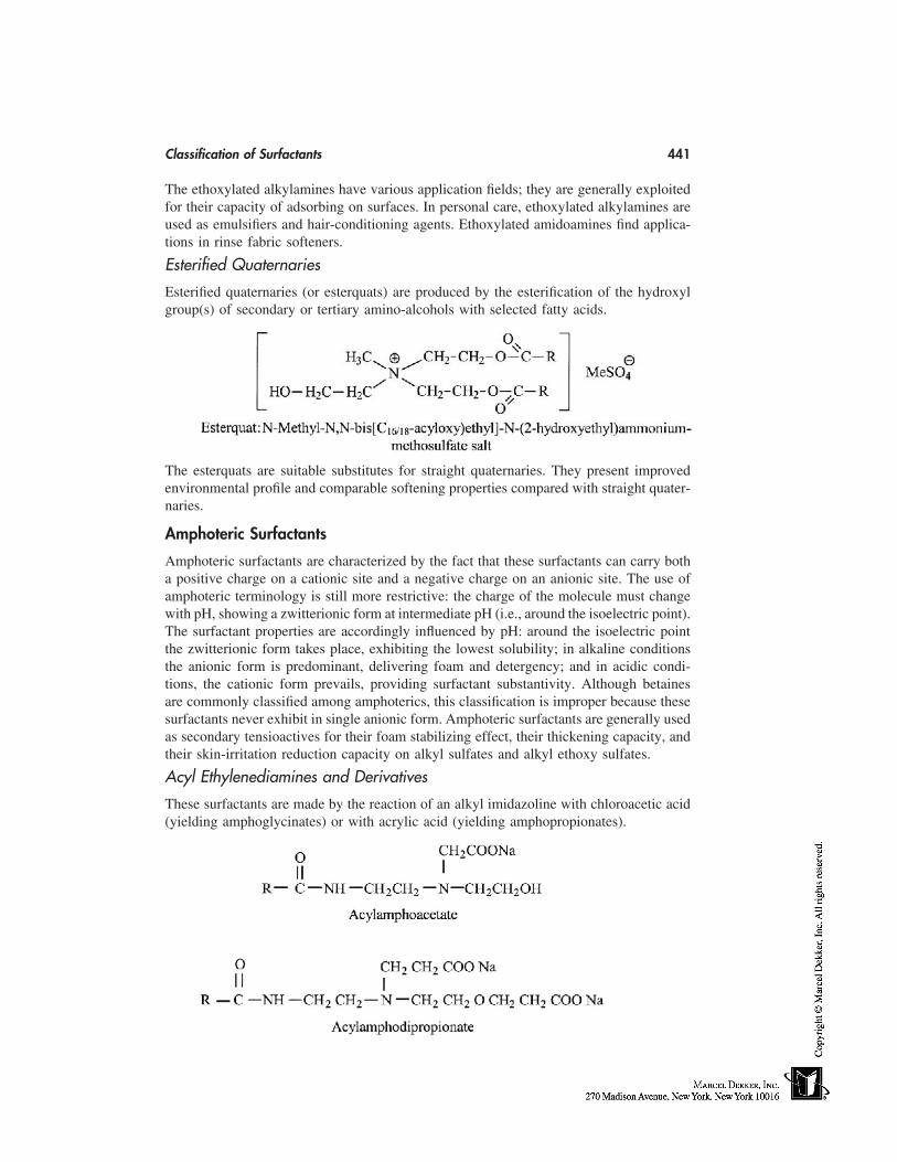
Classification of Surfactants 441
The ethoxylated alkylamines have various application fields; they are generally exploitedfor their capacity of adsorbing on surfaces. In personal care, ethoxylated alkylamines areused as emulsifiers and hair-conditioning agents. Ethoxylated amidoamines find applica-tions in rinse fabric softeners.
Esterified Quaternaries
Esterified quaternaries (or esterquats) are produced by the esterification of the hydroxylgroup(s) of secondary or tertiary amino-alcohols with selected fatty acids.
The esterquats are suitable substitutes for straight quaternaries. They present improvedenvironmental profile and comparable softening properties compared with straight quater-naries.
Amphoteric Surfactants
Amphoteric surfactants are characterized by the fact that these surfactants can carry botha positive charge on a cationic site and a negative charge on an anionic site. The use ofamphoteric terminology is still more restrictive: the charge of the molecule must changewith pH, showing a zwitterionic form at intermediate pH (i.e., around the isoelectric point).The surfactant properties are accordingly influenced by pH: around the isoelectric pointthe zwitterionic form takes place, exhibiting the lowest solubility; in alkaline conditionsthe anionic form is predominant, delivering foam and detergency; and in acidic condi-tions, the cationic form prevails, providing surfactant substantivity. Although betainesare commonly classified among amphoterics, this classification is improper because thesesurfactants never exhibit in single anionic form. Amphoteric surfactants are generally usedas secondary tensioactives for their foam stabilizing effect, their thickening capacity, andtheir skin-irritation reduction capacity on alkyl sulfates and alkyl ethoxy sulfates.
Acyl Ethylenediamines and Derivatives
These surfactants are made by the reaction of an alkyl imidazoline with chloroacetic acid(yielding amphoglycinates) or with acrylic acid (yielding amphopropionates).
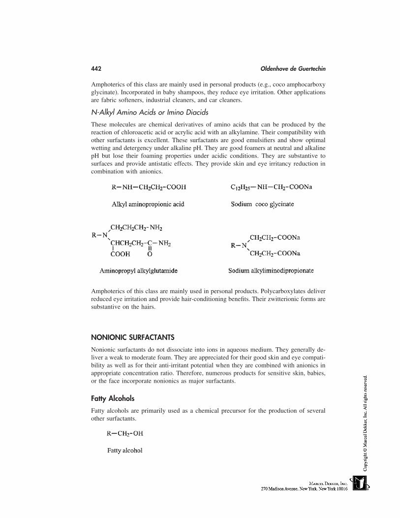
442 Oldenhove de Guertechin
Amphoterics of this class are mainly used in personal products (e.g., coco amphocarboxyglycinate). Incorporated in baby shampoos, they reduce eye irritation. Other applicationsare fabric softeners, industrial cleaners, and car cleaners.
N-Alkyl Amino Acids or Imino Diacids
These molecules are chemical derivatives of amino acids that can be produced by thereaction of chloroacetic acid or acrylic acid with an alkylamine. Their compatibility withother surfactants is excellent. These surfactants are good emulsifiers and show optimalwetting and detergency under alkaline pH. They are good foamers at neutral and alkalinepH but lose their foaming properties under acidic conditions. They are substantive tosurfaces and provide antistatic effects. They provide skin and eye irritancy reduction incombination with anionics.
Amphoterics of this class are mainly used in personal products. Polycarboxylates deliverreduced eye irritation and provide hair-conditioning benefits. Their zwitterionic forms aresubstantive on the hairs.
NONIONIC SURFACTANTS
Nonionic surfactants do not dissociate into ions in aqueous medium. They generally de-liver a weak to moderate foam. They are appreciated for their good skin and eye compati-bility as well as for their anti-irritant potential when they are combined with anionics inappropriate concentration ratio. Therefore, numerous products for sensitive skin, babies,or the face incorporate nonionics as major surfactants.
Fatty Alcohols
Fatty alcohols are primarily used as a chemical precursor for the production of severalother surfactants.
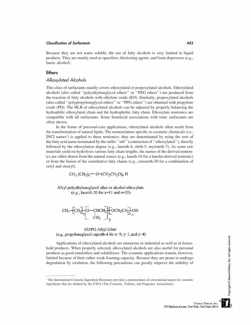
Classification of Surfactants 443
Because they are not water soluble, the use of fatty alcohols is very limited in liquidproducts. They are mainly used as opacifiers, thickening agents, and foam depressors (e.g.,lauric alcohol).
Ethers
Alkoxylated Alcohols
This class of surfactants mainly covers ethoxylated or propoxylated alcohols. Ethoxylatedalcohols (also called ‘‘polyethyleneglycol ethers’’ or ‘‘PEG ethers’’) are produced fromthe reaction of fatty alcohols with ethylene oxide (EO). Similarly, propoxylated alcohols(also called ‘‘polypropyleneglycol ethers’’ or ‘‘PPG ethers’’) are obtained with propyleneoxide (PO). The HLB of ethoxylated alcohols can be adjusted by properly balancing thehydrophilic ethoxylated chain and the hydrophobic fatty chain. Ethoxylate nonionics arecompatible with all surfactants. Some beneficial associations with ionic surfactants areoften shown.
In the frame of personal-care applications, ethoxylated alcohols often result fromthe transformation of natural lipids. The nomenclature specific to cosmetic chemicals (i.e.,INCI names1) is applied to these nonionics: they are denominated by using the root ofthe fatty acid name terminated by the suffix ‘‘eth’’ (contraction of ‘‘ethoxylated’’), directlyfollowed by the ethoxylation degree (e.g., laureth-4, oleth-5, myristeth-7). As some rawmaterials yield on hydrolysis various fatty chain lengths, the names of the derived nonion-ics are either drawn from the natural source (e.g., laneth-16 for a lanolin-derived nonionic)or from the fusion of the constitutive fatty chains (e.g., ceteareth-20 for a combination ofcetyl and stearyl).
Applications of ethoxylated alcohols are numerous in industrial as well as in house-hold products. When properly selected, alkoxylated alcohols are also useful for personalproducts as good emulsifiers and solubilizers. The cosmetic applications remain, however,limited because of their rather weak foaming capacity. Because they are prone to undergodegradation by oxidation, the following precautions can greatly improve the stability of
1 The International Cosmetic Ingredient Dictionary provides a nomenclature of conventional names for cosmeticingredients that are defined by the CTFA (The Cosmetic, Toiletry, and Fragrance Association).
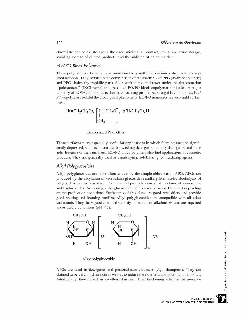
444 Oldenhove de Guertechin
ethoxylate nonionics: storage in the dark, minimal air contact, low temperature storage,avoiding storage of diluted products, and the addition of an antioxidant.
EO/PO Block Polymers
These polymeric surfactants have some similarity with the previously discussed alkoxy-lated alcohols. They consist in the combination of the assembly of PPG (hydrophobic part)and PEG chains (hydrophilic part). Such surfactants are known under the denomination‘‘poloxamers’’ (INCI name) and are called EO/PO block copolymer nonionics. A majorproperty of EO/PO nonionics is their low-foaming profile. As straight EO nonionics, EO/PO copolymers exhibit the cloud point phenomena. EO/PO nonionics are also mild surfac-tants.
These surfactants are especially useful for applications in which foaming must be signifi-cantly depressed, such as automatic dishwashing detergents, laundry detergents, and rinseaids. Because of their mildness, EO/PO block polymers also find applications in cosmeticproducts. They are generally used as emulsifying, solubilizing, or fluidizing agents.
Alkyl Polyglucosides
Alkyl polyglucosides are most often known by the simple abbreviation APG. APGs areproduced by the alkylation of short-chain glucosides resulting from acidic alcoholysis ofpolysaccharides such as starch. Commercial products consist of mixtures of mono-, di-,and triglucosides. Accordingly the glucosidic chain varies between 1.2 and 3 dependingon the production conditions. Surfactants of this class are good emulsifiers and providegood wetting and foaming profiles. Alkyl polyglucosides are compatible with all othersurfactants. They show good chemical stability at neutral and alkaline pH, and are impairedunder acidic conditions (pH �5).
APGs are used in detergents and personal-care cleansers (e.g., shampoos). They areclaimed to be very mild for skin as well as to reduce the skin irritation potential of anionics.Additionally, they impart an excellent skin feel. Their thickening effect in the presence
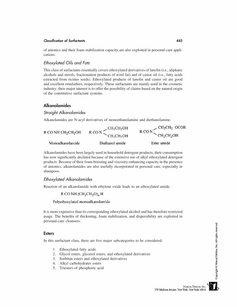
Classification of Surfactants 445
of anionics and their foam stabilization capacity are also exploited in personal-care appli-cations.
Ethoxylated Oils and Fats
This class of surfactants essentially covers ethoxylated derivatives of lanolin (i.e., aliphaticalcohols and sterols, fractionation products of wool fat) and of castor oil (i.e., fatty acidsextracted from ricinus seeds). Ethoxylated products of lanolin and castor oil are goodand excellent emulsifiers, respectively. These surfactants are mainly used in the cosmeticindustry; their major interest is to offer the possibility of claims based on the natural originof the constitutive surfactant systems.
Alkanolamides
Straight Alkanolamides
Alkanolamides are N-acyl derivatives of monoethanolamine and diethanolamine.
Alkanolamides have been largely used in household detergent products; their consumptionhas now significantly declined because of the extensive use of alkyl ethoxylated detergentproducts. Because of their foam-boosting and viscosity-enhancing capacity in the presenceof anionics, alkanolamides are also usefully incorporated in personal care, especially inshampoos.
Ethoxylated Alkanolamides
Reaction of an alkanolamide with ethylene oxide leads to an ethoxylated amide.
It is more expensive than its corresponding ethoxylated alcohol and has therefore restrictedusage. The benefits of thickening, foam stabilization, and dispersibility are exploited inpersonal-care cleansers.
Esters
In this surfactant class, there are five major subcategories to be considered:
1. Ethoxylated fatty acids2. Glycol esters, glycerol esters, and ethoxylated derivatives3. Sorbitan esters and ethoxylated derivatives4. Alkyl carbohydrates esters5. Triesters of phosphoric acid
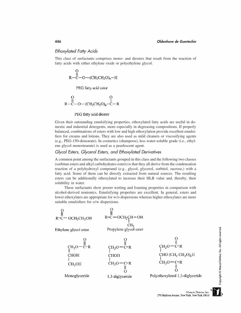
446 Oldenhove de Guertechin
Ethoxylated Fatty Acids
This class of surfactants comprises mono- and diesters that result from the reaction offatty acids with either ethylene oxide or polyethylene glycol.
Given their outstanding emulsifying properties, ethoxylated fatty acids are useful in do-mestic and industrial detergents, more especially in degreasing compositions. If properlybalanced, combinations of esters with low and high ethoxylation provide excellent emulsi-fiers for creams and lotions. They are also used as mild cleaners or viscosifying agents(e.g., PEG-150-distearate). In cosmetics (shampoos), less water-soluble grade (i.e., ethyl-ene glycol monostearate) is used as a pearlescent agent.
Glycol Esters, Glycerol Esters, and Ethoxylated Derivatives
A common point among the surfactants grouped in this class and the following two classes(sorbitan esters and alkyl carbohydrates esters) is that they all derive from the condensationreaction of a polyhydroxyl compound (e.g., glycol, glycerol, sorbitol, sucrose,) with afatty acid. Some of them can be directly extracted from natural sources. The resultingesters can be additionally ethoxylated to increase their HLB value and, thereby, theirsolubility in water.
These surfactants show poorer wetting and foaming properties in comparison withalcohol-derived nonionics. Emulsifying properties are excellent. In general, esters andlower ethoxylates are appropriate for w/o dispersions whereas higher ethoxylates are moresuitable emulsifiers for o/w dispersions.
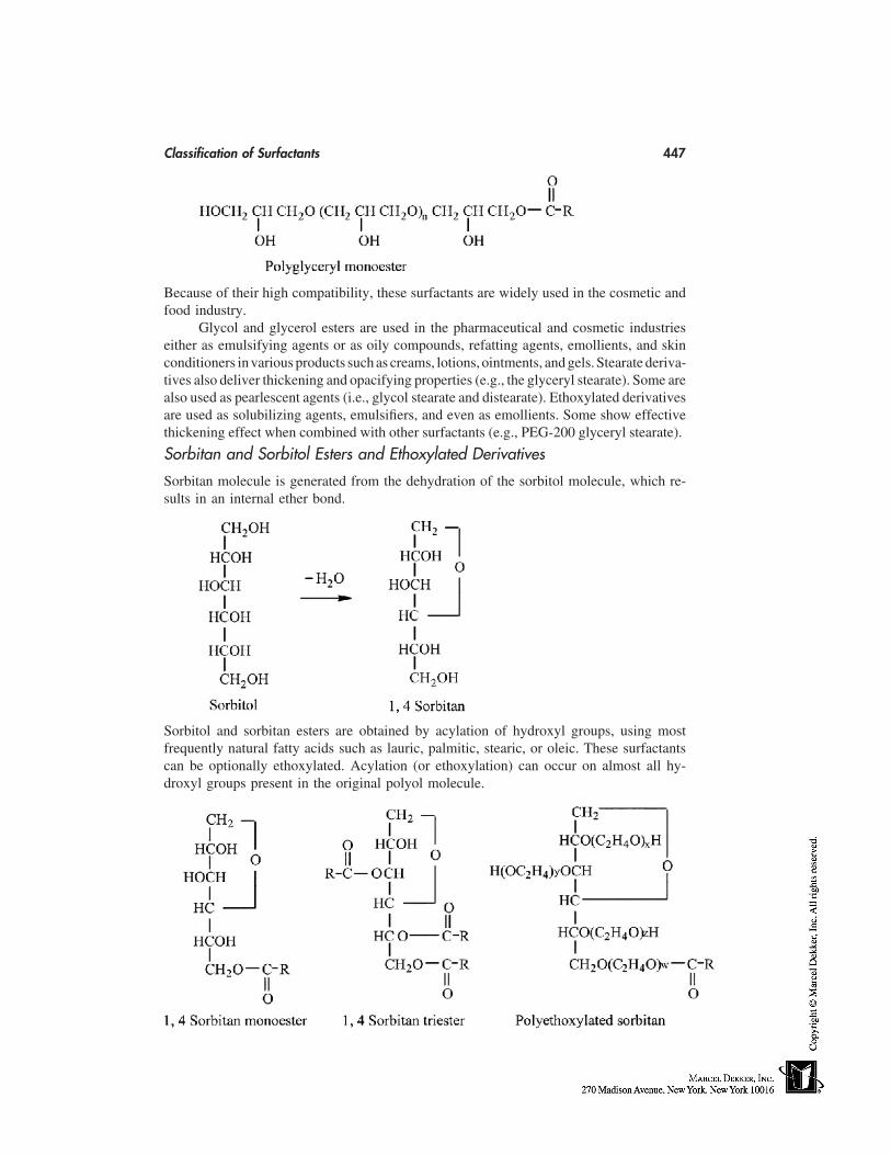
Classification of Surfactants 447
Because of their high compatibility, these surfactants are widely used in the cosmetic andfood industry.
Glycol and glycerol esters are used in the pharmaceutical and cosmetic industrieseither as emulsifying agents or as oily compounds, refatting agents, emollients, and skinconditioners in various products such as creams, lotions, ointments, and gels. Stearate deriva-tives also deliver thickening and opacifying properties (e.g., the glyceryl stearate). Some arealso used as pearlescent agents (i.e., glycol stearate and distearate). Ethoxylated derivativesare used as solubilizing agents, emulsifiers, and even as emollients. Some show effectivethickening effect when combined with other surfactants (e.g., PEG-200 glyceryl stearate).
Sorbitan and Sorbitol Esters and Ethoxylated Derivatives
Sorbitan molecule is generated from the dehydration of the sorbitol molecule, which re-sults in an internal ether bond.
Sorbitol and sorbitan esters are obtained by acylation of hydroxyl groups, using mostfrequently natural fatty acids such as lauric, palmitic, stearic, or oleic. These surfactantscan be optionally ethoxylated. Acylation (or ethoxylation) can occur on almost all hy-droxyl groups present in the original polyol molecule.
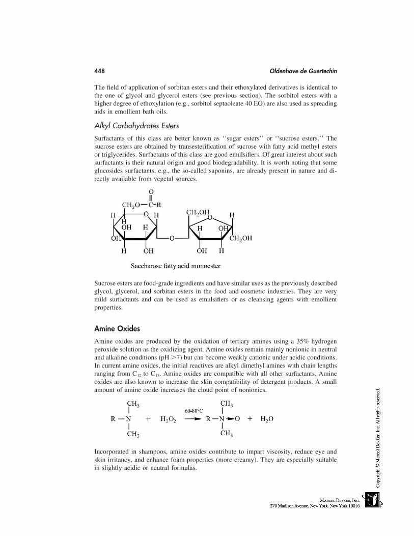
448 Oldenhove de Guertechin
The field of application of sorbitan esters and their ethoxylated derivatives is identical tothe one of glycol and glycerol esters (see previous section). The sorbitol esters with ahigher degree of ethoxylation (e.g., sorbitol septaoleate 40 EO) are also used as spreadingaids in emollient bath oils.
Alkyl Carbohydrates Esters
Surfactants of this class are better known as ‘‘sugar esters’’ or ‘‘sucrose esters.’’ Thesucrose esters are obtained by transesterification of sucrose with fatty acid methyl estersor triglycerides. Surfactants of this class are good emulsifiers. Of great interest about suchsurfactants is their natural origin and good biodegradability. It is worth noting that someglucosides surfactants, e.g., the so-called saponins, are already present in nature and di-rectly available from vegetal sources.
Sucrose esters are food-grade ingredients and have similar uses as the previously describedglycol, glycerol, and sorbitan esters in the food and cosmetic industries. They are verymild surfactants and can be used as emulsifiers or as cleansing agents with emollientproperties.
Amine Oxides
Amine oxides are produced by the oxidation of tertiary amines using a 35% hydrogenperoxide solution as the oxidizing agent. Amine oxides remain mainly nonionic in neutraland alkaline conditions (pH �7) but can become weakly cationic under acidic conditions.In current amine oxides, the initial reactives are alkyl dimethyl amines with chain lengthsranging from C12 to C18. Amine oxides are compatible with all other surfactants. Amineoxides are also known to increase the skin compatibility of detergent products. A smallamount of amine oxide increases the cloud point of nonionics.
Incorporated in shampoos, amine oxides contribute to impart viscosity, reduce eye andskin irritancy, and enhance foam properties (more creamy). They are especially suitablein slightly acidic or neutral formulas.
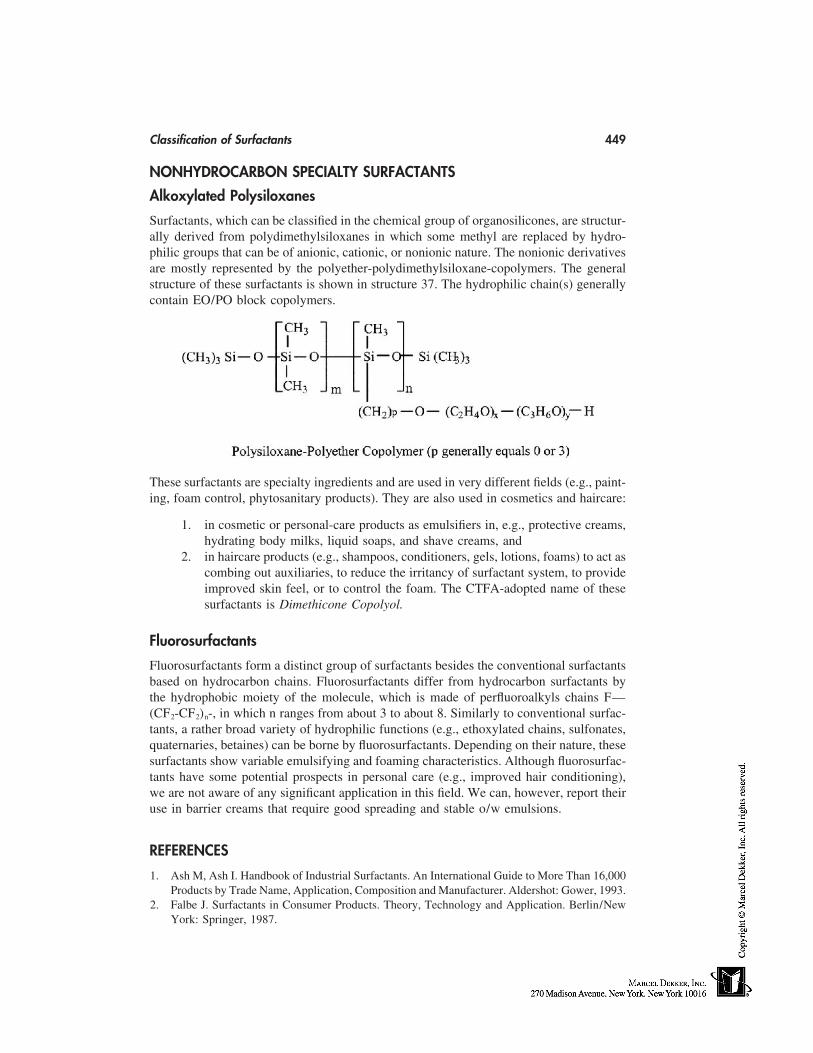
Classification of Surfactants 449
NONHYDROCARBON SPECIALTY SURFACTANTS
Alkoxylated Polysiloxanes
Surfactants, which can be classified in the chemical group of organosilicones, are structur-ally derived from polydimethylsiloxanes in which some methyl are replaced by hydro-philic groups that can be of anionic, cationic, or nonionic nature. The nonionic derivativesare mostly represented by the polyether-polydimethylsiloxane-copolymers. The generalstructure of these surfactants is shown in structure 37. The hydrophilic chain(s) generallycontain EO/PO block copolymers.
These surfactants are specialty ingredients and are used in very different fields (e.g., paint-ing, foam control, phytosanitary products). They are also used in cosmetics and haircare:
1. in cosmetic or personal-care products as emulsifiers in, e.g., protective creams,hydrating body milks, liquid soaps, and shave creams, and
2. in haircare products (e.g., shampoos, conditioners, gels, lotions, foams) to act ascombing out auxiliaries, to reduce the irritancy of surfactant system, to provideimproved skin feel, or to control the foam. The CTFA-adopted name of thesesurfactants is Dimethicone Copolyol.
Fluorosurfactants
Fluorosurfactants form a distinct group of surfactants besides the conventional surfactantsbased on hydrocarbon chains. Fluorosurfactants differ from hydrocarbon surfactants bythe hydrophobic moiety of the molecule, which is made of perfluoroalkyls chains F—(CF2-CF2)n-, in which n ranges from about 3 to about 8. Similarly to conventional surfac-tants, a rather broad variety of hydrophilic functions (e.g., ethoxylated chains, sulfonates,quaternaries, betaines) can be borne by fluorosurfactants. Depending on their nature, thesesurfactants show variable emulsifying and foaming characteristics. Although fluorosurfac-tants have some potential prospects in personal care (e.g., improved hair conditioning),we are not aware of any significant application in this field. We can, however, report theiruse in barrier creams that require good spreading and stable o/w emulsions.
REFERENCES
1. Ash M, Ash I. Handbook of Industrial Surfactants. An International Guide to More Than 16,000Products by Trade Name, Application, Composition and Manufacturer. Aldershot: Gower, 1993.
2. Falbe J. Surfactants in Consumer Products. Theory, Technology and Application. Berlin/NewYork: Springer, 1987.
450 Oldenhove de Guertechin
3. Lange KR. Detergents and Cleaners: A Handbook for Formulators. München: Hanser Publish-ers, 1994.
4. Porter MR. Handbook of Surfactants. London: Blackie Academic & Professional, 1991.5. Rieger MM. Surfactant Encyclopedia. 2nd ed. Carol Stream: Allured Publishing, 1996.6. Anonymous. Surfactant Encyclopedia. 2nd ed. Cosmetic & Toiletries. Carol Stream: Allured
Publishing Corp., 1989; 104:67–110.

38
UV Filters
Stanley B. LevyUniversity of North Carolina School of Medicine at Chapel Hill, Chapel Hill,North Carolina, and Revlon Research Center, Edison, New Jersey
INTRODUCTION
The presence of ultraviolet (UV) filters in skincare and cosmetic products represents akey benefit that cosmetics can provide consumers. The hazards of UV light exposure arewell known. It is estimated that the incidence of nonmelanoma skin cancer in the UnitedStates exceeds one million cases per year [1]; UV-induced or photoaging accounts for80% to 90% of visible skin aging [2]. UV radiation damages the skin by both direct effectson DNA and indirectly on the skin’s immune system [3].
In animal models, sunscreens prevent the formation of squamous cell carcinomasof the skin [4]. The regular use of sunscreens has been shown to reduce the numberof actinic or precancerous keratosis [5] and solar elastosis [6]. Sunscreens also preventimmunosuppression [7]. Double-blind photoaging studies show consistent improvementin the ‘‘untreated’’ control groups partly because of the use of sunscreens by all studysubjects [8].
The cosmetic formulator has an expanding menu of active sunscreen ingredients forincorporation into a variety of cosmetic formulations. Selection is restricted by regulatoryagencies in the country in which the final product is to be marketed. This chapter willconcentrate on reviewing available UV filters.
DEFINITIONS
Ultraviolet radiation (UVR) reaching the Earth’s surface can be divided into UVB (290–320 nm) and UVA (320–400 nm). UVA can be further subdivided into UVA I (340–400nm), or far UVA, and UVA II (320–340 nm), or near UVA.
The sun protection factor (SPF) is defined as the dose of UVR required to produceone minimal erythema dose (MED) on protected skin after application of 2 mg/cm2 ofproduct divided by the UVR to produce one MED on unprotected skin. A water-resistantproduct maintains the SPF level after 40 minutes of water immersion. A very water-resis-tant or waterproof product is tested after 80 minutes of water immersion. If the SPF levelis diminished by immersion, a separate SPF level may be listed. A broad-spectrum orfull-spectrum sunscreen provides both UVB and UVA protection. Ideally this includesboth UVA I and UVA II coverage.
451
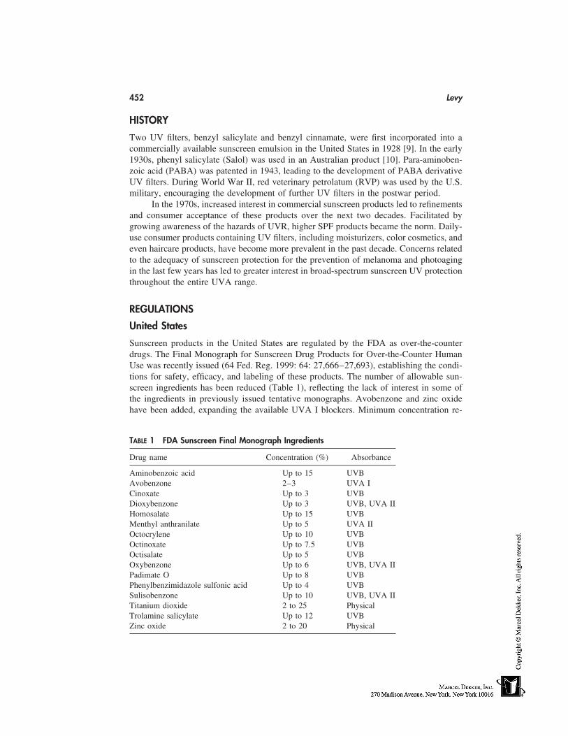
452 Levy
HISTORY
Two UV filters, benzyl salicylate and benzyl cinnamate, were first incorporated into acommercially available sunscreen emulsion in the United States in 1928 [9]. In the early1930s, phenyl salicylate (Salol) was used in an Australian product [10]. Para-aminoben-zoic acid (PABA) was patented in 1943, leading to the development of PABA derivativeUV filters. During World War II, red veterinary petrolatum (RVP) was used by the U.S.military, encouraging the development of further UV filters in the postwar period.
In the 1970s, increased interest in commercial sunscreen products led to refinementsand consumer acceptance of these products over the next two decades. Facilitated bygrowing awareness of the hazards of UVR, higher SPF products became the norm. Daily-use consumer products containing UV filters, including moisturizers, color cosmetics, andeven haircare products, have become more prevalent in the past decade. Concerns relatedto the adequacy of sunscreen protection for the prevention of melanoma and photoagingin the last few years has led to greater interest in broad-spectrum sunscreen UV protectionthroughout the entire UVA range.
REGULATIONS
United States
Sunscreen products in the United States are regulated by the FDA as over-the-counterdrugs. The Final Monograph for Sunscreen Drug Products for Over-the-Counter HumanUse was recently issued (64 Fed. Reg. 1999: 64: 27,666–27,693), establishing the condi-tions for safety, efficacy, and labeling of these products. The number of allowable sun-screen ingredients has been reduced (Table 1), reflecting the lack of interest in some ofthe ingredients in previously issued tentative monographs. Avobenzone and zinc oxidehave been added, expanding the available UVA I blockers. Minimum concentration re-
TABLE 1 FDA Sunscreen Final Monograph Ingredients
Drug name Concentration (%) Absorbance
Aminobenzoic acid Up to 15 UVBAvobenzone 2–3 UVA ICinoxate Up to 3 UVBDioxybenzone Up to 3 UVB, UVA IIHomosalate Up to 15 UVBMenthyl anthranilate Up to 5 UVA IIOctocrylene Up to 10 UVBOctinoxate Up to 7.5 UVBOctisalate Up to 5 UVBOxybenzone Up to 6 UVB, UVA IIPadimate O Up to 8 UVBPhenylbenzimidazole sulfonic acid Up to 4 UVBSulisobenzone Up to 10 UVB, UVA IITitanium dioxide 2 to 25 PhysicalTrolamine salicylate Up to 12 UVBZinc oxide 2 to 20 Physical
UV Filters 453
quirements have been dropped, providing that the concentration of each active ingredientis sufficient to contribute a minimum SPF of not less than 2 to a finished product. Asunscreen product must have a minimum SPF of not less than the number of active sun-screen ingredients used in combination multiplied by 2. Products with SPF values above30 are allowed, but the SPF declaration for sunscreens with SPF values above 30 arelimited to SPF 30 plus. The term ‘‘sunblock’’ is now prohibited. It was previously allowedfor products that contained titanium dioxide. Consideration of labeling and testing proce-dures for UVA protection was deferred and will be addressed in the future.
Europe
In Europe, sunscreen products are considered to be cosmetics, their function being toprotect the skin from sunburn. The Third Amendment of the European Economic Commu-nity (EEC) Directive provides a definition and lists the UV filters that cosmetic productsmay contain. This list is divided into two parts. Table 2 lists UV filters that are fullypermitted updated through the 23rd commission directive of September 3, 1998. Table 3currently lists the three UV filters that are provisionally permitted through June 30, 1999.The numbers referenced with ‘‘S’’ indicate COLIPA numbers (The European Toiletryand Perfumery Association). Unlike the U.S. FDA Monograph, the EEC Directive doesnot list physical UV filters despite their being used in products to enhance protection.
Australia
In 1992, sunscreens were declared to be drugs in Australia. The latest edition of AustralianStandard 2604 was published in 1993 as a joint publication of Australia and New Zealand.Sunscreen products are classified as either primary or secondary, depending on whetherthe primary function of the designated product is to protect from UVR as opposed to aproduct with a primary cosmetic purpose. SPF designations greater than 15 are not permit-ted (SPF 15� represents the maximum designation). In general, Australian ApprovedNames (AAN) for allowed active sunscreen ingredients are the same as FDA drug nomen-clature with few differences.
Other Countries
Most non-EEC European countries follow the EEC Directive. Many other countries followU.S. trends with their own provisions. In Japan, sunscreens are classified as cosmetics.Regulations for each individual country need to be consulted for selection of the variousUV filters for incorporation into a sunscreen product to be marketed in a given jurisdiction.
MECHANISM OF ACTION
UV filters have been traditionally divided into chemical absorber and physical blockersbased on their mechanism of action. Chemical sunscreens are generally aromatic com-pounds conjugated with a carbonyl group [11]. These chemicals absorb high-intensity UVrays with excitation to a higher energy state. The energy lost results in conversion of theremaining energy into longer lower-energy wavelengths with return to ground state. Theevolution of modern sunscreen chemicals represents a prototype study in the use of struc-
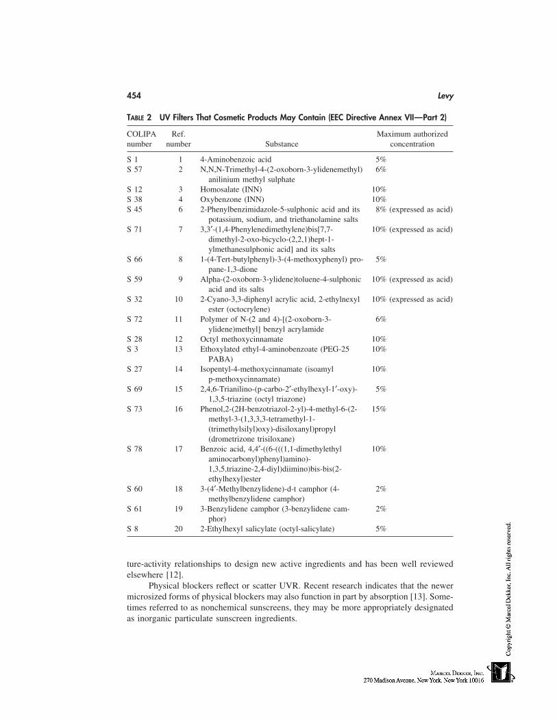
454 Levy
TABLE 2 UV Filters That Cosmetic Products May Contain (EEC Directive Annex VII—Part 2)
COLIPA Ref. Maximum authorizednumber number Substance concentration
S 1 1 4-Aminobenzoic acid 5%S 57 2 N,N,N-Trimethyl-4-(2-oxoborn-3-ylidenemethyl) 6%
anilinium methyl sulphateS 12 3 Homosalate (INN) 10%S 38 4 Oxybenzone (INN) 10%S 45 6 2-Phenylbenzimidazole-5-sulphonic acid and its 8% (expressed as acid)
potassium, sodium, and triethanolamine saltsS 71 7 3,3′-(1,4-Phenylenedimethylene)bis[7,7- 10% (expressed as acid)
dimethyl-2-oxo-bicyclo-(2,2,1)hept-1-ylmethanesulphonic acid] and its salts
S 66 8 1-(4-Tert-butylphenyl)-3-(4-methoxyphenyl) pro- 5%pane-1,3-dione
S 59 9 Alpha-(2-oxoborn-3-ylidene)toluene-4-sulphonic 10% (expressed as acid)acid and its salts
S 32 10 2-Cyano-3,3-diphenyl acrylic acid, 2-ethylnexyl 10% (expressed as acid)ester (octocrylene)
S 72 11 Polymer of N-(2 and 4)-[(2-oxoborn-3- 6%ylidene)methyl] benzyl acrylamide
S 28 12 Octyl methoxycinnamate 10%S 3 13 Ethoxylated ethyl-4-aminobenzoate (PEG-25 10%
PABA)S 27 14 Isopentyl-4-methoxycinnamate (isoamyl 10%
p-methoxycinnamate)S 69 15 2,4,6-Trianilino-(p-carbo-2′-ethylhexyl-1′-oxy)- 5%
1,3,5-triazine (octyl triazone)S 73 16 Phenol,2-(2H-benzotriazol-2-yl)-4-methyl-6-(2- 15%
methyl-3-(1,3,3,3-tetramethyl-1-(trimethylsilyl)oxy)-disiloxanyl)propyl(drometrizone trisiloxane)
S 78 17 Benzoic acid, 4,4′-((6-(((1,1-dimethylethyl 10%aminocarbonyl)phenyl)amino)-1,3,5,triazine-2,4-diyl)diimino)bis-bis(2-ethylhexyl)ester
S 60 18 3-(4′-Methylbenzylidene)-d-t camphor (4- 2%methylbenzylidene camphor)
S 61 19 3-Benzylidene camphor (3-benzylidene cam- 2%phor)
S 8 20 2-Ethylhexyl salicylate (octyl-salicylate) 5%
ture-activity relationships to design new active ingredients and has been well reviewedelsewhere [12].
Physical blockers reflect or scatter UVR. Recent research indicates that the newermicrosized forms of physical blockers may also function in part by absorption [13]. Some-times referred to as nonchemical sunscreens, they may be more appropriately designatedas inorganic particulate sunscreen ingredients.
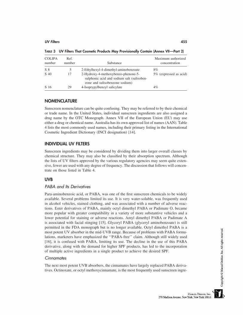
UV Filters 455
TABLE 3 UV Filters That Cosmetic Products May Provisionally Contain (Annex VII—Part 2)
COLIPA Ref. Maximum authorizednumber number Substance concentration
S 8 5 2-Ethylhexyl-4-dimethyl-aminobenzoate 8%S 40 17 2-Hydroxy-4-methoxybenzo-phenone-5- 5% (expressed as acid)
sulphonic acid and sodium salt (sulisoben-zone and sulisobenzone sodium)
S 16 29 4-Isoprypylbenzyl salicylate 4%
NOMENCLATURE
Sunscreen nomenclature can be quite confusing. They may be referred to by their chemicalor trade name. In the United States, individual sunscreen ingredients are also assigned adrug name by the OTC Monograph. Annex VII of the European Union (EU) may useeither a drug or chemical name. Australia has its own approved list of names (AAN). Table4 lists the most commonly used names, including their primary listing in the InternationalCosmetic Ingredient Dictionary (INCI designation) [14].
INDIVIDUAL UV FILTERS
Sunscreen ingredients may be considered by dividing them into larger overall classes bychemical structure. They may also be classified by their absorption spectrum. Althoughthe lists of UV filters approved by the various regulatory agencies may seem quite exten-sive, fewer are used with any degree of frequency. The discussion that follows will concen-trate on those listed in Table 4.
UVB
PABA and Its Derivatives
Para-aminobenzoic acid, or PABA, was one of the first sunscreen chemicals to be widelyavailable. Several problems limited its use. It is very water-soluble, was frequently usedin alcohol vehicles, stained clothing, and was associated with a number of adverse reac-tions. Ester derivatives of PABA, mainly octyl dimethyl PABA or Padimate O, becamemore popular with greater compatibility in a variety of more substantive vehicles and alower potential for staining or adverse reactions. Amyl dimethyl PABA or Padimate Ais associated with facial stinging [15]. Glyceryl PABA (glyceryl aminobenzoate) is stillpermitted in the FDA monograph but is no longer available. Octyl dimethyl PABA is amost potent UV absorber in the mid-UVB range. Because of problems with PABA formu-lations, marketers have emphasized the ‘‘PABA-free’’ claim. Although still widely used[16], it is confused with PABA, limiting its use. The decline in the use of this PABAderivative, along with the demand for higher SPF products, has led to the incorporationof multiple active ingredients in a single product to achieve the desired SPF.
Cinnamates
The next most potent UVB absorbers, the cinnamates have largely replaced PABA deriva-tives. Octinoxate, or octyl methoxycinnamate, is the most frequently used sunscreen ingre-
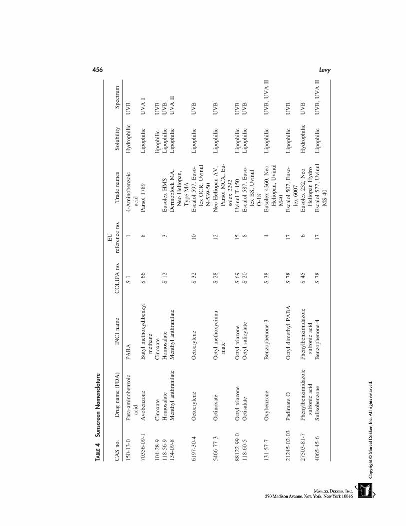
456 Levy
TABL
E4
Suns
cree
nN
omen
clat
ure
EU
CA
Sno
.D
rug
nam
e(F
DA
)IN
CI
nam
eC
OL
IPA
no.
refe
renc
eno
.T
rade
nam
esSo
lubi
lity
Spec
trum
150-
13-0
Para
-am
inob
enzo
icPA
BA
S1
14-
Am
inob
enzo
icH
ydro
phili
cU
VB
acid
acid
7035
6-09
-1A
vobe
nzon
eB
utyl
met
hoxy
dibe
nzyl
S66
8Pa
rsol
1789
Lip
ophi
licU
VA
Im
etha
ne10
4-28
-9C
inox
ate
Cin
oxat
elip
ophi
licU
VB
118-
56-9
Hom
osal
ate
Hom
osal
ate
S12
3E
usol
exH
MS
Lip
ophi
licU
VB
134-
09-8
Men
thyl
anth
rani
late
Men
thyl
anth
rani
late
Der
mob
lock
MA
,L
ipop
hilic
UV
AII
Neo
Hel
iopa
n,T
ype
MA
6197
-30-
4O
ctoc
ryle
neO
ctoc
ryle
neS
3210
Esc
alol
597,
Eus
o-L
ipop
hilic
UV
Ble
xO
CR
,U
vinu
lN
-539
-50
5466
-77-
3O
ctin
oxat
eO
ctyl
met
hoxy
cinn
a-S
2812
Neo
Hel
iopa
nA
V,
Lip
ophi
licU
VB
mat
ePa
rsol
MC
X,
Eu-
sole
x22
9288
122-
99-0
Oct
yltr
iazo
neO
ctyl
tria
zone
S69
15U
vinu
lT
-150
Lip
ophi
licU
VB
118-
60-5
Oct
isal
ate
Oct
ylsa
licyl
ate
S20
8E
scal
ol58
7,E
uso-
Lip
ophi
licU
VB
lex
BS,
Uvi
nul
O-1
813
1-57
-7O
xybe
nzon
eB
enzo
phen
one-
3S
384
Eus
olex
4360
,N
eoL
ipop
hilic
UV
B,
UV
AII
Hel
iopa
n,U
vinu
lM
4021
245-
02-0
3Pa
dim
ate
OO
ctyl
dim
ethy
lPA
BA
S78
17E
scal
ol50
7,E
uso-
Lip
ophi
licU
VB
lex
6007
2750
3-81
-7Ph
enyl
benz
imid
azol
ePh
enyl
benz
imid
azol
eS
456
Eus
olex
232,
Neo
Hyd
roph
ilic
UV
Bsu
lfon
icac
idsu
lfon
icac
idH
elio
pan
Hyd
ro40
65-4
5-6
Sulis
oben
zone
Ben
zoph
enon
e-4
S78
17E
scal
ol57
7,U
vinu
lL
ipop
hilic
UV
B,
UV
AII
MS
40
UV Filters 457
dient [16]. Octyl or ethylhexyl methoxycinnamate is an order of magnitude less potentthan Padimate O and requires additional UVB absorbers to achieve higher SPF levels ina final product. Cinoxate (Ethoxy-ethyl-p-methoxycinnamate) is less widely used. Whena water-soluble cinnamate is indicated in a formulation, diethanolamine (DEA) methoxy-cinnamate may be used.
Salicylates
Salicylates are weaker UVB absorbers. They have a long history of use but were sup-planted by the more efficient PABA and cinnamate derivatives. They are generally usedto augment other UVB absorbers. With the trend to higher SPFs, more octyl salicylate(ethylhexyl salicylate) is being used followed by homomenthyl salicylate. Both materialshave the ability to solubilize oxybenzone and avobenzone. Trolamine or triethanolamine(TEA) salicylate has good water solubility.
Camphor Derivatives
Not approved by the FDA for use in the United States, there are six camphor derivativesapproved in Europe. 4-methyl-benbenzylidene camphor is the most widely used.
Octocrylene
2-Ethylhexyl-2-cyano-3,3 diphenylacrylate, or octocrylene, is chemically related to cinna-mates. It can be used to boost SPF and improve water resistance in a given formulation.Octocrylene is photostabile and can improve the photostability of other sunscreens. It isvery expensive and can present difficulties in formulation.
Phenylbenzimidazole Sulfonic Acid
Phenylbenzimadazole sulfonic acid is a water-soluble UVB absorber that can be used inthe water phase of emulsion systems, in contrast to most oil-soluble sunscreen ingredients,allowing for a less greasy, more aesthetically pleasing formulation, such as a daily-usemoisturizer containing sunscreen. Phenylbenzimidazole sulfonic acid boosts the SPF oforganic and inorganic sunscreens. It can also be used in clear gels because of its watersolubility.
UVA
Benzophenones
Although oxybenzone or benzophenone-3 absorbs most efficiently in the UVB range, ab-sorption extends well into the UVA II range. It is used primarily as a UVA absorber, butboosts SPF values in combination with other UVB absorbers. Oxybenzone is suppliedas a solid material, has poor solubility, and has a relatively low extinction coefficient.Sulisobenzone or benzophenone-4 is water-soluble, somewhat unstable, and used withless frequency.
Menthyl Anthranilate
Anthranilates are weak UVB filters and absorb mainly in the near UVA portion of thespectrum. They are less effective than benzophenones in this range and are less widelyused.
458 Levy
Butylmethoxydibenzoylmethane
Avobenzone, or Parsol 1789, has only recently been approved by the FDA for use inOTC sunscreens in the United States, having been used quite extensively in Europe forconsiderably longer. It provides strong absorption in the UVA I range with peak absorptionat 360 nm. Because an agreed-upon standard for measuring UVA protection in the UnitedStates does not exist, a minimum-use concentration has been set at 2% with a maximumof 3%.
Avobenzone should not be confused with isopropyl dibenzoylmethane (Eusolex8020), which had previously been available in Europe. The high incidence of adversephotosensitivity reported with the combination of isopropyldibenzoylmethane and methyl-benzylidene camphor by coupled reactions in the late 1980s led to a decrease in its usein commercial [17]. In 1993 its production was discontinued and it is no longer listed inAnnex VII. Reported sensitivity to butylmethoxydibenzoylmethane was on the basis ofcross-reactivity to isopropyl dibenzoylmethane. Isolated allergy to butylmethoxydiben-zoylmethane is rare [17].
Photostability refers to the ability of a molecule to remain intact with irradiation.Photostability is potentially a problem with all UV filters. This issue has been raisedspecifically with avobenzone [18], with photolysis shown in a specially designed in vitrosystem [19] that simultaneously irradiates and measures transmittance in situ. This effectmay degrade other sunscreens in a formulation. The relevance of this testing to the invivo situation remains unclear. Overall formulation may be critical in this regard.
Tetraphthalydine Dicamphor Sulfonic Acid
3,3′-(1,4-phenylenedimethylene)bis[7,7-dimethyl-2-oxo-bicyclo-(2,2,1)hept-1-yl]meth-anesulfonic acid (EU Ref. No. 7) or Mexoryl SX is a UVA blocker more recently availablein Europe with comparable efficacy to avobenzone [20].
Physical Blockers
Some of the original sunblocks were opaque formulations reflecting or scattering UVR.Color cosmetics containing a variety of inorganic pigments function in this fashion. Tita-nium dioxide and zinc oxide are chemically inert and protect through the full spectrumof UVR. They offer significant advantages. Poor cosmetic acceptance limited the wide-spread use of these two ingredients until recently, when microsized forms have becomeavailable. By decreasing particle size of these materials to a microsize or ultrafine gradeit is less visible on the skin surface.
Micropigmentary sunblocks function differently than opaque sunblocks of pig-mented color cosmetics by absorbing and not simply reflecting or scattering UVR [13].By varying and mixing particle sizes, differing levels of photoprotection are achievedthroughout the UV spectrum. In addition to avobenzone, micropigmentary TiO2 and ZnOoffer the best available protection in the UVA II range.
Photoreactivity has been raised as an issue with these materials. Both TiO2 and ZnOare semiconductors potentially absorbing light and generating reactive species [21]. Theseeffects have been shown in vitro [22]. Coating these materials reduces their photochemicalreactivity. The in vivo relevance of these effects has not been shown and both materialshave a long history of safe use.
UV Filters 459
Titanium Dioxide
TiO2 was the first micropigment extensively used. Advantages include a broad spectrumof protection and inability to cause contact dermatitis. The use of rutile as opposed toanatase crystal forms of titanium dioxide lessens photoactivity. Newer materials are am-phiphilic, designed to be dispersed in both water- and oil-emulsion phases. Particle sizeand uniformity of dispersion is key to achieving SPF. Primary particle size may be 10 to15 nm with secondary particle assembly to 100 nm. Particle size needs to be less than200 nm to achieve transparency.
Despite advances in the technology and understanding of these materials, whiteningremains a problem secondary to pigment residue. Adding other pigments simulating flesh-tones may partially camouflage this effect. The net effect may be that the user is inclinedto make a less heavy application of product, effectively lowering SPF [23]. ‘‘Hybrid’’formulations using a combination of chemical absorbers with inorganic particulates mayrepresent a practical compromise.
Zinc Oxide
Zinc oxide was only recently approved as an active sunscreen agent for the FDA OTCSunscreen Monograph. Reduced to a particle size of less than 200 nm, light scattering isminimized and the particles appear transparent in thin films [24]. ZnO has a refractiveindex of 1.9, as opposed to 2.6 for TiO2, and therefore causes less whitening than TiO2.ZnO may attenuate UVR more effectively in the UVA I range [25]. Microfine TiO2 atan equal concentration offers somewhat more protection in the UVB range.
ADVERSE REACTIONS—TOXICITY
In a longitudinal prospective study of 603 subjects applying daily either an SPF 15�broad-spectrum sunscreen containing octyl methoxycinnamate and avobenzone or a vehi-cle cream, 19% developed an adverse reaction [26]. Interestingly, the rates of reaction toboth the active and vehicle creams were similar, emphasizing the importance of excipientingredients in the vehicle. The majority of reactions were irritant in nature. Not surpris-ingly, a disproportionate 50% of the reacting subjects were atopic. Less than 10% of thereactions were allergic, with none of the subjects patch tested actually found to be allergicto an individual sunscreen ingredient.
Subjective irritation associated with burning or stinging without objective erythemafrom some organic UV filters [27] is the most frequent sensitivity complaint associatedwith sunscreen use. This is most frequently experienced in the eye area. Longer lastingobjective irritant contact dermatitis may be difficult to distinguish from true allergic con-tact dermatitis. In a postmarket evaluation of sunscreen sensitivity complaints in 57 pa-tients, 20 of the patients had short-lasting symptoms, 26 long-lasting, and 11 mixed orborderline symptoms [28]. Half of the patients were patch and photopatch tested, and onlythree showed positive reactions to sunscreen ingredients.
Contact and photocontact sensitivity to individual sunscreen ingredients has beenextensively reviewed [17]. Considering their widespread use, the number of documentedallergic reactions is not high [29]. PABA and PABA esters accounted for many of theearly reported reactions, but with a decrease in their use reactions to benzophenones maybe increasing [30]. Reactions to dibenzoylmethanes have previously been discussed. Fra-
460 Levy
grances, preservatives, and other excipients account for a large number of the allergicreactions seen [17].
Virtually all sunscreen ingredients reported to cause contact allergy may be photoal-lergens [31]. Although still relatively uncommon, sunscreen actives seem to have becomethe leading cause of photocontact allergic reactions [32,33]. Individuals with pre-existingeczematous conditions have a significant predisposition to sensitization associated withtheir impaired cutaneous barrier. The majority of individuals who develop photocontactdermatitis to sunscreens are patients with photodermatides [17].
CONCLUSION
A limited menu of UV filters for incorporation into sunscreen products is available to theformulating chemist, depending on regulatory requirements in an individual country orjurisdiction. With the demand for higher SPFs, the trend has been to use more individualand a wider variety of agents in newer products. Recent research in sunscreen efficacyhas emphasized the need for products protecting against the full UV spectrum with alimited number of available agents. Regulatory agencies are very slow to approve newingredients.
Sunscreen efficacy remains very dependent on vehicle formulation. Solvents andemollients can have a profound effect on the strength of UV absorbance by the activeingredients and at which wavelengths they absorb [34]. Film formers and emulsifiers deter-mine the uniformity and thickness of the film formed on the skin surface, which in turndetermines SPF level, durability, and water resistance [35]. Lastly, product aesthetics playa large role in product acceptance, particularly with sunscreens being incorporated intodaily-use cosmetics. These constraints provide the sunscreen formulator with significantchallenges in developing new and improved formulations.
REFERENCES
1. Weinstock MA. Death from skin cancer among the elderly: epidemiological patterns. ArchDermatol 1997; 133:1207–1209.
2. Yaar M, Gilchrest BA. Aging versus photoaging: postulated mechanisms and effectors. J In-vest Dermatol Symp Proc 1998; 3:47–51.
3. Naylor MF, Farmer KC. The case for sunscreens: a review of their use in preventing actinicdamage and neoplasia. Arch Dermatol 1997; 133:1146–1154.
4. Gurish MF, Roberts LK, Krueger GG, et al. The effect of various sunscreen agents on skindamage and the induction of tumor susceptibility in mice subjected to ultraviolet irradiation.J Invest Dermatol 1975; 65:543–546.
5. Thompson SC, Jolley D, Marks R. Reduction of solar keratoses by regular sunscreen use. NEngl J Med 1993; 329:1147–1151.
6. Boyd AS, Naylor M, Cameron GS, et al. The effects of chronic sunscreen use on the histologicchanges of dermatoheliosis. J Am Acad Dermatol 1995; 33:941–946.
7. Roberts LK, Beasley DG. Commercial sunscreen lotions prevent ultraviolet-radiation–inducedimmune suppression of contact hypersensitivity. J Invest Dermatol 1995; 105:339–344.
8. Stiller MJ, Bartolone J, Stern R, Smith S, Kollias N, Gillies R, Drake LA. Topical 8% glycolicacid and 8% L-lactic acid creams for the treatment of photodamaged skin: a double-blindvehicle-controlled clinical trial. Arch Dermatol 1996; 132:631–636.
9. Shaath NA. Evolution of modern sunscreen chemicals. In: Lowe NJ, Shaath NA, Pathak MA,
UV Filters 461
eds. Sunscreens: Development, Evaluation, and Regulatory Aspects. 2d ed. New York: MarcelDekker, 1997:3–31.
10. Rebut R. The sunscreen industry in Europe: past, present, and future. In: Lowe NJ, ShaathNA, eds. Sunscreens: Development, Evaluation, and Regulatory Aspects. New York: MarcelDekker, 1990:161–178.
11. Shaath NA. The chemistry of sunscreens. Cosmet Toilet 1986; 101:55–70.12. Shaath NA. On the theory of ultraviolet absorption by sunscreen chemicals. J Soc Cosmet
Chem 1987; 82:193.13. Sayre RM, Killias N, Roberts RL, et al. Physical sunscreens. J Soc Cosmet Chem 1990; 41:
103–109.14. Wenninger JA, McEwen GN Jr, eds. International Cosmetic Ingredient Dictionary and Hand-
book. 7th ed. Washington, DC: The Cosmetic, Toiletry, and Fragrance Association, 1977.15. Frosch PJ, Kligman AM. A method for appraising the stinging capacity of topically applied
substances. J Soc Cosmet Chem 1977; 28:197.16. Steinberg DC. Sunscreen encyclopedia regulatory update. Cosmet Toilet 1996; 111:77–86.17. Schauder S, Ippen H. Contact and photocontact sensitivity to sunscreens. Review of a 15-year
experience and of the literature. Contact Derm 1997; 37(5):221–232.18. Deflandre A, Lang G. Photostability assessment of sunscreens. Benzylidene camphor and di-
benzoylmethane derivatives. Int J Cosmet Sci 1988; 10:53–62.19. Sayre RM, Dowdy JC. Avobenzone and the photostability of sunscreen products. Presented
at the 7th Annual Meeting of the Photomedicine Society. Orlando, February 26, 1998.20. Chardoon A, Moyal D, Hourseau C. Persistent pigment-darkening response as a method for
evaluation of ultraviolet A protection assays. In: Lowe NJ, Shaath NA, Pathak MA, eds. Sun-screens: Development, Evaluation, and Regulatory Aspects. 2d ed. New York: Marcel Dekker,1997:559–581.
21. Murphy GM. Sunblocks: mechanisms of action. Photodermatol Photoimmunol Photomed1999; 15:34–36.
22. Wamer WG, Yin JJ, Wei RR. Oxidative damage to nucleic acids photosensitized by titaniumdioxide. Free Radical Biol Med 197; 23:851–858.
23. Diffey BL, Grice J. The influence of sunscreen type on photoprotection. Br J Dermatol 1977;137:103–105.
24. Fairhurst D, Mitchnik MA. Particulate sun blocks: general principles. In: Lowe NJ, ShaathNA, Pathak MA, eds. Sunscreens: Development, Evaluation, and Regulatory Aspects. 2d ed.New York: Marcel Dekker, 1997:313–352.
25. Mitchnick MA, Fairhurst D, Pinnell SR. Microfine zinc oxide (Z-Cote) as a photostable UVA/UVB sunblock agent. J Am Acad Dermatol 1999; 40:85–90.
26. Foley P, Nixon R, Marks R, et al. The frequency of reactions to sunscreens: results of alongitudinal population-based study on the regular use of sunscreens in Australia. Br J Derma-tol 1993; 128:512–518.
27. Levy SB. Sunscreens for photoprotection. Dermatologic Ther 1997; 4:59–71.28. Fischer T, Bergstrom K. Evaluation of customers’ complaints about sunscreen cosmetics sold
by the Swedish pharmaceutical company. Contact Derm 1991; 25:319–322.29. Dromgoole SH, Maibach HI. Sunscreening agent intolerance: contact and photocontact sensiti-
zation and contact urticaria. J Am Acad Dermatol 1990; 22:1068–1078.30. Lenique P, Machet L, Vaillant L, et al. Contact and photocontact allergy to oxybenzone. Con-
tact Derm 1992; 26:177–181.31. Fotiades J, Soter NA, Lim HW. Results of evaluation of 203 patients for photosensitivity in
a 7.3-year period. J Am Acad Dermatol 1995; 33(4):597–602.32. Trevisi P, Vincenzi C, Chieregato C, et al. Sunscreen sensitization: a three-year study. Derma-
tology 1994; 189:55–57.33. Agrapidis-Paloympis LE, Nash RA, Shaath NA. The effect of solvents on the ultraviolet ab-
sorbance of sunscreens. J Soc Cosmet Chem 1987; 38:209–221.
462 Levy
34. Klein K. Formulating sunscreen products. In: Lowe NJ, Shaath NA, ed. Sunscreens: De-velopment, Evaluation, and Regulatory Aspects. New York: Marcel Dekker, 1990:235–266.
35. Klein K. Sunscreen products: formulation and regulatory consideration. In: Lowe NJ, ShoathNA, Pathok MA, eds. Sunscreens: Development, Evaluation, and Regulatory Aspects. 2d ed.New York: Marcel Dekker, 1997:3–31.
39
Vitamins
Alois Kretz and Ulrich MoserRoche Vitamins Europe Ltd., Basel, Switzerland
INTRODUCTION
Vitamins consist of a mixed group of chemical substances that all occur in nature. Differentfrom most other cosmetic ingredients, they are essential nutrients playing key roles in themetabolism of all human organs, including the largest human organ, the skin. The skinis often the first indicator of a dietary deficiency in one or more vitamins.
Laboratory and clinical studies have shown beneficial effects to the skin for someof the 13 vitamins when topically applied. This scientific evidence is the platform for theincorporation of this substance group in all kinds of cosmetic products.
The most widely used vitamins in cosmetics and toiletries are vitamin A, vitaminE, vitamin C, and panthenol (provitamin B5). Vitamin E and vitamin C are antioxidants.They neutralize unstable oxygen molecules, the free radicals, thereby preventing the dam-age these highly reactive substances can cause to the skin. Vitamin A has shown to beeffective in preventing, retarding, and restoring changes associated with the aging process,such as dry and scaly skin, photodamage, and the formation of wrinkles. Panthenol isincorporated into skin- hair- lip- and nailcare products mainly for its moisturizing property.In addition, panthenol has wound-healing and anti-inflammatory properties. Although notcosmetic properties, these are welcome side effects, in particular when cosmetics are ap-plied to slightly damaged skin.
VITAMIN E
More than other tissues, the skin is exposed to various aggressive effects of the environ-ment. Chemical and physical agents, such as ultraviolet (UV) light, ozone, heavy metals,and many others, cause permanent stress to the outermost cell layers of the skin. In particu-lar, regular and excessive exposure to UV light induces damage and disease in the tissue.The skin becomes wrinkled, appears older, the immune system is weakened, and, moreseriously, skin cancer can develop.
Up to 20% of the solar UVB impinging on the skin reaches the viable cells of theepidermis, and about 10% penetrates to the dermis [1]. An even higher portion of UVAand visible light can reach the dermis. The interaction of UV light with various skincomponents results in the formation of free radicals.
463
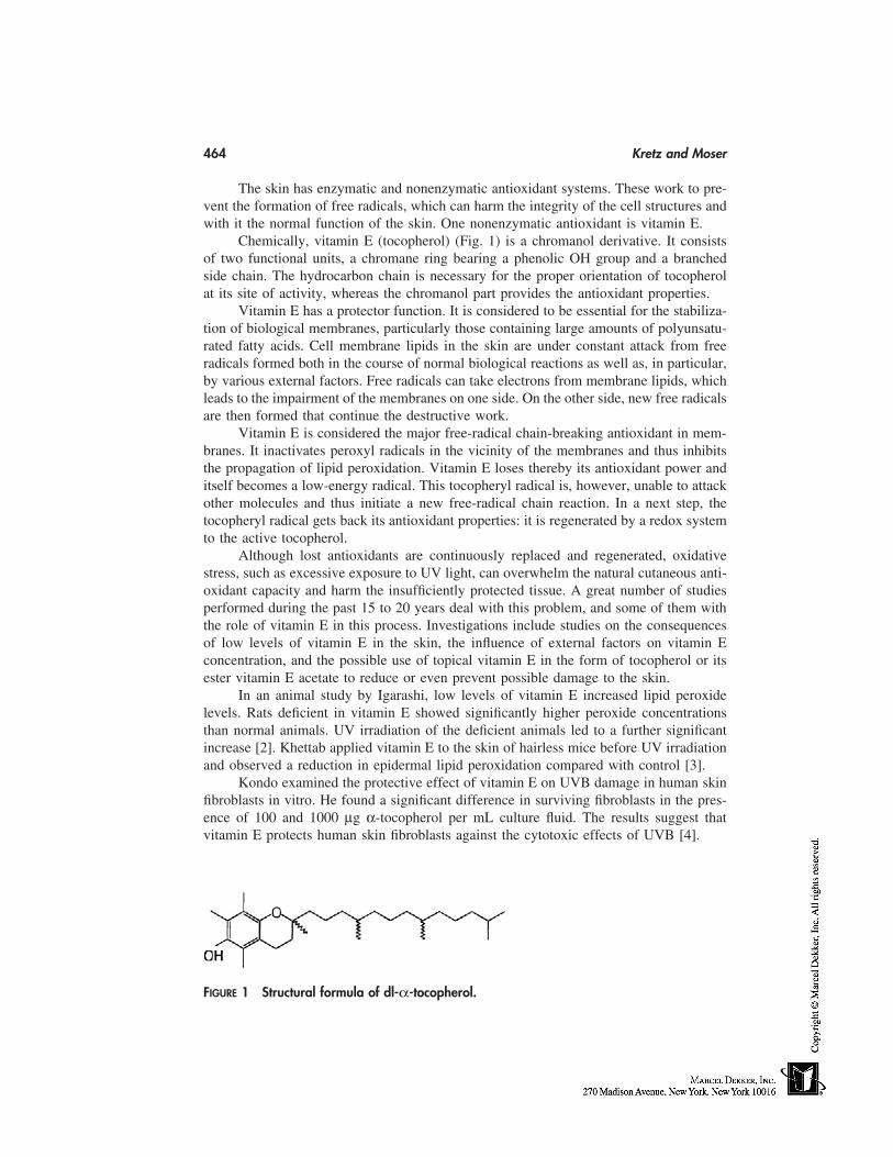
464 Kretz and Moser
The skin has enzymatic and nonenzymatic antioxidant systems. These work to pre-vent the formation of free radicals, which can harm the integrity of the cell structures andwith it the normal function of the skin. One nonenzymatic antioxidant is vitamin E.
Chemically, vitamin E (tocopherol) (Fig. 1) is a chromanol derivative. It consistsof two functional units, a chromane ring bearing a phenolic OH group and a branchedside chain. The hydrocarbon chain is necessary for the proper orientation of tocopherolat its site of activity, whereas the chromanol part provides the antioxidant properties.
Vitamin E has a protector function. It is considered to be essential for the stabiliza-tion of biological membranes, particularly those containing large amounts of polyunsatu-rated fatty acids. Cell membrane lipids in the skin are under constant attack from freeradicals formed both in the course of normal biological reactions as well as, in particular,by various external factors. Free radicals can take electrons from membrane lipids, whichleads to the impairment of the membranes on one side. On the other side, new free radicalsare then formed that continue the destructive work.
Vitamin E is considered the major free-radical chain-breaking antioxidant in mem-branes. It inactivates peroxyl radicals in the vicinity of the membranes and thus inhibitsthe propagation of lipid peroxidation. Vitamin E loses thereby its antioxidant power anditself becomes a low-energy radical. This tocopheryl radical is, however, unable to attackother molecules and thus initiate a new free-radical chain reaction. In a next step, thetocopheryl radical gets back its antioxidant properties: it is regenerated by a redox systemto the active tocopherol.
Although lost antioxidants are continuously replaced and regenerated, oxidativestress, such as excessive exposure to UV light, can overwhelm the natural cutaneous anti-oxidant capacity and harm the insufficiently protected tissue. A great number of studiesperformed during the past 15 to 20 years deal with this problem, and some of them withthe role of vitamin E in this process. Investigations include studies on the consequencesof low levels of vitamin E in the skin, the influence of external factors on vitamin Econcentration, and the possible use of topical vitamin E in the form of tocopherol or itsester vitamin E acetate to reduce or even prevent possible damage to the skin.
In an animal study by Igarashi, low levels of vitamin E increased lipid peroxidelevels. Rats deficient in vitamin E showed significantly higher peroxide concentrationsthan normal animals. UV irradiation of the deficient animals led to a further significantincrease [2]. Khettab applied vitamin E to the skin of hairless mice before UV irradiationand observed a reduction in epidermal lipid peroxidation compared with control [3].
Kondo examined the protective effect of vitamin E on UVB damage in human skinfibroblasts in vitro. He found a significant difference in surviving fibroblasts in the pres-ence of 100 and 1000 µg α-tocopherol per mL culture fluid. The results suggest thatvitamin E protects human skin fibroblasts against the cytotoxic effects of UVB [4].
FIGURE 1 Structural formula of dl-α-tocopherol.
Vitamins 465
Interesting is the result of a study performed by Lopez-Torres et al., in which theyinvestigated the effects of topical tocopherol on epidermal and dermal antioxidants andtheir ability to prevent UV-induced oxidative damage. Topically applied tocopherol tohairless mice in vivo increased dermal superoxide dismutase activity by 30% and protectedepidermal glutathione peroxidase and superoxide dismutase from depletion after UV irra-diation. Total and reduced glutathione levels in the epidermis were also increased, aswere dermal vitamin C levels. The investigators conclude that topical administration ofα-tocopherol protects cutaneous tissues against oxidative damage induced by UV irradia-tion [5].
Application of pure vitamin E acetate to the skin of hairless mice immediately afterUVB irradiation reduced sunbum symptoms such as erythema, skin sensitivity, and skinswelling in a study carried out by Trevithick et al. [6]. Reduced erythema formation afterUV irradiation was also reported by Roshchupkin [7] and Pathak [8].
In a human skin model, antioxidant depletion as a result of UV light exposure wasshown by Podda [9]. Ubiquinol and ubiquinone in particular, as well as α-tocopherol toa lesser extent, were susceptible and decreased with higher UV light intensities to virtuallynon detectable levels. Partial impairment of the cutaneous antioxidant defense system,including vitamin E, by UV light was also observed by Fuchs [10].
Clement-Lacroix et al. tested the protector effect of vitamin E on immune suppres-sion in human epidermal cells in vitro. Cultured cells preincubated with or without vitaminE were irradiated with UVA light. The investigators could show that incubation of cellcultures with vitamin E before irradiation partially protected the cells from the immuno-suppressive effects of UVA radiation [11]. Finally, Weiser [12] and Miyamoto [13] haveshown wound-healing properties of topical vitamin E acetate.
Vitamin E is used in cosmetics for everyday use to strengthen the natural antioxidantpotency of the skin and thus to better cope with oxidative stress. Most of the scientificbackground for the topical use of vitamin E stems from observations in context with UVlight. Vitamin E is often used, therefore, in suncare products for improvement of theprotection achieved with the sun filters. Even high SPF factors still allow the penetra-tion of some UV light onto and into the skin. Whereas the sun filters absorb or reflectmost of the rays on the surface of the skin, vitamin E acts on the inside and reduces therisk of damage that could be caused by rays passing through the sun filter barrier. VitaminE helps, therefore, in the prevention of symptoms caused by UV-induced skin damagesuch as wrinkling and irregular pigmentation.
In nature, Vitamin E appears as tocopherols, of which the alpha form has the highestbiological potency. The unesterified form is present in wheat germ oil and other vegetableoils that are used in cosmetics as sources of Vitamin E. Most often used is dl-alpha tocoph-eryl acetate, because this ester is less prone to oxidation than free tocopherol. In the skin,vitamin E acetate is bioconverted into the biologically active antioxidant tocopherol[14,15].
VITAMIN A
Vitamin A (Fig. 2) and its derivatives belong to a large class of structurally related com-pounds, the retinoids. The term vitamin A is generically used for all derivatives of β-ionone that possess the biological activity of all-trans retinol or are closely related to it.The biological activities of the vitamin A derivatives are expressed in IU (international

466 Kretz and Moser
units.) One IU corresponds to 0.3 µg retinol, 0.34 µg vitamin A acetate, and 0.55 µgvitamin A palmitate.
Vitamin A is best known for its involvement in maintaining normal vision. It exerts,however, a number of other functions in the human organism, of which its activity in theepidermis is of particular interest for cosmetics.
The architecture of the human epidermis is a complex stratified system, its renewala complex process. Epidermal keratinocytes proliferate and differentiate in a multilayeredpattern. These processes are balanced so that new basal cells are formed as the totallycornified cells are shed from the surface of the skin. Proliferation and keratinization ofkeratinocytes are the two key elements for the build-up of a healthy epidermis. In bothprocesses, vitamin A plays the role of a regulator.
On cell proliferation, vitamin A has a stimulating effect, as has been shown in vari-ous studies [16–18]. As little as 10 µg vitamin A acetate suspended in 0.2 mL waterapplied to normal rat skin led to a clear increase in mitotic activity after only 4 hours.Much more pronounced and longer lasting was the effect with 100 µg vitamin A acetate.However, 24 hours after treatment, the mitotic index had returned to original levels withboth concentrations [17]. As can be seen from this study, the effect of vitamin A is dosedependent and disappears after a certain time with decreasing concentration in the tissue.An increase in mitotic activity is the first step in an increase of the number of new keratino-cytes formed [19], which results in a thickening of the epidermis [20–23].
In the process of aging, many aspects of the skin structure are altered because of adecreased metabolic activity of the human organism. A thinning of the epidermis is oneof the characteristics of aging skin. The skin thereby loses part of its barrier function, andas a consequence of reduced water retention capacity it is often dry, scaly, or even cracks.Vitamin A can counteract this development by stimulating the cell-renewal process.
The effect on the keratinization process was investigated by Fuchs and Green [24].Removal of vitamin A from the culture fluid of human keratinocyte cell cultures resultedin a reduced cell motility, an increased adhesiveness of the cells, and a prevention ofpattern formation. They conclude that the nature of the keratins synthesized by the tissuesis regulated by the concentration of vitamin A. Another symptom of skin aging is a de-crease in collagen in the connective tissue. Skin collagen decreases linearly by about 1%per year throughout adult life [25]. Topical administration of vitamin A has shown signifi-cant dose-related changes in collagen content of the dermis. 0.1% vitamin A palmitateapplied to skin of hairless mice for 14 days increased the collagen content by 88%, 0.5%vitamin A palmitate by 101% [20].
Vitamin A not only improves the barrier function of the skin but also its appearanceand elasticity. Application of a lotion with vitamin A palmitate to the temples of a groupof 40- to 60-year-old volunteers has shown an increase in elasticity by 14% after 2 weeksand by over 22% after 6 weeks [26].
There is evidence that UV light strongly affects vitamin A concentration in epider-mis and dermis as was shown in animals and humans [27,28]. Particularly low were thelevels when test animals were exposed to UVA near the absorption maximum of vitaminA. The regeneration of normal levels in the depleted tissue is very slow and took morethan 1 week in rabbit ear skin [28].
Cluver and Politzer measured the vitamin A concentration in blood serum of humansafter 1 hour of exposure to the sun. Depletion was observed immediately after exposure,which lasted at least a further 21/2 hours [29]. It can be assumed that, under similar condi-tions, a depletion also takes place in the skin. The low blood levels could also be an
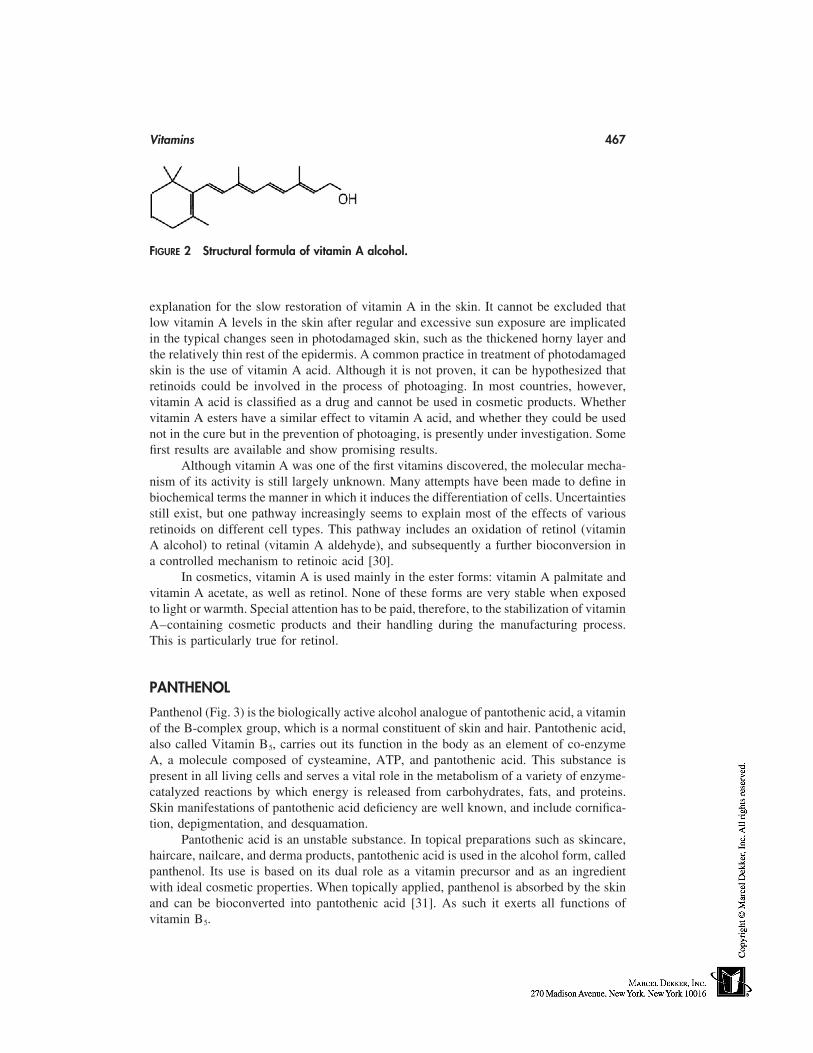
Vitamins 467
FIGURE 2 Structural formula of vitamin A alcohol.
explanation for the slow restoration of vitamin A in the skin. It cannot be excluded thatlow vitamin A levels in the skin after regular and excessive sun exposure are implicatedin the typical changes seen in photodamaged skin, such as the thickened horny layer andthe relatively thin rest of the epidermis. A common practice in treatment of photodamagedskin is the use of vitamin A acid. Although it is not proven, it can be hypothesized thatretinoids could be involved in the process of photoaging. In most countries, however,vitamin A acid is classified as a drug and cannot be used in cosmetic products. Whethervitamin A esters have a similar effect to vitamin A acid, and whether they could be usednot in the cure but in the prevention of photoaging, is presently under investigation. Somefirst results are available and show promising results.
Although vitamin A was one of the first vitamins discovered, the molecular mecha-nism of its activity is still largely unknown. Many attempts have been made to define inbiochemical terms the manner in which it induces the differentiation of cells. Uncertaintiesstill exist, but one pathway increasingly seems to explain most of the effects of variousretinoids on different cell types. This pathway includes an oxidation of retinol (vitaminA alcohol) to retinal (vitamin A aldehyde), and subsequently a further bioconversion ina controlled mechanism to retinoic acid [30].
In cosmetics, vitamin A is used mainly in the ester forms: vitamin A palmitate andvitamin A acetate, as well as retinol. None of these forms are very stable when exposedto light or warmth. Special attention has to be paid, therefore, to the stabilization of vitaminA–containing cosmetic products and their handling during the manufacturing process.This is particularly true for retinol.
PANTHENOL
Panthenol (Fig. 3) is the biologically active alcohol analogue of pantothenic acid, a vitaminof the B-complex group, which is a normal constituent of skin and hair. Pantothenic acid,also called Vitamin B5, carries out its function in the body as an element of co-enzymeA, a molecule composed of cysteamine, ATP, and pantothenic acid. This substance ispresent in all living cells and serves a vital role in the metabolism of a variety of enzyme-catalyzed reactions by which energy is released from carbohydrates, fats, and proteins.Skin manifestations of pantothenic acid deficiency are well known, and include cornifica-tion, depigmentation, and desquamation.
Pantothenic acid is an unstable substance. In topical preparations such as skincare,haircare, nailcare, and derma products, pantothenic acid is used in the alcohol form, calledpanthenol. Its use is based on its dual role as a vitamin precursor and as an ingredientwith ideal cosmetic properties. When topically applied, panthenol is absorbed by the skinand can be bioconverted into pantothenic acid [31]. As such it exerts all functions ofvitamin B5.
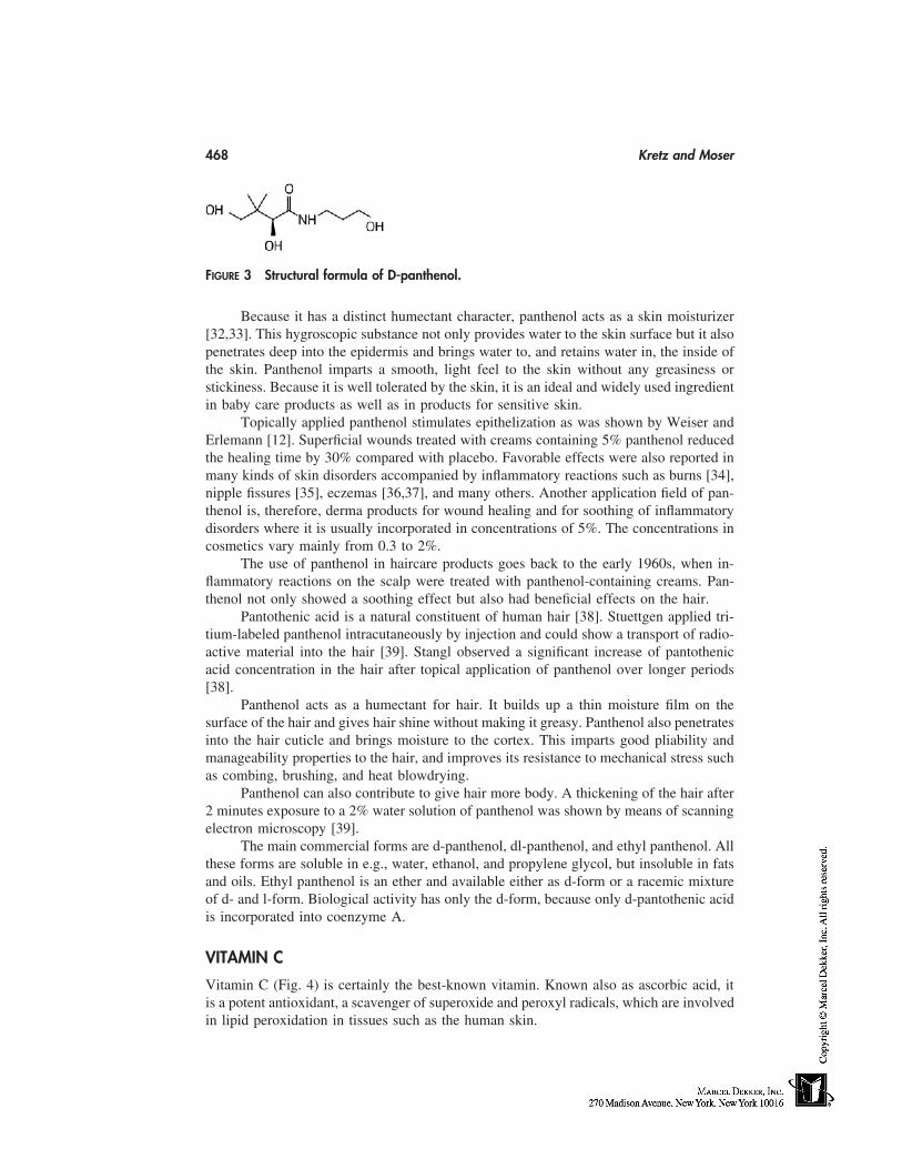
468 Kretz and Moser
FIGURE 3 Structural formula of D-panthenol.
Because it has a distinct humectant character, panthenol acts as a skin moisturizer[32,33]. This hygroscopic substance not only provides water to the skin surface but it alsopenetrates deep into the epidermis and brings water to, and retains water in, the inside ofthe skin. Panthenol imparts a smooth, light feel to the skin without any greasiness orstickiness. Because it is well tolerated by the skin, it is an ideal and widely used ingredientin baby care products as well as in products for sensitive skin.
Topically applied panthenol stimulates epithelization as was shown by Weiser andErlemann [12]. Superficial wounds treated with creams containing 5% panthenol reducedthe healing time by 30% compared with placebo. Favorable effects were also reported inmany kinds of skin disorders accompanied by inflammatory reactions such as burns [34],nipple fissures [35], eczemas [36,37], and many others. Another application field of pan-thenol is, therefore, derma products for wound healing and for soothing of inflammatorydisorders where it is usually incorporated in concentrations of 5%. The concentrations incosmetics vary mainly from 0.3 to 2%.
The use of panthenol in haircare products goes back to the early 1960s, when in-flammatory reactions on the scalp were treated with panthenol-containing creams. Pan-thenol not only showed a soothing effect but also had beneficial effects on the hair.
Pantothenic acid is a natural constituent of human hair [38]. Stuettgen applied tri-tium-labeled panthenol intracutaneously by injection and could show a transport of radio-active material into the hair [39]. Stangl observed a significant increase of pantothenicacid concentration in the hair after topical application of panthenol over longer periods[38].
Panthenol acts as a humectant for hair. It builds up a thin moisture film on thesurface of the hair and gives hair shine without making it greasy. Panthenol also penetratesinto the hair cuticle and brings moisture to the cortex. This imparts good pliability andmanageability properties to the hair, and improves its resistance to mechanical stress suchas combing, brushing, and heat blowdrying.
Panthenol can also contribute to give hair more body. A thickening of the hair after2 minutes exposure to a 2% water solution of panthenol was shown by means of scanningelectron microscopy [39].
The main commercial forms are d-panthenol, dl-panthenol, and ethyl panthenol. Allthese forms are soluble in e.g., water, ethanol, and propylene glycol, but insoluble in fatsand oils. Ethyl panthenol is an ether and available either as d-form or a racemic mixtureof d- and l-form. Biological activity has only the d-form, because only d-pantothenic acidis incorporated into coenzyme A.
VITAMIN C
Vitamin C (Fig. 4) is certainly the best-known vitamin. Known also as ascorbic acid, itis a potent antioxidant, a scavenger of superoxide and peroxyl radicals, which are involvedin lipid peroxidation in tissues such as the human skin.
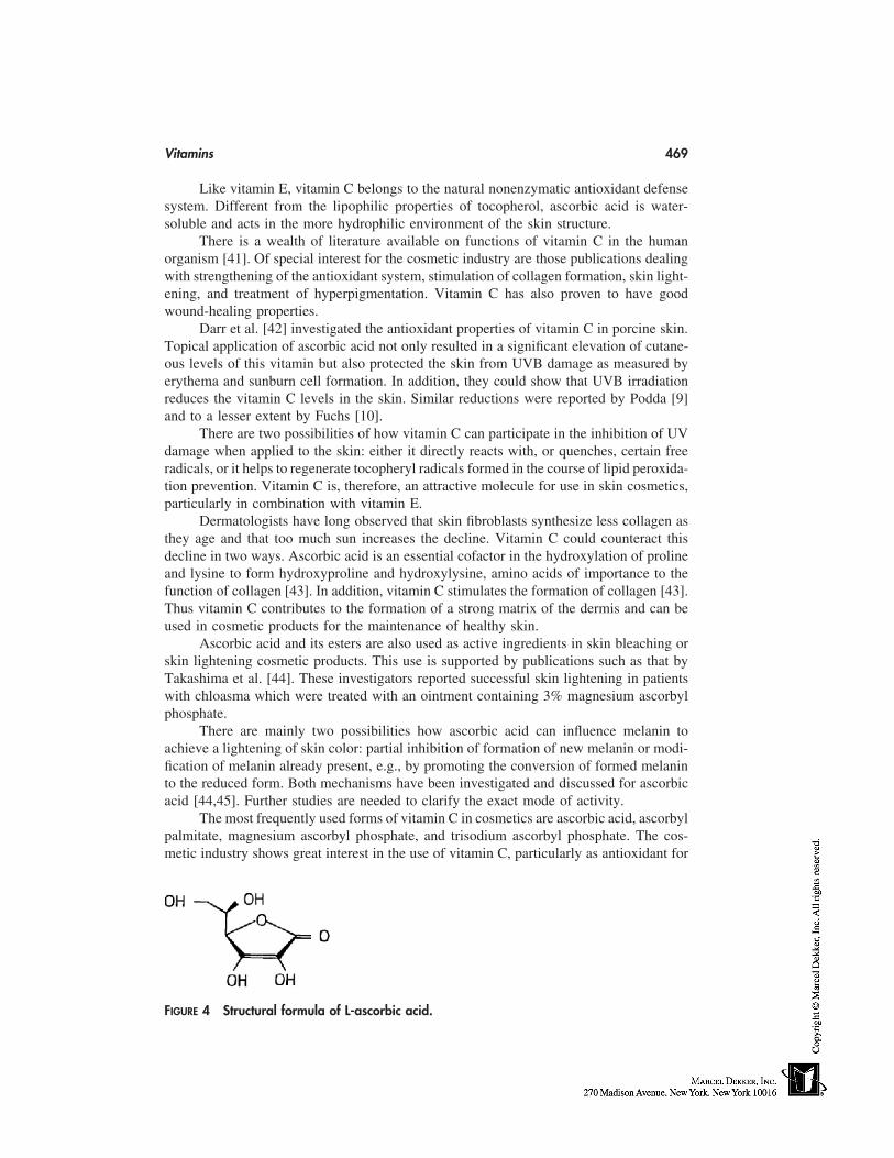
Vitamins 469
Like vitamin E, vitamin C belongs to the natural nonenzymatic antioxidant defensesystem. Different from the lipophilic properties of tocopherol, ascorbic acid is water-soluble and acts in the more hydrophilic environment of the skin structure.
There is a wealth of literature available on functions of vitamin C in the humanorganism [41]. Of special interest for the cosmetic industry are those publications dealingwith strengthening of the antioxidant system, stimulation of collagen formation, skin light-ening, and treatment of hyperpigmentation. Vitamin C has also proven to have goodwound-healing properties.
Darr et al. [42] investigated the antioxidant properties of vitamin C in porcine skin.Topical application of ascorbic acid not only resulted in a significant elevation of cutane-ous levels of this vitamin but also protected the skin from UVB damage as measured byerythema and sunburn cell formation. In addition, they could show that UVB irradiationreduces the vitamin C levels in the skin. Similar reductions were reported by Podda [9]and to a lesser extent by Fuchs [10].
There are two possibilities of how vitamin C can participate in the inhibition of UVdamage when applied to the skin: either it directly reacts with, or quenches, certain freeradicals, or it helps to regenerate tocopheryl radicals formed in the course of lipid peroxida-tion prevention. Vitamin C is, therefore, an attractive molecule for use in skin cosmetics,particularly in combination with vitamin E.
Dermatologists have long observed that skin fibroblasts synthesize less collagen asthey age and that too much sun increases the decline. Vitamin C could counteract thisdecline in two ways. Ascorbic acid is an essential cofactor in the hydroxylation of prolineand lysine to form hydroxyproline and hydroxylysine, amino acids of importance to thefunction of collagen [43]. In addition, vitamin C stimulates the formation of collagen [43].Thus vitamin C contributes to the formation of a strong matrix of the dermis and can beused in cosmetic products for the maintenance of healthy skin.
Ascorbic acid and its esters are also used as active ingredients in skin bleaching orskin lightening cosmetic products. This use is supported by publications such as that byTakashima et al. [44]. These investigators reported successful skin lightening in patientswith chloasma which were treated with an ointment containing 3% magnesium ascorbylphosphate.
There are mainly two possibilities how ascorbic acid can influence melanin toachieve a lightening of skin color: partial inhibition of formation of new melanin or modi-fication of melanin already present, e.g., by promoting the conversion of formed melaninto the reduced form. Both mechanisms have been investigated and discussed for ascorbicacid [44,45]. Further studies are needed to clarify the exact mode of activity.
The most frequently used forms of vitamin C in cosmetics are ascorbic acid, ascorbylpalmitate, magnesium ascorbyl phosphate, and trisodium ascorbyl phosphate. The cos-metic industry shows great interest in the use of vitamin C, particularly as antioxidant for
FIGURE 4 Structural formula of L-ascorbic acid.
470 Kretz and Moser
the skin. Its use, however, has been limited to date due to the insufficient stability ofascorbic acid in aqueous solutions and the prices of some of the more stable derivatives.
OTHERS
Some other vitamins and vitamin precursors used in cosmetic products are biotin, niacin-amide, vitamin D, vitamin B6, beta-carotene, and, in a few products, vitamin K. For allthese substances there exists a rationale for their use in cosmetics but there are either nostudies or insufficient studies available that prove their efficacy when topically applied.
Systemic use of 2.5 mg biotin per day has shown good effect on brittle nails andhas improved hair quality [46–49]. As it has good effect on these keratin structures, itcan be assumed that this vitamin could also have interesting effects on the keratinizationprocess in the epidermis.
Beta-carotene is known for its quenching activity on singlet oxygen. It would bean ideal partner for vitamin E and vitamin C to strengthen the antioxidant defense systemof the skin. Oral supplementation with beta-carotene over several weeks has shown toreduce the risk of UV-induced skin damage [50].
Unfortunately for cosmetics, beta-carotene is a strong coloring agent and concentra-tions of more than 0.05% in a cosmetic product can lead to undesirable coloration of theclothes of its users. Low concentrations of beta-carotene are used in some cosmetics asnatural coloring agent for creams.
Roccheggiani showed a depression of sebum production with the tripalmitate esterof vitamin B6 [51]. Vitamin D could be an ideal partner for total sun blockers, as the UV-ray barrier of these products partly prevents the natural formation of vitamin D in theskin. Vitamin D is on the ‘‘list of substances which must not form part of the compositionof cosmetic products’’ of the European Cosmetic Regulations. It can be used, however,in other countries.
CONCLUSION
Vitamins are a class of naturally occurring active ingredients with well-documented activi-ties when topically applied. They are all essential substances for the well-being and healthof the human organism, including the skin.
Thousands of studies have shown the safety and efficacy of systemically and topi-cally used vitamins. In many cases, topical use is the only way to provide sufficient quanti-ties of these highly active protector and care substances to be able to guarantee an optimalfunctioning of the skin. This is particularly true when the skin is stressed by factors suchas UV light or environmental pollutants such as ozone, especially as these factors canoften even destroy considerable quantities of the vitamins.
REFERENCES
1. Epstein JH. The pathological effects of light on the skin. In: Free Radicals in Biology III.WA Pryor, ed. New York: Academic Press, 1977; 219–249.
2. Igarashi A, Uzuka M, Nakajima K. The effects of vitamin E deficiency on rat skin. Br JDermatol 1989; 121:43–49.
3. Khettab N, Amory M-C, Briand G, Bousquet B, Combre A, Forlot P, Barey M. Photoprotectiveeffect of vitamin A and E on polyamine and oxygenated free radical metabolism in hairlessmouse epidermis. Biochim 1988; 70:1709–1713.
Vitamins 471
4. Kondo S, Mamada A, Yamaguchi J, Fukuro S. Protective effect of dl-α-tocopherol on thecytotoxicity of ultraviolet B against human skin fibroblasts in vitro. Photodermatol Photoim-munol Photomed 1990; 7:173–177.
5. Lopez-Torres M, Thiele JJ, Shindo Y, Han D, Packer L. Topical application of α-tocopherolmodulates the antioxidant network and diminishes ultraviolet-induced oxidative damage inmurine skin. Br J Dermatol 1998; 138:207–215.
6. Trevithick JR, Xiong H, Lee S, Shum DT, Sanford E, Karlik SJ, Norley C, Dilworth GR.Topical tocopherol acetate reduces post-UVB, sunburn associated erythema, edema, and skinsensitivity in hairless mice. Arch Biochem Biophys 1992; 296:575–582.
7. Roschupkin DI, Pistsov MY, Potapenko AY. Inhibition of ultraviolet light-induced erythemaby antioxidants. Arch Dermatol Res 1979; 266:91–94.
8. Pathak MA. 1987; unpublished document of Hoffmann-La Roche.9. Podda M, Traber MG, Weber C, Yan L-J, Packer L. UV irradiation depletes antioxidants and
causes oxidative damage in a model of human skin. Free Radical Biol Med 1998; 24:55–65.10. Fuchs J, Huflejt ME, Rothfuss LM, Wilson DS, Carcamo G, Packer L. Acute effects of near
ultraviolet and visible light on the cutaneous antioxidant defense system. Photochem Photobiol1989; 50:739–744.
11. Clement-Lacroix P, Michel L, Moysan A, Molière P, Dubertret L. UVA-induced immunesuppression in human skin: protective effect of vitamin E in human epidermal cells in vitro.Br J Dermatol 1996; 134:77–84.
12. Weiser H, Erlemann G. Acceleration of superficial wound healing by panthenol and zinc oxide.Preprints of the XIVth IFSCC Congress, Barcelona, 1986; 2:879–887.
13. Mijamoto I, Uchida Y, Shinomiya T, Abe T, Nishijima Y. Effects of cosmetics containingbioactive substances on skin. Preprints of the XIVth IFSCC Congress, Barcelona. 1986; 2:949–959.
14. Norkus EP, Bryce GF, Bhagavan HN. Uptake and bioconversion of α-tocopheryl acetate toα-tocopherol in skin of hairless mice. Photochem Photobiol 1993; 57:613–615.
15. Kramer-Stickland K, Liebler DC. Effect of UVB on hydrolysis of α-tocopherol acetate toα-tocopherol in mouse skin. E J Invest Dermatol 1998; 111:302–307.
16. Lawrence DJ, Bern HA. On the specificity of the response of mouse epidermis to vitamin A.J Invest Derm 1958; 31:313–325.
17. Sherman BS. The effect of vitamin A on epithelial mitosis in vitro and in vivo. J Invest Derm1961; 37:469–480.
18. Zil JS. Vitamin A acid effects on epidermal mitotic activity, thickness and cellularity in thehairless mouse. J Invest Derm 1972; 59:228–232.
19. Chopra DP, Flaxman BA. The effect of vitamin A on growth and differentiation of humankeratinocytes in vitro. J Invest Derm 1975; 64:19–22.
20. Courts DF, Skreko F, McBee J. The effect of retinyl palmitate on skin composition and mor-phometry. J Soc Cosmet Chem 1988; 39:235–240.
21. Jarrett A, Spearman RIC. Histochemistry of the Skin—Psoriasis. London: English UniversitiesPress, 1964; 41–77.
22. Spearman RIC, Jarrett A. Biological comparison of isomers and chemical forms of vitaminA (retinol). Br J Dermatol 1974; 90:553–560.
23. Kang S, Duell EA, Fisher GJ, Datta SC, Wang Z-Q, Reddy AP, Tavakkol A, Yi JY, GriffithsCEM, Elder JT, Voorhees JJ. Application of retinol to human skin in vivo induces epidermalhyperplasia and cellular retinoid binding proteins characteristic of retinoic acid but withoutmeasurable retinoic acid levels or irritation. J Invest Derm 1995; 105:549–556.
24. Fuchs E, Green H. Regulation of terminal differentiation of cultured human keratinocytes byvitamin A. Cell 1981; 25:617–625.
25. Shuster S, Black MM, McVitie E. The influence of age and sex on skin thickness, skin collagenand density. Br J Derm 1975; 93:639–643.
26. Fthenakis CG, Maes DH, Smith WP. In vivo assessment of skin elasticity using ballistometry.J Soc Cosmet Chem 1991; 42:211–222.
472 Kretz and Moser
27. Berne B, Vahlquist A, Fischer T, Danielson BG, Berne C. UV treatment of uraemic pruritusreduces the vitamin A content of the skin. Eur J Clin Invest 1984; 14:203–206.
28. Berne B, Nilsson M, Vahlquist A. UV irradiation and cutaneous vitamin A: an experimentalstudy in rabbit and human skin. J Invest Derm 1984; 83:401–404.
29. Cluver EH, Politzer WM. Sunburn and vitamin A deficiency. S Afr J Sci 1965; 61:306–309.30. Olson AJ. Vitamin A. In: Handbook of Vitamins. 2d ed. J Machlin, ed. New York: Marcel
Dekker, 1991; 1–57.31. Burlet E. Die Percutane Resorption von Panthenol (The percutaneous absorption of panthenol).
Jubilee Volume Emil Barell 1946; 92–97.32. Tronnier H. Determination of the hydration effect of a hydrogel with and without d-panthenol.
Unpublished document of Hoffmann-La Roche, 1992.33. Tronnier H. Efficacy of various cream formulations containing panthenol. Unpublished docu-
ment of Hoffmann-La Roche, 1994.34. Kline PR. 12 years’ experience using pantothenylol topically. Western Med 1963; 4:78–80.35. Dubecq JP, Detchart M. Etude d’un onguent pantothénique dans la prophylaxie et le traitement
des crevasses du sein. La médecine practicienne: special edition 1977; May.36. Matanić V. Ein Beitrag zur Therapie des Zementekzems. Berufsdermatosen 1963; 11:104–
109.37. Jolibois RP. Etude de l’action d’un onguent à la vitamine B5 sur les affections cutanées du
siège du nouveau-né. Médicine Actuelle 1976; 3:716–721.38. Stangl E. Ueber den Pantothensaeure-Gehalt menschlicher Kopfhaut (The pantothenic acid
content of human hair). Int Z Vitaminforschung 1952; 24:9–12.39. Stuettgen G, Krause H. Die percutane Absorption von tritium-markiertem Panthenol bei
Mensch und Tier (Percutaneous absorption of tritium-labeled panthenol in humans and ani-mals). Archiv fuer klin und exp Dermatologie 1960; 209:578–582.
40. Driscoll WR. Panthenol in hair products. Drug Cosmet Ind 1975; 116:42–45, 149–153.41. Moser U, Bendich A. Vitamin C. In: Handbook of Vitamins 2d ed. J Machlin, ed. New York:
Marcel Dekker, 1991; 195–232.42. Darr D, Combs S, Dunston S, Manning T, Pinnell S. Topical vitamin C protects porcine skin
from ultraviolet radiation-induced damage. Br J Dermatol 1992; 127:247–253.43. Pinnell SR. Regulation of collagen biosynthesis by ascorbic acid: a review. Yale J Biol Med
1985; 58:553–559.44. Takashima H, Nomura H, Imai Y, Mima H. Ascorbic acid esters and skin pigmentation. Am
Perfum Cosmet 1971; 86:29–36.45. Takenouchi K, Aso K. The relation between melanin formation and ascorbic acid. J Vitaminol
1964; 10:123–134.46. Floersheim GL. Treatment of brittle finger nails with biotin. H�G Zeitschrift fuer Hautkrank-
heiten 1989; 64:41–48.47. Floersheim GL. An examination of the effect of biotin on alopecia and hair quality. H�G
Zeitschrift fuer Hautkrankheiten 1992; 67:246–255.48. Colombo VE, Gerber F, Bronhofer M, Floersheim GL. Treatment of brittle fingernails and
onychoschizia with biotin: scanning electron microscopy. J Am Acad Dermatol 1990; 23:1127–1132.
49. Hochman LG, Scher RK, Meyerson MS. Brittle nails: response to daily biotin supplementation.Cutis 1993; 51:303–305.
50. Gollnick HPM, Hopfenmueller W, Hemmes C, Chun SC, Schmid C, Sundermeier K, BiesalskiHK. Systemic beta-carotene plus topical UV-sunscreen are an optimal protection against harm-ful effects of natural UV-sunlight: results of the Berlin-Eilath study. Eur J Dermatol 1996; 6:200–205.
51. Roccheggiani G. Erfahrungen über die Einwirkung von fettloeslichen Derivaten der Vita-mingruppe B auf die Haut (Experiences on the influence of lipid-soluble derivatives of the Bgroup vitamins on the skin). Seifen Oele Fette Wachse 1959; 85:777–779, 819–820.
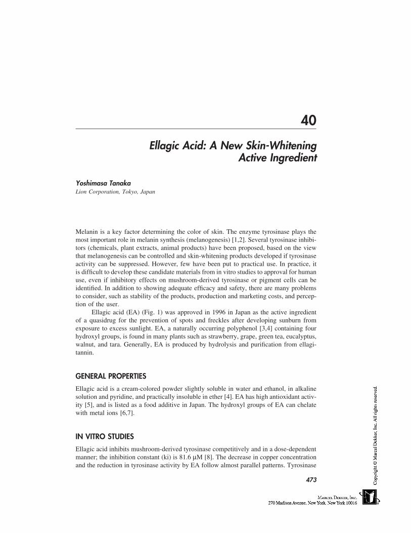
40
Ellagic Acid: A New Skin-WhiteningActive Ingredient
Yoshimasa TanakaLion Corporation, Tokyo, Japan
Melanin is a key factor determining the color of skin. The enzyme tyrosinase plays themost important role in melanin synthesis (melanogenesis) [1,2]. Several tyrosinase inhibi-tors (chemicals, plant extracts, animal products) have been proposed, based on the viewthat melanogenesis can be controlled and skin-whitening products developed if tyrosinaseactivity can be suppressed. However, few have been put to practical use. In practice, itis difficult to develop these candidate materials from in vitro studies to approval for humanuse, even if inhibitory effects on mushroom-derived tyrosinase or pigment cells can beidentified. In addition to showing adequate efficacy and safety, there are many problemsto consider, such as stability of the products, production and marketing costs, and percep-tion of the user.
Ellagic acid (EA) (Fig. 1) was approved in 1996 in Japan as the active ingredientof a quasidrug for the prevention of spots and freckles after developing sunburn fromexposure to excess sunlight. EA, a naturally occurring polyphenol [3,4] containing fourhydroxyl groups, is found in many plants such as strawberry, grape, green tea, eucalyptus,walnut, and tara. Generally, EA is produced by hydrolysis and purification from ellagi-tannin.
GENERAL PROPERTIES
Ellagic acid is a cream-colored powder slightly soluble in water and ethanol, in alkalinesolution and pyridine, and practically insoluble in ether [4]. EA has high antioxidant activ-ity [5], and is listed as a food additive in Japan. The hydroxyl groups of EA can chelatewith metal ions [6,7].
IN VITRO STUDIES
Ellagic acid inhibits mushroom-derived tyrosinase competitively and in a dose-dependentmanner; the inhibition constant (ki) is 81.6 µM [8]. The decrease in copper concentrationand the reduction in tyrosinase activity by EA follow almost parallel patterns. Tyrosinase
473
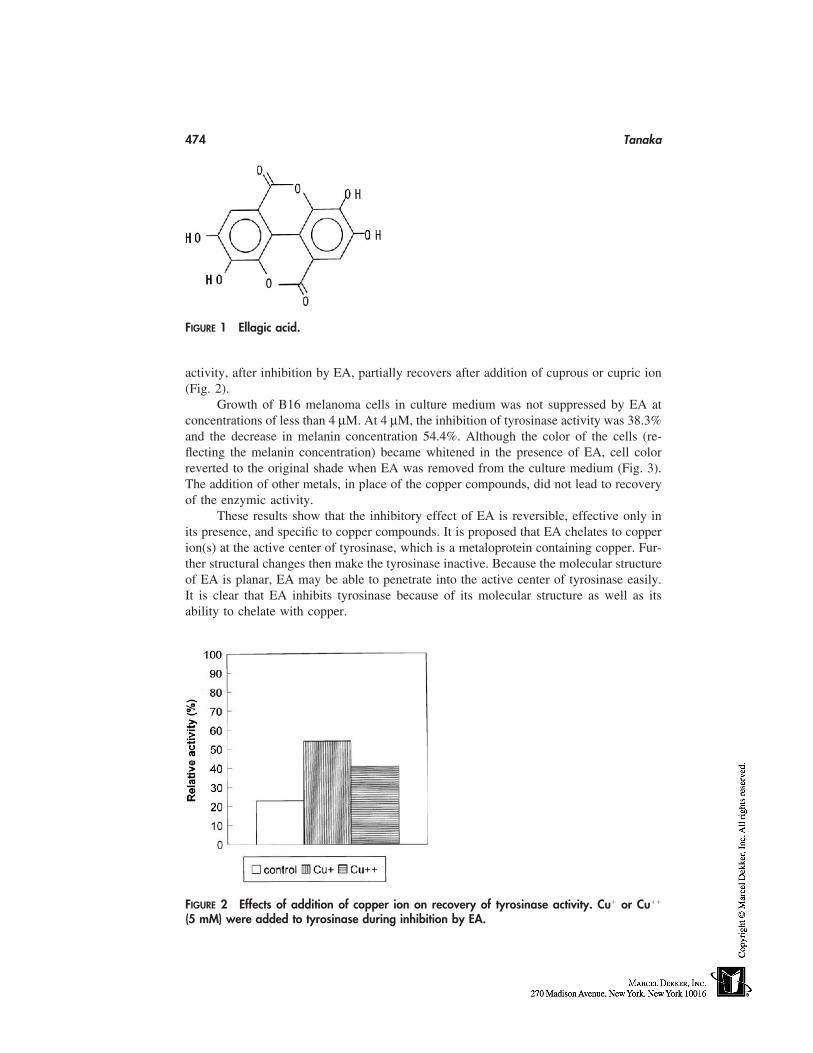
474 Tanaka
FIGURE 1 Ellagic acid.
activity, after inhibition by EA, partially recovers after addition of cuprous or cupric ion(Fig. 2).
Growth of B16 melanoma cells in culture medium was not suppressed by EA atconcentrations of less than 4 µM. At 4 µM, the inhibition of tyrosinase activity was 38.3%and the decrease in melanin concentration 54.4%. Although the color of the cells (re-flecting the melanin concentration) became whitened in the presence of EA, cell colorreverted to the original shade when EA was removed from the culture medium (Fig. 3).The addition of other metals, in place of the copper compounds, did not lead to recoveryof the enzymic activity.
These results show that the inhibitory effect of EA is reversible, effective only inits presence, and specific to copper compounds. It is proposed that EA chelates to copperion(s) at the active center of tyrosinase, which is a metaloprotein containing copper. Fur-ther structural changes then make the tyrosinase inactive. Because the molecular structureof EA is planar, EA may be able to penetrate into the active center of tyrosinase easily.It is clear that EA inhibits tyrosinase because of its molecular structure as well as itsability to chelate with copper.
FIGURE 2 Effects of addition of copper ion on recovery of tyrosinase activity. Cu� or Cu��
(5 mM) were added to tyrosinase during inhibition by EA.
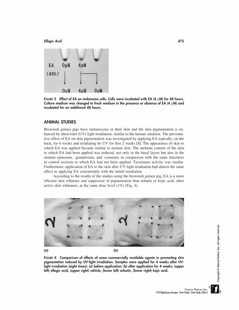
Ellagic Acid 475
FIGURE 3 Effect of EA on melanoma cells. Cells were incubated with EA (4 µM) for 48 hours.Culture medium was changed to fresh medium in the presence or absence of EA (4 µM) andincubated for an additional 48 hours.
ANIMAL STUDIES
Brownish guinea pigs have melanocytes in their skin and the skin pigmentation is en-hanced by ultraviolet (UV) light irradiation, similar to the human situation. The preventa-tive effect of EA on skin pigmentation was investigated by applying EA topically, on theback, for 6 weeks and irradiating by UV for first 2 weeks [8]. The appearance of skin towhich EA was applied became similar to normal skin. The melanin content of the skinto which EA had been applied was reduced, not only in the basal layers but also in thestratum spinosum, -granulosum, and -corneum, in comparison with the same structuresin control sections to which EA had not been applied. Tyrosinase activity was similar.Furthermore, application of EA to the skin after UV-light irradiation had almost the sameaffect as applying EA concurrently with the initial irradiation.
According to the results of the studies using the brownish guinea pig, EA is a moreefficient skin whitener and suppressor of pigmentation than arbutin or kojic acid, otheractive skin whiteners, at the same dose level (1%) (Fig. 4).
(a) (b)
FIGURE 4 Comparison of effects of some commercially available agents in preventing skinpigmentation induced by UV-light irradiation. Samples were applied for 4 weeks after UV-light irradiation (eight times): (a) before application; (b) after application for 4 weeks; (upperleft) ellagic acid, (upper right) vehicle, (lower left) arbutin, (lower right) kojic acid.
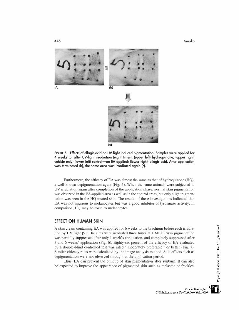
476 Tanaka
FIGURE 5 Effects of ellagic acid on UV-light induced pigmentation. Samples were applied for4 weeks (a) after UV-light irradiation (eight times): (upper left) hydroquinone; (upper right)vehicle only; (lower left) control—no EA applied; (lower right) ellagic acid. After applicationwas terminated (b), the same area was irradiated again (c).
Furthermore, the efficacy of EA was almost the same as that of hydroquinone (HQ),a well-known depigmentation agent (Fig. 5). When the same animals were subjected toUV irradiation again after completion of the application phase, normal skin pigmentationwas observed in the EA-applied area as well as in the control areas, but only slight pigmen-tation was seen in the HQ-treated skin. The results of these investigations indicated thatEA was not injurious to melanocytes but was a good inhibitor of tyrosinase activity. Incomparison, HQ may be toxic to melanocytes.
EFFECT ON HUMAN SKIN
A skin cream containing EA was applied for 6 weeks to the brachium before each irradia-tion by UV light [9]. The sites were irradiated three times at 1 MED. Skin pigmentationwas partially suppressed after only 1 week’s application, and completely suppressed after3 and 6 weeks’ application (Fig. 6). Eighty-six percent of the efficacy of EA evaluatedby a double-blind controlled test was rated ‘‘moderately preferable’’ or better (Fig. 7).Similar efficacy rates were calculated by the image analysis method. Side effects such asdepigmentation were not observed throughout the application period.
Thus, EA can prevent the buildup of skin pigmentation after sunburn. It can alsobe expected to improve the appearance of pigmented skin such as melasma or freckles,
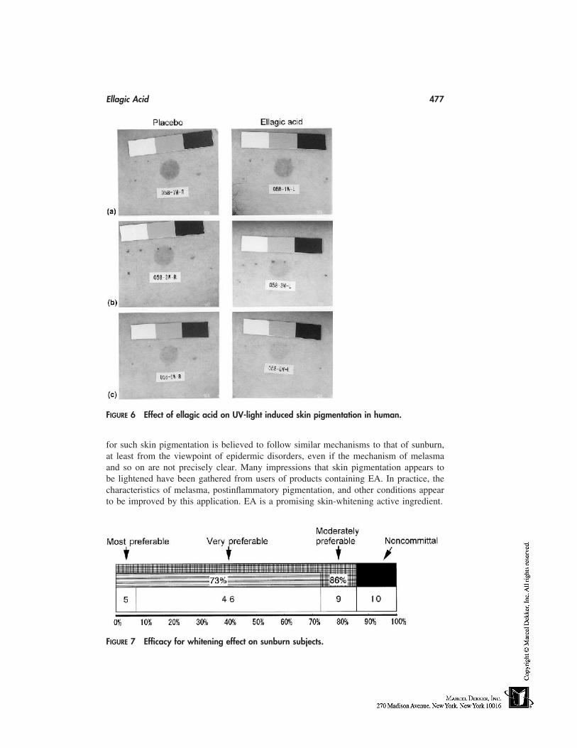
Ellagic Acid 477
FIGURE 6 Effect of ellagic acid on UV-light induced skin pigmentation in human.
for such skin pigmentation is believed to follow similar mechanisms to that of sunburn,at least from the viewpoint of epidermic disorders, even if the mechanism of melasmaand so on are not precisely clear. Many impressions that skin pigmentation appears tobe lightened have been gathered from users of products containing EA. In practice, thecharacteristics of melasma, postinflammatory pigmentation, and other conditions appearto be improved by this application. EA is a promising skin-whitening active ingredient.
FIGURE 7 Efficacy for whitening effect on sunburn subjects.
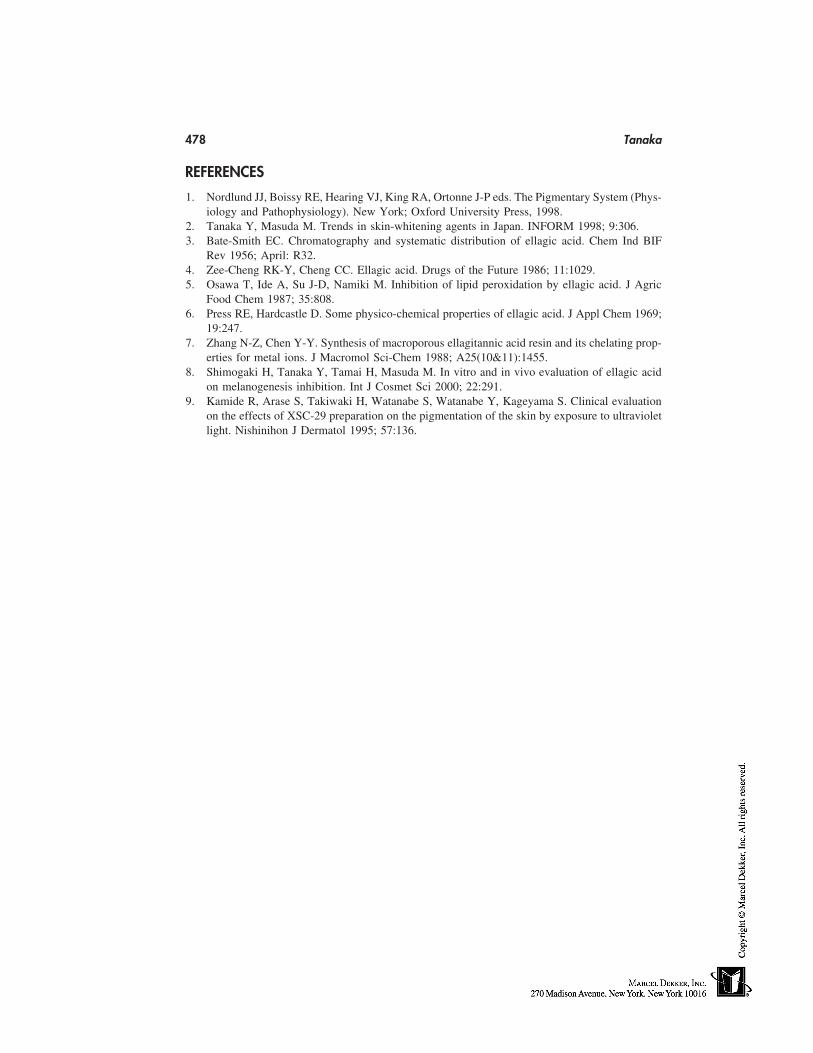
478 Tanaka
REFERENCES
1. Nordlund JJ, Boissy RE, Hearing VJ, King RA, Ortonne J-P eds. The Pigmentary System (Phys-iology and Pathophysiology). New York; Oxford University Press, 1998.
2. Tanaka Y, Masuda M. Trends in skin-whitening agents in Japan. INFORM 1998; 9:306.3. Bate-Smith EC. Chromatography and systematic distribution of ellagic acid. Chem Ind BIF
Rev 1956; April: R32.4. Zee-Cheng RK-Y, Cheng CC. Ellagic acid. Drugs of the Future 1986; 11:1029.5. Osawa T, Ide A, Su J-D, Namiki M. Inhibition of lipid peroxidation by ellagic acid. J Agric
Food Chem 1987; 35:808.6. Press RE, Hardcastle D. Some physico-chemical properties of ellagic acid. J Appl Chem 1969;
19:247.7. Zhang N-Z, Chen Y-Y. Synthesis of macroporous ellagitannic acid resin and its chelating prop-
erties for metal ions. J Macromol Sci-Chem 1988; A25(10&11):1455.8. Shimogaki H, Tanaka Y, Tamai H, Masuda M. In vitro and in vivo evaluation of ellagic acid
on melanogenesis inhibition. Int J Cosmet Sci 2000; 22:291.9. Kamide R, Arase S, Takiwaki H, Watanabe S, Watanabe Y, Kageyama S. Clinical evaluation
on the effects of XSC-29 preparation on the pigmentation of the skin by exposure to ultravioletlight. Nishinihon J Dermatol 1995; 57:136.
41
Cosmetics and Interactions withSuperficial Epidermis
Jørgen SerupLeo Pharmaceutical Products, Copenhagen, Denmark
INTRODUCTION
The superficial epidermis is the part of the skin that we see directly. Our appreciation ofthe condition of the skin, both as consumers and as medical professionals, is primarilydependent on visual inspection. Various examination techniques can be applied addition-ally [1–3]. Techniques that illustrate the skin condition, such as surface microscopy, as-sessment of color, scaling, and surface contour, are first-line methods in the evaluationof cosmetic products because of their direct relevance, whereas other methods such aselectrical conductance, transepidermal water loss, and pH are second-line or surrogatemethods that may only serve as tools in research with a special aim.
From a holistic point of view, consumer appreciation of a cosmetic product is highlycomplex, and only to a minor degree dependent on true and documented biological effectsof the product on skin functions and structures such as the superficial epidermis. Everyproduct has its own aura dependent on culture, society, personal aims, and habits. Thisis supported by marketing activities of companies in a broad range as well as specificallyfor a product promoted by the company with a special profile to make profitable businessin a market with hundreds or thousands of competitors. The highly complex interactionbetween psyche, skin, and product is discussed later in this chapter.
EPIDERMIS: THE SUBLIME BARRIER
Barrier functions of various kinds is the sublime function of the epidermis [4]. The barriersof the skin are structurally located in the superficial epidermis. The interface between thesuperficial epidermis with stratum corneum and the profound epidermis with the stratumMalpighii is the important interface between ambient conditions and environment, includ-ing cosmetic product effects, and the internal milieu with many cellular and metabolicfunctions. The barriers in the superficial epidermis include a temperature moderator andbarriers against evaporation of water, uptake of oxygen, expiration of carbon dioxide,penetration of chemicals from the environment (exposures related to occupation and lei-
479
480 Serup
sure), chemicals contained in products (cosmetics, cosmeceuticals, and drugs), and thepenetration of ultraviolet light, which is reflected or scattered in the superficial epidermis.The superficial epidermis also protects against microbes such as bacteria and fungi.
These critically important interfaces in the epidermis are not a simple structure thatcan be visualized by histology, but by functions and gradients. The epidermis and theskin is also a neurosensory perceptive organ where negative sensations (pain, itching,stinging, burning, hot, cold) and positive sensations (touch, sexual stimulation) are elicited.Cosmetics, cosmeceuticals, and drugs are designed to interact with the different layers ofthe skin. Cosmetics primarily aim to influence the visible, superficial epidermis, whereasdrugs typically aim to influence the inner layers of the skin and heal disease. Some drugs,namely the transdermals, permeate the skin and are absorbed into the blood stream toexert their action at a distant target organ.
It is not clear if cosmetic products and the chemicals ingredient they contain respectepidermal barriers and remain in the superficial epidermis or if they penetrate to deeperlayers of the skin. For example, cosmetic products have to penetrate to the dermis in orderto smoothen coarse wrinkles. Being present in the dermis, such ingredients or chemicalsare expected to be systematically absorbed and reach the blood stream, maybe after metab-olism in the skin to some unknow breakdown chemical with unknown action. However,for safety reasons, cosmetic products are normally claimed not to penetrate the dermis toany significant degree.
The interaction between epidermis and the cosmetic product with its various constit-uents is, as it may be understood, of crucial importance both for the claimed efficacy andthe safety of product. Of course, ingredients are selected carefully, and limited to thoseexpected to be harmless.
NATURE OF INTERACTIONS BETWEEN PRODUCTS AND THESUPERFICIAL EPIDERMIS
Cosmetic products are intended for interaction with the superficial epidermis, and ideallycreate objective and visible changes. The importance of such changes to be visible to thenaked eye and appreciated as improvements was highlighted in the introduction.
The intended, beneficial interactions of cosmetic products with the epidermis arethe traditional ones, namely improvement of scaling, improvement of skin color, improve-ment of wrinkles (fine and coarse), improvement of elasticity, and a range of beneficialeffects on the specialized superficial epidermis, namely the hair and nails. These effects arewell known. However, interactions of products with the epidermis may also be innocent orirrelevant or directly harmful, with adverse events such as irritant or allergic contact der-matitis or special events such as development of comedoes and acne. Fragrance allergyis now the number two allergy in industrialized countries, with increasing prevalence.Fragrances are, however, contained not only in cosmetic products, but also in a broadrange of household products. Harmful effects on the epidermis may be direct or indirect,acute/short term or chronic/long term, predictable and dose dependent (AHA products,urea, and others), or idiosyncratic, occurring unexpectedly in special individuals. More-over, effects may be objective or subjective, and if subjective, real (stinging, burning,itching, pain) or purely imaginative. The list of effect variables is not complete.
Products of topical pharmaceuticals, in principle, carry the heavy burden of completepreregistrational documentation, whereas cosmetic products reach the market with a small
Cosmetics and Superficial Epidermis 481
safety dossier only, mirabile dictu non–animal based, but no or very limited formal re-quirements regarding documentation of efficacy claims.
SELECTED TECHNIQUES FOR THE STUDY OF THESUPERFICIAL EPIDERMIS
Techniques to study scaling, dryness, transepidermal water loss, skin elasticity, color, andwrinkles, among others, are already well covered in this book and in a number of recentmonographs [1–3,5–7]. In each case, techniques need to be adopted and modified relativeto the precise purpose of the study protocol. Errors in the use of biophysical methodswere more or less the standard some years ago when these techniques were in their infancy,but nowadays the state of the art is to develop, e.g., standard operating procedures, andprestudy validation of the techniques and the design used [8]. Guidelines have been devel-oped and the biophysical methods have today, as a result of this development, acquiredacceptance and respect in research and academia.
There are certain study premises to consider:
• A primary claim should be defined, and this should be directly addressed in astudy.
• Methods and techniques must be validated before study, and guidelines andproper conduct should be followed.
• The method should be concluded to be valid for the purpose, i.e., accurate, repro-ducible, linear, and display values within the clinically relevant range.
• The protocols should be orderly and designed with respect to inclusion of, e.g.,individuals, blinding, randomization, product application, observation periods,controls, and regression study following active treatment.
• A statistician should be involved and a proper sample size calculation shouldbe conducted.
• Criteria for success relative to the primary study objects should be predefined.Blinding and randomness should be used whenever applicable and despite theuse of objective measuring systems.
• It should be clearly understood and defined before study whether a test is usedas a first-line method directly to document primary claims or the primary objectof clinical relevance, or if the test is used as an indirect or surrogate method, inwhich case special documentation or arguments are needed to support its rele-vance for the main claim. The pros and the cons of surrogate parameters shouldbe displayed in an open and balanced discussion.
• In their core design, the conduct and conclusion of studies should not be biasedby marketing interests.
• Results should be published irrespective of the outcome.
These modern principles of research are universal, and not only relevant for the study ofthe superficial epidermis. No good argument has been made for why studies of cosmeticproducts in humans shall not follow the standards for the study of products on humansas defined by the International Congress of Harmonization (ICH), standards now obliga-tory in the pharmaceutical industry [9]. In the real world, of course, there is a dilemmabetween resources and ideal demands, and the good clinical practice (GCP) system is
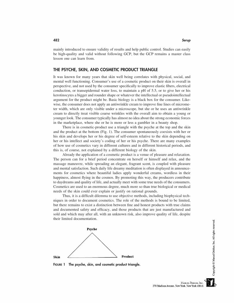
482 Serup
mainly introduced to ensure validity of results and help public control. Studies can easilybe high-quality and valid without following GCP, but the GCP remains a master classlesson one can learn from.
THE PSYCHE, SKIN, AND COSMETIC PRODUCT TRIANGLE
It was known for many years that skin well being correlates with physical, social, andmental well functioning. Consumer’s use of a cosmetic product on their skin is overall inperspective, and not used by the consumer specifically to improve elastic fibers, electricalconduction, or transepidermal water loss, to maintain a pH of 5.5, or to give her or hiskerotinocytes a bigger and rounder shape or whatever the intellectual or pseudointellectualargument for the product might be. Basic biology is a black box for the consumer. Like-wise, the consumer does not apply an antiwrinkle cream to improve fine lines of microme-ter width, which are only visible under a microscope, but she or he uses an antiwrinklecream to directly treat visible coarse wrinkles with the overall aim to obtain a young oryounger look. The consumer typically has almost no idea about the strong economic forcesin the marketplace, where she or he is more or less a gambler in a beauty shop.
There is in cosmetic-product use a triangle with the psyche at the top and the skinand the product at the bottom (Fig. 1). The consumer spontaneously coexists with her orhis skin and develops her or his degree of self-esteem relative to the skin depending onher or his intellect and society’s coding of her or his psyche. There are many examplesof how use of cosmetics vary in different cultures and in different historical periods, andthis is, of course, not explained by a different biology of the skin.
Already the application of a cosmetic product is a venue of pleasure and relaxation.The person can for a brief period concentrate on herself or himself and relax, and themassage maneuvre, while spreading an elegant, fragrant scent, is coupled with pleasureand mental satisfaction. Such daily life dreamy meditation is often displayed in announce-ments for cosmetics where beautiful ladies apply wonderful creams, wordless in theirhappiness, almost flying in the cosmos. By promoting this way, the producers contributeto daydreams and quality of life, and actually meet with some true needs of the consumers.Cosmetics are used to an enormous degree, much more so than true biological or medicalneeds of the skin could ever explain or justify on rational grounds.
Thus, it is a difficult dilemma to use objective methods, including biophysical tech-niques in order to document cosmetics. The role of the methods is bound to be limited,but there remains to exist a distinction between fine and honest products with true claimsand documented safety and efficacy, and those products that are just manufactured andsold and which may after all, with an unknown risk, also improve quality of life, despitetheir limited documentation.
FIGURE 1 The psyche, skin, and cosmetic product triangle.
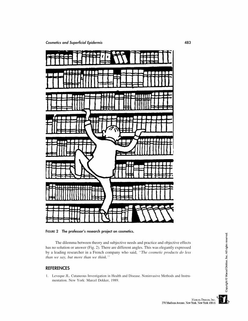
Cosmetics and Superficial Epidermis 483
FIGURE 2 The professor’s research project on cosmetics.
The dilemma between theory and subjective needs and practice and objective effectshas no solution or answer (Fig. 2). There are different angles. This was elegantly expressedby a leading researcher in a French company who said, ‘‘The cosmetic products do lessthan we say, but more than we think.’’
REFERENCES
1. Leveque JL. Cutaneous Investigation in Health and Disease. Noninvasive Methods and Instru-mentation. New York: Marcel Dekker, 1989.
484 Serup
2. Serup J, Jemec GBE. Handbook of Non-Invasive Methods and the Skin. Boca Raton: CRCPress, 1995.
3. Elsner P, Barel AO, Berardesca E, Gabard B, Serup J. Skin Bioengineering. Techniques andApplications in Dermatology and Cosmetology. Basel: S. Karger, 1998.
4. Schaefer H, Redelmeier TE. Skin Barrier. Principles of Percutaneous Absorption. Basel: S.Karger, 1996.
5. Berardesca E, Elsner P, Wilhelm KP, Maibach HI. Bioengineering of the Skin: Methods andInstrumentation. Boca Raton: CRC Press, 1995.
6. Elsner P, Berardesca E, Maibach HI. Bioengineering of the Skin: Water and the Stratum Cor-neum. Boca Raton: CRC Press, 1994.
7. Wilhelm KP, Elsner P, Berardesca E, Maibach HI. Bioengineering of the Skin: Skin SurfaceImaging and Analysis. Boca Raton: CRC Press, 1997.
8. Serup J. Bioengineering and the skin: from Standard error to standard operating procedure.Acta Derm Venereol (Stockh) 1994; 185(Suppl):5–8.
9. ICH Harmonised Tripartite Guideline for Good Clinical Practice. Brookwood: BrookwoodMedical Publications Ltd, 1996.
42
Skin Cleansing Bars
Joshua B. Ghaim and Elizabeth D. VolzColgate-Palmolive Company, Piscataway, New Jersey
INTRODUCTION
Although the origin of soap is not very clear, it is widely accepted that some form ofprimitive soap-making methods existed several thousand years ago, dating as far back to2000 bc. For many centuries, soaps were made by heating a mixture of animal fats (tallow)with lye, a basic solution obtained from wood ashes [1]. Until the late eighteenth century,soap was considered a luxury item available only to royalty and the social upper class.Today, soaps are produced using a variety of much more refined processes and differentfats and oils, resulting in finished products that deliver consumer-relevant performancebenefits with desirable aesthetics [1]. In this section, we will discuss the chemical andphysical properties of commercial soap bars with a focus on skin cleansing, the raw materi-als needed, the manufacturing and process requirements, and the final finished productperformance evaluations.
WHAT IS SOAP?
Soap is generally defined as an alkali salt of a long-chain fatty acid. When a fat or oil issaponified, the sodium or potassium salt formed from the long-chain fatty acids is calleda soap. The term ‘‘soap’’ refers to a group of neutralized long-chain carboxylic acids,which result from two primary ingredients: an alkali and a triglyceride (fat or oil). Thechain length of the aliphatic group is typically between 7 and 21 carbons with one carbox-ylate carbon, yielding a molecule containing 8 to 22 carbons. The cation associated withthe carboxylate head group generally comprises sodium, potassium, or to a lesser extentother cations such as triethanolamine as well as heavy metals and alkali earth metals suchas magnesium.
Soap cleans by altering the surface tension of water and emulsifying and suspendingsoils to be rinsed away. The two ends of soap have different polarities where the longcarbon chain end is nonpolar and hydrophobic, whereas the carboxylate salt end is ionicand hydrophilic. When a soap is used to clean grease or dirt, the nonpolar ends of thesoap molecules solubilize nonpolar fats and oils that accompany dirt. The water-loving(hydrophilic) salt ends of the soap molecules extend outside where they can be solubilizedin water. The soap molecules coat the oil or grease, forming clusters called micelles. The
485
486 Ghaim and Volz
hydrophilic end of the soap molecules provides polarity to the micelles, thus emulsifyingthem in water. As a result, small globules of oil and fat coated with soap molecules arepulled into the water layer and can be rinsed away.
SOAP RAW MATERIALS
Fats and Oils
The naturally occurring fats and oils used in soap making are glycerides with three fattyacid groups randomly esterified with glycerol (trihydroxy alcohol). The difference betweenfats and oils is merely one of their physical states: fats are solids and oils are liquids. Fatsand oils typically comprise both saturated and unsaturated fatty acid molecules containingbetween 7 and 21 carbons randomly distributed on the glycerol backbone. Overall, thereaction of caustic (lye) with triglycerides yields glycerin and soap in a reaction knownas saponification. This is the most widely used soap making process. The second majorsoap making process is the neutralization of fatty acids with an alkali. Fats and oils arehydrolyzed (split) with high-pressure steam to yield crude fatty acids and glycerin. Thefatty acids are then purified by distillation and neutralized with an alkali to produce soapand water (neat soap) [2–7].
The properties of the resulting soap are determined by the quality and compositionof the component fatty acids in the starting fat mixture. In general, chain lengths of lessthan 12 carbon atoms are more irritating to the skin; conversely, saturated chain lengthsgreater than 18 carbon atoms form soaps less soluble for ready solution and sudsing.Similarly, a higher proportion of unsaturated fatty acids (e.g., oleic and linolenic) yieldssoaps susceptible to undesirable atmospheric oxidative changes. For these reasons and thefact that fats and oils are treated as commodities in the open market, the number of fatsand oils suitable for commercial soapmaking is limited. The selection of the appropriatestarting fats and oils forming the base composition of a soap is key to its quality andperformance. Among the fats and oils used throughout the world, beef and sheep talloware the most common fats, and oils from coconut, palm, soy, and babassu are the mostfrequently used oils. Soap compositions containing fractions of oils such as palm stearinand other oils with hydrogenation or other upgrading are also in the formulators’ arsenalfor selection. In the United States, most toilet soaps are made from beef tallow and coconutoil. Some of the common fats and oils used in commercial soapmaking are discussed inthe following sections (Table 1).
Tallow
Tallow, which is the principal animal fat in soapmaking, is obtained from the meat pro-cessing industry as a result of rendering the body fat from beef and in some cases sheep[8]. In the United States, most toilet soaps are made from beef tallow and coconut oil.The properties of these and other fats are dependent on the constituent fatty acids. Tallowfrom different sources may vary considerably in color (both initial and after bleaching),titer (solidification point of the fatty acids), free fatty acid content, saponification value(alkali required for saponification), and iodine value (measure of unsaturation). Tallow iscomposed of mostly long-chain saturated and unsaturated fatty acids—mostly C16 (pal-mitic, 28%), C18 (stearic, 18%), and C18:1 (oleic, 44%)—providing hardness and thick andcreamy long-lasting lather (Table 1).
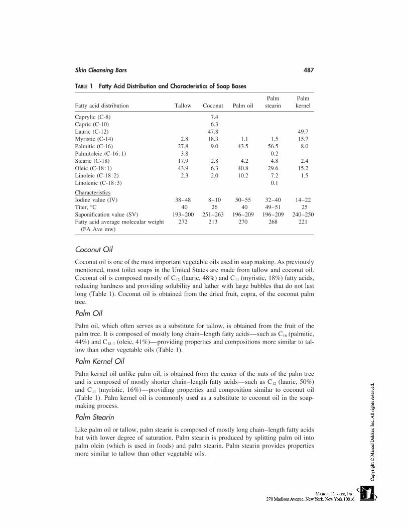
Skin Cleansing Bars 487
TABLE 1 Fatty Acid Distribution and Characteristics of Soap Bases
Palm PalmFatty acid distribution Tallow Coconut Palm oil stearin kernel
Caprylic (C-8) 7.4Capric (C-10) 6.3Lauric (C-12) 47.8 49.7Myristic (C-14) 2.8 18.3 1.1 1.5 15.7Palmitic (C-16) 27.8 9.0 43.5 56.5 8.0Palmitoleic (C-16:1) 3.8 0.2Stearic (C-18) 17.9 2.8 4.2 4.8 2.4Oleic (C-18:1) 43.9 6.3 40.8 29.6 15.2Linoleic (C-18:2) 2.3 2.0 10.2 7.2 1.5Linolenic (C-18:3) 0.1
CharacteristicsIodine value (IV) 38–48 8–10 50–55 32–40 14–22Titer, °C 40 26 40 49–51 25Saponification value (SV) 193–200 251–263 196–209 196–209 240–250Fatty acid average molecular weight 272 213 270 268 221
(FA Ave mw)
Coconut Oil
Coconut oil is one of the most important vegetable oils used in soap making. As previouslymentioned, most toilet soaps in the United States are made from tallow and coconut oil.Coconut oil is composed mostly of C12 (lauric, 48%) and C14 (myristic, 18%) fatty acids,reducing hardness and providing solubility and lather with large bubbles that do not lastlong (Table 1). Coconut oil is obtained from the dried fruit, copra, of the coconut palmtree.
Palm Oil
Palm oil, which often serves as a substitute for tallow, is obtained from the fruit of thepalm tree. It is composed of mostly long chain–length fatty acids—such as C16 (palmitic,44%) and C18:1 (oleic, 41%)—providing properties and compositions more similar to tal-low than other vegetable oils (Table 1).
Palm Kernel Oil
Palm kernel oil unlike palm oil, is obtained from the center of the nuts of the palm treeand is composed of mostly shorter chain–length fatty acids—such as C12 (lauric, 50%)and C14 (myristic, 16%)—providing properties and composition similar to coconut oil(Table 1). Palm kernel oil is commonly used as a substitute to coconut oil in the soap-making process.
Palm Stearin
Like palm oil or tallow, palm stearin is composed of mostly long chain–length fatty acidsbut with lower degree of saturation. Palm stearin is produced by splitting palm oil intopalm olein (which is used in foods) and palm stearin. Palm stearin provides propertiesmore similar to tallow than other vegetable oils.
488 Ghaim and Volz
Although the five oils discussed are the most commonly used fats and oils in thesoap-making industry, other sources such as lard (hog fat), Babassu oil, rice bran oil, palmkernel olein, and soybean oil are also used throughout the world.
SOAP PHASES
The physicochemical nature of soap has been shown to be critical for the in-use properties.It is generally accepted that four distinct sodium soap crystalline phases exist. These soapphases are referred to as the beta, delta, omega, and liquid crystalline phases. Today,radiographic diffraction (XRD) is considered the simplest and most reliable method fordistinguishing the different phases. The phases designate the lattice spacing between thehydrocarbon chains and are predictive of physical properties such as lather, slough, use-up rate, and even the degree of translucency of a soap bar [9]. The large crystals of theomega phase with the liquid phase are formed when neat soap is cooled down (after thedrying step). Beta-phase conversion in soap bars depends on several factors, includingtemperature, type of surfactant, moisture level and number of millings. Delta phase isformed by the recrystallization of saturated higher chain soaps under specific temperatureconditions and moisture level. Ferguson et al. first linked XRD measurements to the physi-cal properties and characteristics of soap bars as finished product. For instance, delta phaseprovides low slough and low wear rate, whereas beta phase has good lather, low wearrate, and high slough [9].
SOAP BASE COMPOSITION AND PERFORMANCE
Product performance profiles are critically dependent on the base composition selection.For example, the relatively less-soluble tallow provides for bar hardness and a dense,stable, small bubbled lather, whereas the more soluble coconut oil provides an easilygenerated lather consisting of large bubbles. In addition to bar hardness, color, odor, andlather considerations, the formulator must be concerned with the solubility of the soap asit impacts on the use-up and sloughing of the final product. A typical soap bar in the UnitedStates uses a tallow/coconut oil base and the ratio of the two components determines latherattributes such as speed, quantity, and richness. An increase of all of these attributes occurswith the increasing proportions of the coconut oil but the higher proportion of coconutoil also results in an increasing degree of irritation to the skin because of the high short-chain–length fatty acid composition. Furthermore, the behavior of the base can be deter-mined not only by the fatty acid chain but also by the cation by which it is neutralized.The cation can also have a significant influence on the solubility and mildness propertiesof the base. For example, a sodium soap would be harder than a potassium soap of thesame carbon chain length [1].
ADDITIVES
Soap manufacturers have developed a variety of formulation approaches to deliver prod-ucts that better meet the consumer needs of today. Even though the base soap compositionhas not changed, consumer needs are met by the inclusion of various additives. As withany other product, the stability (physical and chemical state) of the soap-base–additiveor even additive-additive mix must be considered during the formulation. There are avariety of additives that are formulated into soap bars to provide additional consumer
Skin Cleansing Bars 489
benefits and/or to modify the performance and aesthetics of the final product. A completelist of functional additives can be found in The Cosmetic, Toiletry and Fragrance Associa-tion (CTFA) Cosmetic Ingredient Handbook [10].
Fragrance
Fragrance is by far the most important additive for consumer acceptance of a personalcleansing product. Even though the primary purpose of the selection of a fragrance is totarget a specific user group, it is also used to mask the characteristic base odor associatedwith the fatty acids. Fragrances are compounded from several components including car-boxylic acids, esters, aldehydes, ketones, and glycols where the selection of the compo-nents could adversely effect the stability and/or the processability of the final product.For instance, fragrances with solvents such as dipropylene glycol (glycol) and dieth-ylpthlate (ester) tend to soften and cloud translucent soap bars [2]. The raw-material manu-facturer’s ability to provide cleaner base with significantly less base odor has greatly im-proved in the past two decades, thus allowing soap manufacturers to use less fragrancein the final product or even, in some cases, provide products that are fragrance free. Fra-grances are also known to alter the mildness properties of soap bars. For example, a soapbar that targets consumers with sensitive skin has enough fragrance to mask the base odorof the fatty acid while providing some soft perfume that reinforces their mildness proper-ties. The fragrance levels in the soap bar typically range from 0.3% (sensitive skin) to1.5% (deodorant soaps). Long-term aging studies are always necessary in order to assessthe stability of the fragrance in the soap base and its continued ability to mask the baseodor.
Free Fatty Acid or Superfatting
Traditional soap bars are alkaline in nature with a pH of around 10. A manufacturingprocess with excess fatty acid beyond what is needed by the reaction yields a final productwith free fatty acid, also known as ‘‘superfatted’’ soap. Conversely, a process with causticin excess of what is needed by the reaction yields a base soap with a slight excess of freecaustic. Excess caustic can be neutralized by the addition of excess free fatty acids suchas coconut, palm kernel, or stearic acid, or by postaddition of weak acids such as citricor phosphoric acid. Superfatting enhances the lather profile of the soap bar, eliminates freealkali (lowers the pH), and can provide some improvement of skin mildness attributes [1].
Glycerin
Glycerin is a common ingredient formulated into soap bars that dates back to ancienttimes. As previously discussed, it is the by-product of saponification and thus has alwaysbeen present in soaps in varying levels [2]. Glycerin is well known for its ability to absorbwater (humectancy). This makes it an ideal additive for skincare (moisturization) benefits.Its humectant properties, even at low levels, can alter the rinsability of the soap bar, thusmodifying the consumer perception of the product as clean rinsing product.
Colorants and Pigments
The visual appearance of a soap bar is known to influence the consumer acceptance ofthe product. Because of color differences of some of the base compositions, it is commonfor most manufacturers to alter the appearance of the final product. This is mostly accom-
490 Ghaim and Volz
plished by the addition of colorants and opacifying agents. Some of the common additivesused to alter the appearance of a soap bar include food and/or cosmetic grade dyes andpigments, as well as lakes and opacifiers such as titanium dioxide and zinc oxide [10].
Preservatives
Soap bases with high proportions of unsaturated fatty acids (e.g., oleic, linoleic, linolenic)[11,12] and the presence of certain soap additives, such as fragrance, tend to be susceptibleto undesirable atmospheric oxidative changes. Therefore, preservatives (chelating agentsand antioxidants) are necessary to prevent such oxidation from occurring. Some commonlyused chelating agents (for trace metals present) in soap bars include ethylenediaminetet-raacetate (EDTA); diethylenetriamine pentaacetate (DTPA, also known as pentasodiumpentate); sodium etidronate or ethane-1-hydroxy-1,1-diphosphonic acid (EHDP) [13]; cit-ric acid; and magnesium silicate. The most commonly used antioxidants in conjunctionwith chelating agent in soap bars are butylatedhydroxytoluene (BHT) and, recently, theaddition of tetradibutyl pentaerithrityl hydroxyhydrocinnamate [14]. Both of these antioxi-dants are soluble in fragrances.
Skin Conditioners
As previously mentioned, consumer demand for products that not only cleanse the skinbut also provide skin mildness and moisturizing benefits is constantly changing. Therefore,it is common for manufacturers to add ingredients that are known to provide such benefits.We previously discussed two of the most commonly used additives, free fatty acid andglycerin. Other additives that are commonly used in soap bars include the following:vitamin E, aloe, jojoba oil, lanolin, glyceryl stearate, isopropyl esters, sodium cetearylsulfate, cetyl esters, petrolatum, silicones, beeswax, ceresin, carbomer-934, sodium poly-acrylate, cocoa butter, mineral oil, and polyethyleneoxideglycol-12, to name a few [10].
Antimicrobial Agents
Soap bars are very effective in removing microbial flora that are known to cause skininfections, pimples, and malodor during the washing/bathing process. The addition ofantimicrobial actives to a soap bar extends this benefit for a longer period of time, mainlybetween washing/bathing. Because of safety concerns about the different actives used insoap bars, the number of antimicrobial agents used in soap bars has decreased from severalin the 1970s to only three today. Trichlorocarbanilide (TCC), trichlorodiphenylhydroxy-ether (triclosan), and parachloro m-xylenol (PCMX) are commonly used in soap barstoday. The selection of which active to use in different products is based on claims orproduct positioning, efficacy, and cost of the final product. TCC is effective mostly againstgram-positive bacteria, whereas triclosan and PCMX have been shown to be effectiveagainst both gram-positive and gram-negative bacteria. The use levels of these actives aredependent on the claims associated with the final products and government regulations.For instance, in the United States the maximum use levels allowed for triclosan and TCCare 1.0% and 1.5%, respectively.
Synthetic Surfactants
The formulation of soap bars has become more complex because of the ever-increasingconsumer demand of products that not only provide cleansing properties but also skin-
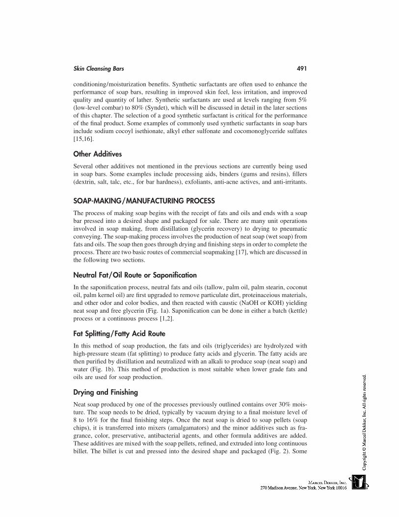
Skin Cleansing Bars 491
conditioning/moisturization benefits. Synthetic surfactants are often used to enhance theperformance of soap bars, resulting in improved skin feel, less irritation, and improvedquality and quantity of lather. Synthetic surfactants are used at levels ranging from 5%(low-level combar) to 80% (Syndet), which will be discussed in detail in the later sectionsof this chapter. The selection of a good synthetic surfactant is critical for the performanceof the final product. Some examples of commonly used synthetic surfactants in soap barsinclude sodium cocoyl isethionate, alkyl ether sulfonate and cocomonoglyceride sulfates[15,16].
Other Additives
Several other additives not mentioned in the previous sections are currently being usedin soap bars. Some examples include processing aids, binders (gums and resins), fillers(dextrin, salt, talc, etc., for bar hardness), exfoliants, anti-acne actives, and anti-irritants.
SOAP-MAKING/MANUFACTURING PROCESS
The process of making soap begins with the receipt of fats and oils and ends with a soapbar pressed into a desired shape and packaged for sale. There are many unit operationsinvolved in soap making, from distillation (glycerin recovery) to drying to pneumaticconveying. The soap-making process involves the production of neat soap (wet soap) fromfats and oils. The soap then goes through drying and finishing steps in order to complete theprocess. There are two basic routes of commercial soapmaking [17], which are discussed inthe following two sections.
Neutral Fat/Oil Route or Saponification
In the saponification process, neutral fats and oils (tallow, palm oil, palm stearin, coconutoil, palm kernel oil) are first upgraded to remove particulate dirt, proteinaceious materials,and other odor and color bodies, and then reacted with caustic (NaOH or KOH) yieldingneat soap and free glycerin (Fig. 1a). Saponification can be done in either a batch (kettle)process or a continuous process [1,2].
Fat Splitting/Fatty Acid Route
In this method of soap production, the fats and oils (triglycerides) are hydrolyzed withhigh-pressure steam (fat splitting) to produce fatty acids and glycerin. The fatty acids arethen purified by distillation and neutralized with an alkali to produce soap (neat soap) andwater (Fig. 1b). This method of production is most suitable when lower grade fats andoils are used for soap production.
Drying and Finishing
Neat soap produced by one of the processes previously outlined contains over 30% mois-ture. The soap needs to be dried, typically by vacuum drying to a final moisture level of8 to 16% for the final finishing steps. Once the neat soap is dried to soap pellets (soapchips), it is transferred into mixers (amalgamators) and the minor additives such as fra-grance, color, preservative, antibacterial agents, and other formula additives are added.These additives are mixed with the soap pellets, refined, and extruded into long continuousbillet. The billet is cut and pressed into the desired shape and packaged (Fig. 2). Some
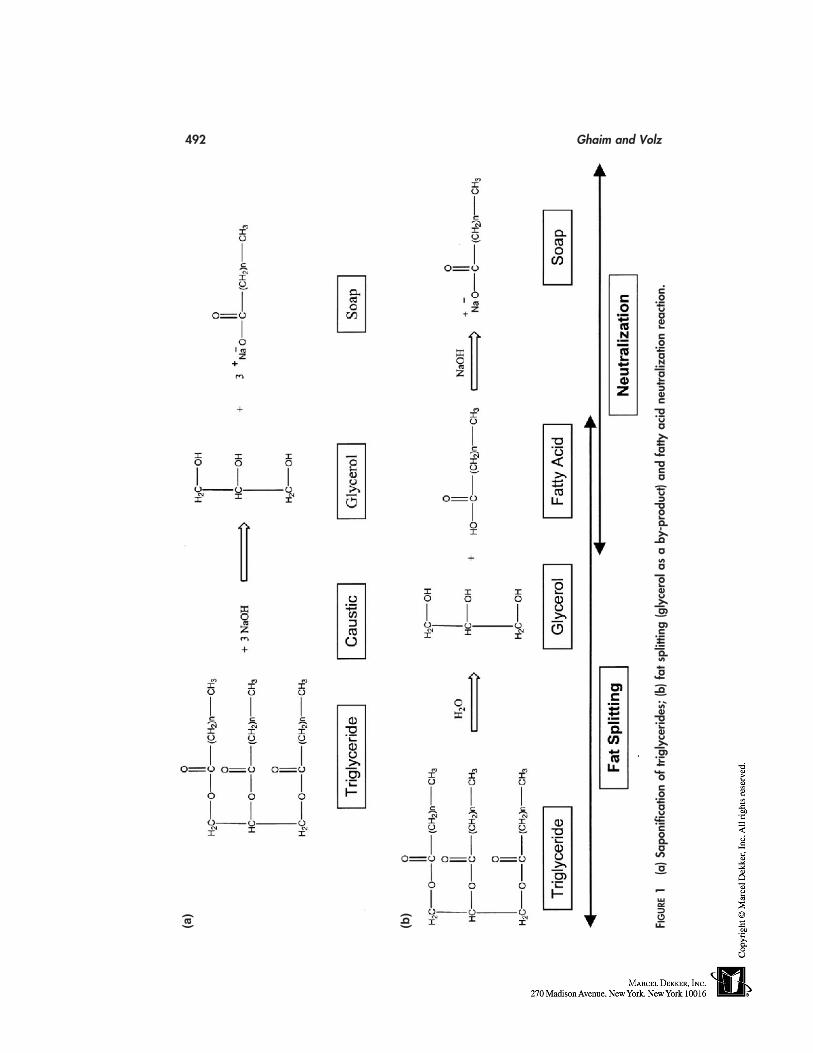
492 Ghaim and Volz
(a)
(b)
F IG
URE
1(a
)Sap
onifi
catio
nof
trig
lyce
rides
;(b)
fats
plitt
ing
(gly
cero
las
aby
-pro
duct
)and
fatty
acid
neut
raliz
atio
nre
actio
n.
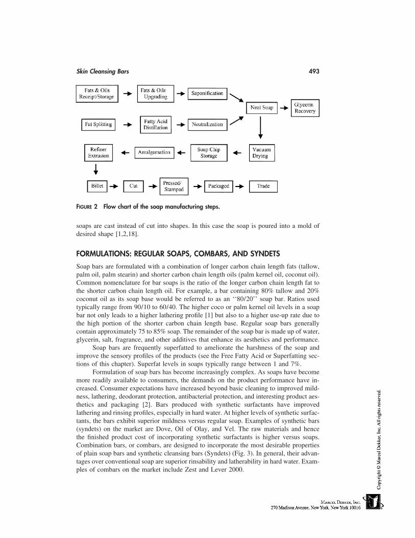
Skin Cleansing Bars 493
FIGURE 2 Flow chart of the soap manufacturing steps.
soaps are cast instead of cut into shapes. In this case the soap is poured into a mold ofdesired shape [1,2,18].
FORMULATIONS: REGULAR SOAPS, COMBARS, AND SYNDETS
Soap bars are formulated with a combination of longer carbon chain length fats (tallow,palm oil, palm stearin) and shorter carbon chain length oils (palm kernel oil, coconut oil).Common nomenclature for bar soaps is the ratio of the longer carbon chain length fat tothe shorter carbon chain length oil. For example, a bar containing 80% tallow and 20%coconut oil as its soap base would be referred to as an ‘‘80/20’’ soap bar. Ratios usedtypically range from 90/10 to 60/40. The higher coco or palm kernel oil levels in a soapbar not only leads to a higher lathering profile [1] but also to a higher use-up rate due tothe high portion of the shorter carbon chain length base. Regular soap bars generallycontain approximately 75 to 85% soap. The remainder of the soap bar is made up of water,glycerin, salt, fragrance, and other additives that enhance its aesthetics and performance.
Soap bars are frequently superfatted to ameliorate the harshness of the soap andimprove the sensory profiles of the products (see the Free Fatty Acid or Superfatting sec-tions of this chapter). Superfat levels in soaps typically range between 1 and 7%.
Formulation of soap bars has become increasingly complex. As soaps have becomemore readily available to consumers, the demands on the product performance have in-creased. Consumer expectations have increased beyond basic cleaning to improved mild-ness, lathering, deodorant protection, antibacterial protection, and interesting product aes-thetics and packaging [2]. Bars produced with synthetic surfactants have improvedlathering and rinsing profiles, especially in hard water. At higher levels of synthetic surfac-tants, the bars exhibit superior mildness versus regular soap. Examples of synthetic bars(syndets) on the market are Dove, Oil of Olay, and Vel. The raw materials and hencethe finished product cost of incorporating synthetic surfactants is higher versus soaps.Combination bars, or combars, are designed to incorporate the most desirable propertiesof plain soap bars and synthetic cleansing bars (Syndets) (Fig. 3). In general, their advan-tages over conventional soap are superior rinsability and latherability in hard water. Exam-ples of combars on the market include Zest and Lever 2000.
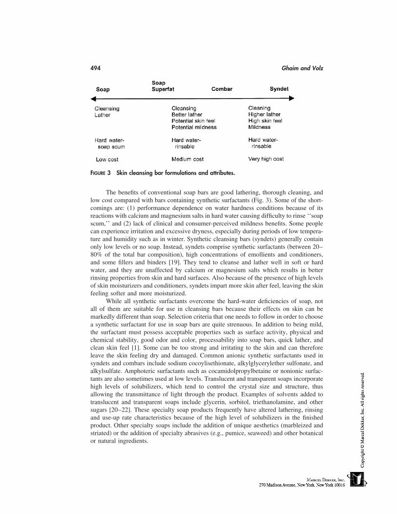
494 Ghaim and Volz
FIGURE 3 Skin cleansing bar formulations and attributes.
The benefits of conventional soap bars are good lathering, thorough cleaning, andlow cost compared with bars containing synthetic surfactants (Fig. 3). Some of the short-comings are: (1) performance dependence on water hardness conditions because of itsreactions with calcium and magnesium salts in hard water causing difficulty to rinse ‘‘soapscum,’’ and (2) lack of clinical and consumer-perceived mildness benefits. Some peoplecan experience irritation and excessive dryness, especially during periods of low tempera-ture and humidity such as in winter. Synthetic cleansing bars (syndets) generally containonly low levels or no soap. Instead, syndets comprise synthetic surfactants (between 20–80% of the total bar composition), high concentrations of emollients and conditioners,and some fillers and binders [19]. They tend to cleanse and lather well in soft or hardwater, and they are unaffected by calcium or magnesium salts which results in betterrinsing properties from skin and hard surfaces. Also because of the presence of high levelsof skin moisturizers and conditioners, syndets impart more skin after feel, leaving the skinfeeling softer and more moisturized.
While all synthetic surfactants overcome the hard-water deficiencies of soap, notall of them are suitable for use in cleansing bars because their effects on skin can bemarkedly different than soap. Selection criteria that one needs to follow in order to choosea synthetic surfactant for use in soap bars are quite strenuous. In addition to being mild,the surfactant must possess acceptable properties such as surface activity, physical andchemical stability, good odor and color, processability into soap bars, quick lather, andclean skin feel [1]. Some can be too strong and irritating to the skin and can thereforeleave the skin feeling dry and damaged. Common anionic synthetic surfactants used insyndets and combars include sodium cocoylisethionate, alkylglycerylether sulfonate, andalkylsulfate. Amphoteric surfactants such as cocamidolpropylbetaine or nonionic surfac-tants are also sometimes used at low levels. Translucent and transparent soaps incorporatehigh levels of solubilizers, which tend to control the crystal size and structure, thusallowing the transmittance of light through the product. Examples of solvents added totranslucent and transparent soaps include glycerin, sorbitol, triethanolamine, and othersugars [20–22]. These specialty soap products frequently have altered lathering, rinsingand use-up rate characteristics because of the high level of solubilizers in the finishedproduct. Other specialty soaps include the addition of unique aesthetics (marbleized andstriated) or the addition of specialty abrasives (e.g., pumice, seaweed) and other botanicalor natural ingredients.
Skin Cleansing Bars 495
BAR SOAP PERFORMANCE EVALUATIONS
Soap bars are evaluated for several characteristics to ensure that they meet consumer needsand expectations.
Lather
The amount of lather, how rapidly a product lathers, and the quality of lather can be judgedby a trained panel. This trained panel rates the product on lather quantity, quality, andquickness by rating it on a numerical scale. Typically panelists are trained to rotate a soapbar a fixed number of times and evaluate it for attributes versus benchmark products. Thisis most useful in the analysis and comparison of formulation similarities and differencesas well as competitive products. Variables affecting lather performance of a product in-clude water temperature, water hardness, and method of washing. Trained panelists needto be trained and validated on a regular basis to ensure consistency of their evaluations.
Laboratory methods of lather evaluation include the Ross-Miles foam height test.This requires measuring the foam height of a soap solution that has been inverted in acylinder for a fixed number of times. Results from this type of test can be misleadingbecause the bar shape and solubility can affect the lather performance in use [1].
Wear Rate/Use-Up
The measurement of how long a bar lasts under normal use conditions is an importantattribute to the consumer perceived value. The use-up rate is measured by first weighingthe soap bar and then washing the bar for a set number and length of times (for example,25 washings for 10 sec each). The bar is then dried and weighed again and the use-up orwear rate is reported as the percent weight loss. Soap bar shape and size impact the reporteduse-up rate. The use-up rate measurement must be controlled for water hardness and tem-perature. For formulation comparison purposes, it is best to compare soap bars with similarsizes and shapes. Bars can be shaved to the same sizes and shapes in order for the measure-ment to reflect the true formula influence. To compare how bars will perform in the handsof consumers, actual commercial sizes and shapes should be used.
Slough/Mush
Slough or mush is the undesirable soft part of the bar that results from the hydration ofa soap bar as it sits in a wet soap dish. Slough is measured by placing a pre-weighed barin a high humidity chamber for a fixed period of time, then removing the soft part of thebar and allowing the soap bar to dry. The weight taken before and after determine theslough or mush measured as the percent weight loss. Syndet bars tend to have high sloughrelative to regular soaps. High humidity conditions exaggerate typical home usage condi-tions, but help differentiate products and formulations. Slough can also be run at roomtemperature. Commercial soap bar shapes can be selected by manufacturers to minimizethe formation of slough or mush in use conditions.
Cracking
Cracking is the splitting of a bar along the side seams or at any part in the bar duringuse. Cracking of a soap bar in use conditions is a perceived as a negative by consumers.
496 Ghaim and Volz
Cracking is evaluated by partially submerging bars in water of fixed hardness and tempera-ture for a set period of time. The bars are then dried and evaluated for cracking after oneto two days. Ideally, there should be no cracks present in the soap bars.
Hardness
Bar hardness is a mechanical measure of how resistant the bar is to physical pressure.Bar hardness can be mechanically measured in finishing trials for machineability as wellas during routine lab evaluations. Bars that are too soft may be difficult to extrude on thefinishing line without significant surface defects.
Bar Feel and Sandiness
Bar soaps are typically evaluated for dry specks and drag. Specks of dry soap (insolublesoap) can occur during the manufacture of the base soap or syndet or from the additivesin the soap bar. These specks show up as distinct bumps on the surface of the bar. Thebar is washed under controlled water conditions with cooler water bringing out moreobvious dry specks. The bar is both evaluated during wash and after drying for feel andappearance and rated against standard quality bars.
Sensory Skin Evaluations
Clearly, next to the fragrance preference at the point of purchase, skin feel and lather arethe most important attributes for consumers. Various skin feel attributes from bar soapsare evaluated by a trained panel of experts. These groups of panelists are trained to evaluatesmall (or large) differences in products focusing on a set of defined attributes. Productsare usually compared with a reference product. Examples of attributes evaluated by atrained panel for skin feel include time to rinse, skin slip, tightness of skin after drying,and smoothness of skin.
Clinical Evaluations
Clinical Evaluations of soap products are used to determine how effective the productsare on certain attributes, primarily mildness/irritation, skin dryness/tightness, antibacterialefficacy, and deodorancy. There are several methods of measuring the clinical attributesof a soap bar ranging from trained panels to biophysical instrumentation [1,2,23].
REFERENCES
1. Spitz L, ed. Soap Technology for the 1990’s. Champaign: American Oil Chemists Society,1990.
2. Spitz L, ed. Soaps and Detergents: A Theoretical and Practical Review. Champaign: AmericanOil Chemists Society, 1996.
3. Woolatt E. The Manufacture of Soaps, Other Detergents and Glycerin. New York: Halstead,1985.
4. Thomsenn EG, Kemp CR. Modern Soap Making. New York: MacNair-Dorland, 1937.5. Joshi D. U.S. Patent, 4,493,786 (1985).6. Jungerman E, Hassapis T, Scott R, Wortzman M. U.S. Patent, 4,758,370 (1988).7. Johnson RW, Fritz E, eds. Fatty Acids in Industry: Process, Properties, Derivatives, Applica-
tions. New York: Marcel Dekker, 1989.
Skin Cleansing Bars 497
8. Patterson HBW. Bleaching and Purifying Fats and Oils: Theory and Practice. Champaign:American Oil Chemists Society, 1993.
9. Ferguson RH, Rosevear FB, Stillman RC. Solid soap phases. Ind & Engineer Chem 1943;35:1005–1012.
10. Wenninger JA, McEwen GN. CTFA International Cosmetic Ingredient Dictionary and Hand-book, 7th ed. Washington, D.C: The Cosmetic, Toiletry and Fragrance Association, 1997.
11. Zaidman B, Kisilev A, Sasson Y, Garti N. Double bond oxidation of unsaturated fatty acids.J Am Oil Chem Soc 1988; 65:611.
12. Rojas-Romero AJ, Morton ID. J Sci Fd Agric 1977; 28:916.13. U. S. Patent, 3,511,783 (1970).14. Payne R, Hwang A, Subramanyam R. U. S. Patent, 5,843,876 (1998).15. Blake-Haskins JC, Scala D, Rhein LD, Robbins CR. J Soc Cosmet Chem 1986; 37:199–210.16. M Hollstein, Spitz L. J Oil Chemists’ Soc October 1982; p. 442.17. Soap and Detergent Association. Soaps and Detergents Handbook, 2nd ed. 1994.18. Krawczyk T. Soap Bars Inform May 1996; pp. 478–486.19. Milwidsky B. Syndet Bars, Happi, May 1985:58–70.20. U. S. Patent, 2,970,116.21. Toma K, Hassapis TJ. U. S. Patent, 3,864,272 (1975).22. Wood-Rethwill JC, Jawarski RJ, Myers EG, Marshal ML. U. S. Patent, 4,879,063 (1989).23. Kajs TM, Gartsein V. Review of the instrumental assessment of skin: effects of cleansing
products. Soc Cosmet Chem 1991; 42(4):249–279.

43
Skin Cleansing Liquids
Daisuke Kaneko and Kazutami SakamotoAminoScience Laboratories, Ajinomoto Co., Inc., Kanagawa, Japan
INTRODUCTION
Skin cleansing liquids are products that clean and refresh the skin by removing soil ordirty materials to help keep the skin’s physiological condition normal. There are residualmetabolites on the skin that are unstable and reactive with oxygen or deposited moleculesby sun exposure or skin micro-organisms to form harmful materials to cause skin trouble.Thus cleansing is a necessary daily skincare practice even for normal skin. Furthermore,special care must be taken for sensitive skin or atopic skin because of its vulnerability.In these troubled types of skin, cleanliness must be attained without contributing to theirsusceptibility [1]. There are different types of cleansing products developed and commonlyused depending on the types of materials to be removed from the skin or types of useconditions.
Typical types of commercial skin cleansing products are listed in Table 1 [2]. Amost common cleansing product contains a relatively high concentration of surfactantsand is applied with water to make foam before washing off thoroughly. Good latheringis the most important feature of these products because sensory feeling of the rich andfine foam is the key factor of repeated use by consumer, although amount and quality offoam are not directly related to the detergency from a physicochemical viewpoint. On theother hand, fine and thick lather serves an important function in shaving foam preparationsfor smooth razor application. Ease of quick rinse and after-feeling are other factors thatrule the quality of skin cleansing products. Refreshed and moist feelings are typical ele-ments that fulfill consumers’ desires, and refreshing seems more important for body wash,especially for Japanese consumers.
In terms of formulations for surfactant-type skin cleansers, soap bars have been themost traditional skin cleansers but there are liquid-, paste-, or aerosol-type cleansers get-ting more popular on the market. Facial cleansing powder—a rather new and niche trendin Japan—contains enzymes to help the cleaning of protein-type deposits because of itsanhydrous formula to preserve enzyme activities.
Solvent type is mainly used to remove oily cosmetics applied to the skin. This typeis further categorized to cleansing creams, lotions, liquids, or gels. The use of makeupproducts, such as waterproof or nonstaining and long-lasting lipsticks, require use of spe-
499
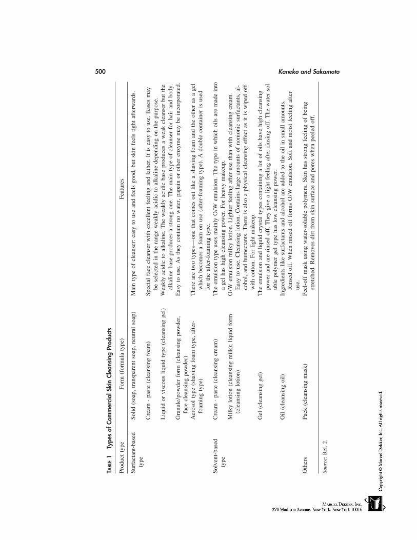
500 Kaneko and Sakamoto
TABL
E1
Type
sof
Com
mer
cial
Skin
Clea
nsin
gPr
oduc
ts
Prod
uct
type
Form
(for
mul
aty
pe)
Feat
ures
Surf
acta
nt-b
ased
Solid
(soa
p,tr
ansp
aren
tso
ap,
neut
ral
soap
)M
ain
type
ofcl
eans
er:
easy
tous
ean
dfe
els
good
,bu
tsk
infe
els
tight
afte
rwar
ds.
type
Cre
am⋅p
aste
(cle
ansi
ngfo
am)
Spec
ial
face
clea
nser
with
exce
llent
feel
ing
and
lath
er.
Itis
easy
tous
e.B
ases
may
bese
lect
edin
the
rang
ew
eakl
yac
idic
toal
kalin
ede
pend
ing
onth
epu
rpos
e.L
iqui
dor
visc
ous
liqui
dty
pe(c
lean
sing
gel)
Wea
kly
acid
icto
alka
line.
The
wea
kly
acid
icba
sepr
oduc
esa
wea
kcl
eans
erbu
tth
eal
kalin
eba
sepr
oduc
esa
stro
ngon
e.T
hem
ain
type
ofcl
eans
erfo
rha
iran
dbo
dy.
Gra
nule
/pow
der
form
(cle
ansi
ngpo
wde
r,E
asy
tous
e.A
sth
eyco
ntai
nno
wat
er,
papa
inor
othe
ren
zym
em
aybe
inco
rpor
ated
.fa
cecl
eans
ing
pow
der)
Aer
osol
type
(sha
ving
foam
type
,af
ter-
The
rear
etw
oty
pes—
one
that
com
esou
tlik
ea
shav
ing
foam
and
the
othe
ras
age
lfo
amin
gty
pe)
whi
chbe
com
esa
foam
onus
e(a
fter
-foa
min
gty
pe).
Ado
uble
cont
aine
ris
used
for
the
afte
r-fo
amin
gty
pe.
Solv
ent-
base
dC
ream
⋅pas
te(c
lean
sing
crea
m)
The
emul
sion
type
uses
mai
nly
O/W
emul
sion
.T
hety
pein
whi
choi
lsar
em
ade
into
type
age
lha
shi
ghcl
eans
ing
pow
er.
For
heav
ym
akeu
p.M
ilky
lotio
n(c
lean
sing
milk
);liq
uid
form
O/W
emul
sion
milk
ylo
tion.
Lig
hter
feel
ing
afte
rus
eth
anw
ithcl
eans
ing
crea
m.
(cle
ansi
nglo
tion)
Eas
yto
use.
Cle
ansi
nglo
tion.
Con
tain
sla
rge
amou
nts
ofno
nion
icsu
rfac
tant
s,al
-co
hol,
and
hum
ecta
nts.
The
reis
also
aph
ysic
alcl
eans
ing
effe
ctas
itis
wip
edof
fw
ithco
tton.
For
light
mak
eup.
Gel
(cle
ansi
ngge
l)T
heem
ulsi
onan
dliq
uid
crys
tal
type
sco
ntai
ning
alo
tof
oils
have
high
clea
nsin
gpo
wer
and
are
rins
edof
f.T
hey
give
alig
htfe
elin
gaf
ter
rins
ing
off.
The
wat
er-s
ol-
uble
poly
mer
gel
type
has
low
clea
nsin
gpo
wer
.O
il(c
lean
sing
oil)
Ingr
edie
nts
like
surf
acta
nts
and
alco
hol
are
adde
dto
the
oil
insm
all
amou
nts.
Rin
sed
off.
Whe
nri
nsed
off
form
sO
/Wem
ulsi
on.
Soft
and
moi
stfe
elin
gaf
ter
use.
Oth
ers
Pack
(cle
ansi
ngm
ask)
Peel
-off
mas
kus
ing
wat
er-s
olub
lepo
lym
ers.
Skin
has
stro
ngfe
elin
gof
bein
gst
retc
hed.
Rem
oves
dirt
from
skin
surf
ace
and
pore
sw
hen
peel
edof
f.
Sour
ce:
Ref
.2.
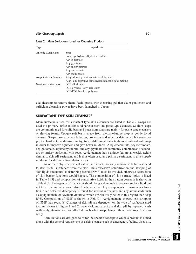
Skin Cleansing Liquids 501
TABLE 2 Main Surfactants Used for Cleansing Products
Type Ingredients
Anionic Surfactants SoapPolyoxyethylene alkyl ether sulfateAcylglutamateAcylglycinateAcylmethyltaurateAcylsurcosinateAcylisethionate
Ampoteric surfactants Alkyl dimethylaminoacetic acid betaineAlkyl amidopropyl dimethylaminoacetic acid betaine
Nonionic surfactants POE alkyl etherPOE glycerol fatty acid esterPOE-POP block copolymer
cial cleansers to remove them. Facial packs with cleansing gel that claim gentleness andsufficient cleansing power have been launched in Japan.
SURFACTANT-TYPE SKIN CLEANSERS
Main surfactants used for surfactant-type skin cleansers are listed in Table 2. Soaps areused as a primary surfactant for solid bar cleansers and paste-type cleansers. Sodium soapsare commonly used for solid bars and potassium soaps are mainly for paste-type cleansersor shaving foams. Opaque soft bar is made from triethanolamine soap as gentle facialcleanser. Soaps have excellent lathering properties and superior detergency but some de-posit in hard water and cause skin tightness. Additional surfactants are combined with soapin order to improve tightness and give better mildness. Alkylethersulfate, acylisethionate,acylglutamate, acylmethyltaurate, and acylglycinate are commonly combined as a second-ary or tertiary surfactant with soap. Acylglutamate has a unique feature as weakly acidicsimilar to skin pH surfactant and is thus often used as a primary surfactant to give superbmildness for different formulation types.
As of their physicochemical nature, surfactants not only remove soils but also tendto strip useful substances from the skin. Thus excessive solubilization and stripping ofskin lipids and natural moisturizing factors (NMF) must be avoided, otherwise destructionof skin-barrier functions would happen. The composition of skin-surface lipids is listedin Table 3 [3] and composition of constitutive lipids in the stratum corneum is shown inTable 4 [4]. Detergency of surfactant should be good enough to remove surface lipid butnot to strip minimally constitutive lipids, which are key components of skin-barrier func-tion. Such selective detergency is found for several surfactants and acylaminoacids suchas acylglutamate or acylmethyltaurate, which are relatively better in this regard than soap[5,6]. Composition of NMF is shown in Ref. [7]. Acylglutamate showed less strippingof NMF than soap. [8] Changes of skin pH are dependent on the type of surfactant usedtoo. As shown in Figure 1 and 2, water-holding capacity and skin pH by repeated washwith acylglutamate was not affected much while soap changed these two properties seri-ously.
Formulations are designed to fit for the specific concept to which a product is aimedalong with the general requirement as a skin cleanser such as detergency, feeling, viscosity,
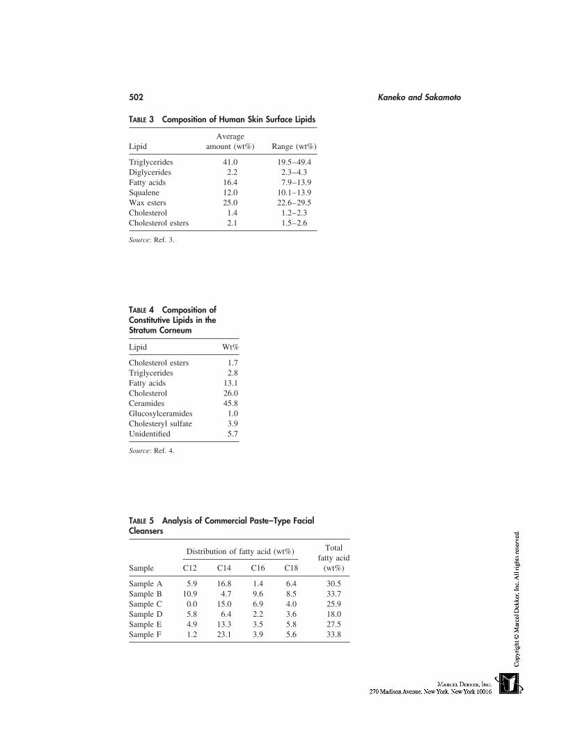
502 Kaneko and Sakamoto
TABLE 3 Composition of Human Skin Surface Lipids
AverageLipid amount (wt%) Range (wt%)
Triglycerides 41.0 19.5–49.4Diglycerides 2.2 2.3–4.3Fatty acids 16.4 7.9–13.9Squalene 12.0 10.1–13.9Wax esters 25.0 22.6–29.5Cholesterol 1.4 1.2–2.3Cholesterol esters 2.1 1.5–2.6
Source: Ref. 3.
TABLE 4 Composition ofConstitutive Lipids in theStratum Corneum
Lipid Wt%
Cholesterol esters 1.7Triglycerides 2.8Fatty acids 13.1Cholesterol 26.0Ceramides 45.8Glucosylceramides 1.0Cholesteryl sulfate 3.9Unidentified 5.7
Source: Ref. 4.
TABLE 5 Analysis of Commercial Paste–Type FacialCleansers
TotalDistribution of fatty acid (wt%)fatty acid
Sample C12 C14 C16 C18 (wt%)
Sample A 5.9 16.8 1.4 6.4 30.5Sample B 10.9 4.7 9.6 8.5 33.7Sample C 0.0 15.0 6.9 4.0 25.9Sample D 5.8 6.4 2.2 3.6 18.0Sample E 4.9 13.3 3.5 5.8 27.5Sample F 1.2 23.1 3.9 5.6 33.8
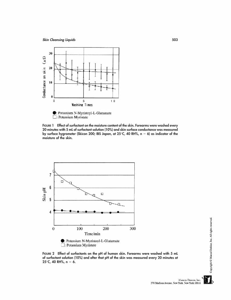
Skin Cleansing Liquids 503
FIGURE 1 Effect of surfactant on the moisture content of the skin. Forearms were washed every20 minutes with 5 mL of surfactant solution (10%) and skin surface conductance was measuredby surface hygrometer (Skicon 200; IBS Japan, at 25°C, 40 RH%, n � 6) as indicator of themoisture of the skin.
FIGURE 2 Effect of surfactants on the pH of human skin. Forearms were washed with 5 mLof surfactant solution (10%) and after that pH of the skin was measured every 20 minutes at25°C, 40 RH%, n � 6.
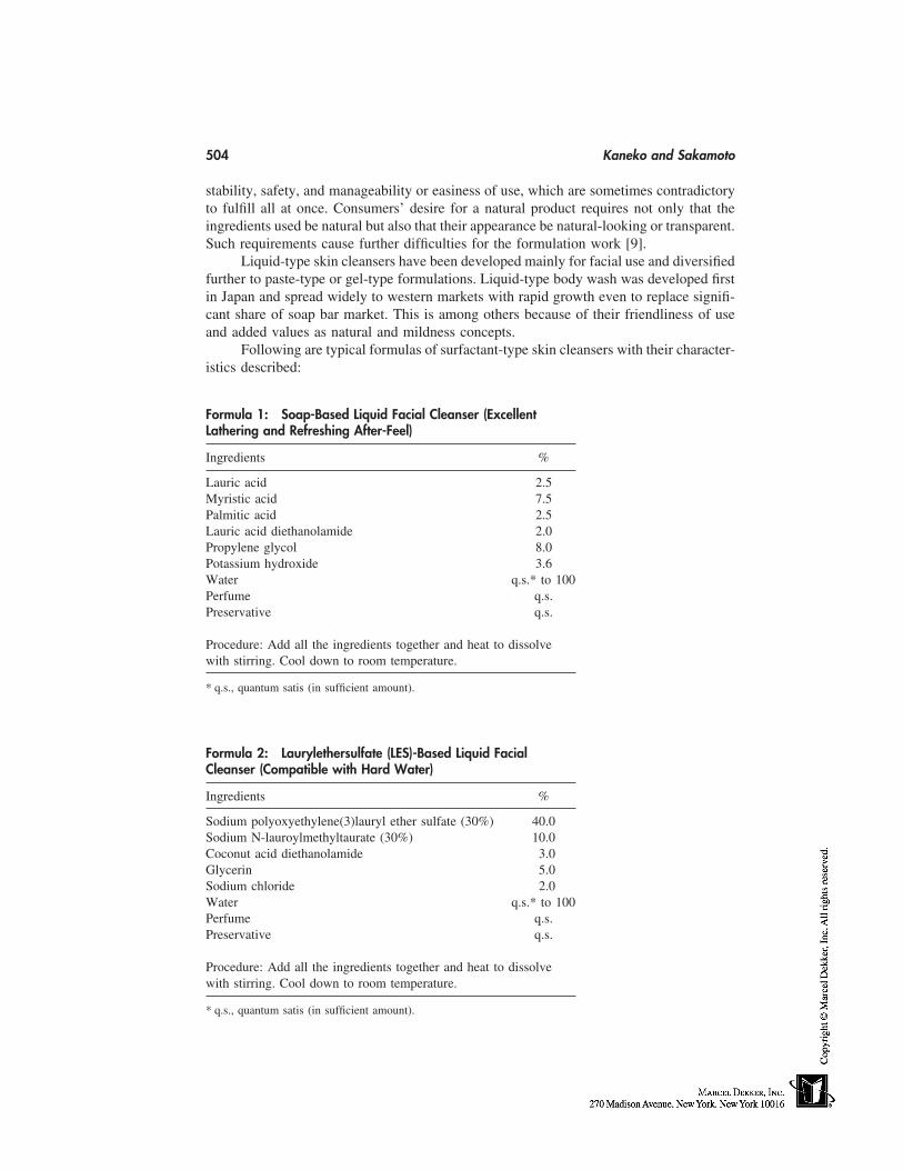
504 Kaneko and Sakamoto
stability, safety, and manageability or easiness of use, which are sometimes contradictoryto fulfill all at once. Consumers’ desire for a natural product requires not only that theingredients used be natural but also that their appearance be natural-looking or transparent.Such requirements cause further difficulties for the formulation work [9].
Liquid-type skin cleansers have been developed mainly for facial use and diversifiedfurther to paste-type or gel-type formulations. Liquid-type body wash was developed firstin Japan and spread widely to western markets with rapid growth even to replace signifi-cant share of soap bar market. This is among others because of their friendliness of useand added values as natural and mildness concepts.
Following are typical formulas of surfactant-type skin cleansers with their character-istics described:
Formula 1: Soap-Based Liquid Facial Cleanser (ExcellentLathering and Refreshing After-Feel)
Ingredients %
Lauric acid 2.5Myristic acid 7.5Palmitic acid 2.5Lauric acid diethanolamide 2.0Propylene glycol 8.0Potassium hydroxide 3.6Water q.s.* to 100Perfume q.s.Preservative q.s.
Procedure: Add all the ingredients together and heat to dissolvewith stirring. Cool down to room temperature.
* q.s., quantum satis (in sufficient amount).
Formula 2: Laurylethersulfate (LES)-Based Liquid FacialCleanser (Compatible with Hard Water)
Ingredients %
Sodium polyoxyethylene(3)lauryl ether sulfate (30%) 40.0Sodium N-lauroylmethyltaurate (30%) 10.0Coconut acid diethanolamide 3.0Glycerin 5.0Sodium chloride 2.0Water q.s.* to 100Perfume q.s.Preservative q.s.
Procedure: Add all the ingredients together and heat to dissolvewith stirring. Cool down to room temperature.
* q.s., quantum satis (in sufficient amount).
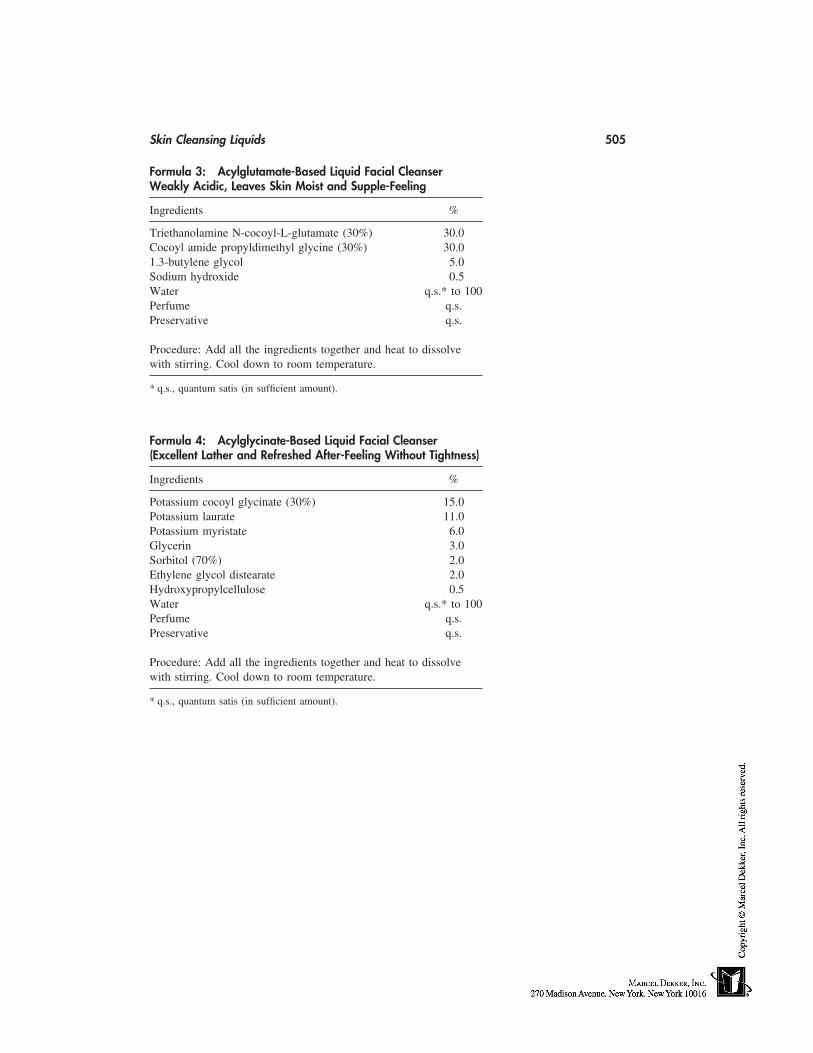
Skin Cleansing Liquids 505
Formula 3: Acylglutamate-Based Liquid Facial CleanserWeakly Acidic, Leaves Skin Moist and Supple-Feeling
Ingredients %
Triethanolamine N-cocoyl-L-glutamate (30%) 30.0Cocoyl amide propyldimethyl glycine (30%) 30.01.3-butylene glycol 5.0Sodium hydroxide 0.5Water q.s.* to 100Perfume q.s.Preservative q.s.
Procedure: Add all the ingredients together and heat to dissolvewith stirring. Cool down to room temperature.
* q.s., quantum satis (in sufficient amount).
Formula 4: Acylglycinate-Based Liquid Facial Cleanser(Excellent Lather and Refreshed After-Feeling Without Tightness)
Ingredients %
Potassium cocoyl glycinate (30%) 15.0Potassium laurate 11.0Potassium myristate 6.0Glycerin 3.0Sorbitol (70%) 2.0Ethylene glycol distearate 2.0Hydroxypropylcellulose 0.5Water q.s.* to 100Perfume q.s.Preservative q.s.
Procedure: Add all the ingredients together and heat to dissolvewith stirring. Cool down to room temperature.
* q.s., quantum satis (in sufficient amount).
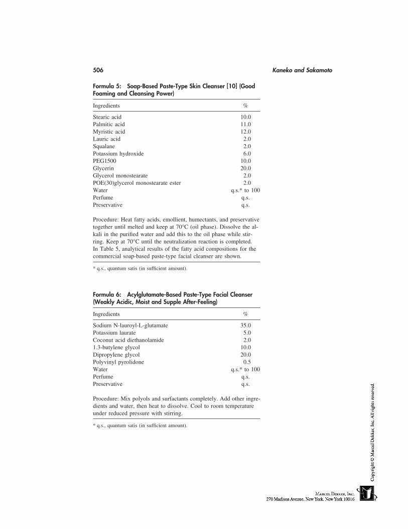
506 Kaneko and Sakamoto
Formula 5: Soap-Based Paste-Type Skin Cleanser [10] (GoodFoaming and Cleansing Power)
Ingredients %
Stearic acid 10.0Palmitic acid 11.0Myristic acid 12.0Lauric acid 2.0Squalane 2.0Potassium hydroxide 6.0PEG1500 10.0Glycerin 20.0Glycerol monostearate 2.0POE(30)glycerol monostearate ester 2.0Water q.s.* to 100Perfume q.s.Preservative q.s.
Procedure: Heat fatty acids, emollient, humectants, and preservativetogether until melted and keep at 70°C (oil phase). Dissolve the al-kali in the purified water and add this to the oil phase while stir-ring. Keep at 70°C until the neutralization reaction is completed.In Table 5, analytical results of the fatty acid compositions for thecommercial soap-based paste-type facial cleanser are shown.
* q.s., quantum satis (in sufficient amount).
Formula 6: Acylglutamate-Based Paste-Type Facial Cleanser(Weakly Acidic, Moist and Supple After-Feeling)
Ingredients %
Sodium N-lauroyl-L-glutamate 35.0Potassium laurate 5.0Coconut acid diethanolamide 2.01.3-butylene glycol 10.0Dipropylene glycol 20.0Polyvinyl pyrolidone 0.5Water q.s.* to 100Perfume q.s.Preservative q.s.
Procedure: Mix polyols and surfactants completely. Add other ingre-dients and water, then heat to dissolve. Cool to room temperatureunder reduced pressure with stirring.
* q.s., quantum satis (in sufficient amount).
Skin Cleansing Liquids 507
Formula 7: Acylglycinate-Based Paste-Type Facial Cleanser(Neutral pH, Fresh After-Feeling)
Ingredients %
Potassium cocoyl glycinate 32.0Potassium myristate 1.5Behenyl alcohol 0.5Citric acid 2.51.3-butylene glycol 15.0Glycerin 17.0Ethylene glycol distearate 2.5Water q.s.* to 100Perfume q.s.Preservative q.s.
Procedure: Mix polyols and surfactants completely. Add other ingre-dients and water then heat to dissolve. Cool to room temperatureunder reduced pressure with stirring.
* q.s., quantum satis (in sufficient amount).
SOLVENT-TYPE SKIN CLEANSERS
Solvent-type cleansers are designed to remove oily residues from cosmetics. Normallythese cleansers are applied by hand to remove oily deposits of colors or pigments fromthe skin, and are then wiped out with tissue or cloth. Water-oil (W/O) emulsions or simpleoils work satisfactorily for this purpose but leave skin oily. Thus surfactant-type cleansersare quite often applied after this treatment. The widespread trend of long-lasting cosmeticsrequires stronger and laborious cleansing with solvent-type cleansers. In order to avoidexcess burden to the skin and achieve effective cleansing of oily deposits, (1) solubilizationand dispersibility, and (2) washability with water are key properties of solvent-type cleans-ers, while mildness is mandatory requirement for the product. For the former need, theproduct should be more lipophilic, and on the contrary for the latter purpose it is betterto be rather hydrophilic. To overcome these contradictory tasks, there are several differentformulations developed that are W/O emulsions, gels, or liquid crystals with special selec-tions and combinations of oil phase and aqueous phase. The principle of these formulasis to have potent oily phase, which can easily interact and solubilize liquid deposits, whenapplied to the skin. Thereafter, by the application of an excess amount of water, a mixturewill form between the cleanser and the oily deposit, which will easily turn into a hydro-philic mixture (such as a W/O emulsion) [11,12].
Following are typical formulas of solvent-type skin cleansers with their characteris-tics described:
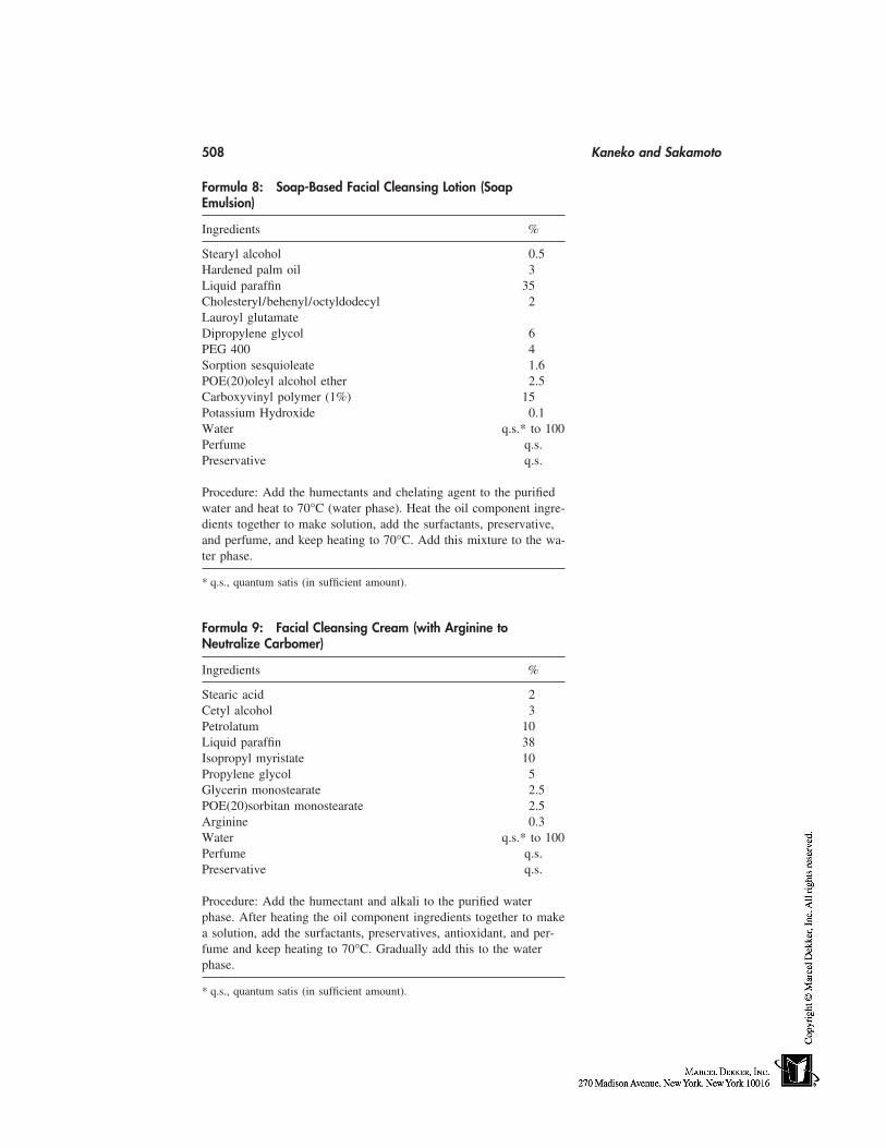
508 Kaneko and Sakamoto
Formula 8: Soap-Based Facial Cleansing Lotion (SoapEmulsion)
Ingredients %
Stearyl alcohol 0.5Hardened palm oil 3Liquid paraffin 35Cholesteryl/behenyl/octyldodecyl 2Lauroyl glutamateDipropylene glycol 6PEG 400 4Sorption sesquioleate 1.6POE(20)oleyl alcohol ether 2.5Carboxyvinyl polymer (1%) 15Potassium Hydroxide 0.1Water q.s.* to 100Perfume q.s.Preservative q.s.
Procedure: Add the humectants and chelating agent to the purifiedwater and heat to 70°C (water phase). Heat the oil component ingre-dients together to make solution, add the surfactants, preservative,and perfume, and keep heating to 70°C. Add this mixture to the wa-ter phase.
* q.s., quantum satis (in sufficient amount).
Formula 9: Facial Cleansing Cream (with Arginine toNeutralize Carbomer)
Ingredients %
Stearic acid 2Cetyl alcohol 3Petrolatum 10Liquid paraffin 38Isopropyl myristate 10Propylene glycol 5Glycerin monostearate 2.5POE(20)sorbitan monostearate 2.5Arginine 0.3Water q.s.* to 100Perfume q.s.Preservative q.s.
Procedure: Add the humectant and alkali to the purified waterphase. After heating the oil component ingredients together to makea solution, add the surfactants, preservatives, antioxidant, and per-fume and keep heating to 70°C. Gradually add this to the waterphase.
* q.s., quantum satis (in sufficient amount).
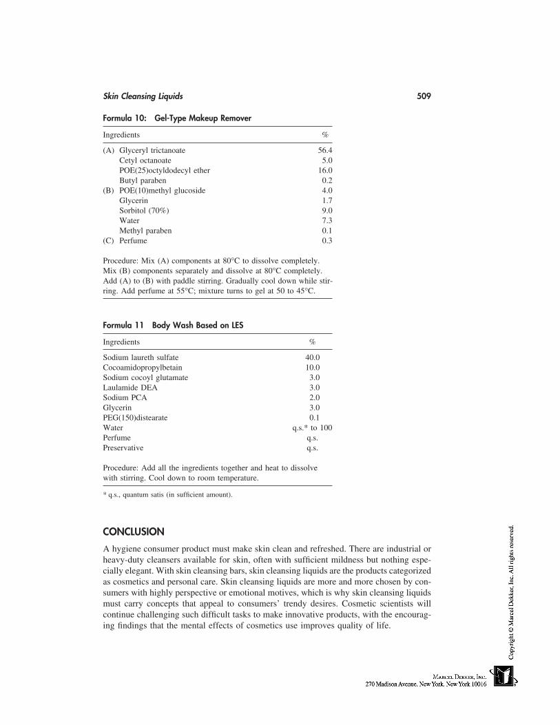
Skin Cleansing Liquids 509
Formula 10: Gel-Type Makeup Remover
Ingredients %
(A) Glyceryl trictanoate 56.4Cetyl octanoate 5.0POE(25)octyldodecyl ether 16.0Butyl paraben 0.2
(B) POE(10)methyl glucoside 4.0Glycerin 1.7Sorbitol (70%) 9.0Water 7.3Methyl paraben 0.1
(C) Perfume 0.3
Procedure: Mix (A) components at 80°C to dissolve completely.Mix (B) components separately and dissolve at 80°C completely.Add (A) to (B) with paddle stirring. Gradually cool down while stir-ring. Add perfume at 55°C; mixture turns to gel at 50 to 45°C.
Formula 11 Body Wash Based on LES
Ingredients %
Sodium laureth sulfate 40.0Cocoamidopropylbetain 10.0Sodium cocoyl glutamate 3.0Laulamide DEA 3.0Sodium PCA 2.0Glycerin 3.0PEG(150)distearate 0.1Water q.s.* to 100Perfume q.s.Preservative q.s.
Procedure: Add all the ingredients together and heat to dissolvewith stirring. Cool down to room temperature.
* q.s., quantum satis (in sufficient amount).
CONCLUSION
A hygiene consumer product must make skin clean and refreshed. There are industrial orheavy-duty cleansers available for skin, often with sufficient mildness but nothing espe-cially elegant. With skin cleansing bars, skin cleansing liquids are the products categorizedas cosmetics and personal care. Skin cleansing liquids are more and more chosen by con-sumers with highly perspective or emotional motives, which is why skin cleansing liquidsmust carry concepts that appeal to consumers’ trendy desires. Cosmetic scientists willcontinue challenging such difficult tasks to make innovative products, with the encourag-ing findings that the mental effects of cosmetics use improves quality of life.
510 Kaneko and Sakamoto
REFERENCES
1. Sakamoto K. Surfactant and skin: surfactant suitable for sensitive or atopic skin. J Jpn CosmetSci Soc 1997; 21:125.
2. Naito N, Munakata A. Cleansing of sebum and skin treatment. Fragrance J 1988; 92:42.3. Dawning DT, Strauss JS, Pochi PE. Variability in the chemical composition of human skin
surface lipids. J Invest Dermatol 1969; 53:232.4. Schwartzendruber DC, Wertz PW, Madison KC, Downing DT. Evidence that the corneocyte
has a chemically bound lipid envelope. J Invest Dermatol 1978; 88:709.5. Miyazawa K, Tamura U, Katsumura Y, Uchikawa K, Sakamoto T, Tomita K. Anionic surfac-
tants as detergents for scalp and hair. Yukagaku 1989; 38:297.6. Miyazawa K, Evaluation of haircare products: shampoo and rinse. J Soc Cosmet Chem Jpn
1995; 29:95.7. Jacobi OK. About the mechanism of moisture regulation in the horny layer of the skin. Proc
Sci Toilet Goods Assoc 1959; 31:22.8. Nozaki T. Research and development of body cleanser. Fragrance J 1996; 8:24.9. Fukuda T. Research and development of a face cleanser of liquid type. Fragrance J 1996; 7:
24.10. Mistui T, ed. New Cosmetic Science. Elsevier 1997.11. Suzuki T, Takai H, Yamazaki S. Formation of fine three-phase emulsions by the liquid crystal
emulsification method with arginine-branched monoalkylphosphate. J Colloid Interface Sci1989; 129:491.
12. Sakai Y, Hashimoto F. Development of a high function make-up remover applying a newpolar oil. 40th Ann Sci Conf of Soc Cosmet Chem Jpn, June 17, 1997 Osaka, Japan.
44
Emulsion-Based Skincare Products:Formulating and Measuring Their
Moisturizing Benefits
Howard Epstein and F. Anthony SimionThe Andrew Jergens Company, Cincinnati, Ohio
AN OVERVIEW OF EMULSION-BASED SKINCARE PRODUCTS
A variety of skincare products exist in today’s marketplace. They fulfill a variety of func-tions by either acting directly on the skin (e.g., moisturizers) or being a cosmeticallyelegant vehicle for the delivery of specific active ingredients (e.g., sunscreens or antipure-tic or antiacne medicaments). In general, these products may be categorized into threefunctional groups:
• Drugs. To Prevent or ameliorate diseases by altering the structure and/or func-tion of the body.
• Cosmetics. To beautify and improve the feeling or sensory aspects of normaland/or nondiseased skin. Dry skin would be included in this category.
• Cosmeceuticals. An intermediate classification for cosmetic products that mayenhance the function of skin. Currently, this category is not recognized by theUnited States Food and Drug Administration (FDA) [1].
There is a similar classification in the European Union.The three product groups can also be classified by their physical properties. Most
common forms of skincare products are emulsions. Emulsions are mixtures of two insolu-ble materials that are stabilized against separation. An example is oil and water, whichwill not mix unless an intermediate emulsifier is incorporated into the mixture.
Different Types of Emulsions
Emulsifiers can act as solubilizers as well as spreading or dispersing agents. Correct useof emulsifiers permits the formulation of homogeneous mixtures, dispersions or emulsionsof oily, waxy substances with water. Solids may be dispersed in liquids or insoluble liquidswithin other liquids. Greasy anhydrous ointments can be designed to be more washable.These types of properties may be achieved by appropriate selection of emulsifiers, activeingredient, and other compatible ingredients in the vehicle.
Emulsions may be water-in-oil (w/o), oil-in-water (o/w), aqueous gel, and siliconein water. Other products may be formulated as semisolids containing oleaginous ingredi-
511
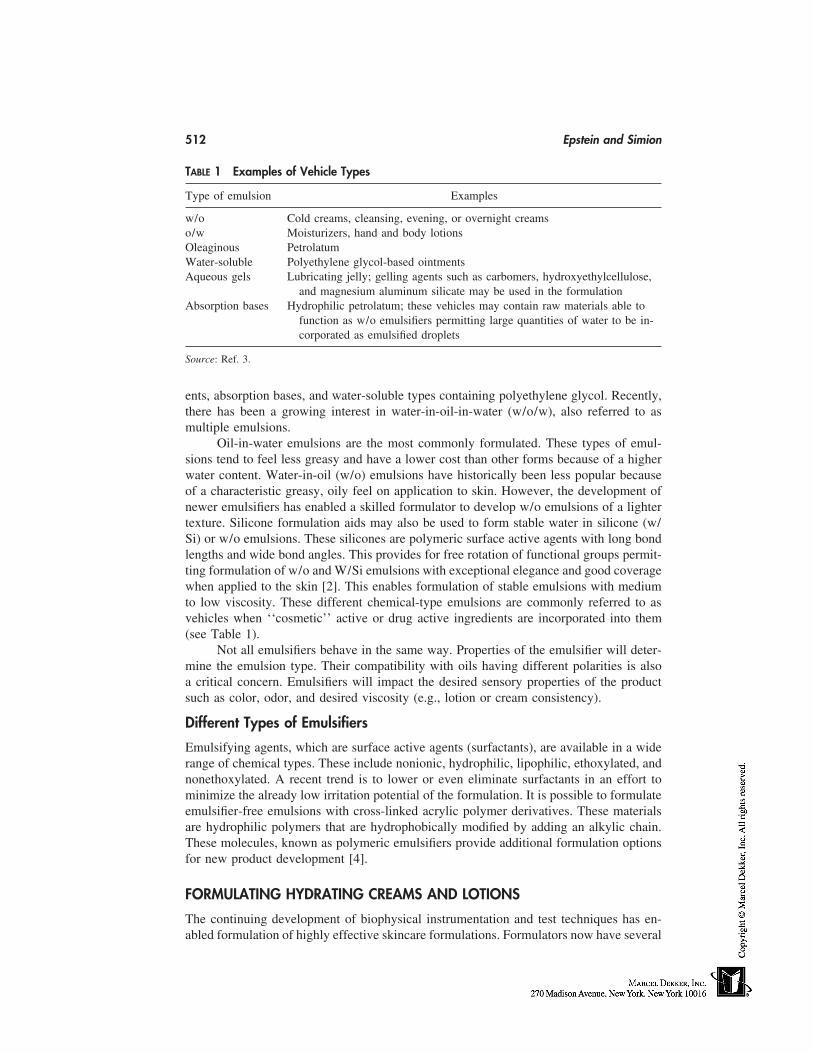
512 Epstein and Simion
TABLE 1 Examples of Vehicle Types
Type of emulsion Examples
w/o Cold creams, cleansing, evening, or overnight creamso/w Moisturizers, hand and body lotionsOleaginous PetrolatumWater-soluble Polyethylene glycol-based ointmentsAqueous gels Lubricating jelly; gelling agents such as carbomers, hydroxyethylcellulose,
and magnesium aluminum silicate may be used in the formulationAbsorption bases Hydrophilic petrolatum; these vehicles may contain raw materials able to
function as w/o emulsifiers permitting large quantities of water to be in-corporated as emulsified droplets
Source: Ref. 3.
ents, absorption bases, and water-soluble types containing polyethylene glycol. Recently,there has been a growing interest in water-in-oil-in-water (w/o/w), also referred to asmultiple emulsions.
Oil-in-water emulsions are the most commonly formulated. These types of emul-sions tend to feel less greasy and have a lower cost than other forms because of a higherwater content. Water-in-oil (w/o) emulsions have historically been less popular becauseof a characteristic greasy, oily feel on application to skin. However, the development ofnewer emulsifiers has enabled a skilled formulator to develop w/o emulsions of a lightertexture. Silicone formulation aids may also be used to form stable water in silicone (w/Si) or w/o emulsions. These silicones are polymeric surface active agents with long bondlengths and wide bond angles. This provides for free rotation of functional groups permit-ting formulation of w/o and W/Si emulsions with exceptional elegance and good coveragewhen applied to the skin [2]. This enables formulation of stable emulsions with mediumto low viscosity. These different chemical-type emulsions are commonly referred to asvehicles when ‘‘cosmetic’’ active or drug active ingredients are incorporated into them(see Table 1).
Not all emulsifiers behave in the same way. Properties of the emulsifier will deter-mine the emulsion type. Their compatibility with oils having different polarities is alsoa critical concern. Emulsifiers will impact the desired sensory properties of the productsuch as color, odor, and desired viscosity (e.g., lotion or cream consistency).
Different Types of Emulsifiers
Emulsifying agents, which are surface active agents (surfactants), are available in a widerange of chemical types. These include nonionic, hydrophilic, lipophilic, ethoxylated, andnonethoxylated. A recent trend is to lower or even eliminate surfactants in an effort tominimize the already low irritation potential of the formulation. It is possible to formulateemulsifier-free emulsions with cross-linked acrylic polymer derivatives. These materialsare hydrophilic polymers that are hydrophobically modified by adding an alkylic chain.These molecules, known as polymeric emulsifiers provide additional formulation optionsfor new product development [4].
FORMULATING HYDRATING CREAMS AND LOTIONS
The continuing development of biophysical instrumentation and test techniques has en-abled formulation of highly effective skincare formulations. Formulators now have several
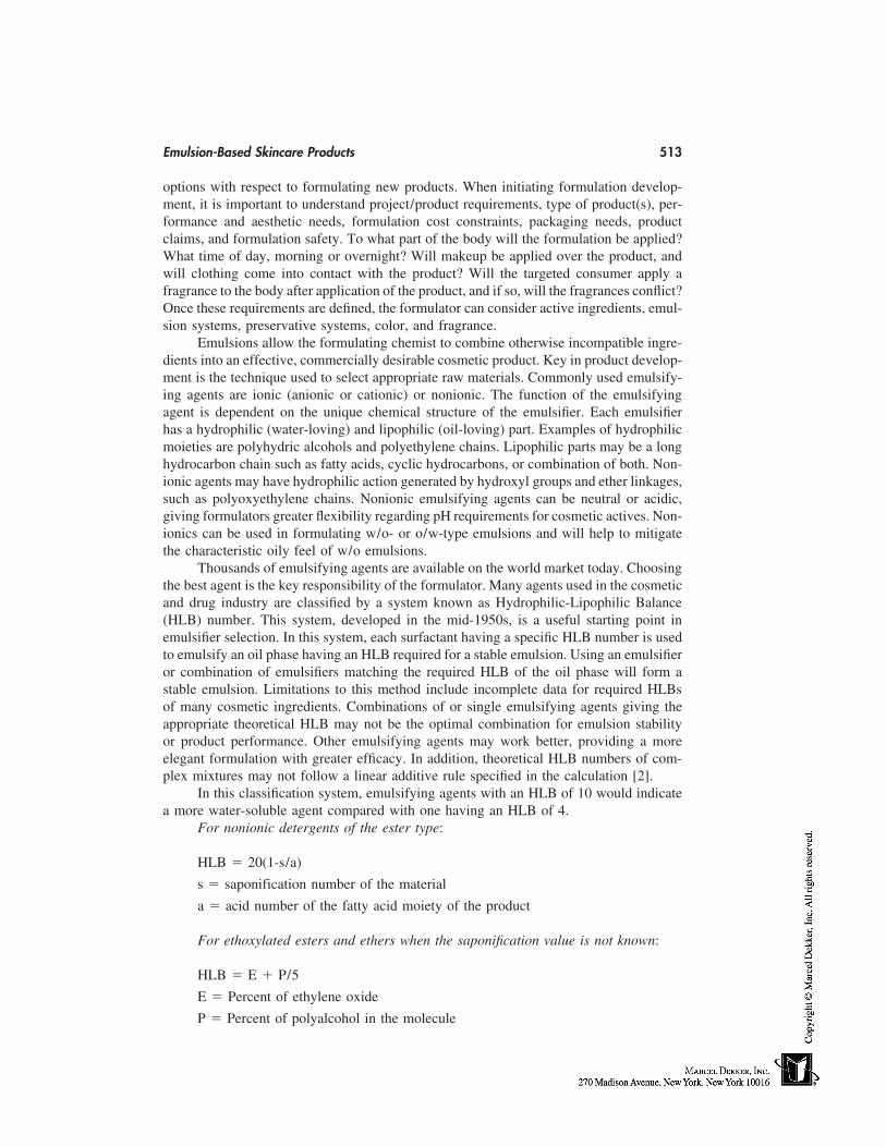
Emulsion-Based Skincare Products 513
options with respect to formulating new products. When initiating formulation develop-ment, it is important to understand project/product requirements, type of product(s), per-formance and aesthetic needs, formulation cost constraints, packaging needs, productclaims, and formulation safety. To what part of the body will the formulation be applied?What time of day, morning or overnight? Will makeup be applied over the product, andwill clothing come into contact with the product? Will the targeted consumer apply afragrance to the body after application of the product, and if so, will the fragrances conflict?Once these requirements are defined, the formulator can consider active ingredients, emul-sion systems, preservative systems, color, and fragrance.
Emulsions allow the formulating chemist to combine otherwise incompatible ingre-dients into an effective, commercially desirable cosmetic product. Key in product develop-ment is the technique used to select appropriate raw materials. Commonly used emulsify-ing agents are ionic (anionic or cationic) or nonionic. The function of the emulsifyingagent is dependent on the unique chemical structure of the emulsifier. Each emulsifierhas a hydrophilic (water-loving) and lipophilic (oil-loving) part. Examples of hydrophilicmoieties are polyhydric alcohols and polyethylene chains. Lipophilic parts may be a longhydrocarbon chain such as fatty acids, cyclic hydrocarbons, or combination of both. Non-ionic agents may have hydrophilic action generated by hydroxyl groups and ether linkages,such as polyoxyethylene chains. Nonionic emulsifying agents can be neutral or acidic,giving formulators greater flexibility regarding pH requirements for cosmetic actives. Non-ionics can be used in formulating w/o- or o/w-type emulsions and will help to mitigatethe characteristic oily feel of w/o emulsions.
Thousands of emulsifying agents are available on the world market today. Choosingthe best agent is the key responsibility of the formulator. Many agents used in the cosmeticand drug industry are classified by a system known as Hydrophilic-Lipophilic Balance(HLB) number. This system, developed in the mid-1950s, is a useful starting point inemulsifier selection. In this system, each surfactant having a specific HLB number is usedto emulsify an oil phase having an HLB required for a stable emulsion. Using an emulsifieror combination of emulsifiers matching the required HLB of the oil phase will form astable emulsion. Limitations to this method include incomplete data for required HLBsof many cosmetic ingredients. Combinations of or single emulsifying agents giving theappropriate theoretical HLB may not be the optimal combination for emulsion stabilityor product performance. Other emulsifying agents may work better, providing a moreelegant formulation with greater efficacy. In addition, theoretical HLB numbers of com-plex mixtures may not follow a linear additive rule specified in the calculation [2].
In this classification system, emulsifying agents with an HLB of 10 would indicatea more water-soluble agent compared with one having an HLB of 4.
For nonionic detergents of the ester type:
HLB � 20(1-s/a)
s � saponification number of the material
a � acid number of the fatty acid moiety of the product
For ethoxylated esters and ethers when the saponification value is not known:
HLB � E � P/5
E � Percent of ethylene oxide
P � Percent of polyalcohol in the molecule
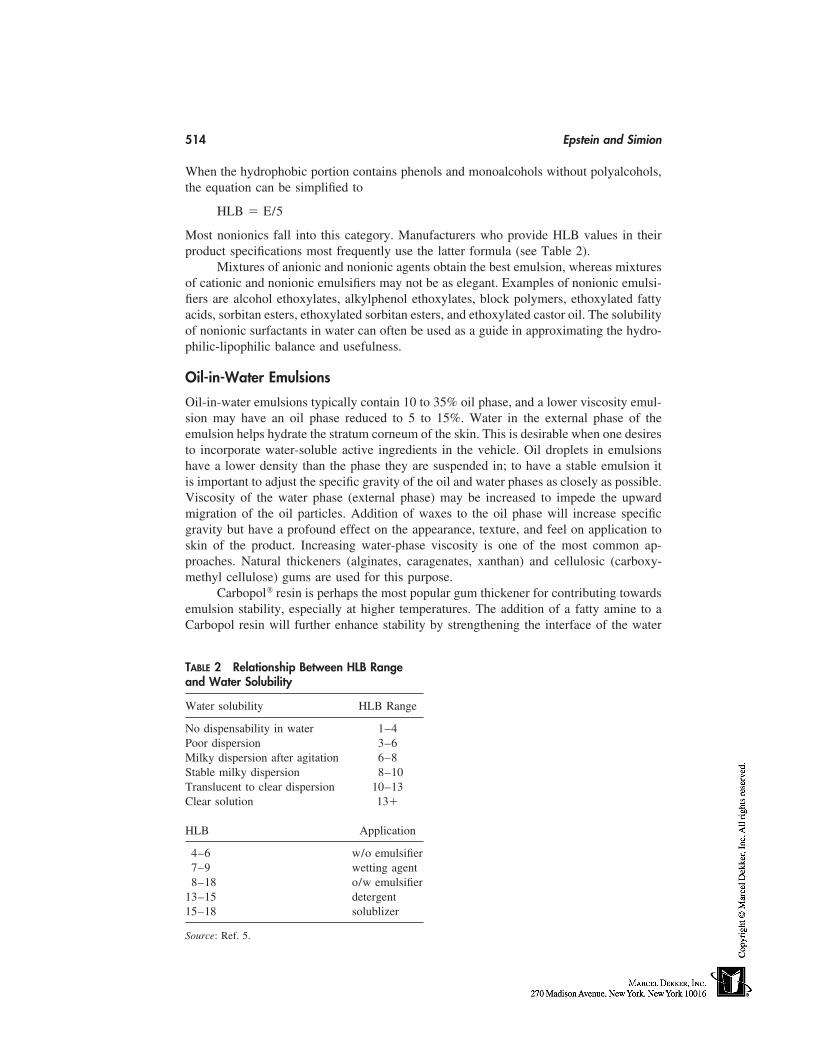
514 Epstein and Simion
When the hydrophobic portion contains phenols and monoalcohols without polyalcohols,the equation can be simplified to
HLB � E/5
Most nonionics fall into this category. Manufacturers who provide HLB values in theirproduct specifications most frequently use the latter formula (see Table 2).
Mixtures of anionic and nonionic agents obtain the best emulsion, whereas mixturesof cationic and nonionic emulsifiers may not be as elegant. Examples of nonionic emulsi-fiers are alcohol ethoxylates, alkylphenol ethoxylates, block polymers, ethoxylated fattyacids, sorbitan esters, ethoxylated sorbitan esters, and ethoxylated castor oil. The solubilityof nonionic surfactants in water can often be used as a guide in approximating the hydro-philic-lipophilic balance and usefulness.
Oil-in-Water Emulsions
Oil-in-water emulsions typically contain 10 to 35% oil phase, and a lower viscosity emul-sion may have an oil phase reduced to 5 to 15%. Water in the external phase of theemulsion helps hydrate the stratum corneum of the skin. This is desirable when one desiresto incorporate water-soluble active ingredients in the vehicle. Oil droplets in emulsionshave a lower density than the phase they are suspended in; to have a stable emulsion itis important to adjust the specific gravity of the oil and water phases as closely as possible.Viscosity of the water phase (external phase) may be increased to impede the upwardmigration of the oil particles. Addition of waxes to the oil phase will increase specificgravity but have a profound effect on the appearance, texture, and feel on application toskin of the product. Increasing water-phase viscosity is one of the most common ap-proaches. Natural thickeners (alginates, caragenates, xanthan) and cellulosic (carboxy-methyl cellulose) gums are used for this purpose.
Carbopol resin is perhaps the most popular gum thickener for contributing towardsemulsion stability, especially at higher temperatures. The addition of a fatty amine to aCarbopol resin will further enhance stability by strengthening the interface of the water
TABLE 2 Relationship Between HLB Rangeand Water Solubility
Water solubility HLB Range
No dispensability in water 1–4Poor dispersion 3–6Milky dispersion after agitation 6–8Stable milky dispersion 8–10Translucent to clear dispersion 10–13Clear solution 13�
HLB Application
4–6 w/o emulsifier7–9 wetting agent8–18 o/w emulsifier
13–15 detergent15–18 solublizer
Source: Ref. 5.
Emulsion-Based Skincare Products 515
and oil phases through partial solubilization into the oil droplets. Electrolytes and cationicmaterials will have a destabilizing effect on anionic sodium carboxymethyl cellulose andshould not be used together. Veegum, an inorganic aluminum silicate material, is alsocommonly used to thicken emulsions. Carbopol and Veegum may be used together tomodify the characteristic draggy feel of Carbopol when used at the higher levels.
Emulsifier blends with HLBs ranging from 7 to 16 are used for forming o/w emul-sions. In the blend, the hydrophilic emulsifier should be formulated as the predominateemulsifier to obtain the best emulsion. A popular emulsifier, the glycerol monostearateand polyoxyethylene stearate blend is self-emulsifying and acid-stable. Emulsifiers arecalled self-emulsifying when an auxiliary anionic or nonionic emulsifier is added for easieremulsification of the formulation. Formulating with self-emulsifying materials containingnonionic emulsifiers permit a wide range of ingredient choice for the formulator, especiallywith acid systems. In alkaline formulations, polyoxyethylene ether–type emulsifiers arepreferred with respect to emulsion stability.
An alternative to glycerol monostearate self-emulsifying emulsifier is EmulsifyingWax, National Formulary (NF). This emulsifier, when used with a fatty alcohol will formviscous liquids to creams depending on the other oil-phase ingredients used. Use levelsmay vary from 2 to 15%; at lower levels a secondary emulsifier such as the oleths orPEG-glycerides will give good stability. This system is good for stabilizing electrolyteemulsions or when other ionic materials are formulated into the vehicle. Polysorbates areo/w emulsifiers, wetting agents, and solubilizers often used with cetyl or stearyl alcoholat 0.5 to 5.0% to produce o/w emulsions [6].
Water-in-Oil Emulsions
Although less popular than o/w emulsions, these systems may be desirable when greaterrelease of a medicating agent or the perception of greater emolliency is desired. Emulsifiershaving an HLB range of 2.5 to 6 are frequently selected. When multiple emulsifiers areused, the predominant one is generally lipophilic with a smaller quantity of a hydrophilicemulsifier. These emulsions typically have a total of 45 to 80% oil phase.
During the last few years, formulators have become interested in more elegant w/oemulsions. This has been achieved by formulating with new emulsifying agents, emollientsuch as esters, Guerbet alcohols, and silicones. Selection of a suitable emollient depends onability of the material to spread on skin with low tack, dermal compatibility, and perceivedelegance by the user. In achieving this elegance, some researchers suggest a correlationof emollient and molecular weight of the emollients. In these studies, viscosity of w/ocreams has correlated with molecular weight of the emollients used in test formulations.High–molecular-weight co-emulsifiers formulated with high–molecular-weight emol-lients gave more stable w/o emulsions. The polarity of the emollients used was found tobe important as well. Emollients or mixtures of emollients with medium polarity gavetest lotions the most desirable stability results [7]. Anionic emulsifiers are generally ineffi-cient w/o emulsion stabilizers, because more surface active agents are often needed tostabilize these emulsions. Sorbitan stearates and oleates are effective emulsifiers whenused at 0.5 to 5.0% sorbitan isostearates, being branched chain materials, give a veryuniform particle size for w/o emulsions.
Multiple Emulsions
Multiple emulsions are of interest to the skincare formulator because of the elegant appear-ance and less greasy feel of these formulation types. Two types of multiple emulsions are
516 Epstein and Simion
encountered in skincare: (w/o/w), where the internal and external water phases are sepa-rated by oil, and oil-in-water-in-oil (o/w/o) where the water phase separates the two oilphases. The method of preparation for each multiple emulsion type is similar. Benefits ofthese types of formulations are the claimed sustained release of entrapped materials in theinternal phase and separation of various incompatible ingredients in the same formulation.
A suggested technique for forming a w/o/w emulsion is to first create a w/o primaryemulsion by combining water as one phase with oil and a lipophilic emulsifier as thesecond phase in the traditional method. Next, water and a hydrophilic emulsifier are com-bined with the w/o primary emulsion at room or warm (i.e., 40°C) temperature with mixingforming a w/o/w multiple emulsion. These emulsions typically contain about 18 to 23%oils and 3 to 8% lipophilic emulsifier. The continuous oily phase is stabilized with about0.5 to 0.8% magnesium sulfate. Water-in-oil emulsifiers have an HLB less than 6 and arefrequently nonionic or polymeric. Oil-in-water emulsifiers have an HLB greater than 15and are ionic with high interfacial activity. For o/w/o multiple emulsions, w/o emulsifiershave an HLB less than 6 with similar properties as a w/o/w w/o emulsifier. Oil-in-wateremulsifiers have an HLB greater 15 and are nonionic with lower interfacial activity.
Water-in-Silicone Emulsions
Silicone compounds have evolved into a class of specialty materials used for replacements,substitutes, or enhancers for a variety of organic surface-active agents, resulting in theability to formulate products with unique properties. Previously, silicone compounds wereavailable as water-insoluble oily materials almost exclusively. Newer silicone compoundssuch as polyethylene-oxide bases grafted to polydimethylsiloxane hydrophobic polymers,known as dimethicone copolyol emulsifiers, have been developed. These types of emulsi-fiers permit formation of water-in-cyclomethicone emulsions. Further work in this fieldled to adding hydrocarbon chains to silicone polyether polymers. This resulted in improvedaesthetics to o/s emulsions. Silicone copolyols exhibit high surface activity and functionsimilarly to traditional emulsifiers. Unlike hydrocarbon emulsifiers with higher molecu-lar weights, high–molecular-weight silicone emulsifiers can remain fluid. This givesvery stable viscoelastic films at the w/o interface. The ability to make silicones moreformulator-friendly has led to development of several new silicone-based surfactants. Botha water-soluble and an oil-soluble portion are needed to make a surface-active molecule.Silicone surfactants substitute or add silicone-based hydrophobicity, creating a distinctiveskin feel and other attributes of typical silicones as well as attributes of fatty surfactants.These emulsions may be prepared in a traditional two-phase method, e.g., 2 to 3% w/wof laurylmethicone copolyol in 23% w/w oil phase can be mixed in a separate water phasewith electrolyte to form a hydrating cream. [8]
Water-Soluble Ointment Bases
Polyethylene glycol polymers (PEGs) are available in a variety of molecular weights.These materials are water-soluble and do not hydrolyze or support mold growth. For thesereasons, PEGs make good bases for washable ointments and can be formulated to havea soft to hard consistency. Polyethylene glycols dissolve in water to form clear solutions.They are also soluble in organic solvents such as mineral and produce formulations thatare more substantive on skin. Polyethylene glycol ointment USP is a mixture of polyethyl-ene glycol 3350 and polyethylene glycol 400 heated to 65°C and cooled and mixed until
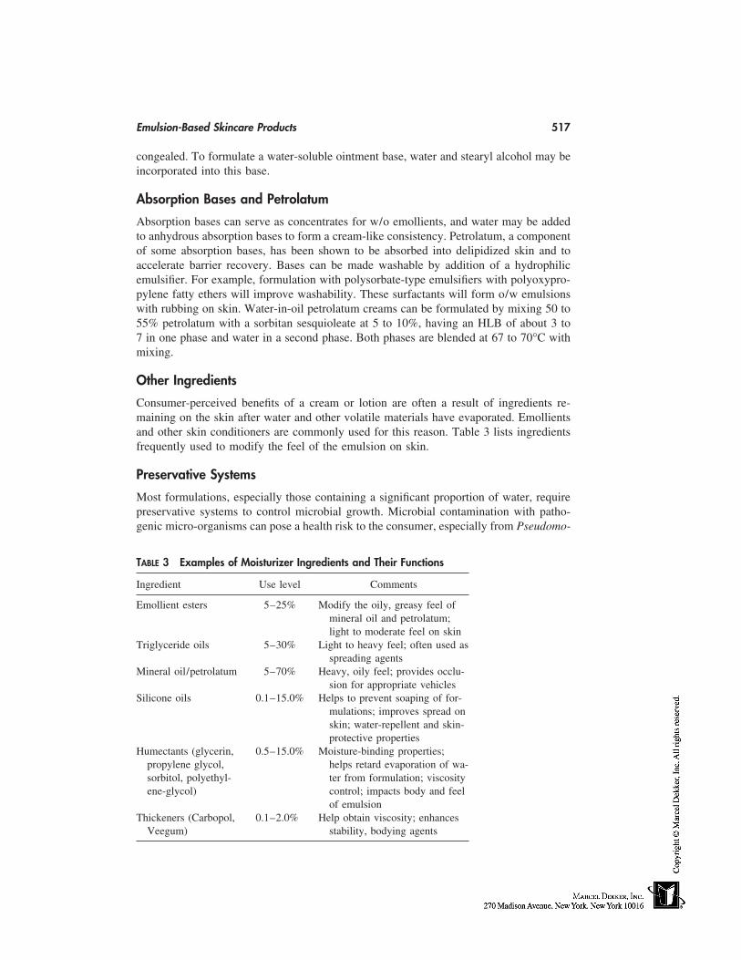
Emulsion-Based Skincare Products 517
congealed. To formulate a water-soluble ointment base, water and stearyl alcohol may beincorporated into this base.
Absorption Bases and Petrolatum
Absorption bases can serve as concentrates for w/o emollients, and water may be addedto anhydrous absorption bases to form a cream-like consistency. Petrolatum, a componentof some absorption bases, has been shown to be absorbed into delipidized skin and toaccelerate barrier recovery. Bases can be made washable by addition of a hydrophilicemulsifier. For example, formulation with polysorbate-type emulsifiers with polyoxypro-pylene fatty ethers will improve washability. These surfactants will form o/w emulsionswith rubbing on skin. Water-in-oil petrolatum creams can be formulated by mixing 50 to55% petrolatum with a sorbitan sesquioleate at 5 to 10%, having an HLB of about 3 to7 in one phase and water in a second phase. Both phases are blended at 67 to 70°C withmixing.
Other Ingredients
Consumer-perceived benefits of a cream or lotion are often a result of ingredients re-maining on the skin after water and other volatile materials have evaporated. Emollientsand other skin conditioners are commonly used for this reason. Table 3 lists ingredientsfrequently used to modify the feel of the emulsion on skin.
Preservative Systems
Most formulations, especially those containing a significant proportion of water, requirepreservative systems to control microbial growth. Microbial contamination with patho-genic micro-organisms can pose a health risk to the consumer, especially from Pseudomo-
TABLE 3 Examples of Moisturizer Ingredients and Their Functions
Ingredient Use level Comments
Emollient esters 5–25% Modify the oily, greasy feel ofmineral oil and petrolatum;light to moderate feel on skin
Triglyceride oils 5–30% Light to heavy feel; often used asspreading agents
Mineral oil/petrolatum 5–70% Heavy, oily feel; provides occlu-sion for appropriate vehicles
Silicone oils 0.1–15.0% Helps to prevent soaping of for-mulations; improves spread onskin; water-repellent and skin-protective properties
Humectants (glycerin, 0.5–15.0% Moisture-binding properties;propylene glycol, helps retard evaporation of wa-sorbitol, polyethyl- ter from formulation; viscosityene-glycol) control; impacts body and feel
of emulsionThickeners (Carbopol, 0.1–2.0% Help obtain viscosity; enhances
Veegum) stability, bodying agents
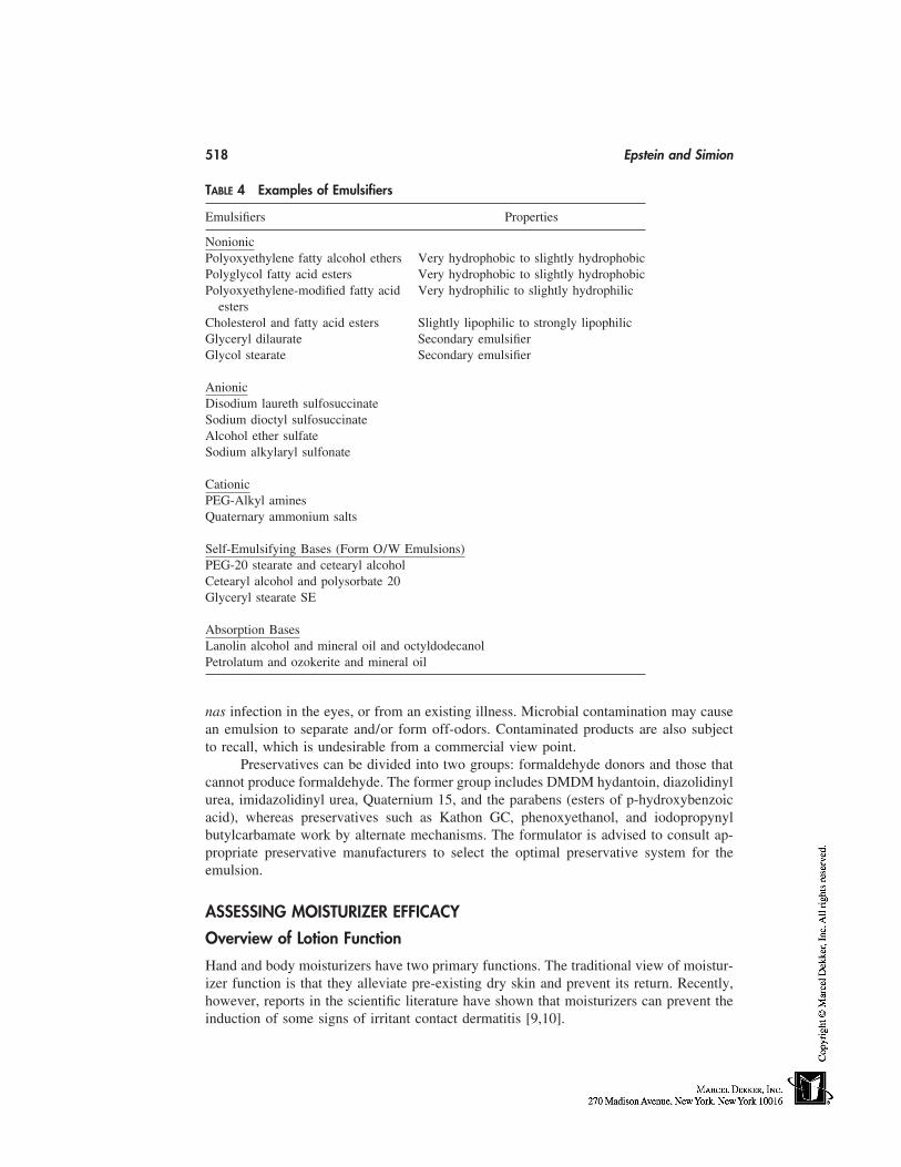
518 Epstein and Simion
TABLE 4 Examples of Emulsifiers
Emulsifiers Properties
NonionicPolyoxyethylene fatty alcohol ethers Very hydrophobic to slightly hydrophobicPolyglycol fatty acid esters Very hydrophobic to slightly hydrophobicPolyoxyethylene-modified fatty acid Very hydrophilic to slightly hydrophilic
estersCholesterol and fatty acid esters Slightly lipophilic to strongly lipophilicGlyceryl dilaurate Secondary emulsifierGlycol stearate Secondary emulsifier
AnionicDisodium laureth sulfosuccinateSodium dioctyl sulfosuccinateAlcohol ether sulfateSodium alkylaryl sulfonate
CationicPEG-Alkyl aminesQuaternary ammonium salts
Self-Emulsifying Bases (Form O/W Emulsions)PEG-20 stearate and cetearyl alcoholCetearyl alcohol and polysorbate 20Glyceryl stearate SE
Absorption BasesLanolin alcohol and mineral oil and octyldodecanolPetrolatum and ozokerite and mineral oil
nas infection in the eyes, or from an existing illness. Microbial contamination may causean emulsion to separate and/or form off-odors. Contaminated products are also subjectto recall, which is undesirable from a commercial view point.
Preservatives can be divided into two groups: formaldehyde donors and those thatcannot produce formaldehyde. The former group includes DMDM hydantoin, diazolidinylurea, imidazolidinyl urea, Quaternium 15, and the parabens (esters of p-hydroxybenzoicacid), whereas preservatives such as Kathon GC, phenoxyethanol, and iodopropynylbutylcarbamate work by alternate mechanisms. The formulator is advised to consult ap-propriate preservative manufacturers to select the optimal preservative system for theemulsion.
ASSESSING MOISTURIZER EFFICACY
Overview of Lotion Function
Hand and body moisturizers have two primary functions. The traditional view of moistur-izer function is that they alleviate pre-existing dry skin and prevent its return. Recently,however, reports in the scientific literature have shown that moisturizers can prevent theinduction of some signs of irritant contact dermatitis [9,10].
Emulsion-Based Skincare Products 519
The ability to prevent irritant contact dermatitis has relevance to a significant seg-ment of the population. Epidemiological studies have shown that the prevalence of diag-nosable hand and forearm eczema can be as high as 5.4% of the population at any onetime, and from 8 to 11% in the preceding year [11,12]. This often has an irritant componentespecially from repeated exposure to surfactant solutions. Being able to prevent irritationmay provide a significant benefit to these individuals, as well as those with dry skin (xero-sis), which frequently affects the arms and legs of consumers. Although symptoms areusually less intense than eczema, dry skin probably affects a larger proportion of thepopulation.
Measuring lotions’ effects on dryness and primary irritation is key to assessing mois-turizer efficacy. Clinical methods have been developed that assess dry skin or its absencevia visual scoring by a trained observer and by using biophysical measurements of theskin. Similarly, erythema and stratum corneum barrier damage associated with primaryirritation can be measured clinically. Clinical efficacy alone is not sufficient to make aproduct commercially successful. To appeal to consumers, the lotion must be both effica-cious and aesthetically pleasing, i.e., pleasantly scented (or unscented) and have acceptabletactile characteristics during and immediately after application.
Clinical Evaluation of Moisturizer Efficacy
To effectively assess the clinical efficacy of moisturizers, it is important to assess severalparameters that relate to skin condition. As lotions can have multiple effects on the skin,using only one modality such as observer scoring may be misleading. For instance, visualobservation suggests that Lotion ‘‘E’’ is as effective as Lotion ‘‘C’’ at reducing skindryness. Skin that was not treated also showed a reduction in visually scored dryness,indicating the effect of prevailing weather conditions. DeSquame sticky tape (CudermInc, Dallas, TX) and its quantification by image analysis was able to differentiate betweenthe three test sites. DeSquames show that at day 4 (end of treatment phase), Lotion E didnot remove corneocytes from the skin’s surface as effectively as Lotion C. At day 7,Lotion E was similar to the ‘‘No product’’ site. This suggests that Lotion E may maskthe dryness. In contrast, Lotion C caused corneocyte removal at days 4 and 7 (Fig. 1).Visual assessment of skin dryness is useful because it is a direct link to the benefits ofmoisturization that consumers readily recognize, such as skin flaking, scaling, ashiness,and cracking. These visual assessments should be supplemented with instrumental mea-sures of skin flaking, hydration, surface topography, or elasticity. These instrumental mea-surements yield a more complete understanding of how moisturizers affect the skin, andcan be more easily standardized than observer assessments.
Alleviating Dry SkinThe majority of clinical studies that measure the relief of dry skin after lotion applicationuse either the Kligman regression protocol or a modification [13–17]. Typically thesestudies start out with dry skin, which is treated for an extended period, followed by ashort regression phase during which product usage is discontinued. Kligman originallystudied the effect of ingredients and products on the lower legs of 12 to 30 female panelists(Fig. 2). These dry skin sites were treated with an ingredient or lotion (2 mg/cm2) twicedaily for up to 3 weeks. The visual dryness was assessed before treatment (baseline) andat the end of each week. Panelists started with dry skin and the improvement in drynessfrom baseline was the measure of moisturizing efficacy, or the relief of dryness.
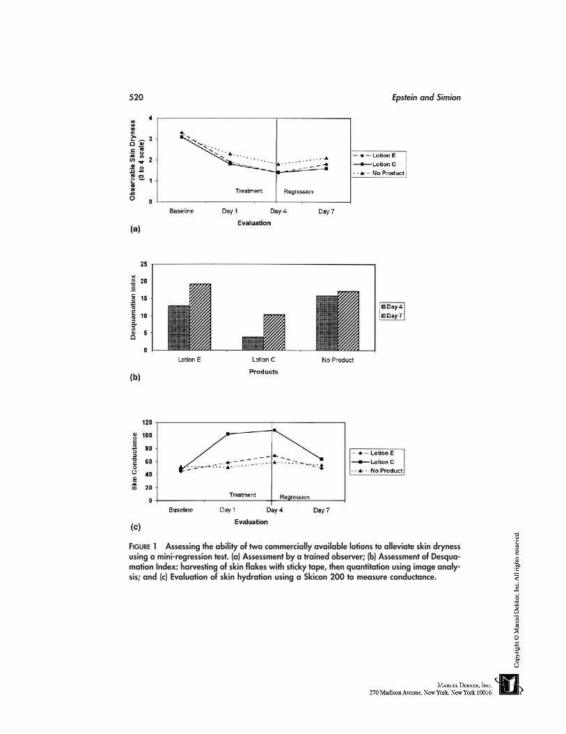
520 Epstein and Simion
FIGURE 1 Assessing the ability of two commercially available lotions to alleviate skin drynessusing a mini-regression test. (a) Assessment by a trained observer; (b) Assessment of Desqua-mation Index: harvesting of skin flakes with sticky tape, then quantitation using image analy-sis; and (c) Evaluation of skin hydration using a Skicon 200 to measure conductance.
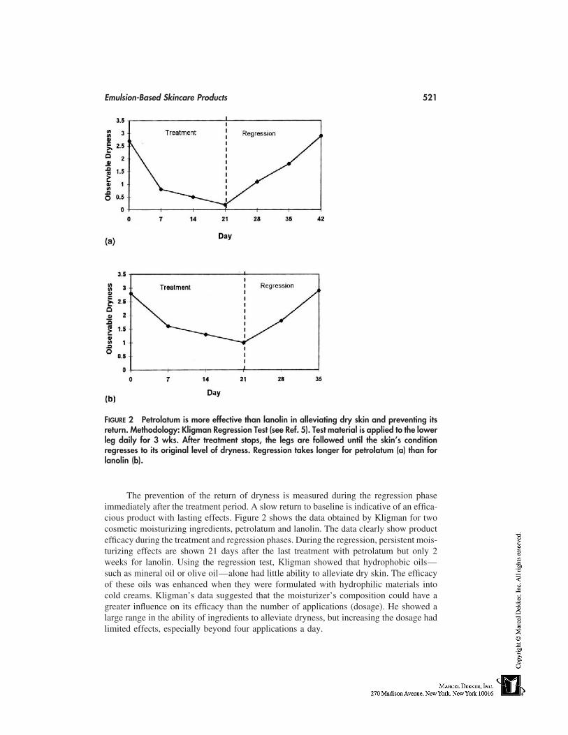
Emulsion-Based Skincare Products 521
FIGURE 2 Petrolatum is more effective than lanolin in alleviating dry skin and preventing itsreturn. Methodology: Kligman Regression Test (see Ref. 5). Test material is applied to the lowerleg daily for 3 wks. After treatment stops, the legs are followed until the skin’s conditionregresses to its original level of dryness. Regression takes longer for petrolatum (a) than forlanolin (b).
The prevention of the return of dryness is measured during the regression phaseimmediately after the treatment period. A slow return to baseline is indicative of an effica-cious product with lasting effects. Figure 2 shows the data obtained by Kligman for twocosmetic moisturizing ingredients, petrolatum and lanolin. The data clearly show productefficacy during the treatment and regression phases. During the regression, persistent mois-turizing effects are shown 21 days after the last treatment with petrolatum but only 2weeks for lanolin. Using the regression test, Kligman showed that hydrophobic oils—such as mineral oil or olive oil—alone had little ability to alleviate dry skin. The efficacyof these oils was enhanced when they were formulated with hydrophilic materials intocold creams. Kligman’s data suggested that the moisturizer’s composition could have agreater influence on its efficacy than the number of applications (dosage). He showed alarge range in the ability of ingredients to alleviate dryness, but increasing the dosage hadlimited effects, especially beyond four applications a day.
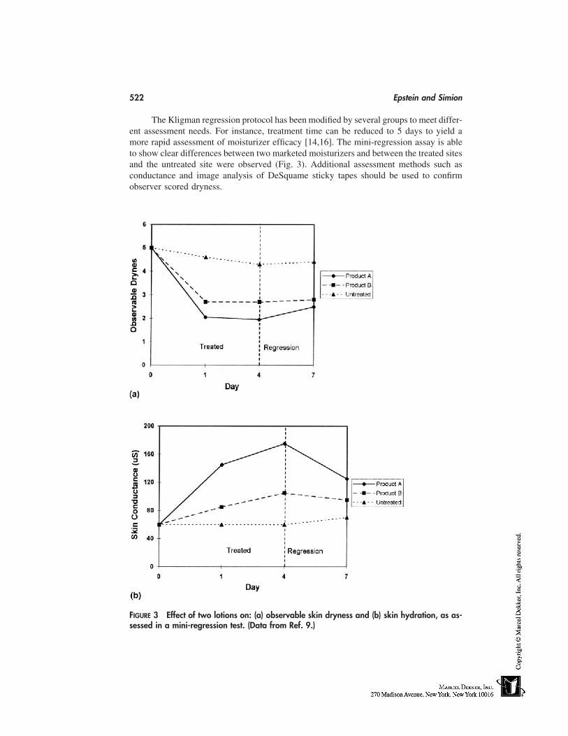
522 Epstein and Simion
The Kligman regression protocol has been modified by several groups to meet differ-ent assessment needs. For instance, treatment time can be reduced to 5 days to yield amore rapid assessment of moisturizer efficacy [14,16]. The mini-regression assay is ableto show clear differences between two marketed moisturizers and between the treated sitesand the untreated site were observed (Fig. 3). Additional assessment methods such asconductance and image analysis of DeSquame sticky tapes should be used to confirmobserver scored dryness.
FIGURE 3 Effect of two lotions on: (a) observable skin dryness and (b) skin hydration, as as-sessed in a mini-regression test. (Data from Ref. 9.)
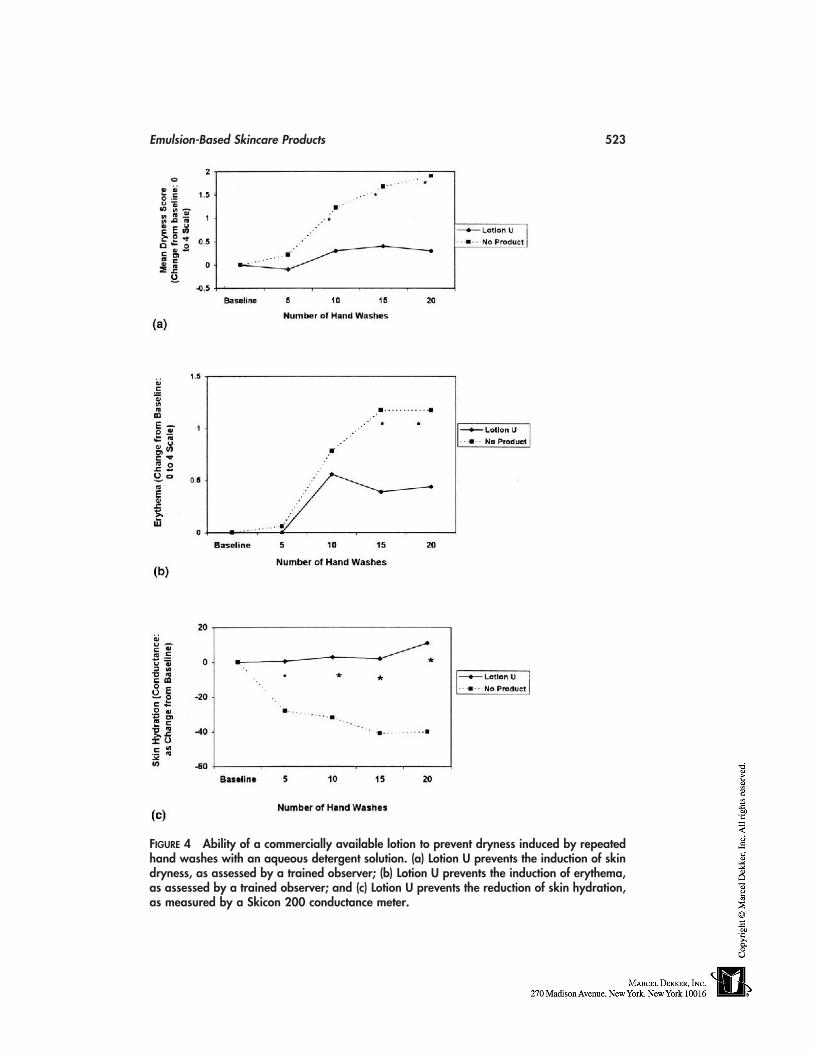
Emulsion-Based Skincare Products 523
FIGURE 4 Ability of a commercially available lotion to prevent dryness induced by repeatedhand washes with an aqueous detergent solution. (a) Lotion U prevents the induction of skindryness, as assessed by a trained observer; (b) Lotion U prevents the induction of erythema,as assessed by a trained observer; and (c) Lotion U prevents the reduction of skin hydration,as measured by a Skicon 200 conductance meter.
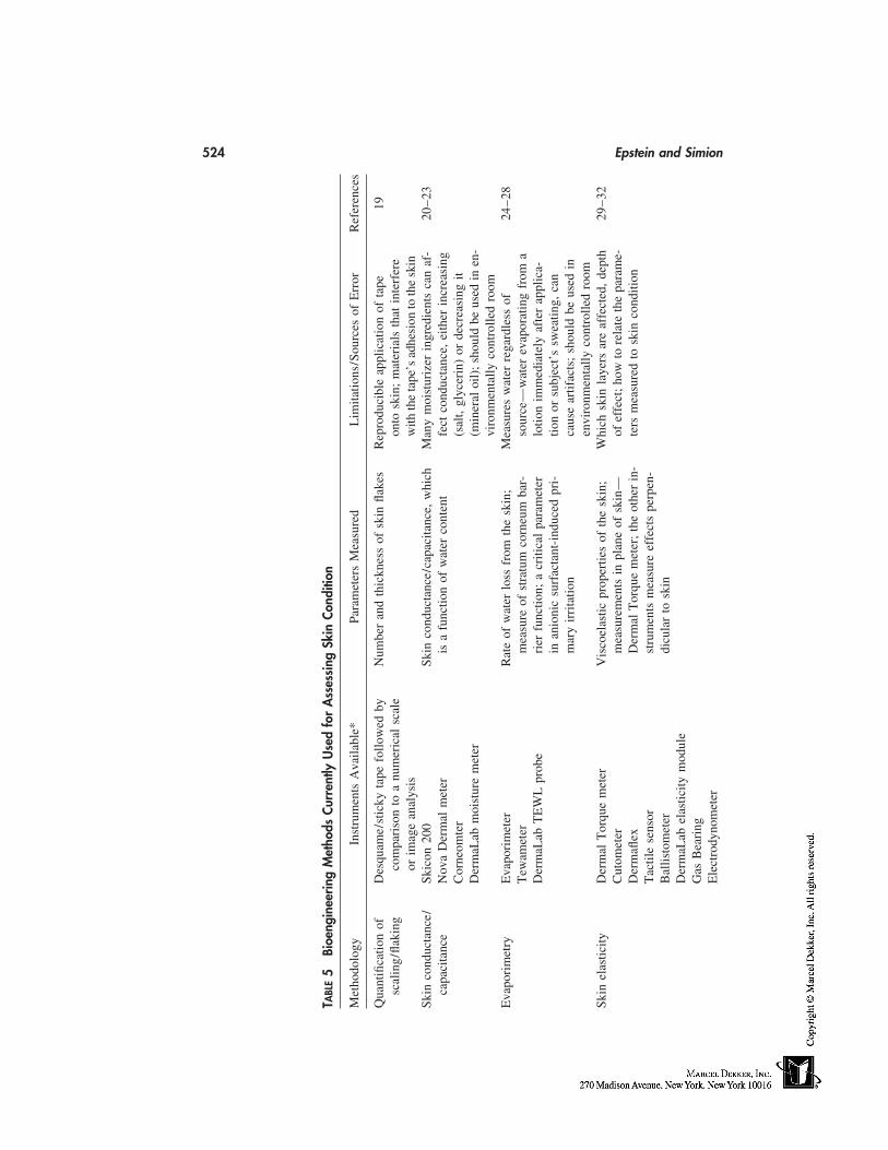
524 Epstein and Simion
TABL
E5
Bioe
ngin
eerin
gM
etho
dsCu
rren
tlyU
sed
for
Ass
essi
ngSk
inCo
nditi
on
Met
hodo
logy
Inst
rum
ents
Ava
ilabl
e*Pa
ram
eter
sM
easu
red
Lim
itatio
ns/S
ourc
esof
Err
orR
efer
ence
s
Qua
ntifi
catio
nof
Des
quam
e/st
icky
tape
follo
wed
byN
umbe
ran
dth
ickn
ess
ofsk
infla
kes
Rep
rodu
cibl
eap
plic
atio
nof
tape
19sc
alin
g/fla
king
com
pari
son
toa
num
eric
alsc
ale
onto
skin
;m
ater
ials
that
inte
rfer
eor
imag
ean
alys
isw
ithth
eta
pe’s
adhe
sion
toth
esk
inSk
inco
nduc
tanc
e/Sk
icon
200
Skin
cond
ucta
nce/
capa
cita
nce,
whi
chM
any
moi
stur
izer
ingr
edie
nts
can
af-
20–2
3ca
paci
tanc
eN
ova
Der
mal
met
eris
afu
nctio
nof
wat
erco
nten
tfe
ctco
nduc
tanc
e,ei
ther
incr
easi
ngC
orne
omte
r(s
alt,
glyc
erin
)or
decr
easi
ngit
Der
maL
abm
oist
ure
met
er(m
iner
aloi
l);
shou
ldbe
used
inen
-vi
ronm
enta
llyco
ntro
lled
room
Eva
pori
met
ryE
vapo
rim
eter
Rat
eof
wat
erlo
ssfr
omth
esk
in;
Mea
sure
sw
ater
rega
rdle
ssof
24–2
8T
ewam
eter
mea
sure
ofst
ratu
mco
rneu
mba
r-so
urce
—w
ater
evap
orat
ing
from
aD
erm
aLab
TE
WL
prob
eri
erfu
nctio
n;a
criti
cal
para
met
erlo
tion
imm
edia
tely
afte
rap
plic
a-in
anio
nic
surf
acta
nt-i
nduc
edpr
i-tio
nor
subj
ect’
ssw
eatin
g,ca
nm
ary
irri
tatio
nca
use
artif
acts
;sh
ould
beus
edin
envi
ronm
enta
llyco
ntro
lled
room
Skin
elas
ticity
Der
mal
Tor
que
met
erV
isco
elas
ticpr
oper
ties
ofth
esk
in;
Whi
chsk
inla
yers
are
affe
cted
,de
pth
29–3
2C
utom
eter
mea
sure
men
tsin
plan
eof
skin
—of
effe
ct;
how
tore
late
the
para
me-
Der
mafl
exD
erm
alT
orqu
em
eter
;th
eot
her
in-
ters
mea
sure
dto
skin
cond
ition
Tac
tile
sens
orst
rum
ents
mea
sure
effe
cts
perp
en-
Bal
listo
met
erdi
cula
rto
skin
Der
maL
abel
astic
itym
odul
eG
asB
eari
ngE
lect
rody
nom
eter
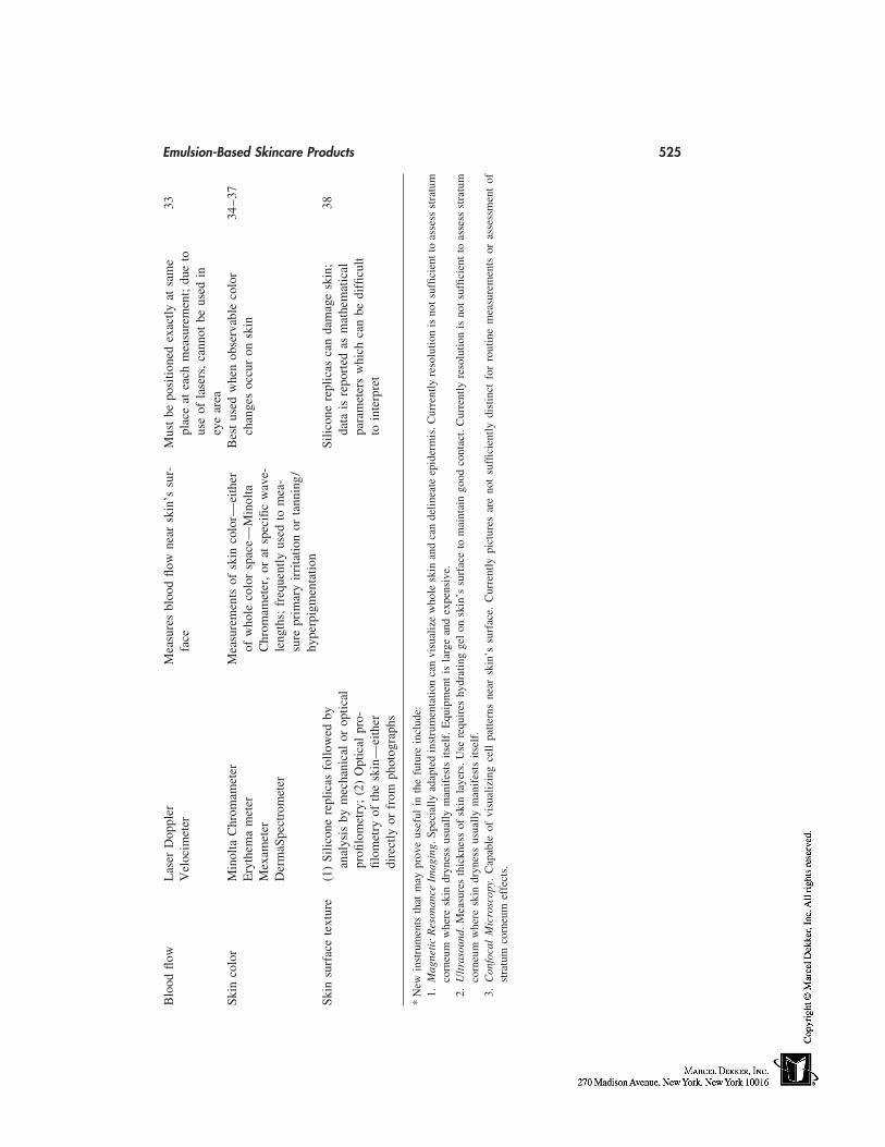
Emulsion-Based Skincare Products 525B
lood
flow
Las
erD
oppl
erM
easu
res
bloo
dflo
wne
arsk
in’s
sur-
Mus
tbe
posi
tione
dex
actly
atsa
me
33V
eloc
imet
erfa
cepl
ace
atea
chm
easu
rem
ent;
due
tous
eof
lase
rs,
cann
otbe
used
iney
ear
eaSk
inco
lor
Min
olta
Chr
omam
eter
Mea
sure
men
tsof
skin
colo
r—ei
ther
Bes
tus
edw
hen
obse
rvab
leco
lor
34–3
7E
ryth
ema
met
erof
who
leco
lor
spac
e—M
inol
tach
ange
soc
cur
onsk
inM
exam
eter
Chr
omam
eter
,or
atsp
ecifi
cw
ave-
Der
maS
pect
rom
eter
leng
ths;
freq
uent
lyus
edto
mea
-su
repr
imar
yir
rita
tion
orta
nnin
g/hy
perp
igm
enta
tion
Skin
surf
ace
text
ure
(1)
Silic
one
repl
icas
follo
wed
bySi
licon
ere
plic
asca
nda
mag
esk
in;
38an
alys
isby
mec
hani
cal
orop
tical
data
isre
port
edas
mat
hem
atic
alpr
ofilo
met
ry;
(2)
Opt
ical
pro-
para
met
ers
whi
chca
nbe
diffi
cult
filom
etry
ofth
esk
in—
eith
erto
inte
rpre
tdi
rect
lyor
from
phot
ogra
phs
*N
ewin
stru
men
tsth
atm
aypr
ove
usef
ulin
the
futu
rein
clud
e:1.
Mag
neti
cR
eson
ance
Imag
ing.
Spec
ially
adap
ted
inst
rum
enta
tion
can
visu
aliz
ew
hole
skin
and
can
delin
eate
epid
erm
is.C
urre
ntly
reso
lutio
nis
nots
uffic
ient
toas
sess
stra
tum
corn
eum
whe
resk
indr
ynes
sus
ually
man
ifes
tsits
elf.
Equ
ipm
ent
isla
rge
and
expe
nsiv
e.2.
Ult
raso
und.
Mea
sure
sth
ickn
ess
ofsk
inla
yers
.Use
requ
ires
hydr
atin
gge
lon
skin
’ssu
rfac
eto
mai
ntai
ngo
odco
ntac
t.C
urre
ntly
reso
lutio
nis
not
suffi
cien
tto
asse
ssst
ratu
mco
rneu
mw
here
skin
dryn
ess
usua
llym
anif
ests
itsel
f.3.
Con
foca
lM
icro
scop
y.C
apab
leof
visu
aliz
ing
cell
patte
rns
near
skin
’ssu
rfac
e.C
urre
ntly
pict
ures
are
not
suffi
cien
tlydi
stin
ctfo
rro
utin
em
easu
rem
ents
oras
sess
men
tof
stra
tum
corn
eum
effe
cts.
526 Epstein and Simion
The legs are not the only site that can be used in regression testing. The groupsworking with both Prall and Grove have used the lower arms to assess moisturizer efficacy.The regression phase of the clinical evaluation may be used to examine the persistenceof the moisturization efficacy when skin is stressed by winter weather or washing withsoap.
Preventing Skin Dryness and Irritation
There are two main approaches to assessing the ability of a lotion to prevent the inductionof skin dryness. First, the rate at which dry skin returns after treatment ceases can beassessed from the regression phase of the Kligman regression test. It is evident that manyeffective moisturizers and moisturizing ingredients do have a residual effect on the skinand will maintain it in good condition for several days, despite prevailing adverse condi-tions such as winter weather.
An alternate approach to measure the prevention of dry and irritated skin was devel-oped by Highley et al. [18]. In the Highley Hand Wash protocol, the analysis begins withnondry, healthy skin. The panelists wash their hands with a detergent based cleanser for1 minute, 5 times a day for several days. Lotions are applied to test sites after the firstfour washes each day. There are control areas of skin that are washed, but to which nomoisturizer is applied. The dryness of the hands are assessed by a trained observer andby instrumental methods, before the first wash of the study (baseline) and approximately1 hour after the last (fifth) wash each day. Results show that ingredients such as petrolatumand commercial lotions can prevent the induction of dry skin, which can be considerableon the untreated skin (Fig. 4). Products and ingredients can be compared by determiningthe difference between the sites treated with moisturizers and nonmoisturized skin. Al-though panels as small as 5 have been used, it is more usual to use panels of 10 or moreto enable the data to be statistically analyzed.
Hannuksela and Kinnunen [10] also showed that moisturizers could prevent surfac-tant induced irritation and speed skin’s recovery. Arms were washed with dishwashingliquid for 1 minute, twice a day, for 7 days. The investigators evaluated cleanser-inducedirritation using transepidermal water loss (TEWL) as a measure of stratum corneum integ-rity and Laser-Doppler flowmetry to assess blood flow. They showed that moisturizerapplication could prevent surfactant induced skin damage and accelerate repair comparedwith no treatment, but were unable to differentiate between products.
The ability of moisturizers to prevent detergent induced skin dryness has importantpublic health implications. In Denmark, dermatitis is the third leading cause leading occu-pational disease, and it is reasonable to assume that it has a high incidence in other coun-tries. Such dermatitis which is frequently expressed as hand or forearm eczema, can lastfor many years as patients are exposed to irritants such as cleansers in both the workplaceand at home. Professions that involve frequent hand washings, such as healthcare workers,day-care workers, and cleaners and food preparers, are at particular risk. Frequent, effec-tive moisturization may provide a significant preventative benefit.
Instrumental Evaluations of Moisturizer Efficacy
Instrumental evaluation of skin condition should be used to supplement visual assessmentsin clinical moisturization studies. They will provide a more complete measure of skincondition that visual scoring alone. Conversely, because each instrumental method mea-sures a physical parameter, care must be taken in using the data to interpret the biological
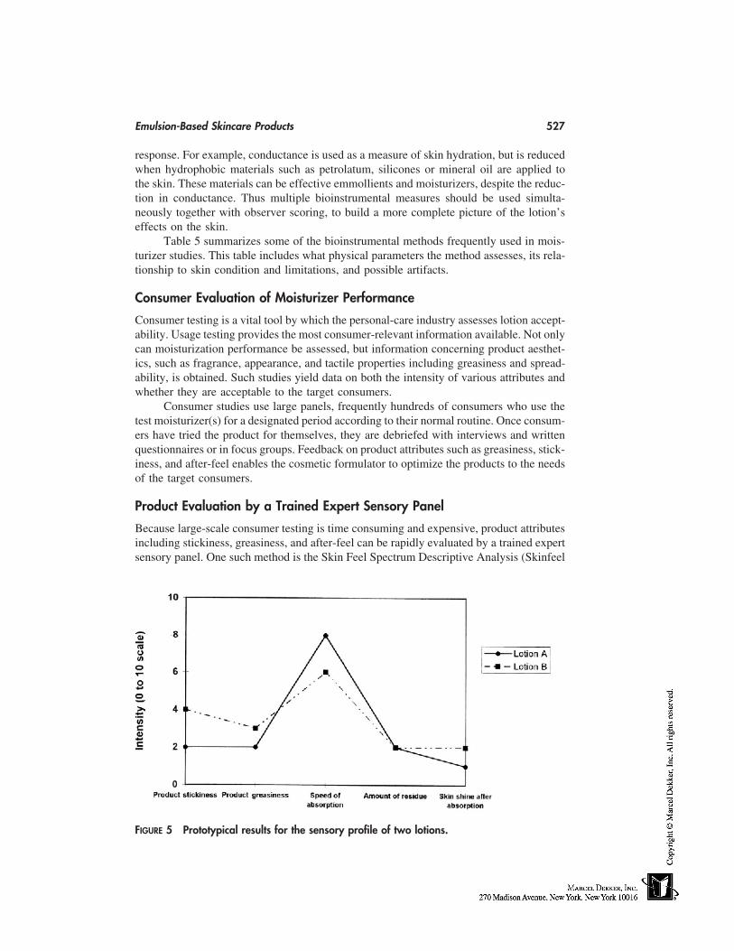
Emulsion-Based Skincare Products 527
response. For example, conductance is used as a measure of skin hydration, but is reducedwhen hydrophobic materials such as petrolatum, silicones or mineral oil are applied tothe skin. These materials can be effective emmollients and moisturizers, despite the reduc-tion in conductance. Thus multiple bioinstrumental measures should be used simulta-neously together with observer scoring, to build a more complete picture of the lotion’seffects on the skin.
Table 5 summarizes some of the bioinstrumental methods frequently used in mois-turizer studies. This table includes what physical parameters the method assesses, its rela-tionship to skin condition and limitations, and possible artifacts.
Consumer Evaluation of Moisturizer Performance
Consumer testing is a vital tool by which the personal-care industry assesses lotion accept-ability. Usage testing provides the most consumer-relevant information available. Not onlycan moisturization performance be assessed, but information concerning product aesthet-ics, such as fragrance, appearance, and tactile properties including greasiness and spread-ability, is obtained. Such studies yield data on both the intensity of various attributes andwhether they are acceptable to the target consumers.
Consumer studies use large panels, frequently hundreds of consumers who use thetest moisturizer(s) for a designated period according to their normal routine. Once consum-ers have tried the product for themselves, they are debriefed with interviews and writtenquestionnaires or in focus groups. Feedback on product attributes such as greasiness, stick-iness, and after-feel enables the cosmetic formulator to optimize the products to the needsof the target consumers.
Product Evaluation by a Trained Expert Sensory Panel
Because large-scale consumer testing is time consuming and expensive, product attributesincluding stickiness, greasiness, and after-feel can be rapidly evaluated by a trained expertsensory panel. One such method is the Skin Feel Spectrum Descriptive Analysis (Skinfeel
FIGURE 5 Prototypical results for the sensory profile of two lotions.
528 Epstein and Simion
SDA) used by Meilgaard et al. [19]. This method outlines the product attribute descriptorsand scoring scales used to evaluate moisturizers. An expert panel of 8 to 15 persons isrequired to complete over a 100 hours of training to ensure they can reproducibly quantifymoisturizer and skin attributes such as spreadibility, amount of residue, and absorbency,which are scored using a 0-to-10 scale (Fig. 5.) Once the panel is calibrated, they can beused to evaluate competitors’ products and optimize new formulas. Validation requiresthat the sensory panel correctly predict the intensity of attributes from a large-scale con-sumer test. It should be noted that sensory panels measure attribute intensity only, anddo not assess the preference of distinct types of consumer for different products.
REFERENCES
1. Vermeer BJ, Gilchrest B. Arch Dermatol 1996; 132(3):340.2. Kasprzak R. Drug and Cosmetic Industry (May 1996).3. Block H. Medicated applications. In: Gennaro AR, ed. Remington’s Pharmaceutical Sciences
18th ed. Easton: Mack Publishing Co. PA, 1990, p. 1603.4. Konish PN, Gruber JV. J Cosmet Sci 1998; 49:335–342.5. The HLB System. ICI Americas Inc. August 1984.6. Emulsification of Basic Cosmetic Ingredients. ICI United States Inc., 1975:102–106.7. Henkel Symposium, October 1991.8. Silicone Formulation Aids. Dow Corning, 1997.9. Zhai H, Maibach HI. Moisturizers in preventing irritant contact dermatitis: an overview. Con-
tact Dermatitis 1998; 38:241–244.10. Hannuksela A, Kinnunen T. Moisturizers prevent irritant dermatitis. Acta Derm Venereol
(Stockh.) 1992; 72:42–44.11. Meding B. Epidemiology of hand eczema in an industrial city. Acta Dermatol Venereol 1990;
(suppl) 153:1–43.12. Lantinga H, Nater JP, Coenraads PJ. Prevalence, incidence and course of eczema on the hands
and forearms in a sample of the general population. Contact Dermatitis 1984; 10:135–139.13. Kligman AM. Regression method for assessing the efficacy of moisturizers. Cosmetics and
Toiletries 1978; 93:27–35.14. Prall JK, Theiler RF, Bowser PA, Walsh M. The effectiveness of cosmetic products in alleviat-
ing a range of dryness conditions as determined by clinical and instrumental techniques. IntJ Cosmet Sci 1986; 8:159–174.
15. Boisits EK, Nole GE, Cheney MC. The refined regression method. J Cutan Aging & CosmetDermatol 1989; 1:155–163.
16. Grove GL. Skin surface hydration changes during a mini regression test as measured in vivoby electrical conductivity. Curr Ther Res 1992; 52:556–561.
17. Grove GL, Jackson R, Czernielewski MD, Tuley M. Poster Presentation at the 53rd AnnualMeeting of the American Academy of Dermatology, March 1995.
18. Highley DR, Savoyka VO, O’Neil JJ, Ward JB. A stereomicroscopic method for the determina-tion of moisturizing efficacy in humans. J Soc Cosmet Che 1976; 27:351–363.
19. Schatz H, Altermeyer PJ, Kligman AM. Dry skin and scaling evaluated by D-Squames andimage analysis. In: Serup J, Jamec GBE, eds. Handbook of Non-Invasive Methods and theSkin. Boca Raton, CRC Press 1995:153–157.
20. Tagami H. Measurement of electrical conductance and impedance. In: Serup J, Jemec GBE,eds. Handbook of Non-Invasive Methods and the Skin. Boca Raton: CRC Press, 1995:159–164.
21. Barel AO, Clarys P. Meaurement of electrical capacitance. In: Serup J, Jemec GBE, eds. Hand-book of Non-Invasive Methods and the Skin. Boca Raton: CRC Press, 1995; 165–170.
Emulsion-Based Skincare Products 529
22. Morrison BM, Scala DD. Comparison of instrumental methods of skin hydration. J ToxicolCutan Occular Toxicol 1996; 15:305–314.
23. Loden M, Lindberg M. The influence of a single application of different moisturizers on theskin capacitance. Acta Derm Venereol (Stockh.) 1991; 71:79–82.
24. Pinnagoda J, Tupker RA. Measurement of the transepidermal water loss. (1995) In: Serup J,Jemec GBE, eds. Handbook of Non-invasive Methods and the Skin. Boca Raton: CRC Press,1995; 173–178.
25. Barel AO, Clarys P. Comparison of methods for measurement of transepidermal water loss.In: Serup J, Jemec GBE, eds. Handbook of Non-Invasive Methods and the Skin. Boca Raton:CRC Press, 1995; 179–184.
26. Grove GL, Grove MJ, Zerwick C, Pierce E. Comparative metrology of evaporimeter and theDermaLab TEWL probe. Skin Res Tech 1999; 5:1–8.
27. Grove GL, Grove MJ, Zerwick C, Pierce E. Computerized evaporimetry using the DermaLab
TEWL probe. Skin Res Tech 1999; 5:9–13.28. Simion FA, Rhein LD, Grove GL, Wojtowski J, Cagan RH, Scala DD. Sequential order of
skin responses to surfactants in a soap chamber test. Contact Dermatitis 1991; 25:242–245.29. Agache PG. (1995) Twistometry measurement of skin elasticity. In: Serup J, Jemec GBE, eds.
Handbook of Non-Invasive Methods and the Skin. Boca Raton: CRC Press, 1995; 319–328.30. Barel AO, Courage W, Clarys P. Suction methods for measurement of skin mechanical proper-
ties: the cutometer. In: Serup J, Jemec GBE eds. Handbook of Non-Invasive Methods and theSkin. Boca Raton: CRC Press, 1995; 335–340.
31. Elsner P. Skin elasticity. In: Berardesca E, Elsner P. Wilhelm K-P, Maibach HI, eds. Bioengin-eering and the Skin: Methods and Instrumentation. Boca Raton: CRC Press, 1995:53–64.
32. Omata S, Terunuma Y. New tactile sensor like the human hand and its applications. Sensorsand Actuators 1992; 35:9–15.
33. Belcaro G, Nicolaides AN. Laser–doppler flowmetry: principles of technology and clinicalapplications. In: Serup J, Jemec GBE, eds. Handbook of Non-invasive Methods and the Skin.Boca Raton: CRC Press, 1995:405–410.
34. Takiwaki H, Serup J. Measurement of erythema and melanin indices. In: Serup J, Jemec GBE,eds. Handbook of Non-Invasive Methods and the Skin. Boca Raton: CRC Press, 1995; 377–384.
35. Westerhof W. CIE Colorimetry. In: Eds Serup J, Jemec GBE, eds. Handbook of Non-InvasiveMethods and the Skin. Boca Raton: CRC Press, 1995; 385–397.
36. Babulak SW, Rhein LD, Scala DD, Simion FA, Grove GL. Quantitation of erythema in asoap chamber test using a Minolta Chroma (Reflectance) meter: comparison of instrumentalresults with visual assessments. J Soc Cosmet Chem 1986; 37:475–477.
37. Berardesca E, Maibach HI. Bioengineering and the patch test. Contact Dermatitis 1988; 18:3–9.
38. Corcuff P, Leveque J-L. Skin Surface replica image analysis of furrows and wrinkles. In:Serup J, Jemec GBE, eds. Handbook of Non-Invasive Methods and the Skin. Boca Raton:CRC Press, 1995; 89–96.
39. Meilgaard M, Civielle GV, Carr BT. Sensory Evaluation Techniques. Boca Raton: CRC Press,1987.

45
Anticellulite Products and Treatments
André O. BarelFree University of Brussels, Brussels, Belgium
INTRODUCTION
Cellulite is a localized condition of subcutaneous fat and connective tissues with the typicalvisual appearance of the orange-peel look of the skin. Cellulite, or more correctly locallipodystrophy affects mostly women and rarely men, and is considered to be a commonaesthetic problem for many women. Cellulite generally appears after puberty and worsenswith age. There are preferential places of cellulite: buttocks, thighs, upper part of the arms,knees and more rarely the lower parts of the legs and the back of the neck (Fig. 1). Theaims of this chapter are to describe (1) the histological, physiological, and biochemicalcharacteristics of subcutaneous lipodystrophy, (2) the different objective evaluation meth-ods of lipodystrophy, and (3) the different anticellulite treatments available and their effi-cacy.
CLINICAL VISUAL AND TACTILE SYMPTOMS OF CELLULITE
Upon clinical examination of cellulite, the following symptoms of lipodystrophy can benoticed [1–11].
• Presence of orange-peel skin upon normal visual examination and after pinchingof the skin.
• Deep palpation of the skin reveals differences in the mobility of fat tissue: pres-ence of micro- and macronodules and fibrosclerosis.
• Irregularities in skin-surface temperature: touching the skin reveals the presenceof cold spots.
• Sometimes presence of painful subcutaneous nodules through deep palpation.
There are different stages in the evolution of cellulite with age. It is difficult to detectcellulite by visual examination and palpation at the first stages: orange-peel skin is notpermanently present, and is only visible after pinching the skin.
The clinical symptoms are clearly more visible at later stages of cellulite: permanentorange peel, colder skin areas, diminution in mobility of fat tissue upon palpation andincreased skin sensibility.
531
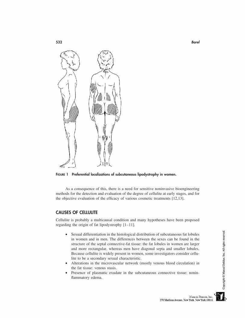
532 Barel
FIGURE 1 Preferential localizations of subcutaneous lipodystrophy in women.
As a consequence of this, there is a need for sensitive noninvasive bioengineeringmethods for the detection and evaluation of the degree of cellulite at early stages, and forthe objective evaluation of the efficacy of various cosmetic treatments [12,13].
CAUSES OF CELLULITE
Cellulite is probably a multicausal condition and many hypotheses have been proposedregarding the origin of fat lipodystrophy [1–11].
• Sexual differentiation in the histological distribution of subcutaneous fat lobulesin women and in men. The differences between the sexes can be found in thestructure of the septal connective-fat tissue: the fat lobules in women are largerand more rectangular, whereas men have diagonal septa and smaller lobules.Because cellulite is widely present in women, some investigators consider cellu-lite to be a secondary sexual characteristic.
• Alterations in the microvascular network (mostly venous blood circulation) inthe fat tissue: venous stasis.
• Presence of plasmatic exudate in the subcutaneous connective tissue: nonin-flammatory edema.
Anticellulite Products and Treatments 533
• Alterations in the reticular fibrillar network surrounding the blood vessels andadipocytes: fibrosclerosis. Stiffening and decrease in mobility of fibers.
• Alterations in the interstitial fundamental substance (proteoglycans).• Modifications and hypertrophy of adipose tissues. Although cellulite is not al-
ways synonymous with obesity (skinny persons can sometimes present symp-toms of cellulite), there is a relation between cellulite and hypertrophy of fattissues. Formation of, first, micronodules and, later, of macronodules in adiposetissues.
The combined effect of modifications and hypertrophy of adipose tissues, alterations inthe fibrillar connective tissue, and alterations in the microvascular venous network alwaysleads to the presence of cellulite.
HISTOLOGICAL DESCRIPTION OF THE DIFFERENT STAGES OFLIPODYSTROPHY OF FAT TISSUES
Skin-surface contact thermographic pictures using thermographic foils give an indicationof the degree of cellulite, because the skin-surface temperature correlates to some extentwith the clinical symptoms of cellulite. Based on these thermographic patterns and clinicalsymptoms, Curri and coworkers proposed a classification of cellulite in four stages[4,5,14,15]. In normal adipose tissues, a fine mesh of blood vessels and lymph vesselssupplies this adipose tissue with the necessary nutrients and oxygen, and takes care ofthe removal of metabolized products. In the early stage of cellulite (stage I), the capillaryblood-vessel walls become more permeable, causing leakage of blood plasma from thevessels in between the adipose tissues, which cause an edema in the adipose tissues. Inaddition, probably, problems with the lymph circulation hampers removal of accumulatingfluids. The aggregation of adipose cells and the amplification of the fibrillar network ofcollagen bundles interconnecting the adipose cells hampers blood circulation leading tosome hemostase (stage II).
Adipose cells aggregate into ‘‘micronodules’’ surrounded by less-mobile collagenfibers (stage III). The size of these ‘‘micronodules’’ is on the order of millimeters. Finally,many of these ‘‘micronodules’’ aggregate into ‘‘macronodules’’ with larger sizes (2–20mm) (stage IV). As nerves may be squeezed by this larger nodules, persons with severecellulite often suffer from sensitive to painful skin.
Stages I, II, and III of lipodystrophy are not considered clinically as pathologicalsymptoms but more as aesthetic–cosmetic problems of the skin. Only in stage IV areclinical symptoms such as an increased skin sensitivity, extensive fibrosclerosis of connec-tive tissue and very advanced edema considered to be light pathology symptoms. Further-more it is believed that the first stages are more or less reversible, whereas the latter stagesare irreversible. However, it must be said that the microscopic description of cellulite andthe different stages in the evolution of lipodystrophy, as described by Curri, are not univer-sally accepted [16,17].
OBJECTIVE EVALUATION OF THE SYMPTOMS OF LIPODYSTROPHY OFTHE SKIN
In addition to the visual and tactile clinical evaluations of the symptoms of cellulite, vari-ous noninvasive bioengineering measurements may be used [11,12].
534 Barel
However, the clinical evaluation of cellulite remains important. The clinical evalua-tion of cellulite is based on visual examination and palpation of the orange-peel skin witha diminution of the mobility of the hypodermis (appearance of nodules of fat tissues andfibrosclerosis), appearance of differences in skin surface and temperature and patient com-plaints of hypersensitive skin and pain.
The different noninvasive bioengineering measurements are as follows:
• Contact skin-surface thermographic measurements using liquid crystals• Non–contact skin-surface thermography of skin surface using infrared video
camera• Microblood circulation using Laser Doppler image analysis• Ultrasonic skin analysis of skin density• Measurement of thickness of the hypodermis at 10 to 14 MHz• Measurement of the surface of the interface between dermis and hypodermis at
20 MHz• Skin-surface topographical imaging• Macroscopic normal and digitalized photographic pictures of the skin surface
DESCRIPTION AND VALIDATION OF THE DIFFERENTBIOENGINEERING MEASUREMENTS USED FOR OBJECTIVEEVALUATION OF CELLULITE
Skin-Surface Contact Thermography Using Encapsulated LiquidCrystals in the Evaluation of Cellulite [14,15,18]
The principle of the encapsulated cholesteric liquid-crystal contact thermography consistsof different color plates presenting a pattern of different colors corresponding to a tempera-ture range of about 3°C. Application of the color sheet with uniform pressure on the skinsurface and photographic recording of the thermographic pattern using a Polaroid cameraare carried out. A qualitative global analysis of the thermographic pictures in relation withthe different stages of cellulite can be made. A cellulite-free skin-surface thermographyshows a uniform color pattern without hypothermic and hyperthermic areas. A celluliteskin-surface thermography shows a nonuniform color pattern with the presence of hypo-thermic (cold spots) and hyperthermic (warm spots) areas. Quantitative analysis of thethermographic pictures can also be carried out by image analysis. Computerized color-image analysis gives the mean temperature of the thermogram and the number and percentarea of the hypo- and hyperthermic areas, respectively, present on a well-defined skinarea. As experimentally observed, an anticellulite treatment will induce an increase of themean temperature of the skin surface and a decrease of the percent hypothermic zones(with a concomitant increase of the percent hyperthermic zones).
This method is rapid, easy to use and inexpensive for screening subjects for celluliteand for confirmation of the clinical diagnosis. However, considering the low accuracy andreproducibility of the photographic pictures, quantitative image analysis of the thermo-grams is very difficult. One observes large interindividual variations in skin-surface tem-perature (a large number of subjects is necessary in a study) and long acclimatization timefor temperature equilibrium of the skin (influence of external temperature). This methodremains a qualitative test of cellulite at different stages.

Anticellulite Products and Treatments 535
Validation of Skin-Surface Thermography Using Infrared ThermalImaging System in the Evaluation of Cellulite
Using an infrared video camera, an infrared thermal image of the skin surface is obtained ina noninvasive manner. The thermographic picture can be quantitatively analyzed [12,13].
In the validation of this infrared video-imaging technique the same problems areencountered as with the contact thermography with liquid crystals such as large interindi-vidual variations in skin-surface temperature, long acclimatization time for temperatureequilibrium of the skin, and influence of external temperature.
Validation of Laser Doppler Imaging System in the Evaluationof Cellulite
Using a Laser Doppler Perfusion Imager, an image of the superficial blood circulationcan be obtained [12,13]. The He–Ne laser light emitting at 633 nm has a penetrationpower in the skin of only about 300 µm. This instrument measures the superficial bloodflux of the skin (papillary dermis). The blood perfusion of the deeper layers of the skin,such as the hypodermis, cannot be measured with this technique. However, a high correla-tion is obtained between the skin-surface thermographic pictures and the Laser Dopplerimaging system when studying skin with cellulite. However, the measurements are delicate(long measuring times during which the volunteer must remain immobile).
Validation of the Ultrasonic Imaging of the Skin in the Evaluationof Cellulite
A promising method appears to be high-frequency ultrasound C-mode imaging (10–20MHz). This noninvasive method has been frequently used both clinically and in researchfor studying the epidermis, dermis and hypodermis [19–21].
Different investigators have used the technique of measuring the thickness of thesubcutaneous fatty layer using ultrasound imaging at 10 to 14 MHz [22–26]; however,the determination of the echographic border line between subcutaneous fat and connectivetissues/muscles is very delicate. As a consequence, the determination of the mean thick-ness of the hypodermis is not very accurate. Measurement of the interface between thedermis and the subcutaneous fat using ultrasound imaging at 29 MHz is possible [27,28].The interface between the echogenic epidermis–dermis and the hypoechoic subcutaneousfat is clearly visible, allowing measurements of skin thickness and of the surface of thisborder.
Quantification of the surface of the interface between the dermis and the hypodermis(fat tissue) is possible [27,28]. In normal cellulite-free skin, the interface between thedermis and the fat tissue is irregular but rather smooth. In skin with cellulite, this surfaceis not smooth and very irregular. The surface of this interface is quantified and can beused as a measure of the degree of cellulite.
Measurement of Skin-Surface Topography
Cellulite skin surface presents irregularities (orange-peel skin) and in principle the classicskin-surface roughness measurements, which are used in cosmetic research, can be appliedfor studying cellulite. It involves stylus profilometry, image analysis by shadow method
536 Barel
and optical focus laser profilometry [29–31]. These measurements are carried out on softor hard skin replicas of general small size (2–3 cm2 area) and have a limited vertical rangeof roughness capability (maximum 400–500 µm). These techniques are well suited forthe determination of the microrelief of the skin surface (50–200 µm) but not for assessingthe skin surface with cellulite. The skin-surface topography of skin with cellulite can beevaluated using photographic pictures, skin-surface contour measurements, and other opti-cal measurements such as Fringe Projection Topography [28,32–34].
The macrorelief of the skin surface can also be evaluated using an optical triangularLaser profilometry. This method involves measurements on large-size soft replicas withan extended vertical range of skin irregularities (up to 8–10 mm). Quantification of theskin surface macrorelief involves a computerized correction for the curvature of the skinsurface with cellulite [32].
Normal and Digitalized Macroscopic Photographic Pictures of theSkin Surface
The macrorelief of the skin can be evaluated by taking photographic pictures (classic ordigitalized) under standardized experimental conditions. These photographic pictures arethen visually graded in a double-blind manner by expert oberservers for the intensity ofcellulite (photograding with numerical scales). It has been known for many years that thestandardization of classic photographic pictures is not easy, considering the problems ofreproducibility of the processing of color film. In addition double-blind visual scoring ofthese photographic pictures remain subjective. However, some investigators have usedphotographic pictures in order to evaluate the efficacy of anticellulite treatments [35].
The use of digitalized photographic pictures is aimed to overcome the standardiza-tion problems of classic processing of the color film. Macroscopic digitalized photographicpictures (with the use of a CCD camera) of the external part of the thighs were takenafter application of a gripping system around the thigh in order to increase the orange-peel look of the skin. The degree of cellulite was photograded by experts using a 0 to 7scale of intensity of cellulite [24,36].
TREATMENTS OF CELLULITE
Different anticellulite treatments are available [12,13], such as manual and electromechan-ical deep massage (‘‘pincer-rouler’’), manual lymph drainage, sequential pneumatic com-pression (lymph drainage), electrolipolysis, mesotherapy, and topical applications of der-matocosmetic products with and without massage.
Physiotherapeutic treatments such as deep massage and manual and pneumaticlymph drainage, stimulate the blood and the lymph microcirculation and increase the re-moval of extra fluid in the adipose tissues. In addition, these massage techniques willretard the further development of fibrosclerosis and the aggregation of fat cells in nodules.These physiotherapeutic treatments are generally combined with the topical use of anticel-lulite dermatocosmetic products (during massage or pre- or postmassage).
Electrolipolysis and mesotherapy are invasive medical treatments of cellulite; thesetechniques will not be described in this chapter.
Various topical dermatocosmetic products have been used, generally with massage,in the treatment of cellulite and/or as slimming for many years [37]. A list of the ‘‘active’’
Anticellulite Products and Treatments 537
TABLE 1 List of Dermatocosmetic Ingredients MostFrequently Used in Anticellulite Treatments
CaffeineBarley (Hordeum vulgare)Butcher’s broom (Ruscus aculeatus)Centella (Centella asiatica)Cola (Cola nitida)Gingko (Gingko biloba)Green tea (Thea sinensis)Horse chestnut (Aesculus hippocastanum)Horsetail (Equisetum arvensis)Ivy (Hedera helix)Thistle (Cnicus benedictus)Witch hazel (Hamamelis virginiana)AlgaeFucus vesiculosus, Garcinia combogia, Laminaria flex-
icaulis, and Ascophyllum nodosum
ingredients mostly used for this purpose [37–38] is given in Table 1. The main purposeof these topical slimming/anticellulite products is to influence the metabolism of the adipo-cytes. In vitro metabolism studies on fat cells have shown that it is possible to slow downthe lipogenesis and to stimulate the lipolysis in different ways [37,39]:
• Diminution of the uptake of glucose by interfering with the membrane-boundglucose transport proteins (e.g., rutin plant flavonoids, Ruta graveolens)
• Stimulation of the hydrolysis of the triglycerides by blocking the enzyme (fosfo-diesterase) that hydrolyzes cAMP (e.g., caffeine) and by binding of the mem-brane-bound beta receptors (Gingko biloba and horse chestnut)
• Inhibition of lipogenesis by binding with the alpha receptors (gingko biloba andhorse chestnut).
In addition some of these slimming/anticellulite ingredients present properties of stimula-tion of the blood and lymph circulation and further inhibit the fibrosclerosis of the fatsurrounding collagen matrix. A few examples of typical slimming ingredients are:
• Ivy (Hedera helix) stimulation of the lymph circulation• Butcher’s broom (Ruscus aculeatus) vasoconstrictive and anti-inflammatory
properties• Horse chestnut (Aesculus hippocastanum) and witch hazel (Hamamelis virgin-
iana) are also used for their supposed beneficial effects on venous circulation• Various algae species, such as Fucus vesiculosus, Laminaria flexicaulis, and
Ascophyllum nodosum, are incorporated in anticellulite cosmetic preparationsfor their hypothetical beneficial effect on the skin surface.
CRITICAL REVIEW OF CLINICAL ANTICELLULITE STUDIES
Very few anticellulite studies that were performed under well-controlled experimentalconditions (i.e., double-blind, vehicle-controlled) and under medical and paramedical su-pervision are published.
538 Barel
A clinical study on 27 female subjects with cellulite at the thighs involving a dailymassage with a commercial preparation containing caffeine, Hedera helix and Butcher’sbroom (massage carried out by the subjects themselves) showed after 1 month a significantdiminution of the thickness of subcutaneous fat tissues as examined by ultrasonic echogra-phy, skinfold and by visual and tactile examination [40].
However, these findings were not confirmed by a similar clinical study carried outon 15 female subjects with cellulite at the thighs using the same cosmetic product ina double-blind vehicle-controlled manner [41]. After 21 days treatment, no significantmodifications were observed in skin-surface color (Chromameter), superficial blood flow(Laser Doppler), skin-surface topography (profilometry on skin replicas), and in anthropo-metric parameters such as thigh perimeter and skinfold.
A double-blind vehicle-controlled clinical study on 15 female volunteers with mod-erate cellulite at the upper and middle thighs, involving a topical application of a commer-cial preparation containing mixture of algaes (a 30-min topical application under plasticfoil with a thermal electrical blanket) has been published. This typical balneotherapeutictreatment was carried out every 3 days during 3 consecutive weeks under the medical andphysiotherapeutic control [42]. A significant decrease in thigh perimeter was observedequally for the vehicle alone and the vehicle with the ‘‘active’’ algaes extract, probablybecause of the combined effect of plastic foil occlusion and heating with the blanket. Nosignificant modifications were observed in skin-surface color (Chromameter), and super-ficial blood flow (Laser Doppler) after 3 weeks treatment with the vehicle and the algaesextract.
A double-blind vehicle-controlled clinical study was carried out on 15 female volun-teers with cellulite at the upper and middle thighs, involving a manual massage with acream containing various plant extracts every 3 days during 3 consecutive weeks (massagecarried out by a physiotherapist), showed after this period of treatment a significant dimi-nution of the extent of cellulite as examined by skin-surface thermography using liquidcrystal sheets [43]. However, no significant differences were obtained between massagetreatment with the vehicle containing ‘‘active’’ plant extracts (e.g., ivy, thyme, centella,nettle, horse chesnut, bark, witch hazel) and with the placebo vehicle alone. Recently, aclinical anticellulite study was published consisting of a massage treatment with the helpof a hand-held electromechanical apparatus consisting of a low-pressure chamber (200mBar) and two rollers. The duration of the treatment was 3 months, (three times a week,during 15 minutes on each upper leg (thigh region), on 19 healthy female volunteers withmoderate symptoms of cellulite on the thighs. The efficay of this treatment was evaluatedusing ultrasound measurements at 20 MHz [27].
This electromechanical treatment induces a significant smoothening of the dermis/hypodermis surface after 1, 2, and 3 months treatment. After the treatment was stopped,the dermis/hypodermis surface gradually increased again, which indicates that the effectof this massage on the skin is not permanent.
This modification of the interface structure (smoothening) after this mechanicaltreatment of the skin can be interpreted as the result of the diminution of the venous stasis(positive effect on the venous microcirculation) and an improvement also of the lymphcirculation and prevention of further fibrosclerosis and of aggregation of fat micro- andmacronodules. Similar positive improvements as measured by ultrasound echography wereobtained after comparable manual-massage treatments and lymph drainage with presso-therapy of cellulite skin located at the thighs [44].
Anticellulite Products and Treatments 539
A clinical study was carried out on 55 healthy female volunteers with lipodystrophyon the hips and thighs. The topical anticellulite treatment (massage with a cream con-taining caffeine, rutin, horse chestnut, and gingko) consisted of a twice daily light massageduring 28 days of both legs [40]. The intensity of cellulite was rated by visual skin-surfaceroughness scoring, skin thickness using a caliper, and the thickness of subcutaneous fatlayer. Significant decreases were observed for these 3 experimental parameters after 28days treatment.
In a double-blind placebo-controlled clinical study, 30 healthy female volunteerswith cellulite on the thighs were twice daily treated during 2 months with a massageproduct containing various plant extracts [24,25]. The intensity of lipodystrophy was ratedusing photographic digital pictures and thickness of subcutaneous fat tissue by echography.Significant decreases of the mean score of cellulite intensity (photogradation) and thethickness of subcutaneous fat were observed after 2 months treatment only with the activeproduct.
The critical analysis of the efficiency of the different anticellulite treatments gener-ally indicate that similar if not identical improvements of cellulite were observed with theinert massage product and the massage product with the ‘‘active ingredients.’’ These find-ings substantiate the hypothesis that almost all cellulite improvements are attributable tophysiotherapeutic treatments such as massage, lymph drainage or thermal occlusion ofthe skin, and not to the so-called active anticellulite dermatocosmetic ingredients. As aconsequence, we must at present time admit that there are very few cosmetic productswith a clearly scientifically proven anticellulite activity.
REFERENCES
1. Léonard GJ. La cellulite, Ed; Les éditions de l’homme, Montréal, 1970; 12–222.2. Bartoletti CA. La cellulite. J Médecine Esthétique 1975; 8:11–16.3. Merleen JF, Curri SB, Sarteel AM. La cellulite, affection micro-vasculo-conjective. Phlebolo-
gie 1979; 32:279–280.4. Curri SB. Lipödem and zellulitis. In: Foldi M, Tischendorf F, eds. Ein Symposium. Munich;
Medizischer Verlag Erdmann-Brenger, 1983:9–77.5. Curri SB. Ödem, lymphödem und perivaskuläee grundsunstanz. In: Schriftenreihe Manuel
Lymphdrainage nach Dr. Vodder, Band 2, Editor. Karl F. Haug Verlag, Heidelberg: 1988:7–101.
6. Gasbarro V, Zamboni P. Varicosités et cellulite: approche thérapeutique combinée. J MédecineEsthétique Chirurgie Dermatologique 1988; 15:49–55.
7. Curri SB, Ryan TJ. Panniculopathy and fibrosclerosis of the femal breast and thigh. In: RyanTJ, Curry SB, eds. Cutaneous Adipose Tissue. Philadelphia: Lippincott, 1989:107–119.
8. Curri SB, Bombardelli E. Local liposystrophy and districtual microcirculation. Cosmet Toilet1994; 109:51–65.
9. Parienti IJ, Serres P. La cellulite. Les cahiers de médecine esthétique, Marseille; Solal, 1990:7–46.
10. Di Salvo RM. Controlling the appearance of cellulite. Cosmet Toilet 1995; 110:50–59.11. Smith WP. Cellulite treatments. Cosmet Toilet 1995; 110:61–70.12. Barel AO. Study of subcutaneous fat tissue (normal and lipodystrophy, cellulite) using nonin-
vasive bioengineering methods. Abstract of the 12th International Symposium on Bioengineer-ing and the Skin, Boston, MA, 1998.
13. Barel AO. Etude objective de la lipodystrophie des tissus graisseux au moyen de méthodesde bioenginering non invasives. J Médecine Esthetique 1998; 25:181–189.
540 Barel
14. Ippolito F, Di Carlo A. La thermographie: son utilité comme critère de diagnostic et d’efficacitédans le traitement de la cellulite. J Médecine Esthétique Chirurgie Dermatologique 1984; 11:81–86.
15. Marzorati V, Curri SB. Contact thermography and cellulitis, technical information IPS, Milan,1990.
16. Nurnberger F, Muller G. So-called cellulite; an invented disease. J Dermatol Surg Oncol 1978;4:221–229.
17. Kligman AM. The reality and mythology of cellulite. Abstract of the 12th International Sympo-sium on Bioengineering and the Skin, Boston, MA, 1998.
18. Barel AO, Noël G, Vandermeulen S, Goemare K, Clarys P. The use of contact thermographyusing liquid crystal in the objective evaluation of a topical anti-cellulitis treatment. Abstractof the 3d Congress International Society for Ultrasound and the Skin, Elsinore, Denmark, 1993.
19. Serup J. Ten years experience with high-frequency ultrasound examination of the skin: devel-opment and refinement of technique and equipment. In: Altmeyer P, ed. Ultrasound in Derma-tology. Berlin: Springer Verlag, 1992:41–54.
20. Serup J, Keiding J, Fullerton A, Gniadecka M, Gniadecka R, Fornage B. High frequencyultrasound examination of skin: introduction and guide. In: Serup J, Jemec GBE, eds. Non-Invasive Methods and the Skin. Boca Raton: CRC Press, 1995:239–256.
21. Fornage B. Ultrasound examination of the skin and subcutaneous tissues at 7.5 to 10 MHz.In: Serup J, Jemec GBE, eds. Non-Invasive Methods and the Skin. Boca Raton: CRC Press,1995:279–288.
22. Pittet JC, Perrier C, Schnebert S, Perrier P, Tranquart F, Beau P. Variability of fatty tissuethighness measurements using ultrasonography. Abstract of the 5th meeting of the Interna-tional Society for Skin Imaging, Vienna, 1997.
23. Perin F, Pittet JC, Perrier P, Schnebert S, Beau P. Ultrasound imaging assessment of adiposetissue thickness variations during the menstrual cycle. Abstract of the 5th Meeting of theInternational Society for Skin Imaging, Vienna, 1997.
24. Perin F, Perrier C, Pittet JC, Schnebert S, Perrier P, Beau P. Assessment of anti-cellulitetreatment efficacy using the photograding of mechanically-accentuated macrorelief of thighskin. Spincontrol, Tours, France and Parfums Christian Dior, Saint-Jean-de Braye, France.Unpublished results, 1999.
25. Schnebert S, Perin F, Pittet JC, Beau P, Perrier P, Pourcelot L. Evaluation de l’efficacité deproduits ou de traitements amincissants par échographie mode B. To be published in Cosméto-logie, 1999.
26. Adenola J, Maibach H. Ultrasonography, thermography and the Cutometer in the assessmentof cellulite treatments. Abstract of the 12th International Symposium on Bioengineering andthe Skin, Boston, MA, 1998.
27. Lucassen G, Van der Sluys W, Van Herk J, Nuijs T, Wierenga P, Barel AO, Lambrecht R.The effectiveness of massage treatment on cellulite as monitored by ultrasound imaging. SkinRes Technol 1997; 3:154–160.
28. Nuijs AM, Van Herk J. Characterizing the texture of cellulite skin. Abstract of the 12th Interna-tional Symposium on Bioengineering and the Skin, Boston, MA, 1998.
29. Gassmüller J, Kecskes A, Jah P. Stylus method for skin surface contour measurement. In:Handbook of Non-Invasive Methods and the Skin. Serup J, Jemec GBE, eds. Boca Raton:CRC Press, 1995:83–89.
30. Corcuff P. Lévêque JL. Skin surface replica image analysis of furrows and wrinkles. In: Hand-book of Non-Invasive Methods and the Skin. Serup J, Jemec GBE, eds. Boca Raton: CRCPress, 1995:89–97.
31. Efsen J, Hansen HN, Christiansen S, Keiding J. Laser profilometry. In: Handbook of Non-Invasive Methods and the Skin. Serup J, Jemec GBE, eds. Boca Raton: CRC Press, 1995:97–107.
32. Mignot J. Three-dimensional evaluation of skin surface: micro- and macrorelief. In: Handbook
Anticellulite Products and Treatments 541
of Non-Invasive Methods and the Skin. Serup J, Jemec GBE, eds. Boca Raton: CRC Press,1995:97–107.
33. Tympanidou P, Tympanidou B. A non contact technique for the objective evaluation of cellu-lite and local mobilization. Abstract of the 12th International Symposium on Bioengineeringand the Skin, Boston, MA, 1998.
34. Lagarde JM, Vié K, Beau P, Zahouani H, Gall Y. Evaluation of a slimming product usingmulti-scale analysis of 3-D topographical skin imaging with continuous wavelet transforma-tion. Abstract of the 12th International Symposium on Bioengineering and the Skin, Boston,MA, 1998.
35. Endermology and cellulitis. Technical Information LPG Systems, Valence, France.36. Perrier C, Pittet JV, Schnebert S, Perrier P, Beau P. Photographic assessment of so-called
cellulite. Abstract of the 5th Congress of the International Society for Skin Imaging, Vienna,Austria, 1997.
37. Basset F. Amincissants: limites et réalités. Parfums Cosmétiques Actualités 1998; 144:32–37.
38. Tyler VE. The Honest Herbal: A Sensitive Guide to the Use of Herbs and Related Remedies.3rd ed. New York: Pharmaceutical Product Press, 1993.
39. GlucoBlock, Lipo-Afslankgel met Glucoseremmer. Technical information, Laboratoires Vi-chy, France, 1999.
40. Clinical study of the efficiency of a cosmetic anti-cellulite treatment (Elancyl MP24). Techni-cal information, Pierre Fabre, France, 1987.
41. Verdaet D. Kritische evaluatie van de ‘‘anti-cellulitische werking’’ van een cosmetisch ver-slankingsprodukt. Licentiate thesis Bachelor in Physiotherapy, Vrije Universiteit Brussel,Brussels, Belgium, 1991.
42. Beelen I, Smeets K. Experimentele studie van het lokaal effect van een cosmetisch algenpro-duct op cellulitis door middel van niet invasieve metingen, delen I en II. Licentiate thesisBachelor in Physiotherapy, Vrije Universiteit Brussel, Brussels, Belgium, 1991.
43. Ghislain N, Vandermeulen S. Objecteve evaluatie van een lokale anti-cellulitis massage behan-deling door middel van contact thermografie, delen I en II. Licentiate thesis Bachelor in Physio-therapy, Vrije Universiteit Brussel, Brussels, Belgium, 1996.
44. Debremaeker N. Experimentele studie van een anti-cellulitis lymfedrainage behandeling. Li-centiate thesis Bachelor in Physiotherapy, Vrije Universiteit Brussel, Brussels, Belgium, 1996.


46
Antiwrinkle Products
William J. CunninghamCU-TECH, Mountain Lakes, New Jersey
INTRODUCTION
Skincare products that affect wrinkles are a reality and are well established in consumer,practitioner, and corporate perspectives. In the broadest definition, ‘‘products’’ range fromclassic and simple cosmetic preparations through vitamins, antioxidants, topical and oralcosmeceutical and pharmaceutical preparations, and even to surgical and laser interven-tions. Substantiation of product effect ranges from user testimonials through rigorous con-sumer testing and claim substantiation to classical pharmaceutical trials. Methodologiesvary from casual visual and tactile observations to elaborate scoring of specific clinicalparameters, and may be enhanced and embellished by use of many sensitive, accurate,reproducible, and validated instrumental techniques. The topic is currently exceptionallyrich and expansive.
BACKGROUND
Definition of Wrinkles
Although intuitively obvious, the strict scientific definition of a wrinkle has been somewhatelusive. The consumer easily observes the fine and coarse indented lines of the skin ofthe face and attributes them to ‘‘aging.’’ Although many cultures of the past recognizedthe damaging effects of sun exposure, only recently, in fact, has science verified the excep-tionally strong link between wrinkles and repetitive, chronic, even suberythrogenic ultravi-olet irradiation (UVR). Difficult as it is to histologically identify or quantify individualwrinkles, there is much scientific evidence of distinct dermal structural alterations of colla-gen and elastin that correlate generally with wrinkled skin. Easily conceptualized, theunderlying ‘‘weaknesses’’ caused by this damaged infrastructure of the skin allows variouslength and depth infoldings of the skin to occur as a result of repetitive and chronic contrac-tions of the exceptionally varied superficial musculature of facial expression.
Causes of Wrinkles
All scientific evidence points to UVR as the primary cause of wrinkles and other stigmataof ‘‘photoaging,’’ and plausible mechanisms of pathogenesis have been elucidated. The
543
544 Cunningham
pleotropic effects of UVR on many different cellular and subcellular systems make itdifficult however to establish a strictly linear sequence of events, and it is likely that asin most biological systems, interrelated damage and reparative pathways interplay to estab-lish progression, regression, or equilibrium. It is most helpful in rationalizing the potentialof various products for prevention or reversal of wrinkles to understand the underlyingmolecular events. UVR has long been thought to damage skin partly through its generationof reactive oxygen species and subsequent damage to membrane lipids, various cellularproteins, and DNA. It has recently been shown that, within minutes of suberythrogenicUVB exposure, there is induction in human skin of matrix-degrading metaloproteinasemessenger RNAs, their translated proteins, and consequent activities, possibly through acomplex process involving signal transduction, transcription factors, and cytokine release[1]. Because the metalloproteinases are a large group of zinc-requiring enzymes that in-cludes collagenases, elastases, and several other other proteinases, their induction, requiredcofactors, and potential inhibitors are logically of considerable interest in wrinkle causa-tion, prevention, and treatment. Repetitive UVR radiation, presumably by chronic produc-tion of matrix damage attributable to this mechanism, would then, if inadequately repaired,lead to dermal ‘‘scars’’ and thus wrinkle formation [2]. This theory logically leads tomany diverse, possible therapeutic interventions to prevent, stabilize, or reverse pho-toaging, along with its characteristic and prominent stigmatum of wrinkles.
PREVENTION OF WRINKLES OF PHOTOAGING
Quite apart from specific products, elimination of UVR exposure essentially prevents wrin-kles. The effect of lifelong UVR avoidance is easily shown by comparison of the never-exposed skin of the buttocks to even suberythrogenic exposed skin of the face in anyindividual of types I to III skin. Although wrinkles usually appear only after some yearsof exposure and are noticeable beginning in the second or third decade of life, other seem-ingly benign yet insidious signs of photoaging, such as freckling, can be shown even inyoung children, especially those with light skin and high solar exposure as in Australia[3]. Complete avoidance of UVR is impractical, but avoidance during peak solar flux ofmidday is frequently possible. Protective hats and clothing are practical and highly desir-able. Sunscreens of various types have definite utility in reducing UVR damage. Less wellestablished is the potential role of a host of purported preventatives and treatments suchas vitamins and antioxidants, many of which would appear to have a theoretical basis forconsideration.
SUBSTANTIATION OF ANTIWRINKLE CLAIMS
Clinical Methodologies
Adequate methodologies of many and varied types now exist to accurately, precisely,reproducibly, and validly examine and quantitate the effects of products on wrinkles [4].Consumers can judge for themselves if a product meets their needs in wrinkle effacementand, even if objective proof of efficacy is lacking, this positive perception is sometimessufficient. There is a human tendency to estimate the age of other adults primarily bycasual estimate of the degree of wrinkling of the skin of the face and, whether applied toothers or the self, this quick estimate is fairly accurate [5].
Antiwrinkle Products 545
Consumer-panel testing of many types can be quite rigorous and can quantify effectsurprisingly effectively. Global grading of overall appearance is performed by using photo-graphically derived scales of severity, with 0 � none, 1 to 3 � mild, 4 to 6 � moderate,and 7 to 9 � severe photodamage [6]. Specific grading of wrinkling and other parametersusing visual analogue scales is simple and reproducible when used alone, and can becombined in very elegant clinical-panel testing [7]. The scale may be continuous, ratingfrom 0 to 100 the condition as absent to severe to balanced, with a score of 0 designatingno change from baseline, improvement recorded to the right side of 0 (to a maximumof �50 mm), and worsening recorded to the left side of 0 (to a minimum of 50 mm).Pharmaceutically oriented trials have successfully used similar methodologies with goodcorrelation between subject and investigator evaluations.
Instrumentation
The evolving ‘‘gold standard’’ is doubtlessly the area of bioengineering devices. For wrin-kling, optical profilometry is the most useful technology and has been widely and success-fully used even in large clinical trials [8]. Most commonly, skin replicas of representativeareas of wrinkling are evaluated by using image-analysis computer software that reflectswrinkle width and depth [9].
REPRESENTATIVE PRODUCTS FOR WRINKLES
Adequate sun avoidance and sunscreen use are partially prophylactic in the prevention ofwrinkle formation. Purely cosmetic and emolliating products may substantially reduce theappearance of wrinkles without change in structure or function of the skin, whereas anumber of cosmeceutical and pharmaceutical products fulfill both criteria.
Sunscreens
UVR, even in suberythrogenic doses, is damaging to skin. Prevention of wrinkles, espe-cially in those most genetically predisposed, requires early initiation and lifelong minimi-zation of exposure by sun avoidance and correct use of sunscreens. As multiple wave-lengths of UVR are incriminated, it is prudent to use the most complete chemical blockthat the consumer and physical activity will permit. Substantial block of UVB and UVAis now available in many products, and with the addition of zinc oxide or titanium dioxide,nearly complete block of all damaging wavelengths is achieved.
Cosmetics
Innumerable cosmetic products exist, many of which claim to affect wrinkles and someof which may considerably minimize the appearance of wrinkles. Cosmetics of a simple,occlusive nature may essentially ‘‘fill in’’ the wrinkle valleys; others are of a color orsubstance that changes reflected light from the wrinkle sufficiently to minimize its appear-ance. Some products currently regulated as cosmetic contain ingredients such as alpha-hydroxy acids or retinol with potential pharmacological actions, and could more logicallybe designated cosmeceutical. The effect of removing dead, loosely coherent surface kera-tinocytes, or of stimulating epidermal or dermal processes, may significantly improve theappearance of wrinkles. It is important to remember that, at least in the United States, if
546 Cunningham
pharmaceutical claims are not stated, the product is legally cosmetic in nature and thusits ingredients and marketing claims may vary considerably and creatively.
Moisturizers
Definite effects on skin appearance, and potentially on structure and function, can beachieved with moisturizers, especially those currently available, many of which are ofsophisticated and elegant composition. Improvement in stratum-corneum structure andhydration, and decrease in transepidermal water loss (TEWL) can be quickly achievedand may result in improvement in the appearance of wrinkles.
Alpha- and Beta-Hydroxy Acids
There is substantial evidence that meaningful improvement can be obtained in multiplesigns and symptoms of photodamaged skin by the sustained topical application of alpha-hydroxy acids. Specifically, wrinkle effacement has been shown in multiple well-designedand executed clinical trails using clinical and instrumental endpoints [10,11]. Fewer pub-lished trials are available that document a similar effect by use of alpha-hydroxy acids,but they nonetheless appear to have utility [12].
Retinoids
Incontrovertible evidence of wrinkle effacement by topical application of retinoids hasbeen extensively shown in numerous large, published clinical trials. Tretinoin (all transreti-noic acid) has been the most studied [13,14], but results with topical isotretinoin (13cis-retinoic acid) appear comparable [15,16]. Retinol, the parent compound, may requiremetabolism to the purported active transretinoic acid for pharmacological effect and isincreasingly incorporated in cosmetic products claiming benefit in wrinkle appearance.Similarly, retinaldehyde has been shown to be active in wrinkle effacement [17]. Themost recently marketed retinoids, adapalene and tazorotene, will most likely be studiedfor similar effect.
Vitamins
Many vitamins, including vitamins A, C, D, and E, are vital in normal metabolic processes,and clinical skin changes resulting from their deficiencies were identified in many caseseven in the 1800s. Some of these changes have been shown to be secondary to abnormalkeratinization, altered differentiation, or impaired collagen synthesis. Nevertheless, it hasbeen difficult to scientifically confirm cosmeceutical activity or utility of these vitaminsunder the conditions of normal nutritional status. Retinoids (vitamin A class), which werepreviously discussed, at pharmaceutical concentrations are the most thoroughly substanti-ated class in their general effect in photoaging and specific effect on wrinkles.
Vitamin E is an exhaustively studied antioxidant in many systems and could there-fore logically be studied in photoaging [18]. Some evidence for pharmaceutical effect intreatment of wrinkles is available. A 4-week study of 5% RRR alpha tocopherol naturallyoccurring oil-in-water (o/w) cream applied to the crows feet area showed, by optical profi-lometry, decreased skin roughness, length of facial lines, and depth of wrinkles comparedwith placebo [19].
An increasing number of vitamin C–containing topical products are being marketedwith claims of improvement in skin wrinkling.
Antiwrinkle Products 547
Vitamin D analogues have been highly successful in treatment of psoriasis and be-cause of their modulating effect on keratinization, should be studied in photoaging.
Hormones
Estrogens and their diminution at menopause have profound effects, especially on epithe-lium of the skin and vagina. Wrinkle effacement has been convincingly shown in at leastone controlled clinical trail of topical application of 0.01% estradiol or 0.3% estriol-con-taining preparations [20]. Other studies have shown beneficial changes in skin thicknessand texture with topical estrogen application [21,22].
Minerals
That many minerals, such as sodium, potassium, calcium, magnesium, selenium, and zinc,are critical in normal mammalian physiology is well established. A potential cosmeceuticalrole in improvement of skin appearance has been suggested and requires confirmation[23].
Miscellaneous Agents
Hyaluronic acid is a normal component of epidermis and especially dermis. Stimulationof hyaluronic-acid production in skin by a device that produces a specific pulsed electro-magnetic field (electrorydesis) produced improvement in appearance of wrinkles in a smallstudy [24].
Natural cartilage polysaccharides as oral formulations derived from cartilage of ma-rine fish have purported to improve dermal thickness and elasticity [25].
SUMMARY AND CONCLUSIONS
Skincare products now exist that have various degrees of utility for preventing, minimizingthe appearance of, or treating wrinkles caused by UVR. Conscientious use of sunscreenscan minimize photoaging and wrinkle formation. Rigorous consumer-panel testing canshow consistent improvement of the appearance of wrinkles with many products of apurely cosmetic nature. Application of well-established clinical methodologies and in-creasingly sophisticated instrumental techniques have conclusively shown pharmacologi-cally mediated wrinkle improvement, especially with topical use of retinoids or alpha-hydroxy acids.
In conclusion, the substantial scientific progress that has driven the development ofelegant cosmetic and pharmaceutically active products to ameliorate skin wrinkles war-rants optimism for the future. Can the day be far in the future when present cosmetic andcosmeceutical treatments will be eclipsed by specific genetic manipulations to rejuvenateaging skin [26]?
REFERENCES
1. Fisher GJ, Datta SC, Talwar HS, Wang ZQ, Varani J, Kang S, Voorhees JJ. Molecular basisof sun-induced premature skin ageing and retinoid antagonism. Nature 1996; 379:335–339.
2. Fisher GJ, Wang ZQ, Datta SC, Varani J, Kang S, Voorhees JJ. Pathophysiology of prematureskin aging induced by ultraviolet light. N Engl J Med 1997; 337(20):1419–1428.
548 Cunningham
3. Fritschi L, Green A. Sun damage in teenagers’ skin. Aust J Public Health 1995; 19(4):383–386.
4. Cunningham WJ. Photoaging. In: Cutaneous Biometrics. New York: Plenum Press. In press.5. Warren R, Gartstein V, Kligman AM, Montagna W, Allendorf RA, Ridder GM. Age, sunlight,
and facial skin: a histologic and quantitative study. [Published erratum appears in J Am AcadDermatol 1992; 26(4):558.] J Am Acad Dermatol 1991; 25(5 pt 1):751–760.
6. Griffiths CE, Wang TS, Hamilton TA, Voorhees JJ, Ellis CN. A photonumeric scale for theassessment of cutaneous photodamage. Arch Dermatol 1992; 128(3):347–351.
7. Armstrong RB, Lesiewicz J, Harvey G, Lee LF, Spoehr KT, Zultak M. Clinical panel assess-ment of photodamaged skin treated with isotretinoin using photographs. Arch Dermatol 1992;128(3):352–356.
8. Grove GL, Grove MJ, Leyden JJ, Lufrano L, Schwab B, Perry BH, Thorne EG. Skin replicaanalysis of photodamaged skin after therapy with tretinoin emollient cream. J Am Acad Der-matol 1991; 25(2 pt 1):231–237.
9. Grove GL, Grove MJ. Effects of topical retinoids on photoaged skin as measured by opticalprofilometry. Methods Enzymol 1990; 190:360–371.
10. Ditre CM, Griffin TD, Murphy GF, Sueki H, Telegan B, Johnson WC, Yu RJ, Van Scott EJ.Effect of α-hydroxy acids on photoaged skin: a pilot clinical, histologic, and ultrastructuralstudy. J Am Acad Dermatol 1996; 34(2 pt 1):187–195.
11. Stiller MJ, Bartolone J, Stern R, Smith S, Kollias N, Gillies R, Drake LA. Topical 8% glycolicacid and 8% L-lactic acid creams for the treatment of photoaged skin. Arch Dermatol 1996;132:631–636.
12. Kligman AM. Salicylic acid: an alternative to alpha hydroxy acids. J Ger Dermatol 1997;5(3):128–131.
13. Weiss JS, Ellis CN, Headington JT, Tincoff T, Hamilton TA, Voorhees JJ. Topical tretinoinimproves photoaged skin: a double-blind vehicle-controlled study. JAMA 1988; 259:527–532.
14. Weinstein GD, Nigra TP, Pochi PE, Savin RC, Allan A, Benik K, Jeffes E, Lufrano L, ThorneEG. Topical tretinoin for treatment of photodamaged skin. Arch Dermatol 1991; 127:659–665.
15. Cunningham WJ, Bryce GF, Armstrong RA, Lesiewicz J, Kim HJ, Sendagorta E. Topicalisotretinoin and photodamage. In: Saurat J-H, ed. Retinoids: 10 Years On. Basel: Karger, 1991:182–190.
16. Sendagorta E, Lesiewicz J, Armstrong RB. Topical isotretinoin for photodamaged skin. J AmAcad Dermatol 1992; 27(6 pt 2):S15–18.
17. Creidi P, Vienne MP, Ochonisky S, Lauze C, Turlier V, Lagarde JM, Dupuy P. Profilometricevaluation of photodamage after topical retinaldehyde and retinoic acid treatment. J Am AcadDermatol 1998; 39:960–965.
18. Nachbar F, Korting HC. The role of vitamin E in normal and damaged skin. J Mol Med 1995;73:7–17.
19. Mayer P. The effects of vitamin E on the skin. Cosmet Toilet 1993; 108:99–109.20. Schmidt JB, Binder M, Demschik G, Bieglmayer C, Reiner A. Treatment of skin aging with
topical estrogens. Int J Dermatol 1996; 35(9):669–674.21. Creidi P, Faivre B, Agache P, Richard E, Haudiquet V, Sauvanet JP. Effect of a conjugated
oestrogen (Premarin) cream on aging facial skin. A comparative study with a placebo cream.Maturitas 1994; 19:211–223.
22. Callens A, Vaillant L, Lecomte P, Berson M, Gall Y, Lorette G. Does hormonal skin agingexist? A study of the influence of different hormone therapy regimens on the skin of postmeno-pausal women using non-invasive measurement techniques. Dermatology 1996; 193(4):289–294.
23. Ma’or Z, Magdassi S, Efron D, Yehuda S. Dead Sea mineral-based cosmetics—facts andillusions. Isr J Med Sci 1996; 32(suppl):S28–35.
24. Ghersetich I, Teofoli P, Benci M, Lotti T. Ultrastructural study of hyaluronic acid before and
Antiwrinkle Products 549
after the use of a pulsed electromagnetic field, electrorydesis, in the treatment of wrinkles.Int J Dermatol 1994; 33(9):661–663.
25. Eskelinin A, Santalahti J. Natural cartilage polysaccharides for the treatment of sun-damagedskin in females: a double-blind comparison of Vivida and Imedeen. J Int Med Res 1992; 20(2):227–233.
26. Zhang L, Li L, Hoffmann GA, Hoffman RM. Depth-targeted efficient gene delivery and ex-pression in the skin by pulsed electric fields: an approach to gene therapy of skin aging andother diseases. Biochem Biophys Res Commun 1996; 220(3):633–636.

47
Artificial Tanning Products
Stanley B. LevyUniversity of North Carolina School of Medicine at Chapel Hill, Chapel Hill,North Carolina, and Revlon Research Center, Edison, New Jersey
INTRODUCTION
The desire for tanned skin alongside increasing awareness of the hazards of ultraviolet(UV) light exposure has led to renewed interest in artificial tanning products. Better formu-lations of sunless or self-tanners with improved aesthetics are more widely available. Asconsumer experience with the newer products has grown this category has become morepopular, resulting in an increasing proportion of overall suncare sales. Dihydroxyacetone(DHA) is the active ingredient in sunless or self-tanners, and is responsible for darkeningthe skin by staining. DHA is classified in the International Cosmetic Ingredient Dictionaryand Handbook [1] as a colorant or a colorless dye. Tan accelerators containing tyrosineand other ingredients and tanning promoters containing psoralens require UV exposureand will not be discussed here.
HISTORY
The first mention of DHA as an active ingredient in medicine appeared in the 1920s, whenit was proposed as a substitute for glucose in diabetics. In the 1950s the oral administrationof DHA was restudied as a diagnostic procedure for glycogen storage disease when itwas given in large doses orally [2]. When children in the study spit up this sweet concen-trated material, the skin became pigmented in splattered areas on the skin without stainingclothing. Aqueous solutions were then applied to the skin directly and the pigmentationreproduced [3]. In the late 1950s, cosmetic tanning preparations first appeared in the mar-ketplace. Cosmetic acceptance of these initial products was limited because of the unevenorange-brown color they imparted to the skin.
CHEMISTRY
Dihydroxyacetone (C3 H6 O3) is a white, crystalline, hygroscopic powder. This 3-carbonsugar forms a dimer in freshly prepared aqueous solution (Fig. 1). With heating to effecta solution in alcohol, ether, or acetone, it reverts to the monomer. The monomeric formis more important in the browning reaction, which leads to the skincolor change [4]. DHA
551
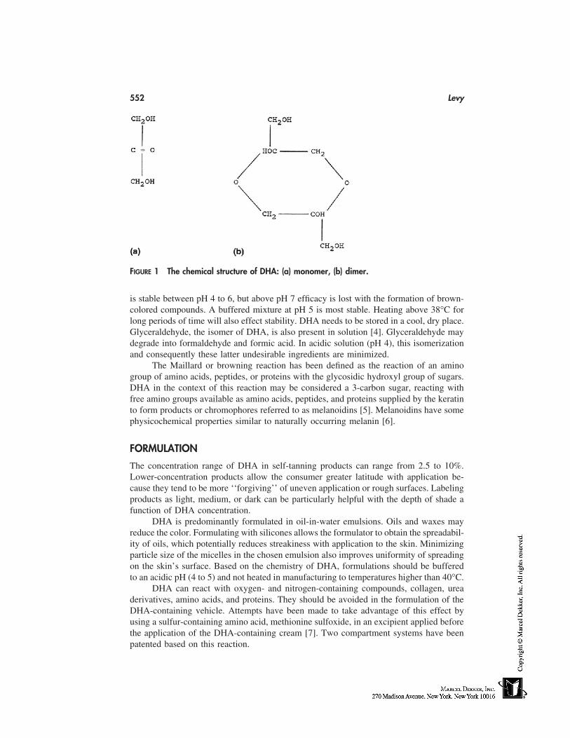
552 Levy
FIGURE 1 The chemical structure of DHA: (a) monomer, (b) dimer.
is stable between pH 4 to 6, but above pH 7 efficacy is lost with the formation of brown-colored compounds. A buffered mixture at pH 5 is most stable. Heating above 38°C forlong periods of time will also effect stability. DHA needs to be stored in a cool, dry place.Glyceraldehyde, the isomer of DHA, is also present in solution [4]. Glyceraldehyde maydegrade into formaldehyde and formic acid. In acidic solution (pH 4), this isomerizationand consequently these latter undesirable ingredients are minimized.
The Maillard or browning reaction has been defined as the reaction of an aminogroup of amino acids, peptides, or proteins with the glycosidic hydroxyl group of sugars.DHA in the context of this reaction may be considered a 3-carbon sugar, reacting withfree amino groups available as amino acids, peptides, and proteins supplied by the keratinto form products or chromophores referred to as melanoidins [5]. Melanoidins have somephysicochemical properties similar to naturally occurring melanin [6].
FORMULATION
The concentration range of DHA in self-tanning products can range from 2.5 to 10%.Lower-concentration products allow the consumer greater latitude with application be-cause they tend to be more ‘‘forgiving’’ of uneven application or rough surfaces. Labelingproducts as light, medium, or dark can be particularly helpful with the depth of shade afunction of DHA concentration.
DHA is predominantly formulated in oil-in-water emulsions. Oils and waxes mayreduce the color. Formulating with silicones allows the formulator to obtain the spreadabil-ity of oils, which potentially reduces streakiness with application to the skin. Minimizingparticle size of the micelles in the chosen emulsion also improves uniformity of spreadingon the skin’s surface. Based on the chemistry of DHA, formulations should be bufferedto an acidic pH (4 to 5) and not heated in manufacturing to temperatures higher than 40°C.
DHA can react with oxygen- and nitrogen-containing compounds, collagen, ureaderivatives, amino acids, and proteins. They should be avoided in the formulation of theDHA-containing vehicle. Attempts have been made to take advantage of this effect byusing a sulfur-containing amino acid, methionine sulfoxide, in an excipient applied beforethe application of the DHA-containing cream [7]. Two compartment systems have beenpatented based on this reaction.
Artificial Tanning Products 553
As will all cosmetic products, aesthetics are determined by vehicle formulation.Products may be formulated for dry skin types by the addition of emollients and humec-tants. Products formulated in gel or alcoholic vehicles may be more suitable for oily skin.
MECHANISM OF ACTION
The site of action of DHA is the stratum corneum. Tape stripping of the skin quicklyremoves the color [8], as does mechanical rubbing. Deeper staining in areas with thickerstratum corneum and no staining of mucous membranes without a stratum corneum isalso consistent with this being the site of action. DHA may be used as a substitute fordansyl chloride as a measure of stratum corneum turnover time [9]. Microscopic studiesof stripped stratum corneum and hair reveal irregular pigment masses in the keratin layers[10] known as melanoidins. These melanoidins are formed via the Maillard reaction withDHA as a sugar reacting with the amino groups supplied by the keratin.
APPLICATION
After application of a typical DHA-containing self-tanning lotion, color change may beobserved within an hour [11]. This color change may be seen under Wood’s light (blacklight) within 20 minutes. Maximal darkening may take 8 to 24 hours to develop. Individu-als can make several successive applications every few hours to achieve their desiredcolor. Color may last as long as 5 to 7 days with a single application. Depending onanatomical application, the same color can be maintained with repeat applications every1 to 4 days. The face requires fewer applications but more frequent reapplication to main-tain color than the extremities. Depth of color varies with the thickness and compactnessof the stratum corneum. Palms and soles stain deepest, necessitating washing of handsafter application to avoid staining. Hair and nails will color, but not mucous membraneslacking a stratum corneum or keratin layer. Rougher hyperkeratotic skin over the knees,elbows, and ankles will color more unevenly as will older skin with keratoses and mottledpigmentation. Color will also be maintained longer in these areas.
As in the formulation, the pH of the skin before application may have an effect onthe tonality of the skin color [4]. Alkaline residues from soaps or detergents may interferewith the reaction between DHA and the amino acids on the skin surface. Wiping the skinsurface with a hydroalcoholic, acidic toner just before DHA application may improveresults.
Careful directions provided with these products are, therefore, quite important indetermining consumer satisfaction. The skin may be prepared with a mild form of exfolia-tion. Even application is required with lighter application around elbows, knees, and anklesto avoid excessive darkening in these areas. Care also needs to taken around the hairlinewhere lighter hair may darken. Hands need to be washed immediately after use to avoiddarkening of the palms, fingers, and nails. Clearly, care, skill, and experience are necessarywhen using these products.
ADDITIVES
As commonly occurs, growth in this category has compelled both formulators and market-ers to seek points of differentiation between their product and that of their competitors.Besides formulating for specific skin types, active treatment ingredients may be incorpo-
554 Levy
rated into DHA-containing formulations. Vitamins, botanical extracts, antioxidants, anti-irritants, and even alpha-hydroxy acids may be added to broaden the claims made by agiven product. The addition of sunscreen ingredients to self-tanners warrants a more de-tailed discussion.
SUNSCREEN ACTIVITY
In the United States, the FDA Tentative Final Over-the-Counter Monograph on Sunscreens(Fed Reg. 1993) lists DHA as an approved sunscreen ingredient when used sequentiallywith lawsone (2-hydroxy-1, 4-napthoquinone). The European Economic Community Di-rective does not list DHA as a permitted UV filter. DHA itself has at most a modest effecton SPF [12], providing perhaps SPF 3 or 4 protection. The brown color obtained on theskin does absorb in the low end of the visible spectrum with overlap into long UVA andmay provide some UVA I protection [13].
Individuals using DHA-containing tanning products need to be cautioned that, de-spite visible darkening of their skin, these products provide minimal sun protection. Confu-sion may be compounded by the addition of UV filters to the formulation providing sig-nificant sun protection. The stated SPF for the product is applicable for a few hours afterapplication, but not for the days during which the skin color change may remain percep-tible.
INDICATIONS
Even with recent improvement in DHA formulations, the color achieved remains depen-dent on skin type. Individuals of medium complexion with skin phototypes II or III [14],as opposed to those who are lighter or darker, will obtain a more pleasing color. Individualswith underlying golden skin tones will achieve better results than individuals with rosy,sallow, or olive complexions. Older consumers with roughened, hyperkeratotic skin ormottled pigmentation with freckling may be less pleased with their use. Dermatologistsregularly recommend these products for tanning as a safe alternative to UV exposure.They may be used to camouflage some skin irregularities such as leg spider veins. Light-to medium-complected patients with vitiligo who show increased contrast with the vitiligi-nous areas with natural or unavoidable tanning in their normal skin may also benefit. Theymay even provide some protection for individuals with certain photosensitivity disorders[15].
SAFETY
The visible color change associated with the use of artificial tanning products might sug-gest to some users that these products are hazardous. Based on the chemistry of DHAand its toxicological profile, it can be considered nontoxic. It reacts quickly in the stratumcorneum minimizing systemic absorption. The acute toxicity of DHA was investigatedfor diabetics in the 1920s with their oral intake well tolerated [6]. The phosphate of DHAis found naturally as one of the intermediates in the Kreb’s cycle. Contact dermatitis toDHA has only rarely been reported [16]. As with other topical products with active ingredi-ents, such as sunscreens, much of the reported sensitivity is secondary to other ingredientsin the vehicle [17]. Adverse reactions are more likely to occur on the basis of irritation
Artificial Tanning Products 555
and not true allergy. Ultimately all claims related to product safety are based on testingthe final formulation.
ALTERNATIVE TANNING AGENTS
Lawsone found in the henna plant and juglone (5-hydroxy-1,4,-napthoquinone) derivedfrom walnuts also stain hair, skin, and nails. They have been used for centuries for haircoloring. Both substances lack skin substantivity and readily discolor clothing [18]. Theskin color they produce does not resemble a natural tan.
Based on the underlying principle of the Maillard reaction, other molecules with aketone function have been investigated [19]. An alpha-hydroxy group with attaching elec-tron withdrawing groups can also increase reactivity. Substances such as glyceraldehydeand glyoxal [20] have been described but found ineffective. Mucondialdehyde as describedby Eichler [21] is an effective agent but is also associated with toxicity, which mitigatesagainst its use [19]. Although several other aldehydes have been shown to have bettercolor properties, stability issues limit their use [19].
CONCLUSION
Increasing consumer awareness as to the hazards of UV light should fuel ongoing interestin self-tanning products. The benign toxicological profile of DHA reinforces the notionthat these products represent a safe alternative to a UV-induced tan. The results obtainedwith these products are dependent on the final formulation, individual application tech-nique, and consumers’ complexion type. Greater experience in formulation combined withincreasing sophistication on the part of the consumer should lead to continuing growthand satisfaction with the use of these products.
Consumers need to be clearly informed that these products do not offer significantprotection against UVB. If formulated with standard sunscreens, consumers should becautioned that the duration of UV protection is more short-lived than the color change.
REFERENCES
1. Wenninger JA, McEwen GN Jr, eds. International Cosmetic Ingredient Dictionary and Hand-book. 7th ed. Washington, D.C.: The Cosmetic, Toiletry, and Fragrance Association, 1997.
2. Guest GM, Cochrane W, Wittgenstein E. Dihydroxyacetone tolerance test for glycogen storagedisease. Mod Prob Paediat 1959; 4:169–178.
3. Wittgenstein E, Berry HK. Staining of skin with dihydroxyacetone. Science 1960; 132:894–895.
4. Maes DH, Marenus KD. Self-tanning products. In: Baran R, Maibach HI, eds. Cosmetic Der-matology. London: Martin Dunitz, 1994: 227–230.
5. Wittgenstein E, Berry HK. Reaction of dihydroxyacetone (DHA) with human skin callus andamino compounds. J Invest Dermatol 1961; 36:283–286.
6. Meybeck A. A spectroscopic study of the reaction products of dihydroxyacetone with aminoacids. J Soc Cosmet Chem 1977; 28:25–35.
7. Bobin MF, Martini MC, Cotte J. Effects of color adjuvants on the tanning effect of dihydroxy-acetone. J Soc Cosmet Chem 1984; 35:265–272.
8. Maibach HI, Kligman AM. Dihydroxyacetone: a suntan-simulating agent. Arch Dermatol1960; 82:505–507.
556 Levy
9. Pierard GE, Pierard-Franchimont C. Dihydroxyacetone test as a substitute for the dansyl chlo-ride test. Dermatology 1993; 186(2):133–137.
10. Goldman L, Barkoff J, Blaney D, Nakai T, Suskind R. The skin coloring agent dihydroxyace-tone. General Practioner 1960; 12:96–98.
11. Levy SB. Dihydroxyacetone-containing sunless or self-tanning lotions. J Am Acad Dermatol1992; 27:989–993.
12. Muizzuddin N. Marenus KD, Maes DH. UV-A and UV-B protective effect of melanoidsformed with dihydroxyacetone and skin. Poster 360 presented at the 55th Annual Meeting ofthe American Academy of Dermatology, San Francisco, CA, 1997.
13. Johnson JA, Fusaro RM. Protection against long ultraviolet radiation: topical browning agentsand a new outlook. Dermatologica 1987; 175:53–57.
14. Fitzpatrick TB. The validity and practicality of sunreactive skin types I through IV. ArchDermatol 1988; 124:869–871.
15. Fusaro RM, Johnson JA. Photoprotection of patients sensitive to short and/or long ultravioletlight with dihydroxyacetone/naphthoquinone. Dermatologica 1974; 148:224–227.
16. Morren M, Dooms-Goossens A, Heidbuchel M, Sente F. Damas M. Contact allergy to dihy-droxyacetone. Contact Dermatitis 1991; 25:326–327.
17. Foley P, Nixon R, Marks R, Frowen K, Thompson S. The frequency of reaction to sunscreens:results of a longitudinal population-based study on the regular use of sunscreens in Australia.Br J Dermatol 1993; 128:512–518.
18. Reiger MM. The chemistry of tanning. Cosmet & Toilet 1983; 98:47–50.19. Kurz T. Formulating effective self-tanners with DHA. Cosmet & Toilet 1994; 109:11:55–61.20. Goldman L, Barkoff J. Blaney D, Nakai T, Suskind R. Investigative studies with the skin
coloring agents dihydroxyacetone and glyoxal. J Invest Dermatol 1960; 35:161–164.21. Eichler J. Prinzipien der Haptbraunung. Kontakte (Merck) 1981; 111:24–30.
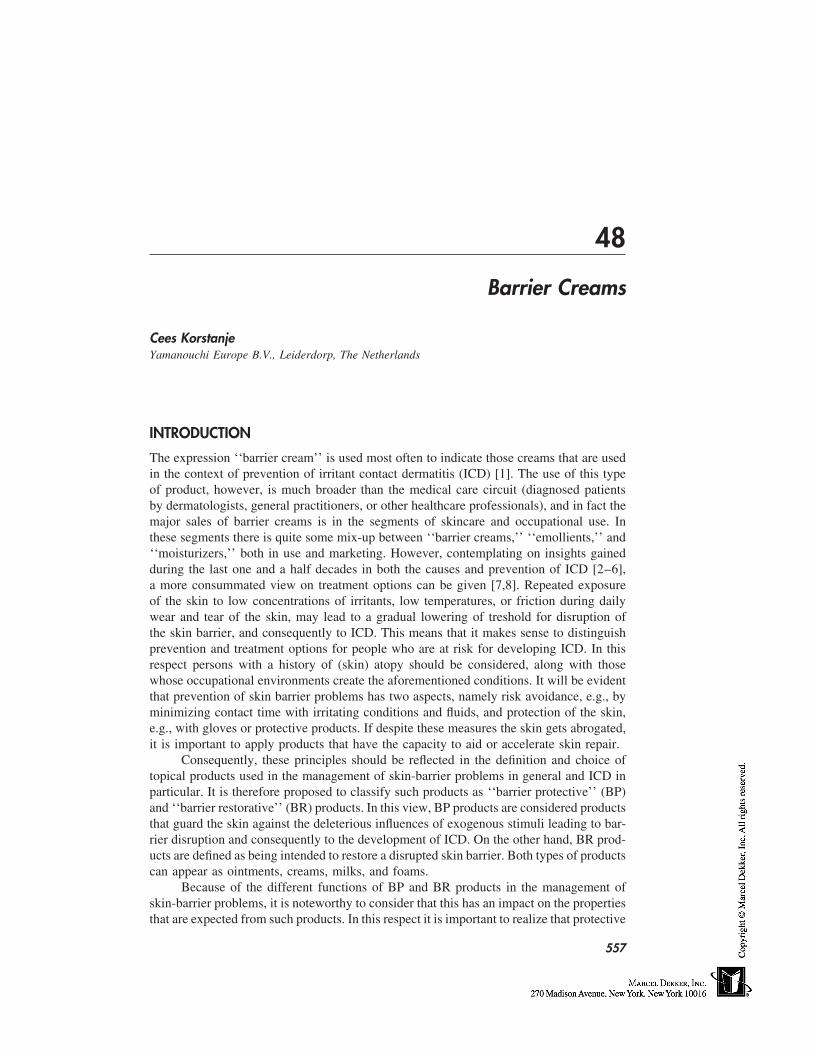
48
Barrier Creams
Cees KorstanjeYamanouchi Europe B.V., Leiderdorp, The Netherlands
INTRODUCTION
The expression ‘‘barrier cream’’ is used most often to indicate those creams that are usedin the context of prevention of irritant contact dermatitis (ICD) [1]. The use of this typeof product, however, is much broader than the medical care circuit (diagnosed patientsby dermatologists, general practitioners, or other healthcare professionals), and in fact themajor sales of barrier creams is in the segments of skincare and occupational use. Inthese segments there is quite some mix-up between ‘‘barrier creams,’’ ‘‘emollients,’’ and‘‘moisturizers,’’ both in use and marketing. However, contemplating on insights gainedduring the last one and a half decades in both the causes and prevention of ICD [2–6],a more consummated view on treatment options can be given [7,8]. Repeated exposureof the skin to low concentrations of irritants, low temperatures, or friction during dailywear and tear of the skin, may lead to a gradual lowering of treshold for disruption ofthe skin barrier, and consequently to ICD. This means that it makes sense to distinguishprevention and treatment options for people who are at risk for developing ICD. In thisrespect persons with a history of (skin) atopy should be considered, along with thosewhose occupational environments create the aforementioned conditions. It will be evidentthat prevention of skin barrier problems has two aspects, namely risk avoidance, e.g., byminimizing contact time with irritating conditions and fluids, and protection of the skin,e.g., with gloves or protective products. If despite these measures the skin gets abrogated,it is important to apply products that have the capacity to aid or accelerate skin repair.
Consequently, these principles should be reflected in the definition and choice oftopical products used in the management of skin-barrier problems in general and ICD inparticular. It is therefore proposed to classify such products as ‘‘barrier protective’’ (BP)and ‘‘barrier restorative’’ (BR) products. In this view, BP products are considered productsthat guard the skin against the deleterious influences of exogenous stimuli leading to bar-rier disruption and consequently to the development of ICD. On the other hand, BR prod-ucts are defined as being intended to restore a disrupted skin barrier. Both types of productscan appear as ointments, creams, milks, and foams.
Because of the different functions of BP and BR products in the management ofskin-barrier problems, it is noteworthy to consider that this has an impact on the propertiesthat are expected from such products. In this respect it is important to realize that protective
557
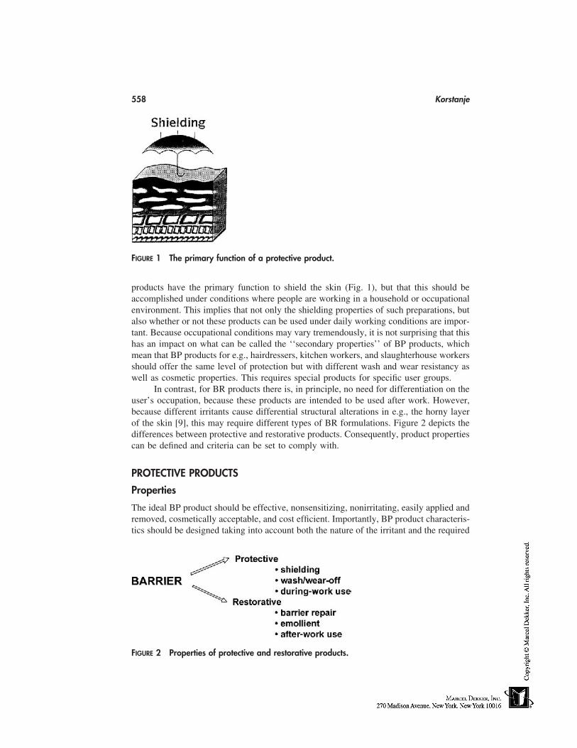
558 Korstanje
FIGURE 1 The primary function of a protective product.
products have the primary function to shield the skin (Fig. 1), but that this should beaccomplished under conditions where people are working in a household or occupationalenvironment. This implies that not only the shielding properties of such preparations, butalso whether or not these products can be used under daily working conditions are impor-tant. Because occupational conditions may vary tremendously, it is not surprising that thishas an impact on what can be called the ‘‘secondary properties’’ of BP products, whichmean that BP products for e.g., hairdressers, kitchen workers, and slaughterhouse workersshould offer the same level of protection but with different wash and wear resistancy aswell as cosmetic properties. This requires special products for specific user groups.
In contrast, for BR products there is, in principle, no need for differentiation on theuser’s occupation, because these products are intended to be used after work. However,because different irritants cause differential structural alterations in e.g., the horny layerof the skin [9], this may require different types of BR formulations. Figure 2 depicts thedifferences between protective and restorative products. Consequently, product propertiescan be defined and criteria can be set to comply with.
PROTECTIVE PRODUCTS
Properties
The ideal BP product should be effective, nonsensitizing, nonirritating, easily applied andremoved, cosmetically acceptable, and cost efficient. Importantly, BP product characteris-tics should be designed taking into account both the nature of the irritant and the required
FIGURE 2 Properties of protective and restorative products.
Barrier Creams 559
cosmetic properties (e.g., compatible with daily life activities) to guarantee use suitability.This implies that in order to define properties, one should first identify user-group specifi-cations and make an inventory of property—user combinations. An attempt at this isshown in Table 1. Accordingly, product profiles can be defined and assessed as productrequirements after testing the product. It is particularly important to realize that the usesuitability of BP products is dependent on the fulfillment of all different types of productrequirements at the same time. This is not easily accomplished, however. Unfortunatelythere is an almost inverse relationship between shielding properties and cosmetic accept-ability for ointments, creams, and foams, as is depicted in Figure 3.
Formulations
To date, very few preparations have characteristics that make them especially suited forany of the user groups given in Table 1 [10], although some general remarks can be made:current protective products against water-based irritants (soap, alkaline, acids) act in arather nonspecific way by depositing mineral oils, isopropylmyristate, long-chain alcohols,fats, or waxes on top of the skin or into the outer stratum corneum cell layers in order tocreate a physical lipids barrier. Water repellants, like silicone oils or perfluoroether, aresometimes included. However, because these are highly inert molecules, high percentagesof emulsifier are required to stabilize such formulations. This means that the net increasein protective properties with these supplements is disappointing (water-dragging effect),while high emulsifier concentrations may also cause irritation. More successful attemptshave been made by including chemicals that are intended to bind to skin constituents,such as Eucoriol (sodium bischlorophenyl sulfamine), which is included in a water-in-oil(W/O) ointment. A disadvantage is that the product is rather greasy on the skin. Theemulsion type of preparations against water-based irritants is usually W/O, although thereare some exceptions: a high-fat product in an oil-in-water (O/W) fatty cream*, and aproduct with petroleum jelly and silicone oil in a gel structure. The latter products havebetter cosmetic properties. Despite the fair-to-good protective properties offered byW/O products, their poor cosmetic properties make these products less suitable for useby, e.g., hairdressers and hospital nurses.
Recently, O/W emulsions, including CM glucan, a polysaccharide isolated frombaker’s yeast, was proposed and tested for its protective properties in surfactant-challengedskin [11]. The clinical value of this type of formulation has not been shown, however. Inorder to increase cosmetic properties for BP products, foam-based products have beendeveloped. An example is a foam containing stearic acid and dimethylpolysiloxane. Unfor-tunately, comparative tests have shown that the apparent advantage in cosmetic propertiesfor this product does not extend to acceptable protective properties [12].
Cream and gel preparations for the prevention of nickel-induced ICD with ethylenediamine as a chelator have been made and tested in in vitro tests and patch tests [13].Despite encouraging results in these types of tests, clinical efficacy of this type of prepara-tion has not been shown. For protection against organic solvents, O/W creams are recom-mended [14], although in efficacy tests using toluene, or poison ivy extracts, this is notwell accomplished with currently marketed products [1,10,15], thus casting doubt on thisrecommendation. Unfortunately, it can be stated that despite the many technological ad-
* Product protected by, e.g., Canadian patent 1200504.
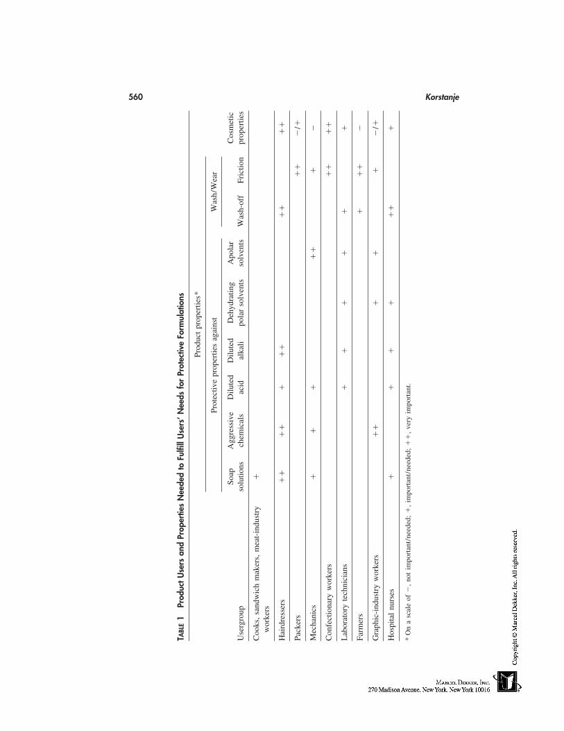
560 Korstanje
TABL
E1
Prod
uctU
sers
and
Prop
ertie
sN
eede
dto
Fulfi
llU
sers
’Nee
dsfo
rPr
otec
tive
Form
ulat
ions
Prod
uct
prop
ertie
s*
Prot
ectiv
epr
oper
ties
agai
nst
Was
h/W
ear
Soap
Agg
ress
ive
Dilu
ted
Dilu
ted
Deh
ydra
ting
Apo
lar
Cos
met
icU
serg
roup
solu
tions
chem
ical
sac
idal
kali
pola
rso
lven
tsso
lven
tsW
ash-
off
Fric
tion
prop
ertie
s
Coo
ks,
sand
wic
hm
aker
s,m
eat-
indu
stry
�w
orke
rs
Hai
rdre
sser
s�
��
��
��
��
��
Pack
ers
��
�/�
Mec
hani
cs�
��
��
��
Con
fect
iona
ryw
orke
rs�
��
�
Lab
orat
ory
tech
nici
ans
��
��
��
Farm
ers
��
��
Gra
phic
-ind
ustr
yw
orke
rs�
��
��
�/�
Hos
pita
lnu
rses
��
��
��
�
*O
na
scal
eof
�,
not
impo
rtan
t/ne
eded
;�
,im
port
ant/
need
ed;
��
,ve
ryim
port
ant.
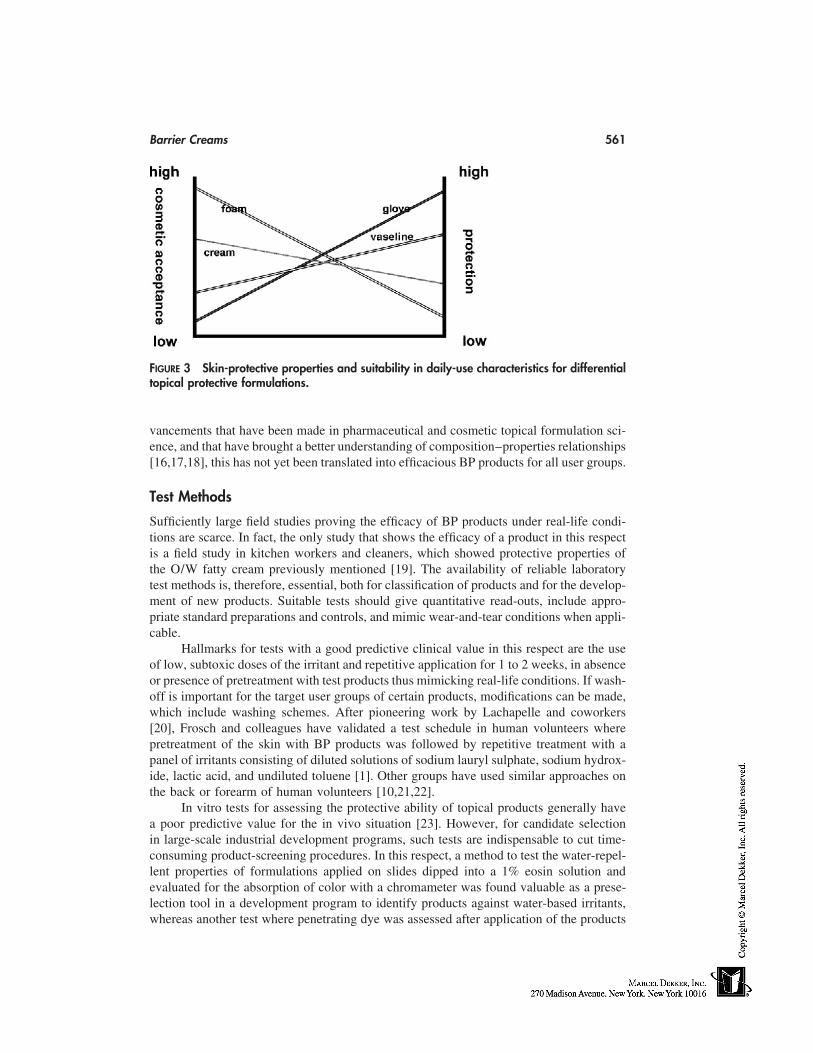
Barrier Creams 561
FIGURE 3 Skin-protective properties and suitability in daily-use characteristics for differentialtopical protective formulations.
vancements that have been made in pharmaceutical and cosmetic topical formulation sci-ence, and that have brought a better understanding of composition–properties relationships[16,17,18], this has not yet been translated into efficacious BP products for all user groups.
Test Methods
Sufficiently large field studies proving the efficacy of BP products under real-life condi-tions are scarce. In fact, the only study that shows the efficacy of a product in this respectis a field study in kitchen workers and cleaners, which showed protective properties ofthe O/W fatty cream previously mentioned [19]. The availability of reliable laboratorytest methods is, therefore, essential, both for classification of products and for the develop-ment of new products. Suitable tests should give quantitative read-outs, include appro-priate standard preparations and controls, and mimic wear-and-tear conditions when appli-cable.
Hallmarks for tests with a good predictive clinical value in this respect are the useof low, subtoxic doses of the irritant and repetitive application for 1 to 2 weeks, in absenceor presence of pretreatment with test products thus mimicking real-life conditions. If wash-off is important for the target user groups of certain products, modifications can be made,which include washing schemes. After pioneering work by Lachapelle and coworkers[20], Frosch and colleagues have validated a test schedule in human volunteers wherepretreatment of the skin with BP products was followed by repetitive treatment with apanel of irritants consisting of diluted solutions of sodium lauryl sulphate, sodium hydrox-ide, lactic acid, and undiluted toluene [1]. Other groups have used similar approaches onthe back or forearm of human volunteers [10,21,22].
In vitro tests for assessing the protective ability of topical products generally havea poor predictive value for the in vivo situation [23]. However, for candidate selectionin large-scale industrial development programs, such tests are indispensable to cut time-consuming product-screening procedures. In this respect, a method to test the water-repel-lent properties of formulations applied on slides dipped into a 1% eosin solution andevaluated for the absorption of color with a chromameter was found valuable as a prese-lection tool in a development program to identify products against water-based irritants,whereas another test where penetrating dye was assessed after application of the products
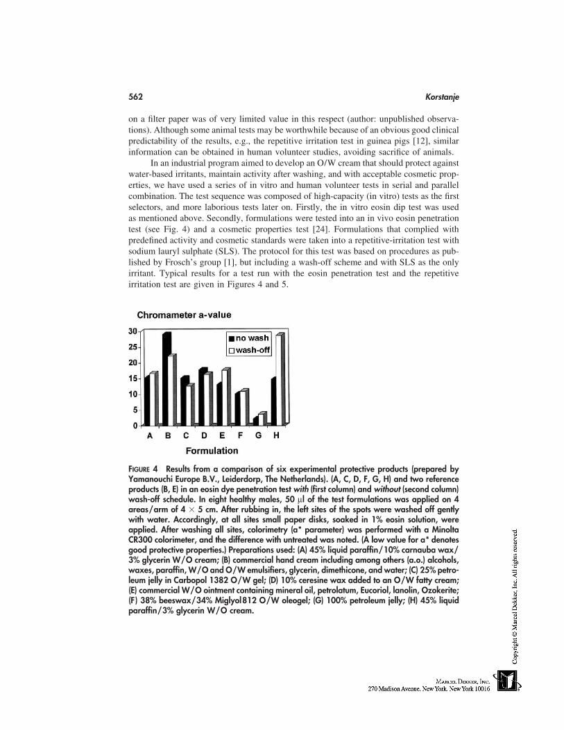
562 Korstanje
on a filter paper was of very limited value in this respect (author: unpublished observa-tions). Although some animal tests may be worthwhile because of an obvious good clinicalpredictability of the results, e.g., the repetitive irritation test in guinea pigs [12], similarinformation can be obtained in human volunteer studies, avoiding sacrifice of animals.
In an industrial program aimed to develop an O/W cream that should protect againstwater-based irritants, maintain activity after washing, and with acceptable cosmetic prop-erties, we have used a series of in vitro and human volunteer tests in serial and parallelcombination. The test sequence was composed of high-capacity (in vitro) tests as the firstselectors, and more laborious tests later on. Firstly, the in vitro eosin dip test was usedas mentioned above. Secondly, formulations were tested into an in vivo eosin penetrationtest (see Fig. 4) and a cosmetic properties test [24]. Formulations that complied withpredefined activity and cosmetic standards were taken into a repetitive-irritation test withsodium lauryl sulphate (SLS). The protocol for this test was based on procedures as pub-lished by Frosch’s group [1], but including a wash-off scheme and with SLS as the onlyirritant. Typical results for a test run with the eosin penetration test and the repetitiveirritation test are given in Figures 4 and 5.
FIGURE 4 Results from a comparison of six experimental protective products (prepared byYamanouchi Europe B.V., Leiderdorp, The Netherlands). (A, C, D, F, G, H) and two referenceproducts (B, E) in an eosin dye penetration test with (first column) and without (second column)wash-off schedule. In eight healthy males, 50 µl of the test formulations was applied on 4areas/arm of 4 � 5 cm. After rubbing in, the left sites of the spots were washed off gentlywith water. Accordingly, at all sites small paper disks, soaked in 1% eosin solution, wereapplied. After washing all sites, colorimetry (a* parameter) was performed with a MinoltaCR300 colorimeter, and the difference with untreated was noted. (A low value for a* denotesgood protective properties.) Preparations used: (A) 45% liquid paraffin/10% carnauba wax/3% glycerin W/O cream; (B) commercial hand cream including among others (a.o.) alcohols,waxes, paraffin, W/O and O/W emulsifiers, glycerin, dimethicone, and water; (C) 25% petro-leum jelly in Carbopol 1382 O/W gel; (D) 10% ceresine wax added to an O/W fatty cream;(E) commercial W/O ointment containing mineral oil, petrolatum, Eucoriol, lanolin, Ozokerite;(F ) 38% beeswax/34% Miglyol812 O/W oleogel; (G) 100% petroleum jelly; (H) 45% liquidparaffin/3% glycerin W/O cream.
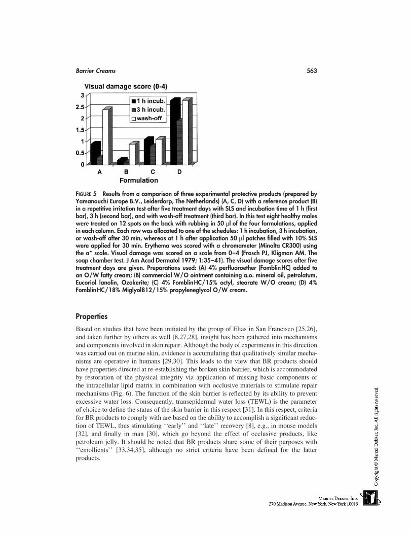
Barrier Creams 563
FIGURE 5 Results from a comparison of three experimental protective products (prepared byYamanouchi Europe B.V., Leiderdorp, The Netherlands) (A, C, D) with a reference product (B)in a repetitive irritation test after five treatment days with SLS and incubation time of 1 h (firstbar), 3 h (second bar), and with wash-off treatment (third bar). In this test eight healthy maleswere treated on 12 spots on the back with rubbing in 50 µl of the four formulations, appliedin each column. Each row was allocated to one of the schedules: 1 h incubation, 3 h incubation,or wash-off after 30 min, whereas at 1 h after application 50 µl patches filled with 10% SLSwere applied for 30 min. Erythema was scored with a chromameter (Minolta CR300) usingthe a* scale. Visual damage was scored on a scale from 0–4 (Frosch PJ, Kligman AM. Thesoap chamber test. J Am Acad Dermatol 1979; 1:35–41). The visual damage scores after fivetreatment days are given. Preparations used: (A) 4% perfluoroether (FomblinHC) added toan O/W fatty cream; (B) commercial W/O ointment containing a.o. mineral oil, petrolatum,Eucoriol lanolin, Ozokerite; (C) 4% FomblinHC/15% octyl, stearate W/O cream; (D) 4%FomblinHC/18% Miglyol812/15% propyleneglycol O/W cream.
Properties
Based on studies that have been initiated by the group of Elias in San Francisco [25,26],and taken further by others as well [8,27,28], insight has been gathered into mechanismsand components involved in skin repair. Although the body of experiments in this directionwas carried out on murine skin, evidence is accumulating that qualitatively similar mecha-nisms are operative in humans [29,30]. This leads to the view that BR products shouldhave properties directed at re-establishing the broken skin barrier, which is accommodatedby restoration of the physical integrity via application of missing basic components ofthe intracellular lipid matrix in combination with occlusive materials to stimulate repairmechanisms (Fig. 6). The function of the skin barrier is reflected by its ability to preventexcessive water loss. Consequently, transepidermal water loss (TEWL) is the parameterof choice to define the status of the skin barrier in this respect [31]. In this respect, criteriafor BR products to comply with are based on the ability to accomplish a significant reduc-tion of TEWL, thus stimulating ‘‘early’’ and ‘‘late’’ recovery [8], e.g., in mouse models[32], and finally in man [30], which go beyond the effect of occlusive products, likepetroleum jelly. It should be noted that BR products share some of their purposes with‘‘emollients’’ [33,34,35], although no strict criteria have been defined for the latterproducts.
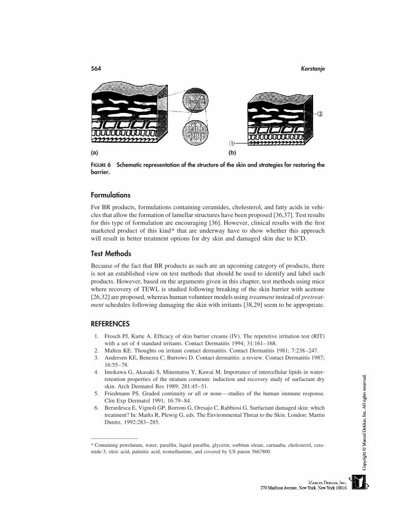
564 Korstanje
(a) (b)
FIGURE 6 Schematic representation of the structure of the skin and strategies for restoring thebarrier.
Formulations
For BR products, formulations containing ceramides, cholesterol, and fatty acids in vehi-cles that allow the formation of lamellar structures have been proposed [36,37]. Test resultsfor this type of formulation are encouraging [36]. However, clinical results with the firstmarketed product of this kind* that are underway have to show whether this approachwill result in better treatment options for dry skin and damaged skin due to ICD.
Test Methods
Because of the fact that BR products as such are an upcoming category of products, thereis not an established view on test methods that should be used to identify and label suchproducts. However, based on the arguments given in this chapter, test methods using micewhere recovery of TEWL is studied following breaking of the skin barrier with acetone[26,32] are proposed, whereas human volunteer models using treatment instead of pretreat-ment schedules following damaging the skin with irritants [38,29] seem to be appropriate.
REFERENCES
1. Frosch PJ, Kurte A. Efficacy of skin barrier creams (IV). The repetetive irritation test (RIT)with a set of 4 standard irritants. Contact Dermatitis 1994; 31:161–168.
2. Malten KE. Thoughts on irritant contact dermatitis. Contact Dermatitis 1981; 7:238–247.3. Andersen KE, Benezra C, Burrows D. Contact dermatitis: a review. Contact Dermatitis 1987;
16:55–78.4. Imokawa G, Akasaki S, Minematsu Y, Kawai M. Importance of intercellular lipids in water-
retention properties of the stratum corneum: induction and recovery study of surfactant dryskin. Arch Dermatol Res 1989; 281:45–51.
5. Friedmann PS. Graded continuity or all or none—studies of the human immune response.Clin Exp Dermatol 1991; 16:79–84.
6. Berardesca E, Vignoli GP, Borroni G, Oresajo C, Rabbiosi G. Surfactant damaged skin: whichtreatment? In: Marks R, Plewig G, eds. The Environmental Threat to the Skin. London: MartinDunitz, 1992:283–285.
* Containing petrolatum, water, paraffin, liquid paraffin, glycerin, sorbitan oleate, carnauba, cholesterol, cera-mide-3, oleic acid, palmitic acid, tromethamine, and covered by US patent 5667800.
Barrier Creams 565
7. Lachapelle JM. Prevention of allergic contact dermatitis. In: Grob JJ, Stern RS, MacKie RM,Weinstock WA, eds. Epidemiology, Causes and Prevention of Skin Diseases. Oxford: Black-well Science, 1997:318–323.
8. Halkier-Sorensen L. Occupational skin diseases. Contact Dermatitis 1996; 35(suppl 1):94–97.
9. Fartasch M. Electron microscopic imaging of the skin barrier dysfunction: the structural level.4th EADV Congress, Satellite symposium: Clinical Management of Skin Barrier Problems,Brussels, Belgium, Oct. 13, 1995.
10. Schlüter-Wigger, Elsner P. Efficacy of 4 commercially available protective creams in the repet-itive irritation test (RIT). Contact Dermatitis 1996; 34:278–283.
11. Zülli F, Suter F, Biltz H, Nissen HP. Improving skin function with CM-glucan, a biologicalresponse modifier from yeast. Int J Cosmet Sci 1998; 20:79–86.
12. Frosch PJ, Schulze-Dirks A, Hoffmann M, Axthelm I. Efficacy of skin barrier creams (II).Ineffectiveness of a popular ‘‘skin protector’’ against various irritants in the repetitive irritationtest in the guinea pig. Contact Dermatitis 1993; 29:74–77.
13. Fullerton A, Menné T. In vitro and in vivo evaluation of the effect of barrier gels in nickelcontact allergy. Contact Dermatitis 1995; 32:100–106.
14. Forssman T, Gloor M. Hand creams. In: Baran R, Maibach HI, eds. Cosmetic Dermatology.London: Martin Dunitz, 1994:181–187.
15. Smith WB, Baunchalk JM, Grabski WJ. Lack of efficacy of a barrier cream in preventingrhus dermatitis. Arch Dermatol 1993; 129:787–788.
16. Friberg SE. Micelles, microemulsions, liquid crystals, and the structure of stratum corneumlipids. J Soc Cosmet Chem 1990; 41:155–171.
17. Brinon L, Geiger S, Alard V, Tranchant JF, Pouget T, Couarraze G. Influence of lamellarliquid crystal structure on percutaneous diffusion of a hydrophilic tracer from emulsions.J Cosmet Sci 1998; 49:1–11.
18. Junginger HE, Boddé HE, Bouwstra JA. Water in dermatological preparations and its impacton skin. In: Crommelin DJA, Midha KK, Nagai T, eds. Topics in Pharmaceutical Sciences1993. Stuttgart: Medpharm, 1994:435–458.
19. Halkier-Sorensen L, Thestrup-Pedersen K. The efficacy of a moisturizer (Locobase) amongcleaners and kitchen assistants during everyday exposure to water and detergents. ContactDermatitis 1993; 29:266–271.
20. Mahmoud G, Lachapelle JM, Van Neste D. Histological assessment of skin damage by irri-tants: its possible use in the evaluation of a ‘‘barrier cream’’. Contact Dermatirtis 1988; 11:179–178.
21. Allenby CF, Basketter DA, Dickens A, Barnes EG, Brough HC. An arm immersion modelof compromised skin (I). Influence on irritation reactions. Contact Dermatitis 1993; 28:84–88.
22. Bettinger J, Gloor M, Gehring W. Influence of a pretreatment with emulsions on the dehydra-tion of the skin by surfactants. Int J Cosmet Sci 1994; 16:53–60.
23. Gehring W, Dördelmann C, Gloor M. Effektivitätsnachweis von Hautschutzpräparaten. [InGerman]. Allergologie 1994; 17:97–101.
24. Spiertz C, Korstanje C. A method for assessing the tactile properties of dermatological creambases. J Dermatol Treatment 1995; 6:155–157.
25. Grubauer G, Feingold KR, Elias PM. Relationship of epidermal lipogenesis to cutaneous bar-rier function. J Lipid Res 1987; 28:746–752.
26. Mao-Qiang M, Feingold KR, Thornfeldt CR, Elias PM. Optimization of physiological lipidmixtures for barrier repair. J Invest Dermatol 1996; 106:1096–1101.
27. EkanayakeMudiyanselage S, Aschauer H, Schmook FP, Jensen JM, Meingassner JG, ProkschE. Expression of epidermal keratins and the cornified envelope protein involucrin is influencedby permeability barrier disruption. J Invest Dermatol 1998; 111:517–523.
28. Fartasch M, Diepgen TL. The barrier function in atopic dry skin. Disturbance of membrane-
566 Korstanje
coating granule exocytosis and the formation of epidermal lipids? Acta Derm Venereol 1992;176(suppl):26–31.
29. Mao-Qiang M, Feingold KR, Wang F, Thornfeldt CR, Elias PM. A natural lipid mixture im-proves barrier function and hydration in human and murine skin. J Soc Cosmet Chem 1997;47:157–166.
30. Di Nardo A, Sugino K, Wertz P, Ademola J, Maibach HI. Sodium lauryl sulphate (SLS)induced irritant contact dermatitis: a correlation study between ceramides and in vivo parame-ters of irritation. Contact Dermatitis 1996; 35:86–91.
31. Maibach HI, Bronaugh R, Guy R, Turr E, Wilson D, Jacques S, Chaing D. Noninvasive tech-niques for determining skin function. In: Drill V, Lazar P eds. Cutaneous Toxicity. New York:Raven Press, 1984; 63–97.
32. Erlandsen M, Halkier-Sorensen L, De Vringer T. Proc Fourth Congress of the European Socof Contact Dermatitis, Helsinki, Finland, July 8–11, 1998:49.
33. Lodén M. Biophysical properties of dry atopic and normal skin with special reference to effectsof skin care products. Acta Derm Venereol 1995; 192(suppl):1–48.
34. Rycroft RJG. Occupational hand eczema: the role of emollients in treatment and prophylaxis.J Dermatol Treatment 1997; 8(suppl 1):S23–S24.
35. Marks R. How to measure the effects of emollients. J Dermatol Treatment 1997; 8(suppl. 1):S15–S18.
36. Iwai H, Fukasawa J, Suzuki T. A liquid crystal application in skin care cosmetics. Int J CosmSci 1998; 20:87–102.
37. De Vringer T. A rational design of topical formulations with skin barrier restoration properties.Proc Fourth Congress of the European Soc of Contact Dermatitis, Helsinki, Finland, July 8–11, 1998:152.
38. Gabard B, Elsner P, Treffel P. Barrier function of the skin in a repetitive irritants model andinfluence of 2 different treatments. Skin Res Technol 1996; 2:78–82.
49
Skin-Whitening Products
Hongbo Zhai and Howard I. MaibachUniversity of California at San Francisco School of Medicine,San Francisco, California
Skin-whitening products have been widely used in the cosmetic field and clinic therapy.They are supposed to either lighten skin (individuals who wish to change or modify theirskin color) or depigment skin (treatment for abnormal-hyperpigmentation skin such asmelasma, freckles, and senile lentigines). Whitening agents, such as hydroquinone, kojicacid, and ascorbic acid derivatives have shown efficacy in a variety of hyperpigmentarydisorders [1–14] but with varying success [1,2,7–9]. Their mechanism of action has beenstudied in vitro and in vivo [3,10–17]. Recently, their safety of application have beenextensively investigated [18–32]. This chapter includes the most popular active ingredi-ents of whitening agents and emphasizes their efficacy and safety.
HYDROQUINONE (1,4-DIHYDROXYBENZENE)
Hydroquinone is a nonvolatile chemical used in the photographic, rubber, chemical, andcosmetic industries. In the late 1930s, it was observed that a chemical used in rubbermanufacture, monobenzyl ether of hydroquinone, caused depigmented skin in some work-ers [1]. The efficacy of hydroquinone (1,4-dihydroxybenzene) as a skin-lightening agenthas been established in both human and animal studies. The chemical structure of hydro-quinone is shown in Figure 1. Clinically, hydroquinone is applied topically in the treatmentof melasma, freckles, and senile lentigines, as well as postinflammatory hyperpigmenta-tion. In the United States, hydroquinone is readily available in concentrations up to 2.0%as an over-the-counter (OTC) drug and by prescription at higher concentrations [1,2].Thus, hydroquinone is readily applied to the skin for medical and cosmetic reasons [33].
Hydroquinone inhibits the conversion of dopa to melanin by inhibiting the tyrosinaseenzyme [1–3]. Other proposed mechanisms are inhibition of DNA and RNA synthesis,degradation of melanosomes, and destruction of melanocytes [2]. Electron microscopicstudies of black guinea-pig skin treated with hydroquinone show the anatomic conse-quences of this action: (1) the melanosome structure is disturbed, resulting in decreasedproduction or increased degradation of these organelles, or both; (2) hydroquinone expo-sure can ultimately lead to the degradation of the melanocyte; and (3) keratinocytes arespared, showing no apparent injury [1].
567
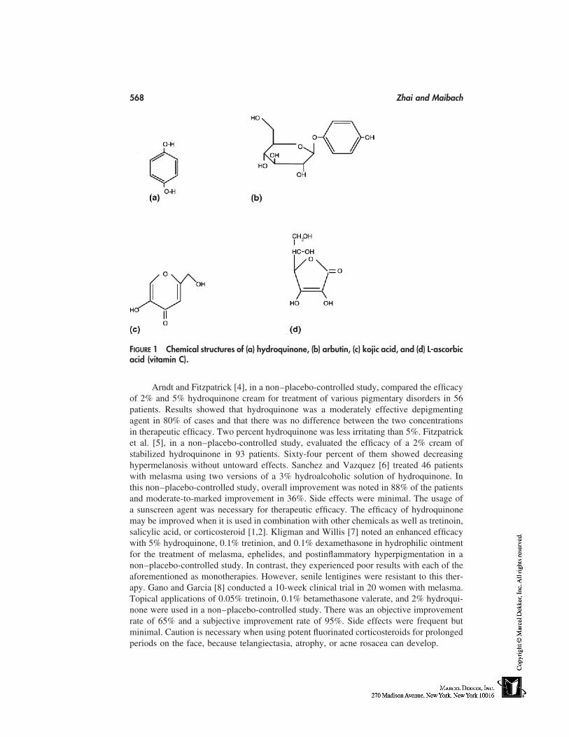
568 Zhai and Maibach
FIGURE 1 Chemical structures of (a) hydroquinone, (b) arbutin, (c) kojic acid, and (d) L-ascorbicacid (vitamin C).
Arndt and Fitzpatrick [4], in a non–placebo-controlled study, compared the efficacyof 2% and 5% hydroquinone cream for treatment of various pigmentary disorders in 56patients. Results showed that hydroquinone was a moderately effective depigmentingagent in 80% of cases and that there was no difference between the two concentrationsin therapeutic efficacy. Two percent hydroquinone was less irritating than 5%. Fitzpatricket al. [5], in a non–placebo-controlled study, evaluated the efficacy of a 2% cream ofstabilized hydroquinone in 93 patients. Sixty-four percent of them showed decreasinghypermelanosis without untoward effects. Sanchez and Vazquez [6] treated 46 patientswith melasma using two versions of a 3% hydroalcoholic solution of hydroquinone. Inthis non–placebo-controlled study, overall improvement was noted in 88% of the patientsand moderate-to-marked improvement in 36%. Side effects were minimal. The usage ofa sunscreen agent was necessary for therapeutic efficacy. The efficacy of hydroquinonemay be improved when it is used in combination with other chemicals as well as tretinoin,salicylic acid, or corticosteroid [1,2]. Kligman and Willis [7] noted an enhanced efficacywith 5% hydroquinone, 0.1% tretinion, and 0.1% dexamethasone in hydrophilic ointmentfor the treatment of melasma, ephelides, and postinflammatory hyperpigmentation in anon–placebo-controlled study. In contrast, they experienced poor results with each of theaforementioned as monotherapies. However, senile lentigines were resistant to this ther-apy. Gano and Garcia [8] conducted a 10-week clinical trial in 20 women with melasma.Topical applications of 0.05% tretinoin, 0.1% betamethasone valerate, and 2% hydroqui-none were used in a non–placebo-controlled study. There was an objective improvementrate of 65% and a subjective improvement rate of 95%. Side effects were frequent butminimal. Caution is necessary when using potent fluorinated corticosteroids for prolongedperiods on the face, because telangiectasia, atrophy, or acne rosacea can develop.

Skin-Whitening Products 569
Pathak et al. [9] clinically tested the efficacy of hydroquinone in varying concentra-tions supplemented with corticosteroids or retinoic acid (tretinoin) in 300 Hispanic womenwith melasma in a non–placebo-controlled study, and concluded that cream or lotion for-mulations of 2% hydroquinone and 0.05 to 0.1% retinoic acid provided the most favorableresults. In addition, avoidance of sun exposure and constant use of broad-spectrum sun-screens are necessary for the best therapy effects. Recently, Clarys and Barel [34] testedthe efficacy of an ascorbate-phytohydroquinone complex in 14 patients with lentigo senilelesions in a non–placebo-controlled study. Objective skin-color changes were evaluatedwith a chromameter. After 1 month of treatment, a clear depigmentation of the maculeswas measured. None of the patients reported adverse effects.
Gellin et al. [35] established a reliable in vivo method to predict the depigmentingaction of chemicals on mammalian melanocytes. They used black guinea pigs and blackmice as animal models to screen the depigmenting capacity of several phenols, catechols,and organic antioxidants. Results showed that complete depigmentation on all test siteswas achieved with monomethyl ether of hydroquinone and tertiary butyl catchall in theblack guinea pig. Less-pronounced pigment loss was noted with these chemicals in blackmice.
To treat some cases, higher concentrations of hydroquinone may be used. The formu-lations contain concentrations as high as 10% combined with nonfluorinated corticoidcreams with or without the additional use of tretinoin or salicylic acid. Extemporaneouslycompounded preparations are often effective in patients that have failed to respond tolower concentrations of hydroquinone. With controlled use and monitoring, side effectsfrom these preparations have proved minimal [2]. Note, however, that hydroquinone maybe quickly oxidized in such formulations.
Hydroquinone occurs in nature as the beta-glucopyranoside conjugate arbutin. Ar-butin is a safe and mild agent for treating cutaneous hyperpigmentation disorders, includ-ing melasma and UV-induced ephelides [10]. Arbutin is an active ingredient of the crudedrug Uvae Ursi Folium-traditionally used in Japan and contained in the leaves of peartees and certain herbs. The chemical structure of arbutin is shown in Figure 1. Maeda andFukuda [10] determined the arbutin’s inhibitory action on the melanin synthetic enzymeand its effects on melanin intermediates and melanin production in cultured human mela-nocytes. They indicated that the depigmentation effect of arbutin works through a inhibi-tion of the melanosomal tyrosinase activity, rather than suppression of the expressionand synthesis of tyrosinase in human melanocytes. Arbutin was much less cytotoxic thanhydroquinone to cultured human melanocytes.
Adverse reactions associated with hydroquinone use include acute and chronic com-plications. Acute reactions include irritant dermatitis, nail discoloration, and postinflam-matory hyperpigmentation [1]. Although commonly assumed to be a common allergen,the documentation of hydroquinone allergic contact dermatitis is weak [1]. Hydroquinoneuse can also induce hypopigmentation and, rarely, depigmentation of treated surroundingnormal skin. However, these changes are temporary and resolve on cessation of hydroqui-none treatment, in contrast to monobenzone use, which can cause permanent depigmenta-tion [36]. Hence, the only indication for monobenzone therapy is in the treatment of severevitiligo.
A more recent concern regarding the use of hydroquinone is the occurrence of hydro-quinone-induced ochronosis, a chronic disfiguring condition resulting, in general, fromthe prolonged use of strong concentrations of hydroquinone [36]. Hydroquinone’s acuteand chronic toxicity toward higher terrestrial organisms appears to be minimal in humans
570 Zhai and Maibach
[20,21]. An epidemiologic investigation in 478 photographic processors has shown nosignificant excess mortality, sickness absence, or cancer incidence [20]. The reported ne-phropathy and cell proliferation, as evidence of carcinogenicity, observed in Fischer 344/N rats [22,23] appear to be strain and sex specific [23]. Hydroquinone was negative inthe Ames/Salmonella and Drosophila genotoxicity assays [24]. Others suggest that carcin-ogenic and teratogenic potentials have been, at present inadequately studied [20,25], andthat both hydroquinone and benzoquinone produce cytotoxic effects on human and mousebone-marrow cells [26]. Hydroquinone readily penetrates human forehead skin in vivofollowing a single topical exposure in an alcoholic vehicle of 24-hour duration. Elimina-tion was complete within 5 days [19]. Wester et al. [18] determined the topical bioavail-ability, metabolism, and disposition of hydroquinone on humans in vivo and in vitro;dose recovery in urine was 45.3%, of which the majority was excreted in the first 24hours.
KOJIC ACID
Kojic acid, a fungal metabolic product, is increasingly being used as a skin-lighteningagent in skincare products marketed in Japan since 1988. It was first isolated from Asper-gillus in 1907 [31]. The structure is shown in Figure 1. The mode of action of kojic acidis to suppress free tyrosinase, mainly attributable to chelation of its copper [11,16,31],and it has been shown to be responsible for therapy and prevention of pigmentation, bothin vitro and in vivo [11,31].
In Japan it is used in nonprescription skincare products up to a concentration of 1%.To increase percutaneous absorption and thus therapeutic activity, it is usually used at thehighest concentration allowed [31]. Because it is used intensively in foods (e.g., beanpaste, soy, and sake) in some countries, particularly Japan, its oral safety has been studied.Shibuya et al. [28], investigating the mutagenicity of kojic acid by the Ames test, forwardmutation test in cultured Chinese hamster cells, and dominant lethal test in mice, concludedthat, although kojic acid is a weak mutagen in bacteria, it is nonmutagenic in eukaryoticsystem either in vivo or in vitro. Abdel-Hafez and Shoreit [30] tested the mycotoxinsusing the dilution-plate method. Results showed that kojic acid may induce some toxins.Fujimoto et al. [32] examined the tumorigenicity of kojic acid in B6C3F1 mice. Threegroups of animals were given 0, 1.5, and 3.0% kojic acid–containing food for 6 weeks;kojic acid groups significantly induced thyroid tumors in B6C3F1 mice. But true adverseeffects after human oral ingestion have not been shown. Nakagawa et al. [31] noted thatthere were no signs of relapse of dermatitis or any other adverse effects on sensitizedpatients upon ingestion of foods containing kojic acid. However, they reported that topicalapplication of kojic acid may induce allergic contact dermatitis with sensitized patients.They postulated that kojic acid was considered to have a high sensitizing potential, becauseof the comparatively high frequency of contactsensitivity in patients using 1 or more kojicacid–containing products. Recently, Majmudar et al. [37] used an in vitro model to evalu-ate the efficacy, stability, and cytotoxicity of whitening agents. They also conducted anon–placebo-controlled clinical study that indicated that kojic acid in an anhydrous basecan induce more skin lightening than in the aqueous base.
ASCORBIC ACID (VITAMIN C) AND ITS DERIVATIVES
Ascorbic acid may inhibit melanin production by reducing o-quinones [12] so that melanincannot be formed by the action of tyrosinase until all vitamin C is oxidized. The chemical
Skin-Whitening Products 571
structure of vitamin C is shown in Figure 1. Although the lightening effect of vitamin Cis considered, it is quickly oxidized and decomposes in aqueous solution and is thus notgenerally useful as a depigmenting agent. Numerous stable derivatives of vitamin C havebeen synthesized to minimize this problem [12–14,17]. Magnesium-L-ascorby-2-phos-phate (VC-PMG) is a vitamin-C derivative that is stable in water, especially in neutral oralkaline solution containing boric acid or its salt [12]. VC-PMG is hydrolyzed by phospha-tases of liver or skin to vitamin C and thus exhibits vitamin C-reducing activity [12].Kameyama et al. [12] investigated the effects of VC-PMG on melanogenesis in vitro andin vivo. Results from this non–placebo-controlled study suggested the topical applicationof VC-PMG was significantly effective in lightening the skin in 19 of 34 patients withchloasma or senile freckles, and in 3 of 25 subjects with normally pigmented healthy skin.
OTHER AGENTS
Various systemic drugs and natural products may be used as protective agents, such aschloroquine, indomethacin, vitamin C and E, fish oil, and green tea, etc. Topical agentsinclude azelaic acid and melawhite except where previously described [38]. Recently,Kobayashi et al. [39] reported that neoagarobiose could be useful as a novel whiteningagent as it has shown moisturizing and whitening effects with low cytotoxicity. Ando etal. [40] evaluated the effects of unsaturated fatty acids on UV-induced hyperpigmentationof the skin in a placebo (vehicle)-controlled study. Skin hyperpigmentation was inducedon the backs of guinea pigs by UVB exposure. Oleic acid, linoleic acid, and α-linolenicacid (0.5% in ethanol), or ethanol alone as a control, were then topically applied dailyfive times per week for 3 successive weeks. Results suggest that the pigment-lighteningeffects of linoleic acid and α-linolenic acid are, at least in part, attributable to suppressionof melanin production by active melanocytes as well as to enhanced desquamation ofmelanin pigment from the epidermis.
CONCLUSIONS
In general, skin-whitening products are considered modestly effective. High concentra-tions are not recommended except under a physician’s supervision. The application as acombination with certain chemicals (retinoic acid and alpha-hydroxy acids) may enhancelightening. Optimal whitening agents remain a future goal.
REFERENCES
1. Engasser PG, Maibach HI. Cosmetics and dermatology: bleaching creams. J Am Acad Derma-tol 1981; 5:143.
2. Grimes PE Melasma. Etiologic and therapeutic considerations. Arch Dermatol 1995; 131:1453.
3. Jimbow K, Obata H, Pathak MA, Fitzpatrick TB. Mechanism of depigmentation by hydroqui-none. J Invest Dermatol 1974; 62:436.
4. Arndt KA, Fitzpatrick TB. Topical use of hydroquinone as a depigmenting agent. JAMA 1965;194:965.
5. Fitzpatrick TB, Arndt KA, el-Mofty AM, Pathak MA. Hydroquinone and psoralens in thetherapy of hypermelanosis and vitiligo. Arch Dermatol 1966; 93:589.
6. Sanchez JL, Vazquez MA hydroquinone solution in the treatment of melasma. Int J Dermatol1982; 21:55.
572 Zhai and Maibach
7. Kligman AM, Willis I. A new formula for depigmenting human skin. Arch Dermatol 1975;111:40.
8. Gano SE, Garcia RL. Topical tretinoin, hydroquinone, and betamethasone valerate in the ther-apy of melasma. Cutis 1979; 23:239.
9. Pathak MA, Fitzpatrick TB, Kraus EW. Usefulness of retinoic acid in the treatment of mel-asma. J Am Acad Dermatol 1986; 15:894.
10. Maeda K, Fukuda M. Arbutin: mechanism of its depigmenting action in human melanocyteculture. J Pharm Exp Ther 1996; 276:765.
11. Cabanes J, Chazarra S, Garcia-Carmona F. Kojic acid, a cosmetic skin whitening agent, is aslow-binding inhibitor of catecholase activity of tyrosinase. J Pharm Pharmacol 1994; 46:982.
12. Kameyama K, Sakai C, Kondoh S, Yonemoto K, Nishiyama S, Tagawa M, Murata T, OhnumaT, Quigley J, Dorsky A, Bucks D, Blanock K. Inhibitory effect of magnesium L-ascorbyl-2-phosphate (VC-PMG) on melanogenesis in vitro and in vivo. J Am Acad Dermatol 1996; 34:29.
13. Nomura H, Ishiguro T, Morimoto S. Studies on L-ascorbic acid derivatives. II. L-ascorbicacid 3-phosphate and 3-pyrophosphate. Chem Pharm Bull 1969; 17:381.
14. Nomura H, Ishiguro T, Morimoto S. Studies on L-ascorbic acid derivatives. 3. Bis(L-ascorbicacid 3,3′)phosphate and L-ascorbic acid 2-phosphate. Chem Pharm Bull 1969; 17:387.
15. Nakajima M, Shinoda I, Fukuwatari Y, Hayasawa H. Arbutin increases the pigmentation ofcultured human melanocytes through mechanisms other than the induction of tyrosinase activ-ity. Pig Cell Res 1998; 11:12.
16. Kahn V. Effect of kojic acid on the oxidation of DL-DOPA, norepinephrine, and dopamineby mushroom tyrosinase. Pig Cell Res 1995; 8:234.
17. Morisaki K, Ozaki S. Design of novel hybrid vitamin C derivatives: thermal stability andbiological activity. Chem Pharm Bull 1996; 44:1647.
18. Wester RC, Melendres J, Hui X, Cox R, Serranzana S, Zhai H, Quan D, Maibach HI. Human invivo and in vitro hydroquinone topical bioavailability, metabolism, and disposition. J ToxicolEnviron Health 1998; 54:301.
19. Bucks DAW, McMaster JR, Guy RH, Maibach HI. Percutaneous absorption of hydroquinonein humans: effect of 1-dodecylazacycloheptan-2-one (azone) and the 2-ethylhexyl ester of 4-(dimethylamino)benzoic acid (escalol 507). J Toxicol Environ Health 1988; 24:279.
20. Friedlander BR, Hearne FT, Newman BJ. Mortality, cancer incidence, and sickness-absencein photographic processors: an epidemiologic study. J Occup Med 1982; 24:605.
21. Pifer JW, Hearne FT, Swanson FA, O’Donoghue JL. Mortality study of employees engagedin the manufacture and use of hydroquinone. Int Arch Occup Environ Health 1995; 67:267.
22. English JC, Hill T, O’Donoghue JL, Reddy MV. Measurement of nuclear DNA modificationby 32P-postlabeling in the kidneys of male and female Fischer 344 rats after multiple gavagedoses of hydroquinone. Fundam Appl Toxicol 1994; 23:391.
23. English JC, Perry LG, Vlaovic M, Moyer C, O’Donoghue JL. Measurement of cell prolifera-tion in the kidneys of Fischer 344 and Sprague-Dawley rats after multiple gavage administra-tion of hydroquinone. Fundam Appl Toxicol 1994; 23:397.
24. Gocke E, King MT, Eckhardt K, Wild D. Mutagenicity of cosmetics ingredients licensed bythe European community. Mutat Res 1981; 90:91.
25. Whysner J, Verna L, English JC, William GM. Analysis of studies related to tumorigenicityinduced by hydroquinone. Regul Toxicol Pharmacol 1995; 21:158.
26. Colinas RJ, Burkart PT, Lawrence DA. In vitro effects of hydroquinone, benzoquinone, anddoxorubicin on mouse and human bone marrow cells at physiological oxygen partial pressure.Toxicol Appl Pharmacol 1994; 129:95.
27. Goffin V, Pierard GE, Henry F, Letawe C, Maibach HI. Sodium hypochlorite, bleachingagents, and the stratum corneum. Ecotoxicol Environ Safety 1997; 37:199.
28. Shibuya T, Murota T, Sakamoto K, Iwahara S, Ikeno M. Mutagenicity and dominant lethal
Skin-Whitening Products 573
test of kojic acid: Ames test, forward mutation test in cultured Chinese hamster cells anddominant lethal test in mice. J Toxicol Sci 1982; 7:255.
29. Wei CI, Huang TS, Fernando SY, Chung KT. Mutagenicity studies of kojic acid. ToxicolLetters 1991; 59:213.
30. Abdel-Hafez SI, Shoreit AA. Mycotoxins producing fungi and mycoflora of air-dust fromTaif, Saudi Arabia. Mycopathologia 1985; 92:65.
31. Nakagawa M, Kawai K, Kawai K. Contact allergy to kojic acid in skin care products. ContactDermatitis 1995; 32:9.
32. Fujimoto N, Watanabe H, Nakatani T, Roy G, Ito A. Induction of thyroid tumours in (C57BL/6N � C3H/N)F1 mice by oral administration of kojic acid. Food Chem Toxicol 1998; 36:697.
33. Strauch E, Burke P, Maibach HI. Hydroquinone. J Derm Treatment 2000. Submitted.34. Clarys P, Barel A. Efficacy of topical treatment of pigmentation skin disorders with plant
hydroquinone glucosides as assessed by quantitative color analysis. J Dermatol 1998; 25:412.35. Gellin GA, Maibach HI, Mislaszek MH, Ring M. Detection of environmental depigmenting
substances. Contact Dermatitis 1979; 5:201.36. Grimes PE. Vitiligo. An overview of therapeutic approaches. Dermatol Clin 1993; 11:325.37. Majmudar G, Jacob G, Laboy Y, Fisher L. An in vitro method for screening skin-whitening
products. J Cosmet Sci 1998; 49:361.38. Piamphongsant T. Treatment of melasma: a review with personal experience. Int J Dermatol
1998; 37:897.39. Kobayashi R, Takisada M, Suzuki T, Kirimura K, Usami S. Neoagarobiose as a novel moistur-
izer with whitening effect. Biosci Biotechnol Biochem 1997; 61:162.40. Ando H, Ryu A, Hashimoto A, Oka M, Ichihashi M. Linoleic acid and α-linolenic acid lightens
ultraviolet-induced hyperpigmentation of the skin. Arch Dermatol Res 1998; 290:375.

50
Interactions with Hair and Scalp
Dominique Van Neste and Ghassan ShakerSkinterface sprl, Tournai, Belgium
PSYCHOSOCIAL FACTORS INVOLVED IN HAIR COSMETICS
Haircare and psyche reciprocally reflect each other both positively and negatively (badhair days). Contrary to the bad haircare and negligence of a depressed person or a manin grief, generally people tend to offer themselves the best of haircare when they arefeeling happy or when they want to show their internal feelings to others through bodylanguage. This is particularly obvious during public appearances and important socialgatherings (e.g., parties, marriage ceremonies). Haircare by itself can induce a state ofself-confidence and may reflect social status. This may explain significant differences inshampooing regimens, which range from once or twice a week to once a day.
Hair is midway between nature and culture [1]. Haircare attitudes are different fromone society to another regardless of economic differences, and from one person to anotherwithin societies; e.g., hair loss is not equally perceived by everybody in all societies inthe same manner [2–5]. Some people are seriously psychologically affected and ready tospend a fortune in order to cope with the problem, whereas others just do not care at all.In the former group, styling is of high significance as is the selection of cosmetic agents.
The intersocial and interpersonal attitude of adult males towards greying of hair isquite evident, added to the difference in attitude between men and women toward thesame problem. However, this is not an exclusivity of mankind; the social significance ofhair/pelage/beard/crown is very pronounced in other mammals (e.g., primates, lions).Grooming in humans is specifically a private activity or limited to one professional body(hair stylists).
NATURAL PROPERTIES OF HAIR AND THEIR IMPORTANCEFOR HAIR APPEARANCE
Physical Properties of Hair as a Basis for Appearance and Perception
Optical properties (absorption and reflection of visible light); the role of pigmentation ininducing a contrast between skin and hair; and the role of cuticle, cortex, and medullaare some physical properties playing an important role in hair appearance and perception[6]. Apart from albinos, all normal subjects have melanin. The production of these pig-
575
576 Van Neste and Shaker
ments is genetically determined and results in the production of various proportions ofthe following: (1) eumelanin, which gives colors from brown to black; and (2) pheome-lanin, which gives colors from yellow-red to red. The hair color of each individual dependson the preponderant type of melanin as well as its quantity and distribution in the skinand hair. Melanin is a polymer of high molecular weight, insoluble in water and mostsolvents. It originates from melanocytes located in the basal cell layer of the hair matrix.Melanogenesis involves a complex sequence of chemical reactions corresponding to anoxidative polymerization catalyzed by certain enzymes; these complex processing phasesoccur in small vesicles named melanosomes. An intimate relationship must exist betweenthe factors controlling melanocytes and matrix cell activity, because melanosome andpigment transfer from the melanocyte to the hair matrix occur only during the anagenphase of the hair cycle. Hair color also varies with age. There is first an intensificationand then a slowdown, or sometimes even a halt, in pigment formation despite the ratherconstant number of melanocytes. This points to functional and regulatory aspects.
Melanin granules are distributed throughout the hair cortex but in greater concentra-tion towards the periphery [6]. The color of hair is an optical phenomenon attributable tothe reflection and refraction of incident light from various interfaces, especially the bulkof melanin contained in the cortex. Newly formed unpigmented hair with no medullaappears yellowish rather than white. This is probably the intrinsic color of dense and well-organized arrangement of keratin fibers [6]. Another important physical factor that helpsthe ease with which hair can be styled and given a desired shape is the elimination ofstatic electricity, which causes repulsion between individual hairs and is an obstacle tostyling and arranging hair [7]. The development of electrical charges on hairs during comb-ing and brushing is a complicated phenomenon that varies according to hair type, surfacestate, and the humidity of the surrounding environment. A product’s antistatic propertiescan be assessed in vitro by measuring the electrical potential build-up of hair during comb-ing. If opposed electric charges are face to face, matting of hair may occur. There is noway of untangling it and a substantial haircut is the only solution.
Mechanical Properties of Hair Appearance
Hair fibers are generally elliptical, with cross sections having minor and major axis ratioin the range of 0.63 to 0.91, the most elliptical being black hair, the most circular Asianhair. Resistance to longitudinal deformation, bending and torsion stiffness, and hold ofset hair are related to fiber diameter. The relationship between the constraint and elonga-tion obtained follows a curve of three regions (preyield, yield, and postyield) accordingto the stretching force [7]. Fiber breakage occurs mainly in the postyield region. The loadvalues depend on the cohesion of α-keratin. All factors diminishing this cohesion bringthe load value down, e.g., wet hair. Examination of load elongation curves helps in study-ing how hair behaves in the course of various hairdressing procedures including the widerange of temperature, humidity and chemical agents involved.
The hair shaft is a strong enough fiber. It behaves like reinforced wire. Curled blackhair is fairly fragile because of the highly twisted configuration and flattening as opposedto Asiatic hair [7]. The disruptive load for hair varies with age, peaking at about 20 yearsof age.
The length of hair plays a role in perception. A typical example is when the hairis cut short, people usually interpret the perception of stubbles as thickening of the newly
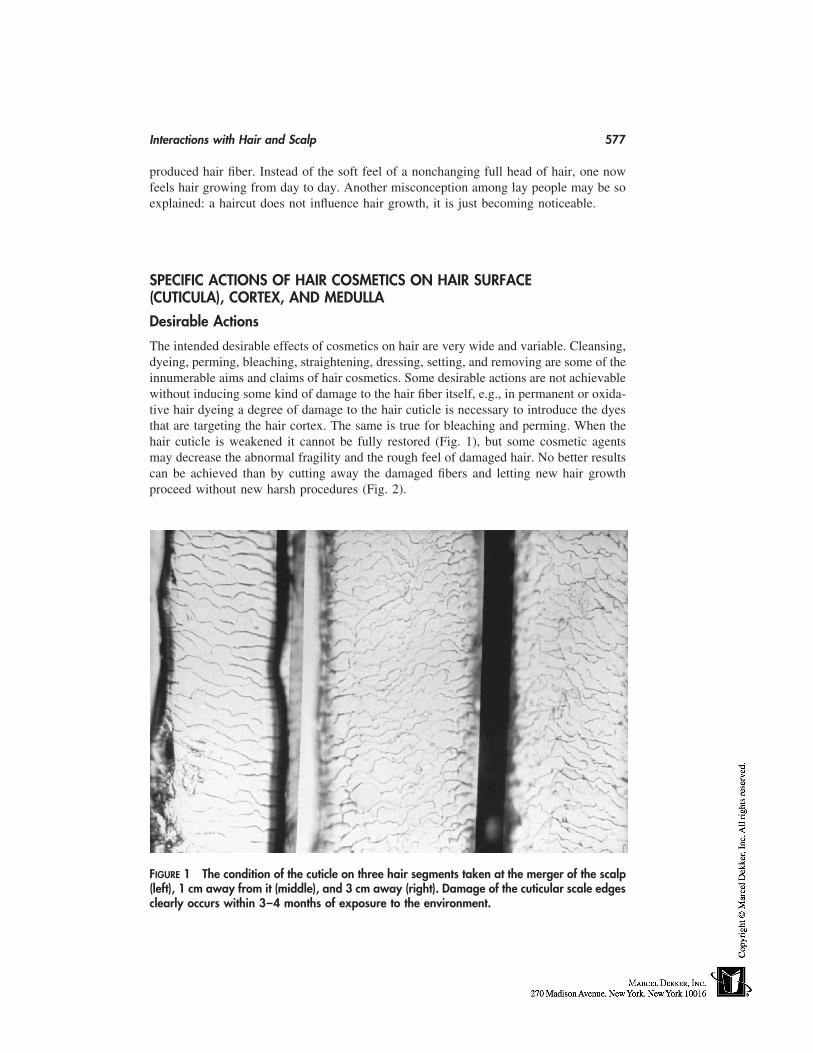
Interactions with Hair and Scalp 577
produced hair fiber. Instead of the soft feel of a nonchanging full head of hair, one nowfeels hair growing from day to day. Another misconception among lay people may be soexplained: a haircut does not influence hair growth, it is just becoming noticeable.
SPECIFIC ACTIONS OF HAIR COSMETICS ON HAIR SURFACE(CUTICULA), CORTEX, AND MEDULLA
Desirable Actions
The intended desirable effects of cosmetics on hair are very wide and variable. Cleansing,dyeing, perming, bleaching, straightening, dressing, setting, and removing are some of theinnumerable aims and claims of hair cosmetics. Some desirable actions are not achievablewithout inducing some kind of damage to the hair fiber itself, e.g., in permanent or oxida-tive hair dyeing a degree of damage to the hair cuticle is necessary to introduce the dyesthat are targeting the hair cortex. The same is true for bleaching and perming. When thehair cuticle is weakened it cannot be fully restored (Fig. 1), but some cosmetic agentsmay decrease the abnormal fragility and the rough feel of damaged hair. No better resultscan be achieved than by cutting away the damaged fibers and letting new hair growthproceed without new harsh procedures (Fig. 2).
FIGURE 1 The condition of the cuticle on three hair segments taken at the merger of the scalp(left), 1 cm away from it (middle), and 3 cm away (right). Damage of the cuticular scale edgesclearly occurs within 3–4 months of exposure to the environment.
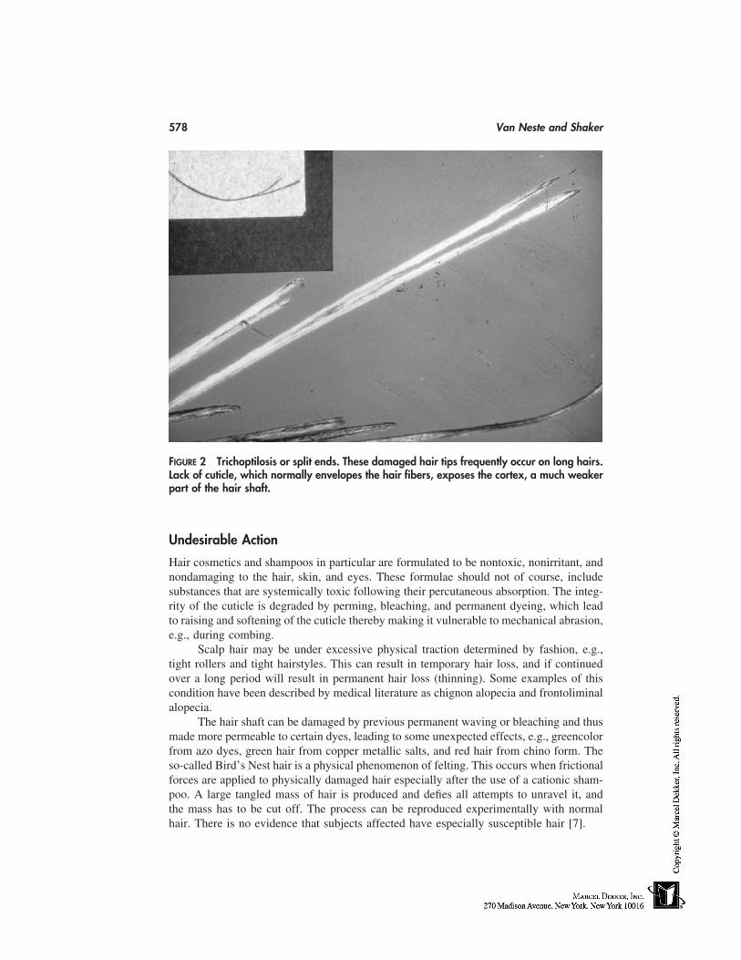
578 Van Neste and Shaker
FIGURE 2 Trichoptilosis or split ends. These damaged hair tips frequently occur on long hairs.Lack of cuticle, which normally envelopes the hair fibers, exposes the cortex, a much weakerpart of the hair shaft.
Undesirable Action
Hair cosmetics and shampoos in particular are formulated to be nontoxic, nonirritant, andnondamaging to the hair, skin, and eyes. These formulae should not of course, includesubstances that are systemically toxic following their percutaneous absorption. The integ-rity of the cuticle is degraded by perming, bleaching, and permanent dyeing, which leadto raising and softening of the cuticle thereby making it vulnerable to mechanical abrasion,e.g., during combing.
Scalp hair may be under excessive physical traction determined by fashion, e.g.,tight rollers and tight hairstyles. This can result in temporary hair loss, and if continuedover a long period will result in permanent hair loss (thinning). Some examples of thiscondition have been described by medical literature as chignon alopecia and frontoliminalalopecia.
The hair shaft can be damaged by previous permanent waving or bleaching and thusmade more permeable to certain dyes, leading to some unexpected effects, e.g., greencolorfrom azo dyes, green hair from copper metallic salts, and red hair from chino form. Theso-called Bird’s Nest hair is a physical phenomenon of felting. This occurs when frictionalforces are applied to physically damaged hair especially after the use of a cationic sham-poo. A large tangled mass of hair is produced and defies all attempts to unravel it, andthe mass has to be cut off. The process can be reproduced experimentally with normalhair. There is no evidence that subjects affected have especially susceptible hair [7].
Interactions with Hair and Scalp 579
SPECIFIC ACTIONS OF HAIR COSMETICS ON THE SKIN
Intended Contact with the Skin
Many cosmetic compounds target the skin rather than the hair. Antidandruff and antisebor-rehic cosmetic compounds target the scalp skin, not the scalp hair. Many other cosmeticsintend to modify the hair/skin system, e.g., preshave and aftershave lotions, along withdepilatories, where the effect on hair is always associated with some effect on the skin.
Nonintended Contact with the Skin
Ideally, hair cosmetics should not have contact with the skin, but in practice this is hardlyachievable. In some procedures, such as hair dyeing and hair bleaching, skin contact isunnecessary to perform the procedure itself but hardly avoidable during such a procedure.
Many consumers report hair shedding with changes in the shampooing regime. Thisis not because of a biological process in particular but because of a detachment of telogenhairs, which may modify the usual daily shedding of hair. Another reason for this is thatwhen consumers change products, they tend to be more attentive to the condition of theirhair and scalp and attribute any perceived change to the new product, especially if viewedas negative. Properly conducted trials showed that shampooing regimes did not modifyhair shedding [8].
The aim of wet and dry shaving is to cut facial hair without harming the skin, whichis a frequent adverse side effect of these procedures. These side effects vary in intensity,from a slight irritation—that is going to disappear with time during a process of adapta-tion—to a very severe and persistent reaction. This will mostly force the consumer togive up using that particular method and select a more ‘‘friendly’’ alternative.
Permanent wave solutions or their neutralizing chemicals can cause chemical burnor necrosis of the scalp epidermis if allowed to contact the scalp skin in certain concentra-tions for too long. The chemical burn may affect the skin of the scalp, forehead, face,and neck.
Physical burn can result from heated rollers or other apparatus that can cause damageto the superficial layers of the epidermis. The risk of burn also exists during or just afterthe use of flammable vehicles (e.g., alcoholic lotions) in close proximity to a fire or heatsource.
Contact dermatitis to cosmetics in general and to hair cosmetics in particular is notuncommon in clinical dermatology. Following are leading examples from a long list ofhair cosmetics reported to be skin sensitizers: hair dyes (p-phenylenediamine, resorcinol),shampoos (surfactants, zinc pyrithione, hydroxyquinolines), hair creams and gels (lanolin,parabens), hair lacquers (benzoin, cyclohexanone-formaldehyde resin), hair lotions (qui-nine, resorcinol), deodorants (hydroxyquinolines, Irgasan DP 300), bleachers, and shavinglotions (musk ambrette, antimicrobial agents) [9]. Acute and chronic allergic contact der-matitis have been associated with significant though usually transient or reversible hair loss[6]. This is a very often neglected or even unrecognized cause of diffuse hair loss [10].
The irritant effect of cleansing agents is attributable to the removal of surface lipidfilm and water-holding substances in the stratum corneum. They may denature proteinand damage the cell membrane as well. The risk of irritant and allergic contact dermatitisinduced by deodorants is greatly enhanced by the natural occlusive properties of bodysites such as the armpits.
580 Van Neste and Shaker
REFERENCES
1. Dawber R. Shampoos—scientific basis and clinical aspects. Proceedings of the Hair CareForum Sponsored by Procter and Gamble Ltd, Florence, Italy. London: Royal Society of Medi-cine Press Limited, 1997.
2. Cash T. The psychological effects of androgenetic alopecia in men. J Am Acad Dermatol1992; 26:926–931.
3. Cash T, Price V, Savin R. Psychological effects of androgenetic alopecia on women: compari-sons with balding men and with female control subjects. J Am Acad Dermatol 1993; 29:568–575.
4. Passchier J. Quality of life issues in male pattern hair loss. Dermatology 1998; 197:217–218.5. Girman C, Rhodes T, Lilly F, Guo S, Siervogel R, Patrick D, Chumlea W. Effects of self-
perceived hair loss in a community sample of men. Dermatology 1998; 197:223–229.6. Dawber R, Van Neste D, eds. Hair and Scalp Disorders. London: Martin Dunitz Ltd, 1995.7. Zviak C. The Science of Hair Care. New York: Marcel Dekker, 1986.8. Kullavanijaya P, Gritiyarangsan P, Bisalbutra P, Kulthanan R, Cardin CW. Absence of effects
of dimethicone and non-dimethicone containing shampoos on daily hair loss rates. J Soc Cos-met Chem 1992; 43:195–206.
9. Rycroft RJG. Principle irritants and sensitizers. In: Rook, Wilkinson, Ebling, eds. Textbookof Dermatology. Vol. 1. Oxford: Blackwell Science, 1998:821–860.
10. Tosti A, Piraccini BM, Van Neste D. Telogen effluvium following allergic contact dermatitisof the scalp. Arch Dermatol. In press.

51
Hair Cosmetics
Leszek J. WolframIndependent Consultant, Stamford, Connecticut
INTRODUCTION
Throughout recorded history, hair has always been an important element of personaladornment. From the beautifully regular beard curls of the Assyrian kings to the eleganthair cuts of Egyptian pharaohs to the carefully coiffured wigs of the European nobility,hair has been shown, admired, and envied. Over the years, what had been the privilegeof the affluent few has become an almost consuming passion of many. The explosivegrowth of the haircare market since the middle of the twentieth century is the result ofdeep, socioeconomic changes combined with an increasing focus on personal aesthetics,assisted by affordability of products. The attempt to satisfy the genuine needs of the con-sumer and the drive for competitive advantage among marketers has led to a variety ofgrooming aids and products, such as shampoos to cleanse the hair, hair conditioners tomake it soft and combable, hair colorants and permanent waves to impart to hair propertiesit does not have, and hair sprays to keep hair in the desired style. Hair products are inthe cosmetic category and, as such, are subject to all laws and regulations that control thelabeling and claims of all cosmetic products.
The Structure and Properties of Hair
Hair follicles, which in tens of thousands are deeply invaginated in the scalp tissue, arethe essential growth structures of hair. At the base of each follicle, the cells proliferateand, as they stream upwards, the complex and intertwined processes of protein synthesis,structural alignment, and keratinization transform the cytoplasm into the tough fibrousmaterial known as hair. Hair is unique in that its structural and growth characteristics aredifferent between races, sexes, individuals of the same race, areas in the same individuals,and even within the same follicle. The development of hair is a dynamic, cyclical processin which duration of the growth cycle depends not only on the body site, but also on suchvariables as the individual’s age, nutritional habits, and hormonal factors. In the scalp,each hair grows steadily (about 1 cm per month) and continuously for 3 to 5 years (anagenphase); growth then stops and is followed by a brief transient stage (catagen) and a 2- to
581
582 Wolfram
4-month resting stage (telogen) during which the old hair is shed. With the onset of theanagen, the new hair starts to grow from the same follicle. The growth process functionsindependently in each follicle, so hairs are not shed simultaneously as they are in mostanimals. At any given time, some hairs are growing, some are resting, and some are beingshed. Normally, of about 150,000 scalp hairs, 90% are in the anagen phase and the re-maining 10% are in the catagen and telogen phases, with 50 to 100 hairs being shed daily.Scalp hair is a fiber of 50-80 µm in diameter and its exterior consists of a layer of flat,imbricated cuticle cells pointing outward from root to tip. This ratched-like structure ofthe cuticle scales serves as an effective self-cleaning feature and, by interlocking with thescales of the inner root sheath of the follicle, helps to hold the hair firmly in place. Thecuticles are thin (0.5 µm), 50 to 60 µm square sheets, attached at their approximal endsto the underlying cortex. Their longitudinal overlap is substantial resulting in an averageseparation of scale edges of approximately 5 µm. This overlap generates a multilayeredshield 3 to 4 µm thick around the hair fiber. The structure of the cuticle fulfills well therole of a protective barrier for hair. A thin film of covalently-bounded lipid on the exteriorof the cuticle assures a low friction surface, together with water repellency. Just under-neath, the highly cross-linked lamellae of the A-layer and exocuticle augment the mechani-cal stability of the scales, whereas the soft and water-absorbing endocuticle cushions ef-fects of mechanical impact. The high water swellability of the endocuticle is the likelysource of pronounced cuticle lifting on wetting of hair.
Enveloped by this formidable protective sheath of the cuticle layer is hair cortex,which constitutes the bulk of the fiber and is mainly responsible for the mechanical proper-ties of hair. The spindle-shaped cortical cells are arranged parallel to the fiber axis, overlap-ping each other with frequent interdigitation. They have a unique arrangement of theconstituent proteins, comprising intermediate filaments (IF), traditionally termed microfi-brills, aligned in the direction of fiber growth and are surrounded by a matrix of IF associ-ated proteins (IFAP). The filaments are composed of high–molecular weight protein chainsof low sulfur (cystine) content and possess a high degree of molecular organization(α-helical), whereas the surrounding matrix of IFAP is made up of proteins more exten-sively cross-linked by cystine lacking definite structural pattern.
During the process of keratinization, the cell plasma membranes are modified toestablish a strongly adhesive layer between the adjacent cells known as the cell membranecomplex (CMC). This is the only continuous phase in the hair fiber providing adhesionbetween the cortical cells as well as between and with the cuticle cells.
Dispersed throughout the structure of cortex are melanin pigment particles. Theirnumber, chemical characteristics, and distribution pattern determine the color of hair. Insome hairs, coarse hairs in particular, vacuolated medulla cells are present in the centralregion of the fiber.
Although hair of different racial origin differs in shape, degree of curliness, andcolor, there is little difference in the underlying chemical properties and fiber structure.The amino-acid composition of the constituent proteins and most physical properties aresimilar [1,2]. The differences between hair of different ethnic groups are often smallerthan the variation in the properties of hair taken from different individuals within oneethnic group.
Compared with Caucasian or Asian hair, African hair is more irregular in the shapeof its cross-section. The sharp kinks seen in such hair are often associated with randomunevenness of fiber diameter, resulting in weak spots along the fiber length. These arelikely to cause problems during combing or chemical treatments.
Hair Cosmetics 583
SHAMPOOS
General Comments
Cleansing is clearly a dominant element of personal hygiene and, when reinforced by theaspect of attractive appearance, translates into a powerful and highly marketable stimulus.Shampooing has become, thus, a factor sine qua non in maintaining the aesthetics of hair.
The cleansing task is formidable. A mass of 100,000 to 150,000 flexible fibers hasto be cleansed of oily deposits of sebum, sweat, entrapped desquamated scalp cells, alongwith the residues of mousses, gels, and hair sprays. All this has to be done within thespan of a few minutes, leaving the individual hairs clean and free of tangles to which theratched structure of hair cuticles makes it particularly vulnerable. It should also be keptin mind that although cleansing action is the fundamental assignment of a shampoo formu-lation, it is by no means the only goal. The promise of hair shine, softness, body, andmanageability is inherently tied to product performance. Furthermore, one must not ignorethe process of shampooing itself. It is expected to provide a pleasurable experience inworking up a rich and lubricous lather that seems almost to caress the hair and leave it,after rinsing, with a touch of refreshing fragrance.
Hair Soiling and Soil Removal
In the course of its residence on the scalp, hair is exposed to a variety of events thatcontribute to its soiling. Among them are the innate processes of scalp desquamation,sweating, and sebum secretion, which are supplemented by deposition of extraneous sub-stances arising either from environmental pollution (dust and other airborne contaminants)or from hair-grooming preparations, such as oils, waxes, hair spray, and mousse residues.Of all these, sebum, because of to its steady replenishment, greasy characteristics, highadhesiveness to hair, and ability to cement the other soil particulates together and to thehair surface, appears most insidious and thus it is not surprising that its efficacious removalis key in hair cleansing.
The sebaceous glands attached to each of the hair follicles provide a continuoussupply [3] of this oily substance to the surface of hair. There are seasonal variations inthe amount of sebum secreted [4], but more importantly its output is under hormonalcontrol [3], reaching a maximum at puberty. Oily hair of adolescents is the obvious andoften annoying consequence of the high activity of the sebaceous glands, and this at atime in one’s life when personal adornment is particularly important. Sebum secretedfrom the sebaceous ducts spreads within the mass of hair primarily via physical contactbetween the fibers [5]. Brushing and combing (as well as contact with a pillow) furtherredistributes the sebum and partly assists in its removal. The quantity of sebum on hairat a particular moment thus reflects the relative efficacy of these two processes (sebumsecretion and removal). The term ‘‘oily hair’’ often connotes a highly undesirable imageof stringy and dull hair with little body and greasy feel. It is, however, worth bearing inmind that such a perception is not universal being strongly influenced by fiber texture andgeometry. Thus, a visual appearance of curly African hair can visibly benefit from anincrease in oiliness, a fact that is exploited in grooming products for such hair.
Because of the adhesiveness and sticky consistency of sebum-containing soil, itsadequate removal by simple mechanical means is virtually impossible, and satisfactorycleansing can only be attained by use of aqueous solutions of detergents. In the broadestsense, all materials used in cleansing that are water and other solvents, soaps and synthetic
584 Wolfram
surfactants, salts, and abrasives may be considered as detergents. However, more specifi-cally, the term ‘‘detergent’’ is limited to those surface-active agents that, in addition tothe property of lowering surface tension, are effective in deflocculating soil and dirt clumpsand keeping them in suspension so that they can be washed away before redepositing onthe surface that is being cleaned. This property is exhibited by compounds that containboth a hydrophilic group and a hydrophobic tail that serves as an emulsifying agent. Inessence, the removal of soil from hair is governed by the same basic processes that hadbeen previously identified as being involved in laundering of fabrics [6]. Without elaborat-ing on theories underlying the detergency, one should allude to the three fundamentalmechanisms that have been proposed to account for the cleansing action of detergents.
1. The ‘‘roll-up’’ mechanism [6], particularly relevant to oily deposits in whichthe progressive wetting of the fiber surface leads to rapid detachment of oildroplets;
2. In the micellar solubilization mechanism [7] the soil is solubilized into micellesthat come into contact with the soiled surface. The efficacy of this cleansingmode depends on the availability of sufficient quantity (concentration) of mi-celles, which does not usually present a problem with conventional shampooformulations; and
3. The third mechanism [8] invokes the dispersion and emulsification of soil parti-cles penetrated by the diffusing detergent. The amphiphilic components of se-bum might enhance cleansing by direct interaction with the molecules of thesurfactant.
There is no precise information presently available as to which mechanism is dominantin hair cleansing. Quite possibly all three might be involved, depending on the characteris-tics of the soil. In any case, the vast majority of shampoo products are formulated tobe operative under diverse conditions of detergent action, thus assuring their cleansingefficacy.
Shampoo Ingredients
Almost without exception, shampoos consist of an aqueous solution, emulsion, or disper-sion of one or more surfactants together with some additives to enhance performance andaesthetic properties of the product. Additives are used to provide fragrance and color,thicken, opacify, and convey specific tactile attributes. They include stabilizers, foam mod-ifiers, preservatives, conditioning, and antidandruff agents.
Surfactants
Surfactants are long-chain electrolytes and are usually classified according to the natureof their hydrophilic group, which may be anionic, nonionic, amphoteric, or cationic.
Anionic Surfactants
Soaps are salts of fatty acids and, not in the distant past, were the mainstay of shampooproducts. In soft water, they lather copiously, cleanse well, and leave the hair in a well-conditioned style. Unfortunately, in hard water the lather is poor, and as the soap combineswith calcium or magnesium salts present in hard water it deposits on hair a dulling film.The introduction of synthetic surfactants brought about the end of soap-based shampoos,
Hair Cosmetics 585
although some products still contain a small quantity of soap to exploit its conditioningproperty.
Alkyl sulfates are the most widely used anionic in shampoos, displaying excellentfoaming and cleansing properties unaffected by hard water. Lauryl sulfate is the dominateingredient being present in most shampoo formulations in the form of its ammonium ortriethanol ammonium salt at a level of 6 to 18% w/w. Although very effective cleansers,the alkyl sulfates, particularly at high concentrations, have a tendency to irritate the scalpand remove some lipid constituents of hair cuticle. To make the alkyl sulfate-based sham-poos milder, they are frequently modified by incorporation of less-irritating alkyl ethersulfates or amphoteric surfactants.
Alkyl ether sulfates are sulfated products of ethoxylated fatty alcohols. They aremore water soluble than alkyl sulfates, are excellent solubilizers for fragrances and otheroleophilic additives, and are particularly suitable for formulations of clear shampoos. Asalluded to earlier, these surfactants are less irritating than the alkyl sulfates and are used,at a higher degree of ethoxylation, in baby shampoos.
Alpha-olefin sulfonates are complex mixtures resulting from sulfonation of alpha-olefins. These detergents exhibit excellent foaming in the presence of sebum, are effectiveover a wide range of pH, and compare favorably with other surfactants in dermal and eyeirritation [9].
Other anionic surfactants worthy of note include alkyl monoglyceride sulfates andalkyl sulfosuccinates. Both are very mild to the skin and, although the former are goodfoamers and can be used in shampoo formulation in their own right, the latter are primarilyused in combination with alkyl sulfates.
Nonionic Surfactants
They are considered to be the mildest of surfactants. Although poor foamers, owing totheir good solubilizing and dispersing properties, they have been extensively utilized tosupplement the action of the primary cleanser.
Alkanolamides are prepared by condensation of fatty acid (usually lauric) and pri-mary or secondary alkanolamines. Their presence in a shampoo formulation can have apronounced effect on stabilizing the foam level and improving lather consistency. Aminooxides are formed by oxidation of tertiary fatty amines and are used in shampoos primarilyas foam modifiers and as antistatic agents to improve the overall manageability of hair.
Polyethoxylated surfactants represent the largest group of nonionics and include theethoxylated derivatives of alkylphenols, fatty alcohols, fatty esters, and diglycerides. Theyexhibit excellent detersive power and cleansing properties, but because of poor foaming,their use has been restricted to solubilizing of shampoo fragrances and other oleophilicadditives.
Amphoteric Surfactants
Often referred to as ampholytic, these surfactants contain both cationic and anionic groupsin one molecule. Because the charge of these surfactants are pH dependent, their proper-ties, such as foaming potential, solubility, and CMC, also vary with the change in pH.Most amphoterics are derivatives of imidazoline or betaine. They are quite compatiblewith anionic, nonionic, or cationic surfactants, and have been extensively used to formulatemild (baby) shampoos or as mollifying agents in the more irritating anionic compositions.
586 Wolfram
Shampoo Additives
These are materials incorporated into a shampoo formulation to enhance its aesthetics aswell as improve its performance.
Thickeners comprise a broad variety of compounds that are used to increase viscosityof the formulations, modifying their consistency from viscous liquids to thick gels. Amongthe most frequently used are electrolytes, such as sodium chloride, alkanolamides andwater-soluble cellulose derivatives, such as carboxymethylcellulose, hydroxyethylcellu-lose, carboxy vinyl polymers of the Carbopol type, polyvinyl alcohols, and natural gums,such as tragacanth. Magnesium aluminum silicates have found application as thickenersand suspending agents in antidandruff shampoos.
Opacifiers serve to impart to shampoo a pearlescent or opaque appearance. For thispurpose, high-melting, wax-like materials are blended into formulations. Of particularutility in this respect are cetyl and stearyl alcohols and their esters as well as the latexemulsions of vinyl-, styrene-, and acrylic polymers.
The shampoo milieu offers itself as an ideal ground for microbial growth, particu-larly of the aerobic gram-negative organisms of Pseudomones. This may have a deleteriouseffect on the shampoo properties, posing at the same time a health hazard to the consumer.The function of preservatives is to inhibit such bacterial development. Although formalde-hyde has been one of the most popular and effective preservatives, its use has declinedas other compounds have come to the fore. Examples include methyl and propyl parabenes,DMDM hydantoin, quaternium-15, imidazolidynyl urea and others. The selection of asuitable preservative is made through a challenge test in which the product is subjectedto the worst possible conditions anticipated during manufacture, shelf storage and actualuse.
Other additives. Fragrance is an essential ingredient, often deciding the market ap-peal and success of the product. Addition of alcohols (ethanol, isopropanol) or glycolsmay be required to maintain the clarity of clear shampoos, while the presence of sequester-ing agents like EDTA prevents the formation of insoluble calcium or magnesium soapswhen the shampoo is rinsed off the hair. FD&C and D&C dyes are commonly added toenhance the aesthetics of shampoo formulations. ‘‘Squeaky’’ clean feel of shampooedhair is frequently accompanied by difficult combing and substantial ‘‘fly away.’’ To over-come this, the shampoos contain ‘‘conditioning’’ additives that are substantive to hairremaining adsorbed on the surface after rinsing. A plethora of materials has been used tothis end. To these belong amine oxides, protein hydrolysates, cationic surfactants, cationicpolymers, lanolin and its derivatives, as well as natural materials, such as beer, honey,and egg.
Shampoo Formula
It must have become clear from the foregoing that a shampoo product, although straightforward in its purpose, is a complex blend of ingredients carefully chosen and attuned toeffectively address the need of individual consumers. Table 1 shows the nature and relativeconcentration of materials contained in a typical shampoo formulation:
Specialty Shampoos
Baby shampoos place stringent requirements for nonirritancy of the scalp and eye. Themajority of products are based on amphoteric detergent systems. Thus, derivatives of
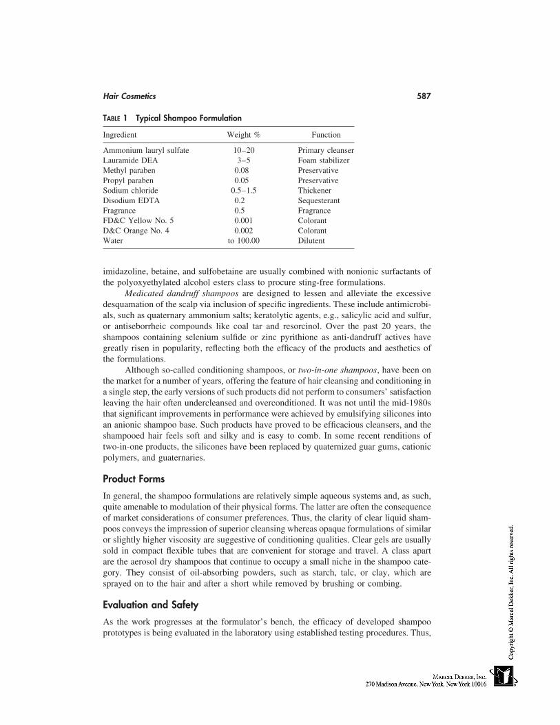
Hair Cosmetics 587
TABLE 1 Typical Shampoo Formulation
Ingredient Weight % Function
Ammonium lauryl sulfate 10–20 Primary cleanserLauramide DEA 3–5 Foam stabilizerMethyl paraben 0.08 PreservativePropyl paraben 0.05 PreservativeSodium chloride 0.5–1.5 ThickenerDisodium EDTA 0.2 SequesterantFragrance 0.5 FragranceFD&C Yellow No. 5 0.001 ColorantD&C Orange No. 4 0.002 ColorantWater to 100.00 Dilutent
imidazoline, betaine, and sulfobetaine are usually combined with nonionic surfactants ofthe polyoxyethylated alcohol esters class to procure sting-free formulations.
Medicated dandruff shampoos are designed to lessen and alleviate the excessivedesquamation of the scalp via inclusion of specific ingredients. These include antimicrobi-als, such as quaternary ammonium salts; keratolytic agents, e.g., salicylic acid and sulfur,or antiseborrheic compounds like coal tar and resorcinol. Over the past 20 years, theshampoos containing selenium sulfide or zinc pyrithione as anti-dandruff actives havegreatly risen in popularity, reflecting both the efficacy of the products and aesthetics ofthe formulations.
Although so-called conditioning shampoos, or two-in-one shampoos, have been onthe market for a number of years, offering the feature of hair cleansing and conditioning ina single step, the early versions of such products did not perform to consumers’ satisfactionleaving the hair often undercleansed and overconditioned. It was not until the mid-1980sthat significant improvements in performance were achieved by emulsifying silicones intoan anionic shampoo base. Such products have proved to be efficacious cleansers, and theshampooed hair feels soft and silky and is easy to comb. In some recent renditions oftwo-in-one products, the silicones have been replaced by quaternized guar gums, cationicpolymers, and guaternaries.
Product Forms
In general, the shampoo formulations are relatively simple aqueous systems and, as such,quite amenable to modulation of their physical forms. The latter are often the consequenceof market considerations of consumer preferences. Thus, the clarity of clear liquid sham-poos conveys the impression of superior cleansing whereas opaque formulations of similaror slightly higher viscosity are suggestive of conditioning qualities. Clear gels are usuallysold in compact flexible tubes that are convenient for storage and travel. A class apartare the aerosol dry shampoos that continue to occupy a small niche in the shampoo cate-gory. They consist of oil-absorbing powders, such as starch, talc, or clay, which aresprayed on to the hair and after a short while removed by brushing or combing.
Evaluation and Safety
As the work progresses at the formulator’s bench, the efficacy of developed shampooprototypes is being evaluated in the laboratory using established testing procedures. Thus,
588 Wolfram
foamability and lather characteristics are measured in the presence and absence of sebum,gaining some insight into the detersive aspects of the formulas. The properties of sham-pooed hair, with respect to its luster, combability, body, and fly-away, are instrumentallyassessed together with the subjective evaluation of hair appearance. The ultimate proof,however, of the potential success of the formulation is in the practical use. Thus, theconsumer evaluation of the product either with outside panelists or in-house testing facilityis imperative. The preference of consumers for a particular fragrance is of vital importanceand their comments as to the aesthetic characteristics of shampoo and the feel of sham-pooed hair when combined with the results of laboratory tests provide firm ground forpotential product claims.
Shampoo ingredients do not pose a particular hazard with regard to skin or eyesafety. The contact time is short and a water rinse follows. The irritation potential of somesurfactants has already been alluded to. It is a common practice for most of the manufactur-ers to make provisions to evaluate their product for skin and eye irritation.
HAIR CONDITIONERS
It is worth noting that the subject of hair ‘‘condition’’ appears to be restricted almostentirely to the domain of women’s hair. Although, as a woman’s ‘‘crowning glory’’ thehair evokes in her a particularly profound concern for its beauty, there are at work somemore mundane factors. Unlike men’s hair, that of a woman’s is subject to more frequentand diverse assaults that are injurious to its properties. It is perhaps ironic that except forenvironmental effects (weathering), most of these are associated with what we call the‘‘haircare’’ practices. Thus, the handling of hair in the course of its daily shampooing,combing and brushing, and blow drying cause, even in the case of intact hair, gradualabrasion of the hair cuticle signaling the onset of hair damage. This process of cuticleloss is particularly evident in longer hair leading often to the generation of split ends.Hair coloring, bleaching, waving, or straightening, although imparting to hair a muchsought after different or novel appearance, impair the surface lipid layer of the cuticles,further aggravating the abrasive effects of daily hair regimens. Although gradual, thesedeleterious effects are additive and further exacerbated by sun exposure.
Clearly, by the use of conditioning shampoos, avoiding practices singularly injuriousto the cuticle, such as teasing, and keeping the hair relatively short and shielded from sun,one might, for a considerable length of time, maintain the intact hair in satisfactory condi-tion. Alternatively, one can go a step further and by the use of products designed explicitlyfor conditioning supplement the benefits obtained from a shampoo and significantly extendtheir range. A good conditioner eliminates tangling, makes the hair easy to comb andstyle, eliminates static charge, and, by fostering fiber alignment, enhances the luster andshine of hair. The soft feel of hair and improved manageability are additional importantattributes of conditioned hair. It is important to stress that these effects are universal, i.e.,irrespective of cosmetic history of hair, whether the hair is intact, waved, colored, orbleached, the conditioner delivers its benefits.
Two general forms of conditioners are currently in use: 1) hair rinses and 2) leave-in products, often referred to as ‘‘deep’’ conditioners. Both are applied to freshly sham-pooed hair. True to their name, the rinse product is rinsed off after a few minutes, whereasthe leave-in product is left on the hair for up to 30 minutes, after which it is rinsed off.The purpose of the longer time is to allow the product to penetrate further (thus the name‘‘deep’’) into the hair shaft thereby extending the conditioning effects.
Hair Cosmetics 589
The active ingredients in most conditioners are based on quaternary ammonium salts(cationic surfactants) such as steartrimonium chloride and, cetrimonium chloride, and thelike. Because of their great affinity for hair, these compounds bind strongly to the cuticles,providing a low-friction surface, thus making the cuticles slick and less prone to abrasion.Other components present in the conditioning formulations, such as fatty amines, fattyalcohols, and amine oxides, supplement the action of cationic surfactants, adding primarilyto the tactile benefits. The leave-in conditioners that are recommended for use on damagedhair frequently contain protein and lanolin derivatives.
Conditioning effects are usually lost in shampooing, and a reapplication is recom-mended to reinforce the protective effect. Conditioning formulations containing cationicpolymers are somewhat longer lasting. The same is true for conditioners based on emul-sions of polymeric silicones.
HAIRDRESSINGS
Hairdressing is a broad term describing products applied for final grooming. Includingbrilliantines, tonics, and gels, this category follows new fashions, hairstyle trends, and isattuned to progress in styling techniques. Hairdressings are applied by spreading the prod-uct through the hair with the fingers and then combing through for an even distribution.As they are not rinsed off after application, care must be taken to avoid excessive build-up.
The primary purpose of brilliantines is to add sheen to hair. Thus, the main constit-uent of these products is oil—usually mineral oil—which is spread on fiber strands in-creasing their luster and providing grooming effects. Solid brilliantines (pomades) arebased on petrolatum to which various waxes are added to attain the desired consistencyand texture. Tonics might be viewed as lighter versions of brilliantines and usually consistof alcoholic solutions of various oils. The alcohol wets the hair and after evaporationleaves a thin film of oil. By using synthetic, rather than natural oils, much less greasyformulations can be obtained. Using a high concentration of ethoxylated emulsifiers,grooming oils can also be readily blended into clear gels. On the other hand, setting gelsbased on hydroalcoholic solutions of carboxyvinyl polymers or methylcellulose ethers areoil free. They range in consistency from liquid to rigid gels and provide a good range oftextures, volume, and hold.
Styling Products
Whereas most of the styling needs of short hair are satisfactorily met by a good haircut,those with longer hair require more effort which is, however, well rewarded by the diver-sity of styles that can be imparted. The underlying principle of all styling processes ishair setting and a few comments on the subject seems appropriate. Hair fibers are flexibleand elastic, and when dry bounce back immediately to their original configuration (straightor curly) when bent, extended, or twisted. On wetting, however, they become pliant andmalleable and can be readily molded (set) to almost any desired form. On drying, theyretain the new shape until exposed to water (moisture) again.
The primary function of all styling products is to assist in the setting process and/or to ensure the stability of the newly imparted configuration. Depending on the type ofstyling product, different mechanism of action are operative.
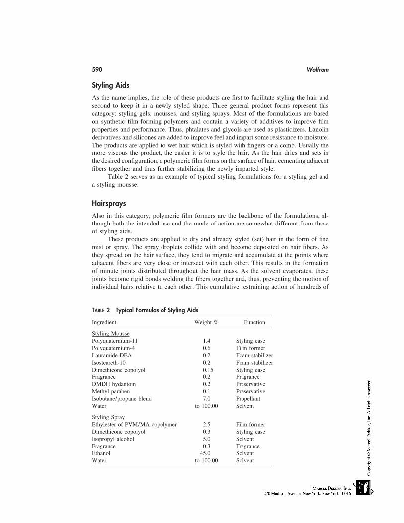
590 Wolfram
Styling Aids
As the name implies, the role of these products are first to facilitate styling the hair andsecond to keep it in a newly styled shape. Three general product forms represent thiscategory: styling gels, mousses, and styling sprays. Most of the formulations are basedon synthetic film-forming polymers and contain a variety of additives to improve filmproperties and performance. Thus, phtalates and glycols are used as plasticizers. Lanolinderivatives and silicones are added to improve feel and impart some resistance to moisture.The products are applied to wet hair which is styled with fingers or a comb. Usually themore viscous the product, the easier it is to style the hair. As the hair dries and sets inthe desired configuration, a polymeric film forms on the surface of hair, cementing adjacentfibers together and thus further stabilizing the newly imparted style.
Table 2 serves as an example of typical styling formulations for a styling gel anda styling mousse.
Hairsprays
Also in this category, polymeric film formers are the backbone of the formulations, al-though both the intended use and the mode of action are somewhat different from thoseof styling aids.
These products are applied to dry and already styled (set) hair in the form of finemist or spray. The spray droplets collide with and become deposited on hair fibers. Asthey spread on the hair surface, they tend to migrate and accumulate at the points whereadjacent fibers are very close or intersect with each other. This results in the formationof minute joints distributed throughout the hair mass. As the solvent evaporates, thesejoints become rigid bonds welding the fibers together and, thus, preventing the motion ofindividual hairs relative to each other. This cumulative restraining action of hundreds of
TABLE 2 Typical Formulas of Styling Aids
Ingredient Weight % Function
Styling MoussePolyquaternium-11 1.4 Styling easePolyquaternium-4 0.6 Film formerLauramide DEA 0.2 Foam stabilizerIsosteareth-10 0.2 Foam stabilizerDimethicone copolyol 0.15 Styling easeFragrance 0.2 FragranceDMDH hydantoin 0.2 PreservativeMethyl paraben 0.1 PreservativeIsobutane/propane blend 7.0 PropellantWater to 100.00 Solvent
Styling SprayEthylester of PVM/MA copolymer 2.5 Film formerDimethicone copolyol 0.3 Styling easeIsopropyl alcohol 5.0 SolventFragrance 0.3 FragranceEthanol 45.0 SolventWater to 100.00 Solvent
Hair Cosmetics 591
such microscopic welds throughout the hair assembly accounts for the style-stabilizingperformance of hair sprays, protecting the hair from mechanical deformation,wind, andhumidity.
The strength of these hairspray bonds depends on a number of factors, of which thenature of the polymeric resin is of paramount importance. Most of the polymers used formadequately strong bonds at low relative humidity (RH). As the RH increases beyond 80%,however, most resins begin to absorb moisture from the environment, softening the welds.At the same time, water absorption by hair causes rapid relaxation of the set configurationof the fibers and it is the tenacity of the hairspray welds alone that stabilizes the impartedstyle. Clearly, the polymers that are least sensitive to the plastizing effect of water arelikely to be the better performers and are thus preferred for a hairspray product.
It should be stressed that in addition to the intrinsic strength of the resin, otherfactors may affect bond formation and/or bond toughness. For example, the characteristicsof the solvent system used to deliver the resin to hair plays an important role. Efficientweld formation depends on the wetting and spreading properties of the resin droplets onthe hair surface. As mentioned earlier, the welds are formed by the accumulation of liquidspray at contact points between fibers. Thus, an aerosol formulation with 30% alcoholand 70% highly volatile propellant will dry much faster than a solvent vehicle with 50%or more alcohol. As the solvent evaporates, the viscosity of droplets increases and mobilitydecreases. This reduced mobility results in relatively small bonds between adjacent orintersecting fiber which might negatively affect the product performance. One might beled to a conclusion that the spray that stays ‘‘wetter’’ longer generates better performingwelds. This may hold true for nonaqueous systems as the organic solvents used in hairformulation do not have any adverse effect on the set of the styled hair. With the hydro-alcoholic systems, however, and the water content of over 20% the long ‘‘residence’’ timeof hairspray droplets on hair may lead to a significant loss of set caused by the selectivewater absorption by hair fibers.
Although a number of hairspray resins have been developed over the years andmany of them have been in use, the combination of regulatory restrictions and increaseddemands on the aesthetics of product performance has narrowed the field somewhat. Thusthe butyl and ethyl esters of poly (vinyl methyl ether/maleic anhydride) copolymers, whichfor years have been the most widely used polymers in hair sprays, have suffered a rapiddecline, being surpassed by octylacrylamide/acrylates/butylaminoethyl methacrylate co-polymer. The latter provides excellent holding properties at relatively low resin concentra-tion. For the aerosol hairsprays, the resin of choice is vinyl acetate/crotonates/neodec-anoate, which, by modulation of the extent of its neutralization, can substantially modifythe film properties.
Essential as it is, the set holding is not the only attribute that has to be consideredin formulating hair sprays. Clearly, the aesthetic aspect of sprayed hair cannot be ne-glected. Thus, the resin film should add shine (gloss) and not dull the hair, nor shouldthe hair become tacky in humid weather. It should resist flaking, but be readily removedby shampoo. By selection of appropriate additives and solvents, both the holding andaesthetic goals can be readily attained. Table 3 provides ingredient listings for typicalaerosol and pump sprays.
Safety and Regulatory Issues
All aerosol hairsprays, whether containing hydrocarbon or carbon dioxide propellant, areclassified as flammable by virtue of their flame propagative properties. The same is true
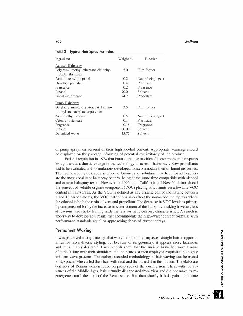
592 Wolfram
TABLE 3 Typical Hair Spray Formulas
Ingredient Weight % Function
Aerosol HairsprayPoly(vinyl methyl ether)-maleic anhy- 5.0 Film former
dride ethyl esterAmino methyl propanol 0.2 Neutralizing agentDimethyl phthalate 0.4 PlasticizerFragrance 0.2 FragranceEthanol 70.0 SolventIsobutane/propane 24.2 Propellant
Pump HairsprayOctylacrylamine/acrylates/butyl amino 3.5 Film former
ethyl methacrylate copolymerAmino ethyl propanol 0.5 Neutralizing agentCetearyl octanoate 0.1 PlasticizerFragrance 0.15 FragranceEthanol 80.00 SolventDeionized water 15.75 Solvent
of pump sprays on account of their high alcohol content. Appropriate warnings shouldbe displayed on the package informing of potential eye irritancy of the product.
Federal regulation in 1978 that banned the use of chlorofluorocarbons in hairspraysbrought about a drastic change in the technology of aerosol hairsprays. New propellantshad to be evaluated and formulations developed to accommodate their different properties.The hydrocarbon gases, such as propane, butane, and isobutane have been found to gener-ate the most consistent hairspray pattern, being at the same time compatible with alcoholand current hairspray resins. However, in 1990, both California and New York introducedthe concept of volatile organic component (VOC) placing strict limits on allowable VOCcontent in hair sprays. As the VOC is defined as any organic compound having between1 and 12 carbon atoms, the VOC restrictions also affect the nonaerosol hairsprays wherethe ethanol is both the resin solvent and propellant. The decrease in VOC levels is primar-ily compensated for by the increase in water content of the hairspray, making it wetter, lessefficacious, and sticky leaving aside the less aesthetic delivery characteristics. A search isunderway to develop new resins that accommodate the high–water content formulas withperformance standards equal or approaching those of current sprays.
Permanent Waving
It was perceived a long time ago that wavy hair not only surpasses straight hair in opportu-nities for more diverse styling, but because of its geometry, it appears more luxuriousand, thus, highly desirable. Early records show that the ancient Assyrians wore a massof curls falling over their shoulders and the beards of men displayed exquisite and highlyuniform wave patterns. The earliest recorded methodology of hair waving can be tracedto Egyptians who curled their hair with mud and then dried it in the hot sun. The elaboratecoiffures of Roman women relied on prototypes of the curling iron. Then, with the ad-vances of the Middle Ages, hair virtually disappeared from view and did not make its re-emergence until the time of the Renaissance. But then shortly it hid again—this time
Hair Cosmetics 593
under wigs. The latter, made of human hair, were processed to desired configurations bytechniques not greatly different from those developed by Egyptians and Romans. It wasnot until the early 20th century and the pioneering work of Nessler on hot waving thatgenerated stimuli for affordable and simple waving procedures. Basic precepts of modernpermanent waving were developed in the 1930s. Over the years, these principles havebeen further explored and creatively utilized to yield safe and efficacious products.
Hair-Waving Process
The immediate objective of waving is to impart to hair a durable configuration that isdifferent from what the hair exhibits in its native form. Each hair has a geometry that isthe result of processes of keratinization and follicular extrusion that transforms a viscousmixture of proteins into strong, resilient, and rigid keratin fiber. In principle, waving canbe viewed as a combination of reversal and a stepwise restaging of these processes, as itentails softening of keratin, molding it to a desired shape, and annealing the newly im-parted geometry. The underlying mechanism of waving is, thus, essentially molecular andinvolves manipulation of physicochemical interactions that stabilize the keratin structure.
It might be useful at his point to emphasize the essential difference between wavingand setting of hair. Although both cases involve the impartation of new geometry to hair,only water-labile bonds are manipulated in setting, and thus the imparted geometry ismoisture sensitive and lost on shampooing. In waving, both the covalent and secondarybonds are involved and the new geometry is stable to repeated washing cycles. The cleav-age of covalent bonds (disulphide cross-links of cystine) is conveniently attained by reduc-ing agents that convert them to cysteine residues that can be relinked in the last phase(neutralization step) of the waving process.
In a typical waving procedure, freshly shampooed, damp (but not wet) hair is sepa-rated into 30 to 60 tresses. Each tress is wetted with the waving lotion and wound ontoplastic rods or curlers with the help of a porous end paper or sponge. The size of thecurler determines the character of the resulting wave; the smaller the curler, the tighterthe wave. After 10 to 20 minutes, the hair is rinsed thoroughly and, while still on rods,wetted with the neutralizing lotion. The hair is then unwound, rinsed again, and eitherfreely dried or set in the desired style. The waving procedure depends on the type of thewaving product used and the desired end result. Thus, instead of wrapping with lotion,the hair can be wound wet and the lotion applied to curled hair. Sometimes a suggestionfor a ‘‘creep’’ step is made to obtain a tighter and longer lasting curl. This involves anapproximate 30-minute wait between rinsing off the lotion and application of the neutral-izer.
The tight curl produced by permanent waving is frequently not the configurationdesired for the final hairstyle. Often a water set of a larger curl configuration is superim-posed on the wave. Then, as the temporary set begins to relax under the influence ofmoisture, the change of the hair form towards the tighter, waved configuration counterbal-ances the forces to straighten the hair with the net result of a greater set stability and morebody than if the hair had not been waved.
Waving with Mercaptans as the Active Ingredients
European, American, and large segments of the Asian markets are dominated today bythe formulations based on thioglycolic acid (TGA) and its derivatives. The popularity ofTGA stems from a number of factors. The long history of use has built an impressiveevidence of adequate medical safety. The incidence of injury has been extremely low and
594 Wolfram
so has been the frequency of sensitization. High adaptability of TGA to various formula-tion types that provided markedly different end benefits coupled with performance reliabil-ity and a low price all contributed to its success. The unpleasant odor of TGA has remainedit most perceptible drawback. Although some progress has been made in the fragrancingof TGA-based lotions, the results so far are at best mediocre.
Conventional waving lotions contain 0.5 to 0.8M TGA adjusted to pH 9.1 to 9.5.The neutralizing base can be ammonia, alkanol amines, sodium carbonate, or a mixturethereof. Ammonia appears to be more effective than the other bases in facilitating diffusionof TGA through hair. It is also preferred over nonvolatile amines because it escapes duringprocessing and the resultant drop in pH reduces the activity of the lotion with time andthus minimizes the danger of overprocessing.
Over the years, several TGA derivatives (primarily amides and esters) have beentried, but as of now, only one—glyceryl monothioglycollate (GMTG)—is of practicalimportance and used in so-called acid waves. In terms of waving performance, GMTGworks better than TGA at low pH under such conditions, however, the resulting wave lacksthe crispness and durability of the conventional alkaline TGA wave. This is somewhatcompensated for by less hair damage. To increase the efficacy of GMTG, the wavingprocess is often carried out with the aid of heat.
Apart from the weaker waving performance of GMTG, when compared with TGAthere are several other disadvantages associated with the use of this mercaptan. Its lowwater solubility and propensity for hydrolysis necessitates a separate package (container),which represents inconvenience for the consumer and additional cost. Occasional reportsof skin sensitization has limited the use of GMTGA to salon applications. Finally, itsrather pungent odor has a tendency to stay on the hair even after the neutralization step.Perhaps because of its hydrophobic character, GMTGA may be tightly bound to the apolardomains of the keratin structure, and therefore be more resistant to rinsing.
There are on the market several types of TGA-based formulations that claim pointof difference from the conventional lotions. One is called a ‘‘self-timing’’ wave, the othera ‘‘self-heating’’ or ‘‘exothermic’’ wave. Both use TGA under alkaline conditions. Theself-timing wave contains, however, dithiodiglycollic acid (DTDGA), which is the oxida-tion product of TGA. The function of DTDGA is to prevent hair overprocessing withoutnegatively affecting the waving performance. In the United States self-timing formulationscommand approximately 20% of the market share.
The exothermic wave product contains a small vial of aqueous H 2O 2 (separate fromthe neutralizer), which is to be added to the waving lotion just before its use. Oxidationof TGA (which in this case is in excess of concentration required for waving), generatessome heat as well as small quantities of DTDGA. Although the warmth can be readilyperceived on mixing, the heat dissipates quickly as the lotion is applied to hair and equilib-riates itself with that of the environment.
The acid wave based on TGA is a conventional TGA formulation adjusted to alower pH (6.8-8). Unlike the acid wave with its esters (GMTGA), these formulationsperform poorly and often require heat to improve the result.
In the Far East, particularly in Japan, the use of cysteine as a waving agent is wide-spread. This amino acid is claimed to provide a ‘natural’ and nonodorous alternative toTGA and to wave the hair without damage. Although some of these assertions are doubt-lessly true, the waving efficacy of cysteine is mediocre. One can significantly increase itsefficacy by the incorporation of a high concentration of urea (2-3M). Most of the Japanese
Hair Cosmetics 595
formulations contain, apart from cysteine, hefty amounts of TGA as the effective ingre-dient.
Waving Formulations with Sulfite as the Active Ingredient
Sulfite, as a permanent setting agent, has found wide application in the wool industry(pleating, lustering, flat setting) well ahead of TGA on account of its effectiveness andlack of odor. Sporadic attempts to use it as a waving agent had not been very successfuluntil the late 1970s when it was successfully introduced. The rapid rise of sulfite productsappeared initially to spell demise for conventional TGA formulations. Readily consumer-perceptible attributes, such as lack of odor and low hair damage, combined with the thenpreference for softer hairstyles greatly favored sulfite systems. A number of companiesrushed to the market with offerings of formulations for tight curls, body waves, and hairstraighteners. However, attractive as these formulations appeared to be, they could notmatch the waving efficacy or durability aspects of TGA systems. The TGA-based productsregained their ubiquity, although the sulfite product held on to a stable, though small,market share.
It appears appropriate at this junction to re-emphasize that the current methodologyof hair waving (ambient temperature, medically safe reagents, short treatment time) reliesheavily on the disulfide bond reactivity as the cornerstone of the process. The reductivecleavage of disulfide cross-links is as essential to fiber softening as is their reformationto the stability of newly imparted configuration. Needless to say, throughout the wavingprocess, secondary interactions (hydrogen bonds, salt links, Van der Waals interaction)participate therein, and their more or less intense contributions reflect themselves in theoverall efficacy of the process. Nevertheless, so far it is the disulphide bonds that representthe sine qua non condition for waving.
Over the years, there have been numerous attempts to explore the ways of perma-nently altering the configuration of keratin fibers by exclusive manipulation of secondarybonds. Some success has been shown in fibers modified by inclusion of bulky apolarresidues, high-temperature steam setting, or by blocking cysteine side chains with hy-drophobic maleimides. Except for high-temperature steam setting of wool (in the crimpingprocess), these approaches found little, if any, practical applications either because of thecomplexity and severity of treatment conditions or because of less-than-acceptable results.
Neutralizing Compositions
The principal active ingredient in most of the neutralizing formulation is acidic hydrogenperoxide at a concentration of 1 to 3 %. Sodium bromate and sodium chlorite are occasion-ally used on account of their good stability and absence of bleaching power. H2 O2-compat-ible conditioning agents, such as cationic surfactants or silicone emulsions, are often in-cluded to ensure easy combing, smooth texture, and control of fly-away of the waved hair.
Evaluation of Waving Efficacy
Although ‘‘permanent’’ is the defining adjective of the imparted wave, there are manyother considerations that are important in the assessment of wave quality. Among themare tightness of curl, its springiness, feel of the hair, its luster, and combability. Ultimately,the most reliable way of judging the characteristic of a wave is on the head of the con-sumer, and thus it is not surprising that this subjective approach has always been used asthe final evaluative tool of product prototypes. The importance of using the consumer
596 Wolfram
as the testing probe is of particular importance in assessing the wearing characteristics ofthe imparted wave. So far, no satisfactory laboratory procedures have been developed toaccurately mimic this important aspect.
The objective laboratory measurements on both hair tresses and single fibers are thebackbone of the development of new prototypes, screening processes, and further evalua-tion of competitive products.
Single-fiber technique is particularly useful in differentiating between differentchemical systems (e.g., TGA vs. sulfite, alkaline vs. acid wave) providing rapid informa-tion as to the efficacy of the process. Some measure of the durability can be gained bysubmitting these microsprings to the action of hot water, detergents, and stress. Usingcalibrated fibers, the mechanical measurements can provide the first impression of processaggressiveness.
Clearly, hair tresses are required for evaluation of assembly characteristics—comba-bility, fly-away, luster, and feel—as well as for porosity determination by liquid retention.The curl appearance, both wet and dry, can be assessed and appropriate recordings (photo-graphs) made. The tresses are also required for water-setting evaluations where the im-parted wave is used as a background to the consequent hair-setting experiments. In thiscase, conventional techniques of set impartation and durability evaluation in the humiditychamber are used.
The cosmetic history of hair (before waving, straightening, color, bleaching, weath-ering) influences not only the degree of damage that the waving lotion can inflict, butalso the quality of wave it can impart. Both single-fiber techniques and tresses should beused in the manner previously described.
Prevention and/or Masking of Damage
Hair damage has become a constant companion and by-product of most of our hair carepractices (e.g., combing, brushing, heat setting, coloring, bleaching), with hair wavingmaking its own contribution. Because the problem of damage is so widespread, there hasbeen vigorous activity over the years to develop some general specific ways of damagerepair. So far, none that are effective and reliable are available. A more promising route isthat of damage prevention (the word ‘‘alleviation’’ would be more appropriate) or damagemasking.
Taking a somewhat detached view, one should add that there is no evidence for theepidemic of hair damage with almost any of the cosmetic treatments of hair, and thedamage reflected is usually well tolerated by the consumer for the benefits gained. Never-theless, even from the discussion presented, some measures can be taken to at least limitthe damage inherent in the process. Thus, if a gentle wave is required, an acid type of athiowave or one based on sulfite might be an alternative. With alkaline waving, the poten-tial of acid-buffered salt solution before water rinsing should be considered, primarily forfine or weathered hair. Recovery of disrupted membrane structures can apparently beattained in the use of sulfite waves by using a cysteine after-treatment (a genuine harbingerof damage repair?). Consumers considering combined treatments (e.g., waving and bleach-ing or haircoloring) should wave the hair first–as the reduction step, irrespective ofwhether sulfite or TGA is used, is much more damaging to hair with an oxidative cosmetichistory.
To mask and/or limit the damage after waving (and hair combing comes here tothe fore), the use of both conditioning shampoos and conditioners is imperative. Clearly,the waving formulations containing effective cationic polymers are at an advantage, as
Hair Cosmetics 597
every anionic detergent used in the shampoo (and the shampooing process can be quiteabrasive to the wave-sensitized cuticle) forms a lubricating complex with the surface-adsorbed polymer.
Finally, as previously indicated hair undergoes faster weathering and sun lighteningafter waving than before it. Here, sunscreens would come in handy as long as they aredelivered from an effective vehicle, such as a hairspray or mousse. The protection attainedfrom sunscreen-containing shampoos or conditioners has been virtually nil up to now.
Hair Straightening
Although the molecular mechanism underlying hair-straightening parallels that of wavingor setting of hair, there are some distinct differences in the composition of formulationsand, naturally, in the mode of their application. There are essentially two different catego-ries of straightening preparations; 1) those that aim at temporary straightening and 2) thosedesigned to accomplish permanent effects.
Temporary Hair Straightening
The most frequently used technique in this category is hot combing. An oily material(pressing oil) is applied to hair, which is then combed under slight tension with a heatedcomb. The straightening effect is produced by the combined action of heat and the moisturepresent in hair. The function of the pressing oil is threefold: 1) to act as a protective heat-transfer agent between the comb and the hair 2) to serve as a lubricant reducing the dragof the comb, and 3) to function as a barrier slowing diffusion into the hair of moisturefrom the scalp and environment, and thus delaying reversal of the straightening effect.Pressing oils are mostly based on petrolatum and mineral oil blended with some waxand perfume. Frequent combing dulls and damages the hair, leading ultimately to hairbreakage.
Permanent Hair Straightening
The most effective class of permanent straighteners (relaxers) is that based on alkali asan active ingredient. Sodium or potassium hydroxide or sodium carbonate in combinationwith guanidine are used at concentrations of 1.5 to 3% in a heavy cream base. Even thoughthe recommended treatment time is only 5 to 20 minutes, the straightening effects ingeneral, surpass those obtained with either thioglycollates or bisulfites because of thedifferent chemistry of the process and the greater aggressiveness of alkaline relaxers. A15-minute treatment irreversibly decreases the cystine content of hair to two thirds of itsinitial value.
The damaging action of strong alkali on hair is not restricted to disulfide bondsalone. Apart from the potential of mainchain scission (peptide bond hydrolysis), the verynature of the base (high pH) leads to a build-up of negative charges in hair that resultsin increased swelling, which is intensified by concurrent breakdown of the disulfide bonds.Great care must be exercised in the use of alkaline relaxers because even brief contactwith skin can cause blistering. It should be pointed out that the chemistry underlying thehair-straightening process with alkaline relaxers is fundamentally different from the sys-tems based on thioglycollates or sulfites. The alkalis (irrespective of their nature, i.e.,sodium hydroxide [lye], calcium hydroxide, or guanidine) cleave the disulfide bonds, andthis cleavage is almost instantly followed by formation of new (monosulfide) cross-links.The efficacy of this secondary process varies between 50 and 70%, and this, to a great
598 Wolfram
extent, accounts for the observed alkali damage. If the cross-linking step is not accom-plished at that time, there is no known way of cross-link reformation at a later stage ofthe process. The so-called neutralization step in alkaline relaxing should never be confusedwith that used in thio or sulfite processes, where its main function is bond rebuilding. Inthe case of alkaline relaxing, the neutralization aims at removing the excess alkali fromhair, which is accomplished by acid-containing (or acid-buffered) shampoo.
Alkaline thioglycollate has also been used as the active ingredient in relaxers, al-though in somewhat different form from that encountered in conventional waving lotions.The latter are always thin, promoting a fast lotion penetration into the tightly wrappedhair on the curler. Relaxers, on the other hand, are formulated into thick (viscous) oil-and-water (o/w) emulsions or creams using a high concentration of cetyl and stearyl alco-hols and high–molecular weight polyethylene glycols together with fatty alcohol sulfateas an emulsifier. The cream is worked into the hair while it is combed straight. The highviscosity of the formulation helps to maintain the extended configuration of the hair duringprocessing, which may take from 30 minutes to 2 hours depending on the initial curlinessof the hair. In the course of the treatment, the hair is often recombed to assure its straightconfiguration. Upon thorough rinsing, conventional oxidizing neutralizers (hydrogen per-oxide, bromates, or perborates) are used as a final step of the process.
In recent years, hair-straightening compositions based on mixtures of ammoniumbisulfite and urea have been introduced and found to be of some use, primarily in theCaucasian hair-straightening market. The recross linking of bisulfite-treated hair is moreeffectively accomplished with an alkaline rinse (pH 8–10) than with oxidizing agents,although the latter can also be used to destroy the residual sulfite reductant.
Hair Coloring
Not belittling the importance of hair texture and its geometry, it is perhaps not surprisingthat the quintessence of hair beauty manifests itself in its color. This has been well recog-nized as much in the distant past as it is now. It is truly remarkable how nature, usingthe melanin pigment (a substance without an identifiable chromophore) as its primarycolorant, has been able, via clever manipulation of physics and chemistry, to generatehundreds of shades ranging from the Scandinavian blondes through Scottish redheads tothe intense black hair of Africans and Asians. Still, the need for color enhancement, orindeed its change, continues to exist and is clearly the driving force of the hair-coloringmarket as reflected by the variety of products available to the consumer.
Setting aside the diversity of claims and application techniques, hair-coloring prod-ucts fall into two general categories: 1) those that are based on materials that are inherentlycolored, and 2) those that use colorless precursors and develop their hair coloring charac-teristics only on interaction with an oxidant. Dyes of the first category are used in tempo-rary (or shampoo-removable) products and semipermanent color formulations (color stableto several shampooings). The second category forms the mainstay of so-called permanentor oxidative hair colors. Their importance lies not only in the durability of the effect, butalso in that the natural color of hair can be modified, almost at will, to any desirable hueor shade, whether darker or lighter than the original. This is accomplished in one stepthrough a combination of bleaching of the natural pigment present in the hair and simulta-neous color development. Such shade manipulation is clearly not available in the tempo-rary or semipermanent products, the function of which is primarily restricted to the build-up of color intensity. Although semipermanent colorants lack the versatility of oxidative
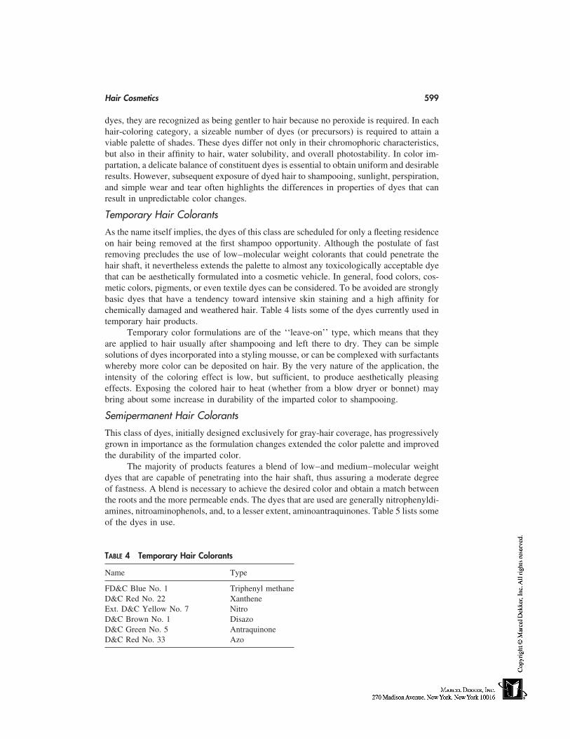
Hair Cosmetics 599
dyes, they are recognized as being gentler to hair because no peroxide is required. In eachhair-coloring category, a sizeable number of dyes (or precursors) is required to attain aviable palette of shades. These dyes differ not only in their chromophoric characteristics,but also in their affinity to hair, water solubility, and overall photostability. In color im-partation, a delicate balance of constituent dyes is essential to obtain uniform and desirableresults. However, subsequent exposure of dyed hair to shampooing, sunlight, perspiration,and simple wear and tear often highlights the differences in properties of dyes that canresult in unpredictable color changes.
Temporary Hair Colorants
As the name itself implies, the dyes of this class are scheduled for only a fleeting residenceon hair being removed at the first shampoo opportunity. Although the postulate of fastremoving precludes the use of low–molecular weight colorants that could penetrate thehair shaft, it nevertheless extends the palette to almost any toxicologically acceptable dyethat can be aesthetically formulated into a cosmetic vehicle. In general, food colors, cos-metic colors, pigments, or even textile dyes can be considered. To be avoided are stronglybasic dyes that have a tendency toward intensive skin staining and a high affinity forchemically damaged and weathered hair. Table 4 lists some of the dyes currently used intemporary hair products.
Temporary color formulations are of the ‘‘leave-on’’ type, which means that theyare applied to hair usually after shampooing and left there to dry. They can be simplesolutions of dyes incorporated into a styling mousse, or can be complexed with surfactantswhereby more color can be deposited on hair. By the very nature of the application, theintensity of the coloring effect is low, but sufficient, to produce aesthetically pleasingeffects. Exposing the colored hair to heat (whether from a blow dryer or bonnet) maybring about some increase in durability of the imparted color to shampooing.
Semipermanent Hair Colorants
This class of dyes, initially designed exclusively for gray-hair coverage, has progressivelygrown in importance as the formulation changes extended the color palette and improvedthe durability of the imparted color.
The majority of products features a blend of low–and medium–molecular weightdyes that are capable of penetrating into the hair shaft, thus assuring a moderate degreeof fastness. A blend is necessary to achieve the desired color and obtain a match betweenthe roots and the more permeable ends. The dyes that are used are generally nitrophenyldi-amines, nitroaminophenols, and, to a lesser extent, aminoantraquinones. Table 5 lists someof the dyes in use.
TABLE 4 Temporary Hair Colorants
Name Type
FD&C Blue No. 1 Triphenyl methaneD&C Red No. 22 XantheneExt. D&C Yellow No. 7 NitroD&C Brown No. 1 DisazoD&C Green No. 5 AntraquinoneD&C Red No. 33 Azo
600 Wolfram
TABLE 5 Semipermanent Hair Colorants
Name Color
4-nitro-o-phenylenediamine Yellow1,4,5,8-tetra amino anthraquinone Blue1,4-diamino anthraquinone VioletN′-(2-hydroxy ethyl)-2-nitro-p-phe- Red
nylene diamine
Several product forms are available: lotions, shampoo-in formulations, or mousses.In all cases, the dyes are dissolved or dispersed in a detergent base that contains a thickenerso the product stays on the hair without running or dripping. Application time of 20 to40 minutes is common, after which time the product is rinsed off and frequently followedby a conventional shampoo.
Recently, formulations providing more durable (color stability up to 20 shampoos)effects have become available. They consist of the conventional semipermanent dyesblended with oxidative dye precursors, which in conjunction with dilute hydrogen peroxideproduces longer-lasting color moieties. Such products are occasionally referred to as ‘‘de-mipermanents.’’ Unlike the conventional semipermanent products that are sold in singlecontainers, they, in addition to the dye mixture, contain a separate package of the oxidant.
Often included in the semipermanent category is also the only vegetable dye thatis permitted to be used in the United States: henna. Henna consists of the dried leaves ofthe plant Lawsonia alba, which grows in North Africa, the Middle East, and India. Theactive ingredient, lawsone (2-hydroxy-1-4-naphtoquinone), constitutes about 1% of thedried leaves [10]. Using henna, only limited reddish shades can be achieved. In someproducts, henna is mixed with other dyes to obtain more variety in color. Such productsare then subject to the label warnings used for coal-tar dyes.
A mention should also be made of metallic dyes, which are still popular with men.These products usually contain dissolved lead acetate and elemental sulfur. After applica-tion to hair and subsequent air exposure, the lead salt reacts to form a mixture of insolublesulfides and oxides imparting to the hair a darker color, thus providing a gradual graycoverage.
Permanent Hair Colorants
Unmatched by other colorants in the shade palette, durability to shampooing, resistanceto fading, and absence of skin staining, the permanent (oxidative) hair colorants havejustifiably carved off the largest market share in hair dyes worldwide. Available in a varietyof forms (e.g., lotions, gels, shampoos, creams.), these products deliver reliable resultsthat last until the new hair grows out. Most often, the colorant is supplied as a two-component kit consisting of a mixture of colorless dye precursor and of a stabilized solu-tion of hydrogen peroxide. Occasionally, the peroxide is provided in the form of a powder,such as urea peroxide or sodium perborate. The two components are mixed immediatelybefore use, applied to hair, and left for 20 to 40 minutes before being rinsed out withwater.
The color formation commences upon mixing and involves complex reactions be-tween precursors and the oxidant. The precursors consist of two classes of reactants: 1)primary intermediates, comprising o- and p-aminophenols and phenylenediamines, which
Hair Cosmetics 601
upon oxidation by peroxide form colored quinone imines; and 2) secondary intermediates(couplers). The latter condense with the imines to yield the final dye molecules. Whilethe color-forming reactions take place in the dye mixture, a significant fraction of the dyeprecursors diffuse rapidly into the hair together with the hydrogen peroxide forming thecolorant moieties throughout the hair fiber. The process is carried out at alkaline pH whichalso favors the bleaching of the melanin pigment by H2 O2. Table 6 lists some of theprimary and secondary intermediates and colors they produce.
Depending on product form, the formulation of the dye base varies. Ammonia andethanol amines are preferred alkalizing agents, and a mixture of surfactants and solventsare used to solubilize the dyes and assure wetting of hair. A small quantity of reducingagents are added to prevent the auto-oxidation of the dyes during storage. It is importantto realize that hydrogen peroxide, which so effectively assists in both the color develop-ment and lightening of hair pigment, also displays a less desirable role in causing oxidativehair damage. Although the damage associated with a single application is slight, the cumu-lative effect of subsequent treatments is quite perceivable.
Hair Repigmenting
The idea of dyeing the hair by melanin has always been alluring. The ‘‘natural’’ aspectof the colorant implied durability of the coloring effect and its insensitivity to haircareregimens, or shade fading—all these have been factors providing continuous incentive touse the potential of such process. Apart from intense patenting in this field, several papershave recently appeared [11,12] that describe such coloring systems as well as the character-istics of repigmented hair. Recently, products based on the principle of melanin repigmen-tation of hair have appeared on the market, but the information available to date is tooscanty to offer a reliable judgment as to the market viability of these products.
Bleaching
The bleaching action of hydrogen peroxide has been already alluded to in the context ofpermanent hair coloring, and quite satisfactory levels of lightening can be obtained withsuch products.
To attain a significantly greater level of bleaching, hydrogen peroxide is combinedwith bleach accelerators or ‘‘boosters.’’ The latter are mixtures of ammonium, potassium,or sodium persulfates. The salts are packaged as dry powders and mixed with hydrogenperoxide just before use. Thickeners and alkalizers (usually sodium silicates) are includedin the booster package. Processing time depends primarily on the initial hair color andthe desired level of lightening. The pH of these formulations is usually much higher than
TABLE 6 Oxidation Dye Colors
Colors on hair with
Coupler PPD p-Aminophenol
Recorcinol Greenish-brown Yellow-brownm-Phenylenediamine Blue purple Violetm-Amino phenol Red-brown Light orange1-Naphtrol Blue violet Red-violet2-Methyl resorcinol Yellow brown Yellowish-beige2-Amino pyridine Dark grayish-blue Light grayish-green
602 Wolfram
that of the permanent hair-color products and so is the concentration of H2 O2. All of thesefactors—high concentration of peroxide, presence of oxidizing salts, and high pH of theprocess—connote significant oxidative damage of hair. After thoroughly rinsing off thebleaching mixture, the hair should be given an acidic ‘‘bath’’ (lemon juice or solution ofcitric acid or diluted vinegar) followed by a 5 to 10 minute treatment with a ‘‘deep’’conditioner.
Hair-Color Safety and Regulatory Issues
Because of the allergenic potential of some of the materials used in hair dyes (primarilyp-phenylenediamine, or PPD), hair colorants in the United States display on the label asa legal requirement a warning, plus instructions for a 24-hour patch test with the precursorsand hydrogen peroxide mixed in the same manner as in use. As required by Section 601(a)of the Federal Food, Drug and Cosmetic Act, the warnings reads as follows:
This product contains ingredients which may cause skin irritation on certain individualsand a preliminary test according to accompanying directions should be made. This productmust not be used for dyeing the eyebrows or eyelashes; to do so may cause blindness.
It should be noted that allergic contact dermatitis to hair dyes appears to be far less com-mon today than decades ago. It has been suggested that PPD, although a strong sensitizer,is not likely to produce skin sensitization because of the short contact time with skin andrapid reaction of PPD with the oxidizing agent and couplers [13].
Concerns as to the possible carcinogenicity of some hair ingredients arose in 1975when these were reported to be mutagenic for bacteria in bioassays [14]. Presently, it isnot clear how significant a risk this poses to users of hair dyes. Because hair dyes have beenin common use for over 50 years, epidemiological studies on cancer rates in occupationallyexposed groups or the users of hair dyes are of particular value. So far, the results of mostof these suggest that hair dyes do not pose a carcinogenic risk [13].
CONCLUDING REMARKS
Available space limits this chapter on hair cosmetics to only a brief overview of what isused and practiced in this broad and important segment of personal-care products. Manyaspects of hair chemistry and physics have only been fleetingly discussed, including prop-erties of single hair fibers and their assemblies. The whole area of claim substantiationhas been left out, together with the description of physicochemical techniques that arerelevant to this subject. For a fuller account on these topics, the reader is referred to anexcellent book by Zviak [15] and recent publications on haircare [16] and cosmetic-claimsubstantiation [17].
REFERENCES
1. Menkart J, Wolfram LJ, Mao I. J Soc Cosmet Chem 1966; 17:769.2. Wolfram LJ. (1981) In: Orfanos, Montagna, Stütgen, eds. Hair Research. Berlin: Springer
Verlag, 1981:479.3. Kligman AM, Shelley WD. J Inv Dermatol 1958; 30:99.4. Cunliffe WJ, Perera WD, Thackeray P, Williams M, Foster RA, Williams SM. Br J Dermatol
1975; 95:153.5. Breuer MM. J Soc Cosmet Chem 1981; 32:437.
Hair Cosmetics 603
6. Schwartz AM, Perry JW, Belch J. Surface Active Agents and Detergents. Vol. 2. Robert E.Krieger: Huntington, NY; 1977.
7. Preston WC. J Phys Chem 1948; 52:84.8. Stevenson DG. J Text Inst 1959; 50:T548.9. Zviak C, Vanlerberghe G. In: Zviak C, ed. The Science of Hair Care. New York: Dekker,
1986:57.10. Stamberg J, Werczberger R, Koltin Y. Mutat Res 1979; 62:383.11. Brown K, Mayer A, Murphy B, Schultz T, Wolfram LJ. J Soc Cosmet Chem 1989; 40:65.12. Brown K, Marlowe E, Prota G, Wenke G. J Soc Cosmet Chem 1997; 48:133.13. Corbett JF. Rev Prog Coloration 1985; 15:53.14. Ames BN, Kammen DH, Yannesaki E. Proc Nat Acad Sci USA 1975; 72:2423.15. Zviak C, ed. The Science of Hair Care. New York: Marcel Dekker, 1986.16. Johnson DH, ed. Hair and Hair Care. New York: Marcel Dekker, 1997.17. Aust LB, ed. Cosmetic Claims Substantiations. New York: Marcel Dekker, 1998.

52
Ethnic Differences in Haircare Products
Joerg KahreHenkel KGaA, Düsseldorf, Germany
INTRODUCTION
Hair is undoubtedly one of the most important personal features of people in all cultures.For the past several centuries hair has played an important role. Style, length, and colorchanges are influenced by fashion trends. Hair often allows for feelings of health andbeauty, and thus its influence is of great importance. Therefore hair has been studiedgreatly as cited in numerous publications. Three major types of hair are known: African,Asian, and Caucasian. The differences between these hair types are related to diameter,geometry, and other physical parameters [1,2]. Closely related to these parameters arebiophysical factors, tensile strength, and combing forces, which might be influenced bycosmetic formulations that are applied to hair. Caucasian hair is also called European hairand African hair is also called Negroid hair. The names only summarize the complexityof hair types, e.g., Asian hair is the sum term for Japanese, Chinese, and other Asianethnic groups. And furthermore, even in such ethnic subgroups we do not really see asingle hair quality. Therefore, taking Asian, Caucasian, or African hair is an overall exam-ple for the corresponding hair types of these regions. Table 1 shows an overview aboutthe most important hair fiber characteristics [3,4].
The demand for hair care is closely related to the condition and length of the hairand fashion trends. Hair is exposed daily to a wide variety of influences that can damageit to a greater or lesser degree. Especially the surface of hair that has been exposed toenvironmental influences (e.g., sunlight, combing, blow drying, etc.) or chemical treat-ments (e.g., cold waving, dyeing, bleaching) carries a stronger negative charge than thesurface of untreated hair.
The resulting change in the hair’s structure may reduce its natural gloss or cause amild build-up of static charge, and in extreme cases the hair may break, especially in theregion of the tip.
Such changes in the hair’s appearance can be avoided by, in the first place, usingmild hair-cleansing products or conditioning shampoos. The second protection step, how-ever, is provided by hair aftertreatment products such as hair conditioners or rinses.
The effect of conditioning preparations is restricted to the part of the hair shaft thatprojects out of the epidermis. Both the cortex and the cuticle of the hair shaft can benegatively affected by the aforementioned influences.
605
606 Kahre
TABLE 1 Hair Fiber Characteristics
Caucasian Asian African
Thickness fine coarse coarseCurvature straight to curly straight to wavy wavy to woolyCross sectional nearly round to nearly round to slightly oval to ellip-
shape slightly oval slightly oval ticalColor blond to dark brown dark brown to brown- brown-black to black
blackCross section �70 �90 �70
area (µm2)
Chemical hair treatments such as cold waving, bleaching, or the use of straightenersand relaxers have a particularly unfavorable effect on the cortex, because they influenceor change, e.g., the disulfide bonds between and in proteins. This generally results in aloss of mechanical strength, a more marked tendency to absorb moisture and therefore toswell, a greater susceptibility to alkalines, and an increase in electrostatic charge [5].
Mechanical stresses such as frequent combing and brushing, blow drying, and inten-sive exposure to sunlight cause damage to the cuticle; scales either break off completelyor along their edges. This makes the hair rougher and reduces its natural gloss [6,7]. Anoverview of the properties of damaged hair is shown in Figure 1. The requirements to besatisfied by an optimally formulated hair-treatment preparation derive from the listed hair-damaging processes.
Objective test methods are developed to investigate all the effects. Some of the mostimportant methods to understand the actions of hair-treatment preparations are listed inTable 2 [8]. In addition to these tests, cosmetic assessment is usually obtained from thehalf-head test, the panel test, and the home-use test.
AFRICAN HAIR
The biophysical properties of African hair are more closely related to wool fibers than tothe other hair types. African hair shows some special properties as a result of its verycurly structure [9–11]. These properties are listed in Figure 2. African hair is treated withhair relaxers, perms, or straighteners in order to get a straight to light curled style. This
FIGURE 1 Properties of damaged hair.
Ethnic Differences in Haircare Products 607
TABLE 2 Objective Measuring Methods
Term Objective measuring methods Measurement parameter
Combability, Resistance to combing Wet combing workdetangling Dry combing work
Strength Resonance frequency Modulus of elasticityTensile strength measurement Elastic range (range of Hooke’s law)Breaking strength of single hair Breaking forceBreaking strength of hair tress Breaking force
Charging capacity Faraday cage Charge differenceSplitting Electron-scanning microscopeGloss Goniophotometer Reflection
has an influence on the hair structure. We measured the wet tensile strength of hair tressesafter the application of typical hair relaxers. As expected, straighteners and relaxers havea strong influence in decreasing the tensile strength. Relative to untreated hair we founda residual tensile strength of only 60% depending upon the relaxing agent. Thioglycolate(7.5% active at pH 9.3) is milder than sodium hydroxide (2.0% at pH 13.5) and this isbetter than calcium hydroxide (0.6% active at pH 13.5). A more detailed description ofthe physical properties and differences of African hair relative to Caucasian hair is reportedin the literature [9–11].
Influence of Surfactants and Protein Hydrolysates on African Hair
The use of mild surfactants in shampoos is necessary and avoids additional damage.Applying 2% sodium hydroxide (pH 13.5) to kinky hair resulted in a residual strength ofabout 73%. Shampooing this hair with a 12% active solution of sodium laureth sulfate(SLES) gave a further decrease of the tensile strength to 64%. If we choose decyl glucosideas surfactant, there is no further damage and a significantly higher wet tensile strengthrelative to the SLES result.
Adding protein hydrolysates to relaxers or straighteners strongly increases tensilestrength. Using 2% active hydrolysed collagen in a 0.6%-containing calcium hydroxidestraightener at pH 13.5 resulted in an increase of the wet tensile strength up to 142%relative to the same straightener without hydrolyzed collagen. This is important for theformulation not only for these products, but for shampoos and conditioners in general.The addition of protein hydrolysates to restructure the hair is strongly recommended.
FIGURE 2 Characteristics of African hair.
608 Kahre
FIGURE 3 Dry and wet tensile strength of single fibers.
DIFFERENCES OF UNTREATED ASIAN AND CAUCASIAN HAIR
Because of the differences in cross-section, we found different wet and dry tensilestrengths of single hair fibers. Asian hair shows higher values than Caucasian hair. Theapplication of a perm, bleach, or both results in decreasing tensile forces for both hairtypes (Fig. 3). The higher values for dry combing forces are found again for Asian hair(Fig. 4). The wet combing work shows comparable results for the untreated hair types.
In addition to these results we measured the bounce of curls. For a natural appearanceof hair the physical parameters attenuation, maximum zero amplitude and force of elonga-
FIGURE 4 Combing work of Asian and Caucasian hair.
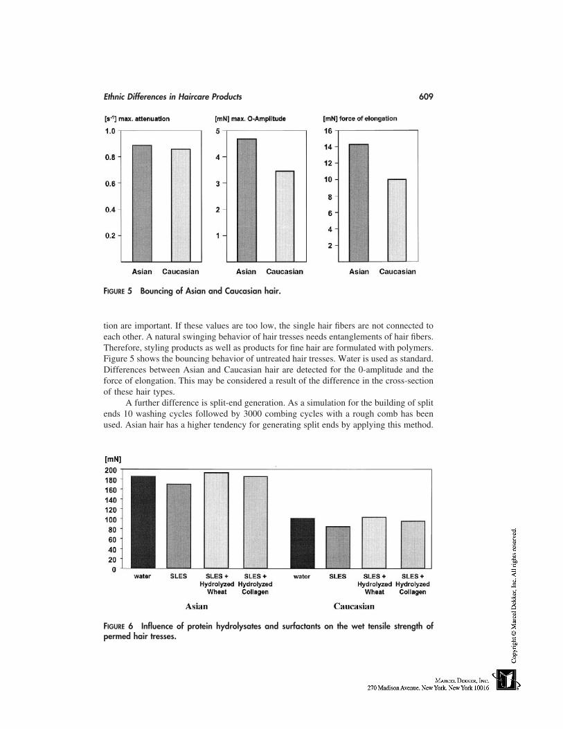
Ethnic Differences in Haircare Products 609
FIGURE 5 Bouncing of Asian and Caucasian hair.
tion are important. If these values are too low, the single hair fibers are not connected toeach other. A natural swinging behavior of hair tresses needs entanglements of hair fibers.Therefore, styling products as well as products for fine hair are formulated with polymers.Figure 5 shows the bouncing behavior of untreated hair tresses. Water is used as standard.Differences between Asian and Caucasian hair are detected for the 0-amplitude and theforce of elongation. This may be considered a result of the difference in the cross-sectionof these hair types.
A further difference is split-end generation. As a simulation for the building of splitends 10 washing cycles followed by 3000 combing cycles with a rough comb has beenused. Asian hair has a higher tendency for generating split ends by applying this method.
FIGURE 6 Influence of protein hydrolysates and surfactants on the wet tensile strength ofpermed hair tresses.
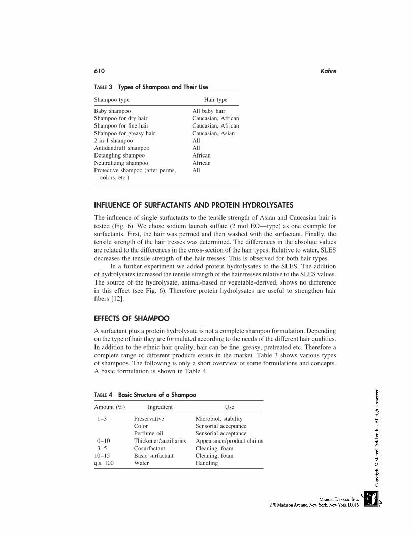
610 Kahre
TABLE 3 Types of Shampoos and Their Use
Shampoo type Hair type
Baby shampoo All baby hairShampoo for dry hair Caucasian, AfricanShampoo for fine hair Caucasian, AfricanShampoo for greasy hair Caucasian, Asian2-in-1 shampoo AllAntidandruff shampoo AllDetangling shampoo AfricanNeutralizing shampoo AfricanProtective shampoo (after perms, All
colors, etc.)
INFLUENCE OF SURFACTANTS AND PROTEIN HYDROLYSATES
The influence of single surfactants to the tensile strength of Asian and Caucasian hair istested (Fig. 6). We chose sodium laureth sulfate (2 mol EO—type) as one example forsurfactants. First, the hair was permed and then washed with the surfactant. Finally, thetensile strength of the hair tresses was determined. The differences in the absolute valuesare related to the differences in the cross-section of the hair types. Relative to water, SLESdecreases the tensile strength of the hair tresses. This is observed for both hair types.
In a further experiment we added protein hydrolysates to the SLES. The additionof hydrolysates increased the tensile strength of the hair tresses relative to the SLES values.The source of the hydrolysate, animal-based or vegetable-derived, shows no differencein this effect (see Fig. 6). Therefore protein hydrolysates are useful to strengthen hairfibers [12].
EFFECTS OF SHAMPOO
A surfactant plus a protein hydrolysate is not a complete shampoo formulation. Dependingon the type of hair they are formulated according to the needs of the different hair qualities.In addition to the ethnic hair quality, hair can be fine, greasy, pretreated etc. Therefore acomplete range of different products exists in the market. Table 3 shows various typesof shampoos. The following is only a short overview of some formulations and concepts.A basic formulation is shown in Table 4.
TABLE 4 Basic Structure of a Shampoo
Amount (%) Ingredient Use
1–3 Preservative Microbiol, stabilityColor Sensorial acceptancePerfume oil Sensorial acceptance
0–10 Thickener/auxiliaries Appearance/product claims3–5 Cosurfactant Cleaning, foam
10–15 Basic surfactant Cleaning, foamq.s. 100 Water Handling
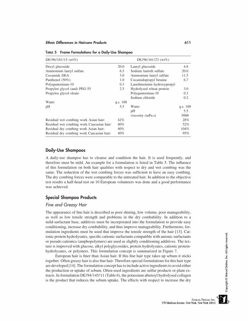
Ethnic Differences in Haircare Products 611
TABLE 5 Frame Formulations for a Daily-Use Shampoo
DE/96/161/15 (wt%) DE/96/161/23 (wt%)
Decyl glucoside 20.0 Lauryl glucoside 4.8Ammonium lauryl sulfate 6.5 Sodium laureth sulfate 20.0Cocamide DEA 3.0 Ammonium lauryl sulfate 11.5Panthenol (50%) 1.0 Cocamidopropyl betaine 6.7Polyquaternium-10 0.3 Laurdimonium hydroxypropylPropylen glycol (and) PEG-55 2.5 Hydrolysed wheat protein 3.0Propylen glycol oleate Polyquaternium-10 0.3
Sodium chloride 0.2Water q.s. 100pH 5.5 Water q.s. 100
pH 5.5viscosity (mPa.s) 5000
Residual wet combing work Asian hair: 42% 28%Residual wet combing work Caucasian hair: 40% 52%Residual dry combing work Asian hair: 40% 104%Residual dry combing work Caucasian hair: 40% 95%
Daily-Use Shampoos
A daily-use shampoo has to cleanse and condition the hair. It is used frequently, andtherefore must be mild. An example for a formulation is listed in Table 5. The influenceof this formulation on both hair qualities with respect to dry and wet combing was thesame. The reduction of the wet combing forces was sufficient to have an easy combing.The dry combing forces were comparable to the untreated hair. In addition to the objectivetest results a half-head test on 10 European volunteers was done and a good performancewas achieved.
Special Shampoo Products
Fine and Greasy Hair
The appearance of fine hair is described as poor shining, low volume, poor manageability,as well as low tensile strength and problems in the dry combability. In addition to amild-surfactant base, additives must be incorporated into the formulation to provide easyconditioning, increase dry combability, and thus improve manageability. Furthermore, for-mulation ingredients must be used that improve the tensile strength of the hair [13]. Cat-ionic protein hydrolysates, specific cationic surfactants compatible with anionic surfactantsor pseudo cationics (amphopolymers) are used as slightly conditioning additives. The tex-ture is improved with glucose, alkyl polyglycosides, protein hydrolysates, cationic protein-hydrolysates, or polymers. This formulation concept is summarized in Figure 7.
European hair is finer than Asian hair. If this fine hair type takes up sebum it stickstogether. Often greasy hair is also fine hair. Therefore special formulations for this hair typeare developed [14]. The formulation concept has to include active ingredients to avoid eitherthe production or uptake of sebum. Often-used ingredients are sulfur products or plant ex-tracts. In formulation DE/94/145/11 (Table 6), the potassium abietoyl hydrolysed collagenis the product that reduces the sebum uptake. The effects with respect to increase the dry
612 Kahre
FIGURE 7 Formulation concept for fine hair.
combing work as well as the wet combing work is sufficient for Caucasian but not Asianhair. By applying this formulation to Asian hair the amount of quaternary polymer has tobe slightly increased. This formulation also reduces the uptake of sebum. The action andmechanism for the reduction of sebum uptake is described in the literature [14].
2-in-1 Shampoos
This type of shampoo is very popular. Washing and conditioning is only one step. There-fore these products are well accepted by the consumer. The formulation concept is notso different from the others (Fig. 8). In this case silicones are mostly formulated to achievethe special conditioning effect. New concepts are using monoglycerides in combinationwith alkyl glucosides and cationic polymers to get this conditioning effect. With such aconcept, the build-up effect can be avoided [15]. Formulation DE/94/005/25 is one exam-ple tested on Asian and Caucasian hair and one example formulated without the use ofsilicones. Important is the reduction of wet-combing forces and the increase of the dry-combing forces in order to get a style and volume. The formulation and the results areshown in Table 7. The performance is best on Caucasian hair. For Asian hair the amountof cationic polymer has to be increased.
AFTER HAIR TREATMENTS
Modern preparations are divided into rinse-off types, which are rinsed off after being leftto take effect for a certain time, possibly with the help of a slight increase in temperature,
TABLE 6 Shampoo for Fine and Greasy Hair
DE/94/145/11(wt%)
Decyl glucoside 10.0Sodium laureth sulfate 14.3Cocamidopropyl betaine 10.0Potassium abietoyl hydrolyzed 5.1
collagen (PAHC)Polyquaternium-10 0.2Laureth-3 1.0Sodium chloride 1.0Water q.s. 100pH 5.5
Ethnic Differences in Haircare Products 613
FIGURE 8 Formulation concept for 2-in-1 shampoo.
and leave-on types, which remain in the hair. In Figure 9 an overview about the hairaftertreatments is listed. We studied the effects of typical conditioners (Table 8). Aftertreatments are used to restructure and improve the hair quality. Such preparations mustbe effective not only superficially but also below the surface of the hair. Changing theproperties of the hair surface can cause improvements in properties such as combability,feel, and manageability, and can reduce the build-up of static charge. Moreover, a protec-tive action can be achieved with chemical hair treatments, and special additives that pene-trate inside the hair can improve its mechanical strength. Therefore a schematic formula-tion makeup is based on the described general requirements derived from hair-damagingprocesses described in the introduction [16,17].
Cationic surfactants act by being adsorbed onto the surface of negatively chargedhair [19,20]. In contrast, active agents such as cationic protein hydrolysates, protein hy-drolsates, panthenol, and glucose penetrate at least partially below the surface of the hair.
In order to evaluate differences between conditioners we applied several formula-tions containing 1.0% active distearoylethyl hydroxyethylmonium methosulfate and 2.5%cetearyl alcohol to Asian and Caucasian hair. The combing work was measured beforeand after the application of the conditioner. The absolute values for the combing work ofAsian and Caucasian hair are different as shown previously in Figure 4. For testing theefficacy it is not important to see the absolute values of combing work; it is more importantto know the degree of reduction. This means a residual combing work of 40% is a relativereduction of 60%. All formulations have the same efficacy on Asian and Caucasian hair.There is no significant difference in the relative change of the combing work.
TABLE 7 Example of a 2-in-1 Shampoo
DE/94/005/25(wt%)
Sodium laureth sulfate (and) lauryl glucoside 21.0Glycol distearate (and) glycerin (and) laureth-4 2.0
(and) cocamidopropyl betainePEG-7 glyceryl cocoate 1.0Guar hydroxypropyl trimonium chloride 0.5Water q.s. 100pH 5.5residual combing work dry wet
Asian hair 87% 87%Caucasian hair 87% 60%
614 Kahre
FIGURE 9 Overview of hair treatment preparations.
TABLE 8 General Conditioner Formulation
Use Hair rinses Amount %
Formulation auxiliary Emulsifier 0–2Consistency, conditioning agent Consistency factors 1–5Emollient, care component Oily components, auxiliaries 0–3Conditioning agent Cationic components 0.5–1.5Sensorial acceptance Perfume oil 0–2Microbiological stability Color
PreservativeWaterpH value: 3–5
FIGURE 10 Formulation with emollients in conditioners.
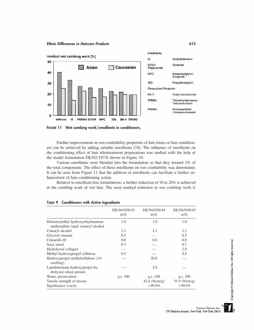
Ethnic Differences in Haircare Products 615
FIGURE 11 Wet combing work/emollients in conditioners.
Further improvements in wet combability properties of hair rinses or hair condition-ers can be achieved by adding suitable emollients [18]. The influence of emollients onthe conditioning effect of hair aftertreatment preparations was studied with the help ofthe model formulation DE/92/197/6 shown in Figure 10.
Various emollients were blended into the formulation so that they formed 2% ofthe total components. The effect of these emollients on wet combability was determined.It can be seen from Figure 11 that the addition of emollients can facilitate a further en-hancement of hair-conditioning action.
Relative to emollient-free formulations, a further reduction of 10 to 20% is achievedin the combing work of wet hair. The most marked reduction in wet combing work is
TABLE 9 Conditioners with Active Ingredients
DE/94/038/43 DE/94/038/44 DE/94/038/45wt% wt% wt%
Distearoylethyl hydroxyethylmonium 1.0 1.0 1.0methosulfate (and) cetearyl alcohol
Cetearyl alcohol 2.1 2.1 2.1Glyceryl stearate 0.5 — 0.5Ceteareth-20 0.8 0.8 0.8Soya sterol 0.7 — 0.7Hydrolyzed collagen — — 2.0Methyl hydroxypropyl cellulose 0.5 — 0.5Hydroxypropyl methylcellulose (1% — 20.0 —
swelling)Laurdimonium hydroxypropyl hy- — 2.8 —
drolyzed wheat proteinWater, preservation q.s. 100 q.s. 100 q.s. 100Tensile strength of tresses 42.4 (Ncm/g) 51.9 (Ncm/g)Significance (t-test) �99.9% �99.9%
616 Kahre
TABLE 10 Leave-On Conditioner
DE/96/099/4wt%
Polyacrylamide (and) C13-14 isoparafin 3.0(and) laureth-7
Cocamide DEA 1.0Glycerin (86%) 5.0Lauryl glucoside 0.5Oleyl erucate 0.5Tocopherol 0.2Hydrolyzed sweet almond protein 3.0Laurdimonium hydroxypropyl hydrolyzed 0.8
wheat proteinEthanol (96%) 10.0Water, preservation q.s. 100pH 7.0Viscosity (Brookfield RVF, 23°C, spindle 4, ca. 3000 mPa.s
10 rpm)
brought about by high molecular emollients. When the influence of these emollients onthe combability of dry hair was studied, it was found that they cause almost no changes.
Not only the hair’s characteristics were favorably influenced by the oils in hairrinses, but the physicochemical properties of hair rinses that contain fatty alcohol werealso improved. Emollients generally have the effect of stabilizing viscosity during storage.
EXAMPLES OF FORMULATIONS AND EFFECTS
Some further examples of different formulations are listed. If any effects have been mea-sured, these are mentioned. Conditioners for colored, permed, bleached, or straightenedhair are given in Tables 9 to 12. These formulations may be used on all ethnic hair types.The listed examples are rinse-off (Tables 9, 10) as well as leave-on (Table 11) condition-ers. The so-called liquid hair (Table 12) is a special leave-on product that acts as a re-structuring agent for damaged hair. Pretreatments (Table 13) are used to reduce the effectscaused by the following application of a perm, bleach, or coloring. They are useful for allethnic hair types. The last two formulations are shampoos for African hair (Tables 14, 15).
TABLE 11 Hot Oil Treatment
DE/91/303/11wt%
Cetrimonium chloride 8.0Hydroxyethyl cellulose (2% swelling) 20.0Polysorbate-20 1.5Hydrolyzed collagen 0.3D-panthenol (50%) 0.2Water, preservative, perfume, etc. q.s. 100
Ethnic Differences in Haircare Products 617
TABLE 12 Liquid Hair (Leave-On)
DE/94/211/6wt%
Decyl glucoside 4.0Laurdimonium hydroxypropyl 2.0
hydrolyzed collagenHydrolyzed keratin 3.0Glycerin (86%) 20.0Ethanol (96%) 5.0Water, preservative q.s. 100pH 5–5.5
TABLE 13 Pretreatment Preparation
Wt%
Hydrolyzed keratin 2.0Citric acid 0.1PEG-hydrogenated castor oil 1.0Fragrance, preservative, water q.s. 100
TABLE 14 Detangling Shampoo for African Hair
Wt%
Ammonium lauryl sulfate 30.0Cocamidopropyl betaine 10.0Coco glucoside (and) glyceryl oleate 5.0Hydrolyzed wheat protein 3.0Laurdimonium hydroxypropyl hydrolyzed 3.0
wheat proteinWater, preservative q.s. 100pH 5.5
TABLE 15 Neutralizing Shampoo for AfricanHair
Wt%
Ammonium lauryl sulfate 35.0Cocamidopropyl betaine 10.0Coco glucoside (and) glyceryl oleate 2.0Laurdimonium hydroxypropyl hydrolyzed 1.0
wheat proteinHydrolyzed wheat protein 1.0Polyquaternium-10 0.4Guar hydroxypropyl trimonium chloride 0.2Water, preservative q.s. 100pH (adjusted with citric acid) 5.5
618 Kahre
REFERENCES
1. Taesdale D, Schlüter R, Blankenburg G. Querschnittsparameter von Humanhaaren Teil 1,Ärztl. Kosmetologie 1981; 11:161–170.
2. Taesdale D, Schlüter R, Blankenburg G. Querschnittsparameter von Humanhaaren Teil 2,Ärztl. Kosmetologie 1981; 11:252–259.
3. Hensen H, Kahre J. Haarnachbehandlungsmittel im Überlick, Seifen Öle Fette Wachse 1998;124, 806–815.
4. Eva Tolgyesi, Coble DW, Fang FS, Kairinen EO. A comparative study of beard and scalphair. J Soc Cosmet Chem 1983; 34:361–382.
5. Kahre J, Busch P, Salka B, Totani N, Poly W. Asian and caucasian hair—differences of influ-ence for the formulation? Annual Scientific Seminar 1998, preprints, 7.
6. Koester J. Eigenschaften und Anwendung kationischer Haarpflegeadditive. Parfümerie undKosmetik 1991; 72/4:218–225.
7. Hollenberg D, Müller R. Möglichkeiten zur Beeinflussung der Haarstruktur durch Pflegepro-dukte. Seifen Öle Fette Wachse 1995; 121/2:82–89.
8. Busch P, Förster Th, Hensen H, H Th. Müller-Kirschbaum. Tesmann, Subjektiv/objektiv-Bewertung kosmetischer Effekte. Ärztliche Kosmetologie 1990; 20:498–502.
9. Syed AN, Kuhajda A, Ayoub H, Ahmad K. African-American hair. Cosmet Toilet 1995; 110:39–48.
10. Burmeister F, Bollatti D, Brooks G. Ethnic hair: moisturizing after relaxer use. Cosmet Toilet1991; 106:49–51.
11. Menkart J, Wolfram LJ, Mao I. Caucasian hair, negro hair, and wool: similarities and differ-ences. J Soc Cosmet Chemists 1966; 17:769–787.
12. Kahre J, Seipel W, Wachter R. Pflanzliche Proteinhydrolysate und Derivate mit universellenEigenschaften, SEPAWA-Jahrestagung, 35–42, 1995.
13. Hensen H. Surfactant preparations. Eurocosmetics 5/95.14. Busch P, Hensen H, Fischer D, Ruhnke A, Franklin J. An abietic acid protein condensate for
treating greasy hair. Cosmet Toilet 1995; 110:59–63.15. Both W, Gassenmeier T, Hensen H, Hörner V, Seipel W. Pflegende, lipidhaltige Reinigungsp-
räparate für Haut und Haar, SEPAWA Kongress, 34–38, 1997.16. Kahre J, Busch P, Totani N, Poly W. Asian and European hair—influence of the difference
on the formulation. Poster presentation, IFSCC—Sydney, 1996.17. Spiess E. The influence of chemical structure on performance in hair care preparations. Par-
fümerie und Kosmetik 1991; 72:370–376.18. Busch P, Hensen H, Kahre J, Tesmann H. Alkylpolyglycosides—a new cosmetic concept for
mildness and care. Agro-Food-Industry. Sept/Oct 1994; pp. 23–28.
53
Oral-Care Products
Abdul GaffarColgate-Palmolive Company, Piscataway, New Jersey
THE TEETH AND ORAL ENVIRONMENT
Like all mammals, humans generally have two sets of teeth during a lifetime. The firstset, known as deciduous, primary, or ‘‘milk’’ teeth, begins to appear in infants betweenthe age of 5 and 9 months. All 20 of these ‘‘baby’’ teeth are generally in place by age21/2 years. The second set, or permanent teeth, forms within the gums during the periodfrom infancy to puberty. These teeth, also known as succedaneous teeth, begin to eruptat around age 5, displacing the deciduous set as they appear. There are 32 permanentteeth. An individual will spend 91% of his or her lifetime chewing with these permanentteeth if they are properly cared for. Of the 32 permanent teeth, 16 are located in the upperjaw, or maxillary dental arch, which is part of the cranium, or skull, and is immoveable.The other 16 are located in the mandibular dental arch which is part of the lower jaw andis the moveable part of the skull. Each type of tooth is equally divided between these twodental arches (Figs. 1 and 2).
The Parts of a Tooth
Each tooth consists of three parts: the area above the gum that can be seen, the area belowthe gum that is not visible, and the constricted portion, or neck, between the other twoparts. The crown is the enamel-covered portion of the tooth. The root is the portion ofthe tooth which, by means of the periodontal ligament, relates to the osseous (bony) struc-tures of the jaw. The root makes up about two thirds of the total length of a tooth (Fig. 3).
The Tissues of a Tooth
A tooth is made up of five different tissues, each with a specific and important function.Serious disease in any of these tissues can affect the entire tooth and result in its decayand/or destruction. These tissues are as follows:
1. Enamel, which is a hard white outer covering surrounds the crown of the toothand protects it from wearing away as a result of the pressure of chewing. Itconsists largely (96 to 98 percent) of inorganic substances, mainly calcium andphosphate.
619
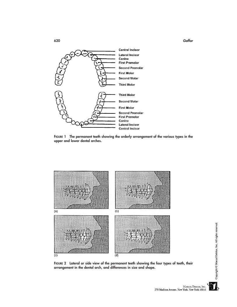
620 Gaffar
FIGURE 1 The permanent teeth showing the orderly arrangement of the various types in theupper and lower dental arches.
FIGURE 2 Lateral or side view of the permanent teeth showing the four types of teeth, theirarrangement in the dental arch, and differences in size and shape.
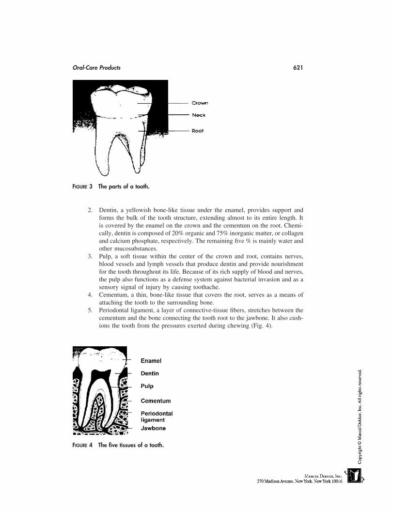
Oral-Care Products 621
FIGURE 3 The parts of a tooth.
2. Dentin, a yellowish bone-like tissue under the enamel, provides support andforms the bulk of the tooth structure, extending almost to its entire length. Itis covered by the enamel on the crown and the cementum on the root. Chemi-cally, dentin is composed of 20% organic and 75% inorganic matter, or collagenand calcium phosphate, respectively. The remaining five % is mainly water andother mucosubstances.
3. Pulp, a soft tissue within the center of the crown and root, contains nerves,blood vessels and lymph vessels that produce dentin and provide nourishmentfor the tooth throughout its life. Because of its rich supply of blood and nerves,the pulp also functions as a defense system against bacterial invasion and as asensory signal of injury by causing toothache.
4. Cementum, a thin, bone-like tissue that covers the root, serves as a means ofattaching the tooth to the surrounding bone.
5. Periodontal ligament, a layer of connective-tissue fibers, stretches between thecementum and the bone connecting the tooth root to the jawbone. It also cush-ions the tooth from the pressures exerted during chewing (Fig. 4).
FIGURE 4 The five tissues of a tooth.
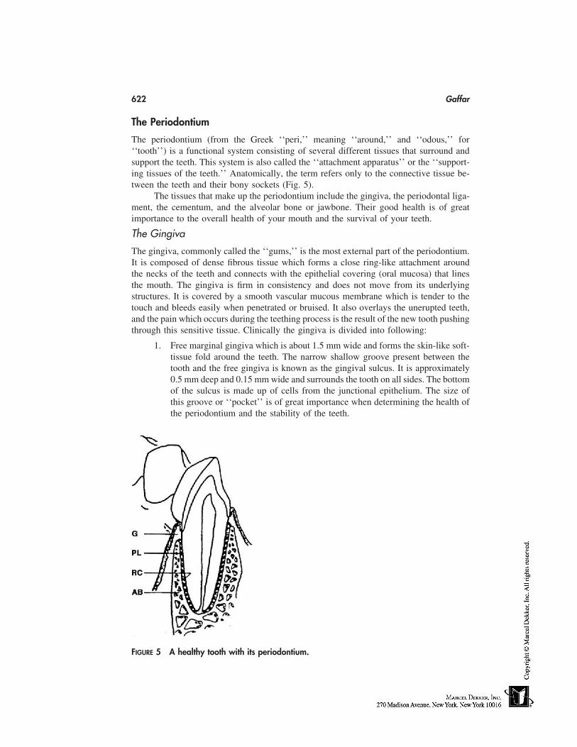
622 Gaffar
The Periodontium
The periodontium (from the Greek ‘‘peri,’’ meaning ‘‘around,’’ and ‘‘odous,’’ for‘‘tooth’’) is a functional system consisting of several different tissues that surround andsupport the teeth. This system is also called the ‘‘attachment apparatus’’ or the ‘‘support-ing tissues of the teeth.’’ Anatomically, the term refers only to the connective tissue be-tween the teeth and their bony sockets (Fig. 5).
The tissues that make up the periodontium include the gingiva, the periodontal liga-ment, the cementum, and the alveolar bone or jawbone. Their good health is of greatimportance to the overall health of your mouth and the survival of your teeth.
The Gingiva
The gingiva, commonly called the ‘‘gums,’’ is the most external part of the periodontium.It is composed of dense fibrous tissue which forms a close ring-like attachment aroundthe necks of the teeth and connects with the epithelial covering (oral mucosa) that linesthe mouth. The gingiva is firm in consistency and does not move from its underlyingstructures. It is covered by a smooth vascular mucous membrane which is tender to thetouch and bleeds easily when penetrated or bruised. It also overlays the unerupted teeth,and the pain which occurs during the teething process is the result of the new tooth pushingthrough this sensitive tissue. Clinically the gingiva is divided into following:
1. Free marginal gingiva which is about 1.5 mm wide and forms the skin-like soft-tissue fold around the teeth. The narrow shallow groove present between thetooth and the free gingiva is known as the gingival sulcus. It is approximately0.5 mm deep and 0.15 mm wide and surrounds the tooth on all sides. The bottomof the sulcus is made up of cells from the junctional epithelium. The size ofthis groove or ‘‘pocket’’ is of great importance when determining the health ofthe periodontium and the stability of the teeth.
FIGURE 5 A healthy tooth with its periodontium.
Oral-Care Products 623
2. Attached gingiva which is firmly connected to the hard surface of the tooth bymeans of a ring of specialized tissue known as the junctional epithelial attach-ment. The attached gingiva becomes wider with age and may vary considerablyamong individuals and from tooth to tooth.
3. The cells in the junctional epithelium are continuously being renewed duringlife and have a turnover rate of every 4 to 6 days. This results in a very perme-able tissue which serves as a pathway for the metabolic products produced bythe bacteria present in the mouth. This area plays a key role in the maintenanceof periodontal health.
4. Interdental gingiva which varies in depth and width and occupies the area be-tween adjacent teeth.
The Periodontal Ligament and the Cementum
The periodontal ligament occupies the space between the root surface of the tooth andthe alveolar bone or jawbone. It is composed of connective tissue fibers, blood vessels,nerves and other cells. Its function is to provide the connection between the cementumlayer of the tooth and the jawbone, the teeth and the gingiva, and between each tooth andits neighbor. Anatomically the cementum is a part of the tooth, but functionally, it belongsto the tooth-supporting apparatus because the gingival and periodontal ligaments are an-chored in it.
The Alveolar Bone
Alveolar bone, also referred to as the jawbone, develops along with the formation of theteeth throughout pregnancy and continues to grow during the eruption of the teeth inchildhood. Three types of alveolar bone have been defined: compact bone, trabecular bone,and alveolar bone proper. The trabecular bone provides the major support structure of theteeth and is composed mainly of fatty marrow in adults.
Other Parts of the Mouth
There are several other areas in the mouth which are important. These include the tongue,palate, salivary glands, and the oral mucosa or lining of the mouth or oral cavity itself.
Palate
The palate forms the roof of the mouth and consists of two portions: the hard palate inthe front area behind the upper teeth and soft palate at the back at the entrance to thepharynx or throat area. The hard palate separates the mouth from the nasal cavity andserves as the roof of the mouth and the floor of the nose. The soft palate aids in swallowingand sucking functions.
Tongue
The tongue is the main organ of the sense of taste and an important organ of speech. Italso assists the teeth in the chewing and swallowing of food. The tongue is situated inthe floor of the mouth and is connected to various muscles in the epiglottis and pharynx,or throat. It is covered by mucous membranes, and numerous mucous and serous glandsas well as taste-buds. Internally, it consists of fibrous tissue, muscles, blood vessels andnerves (Fig. 6).
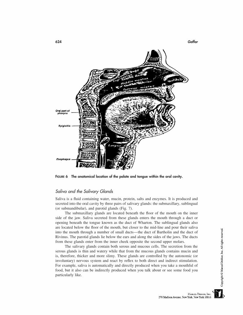
624 Gaffar
FIGURE 6 The anatomical location of the palate and tongue within the oral cavity.
Saliva and the Salivary Glands
Saliva is a fluid containing water, mucin, protein, salts and enzymes. It is produced andsecreted into the oral cavity by three pairs of salivary glands: the submaxillary, sublingual(or submandibular), and parotid glands (Fig. 7).
The submaxillary glands are located beneath the floor of the mouth on the innerside of the jaw. Saliva secreted from these glands enters the mouth through a duct oropening beneath the tongue known as the duct of Wharton. The sublingual glands alsoare located below the floor of the mouth, but closer to the mid-line and pour their salivainto the mouth through a number of small ducts—the duct of Bartholin and the duct ofRivinus. The parotid glands lie below the ears and along the sides of the jaws. The ductsfrom these glands enter from the inner cheek opposite the second upper molars.
The salivary glands contain both serous and mucous cells. The secretion from theserous glands is thin and watery while that from the mucous glands contains mucin andis, therefore, thicker and more slimy. These glands are controlled by the autonomic (orinvoluntary) nervous system and react by reflex to both direct and indirect stimulation.For example, saliva is automatically and directly produced when you take a mouthful offood, but it also can be indirectly produced when you talk about or see some food youparticularly like.
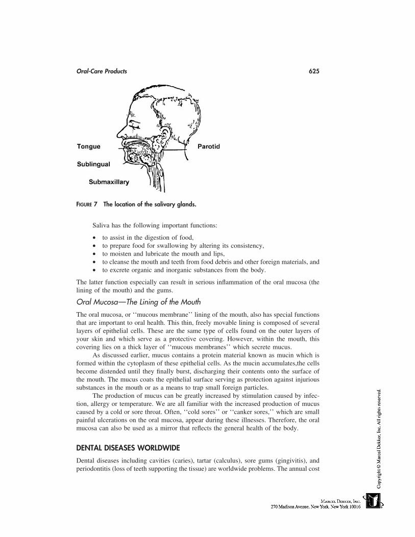
Oral-Care Products 625
FIGURE 7 The location of the salivary glands.
Saliva has the following important functions:
• to assist in the digestion of food,• to prepare food for swallowing by altering its consistency,• to moisten and lubricate the mouth and lips,• to cleanse the mouth and teeth from food debris and other foreign materials, and• to excrete organic and inorganic substances from the body.
The latter function especially can result in serious inflammation of the oral mucosa (thelining of the mouth) and the gums.
Oral Mucosa—The Lining of the Mouth
The oral mucosa, or ‘‘mucous membrane’’ lining of the mouth, also has special functionsthat are important to oral health. This thin, freely movable lining is composed of severallayers of epithelial cells. These are the same type of cells found on the outer layers ofyour skin and which serve as a protective covering. However, within the mouth, thiscovering lies on a thick layer of ‘‘mucous membranes’’ which secrete mucus.
As discussed earlier, mucus contains a protein material known as mucin which isformed within the cytoplasm of these epithelial cells. As the mucin accumulates,the cellsbecome distended until they finally burst, discharging their contents onto the surface ofthe mouth. The mucus coats the epithelial surface serving as protection against injurioussubstances in the mouth or as a means to trap small foreign particles.
The production of mucus can be greatly increased by stimulation caused by infec-tion, allergy or temperature. We are all familiar with the increased production of mucuscaused by a cold or sore throat. Often, ‘‘cold sores’’ or ‘‘canker sores,’’ which are smallpainful ulcerations on the oral mucosa, appear during these illnesses. Therefore, the oralmucosa can also be used as a mirror that reflects the general health of the body.
DENTAL DISEASES WORLDWIDE
Dental diseases including cavities (caries), tartar (calculus), sore gums (gingivitis), andperiodontitis (loss of teeth supporting the tissue) are worldwide problems. The annual cost
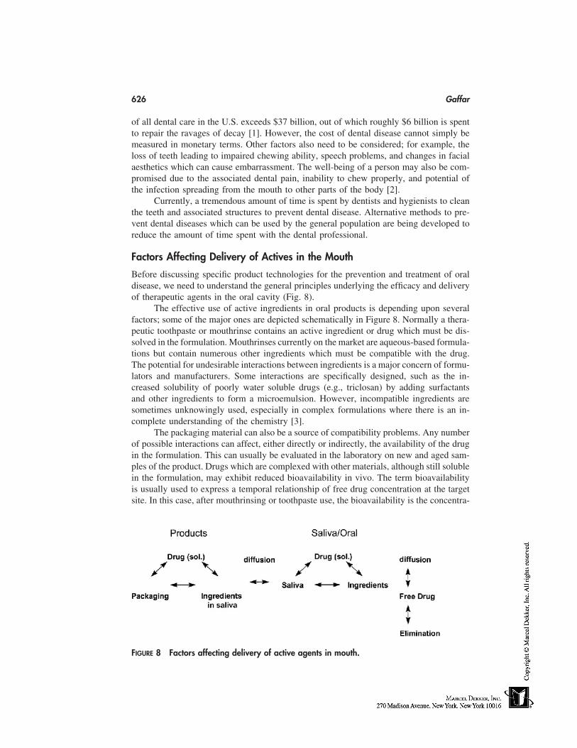
626 Gaffar
of all dental care in the U.S. exceeds $37 billion, out of which roughly $6 billion is spentto repair the ravages of decay [1]. However, the cost of dental disease cannot simply bemeasured in monetary terms. Other factors also need to be considered; for example, theloss of teeth leading to impaired chewing ability, speech problems, and changes in facialaesthetics which can cause embarrassment. The well-being of a person may also be com-promised due to the associated dental pain, inability to chew properly, and potential ofthe infection spreading from the mouth to other parts of the body [2].
Currently, a tremendous amount of time is spent by dentists and hygienists to cleanthe teeth and associated structures to prevent dental disease. Alternative methods to pre-vent dental diseases which can be used by the general population are being developed toreduce the amount of time spent with the dental professional.
Factors Affecting Delivery of Actives in the Mouth
Before discussing specific product technologies for the prevention and treatment of oraldisease, we need to understand the general principles underlying the efficacy and deliveryof therapeutic agents in the oral cavity (Fig. 8).
The effective use of active ingredients in oral products is depending upon severalfactors; some of the major ones are depicted schematically in Figure 8. Normally a thera-peutic toothpaste or mouthrinse contains an active ingredient or drug which must be dis-solved in the formulation. Mouthrinses currently on the market are aqueous-based formula-tions but contain numerous other ingredients which must be compatible with the drug.The potential for undesirable interactions between ingredients is a major concern of formu-lators and manufacturers. Some interactions are specifically designed, such as the in-creased solubility of poorly water soluble drugs (e.g., triclosan) by adding surfactantsand other ingredients to form a microemulsion. However, incompatible ingredients aresometimes unknowingly used, especially in complex formulations where there is an in-complete understanding of the chemistry [3].
The packaging material can also be a source of compatibility problems. Any numberof possible interactions can affect, either directly or indirectly, the availability of the drugin the formulation. This can usually be evaluated in the laboratory on new and aged sam-ples of the product. Drugs which are complexed with other materials, although still solublein the formulation, may exhibit reduced bioavailability in vivo. The term bioavailabilityis usually used to express a temporal relationship of free drug concentration at the targetsite. In this case, after mouthrinsing or toothpaste use, the bioavailability is the concentra-
FIGURE 8 Factors affecting delivery of active agents in mouth.
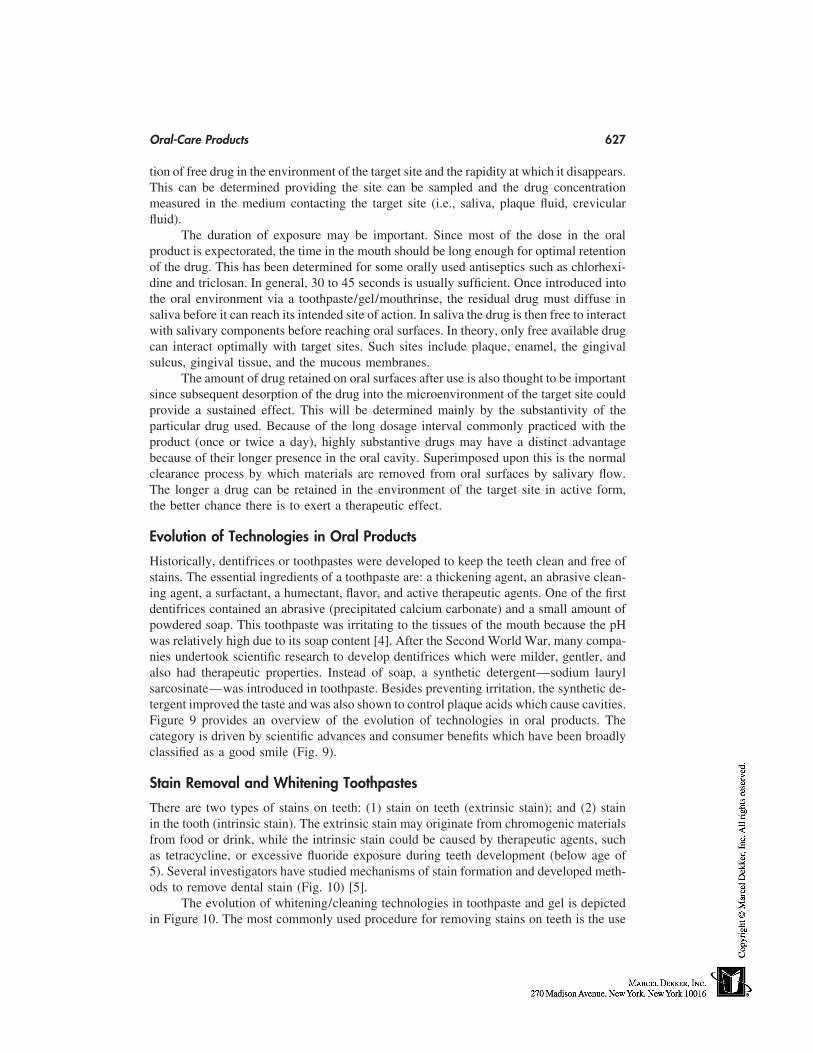
Oral-Care Products 627
tion of free drug in the environment of the target site and the rapidity at which it disappears.This can be determined providing the site can be sampled and the drug concentrationmeasured in the medium contacting the target site (i.e., saliva, plaque fluid, crevicularfluid).
The duration of exposure may be important. Since most of the dose in the oralproduct is expectorated, the time in the mouth should be long enough for optimal retentionof the drug. This has been determined for some orally used antiseptics such as chlorhexi-dine and triclosan. In general, 30 to 45 seconds is usually sufficient. Once introduced intothe oral environment via a toothpaste/gel/mouthrinse, the residual drug must diffuse insaliva before it can reach its intended site of action. In saliva the drug is then free to interactwith salivary components before reaching oral surfaces. In theory, only free available drugcan interact optimally with target sites. Such sites include plaque, enamel, the gingivalsulcus, gingival tissue, and the mucous membranes.
The amount of drug retained on oral surfaces after use is also thought to be importantsince subsequent desorption of the drug into the microenvironment of the target site couldprovide a sustained effect. This will be determined mainly by the substantivity of theparticular drug used. Because of the long dosage interval commonly practiced with theproduct (once or twice a day), highly substantive drugs may have a distinct advantagebecause of their longer presence in the oral cavity. Superimposed upon this is the normalclearance process by which materials are removed from oral surfaces by salivary flow.The longer a drug can be retained in the environment of the target site in active form,the better chance there is to exert a therapeutic effect.
Evolution of Technologies in Oral Products
Historically, dentifrices or toothpastes were developed to keep the teeth clean and free ofstains. The essential ingredients of a toothpaste are: a thickening agent, an abrasive clean-ing agent, a surfactant, a humectant, flavor, and active therapeutic agents. One of the firstdentifrices contained an abrasive (precipitated calcium carbonate) and a small amount ofpowdered soap. This toothpaste was irritating to the tissues of the mouth because the pHwas relatively high due to its soap content [4]. After the Second World War, many compa-nies undertook scientific research to develop dentifrices which were milder, gentler, andalso had therapeutic properties. Instead of soap, a synthetic detergent—sodium laurylsarcosinate—was introduced in toothpaste. Besides preventing irritation, the synthetic de-tergent improved the taste and was also shown to control plaque acids which cause cavities.Figure 9 provides an overview of the evolution of technologies in oral products. Thecategory is driven by scientific advances and consumer benefits which have been broadlyclassified as a good smile (Fig. 9).
Stain Removal and Whitening Toothpastes
There are two types of stains on teeth: (1) stain on teeth (extrinsic stain); and (2) stainin the tooth (intrinsic stain). The extrinsic stain may originate from chromogenic materialsfrom food or drink, while the intrinsic stain could be caused by therapeutic agents, suchas tetracycline, or excessive fluoride exposure during teeth development (below age of5). Several investigators have studied mechanisms of stain formation and developed meth-ods to remove dental stain (Fig. 10) [5].
The evolution of whitening/cleaning technologies in toothpaste and gel is depictedin Figure 10. The most commonly used procedure for removing stains on teeth is the use
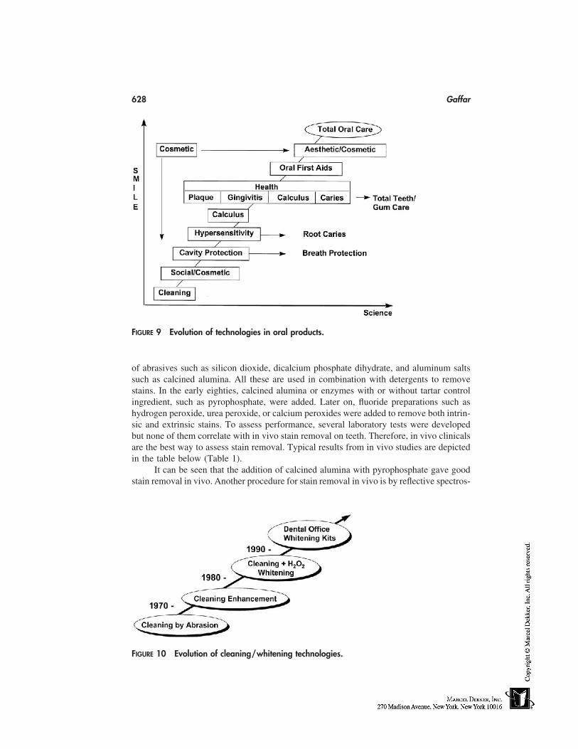
628 Gaffar
FIGURE 9 Evolution of technologies in oral products.
of abrasives such as silicon dioxide, dicalcium phosphate dihydrate, and aluminum saltssuch as calcined alumina. All these are used in combination with detergents to removestains. In the early eighties, calcined alumina or enzymes with or without tartar controlingredient, such as pyrophosphate, were added. Later on, fluoride preparations such ashydrogen peroxide, urea peroxide, or calcium peroxides were added to remove both intrin-sic and extrinsic stains. To assess performance, several laboratory tests were developedbut none of them correlate with in vivo stain removal on teeth. Therefore, in vivo clinicalsare the best way to assess stain removal. Typical results from in vivo studies are depictedin the table below (Table 1).
It can be seen that the addition of calcined alumina with pyrophosphate gave goodstain removal in vivo. Another procedure for stain removal in vivo is by reflective spectros-
FIGURE 10 Evolution of cleaning/whitening technologies.
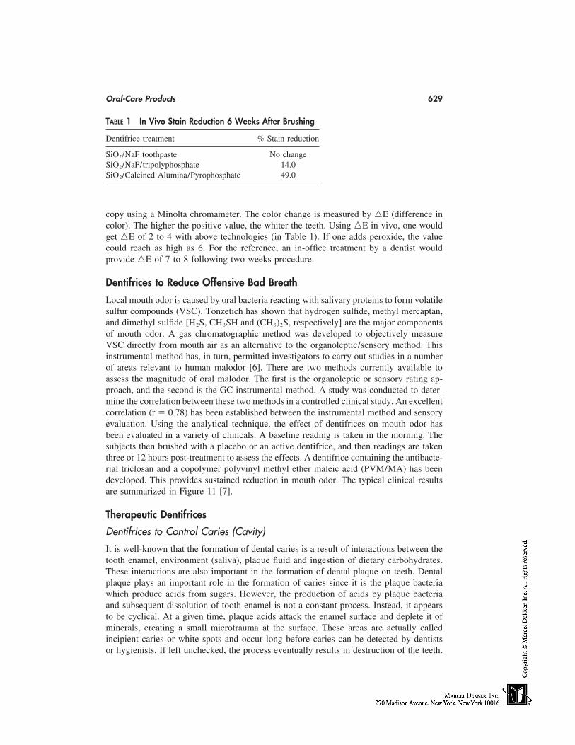
Oral-Care Products 629
TABLE 1 In Vivo Stain Reduction 6 Weeks After Brushing
Dentifrice treatment % Stain reduction
SiO2/NaF toothpaste No changeSiO2/NaF/tripolyphosphate 14.0SiO2/Calcined Alumina/Pyrophosphate 49.0
copy using a Minolta chromameter. The color change is measured by �E (difference incolor). The higher the positive value, the whiter the teeth. Using �E in vivo, one wouldget �E of 2 to 4 with above technologies (in Table 1). If one adds peroxide, the valuecould reach as high as 6. For the reference, an in-office treatment by a dentist wouldprovide �E of 7 to 8 following two weeks procedure.
Dentifrices to Reduce Offensive Bad Breath
Local mouth odor is caused by oral bacteria reacting with salivary proteins to form volatilesulfur compounds (VSC). Tonzetich has shown that hydrogen sulfide, methyl mercaptan,and dimethyl sulfide [H2S, CH3SH and (CH3)2S, respectively] are the major componentsof mouth odor. A gas chromatographic method was developed to objectively measureVSC directly from mouth air as an alternative to the organoleptic/sensory method. Thisinstrumental method has, in turn, permitted investigators to carry out studies in a numberof areas relevant to human malodor [6]. There are two methods currently available toassess the magnitude of oral malodor. The first is the organoleptic or sensory rating ap-proach, and the second is the GC instrumental method. A study was conducted to deter-mine the correlation between these two methods in a controlled clinical study. An excellentcorrelation (r � 0.78) has been established between the instrumental method and sensoryevaluation. Using the analytical technique, the effect of dentifrices on mouth odor hasbeen evaluated in a variety of clinicals. A baseline reading is taken in the morning. Thesubjects then brushed with a placebo or an active dentifrice, and then readings are takenthree or 12 hours post-treatment to assess the effects. A dentifrice containing the antibacte-rial triclosan and a copolymer polyvinyl methyl ether maleic acid (PVM/MA) has beendeveloped. This provides sustained reduction in mouth odor. The typical clinical resultsare summarized in Figure 11 [7].
Therapeutic Dentifrices
Dentifrices to Control Caries (Cavity)
It is well-known that the formation of dental caries is a result of interactions between thetooth enamel, environment (saliva), plaque fluid and ingestion of dietary carbohydrates.These interactions are also important in the formation of dental plaque on teeth. Dentalplaque plays an important role in the formation of caries since it is the plaque bacteriawhich produce acids from sugars. However, the production of acids by plaque bacteriaand subsequent dissolution of tooth enamel is not a constant process. Instead, it appearsto be cyclical. At a given time, plaque acids attack the enamel surface and deplete it ofminerals, creating a small microtrauma at the surface. These areas are actually calledincipient caries or white spots and occur long before caries can be detected by dentistsor hygienists. If left unchecked, the process eventually results in destruction of the teeth.
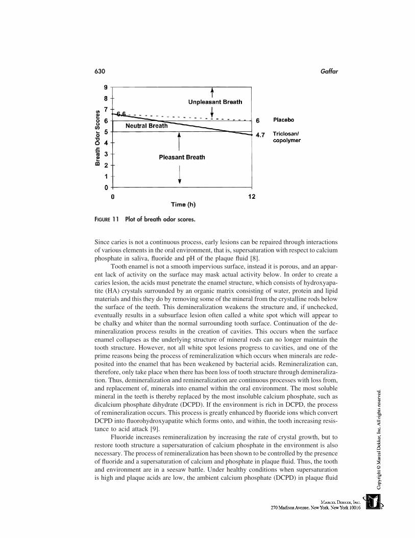
630 Gaffar
FIGURE 11 Plot of breath odor scores.
Since caries is not a continuous process, early lesions can be repaired through interactionsof various elements in the oral environment, that is, supersaturation with respect to calciumphosphate in saliva, fluoride and pH of the plaque fluid [8].
Tooth enamel is not a smooth impervious surface, instead it is porous, and an appar-ent lack of activity on the surface may mask actual activity below. In order to create acaries lesion, the acids must penetrate the enamel structure, which consists of hydroxyapa-tite (HA) crystals surrounded by an organic matrix consisting of water, protein and lipidmaterials and this they do by removing some of the mineral from the crystalline rods belowthe surface of the teeth. This demineralization weakens the structure and, if unchecked,eventually results in a subsurface lesion often called a white spot which will appear tobe chalky and whiter than the normal surrounding tooth surface. Continuation of the de-mineralization process results in the creation of cavities. This occurs when the surfaceenamel collapses as the underlying structure of mineral rods can no longer maintain thetooth structure. However, not all white spot lesions progress to cavities, and one of theprime reasons being the process of remineralization which occurs when minerals are rede-posited into the enamel that has been weakened by bacterial acids. Remineralization can,therefore, only take place when there has been loss of tooth structure through demineraliza-tion. Thus, demineralization and remineralization are continuous processes with loss from,and replacement of, minerals into enamel within the oral environment. The most solublemineral in the teeth is thereby replaced by the most insoluble calcium phosphate, such asdicalcium phosphate dihydrate (DCPD). If the environment is rich in DCPD, the processof remineralization occurs. This process is greatly enhanced by fluoride ions which convertDCPD into fluorohydroxyapatite which forms onto, and within, the tooth increasing resis-tance to acid attack [9].
Fluoride increases remineralization by increasing the rate of crystal growth, but torestore tooth structure a supersaturation of calcium phosphate in the environment is alsonecessary. The process of remineralization has been shown to be controlled by the presenceof fluoride and a supersaturation of calcium and phosphate in plaque fluid. Thus, the toothand environment are in a seesaw battle. Under healthy conditions when supersaturationis high and plaque acids are low, the ambient calcium phosphate (DCPD) in plaque fluid
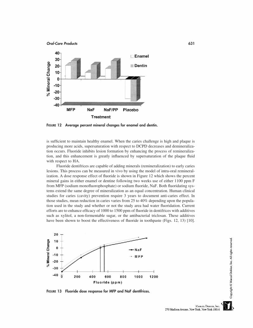
Oral-Care Products 631
FIGURE 12 Average percent mineral changes for enamel and dentin.
is sufficient to maintain healthy enamel. When the caries challenge is high and plaque isproducing more acids, supersaturation with respect to DCPD decreases and demineraliza-tion occurs. Fluoride inhibits lesion formation by enhancing the process of remineraliza-tion, and this enhancement is greatly influenced by supersaturation of the plaque fluidwith respect to HA.
Fluoride dentifrices are capable of adding minerals (remineralization) to early carieslesions. This process can be measured in vivo by using the model of intra-oral remineral-ization. A dose response effect of fluoride is shown in Figure 12 which shows the percentmineral gains in either enamel or dentine following two weeks use of either 1100 ppm Ffrom MFP (sodium monofluorophosphate) or sodium fluoride, NaF. Both fluoridating sys-tems extend the same degree of mineralization as an equal concentration. Human clinicalstudies for caries (cavity) prevention require 3 years to document anti-caries effect. Inthose studies, mean reduction in caries varies from 25 to 40% depending upon the popula-tion used in the study and whether or not the study area had water fluoridation. Currentefforts are to enhance efficacy of 1000 to 1500 ppm of fluoride in dentifrices with additivessuch as xylitol, a non-fermentable sugar, or the antibacterial triclosan. These additiveshave been shown to boost the effectiveness of fluoride in toothpaste (Figs. 12, 13) [10].
FIGURE 13 Fluoride dose response for MFP and NaF dentifrices.
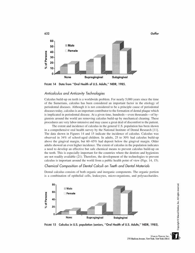
632 Gaffar
FIGURE 14 Data from ‘‘Oral Health of U.S. Adults,’’ NIDR, 1985.
Anticalculus and Anticavity Technologies
Calculus build-up on teeth is a worldwide problem. For nearly 5,000 years since the timeof the Sumerians, calculus has been considered an important factor in the etiology ofperiodontal diseases. Although it is not considered to be a principle cause of periodontaldiseases today, calculus is an important contributor to the formation of dental plaque whichis implicated in periodontal disease. At a given time, hundreds—even thousands—of hy-gienists around the world are removing calculus build-up by mechanical cleaning. Theseprocedures are very labor-intensive and may cause a great deal of discomfort to the patient.
The extent and incidence of calculus in the general U.S. population has been shownin a comprehensive oral health survey by the National Institute of Dental Research [11].The data shown in Figures 14 and 15 indicate the incidence of calculus. Calculus wasobserved in 34% of school-aged children. In adults, 25 to 30% had calculus build-upabove the gingival margin, but 60–65% had deposit below the gingival margin. Olderadults showed an even higher incidence. The extent of calculus in the population indicatesa need to develop an effective but safe chemical means to prevent calculus build-up onthe teeth. This is especially important for the countries where the dentists and hygienistsare not readily available (21). Therefore, the development of the technologies to preventcalculus is important around the world from a public health point of view (Figs. 14, 15).
Chemical Composition of Dental Calculi on Teeth and Dental Materials
Dental calculus consists of both organic and inorganic components. The organic portionis a combination of epithelial cells, leukocytes, micro-organisms, and polysaccharides.
FIGURE 15 Calculus in U.S. population (seniors, ‘‘Oral Health of U.S. Adults,’’ NIDR, 1985).
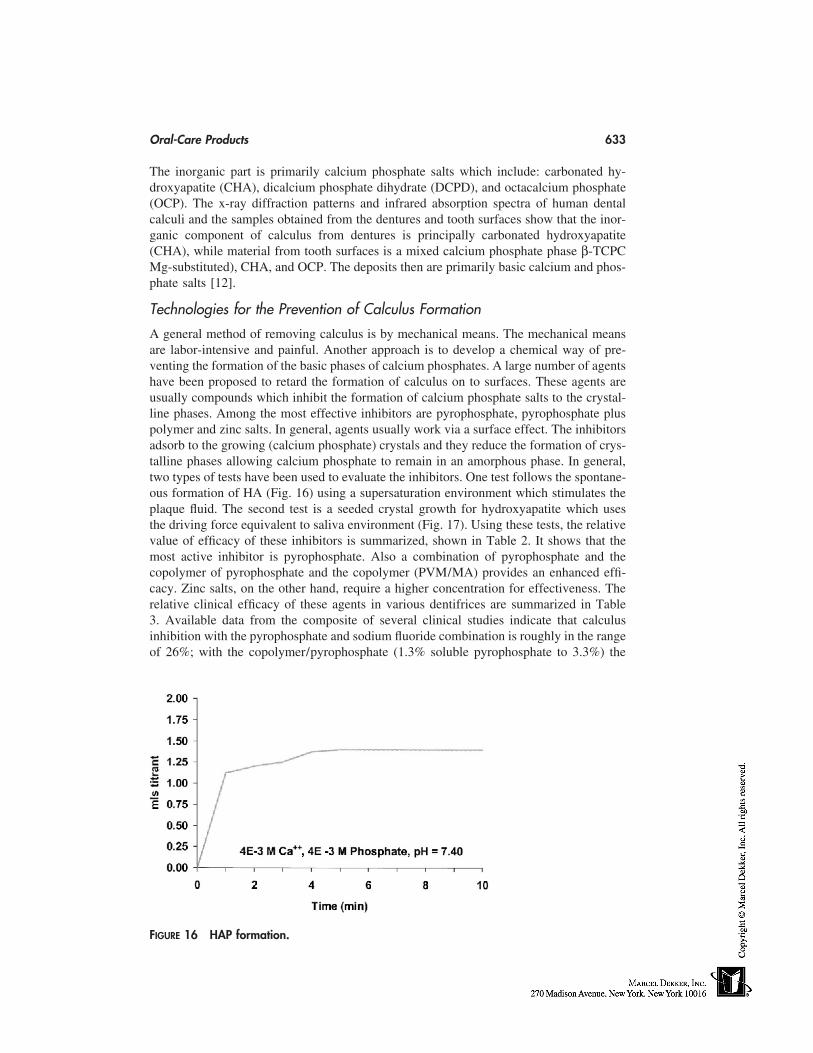
Oral-Care Products 633
The inorganic part is primarily calcium phosphate salts which include: carbonated hy-droxyapatite (CHA), dicalcium phosphate dihydrate (DCPD), and octacalcium phosphate(OCP). The x-ray diffraction patterns and infrared absorption spectra of human dentalcalculi and the samples obtained from the dentures and tooth surfaces show that the inor-ganic component of calculus from dentures is principally carbonated hydroxyapatite(CHA), while material from tooth surfaces is a mixed calcium phosphate phase β-TCPCMg-substituted), CHA, and OCP. The deposits then are primarily basic calcium and phos-phate salts [12].
Technologies for the Prevention of Calculus Formation
A general method of removing calculus is by mechanical means. The mechanical meansare labor-intensive and painful. Another approach is to develop a chemical way of pre-venting the formation of the basic phases of calcium phosphates. A large number of agentshave been proposed to retard the formation of calculus on to surfaces. These agents areusually compounds which inhibit the formation of calcium phosphate salts to the crystal-line phases. Among the most effective inhibitors are pyrophosphate, pyrophosphate pluspolymer and zinc salts. In general, agents usually work via a surface effect. The inhibitorsadsorb to the growing (calcium phosphate) crystals and they reduce the formation of crys-talline phases allowing calcium phosphate to remain in an amorphous phase. In general,two types of tests have been used to evaluate the inhibitors. One test follows the spontane-ous formation of HA (Fig. 16) using a supersaturation environment which stimulates theplaque fluid. The second test is a seeded crystal growth for hydroxyapatite which usesthe driving force equivalent to saliva environment (Fig. 17). Using these tests, the relativevalue of efficacy of these inhibitors is summarized, shown in Table 2. It shows that themost active inhibitor is pyrophosphate. Also a combination of pyrophosphate and thecopolymer of pyrophosphate and the copolymer (PVM/MA) provides an enhanced effi-cacy. Zinc salts, on the other hand, require a higher concentration for effectiveness. Therelative clinical efficacy of these agents in various dentifrices are summarized in Table3. Available data from the composite of several clinical studies indicate that calculusinhibition with the pyrophosphate and sodium fluoride combination is roughly in the rangeof 26%; with the copolymer/pyrophosphate (1.3% soluble pyrophosphate to 3.3%) the
FIGURE 16 HAP formation.
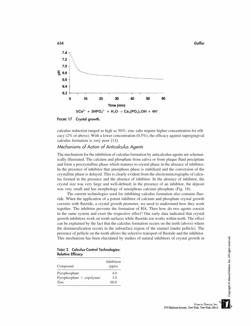
634 Gaffar
5Ca2� � 3HPO42� � H2O → Ca5(PO4)3OH � 4H�
FIGURE 17 Crystal growth.
calculus reduction ranged as high as 50%; zinc salts require higher concentration for effi-cacy (2% or above). With a lower concentration (0.5%), the efficacy against supergingivalcalculus formation is very poor [13].
Mechanisms of Action of Anticalculus Agents
The mechanism for the inhibition of calculus formation by anticalculus agents are schemat-ically illustrated. The calcium and phosphate from saliva or from plaque fluid precipitateand form a precrystalline phase which matures to crystal phase in the absence of inhibitor.In the presence of inhibitor that amorphous phase is stabilized and the conversion of thecrystalline phase is delayed. This is clearly evident from the electronmicrographs of calcu-lus formed in the presence and the absence of inhibitor. In the absence of inhibitor, thecrystal size was very large and well-defined; in the presence of an inhibitor, the depositwas very small and has morphology of amorphous calcium phosphate (Fig. 18).
The current technologies used for inhibiting calculus formation also contains fluo-ride. When the application of a potent inhibitor of calcium and phosphate crystal growthcoexists with fluoride, a crystal growth promoter, we need to understand how they worktogether. The inhibitor prevents the formation of HA. Then how do two agents coexistin the same system and exert the respective effect? Our early data indicated that crystalgrowth inhibitors work on tooth surfaces while fluoride ion works within teeth. The effectcan be explained by the fact that the calculus formation occurs on the teeth (above) wherethe demineralization occurs in the subsurface region of the enamel (under pellicle). Thepresence of pellicle on the tooth allows the selective transport of fluoride and the inhibitor.This mechanism has been elucidated by studies of natural inhibitors of crystal growth in
TABLE 2 Calculus-Control Technologies:Relative Efficacy
InhibitionCompound (ppm)
Pyrophosphate 4.0Pyrophosphate � copolymer 3.0Zinc 60.0
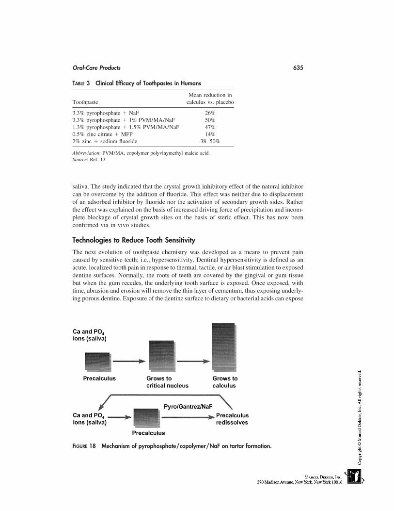
Oral-Care Products 635
TABLE 3 Clinical Efficacy of Toothpastes in Humans
Mean reduction inToothpaste calculus vs. placebo
3.3% pyrophosphate � NaF 26%3.3% pyrophosphate � 1% PVM/MA/NaF 50%1.3% pyrophosphate � 1.5% PVM/MA/NaF 47%0.5% zinc citrate � MFP 14%2% zinc � sodium fluoride 38–50%
Abbreviation: PVM/MA, copolymer polyvinymethyl maleic acid.Source: Ref. 13.
saliva. The study indicated that the crystal growth inhibitory effect of the natural inhibitorcan be overcome by the addition of fluoride. This effect was neither due to displacementof an adsorbed inhibitor by fluoride nor the activation of secondary growth sides. Ratherthe effect was explained on the basis of increased driving force of precipitation and incom-plete blockage of crystal growth sites on the basis of steric effect. This has now beenconfirmed via in vivo studies.
Technologies to Reduce Tooth Sensitivity
The next evolution of toothpaste chemistry was developed as a means to prevent paincaused by sensitive teeth; i.e., hypersensitivity. Dentinal hypersensitivity is defined as anacute, localized tooth pain in response to thermal, tactile, or air blast stimulation to exposeddentine surfaces. Normally, the roots of teeth are covered by the gingival or gum tissuebut when the gum recedes, the underlying tooth surface is exposed. Once exposed, withtime, abrasion and erosion will remove the thin layer of cementum, thus exposing underly-ing porous dentine. Exposure of the dentine surface to dietary or bacterial acids can expose
FIGURE 18 Mechanism of pyrophosphate/copolymer/NaF on tartar formation.
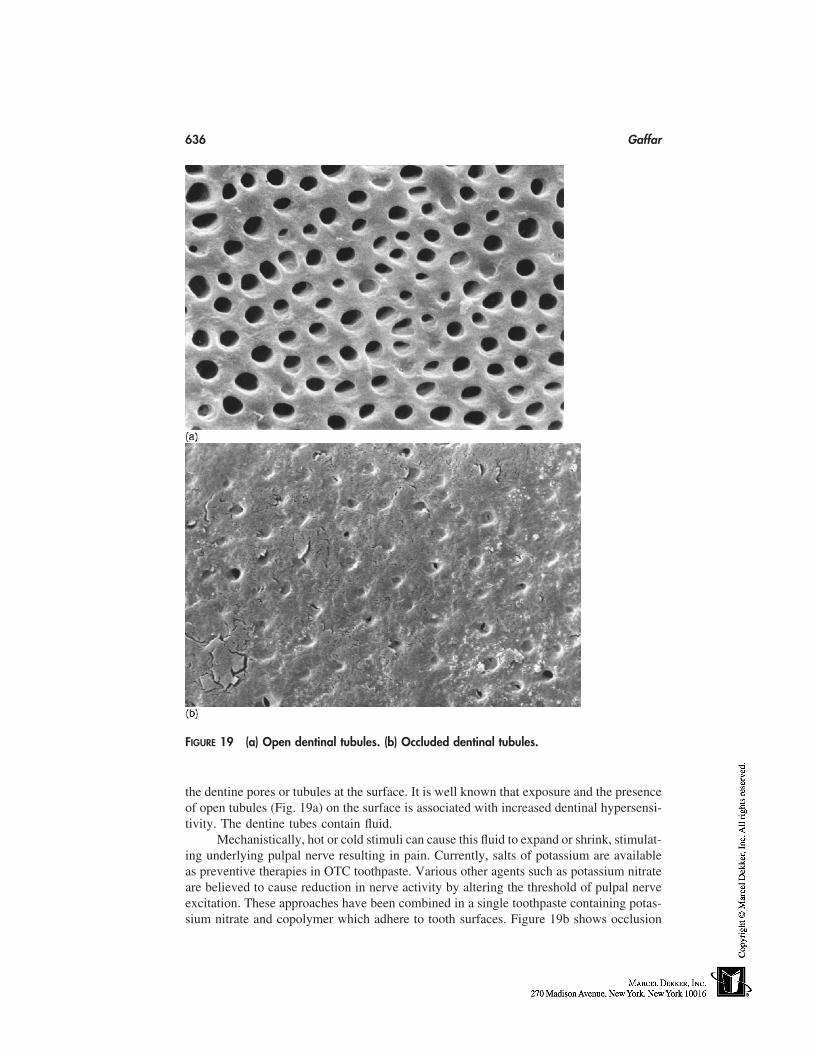
636 Gaffar
FIGURE 19 (a) Open dentinal tubules. (b) Occluded dentinal tubules.
the dentine pores or tubules at the surface. It is well known that exposure and the presenceof open tubules (Fig. 19a) on the surface is associated with increased dentinal hypersensi-tivity. The dentine tubes contain fluid.
Mechanistically, hot or cold stimuli can cause this fluid to expand or shrink, stimulat-ing underlying pulpal nerve resulting in pain. Currently, salts of potassium are availableas preventive therapies in OTC toothpaste. Various other agents such as potassium nitrateare believed to cause reduction in nerve activity by altering the threshold of pulpal nerveexcitation. These approaches have been combined in a single toothpaste containing potas-sium nitrate and copolymer which adhere to tooth surfaces. Figure 19b shows occlusion
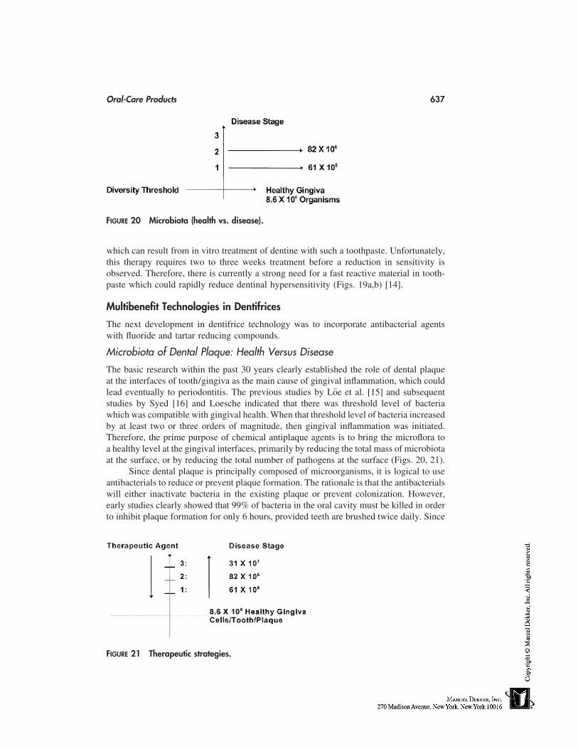
Oral-Care Products 637
FIGURE 20 Microbiota (health vs. disease).
which can result from in vitro treatment of dentine with such a toothpaste. Unfortunately,this therapy requires two to three weeks treatment before a reduction in sensitivity isobserved. Therefore, there is currently a strong need for a fast reactive material in tooth-paste which could rapidly reduce dentinal hypersensitivity (Figs. 19a,b) [14].
Multibenefit Technologies in Dentifrices
The next development in dentifrice technology was to incorporate antibacterial agentswith fluoride and tartar reducing compounds.
Microbiota of Dental Plaque: Health Versus Disease
The basic research within the past 30 years clearly established the role of dental plaqueat the interfaces of tooth/gingiva as the main cause of gingival inflammation, which couldlead eventually to periodontitis. The previous studies by Löe et al. [15] and subsequentstudies by Syed [16] and Loesche indicated that there was threshold level of bacteriawhich was compatible with gingival health. When that threshold level of bacteria increasedby at least two or three orders of magnitude, then gingival inflammation was initiated.Therefore, the prime purpose of chemical antiplaque agents is to bring the microflora toa healthy level at the gingival interfaces, primarily by reducing the total mass of microbiotaat the surface, or by reducing the total number of pathogens at the surface (Figs. 20, 21).
Since dental plaque is principally composed of microorganisms, it is logical to useantibacterials to reduce or prevent plaque formation. The rationale is that the antibacterialswill either inactivate bacteria in the existing plaque or prevent colonization. However,early studies clearly showed that 99% of bacteria in the oral cavity must be killed in orderto inhibit plaque formation for only 6 hours, provided teeth are brushed twice daily. Since
FIGURE 21 Therapeutic strategies.
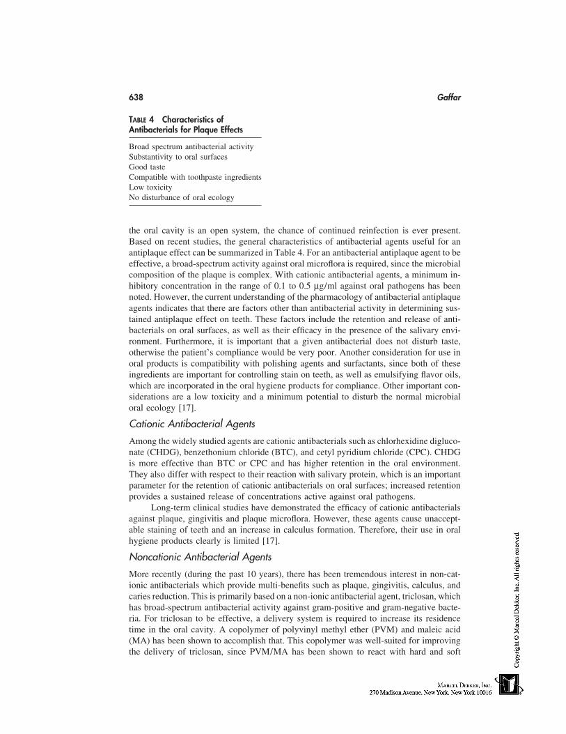
638 Gaffar
TABLE 4 Characteristics ofAntibacterials for Plaque Effects
Broad spectrum antibacterial activitySubstantivity to oral surfacesGood tasteCompatible with toothpaste ingredientsLow toxicityNo disturbance of oral ecology
the oral cavity is an open system, the chance of continued reinfection is ever present.Based on recent studies, the general characteristics of antibacterial agents useful for anantiplaque effect can be summarized in Table 4. For an antibacterial antiplaque agent to beeffective, a broad-spectrum activity against oral microflora is required, since the microbialcomposition of the plaque is complex. With cationic antibacterial agents, a minimum in-hibitory concentration in the range of 0.1 to 0.5 µg/ml against oral pathogens has beennoted. However, the current understanding of the pharmacology of antibacterial antiplaqueagents indicates that there are factors other than antibacterial activity in determining sus-tained antiplaque effect on teeth. These factors include the retention and release of anti-bacterials on oral surfaces, as well as their efficacy in the presence of the salivary envi-ronment. Furthermore, it is important that a given antibacterial does not disturb taste,otherwise the patient’s compliance would be very poor. Another consideration for use inoral products is compatibility with polishing agents and surfactants, since both of theseingredients are important for controlling stain on teeth, as well as emulsifying flavor oils,which are incorporated in the oral hygiene products for compliance. Other important con-siderations are a low toxicity and a minimum potential to disturb the normal microbialoral ecology [17].
Cationic Antibacterial Agents
Among the widely studied agents are cationic antibacterials such as chlorhexidine digluco-nate (CHDG), benzethonium chloride (BTC), and cetyl pyridium chloride (CPC). CHDGis more effective than BTC or CPC and has higher retention in the oral environment.They also differ with respect to their reaction with salivary protein, which is an importantparameter for the retention of cationic antibacterials on oral surfaces; increased retentionprovides a sustained release of concentrations active against oral pathogens.
Long-term clinical studies have demonstrated the efficacy of cationic antibacterialsagainst plaque, gingivitis and plaque microflora. However, these agents cause unaccept-able staining of teeth and an increase in calculus formation. Therefore, their use in oralhygiene products clearly is limited [17].
Noncationic Antibacterial Agents
More recently (during the past 10 years), there has been tremendous interest in non-cat-ionic antibacterials which provide multi-benefits such as plaque, gingivitis, calculus, andcaries reduction. This is primarily based on a non-ionic antibacterial agent, triclosan, whichhas broad-spectrum antibacterial activity against gram-positive and gram-negative bacte-ria. For triclosan to be effective, a delivery system is required to increase its residencetime in the oral cavity. A copolymer of polyvinyl methyl ether (PVM) and maleic acid(MA) has been shown to accomplish that. This copolymer was well-suited for improvingthe delivery of triclosan, since PVM/MA has been shown to react with hard and soft
Oral-Care Products 639
TABLE 5 Noncationic Antibacterials: ComparativeStudy for In Vivo Plaque Inhibition
Mean P on all SNKTreatment surfaces � SD group
Placebo 1.46 � 0.12 A0.12% CHDG 0.53 � 0.17 B0.2% SnF2 (rinse) 1.10 � 0.16 C0.06 Triclosan 1.00 � 0.14 C0.06 Triclosan � Gantrez 0.72 � 0.17 B0.06 Triclosan � PVPA 0.67 � 0.16 B
Abbreviations: Gantrez, PVM/MA, polyvinyl methyl/maleicacid; PVPA, polyvinylphosphonic acid; SNK, Student NeumanKeuls test; P, plaque index.
surfaces in the oral cavity. In a four-day short-term study of de novo plaque formation,we evaluated a series of different antibacterial agents. We found that triclosan actuallyneeds an improved delivery system, primarily a copolymer, to enhance its retention toboth tooth and oral epithelial surfaces [18].
One of the important principles developed is that retention per se is not the onlyfactor in antiplaque activity; the retained concentration has to be active biologically. Todemonstrate this principle, we conducted a series of studies to understand how muchtriclosan was retained post-brushing. In one of the studies, we compared three triclosanformulations, each having a different enhancing system (Table 5). As can be seen in Table6, even after 14 hours, a significant amount is retained in plaque, a concentration abovethe MIC’s of triclosan for oral bacteria (MIC being 0.3-4 µg/mL). The next importantstep was to determine whether this retained amount was active biologically. A plaqueviability assay was used, in which we exposed the plaque to two fluorescent dyes to dis-criminate between live and dead bacteria by measuring the ratio of green to red fluores-cence. In this study, one could quantitatively measure the ratio and ascertain whether theretained amount was active biologically. In one of the typical studies shown here, brushingwith the placebo toothpaste gave some reduction of plaque viability; the triclosan copoly-mer system gave the highest reduction in viability, and the other systems, such as triclosan/pyrophosphate and triclosan/zinc citrate, were not significantly different from the placebo(Fig. 22). These results have been corroborated by an independent six-month clinical studyby Renvert and Birkhed (Table 6) [19].
The mechanism by which the copolymer enhances the delivery of triclosan has beenelucidated (Fig. 23). The polymer has two groups: one is the attachment group and theother is the solubilizing group. The solubilizing group retains triclosan in surfactant mi-
TABLE 6 Plaque Triclosan Levels After Brushing (µg/mL)
0.3% Triclosan/ 0.3% Triclosan/ 0.3% Triclosan/After copolymer pyrophosphate 1% zincbrushing n � 12 n � 12 n � 12
2 h 38.83 � 18.28* 20.90 � 14.14 30.60 � 13.614 h 4.14 � 1.72 2.74 � 2.11 3.95 � 1.79
* P � 0.05, compared with a placebo toothpaste.
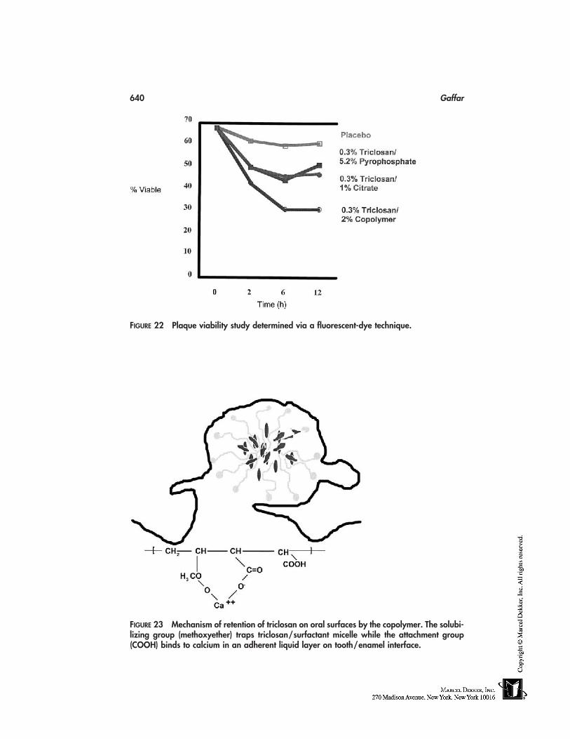
640 Gaffar
FIGURE 22 Plaque viability study determined via a fluorescent-dye technique.
FIGURE 23 Mechanism of retention of triclosan on oral surfaces by the copolymer. The solubi-lizing group (methoxyether) traps triclosan/surfactant micelle while the attachment group(COOH) binds to calcium in an adherent liquid layer on tooth/enamel interface.
Oral-Care Products 641
TABLE 7 Therapeutic Mouthrinses
Typical reduction in the diseasesMouthrinse Active agents vs. placebo
Fluoride rinses 225 ppm F 50% reduction in caries, children(3 years)
Tartar � calculus 1% pyrophosphate amion; 100 30–35% reduction in tartar forma-ppm F plus a copolymer of tion after 6 months usePVM/MA (0.5%)
Antiplaque/antigingivitis 0.03 to 0.06 Triclosan � 1% 20–30% reduction in plaque/gingi-copolymer PVM/MA � vitis after 3 months of usefluoride
celles, and the attachment group reacts with the oral surfaces via calcium in the liquidadherent layer. Triclosan is then slowly released via interactions with salivary environ-ment. In terms of long-term clinical trials, this technology has now been evaluated aroundthe world in 12 six-month plaque/gingivitis studies, three calculus studies, three cariesclinical trials, and five long-term studies monitoring the oral microbial population. Theresults of all these studies indicated that this technology was effective against plaque,gingivitis, calculus, and caries. No side effects of staining or calculus increase were seen.There was also no disturbance of the oral microbial ecology.
One of the most exciting aspects of triclosan is its ‘‘double-barrel’’ effect. Thisunique antibacterial not only kills bacteria, but also neutralizes the products of bacteriawhich could provoke inflammation. We have shown that triclosan was a potent inhibitorof both cyclo-oxygenase and lipoxygenase pathways. It not only inhibited these enzymesin vitro but also inhibited the release of their products (prostaglandins and leukotrienes)in gingival fibroblasts which were stimulated by interleukin 1-β. These data were clinicallyconfirmed in a study in which we blocked the antibacterial effect of triclosan but main-tained its anti-inflammatory effect. Thus, triclosan has a ‘‘double barrel’’ effect—bothantibacterial and anti-inflammatory. This unique feature is not provided so far by otherantibacterial, anti-plaque agents [20].
MOUTHWASHING
Mouth rinses currently on the markets are aqueous-based formulation where the therapeu-tic agents are at lower concentrations than toothpaste. For example, the general populationuses toothpaste at 1 g or 1 mL on the brush, but the rinses are used in 10 to 15 mL andsome lower concentration of the actives are incorporated. Also, the rinses do not containpolishing agents or thickeners. A typical therapeutic rinse contains surfactants, flavor,active agent, and water. The general principles of active agent delivery which were out-lined above also apply for the active agent delivery in the mouthwash. Table 7 summarizesthe typical clinical performance vs. a placebo rinse of therapeutic rinse (Table 7).
STRATEGY FOR CLINICAL STUDIES IN ORAL-CARE PRODUCTS
Preclinical Evaluation → Pilot Studies → Controlled Studies → Field Trials
642 Gaffar
To document the effectiveness of oral products against dental diseases, the strategy forclinical studies is outlined in the above chart. The preclinical studies include laboratoryand animal tests. For example, for fluoride efficacy a test would include fluoride uptakein teeth or hydroxyapatite, reduction in enamel solubility following fluoride treatment,followed by an acid challenge. The effectiveness in rats include the effects of topicalapplication of fluoride solution in reducing caries. The pilot studies in humans are doneto assess the effectiveness of fluoride to promote mineral deposition or prevent dissolutionof artificially created lesions in enamel slabs implanted in partial dentures. Such studiesare of 2 to 4 weeks duration and conducted in 20 to 30 subjects per group. If the pilotstudy significantly enhance remineralization of artificial lesions, control studies in 30 to60 subjects for 3 to 6 months are conducted with the final formula for efficacy. The parame-ters could include promotion of mineralization, regression of early cavity lesions, andfluoride uptake in dental plaque and saliva. The field trials are conducted in children (1000per group) for a period of 3 years to assess the effects on cavity development. For preven-tion of plaque and gingivitis formation, such trials are conducted for 6 months. The calcu-lus reduction field trials are also conducted for a period of 6 months. Such field trials areof parallel/double-blind design.
FUTURE TRENDS
The global needs for prevention of dental diseases can be met by the development ofknowledge in academia and industry and its subsequent applications. With a better under-standing of processes occurring in the mouth, we will be able to design better actives andactive agent delivery systems for the control of oral diseases. The technological trendsare leading toward the goal.
REFERENCES
1. Dental Spending Hits $37 Billion. Am Dent Assoc News, Jan. 6, 1992.2. Periodontal aspects of systemic health. Symposium proceedings, WD Cohen, ed. Compendium
Continuing Education, Fall, 1998.3. Gaffar A, Afflitto J. General principles for delivery of active agents for mouthrinses. Int Dent
J 1996; 42(4):251–256.4. Fishman SL. Hare’s teeth to fluorides, historical aspects of dentifrice use. In: Emery G, Rölla
G, eds. Clinical and Biological Aspects of Dentifrice. Oxford: Oxford University Press, 1972:1–7.
5. Nathoo SA, Gaffar A. Studies on dental stain induced by antibacterial agent and rational ap-proaches for bleaching dental stains. Adv Dent Res 1995; 9(4):462–470.
6. Solis-Gaffar M, Niles HP. Instrumental evaluation of mouth odor in a human clinical study.J Dent Res 1977; 54:851–857.
7. Niles HP, Gaffar A. Relationship between sensory and instrumental evaluation of mouth odor.J Soc Cos Chem 1993; 44:101–107.
8. Gaffar A, Blake-Haskins J, Mellberg J. In vivo studies with dicalcium phosphate dihydrate/MFP system for caries prevention. Int Dent J 1993; 63(1):81–90.
9. Sullivan RJ, Fletcher R, Barchman R, Legeros RZ. Intra-oral comparison and evaluation of theability of dentifrices to promote remineralization of caries-like lesions in dentin and enamel. JClin Dent 1995; 6:135–138.
10. Gaffar A, Blake-Haskins J, Sullivan RJ, Simone A, Saunders F. Cariostatic effects of a xylitol/NaF dentifrice in vivo. Int Dent J 1998; 48:32–39.
Oral-Care Products 643
11. Mandel ID. Rinses for the control of supragingival calculus formation. Int Dent J 1992; 42:270–285.
12. Gaffar A, Legeros RZ, Gambogi RS, Afflitto J. Inhibition of formation of calcium phosphatedeposits on teeth and dental materials: recent advances. Adv Dent Res 1995; 9(4):419–426.
13. Volpe AR, Petrone M, Davies RM. A review of calculus clinical efficacy studies. J Clin Den-tistry 1993; 4:71–81.
14. Miller S, Gaffar A, Sullivan RJ, Troung T, Stranick M. Evaluation of new dentifrice for treat-ment of sensitive teeth. J Clin Dent 1994; 5:71–79.
15. Löe H, Theilde E, Jensen SB. Experimental gingivitis in man. J Periodont 1970; 36:177–187.16. Syed SA, Loesche WJ. Bacteriology of experimental gingivitis: effect of plaque. Info Immun
1978; 21:821–829.17. Gaffar A, Volpe AR, Lindhe J. Recent advances in plaque/gingivitis control. In: Emery G,
Rölla G, eds. Chemical Biological Aspects of Dentifrices. Oxford: Oxford University Press,1992: 229–247.
18. Gaffar A, Afflitto J, Herles S, Nabi N. Recent advances in plaque, gingivitis, tartar and cariesprevention. Int Dent J 1994; 44:63–70.
19. Renvert St, Birkhed D. Comparison of three triclosan dentifrices on plaque, gingivitis andsalivary microflora. J Clin Periodont 1995; 23:63–70.
20. Gaffar A, Scherl D, Afflitto J, Coleman EJ. The effects of triclosan on the mediators of gingivalinflammation. J Clin Periodont 1995; 22:280–284.
21. Mandel ID. Chemotherapeutic mouthrinses for control of oral disease. Int Dent J 1992; 42(4):251–285.

54
Decorative Products
Mitchell L. SchlossmanKobo Products, Inc., South Plainfield, New Jersey
INTRODUCTION
Decorative cosmetics are principally concerned with beautifying and decoration ratherthan functionality. No discussion of decorative products can be complete without a fullunderstanding of the importance of color, a prime component of every decorative cosmetic.Conventional pigments create color by absorption of certain wavelengths of incident light.The color perceived corresponds to that of the wavelengths reflected. Formulation of deco-rative cosmetics has been an exciting challenge for cosmetic chemists. Before formulatingany color cosmetic product, one must check the current regulations in the country wherethe proposed product will be sold to make sure all the colors conform to those regulations.The following is a practical guide for the formulator and covers a maximum of technicaland regulatory issues in an easy-to-use format.
COLOR
Color-Additive Regulation
In the past, colorants had been used in cosmetics without any consideration being givento their possible toxicity. Today, all countries have regulations that control the type andpurity of colors that may be used in cosmetics.
United States: U.S. Food and Drug Administration (FDA)
21 CFR 73, 74; Positive List [1]: Colors listed for general cosmetic use, including eyearea only if stated specifically, or external only, meaning no contact with mucous mem-branes. Hair dyes and true soaps are exempt.
European Union (EU): European Commission (EC)
Directive 76/786, Annex IV [2]; Positive List: Colors listed for ingested use, general,including eye area, external, or rinse-off.Annex II: Negative List
Japan: Ministry of Health and Welfare (MHW)
MHW Ordinance No. 30 [3]; Positive List, Coal-Tar Colors: Premarket approval by MHWfor all other cosmetic ingredients, including inorganic and natural colorants.
645
646 Schlossman
Color-Additives Definitions
Primary/Straight Color: A color that is pure, containing no extenders or dilutents.Dye: A color that is soluble in the medium in which it is dispersed. (e.g., FD&C
Blue #1).Pigment: A color that is insoluble in the medium in which it is dispersed. (e.g.,
FD&C Blue #1 Al lake, Black iron oxide).Lake: A water-insoluble pigment composed of a water-soluble straight color
strongly absorbed onto an insoluble substratum through the use of a precipitant(e.g., FD&C Blue #1 Al Lake). Generally, 10 to 40% color.*
Toner: A pigment that is produced by precipitating a water-soluble dye as an insolu-ble metal salt (e.g., D&C Red #6 barium salt, D&C Red #7 calcium salt).
True Pigment: A pigment that, based on its chemistry, precipitates as it is formed(e.g., D&C Red #36).
Extender: A pigment diluted on substrate1. during manufacture by precipitation, or2. postmanufacture by intimate milling or mixing.
U.S. Regulations
21 CFR Part 73 [4]: Listing of Color Additives Exempt fromCertification
Inorganic pigments, powdered metals, and naturally derived colorants approved for food,drug, and/or cosmetic use. Listed permitted uses are as follows:
• Food• Ingested/externally applied drugs• General cosmetic• Eye area only if mentioned• External (no mucous membrane), i.e., ultramarines, ferric ammonium ferrocya-
nide not permitted in lip or bath products
21 CFR Part 74 [5]: Listing of Color Additives Subject to Certification
Synthetic organic dyes and pigments. Each batch must be submitted by the manufacturerto the FDA for certification that specifications are met. Permitted uses as in Part 73.
Four certified organic dyes and their lakes are now permitted for eye-area use:
1. FD&C Blue #12. FD&C Red #403. FD&C Yellow #54. D&C Green #5
21 CFR Part 82 [6]: Listing of Certified Provisionally Listed Colors
Lakes:
FD&C: Aluminum or calcium salt on alumina.
* FDA has considered any certified colorant mixed with a diluent to be a lake; e.g., D&C Red 30 Plus talc, andD&C Red #7 CA lake on calcium carbonate.
Decorative Products 647
D&C: Sodium, potassium, barium, calcium, strontium, or zirconium salt on alu-mina, blanc fixe, gloss white, clay, titanium dioxide, zinc oxide, talc, rosin, alumi-num benzoate, calcium carbonate.
A salt prepared from straight color, i.e., D&C Red #6, by combining the color with abasic radical.
Proposed Permanent Listing of Color Additive Lakes (FR Vol. 61 #43),March 4, 1996 [7]
• List substrate, e.g., D&C Red #27 aluminum lake on alumina• Extenders of insoluble straight colors will no longer be called lakes, e.g., D&C
Red #30• Permit blends of previously certified straight colors in a lake, e.g., FD&C Blue
#1 and Yellow #5 aluminum lake• All lakes to be prepared from previously certified batches of straight color would
necessitate process changes for D&C Reds #6, #7, and #34• Abbreviations permitted for cosmetic ingredient labeling, omitting FD&C, pre-
cipitate and substrate designation e.g., Blue 1
European Community
Directive 76/786, as amended [8].
Annex IV
This is a list of coloring agents allowed in cosmetic products.List by color index number:
Part 1: Permanently listedPart 2: Provisionally listed
Four fields of application:
1. All cosmetic products2. All cosmetic products, except those intended to be applied in the vicinity of the
eyes, in particular eye makeup and makeup remover3. Allowed exclusively in cosmetic products intended not to come into contact
with mucous membranes (including the eye area)4. Allowed exclusively in cosmetic products intended to come into contact only
briefly with skin (not permitted in nail preparations)
Lakes and Salts
If a color index number is listed in Annex IV, then the pure color plus its salts and lakesare allowed, unless prohibited under Annex II (the list substances that cosmetics may notcontain). Exceptions include barium, strontium, and zirconium.
Prohibited under Annex II, but where Footnote 3 appears in Annex IV, ‘‘the insolu-ble barium, strontium, and zirconium lakes, salts, and pigments . . . shall also be permit-ted.’’ They must pass the test for insolubility which will be determined by the procedurein Article 8. (Insoluble in 0.1 N HCl).
648 Schlossman
Purity Criteria
Only colors designated by an ‘‘E,’’ those also permitted for food use, must meet thegeneral specification for food colors.
�5 ppm As�20 ppm Pb�100 ppm Sb, Cu, Cr, Zn, BaSO4 separately�200 ppm Of those togetherNone detectable Cd, Hg, Se, Te, Th, U Cr�6, or soluble Ba
Sixth Amendment to the directive is currently adopted. Update of purity criteria is beingconsidered, test methods may be stipulated.
Japan
MHW ordinance No. 30 (1966) as amended by MHW ordinance No. 55 (1972) [9].
Positive List
83 Coal-tar colors:
• Must be declared on cosmetic product label• Fields of application: oral, lip, eye area, external, rinse-off
Inorganic/Natural Colorants
Listing, specifications, test methods:
• Japan standards of cosmetic ingredients (JSCI)• Comprehensive licensing standards of cosmetics by category (CLS)• Japan cosmetic ingredient dictionary (CLS)
U.S. Colorants Not Permitted/Restricted in Japan
Pigments
D&C RED #6 Ba LakeD&C RED #21 Al LakeD&C RED #27 Al LakeD&C RED #33 Zr LakeD&C ORANGE #5 Al Lake
Substrates
Aluminum benzoate 0.5% maximum in lipstickRosin 7.0% maximum in lipstickCalcium carbonate Not permitted
Decorative Products 649
Inorganic Pigments
In general, inorganic colors are more opaque, more light fast, more solvent resistant, butnot as bright as organic colors. They may be affected by alkali and acid. Inorganic color-ants are formed from compounds of the transition elements. Color is produced as a resultof the ease with which the outer ‘‘d’’ electrons can absorb visible light and be promotedto the next higher energy level.
Iron OxidesGood stability, opacity Red Fe3O4
BrownBurgundy Fe2O3
Black Fe3O4
Yellow FeOOHChromium Oxide
Good stability, opacity Green Cr2O3
Chromium HydroxideGood stability, lower tinting strength Aqua Cr2O3XH2O
UltramarinesGood light stability, lower tinting strength, Blue
unstable to acid Violet Nax(AlSiO4)ySz
PinkManganese Violet
Good light stability, lower tinting strength, Violet NH4MnP2O7
unstable to waterFerric Ammonium, Ferrocyanide
Lower light stability, high tinting strength, Deep Blue FeNH4Fe(CN)6
unstable to alkali and salts, difficultdispersion
Ferric FerrocyanidePhysical/chemical stability as above, Deep Blue Fe[Fe(CN)6]3
precipitated on a substrate (i.e., mica) XH2OTitanium Dioxide
Medium light stability, good chemical White TiO2
stability, high opacity AnataseRutile
Organic Pigments
Organic pigments are characterized by:
• Transparency• Variable chemical and physical stability• ‘‘Clean,’’ bright colors
Color is produced by chromophoric groups, generally electron acceptors.
ENCNE ECCOENO2 ECCSENO
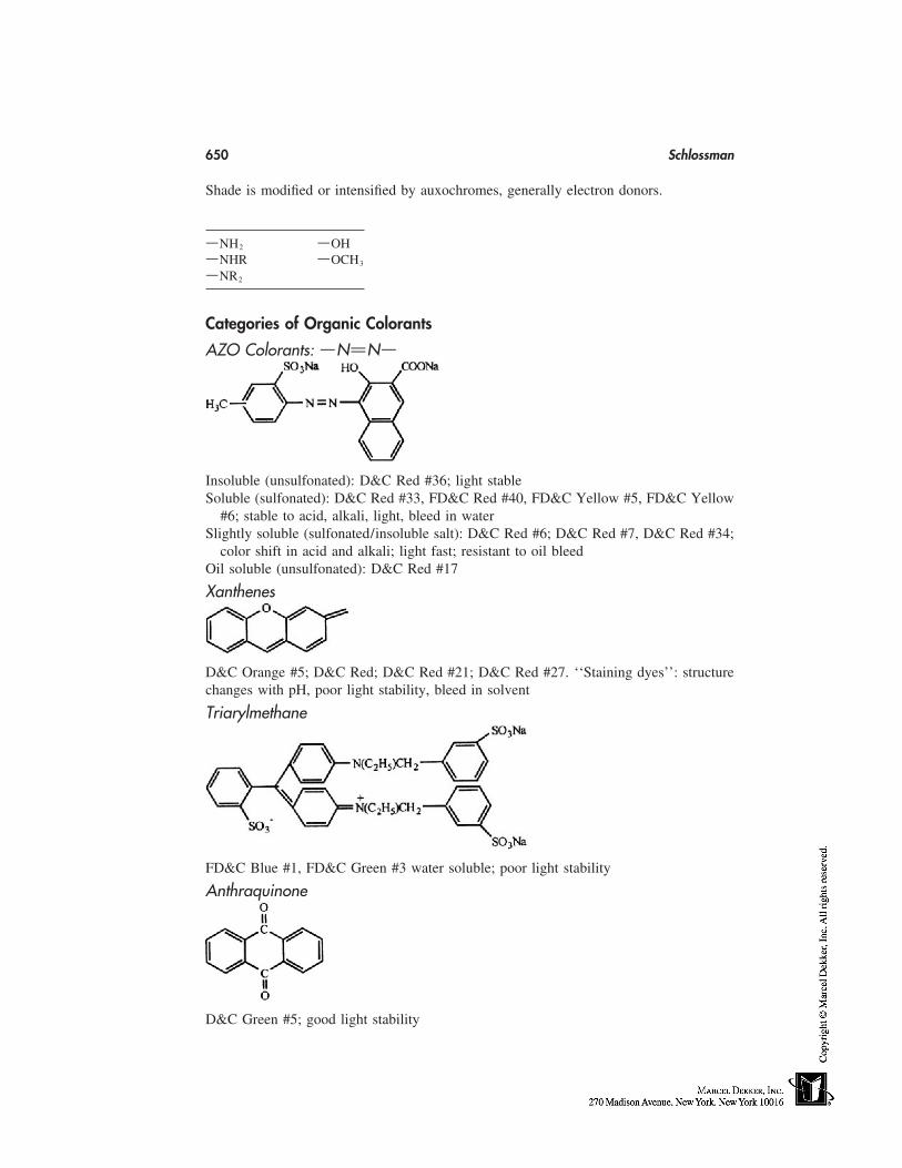
650 Schlossman
Shade is modified or intensified by auxochromes, generally electron donors.
ENH2 EOHENHR EOCH3
ENR2
Categories of Organic Colorants
AZO Colorants: ENCNE
Insoluble (unsulfonated): D&C Red #36; light stableSoluble (sulfonated): D&C Red #33, FD&C Red #40, FD&C Yellow #5, FD&C Yellow
#6; stable to acid, alkali, light, bleed in waterSlightly soluble (sulfonated/insoluble salt): D&C Red #6; D&C Red #7, D&C Red #34;
color shift in acid and alkali; light fast; resistant to oil bleedOil soluble (unsulfonated): D&C Red #17
Xanthenes
D&C Orange #5; D&C Red; D&C Red #21; D&C Red #27. ‘‘Staining dyes’’: structurechanges with pH, poor light stability, bleed in solvent
Triarylmethane
FD&C Blue #1, FD&C Green #3 water soluble; poor light stability
Anthraquinone
D&C Green #5; good light stability
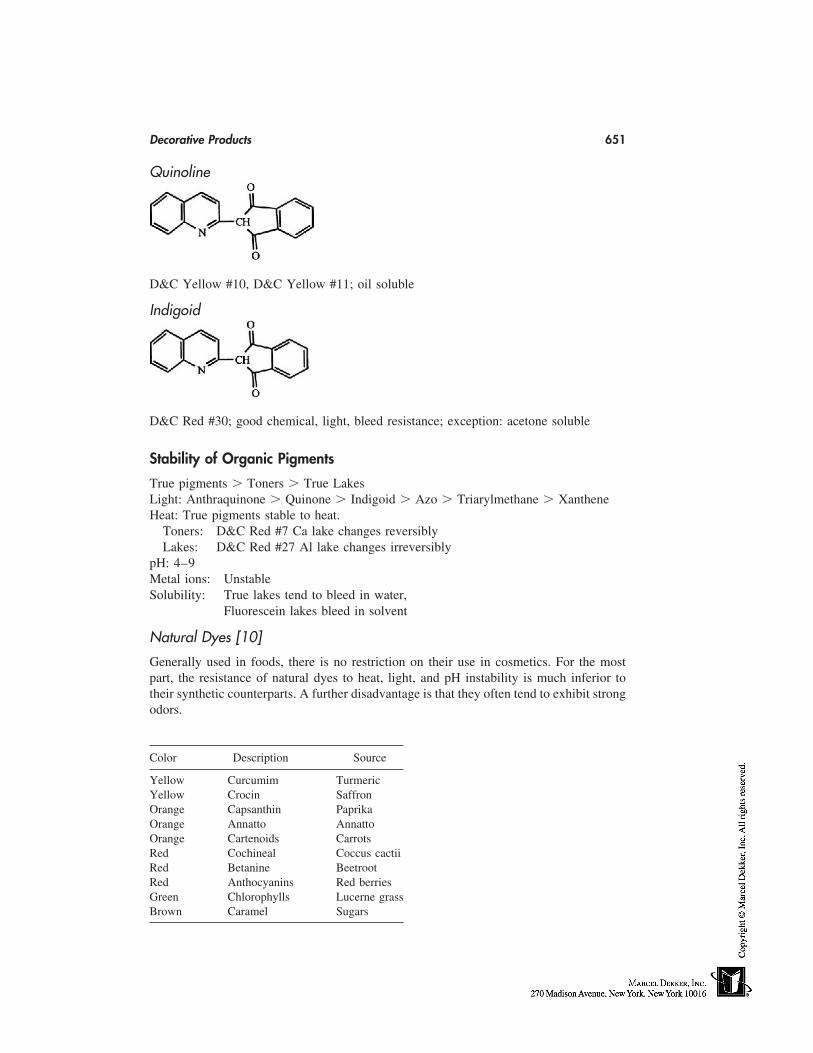
Decorative Products 651
Quinoline
D&C Yellow #10, D&C Yellow #11; oil soluble
Indigoid
D&C Red #30; good chemical, light, bleed resistance; exception: acetone soluble
Stability of Organic Pigments
True pigments � Toners � True LakesLight: Anthraquinone � Quinone � Indigoid � Azo � Triarylmethane � XantheneHeat: True pigments stable to heat.
Toners: D&C Red #7 Ca lake changes reversiblyLakes: D&C Red #27 Al lake changes irreversibly
pH: 4–9Metal ions: UnstableSolubility: True lakes tend to bleed in water,
Fluorescein lakes bleed in solvent
Natural Dyes [10]
Generally used in foods, there is no restriction on their use in cosmetics. For the mostpart, the resistance of natural dyes to heat, light, and pH instability is much inferior totheir synthetic counterparts. A further disadvantage is that they often tend to exhibit strongodors.
Color Description Source
Yellow Curcumim TurmericYellow Crocin SaffronOrange Capsanthin PaprikaOrange Annatto AnnattoOrange Cartenoids CarrotsRed Cochineal Coccus cactiiRed Betanine BeetrootRed Anthocyanins Red berriesGreen Chlorophylls Lucerne grassBrown Caramel Sugars
652 Schlossman
All of the above are of vegetable origin, with the exception of cochineal which is extractedfrom the crushed insects Coccus cactii.
Color Chemistry and Manufacture
The property of a colorant that makes it absorb more in one part of the visible spectrumthan another is its chemical constitution. Molecules, like atoms, exist in different electronicstates. Because molecules contain two or more nuclei, they also possess energies of rota-tion and vibration. This theory applies to both organic and inorganic colorants. With theinorganic colorants, colored compounds are obtained with the ions of the transition ele-ments that have atomic numbers 22 to 29.
Inorganic Pigments
Titanium Dioxide
A brilliant white pigment. Two crystal types occur: anatase and rutile. Two manufacturingprocesses are used:
1. Sulfate—either crystal may be produced.2. Chloride—only rutile crystals are formed properties. Crystals of both rutile and
anatase are tetragonal, rutile having greater hiding power because of the closerpacking of the atoms in the crystal. Refractive indices are 2.55 for anatase and2.71 for rutile. Opacity is the result of the light-scattering ability of titaniumdioxide. Light, heat and chemical stability are excellent. Additionally, in theUnited States, titanium dioxide is a Category I sunscreen.
Zinc Oxide
Zinc ore is roasted and purified at 1000°C. Two methods of manufacture are used: 1)French (indirect) and 2) American (direct).
Properties. Zinc oxide forms transparent hexagonal crystals; whiteness isattributable to the light scattering of the extremely fine particles. Refractive index is 2.0.Hiding power is less than titanium dioxide. Primary use is for antibacterial and fungicidalproperties. Heat and light stability are good. It is soluble in acid and alkali. Zinc oxidein the United States is a Category I skin protectant and a Category III sunscreen.
Iron Oxides
These are used in all types of cosmetic products. By blending black, red, and yellow incertain properties, brown, tans, umbers and sienna may be produced. Yellow iron oxideis hydrated iron II (ferrous) oxide, Fe2O3XH2O. It is produced by the controlled oxidationof ferrous sulfate. Red iron oxide is chemically Fe2O3 and is obtained by the controlledheating (at about 1000°C) of yellow iron oxide. Black iron oxide is Fe2O4 and is a mixtureof ferrous and ferric oxide and is prepared by controlled oxidation of ferrous sulfate underalkaline conditions.
Ultramarines
Theoretically they are polysulfide sodium/aluminum sulfosilicates. They range in colorfrom blue to violet, pink, and even green. A mixture is calcined at 800°C to 900°C, for4 to 5 days. Shades are determined by reaction time, formula variations, and particle size,
Decorative Products 653
whereas ultramarine violets and pinks are obtained by treating ultramarine blue with HClat 275°C, removing some sodium and sulfur from the molecule.
Manganese Violet
Chemically this is MnNH4P2O. Manufactured by heating manganese dioxide with ammo-nium dihydrogen phosphate and water. Phosphorus acid is added and the mixture is heateduntil the violet color develops.
Iron Blue
Chemically ferric ammonium ferrocyanide. Fe[Fe(Cn)6]3. Sodium ferrocyanide and fer-rous sulfate are reacted in the presence of ammonium sulfate. Pigments prepared withsodium or potassium salts are called ferric ferrocyanide.
Chromium Oxide (Cr2O3)
A dull yellow green pigment may be prepared by blending an alkali dichromate withsulfur or a carbonaceous material. Reduction to chrome (III) oxide is achieved in a kilnat 1000°C.
Chromium Hydroxide (Cr2O(OH)4)
A bright bluish green pigment prepared by the calcination of a bichromate with boric acidat 500°C. During cooling, the mass is hydrolyzed with water, yielding a hydrate.
Hydrated Alumina
Chemically Al2O3 X H2O gives little opacity and is almost transparent.
Barium Sulfate
It is relatively translucent and may by used as a pigment extender.
Organic Pigments
These are chiefly conjugated cyclic compounds based on a benzene ring structure, althoughsome heterocyclic ones exist. There are three main types: lakes, toners, and true pigments.Organic pigments are seldom used without a diluent or substrate in order to maintain colorconsistency from batch to batch. A true pigment is an insoluble compound that containsno metal ions, examples of which are D&C Red #30 and D&C Red #36. They are themost stable. A lake is essentially an insoluble colorant, produced by precipitating a permit-ted soluble dye to a permitted substrate. In cosmetics, most lakes are based on aluminum,although zinconium lakes are also found. Stability-wise, true aluminum lakes can be af-fected by extremes of pH, resulting in reforming of the soluble dye or ‘‘bleeding.’’ Theyare fairly transparent and not particularly light-fast. Toners are colorants made with otherapproved metals besides aluminum, such as barium and calcium. Generally, they are moreresistant to heat, light and pH, although extremes of pH can result in shade changes.Generally, many organic colorants are unsuitable for certain cosmetics because of theirchemical nature. D&C Red #36 is a typical nonsoluble azo color is not recommended forlipstick because of its very slight solubility in oils and waxes it tends to crystallize uponcontinual reheating of the lipstick mass. Soluble azo dyes such as FD&C Yellow #5 and#6 Red #33 lakes are often used in lipstick and nail lacquer. Sparingly soluble types suchas D&C Red #6 are not highly soluble but the barium lake of Red #6 and the calcium
654 Schlossman
lake of Red #7 are the most popular colors for cosmetics. Colors in this group do notneed a substrate to make them insoluble. The D&C Red #6 and #7 lakes are widely usedin lipstick and nail lacquer because of high strength, bright hues, good light fasteness, aswell as chemical and heat stability. Non–azo-soluble dyes such as D&C Red #21, Orange#5, and Red #27 are all fluoresceins and act as a pH indicator and will change accordingly.They all strain the skin and D&C Red #27 gives the strongest blue stain.
Quality Control of Colorants
Establishment of Standards
• Ensure that product development is performed with material representative ofsupplier’s production
• Before purchase, evaluate at least three lots, establish standard in consultationwith the supplier
• Supplier and end user should agree on specifications, standard, and test methods
Test Methods
Shade Evaluation. Methods should predict performance of the colorant under useconditions.
Light Source for Visual Evaluations to Be Specified.
• Dyes: Visual or spectrophotometric evaluation of solutions.• Pigments: Cannot be evaluated as received due to variable degree of agglomera-
tion. Visual or instrumental evaluation is made of wet and dry dispersions pre-pared under defined conditions to a defined degree of dispersion.
Vehicles: Dispersion equipment:
Talc OsterizerNitrocellulose lacquer Hoover muller,Acrylic lacquer Three roll mill, orCastor oil Ball mill
Heavy Metals:
Wet chemicalAtomic absorption spectroscopy (AAS)Inductive coupled plasma (ICP)
Particle Size:
Wet/dry sieve analysisOptical microscopyLaser diffractionSedimentation
Decorative Products 655
Bulk Density:
Fischer-Scott VolumeterpH
Pearlescent Pigments and Other Specialty Pigments
Pearlescent Pigments:
The most important requirement for a substance to be pearlescent is that its crystals shouldbe plate-like and have a high refractive index. A thin, transparent, platy configurationallows light to be transmitted. A pearlescent material should have a smooth surface toallow specular reflection and be nontoxic. Generally, when using pearlescent pigmentsone must use the most transparent formulation, avoiding grinding or milling the pearlpigments and blend pearls complement one another.
1. Organic Pearls. These pearls produce a bright silver effect and are obtainablefrom fish scales as platelets or needles, which are highly reflective. The materials responsi-ble for the pearl effect are crystals of a purine called guanine. Guanine is chiefly used innail-enamel.
2. Inorganic Pearls.(A) Bismuth oxychloride:
Bismuth oxychloride produces a silvery-grey pearlescent effect and is synthe-sized as tetragonal crystals. Crystal sizes vary from approximately 8 microns,which give a soft, opaque, smooth luster, and 20 microns, which give a morebrilliant sparkling effect. Its major disadvantage in use is poor light stability,which may cause darkening after prolonged exposure. UV absorbs in the fin-ished products are used to overcome this defect. BioCl is chiefly used to pearlnail enamels, lipsticks, blushes, and eye shadows. BioCl may be modified bydeposition on mica, titanium dioxide and mica, or talc. Inorganic pigments maybe bonded to BioCl then deposited on mica. All these alter the final effect onthe finished product.
(B) Titanium dioxide–coated micas:Titanium dioxide–coated micas are extensively used in decorative cosmetics.They exist in several different forms: (1) silver-titanium dioxide uniformly coatsplatelets of mica, rutile crystals give a brilliant pearl effect because of a higherrefractive index than the anatase grade; and (2)interference pearlescent productscan be made by altering the thickness of the film. At a certain thickness, interfer-ence of light can take place so that some wavelengths of the incident light arereflected and others transmitted. The colors created are complementary to eachother. As the layers become thicker, the reflection goes from silvery white, thenyellow-gold, red, blue, and green. Additionally, colorants such as iron oxidescan be laminated with this interference film, providing a two-color effect.
3. Pigment Pearls. Colored pearls are produced by laminating a layer of ironoxides on titanium dioxide–coated mica, producing a color and luster effect.
4. Specialty Pigments. In addition to BioCl and the titanium dioxide–coatedmica systems, polyester foil cut into regular shapes, which have been epoxy coated withlight fast pigments, have been used for nail enamels and body makeup. Finally, aluminumpowder and copper/bronze powder have been used as reflective pigments, especially ineye shadows. For cosmetic-use aluminum powder, 100% of the particles must pass througha 200 mesh screen, and 95% must pass through a 325 mesh (44 millimicron) screen.
656 Schlossman
Treated Pigments
Surface-treated colors and substrates allowed chemists to enhance the aesthetic and func-tional qualities of their formulations. The benefits of using these treatments may be dividedinto two categories: those evident in the finished cosmetic product, and the benefits derivedfrom process improvements. Consumer benefits include hydrophobicity yielding greaterwear, improved skin adhesion, smoother product feel, improved optical appearance, moist-urization, and ease of application. Processing benefits include ease of dispersion, pressabil-ity, less oil absorption, uniformity, and less moisture absorption. The following surfacetreatments are commercially available:
• Amino Acids (N-Lauroyl lysine, acyl amino acid [11])NaturalGood skin adhesionpH balancedHeat sensitive
• Fluorochemical (Perfluoropolymethylisopropyl ether perfluoroalkyl phosphate)Hydrophobic and lipophobic greatly enhance wearHeat and shear resistance
• Lecithin [12]NaturalExceptionally smooth, silky skin feel, particularly in pressed productsHeat sensitive, slightly soluble in water
• Metal Soaps (ZnMg Stearate)Good skin adhesionEnhanced compressibility
• Natural WaxNaturalMoisturizing skin feelGood skin adhesionHeat sensitive (low m.p.)
• Nylon (pure mechanically coated)Smooth skin feel
• PolyacrylateEnhanced wetting in aqueous systemsFeel is not very goodbut is usually used in dispersion
• PolyethyleneHydrophobicWaxy, smooth skin feelEnhanced compressibilityHeat sensitive
• Silicone (Polymethylhydrogensiloxane; methicone will be chemically bondedand cannot be removed later)
HydrophobicAchieves full color developmentMain use is to improve wetting
• Other Silicones (No potential for hydrogen evolution)Dimethiconol
Decorative Products 657
Absorbed dimethiconeSilicone/lecithin
• SilaneExtremely hydrophobic, lipophilicNo hydrogen potential
• Titanate Ester Isopropyl triisosteryl titanate [13]Enhances wetting in oilSmooth skin feelHigh pigment loadingLowers oil absorption of pigments
Microfine/Ultrafine/Nanosized Pigments
These pigments have a primary particle size below 100 nm; larger agglomerates/aggre-gates can be present. Properties such as surface area, bulk density, vehicle absorption,and UV absorption differ significantly from those of conventional pigment. Microfinetitanium dioxide, zinc oxide, and iron oxides can be used in a range of color cosmeticsto provide unique visual effects as well as UV protection. In pressed powders and anhy-drous and emulsified formulations, significant SPF values can be achieved in formulationshaving a translucent, natural-looking finish. With microfine pigments, formulations fordarker skin tones can be formulated that avoid the ‘‘ashy’’ or ‘‘made-up’’ appearancecaused by conventional opaque pigments.
Light-Diffusing Pigments
Some of the requirements for light-diffusing pigments include a high refractive index,reflection to be diffused, translucency, and its transmission must be primarily diffuse. Skinhas a refractive index of 1.60. Examples of light diffusers include BaSO4, silica, silicaspheres coated on mica, TiO2/BaSO4-coated mica, Al2OH3/mica, ultrafine TiO2/mica,ultrafine TiO2/polyethylene, ethylene acyrates copolymer, polymethyl methacrylate,among others. These products are chiefly used in powders to create illusions and hidewrinkles.
MAKEUP TECHNOLOGY
• Types of Color CosmeticsFoundationBlushersMascaraEyelinerEye shadowLip colorNail color
• PurposeImprove appearanceImpart colorEven-out skin tonesHide imperfectionsProtection
658 Schlossman
• Types of FormulationsSuspensionsAqueousAnhydrous
• EmulsionsOil-in-waterWater-in-oil
• PowderPressedLoose
• Anhydrous (wax, solvent)StickPanTube
Powder
Powdered cosmetics are generally used to describe face powders, eye shadows, and blush-ers. When the product is applied to the skin, the shade must not significantly change asit is worn; must feel smooth in use, making it easy to apply; and adhere well for a reason-able time, without reapplication.
Face Powders
Some of the attributes of a satisfactory face powder are the following: (1) gives smoothnessto overall texture, (2) gives added skin translucency when excess is buffed, (3) makes theskin appear more refined and finer textured, (4) helps set the makeup base and adds longev-ity to the makeup overall, and (5) suppresses surface oil and shine. Generally there is awide range of raw materials used in powdered cosmetics and many of these carry overinto the formulation of other decorative cosmetics.
Talc
Talc is the major component of most face powders, eye shadows, and blushers. Chemicallyit is a hydrated magnesium silicate. Cosmetic talcs are mined in Italy, France, Norway,India, Spain, China, Egypt, Japan, and the United States. Typically, talcs are sterilized bygamma irradiation. Particle size should pass through a 200 mesh sieve. Cosmetic talcshould be white, free of asbestos, and have high spreadibility or slip with low coveringpower. Micronized talc is generally lighter and fluffier but less smooth on the skin thanregular grades. Although talc is fairly hydrophobic, treated talcs have been used to enhanceits texture. In some products, talc is present up to 70% of the formulation.
Kaolin
Kaolin, or china clay, is a naturally occurring, almost white, hydrated aluminum silicate.It does not exhibit a high degree of slip. Kaolin has good absorbency, is dense, and some-times used to reduce bulk densities in loose-powder products. It provides a matte surfaceeffect that can reduce slight sheen left by some talc products.
Decorative Products 659
Calcium Carbonate
Calcium carbonate, or precipitated chalk, has excellent absorption properties. It providesa matte finish and had moderate covering powder. High levels should be avoided; other-wise an undesirable, dry, powdery feel can result.
Magnesium Carbonate
Magnesium carbonate is available in a very light, fluffy grade that absorbs well and isoften used to absorb perfume before mixing into face powders.
Metallic Soap
Zinc and magnesium stearate are important materials for imparting adhesion to face pow-ders. They are usually incorporated at 3 to 10% of the formulation. Stearates add somewater repellency to formulas. They are although too-high levels give a blotchy effect onthe skin. Zinc stearate, besides imparting adhesions, gives a smoothing quality to facepowders. Aluminum stearate and lithium stearates have also been used. High levels canmake pressed formulations too hard.
Starch
Starch is used in face powders to give a ‘‘peach-like’’ bloom and to provide a smoothsurface on the skin. One problem attributed to rice starch is that, when moistened, it tendsto cake. Also, the wet product may provide an environment for bacterial growth.
Mica
Mica is chemically potassium aluminum silicate dihydrate. Cosmetic mica is refined andground to particles of 150 microns or less. It imparts a natural translucence when usedup to 20% in formulations of face-powder blushes. Mica is available as wet ground, whichis creamy, or dry ground, which is matte. Sericite is a mineral, similar to white mica inshape and composition. It has a very fine grain size and silky shine. It is soft and smoothand has a slippery feel on the skin. Sericite may be coated with silicone and other treat-ments for better water repellency and skin adhesion.
Polymers
Polymers are chiefly texture enhancers used at levels of 3 to 40%, depending on whetherthey are to be included in a loose or pressed powder. Among these polymers, we findNylon-12 and Nylon-6, lauroyl lysine, boron nitride (makes active ingredients spread moreuniformly on inactive bases), polyethylene, polypropylene, ethylene acrylates copolymer(very sheer, will not affect binder in pressed powders, processing temperature less than85–90°), polymethyl methacrylate (PMMA) and silica beads (can carry oily ingredientsinto a system, increase wear on oily skin), polyurethane powders, silicone powders, boro-silicate, microcrystalline cellulose, acrylate copolymers, Teflon and Teflon composites(effective at low concentrations, 1–5%), polyvinylidene copolymers (very light, ultra-lowdensity), and composite powders that are coated on inexpensive beads to reduce costs andincrease effectiveness, like nylon/mica, silica/mica, lauryl lysine/mica and boron nitride/
660 Schlossman
mica. Many of these polymers are treated with silicones, titanates, lecithin, etc. for in-creased effectiveness.
Colorants
Titanium dioxide and zinc oxide, both pigmentary and ultrafine organics, inorganics, car-mine, and pearlescent pigments either predispersed or treated are found in all face powdersbecause the textures of these colorants are not very satisfactory.
Perfumes
The use of perfumes is important for face powder, which requires them because most ofthe raw materials used are earthy smelling and should be masked. Perfumes should showstability and low volatility.
Preservatives
Preservation of face powders are usually not a problem because they are used dry, butsmall amounts of antibacterials are recommended. Powdered eye shadows should alwayscontain antibacterials such as parabens, imidazolidinyl urea, and others.
Loose Face Powders
This type has declined in popularity in favor of pressed face-powder products. The smooth-ness of loose face powder can be enhanced by use of the aforementioned texture enhancers.In the manufacturing process, all ingredients except the pearls, if required, are combinedin a stainless steel ribbon blender. Mixing time can be as long as 1 or 2 hours, dependingon the size of the batch and evenness of the color. The perfume, if required, is slowlysprayed into the batch, blended until homogenous. The batch is then pulverized througha hammer mill and the color is checked. Color adjustments are made, if necessary, in theribbon blender, and the batch is repulverized. Any pearl or mica is then added for a finalmix. Batch is then stored and made ready for filling into appropriate containers.
Pressed Face Powders
Pressed face powders are more popular than loose powders because of their ease of appli-cation and portability. The basic raw materials are the same as loose powder except thatone must use a binder to press the cake into a tin-plate godet. If water-based binders areused, aluminum godets should be considered to prevent corrosion. The properties of abinder is as follows: provides creaminess to the powder, aids in compression and adhesion,develops colorants, and enhances water resistance, pick-up, and deposit. If the binder levelis too high, it may be difficult to remove the powder with a puff. Also, high levels maylead to glazing of the powder surface, making it waxy looking, with little or no pay-off.Fatty soaps, kaolin, polyethylene, Teflon synthetic wax, and calcium silicate are someof the binder systems used. Use levels of binder are between 3 to 10%, depending onformulation variables. Silicone-treated pigments have given rise to pressed face powdersthat may be used wet or dry. When used dry, they are usually smoother than regularpressed powders. When a wet sponge is applied to the cake, no water penetrates the cake;the water is repelled. These ‘‘two-way’’ cakes can be used either as a foundation or facepowder. When formulating pressed powders, one must be careful that the raw materials
Decorative Products 661
used do not corrode the godets or attack the plastic packaging materials. The manufactureof pressed powders, including the mixing and color-matching process, is similar to loosepowders. Sometimes the powder mix is pulverized without binder and then again afterits addition. Pearls are usually added during the blending process and preferably withoutthe milling operation which can damage the pearl. If milling a batch containing pearlbecomes necessary, it should be done with the mill screen removed. Powder pressing isoften times more successful if the powder is kept for a few days to allow the binder systemto fully spread, especially when pearls are present. The most common used pressed forface powder are the ALITE-high speed hydraulic press and the KEMWALL, CAVALLA,or VE. TRA. CO. presses. The pressures used and the speed of pressing depends on thecharacteristics of the individual formulation and the size of the godet.
Powder Blushers
The attributes of blushers are as follows: (1) add color to the face; (2) can give moredimension to the cheekbones; (3) harmonizes the face-balance between eye makeup andlipstick; and (4) creates subtle changes in the foundation look when lightly dusted overthe face. Pressed powder blushers are similar to face-powder formulations, except that agreater range of color pigments is used. The three basic iron oxides and one or more ofthe lakes are used to achieve various blusher shades. Blushers are usually applied with abrush. Manufacture and pressing is similar to face powders. Care should be taken thatonly nonbleeding pigments be used to avoid skin staining. Total pigment concentrationranges from 2 to 10%, excluding pearls. Pressed-powder rouges were once popular andcontained high levels of colorants (10–30%). Usually they are applied from the godetwith the finger, so glazing may frequently occur if the rouge is improperly formulated.
Pressed-Powder Eyeshadows
Eye shadows in general have the following functions: (1) adds color and personality tothe face; (2) sharpens or softens the eyeball itself; (3) creates the illusion of depth orbrings out deep set eyes; (4) creates light and dark illusions for subtle character changes;and (5) can be used wet or dry for different illusions. The technology is similar to otherpressed-powder products, but the permitted color range is limited. In the US the onlysynthetic organic pigments that may be used in eye products are FD&C Red No. 40, FD&C Blue #1, FD&C Yellow #5, and Green #5. Carmine, N.F. is the only natural organicpigment allowed, and all of the inorganic pigments and a wide range of pearls may beused. Preservation is very important in eye-makeup products. Problems of poor adherenceto the skin, color matching, and creasing in the eyelid are common when the binder formu-lation is ineffective with the type and level of pearls used. High binder levels may resultin uneven pressing of the godets. In manufacture, formulas with high pearl content shouldbe allowed to settle to remove entrapped air before pressing.
Quality Assurance on Powder Products
Color
Production batch and standard are placed side by side on white paper and pressed flatwith a palette-knife. Shades are compared with one another. Shades of eye shadows andblushers are checked on the skin using a brush or wand.
662 Schlossman
Bulk Density
Carried out on loose powder to ensure that no entrapped air is present so that incorrectfilling weights are minimized.
Penetration and Drop Tests
Are carried out on pressed godets. A penetrometer is used to determine the accuracy ofthe pressure used during filling. A drop test is designed to test the physical strength ofthe cake. Normally, the godet is dropped on to a wooden floor or rubber matte (1–3 times)at a height of 2 to 3 feet to note damage to the cake.
Glazing and Pay-Off
The pressed cake is rubbed through to the base of the godet with a puff, and any sign ofglazing is noted. Pay-off must be sufficient and the powder should spread evenly withoutlosing adhesion to the skin.
Foundation
In general, foundation makeup’s chief functions are to hide skin flaws, even-out variouscolor tones in the skin, act as a protectant from the environment, and make the skin surfaceappear smoother. Requirements for an ideal makeup foundation’s application are as fol-lows: (1) should be moderately fast drying to allow for an even application; (2) shouldbe nonsettling, pour easily, be stable in storage; (3) should not feel tacky, greasy, or toodry; (4) it should improve appearance, not artificially; and (5) should have proper ‘‘playtime’’ and slip. Depending on the formulations, several contain treated pigments and vola-tile silicones to add water-resistance properties. There should be shade consistency be-tween the bottle and skin tone. Products should be uniform. Coverage or capacity willvary with skin types; finish on the skin may by matte, shiny, or ‘‘dewy.’’ Wear is extremelyimportant—product should not peel-off, go orangy on the skin or rub-off on clothes.
Foundation makeup is available in the following forms:
• Emulsions. O/W, anionic, nonionic, and cationic. W/O; became more popularfor water-proofness and contains volatile silicone, hydrocarbones, mineral oil,and light esters.
• Anhydrous. Cream powder and stick.• Suspensions. Oil and aqueous.
Emulsified Foundations
Composition can vary widely depending on degree of coverage and emolliency desired.Although nonionic (usually not stable), cationic (difficult to make, not on market), andW/O systems have been marketed, most emulsified foundations are anionic O/W emul-sions because of the ease of formulation. Anionics possess the following properties:
• emulsion stability• pigment wetting and dispersion• easy spreading and blending• good skin feel• slippery (soap-like) feeling
Decorative Products 663
Formulation Considerations
1. Prolonged skin contact. Minimize emulsifier levels to avoid irritation.2. Choose oils based on low comedogenicity.3. Preservation—foundations may be difficult to preserve containing such ingredi-
ents as water and gums.
Emulsion Makeup Manufacturing Equipment
• Pigment Extenders: hammer mill and jet mill• Internal Phase: propeller mixer/SS steam–jacketed kettle• External Phase: colloid mill, homogenizer/sidesweep, and SS steam–jacketed
finishing kettle• Emulsification: sidesweep, homogenizer, and recirculating mill, i.e., colloid mill• With high-viscosity systems planetary mixer is needed
Manufacturing
The coloration of the emulsion base may be handled in different ways: direct pigment,pigment dispersions, mixed pigment blender, and monochromatic color solutions [14].Each has its advantages and disadvantages. In the direct pigment method, the pigmentsare weighed directly into the aqueous phase and dispersed or colloid milled; then theemulsion is formed in the usual manner. The major problem is that there are too many coloradjustments needed and accurate color matching is difficult. With the pigment dispersionmethod, the pigment is mixed with talc as a 50:50 dispersion and pulverized to match astandard. This reduces the number of color corrections needed but storage may be a prob-lem as well as the time taken to make these dispersions. During the mixed-pigment blendermethod, the pigments and extenders are premixed, pulverized, and matched to a standard;it is then dispersed in the aqueous phase of the emulsion and the emulsion is formed inthe normal way. The finished shade is color matched at the powder blender stage. Chancesof error are reduced. The last method—the monochromatic color solutions—required oneto make color concentrates of each pigment in a finished formula. It is easy to colormatch by blending finished base, but much storage space is needed and the possibility forcontamination is increased.
Anhydrous Foundations
Anhydrous foundations are generally powdery, not fluid, and easy to travel with.Ingredients needed include:
1. Emollients. Often texturally light and low viscosity; include oils, esters, andsilicones.
2. Waxes.(A) Natural: Beeswax, jojoba, orange, carnauba, candelilla, and castor.(B) Beeswax derivatives: Dimethicone copolyol beeswax, polyglyceryl-3 bees-
wax, butyloctanol, and hexanediol beeswax (nice texture, compatibilitywith silicone material).
(C) Synthetic: Paraffins, microcrystalline, polyethylene, and ‘‘synthetic wax’’(highly branched olefin polymers).
(D) Fatty alcohols and fatty alcohol ethoxylates: Unithox and unilin.
664 Schlossman
(E) Fatty esters: Croda (syncrowaxes), koster keunen (kester waxes), PheonixChemical, Scher, Flora Tech, and RTD.
3. Pigments. Often surface treated.(A) TiO2: Pigmentary and ultrafine.(B) ZnO: Pigmentary and ultrafine.(C) Iron Oxides: Pigmentary and ultrafine (enhances SPF value).
4. Texturizing Agents. Often surface treated; include nylon, PMMA, sericite, talc,mica, boron nitride, Teflon, borosilicates copolymer, polyvinylidene copoly-mer, spherical silica, starches (oats, rice, wheat, corn, dry flo-starch), BiOCl,microcrystalline cellulose, polyurethane powder, and silicone powder.
5. Wetting Agents. Small amount to be used; include low HLB emulsifiers, poly-glyceryl esters, e.g., polyglyceryl-3 diisostearate, hydrogenated lecithin, lanolinalcohols, polyhydroxy stearic acid, and soya sterols.
Basic Formulation
Emollients (fluids, low melting 30–60%point waxes, gel-like raws)
Waxes 5–10%Wetting agents 0.50–1.00%Texturing agents 30–60%
Surface-treated raw materials are frequently used in these types of formulations forthe following reasons:
• Improves dispersibility• Enhances solids loading
provides drier texturecreates matte appearanceimproves wearoverall improved aesthetics
Manufacturing Procedure
1. Emollients, waxes, and wetting agent(s) are introduced into a jacketed kettleand heated until phase is clear and uniform.
2. Pigments and texturizing agents are slowly introduced into the oil phase withhigher shear mixing. Continue high shear mixing until dispersion is uniformand colorants are completely ‘‘extended.’’
If surface treatments are temperature-sensitive, care must be taken to prevent thedisplacement of that treatment from the surface of the powder into the oil phase itself.
EYE MAKEUP
Mascara
1. Brings out the contrast between the iris and the white of the eye, sharpens whiteof the eye
Decorative Products 665
2. Thickens the appearance of the lashes3. Lengthens the appearance of the eye4. Adds depth and character to the overall look5. Sharpens the color of the eye shadow when worn.
Mascara’s performance is usually judged by application, appearance, wear, and ease ofremoval. It is critical that the proper brush is supplied for the chosen formulation. Gener-ally, mascara and eyeliners consist of one or more film formers, pigment, and the vehiclethat mostly evaporates to allow the film to set.
Three Types of Formulations Are Currently in Use
In the past, cake or block mascara was popular. This was basically a wax base with asoap or nonionic emulsifier present so that color could be applied with a wetted brush.Mascara and eyeliners consist of one or more film formers, pigment, and the vehicle thatmostly evaporates to allow the film to set.
• Anhydrous solvent based suspension: waterproof but not smudge-proof and dif-ficult to remove
• W/O emulsion: also waterproof but not smudge-proof and can be removed withsoap and water
• O/W emulsion: water-based if the film is sufficiently flexible, can be flake-proofand smudge-proof. Water resistance can be achieved with the addition of emul-sion polymers, i.e., acrylics, polyvinyl acetates, or polyurethanes.
Oil-in-Water (O/W)
Water PhaseWaterSuspending agent: hydroxyethylcelluloseFilm former/dispersing agent: polyvinylpyrrolidonePigmentHydrophilic emulsifier: alkali, high HLB nonionic
Wax PhaseHigh melting point waxesLipophilic emulsifier: fatty acid, low HLB nonionic, co-emulsifierPlasticizer: lanolin or derivatives, liquid fatty alcoholPetroleum solvent (optional) as extender for water phasePreservative: propyl paraben
Additional Film FormerSolution polyacrylate (improves flake resistance)Emulsion polyacrylatePolyurethanePolyvinyl acetateRosin derivativesDimethiconolProteins: wheat, soy, corn, keratin, oat, silk
PreservativeFormaldehyde releaser (not for use in Japan)
666 Schlossman
ManufacturingProcedure is generally o/w emulsification procedure except that iron oxides arefirst wet and milled in the water phase before emulsification and final productgoes through a colloid mill, roller mill, or homogenizer.
Solvent-Based
Hard, high melting point waxesRosin derivative (optional)Wetting agentPigmentSuspending agent (organoclay)Volatile solvent (to achieve wax solubility)
Petroleum distillateCyclomethicone
Preservatives: parabensPlasticizer: lanolin or derivative, liquid fatty alcohol
Water-in-Oil (W/O)
Wax PhaseHigh melting point waxes (carnauba, candellila, polyethylene)Rosin derivative (optional)Lipophilic emulsifier (lanolin acids, low HLB nonionic)PigmentPreservative: propyl parabenPetroleum solvent, some cyclomethicone
Water PhaseHydrophilic emulsifier (alkali, medium HLB nonionic)Preservative: methyl paraben
AdditivesEmulsion polymer (optional)Preservative: formaldehyde donor (not for use in Japan)
Anhydrous Mascara
Ingredients
• Solvents: Branched chain hydrocarbons and petroleum distillates, isoparaffinichydrocarbons, and volatile silicones
• Waxes: Beeswax and its derivatives, candelilla, carnauba, paraffin, polyethylene,microcrystalline, castor, synthetic, ceresin, and ozokerite
• Resins: (could be introduced, but do not have to be); Include aromatic/aliphatic,hydrogenated aromatics, polyterpene, synthetic, rosin, acrylics, and silicones
• Gellants: Clays (stearalkonium hectorite, quaternium-18 bentonite, quaternium-18 hectorite), metal soaps (Al, Zn stearates)
• Colorants: Most often use a classic iron oxide without any surface treatment• Functional Fillers: Spherical particles (PMMA, Silica, Nylon), boron nitride,
starches, Teflon
Decorative Products 667
Purpose
• Provides body to film to enhance thickening properties• Improves transfer resistance• Improves deposit on lashes
Basic Formulation:
Solvent(s) 40–60%Waxes 10–20%Resin(s) 3–10%Gellant 3–7%Colorant(s) 5–15%Filler(s) 2–10%
Procedure
1. Heat waxes, solvents, and resins in a jacketed kettle until uniform and clear.Slowly add pigments under high shear and mill until dispersion is uniform.
2. Under high shear, add gellant and mill until uniform. Activate gellant with polaradditive like propylene carbonate. Under high shear, add fillers and mill untiluniform. Cool to desired temperature.
Mascara Componentry
Bottle
PVC-polyvinyl chloride for solvent based and H.D. polyethylene/polypropylene for water-based types.
Brush/Rod/Wiper
Works complementary with each other to deliver required product attributes.
Required for a Thickening Mascara
Larger diameter rodLarger diameter wiperLarger brush with significant spacing between the bristles
Suggested for a Defining Mascara
A smaller diameter rodSmaller diameter wiperBrush with minimal spacing between the bristles
Brush materials, fiber diameter, brush shape, fiber shape, fiber length, wire diameter, andthe number of turns in the wire all affect performance.
Cream Eyeshadows
Generally, cream eye shadows are another form of eye shadow not as popular as thepressed form. Care must be taken in formulation to avoid creasing and other wear prob-lems. In the past, stick eye shadows were popular. They are similar to cream eye shadows
668 Schlossman
but contain high melting point waxes to make them moldable. The ingredients used areas follows.
Ingredients:
• Volatile solvents: Cyclomethicone, hydrocarbons, isoparaffins• Waxes: Similar to those used in the anhydrous waterproof mascaras, although
at lower concentrations• Emollients: Esters, oils, silicones• Gellants: Bentonite derivatives, hectorite derivatives• Colorants and Pearls: Classical• Fillers: Mica, talc, sericite• Functional fillers: Boron nitride, PMMA, nylon, starches, Silica, Teflon, Lauroyl
lysine
For enhanced textural properties, higher solids loading, and improved application andcoverage, use surface-treated raw materials whose coatings are neither temperature norsolvent sensitive. Balance the absorption of fillers to maintain similar textures throughoutthe shade range.
Basic Formulation
Solvent 35–55%Gellants 1.50–3.50%Waxes 7–12%Emollients 3–8%Colorants/pearls 5–20%Fillers 10–20%Functional fillers 5–15%
The manufacturing procedure is identical to that of anhydrous mascaras.
Eyeliners
Eyeliners frame the eye while adding shape to or changing the shape of the eye. Theygive the illusion of a larger or smaller eye bringing out the color contrast between theiris and white of the eye. Lastly, eyeliners assist in making the lashes appear thicker.Generally, liquid eyeliners are the most popular and will be chiefly outlined. Cake eyelinerwas popular in the past and was a wettable pressed cake applied with a wet brush. Itcontained powder fillers, waxes, resins, and a soap or nonionic. Liquid eyeliners includethe following list of ingredients:
• Solvent: Water• Gellant: Gums (magnesium aluminum silicate and bentonite)• Wetting agents: Water-soluble esters, and high HLB emulsifiers• Polyols: Propylene glycol, butylene glycol, and 2-methyl-1, 3 propanediol• Colorants: Surface treatment is not essential but will enhance ease of dispersibil-
ity, maintain fluidity, improve adhesion and may also enhance water resistance.Chiefly, iron oxides and other inorganic are used
Decorative Products 669
• Alcohol: Can solubilize resins and improve dry time• Film Formers: PVP, PVA, Acrylics, PVP/VA, PVP/Urethanes
Basic Formulations
Water 50–70%Gellant 0.50–1.50%Wetting Agent(s) 1–3%Polyol 4–8%Colorants 10–20%Alcohol 5–10%Film former 3–8%
Manufacturing Procedure
Gellant is premixed with the polyol and added to a heated water phase which also containsthe wetting agent. Disperse with high shear until uniform. Add colorants and disperseuntil uniform. Cool and add alcohol and film former with low shear.
Pencils
Pencils are used in general for coloring the eyebrows and eyelids, although they are nowpopular as lipsticks, lip liner, and blushers, depending on the hardness of the pencil andthe color composition.
Products are nearly always manufactured by a handful of contract manufacturers.The chemists’ responsibility is to evaluate the finished product, rather than create one.Evaluation includes shade, texture, sharpenability, wear, application, stability (freeze-thawand at 40–45°C), and penetration. Generally, extruded pencils are less stable than themolded ones.
Raw Materials
• Oils, esters, silicones• High–melt point triglycerides• Stearic acid–helps the extrusion• Synthetic waxes• Japan wax• Bright colorants and pearls in leads increase the variety available in cosmetic
pencils• Fillers, Mica, talc, sericite• Functional Fillers, boron nitride, Teflon, PMMA, Silicas
Product Types
Product types include eyeliner, lipliner, eyeshadow, lipstick, brow, blush, and concealerManufacturing Procedure:
Molded and extruded; significant differences exist in how these products are evalu-ated initially after manufacturing. Molded pencils set up within a few days. Extrudedpencil set up slowly over a few weeks. The molded or extruded lead is placed in a slat
670 Schlossman
of wood grooved lengthwise. A second grooved slat, is glued onto the first slat and pressedtogether.
LIPSTICKS
Lipsticks add color to the face for a healthier look, shape the lips, and sometimes condition.They Harmonize the face between the eyes, hair, and clothes. Created the illusion ofsmaller or larger lips depending on the color.
There are two types of lipsticks; classical and volatile based.
The Ingredients in a Classic Lipstick
• Emollients. Castor oil, esters, lanolin/lanolin oil, oily alcohols (octyl dodeca-nol), organically modified silicones (Phenyltrimethicone and alkyl dimethi-cones), Meadowfoam seed oil, jojoba oil and esters and triglycerides
• Waxes. Candelilla, carnauba, beeswax and derivatives, microcrystalline,ozokerite/ceresein, alkyl silicone, castor, polyethylene, lanolin, paraffin, Syn-thetic and Ester
• Wax Modifiers. Work in conjunction with the waxes to improve texture,application and stability include cetyl acetate and acetylated lanolin, oleylalcohol, synthetic lanolin, acetylated lanolin alcohol, and petroleum (white andyellow)
• Colorants Widely Used.D&Cs
Red #6 and Ba LakeRed #7 and Ca LakeRed #21 and Al Lake (stains)Red #27 and Al Lake (stains)Red #33 and Al LakeRed #30Red #36Yellow #10
FD&CsYellow #5,6 Al LakeBlue #1 Al Lake
Iron OxidesTiO2
ZnOPearlsNo Fe Blue, Ultramarines, Mn Violet
• Actives. Raw materials are added for claims and moisturization; tocopherylacetate, sodium hyaluronate, aloe extract, ascorbyl palmitate, silanols, cera-mides, panthenol, amino acids, and beta carotene
• Fillers (Matting and Texturizing Agents). Mica, silicas (classic and spherical),nylon, PMMA, teflon, boron nitride, BiOCl, starches, lauroyl lysine, compositepowders, and acrylates copolymers
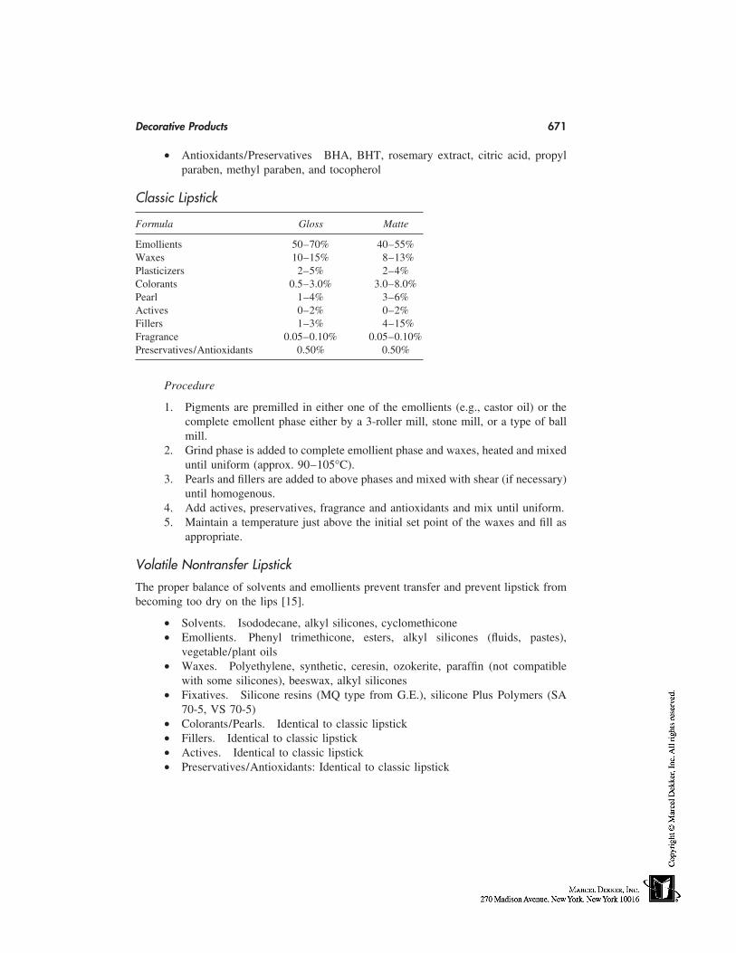
Decorative Products 671
• Antioxidants/Preservatives BHA, BHT, rosemary extract, citric acid, propylparaben, methyl paraben, and tocopherol
Classic Lipstick
Formula Gloss Matte
Emollients 50–70% 40–55%Waxes 10–15% 8–13%Plasticizers 2–5% 2–4%Colorants 0.5–3.0% 3.0–8.0%Pearl 1–4% 3–6%Actives 0–2% 0–2%Fillers 1–3% 4–15%Fragrance 0.05–0.10% 0.05–0.10%Preservatives/Antioxidants 0.50% 0.50%
Procedure
1. Pigments are premilled in either one of the emollients (e.g., castor oil) or thecomplete emollent phase either by a 3-roller mill, stone mill, or a type of ballmill.
2. Grind phase is added to complete emollient phase and waxes, heated and mixeduntil uniform (approx. 90–105°C).
3. Pearls and fillers are added to above phases and mixed with shear (if necessary)until homogenous.
4. Add actives, preservatives, fragrance and antioxidants and mix until uniform.5. Maintain a temperature just above the initial set point of the waxes and fill as
appropriate.
Volatile Nontransfer Lipstick
The proper balance of solvents and emollients prevent transfer and prevent lipstick frombecoming too dry on the lips [15].
• Solvents. Isododecane, alkyl silicones, cyclomethicone• Emollients. Phenyl trimethicone, esters, alkyl silicones (fluids, pastes),
vegetable/plant oils• Waxes. Polyethylene, synthetic, ceresin, ozokerite, paraffin (not compatible
with some silicones), beeswax, alkyl silicones• Fixatives. Silicone resins (MQ type from G.E.), silicone Plus Polymers (SA
70-5, VS 70-5)• Colorants/Pearls. Identical to classic lipstick• Fillers. Identical to classic lipstick• Actives. Identical to classic lipstick• Preservatives/Antioxidants: Identical to classic lipstick
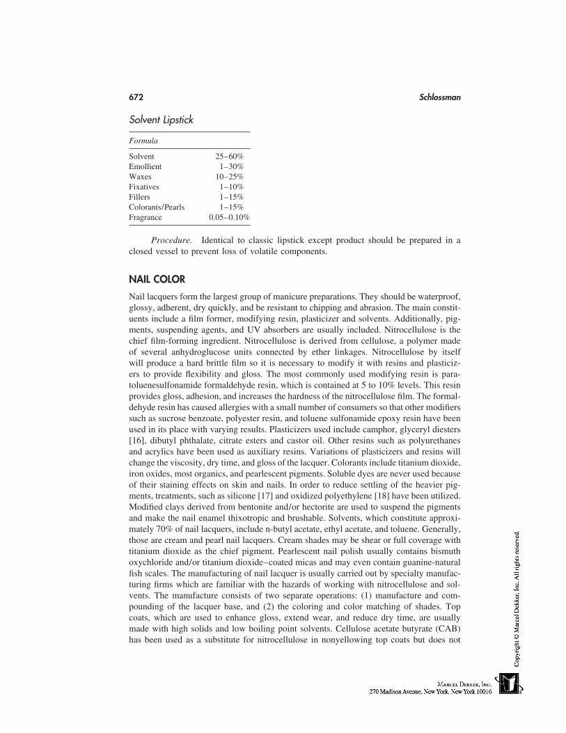
672 Schlossman
Solvent Lipstick
Formula
Solvent 25–60%Emollient 1–30%Waxes 10–25%Fixatives 1–10%Fillers 1–15%Colorants/Pearls 1–15%Fragrance 0.05–0.10%
Procedure. Identical to classic lipstick except product should be prepared in aclosed vessel to prevent loss of volatile components.
NAIL COLOR
Nail lacquers form the largest group of manicure preparations. They should be waterproof,glossy, adherent, dry quickly, and be resistant to chipping and abrasion. The main constit-uents include a film former, modifying resin, plasticizer and solvents. Additionally, pig-ments, suspending agents, and UV absorbers are usually included. Nitrocellulose is thechief film-forming ingredient. Nitrocellulose is derived from cellulose, a polymer madeof several anhydroglucose units connected by ether linkages. Nitrocellulose by itselfwill produce a hard brittle film so it is necessary to modify it with resins and plasticiz-ers to provide flexibility and gloss. The most commonly used modifying resin is para-toluenesulfonamide formaldehyde resin, which is contained at 5 to 10% levels. This resinprovides gloss, adhesion, and increases the hardness of the nitrocellulose film. The formal-dehyde resin has caused allergies with a small number of consumers so that other modifierssuch as sucrose benzoate, polyester resin, and toluene sulfonamide epoxy resin have beenused in its place with varying results. Plasticizers used include camphor, glyceryl diesters[16], dibutyl phthalate, citrate esters and castor oil. Other resins such as polyurethanesand acrylics have been used as auxiliary resins. Variations of plasticizers and resins willchange the viscosity, dry time, and gloss of the lacquer. Colorants include titanium dioxide,iron oxides, most organics, and pearlescent pigments. Soluble dyes are never used becauseof their staining effects on skin and nails. In order to reduce settling of the heavier pig-ments, treatments, such as silicone [17] and oxidized polyethylene [18] have been utilized.Modified clays derived from bentonite and/or hectorite are used to suspend the pigmentsand make the nail enamel thixotropic and brushable. Solvents, which constitute approxi-mately 70% of nail lacquers, include n-butyl acetate, ethyl acetate, and toluene. Generally,those are cream and pearl nail lacquers. Cream shades may be shear or full coverage withtitanium dioxide as the chief pigment. Pearlescent nail polish usually contains bismuthoxychloride and/or titanium dioxide–coated micas and may even contain guanine-naturalfish scales. The manufacturing of nail lacquer is usually carried out by specialty manufac-turing firms which are familiar with the hazards of working with nitrocellulose and sol-vents. The manufacture consists of two separate operations: (1) manufacture and com-pounding of the lacquer base, and (2) the coloring and color matching of shades. Topcoats, which are used to enhance gloss, extend wear, and reduce dry time, are usuallymade with high solids and low boiling point solvents. Cellulose acetate butyrate (CAB)has been used as a substitute for nitrocellulose in nonyellowing top coats but does not
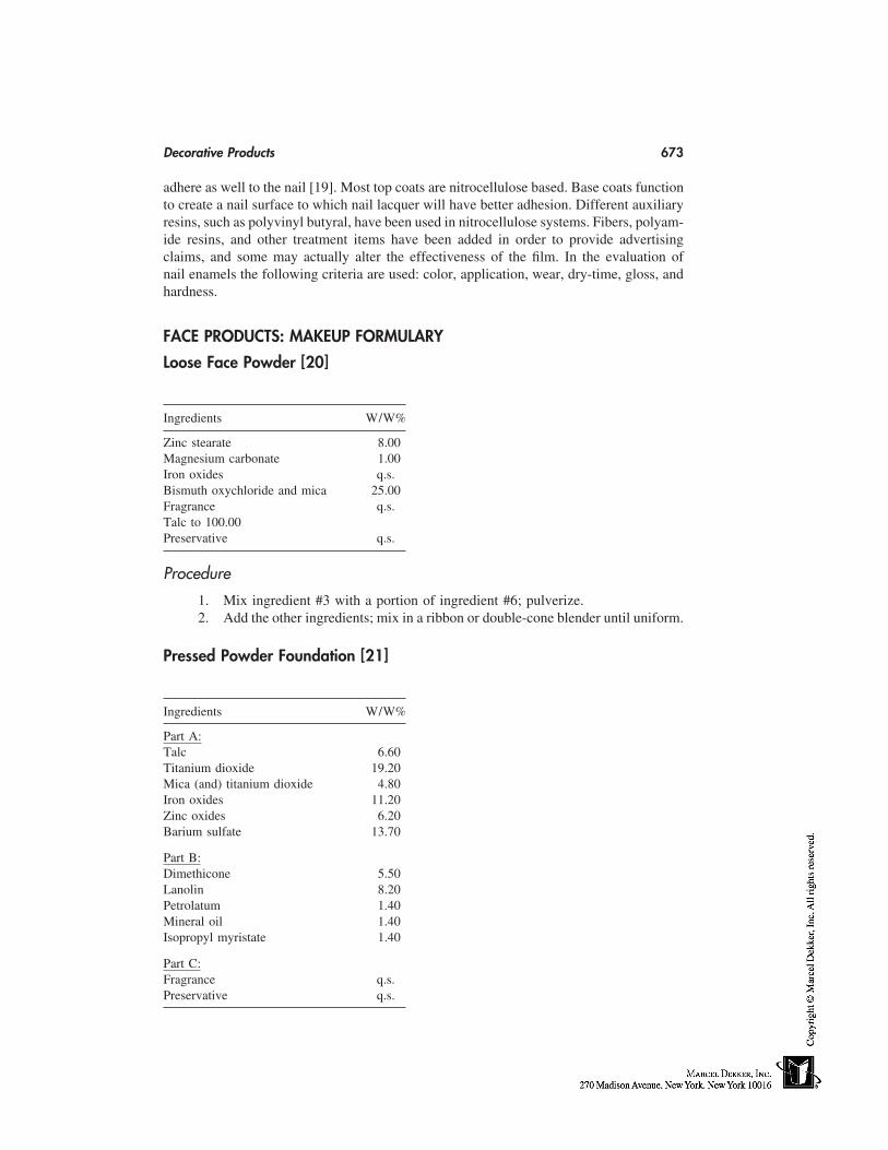
Decorative Products 673
adhere as well to the nail [19]. Most top coats are nitrocellulose based. Base coats functionto create a nail surface to which nail lacquer will have better adhesion. Different auxiliaryresins, such as polyvinyl butyral, have been used in nitrocellulose systems. Fibers, polyam-ide resins, and other treatment items have been added in order to provide advertisingclaims, and some may actually alter the effectiveness of the film. In the evaluation ofnail enamels the following criteria are used: color, application, wear, dry-time, gloss, andhardness.
FACE PRODUCTS: MAKEUP FORMULARY
Loose Face Powder [20]
Ingredients W/W%
Zinc stearate 8.00Magnesium carbonate 1.00Iron oxides q.s.Bismuth oxychloride and mica 25.00Fragrance q.s.Talc to 100.00Preservative q.s.
Procedure
1. Mix ingredient #3 with a portion of ingredient #6; pulverize.2. Add the other ingredients; mix in a ribbon or double-cone blender until uniform.
Pressed Powder Foundation [21]
Ingredients W/W%
Part A:Talc 6.60Titanium dioxide 19.20Mica (and) titanium dioxide 4.80Iron oxides 11.20Zinc oxides 6.20Barium sulfate 13.70
Part B:Dimethicone 5.50Lanolin 8.20Petrolatum 1.40Mineral oil 1.40Isopropyl myristate 1.40
Part C:Fragrance q.s.Preservative q.s.
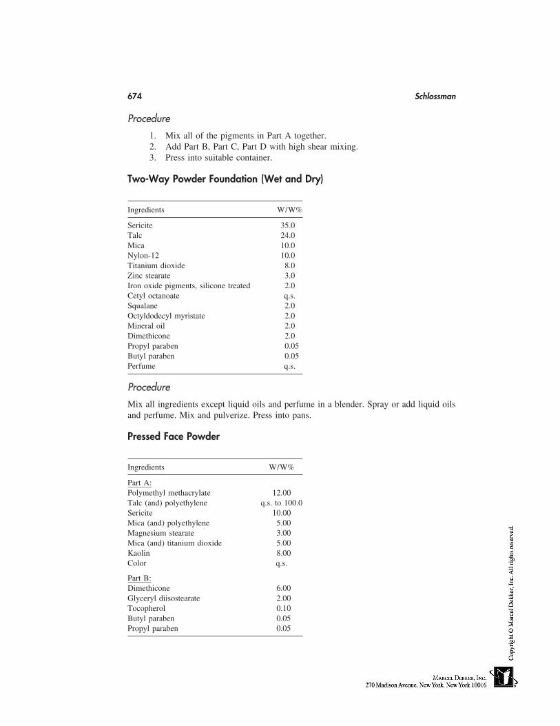
674 Schlossman
Procedure
1. Mix all of the pigments in Part A together.2. Add Part B, Part C, Part D with high shear mixing.3. Press into suitable container.
Two-Way Powder Foundation (Wet and Dry)
Ingredients W/W%
Sericite 35.0Talc 24.0Mica 10.0Nylon-12 10.0Titanium dioxide 8.0Zinc stearate 3.0Iron oxide pigments, silicone treated 2.0Cetyl octanoate q.s.Squalane 2.0Octyldodecyl myristate 2.0Mineral oil 2.0Dimethicone 2.0Propyl paraben 0.05Butyl paraben 0.05Perfume q.s.
Procedure
Mix all ingredients except liquid oils and perfume in a blender. Spray or add liquid oilsand perfume. Mix and pulverize. Press into pans.
Pressed Face Powder
Ingredients W/W%
Part A:Polymethyl methacrylate 12.00Talc (and) polyethylene q.s. to 100.0Sericite 10.00Mica (and) polyethylene 5.00Magnesium stearate 3.00Mica (and) titanium dioxide 5.00Kaolin 8.00Color q.s.
Part B:Dimethicone 6.00Glyceryl diisostearate 2.00Tocopherol 0.10Butyl paraben 0.05Propyl paraben 0.05
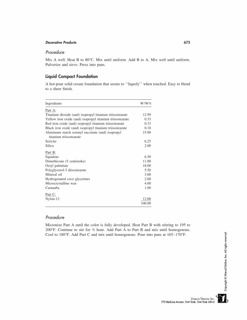
Decorative Products 675
Procedure
Mix A well. Heat B to 80°C. Mix until uniform. Add B to A. Mix well until uniform.Pulverize and sieve. Press into pans.
Liquid Compact Foundation
A hot-pour solid cream foundation that seems to ‘‘liquefy’’ when touched. Easy to blendto a sheer finish.
Ingredients W/W%
Part A:Titanium dioxide (and) isopropyl titanium triisostearate 12.99Yellow iron oxide (and) isopropyl titanium triisostearate 0.33Red iron oxide (and) isopropyl titanium triisostearate 0.33Black iron oxide (and) isopropyl titanium triisostearate 0.10Aluminum starch octenyl succinate (and) isopropyl 15.00
titanium triisostearateSericite 6.25Silica 2.00
Part B:Squalene 6.50Dimethicone (5 centistoke) 11.00Octyl palmitate 18.00Polyglycerol-3 diisostearate 5.50Mineral oil 3.00Hydrogenated coco glycerines 2.00Microcrystalline wax 4.00Carnauba 1.00
Part C:Nylon-12 12.00
100.00
Procedure
Micronize Part A until the color is fully developed. Heat Part B with stirring to 195 to200°F. Continue to stir for 1/2 hour. Add Part A to Part B and mix until homogenous.Cool to 180°F. Add Part C and mix until homogenous. Pour into pans at 165–170°F.
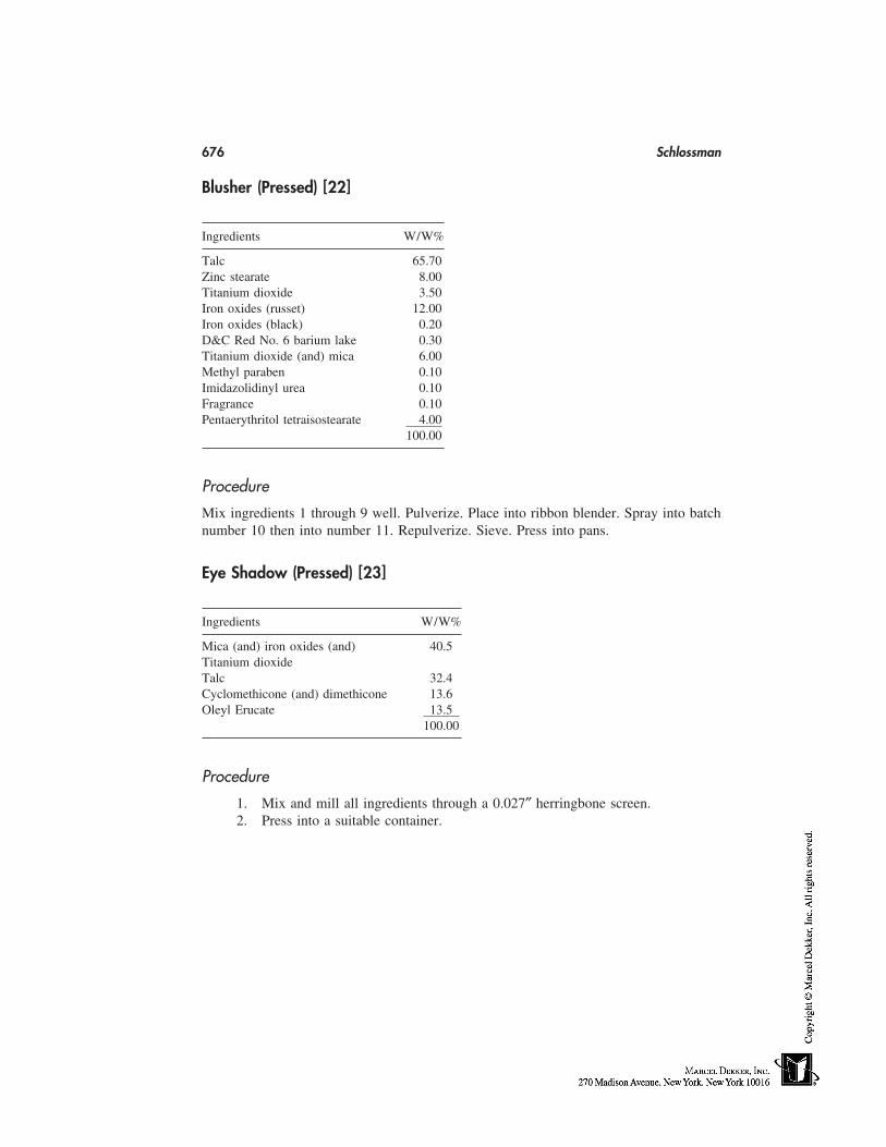
676 Schlossman
Blusher (Pressed) [22]
Ingredients W/W%
Talc 65.70Zinc stearate 8.00Titanium dioxide 3.50Iron oxides (russet) 12.00Iron oxides (black) 0.20D&C Red No. 6 barium lake 0.30Titanium dioxide (and) mica 6.00Methyl paraben 0.10Imidazolidinyl urea 0.10Fragrance 0.10Pentaerythritol tetraisostearate 4.00
100.00
Procedure
Mix ingredients 1 through 9 well. Pulverize. Place into ribbon blender. Spray into batchnumber 10 then into number 11. Repulverize. Sieve. Press into pans.
Eye Shadow (Pressed) [23]
Ingredients W/W%
Mica (and) iron oxides (and) 40.5Titanium dioxideTalc 32.4Cyclomethicone (and) dimethicone 13.6Oleyl Erucate 13.5
100.00
Procedure
1. Mix and mill all ingredients through a 0.027″ herringbone screen.2. Press into a suitable container.
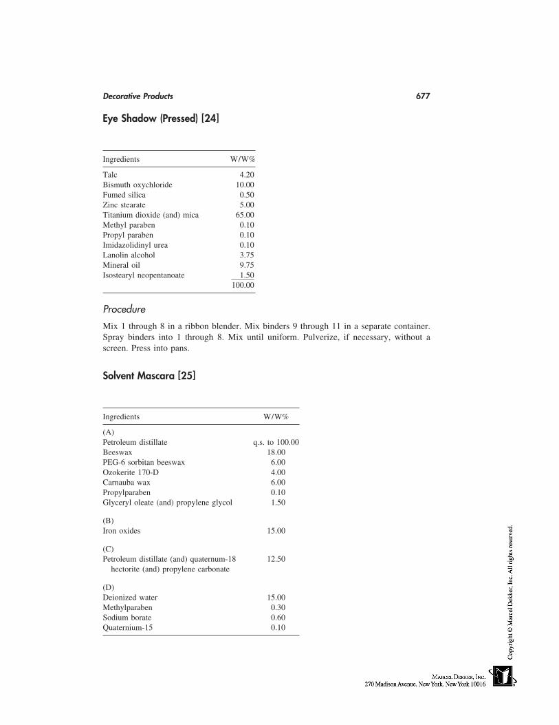
Decorative Products 677
Eye Shadow (Pressed) [24]
Ingredients W/W%
Talc 4.20Bismuth oxychloride 10.00Fumed silica 0.50Zinc stearate 5.00Titanium dioxide (and) mica 65.00Methyl paraben 0.10Propyl paraben 0.10Imidazolidinyl urea 0.10Lanolin alcohol 3.75Mineral oil 9.75Isostearyl neopentanoate 1.50
100.00
Procedure
Mix 1 through 8 in a ribbon blender. Mix binders 9 through 11 in a separate container.Spray binders into 1 through 8. Mix until uniform. Pulverize, if necessary, without ascreen. Press into pans.
Solvent Mascara [25]
Ingredients W/W%
(A)Petroleum distillate q.s. to 100.00Beeswax 18.00PEG-6 sorbitan beeswax 6.00Ozokerite 170-D 4.00Carnauba wax 6.00Propylparaben 0.10Glyceryl oleate (and) propylene glycol 1.50
(B)Iron oxides 15.00
(C)Petroleum distillate (and) quaternum-18 12.50
hectorite (and) propylene carbonate
(D)Deionized water 15.00Methylparaben 0.30Sodium borate 0.60Quaternium-15 0.10
678 Schlossman
Procedure
Mill pigment (B) into (A), which has been heated to 90°C. After (C) has been addedslowly and heated with (A), emulsify by adding (D) at 90°C to (A), (B), and (C) mixtures.Continue mixing until cool.
Emulsion-Resistant Mascara [26]
Ingredients W/W%
(A)Deionized water 41.00Hydroxyethyl cellulose 1.00Methylparaben 0.30Aqueous 0.10% phenyl mercuric acetate 4.00Triethanolamine 1.00Ammonium hydroxide, 28% 0.50
(B)Iron oxides 10.00Ulltramarine blue 2.00
(C)Isostearic acid 2.00Stearic acid 2.00Glyceryl monostearate 1.00Beeswax 9.00Carnauba wax 6.00Propylparaben 0.10
(D)Quaternium-15 0.10
(E)30% Acrylic/acrylate copolymer solution 20.00
in ammonium hydroxide100.00
Procedure
Mill the pigments of (B) in the water phase (B). Heat to 80°C. Heat the oil phase (C) to82°C. Emulsify. Cool to 50°C. Add (D), then (E). Cool to 30°C.
Decorative Products 679
Waterproof Eyeliner [27]
Ingredients W/W%
Beeswax 16.50PVP/Eicosene copolymer 5.00Petroleum distillate 35.00Petroleum distillate (and) quaternium-18 33.50
hectorite (and) propylene carbonatePreservative 0.20Titanium dioxide (and) mica (and) fer- 9.80
ric ferrocyanide100.00
Procedure
1. Heat ingredients 1 and 2 to 70°C and blend in (3) (n.b. flammable).2. Blend in (4) with low shear mixing.3. Cool to 50°C while continuing to mix.4. Blend in ingredients (2), (5), and (6) and mix until uniform.
Aqueous Eyeliner [28]
Ingredients W/W%
Part 1Ammonium vinyl acetate/actylates copolymer 55.00Polysorbate 80 1.00Isopropyl myristate 4.00
Part 2Propylene glycol USP 2.50Methylparaben USP 0.25Water, deionized 29.50Hectorite (and) hydroxyethylcellulose 0.25Iron oxides 7.50
100.00
680 Schlossman
Makeup Pencil [29]
Ingredients W/W%
Part 1Cyclomethicone 40.0Bis phenylhexamethicone 40.0Diphenyl dimethicone 40.0
Part 2Beeswax 15.0Carnauba 7.0Ozokerite 7.0Paraffin 20.0Mineral oil q.s. to 100.0Cetyl alcohol 1.0
Part 3Pigments q.s.Titanium dioxide q.s.
Procedure
1. The ingredients of Part 2 are melted and homogenized at 78–82°C, then main-tained by a thermostatic bath regulated to 58–62°C.
2. The ingredients of Part 3 are dispersed in Part 1; the mixture is placed in athermostatic bath at 58–62°C.
3. Part 3 is then added.4. After homogenization, the whole is cooled in a silicone-treated mold (with di-
methicone).
Classic Lipstick [30]
Ingredients W/W%
Carnauba wax 2.50Beeswax, white 20.00Ozokerite 10.00Lanolin, anhydrous 5.00Cetyl alcohol 2.00Liquid paraffin 3.00Isopropyl myristate 3.00Propylene glycolricinoleate 4.00Pigments 10.00Bromo acids 2.50Castor oil q.s. to 100.00
Decorative Products 681
Solvent Lipstick [31]
Ingredients W/W%
Synthetic wax 6.00Ceresin 4.00Isododecane 10.00Paraffin 3.00Cetyl acetate/acetylated lanolin alcohol 5.00Methylparaben 0.30Propylparaben 0.10BHA 0.10D&C Red No. 7 calcium lake 4.00FD&C Yellow No. 5 aluminum lake 3.00Titanium dioxide/mica 5.00Titanium dioxide/mica/iron oxides 3.00Bismuth oxychloride 10.00Cyclomethicone 41.50Isostearyl trimetholpropane siloxy silicate 5.00
100.00
Procedure
Mix the dry ingredients with the volatiles and silicone ester wax. The waxes and oils areadded with heating. The powders are added next. The mixture is then stirred before pouringinto molds and allowed to cool.
Cream Nail Enamel [32]
Ingredients W/W%
n-Butylacetate—solvent 28.23Toluene—diluent 24.54Nitrocellulose 1/2 sec wet—film-former 12.00Ethyl acetate—solvent 11.00Toluene sulfonamide/formaldehyde 10.00
resin—secondary resinAcrylates copolymer—resin 0.50Dibutyl phthalate—plasticizer 5.00Isopropyl alcohol, 99%—diluent 4.25Stearalkonium hectorite—suspending agent 1.00Camphor—plasticizer 1.50D&C Red No. 6 barium lake—color 0.08Titanium dioxide 0.75Iron oxides 0.15
100.00
682 Schlossman
Pearlescent Nail Enamel [33]
Ingredients W/W%
n-Butyl acetate 34.04Toluene 30.00Nitrocellulose 1/2 sec. wet 14.90Toluene sulfonamide/formaldehyde resin 7.10Dibutyl phthalate 4.80Camphor 2.40Stearalkonium hectorite 1.20Benzophenone-1 0.20D&C Red No. 7 calcium lake 0.08D&C Red No. 34 calcium lake 0.05FD&C Yellow No. 5 aluminum lake 0.08Iron oxides 0.15Bismuth oxychloride (25%) 5.00
100.00
Acrylic Nail Hardener [34]
Ingredients W/W%
Ethyl acetate 41.20Butyl acetate 30.00Nitocellulose 1/2 sec. wet 14.00Toluene sulfonamide/formaldehyde resin 10.00Dibutyl phthalate 4.00Camphor 0.50Acrylates copolymer 0.20Benzophenone-1 0.10
100.00
REFERENCES
1. 21 CFR, Parts 1–99. April 1, 1998.2. EC Cosmetics Directive 76/768/EEC, Annex IV, Part 1. September 3, 1998.3. MHW Ordinance No. 30. August 31, 1966.4. 21 CFR Parts 1–99. April 1, 1998.5. 21 CFR Parts 1–99. April 1, 1998.6. 21 CFR Parts 1–99. April 1, 1998.7. 61 Federal Register 8372. March 6, 1996.8. EC Cosmetics Directive 76/768/EEC, Annex IV, Part 1. September 3, 1998.9. MHW Ordinance No. 30. August 31, 1996.
10. Decorative cosmetics. In: Handbook of Cosmetic Science and Technology. Knowlton JL,Pearce SEM, eds. Oxford: Elsevier Advanced Technology, 1993: 128.
11. Miyoshi R. U.S. Patent No. 4,606,914. 1986.12. Miyoshi R, Isao Imai. U.S. Patent No. 4,622,074. 1986.13. Schlossman ML. U.S. Patent No. 4,877,604. 1989.
Decorative Products 683
14. Dweck AC. Foundations—a guide to formulation and manufacture. Cosmetic Toilet 1986; 4:41–44.
15. Castrogiavanni A, Barone SJ, Krog A, McCulley ML, Callelo JF. U.S. Patent No. 5,505,937.1996.
16. Castrogiavanni A, Sandewicz RW, Amato SW. U.S. Patent No. 5,066,484. 1991.17. Socci RL, Ismailer AA, Castrogiavanni A. U.S. Patent No. 4,832,944. 1989.18. Weber RA, Frankfurt CC, Penicnak AJ. U.S. Patent No. 5,174,996. 1992.19. Martin FL, Onofrio MV. U.S. Patent No. 5,130,125. 1992.20. Hunting ALL. Face cosmetics. In: Decorative Cosmetics. Weymouth, Dorset, England: Mi-
celle Press, 1991:3.21. Personal Care Formulary. GE Silicones Waterford, NY. 1996; p. 151.22. Knowlton JL, Pearce SEM. Decorative products. In: Handbook of Cosmetic Science and Tech-
nology. Oxford: Elsevier Advanced Technology, 1993:143.23. Personal Care Formulary. GE Silicones, Waterford, NY. 1996; p. 149.24. Knowlton JL, Pearce SEM. Decorative cosmetics. In: Handbook of Cosmetic Science and
Technology. Oxford: Elsevier Advanced Technology, 1993:145.25. Schlossman ML. Application of color cosmetics. Cosmet Toilet 1985; 100:36–40.26. Schlossman ML. Application of color cosmetics. Cosmet Toilet 1985; 100:36–40.27. Hunting ALL. Eye cosmetics. In: Decorative Cosmetics. Weymouth, Dorset, England: Micelle
Press, 1991:173.28. Hunting ALL. Eye cosmetics. In: Decorative Cosmetics. Weymouth, Dorset, England: Micelle
Press, 1991:170.29. Hunting ALL. Eye cosmetics. In: Decorative Cosmetics. Weymouth, Dorset, England: Micelle
Press, 1991:174.30. Bryce DM. Lipstick. In: Poucher’s Perfumes, Cosmetics and Soaps. Butler H, ed. London:
Chapman & Hall, 1992:234.31. Castrogiavanni A, Barone SJ, Krog A, McCulley ML, Callelo JF. U.S. Patent No. 5, 505,
937. 1996.32. Schlossman ML. Manicure preparations. In: Poucher’s Perfumes, Cosmetics and Soaps. Butler
H, ed. London: Chapman & Hall, 1992:253, 254.33. Schlossman ML. Manicure preparations. In: Poucher’s Perfumes, Cosmetics and Soaps. Butler
H, ed. London: Chapman & Hall, 1992:254.34. Schlossman ML Make-up formulary. Cosmet Toilet 1994; 109:104.

55
Cosmetics for Nails
Douglas SchoonCreative Nail Design Inc., Vista, California
Robert BaranNail Disease Center, Cannes, France
The purpose of this chapter is to present the cosmetics used for the decoration of the nail,of which the nail coating is of prime importance. Fingernail coatings consist of two types[1–3]:
1. Coatings that harden upon evaporation: these products include nail polishes,topcoats and base coats.
2. Coatings that polymerize: nail enhancements are a special type of coating usedto create artificial fingernails.
EVAPORATION COATINGS
Base coat, top coat, and nail enamel have similar basic formulas.They consist of the following:1. A film former such as nitrocellulose. This organic polymer creates a continu-
ous coating over the nail plate. Other nonnitrated cellulosic materials are also used withvarying degrees of success, namely cellulose acetate and derivatives. Polyurethanes, poly-amides, and polyesters have also been used. However, these cannot match the toughnessand surface hardness of nitrocellulose. One of the most commonly used, nitrocellulosehas several disadvantages: the surfaces produced by this polymer have low gloss and thefilms are brittle and adhere poorly to the nail plate. Upon evaporation, nitrocellulose filmsshrink excessively, which leads to poor adhesion. To overcome these drawbacks, addi-tional film modifiers will offset some deficiencies of the primary film form.
2. Film modifiers. They are specifically used to improve adhesion and gloss. Themost commonly used modifier is toluene sulfonamide/formaldehyde resin (TSFR), whichis considered to be the heart of the product. This thermoplastic resin improves nail-plateadhesion while producing water-resistant, glossy surfaces with improved flexibility. Un-fortunately, this resin is the main culprit of users’ sensitization. Use of this resin impartsbetween 0.05 to 0.1% free formaldehyde (as impurity) into the formulation. Therefore,many alternate modifiers have been tried, including toluene/sulfonamide/expoxy resin,
685
686 Schoon and Baran
polyester sucrose benzoate, polyesters, acrylic ester oligomers, SAIB, arylsulfonyl meth-anes, and glyceryl tribenzoate.
3. Plasticizers. Plasticizers are chemical flexibilizers for polymer films that im-prove their durability. They may also improve adhesion and gloss. Dibutyl phthalate andcamphor are the most common examples of low–molecular weight, high–boiling pointplasticizers. Other examples of plasticizers are castor oil, glyceryl tribenzoate, acetyl tribe-nzoate citrate PPG-2 dibenzoate, glycerol, citrate esters, triacetin, and a polyether urethane.
4. Solvents/diluents. The solid film-forming polymers, upon evaporation, are de-posited on the nail plate. The most commonly used solvents are alkyl esters and glycolethers. Coupling agents (aliphatic alcohols) are useful in varnishes to increase the overallsolubility and flow of the system. Diluents are usually nonpolar compounds that will notdissolve nitrocellulose. Toluene was commonly used until the appearance of CaliforniaProposition 65. Most companies are now developing toluene-free formulas.
5. Viscosity modifiers or thixotropic agents. Ideally, a nail enamel should be gel-like when sitting on the shelf but significantly thin when brushed on. Both consistenciesare possible in one bottle by using thixotropic agents such as stearalkonium hectorite.
6. Color additives. Colorants should be nonsoluble pigments to prevent stainingof the nail plate. Guanine, derived from scales of Atlantic herring, produces pearlescentpigment. Bismuth oxychloride and mica coated with titanium dioxide are used to createiridescent shades.
7. Base and top coats. Base coats contain a high percentage of TSFR. They areapplied to the nail before application of nail varnish. They are adhesion promoters thatimprove retention and coating toughness. Top coats use higher levels of film formers,such as nitrocellulose, to maximize surface gloss and hardness. Often the top coat containsUV-absorbing materials.
POLYMERIZING COATINGS
Sculptured Artificial Nails
Liquid-and-powder systems are based on methacrylates. They consist of a liquid monomer(ethyl methacrylate) mixed with a polymer powder (polyethyl and/or polymethyl methac-rylate), the latter carrying only the heat-sensitive initiator (usually benzoyl peroxide) tothe monomer. UV absorbers are polymer additives that prevent sunlight yellowing. Cata-lysts speed up polymerization.
Light-Curing Gels
UV or visible light-curing gels are made primarily of urethane acrylate and other acrylatedoligomers. Associated with initiator, the catalyst and oligomers are combined into a singleproduct; they come premixed and ready to use. They may be considered a variant ofsculptured artificial nails.
Preformed Artificial Nails
These are usually made of ABS plastic, nylon, or acetate, and are adhered to the naturalnail with cyanoacrylate monomer. Home-use, retail versions of these tips may be used astemporary natural overlays, not worn for longer than 48 hours on any one occasion. Theyare more often used as permanent nail-tip extensions. Professional nail technicians usually
Cosmetics for Nails 687
coat these tips with artificial nail products to create longer lasting nail extensions. Mostnail technicians feel it is too time consuming to sculpt nails, and these tips speed theprocess. The tip can be coated or overlaid with wraps or liquid-and-powder or gel products.
Wraps
Wraps can be used to coat the nail plate or add strength to thin, weak nails. The monomersused to create wraps are cyanoacrylates. In nail wrapping, the free edge of the nail shouldbe long enough to be splinted by the various types of fabrics providing support and addedstrength to the coating. There are three fabrics in wide use: fiberglass, silk, and linen.
No-Light Gels
These products are wrap monomers that have been thickened to have a gel-like appearance.They should be used and handled as any other wrap product.
Removal of Fingernail Coatings
The most commonly used solvent for removal of nail products is acetone. Warming thesolvent with great care can cut product removal time in half. However most gels aredifficult to remove because they are highly cross-linked and resistant to many solvents.Therefore, if gel enhancements have to be removed, slowly file (do not drill) the enhance-ment with a medium-grit file, leaving a very thin layer of product. Soak in warm productremover and, once softened, scrape the remaining product away with a wooden pusherstick [1].
Cuticle Removers
These are lotions or gels containing approximately 0.4% sodium or potassium hydroxide.The lotion is left in place for 1 to 3 minutes and then washed off. Creams containing 1to 5% lactic acid (pH 3–3.7) are also used.
Nail Whitener
This is a pencil-like device with a white clay (kaolin) core used to deposit color on theundersurface of the free edge of the nail.
REFERENCES
1. Schoon DD. Nail Structure and Product Chemistry. Albany: Milady Publishing, 1996.2. Baran R, Schoon DD. Cosmetology of normal nails. In: Baran R, Maibach HI. Textbook of
Cosmetic Dermatology. London: Martin Dunitz, 1998:213–231.3. Baran R, Schoon DD. Cosmetics for abnormal and pathological nails. In: Baran R, Maibach
HI. Textbook of Cosmetic Dermatology. London: Martin Dunitz, 1998:233–244.

56
Antiperspirants
Jörg SchreiberBeiersdorf AG, Hamburg, Germany
GENERAL INTRODUCTION
This chapter presents an overview concerning the current knowledge of antiperspirantactives and their interactions with the human axilla. It is my intention to give the interestedreader a short introduction about formulation work, drug delivery systems, and applicationforms developed for antiperspirant actives. The final section lists references that shouldbe useful for anyone who wants to learn more about a specific topic of antiperspiranttechnology.
BIOLOGY OF SWEAT GLANDS IN THE HUMAN AXILLA
The axilla region of humans contains apocrine, eccrine, and sebaceous glands. Approxi-mately 25,000 sweat glands/axilla can produce up to 12 g sweat/h [1]. The current under-standing concerning structure and function of sweat glands is that thermoregulation isonly one aspect of the body participating in immmunological, metabolic, and hormonalaspects of human life [2].
Eccrine Glands
This is the organ responsible for the majority of sweat production. It has a sensory andexcretory function and can be stimulated by emotional and thermal stimuli [3]. It producesa clear, colorless and odorless liquid containing 98 to 99% water and 1 to 2% inorganicand organic compounds [4]. Inorganic components include NaCl, traces of K�, Ca2�,Mg2�, Fe3�, and Cu2� ions. Organic components include: lactic acid, citric acid, formicacid, propionic acid, butyric acid, urea, and ammonia. Underarm wetness comes mostlyfrom the secretion of eccrine glands. Antiperspirants reduce the amount of sweat onlyfrom eccrine glands.
Apocrine Glands
Apocrine glands are apparently a relict from the phylogenetic development of man. Theseglands start to produce a milky, viscous fluid during puberty on special locations of thebody, especially the underarm pit [5]. In contrast to eccrine glands, the openings of the
689
690 Schreiber
glands are not at the skin surface but appear at the hair follicle. Decomposition of apocrinesweat by skin bacteria are responsible for the characteristic malodor of human sweat.Apocrine sweat consists among water of proteins, carbohydrates and ammonium salts [6].Other investigators have reported that these glands secrete lipids, cholesterol, and steroids[7]. Furthermore, it has been shown that androgen-converting enzymes in the apocrineglands are responsible for circulating androgens to dihydrotestosterone [5].
ANTIPERSPIRANTS
Antiperspirants are topically applied products designed to reduce underarm wetness bylimiting eccrine sweat production. In the United States these products are regulated bythe FDA as over-the-counter (OTC) drugs because they are intended to affect a ‘‘functionof the body’’ (in this case, perspiration). Products containing antiperspirant actives haveto reduce perspiration to minimum 20% by 50% of the test population under validatedtest conditions. Test protocols (in vivo clinical trials) to develop a safe and effective prod-uct have been designed to substantiate the desired claims [8–14].
Comparative quantitative determination of the activity of sweat glands on the fore-arm after application of aluminum chlorohydrate solutions is now possible by combiningthe classic starch iodine visualization technique with digital image analysis [15]. A nonin-vasive optical technique that allows the analysis of the function of a number of glandssimultaneously in vivo was recently reported [16]. A new method for parallel testing ofup to eight formulations on the backs of volunteers allows a very fast evaluation of productprototypes [1].
Sweat Reduction by Antiperspirants: Current Model/Theory
The reader should be aware that theories concerning the action of sweat-reducing agentsdepend strongly on the type of actives (aluminum salts, nonionics, ionic agents). Theefficacy of antiperspirants based on aluminum and/or aluminum zirconium salts (see dis-cussion p. 691) can be understood by the formation of an occlusive plug of metal hydroxidein the eccrine duct [17]. Tape-stripping experiments followed by analysis of transmissionelectron micrographs of an ACH-treated eccrine sweat-gland duct (see discussion p. 691)shows an obstructive amorphous material supporting the theory of a mechanical blockageof sweat glands from diffusion of the soluble ACH solution into the sweat gland andsubsequent neutralization to a polymeric aluminum hydroxide gel [18,19]. There seemsto be no correlation concerning efficacy of aluminum salts and the location of the plugin the duct because it is known that, compared with ACH, the more effective Al-Zr com-pounds do not penetrate as deep as the also highly effective AlCl3 solutions [17]. Thereader is referred to the literature concerning other theories of sweat reduction by alumi-num salts [20].
Active Ingredients for Controlling Underarm Wetness—State of the Art
Buffered Aluminum Salts (ACH)
The first antiperspirant, Ever Dry, based on AlCl3, was introduced to the market in 1903[21]. The first cream-containing aluminumsulfate was introduced during the 1930s. Theacidic pH value (2.5–3.0) was a drawback of these products, leading to skin irritation inthe underarm pit. History tells us that the development of actives with a higher pH value,
Antiperspirants 691
so-called buffered aluminumchlorides (aluminum chlorohydrate, ACH, pH � 4.0–4.2)was an appropriate step with the additional benefit of reduced destruction of fabric clothes.The formula of this buffering salt is {Al2(OH)5}� � {Cl�}, or more convenientlyAl2(OH)5Cl.
The historical development from AlCl3 to Al2(OH)5Cl can be easily understood bythe following consideration:
AlCl3 � 1/2 Al2Cl6 Substitute 5 Cl� � ions against OH� � ions
⇒ Al2(OH)5Cl
Al2(OH)5Cl is a 5/6 basic aluminumtrichloride. The accepted definition of ACH is the ratioof Al to Cl � 2.1 to 1.0. Lower levels lead to aluminum dichlorohydrate (Al2(OH)4Cl2) orto aluminum sesquichlorohydrate (Al2(OH)4.5Cl1.5—both actives are also generally re-garded as safe (GRAS). ACH is supplied as a powder or a 50% solution in water. It canbe formulated up to 25% calculated on an anhydrous basis. The 20% aqueous solutionreduces perspiration by 35 to 40% on average [22]. Some dyes used in clothing may beacid sensitive and will change color when in contact with an antiperspirant.
The structure of the Lewis acid ACH is very complex because ACH in water formsso-called isopolyoxo-cations with chloride ions as couterions [23–25]. There exists severalpolymer equilibria of the polycationic aluminum species in water-based systems. Short-chain polycationic species are more effective in reduction of sweat.
Aluminum Zirconium Chlorohydrate-Glycine Complexes (AZG or ZAG)
Aluminum zirconium chlorohydrate is obtained by reaction of ACH with zirconylchloride.Reaction of the former ingredients in the presence of glycine leads ZAG complexes. Gly-cine is used as a buffering agent. These antiperspirant actives form very complex poly-meric structures in water. The actives are defined by the ratio of Al � Zr metal–to–chloride ratio and the Al to Zr atomic ratio. The interested reader is referred to the literatureconcerning available actives [26,27] and nomenclature of the Al-Zr complexes [21,22].These antiperspirant actives were developed especially for anhydrous formulations be-cause they show, compared with ACH, enhanced sweat reduction [28–30]. The maximalconcentration of ZAG calculated on an anhydrous basis is 20%. They are not allowed tobe formulated for use in aerosols.
New Concepts for Controlling Underarm Wetness
Titanium Metal Chelates
The understanding of the complex solution chemistry of aluminum-based antiperspirantsgave input to the search for alternative antiperspirant salts. Titanium derivatives like par-tially neutralized ammonium titanium lactate (ATL) salts were shown to be effective inin-vitro efficacy tests [31]. The titanium metal chelates can be synthesized from the corre-sponding titanium alkoxides and organic acids followed by neutralization with ammonia.Under acidic to neutral pH conditions the ATL active seems to be relatively stable tohydrolysis and therefore probably a suitable antiperspirant active in water-based or anhy-drous drug delivery systems.
Film-Forming Antiperspirant Polymers
So-called polybarrier technology is another approach to reduce perspiration by using apolymer that forms an insoluble occlusive film barrier on the underarm skin [32]. It was

692 Schreiber
mentioned that the occlusive film is a barrier to the passage of moisture. The main advan-tage of this technology has been described as reduced skin irritation, applicable after under-arm shaving, and higher sweat reduction compared with today’s classic antiperspirantsalts. The preferred polymer is an olefinic acid amide/olefinic acid or ester copolymer–like octylacrylamide/acrylate copolymer (Versacryl-40). This copolymer can be usedalone or in combination with PVP/eicosene-copolymer in sticks, roll-ons, or alcohol-basedproducts [33]. The reduction of sweat depends on the choice of vehicle and exceeds insome formulations 40%.
Lyotropic Liquid Crystals
Certain surfactant/cosurfactant combinations in water form depending on the variables ofconcentration/temperature instead of micelles lamellar, hexagonal, inverted hexagonal,inverted micellar, or even cubic phases. The cubic phases can be of micellar or bicontinoustype [34]. The water domains in lamellar or cubic phases can swell to a certain degreewhile taking up water. The use of this swelling behavior is the basis of a patent where asurfactant/cosurfactant combination is applied to the underarm pit [35]. Sweat (water)transfers the applied composition to a lyotropic liquid crystal of cubic structure, thus creat-ing a sweat-absorbing system in the axilla. Oleic acid/glycerol monolaurate is one of thesurfactant combinations in the patent. Both components are also well known as deodor-izers.
DRUG-DELIVERY SYSTEMS AND APPLICATION FORMSFOR ANTIPERSPIRANT ACTIVES
Antiperspirant actives can be formulated in a variety of delivery systems like anhydroussuspensions, water- or hydroalcoholic-based solutions, and emulsions. Typical applicationforms for antiperspirants are sticks, roll-ons, creams, pump sprays, aerosols, gels, andpowders. A new technology for pump sprays is discussed in the chapter 57. On a globalbasis, the three most important product forms are sticks, roll-ons, and aerosols.
Formulation Work
After the decision for the desired application form has been made the formulator has todecide on the vehicle system for the antiperspirant active. It is the intent of this sectionto summarize some of the current knowledge concerning influence of actives with theformula, efficacy of different delivery systems, and the function of the ingredients usedin antiperspirants.
Antiperspirant actives like ACH or ZAG complexes are soluble in water. Applica-tion of a concentrated aqueous solution of an antiperspirant active gives a rather tackyfeeling [36]. Reduction of tackiness can be best achieved by silicone oils (cyclomethi-cones) or ester oils like Di-(2 ethylhexyl) adipate [27]. The acidic pH value (pH 4.0–4.2)has to be taken into account by selecting additional components for the desired drug deliv-ery system. Loss of viscosity and problems of a final formula with color stability are oftena hint to change the gellant and/or perfume. Aluminum powders in anhydrous systems(aerosols, suspension sticks) often leave visible white residues on skin or clothing. Liquidemollients, like PPG-14 butylether or the aforementioned adipate ester, minimize theseresidues. Another approach is to use the solid emollient isosorbide monolaurate (ICI, Arla-mol ISML) [37]. In anhydrous aerosol formulations the ACH powder settles down and
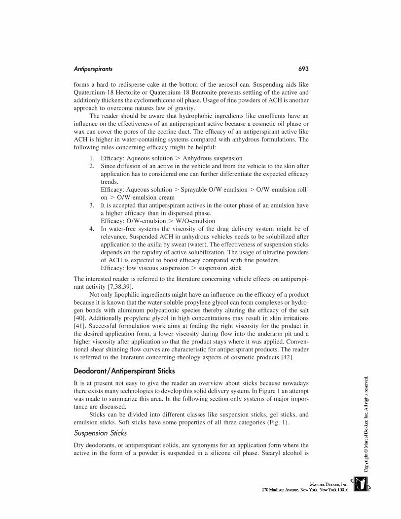
Antiperspirants 693
forms a hard to redisperse cake at the bottom of the aerosol can. Suspending aids likeQuaternium-18 Hectorite or Quaternium-18 Bentonite prevents settling of the active andadditionly thickens the cyclomethicone oil phase. Usage of fine powders of ACH is anotherapproach to overcome natures law of gravity.
The reader should be aware that hydrophobic ingredients like emollients have aninfluence on the effectiveness of an antiperspirant active because a cosmetic oil phase orwax can cover the pores of the eccrine duct. The efficacy of an antiperspirant active likeACH is higher in water-containing systems compared with anhydrous formulations. Thefollowing rules concerning efficacy might be helpful:
1. Efficacy: Aqueous solution � Anhydrous suspension2. Since diffusion of an active in the vehicle and from the vehicle to the skin after
application has to considered one can further differentiate the expected efficacytrends.Efficacy: Aqueous solution � Sprayable O/W emulsion � O/W-emulsion roll-on � O/W-emulsion cream
3. It is accepted that antiperspirant actives in the outer phase of an emulsion havea higher efficacy than in dispersed phase.Efficacy: O/W-emulsion � W/O-emulsion
4. In water-free systems the viscosity of the drug delivery system might be ofrelevance. Suspended ACH in anhydrous vehicles needs to be solubilized afterapplication to the axilla by sweat (water). The effectiveness of suspension sticksdepends on the rapidity of active solubilization. The usage of ultrafine powdersof ACH is expected to boost efficacy compared with fine powders.Efficacy: low viscous suspension � suspension stick
The interested reader is referred to the literature concerning vehicle effects on antiperspi-rant activity [7,38,39].
Not only lipophilic ingredients might have an influence on the efficacy of a productbecause it is known that the water-soluble propylene glycol can form complexes or hydro-gen bonds with aluminum polycationic species thereby altering the efficacy of the salt[40]. Additionally propylene glycol in high concentrations may result in skin irritations[41]. Successful formulation work aims at finding the right viscosity for the product inthe desired application form, a lower viscosity during flow into the underarm pit and ahigher viscosity after application so that the product stays where it was applied. Conven-tional shear shinning flow curves are characteristic for antiperspirant products. The readeris referred to the literature concerning rheology aspects of cosmetic products [42].
Deodorant/Antiperspirant Sticks
It is at present not easy to give the reader an overview about sticks because nowadaysthere exists many technologies to develop this solid delivery system. In Figure 1 an attemptwas made to summarize this area. In the following section only systems of major impor-tance are discussed.
Sticks can be divided into different classes like suspension sticks, gel sticks, andemulsion sticks. Soft sticks have some properties of all three categories (Fig. 1).
Suspension Sticks
Dry deodorants, or antiperspirant solids, are synonyms for an application form where theactive in the form of a powder is suspended in a silicone oil phase. Stearyl alcohol is
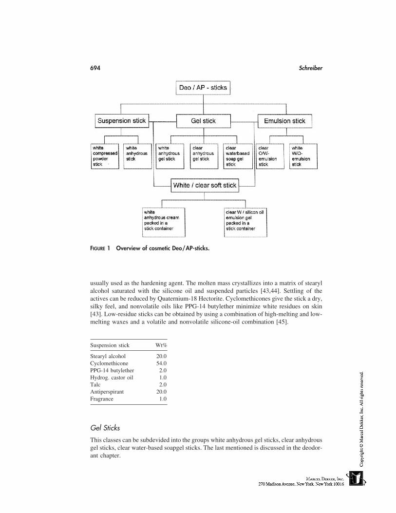
694 Schreiber
FIGURE 1 Overview of cosmetic Deo/AP-sticks.
usually used as the hardening agent. The molten mass crystallizes into a matrix of stearylalcohol saturated with the silicone oil and suspended particles [43,44]. Settling of theactives can be reduced by Quaternium-18 Hectorite. Cyclomethicones give the stick a dry,silky feel, and nonvolatile oils like PPG-14 butylether minimize white residues on skin[43]. Low-residue sticks can be obtained by using a combination of high-melting and low-melting waxes and a volatile and nonvolatile silicone-oil combination [45].
Suspension stick Wt%
Stearyl alcohol 20.0Cyclomethicone 54.0PPG-14 butylether 2.0Hydrog. castor oil 1.0Talc 2.0Antiperspirant 20.0Fragrance 1.0
Gel Sticks
This classes can be subdevided into the groups white anhydrous gel sticks, clear anhydrousgel sticks, clear water-based soapgel sticks. The last mentioned is discussed in the deodor-ant chapter.
Antiperspirants 695
White Anhydrous Gel Sticks.Shear solids or ultra-clear solids are synonyms for sticks with improved wash-out perfor-mance compared with the classic suspension sticks. They contain N-acyl aminoacid am-ides (N-lauroyl-L-glutamic acid dibutylamide) and 12-hydroxyacid as gelling agents foran oil-phase mixture (e.g., silicone oil/mineral oil). The wash-out agent is an ethoxylatedsolubilizer like Ceteareth-20. These white sticks turn clear after application to the skin(no-residue stick) [46].
Clear Anhydrous Gel Sticks.They are quite popular in the United States because clarity is associated by the consumerwith a lack of white residue on skin, no dangerous ingredients, and high efficacy. A typicalgelling agent is dibenzylidene sorbitol (dibenzyaldehyd monosorbitol acetal, DBMSA).This acetale is not stable in an acidic aqueous environment [47]. The sticks usually containa high level of alcohol and/or polyols. At high polyol concentration the active is regardedto be solubilized instead of suspended in the gel matrix [48]. An alternative gelling agentis a polyamide [49].
White anhydrous gel sticks Wt% Clear anhydrous gel sticks Wt%
N-Lauroyl-glutamic acid dibutyl amide 5.0 Dibenzylidene sorbitol 2.012-Hydroxystearic acid 5.0 Dimethicone copolyol 2.0Cyclomethicone 40.0 Diisopropyl sebacate 2.0Hydrog. polyisobutene 15.0 Glycine 1.0Diisopropyl myristate 15.0 Dipropyleneglycol 10.0Antiperspirant powder 20.0 Propyleneglycol 33.0
Antiperspirant powder 50.0
Source: Ref. 58.
Emulsion Sticks:
They can be grouped into clear o/w emulsions, white w/o emulsions, and clear w/s: emul-sion gels. The last mentioned is will be discussed shortly.
Clear O/W Emulsions.They contain a high surfactant combination with the active solubilized in the externalwater phase. The high concentration of surfactants is a disadvantage; no products basedon this technology are known to the author [47].
W/O-Emulsion Sticks.The water phase containing the active is solubilized by a surfactant like Polyglycerol-4Isostearate. A typical example for an oil/wax-phase combination is a mixture of siliconeoil/stearylalkohol [50].
W/O Emulsion Stick Wt%
Stearyl alcohol 19.0Volatile silicone 26.0Mineral oil 1.02-Methyl-2,4 pentandiol 2.0Polyglyceryl-4 isost. 2.0ACH solution (50%) 50.0
Source: Ref. 50.
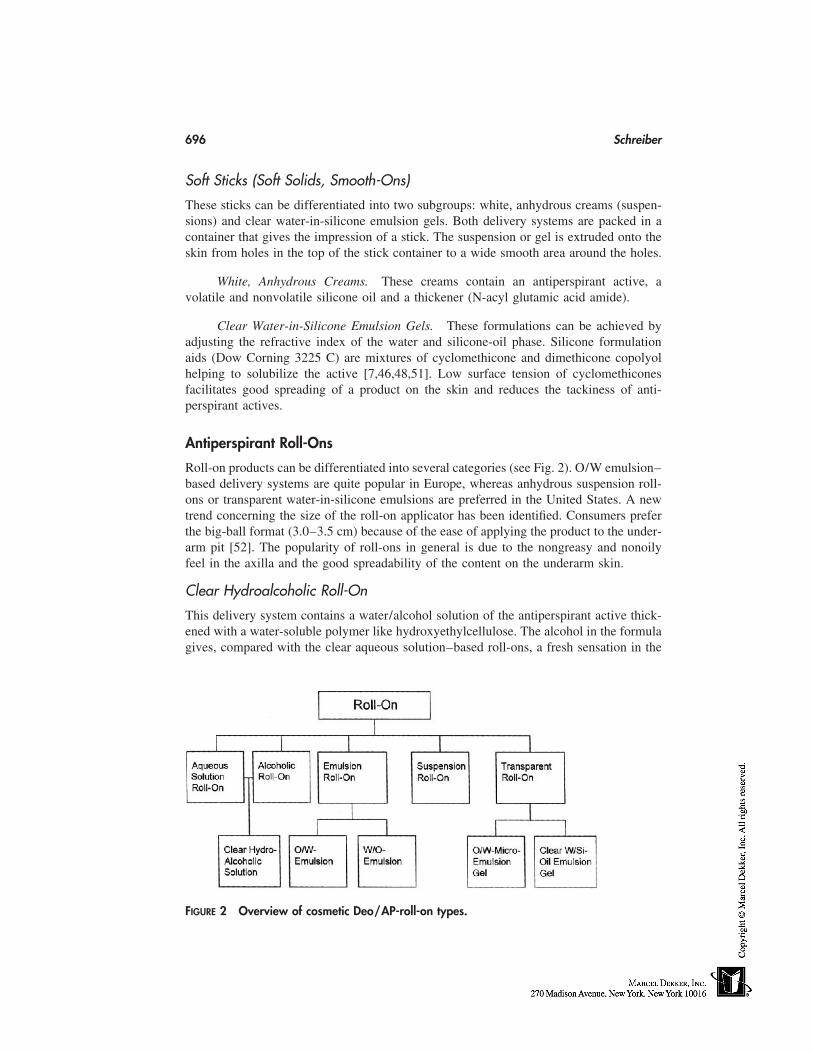
696 Schreiber
Soft Sticks (Soft Solids, Smooth-Ons)
These sticks can be differentiated into two subgroups: white, anhydrous creams (suspen-sions) and clear water-in-silicone emulsion gels. Both delivery systems are packed in acontainer that gives the impression of a stick. The suspension or gel is extruded onto theskin from holes in the top of the stick container to a wide smooth area around the holes.
White, Anhydrous Creams. These creams contain an antiperspirant active, avolatile and nonvolatile silicone oil and a thickener (N-acyl glutamic acid amide).
Clear Water-in-Silicone Emulsion Gels. These formulations can be achieved byadjusting the refractive index of the water and silicone-oil phase. Silicone formulationaids (Dow Corning 3225 C) are mixtures of cyclomethicone and dimethicone copolyolhelping to solubilize the active [7,46,48,51]. Low surface tension of cyclomethiconesfacilitates good spreading of a product on the skin and reduces the tackiness of anti-perspirant actives.
Antiperspirant Roll-Ons
Roll-on products can be differentiated into several categories (see Fig. 2). O/W emulsion–based delivery systems are quite popular in Europe, whereas anhydrous suspension roll-ons or transparent water-in-silicone emulsions are preferred in the United States. A newtrend concerning the size of the roll-on applicator has been identified. Consumers preferthe big-ball format (3.0–3.5 cm) because of the ease of applying the product to the under-arm pit [52]. The popularity of roll-ons in general is due to the nongreasy and nonoilyfeel in the axilla and the good spreadability of the content on the underarm skin.
Clear Hydroalcoholic Roll-On
This delivery system contains a water/alcohol solution of the antiperspirant active thick-ened with a water-soluble polymer like hydroxyethylcellulose. The alcohol in the formulagives, compared with the clear aqueous solution–based roll-ons, a fresh sensation in the
FIGURE 2 Overview of cosmetic Deo/AP-roll-on types.
Antiperspirants 697
axilla and faciliates drying of the product. Excellent antiperspirant efficacy is anotherbenefit of hydroalcoholic roll-ons.
O/W Emulsion Roll-On
This delivery system uses ethoxylated surfactants like PEG-40 stearate to solubilize anoil phase like mineral oil. The active is dissolved in the outer phase, allowing the formula-tion of a highly effective product. In alcohol-free formulated systems microbiologicalstability has to be checked.
O/W emulsion roll-on Wt% Hydroalcoholic roll-on Wt%
PEG-40 stearate 5.0 Antiperspirant active 20.0Cetyl alcohol 3.0 PPG-5 ceteth 20 2.0Mineral oil 2.0 Water 35.4Polysorbate-80 1.0 Ethanol 42.1Glycerin 1.5 Hydroxyethylcellulose .5Mg-aluminum silicate .8Antiperspirant active 20.0Water 66.7
W/O Emulsion Roll-On
They are weaker in efficacy because the actives are encapsulated and the external oil phaseoften gives a sticky feeling.
W/Si Emulsion Roll-On
Silicone oils allow to formulate products based on a ‘‘W/O-technology’’ because the skinfeeling is not comparable to traditional oily components like ester oils or triglycerides.The concentration of the thickener is reduced compared with sticks based on this type.The technology is discussed under soft sticks (see p. 696).
O/W Microemulsion Gel
An alternative approach to transparent products uses the PIT technology. A suitable mix-ture of surfactants, oils, and water is heated to 60 to 90°C to give a w/o emulsion abovethe phase inversion temperature (PIT). During cooling the mixture shows phase inversionto give white or transparent o/w emulsions. o/w Microemulsion gels are obtained in thepresence of hydrophobically modified water-soluble polymers [53]. The technology isexplained in more detail in the deodorant chapter.
Suspension Roll-On
The antiperspirant active in powder form is suspended in cylomethicone. The roll-on canbe formulated with or without ethanol. Quaternium-18 Hectorite is used as a thickener toprevent settling of the active. Consumers in the United States prefer this delivery systemsince it does not give a wet feeling after application and because of the easy drying [39].Actives like ZAG-complexes give high efficacy underarm products.
698 Schreiber
Suspension roll-on Wt%
Volatile silicone 65.0Quaternium-18 hectorite 13.5Silica .5Antiperspirant powder 20.0Fragrance 1.0
Antiperspirant Aerosols
Aerosols in Europe and Asia are popular delivery systems for consumers who prefer ahygienic and easy-to-use application form. Typical ingredients for aerosols include isopro-pylmyristate, isopropylpalmitate, volatile silicone, dimethicone, silica, clays, propylenecarbonate, and ethanol. Propellants include propane, butane, and isobutane.
Antiperspirant aerosol Wt%
Volatile silicone 13.4Quaternium-18 hectorite .8Ethanol .8Antiperspirant powder 10.0Propellant (butane/propane) 75.0
Because acidic aqueous ACH solutions lead to corrosion of the aerosol can, current aerosolantiperspirant products are formulated as water-free suspensions. The active is suspendedas a powder in an oil phase like cyclomethicone or in a mixture of ester oils/cyclomethi-cone. Agglomeration of solid particles and settling of actives can be minimized by usageof suspending agents like fumed silica (amorphous silicon dioxide) or clays (bentonite,hectorite). The clays form a weak gel in the presence of an oil phase that can be destroyedby shaking the aerosol can before usage. The gel structure is reformed on standing, therebyholding the active in suspension. Because the organoclays are agglomerated, shear isneeded to deagglomerate the platelets, and a polar activator like propylene carbonate orethanol is used to disperse them and induce the gelation of the oil phase.
The steps involved to prepare an aerosol product can be summarized in the followingsequence [7]:
1. Preparing a bentonite or hectorite clay with the emollient in the presence of thepolar activator and shearing the mixture
2. Adding the antiperspirant active until a uniform agglomeration-free suspensionis obtained
3. Filling the concentrate into the aerosol can and adding the propellant (pressurefilling)
Efficacy studies of aerosols including comparison with other drug delivery systems havebeen reported in the literature [30]. ZAG-complexes (see discussion p. 691) are not al-lowed to be used in aerosols.
Antiperspirants 699
Environmental Issues
Aerosols contain volatile organic compounds (VOCs) usually in a weight ratio propellant/concentrate of 75/25 [54]. The environmental impact of VOC like the reaction with NOx
in the presence of sunlight causes formation of unwanted ozone in the lower atmosphere.U.S. antiperspirant companies especially were forced to reduce VOC emissions by re-formulating and/or exchanging of hydrocarbon propellants to the fluorohydrocarbons 1,1difluorethane (Propellant 152 a) or 1,1,2,2 tetrafluorethane (Propellant 134 a). The water-soluble dimethoxyethane (DME) is another propellant that is thought to have no impacton the damage of the ozone layer [55].
The current trends in the aerosol market can be summarized as follows:
• Higher ratio of concentrate/hydrocarbon propellant• Higher amount of silicone oils• Usage of 1,1 difluorethane (Propellant 152 a)• Formulations with lower vapor pressure• Usage of smaller aerosol cans
Aerosols containing 20 to 50% propellants with a concentrate/propellant ratio from 1.0to 1.0 to 2.3 to 1.0 have been patented [56].
FUTURE TRENDS
Some new trends in the antiperspirant field concerning new actives and delivery systemshave been described in this chapter. Improvements of current formulations and innovativeconcepts will need the ongoing investigation and better understanding of the interactionactive/vehicle and vehicle/skin. Improving efficacy and skin compatibility is another ma-jor trend in the antiperspirant field. New packaging concepts like the extrudable gels, thebig ball applicator for roll-ons, and reduced size aerosol cans with ozone-friendly propel-lants are probably in a few years state of the art. The influence of perfume componentsto the skin, the increasing rate of contact allergies attributable to fragrance ingredientshave to be closely monitored [57].
REFERENCES
1. Bielfeldt S, Frase T, Gassmüller J. New sensitive method for assessment of antiperspirantswith intraindividual comparison of eight formulations. SÖFW 1997; 1237:639–642.
2. Gebhardt W. Do cutaneous coryneform bacteria produce short-chain fatty acids in vitro? Der-matologica 1989; 178:121–122.
3. Sato K, Kang WH, Saga K, Sato KT. Biology of sweat glands and their disorders. I. Normalsweat gland function. J Am Acad Dermatol 1989; 20:537–563.
4. Anonymous. Deodorants and antitranspirants. In: Harry RG, ed. Harry’s Cosmeticology. Ayles-bury, England: Leonhard Hill Books, 1973:251–275.
5. Barth JH, Kealey T. Androgen metabolism by isolated human axillary apocrine glands inhidradenitis suppurativa. J Dermatol 1991; 125:304–308.
6. Klein RW. pH and perspiration. Cosmet Toilet 1980; 95:19–24.7. Giovanniello R. Antiperspirants and deodorants. In: Williams DF, Schmitt WH, eds. Chemistry
and Technology of the Cosmetics and Toiletries Industry, 2nd edition. London: Blackie Aca-demic Professional, 1996:310–343.
700 Schreiber
8. Wooding WM, Finkelstein P. A critical comparison of two procedures for antiperspirant evalu-ation. J Soc Cosmet Chem 1975; 26:255–275.
9. Wooding WM, Finkelstein P. Procedures for evaluation of antiperspirant efficacy. CosmetToilet 1976; 91:28–32.
10. Majors PA, Wild JE. The evaluation of antiperspirant efficacy: influence of certain variables.J Soc Cosmet Chem 1974; 25:139–152.
11. Bakiewicz TA. A critical evaluation of the methods available for measurements of antiperspi-rants. J Soc Cosmet Chem 1973; 24:245–258.
12. Palanker AL. Substantiating the safety of antiperspirants. Cosmet Toilet 1985; 100:43–45.13. Murphy TD, Levine MJ. Analysis of antiperspirant efficacy test results. J Soc Cosmet Chem
1991; 42:167–197.14. Wild JE, Bowman JP, Oddo LP, Aust LB. Methods for claim substantiation of antiperspirants
and deodorants. Cosmet Sci Technol Ser 1998; 18:131–151.15. Sauermann G, Hoppe U, Kligman M. The determination of the antiperspirant activity of alumi-
num chlorohydrate by digital image analysis. Int J Cosmet Sci 1992; 14:32–38.16. Beck JS, Coulson HF, Hough GL, Mahers EG. Novel technique to investigate individual
eccrine sweat gland function in vivo. 19th IFSCC Congress, Sydney, Australia, 1996; 3:95–98.
17. Quatrale RP. The mechanism of antiperspirant action. Cosmet Toilet 1985; 100:23–26.18. Quatrale RP, Coble DW, Stoner KL, Felger CB. The mechanism of antiperspirant action on
aluminum salts II. Historical observations of human eccrine sweat glands inhibited by alumi-num chlorohydrate. J Soc Cosmet Chem 1981; 32:107–136.
19. Quatrale RP, Coble DW, Stoner KL, Felger CB. Mechanism of antiperspirant action on alumi-num salts III. Historical observations of human sweat glands inhibited by aluminum zirconiumchlorohydrate glycine complex. J Soc Cosmet Chem 1981; 32:195–221.
20. Laden K, Felger CB. Antiperspirants and Deodorants. New York: Marcel Dekker, 1988.21. IFSCC Monograph No 6. Antiperspirants and Deodorants, Principles of Underarm Technol-
ogy. Weymouth, MA: Micelle Press, 1998.22. Cuzner B, Klepak P. Antiperspirants and deodorants. In: Butler H. Poucher’s Perfumes, Cos-
metics and Soaps. Vol. 3, 9th ed. London: Chapman & Hall, 1993:3–26.23. Teagarden DL, Kozlowski JF, White JL, Hem SL. Aluminum chlorohydrate I: structure stud-
ies. J Pharm Sci 1981; 70:758–761.24. Teagarden DL, Radavich JF, Hem SL. Aluminum chlorohydrate II: physicochemical proper-
ties. J Pharm Sci 1981; 70:762–764.25. Teagarden DL, White JL, Hem SL. Aluminum chlorohydrate III: conversion to aluminum
hydroxide. J Pharm Sci 1981; 70:808–810.26. Woodruff J. On the scent of deodorant trends. Manufacturing Chemists 1994; 65:34–38.27. Alexander P. Monograph antiperspirants and deodorants. SÖFW 1994; 120:117–121.28. Klepak P. In vitro killing time studies of antiperspirant salts. SÖFW 1990; 116:478–481.29. Rosenberg A. Enhanced efficacy antiperspirant actives. Soap Perfume Cosmet 1997; 7:27–
30.30. Fondots DC. Antiperspirants, a look across the Atlantic. Cosmetic Toilet Manuf Worldwide
1993; 108:181–185.31. Hagan DB, Leng FJ, Smith PM, Snow M, Watson A. Antiperspirant compositions based on
titanium salts. Int J Cosmet Sci 1997; 19:271–280.32. Tranner F. Polybarrier: the future of antiperspirant technology? Soap, Cosmetics, Chemical
Specialities. October 1998; 74:56–58.33. Tranner F. Mineral salt-free topical antiperspirant compositions—comprises water insoluble,
occlusive, film-forming polymers. US Patent No. 5508024.34. Fontell K. Cubic phases in surfactant and surfactant-like lipid systems. Colloid Polym Sci
1990; 268:264–285.35. Leng FJ, Parrot DT. Antiperspirant materials and compositions. US Patent No. 5593663.
Antiperspirants 701
36. Abrutyn ES, Bahr BC. Formulation enhancements for underarm applications. Cosmet Toilet1993; 108:51–54.
37. ICI Speciality Chemicals. A new emollient for antiperspirant sticks. HAPPI October 1989;50–51.
38. Osborne GE, Lausier JM, Lawing WD, Smith M. Statistical evaluation of vehicle effect onantiperspirant activity with a limited number of subjects. J Soc Cosmet Chem 1982; 33:179–191.
39. Klepak P. Formulierungsbeispiele bei wasserhaltigen Antitranspirant Kompositionen. SÖFW1989; 115:415–418.
40. Abrutyn ES, Bahr BC, Fuson SM. Overview of the antiperspirant market. Technology andtrends. DCI 1992; 151:40–47.
41. Stephens TJ, Oresago C. Ethnic sensitive skin. Cosmet Toilet 1994; 109:75–80.42. IFSCC Monograph No 3. An Introduction to Rheology. Weymouth, MA: Micelle Press, 1997.43. Geria N. Formulation of stick antiperspirants and deodorants. Cosmet Toilet 1984; 99:55–66.44. Geria N. Antiperspirant sticks. Cosmet Toilet 1996; 111:53–69.45. Shevade M, Bianchini R, Lee R. Low residue antiperspirant solid stick composition. US Patent
No. 5531986.46. Fox C. OTC products. Cosmet Toilet 1996; 111:53–69.47. Jungerman E. Clear antiperspirant stick technology. A review. Cosmet Toilet 1995; 110:49–
56.48. Smith J, Madore L, Fuson S. Attacking residue in antiperspirants. DCI 1995; 12:46–51.49. Fox C. Technically speaking. Cosmet Toilet 1996; 111:23–26.50. Hourihan JC, Krevald H. Water-in-oil emulsion antiperspirant sticks. US Patent No. 4704271.51. Fox C. Cosmetic and pharmaceutical vehicles. Cosmet Toilet 1997; 112:31–48.52. Anonymous. Does size matter? Soap Parf Cosmet 1998; 7:46–51.53. Schreiber J, Klier M, Wolf F, Diec KH, Gers-Barlag H. Kosmetische oder dermatologische
Gele auf der Basis von Mikroemulsionen. DE Patent No. 19509079.54. Calagero AV. Antiperspirant and deodorant formulation. Cosmet Toilet 1992; 107:63–69.55. Romanowski R, Schueller R. Aerosols for apprentices. Cosmet Toilet 1996; 111:35–40.56. Fox C. Technically speaking. Cosmet Toilet 1997; 112:21–25.57. Johansen JD, Anderson TF, Kjoller M, Veien N, Avnstorp C, Andersen KE, Menne T. Identi-
fication of risk products for fragrance contact allergy: a case-referent study based on patient’shistories. Am J Contact Derm 1998; 9:80–87.
58. Motley CB. Gel stick compositions comprising optically enriched gellants. US5552136.

57
Deodorants
Jörg SchreiberBeiersdorf AG, Hamburg, Germany
INTRODUCTION
It is the intention of this chapter to give an overview on the current knowledge about theorigin of underarm odor, the biology of the underarm microflora and its interaction withdeodorizing agents. The contents of this chapter have been arranged in particular sequenceto facilitate the understanding of rational deodorant product development.
BIOLOGY OF THE UNDERARM MICROFLORA
The resident microflora of the human underarm skin consists of up to 106/cm2 organisms,eg. aerobic cocci, lipophilic diphteroids and varying species of gram-negative bacteria[1]. In the axillae two types of bacterial flora exists—coryneform bacteria and micrococca-ceae like Staphylococus epidermidis. Coryneform- or St. epidermidis-dominated popula-tions are characteristic for human beings. The resident microflora is a quite stable popula-tion not varying a lot between both axillaes [2]. The organisms are perfectly adapted totheir ecological niche with its higher pH value and higher moisture content compared toother skin areas [3]. Hair in the axilla is according to the literature not a good substratefor bacterial growth, the bacteria prefer to reside on the underarm skin [2]. Moisture isrequired for bacterial proliferation and is secreted especially from the eccrine sweat glands[4]. The origin of strong compared to low underarm odor is associated with a numericaldominance of Coryneform bacteria [5]. Components of apocrine secretion like e.g. isoval-eric acid and androstenone, were proposed to contribute to axillary odor. Hydrolytic exoen-zymes of skin bacteria cleave the ester bonds of odorless water soluble precursors ofandrostenol to the corresponding volatile steroid [6]. Other studies proposed that the keyodorants are branched, straight-chain and unsaturated C6-C11 fatty acids [7]. (E)-3-methyl-2-hexenoic acid (E-3M2H) is the most abundant fatty acid compared to the rest of C6-C11 fatty acids that contribute to the axillary odor bouquet. Apocrine sweat extracts havebeen analyzed and concentrations of 0.5 ng/µl for androstenone and 357 ng/µL for E-3M2H were detected [8]. Volatile odor molecules of E-3M2H found in sweat secretionsare transported according to the authors in a nonvolatile fashion to the skin surface. Twoapocrine secretion odor binding proteins (ASOB1 and ASOB2) were identified, carrying
703
704 Schreiber
3M2H-molecules to the skin surface. Coryneform bacteria liberate the odor moleculesfrom the protein precursor/odorant-complex [8].
The reader should be aware that occurrence of these chemical compounds does notmean that all of us can smell them. Individual differences in odor perception for bothisomers of 3M2H [9] and for the steroid androstenone are well known [8]. Approximately50% of the adult population are not able to smell androstenones, this anosmia to androsten-one—or to 3-methyl-2-hexenoic acid—is genetically determined.
DEODORANTS
Deodorants are topically applied products designed to reduce underarm odor. They areconsidered in the United States as being cosmetics while antiperspirants are treated bythe FDA as drugs. Deodorants tend to be less irritating than antiperspirants. In continentalEurope the consumer today prefer deodorants compared to antiperspirants. In the UnitedStates the trend is approximately reversed.
Concepts for Controlling Underarm Odor: State of the Art
The current knowledge of the biology of the underarm microflora and the origin of under-arm odor is the basis for developing strategies against odor formation. Numerous patentsand literature articles disclose the incorporation of chemical compounds for their deodoriz-ing properties. It is the intention here to describe and exemplify major strategies, but notall deodorant actives that were developed in the past.
Strategies to reduce underarm odor include the following:
• Antiperspirant active–containing deodorants• Odor-masking deodorants• Odor-neutralizing deodorants• Odor-quenching deodorants• Esterase inhibitors• Antimicrobial active-containing deodorants
Antiperspirant Active–Containing Deodorants
Antiperspirant actives like aluminum chlorohydrate or the Al-Zr complexes (see Chapter56) reduce the secretion of eccrine sweat. Their excellent antimicrobial properties againstSt. epidermidis and coryneform bacteria have been published [10]. The acidity of thealuminum salts may be a major factor in bacterial growth inhibition.
Odor-Masking Deodorants
Fragrance compositions (such as perfumes) have been used to mask odors since ancienttimes. It is conventional to incooperate 0.2–1.5% of a perfume in body deodorants [11].They are designed to blend with the underarm odor and thus act as a masking agent. Theperception of a perfume may differ significantly between individuals because of differentinteractions with the skin, washing habits and specific underarm odor. The fragrance mate-rials are blended in order to achieve what is known as ‘‘top note,’’ ‘‘middle note,’’ and‘‘bottom note’’ components. The first is the refreshing note upon application while thelast are the olfactoric components which stay on after application to the underarm skin.
Deodorants 705
Perfumes with antimicrobial properties have been described in patents and in theliterature [12–14]. An additional benefit, especially for emulsion-based products, is thatthey might also act as a preservative. The increasing rate of contact allergies against fra-grance ingredients should be taken into account using this approach to combat underarmodor [15].
Odor-Neutralizing Deodorants
In Chapter 56 it was mentioned that odorous C6-C11 fatty acids contribute to underarmodor. Chemical neutralization with sodium bicarbonate (NaHCO3) yields the correspond-ing odorless soaps [16]. This active however is not stable for a long time in aqueouscompositions. Patents for deodorant applications and usage of sodium bicarbonate in thepresence of antiperspirant actives have been filed [17,18]. Zinc carbonate containing de-odorants are also content of a patent [19].
Odor-Quenching Deodorants
Zinc Ricinoleate
Zinc salts of ricinoleic acid have no bacteriostatic or antiperspirant effect [20]. Theystrongly bind odorous fatty acids, amines and mercaptanes. Ligand-exchange reactions ofricinoleic acid for odor molecules are probably the reason for the quenching propertiesof zinc ricinoleate [21]. Interactions with perfume components in a deodorant formulationmay weaken the desired quenching effect of the odor molecules after topical applicationto the underarm.
Metal Oxides
The oxides of calcium, magnesium, and zinc form in the presence of fatty acids the corre-sponding metal soaps [22]. Zinc oxide particles aggregate to form a massive lump. Thisleads to clogging of aerosol products [23]. Hybrid powders were developed in which themetal oxide covers the surface of a spherical nylon powder [23]. The advantage of thistechnology is the increased surface area of zinc oxide and thus enhanced odor quenchingefficacy and the reduced particle aggregation in aerosols.
Esterase Inhibitors
Zinc Glycinate
The inhibition of exoenzymes from the underarm bacteria (see discussion p. 703) shouldalso result in odor reduction. Zinc glycinate has been described as a suitable active [24].Antimicrobial tests showed no inhibitory effect against St. epidermidis or against the lipo-philic diphtheroid bacteria supporting the suggested mechanism against microbial exoen-zymes.
Triethylcitrate
The optimal pH value for development of underarm odor caused by coryneform bacteriais approximately about pH 6 in axillary extracts [25]. Shifting the skin surface pH to theacidic side should decrease the activity of skin esterases, proposed to be responsible fordegradation of underarm secretions. Triethylcitrate was proposed to form citric acid byan enzymatic process on the underarm skin. In 1991 it was shown that this active has no
706 Schreiber
pH-reducing effect after application to the underarm skin [26]. Nevertheless deodorantscontaining this active are still in the market.
Antimicrobial Active–Containing Deodorants
This approach is currently the most commonly used strategy to prevent underarm odor.Ethanol is probably one of the best known actives for deodorization [27]. Additional effi-cacy is normally required for a long term deodorization and this can be achieved by theadditional usage of fragrance, an antiperspirant active or other antimicrobial actives (farne-sol, phenoxyethanol, etc.).
Triclosan (2,4,4′-Trichloro-2 ′-Hydroxydiphenylether)
This active has a broad-spectrum antimicrobial activity against most gram-positive andgram-negative bacteria, molds and yeasts. The presence of triclosan in antiperspirant sticksand roll-ons leads to a higher reduction of the bacterial microflora versus the triclosanfree antiperspirant composition [28]. Triclosan is also used in skincare products, handdisinfectants and household products [29].
Glyceryl Fatty Acid Ester
Mono- and oligoglyceryl fatty acid ester like glyceryl monocaprylate, -moncaprinate, -monolaurate and diglyceryl monocaprinate are effective deodorizers [30]. Combinationsof glyceryl monolaurate with farnesol and phenoxyethanol showed synergistic efficacyeffects against coryneform bacteria [31]. The advantage of this ingredient combinationover the first generation deodorant actives like triclosan is attributed to their higher biode-gradability and their selective bacterial action. These actives are all natural occurring inplants and animal species. In addition, it could be demonstrated that combinations ofmono- and oligoglyceryl fatty acid esters with a variety of natural antimicrobials (e.g.,wool wax acids) displayed a synergistic antimicrobial efficacy against underarm bacteriaand serve as highly effective deodorant actives [32–35]. Products containing such activeshave been successfully marketed for a number of years.
Sucrose Fatty Acid Ester
The fatty acid ester of sucrose are well known as emulsifiers in food products [36]. Sucrosecan be substituted on eight hydroxyl groups with fatty acids. The antimicrobial potentialdepends strongly on the substitution degree of the sucrose. Sucrose monostearate andsucrose monolaurate have been described as deodorizers in the literature and in patents[37–39].
Glycerolether
2-Ethylhexyl glycerolether (octoxyglycerol) is a clear liquid with good solubility in cos-metic oils, polyols and alcohol but only moderate solubility in water (0.2%). Synergisticantimicrobial activity with other ingredients has been described [40]. This active has be-come popular recently in European deodorant formulations.
New Concepts for Controlling Underarm Odor
Ongoing research activities focussing on a better understanding of the interaction betweenunderarm skin/skin microflora and skin microflora/odor formation, in combination with
Deodorants 707
the discovery of highly selective actives allows today more specific designs for deodorantproducts. In the next sections some of the new trends are discussed in detail.
New concepts for controlling underarm odor include the following:
• Chitosan• Bacterial enzyme inhibitors• Odor-inhibiting precursor mimics• Product- and skin-mediated perfume transformations• Antiadhesives
Chitosan
Chitin is a natural occurring polysaccharide (e.g., in insects, lobster, crabs, or fungi) con-taining N-acetylated D-glucosamin units. Deacetylation of the amino group leads to theslightly water soluble chitosan. The deodorizing properties of chitosan and the combina-tion of this active with aluminum salts have been the subject of a patent [41].
Bacterial Enzyme Inhibitors
The enzyme amino acid β-lyase is, according to a patent filed in 1990, a catalyst for theformation of underarm odor [42]. This enzyme is located in odor-releasing bacterial cellsand cleaves the apocrine precursors of sweat components, like amino acids with the struc-ture unit COOH-CH-(NH2)-CH2-S-R, to the corresponding odorous sulfur products. Sev-eral classes of enzyme inhibitors like derivatives of hydroxylamines, β-substituted aminoa-cids, cycloserine and pyridoxal were identified.
Odor-Inhibiting Precursor Mimics
Another approach to the inhibition of the above-mentioned enzyme β-lyase is to providean alternative substrate for the bacteria that cleave the structure unit CH(NH2)CH2-O-C(O)-R instead of the sulfur-containing amino acid sequence [43]. This approach leadsto the corresponding non-odorous ingredients, like benzoic acid, or to pleasant odor gener-ating substances, like phenylacetic acid.
Product- and Skin-Mediated Perfume Transformations
The physical and chemical interaction of a perfume with the underarm skin is a verycomplicated matter. Research activities in this area focused on the question which compo-nents of a perfume stay on and above the skin after topical application [44]. Headspaceanalysis is one of the techniques to gain more informations concerning skin/perfume inter-actions. It could be demonstrated that the long lastingness of a fragrance can be achievedby using a prodrug (ester, acetale) of a perfume ingredient [45]. The esters or acetales ofa fragrance composition hydrolyze on human skin due to the slightly acid pH value. Thehydrolysis products (acids, alcohols, aldehydes) impart a pleasant smell to the underarmskin. These product and skin-mediated perfume transformations are especially suitable foralkaline formulations like soap-based deodorant sticks. The advantage of the perfumeprecursor approach is attributed to a prolonged fragrance impression of a deodorant aftertopical application to the underarm skin.
Antiadhesives
An alternative concept to reduce the amount of skin bacteria in the underarm skin is theanti-adhesion approach. The understanding of the adhesion mechanisms of the resident
708 Schreiber
underarm microflora to the skin surface is the basis for developing strategies against bacte-rial adhesion. Numerous skin microorganism adhere preferentially to specific sites on vari-ous body surfaces. For example, S. aureus and P. aeruginosa adhere to collected nasalepithelial cells [46]. C. xerosis binds to epidermal cells whereas yeasts species like Can-dida albicans bind to corneocytes. Structures of the skin specifically involved in adherenceto the underarm bacteria are thought to be proteins, oligosasccharide structures, lipids andhydrophobic surfaces. Imitation of these adhesion motifs by saccharides, oligosaccharides,polysaccharides and glycoproteins allows to inhibit the bacterial adherence to the skin.Additionally it was discovered recently that among others sucrose ester like sucrose myris-tate and sucrose laurate have anti-adhesive properties to various microorganism includingthe typical microflora of the underarm skin [47].
DRUG-DELIVERY SYSTEMS AND APPLICATION FORMSFOR DEODORANT ACTIVES
Products designed to reduce underarm odor can be formulated in a variety of deliverysystems such as suspensions, water or hydroalcoholic solutions, and emulsions. Typicalapplication forms are sticks, roll-ons, creams, pump sprays, aerosols, and gels. Sticks,roll-ons, and aerosols are discussed in detail in the antiperspirant chapter. Lowering theamount of an antiperspirant active, like aluminum chlorohydrate, in an antiperspirant isone option to formulate a deodorant. In this case the antiperspirant active has only deodor-izing properties and nearly no impact on the eccrine sweat glands. Deodorants can beformulated in acidic, neutral or alcaline environment. Designing a deodorant the formula-tor should have in mind the following points:
• Long-term deodorization• No irritation potential• Good solubility of the active in the delivery system• Selection of a stable fragrance• Viscosity control of the product• Good skin feeling of the product
Protocols for the in vitro and in vivo evaluation of deodorants have been designed. Thereader is referred to the literature [48]. A new method for in vivo evaluation of antimicro-bial agents was recently developed where the underarm bacteria were translocated to theforearm allowing the simultaneous evaluation of multiple deodorizers in a single indivi-duum [49].
Deodorant Sticks
Deodorant sticks are solidified by 6 to 8% of sodium stearate. The deodorizing agent anda fragrance are dissolved in a hydrophilic carrier. Two stick categories can be differenti-ated, the ethanol based and the propylene glycol based sticks [50].
Transparency is usually achieved by usage of a high polyol content. Clarifyingagents for sticks like PPG-14 butylether, Cocamide DEA, Lauramide DEA, Steareth-100have been patented [51,52]. Ethanol based sticks are preferred if it is the intent of theformulator to create a cooling sensation for the consumer. Shrinkage of the stick has tobe taken into account because of evaporation of the alcohol. Propylene glycol based sticks
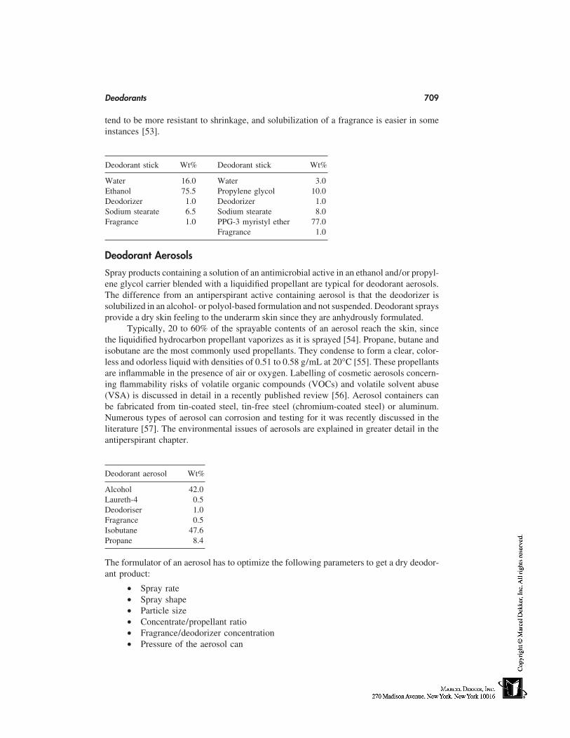
Deodorants 709
tend to be more resistant to shrinkage, and solubilization of a fragrance is easier in someinstances [53].
Deodorant stick Wt% Deodorant stick Wt%
Water 16.0 Water 3.0Ethanol 75.5 Propylene glycol 10.0Deodorizer 1.0 Deodorizer 1.0Sodium stearate 6.5 Sodium stearate 8.0Fragrance 1.0 PPG-3 myristyl ether 77.0
Fragrance 1.0
Deodorant Aerosols
Spray products containing a solution of an antimicrobial active in an ethanol and/or propyl-ene glycol carrier blended with a liquidified propellant are typical for deodorant aerosols.The difference from an antiperspirant active containing aerosol is that the deodorizer issolubilized in an alcohol- or polyol-based formulation and not suspended. Deodorant spraysprovide a dry skin feeling to the underarm skin since they are anhydrously formulated.
Typically, 20 to 60% of the sprayable contents of an aerosol reach the skin, sincethe liquidified hydrocarbon propellant vaporizes as it is sprayed [54]. Propane, butane andisobutane are the most commonly used propellants. They condense to form a clear, color-less and odorless liquid with densities of 0.51 to 0.58 g/mL at 20°C [55]. These propellantsare inflammable in the presence of air or oxygen. Labelling of cosmetic aerosols concern-ing flammability risks of volatile organic compounds (VOCs) and volatile solvent abuse(VSA) is discussed in detail in a recently published review [56]. Aerosol containers canbe fabricated from tin-coated steel, tin-free steel (chromium-coated steel) or aluminum.Numerous types of aerosol can corrosion and testing for it was recently discussed in theliterature [57]. The environmental issues of aerosols are explained in greater detail in theantiperspirant chapter.
Deodorant aerosol Wt%
Alcohol 42.0Laureth-4 0.5Deodoriser 1.0Fragrance 0.5Isobutane 47.6Propane 8.4
The formulator of an aerosol has to optimize the following parameters to get a dry deodor-ant product:
• Spray rate• Spray shape• Particle size• Concentrate/propellant ratio• Fragrance/deodorizer concentration• Pressure of the aerosol can
710 Schreiber
Deodorant Pump Sprays
Hydroalcoholic Pump Sprays
An alternative to aerosols are pump sprays. This category is quite popular in Europewhereas it is of lower interest for the consumers in the United States, since they tend toprefer a dry application form, like the anhydrous sticks. Pump sprays allow a good dosageof the formulation to be delivered to the underarm skin in a hygienic way. They consistof low vicosity hydroalcoholic solutions of a deodorizer and a perfume. Usually a solubi-lizer, like PEG-40 hydrogenated castor oil, is incorporated into the formulation to maintaina clear and homogeneous solution.
Pump spray Wt%
Water 35.6Alcohol 60.0PEG-40 hyd. 2.0Castor oilDeodorizer 2.0Fragrance 0.4
PIT-Emulsion Pump Sprays
A disadvantage of hydroalcoholic pump sprays is the alcohol content in the formulationthat may contribute to unwanted side reactions especially in the shaved axilla. BeiersdorfAG in Hamburg, Germany introduced into the European market under the brand name‘‘Nivea’’ a new pump spray based on an emulsion in 1995. The sprayable low viscousdeodorant is based on the PIT technology. Suitable mixtures of ethoxylated surfactants,oils and water in the presence of antiperspirant and deodorizing actives are heated to 60–90°C. Cooling the resulting W/O emulsion to room temperature yields via a phase inver-sion temperature (PIT) process a finely dispersed bluish-white O/W emulsion [58–60].The droplet size distribution of such PIT emulsions is in the range from 80–250 nm.The above-mentioned pump spray contained a skin-friendly deodorizing combination ofglyceryl monocaprinate and wool wax acids (see discussion p. 706) in an alcohol-freedelivery system.
PIT-emulsion pump spray Wt%
Glyceryl stearate, ceteareth-20,ceteareth-10, cetearyl alcohol,cetyl palmitate (Emulgade SE) 4.5
Ceteareth-20 1.0Dioctyl cyclohexane 5.0Dicaprylylether 5.0Deodorizer 2.0Aluminum chlorohydrate 5.0Water 77.5
Source: Ref. 60.
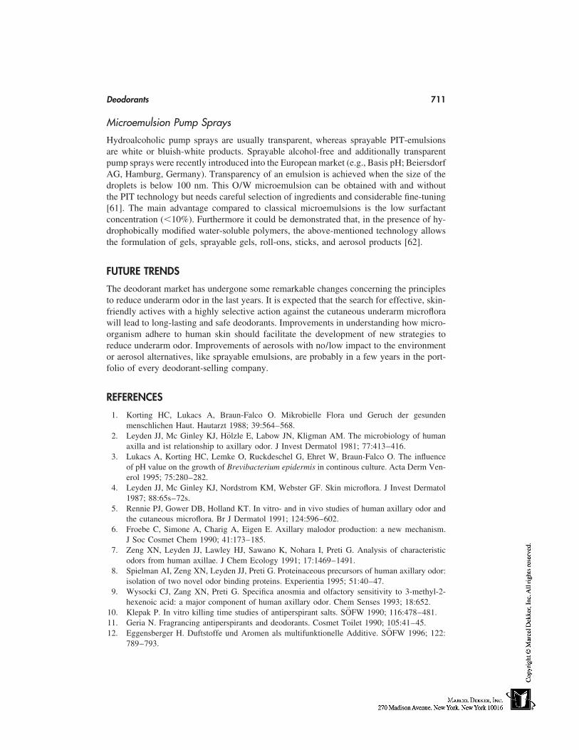
Deodorants 711
Microemulsion Pump Sprays
Hydroalcoholic pump sprays are usually transparent, whereas sprayable PIT-emulsionsare white or bluish-white products. Sprayable alcohol-free and additionally transparentpump sprays were recently introduced into the European market (e.g., Basis pH; BeiersdorfAG, Hamburg, Germany). Transparency of an emulsion is achieved when the size of thedroplets is below 100 nm. This O/W microemulsion can be obtained with and withoutthe PIT technology but needs careful selection of ingredients and considerable fine-tuning[61]. The main advantage compared to classical microemulsions is the low surfactantconcentration (�10%). Furthermore it could be demonstrated that, in the presence of hy-drophobically modified water-soluble polymers, the above-mentioned technology allowsthe formulation of gels, sprayable gels, roll-ons, sticks, and aerosol products [62].
FUTURE TRENDS
The deodorant market has undergone some remarkable changes concerning the principlesto reduce underarm odor in the last years. It is expected that the search for effective, skin-friendly actives with a highly selective action against the cutaneous underarm microflorawill lead to long-lasting and safe deodorants. Improvements in understanding how micro-organism adhere to human skin should facilitate the development of new strategies toreduce underarm odor. Improvements of aerosols with no/low impact to the environmentor aerosol alternatives, like sprayable emulsions, are probably in a few years in the port-folio of every deodorant-selling company.
REFERENCES
1. Korting HC, Lukacs A, Braun-Falco O. Mikrobielle Flora und Geruch der gesundenmenschlichen Haut. Hautarzt 1988; 39:564–568.
2. Leyden JJ, Mc Ginley KJ, Hölzle E, Labow JN, Kligman AM. The microbiology of humanaxilla and ist relationship to axillary odor. J Invest Dermatol 1981; 77:413–416.
3. Lukacs A, Korting HC, Lemke O, Ruckdeschel G, Ehret W, Braun-Falco O. The influenceof pH value on the growth of Brevibacterium epidermis in continous culture. Acta Derm Ven-erol 1995; 75:280–282.
4. Leyden JJ, Mc Ginley KJ, Nordstrom KM, Webster GF. Skin microflora. J Invest Dermatol1987; 88:65s–72s.
5. Rennie PJ, Gower DB, Holland KT. In vitro- and in vivo studies of human axillary odor andthe cutaneous microflora. Br J Dermatol 1991; 124:596–602.
6. Froebe C, Simone A, Charig A, Eigen E. Axillary malodor production: a new mechanism.J Soc Cosmet Chem 1990; 41:173–185.
7. Zeng XN, Leyden JJ, Lawley HJ, Sawano K, Nohara I, Preti G. Analysis of characteristicodors from human axillae. J Chem Ecology 1991; 17:1469–1491.
8. Spielman AI, Zeng XN, Leyden JJ, Preti G. Proteinaceous precursors of human axillary odor:isolation of two novel odor binding proteins. Experientia 1995; 51:40–47.
9. Wysocki CJ, Zang XN, Preti G. Specifica anosmia and olfactory sensitivity to 3-methyl-2-hexenoic acid: a major component of human axillary odor. Chem Senses 1993; 18:652.
10. Klepak P. In vitro killing time studies of antiperspirant salts. SÖFW 1990; 116:478–481.11. Geria N. Fragrancing antiperspirants and deodorants. Cosmet Toilet 1990; 105:41–45.12. Eggensberger H. Duftstoffe und Aromen als multifunktionelle Additive. SÖFW 1996; 122:
789–793.
712 Schreiber
13. Diehl KH, Oltmanns P, Ramsbotham J. Parfüminhaltsstoffe-eine Alternative für die konser-vierung von kosmetischen Produkten? SÖFW 1992; 118:546–550.
14. Morris JA, Khettry J, Seitz EW. Antimicrobial activity of aroma chemicals and essential oils.J Am Oil Chem Soc 1979; 96:595–603.
15. Rastogi SC, Johansen JD, Frosch P, Menne T, Bruze M, Lepoitthevin JP, Dreier B, AndersenKE, White IR. Deodorants on the European market: quantitative chemical analysis of 21fragrances. Contact Dermatol 1998; 38:29–35.
16. Lamp JH. Sodium bicarbonate. An excellent deodorant. J Invest Dermatol 1946; 7:131–133.17. Berschied JR. Antiperspirant-deodorant cosmetic stick products containing active agent parti-
cles in organic matrix, which matched densities for homogeneous products. Patent No. WO9413256.
18. Winston AE. Microporous alkali metal carbonate powder—comprises particles of averageparticle size of 0.1–50 microns, surface area of 5–20 sq.m/f, average pore size of 10–500nm and total pore volume of 0.1–2 cc/g and is useful as lightweight deodorant ingredient.Patent No. WO 9424996.
19. Park AC. Propellant free deodorant composition, for topical application—comprising spar-ingly water soluble salts or oxide (s) of zinc or magnesium, water absorbing cellulosic polymerand volatile silicone. Patent No. EP 471392 A.
20. Zekorn R. Deowirkstoff auf Basis Zinkricinoleat. Parf Kosmet 1996; 77:682–684.21. Zekorn R. Zinc ricinoleate. Cosmet Toilet 1997; 112:37–40.22. Kanda F, Yagi E, Fukuda M, Matsuoka M. Quenching short chain fatty acids responsible for
human body odors. Cosmet Toilet 1993; 108:67–72.23. Kanda F, Nakame T, Matsuoka M, Tomita K. Efficacy of novel hybrid powders to quench
body malodors. J Soc Cosmet Chem 1990; 41:197–207.24. Charig A, Froebe C, Simone A, Eigen E. Inhibitor of odor producing axillary bacterial exoen-
zymes. J Soc Cosmet Chem 1991; 42:133–145.25. Rennie PJ, Gower DB, Holland KT, Mallet AI, Watkins WJ. The skin microflora and the
formation of human axillary odor. Int J Cosmet Sci 1990; 12:197–207.26. Lukacs A, Korting HC, Braun-Falco O, Stanzl K. Efficacy of a deodorant and its components.
Triethylcitrate and perfume. J Soc Cosmet Chem 1991; 42:159–166.27. Baxter PM, Reed JV. The evaluation of underarm deodorants. Int J Cosmet Sci 1983; 5:85–
95.28. Cox AR. Efficacy of the antimicrobial agent triclosan in topical deodorant products. J Soc
Cosmet Chem 1987; 38:223–231.29. Nissen HP, Ochs D. Triclosan. Cosmet Toilet 1998; 113:61–64.30. Dillenburg H, Jakobson G, Klein W, Siemanowski W, Uhlig KH, Wolf F. Cosmetic deodorant
preparations containing di- or triglycerin esters. Patent No. EP 666732 A1/B1.31. Haustein UF, Herrmann J, Hoppe U, Engel W. Growth inhibition of coryneforme bacteria by
a mixture of three natural products. Farnesol, glyceryl monolaurate and phenoxyethanol: HGQ.J Soc Cosmet Chem 1993; 44:211–220.
32. Klier M, Schneider G, Traupe B, Voss I, Wolf F. Desodorierende Wirkstoffkombinationenauf der Basis von Wollwachssäuren und Monocarbonsäuren. DE 4305889.2
33. Klier M, Röckl M, Schneider G, Siemanowski W, Traupe B, Uhlig KH, Voss I, Wolf F.Deodorant active substance combinations made from wool grease acids and partial glycerides.EP 689418 A1.
34. Klier M, Röckl M, Traupe B, Wolf W. Deodorizing combinations of agents based on α,ω-alkane dicarboxylic acid and fatty acid partial glycerides. EP 729345 A1.
35. Klier M, Traupe B, Wolf F. Deodorant agent compositions containing α,ω-alcanoic di-acidsand mono-carboxylic esters of oligomer glyerols. EP 691125 A1.
36. Friberg SE, Larsson K. Food Emulsions. New York: Marcel Dekker, 1997.37. Meyer PD, Vianen GM, Baal HCI. Sucrose fatty acid esters in deodorant formulations. Aerosol
and Spray Report 1998; 37:18–22.
Deodorants 713
38. Meyer PD, Vianen GM, Baal HCI. Saccharose-Fettsäureester in deodorants. Parf Kosmet1997; 78:22–24.
39. Vianen GM, Watraven BW, Meyer PD. Deodorant composition. EP 0750903 A1.40. Beilfuss W. A multifunctional ingredient for deodorants. SÖFW 1998; 124:360–366.41. Wachter R, Lehmann R, Panzer C. Desodorierende Zubereitungen. DE 19540296.42. Lyon S, O’Neal C, van der Lee H, Rogers B. Amino acid β-Lyase enzyme inhibitors as deodor-
ants. WO 9105541.43. Laney J. O-Acyl Serines as deodorants. WO 9507069.44. Behan JM, Macmaster AP, Perring KD, Tuck KM. Insight how skin changes perfume. Int J
Cosmet Sci 1996; 18:237–246.45. Suffis R, Barr ML, Ishida K, Sawano K, van Loveren AG, Nakatsu T, Green CB, Reitz GA,
Kang RKL, Sato T. Composition containing body activated fragrance for contacting the skinand method of use. US 5626852.
46. Carson RG, Schilling KM, Harichian B, Au V. Biospecific emulsions. US 5416075.47. Bünger J, Schreiber J, Wolf F. Anti-adhesive active principles. EP 806935 A2.48. IFSCC Monograph No 6. Antiperspirants and Deodorants. Weymouth, MA: Micelle Press,
1998.49. Leyden JJ, McGinley K, Foglia AN, Wahrmann JE, Gropper CN, Vowels BR. A new method
for in vivo evaluation of antimicrobial agents by translocation of complex dense populationsof cutaneous bacteria. Skin Pharmacol 1996; 9:60–68.
50. Calogero AV. Antiperspirant and deodorant formulation. Cosmet Toil 1992; 107:63–69.51. Dawn R, Morton B. Clear cosmetic stick composition. WO 9427567.52. Kellner DM. Clear, stable deodorant compositions—containing soap, antimicrobial agent,
water, polyhydric alcohol, pentadoxynol 200 and alcanolamide-alkoxylated alcohol mixture.US 5407668.
53. Geria N. Formulation of stick antiperspirants and deodorants. Cosmet Toilet 1984; 99:55–66.54. Meyer G, Listro JA. Liquid deodorant compositions. WO 9301793.55. Johnsen MA. The safety assessment of hydrocarbon aerosol propellants. Spray Technology &
Marketing. March 1996; pp. 18–24.56. Redbourn D. Cosmetic aerosol regulations. Living with labelling. Soap Perf Cosmet Sept.
1998; pp. 45–48.57. Tait WS. Aerosol container corrosion and corrosion testing: what is state of the art? Spray
Technology & Marketing Sept. 1997; pp. 47–56.58. Wadle A, Förster T, von Rybinski W. Influence of the microemulsion phase structure on the
phase inversion temperature emulsification of polar oils. Colloids Surf A 1993; 76:51–57.59. Förster T, von Rybinski W, Tesman H, Wadle A. Calculation of optimum emulsifier mixtures
for phase inversion emulsification. Int J Cosmet Sci 1994; 16:84–92.60. Wadle A, Ansmann A, Jackwerth B, Tesmann H. PIT-Emulgiertechnologie in der Kosmetik
Parf Kosmet 1996; 77:250–254.61. Schreiber J, Eitrich A, Gohla S, Klier M, Wolf F. Cosmetic or pharmaceutical microemulsions.
WO 9628131 A2/A3.62. Schreiber J, Diec KH, Gers-Barlag H, Klier M, Wolf F. Cosmetic and pharmaceutical gels
based on microemulsions. WO 9628132 A2/A3.

58
Baby Care
Uwe SchönrockBeiersdorf AG, Hamburg, Germany
INTRODUCTION
Skin undergoes an extraordinary development. It must grow rapidly and expand dramati-cally in size to cover the entire developing body. It is exposed to both internal and externalenvironmental influences throughout the entire phase of its existence. However, despitethe multitude of regionally specific influences that play a role in the development of skin,there is a remarkable similarity in its developmental pattern and in the ultimate end productof differentiation in every part of the body. The purpose of this chapter is to outline whatis known about the development and physiology of baby skin and its implications on ourdaily care regime of skin at this early stage of life.
THE DEVELOPMENT OF BABY SKIN
The development of skin usually begins 7 to 8 days after fertilization, during which anouter blastodermic layer, the ectoderm, is formed. During the embryonic phase of develop-ment, two layers evolve from the ectoderm, the underlying basal layer from which theuppermost skin layer—the epidermis—and the cutaneous appendages develop, along withthe periderm, which faces the fetal cavity. When the epidermis is keratinized betweenweek 22 and 24 of pregnancy, the periderm separates itself from most parts of the body.In the third trimenon all cell layers in the epidermis that are typical for mature skin aredeveloped. However, until birth, the stratum corneum has still not developed a significantbarrier function. This is made clear when premature babies are observed. One of the big-gest problems in preemies is high transepidermal water loss (TEWL), although this de-creases exponentially with increasing gestation age. High TEWL in turn may lead to hypo-thermia and difficulty in fluid balance [1–6].
The mesoderm, a middle layer, develops at day 18 or 19 after fertilization. Themesoderm, with its mesenchymal cells, forms the dermis (corium). Epidermis and dermisare connected by a membrane (basal lamina). Within the third trimenon this contact area(the dermo-epidermal junctional zone) between the dermis and epidermis can now beclearly identified by commencing undulations and by the development of epithelial crestsand papillae. The development of the dermis also continues until the birth of the baby.In newborns it is about 60% as thick as in adults [1].
715
716 Schönrock
In the embryonic phase the dermis and the underlying subcutis cannot be differenti-ated from one another. In week 15 of gestation the subcutis can clearly be recognized.The lobuli network, in which the adipocytes (fat cells) spread, is formed. Within the thirdtrimenon large fat lobuli develop in the subcutis, which protect the organism from heatloss in cold conditions. Today it is still not clear what exactly stimulates the adipocytesto produce fat. The subcutis does not become thicker until after birth, depending on thebaby’s nutritional condition [1,2].
The sweat glands begin forming on the palms of the hands and the soles of the feetbetween weeks 10 and 12 of gestation. A portion of the excretory glands, however, remainsclosed until the end of month 7 of gestation. This is the reason why babies born prema-turely have developed, if at all, a limited ability to sweat [2]. They also show a limitedability to regulate body temperature as well as an increased TEWL, both of which needto be considered when setting up a daily skincare regime [7]. In contrast, the skin ofpreterm and full-term infants usually shows no signs of a physiological deficit.
THE PHYSIOLOGY OF BABY SKIN
Protection Against Water Loss
The following two basic mechanisms account for fluid transport through the skin:
1. Perspiration: active process in which water is excreted through the openings ofthe sweat glands. (Perspiration plays an important role in thermoregulation) [8].
2. TEWL: passive diffusion of water through the skin [9,10].
Gestation age plays an essential role in the birth of a baby [11]. The TEWL decreaseswith increasing gestation age. In a fully developed newborn, a TEWL of 6 to 8 g waterper m2 of skin per hour is low. However, the TEWL is considerably higher in prematurelyborn babies, especially those born before week 30 of pregnancy. In the first months oflife, water loss in infants increases slightly. The explanation for this is that the babiesbegin to perspire slightly [12].
As the body temperature rises, the permeability of the skin also increases, leadingto higher water loss. As environmental temperatures rise, water evaporates faster. Thisfact must be considered especially when caring for newborns. Creams and ointments withoccluding effect can lower TEWL. The application of liquid paraffin on the skin can reduceTEWL by up to 50%.
Protection Against Percutaneous Absorption of Harmful Substances
In addition to providing protection against water loss, the skin barrier function ensuresthat chemical agents, which could harm the organism, cannot penetrate percutaneously(through the skin). The permeability rate in prematurely born babies is 5 to 50 times higherthan in fully developed newborns. The ratio between body surface and body weight isalmost 2.5 times higher in newborns than in adults. This surface volume ratio is oneof the essential points that must be considered in the application of topically affectivetherapeutics. Particularly with treatment of large areas, e.g., of dermatologicals containingcorticoids, there is the danger of increased systemic absorption [13].
With increasing maturation, the epidermal cells develop increased metabolic activ-ity. This means that the activation of enzymes can render potentially harmful substancesharmless. They are modified through oxidation, hydrolysis, reduction, deamination, or
Baby Care 717
conjugation and thereby inactivated. This enzyme activity is very restricted, especially inprematurely born babies, so that potentially harmful substances can enter the blood streamin an unaltered state if absorbed percutaneously [13].
Protection Against Pathogenic Micro-Organisms
After birth, the body of the baby is exposed to numerous germs. The skin barrier notonly protects mechanically against invading micro-organisms, but also through the slightlyacidic milieu of the hydrolipidic film on the surface of the skin. The surface of the skinis physiologically populated by specific germs (saprophytes), which are not pathogens butrather a vital microbial defense system on the skin’s surface. For optimal living conditions,the saprophytes require an acidic milieu. Directly after birth, however, alkaline valuesprevail on the surface of the body of the newborn. It can be assumed that these alkalinevalues result from the vernix caseosa residue. Neither weight at birth nor gestation ageseem to have an influence on the pH value. Within the first 24 hours after birth, the pHvalue drops noticeably. In the first month of life, the pH value then stabilizes at a slightlyacidic range (slightly below a pH value of 6) [14].
The natural acid mantle of the skin on the newborn is already developed in the firstfew days of life, so that pathogenic micro-organisms generally find the conditions unsuit-able for their survival. However, the alkaline-neutralizing properties of the skin of new-borns and small children is restricted. After contact with alkaline substances (e.g., alkalinesoaps), the skin requires a longer time to restore its slightly acidic physiological pH valueas compared with adult skin [15].
FREQUENT SKIN PROBLEMS IN NEWBORNS
Diaper Dermatitis
At the beginning of this century, in 1905, a French pediatrician by the name of Jacquetgave the peculiar frequently occurring skin rash in the diaper area the name diaper derma-titis [16]. The skin alterations subcategorized under the diagnosis diaper dermatitis canhave a variety of causes. They can be directly related to the contact dermatitis, which isdiaper dermatitis in a narrow sense. The occurence can also be unrelated to the use ofdiapers. Today the factors that enhance this irritating contact dermatitis are known:
1. Diapers that have an occluding effect in an already moist environment, whichresults in an increased hydration of the stratum corneum.
2. The increased hydration facilitates penetration of xenobiotics.3. The still very thin epidermis of the newborn reacts sensitively to mechanical
stress and friction.4. The skin barrier function is weakened, and the skin shows an increased irrita-
bility.
In addition, an increase of the pH value in the diaper area can also encourage an outbreakof diaper dermatitis. The alkaline urine activates enzymes (lipases and proteases) in thefeces, which irritate the skin [17–19].
Boys and girls are equally afflicted with diaper dermatitis. It mainly occurs between3 and 10 months of age, with a frequency peak between 6 and 9 months. Typically, askin erythema can be found on the inside of the thighs and on the baby’s bottom. The
718 Schönrock
skin is increasingly reddened, has a shiny, glassy appearance, and is wrinkled on thesurface.
Corticosteroids are used occasionally in the treatment of diaper dermatitis. In thefollow-up treatment, emollients containing zinc oxide are mainly used. Zinc oxide has anastringent, slightly disinfectant effect and offers the skin protection against urine and feces[17–21].
Protective creams containing zinc oxide are usually used to cover the skin of thediaper area with a highly viscous film, which inhibits the penetration of xenobiotics with-out fully occluding the skin. In order to accomplish this goal, usually water-in-oil (W/O)creams are used, which contain one or more of the following ingredients: petrolatum,lanolin, lanolin alcohol, paraffin oil, natural oils, waxes, zinc oxide, and possibly cod liveroil, vitamins, plant extracts, and titanium dioxide.
Diaper candidiasis is a fungal-infected diaper dermatitis. The most common caus-ative agent is a yeast fungus called Candida albicans. It is a known fact that extensive useof antibiotics in newborns and small children increases that incidence of diaper candidiasis.Initially, diaper candidiasis can be treated with a specific antimycotic therapy (nystatin,clotrimazole), then followed up with the healing methods for basic diaper dermatitis aspreviously described [22].
Neurodermatitis
Neurodermatitis, also called atopic dermatitis, is a skin disease that may occur at a veryearly age. It can be identified by the so-called milk crust on the reddened, damp skin ofthe head and cheeks of the newborn. As the first indication of an outbreak of neurodermati-tis, the milk crust often provides the starting point for other skin disorders. The skinbecomes cracked and transparent, and the permeability increases. Once the skin is dam-aged, the risk of infection is higher. The skin becomes increasingly dry, transparent, andirritated, with intensified itchiness. The temptation to keep on scratching the skin is usuallyalmost irresistable for small children. Atopic dermatitis is an immunological reaction thataffects the skin to an especially large extent. More than 10% of children in industrializedcountries are already afflicted, with a rising tendency. The combination of the geneticpredisposition and environmental influences as well as psychological and neurovegetativefactors can result in an outbreak of this disease [23–25].
Adequate skincare, which reinforces the skin’s vital barrier, is a meaningful prophy-laxis for avoiding a first outbreak of neurodermatitis in high-risk allergy children. Thefollowing measures can help:
• Mild cleansing agents• Moisturizing emulsions to support the skin’s barrier function• Skincare products with proven skin tolerability• Skincare products and cleansers with few, carefully selected ingredients, in order
to keep the risk of allergies as low as possible [23–25]
THE CARE OF BABY SKIN
The effects of baby-care products can usually be divided into the following categories:cleansing, caring, and protection. Currently, a multitude of product types can be found inthe market. Although the shear number of products is overwhelming, there are features
Baby Care 719
they all have in common. The following three sections will deal with product characteris-tics and general usage advice in the various segments of baby care.
Cleansing
Bath Additives
As soon as the umbilical cord has fallen off, the baby can be bathed [26–29]. However,daily bathing of the baby is not advisable, as this would dry out the skin too much. Abath every 2 to 3 days is sufficient. The bath temperature should lie between 36 and 37°C.
Bath additives usually contain a mixture of various anionic (e.g., fatty alcohol ethersulfates, protein fatty acid condensates), nonionic (e.g., ethoxylated fatty alcohols, fattyacid glycerides), and amphoteric (e.g., betaines) surfactants. Numerous protein hydroly-sates, superfatting agents, solubilizing agents, plant extracts, colorants, and perfumes arealso found in this product category. In general, bath additives contain mild surfactantmixtures, which neither dry out the skin nor burn in the eyes.
Cleansers for the Diaper Area
Baby oils containing mineral oils as well as oil-impregnated towelets are widely used.(Towelets are usually supplied in dispenser boxes securing product hygiene up to the lasttowelet used.) Liquid petrolatum is a very desirable ingredient in view of its stability,touch, barrier function, and cost. Liquid petrolatum also has a remarkable occlusivity.Intertrigo areas should therefore be frequently cleansed (1–3 times daily) with oil or oil-containing towelets.
Soft towelets containing mild oil-in-water (o/w) cleansing milks or, alternatively,clear cleansing lotions are also frequently found. They normally contain anionic and/ornonionic surfactants in low concentrations as well as varying amounts of skincare ingredi-ents like plant extracts and protein hydrolysates. These towelets are also offered in dis-penser boxes.
Whereas the irritating effect of soaps mainly results from their alkalinity, the useof alkaline-free soaps has shown that all detergents induce a significant delipidizing effect,which can also contribute to skin irritation [26–29].
Liquid cleansers are usually used for cleansing of the face, arm pits, and the genitalarea. Normally alkaline free, their composition resembles the composition of baby sham-poos, whereas the concentration of surfactants is normally higher. The reasoning behindthe higher surfactant level lies in the smaller product amount used for cleansing [26–29].
Shampoos
Baby shampoos are usually formulated to be nonirritating to the eyes. This guaranteesextraordinary product safety and also ensures that babies do not object to shampooing.Although basically the ingredients used are comparable with the ingredients found in bathadditives, the concentration of surfactants is normally lower. Viscosity is adjusted to about1000 centipoise (cps) to make it hard for the shampoo to migrate into the eyes.
Care
Face and Body Creams/Body Lotions
Face creams are especially important for the protection against environmental influenceslike sunlight, wind, and cold temperatures, which may dry out baby skin. The composition
720 Schönrock
resembles that of the body-care creams, although the moisturizer content is often higher.The ingredients used are often more compatible with the mucous membranes (especiallyin the area of the eyes) than in the case of body creams. Body-care creams are frequentlyused for their excellent superfatting properties. Both o/w and w/o emulsions are foundon the market.
Body-care lotions are normally used for large-area body care, e.g., after baby bath.Both o/w and w/o emulsions are found in the market. Classic ingredients used are lanolin,lanolin alcohol, paraffin oil, vaseline, natural and synthetic wax esters, natural oils, fattyalcohols, and emulsifiers (e.g., fatty acid glycerides, ethoxylated fatty alcohols). Manyskin-caring, soothing active ingredients are also found.
Protection
Sun Protection
Spending summer vacation at the seashore is a tradition of many families. Unfortunately,the beach is a high-risk environment for future skin cancer because it allows for maximumsunlight exposure. Heat, wind, and humidity are often present. These factors can enhanceor intensify UV injury. With or without topical sun-protection measurements, babies andsmall children should be kept out of direct sunlight. As soon as children begin to exploretheir environment, it usually becomes impossible to confine them to the shade. In suchcases, sunscreens need to be applied.
A wide variety of different o/w and w/o emulsions, hydrogels and oleogels arefound in the market using a variety of UV-filter systems. Many products contain broadspectrum (UVA and UVB) sunscreens with a moderate SPF. Products with a water-resis-tant SPF are favorable at the seashore [30–33].
Cold Protection
Mild facial creams are especially important in the winter for protection against the harsheffects of a dry, cold climate. At freezing temperatures, significant protection against frostbite is obviously helpful. Specific petrolatum-based water-free formulations, which option-ally contain zinc oxide and skin-soothing agents like panthenol, can protect the skin attemperatures below freezing.
QUALITY MANAGEMENT IN BABY CARE
Despite careful research with respect to the good skin tolerability of each individual ingre-dient in baby-care formulations, it should be made certain that this data will also applyto the final product after these ingredients are integrated into the formula. In order to ruleout the possibility of contact allergies or sensitizing skin reactions, products are frequentlytested using the repeated-insult patch test (RIPT). This test is a validated, recognizedmethod for the testing of skin sensitization. The test preparations are repeatedly appliedto the same localization for 3 weeks. After a 2-week break, the test materials are appliedonce again on another location and the skin is assessed for any allergic reaction that couldpossibly have been induced [34]. Exposure to sunlight can cause certain ingredients totrigger photoallergic or phototoxic skin reactions. Photopatch or phototoxicity tests enablethe detection of UV-induced irritant or allergic skin reactions.
In the elbow-wash test, the skin tolerability of cleansing formulas is tested in thesensitive crease of the elbow under controlled and extreme washing conditions, and com-
Baby Care 721
pared with a skin-friendly standard product. The evaluation of the skin reaction is per-formed after repeated washings over a period of 5 days, based on subjective and objectivereports [35].
In a clinical application test, skin tolerability as well as the skincare properties ofbaby products can be tested. At the start, and again after 4 weeks of practical applicationof baby-care products, dermatological examinations are carried out. Parents are givendiaries for the daily evaluation of product properties. Children known to have skin allergiesto ingredients in the test products are excluded from the testing.
SUMMARY
Normal baby skin shows no natural inborn deficits that need special treatment. However,the elevated skin permeability in newborns needs to be considered when establishing aroutine skincare regime. The sensible use of skin-cleansing and caring products surelyneeds to be remembered.
However, there is a growing demand for specific dermatological treatments of new-borns, as the number of skin disorders (e.g., neurodermatitis) in this age group are on theincrease.
REFERENCES
1. Holbrook KA, Sybert VP. Basic science. In: Schachner L, Hansen R, eds. Pediatric Dermatol-ogy 2d ed., Vol. 1. New York: Churchill-Livingstone, 1995; 1–70.
2. Holbrook KA. Structure and function of the developing skin. In: Goldsmith LE, ed. Physiol-ogy, Biochemistry and Molecular Biology of the Skin. 2d ed., Vol. 1. New York: OxfordUniversity Press, 1991: 63–110.
3. Hammerlund K, Sedin G, Stromberg B. Transepidermal water loss in newborn infants. VII.Relation to postnatal age in very pre-term and full-term appropriate for gestational age infants.Acta Paediatr Scand 1982; 71:369–374.
4. Doty SE, McCormack WB, Seagrave RC. Predicting insensible water loss in premature neo-nates. Biol Neonate 1994; 66:33–44.
5. Wilson DR, Maibach HI. Transepidermal water loss in vivo. Preterm and term infants. BiolNeonate 1980; 37:180–185.
6. Rutter N, Hull D. Water loss from the skin of term and preterm babies. Arch Dis Child 1979;54:858–868.
7. Lane AT. Development and care of the premature infant’s skin. Pediatr Derm 1987; 4:1–5.8. Hey EN, Katz G. Optimum thermal environment for naked bodies. Arch Dis Child 1970; 45:
328–334.9. Hammerlund K, Nilsson GE, Öberg PA, Sedin G. Transepidermal water loss in newborn in-
fants II. Acta Paediatr Scand 1978; 68:371–376.10. Fanaroff AA, Wald M, Gruber HS, Klaus MH. Insensible water loss in low birth weight in-
fants. Pediatrics 1972; 50:236–245.11. Dubowitz LMS, Dubowitz V, Golberg C. Clinical assessment of gestational age in the newborn
infant. J Pediatr 1970; 77:1–10.12. Harpin VA, Rutter N. Barrier properties of the newborn infant’s skin. J Pediatr 1983; 102:
419–425.13. Barrett DA, Rutter N. Transdermal delivery and the premature neonate. Crit Rev Therapeutic
Drug Carrier Systems 1994; 11(1): 1–30.14. Tunnessen WW. Practical aspects of bacterial skin infections in children. Pediatr Derm 1985;
2(s):255–265.
722 Schönrock
15. Braun F, Lachmann D, Zweymüller E. Der Einfluss eines synthetischen Detergens auf denpH der Haut von Säuglingen. Der Hautarzt 1986; 37:329–334.
16. Jacquet L. Traite de Maladie de l’Enfance. Paris: Grauncher & Comby, 1905.17. Lane AT. Diaper rash: causes and cures. Patient Care 1988:167–173.18. Honig, PJ. Diaper dermatitis. Postgrad Med 1983; 74(6):79–88.19. Rasmussen JE. Classification of diaper dermatitis: an overview. Pediatr 1987; 14(Suppl 1):
6–10.20. Agren MS. Percutaneous absorption of zinc and zinc oxide applied topically to intact skin in
man. Dermatol 1990; 180:36–39.21. Derry JE, McLean WM, Freeman JB. A study of the percutaneous adsorption from topically
applied zinc oxide ointment. J Parenteral Enteral Nutri 1983; 7:131–135.22. Stögmann W. Empfehlungen zur Lokaltherapie banaler Dermatosen im Kindealter. WMW
1984; 1:19–24.23. Stögmann W. Empfehlungen zur Behandlung und Prophylaxe der Atopischen Dermatitis im
Kindesalter. WMW 1989; 18:414–421.24. Saurat JH. Atopische Dermatitis beim Kind. Annales Nestle 1987; 45:10–28.25. Queille-Roussel C, Raynaud F, Saurat JH. A prospective computerized study of 500 cases of
atopic dermatitis in childhood. Acta Derm Venerol (Stockholm) 1985; 114:87–92.26. Schneider W. Nutzen und Schaden von Seifen und Syndets. Kosmetologie H 1971; 2:54–56.27. Debsi S, Jonte G. Skin cleansing and skin care in infants. Ärztl Kosmet 1987; 17(1):65–69.28. Vergesslich KA, Zweymüller E. Sind die neuen Waschmittel in der Pediatrie von Vorteil?
Wiener klinische Wochenzeitschrift 1982; Jg 94, Heft 12; 4:321–359.29. Cowan ME, Frost MR. A comparison between a detergent baby additive and baby soap on
the skin flora of neonates. J Hosp Infect 1986; 7:91–95.30. Stern RS, Weinstein MC, Baker SG. Risk reduction for nonmelanoma skin cancer with child-
hood sunscreen use. Arch Dermatol 1986; 122:537–545.31. Owens DW, Knox JM, Hudson HT, Troll D. Influence of humidity on ultraviolet injury. J
Invest Derm 1975; 64:250–252.32. Freeman RG, Knox JM. The influence of temperature on ultraviolet injury. Arch Derm 1967;
89:858–864.33. Owens DW, Knox JM, Hudson HT, Troll D. Influence of wind on ultraviolet injury. Arch
Derm 1974; 109:200–201.34. Schelanski H., Schelanski M. A new technique of human patch test. Proc Sci Section 1953;
19:46–49.35. Lukacovic MF, Dunlpa FE, Michaels SE, Visscher MD,Watson DD. Forearm wash test to
evaluate the clinical mildness of cleansing products. J Soc Cosmet Chem 1988; 39:355–366.
59
Cosmetics for the Elderly
Uwe SchönrockBeiersdorf AG, Hamburg, Germany
INTRODUCTION
Aging is a basic biological process common to all living organisms. Its biochemical mech-anisms have yet to be elucidated in detail. Aging is usually understood as an irreversible,progressive loss of homeostatic capacity. By definition, aging affects everyone, but at avariable rate. At present, aging is widely assumed to result partly from a genetically deter-mined program and partly from endogenous and exogenous insults. Both processes occurat the level of individual cells.
At the organ level generally and in the skin specifically, aging is manifested by aloss of maximum metabolic activity and increasing sensitivity or susceptibility to certaindiseases and environmental factors. The purpose of this chapter is to outline what is knownabout morphological and physiological aging of the skin and its implications for a tailoredskincare of the elderly.
AGE-ASSOCIATED CHANGES IN HUMAN SKIN
Morphological and Histological Changes
The major aging changes in the morphology of the skin include dryness (roughness andscaliness), wrinkling, and laxity [1]. The most striking and consistent change is a flatteningof the dermal-epidermal junction [2]. This results in a considerably smaller surface be-tween the two compartments. This presumably leads to less nutrient transfer and may causethe relatively smaller proliferative compartment in the epidermis. It is also responsible forthe lower resistance to shear forces [1]. However, most of the apparent clinical changesassociated with advanced age are attributable to chronic sun damage [3,4].
Physiological Changes
An age-associated decrease in the epidermal turnover rate of approximately 30 to 50%between the third and the eighth decade has been determined by a study of desquamationrates at selected body sites [5]. The thymidine labeling index of the epidermis in vivo hasbeen reported to decline nearly 50% during the human life span [6].
723
724 Schönrock
Recent studies using highly sensitive techniques for the measurement of sebum se-cretion rates have documented a decline of approximately 23% per decade [7]. The physio-logical consequences of decreased sebum production in old age, if any, are unknown [1].
Clinical studies showed that eccrine sweating is markedly impaired with age. Spon-taneous sweating in response to dry heat, measured on digital pads, was reduced by morethan 70% in healthy old subjects [8], primarily attributable to a decreased output per gland.
The decreased vascular reponsiveness in elderly skin has been documented by clini-cally assessing vasodilation and transduction after application of standard irritants likehistamine [9]. The intensity of erythema after UV exposure also decreases with age [10].
An age-associated decrease in delayed hypersensitivity reactions in human skin ismanifested by a relative inability of healthy elderly subjects to develop sensitivity to dini-trochlorobenzene (DNCB), and by their lower rate of patch-test reactions to standard recallantigens. The cutaneous manifestations of immediate hypersensitivity similarly declineswith age [1].
Langerhans cells are the epidermal cell population, which is largely responsible forrecognition of foreign antigens. An approximately 25 to 50% reduction in the number ofepidermal Langerhans cells occurs between early and late adulthood [11] and substantiallycontributes to the age-associated decrease in cutaneous immune responsiveness. Theamount of dermal mast cells likewise decreases with age. The resulting consequencesbeyond the reduced rate of immediate hypersensitivity reactions, such as a positive ‘‘prick-test’’ [12] or acute urticaria, are unknown.
Photoaging
Photoaging is a term used to describe the array of clinical and histological findings in thechronically sun-exposed skin of middle-aged and elderly adults. It has also been calleddermatoheliosis [13] and heliodermatitis [14], the latter term reflecting the low-grade in-flammatory nature of the process.
Clinical features of actinically damaged skin include coarseness, wrinkling, irregularpigmentation, telangiectasia, and scaliness, as well as a variety of premalignant and malig-nant neoplasms. The relative severity of these changes varies considerably among individ-uals. This undoubtedly reflects strong differences in past sun exposure and marked individ-ual differences in vulnerabilities and repair capacities for solar insults. Photoaging usuallyinvolves most severely the face, neck, or extensor surface of the upper extremities [15].
THE COSMETIC CARE OF ELDERLY SKIN
Cosmetics for elderly skin can usually be divided into the categories of facecare andbodycare. Currently, a multitude of product types can be found. Although the number ofproducts is overwhelming, there are common features to be mentioned. The followingtwo sections will deal with product characteristics in various segments for the cosmeticcare of elderly skin.
Facecare
Skincare
Concepts for cosmetics suited for elderly people are often based on the dry skin conditionstypical for elderly skin. Many skincare formulations contain humectants, which enable
Cosmetics for the Elderly 725
excellent transient hydrational/moisturizing effects. While lessening the prominence ofundesirable surface defects, these formulations have only minor influence on dermallosses. However, evidence is accruing that the following groups of topically applied activesdo seem to reverse the degenerative skin changes seen with aging.
By far the most exciting discovery in cutaneous gerontology during the past decadeis the effect of tretinoin (all-trans retinoic acid) on the clinical and histological appearanceof photoaged skin. Kligman first realized that topical tretinoin improved the appearanceof middle-aged women using the drug to control facial acne. Support for the concept wasprovided by a double-blind vehicle-controlled trial documenting tretinoin’s effectivenesson human photoaging. After 4 months of daily application, 0.1% tretinoin cream producedstatistically significant improvement in fine and coarse wrinkling, sallowness, androughness of sun-damaged facial and arm skin [16].
Tretinoin was the first agent shown to reverse age-associated changes in any tissue.This statement must be qualified in that it is unclear whether tretinoin truly reverses agingchanges or simply produces new changes that mimic a reversal. It is unclear whethertretinoin affects exclusively sunlight-induced pathologies or a combination of sun damageand intrinsic aging changes [1].
In the past years, estrogen supplementation of the climacteric women has openednew aspects on the wide variability of estrogen effects in various tissues. In skin, estrogensincrease vascularization and show effects at various levels of dermal tissue [17,18].
Several attempts have been made to check the skincare efficacy of estriol (0.3%)or estradiol (0.01%) in perimenopausal women. Daily application of a cream over a periodof 7 months resulted in a significant increase of skin parameters like skin firmness, wrin-kles, and skin moisture content. Hormonal levels showed a slight increase in the prolactinlevel, whereas the estradiol level was unchanged. Side effects were not found [19,20].
Many further topical actives with excellent antiaging potential are currently used inmarketed formulations, the number of which is increasing year after year [21–23].
Skin Cleansing
Active detergent substances contained in cleansing agents for human skin inevitably resultin a degreasing of the keratinous layer, so that natural, moisture-retaining substances arealso rinsed out in the process. It is, however, possible, by selecting the proper cleansingagents and reducing the frequency and intensity of their application, to reduce the unfavor-able influence of various washing procedures on the skin of such elderly persons to aconsiderable extent.
Facial skin cleansers for elderly skin are usually particularly mild and superfatting.Both surfactant-based and oil-in-water (o/w) emulsion-based formulations are currentlyfound. In the surfactant-based formulations, surfactants like ampholytes, betaines, sulfo-succinates, and various types of alkylpolyglucose are frequently used, whereas o/w emul-sion-based formulas frequently contain superfatting agents and various humectants, whichsecure good cleansing efficacy without drying out the skin.
Bodycare
Skincare
For active care by means of humidity and lipid substitution, mainly o/w and water-in-oil(w/o) emulsions are used, which combine occlusive effects and moisturizing action. Inaddition to pyrrolidone carboxylic acid salt and urea, other humectant substances such as
726 Schönrock
alpha-hydroxy acid and hyaluronic acid, a highly efficient moisturizer, are frequentlyfound. It is self-evident that such formulation ingredients as glycerine, propylene glycol,and other glycols also contribute to their humectancy.
Polar and unpolar lipids are frequently used in bodycare formulations. They act asemollients, as protective lipids, and as structure formers of the liquid crystalline bilayersbetween the corneocytes. These three functions are usually performed by fatty alcohols,fatty acids, and short- and long-chain esters, along with triglycerides and waxes. Specialeffects are frequently delivered by liposomes containing phospholipids, sphingolipids, andceramides, and lead to the desired long-term effects. This is attributable to special bindingmechanisms in the skin, an anchor capacity of transported and/or encapsulated activeingredients, and their slow release.
Skin Cleansing
Bath additives usually contain a mixture of various anionic, nonionic, and amphotericsurfactants. Numerous superfatting agents, solubilizing agents, plant extracts, and per-fumes are also found in products within this category. However, only an oil bath forelderly skin may provide skin cleansing and conditioning at the same time. For seriousdry-skin conditions, oil baths are indispensable.
A variety of shower products, meanwhile, also contain high amounts of superfattingagents, thus securing their good skin compatibility and low drying-out potential.
SUMMARY
There is an increasing demand for face- and bodycare formulations tailormade for thecosmetic treatment of elderly skin. Modern topical formulations not only deliver excellentmoisturizing and superfatting capabilities, but many products, especially facecare prod-ucts, contain one or more actives counteracting the signs of intrinsic and/or photoaging.However, it is still not clear whether these actives reverse the signs of aging or induceother effects on the skin that mimic a reversal of skin aging.
REFERENCES
1. Gilchrest BA. Physiology and pathophysiology of aging skin. In: Goldsmith LA, ed. Physiol-ogy, Biochemistry, and Molecular Biology of the Skin. Vol. 2, 2nd ed. New York, OxfordPress, 1991:1425–1444.
2. Hull MT, Warfel KA. Age- related changes in the cutaneous basal lamina: scanning electronmicroscopic study. J Invest Derm 1983; 81:378–380.
3. Tindall JP, Smith JG. Skin lesions of the aged and their association with internal changes. JAm Med Assoc 1963; 186:73–76.
4. Beauregard SB, Gilchrest BA. A survey of skin problems and skin care regimes in the elderly.Arch Derm 1987; 123:1638–1643.
5. Tan CY, Statham B, Marks R, Payne PA. Skin thickness measurement by pulsed ultrasound:its reproducability, validation and variability. Br J Derm 1982; 106:657–662.
6. Kligman AM. Perspectives and problems in cutaneous gerontology. J Inv Derm 1979; 73:39–46.
7. Jacobsen E, Billings JK, Frantz RA. Age- related changes in sebum secretion rate in men andwomen. J Inv Derm 1985; 85:483–485.
8. Silver AF, Motagna W, Karacan I. The effect of age on human eccrine sweating. In: MotagnaW, ed. Advances in the Biology of the Skin Aging. Vol. 6. Pergamon Press, 129–137.
Cosmetics for the Elderly 727
9. Grove GL, Lavker RM, Hölzle E, Kligman AM. Use of nonintrusive tests to monitor age-associated changes in human skin. J Soc Cosm Chem 1981; 32:15–19.
10. Gilchrest BA, Stoff JS, Boter NA. Chronologic aging alters the response to UV-induced in-flammation in human skin. J Inv Derm 1982; 79:11–15.
11. Thiers BH, Maize JC, Spicer SS, Cantor AB. The effect of aging and chronic sun exposureon human Langerhans cell populations. J Inv Derm 1984; 82:223–226.
12. Barbee RA, Levowitz MD, Thompson HC, Burrows B. Immediate skin-test reactivity in ageneral population sample. Ann Intern Med 1976; 84:129–133.
13. Gilchrest BA. Skin and Aging Processes. Boca Raton: CRC Press, 1984.14. Lavker RA, Kligman AM. Chronic heliodermatitis: a morphologic evaluation of chronic ac-
tinic dermal damage with emphasis on the role of mast cells. J Inv Derm 1988; 90:325–330.15. Knox JM, Cockcrall EG, Freeman RB. Etiological factors and premature aging. J Am Med
Assoc 1962; 179:630–634.16. Weiss JS, Ellis CN, Headington JT, Voorhees JJ. Topical tretinoin in the treatment of aging
skin. J Am Acad Dermatol 1988; 19:169–175.17. Schmidt JB. Externe Östrogenapplikation bei Hautalterung im Klimakterium—Ein Ther-
paieansatz-. H � G. 1993; Band 68; Heft 2:84–87.18. Artner J, Gitsch E. Über Lokalwirkungen von Östriol. Geburtshilfe und Frauenheilkunde.
1959; 19:812–819.19. Punnonnen R, Vaajalahti P, Teisala K. Local oestriol treatment improves the structure of elastic
fibres in the skin of postmenopausal women. Ann Chir Gynaecol (Suppl.) 1987; 202:39–41.20. Schmidt JB, Binder M, Demschick G, Biegelmayer C, Reiner A. Treatment of skin aging with
topical estrogens. Int J Derm 1996; 35:669–674.21. Smith WP. Hydroxy acids and skin aging. Cosmet Toilet 1994; 109:41–48.22. Pierrefriche G, Laborit H. Oxygen free radicals, melatonin, and aging. Exp Gerontol 1995;
30:213–227.23. Coles LS, Harris SB. Coenzyme Q10 and lifespan extension. In: Klatz RM. ed. Advances in
Anti-Aging Medicine. Larchmont, NY, Mary Ann Liebert, Inc., 1996:205–216.

60
EEC Cosmetic Directive and Legislation in Europe
René Van EsscheFree University of Brussels, Brussels, Belgium
THE LAWS OF THE MEMBER STATES RELATING TO COSMETICPRODUCTS AND THE 6TH AMENDMENT
The Council of the European Communities in regard to the Treaty establishing the Euro-pean Economic Community (today, the European Union [EU]) and in particular Article100 thereof has decided to harmonize legislation in the EU [1,2]. The Directive gives aclear definition of cosmetic products: ‘‘Any substance or preparation intended to be placedin contact with the various external parts of the human body or with the teeth and andthe mucous membranes of the oral cavity, with a view exclusively or mainly to cleanthem, perfuming them, changing their appearance and—or correcting body odours and—or protecting them or keeping them in good condition.’’ The philosophy of the Directiveis that all products should have equal and immediate access to the market throughout theEU provided that they are proven safe for human use. The Directive has been adaptedand modified 29 times between 1976 and 1998. The 6th Amendment has made mandatoryby January 1, 1997 that cosmetic products may be marketed only if the labeling bearsspecific information in legible and visible lettering (Article 6) as follows: the name andaddress or registered office of the manufacturer or the responsible person for marketingin the Union, the nominal content at the time of packaging, the date of minimum durabilityand the conditions of storage if appropriate, the conditions of use and warnings, the batchnumber, the function, the list of ingredients in descending order of weight. Article 7arequires that for control purposes the following information be readily accessible to thecompetent authorities of the Member State: the qualitative and quantitative compositionof the product (perfumes may be coded) (good laboratory procedures [GLP], O.J. EU n°L 15, 17—01—87, p. 29), the physicochemical and microbiological specifications of theraw materials and the finished product, the purity and the microbiological control criteriaof the cosmetic product, the method of manufacture (good manufacturing procedures,GMP), the person responsible for the manufacturing or first importation into the EU shallpossess an appropriate level of qualification, the assessment of the safety (GLP, CouncilDirective 87—18—EEC of 18 December 1986), the name and address of the responsibleperson (who must hold a diploma according to Article 1 of Council Directive 89—48—EEC), undesirable effects if existing, and proof of effect by the nature of effect. The
729
730 Van Essche
competent authority of the Member State shall be notified of the place of manufacture orinitial import into the EU of the cosmetic products before the latter are placed on themarket, the Poison Information Center shall be informed about the formula, and The Euro-pean Cosmetic, Toiletry and Perfumery Association (COLIPA) [3] has negotiated thatonly major deviations from basic formulas shall be indicated (the basic formulas havingbeen given by COLIPA).
The Committee on the Adaptation to Technical Progress of the Directives on theRemoval of Technical Barriers to Trade in Cosmetic Products in set up. This Committeeis located in Brussels at the European Commission (DG Enterprise, Industry, 200 rue dela Loi, B-1029 Brussels, Belgium, tel; 32 2 299 1111). Article 12 deals with product, thatalthough complying with the Directive, may represent a risk to human health.
The Directive includes seven annexes, and the eighth is pending.
Annex I. Illustrative list by category of cosmetic products.Annex II. List of substances that must not form part of the composition of cos-
metic products. 420 substances are listed. On a time-to-time basis, new substancesare included in the list. The cosmetics on the market, containing a newly forbiddensubstance or an authorized substance revised for a lower concentration, are regu-lated in the sense that they are ‘‘authorized for a short defined period of time,the manufacturing of the cosmetic in question becoming often forbidden.’’ Hor-mones, anesthetics, chloroform, drug type molecules, and, recently, crude andrefined coal tar fall in this category.
Annex III. List of substances that cosmetic products must not contain, exceptsubject to restrictions and conditions. For instance: hydrogen peroxide containingor releasing cosmetics for haircare 12% H2O2 is authorized, but for oral hygieneconcentration 0.1% only is authorized, and fluorides for oral hygiene productsare limited to concentration 0.15% as F.
Annex IV. List of coloring agents allowed for use in cosmetic products. Fourclasses are given: (1) all purposes, (2) not for use around the eye, (3) exclusivelyfor products not in contact with mucous membranes, (4) and products briefly incontact with the skin.
Annex V. List of substances excluded from the scope of the Directive.Annex VI. List of preservatives that cosmetic products may contain. For instance,
Hexetidine 0.1% as preservative for the product but may be present at higherconcentration (justify) as deodorant in soap or antidandruff shampoos.
Annex VII. List of UV filters that cosmetic products may contain.Annex VIII. A proposal for a pictogram calling the attention of the customer to
the information for use.
In summary, the Directive covers every cosmetic (see definition) imported or manufac-tured within the EU. Cosmetics not allowed for children for safety reasons must carry thewarning ‘‘not for children’’ or ‘‘not below some year of age.’’ Samples and testers arehandled under the same Directive. National language for the labels is often required, andingredients may be given in INCI (International Nomenclature for Cosmetic Ingredients).Manufacturing date is not required, expiration date is required for less than 30 monthsshelflife. In case of damage and in order to deal with emergency situations, a channel ofinformation is built between the Member States through the ‘‘Poison Information Centers’’or some other national medical instances. Cosmetics are controlled regularly on a randombasis, by the Competent Authorities either at the manufacturing site in the EU or at the
Cosmetic Legislation in Europe 731
distribution centers, or at the selling points. The methodology for adding new cosmeticingredients to the existing positive list or modifying the restrictions is as follows: preparea full dossier from the analytical and safety point of view and submit it to COLIPA [3].After evaluation, the dossier is sent by the COLIPA ad hoc working party, to the EuropeanCommission. At the Commission level the dossier is discussed in the scientific advisorybody, the Committee for Cosmetics, and will be published as an amendment in the O.J.EU.
The application may be submitted directly by the applicant to the DG Enterprise,Cosmetic Division in Brussels. The animal testing ban on cosmetic ingredients and combi-nations is postponed until December 1, 2002. In November 1995, COLIPA [4,5] publishedtwo important documents related to the safety information and provision for cosmeticsand raw materials in order to prepare the dossiers required by the 6th Amendment. Forthe provision of safety information for finished products, a process is recommended tobe followed by the safety assessor in arriving at the safety assessment. First, a toxicologicalprofile of ingredients must be identified, and second for finished products. For finishedproducts the assessment may take into consideration formulas that can be compared bycomposition, and a general statement including several products is acceptable.
The information for raw materials is often required at the supplier level. One expectsthe supplier to consolidate, identity, safety data sheet, toxicology, and human experience(if available). The chairman of the Scientific Committee on Cosmetology of the Commis-sion of the EU, Pr. Loprieno, published in 1992 the views of the Committee [6]. Categoriesof cosmetic products and exposure levels in use, physicochemical specifications, safetystudies in vitro and in vivo, and observation on human subjects are examined in his article,together with toxicokinetics and long-term studies.
The microbiological information on raw materials and finished products is an impor-tant part of the dossier [7]. The microbiological quality is identified, by validated methods,for quantitative limits of microorganisms to be 103 g or mL and 102 g or mL for eyeproducts, baby care, and intimate hygiene, and for qualitative limits the absence of Pseu-domonas aeruginosa, other gram-negative organisms (enterobacteria), and Staphylococcusaureus (Candida albicans?).
IMPLEMENTATION OF THE EUROPEAN DIRECTIVE ON COSMETICPRODUCTS IN THE DIFFERENT MEMBER STATES OF THE EUROPEANUNION (STATUS JUNE 1998)
The Directive had to be ‘‘normally’’ implemented in the 15 Member States within 18months after the publication in 1993 (6th Amendment). This was not always the case fornationalistic protection and political reasons. The Council of Europe will call the attentionof the ‘‘slow’’ Member States and even the Justice Court of Luxemburg for nonimplemen-tation. A summary of the situation in the 15 Member States and Norway follows—thedata hereafter may have been modified recently, but remains a good way to locate Centersand Authorities.
Austria
After the action of the Commission against the Austrian government, the Directive is nowfully implemented, excluding the requirement for licensing and the positive list of activesubstances. Labelling for ingredients still pending. Qualification: broad definition but re-
732 Van Essche
lated to qualification in chemistry, food, and drugs. Competent authority: Bundesminister-ium fur Gesundheit und Konsumentenschutz, Abteilung II—C—16, Radetzkystrasse 2,A-1030 Wien, tel.: 43 1 71172-4668.
Belgium
Implemented since the publication of the Arrêté Royal of October 15, 1997 published inthe Moniteur Belge of January 16, 1998. The Belgian Arrêté Royal is for some pointsmore requiring: labelling of ‘‘tested on animals’’ must specify for raw materials and/or finished product. Import and manufacturing of products not labelled according to therequirements of Article 5 are authorized until July 1, 1999; after that date only productswith a manufacturing date anterior to July 1, 1999 will be accepted. Responsible personqualification as the EEC Directive required. Poison Information Center: Centre Antipoi-son, rue Joseph Stallaert 1, B-1050 Bruxelles Belgium, tel.: 32 2 345 4545. Competentauthority: Ministère des Affaires Sociales, de la Santé Publique et de l’Environment, In-specteur Mr Féroumont, Inspection Générale des Produits Cosmétiques, Cité Administra-tive de l’État, Quartier Vésale, B-1010 Bruxelles. Belgium, tel.: 32 2 210 4869.
Denmark
Directive implemented in June 1995. Labelling of all ingredients mandatory since January1, 1998. Product licencing once a year. Qualification requested according to the CosmeticDirective. Poison information to: Sundhedsstyrelsen, Fredreikssundsvej 378, DK-2700Bronshoj, Denmark, tel.: 54 44 889111. Competent Authority: Danish Environmental Pro-tection Agency, Strangade 29, DK-1401 Köbenhaven, Denmark.
Finland
Implementation finalized in the Cosmetic Statute 189—96 and the Decision on CosmeticProducts by the Minister of Trade and Industry 191—96. Fee required for notifications (12categories and 60 sections). After January 1, 1997 notification before marketing. Poisoninginformation Center: Central University Hospital in Helsinki. Competent Authority: FinnishConsumer Administration Apnasgatan 4, PB 5 FIN-00531 Helsinki (National Agency:358 9 473341).
France
As of December 12, 1998 no official implementation of the Directive is known in France;however, the Journal Officiel de la République Français has published until recently sev-eral ‘‘décrets’’ and ‘‘arrêtés’’ on manufacturing sites, preparation of the file, dangeroussubstances, protection agents, and dyes from 1977 until 1995. These publications makethe French Laws (Décret n° 77-1558 du 28 décembre 1977) very close to the Directive,which in practice is applied. The arrêté du 27 janvier 1978 (Journal Officiel- N.C. du 7février 1978) gives the list of the 16 Poison Information Centers. In Paris, the CentreAntipoisons de Paris is located in the Hospital Fernand Vidal, Madame le ProfesseurEfthymiou, 200 rue du Faubourg-Saint-Denis, F-75010 Paris, France. Competent Author-ity: Directions Departementales des Affaires Sanitaires et Sociales (DDASS) via MonsieurLuc Lafay, Ministère de l’Emploi et de la Solidarité, Administration Sanitaire et Sociale,Service de l’Information et de la Communication (SICOM), Bureau de la CommunicationInterne, 1, Place Fontenoy, F-75007 Paris, France, tel.: 33 1 40 567009.
Cosmetic Legislation in Europe 733
Germany
The Directive 93-35 EEC has been implemented since December 19, 1996. GMP has beenmandatory since June 30, 1997. Information file required since June 30, 1998. Last datefor products not in accordance on the market is June 30, 1999. Import notification isrequired since June 30, 1997. Confidentiality for specific ingredients is authorized (per-fumes-coded). For colored cosmetics (makeup, etc.) testing on animals is forbidden. Quali-fication for responsible person includes chemistry, medical, and pharmaceutical sciences,and many others. The Poison Information Center is: IKW, Karistrasse 21, Frankfurt amMain, D-60329, tel.: 49 692556 1323. Competent Authority: BgW, z. Hd Hern Prof. Dr.Heinemeyer, Tielallee 8892, Berlin, D-14195.
Greece
Directive implemented since April 21, 1997. Notification before the marketing of importedproducts if Greece is the first Member State. Labelling in Greek language is required incase of difficulty to understand foreign language. Poison Information Center address, viathe competent authority: National Drug Organisation (EOF), 284 Mesogion avenue, GR-15562 Holargos, Greece, tel.: 301 654 1964.
Ireland
Directive implemented March 1, 1997 for new products and January 1, 1998 for otherproducts. Notification of manufacturing site or first importation. Qualification as requestedby the Directive. Competent Authority: Irish Department of Health, The Earlsfort Center,Earlsfort Terrace, IRL-Dublin 2, Ireland, tel.: 353 1676 8490. Poison Information Centernot yet identified.
Italy
Implementation of the Directive: May 16, 1997. June 1998 was the latest date for sale ofproducts not in conformation with the Directive. Ethanol must be labelled for Italian prod-ucts only. Poison Information Center location to be obtained from the competent authority:Ministero di Sanita, Istituto Superiore di Sanita, Via Regina Helena, 299, I-00161 Roma,Italia, tel.: 39 6 493 87114.
Luxemburg
Directive implemented August 3, 1994. Poison Information Center via competent author-ity: Ministère de la Santé, rue Auguste Lumière 1, L-2546 Luxembourg, tel.: 352 491191.
Netherlands
Directive implemented October 3, 1995. Poison Information Center via competent author-ity: Inspectie Gezondheidbescherming, Keuringdienst van Waren, Postbus 777, NL-7500AT Entschede, tel.: 31 53471111.
Portugal
Directive implemented early 1998, Poison Information Center via competent authority:Instituto da Farmacia e do Medicamento, Parque de Saude de Lisboa, av. do Brazil 53,P-1700 Lisboa, Portugal, tel.: 351 1 790 8500.
734 Van Essche
Spain
The Directive is implemented since the end of 1998 into a Royal Decrete. Notification tobe made at the level of the ‘‘provinces’’ who in turn mails a copy to the Dirección Generalde Farmacia y Productos Sanitarios (DGFPS). Labelling must be understandable to Span-ish consumers. Qualification of the responsible person: university degree or equivalent.The information related to poisoning are to be given in urgency to the DGFPS who informsthe National Institute for Toxicology (Mahadagonda). Competent Authority: Ministeriode Sanidad y Consumo, Dirección General de Farmacia y Productos Sanitarios, Paseo delPrado 18-20, E-28014 Madrid, Espana, tel.: 34 1 596 4070 (fax preferred for languageproblems: 34 1 596 1547).
Sweden
Directive implemented since November 4, 1995. Fees 200 Swk per product, maximum415000 Swk per Company. Poison Information Center: Giftinformationcentrale, Karolin-ska Sjukuset, Box 60500, S-10401 Stockholm 80, Sweden. Competent Authority: Ma-kamedelsverket (Medical Products Agency) Box 26, Husargatan 8, S-75103, Uppsala, tel.:46 18174687.
United Kingdom
Directive implemented June 1996, nonconform products accepted until January 1, 1999.Notification for manufacturing site and importation per categories: perfumes, decorativecosmetics, skincare, haircare, and toiletries. Animal testing forbidden from 1998, but thedelay will be regulated soon. Qualification: Safety certificates must be signed by pharma-cist or a physician holding a United Kingdom diploma. Poison Information Center viacompetent authorities: Consumer Safety Unit, Department of Trade and Industry, 1, Victo-ria street London SW1H OET, fax preferred: 44 171 215 0357.
Norway
Not a Member State. Directive implemented in October 1995.
Other European Countries
The Directive 78-768 and the 6th Amendment are applied, sometimes more restrictive inthe forbidden molecules. The applicant for importation or local manufacturing is ‘‘recom-mended’’ to follow the Directive. A hearing with the competent authority, the Ministryof Health, is hardly recommended.
REFERENCES
1. Council Directive of July 27, 1976 on the approximation of the laws of the Member Statesrelating to cosmetic products. (Dir. 76—768—EEC) O.J. EEC September 27, 1976 n° L 262.
2. 6th Amendment to the Directive 76—768, 93—35, June 14, 1993. O.J. EEC June 23, 1993.n° L 151.
3. COLIPA. The European Cosmetic, Toiletry and Perfumery association, rue du Congrès 5-7, B-1000 Brussels, Belgium, tél.: 32 2 227 6610, fax.: 32 2 227 6627, E-mail: [email protected].
4. COLIPA. Cosmetic product information requirement in the European Union. Information re-quired for the safety evaluation of cosmetic raw materials 95—242-mc. November 1995.
Cosmetic Legislation in Europe 735
5. COLIPA. Cosmetic product information requirement in the European Union. The provision ofsafety information for a cosmetic product 95—200-mc. November 1995.
6. Loprieno N. 1992. Guidelines for safety evaluation of cosmetic ingredients in the EC countries.Food Chem Toxic 1992; 30:809–815.
7. Pr MJ Devleeshouwer. Free University of Brussels, Laboratory of Microbiology and Hygiene.Presentation Colgate, October 4, 1994. Incidence for the cosmetic industry of the 6th Amend-ment of the European Directive concerning the cosmetics.
8. Poppe K, Van Essche R, Devleeschouwer M, Hanoaq M, De Meerleer M, Feroumont Y-M,Masson PL. Guide Pratique de la mise en oeuvre de la directive européenne sur les produitscosmétiques. Free University of Brussels, Technopol, 1998.

61
Regulatory Requirements for the Marketing ofCosmetics in the United States
Stanley R. Milstein, John E. Bailey, and Allen R. HalperOffice of Cosmetics and Colors, Center for Food Safety and Applied Nutrition (CFSAN),U.S. Food and Drug Administration, Washington, D.C.
SCOPE
This chapter discusses the Federal regulatory requirements for the marketing of cosmeticsin the United States, under the laws administered by the U.S. Food and Drug Administra-tion (FDA). Federal control of cosmetics is a complex and shared responsibility, and,although this chapter focuses on the FDA’s regulation of cosmetic products and theirlabeling, it also must take note of the overlapping jurisdictions of its sister agencies, theU.S. Federal Trade Commission (FTC), the U.S. Consumer Product Safety Commission(CPSC), and the U.S. Environmental Protection Agency (EPA). It is clearly beyond thescope of this writing to discuss the role played by the State Legislatures and by the StateAttorneys-General, but such discussions are readily available to the interested reader else-where (1). The role of ‘‘self-regulation’’ in the joint oversight responsibility for cosmeticsby the FDA and its stakeholders in the industry is also discussed. Finally, the chapterconcludes with a brief mention of international harmonization and its impact on cosmeticregulation in the United States.
BASIC U.S. LEGAL STRUCTURE FOR COSMETICS
The FDA is the principal regulatory agency charged with the enforcement of the Lawsgoverning the marketing of cosmetics in the United States. The Laws are the basic enablingauthority enacted by Congress. For cosmetics, the agency is given the mandate for enforc-ing the statutory requirements of the 1938 Federal Food and Drug and Cosmetic Act (FD&C Act, also referred to as the ‘‘Act’’), the 1960 Color Additive Amendments to the Act,and the 1966 Federal Fair Packaging and Labeling Act (FPLA). Under the authority ofthese statutes, the FDA has promulgated Regulations (or Rules) to implement the mandateconferred by the Laws. Guidance Documents, which include Policy Statements (and thosedocuments formerly termed Advisory Opinions) have also been issued by the agency.Although not legally binding on the public or on the agency, Guidance Documents none-
737
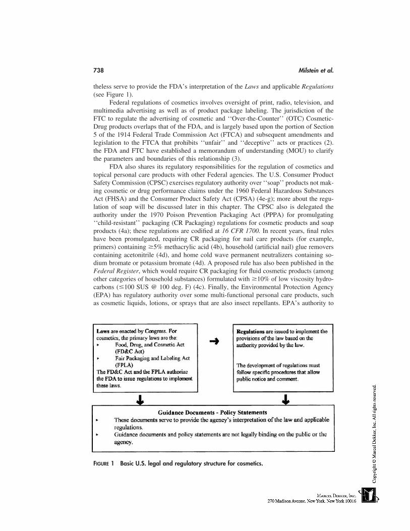
738 Milstein et al.
theless serve to provide the FDA’s interpretation of the Laws and applicable Regulations(see Figure 1).
Federal regulations of cosmetics involves oversight of print, radio, television, andmultimedia advertising as well as of product package labeling. The jurisdiction of theFTC to regulate the advertising of cosmetic and ‘‘Over-the-Counter’’ (OTC) Cosmetic-Drug products overlaps that of the FDA, and is largely based upon the portion of Section5 of the 1914 Federal Trade Commission Act (FTCA) and subsequent amendments andlegislation to the FTCA that prohibits ‘‘unfair’’ and ‘‘deceptive’’ acts or practices (2).the FDA and FTC have established a memorandum of understanding (MOU) to clarifythe parameters and boundaries of this relationship (3).
FDA also shares its regulatory responsibilities for the regulation of cosmetics andtopical personal care products with other Federal agencies. The U.S. Consumer ProductSafety Commission (CPSC) exercises regulatory authority over ‘‘soap’’ products not mak-ing cosmetic or drug performance claims under the 1960 Federal Hazardous SubstancesAct (FHSA) and the Consumer Product Safety Act (CPSA) (4e-g); more about the regu-lation of soap will be discussed later in this chapter. The CPSC also is delegated theauthority under the 1970 Poison Prevention Packaging Act (PPPA) for promulgating‘‘child-resistant’’ packaging (CR Packaging) regulations for cosmetic products and soapproducts (4a); these regulations are codified at 16 CFR 1700. In recent years, final ruleshave been promulgated, requiring CR packaging for nail care products (for example,primers) containing 5% methacrylic acid (4b), household (artificial nail) glue removerscontaining acetonitrile (4d), and home cold wave permanent neutralizers containing so-dium bromate or potassium bromate (4d). A proposed rule has also been published in theFederal Register, which would require CR packaging for fluid cosmetic products (amongother categories of household substances) formulated with 10% of low viscosity hydro-carbons (�100 SUS @ 100 deg. F) (4c). Finally, the Environmental Protection Agency(EPA) has regulatory authority over some multi-functional personal care products, suchas cosmetic liquids, lotions, or sprays that are also insect repellants. EPA’s authority to
FIGURE 1 Basic U.S. legal and regulatory structure for cosmetics.
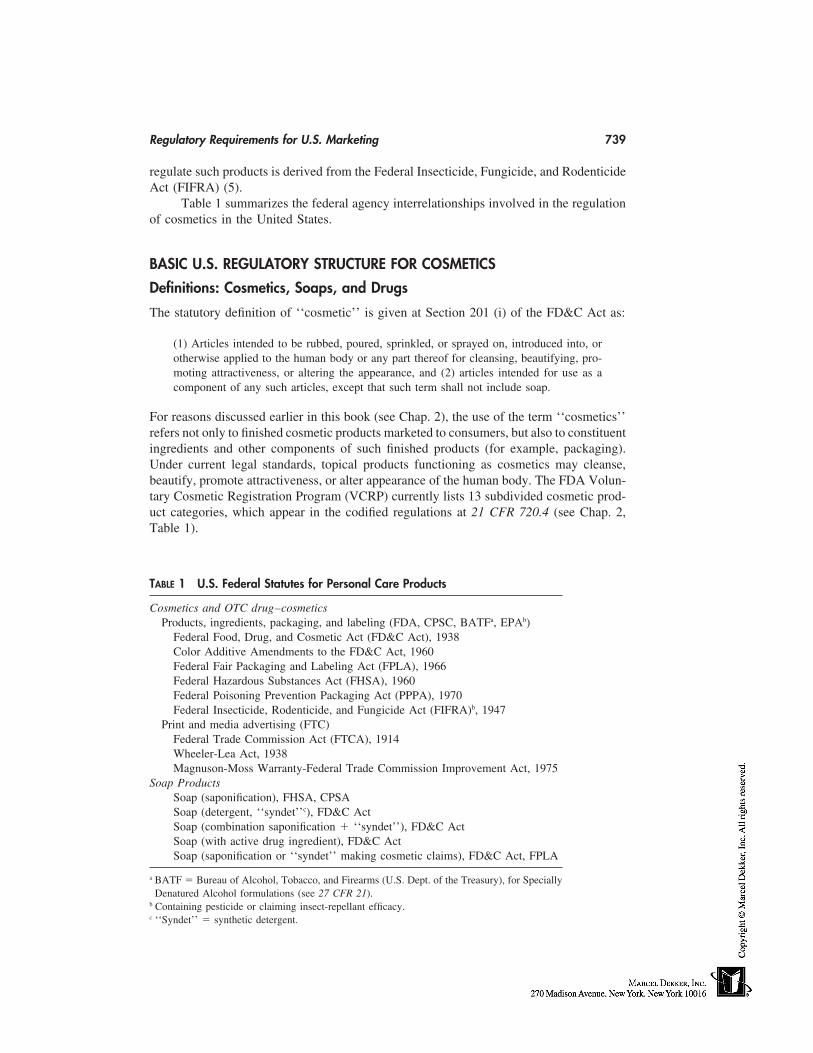
Regulatory Requirements for U.S. Marketing 739
regulate such products is derived from the Federal Insecticide, Fungicide, and RodenticideAct (FIFRA) (5).
Table 1 summarizes the federal agency interrelationships involved in the regulationof cosmetics in the United States.
BASIC U.S. REGULATORY STRUCTURE FOR COSMETICS
Definitions: Cosmetics, Soaps, and Drugs
The statutory definition of ‘‘cosmetic’’ is given at Section 201 (i) of the FD&C Act as:
(1) Articles intended to be rubbed, poured, sprinkled, or sprayed on, introduced into, orotherwise applied to the human body or any part thereof for cleansing, beautifying, pro-moting attractiveness, or altering the appearance, and (2) articles intended for use as acomponent of any such articles, except that such term shall not include soap.
For reasons discussed earlier in this book (see Chap. 2), the use of the term ‘‘cosmetics’’refers not only to finished cosmetic products marketed to consumers, but also to constituentingredients and other components of such finished products (for example, packaging).Under current legal standards, topical products functioning as cosmetics may cleanse,beautify, promote attractiveness, or alter appearance of the human body. The FDA Volun-tary Cosmetic Registration Program (VCRP) currently lists 13 subdivided cosmetic prod-uct categories, which appear in the codified regulations at 21 CFR 720.4 (see Chap. 2,Table 1).
TABLE 1 U.S. Federal Statutes for Personal Care Products
Cosmetics and OTC drug–cosmeticsProducts, ingredients, packaging, and labeling (FDA, CPSC, BATFa, EPAb)
Federal Food, Drug, and Cosmetic Act (FD&C Act), 1938Color Additive Amendments to the FD&C Act, 1960Federal Fair Packaging and Labeling Act (FPLA), 1966Federal Hazardous Substances Act (FHSA), 1960Federal Poisoning Prevention Packaging Act (PPPA), 1970Federal Insecticide, Rodenticide, and Fungicide Act (FIFRA)b, 1947
Print and media advertising (FTC)Federal Trade Commission Act (FTCA), 1914Wheeler-Lea Act, 1938Magnuson-Moss Warranty-Federal Trade Commission Improvement Act, 1975
Soap ProductsSoap (saponification), FHSA, CPSASoap (detergent, ‘‘syndet’’c), FD&C ActSoap (combination saponification � ‘‘syndet’’), FD&C ActSoap (with active drug ingredient), FD&C ActSoap (saponification or ‘‘syndet’’ making cosmetic claims), FD&C Act, FPLA
a BATF � Bureau of Alcohol, Tobacco, and Firearms (U.S. Dept. of the Treasury), for SpeciallyDenatured Alcohol formulations (see 27 CFR 21).
b Containing pesticide or claiming insect-repellant efficacy.c ‘‘Syndet’’ � synthetic detergent.
740 Milstein et al.
‘‘Soap’’ products are generally exempt from the cosmetic provisions of the FD&CAct, and, indeed, from the definition of ‘‘cosmetic’’ given in the statute. The FDA inter-prets the term ‘‘soap’’ at 21 CFR 701.20 to apply to products
Intended for cleansing the human bodyLabeled, sold, and represented solely as soapConsisting primarily of alkali metal salts of free fatty acids (i.e., the bulk of its
nonvolatile matter that serves as the detergent)Detergent properties of which articles are due to the alkali metal salts of free fatty
acids
Liquid and solid product formulations consisting of synthetic detergents (‘‘syndets’’),combinations of soap and synthetic detergents (‘‘combo’’ bars) intended not only forcleansing but also claiming other cosmetic product performance attributes (e.g., ‘‘beautybars’’ or ‘‘body bars’’ claiming to beautify, moisturize, soften, or smooth the skin) mustcomply with the regulatory requirements applicable to cosmetics (e.g., bear ingredientdeclarations required at 21 CFR 701.3). Indeed, even if such detergent or combinationsoap–detergent products are intended solely for cleansing of the human body, possess thecharacteristics consumers generally ascribe to ‘‘soap,’’ and are identified in labeling as‘‘soap’’ or some fanciful adaptation of this descriptor (e.g., ‘‘sope,’’ ‘‘jabon,’’ ‘‘liquidsoap,’’ etc.), these products are still regulated as cosmetics.
The statutory definition of the term ‘‘drug’’ is given at Section 201 (g)(1) of theFD&C Act, in pertinent part, as:
(B) articles intended for use in the diagnosis, cure, mitigation, treatment, or preventionin man . . . and (C) articles (other than food) intended to affect the structure or any functionof the body of man . . . and (D) articles intended for use as a component of any [such]articles.
Regardless of their respective legal standings as ‘‘cosmetics’’ regulated under theFD&C Act or ‘‘soaps’’ regulated under the CPSA/FHSA, personal-care products that arealso intended to treat or prevent disease or otherwise affect the structure or functions ofthe human body are considered ‘‘drugs’’ and must comply with these provisions of thelaw as well as any other provisions as cosmetics or soaps, respectively. Most currentlymarketed cosmetics that are also drugs are OTC drugs (e.g., ‘fluoride’ anticaries tooth-pastes, antiperspirant deodorants, antidandruff shampoos, and sunscreen lotions). How-ever, several drug–cosmetics are ‘‘new drugs’’ [6], for which safety and effectivenesshad to be proven to the agency before they could be marketed. Analogously, soap productsformulated to contain ‘‘active ingredients,’’ if intended to cure, treat, or prevent disease,or if intended to affect the structure or any function of the human body, may also beregulated as drugs. This would include, for example ‘‘medicated’’ anti-acne soaps, the‘‘antibacterial’’ bar and liquid soaps first introduced into the market in the late 1980s [7],and the alcohol-based liquid ‘‘hand sanitizers’’ of the late 1990s [8].
Statutory Controls on Cosmetics
The FD&C Act not only defines the term ‘‘cosmetic,’’ but sets forth the basic requirementthat cosmetic products introduced into interstate commerce within the United States mustbe safe for their intended use and properly labeled. The act accomplishes this by explicitlyprohibiting the adulteration or misbranding of cosmetics, and the introduction into, or
Regulatory Requirements for U.S. Marketing 741
receipt in, interstate commerce of ‘‘adulterated’’ or ‘‘misbranded’’ cosmetics (see FD&C Act, Sections 601 and 602, respectively).
Adulterated Cosmetics
A cosmetic is ‘‘adulterated’’ according to the FD&C Act, Section 601 (a)–(e) if:
It bears or contains any poisonous or deleterious substance, which may render itinjurious to users under the conditions of use prescribed in the labeling or under‘‘customary or usual’’ uses
It consists wholly or in part of any filthy, putrid, or decomposed substanceIt has been prepared, packed, or held under insanitary conditions whereby it may
have become contaminated with filth, or whereby it may have been rendered inju-rious to health
Its container is composed, wholly or in part, of any poisonous or deleterious sub-stance that may render the contents injurious to health
It is not a hair dye and it is, or bears or contains, a color additive that is unsafewithin the meaning of the act
Coal-Tar Hair-Dye Exemption. The FD&C Act exempts so-called ‘‘coal-tar’’hair-dyes from the adulteration provision at Section 601 (a), if they bear the cautionarystatement prescribed by law on the label and give ‘‘patch test’’ instructions, even if theyare irritating to the skin or are otherwise harmful to the human body. The ‘‘coal-tar hair-dye exemption,’’ named for the synthetic organic colors originally derived from the coaltar derivative, aniline, to which the exemption was initially applied [9], does not includeeyelash and eyebrow dyes since coal-tar derived color additives may cause blindness whenused for dyeing the eyelashes or eyebrows (9c). The exemption also does not apply tonon-coal tar color additives in hair dyes (9c).
Sources of Adulteration. Cosmetic adulteration may be associated with uninten-tional trace level contaminants (e.g., N-nitrosamines, or 1,4-Dioxane) of the ingredients(also referred to as ‘raw materials’) employed in finished cosmetic products [10–12] orto the manner of product formulation. Quality control problems (e.g., pH) or failure tofollow good manufacturing practices guidelines [13] can also result in deviations of partic-ular product batches from master formula specifications. In the past four (4) fiscal years(FY96–FY99), the FDA has found that approximately 88% of cosmetic product adultera-tions subject to voluntary recall actions (see ‘‘Recalls’’ in Law Enforcement of FD&C Act Violations, below) were most frequently related to problems of microbiologicalcontamination (see Table 2) [14].
TABLE 2 Cosmetic Product Voluntary Recalls
FY 1996 FY 1997 FY 1998 FY 1999Total recalls 26 9 9 9Microbiology recalls 23 8 8a 8Misbranding recalls 3 1 0 1Other recalls 0 0 1b 0
FY � fiscal year.a 6 Class II Microbiology � 2 Mold.b 1 Class II pH.
742 Milstein et al.
Misbranded Cosmetics
A cosmetic is ‘‘misbranded’’ according to the FD&C Act, Sec. 602 (a)–(f) if:
Its labeling is false or misleading in any particularIts package label fails to contain the name and place of business of the manufacturer,
packer, or distributor, as well as an accurate statement of the quantity of thecontents in terms of weight, measure, or numerical count
Any word, statement, or other information required to appear on the label is notprominently and conspicuously placed and in terms likely to be read and under-stood by the ordinary consumer under customary conditions of purchase and use
Its container is made, formed, or filled in a manner likely to be misleadingIt is a color additive, unless its packaging and labeling are in conformity with re-
quirements in the regulationsIts packaging or labeling are in violation of an applicable regulation issued under
the 1970 PPPA.
A cosmetic is misbranded as a consumer commodity according to the FPLA, Section7, if it is introduced or delivered for introduction into commerce in violation of any ofthe provisions of the law or its implementing regulations, including the requirements con-tained in Sections 4 and 5 of the FPLA, which provide that the label of a commoditymust state:
The identity of the commodityThe name and place of business of the manufacturer, packer, or distributorThe net quantity of contents (in terms of weight, measure, or numerical count) sepa-
rately and accurately stated in a uniform location upon the principal display panel(PDP)
The ‘‘common or usual name’’ of the commodity and, if it contains two or moreingredients, the ‘‘common or usual name’’ of each ingredient listed in order ofdecreasing predominance, with the exception of such ingredients deemed to con-stitute a ‘‘trade secret.’’
Law Enforcement of FD&C Act Violations
Violations of the adulteration and misbranding provisions of the act may subject the viola-tor to various enforcement tools available to the FDA. These include (but are not limitedto) (16c):
Warning letters, subject to public disclosure under the Freedom of Information Act(FOIA), may be posted on the Internet FDA Web site and are regularly publicizedin the trade press and industry newsletters such as The Rose Sheet
Targeted establishment inspections and sampling programsSeizure of cosmetic goods alleged to be in violation of the FD&C Act (civil
actions)Detention of imported cosmetics offered for entry into U.S. interstate commerce
that appear to be in violation of the law (see, for example, FD&C Act, Section801(a)
Injunction proceedings against firms or individuals, seeking that a company ceasepresent and future manufacture and distribution of cosmetic products until compli-ance with the law can be assured
Regulatory Requirements for U.S. Marketing 743
Criminal prosecution of responsible persons within violator cosmetic firmsRecalls. Recall and Field Correction are actions taken by a firm to either remove a
product from the market or to conduct a field correction. Recalls of cosmeticproducts, may be conducted on a firm’s own initiative or by FDA request. TheFDA has no authority under the FD&C Act to order the recall of a defective orpossibly harmful cosmetic product, although it can request a firm to recall a prod-uct. Resistance to an FDA request for voluntary recall can, however, trigger otherenforcement actions by the agency, which have recently been reviewed by Calog-ero [15]. The FDA has defined policies concerning such voluntary cosmetic (aswell as food, drug, and medical device) product recalls; these are codified at 21CFR 7.45–7.59, and additional guidance can also be found at the FDA website(http:/ /www.fda.gov). The FDA’s regulations divide recalls into three categories:
Class I Recall Products that are clearly dangerous or defective that pose clearor irreversible hazards to the public health; there is reasonable probabilitythat the use of or exposure to a violative product will cause serious adversehealth consequences or death (21 CFR 7.3 (m)(1))
Class II Recall Products that are intermediate in their potential for adversepublic health consequences, but may cause a temporary or reversible healthproblem; use of or exposure to a violative product may cause temporary ormedically reversible adverse health consequences or where the probabilityof serious adverse health consequences is remote (21 CFR 7.3 (m)(2))
Class III Recall Products that are unlikely to cause any adverse health reac-tion but that violate FDA regulations; use of or exposure to a violative prod-uct is not likely to cause adverse health consequences (21 CFR 7.3 (m)(3)).
Regulatory Controls on Cosmetics
Cosmetics marketed in the United States, whether manufactured domestically or importedfrom abroad, must be in compliance with the provisions of the FD&C Act, the FPLA, andthe regulations published under the authority of these laws. Yet, cosmetics are arguably theleast regulated category of articles subject to the jurisdiction of the FD&C Act [16]. Thereis no premarket approval requirement for cosmetic products or their constituent ingredientsunder the law. Other than color additives and those few ingredients restricted or prohibitedby regulation from use in cosmetics, no mandatory regulatory controls exist on the chemis-try and structure substantiation of the ingredients themselves, conditions of manufactureof the finished cosmetic products, or safety testing that the ingredients and products mustundergo prior to marketing; no premarket test results need be submitted to the FDA.
The FDA has therefore promulgated regulations and guidance documents to helpensure that only cosmetics that are safe for their intended use and are neither ‘‘adulterated’’nor ‘‘misbranded’’ enter interstate commerce. These regulatory documents address thefollowing issues.
Cosmetic Safety
Cosmetics are not currently subject to the same FDA safety and effectiveness standardsas are drugs, biologics, and medical devices. The FD&C Act does not require that cosmeticmanufacturers or marketers test their products for safety, nor does the FDA specify particu-lar test batteries or preclinical (i.e., animal or in vitro alternative tests) and human clinicalsafety tests by cosmetic product category that marketers must use to substantiate cosmetic
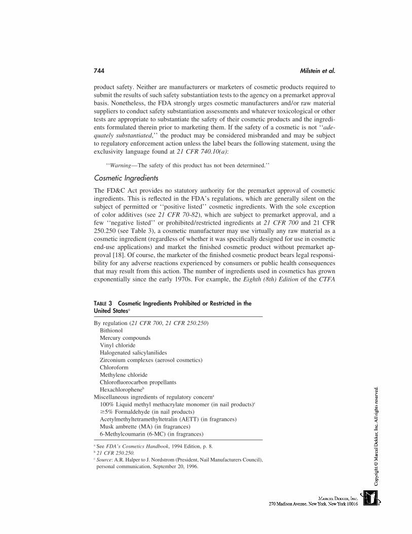
744 Milstein et al.
product safety. Neither are manufacturers or marketers of cosmetic products required tosubmit the results of such safety substantiation tests to the agency on a premarket approvalbasis. Nonetheless, the FDA strongly urges cosmetic manufacturers and/or raw materialsuppliers to conduct safety substantiation assessments and whatever toxicological or othertests are appropriate to substantiate the safety of their cosmetic products and the ingredi-ents formulated therein prior to marketing them. If the safety of a cosmetic is not ‘‘ade-quately substantiated,’’ the product may be considered misbranded and may be subjectto regulatory enforcement action unless the label bears the following statement, using theexclusivity language found at 21 CFR 740.10(a):
‘‘Warning—The safety of this product has not been determined.’’
Cosmetic Ingredients
The FD&C Act provides no statutory authority for the premarket approval of cosmeticingredients. This is reflected in the FDA’s regulations, which are generally silent on thesubject of permitted or ‘‘positive listed’’ cosmetic ingredients. With the sole exceptionof color additives (see 21 CFR 70-82), which are subject to premarket approval, and afew ‘‘negative listed’’ or prohibited/restricted ingredients at 21 CFR 700 and 21 CFR250.250 (see Table 3), a cosmetic manufacturer may use virtually any raw material as acosmetic ingredient (regardless of whether it was specifically designed for use in cosmeticend-use applications) and market the finished cosmetic product without premarket ap-proval [18]. Of course, the marketer of the finished cosmetic product bears legal responsi-bility for any adverse reactions experienced by consumers or public health consequencesthat may result from this action. The number of ingredients used in cosmetics has grownexponentially since the early 1970s. For example, the Eighth (8th) Edition of the CTFA
TABLE 3 Cosmetic Ingredients Prohibited or Restricted in theUnited Statesa
By regulation (21 CFR 700, 21 CFR 250.250)BithionolMercury compoundsVinyl chlorideHalogenated salicylanilidesZirconium complexes (aerosol cosmetics)ChloroformMethylene chlorideChlorofluorocarbon propellantsHexachloropheneb
Miscellaneous ingredients of regulatory concerna
100% Liquid methyl methacrylate monomer (in nail products)c
5% Formaldehyde (in nail products)Acetylmethyltetramethyltetralin (AETT) (in fragrances)Musk ambrette (MA) (in fragrances)6-Methylcoumarin (6-MC) (in fragrances)
a See FDA’s Cosmetics Handbook, 1994 Edition, p. 8.b 21 CFR 250.250.c Source: A.R. Halper to J. Nordstrom (President, Nail Manufacturers Council),
personal communication, September 20, 1996.
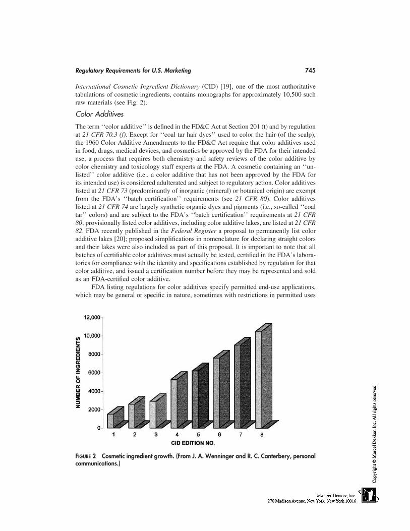
Regulatory Requirements for U.S. Marketing 745
International Cosmetic Ingredient Dictionary (CID) [19], one of the most authoritativetabulations of cosmetic ingredients, contains monographs for approximately 10,500 suchraw materials (see Fig. 2).
Color Additives
The term ‘‘color additive’’ is defined in the FD&C Act at Section 201 (t) and by regulationat 21 CFR 70.3 (f). Except for ‘‘coal tar hair dyes’’ used to color the hair (of the scalp),the 1960 Color Additive Amendments to the FD&C Act require that color additives usedin food, drugs, medical devices, and cosmetics be approved by the FDA for their intendeduse, a process that requires both chemistry and safety reviews of the color additive bycolor chemistry and toxicology staff experts at the FDA. A cosmetic containing an ‘‘un-listed’’ color additive (i.e., a color additive that has not been approved by the FDA forits intended use) is considered adulterated and subject to regulatory action. Color additiveslisted at 21 CFR 73 (predominantly of inorganic (mineral) or botanical origin) are exemptfrom the FDA’s ‘‘batch certification’’ requirements (see 21 CFR 80). Color additiveslisted at 21 CFR 74 are largely synthetic organic dyes and pigments (i.e., so-called ‘‘coaltar’’ colors) and are subject to the FDA’s ‘‘batch certification’’ requirements at 21 CFR80; provisionally listed color additives, including color additive lakes, are listed at 21 CFR82. FDA recently published in the Federal Register a proposal to permanently list coloradditive lakes [20]; proposed simplifications in nomenclature for declaring straight colorsand their lakes were also included as part of this proposal. It is important to note that allbatches of certifiable color additives must actually be tested, certified in the FDA’s labora-tories for compliance with the identity and specifications established by regulation for thatcolor additive, and issued a certification number before they may be represented and soldas an FDA-certified color additive.
FDA listing regulations for color additives specify permitted end-use applications,which may be general or specific in nature, sometimes with restrictions in permitted uses
FIGURE 2 Cosmetic ingredient growth. (From J. A. Wenninger and R. C. Canterbery, personalcommunications.)
746 Milstein et al.
or allowed concentrations. Cosmetic color additives, for example, may be listed for generaluse in imparting color to product formulations, for use in decorative cosmetics intendedfor external application to the hair and other appendages of the human body (other thanthe area of the eye), or may be specifically listed, solely or together with other cosmeticproduct applications, for eye area use [21]. Only color additives specifically authorizedby regulation for use in the area of the eye may be legally used for such applications.Only one color additive, dihydroxyacetone (DHA), is specifically listed for an intendeduse in externally applied cosmetics ‘‘to impart a color to the human body’’; this findswidespread application in today’s ‘‘sunless’’ or ‘‘self-tanning’’ cosmetic products [22].No color additives are currently approved for use in injectable cosmetic tattoos [23]. Fur-ther details about the color additives currently listed (approved) by regulation for use incosmetics in the United States may be found on the Internet at FDA’s website (e.g., http://www.cfsan.fda.gov/cosmetics.html).
Cosmetic Labeling
Cosmetic products distributed in the United States must comply with the labeling regula-tions published by the FDA under the authority of the FD&C Act and the FPLA [24a].Section 10(a) of the FPLA gives the FDA authority to require labeling of products consid-ered ‘‘consumer commodities’’; that is, products regulated under the FD&C Act, whichare ‘‘customarily produced or distributed for sale through retail sales . . . for consumptionby individuals, or use by individuals for purposes of personal care or in the performanceof services ordinarily rendered within the household’’ [24b].
The statute requires that products be honestly and informatively labeled so that con-sumers can conduct ‘‘value comparisons’’ at the point of purchase; that is, in order todetermine what ingredients are in a product and which product among several alternativesbeing considered for purchase is the best value. This determination includes medical con-siderations, since the FDA has previously concluded [25] that a cosmetic product or ingre-dient to which a consumer is allergic (and which the consumer therefore cannot use) hasno value to such a consumer.
‘‘Labeling’’ refers to actual product package labels as well as other written, printed,or graphic material on or accompanying a product (e.g., hangtags, promotional fliers, pack-age inserts). Label statements required under the FD&C Act must appear on both theinside as well as the outside container or wrapper, if any; FPLA requirements need onlyappear on the label of the outer container or wrapper.
Cosmetic product package labeling regulations enacted under authority of the FD&CAct and/or the FPLA require that cosmetic labels bear certain fields of information thatprovide the consumer with proper identification and other data that will enhance the con-sumer’s understanding of the product being purchased and facilitate the ability of theconsumer to contact the manufacturer or distributor of the product, should there be a needto do so. Although the cosmetic labeling regulations at 21 CFR 701 generally require alllabeling information to be written in the English language commonly understood by mostAmerican consumers, 21 CFR 701.2 (b) also provides certain accommodations in the caseof articles distributed in Puerto Rico or other territories in which the predominant languageis other than English. The required fields of information include the following:
Statement of identity (i.e., common name) rendered in bold type on the cosmeticproduct principal display panel; note that this is an FPLA requirement for cosmet-ics, not an FD&C Act requirement per se
Name and address of manufacturer (or packer or distributor)
Regulatory Requirements for U.S. Marketing 747
Net quantity of contents (net weight or count or measure, as customary or as re-quired). English units are mandatory in the United States but a technical amend-ment to the FPLA under the 1991 American Technology Preeminence Act(ATPA), as revised in 1992 [26a,b], and more recent regulatory proposals to im-plement the ATPA provisions for FDA-regulated products [26c], now advocatethe use of the most appropriate units of the metric international system (SI) ofweights or measures, wherever practicable. This proposal includes the dual decla-ration of net quantity of contents in terms of both English units and the interna-tional metric (SI) system of weights or measures
Cosmetic ingredient label declarations (see below)Warning statements (or cautionary statements) concerning safe use, as required at
21 CFR 740 (see below)
A typical cosmetic product package label exemplifying these features is shown in Fig-ure 3.
Cosmetic Ingredient Label Declarations
Section 5(c)(3) of the FPLA specifically authorizes FDA to promulgate regulations requir-ing the declaration of all cosmetic ingredients on product package labels of cosmeticsconsidered ‘‘consumer commodities’’ (loc. cit., Ref. 24(b)); these regulations are codifiedat 21 CFR 701.3. Exempt from the ingredient declaration requirement are professionalcosmetic products, such as hair and skin preparations or makeup products used by cosme-tologists, beauticians, or aestheticians on clients at professional establishments such assalons, spas, and theaters, provided that these products are not also sold to consumersthrough the professional establishments, workplaces, or other miscellaneous beauty supplyretail outlets for their consumption at home; such cosmetics are not legally considered‘‘consumer commodities.’’ Similar exemptions apply to ‘‘free’’ (gratis) samples, gifts,cosmetics distributed as free amenities at hotels, and cosmetics and toiletries made avail-able to workers and visitors (but not sold) for on-site use at occupational settings, suchas construction sites, hospitals, clinics, etc. However, cosmetic products offered as ‘‘giftwith purchase’’ are ‘‘consumer commodities’’ and subject to the ingredient declarationrequirement, because the ‘‘gift’’ is only available in conjunction with a retail sale. Profes-sional cosmetic products exempt from the ingredient declaration requirement are fre-quently labeled ‘‘for professional use only.’’
Ingredient declarations must be ‘‘conspicuous’’ and ‘‘prominent’’ in placement onany appropriate information panel of the outer container, and not less than certain sizespecifications in relationship to the size and shape of the product package, in order toensure that the declaration is likely to be read at the time of purchase by the consumer.
FPLA labeling requirements specify that cosmetic ingredients must be declared, indescending order of predominance (see 21 CFR 701.3 [a]), utilizing ingredient namesderived in hierarchical order of precedence from the nomenclature sources specified byregulation (see 21 CFR 701.3 [c] and 701.30); alternatively, the ingredients may begrouped and the groups declared according to 21 CFR 701.3 (f). The ‘‘common or usual’’names specified by regulation in the United States are required to be stated in the languageunderstood by American consumers, namely English, except as provided at 21 CFR701.2 (b) (see Cosmetic Labeling, p. 746, loc. cit.). Cosmetic ingredients present at onepercent or less (�1%) may be declared after ingredients present at higher levels withoutregard to order of predominance, and fragrance and flavor, if any, being complex com-positions of matter in themselves, may be declared for purposes of product package label-
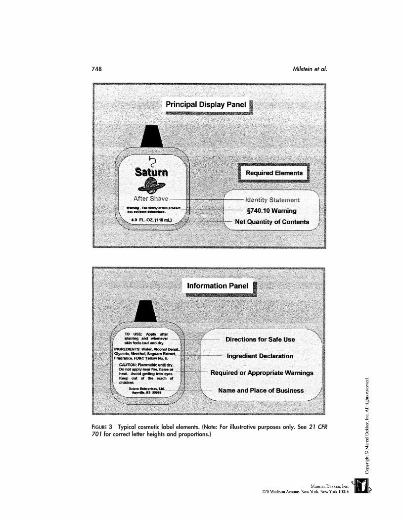
748 Milstein et al.
FIGURE 3 Typical cosmetic label elements. (Note: For illustrative purposes only. See 21 CFR701 for correct letter heights and proportions.)
Regulatory Requirements for U.S. Marketing 749
ing as ‘‘flavor’’ and ‘‘fragrance,’’ respectively. ‘‘Incidental ingredients’’ (see 21 CFR701.3 [1]) need not be declared, and those ingredients accepted by the FDA as exemptfrom public disclosure and granted ‘‘confidentiality’’ or ‘‘trade secret’’ status may bedeclared as ‘‘and other ingredients’’ (see 21 CFR 720.8).
‘‘Soap’’ products meeting the requirements of 21 CFR 701.20(a)(1) and (a)(2) areexempt from the FPLA requirement for mandatory label ingredient declarations applicableto cosmetics.
The manner of declaration of ingredients in OTC drug–cosmetic products is speci-fied at 21 CFR 701.3(d), as recently amended (see 64 FR 13234–13303@13297, March17, 1999). Drug ‘‘active ingredients’’ present in OTC drug–cosmetic product formulationsare declared first, as required at 21 CFR 201.66(c)(2) and (d) of this chapter, and followingthe standard-format ‘‘Drug Facts’’ information fields (i.e., ‘‘Use(s),’’ ‘‘Warnings,’’ ‘‘Di-rections,’’ and ‘‘Other Information’’), any ‘‘inactive’’ or cosmetic ingredients are declaredin descending order of predominance or grouped, in accordance with the provisions of21 CFR 701.3(a) and (f), respectively. An exception in the manner of declaration of inac-tive or cosmetic ingredients is provided for, if there is a difference in the labeling provis-ions in 21 CFR 201.66 and Sections 701.3 or 720.8; under these circumstances, the label-ing provisions at 21 CFR 201.66 are controlling (see 21 CFR 201.66(c)(8) and (d) of thischapter).
Recent efforts to achieve ‘‘international harmonization’’ with cosmetic ingredientnomenclature standards required by the 1976 European Union (EU) Cosmetic Directive[27] and its more recent amendments [28] have resulted in the FDA agreeing to exerciseregulatory discretion toward the interim use of parenthetical ‘‘dual declarations,’’ em-ploying systematic Linne (Latin) taxonomic genus/species nomenclature for certain cate-gories of ingredients (i.e., botanicals and/or ‘‘trivial’’ ingredients) pending review of acitizen petition submitted by CTFA [29]. Color additives are named using the monographtitles in their respective listing regulations (see 21 CFR 73, 74, 82), although, here, too,the impact of ‘‘international harmonization’’ efforts has resulted in the FDA agreeing toexercise regulatory discretion towards the interim use of parenthetical ‘‘color index (CI)numbers’’ in a dual declaration [29]. Examples of the new interim ‘‘harmonized’’ ingredi-ent declarations are given in Table 4.
Cosmetic Label Warnings
Cosmetics that may be hazardous to consumers when misused must bear appropriate labelwarnings and adequate directions for safe use. Manufacturers and marketers of cosmeticshave a general responsibility to ensure that the labels of their finished cosmetic productsbear a warning statement whenever necessary or appropriate to prevent a health hazardthat may be associated with the product (21 CFR 740.1[a]). These warning statementsmust be prominent and conspicuous (21 CFR 740.2). Some cosmetics must also bear morespecific label warnings or cautions prescribed by regulation. Specific cosmetic productcategories requiring such statements currently include:
Cosmetic products for which adequate substantiation of safety has not been obtained(21 CFR 740.10)
Cosmetics in self-pressurized containers (21 CFR 740.11)Feminine deodorant sprays (21 CFR 740.12)Foaming detergent bath products (21 CFR 740.17)‘‘Coal tar’’ hair-dyes posing a risk of cancer (21 CFR 740.18) [Effective date stayed
at 47 FR 7829, February 23, 1982.]
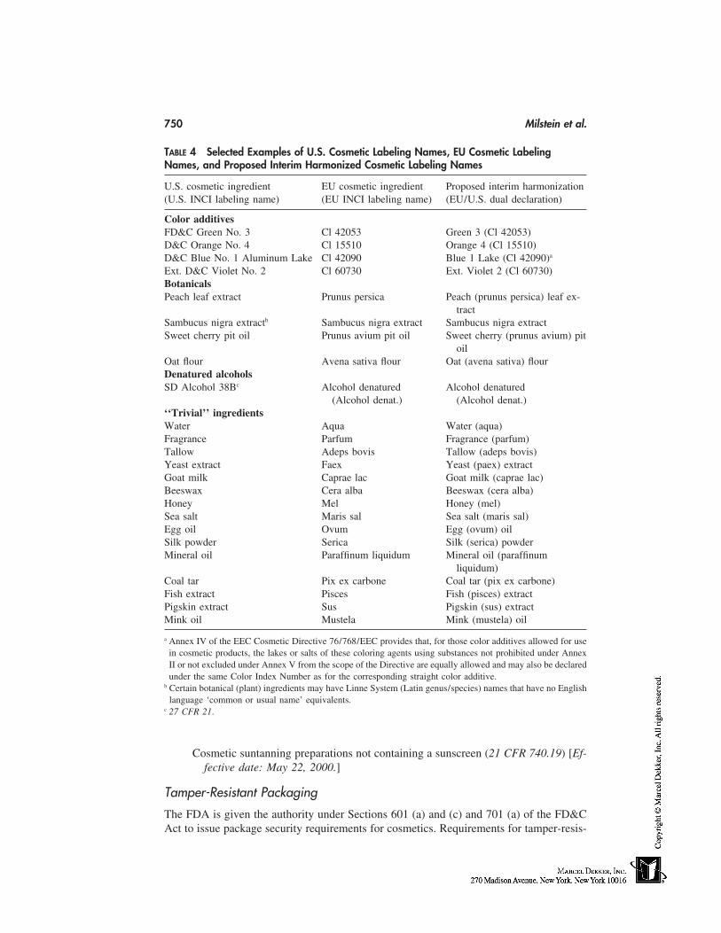
750 Milstein et al.
TABLE 4 Selected Examples of U.S. Cosmetic Labeling Names, EU Cosmetic LabelingNames, and Proposed Interim Harmonized Cosmetic Labeling Names
U.S. cosmetic ingredient EU cosmetic ingredient Proposed interim harmonization(U.S. INCI labeling name) (EU INCI labeling name) (EU/U.S. dual declaration)
Color additivesFD&C Green No. 3 Cl 42053 Green 3 (Cl 42053)D&C Orange No. 4 Cl 15510 Orange 4 (Cl 15510)D&C Blue No. 1 Aluminum Lake Cl 42090 Blue 1 Lake (Cl 42090)a
Ext. D&C Violet No. 2 Cl 60730 Ext. Violet 2 (Cl 60730)BotanicalsPeach leaf extract Prunus persica Peach (prunus persica) leaf ex-
tractSambucus nigra extractb Sambucus nigra extract Sambucus nigra extractSweet cherry pit oil Prunus avium pit oil Sweet cherry (prunus avium) pit
oilOat flour Avena sativa flour Oat (avena sativa) flourDenatured alcoholsSD Alcohol 38Bc Alcohol denatured Alcohol denatured
(Alcohol denat.) (Alcohol denat.)‘‘Trivial’’ ingredientsWater Aqua Water (aqua)Fragrance Parfum Fragrance (parfum)Tallow Adeps bovis Tallow (adeps bovis)Yeast extract Faex Yeast (paex) extractGoat milk Caprae lac Goat milk (caprae lac)Beeswax Cera alba Beeswax (cera alba)Honey Mel Honey (mel)Sea salt Maris sal Sea salt (maris sal)Egg oil Ovum Egg (ovum) oilSilk powder Serica Silk (serica) powderMineral oil Paraffinum liquidum Mineral oil (paraffinum
liquidum)Coal tar Pix ex carbone Coal tar (pix ex carbone)Fish extract Pisces Fish (pisces) extractPigskin extract Sus Pigskin (sus) extractMink oil Mustela Mink (mustela) oil
a Annex IV of the EEC Cosmetic Directive 76/768/EEC provides that, for those color additives allowed for usein cosmetic products, the lakes or salts of these coloring agents using substances not prohibited under AnnexII or not excluded under Annex V from the scope of the Directive are equally allowed and may also be declaredunder the same Color Index Number as for the corresponding straight color additive.
b Certain botanical (plant) ingredients may have Linne System (Latin genus/species) names that have no Englishlanguage ‘common or usual name’ equivalents.
c 27 CFR 21.
Cosmetic suntanning preparations not containing a sunscreen (21 CFR 740.19) [Ef-fective date: May 22, 2000.]
Tamper-Resistant Packaging
The FDA is given the authority under Sections 601 (a) and (c) and 701 (a) of the FD&CAct to issue package security requirements for cosmetics. Requirements for tamper-resis-
Regulatory Requirements for U.S. Marketing 751
tant packaging for cosmetic liquid oral hygiene products (e.g., mouthwashes and breathfresheners) and all cosmetic vaginal products (e.g., douches and tablets) were promulgatedat 21 CFR 700.25. Details about such packaging is found in the FDA’s Cosmetics Hand-book [30] and at the FDA website, http:/ /www.fda.gov.
Cosmetic Good Manufacturing Practices Guidelines
The FDA has never published current good manufacturing practice (cGMP) regulationsfor cosmetics, although the agency has actively promoted good manufacturing practicesby firms marketing cosmetics in the United States. The agency has published CosmeticGood Manufacturing Practice Guidelines, patterned in pertinent part after the food cGMPregulations [13a] but applicable to the cosmetic manufacturing environment, in the FDA’sCosmetics Handbook [13b]; the latter document references the FDA Investigation Opera-tions Manual (IOM) [31]. The Cosmetic Good Manufacturing Practice Guidelines is aguidance document reflecting FDA policy, but it is not legally binding, either on the cos-metics industry or on the agency. The FDA has also published drug cGMP regulations[32], which apply to prescription drugs and cosmetic–drugs (i.e., OTC drug products mak-ing cosmetic claims).
The Voluntary Cosmetic Registration Program
The FD&C Act does not require cosmetic firms to register manufacturing establishmentsor formulations with the FDA, nor does it mandate that companies submit product adversereaction report data. Nevertheless, the FDA has encouraged the voluntary registration ofsuch data as being in the public interest and consistent with the spirit of responsible ‘‘self-regulation’’ advocated by the cosmetic industry. In the early 1970s, the FDA developeda three-part system of regulations, under which manufacturers or distributors of cosmeticsmay submit this information to the agency on a voluntary basis [33]. The three parts ofthe Voluntary Cosmetic Registration Program (VCRP) originally comprised the following:
Part I Cosmetic Establishment Registration Program (CERP), requests that cos-metic manufacturing sites be registered with the FDA (see 21 CFR 710)
Part II Cosmetic Product Ingredient Statements (CPIS), requests that cosmeticformulations and cosmetic raw material composition statements be registered withthe FDA (see 21 CFR 720). This regulation also set forth the 13 product categorycodes (PCC) at 21 CFR 720.4 recognized by the FDA as ‘‘cosmetic’’ functions.Semi-quantitative raw material disclosures were abandoned and purged from theVCRP database in the early 1990s [34].
Part III Product Experience Reports (PER), discontinued in 1996 (35), requestedthe annual filing of ‘‘reportable’’ adverse reactions (see 21 CFR 700.3 [q]) tothe use of cosmetic products by manufacturers which the FDA (euphemisticallycalled ‘product experiences’ (see 21 CFR 730). The use of optional ‘screening’protocols to be filed with the FDA, designed by individual manufacturers, for usein determining the ‘reportability’ of experiences, was also provided for in thePER Program (see 21 CFR 700.3 (p), 730.4 (d)(2)). This data was collected,tabulated, and analyzed for statistical deviations of individual products from in-dustry-wide adverse reaction trends by product category.
Despite its voluntary nature, the VCRP has never enjoyed full industry participation.Table 5 illustrates the VCRP registration statistics for the years 1992–1996, the last fivefiscal years during which all parts of the VCRP were in operation. Part III (PER) annualfilings by firms considered by the FDA to be eligible to participate in the program have
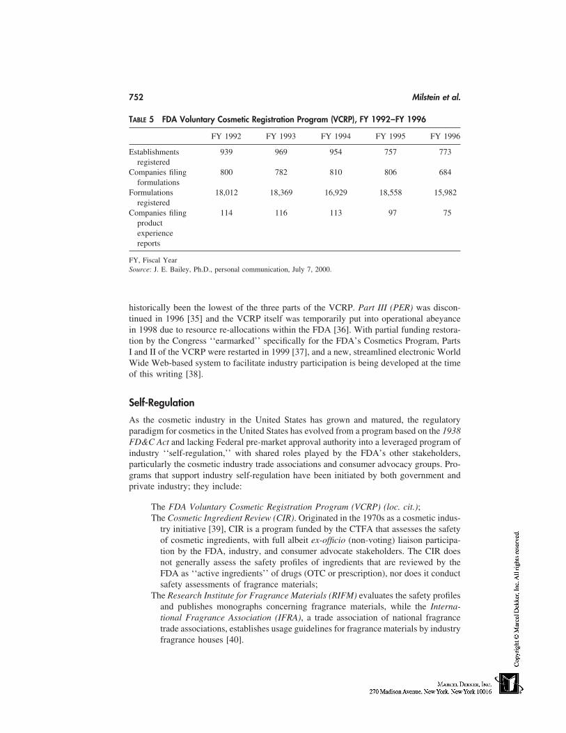
752 Milstein et al.
TABLE 5 FDA Voluntary Cosmetic Registration Program (VCRP), FY 1992–FY 1996
FY 1992 FY 1993 FY 1994 FY 1995 FY 1996
Establishments 939 969 954 757 773registered
Companies filing 800 782 810 806 684formulations
Formulations 18,012 18,369 16,929 18,558 15,982registered
Companies filing 114 116 113 97 75productexperiencereports
FY, Fiscal YearSource: J. E. Bailey, Ph.D., personal communication, July 7, 2000.
historically been the lowest of the three parts of the VCRP. Part III (PER) was discon-tinued in 1996 [35] and the VCRP itself was temporarily put into operational abeyancein 1998 due to resource re-allocations within the FDA [36]. With partial funding restora-tion by the Congress ‘‘earmarked’’ specifically for the FDA’s Cosmetics Program, PartsI and II of the VCRP were restarted in 1999 [37], and a new, streamlined electronic WorldWide Web-based system to facilitate industry participation is being developed at the timeof this writing [38].
Self-Regulation
As the cosmetic industry in the United States has grown and matured, the regulatoryparadigm for cosmetics in the United States has evolved from a program based on the 1938FD&C Act and lacking Federal pre-market approval authority into a leveraged program ofindustry ‘‘self-regulation,’’ with shared roles played by the FDA’s other stakeholders,particularly the cosmetic industry trade associations and consumer advocacy groups. Pro-grams that support industry self-regulation have been initiated by both government andprivate industry; they include:
The FDA Voluntary Cosmetic Registration Program (VCRP) (loc. cit.);The Cosmetic Ingredient Review (CIR). Originated in the 1970s as a cosmetic indus-
try initiative [39], CIR is a program funded by the CTFA that assesses the safetyof cosmetic ingredients, with full albeit ex-officio (non-voting) liaison participa-tion by the FDA, industry, and consumer advocate stakeholders. The CIR doesnot generally assess the safety profiles of ingredients that are reviewed by theFDA as ‘‘active ingredients’’ of drugs (OTC or prescription), nor does it conductsafety assessments of fragrance materials;
The Research Institute for Fragrance Materials (RIFM) evaluates the safety profilesand publishes monographs concerning fragrance materials, while the Interna-tional Fragrance Association (IFRA), a trade association of national fragrancetrade associations, establishes usage guidelines for fragrance materials by industryfragrance houses [40].
Regulatory Requirements for U.S. Marketing 753
The FDA’s VCRP and the industry-sponsored CIR and RIFM/IFRA programs areimportant components of the government-industry cooperation that characterize currentefforts towards the successful implementation of self-regulation of the cosmetic industryin the U.S. Other elements of self-regulation include:
Federal Statutes. The Lanham Act (1946) empowers companies to seek judicialredress in the federal district courts for unfair business practices resulting in nega-tive impact on market share [41]. The Robinson-Patman Act (1936) enables com-panies to seek to recoup lost sales and profits ascribed to anticompetitive, preda-tory pricing tactics [42].
Advertising Self-Regulation, NAD/CBBB. Disagreements regarding product perfor-mance advertising claims are frequently addressed by competitor/peer-reviewchallenges brought through the self-regulatory protocols of the National Advertis-ing Division (NAD), an arm of the Council of Better Business Bureaus (CBBB)[43], and its appeals panel, the National Advertising Review Board (NARB). Fail-ure to resolve advertising controversies through these self-regulatory processescan result in an ultimate referral by the NARB to the FTC. Scrutiny of proposedstoryboards prior to being accepted for mass-media air-time is also undertakenby advertising agency legal departments and television/radio network standardsand practices boards (e.g., network censors) [44]
The cosmetic industry is characterized by highly competitive marketing strategiesand depends on the freedom to rapidly introduce new, innovative cosmetic products tothe marketplace without lengthy delays. It is hardly surprising, therefore, that the industryhas sought to portray itself as responsible enough to self-police its own manufacturingand marketing practices, or that it has argued [45] that existing laws and FDA regulatoryprograms concerning cosmetics, together with the industry’s commitment to self-regula-tion and product safety, provide ample consumer protection, given the apparent low riskinherent in cosmetics relative to other categories of products regulated by the FDA.Steinberg [46] advocates compliance within a self-regulatory environment as being in theindustry’s own self-interest. He observes that regulatory compliance can be a ‘‘win-win’’end result for the industry, consumers, and regulators alike, and cautions that trying to‘‘beat the system may succeed in the short term, but it results in significant long-termlosses.’’ Steinberg notes that lost sales, public reputation, and market share are the obviousshort-term consequences likely to be suffered by noncompliant firms. Widespread non-compliance can also place the current self-regulatory system itself at risk.
International Harmonization and Future Regulatory Challenges
The U.S. regulatory scheme for cosmetics is based on the axiom that cosmetics marketedin the U.S. are safe for their intended use and unlikely to present a major public healthrisk [47], which is reflected in the lack of pre-market approval authority for cosmeticsincluded in the original 1938 FD&C Act.
Although many of the regulatory systems of other countries have similar goals tothose of the United States, such as protecting public health and safety and promoting trade[48], the means by which these goals are achieved may be quite different from the U.S.system. These differences are often based upon the culture of the particular country andcan influence not only specific regulatory requirements, such as labeling, but also thefundamental definition of what constitutes a cosmetic. Several categories of topical prod-
754 Milstein et al.
ucts regulated as OTC drugs or OTC drug–cosmetics in the United States, such as sun-screens, skin bleaches, antiperspirants, and antidandruff shampoos [49], are regulated ascosmetics under the EU Cosmetics Directive of 1976 [27]. Japan, which currently regulatescosmetics according to a system of premarket approval and licensure rather than the post-market surveillance system used by the United States or the notification system used bythe EU, allows cosmetics to have some effect on the structure and function of the skinand hair, provided that the effect is ‘‘mild’’ and provides for a third ‘‘quasi-drug’’ categoryof product accommodating ‘‘mild,’’ borderline physiological effects, such as hair-growthpromoters [50a]. However, initiatives currently underway in Japan promise to alter theregulation of cosmetics by shifting to a postmarketing system more nearly aligned withthose in effect in the U.S. and E.U. [50b]. Some regulatory systems currently reflect fea-tures of both the U.S. and EU systems; this is true, for example, of the system operativein Canada [50c]. In some cases, the concept of a regional consortium is being employedto facilitate international cooperation (such as the Andean Pact and Mercosur groups ofnations in South America) [50d,e]. Still other third-world national regulatory systems arecurrently being updated, often using the U.S. or EU regulatory systems as models, toafford their citizens increased levels of protection.
The unprecedented growth experienced by the cosmetic industry in the 1980s and1990s has also had its impact on international cosmetic regulation. Corporate consolida-tions and acquisitions of American companies and domestic product brands by foreign-based corporations have refashioned the concept of multinational corporations. The eco-nomic imperatives of these new ‘‘world-class’’ companies—to expand market penetrationand market share in global overseas markets—have resulted in regulatory challenges inthe international marketplace.
The modification of existing legislation that is viewed as an impediment to interna-tional trade, with a goal of alignment and harmonization of national laws and cosmeticregulations, has emerged as a central tenet of recent and current international negotiations.Hendrick and Horton [51] observe that:
Precisely because the regulatory requirements of different countries vary considerably,harmonization of regulations among countries is a worthy goal. As we move toward aglobal economy with more countries placing an emphasis on imports and exports, harmo-nization would assist in the reduction of barriers to trade.
The United States, a member of the World Trade Organization (WTO) since its formationin 1995, is a signatory to two principal international trade agreements that are relevant tothe marketing of cosmetics and other FDA-regulated products: the General Agreementon Tariffs and Trade (GATT) and the North American Free Trade Agreement (NAFTA).Both the GATT and NAFTA Agreements contain separate agreements on Technical Barri-ers to Trade (TBT) and Sanitary and Phytosanitary Measures (SBS), whose provisionsseek to eliminate regulations, product standards, and procedures that constitute artificialtechnical barriers to trade. Both, however, also reserve to sovereign signatory states theright to determine whatever level of public health protection they believe necessary forthe benefit of their citizens, agriculture, and environment. In the United States, these initia-tives have become important ‘‘pillars’’ of the Vice President’s National PerformanceReview (NPR), and the FDA, as an agency of the executive branch, has fully supportedthese initiatives across all agency programs.
The FDA’s policy on the international harmonization of regulatory requirementsand guidelines was published in the Federal Register in 1995 [52]; additionally, Section
Regulatory Requirements for U.S. Marketing 755
410(b) of the 1997 FDA Modernization Act (FDAMA) requires that the FDA support theOffice of the U.S. Trade Representative (USTR) in meeting with other countries for thepurposes of harmonizing regulatory approaches and achieving mutual recognition agree-ments, to the extent harmonization continues the consumer protections consistent with theFD&C Act [52c]. Agency goals are to simultaneously facilitate international trade andpromote mutual understanding, while protecting national interests and establishing amodel for resolving issues on the basis of sound scientific evidence in an objective atmo-sphere. The agency is committed to working toward facilitating the exchange of scientificand regulatory information and knowledge with foreign government officials, and ac-cepting the equivalent standards, compliance activities, and enforcement programs of othercountries, provided that the FDA is satisfied such standards, activities, and programs meetthe FDA’s level of public health protection. However, the FDA is equally committed tothe thesis that harmonization activities must not result in a lowering of the gate to further-ance of public health protections afforded by U.S. law (e.g., ‘‘downward harmonization’’).
The FDA Office of Cosmetics and Colors (OCAC), which is responsible for adminis-tering the cosmetics provisions of the FD&C Act, is committed to seeking implementationof the U.S. Government policies on international harmonization. Outreach conferenceswith regulatory authorities in Israel, the Andean Pact nations, the EU, Canada, Japan,China, and others have sought to achieve international harmonization through identifyingareas of commonality among the regulatory schemes in the various administrations, ratherthan hoping to arrive at a single global regulatory structure. In particular, two quadrilateralCosmetic Harmonization and International Cooperation (C.H.I.C.) conferences betweenthe United States, the European Union, Canada, and Japan, held in 1999 and 2000, haveidentified a number of areas of mutual interest, concerning which discussions are continu-ing at the present time; these areas of mutual interest include:
Memoranda of cooperation (MOC)Regulatory reformAnimal testingCosmetic ingredient nomenclatureApproved color additivesSunscreensDrug–cosmetics and quasi-drugsSafety substantiationFragrance allergenicityInternational adverse event safety ‘‘alert system’’
Further details about the second C.H.I.C. meeting are posted on the FDA’s website at theCosmetics Program Homepage (http:/ /www.cfsan.fda.gov/cosmetics.html).
ACKNOWLEDGMENTS
The authors wish to acknowledge the professional assistance in designing the hypotheticalproduct package label for ‘‘Saturn After-Shave Cologne’’ by Ms. Donnie K. Lowther,Cosmetic Toxicology Branch, Division of Science and Applied Technology, Office ofCosmetics and Colors. The expert consultations and aid given by Ms. Beth R. Meyers,Technical Editor, Division of Programs and Enforcement Policy, Office of Cosmetics andColors, FDA-CFSAN in formatting the tables in this chapter and in proofreading thismanuscript are also very significant contributions and are deeply appreciated and acknowl-
756 Milstein et al.
edged by the authors. Additional guidance by Mr. Richard Jewell, Compliance Officer inthe Office of Cosmetics and Colors, and Mr. Charles R. Haynes, Consumer Safety Officerin the Office of Cosmetics and Colors, with respect to field cosmetic inspectional policyand the Cosmetic Good Manufacturing Practice Guidelines is also gratefully acknowl-edged.
DISCLAIMER
The views expressed herein are those of the authors and do not necessarily represent thoseof the FDA.
REFERENCES AND NOTES
1. a) Jackson EM. Consumer products: cosmetics and topical over-the-counter drug products.In: Chengelis CP, Holson JF, Gad SC, eds. Regulatory Toxicology. New York: Raven Press,1995:117–119; b) McEwen GN, Murphy EG. The Federal Food, Drug, and Cosmetic Act andthe Regulation of Cosmetics. In: Schlossman, ML, ed., The Chemistry and Manufacture ofCosmetics, Volume I, Basic Science, Carol Stream, IL: Allured Publishing Corporation, 2000,p. 82.
2. Hobbs CO. Advertising for foods, veterinary products, and cosmetics. In: Brady RP, CooperRM, Silverman RS, eds. Fundamentals of Law and Regulation. Vol. 1. Washington, D.C.:Food and Drug Law Institute (FDLI), 1997:347–379.
3. a) Working Agreement Between FTC and FDA, FTC Press Release, Federal Trade Commis-sion, Washington, D.C. June 9, 1954. b) Memorandum of Understanding (MOU) Betweenthe Federal Trade Commission and the Food and Drug Administration Concerning Exchangeof Information (FDA-225-71-8003), FDA Compliance Policy Guide 7155m.01, April 27, 1971(FDA); Approved and Accepted for the FTC May 14, 1971.
4. a) Poison Prevention Packaging Act of 1970 (15 U.S.C. 1471 n, Public Law 91-601, 84 Stat.1670, December 30, 1970, as amended). b) Household Products Containing Petroleum Distil-lates and Other Hydrocarbons; Advance Notice of Proposed Rulemaking, 62 FR 8659, Febru-ary 26, 1997. c) Requirements for Child-Resistant Packaging; Household Products ContainingMethacrylic Acid; Proposed Rule, 63 FR 71800, December 30, 1998. d) Requirements forChild-Resistant Packaging; Requirements for Household Glue Removers Containing Acetoni-trile and Home Cold Wave Permanent Neutralizers Containing Sodium Bromate or PotassiumBromate, 55 FR 51897, December 18, 1990; e) T.E. Wood, ‘‘Regulatory Considerations forSoap Products in the U.S.A.,’’ Cosmetics and Toiletries, 104(12), 75–76, 78–79 (1989); f)Consumer Product Safety Act, 15 U.S.C. Sec. 2051 et. seq. (Pub. L. No. 92-573, October 27,1972); g) Federal Hazardous Substances Act, 15 U.S.C., Sec. 1261 et. seq. (Pub. L. No. 86-613, July 12, 1960, as amended); codified regulations at 16 CFR 1500.
5. Federal Insecticide, Fungicide, and Rodenticide Act of 1972 (FIFRA, 7 U.S.C. Sec. 136-136y); codified regulations at 40 CFR 162–180.
6. FD&C Act, Section 201 (p) (definitions, ‘‘new drug’’).7. a) Liquid soap category will reach $250 million by 1985. In: The Rose Sheet (June 29, 1981),
p. 3. b) SoftSoap expected to add $65 million to Colgate-Palmolive’s, ibid, August 17, 1987:2–3.
8. a) Antiseptic wash monograph directions with manufacturer reference suggested. In: The RoseSheet. February 10, 1997, p. 6–7. b) Fischler G., Shaffer M. Healthcare continuum: a modelfor the classification and regulation of topical antimicrobial wash products. The HealthcareContinuum Model Symposium, Washington, D.C., June 2–3, 1997.
9. (a) FD&C Act, Section 601 (a). (b) Hair-dye products. In: FDA’s Cosmetics Handbook. Wash-
Regulatory Requirements for U.S. Marketing 757
ington, D.C.: U.S. Government Printing Office, 1992, pp. 11–12; (c) 21 CFR 70.3 (u); (d) 21CFR 73.2150; (e) 21 CFR 70.5(a).
10. a) Nitrosamine-contaminated cosmetics; call for industry action; request for data; notice, 44FR 21365-21367, April 10, 1979. b) FDA’s Cosmetics Handbook. Washington, D.C.: U.S.Government Printing Office, 1992, p. 8–9. c) Greif M, Wenninger JA, Yess N. Cosmeticregulation: an overview of FDA’s role. Cosmetic Technology, 1980:43–44. d) Chou HJ. Deter-mination of diethanolamine and N-nitrosodiethanolamine in fatty acid diethanolamines. J. ofAOAC International. 1998:81(5), 943–947. e) Havery DC, Chou HJ. N-nitrosamines in cos-metic products: an overview. Cosmetics and Toiletries 1994:109(5), 53–58, 61–62.
11. Ref. 10 b., loc cit., p. 9.12. a) Ref. 10, p. 9, loc cit.; b) RE Black, FJ Hurley, and DC Havery, ‘‘Determination of 1,4-
Dioxane in Ethoxylated Cosmetic Raw Materials and in Cosmetic Finished Products,’’ J ofAOAC International, 84 (2001), accepted for publication (in press).
13. a) 21 CFR 110.3–110.93. b) Cosmetic good manufacturing practice guidelines. In: FDA’sCosmetics Handbook. Washington, D.C.: U.S. Government Printing Office, 1992, p. 4–6.
14. a) Halper AR to Milstein SR, personal communication, February 1, 2000. b) Food and DrugAdministration Recall Policies. Informational flier. U.S. Department of Health and HumanServices, Public Health Service, Food and Drug Administration, Center for Food Safety andApplied Nutrition, Washington, D.C.
15. Calogero C. Regulatory review, Cosmetics and Toiletries, 2000; 115(7):26.16. a) Duffy DT. Classification and regulation of cosmetics and drugs: a legal overview and alter-
natives for legislative change,’’ American Law Division, Congressional Research Service, TheLibrary of Congress, Washington, D.C., May 4, 1990, p. CRS-16. b) Yingling GL, Onel S.Cosmetic regulation revisited. In: Brady RP, Cooper RM, Silverman RS, eds. Fundamentalsof Law and Regulation. Vol. 1. Washington, D.C.: Food and Drug Law Institute (FDLI), 1997:315–346. c) Bass IS. Enforcement powers of the Food and Drug Administration: foods, dietarysupplements, and cosmetics’’, ibid, 55–90; d) E.G. Murphy and P.J. Wilson, Regulation ofcosmetic products. In Williams DF and Schmitt WH, eds., Chemistry and Technology of theCosmetics and Toiletries Industry, 2nd Edition, London: Blackie Academic & Professional,1996:344–361.
17. Rumore MM, Strauss S, Kothari AB. Regulatory aspects of color additives. PharmaceuticalTechnology. 68, 70, 72, 74, 76, 78, 80, 82. March 1992.
18. FDA’s Cosmetics Handbook. Washington, D.C.: U.S. Government Printing Office, 1992, p. 2.19. Wenninger JA, Canterbery RC, McEwen GN. International Cosmetic Ingredient Dictionary
and Handbook, 8th Edition. Washington, D.C.: The Cosmetic, Toiletry, and Fragrance Associ-ation, (CTFA), 1999.
20. Permanent Listing of Color Additive Lakes; Proposed Rule, 61 FR 8372–8417, March 4,1996.
21. 21 CFR 70.5 (a).22. 21 CFR 73.1150; 21 CFR 73.2150.23. 21 CFR 70.5 (b).24. a) The Federal Fair Packaging and Labeling Act, 15 U.S.C. Sec. 1451 et. seq. b) 15 U.S.C.
Sec. 1459 a (definitions).25. Cosmetic Ingredient Labeling and Voluntary Filing of Cosmetic Product Experiences. Regula-
tions for the Enforcement of the Federal Food, Drug and Cosmetic Act and the Fair PackagingLabeling Act. Cosmetic Ingredient Labeling. 38 FR 28912-28917 @28912, October 17,1973.
26. a) The American Technology Preeminence Act of 1991 [Pub. L. 102–245, Section 107], Feb-ruary 14, 1992. b) Pub. L. 102–329, August 3, 1992. c) Metric Labeling; Quantity of ContentsLabeling Requirements for Foods, Human and Animal Drugs, Animal Foods, Cosmetics, andMedical Devices; Proposed Rule. 58 FR 67444-67464, December 21, 1993.
27. Council Directive 76/768/EEC on the Approximation of the Member States Relating to Cos-
758 Milstein et al.
metic Products, OJECNI, 169, 262 (July 27, 1976) (hereinafter referred to as the CosmeticDirective).
28. Council Directive 93/35/EEC (June 14, 1993) (hereinafter, referred to as the Sixth Amendmentto the Cosmetic Directive).
29. Bailey JE to McEwen GN, personal communication June 1, 1995. b) Citizen Petition [DocketNo. 96P-0347], September 20, 1996; c) ibid, personal communication, January 17, 1996.
30. Tamper-Resistant Packaging Requirements; Certain Over-the-Counter Human Drugs and Cos-metic Products; Contact Lens Solutions and Tablets; Final Rules. 47 FR 50442-50456 @50447, November 5, 1982.
31. a) FDA Office of Regulatory Affairs. FDA Investigations Operations Manual. Washington,D.C., January 2000, Chapter 10—reference materials, subchapter 1020—guidelines and otherguidance materials, Section 1023—cosmetics. b) Guide to Inspections of Cosmetic ProductManufacturers. FDA/ORA Web site address: http:/ /www.fda.gpv/ora/inspect ref/igs/cos-met.html.
32. a) Beyond approval: drug manufacturer regulatory responsibilities. In: Mathieu M. New DrugDevelopment: A Regulatory Overivew, 4th Ed. Waltham, MA: PAREXEL International Cor-poration, 1997:272–279. b) 21 CFR. 211 (Current Good Manufacturing Practice for FinishedPharmaceuticals), April 1, 2000.
33. a) Subchapter G—Cosmetics. Reorganization and Republication. 39 FR 10054-10064 @1059-10062, March 15, 1974. b) Modification in Voluntary Registration of Cosmetic IndustryData. Final Rule. 46 FR 38073-38074, July 24, 1981. c) Modification of Voluntary Filing ofCosmetic Product Experiences. Final Rule. 51 FR 25687, July 16, 1986.
34. Modification in Voluntary Filing of Cosmetic Product Ingredient and Cosmetic Raw MaterialComposition Statements. Final Rule, 57 FR 3128-3130, January 28, 1992.
35. Food and Cosmetic Labeling; Revocation of Certain Regulations. Final Rule. 62 FR 43071-43075 @ 43073, August 12, 1997.
36. a) Voluntary Cosmetics Registration Program: Suspension of Activity—March 30, 1998. (Let-ter to Industry Participants, Department of Health and Human Services, Public Health Service,Food and Drug Administration). b) FDA Cosmetics Office registration program suspended.The Rose Sheet, April 6, 1998, p. 1.
37. Voluntary cosmetics registration program reinstated with no changes. The Rose Sheet, January11, 1999, p. 3.
38. VCRP reporting incentives to boost industry participation considered. The Rose Sheet, No-vember 15, 1999, pp. 8–9.
39. Bergfeld WF, Elder RL, Schroeter AL. The cosmetic ingredient review self-regulatory safetyprogram. Dermatologic Clinics 1991; 9(1):105–122.
40. Ford RA. The toxicology and safety of fragrances. In: Muller PM, Lamparsky D, eds. Per-fumes, Art, Science, and Technology, London and New York: Elsevier Applied Science, 1991:441–463.
41. a) Morrison T. Using the Lanham Act to achieve truth in advertising. Drug & Cosmetic Indus-try (DCI), 24, 26, 28, 30, 32, 81–83, April 1989. b) Donegan TJ. Section 43 (a) of the LanhamTrademark Act as a private remedy for false advertising. Food Drug Cosmetic Law Journal1982; 37:264–288.
42. a) Government regulation of competition and pricing. In: Anderson RA, Fox I, Twomey DP.Business Law & The Legal Environment. Comprehensive Volume (16th Edition). Cincinnati,OH: South-Western College Publishing, 1996, pp. 60–68. b) Antitrust issues and pricing strat-egy (discriminatory pricing). In: Stern LW, Eovaldi TL. Legal Aspects of Marketing Strategy:Antitrust and Consumer Protection Issues. Englewood Cliffs, NJ: Prentice-Hall, 1984, pp.263–279.
43. a) National Advertising Division, Children’s Advertising Review Unit, & National AdvertisingReview Board Procedures (June 10, 1993). New York: Council of Better Business Bureaus,1996. b) Smithies RH. Substantiating performance claims. Cosmetics and Toiletries 1984;99(3):79–81, 84.
Regulatory Requirements for U.S. Marketing 759
44. a) The social and legal impact of advertising. In: Bovee CL, Arens WF. Contemporary Adver-tising. Homewood, IL: Richard D. Irwin, 1982, pp. 60–86. b) Handler J. The self-regulatorysystem—an advertiser’s viewpoint. Food Drug Cosmetic Law Journal 1982; 37:257–263.
45. McNamara SH. The ‘C’ in the FDC Act. FDA CONSUMER 1981; 15(5):62–63.46. a) Steinberg DC. Compliance with self-regulation. Cosmetics and Toiletries, 2000; 115(4):
37–40.47. Hendrick BS, Horton LR. International harmonization of cosmetic regulation. In: Brady RP,
Cooper RM, Silverman RS, eds. Fundamentals of Law and Regulation. Vol. 1. Washington,D.C. Food, Drug, and Law Institute (FDLI), 1997:485–505.
48. Ibid, p. 488.49. a) Sunscreen Drug Products for Over-the-Counter Human Use; Final Monograph. Final Rule.
64 FR 27666-27693, May 21, 1999. b) Skin Bleaching Drug Products for Over-the-CounterHuman Use; Tentative Final Monograph; Notice of Proposed Rulemaking. 47 FR 39108-39117, September 3, 1982. c) Antiperspirant Drug Products for Over-the-Counter Human Use;Tentative Final Monograph; Proposed Rule. 47 FR 36492-36505, August 20, 1982. d) Dan-druff, Sebborheic Dermatitis, and Psoriasis Drug Products for Over-the-Counter Human Use;Final Rule. 56 FR 63554-63569, December 4, 1991 (as amended as 59 FR 4000, January 28,1994).
50. a) Santucci LG, Rempe JM. Legislation and safety regulations for cosmetics in the UnitedStates, Europe, and Japan. In: Butler H, ed. Poucher’s Perfumes, Cosmetics, and Soaps, 9thEdition. Vol. 3. London: Chapman & Hall, 1993:566–571. b) Steinberg DC. Global under-standing 2000. Toward global harmonization of cosmetic regulation. Cosmetics and Toiletries,2000; 115 (8):27. c) Ref. 47, op. cit., p. 496–498. d) Anon., Minutes of the Third Summit ofthe Public Health Authorities of the Americas, Lima, Peru, June 15–16, 2000. e) Ref. 47, op.cit., p. 498–501.
51. Ref. 47, op. cit., p. 504.52. a) International Harmonization; Draft Policy on Standards; Availability; Notice. 59 FR 60870-
60874 (November 28, 1994). b) International Harmonization; Policy on Standards; Notice. 60FR 53078-53084 (October 11, 1995); c) Food and Drug Administration Modernization Actof 1997 (Pub. L. No. 105–115, November 15, 1997).

62
Legislation in Japan
Mitsuteru MasudaLion Corporation, Tokyo, Japan
REGULATORY ENVIRONMENT
The cosmetic regulations in Japan are extensive and complex [1]. The legal classificationof topically applied products is different from the United States and the European Union,where they are divided into only two categories: drugs and cosmetics. In Japan, thereare additional regulations covering cosmetic products with pharmacological action, calledquasidrugs, which are ranked between cosmetics and drugs [2]. Under the PharmaceuticalAffairs Law, cosmetics, as well as drugs and quasidrugs, are also subject to premarketclearance by the Ministry of Health and Welfare (MHW) [1]. The definitions of drugs,cosmetics, and quasidrugs in the regulations [3] read as follows:
Drugs are defined as:
1. Articles recognized in the official Japanese Pharmacopoeia.2. Articles (other than quasidrugs) that are intended for use in the diagnosis, cure,
or prevention of disease in man or animals, and that are not equipment or instru-ments (including dental materials, medical supplies, and sanitary materials).
3. Articles (other than quasidrugs and cosmetics) that are intended to affect thestructure or any function of the body of man or animals, and that are not equip-ment or instruments (Paragraph 1, Article 2 of the Law).
Quasidrugs are articles that have the purposes given as follows and exert mild actionson the human body, or similar articles designated by the Minister of Health and Welfare.They exclude not only equipment and instruments, but also any article intended, in additionto the following purposes, for the use of drugs previously described in (2) and (3).
1. Prevention of nausea or other discomfort, foul breath, or body odor.2. Prevention of prickly heat, sores, and the like.3. Prevention of hair loss, restoration of hair, or depilation of unwanted hair.4. Killing or prevention of rats, flies, mosquitoes, fleas, etc. for maintaining the
health of man or animals (Paragraph 2, Article 2 of the Law).
Quasidrugs designated by the Minister of Health and Welfare (Notification No. 14, 1961),include cotton products intended for sanitary purposes (including paper cotton), as wellas the following products with a mild action on the human body:
761
762 Masuda
1. Hair dyes2. Agents for permanent waving3. Products that combine the purposes of use as stipulated in Paragraph 3, Article
2 of the Law (on cosmetics), with the purposes of prevention of acne, chapping,itchy skin rash, chilblain, etc., as well as disinfection of the skin and mouth
4. Bath preparations
Among the products just described, the third category comprises the so-called medicatedcosmetics.
The term ‘‘cosmetics’’ means any article intended to be used by means of rubbing,sprinkling, or by similar application to the human body for cleaning, beautifying, promot-ing attractiveness, altering the appearance of the human body, and for keeping the skinand hair healthy, provided that the action of the article on the human body is mild. Sucharticles exclude the articles intended, besides the aforementioned purposes, for the use ofdrugs previously described in (2) or (3), and quasidrugs (Paragraph 3, Article 2 of theLaw).
COSMETICS
At each stage of development, manufacture/import, distribution, and use, the prescribedregulations are put into practice, including systems of the examination for approval,manufacture/importation, distribution control, and postmarketing surveillance, respec-tively [3].
Procedures for premarket clearance have been simplified. As a series of steps forstreamlining the cosmetic approval and licensing system, cosmetics using ingredientslisted in the Comprehensive Licensing Standard of Cosmetics by Category (CLS) and thatare in compliance with the Standards established, do not require approval but require alicense by category (Table 1) [4–6]. Licensing will be granted by category according tothe CLS [7]. As for the cosmetic product category, there were 35 separate categories atone time. These were reduced to 25 in 1994 and integrated into 11 in 1997 (Table 1) [6].Additions to and review of the cosmetic ingredients list have recently been made almostat annual intervals. On the other hand, cosmetics using ingredients that are not in compli-ance with the CLS require approval by category, and a prior evaluation is conducted ofthe particulars indicated in the application filed for approval [4,5]. The following cosmeticsare included in this group [7]:
• Cosmetics containing new ingredient or ingredient not listed in the CLS.• Cosmetics containing ingredient in a larger quantity exceeding the upper limit
specified in the CLS.• Cosmetics containing ingredient not listed in the intended category of the CLS,
but in another category of the CLS.• Cosmetics whose method of use, etc., are clearly different from the cosmetics
defined in the CLS.• Cosmetics containing hormones; these products are not included in the CLS,
and an application for approval must be made.
The following data must be attached to the application where appropriate (these are espe-cially required for cosmetics containing a new ingredient):
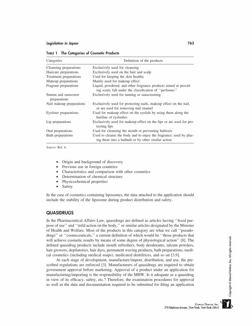
Legislation in Japan 763
TABLE 1 The Categories of Cosmetic Products
Categories Definition of the products
Cleansing preparations Exclusively used for cleansingHaircare preparations Exclusively used on the hair and scalpTreatment preparations Used for keeping the skin healthyMakeup preparations Mainly used for makeup effectFragrant preparations Liquid, powdered, and other fragrance products aimed at provid-
ing scent; fall under the classification of ‘‘perfumes’’Suntan and sunscreen Exclusively used for tanning or sunscreening
preparationsNail makeup preparations Exclusively used for protecting nails, makeup effect on the nail,
or are used for removing nail enamelEyeliner preparations Used for makeup effect on the eyelids by using them along the
hairline of eyelashesLip preparations Exclusively used for makeup effect on the lips or are used for pro-
tecting lipsOral preparations Used for cleansing the mouth or preventing halitosisBath preparations Used to cleanse the body and to enjoy the fragrance; used by plac-
ing them into a bathtub or by other similar action
Source: Ref. 6.
• Origin and background of discovery• Previous use in foreign countries• Characteristics and comparison with other cosmetics• Determination of chemical structure• Physicochemical properties• Safety
In the case of cosmetics containing liposomes, the data attached to the application shouldinclude the stability of the liposome during product distribution and safety.
QUASIDRUGS
In the Pharmaceutical Affairs Law, quasidrugs are defined as articles having ‘‘fixed pur-pose of use’’ and ‘‘mild action on the body,’’ or similar articles designated by the Ministerof Health and Welfare. Most of the products in this category are what we call ‘‘pseudo-drugs’’ or ‘‘cosmeceuticals,’’ a current definition of which would be ‘‘those products thatwill achieve cosmetic results by means of some degree of physiological action’’ [8]. Thedefined quasidrug products include mouth refreshers, body deodorants, talcum powders,hair growers, depilatories, hair dyes, permanent waving products, bath preparations, medi-cal cosmetics (including medical soaps), medicated dentifrices, and so on [3,9].
At each stage of development, manufacture/import, distribution, and use, the pre-scribed regulations are enforced [3]. Manufacturers of quasidrugs are required to obtaingovernment approval before marketing. Approval of a product under an application formanufacturing/importing is the responsibility of the MHW. Is it adequate as a quasidrugin view of its efficacy, safety, etc.? Therefore, the examination procedures for approvalas well as the data and documentation required to be submitted for filing an application
764 Masuda
differ with the indications and effects of each product [3]. The following data must beattached according to the kind of ingredients employed, and so on:
• Origin, background of discovery, use in foreign countries, etc.• Physicochemical properties, specifications, testing methods, etc.• Stability• Safety• Indications or effects
The scope of the data to be attached to the application depends on the type of quasidrug;(1) new quasidrugs that obviously differ from any previously approved products withrespect to active ingredients, usage and dosage, and/or indications or effects; (2) quasi-drugs identical with previously approved quasidrug(s); or (3) other quasidrugs that areother than those specified in (1) and (2) [3].
All products for approval as a quasidrug must be within the scope stipulated by thePharmaceutical Affairs Law. Thus, approval of a product as a quasidrug is determined byan integrated judgement of various factors such as its ingredients, quantity (composition),indications and effects, usage and dosage, and dosage form. For example, those productswhose effects are not mild—hence, coming under the category of poisons or deleteriousdrugs—are not approved even if their indications and effects and dosage forms are withinthe scope of the quasidrugs legislation. Likewise, products for which the intended usedeviates from the scope of quasidrug are also not approved even if their effects are mild [3].
COSMETICS IN THE FUTURE
The Japanese Government sets objectives to relax or abolish many of the current regulatoryitems in various industries. As a part of these plans, cosmetic deregulation has been prog-ressing based on the government’s policy to review current licensing systems and ingredi-ent labeling controls [10]. A committee, which was organized on the basis of a plan draftedby the government, was commissioned in order to figure out how to bring about a deregu-lated domestic market and a harmonized international market [11]. On March 31, 1997the future direction and issues to be addressed in connection with cosmetic regulationswere set out by the committee in the form of an interim report [4]. The following is anoutline that indicates the shift of the regulatory system to one based on the manufacturers’self-responsibility, basically similar to that of the European Union and the United States[4,10].
1. Ingredient substance controls: Recompilation of the Negative List, the PositiveList, and the Existing List of Ingredient Substances in order to abolish the cur-rent premarketing licensing systems.
2. Licensing systems for companies manufacturing and importing cosmetics:Maintenance of current systems in principle, while establishing new quality-control systems and simplifying requirements for license approval.
3. Ingredients labeling control: Creation of regulations that force cosmetic manu-factures and importing companies to include all ingredients on the label in orderto give consumers sufficient information to help them evaluate and select thecosmetics.
4. Promotion of the appropriate uses of cosmetics, and collecting and releasing tothe public information on the safety of cosmetics.
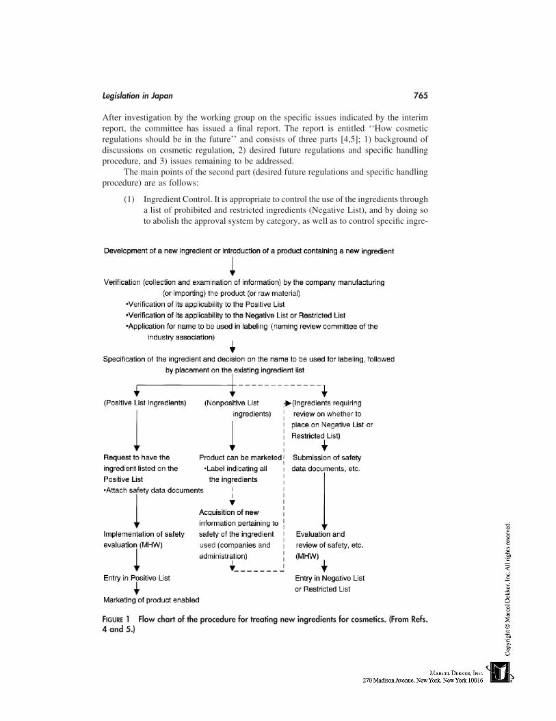
Legislation in Japan 765
After investigation by the working group on the specific issues indicated by the interimreport, the committee has issued a final report. The report is entitled ‘‘How cosmeticregulations should be in the future’’ and consists of three parts [4,5]; 1) background ofdiscussions on cosmetic regulation, 2) desired future regulations and specific handlingprocedure, and 3) issues remaining to be addressed.
The main points of the second part (desired future regulations and specific handlingprocedure) are as follows:
(1) Ingredient Control. It is appropriate to control the use of the ingredients througha list of prohibited and restricted ingredients (Negative List), and by doing soto abolish the approval system by category, as well as to control specific ingre-
FIGURE 1 Flow chart of the procedure for treating new ingredients for cosmetics. (From Refs.4 and 5.)
766 Masuda
dient groups (preservatives, UV absorbents, coal tar colors) that require cau-tious handling under appropriate safety evaluation, by drawing up a list ofingredients that may be used in formulations (Positive List). As for the newingredients, the procedure for introducing them shall be as indicated on theflow chart shown in Figure 1 [4,5].
(2) Licensing System. A manufacturing or importing licensing system should bemaintained.
(3) Regulations on Ingredient Labeling. As it is important to provide adequateinformation to consumers to facilitate their selection and verification of a prod-uct, in principle an ‘‘all-ingredient labeling system’’ shall be adopted for ingre-dients used in formulations.
(4) Cosmetic information, etc.
The MHW is now studying the possibility of amending the law and regulations in orderto implement the new system by fiscal year 2000 according to the final report.
QUASIDRUGS IN THE FUTURE
There has been a great demand by consumers for innovative cosmetic products with phar-macological action, i.e., pseudodrugs or cosmeceuticals such as skin antiaging products.To satisfy their demands, research on the skin has been undertaken to develop new activeingredients for skin antiaging products. How should those products be legally categorized?Quasidrugs would seem to be suitable for such products to be categorized. However, allof the products have not always been approved as quasidrugs to date. Taking antiwrinkleproducts, for example, no new products have been approved under the existing quasidrugspecifications.
Generally, topically applied quasidrugs are intended to mollify unwanted aspects ofthe skin and have a mild action on the human body, whereas medical drugs are intendedto treat specific diseases. Therefore, hair-growth products with a mild action on male-pattern baldness, which is not a disease [2], are quasidrugs. On the other hand, productsintended for alopecia areata, which is a disease, are regarded as drugs. The natural agingof skin, like wrinkling, is not a disease, for example. We should also keep in mind that‘‘high efficacy’’ should not always involve ‘‘strong action.’’ There will be many pseudo-drugs or cosmeceutical products with mild actions showing good efficacy.
Legally, the Minister of Health and Welfare can add new, novel types of productsto the current list of types of quasidrugs [12]. Therefore, we hope that before long theaforementioned new products will be listed as quasidrugs.
REFERENCES
1. Schmitt WH, Murphy EG. An overview of worldwide regulatory programs. In: Estrin NF, ed.The Cosmetic Industry: Scientific and Regulatory Foundations. New York: Marcel Dekker,1984:133–159.
2. Vermeer BJ, Gilchrest BA. Cosmeceuticals: Proposal for rational definition, evaluation, andregulation. Arch Dermatol 1996; 132:337–340.
3. Editorial supervision by Pharmaceuticals and Cosmetics Division, Pharmaceutical Affairs Bu-reau, Ministry of Health and Welfare. Guide to Quasi-drug and Cosmetic Regulations in Japan.Tokyo: Yakuji Nippo, 1992.
Legislation in Japan 767
4. Committee on Cosmetic Regulations. Final report on How Cosmetic Regulations Should Bein the Future. Tokyo, Japan, July 23, 1998.
5. Uemura N. Final report on how cosmetic regulations should be in the future (review). Fra-grance J 1998; 9:79–85.
6. Preface. In: The Comprehensive Licensing Standards Of Cosmetics By Category. Tokyo: Ya-kuji Nippo, 1998:13–14.
7. Supplement. In: Pharmaceuticals and Cosmetics Division, Pharmaceutical Affairs Bureau,Ministry of Health and Welfare, ed. Guide to Quasi-drug and Cosmetic Regulations in Japan.Tokyo: Yakuji Nippo, 1994.
8. Stimson N. Cosmeceuticals: realising the reality of the 21st century. SÖFW 1994; 120:631–641.
9. Society of Japanese Pharmacopoeia. Guide to Quasi-drug and Cosmetic Regulations in Japan.(Japanese ed.) 3d ed. Tokyo: Yakuji Nippo, 1996.
10. Arimoto T. The Current State of Japan’s Cosmetic Regulatory System Liberalization. Interna-tional Regulatory Congress, Florence, Italy, Apr. 22–23, 1998.
11. Deregulation of the Cosmetics Sector in Japan. In: CTFA/TRN Vol. 11. No. 5. 1997:28.12. Komiya H. Regulatory frame and problems related to quasi-drug. J Japanese Cosmet Sci Soc
1991; 15:37–40.

63
Stability Testing of Cosmetic Products
Perry Romanowski and Randy SchuellerAlberto Culver Company, Melrose Park, Illinois
INTRODUCTION
Products formulated by cosmetic chemists are intended to perform a variety of ‘‘miracle’’functions, such as reshaping hair, delivering fragrance, smoothing and softening skin,imparting color to the face, and cleansing the entire body. Chemists can deliver many ofthese miracles by using the variety of technologies described elsewhere in this book. Inusing these technologies to develop products, chemists must be aware of formulation is-sues that might prevent the product from performing optimally. Assessing product stabilityis a critical part of this formulation process. This chapter discusses the basic principlesof stability testing of cosmetic delivery systems. We will begin with a general definitionof stability testing and move on to problems encountered by specific formula types. Wewill conclude this section with a discussion of stability issues that are not necessarilydirectly related to the formulation, such as processing and packaging.
A PRACTICAL DEFINITION OF STABILITY TESTING
Stability testing may be defined as the process of evaluating a product to ensure that keyattributes stay within acceptable guidelines. In order to make this testing meaningful, itis important to accurately establish the nature of these critical product attributes, to mea-sure how they change over time, and to define what degree of change is considered accept-able. Defining which parameters are crucial requires a combination of chemical knowledgeabout the formula and common sense about product usage. The chemist should be awarethat cosmetic products must not only continue to function over time but must also look,feel, and smell the same each time the consumer uses them. Therefore, testing must evalu-ate esthetic characteristics in addition to functional properties. This is an important consid-eration because cosmetic products can change in a number of different ways, which mayaffect consumer perception. For example, fragrances become distorted, colors may fadeor darken, and consistency may change, resulting in a thicker or thinner product. Chemistsmust determine which of these product characteristics will change over time and designappropriate testing to measure the extent of the changes. Nacht cites several technicalissues to be considered, including compatibility between the delivery system and the activeingredient, compatibility with the overall formula, appropriate mechanism of release for
769
770 Romanowski and Schueller
the particular application, the rate of release of the active ingredient, and overall safetyfor the user [1]. This chapter discusses some of the key tests that the chemist can use tomeasure the changes in these characteristics. An important fact to remember is that noproduct remains unchanged forever. Depending on the intended use of the product andits anticipated shelf life, a small change over time may be inconsequential or devastatinglydetrimental. In general, if a change is consumer-perceptible, the product may not be con-sidered stable.
USEFUL INFORMATION PROVIDED BY STABILITY TESTING
Stability data are useful as an ‘‘early warning system’’ that can alert the chemist to poten-tial formulation/package-related problems. Such advance information can be helpful inmany ways.
Guiding the Chemist During Product Development
While you are formulating a product, preliminary testing of its stability can guide you inmaking modifications to ensure that it is stable. If you determine, for example, that anemulsion shows separation after exposure to freeze/thaw conditions, you may elect tomodify the surfactant system to correct the problem and then repeat the test on the modifiedformula to determine whether it performs better or worse. Preliminary stability test dataare an important parts of the trial-and-error development process.
Ensuring That the Product Will Continue to Be Esthetically Acceptableto the Consumer
More than other products, cosmetics are intended to be esthetically pleasing to the con-sumer. For this reason consumers are likely to notice subtle changes in the odor or appear-ance of their favorite products. Since no product remains 100% unchanged as it ages, itis critical that the chemist anticipate the changes that may occur and make sure that theystay within limits that are not consumer-perceptible. Stability testing allows you to seehow the product will behave over time.
Determining That the Product Will Perform as Intended and RemainSafe to Use
Studying the performance of samples that are exposed to accelerated aging lets you assesshow the product will function over time. This is particularly important for cosmetic prod-ucts that use the technologies described in this book to deliver ‘‘active’’ ingredients. Ifthe formula is not stable, the delivery of the active ingredient may be impaired. Take, forexample, the case of an antiperspirant stick with an encapsulated fragrance that is releasedupon exposure to moisture and heat. If the delivery system is poorly designed, the fra-grance may be released too soon or not at all. Properly designed stability testing can revealsuch problems so that corrective action can be taken.
Forewarning the Company About Problems That Might Occur AfterConsumer Purchase
For example, testing can show that the product may thicken somewhat over time and maybe difficult to dispense from the package. Realizing this beforehand is important to thecompany because it will allow the company to anticipate consumer reaction.
Stability Testing of Cosmetic Products 771
Even though stability testing provides much useful information, it is not an exactscience and will not guarantee a trouble-free product, but it can give an idea of the risksinvolved and help provide a solid scientific foundation for evaluation of future problems.
STABILITY TEST DESIGN
When faced with a situation where testing might be appropriate, ask some basic questionsabout the task ahead.
Why Is Testing Being Done?
Why is testing necessary? Are you concerned with product appearance or do you wantto determine if specific performance characteristics change over time? The reasons fordoing the tests will determine what kind of tests are required. Therefore it is criticallyimportant to approach this testing with a scientific mind set and to have a clearly definedhypothesis to be tested. Take, for example, the case of a skin lotion formula that developsan unpleasant odor. The reason for the test is to determine what is causing the odor.Your hypothesis may be that the fragrance you have selected is reacting with the formulaingredients to cause this problem. To test this hypothesis appropriately, you will need toassess the odor of the unfragranced base to determine how the fragrance affects the overallsmell of the product. In this example, the unfragranced samples are the controls becausethe fragrance, which is the scientific variable, has been removed. Evaluation of appropriatecontrol samples can prove or disprove the hypothesis—i.e., that the fragrance is causingthe problem.
Another example illustrating the importance of conducting a properly controlledstudy is the case of an emulsion that separates after prolonged storage in its plastic bottle.In this case the reason for the test is to determine what is causing the separation. Onehypothesis may be that the package is allowing water vapor to escape, thus leading toemulsion instability. To test this hypothesis, you will need to screen out the variable ofconcern: the packaging. Therefore, control samples could be packaged in glass to eliminatethe possibility of moisture loss. If the control samples do not show the same instabilitythat the packaged samples show, you have demonstrated that the packaging material isindeed having a negative effect on the product.
Finally, consider a case where the variable of interest is the viscosity of the product.If you are concerned that the product may become too thick over time and will not dispenseproperly, you could design a study to track product batches with varying initial viscosity.Suppose the target viscosity is 20,000 cps. You could monitor the viscosity of a series ofbatches with viscosities ranging from low to high. You may make batches which areinitially at 5000, 10,000, 15,000, and 20,000 cps, respectively. You would then monitorthe viscosity of these batches as a function of time and temperature. You may learn thatviscosity does not change significantly from the initial value, which means that a verynarrow specification will be required. In other words, the product must be very close toits final viscosity when it is produced. On the other hand, you may discover that as longas the initial viscosity is between 5000 and 15,000 cps, the product will build to 20,000cps within 2 weeks and stay at that level for 2–3 years. In this case your specificationcan be rather broad, since—regardless of the initial value—the consumer will only beexposed to product that is 20,000 cps. In all these cases, understanding why the test needsto be done helps you establish appropriate controls, which are essential if meaningful testresults are to be obtained.
772 Romanowski and Schueller
What Is Being Tested?
Another important factor to understand is the status of the formula being tested. Is it adevelopmental prototype or the final production material? Consider a situation, as in theexample provided above, where you are primarily concerned with the change in productviscosity. Furthermore, consider that the final color and fragrance of the product have notyet been firmly established, although there are several candidates under evaluation. Youcould prepare samples with every possible color/fragrance combination and measure theirviscosity over time. This could involve thousands of samples and tens of thousands ofmeasurements, which are both costly and time-consuming. So, bearing in mind that youare testing a prototype and not a finished product, you may instead opt to test the uncolored,unfragranced base formulation first. In this way you can expeditiously get data on theparameter of interest—in this case viscosity. By evaluating prototypes early on, you havegiven yourself more time to react to problems. Of course, the testing may have to berepeated once the final formula is established because the fragrance may affect viscosity.Similarly, if the final production package is not yet available, you may choose to evaluateformula stability in a packaging material that approximates the characteristics of the finalcontainer. Here too, the final formula and package combination must eventually be testedtogether, because the formula may interact unfavorably with the package. Asking the‘‘what’’ question will help make your testing meaningful without forcing you to go toexcessive lengths.
Where Will Test Samples Be Stored and How Many Are Necessary?
Ideally, you could gain information on formula stability by performing exhaustive testson every variable involved in every formulation you work with, but this is not alwaysfeasible, because proper testing requires a significant commitment of time and resources.Therefore, most companies have standardized test procedures for the storage of stabilitysamples which depend on the objective of the study. Such procedures involve evaluationsof samples stored at a variety of conditions and include enough samples to be statisticallysignificant. Usually sample storage is done at elevated temperatures, under freeze and/orfreeze thaw conditions, and with exposure to various types of light. Elevated temperaturestorage is critical, since the rate of chemical reactions roughly doubles for every 10°Cincrease in temperature. Storage at higher temperatures allows you to accelerate the agingprocess and to see certain problems much sooner than they would appear at room tempera-ture. Of course, the potential drawback is that, at high temperatures, you may be forcingreactions to occur that would not happen at all at lower temperatures. Cold storage evalu-ates conditions that may negatively affect the solubility of ingredients or stability of emul-sions. Sunlight and ultraviolet (UV) light exposure can reveal problems with ingredientsthat are reactive to the respective wavelengths; fragrances and colors are particularly sensi-tive in this regard. The most common storage conditions used in this industry are 54°Cor 50°C, 45°C, 37°C or 35°C, room temperature (25°C), 4°C, freeze/thaw, and exposureto fluorescent and natural light.
Since many of the tests that must be conducted to evaluate product performancewill affect the sample physically (e.g., spraying an aerosol can), multiple samples arerequired at each storage condition to ensure there will be enough samples left for evalua-tion at the end of the test period. Depending on the protocol set by your organization, as
Stability Testing of Cosmetic Products 773
many as one hundred or more samples may be required for a complete study. Again, youshould follow your corporate guidelines to make sure that sample quantities will be enoughfor a thorough evaluation of all necessary conditions.
How Samples Are Evaluated and What to Look for—Identificationof Instability
How samples are evaluated depends entirely on the type of product and the nature of theproblems that might occur. Instability is typically identified by evaluating various productcharacteristics either by subjective observation of properties—such as color, odor andappearance—or by objective instrumental evaluation of pH, viscosity, particle size, andelectrical conductivity. For instance, simply looking at a sample that has been stored ataccelerated temperatures can often reveal significant changes such as color changes, emul-sion separation, or rheological changes. Similarly, a quick olfactory evaluation can un-cover major flaws in fragrance stability. More rigorous characterization of product attri-butes can be obtained instrumentally—for example, with a viscometer or pH meter. Theseinstruments are highly sensitive and can distinguish small changes in products. Suchchanges are important to note since, as in the case of a change in pH, they may representchemical reactions that are occurring in the formula.
Other specialized testing can be performed to quantify specific changes in formu-lated systems. For example, microscopic evaluation and light scattering are used to ap-praise changes in particle size and distribution of emulsions. A Coulter counter is alsoused for these determinations [2], as are conductivity measurements [3]. Nuclear magneticresonance (NMR) and x-ray crystallography can also be used to reveal additional informa-tion regarding emulsion structure. In certain systems, specific assays are performed tomeasure the activity of functional ingredients. These types of tests are tailored for thecompound in question. For instance, the bactericidal efficacy of preservatives or otherantimicrobial compounds may be measured over the course of a stability test. In addition,chromatographic tests, spectroscopic measurements, titrametric evaluations, and other wetchemical methods can be used to detect signs of instability. Other indications of instabilityinclude incompatibility of product and package, which can lead to weight loss and packagedegradation (such as softening or cracking of container walls, clogging of orifices, corro-sion of metal parts, etc. [4]). But perhaps the most important question to ask in assessinginstability is to determine how much change is acceptable. Knowlton and Pearce havestated that a useful rule of thumb is to consider product rejection if the attributes beingmeasured deviate by more than 20% of their original value [4]. This value is an interestingreference point; however, for some formulations, much smaller deviations may be critical.The impact of such changes must be assessed on a case-by-case basis.
SITUATIONS THAT REQUIRE STABILITY TESTING
A good chemist should have an understanding of factors that are critical to product stabil-ity, so that appropriate testing can be conducted when necessary. Situations in whichstability testing is generally necessary include but are not limited to the following situa-tions: consideration of a new formulation, qualification of new raw materials, evaluationof new manufacturing processes, and testing of different packaging components. As youwill see, stability testing is not a finite, one-time task; instead, it is an ongoing, dynamic
774 Romanowski and Schueller
process that begins when the product is being developed and continues to evolve as theformula, packaging, or manufacturing processes change.
FORMULA-RELATED REASONS TO STABILITY TEST
Specific Considerations Related to Development of ParticularFormula Types
The process of stability testing a product is closely tied to the process of creating theformulation. As you develop formulations, you should always screen stability samplesearly in the process to make sure that your efforts are headed in the direction that willlead to a stable product. Every formula will have slightly different stability testing require-ments, but for the sake of this discussion, we will give primary consideration to the typesof cosmetic delivery systems detailed in this text.
Emulsions
Emulsions are among the most common types of delivery systems used for cosmetic prod-ucts. They enable a wide variety of ingredients to be quickly and conveniently deliveredto hair and skin. While many definitions of emulsions have been proposed, we will definethem as heterogeneous systems in which at least one immiscible or barely miscible liquidis dispersed in another liquid in the form of tiny droplets of various sizes [5]. Consequently,these systems are inherently unstable and eventually, given enough time or energy, willseparate into separate phases.
Emulsions used for cosmetic products are typically semisolid materials composedof an oil (hydrophobic) phase and a water (hydrophilic) phase. These phases are character-ized as either the internal phase or external phase, depending on the overall compositionof the emulsion. The internal phase is that which is contained inside separate discreteparticles surrounded by surfactants; these particles are known as micelles. The externalphase is the ‘‘solvent’’ or diluent, which surrounds the micelles. Usually, the externalphase is the more abundant one. Depending on the composition of each phase, simpleemulsions can be either oil in water or water in oil, the type of which depends specificallyon what emulsifier is used.
Although the internal-phase particles of an emulsion are polydisperse (meaning theyhave various sizes), their average size is often used for emulsion classification [6]. Whenthe average diameter of internal particles is less than 100 Å, the system is called a micellaremulsion. A particle diameter of 2000 to 100 Å is called a microemulsion. Larger particlesproduce macroemulsions, which are the most common types found in cosmetic formula-tions. More complex emulsions can have multiple internal phases. These emulsions, calledmultiple emulsions, can be oil in water in oil or some combination. For cosmetic applica-tions, they are formed by first making a water-in-oil emulsion and then mixing that emul-sion with a water phase. These types are particularly useful for encapsulating materialsgiving prolonged release when applied to a surface such as skin [7].
Stability Considerations
Since emulsions represent a mixture of two or more materials that are not miscible ineach other, they are, according to the second law of thermodynamics, inherently unstable.This means that eventually the two phases will separate. The degree and speed of instabil-
Stability Testing of Cosmetic Products 775
ity are quite variable. For example, a mixture of mineral oil and water when shaken willform a macroemulsion, which immediately separates upon standing. Other emulsions canremain stable for years, but eventually all emulsions will separate. While the second lawof thermodynamics suggests that emulsions will separate over time, it does not providea mechanism of this destabilization. Investigation into how emulsions destabilize has re-vealed three primary processes leading to instability: flocculation, creaming, and coales-cence [8].
Flocculation
This process is characterized by a weak, reversible association between droplets of theemulsion’s internal phase. Each individual droplet maintains its own identity; thus thereis no change in the basic droplet size [8]. Flocculation represents a less serious sign ofinstability, which can be reversed by shaking the system [9].
Creaming
When particles of an emulsion aggregate, there is a tendency for upward sedimentation.This causes a partial separation of the emulsion into two emulsions, one of which is richerin the internal phase and the other richer in the external phase [9]. As in the case offlocculation, this stability problem can be reversed by agitation.
Coalescence
An aggregation between two particles can, if the two particles combine, lead to the forma-tion of one larger particle. This process, known as coalescence, represents a more seriousstability problem. A related phenomenon is that of Ostwald ripening, in which the particlesall tend to become the same size. Both of these processes are irreversible and can eventu-ally lead to complete separation of the internal and external phases of the emulsion [10].An alternative consequence of these forms of instability is phase inversion, in which theinternal phase becomes the external phase and vice versa [9]. For stability considerations,this change is typically undesirable, since it will change the physical properties of theproduct.
All emulsions are potentially subject to all of these destabilizing processes simulta-neously, and the resulting effects on any given emulsion will vary. For example, micro-emulsions and micellar emulsions are initially transparent. Over time, the size of theirinternal-phase particles may increase, and they will develop translucent appearance. Sincemacroemulsions are opaque, a similar change in appearance will not be notable; however,there may be changes in viscosity and measurable separation. Multiple emulsions aretypically less stable than monoemulsions. Over a short period of time, the number ofmultiple emulsion particles tend to be reduced. This results in the ‘‘leaking out’’ of someof the encapsulated material and reduces the duration of prolonged release.
In addition to the inherent processes that destabilize emulsions, other factors maybe involved. Storage temperature has been shown to affect emulsion product stability.Generally, elevated temperatures result in destabilization, while reduced temperatures im-prove emulsion stability. Aqueous-phase evaporation may also contribute to instabilityover the life of a product. Microbial contamination can also cause a breakdown of emulsionstability. Finally, chemical reactions within the emulsion can lead to a change in the stabil-ity of the emulsion. While these types of reactions can be initiated by temperature in-creases, they can also be prompted by UV light or other types of electromagnetic radiation.
776 Romanowski and Schueller
VESICULAR SYSTEMS—LIPOSOMES AND NIOSOMES
Definition/Description
Vesicular systems encompass a number of delivery technologies, including liposomes andniosomes. Both of these systems employ a ‘‘vessel’’ to contain active ingredients withina formula and to provide controlled delivery of these ingredients. Nacht defines controlleddelivery as a ‘‘system that would result in a predictable rate of delivery of its activeingredients to the skin’’ [1]. Liposomes are a classic example of this technology, in whichphospholipids are used to create lipid ‘‘capsules’’ that can be loaded with various ingredi-ents. Although liposomes are enjoying tremendous popularity in cosmetics today, theyhave their roots back in the early 1960s. At that time Professor Bangham, at the Institutefor Animal Physiology in Cambridge, U.K., was one of the first to speculate that lipidssuch as phosphatidyl choline could be used to create sealed vesicles with bilayer mem-branes similar to cell membranes [1]. Niosomes are another delivery technology relatedto liposomes; the difference is that, unlike liposomes, niosomes are based on nonionicsurfactants. L’Oréal pioneered the development of nonionic liposomes using nonionicsurfactants such as polyoxyethylene alkyl ethers combined with fatty alcohols or fattyacids [1].
Stability Considerations
Liposome and niosome stability may be referred to in terms of leakage of contents, pres-ence of oxidation products, or changing particle size due to aggregation formation andfusion. They are rather fragile capsules, and certain precautions must be taken to makesure that they remain intact and are able to deliver their contents. Leakage can be causedby mechanical forces like high-shear processing, which should be avoided. Similarly, ex-cessive heat, which may destabilize the lipid bilayers, should be avoided. Perhaps mostnotably, liposomes may be solubilized by surfactants that may be present, and thereforethey are not suitable for use in detergent systems. This is particularly true of systems suchas shampoos and body washes, which contain strong anionic surfactants that can dissolvethe lipid walls. In fact, even though liposomes are often used in creams and lotions, theemulsifiers used in these formulas may also be enough to disrupt the fragile walls. Forthese reasons, many formulators believe that gels are the ideal vehicle for liposomes be-cause they lack the high HLB (hydrophilic lipophilic balance) surfactants present in manyconventional emulsions, which might disrupt the lipid bilayers [10]. There is hope forusing liposomes in emulsion. K. Uji et al. report that stable liposome suspensions canbe prepared by using a cross-linked acrylic acid/alkyl acrylate copolymer at very lowconcentrations, because it can effectively stabilize lecithin liposomes in o/w emulsions[11]. Furthermore, there is some evidence in the patent literature that the addition of colla-gen, albumin, or gamma globulin to the liposomes can decrease the harmful effects ofdetergents [10].
In addition to leakage, vesicle systems may fuse together and no longer be availableas discrete units for the delivery of active agents. According to Weiner, such fusion canoccur for several reasons, including preparation below their transition temperature, thepresence of contaminants such as fatty acids and divalent cations, changes in pH, or theaddition of nonelectrolyte hydrophobic molecules [12]. Furthermore, phase separation ofbilayer components can occur upon extended storage. In an excellent review on the subject,Fox refers to an article by Crommelin et al., that reports on preserving the long-term
Stability Testing of Cosmetic Products 777
stability of liposomes. Crommelin discusses the chemical pathways by which phospholip-ids can degrade: by hydrolysis of the ester groups or oxidation of the unsaturated acylchains. This research points to an optimal pH for liposome stability. For phosphotidylcho-line liposomes, the pH for the lowest hydrolysis rate was found to be 6.5. The stabilityof liposomes was further enhanced by using phospholipids with fully saturated acyl chains(like those made from hydrogenated soybeans, so the opportunity for oxidation is reduced)[10]. Similarly, liposomes may be stabilized by sugar esters, for example, maltopentosemonopalmitate have been used to improve stability of cosmetic systems [13].
For a more detailed discussion of the morphology of liposomal bilayers, we referthe reader to Liposomes: From Biophysics to Therapeutics [12]. The author provides anexcellent discussion of the elastic properties and tensile strength of liposomes as well asthe effect of solvents and osmotic effects on liposomal structures.
MOLECULAR CARRIERS
Definition/Description
Molecular carriers represent a delivery system in which one compound is used to bindanother compound to a substrate, thereby changing the former’s characteristics. Thisallows the bound material to be delivered to a surface and released when conditions areappropriate. One example of this type of technology is cyclodextrin chemistry. Cyclodex-trins are created from starch-derived glucopyranose units and are classified as cyclic oligo-saccharides. When formed, they contain a hydrophobic cavity capable of entrapping mole-cules of different sizes, shapes, and polarities. Molecules entrapped as such are found tobe more resistant to environmental stresses and therefore more stable [14]. They can beused to entrap various types of compounds such as fragrances, vitamins, pigments, anddyes. Cyclodextrins have been used in cosmetic products for a variety of reasons, suchas to reduce odor in mercaptan-containing systems [15], improve the stability of hair dyes[16], and as an active ingredient to treat acne [17].
Stability Issues
The complex of the cyclodextrin with a guest molecule is typically quite stable underambient temperatures and dry conditions. However, in the presence of certain materialsthe guest molecule can be prematurely displaced thereby reducing the effectiveness of thedelivery system [18]. This factor is of major concern when developing and particularlywhen assessing the stability of a formula.
PARTICULATE SYSTEMS—MICROCAPSULES, BEADS, ANDMICROSPHERES
Definition/Description
Microcapsules are one of the oldest controlled release technologies. They were developedto produce carbonless carbon paper and are composed of a core with the active ingredientsurrounded by a shell, analogous to an egg. Microcapsules may have a multilayer construc-tion with multiple cores containing the active. The active ingredients are released eitherby rupture of the capsule walls or by diffusion/permeation of the contents [1]. Fairhurstand Mitchnick list a range of materials that are typically used in this regard including
778 Romanowski and Schueller
adhesives, drugs, colors, fragrances, flavors, agricultural chemicals, solvents and oils.Classic shell materials include gelatin or gum arabic, cellulosic polymers, or syntheticpolymers [19]. Starch based capsules are often used to deliver fragrance and cosmeticingredients.
Beads and microspheres are small solid particles onto which other ingredients canbe adsorbed for later delivery. Nylon particles, for example, are useful for delivery ofcertain active ingredients. Antiperspirant salts are said to be more efficacious when deliv-ered via nylon spheres, and the esthetics of the product are said to be improved. Coloringagents may be delivered in this manner as well; Schlossman discloses a patented method(U.S. patent 5,314,683) of coupling cosmetic pigments to microspheres to provide uniformreflectivity, improved dispersion, and superior viscosity characteristics [10]. Tokubo etal. describe a process for preparing spherical hectorite particles, with a diameter of about100 Å, which can be used to deliver glycerin and solid pigments such as titanium dioxide,zinc oxide, and ferric oxide.
Stability Considerations
Microcapsules are somewhat fragile physically and care must be taken to avoid prematurerupture and release of the contents. Excessive temperature should be avoided by addingmicroencapsulated ingredients late in the manufacturing process. Likewise, refrain fromformulating with materials that may act as solvents on the capsules walls. Finally, avoidhigh-shear processing, such as milling and homogenizing, which can physically disruptthe capsules. Additional techniques for enhancing the stability of microcapsules can befound in the technical literature. Fox refers to an interesting Shiseido patent for improvingthe stability of gelatin microcapsules by coating the surface of the capsule with a basicamino acid or its polymer [10]. In general, microcapsules are a stable, efficacious methodof delivering chemicals in cosmetics. In fact, when properly formulated, microcapsulescan actually enhance stability of systems by protecting the ingredients they carry fromexternal forces. For instance, in an example provided by the Mono-Cosmetic Company,ascorbic acid particles are coated with silicone or a polymer—e.g., ethyl cellulose, toprotect the ascorbic acid against oxidation [10]. Similarly, in delivering cosmetic materialsvia beads and microspheres, care must be taken not to disturb the matrices physically. Aswith microcapsules, excessive shear can be a problem, for if the capsules are broken, theirability to retain the ingredient to be delivered will be impaired.
GENERAL CONSIDERATIONS RELATED TO FORMULA MODIFICATION
Regardless of which delivery technology you choose to utilize in a formulation, thereare certain fundamental stability considerations that you must deal with. For each of thetechnologies discussed above, factors such as raw material sources, manufacturing pro-cess, and packaging composition all play a role in product stability.
Raw Material Substitution
Often it becomes necessary to substitute one raw material for another similar material.This frequently occurs because a supplier discontinues one of the raw materials used inyour formula. In exchange, a different, yet supposedly ‘‘identical,’’ material may be of-fered. Depending on the chemistry of the materials involved, there is no way to anticipateif such a change will affect formula stability. Therefore, in such situations you must con-
Stability Testing of Cosmetic Products 779
duct testing to ensure your formula will remain stable. Similarly, you may wish to substi-tute another material that is cheaper but is not anticipated to function differently. Forexample, in a shampoo formula, you may substitute sodium lauryl sulfate for ammoniumlauryl sulfate. Given the functional similarities between the two, you would not anticipatesignificant problems; nonetheless, some degree of stability testing would be prudent.
Alternate Vendor Qualification
You may also elect to qualify alternate raw material suppliers for ingredients in the for-mula. It is desirable to have secondary sources for most raw materials to ensure a steadysupply and competitive pricing. Unfortunately, even though raw materials from differentsuppliers may have the same CTFA (Cosmetics, Toiletries, and Fragrance Association)designation, they may not be chemically identical, because chemical feedstocks and pro-cessing conditions vary between suppliers. Therefore, a raw material from one suppliercannot always be automatically inserted into a formula developed with a different suppli-er’s raw material. The impact of even seemingly inconsequential change in raw materialsmust be established by stability testing.
NON–FORMULA-RELATED REASONS
Processing Issues
In addition to the formulation and raw material issues described above, there are pro-cessing issues that can affect product stability. For example, stability testing is typicallyrequired the first time a new formulation is made on a large scale. This is because theway in which the product is made on a large scale can have a dramatic effect on itsstability. This is particularly true of emulsions, because the energy used in processingdetermines particle size and distribution, which helps determine product stability. The onlyway to fully assess the impact of the chosen manufacturing method on product stability isto evaluate samples made under actual production conditions. This may require that a trialproduction batch be made prior to commercialization of the formula. At the very least,stability testing should be done on the first production batch of any new product, so thatthe impact of actual production processing conditions may be evaluated.
Once a manufacturing process has been shown to be successful, any changes to thatprocess may require additional testing. Alterations in the order of raw material additionmay be necessary to reduce processing time; changes in heating and cooling rates mayoccur due to differences in heat transfer in large batches; and different mixing conditionswill all affect the amount of shear the product experiences. Any one of these changes willcause stability problems.
Packaging Issues
Even with the formulation and manufacturing processes held constant, variations in pack-aging material can cause problems that require stability testing. Not all packages are cre-ated equal: glass and plastic behave differently, and different kinds of plastic vary inproperties such as oxygen permeability, color fastness, and thermal resistance. Certainlya new combination of formula and package should be tested, and even a change in anexisting packaging material or the supplier of that material merits evaluation. The stabilityof aerosol systems, for example, is extremely package-dependent, since the package com-
780 Romanowski and Schueller
position will help to determine how resistant the final product is to corrosion. The overallobjective is to be alert for changes to the formulation/manufacturing/packaging systemthat may necessitate additional testing, so that you can be confident that your product willremain stable. Of course, your observations should not be limited to the formula itself.Changes that result from formulation and packaging interaction may be critical to totalproduct integrity. To this end, weight loss, changes in plastic color and odor, and otherpackage-related observations are important. The objective is to gain as much knowledgeas possible regarding the behavior of the product over time.
CONCLUSION
This chapter is intended to provide insight into the issues associated with the stabilitytesting of cosmetic products. For the beginning chemist, we stress the importance of care-ful, methodical observation to ensure that as many stability problems as possible are identi-fied. For the veteran formulator, we urge periodic review of the latest technical literatureso that it will be possible to keep pace with new developments in stabilizing the specificdelivery systems discussed in this book. Hopefully the references we have provided willbe helpful in this regard.
REFERENCES
1. Nacht S. Encapsulation and other topical delivery systems. Cosmet Toilet 1995; 110(9):25–30.
2. Rieger M. Stability testing of macroemulsions. Cosmet Toilet 1991; 106(5):59–66.3. Jayakrishnan A. Microemulsions: evolving technology for cosmetic applications. J Soc Cosmet
Chem 1983; 34:343.4. Knowlton J, Pearce S. The Handbook of Cosmetic Science and Technology. Oxford, England:
Elsevier Advanced Technology, 1993:436–439.5. Becher P. Emulsions: Theory and Practice. New York: Reinhold, 1965:2.6. Prince L. Microemulsions: Theory and Practice. New York: Academic Press, 1977:1–2.7. Fox C. An introduction to multiple emulsions. Cosmet Toilet 1986; 101(11):101–102.8. Becher P. Encyclopedia of Emulsion Technology. New York: Marcel Dekker, 1983:133–134.9. Eccleston GM. Application of emulsion stability theories to mobile and semisolid O/W emul-
sions. Cosmet Toilet 1986; 101(11):73–135.10. Fox C. Advances in cosmetic science and technology: IV. Cosmetic vehicles. Cosmet Toilet
1995; 110(9):59–68.11. K Uji K et al. J Soc Cosmet Chem Jpn 1993; 27:206–215.12. Ostro MJ, ed. Liposomes: From Biophysics to Therapeutics. New York: Marcel Dekker, 1987:
343.13. Fox C. Cosmetic raw materials literature and patent review. Cosmet Toilet 1991; 106(8):78.14. Dalbe B. Use of cyclodextrins in cosmetics. 16th IFSCC Meeting, New York, 1991. pp. 635–
639.15. Kubo S, Fumiaki N. US patent 4,548,811. Shiseido Company Ltd.16. Oishi T et al. US patent 4,808,189, Hoyu Co.17. Koch J. US patent 4,352,749.18. Duchene D. New Trends in Cyclodextrins and Derivatives. Dermal Uses of Cyclodextrins and
Derivatives. Paris: 1991:473–474.19. Fairhurst D, Mitchnik M. Submicron encapsulation of organic sunscreens. Cosmet Toilet 1995;
110(9):47.
64
Stability Control: Microbiological Tests
Michel J. DevleeschouwerFree University of Brussels, Brussels, Belgium
Françoise SiquetColgate-Palmolive Technology Center, Milmort, Belgium
MICROBIOLOGICAL CONTROL OF RAW MATERIALS
Microbial Health Hazards by Contaminated Products
The microbial spoilage of cosmetics has been reported in the literature for many years[1–3]. One of the first reported incidents [4] is the death by tetanus of four babies in NewZealand in 1946, the vector being a contaminated talcum powder. The same vector wasthe source of two other cases of tetanus in an English hospital [5]. Since the 1960s, casesof cosmetic-induced infections were described in parallel with the awareness of the prob-lem for topical drugs [6–12]. The isolated organisms were Gram-negative bacteria fromthe genus Klebsiella, Enterobacter, Serratia, and Pseudomonas [13,14]. The organismPseudomonas aeruginosa, a particularly virulent hospital pathogen transmitted by eyecosmetics, led to cases of infections and even blindness [15–20], or folliculitis fromsponges [21]. Studies were then conducted to evaluate the importance of the problem [22–29] and to investigate the primary contaminating sources such as raw materials, personnel,water, and packaging, as well as secondary sources, such as the consumer [30].
Sources of Contamination
These can be divided into three groups [11,28,31,32]:
1. The microbiological quality of raw materials, including water;2. The manufacturing process; and3. The galenical form (which is made with vegetable and/or animal extracts) of
the product.
Microbiological Quality of Raw Materials, Including Water
Their quality depends upon their origin. Raw materials from animal or vegetable origincan be heavily contaminated with 106 or even more organisms per gram or milliliter [33–35]. Fecal bacteria are regularly identified. In contrast, synthetic raw materials are rela-
781
782 Devleeschouwer and Siquet
tively free from contamination, with the exception made for some that have steps in theirmanufacture, such as kaolin, some sugars and vitamins, some synthetic surfactants (e.g.,sodium lauryl ether sulfate [SLES]), or partially hydrated salts. A recent study in ourlaboratory (Boussard et al., unpublished data) showed that out of 188 different synthetictested raw materials, only 48, or 25.5%, gave results higher than 100 organisms per gram ormilliliter. The recovered organisms were bacilli or Gram-positive cocci. A microbiologicaltesting program of the raw materials must be set up.
Water remains one of the most important contamination factors of a product. Specieslike Pseudomonas, Achromobacter, Aeromonas, or Flavobacterium are recovered fromnatural waters [36]. Softening or deionization treatments frequently alter the microbiologi-cal water quality. These systems must be well maintained and the water microbiologicallytreated, using, e.g., ultraviolet (UV) lamps or/and bacterial filtration to ensure optimalquality. Microbiological control of production water should be made at least each workingday, and a validation program of the water quality set up.
Manufacturing Process
During the manufacturing process, contamination can occur through contact by the opera-tors, the manufacturing equipment, and the air. The micro-organisms capable of contami-nating a cosmetic from human sources are part of the rhinopharyngal, buccal skin, hair,hand skin, and, in some circumstances, intestinal floras. Among these, fecal streptococci,staphylococci, enterobacteria, and Pseudomonas have sufficient vitality to survive andeven to multiply in a product.
The manufacturing equipment is also an important source of contamination, comingfrom maintenance materials (oils, greases), from poor cleaning and/or disinfection on aregular basis, and from product changeover. The design of the equipment is also participat-ing in this process: a piece of equipment that cannot be totally emptied is critical; theequipment storage conditions must also be optimized to avoid product residues stagnantin the system. The design of cleaning in place (CIP) systems must be carefully evaluated:a CIP that leaves a small quantity of stagnant water together with diluted product willhave a negative effect instead of a beneficial one.
Attention must be paid to the air quality of the manufacturing rooms. The numberof workers and the importance of their movements contributes to 80% of aerial contamina-tion [37]. Air conditioning contributes to 15% of this contamination, and the room struc-ture (materials used) to 5%. It is thus necessary to fix acceptable levels for the biocontami-nation of the air and to control the air quality. According to the European GoodManufacturing Practices (GMPs) [38], the limits of the class D rooms should be used(200 organisms/m3).
Galenical Form of the Product
A parameter of crucial importance in the microbiological stability of a formulation is itswater availability, or aw. This aspect will be discussed at the end of this chapter. Someprocesses, such as manufacturing at high temperature (e.g., lipsticks) can help to reduceor avoid bacterial contamination. Thus high-risk products are aqueous-based products con-taining raw materials from biological origin such as lotions, suspensions, creams, gels,and emulsions, especially if they are manufactured at room temperature.
Microbiological Tests 783
Establishment of Microbial Limits
For many years there have been discussions on whether total count would be sufficientto guarantee the microbiological quality of a cosmetic, or if the exclusion of specifiedmicroorganisms, pathogens, or potential pathogens would also be required. The currenttrend is to require quantitative and qualitative microbial limits. Acceptance criteria forcosmetics and control methods will be issued in the Seventh Amendment of the EuropeanCosmetic Directive. Nevertheless, the acceptance criteria will be minimal criteria thatfulfill the public health expectations, such as:
1. Microbial limits for finished products. Maximum 1000 organisms/g or mL, andabsence of Staphylococcus aureus, Candida albicans, enterobacteria, and Pseudomonasaeruginosa in one gram or milliliter of the product. Exceptions are baby-care products,eye products, and products for intimate hygiene—maximum 100 organisms/g or mL, andabsence of Staphylococus aureus, Candida albicans, enterobacteria, and Pseudomonasaeruginosa in one gram or milliliter of the product.
2. Microbial limits for raw materials. Maximum 100 organisms/g or mL, and ab-sence of Staphylococcus aureus, Candida albicans, enterobacteria, and Pseudomonas aer-uginosa in one gram or milliliter. Limits for water as raw material could be fixed at maxi-mum 100 organisms/mL and absence of coliforms and Pseudomonas aeruginosa in 100mL.
However, what must be the attitude of a manufacturer if one of the following germsis identified in a product: Gram-negative bacilli other than enterobacteria and Pseudomo-nas aeruginosa, staphylococci different from Staphylococcus aureus, or fecal strepto-cocci? What is the significance of this regarding manufacturing hygiene? Are these organ-isms harmless? Furthermore, in addition to the human safety, it must be emphasized thatcontamination of products with nonharmful organisms can partially or totally destroy theproduct aesthetic (e.g., perfume, color) and can alter the product performance. The riseof these questions emphasizes the need of internal quantitative and qualitative microbio-logical safety margins and of a quality-assurance system.
Use of Validated Methods to Control Products and Water
Microbiological Control of Finished Products and Raw Materials
The method described here is based on the method for microbiological analysis of nonster-ile pharmaceuticals in the 3rd edition of the European Pharmacopeia [39,40] and froma publication of a working party of the ‘‘Fédération Internationale Pharmaceutique’’ [41].
Sample Preparation. A 10% homogeneous solution or suspension of the productis prepared with a sterile neutralizing solution or a sterile buffered peptone saline solutionat pH 7. The neutralizing solution is used in case of the presence of known or suspectedantimicrobial substances in the product. The pH 7 solution is used in case of preservative-free raw materials. For nonsoluble products, 0.1% of tween 80 or heating at a temperaturenot higher than 40°C for half an hour maximum can help in the homogenization. Theneutralizing solution is basically letheen broth (Difco) supplemented with variousinhibitors of the preservatives or disinfectants. The 10% homogenate is then used toperform the bacterial and fungal counts and to investigate the presence of specified micro-organisms. If, for technical reasons, the use of 10 g sample is not possible, 5, 2.5, or even1 g can be mixed for a total suspension of 100 mL.
784 Devleeschouwer and Siquet
Validation of the Preservative’s Inactivation. The efficacy of the neutralizingsolution must be validated in order to avoid false-negative results. For this purpose, 1 mLof the preserved sample or 1 mL sterile normal saline is added to 9 mL neutralizingsolution. The two tubes are mixed well and let to rest for 10 minutes. 0.1 mL of a mixedsuspension of Staphylococcus aureus ATCC 6538 and Pseudomonas aeruginosa ATCC9027 at 104 bacteria/mL are then added to the tubes, which are mixed again. The colony-forming units in each tube are estimated. The difference in the results must be lower than1/2 log between the tubes.
Bacterial and Fungal Counts. From the 10% homogenate, an appropriate numberof successive tenfold dilutions in the sterile buffered peptone saline at pH 7 are carriedout. A plate count is then made by transferring duplicates of 1 mL of the dilutions insterile Petri dishes, followed by the addition of 15 mL melted agar. Tryptic Soy agar isused for the bacteria and Sabouraud Chloramphenicol agar for yeast and moulds. For thebacterial counts the dishes are incubated at 30 to 35°C for 5 days, and for the yeast andmoulds, 20 to 25°C for 5 to 7 days. The Petri dishes used for the fungal counts are alsoused to check the presence of Candida albicans.
Investigations for the Presence of Specific Microorganisms1. Enterobacteria and other gram-negative organisms. One milliliter or 1 g of
the 10% homogenate is mixed with 100 mL enterobacteria enrichment broth (EEB) andincubated at 35 to 37°C for 24 to 48 hours. Subcultures are then carried out on violet redbile dextrose VRBG agar dishes and incubated at 35 to 37°C for 18 to 24 hours. Thecolonies of presumptive Gram-negative organisms are then identified.
2. Escherichia coli. One milliliter or 1 g of the 10% homogenate is mixed with100 mL Mac Conkey broth and incubated at 43 to 45°C for 18 to 24 hours. Subculturesare carried out on Mac Conkey agar dishes incubated at 43 to 45°C for 18 to 24 hours.The colonies of lactose-fermenting gram-negative organisms are then identified.
3. Pseudomonas aeruginosa and other gram-negative organisms growing on Ce-trimide agar. One milliliter or 1 g of the 10% homogenate is mixed with 100 mL TrypticSoy broth (TSB) and incubated at 35 to 37°C for 24 to 48 hours. Subcultures are carriedout on Cetrimide agar dishes incubated at 35 to 37°C for 18 to 24 hours. The coloniesare then identified.
4. Staphylococcus aureus. One milliliter or 1 g of the 10% homogenate is mixedwith 100 mL TSB and incubated at 35 to 37°C for 24 to 48 hours. Subcultures are carriedout on Baird Parker agar dishes incubated at 35 to 37°C for 18 to 24 hours. The blackcolonies are then identified.
Validation of the Sterility of the Media. Sterility of all the media must be checked.For example, sterile saline is used instead of the sample and the bacterial counts and theappropriate investigations for specific organisms are performed. No microbial growth mustbe recorded in this assay.
Validation of the Growth-Promoting Properties of the Selective Media. Thefollowing reference strains are incubated separately in TSB at 30 to 35°C for 18 to 24hours: Staphylococcus aureus ATCC 6538, Pseudomonas aeruginosa ATCC 9027, and,Escherichia coli ATCC 8739. Each bacterial suspension is diluted to obtain around 1000organisms per milliliter. The three suspensions are equally mixed together and 0.3 mL ofthe mixture (containing about 100 organisms of each strain) are used as the inoculum to
Microbiological Tests 785
perform the investigations for the specific micro-organisms. The organisms must bedetected in the media used for this assay.
Microbiological Control of Water
The microbiological quality of water is of particular importance and can be checked quan-titatively and qualitatively. For the quantitative determinations of a potential water contam-ination, 100 mL or 10 mL of water are filtered through bacteriological filters (porosity of0.45 µm). After filtration, the filters are deposited on the surface of Tryptic Soy agar Petridishes. Amounts of 1 mL and 0.1 mL of water are also incorporated in two melted trypticsoy agar for a plate count in duplicate. All the dishes are incubated at 30 to 35°C for 3to 5 days. For the qualitative determinations, 100 mL of water are filtered through 0.45µm sterile filters. The filters are laid down on sterile Mac Conkey Petri dishes for thecoliform bacteria and on Cetrimide agar Petri dishes for Pseudomonas. These are incu-bated at 30 to 35°C for 3 to 5 days. Questionable colonies are identified.
CHALLENGE TEST FOR THE EFFICACY OF PRESERVATION
Aim of Preservation
It is generally accepted that adequate preservation of a finished product, with preservativesor based on active preservation of a formulation, implies that the product remains stableand safe during storage (shelf-life) and consumer use [1,42–46]. From a public-healthpoint of view, preservation must avoid infection of the consumer, and for product-qualityreasons it must prevent a deterioration of the preparation. It is especially important topoint out that the use of preservatives must not mask a lack of hygiene during manufacture.It is thus imperious to manufacture any cosmetic product according to Good Manufactur-ing Practices (GMPs) [34] such as imposed by the 6th Amendment of the European Direc-tive 76/768/CEE [47], and to reach at the end of the manufacture the microbiologicalquality level discussed earlier in this chapter. Furthermore, the challenge test to evaluatethe efficacy of preservation must not be simply performed on a lot-per-lot basis. The testhas to be essentially connected with each development phase of the preparation [48]. Itmust be as simplified as possible for routine use, easy to standardize, and reproducible.Moreover, the test method must be able to show the potential intrinsic antibacterial efficacyof a formulation and should thus be performed on each finished product in its intact originalcontainer as well. Indeed, changes in the composition of the preparation have a tremendousinfluence on preservation [49,50]. Even minor changes in perfumes or dyes can affect theglobal behavior of the product [2,51,52]. Moreover, the material of the container and itstype of closure influences the efficacy of the preservation and the protection of the productduring use [45,53–55]. Rubber closures are, for example, known to absorb some amountof preservative from a solution [56,57]. Shave foams are often presented in containersunder pressure with a propeller gaze such as butane. These storage conditions can widelyinfluence the survival of some aerobic contaminants. Moreover, refrigeration can alter thepreservative efficacy [58]. The preservatives may be inactivated by the components ofthe product [59].
Activity Spectrum of a Preservative
The use of the word ‘‘antimicrobial’’ preservative raises the need to define exactly whatkind of activity is needed for a preservative. What are the organisms of concern: bacteria,
786 Devleeschouwer and Siquet
fungi, viruses, or even spores? The scale of the activity spectrum is based on almost threeparameters: (1) the survival, or even multiplication, of particular organisms in a widerange of products; (2) the pathogenicity of these organisms by the route of administration;and (3) the possibility to find effective chemicals at nontoxic concentrations.
Sporicidal action must not be considered because sporicidal chemicals are very rare(e.g., aldehydes are too toxic to be used in a cosmetic product at effective concentrations).Moreover, infectious problems induced by spore formers are very seldom, as previouslydiscussed for the talcum powder in this chapter. Even if aerobic spore formers are oftenfound in raw materials and finished products, according to Davis [13] they should not bea hazard to human health.
Virucidal action is not considered for cosmetics. These facts restrict the spectrum ofa cosmetic preservative to bacteria and fungi. According to the most widespread opinion, abactericidal and a fungicidal effect is needed so that the contaminating organisms acciden-tally introduced in the preparation will be killed. A bacteriostatic or fungistatic actioncould eventually be accepted to stabilize a preparation during the shelf-life of a unidose,nonsterile product. For the fungicidal and bactericidal actions, the concentration of thepreservative must be toxicologicaly acceptable.
Test Organisms
As previously discussed, the range of organisms must contain bacteria and fungi. Withinthese we must find Gram-positive and Gram-negative bacteria because the structure ofthe bacterial wall influences the penetration and thus the efficacy of the preservating agent.For the fungi, representatives of the two fungal forms must be used, namely the vegetativeyeast cell and the mould spore. The choice of species is directed by their skin and mucosalpathogenicity for cosmetics. Product degradation capabilities are also taken into accountto choose the species. So among the Gram-positive species, Staphylococcus aureus is animportant skin pathogen, as is Pseudomonas aeruginosa for the Gram-negative bacteria.This latter organism is also able to use many compounds, such as preservatives or evendisinfectants, as a carbon source and is very adaptative in adverse environmental condi-tions even in pure water [60,61]. For the yeast, Candida albicans is a skin pathogen andAspergillus niger is a representative of the degradation flora. The choice of strains for astandardized assay must be guided by the need to compare results obtained in differentlaboratories, and in this way culture-collection strains are chosen coming from the Ameri-can Type Culture Collection (ATCC). The strains normally used are as follows: (1) Staphy-lococcus aureus ATCC 6538, (2) Pseudomonas aeruginosa ATCC 9027, (3) Candidaalbicans ATCC 10231, and (4) Aspergillus niger ATCC 16404. These strains are to someextent resistant to the antimicrobials, and some are also used for testing disinfectants orantibiotics. For a representative preservation-efficacy test, it is also recommended to addstrains isolated from the environment, water, or contaminated products. These strains livein the vicinity of or even inside the product, are well adapted to adverse conditions, andare often resistant to preservatives or even disinfectants [62–64]. Nevertheless, after afew passages in culture media, this particular resistance can disappear. Precautions mustbe taken to avoid this, such as immediate storage in appropriate medium by deep freezingor in liquid nitrogen.
Test Conditions and Validations
The challenge test consists in an artificial contamination of the tested sample and countingof the survivors during a period of 4 weeks maximum. Even if several preservative efficacy
Microbiological Tests 787
tests exist as described in the USP23 [65], the Japanese Pharmacopeia [66], or the CTFAtest [67–74], the general conditions of the test described here are those of the EuropeanPharmacopeia [43], adapted from a Federation Internationale Pharmaceutique (FIP) work-ing party publication [44]. Several points, such as validations and strain maintenance, aredescribed here in more detail.
Maintenance of Microbial Strains
The cultures can be maintained as described in the CEN 216 PrEN 12353 document [75].Stock cultures are maintained at a temperature below �18°C. To prepare the workingculture, subcultures are originated from the culture stock by streaking onto adequate agarmedium slopes. The second and/or third subcultures can be used as the working cultures.
Preparation of the Inoculum
The subcultures to be used in the test are plated on Petri dishes of suitable media, e.g.,Tryptic Soy Agar (TSA) for the bacteria and Sabouraud Dextrose Agar (SDA) for thefungi. After adequate incubation—18 to 24 hours for the bacteria, 48 hours for the yeast,and 3 to 5 days for the mould—the cultures are collected with sterile, normal saline.The suspensions are then calibrated against a Mac Farland scale or by using any suitablecalibration system. This calibrated suspension homogenized at a maximum ratio of 1:100(0.2 mL in 20 µg or mL, for example) of the tested sample must give between 5.105 and5.106 organisms per millilitre or gram. Such a high inoculum density is imposed not onlyby the counting technique of the survivors, or the ‘‘plate count,’’ but also by the impor-tance of the logarithmic reduction asked for the products.
Test Conditions
The first day of the challenge test, the product and two controls—one comprising thetested product without preservatives and one of normal saline with 1% peptone—are inoc-ulated with each microbial strain. A microbial count is immediately performed after homo-geneization on this group of three vials. Counts are performed after dilution of 1 g or mLof the sample, with 9 mL of neutralizer. The neutralizing solution used is the same as inthe first part of this chapter. Further dilutions are made in normal saline in order to performa plate-count technique according to the estimate contamination. Sampling is performedin the same way for the preserved samples, after 2, 7, 14, and 28 days of storage of theinoculated product kept at room temperature in the dark or in its normal storage conditions.
To estimate the starting value 100%, the product effect must be evaluated on theinoculum. So, the inoculum level is estimated in a nonpreserved test product, if available,and compared with the level measured into normal saline containing 1% of peptone. Ifthe following occur: (1) data obtained in the nonpreserved product are equivalent to thoseobtained in saline, and this value is chosen as the starting level (100%); (2) the dataobtained in the product is � or � 1 log from the saline data, the value obtained in thesaline control is chosen as the starting value; and (3) if the product data are � 1 log fromthe saline control, this is an indication of product contamination and the test is invalid.The results of the test are expressed as logarithmic reduction versus time of the valuetaken as 100%.
Validation of the Contamination of the Sample
The contamination of the sample consists of a homogeneous incorporation into the sampleof a single strain at a maximum ratio of 1% of calibrated suspension. Most of the time,
788 Devleeschouwer and Siquet
the inoculum is aqueous and dispersed in an aqueous phase; for some products, additionof tween 80 ou isopropylic myristate could be useful to homogenize the inoculum. In somecases, a dried inoculum suspended in isododecane is used to contaminate fatty products. Itis indispensable to ascertain that the inoculum can homogeneously be dispersed throughthe product. This is nearly immediate for liquids but much more difficult for oily productssuch as creams or mascaras. A validation is thus performed using a nonpreserved productthat is inoculated with the calibrated suspension and homogenized. At least three differentsamples are taken from the product and the results of the counts obtained for these samplesare compared. The difference between samples must be less than 1 log.
Validation of the Neutralizing Solution
Because a neutralizing solution is used as first dilutant when counting the survivors, theefficacy of the neutralizing solution must be validated in order to avoid false-negativeresults. For this purpose, 1 mL of the preserved sample or 1 mL sterile normal saline areadded to 9 mL neutralizing solution. The two tubes are well mixed and let at rest for 10minutes. 0.1 mL of a 10�3 dilution of the calibrated suspension are then added and mixedto both tubes. The colony-forming units in each tube are estimated, and the difference inresults between the tubes must be less than 1 log.
Interpretation of the Results
The criteria taken by the European Pharmacopeia for the topically applied product are agood base of discussion [43]. For bacteria, the recommended criteria (level A) are a 2log reduction after 2 days, 3 log after 7 days, and no increase in the recovered bacteriaafter 28 days. For fungi, a 2 log reduction is requested after 14 days with no increase ofthe counts after 28 days. This requirement of no increase of the counts at the end of thetest period is of particular importance. Indeed, even if the logarithmic reduction attainedby a product is greater than the requirement, a regrowth of the organisms during theexamination period is unacceptable. This would indicate that the micro-organisms are ableto adapt their metabolic capacities to use the product, and its preservative in particular,as carbon source. In the European Pharmacopeia, it is also stated that, in justified cases,e.g., when adverse reactions could occur, level B criteria can be used to interpret theresults. These are: for bacteria, a 3 log reduction after 14 days and no increase of thecounts after 28 days; and for fungi, 1 log reduction after 14 days and no increase of thecounts after 28 days.
DETERMINATION OF WATER AVAILABILITY OR aw
Water availability (aw) is defined as the water available for bacterial metabolism and isevaluated by measurement of the water vapor pressure at the surface of a product or araw material. It can be defined as the following ratio:
Aw �water vapor pressure over substance at t°water vapor pressure over pure water at t°
It depends on temperature and on formulations. It is not correlated with the total watercontent of a formula but depends of the quantity of water ‘‘trapped’’ into the formulachemicals. Ingredients such as humectants, gums, or others, use the water to swell andso this water is no longer available for bacterial growth. As water is a critical growthfactor for micro-organisms, one of the means to preserve a formula is to decrease thelevel of water availability, optimizing a formula by the inclusion of ingredients that fix
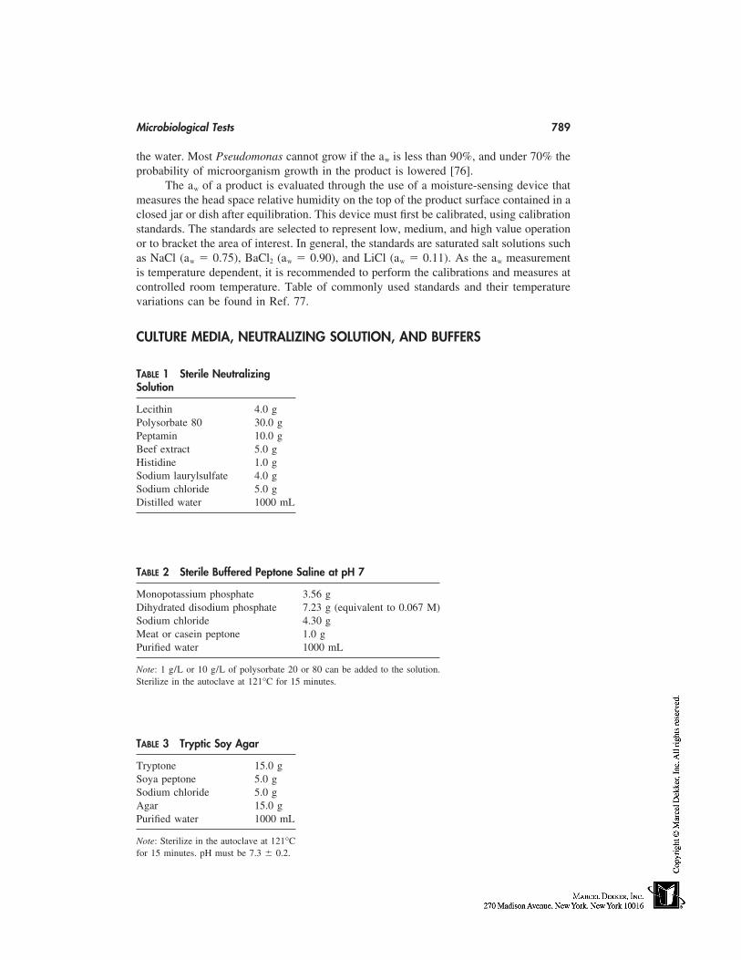
Microbiological Tests 789
the water. Most Pseudomonas cannot grow if the aw is less than 90%, and under 70% theprobability of microorganism growth in the product is lowered [76].
The aw of a product is evaluated through the use of a moisture-sensing device thatmeasures the head space relative humidity on the top of the product surface contained in aclosed jar or dish after equilibration. This device must first be calibrated, using calibrationstandards. The standards are selected to represent low, medium, and high value operationor to bracket the area of interest. In general, the standards are saturated salt solutions suchas NaCl (aw � 0.75), BaCl2 (aw � 0.90), and LiCl (aw � 0.11). As the aw measurementis temperature dependent, it is recommended to perform the calibrations and measures atcontrolled room temperature. Table of commonly used standards and their temperaturevariations can be found in Ref. 77.
CULTURE MEDIA, NEUTRALIZING SOLUTION, AND BUFFERS
TABLE 1 Sterile NeutralizingSolution
Lecithin 4.0 gPolysorbate 80 30.0 gPeptamin 10.0 gBeef extract 5.0 gHistidine 1.0 gSodium laurylsulfate 4.0 gSodium chloride 5.0 gDistilled water 1000 mL
TABLE 2 Sterile Buffered Peptone Saline at pH 7
Monopotassium phosphate 3.56 gDihydrated disodium phosphate 7.23 g (equivalent to 0.067 M)Sodium chloride 4.30 gMeat or casein peptone 1.0 gPurified water 1000 mL
Note: 1 g/L or 10 g/L of polysorbate 20 or 80 can be added to the solution.Sterilize in the autoclave at 121°C for 15 minutes.
TABLE 3 Tryptic Soy Agar
Tryptone 15.0 gSoya peptone 5.0 gSodium chloride 5.0 gAgar 15.0 gPurified water 1000 mL
Note: Sterilize in the autoclave at 121°Cfor 15 minutes. pH must be 7.3 � 0.2.
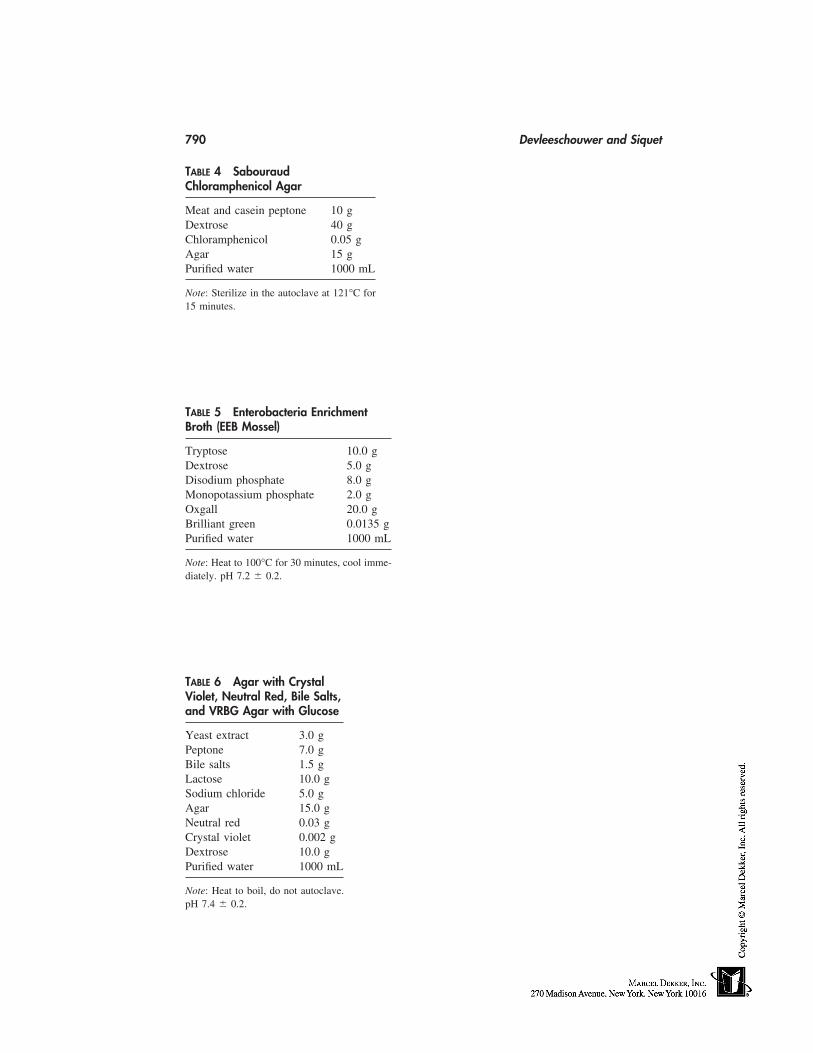
790 Devleeschouwer and Siquet
TABLE 4 SabouraudChloramphenicol Agar
Meat and casein peptone 10 gDextrose 40 gChloramphenicol 0.05 gAgar 15 gPurified water 1000 mL
Note: Sterilize in the autoclave at 121°C for15 minutes.
TABLE 5 Enterobacteria EnrichmentBroth (EEB Mossel)
Tryptose 10.0 gDextrose 5.0 gDisodium phosphate 8.0 gMonopotassium phosphate 2.0 gOxgall 20.0 gBrilliant green 0.0135 gPurified water 1000 mL
Note: Heat to 100°C for 30 minutes, cool imme-diately. pH 7.2 � 0.2.
TABLE 6 Agar with CrystalViolet, Neutral Red, Bile Salts,and VRBG Agar with Glucose
Yeast extract 3.0 gPeptone 7.0 gBile salts 1.5 gLactose 10.0 gSodium chloride 5.0 gAgar 15.0 gNeutral red 0.03 gCrystal violet 0.002 gDextrose 10.0 gPurified water 1000 mL
Note: Heat to boil, do not autoclave.pH 7.4 � 0.2.
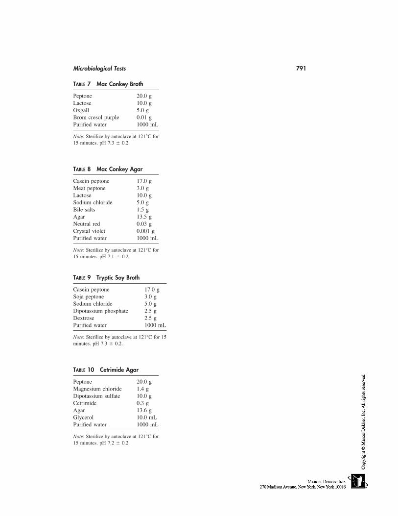
Microbiological Tests 791
TABLE 7 Mac Conkey Broth
Peptone 20.0 gLactose 10.0 gOxgall 5.0 gBrom cresol purple 0.01 gPurified water 1000 mL
Note: Sterilize by autoclave at 121°C for15 minutes. pH 7.3 � 0.2.
TABLE 8 Mac Conkey Agar
Casein peptone 17.0 gMeat peptone 3.0 gLactose 10.0 gSodium chloride 5.0 gBile salts 1.5 gAgar 13.5 gNeutral red 0.03 gCrystal violet 0.001 gPurified water 1000 mL
Note: Sterilize by autoclave at 121°C for15 minutes. pH 7.1 � 0.2.
TABLE 9 Tryptic Soy Broth
Casein peptone 17.0 gSoja peptone 3.0 gSodium chloride 5.0 gDipotassium phosphate 2.5 gDextrose 2.5 gPurified water 1000 mL
Note: Sterilize by autoclave at 121°C for 15minutes. pH 7.3 � 0.2.
TABLE 10 Cetrimide Agar
Peptone 20.0 gMagnesium chloride 1.4 gDipotassium sulfate 10.0 gCetrimide 0.3 gAgar 13.6 gGlycerol 10.0 mLPurified water 1000 mL
Note: Sterilize by autoclave at 121°C for15 minutes. pH 7.2 � 0.2.
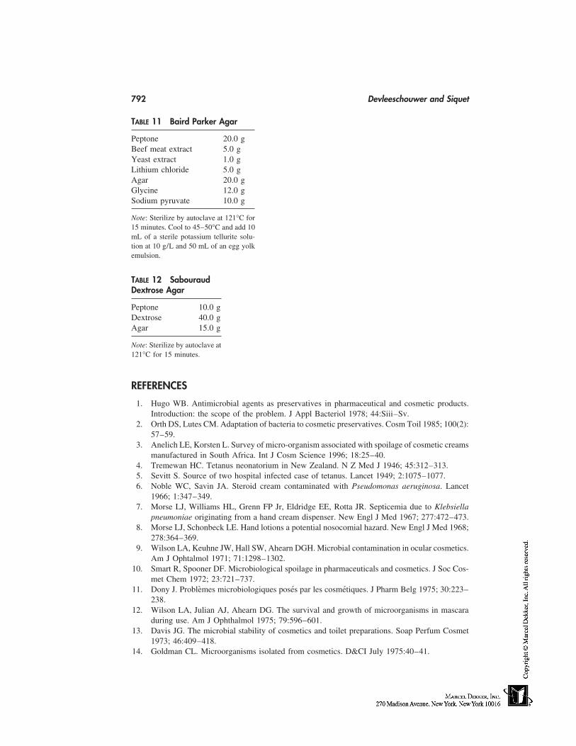
792 Devleeschouwer and Siquet
TABLE 11 Baird Parker Agar
Peptone 20.0 gBeef meat extract 5.0 gYeast extract 1.0 gLithium chloride 5.0 gAgar 20.0 gGlycine 12.0 gSodium pyruvate 10.0 g
Note: Sterilize by autoclave at 121°C for15 minutes. Cool to 45–50°C and add 10mL of a sterile potassium tellurite solu-tion at 10 g/L and 50 mL of an egg yolkemulsion.
TABLE 12 SabouraudDextrose Agar
Peptone 10.0 gDextrose 40.0 gAgar 15.0 g
Note: Sterilize by autoclave at121°C for 15 minutes.
REFERENCES
1. Hugo WB. Antimicrobial agents as preservatives in pharmaceutical and cosmetic products.Introduction: the scope of the problem. J Appl Bacteriol 1978; 44:Siii–Sv.
2. Orth DS, Lutes CM. Adaptation of bacteria to cosmetic preservatives. Cosm Toil 1985; 100(2):57–59.
3. Anelich LE, Korsten L. Survey of micro-organism associated with spoilage of cosmetic creamsmanufactured in South Africa. Int J Cosm Science 1996; 18:25–40.
4. Tremewan HC. Tetanus neonatorium in New Zealand. N Z Med J 1946; 45:312–313.5. Sevitt S. Source of two hospital infected case of tetanus. Lancet 1949; 2:1075–1077.6. Noble WC, Savin JA. Steroid cream contaminated with Pseudomonas aeruginosa. Lancet
1966; 1:347–349.7. Morse LJ, Williams HL, Grenn FP Jr, Eldridge EE, Rotta JR. Septicemia due to Klebsiella
pneumoniae originating from a hand cream dispenser. New Engl J Med 1967; 277:472–473.8. Morse LJ, Schonbeck LE. Hand lotions a potential nosocomial hazard. New Engl J Med 1968;
278:364–369.9. Wilson LA, Keuhne JW, Hall SW, Ahearn DGH. Microbial contamination in ocular cosmetics.
Am J Ophtalmol 1971; 71:1298–1302.10. Smart R, Spooner DF. Microbiological spoilage in pharmaceuticals and cosmetics. J Soc Cos-
met Chem 1972; 23:721–737.11. Dony J. Problèmes microbiologiques posés par les cosmétiques. J Pharm Belg 1975; 30:223–
238.12. Wilson LA, Julian AJ, Ahearn DG. The survival and growth of microorganisms in mascara
during use. Am J Ophthalmol 1975; 79:596–601.13. Davis JG. The microbial stability of cosmetics and toilet preparations. Soap Perfum Cosmet
1973; 46:409–418.14. Goldman CL. Microorganisms isolated from cosmetics. D&CI July 1975:40–41.
Microbiological Tests 793
15. Marzulli FN, Evans JR, Yoder PD. Induced pseudomonas keratitis as related to cosmetics. JSoc Cosmet Chem 1972; 23:89–97.
16. Chowchuvech E, Sawicki L, Tenenbaum S, Galin MA. Effect of various microorganisms foundin cosmetics on the normal and injured eye of the rabbit. Am J Ophthalmol 1973; 75:1004–1009.
17. Wilson LA, Ahearn DG. Pseudomonas induced corneal ulcers associated with contaminatedeye mascaras. Am J Ophthalmol 1977; 54:112–119.
18. Ahearn DG, Sanghvi J, Haller GJ, Wilson LA. Mascara contamination: in use laboratory stud-ies. J Soc Cosmet Chem 1978; 29:127–131.
19. Reid FR, Wood TO. Pseudomonas corneal ulcer: the causative role of eye cosmetics. ArchOphthalmol 1979; 97:1640–1641.
20. Brannan DK. Cosmetic preservation. Cosm Toil 1996; 111:69–83.21. Frenkel LM. Pseudomonas folliculitis from sponges promoted as beauty aids. J Clin Microbiol
1993; 31:2838.22. Jarvis B, Reynolds AJ, Rhodes AC, Armstrong M. A survey of microbiological contamination
in cosmetics and toiletries in the UK (1971). J Soc Cosm Chem 1974; 25:563–575.23. Dawson NL, Reinhardt DJ. Microbial flora of in-use, display eye shadow testers and bacterial
challenges of unused eye shadows. Appl Environ Microbiol 1981; 42:297–302.24. Economou-Stamatelopoulou C, Chitiroglou-Lada A, Papavassiliou J. Contamination micro-
bienne de savons. Pharm Acta Helv 1982; 57:298–300.25. Baird RM. Bacteriological contamination of products used for skin care in babies. Int J Cosmet
Sci 1984; 6:85–90.26. Ashour MSE, Hefani H, El-Tayeb OM, Abdelaziz AA. Microbial contamination of cosmetics
and personal care items in Egypt 1. Cosm Toil 1987; 102:61–68.27. Abdelaziz AA, Alkofahi A. Microbiological profile of selected samples of ⟨⟨Al-Kohl⟩⟩ eye
cosmetics in northern Jordanian provinces before and after use. Zentralbl Bakteriol MikrobiolHyg B 1989; 187:244–253.
28. Dony J, Devleeschouwer MJ. Contamination microbienne des produits bruts d’origine végé-tale: incidence pour les préparations cosmétiques. J Pharm Belg 1989; 44:411–419.
29. Misclivec PB, Bandler R, Allen G. Incidence of fungi in shared-use cosmetics available inthe public. J AOAC Int 1993; 76:430–436.
30. Hingst V. The importance of contaminated dental care commodities results of field research.Zentralbl Bakteriol Mikrobiol Hyg B 1989; 187:337–364.
31. FIP 1972: pureté microbiologique des formes pharmaceutiques non obligatoirement stériles.Rapport commun du Comité des laboratoireset des services officiels de contrôle des médica-ments et de la section des pharmaciens de l’industrie de la FIP. J Mond Pharm 1972; 15:88–100.
32. Devleeschouwer MJ. Flore microbienne des médicaments. Espèces opportunistes et antibioré-sistance. Ph.D. thesis, Université Libre de Bruxelles, Bruxelles, Belgium, 1980.
33. Schiller I, Kuntscher H, Wolff A, Nekola M. Microbial content of nonsterile therapeutic agentscontaining natural or seminatural active ingredients. Appl Microbiol 1968; 16:1924–1928.
34. Pedersen EA, Ulrich K. Microbial contents in nonsterile pharmaceuticals III raw materials.Dansk Tideskr Farm 1968; 42:71–83.
35. Steinberg D. Botanical extracts and preservation issues. Cosm Toil 1991; 106:73–74.36. Wallhausser KH. Sterilisation-Desinfektion-Konservierung-Keimidentifizierung-Betriebshy-
giene. Stuttgart: Georg Thieme Verlag, 1978.37. Agnew B. The Laminar Flow Clean Room Handbook. 3rd ed. California: Agnews Higgins,
1968.38. European Commission, Directorate General III, working party on ⟨control of medicines and
inspections⟩. Revision of the annex 1 of the EU guide to Good Manufacturing Practice. Manu-facturing of sterile medicinal products, 1 January 1997.
39. Contrôle de la contamination microbienne dans des produits non obligatoirement stériles, dé-
794 Devleeschouwer and Siquet
nombrement des germes viables totaux; 2.6.12. Pharmacopée européenne IIIed, Conseil del’Europe, Strasbourg 1997:83–87.
40. Contrôle de la contamination microbienne dans des produits non obligatoirement stériles, re-cherche des microorganismes spécifiés. Pharmacopée européenne IIIed, Conseil de l’Europe,Strasbourg 1997:87–89.
41. FIP 1975: Pureté microbiologique des formes pharmaceutiques non obligatoirement stériles:méthodes d’examen. 2ème rapport commun du Comité des laboratoires et Services Officielsde contrôle des médicaments et de la section des Pharmaciens de l’industrie. Pharm Acta Helv1976; 51:33–40.
42. Baird RM. The occurrence of pathogens in cosmetics and toiletries. J Soc Cosm Chem 1977;28:17–20.
43. Efficacité de la conservation antimicrobienne 5.1.3. Pharmacopée européenne IIIed, Conseilde l’Europe Strasbourg, 1997:296–298.
44. FIP 1980. Essai d’efficacité de la conservation antimicrobienne des préparations pharmaceu-tiques. 3ème rapport commun du Comité des Laboratories et Services offciels de cobntrôledes médicaments et de la section des pharmaciens de l’industrie de la FIP. Pharm Acta Helv1980; 55:40–49.
45. FIP 1984. The test for efficacy of antimicrobial preservatives of pharmaceuticals. 3rd jointreport of the Committee of Official laboratories and drug control services and the section ofindustrial pharmacists. FIP. In: Kabara JJ, ed. New York: Marcel Dekker, 1984:423–440.
46. Lorenzoitti OJ. A preservative evaluation program for dermatological and cosmetic prepara-tion. In: Kabara JJ, ed., New York: Marcel Dekker, 1984:441–463.
47. 6th amendment (93/35/EEC) of the Council Directive of 27 July 1976 on the approximationof the laws of the Member States relating to cosmetic products (76/768/EEC). Official Journaln°L 151, June 23, 1993.
48. Moore KE. Evaluating preservative efficacy by challenge testing during the development stageof pharmaceutical products. J Appl Bacteriol 1978; 44:SXLIII-SLV.
49. Wan LS, Kurup TRR, Chan LW. Partition of preservatives in oil/water systems. Pharm ActaHelv 1986; 61:308–313.
50. Kurup TR, Wan LSC, Chan LW. Availability and activity of preservatives in emulsified sys-tems. Pharm Acta Helv 1991; 66:76–82.
51. Sakamoto T, Yanagi M; Fukushima S, Mitsui T. Effect of some cosmetic pigments on thebactericidal activities of preservatives. J Soc Cosm Chem 1987; 38:83–98.
52. Steinberg DC. Preserving foundations. Cosm Toil 1995; 110:71–74.53. McCarthy TJ. Interaction between aqueous preservative solutions and their plastic containers.
Pharm Weekbld 1970; 105:557–563, 1139–1146.54. Melichar M, Podstatova H, Pokorny J, Hybasek P, Pokorna M. Mikrobiologische reinheit
der arzneizubereitungen Teil 1: Externa: der einfluss von cremetyp, behälter, aufbewahrung,temperatur und applikation. Pharmazie 1980; 35:484–488.
55. Brannan DK, Dille JC. Type of closure prevents microbial contamination of cosmetics duringconsumer use. Appl Environ Microbiol 1990; 56:1476–1479.
56. Lachamn L, Weinstein S, Hopkins G, Slack S, Eisman P, Cooper J. Stability of antibacterialpreservatives in parenteral solutions I. Factors influencing the loss of antimicrobial agentsfrom solutions in rubber-stopped containers. J Pharm Sci 1962; 51:224–232.
57. Lachman L, Urbanyl T, Weinstein S. Stability of antibacterial preservatives in parenteral solu-tions IV. Contribution of rubber closure composition on preservative loss. J Pharm Sci 1963;52:244–249.
58. Lehmann CR. Effect on refrigeration on bactericidal activity of four preserved multiple-doseinjectable drug products. Am J Hosp Pharm 1977; 34:1196–1200.
59. Grigo J. Microorganisms in drugs and cosmetics—occurrence, harms and consequences inhygienic manufacturing. Zentralbl Bakteriol [Orig B] 1976; 162:233–287.
Microbiological Tests 795
60. Yanagi M, Onishi G. Assimilation of selected cosmetic ingredients by microorganisms. J SocCosmet Chem 1971; 22:851–865.
61. Levy E. Insights into microbial adaptation to cosmetic and pharmaceutical products. CosmToil 1987; 102:69–74.
62. Decicco BT, Lee EC, Sorrentino JV. Factors affecting survival of Pseudomonas cepacia indecongestant nasal sprays containing thimerosal as preservative. J Pharm Sci 1982; 71:1231–1234.
63. Bosi C, Davin-Regli A, Charrel R, Rocca B, Monnet D, Bollet C. Serratia marcescens nosoco-mial outbreak due to contaminated hexetidine solution. J Hosp Infect 1996; 33:217–224.
64. Zani F, Minutello A, Maggi L, Santi P, Mazza P. Evaluation of preservative efficacy in phar-maceutical products: the use of a wild strain of Pseudomonas cepacia. J Appl Microbiol 1997;83:322–326.
65. Antimicrobial preservatives efficacy. ⟨51⟩, United States Pharmacopeia 23, United States Phar-macopeial Convention, Rockwell, MD, 1994: 1681.
66. Preservatives—efficacy test. Japanese Pharmacopeial Forums, 1995; 4:664–668.67. McEwen GN, Curry AS. Determination of the adequacy of preservation testing of aqueous
liquid and semi-liquid eye cosmetics (1975). Cosmetic Toiletry and Fragance AssociationGuidelines, Washington, D.C.: CFFA, 1983.
68. Brannan DK, Dille JC, Kaufman DJ. Correlation of in vitro challenge testing with consumeruse testing for cosmetic products. Appl Environ Microbiol 1987; 53:1827–1832.
69. Connolly P, Bloomfield SF, Benyer SP. A study of the use of rapid methods for preservativeefficacy testing of pharmaceuticals and cosmetics. J Appl Bacteriol 1993; 75:456–462.
70. Connolly P, Bloomfield SF, Denyer SP. The use of impedance for preservative efficacy testingof pharmaceuticals and cosmetic products. J Appl Bacteriol 1994; 76:68–74.
71. Farrington JK, Martz EL, Wells SJ, Ennis CC, Holder J, Levchuk JW, Avis KE, HoffmanPS, Hitchins AD, Madden JM. Ability of laboratory methods to predict in-use efficacy ofantimicrobial preservatives in an experimental cosmetic. Appl Environ Microbiol 1994; 60:4553–4558.
72. Hodges NA, Denyer SP, Hanlon GW, Reynolds JP. Preservative efficacy tests in formulatednasal products: reproducibility and factors affecting preservative activity. J Pharm Pharmacol1996; 48:1237–1242.
73. Lenczewski ME, McGavin ST, Vandyke K. Comparison of automated and traditional mini-mum inhibitory concentration procedures for microbiological cosmetic preservatives. J AOACInt 1996; 79:1294–1299.
74. Lenczewski ME, Kananen LL. Automated screening method for determining optimum preser-vative systems for personal and home care products. J AOAC Int 1998; 81:534–539.
75. PrEN12353. Chemical disinfectants and antiseptics. Preservation of microbial strains used forthe determination of bactericidal and fungicidal activity. CEN/TC 216 HWG N114, 18/02/1998.
76. Legenhausen R. Water activity measurements. Microbiological quality of water-based product.Center for Professional Advancement Course, Amsterdam, 1989.
77. Greenspan M. Humidity fixed points of binary saturated aqueous solutions. J Res Nat BureauStandards, 1977; 81a:89–96.

65
Introduction to the Proof of Claims
Marc PayeColgate-Palmolive Research and Development, Inc., Milmort, Belgium
André O. BarelFree University of Brussels, Brussels, Belgium
With the continuous increase in the variety of cosmetic products proposed to consumersover these last decades, it has become more and more difficult for them to decide whatthe most appropriate products are for their needs. Aware of such difficulties, cosmeticmanufacturers have understood that the success of a product today is not only a questionof performance, but also a question of how it is promoted to the potential buyer. Progres-sively, product promotion took more importance and advertising claims became moreaggressive and closer to the limit of what could be scientifically shown and consumer-perceived. In order to monitor the claims made about cosmetic products and protect theconsumer against misleading advertisement, several national/federal agencies have issuedrules under the form of laws, or directives, to ensure that proper substantiation of claimsexists. Furthermore, relying on such rules, competitors always remain ready to challengeunfair or doubtful claims. Last but not least, the consumers themselves have become morecritical and, when they feel that their product does not provide the properties that it claims,do not hesitate to stop buying the product as well as the other products of the same brand.It has thus become a priority for the cosmetic chemist to be able to show and substantiatethe properties that are claimed for his or her product.
The objectives of this introduction to the proof of claims are as follows:
1. To briefly describe the regional requirements related to the proof of claims.2. To explain the different existing categories of claims.3. To review the types of support that can be made.
REGIONAL REQUIREMENTS
Although all over the world the motivation exists to protect the consumer against mis-leading claims, the current situation is quite different between Europe, the United States,and Japan regarding claim substantiation requirements and the limit of definitions of acosmetic product. This latter point has been discussed in previous sections (see Part 7 ofthis book). Specific regulations are summarized hereafter.
797
798 Paye and Barel
The United States
The U.S. federal law does not require premarketing proof of claims but prohibits falseadvertisement. In the case of a challenge (e.g., by a competitor, a consumer association,a government agency), the manufacturer must be prepared to defend the claims made onthe product. However, the challenger has to first provide arguments questioning the valid-ity of the claim. It is quite frequent in the United States for claims to be challenged,and most companies preferably develop scientifically valid claims support strategies anddossiers before marketing their new product.
Several federal authorities controlling cosmetic claims exist. The U.S. Food andDrug Administration (FDA), through the Federal Food, Drug and Cosmetic Act (FDCAct) and the Fair Packaging and Labeling Act (FPL Act), has the main jurisdiction andresponsibility on claims made on the labeling of the products. The Federal Trade Commis-sion (FTC) monitors product advertising (e.g., television, radio, magazine). When a claimis related to both advertising and labeling, the two agencies usually collaborate with eachother. However, even if both agencies condemn in their respective Acts consumer mis-leading, neither clearly defines the legal standard for illegality. The manufacturer will thusrely upon a ‘‘reasonable’’ basis to support its claims, and most challenges will be treatedon a case-by-case basis.
Another significant control of advertising is performed by the National AdvertisingDivision (NAD) of the Council of Better Business Bureaus, which is a self-regulatory,nongovernmental body evaluating the truth and accuracy of challenged advertising. NADis usually the first body to receive complaints about claims from competing companiesor consumer associations. Through several control and communication steps between thetwo challengers, NAD may decide to involve the appropriate government Agency (FDAor FTC). Several examples of challenged claims have been summarized by Davis andMcNamara [1] and Friedel [2], that can help in understanding the U.S. situation.
The European Union
In the European Union, cosmetic claims substantiation is regulated by the 6th Amendmentto the Cosmetic Directive, effective since January 1, 1997. In that amendment, it is statedthat cosmetics and toiletries manufacturers making claims for their products have to dem-onstrate the proof of their claims. The dossier containing these proofs has to be readilyavailable if requested by the competent authority. The dossier may be written in Englishor in the language of the country where it is deposited. More details on the CosmeticDirective can be obtained in Chapter 60.
As in the United States, no clear definition about the meaning of proof of claimshas been given, so manufacturers have to define by themselves what they consider to bea ‘‘reasonable’’ and ‘‘acceptable’’ proof for their claim. Such a consideration will oftendepend on the type and originality of the claim, the type of product and the market inwhich it will compete, the consequences and benefits that the consumer can expect fromthe claimed effect, and the image, scientific honesty, and competency of the manufacturer.
Although the 6th Amendment to the Directive aimed at uniformizing the differencesbetween countries, big differences still exist regarding how to monitor the proof of claimsdossiers, which is basically subject to the interpretation of the Directive within individualstate laws. In most countries, such monitoring will essentially be postmarketing in thecase of a challenge, but in some countries a premarketing review of the claims can be
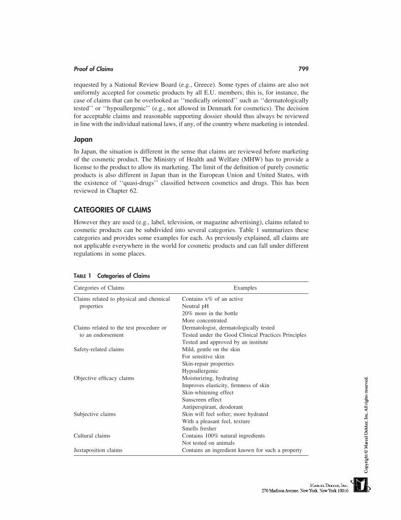
Proof of Claims 799
requested by a National Review Board (e.g., Greece). Some types of claims are also notuniformly accepted for cosmetic products by all E.U. members; this is, for instance, thecase of claims that can be overlooked as ‘‘medically oriented’’ such as ‘‘dermatologicallytested’’ or ‘‘hypoallergenic’’ (e.g., not allowed in Denmark for cosmetics). The decisionfor acceptable claims and reasonable supporting dossier should thus always be reviewedin line with the individual national laws, if any, of the country where marketing is intended.
Japan
In Japan, the situation is different in the sense that claims are reviewed before marketingof the cosmetic product. The Ministry of Health and Welfare (MHW) has to provide alicense to the product to allow its marketing. The limit of the definition of purely cosmeticproducts is also different in Japan than in the European Union and United States, withthe existence of ‘‘quasi-drugs’’ classified between cosmetics and drugs. This has beenreviewed in Chapter 62.
CATEGORIES OF CLAIMS
However they are used (e.g., label, television, or magazine advertising), claims related tocosmetic products can be subdivided into several categories. Table 1 summarizes thesecategories and provides some examples for each. As previously explained, all claims arenot applicable everywhere in the world for cosmetic products and can fall under differentregulations in some places.
TABLE 1 Categories of Claims
Categories of Claims Examples
Claims related to physical and chemical Contains x% of an activeproperties Neutral pH
20% more in the bottleMore concentrated
Claims related to the test procedure or Dermatologist, dermatologically testedto an endorsement Tested under the Good Clinical Practices Principles
Tested and approved by an instituteSafety-related claims Mild, gentle on the skin
For sensitive skinSkin-repair propertiesHypoallergenic
Objective efficacy claims Moisturizing, hydratingImproves elasticity, firmness of skinSkin-whitening effectSunscreen effectAntiperspirant, deodorant
Subjective claims Skin will feel softer; more hydratedWith a pleasant feel, textureSmells fresher
Cultural claims Contains 100% natural ingredientsNot tested on animals
Juxtaposition claims Contains an ingredient known for such a property
800 Paye and Barel
1. Claims related to physical and chemical properties of the product can be sub-stantiated by measuring directly the claimed characteristic in the product by an analyticalmethod. The measurement methodology has, however, to be well established and vali-dated.
2. Claims related to the test procedure or to an endorsement by an outside authoritysimply describe the way, person/title, or place where the product has been tested. Theyare usually perceived by the consumer as proof of a well-tested, quality product. It isessential for such claims to show the property that the consumer can expect from theproduct, even if it is not directly advertised. For example, the claim ‘‘dermatologist tested’’means that the product has been tested by a dermatologist, but also implies that the resultsof the test were good and that the product is, e.g., mild on the skin, or has a skin-beneficialproperty shown by the dermatologist.
3. Safety-related claims make the consumer confident about the innocuousness ofthe product and the benefits to their body. These claims usually require clinical tests onhuman volunteers according to protocols published in the scientific literature and per-formed under high-quality standards. In some cases, in vitro tests can also be accepted ifit can be shown that they are able to prove the claimed property for the type of productin test.
4. Objective efficacy claims are probably the most frequent, and those inducingthe highest expectation from consumers. This is why they require solid efficacy data dos-siers. Many biometric methodologies currently allow getting a direct measurement of theskin properties [3] that are expected to be respected or modified by the cosmetic product.In vivo tests with human volunteers are often recommended or even the only possibilityoffered to the cosmetic chemist, but other types of tests can also be used in some cases,such as cell-culture tests [4–6].
5. Subjective claims are related to a property or function of the product that isperceived by the consumer. The property does not necessarily have to be objectively sub-stantiated by direct measurement. Only tests on human volunteers can be performed, suchas sensory tests (Chap. 71) or well-designed consumer tests.
6. Cultural claims are usually related to the composition of the product and takeadvantage of the current trends. Their value to the consumer is often dependent on theeducation, country, or environment. They link the composition of the product to ecological,ethical, or moral considerations (e.g., naturality of ingredients, absence of tests on ani-mals).
7. Juxtaposition claims refer to the presence of an ingredient in a product and tothe known property of the ingredient, without claiming that the complete product has theproperty. This type of claim can be supported by proving the presence of the ingredientin the product (analytical methods) and relating the claimed property to that ingredientthrough literature data or any type of appropriate test on the pure ingredient.
Several of these categories can be further subdivided in terms of absolute or compar-ative claims. The following four subcategories can be described as follows:
(a) Noncomparative claims: they simply refer to a property of the product withoutany direct comparison to another product. However, it is obvious that even if not classifiedas such, all claims contain a comparative connotation. For instance, claiming that a productis mild means that this is not the case for all other products. Similarly, claiming that aproduct is hydrating for the skin refers to the hypothesis that some other products are not.Examples include claims that a product is mild, hydrating, protects the skin, and softensthe skin.
Proof of Claims 801
(b) Claims comparing a new product to the one it replaces in the market place:in the proof of claim dossier, a direct comparison between the two products will be re-quired. The kind of test depends on the claim. Examples include x% more efficacy, milderthan ever, and even milder than before.
(c) Purely comparative claims comparing the new product to competitive ones forthe claimed property: this kind of claim is likely to be challenged by competitors andrequires a solid supporting dossier where direct comparison between the products is made.The test methodology has to be well justified and validated for the objective. Such compar-ative claims are quite usual in the United States; in Europe they are allowed only underrestricted and severe conditions. Examples include milder than product x and y, and morehydrating than product z.
(d) Absolute claims: the comparison is not limited to a few mentioned productsas previously discussed, but the product claims to be the best in the market for a givenproperty or to completely fulfill a specific function. Such claims require very solid dossierand can be invalidated if even one competitive product can be shown to be superior onthis property. Examples include the mildest, nothing more hydrating, total protection, andcomplete diet for the skin.
TYPE OF SUPPORT
Whenever the nature of the effect or the product justifies it, the claims on cosmetic prod-ucts must be shown. However, the type of support has never been clearly and officiallydefined, so that any kind of support could be acceptable at the condition it can be scientifi-cally and reasonably justified. Different ways to support cosmetic claims [7] are reviewedhereafter; some have already been briefly discussed earlier in this chapter.
Comparison to a Similar Formula
If the product is derived from another formula by a minor modification, it is not alwaysnecessary to repeat the claim-supporting test for the new product. In such a case, it hashowever to be clearly justified that the change is not to affect the claimed property. De-pending on the claims, certain modifications can be considered minor or not. Similarly,for a line of products with minor differences between individual products, some claimscan often be shown on only a few products of the line and then extended to the otherproducts.
Literature Search
For some types of claims, literature data can be considered as effective claim-supportdossier. This is, for instance, the case of claims on ingredients entering into the composi-tion of the cosmetic product; often, the proof of the ingredient property can be found intothe scientific literature. It should be noticed, however, that peer-reviewed literature usuallyhas more credit than supplier literature in the case of a challenge, although the latter canalso be used if supported by well-controlled tests.
In Vitro Tests
In vitro tests never have the same value as in vivo data obtained from clinical tests runon human volunteers. This is why they are mostly used in combination with other types
802 Paye and Barel
of data. However, in some cases (depending on the claim or the availability of alternativetests), in vitro tests can be used on their own to support claims, provided that the test isproven to be scientifically valid for the intended objective. From the most promising andusual in vitro tests, 3-D cell-culture methodologies probably receive the most credit forinvestigating many cosmetic product properties, from skin mildness to more specific prop-erties like sun protection [4–6].
For special dermatocosmetic claims, such as fat reduction and anticellulites effects,in vitro data are mostly presented as direct support. However, in such cases, scientificallyvalid in vivo testing about the efficacy of these treatments is not always available, andoften cumbersome, difficult, and of long duration. Extrapolation of in vitro data or evenof supplier literature on the efficacy of the actives is often used, without really provingthe claims.
In Vivo Tests on Human Volunteers: Clinical Studies
The most direct proof of a claim is to show the product effect directly on the humanvolunteers using the product. Many test protocols may be used depending on the objective.Most protocols have been published in scientific literature and are well-established tests.They go from very exaggerated application conditions [8–11] to normal usage of theproduct by the subjects in the laboratory or at home [12,13]. It is obvious that the morerealistic the application condition, the more powerful the demonstration of the effect.Besides the application procedure, these protocols can also differ by the assessment tech-nique of the claimed effect: scoring of the effect by an expert evaluator, objective measure-ment of the property by a biometric technique, or self-assessment of the subjective effectby the user.
Assessment by an Expert Evaluator
This type of evaluation concerns a cosmetic effect that can be determined by visual, tactile,or olfactive assessment. Examples of scoring scales for the assessment of dry skin havebeen given by Serup [14]. The evaluator is trained to make such an assessment, reliableand fully independent of the product manufacturer. In some cases, the evaluator will bea dermatologist, an ophthalmologist, or a dentist, but this is not mandatory provided thatthe evaluator can justify his/her qualification. When the test protocol is appropriate, expertevaluations are frequently combined with other assessment methods. Examples of claimseasily supported by expert evaluation are: skin whitening, antiwrinkle, hair shine, anddeodorancy. Safety claims are also appropriate for such an assessment to check the absenceof erythema or dryness after product application.
Measurement by Means of Biometric Methodologies
A huge amount of biometric methods have developed over the two last decades, which nowallow objective and quantitative measurement of most skin properties, such as elasticity,firmness, color, barrier properties, moisture content, relief, and blood flow [3,15,16]. Thiskind of evaluation is highly valuable thanks to its objectivity and sensitivity, and canidentify small differences between products to support comparative claims. Those biome-tric measurements must however take into account several key rules: 1) many externalfactors can affect the measurements that have to be made according to specific guidelines[17–20]; 2) the interpretation of the data has to be done by an expert in the field able torelate the collected data to physiological parameters; and 3) the instruments must be highly
Proof of Claims 803
reliable. Under such conditions, instrumental measurements are highly valuable and haverevolutionized the way of supporting cosmetic claims. However, instrumental methodolo-gies have nowadays become so sensitive that questions can sometimes be raised aboutthe relevancy of such small differences between products for consumers who are not al-ways able to detect them.
Self-Assessment of the Effect by the VolunteersWhen used in clinical tests, this type of evaluation is usually combined with other assess-ments. It is, however, not applicable to all test procedures, and requires that the producthas been placed in contact with a sufficiently large area of the body to provide an effectthat can be self-perceived. When confirming objective measurements of the property, thisself-evaluation is extremely powerful because it expresses that the measured effect is reallymeaningful to the consumer.
Sensory Tests with Human Volunteers
The self-perception of the product effect by the volunteers can be obtained independentlyof a clinical test; in such a case a specific test procedure has to be designed. Sensory testsare limited to the so-called sensory claims, which clearly state that the product modifiesthe perception of a property of the skin or hair (e.g., you can feel your skin ‘‘softer’’ or‘‘more hydrated’’). When the sensory effect of the product is obvious and can be easilyperceived by a large majority of people, the test can be performed on a panel of regular(or ‘‘naı̈ve’’) volunteers, without any specific selection criteria regarding their capabilityto feel differences. However, the self-perception of stimuli or of a skin feel is very variablebetween subjects and, often, differences between products are not so obvious for a‘‘naı̈ve’’ user; it is then necessary to run the sensory test either on a very large panel(sometimes several hundreds of volunteers) or to use a panel of volunteers specificallyselected and trained to perceive small differences for the kind of product in test. (Formore details on sensory tests, the reader is referred to Chapter 71.)
Consumer Tests
These tests are performed at the end of the development phase of the product, and consistin providing consumers with the product to use at home for a certain period of time,according to their usual habits and practice. Expected sensory/efficacy properties of theproduct can be checked from these tests by means of a questionnaire filled out by theusers.
The information collected from consumer tests are very helpful in supporting cos-metic claims, as it will reassure the manufacturer that its product is not misleading theuser about the claimed property. Consumer tests, like sensory tests, are mostly used tosupport claims such as those related to odor perception, skin sensation, tactile or visibleproperties of skin or hairs, and taste of oral-care products. However, because of the subjec-tivity of the data, such tests, to be valuable, must be performed very carefully. The ques-tionnaire has to be prepared by a specialist on this kind of test, and cannot be orientedtoward the answers of the users. For more details, the reader is referred to specific guide-lines for consumer testing [21].
Multiapproach for Claim Support
As previously shown, all the approaches described for supporting claims on cosmeticproducts have own advantages as well as weaknesses. In order to combine the strengths
804 Paye and Barel
of several, a multitest or multievaluation approach, combining expert assessments, instru-mental measurements, and subjective data from the user, is often considered to be an idealsupport for a claim. However, depending on the type of claim, for cost and time reasons,it is not always necessary to go so far in the dossier if one test obviously and undoubtlyprovides the proof of the claimed effect.
CONCLUSION
Claims on cosmetic products are extremely varied and often depend on the product, themarket, and the current trends. However, several claims have been used on different prod-uct types for many years. Testing strategy for some is described in the following chapters.They cover some safety-related claims (e.g., mildness, sensitive skin–designed products,noncomedogenicity claims), some efficacy claims (e.g., skin-hydration effect, smoothingand antiwrinkle effect) and sensory claims. The proposed tests especially aim at guidingthe skin scientists to design their own protocols based on reasonable scientific consider-ations, and do not intend to impose strict testing procedures.
REFERENCES
1. Davis JB, McNamara SH. Regulatory aspects of cosmetic claims substantiation. In: Aust LB,ed. Cosmetic Claims Substantiation. Vol. 18. New York: Marcel Dekker, 1998:1–20.
2. Friedel SL. Technical support for advertising claims. J Toxicol-Cutan Ocular Toxicol 1992;11:199–204.
3. Willoughby M, Maibach HI. Cutaneous biometrics and claims support. In: Aust LB, ed. Cos-metic Claims Substantiation. Vol. 18. New York: Marcel Dekker, 1998:69–86.
4. Jackson EM. Supporting advertising claims. Reviewing a three-dimensional in vitro humancell test. Cosmet Toilet 1993; 108:41–42.
5. Majmudar G, Smith M. In vitro screening techniques in dermatology. A review of the tests,models and markers. Cosmet Toilet 1998; 113:69–76.
6. Roguet R. Intérêt des modèles de peaux reconstruites en cosmétologie. Cosmétologie 1997;13:38–43.
7. DGCCRF-D’UMA Commission 30. Evaluation de l’efficacité des produits cosmétiques. Lesrecommandations de la DGCCRF. Cosmétologie 1997; 15:44–46.
8. Frosch PJ, Kligman AM. The soap chamber test: a new method for assessing the irritancypotential of soaps. J Am Acad Dermatol 1979; 1:35–41.
9. Sharko PT, Murahata RI, Leyden JJ, Grove GL. Arm wash with instrumental evaluation—asensitive technique for differentiating the irritation potential of personal washing products. JDerm Clin Eval Soc 1991; 2:19–27.
10. Clarys P, Manou I, Barel AO. Influence of temperature on irritation of the hand/forearm im-mersion test. Contact Dermatitis 1997; 36:240–243.
11. Charbonnier V, Morrison BM Jr, Paye M, Maibach HI. Open application assay in investigationof subclinical irritant dermatitis induced by sodium lauryl sulfate (SLS) in man: advantage ofsquamometry. Skin Res Technol 1998; 4:244–250.
12. Jackson EM, Robillard NF. The controlled use test in a cosmetic product safety substantiationprogram. J Toxicol-Cutan Ocular Toxicol 1982; 1:117–132.
13. Paye M, Gomes G, Zerweg Ch, Piérard GE, Grove GG. A hand immersion test in laboratory-controlled usage conditions: a need for sensitive and controlled assessment methods. ContactDermatitis 1999; 40:133–138.
14. Serup J. EEMCO guidance for the assessment of dry skin (xerosis) and ichtyosis: clinicalscoring systems. Skin Res Technol 1995; 1:109–114.
Proof of Claims 805
15. Wiechers JW, Barlow T. Skin bioengineering techniques for substantiating cosmetics claims.Cosmet Toilet 1998; 113:81–83.
16. Kajs TM, Gartstein V. Review of the instrumental assessment of skin: effects of cleansingproducts. J Soc Cosmet Chem 1991; 42:249–271.
17. Rogiers V, Derde MP, Verleye G, Roseeuw D. Standardized conditions needed for skin surfacehydration measurements. Cosmet Toilet 1990; 105:73–82.
18. Berardesca E. EEMCO guidance for the assessment of stratum corneum hydration: electricalmethods. Skin Res Technol 1997; 3:126–132.
19. Piérard GE. EEMCO guidance for the assessment of skin colour. J Am Acad Derm Venereol1998; 10:1–11.
20. Morrison BM Jr. ServoMed evaporimeter: precautions when evaluating the effect of skin careproducts on barrier function. J Soc Cosmet Chem 1992; 43:161–167.
21. The Advertising Research Foundation. Guidelines for the public use of market and opinionresearch. New York, NY, 1981.

66
Tests for Sensitive Skin
Alessandra Pelosi, Sabrina Lazzerini, and Enzo BerardescaUniversity of Pavia, Pavia, Italy
Howard I. MaibachUniversity of California at San Francisco School of Medicine,San Francisco, California
INTRODUCTION
Sensitive skin is a condition of subjective cutaneous hyperreactivity to environmentalfactors. Subjects experiencing this condition report exaggerated reactions when their skinis in contact with cosmetics, soaps, and sunscreens, and they often report worsening afterexposure to dry and cold climate.
Although no sign of irritation is commonly detected, itching, burning, stinging, anda tight sensation are constantly present. Generally, substances that are not commonly con-sidered irritants are involved in this abnormal response. They include many cosmetic in-gredients such as dimethyl sulfoxide, benzoyl peroxide preparations, salycilic acid, propy-lene glycol, amyldimethylaminobenzoic acid, and 2-ethoxyethyl methoxycinnamate [1].
Sensitive skin and subjective irritation are widespread but still far from being com-pletely defined and understood. Burckhardt [2] hypothesized a correlation between sensi-tive skin and constitutional anomalies and/or other triggering factors such as occupationalskin diseases or chronic exposure to irritants. On the other hand, Bjornberg [3] proposedthat no constitutional factors play a role in the pathogenesis of sensitive skin, althoughthe presence of dermatitis shows a general increase in skin reactivity to primary irritantslasting months.
EPIDEMIOLOGICAL STUDIES
Recent findings suggest that higher sensitivity can be attributable to different mechanisms.Hyperreactors may have a thinner stratum corneum with a reduced corneocyte area, caus-ing a higher transcutaneous penetration of water-soluble chemicals [4]. In 1977, Froschand Kligman [5], by testing different irritants, showed a 14% incidence of sensitive skinin the normal population, likely correlated to a thin permeable stratum corneum, whichmake these subjects more susceptible to chemical irritation.
Many epidemiological studies have been carried out to assess whether or not a corre-lation with sex, skin type, or age could be found. Contradictory findings have been re-
807
808 Pelosi et al.
ported. Some investigators [6,7] documented a higher reactivity to irritants mostly in fe-males, but other experimental studies did not confirm this observation. Bjornberg [8],using six different irritants by patch-test application, found no sex-related differences.Moreover Lammintausta [9], studying the response to open and patch-test application ofsodium lauryl sulphate (SLS), found mild interindividual variations in transepidermal wa-ter loss (TEWL) and dielectric water content (DWC) values, but no sex-related differencesin the reaction pattern.
In 1982, Frosch [10], using dimethylsulfoxide, show a correlation between the mini-mal erythema dose (MED) and the response to irritants; the higher the inflammation, thelower the MED. Subsequently, a correlation between skin reactivity and skin type wasreported; higher reactions were detected in subjects with skin type I [11]. Moreover, ineczema skin reactivity is enhanced [12]. Studies performed on animal models showed thatstrong irritant reactions in guinea pigs significantly reduced the threshold of skin irritation[13]. On the other hand, hyporeactive states may be induced by skin treatment. Subclinicaldermatitis, after repeated cutaneous irritation by open application, may induce skin hypore-activity [14]. This can also be one of the mechanisms of false-negative patch test.
Skin reactivity seems also to change depending on age. The literature is contradic-tory. For example, Nilzen and Voss Lagerlung [15] reported higher reactivity patch-testreactions to soaps and detergents in the elderly, whereas Bettley and Donoghue [16] re-ported a lower reactivity in the same group.
Coenraads et al. [17] showed a higher skin reactivity to croton oil in the older patientgroup, but no differences by testing thimochinone or croton aldehyde. Recently, Grove[18], by testing croton oil, cationic and anionic surfactants, weak acids, and solvents,reported a lower susceptibility in older subjects in terms of less-severe skin reactions.Aged skin seems to have a reduced inflammatory response either to irritants or to irritationinduced by UV light [19]. On the other hand, after irritating the skin, increased TEWLvalues were recorded in the older subjects compared with the young. This finding couldbe related to a deficient ‘‘early warning detection system’’ in the elderly. The lack of anyvisible response can lead to continued exposure to external irritants and higher risk ofdamage to skin-barrier function.
CLINICAL PARAMETERS
Because of the lack of clinical signs, the phenomenon of sensitive skin is difficult todocument. Attempts to identify clinical parameters in subjects with subjective irritationindicate that these individuals tend to have a less hydrated, less supple, more erythematous,and more teleangiectatic skin compared with the normal population. In particular, signifi-cant differences were found for erythema and hydration/dryness [20].
TESTS FOR SENSITIVE SKIN
Recently, because no visible clinical signs of irritation are detected in sensitive skin, newmethods of sensory testing have been increasingly used to provide definite information.
QUANTITATION OF CUTANEOUS THERMAL SENSATION
The superficial skin layer includes sensory nerve fibers connected to specialized receptorssuch as corpuscles or naked nerve endings. A Beta fibers, myelinated (conduction velocity
Tests for Sensitive Skin 809
of 2–30m/sec), mediate the touch, vibration, and pressure sensation. A Delta fibers,smaller and myelinated (conduction velocity of �30m/sec), mediate cold and pain sensa-tion. C fibers, small and nonmyelinated, mediate warm and itching sensation. Quantitativesensory testing (QST) methods have been used mainly to study the impairment of somato-sensory function in neurological diseases; particularly in dermatology, thermal sensationtesting analysis is becoming the most used QST technique [21]. It assessed function infree nerve endings and their associated small myelinated and nonmyelinated fibres. Thismethod is able to measure the threshold of warm and cold sensation as well as hot andcold pain.
All modern automated thermal testing instruments include a thermode (Poltrier de-vice) with semiconductor junctions made of different metals. Depending on the polarityof the electric current, the skin is heated or cooled; different thermic sensations are repro-duced on the different sides of the junctions.
In the center of the thermode, a thermocouple records the temperature. TSA 2001(Medoc Company, Ramat Yshai, Israel) is considered one of the most advanced portablethermal sensory-testing devices. Basically, its measures the hot or cold threshold and thesuprathreshold pain magnitude. It operates between 0°C and 54°C.
The thermode in contact with the skin produces a stimulus whose intensity increasesor decreases until the subject feels the sensation. As the sensation is felt, the subject isasked to press a button. The test is then repeated two more times in order to get a meanvalue. Using this method, artefacts can occur because of the lag time the stimulus needsto reach the brain. This inconvenience can be avoided by using relatively slow rates ofincreasing stimuli. The stimulus can also be increased stepwise and the subject is told tosay whether or not the sensation is felt. When a positive answer is given, the stimulus isdecreased by one-half the initial step and so on, until no sensation is felt. The subject’sresponse determines the intensity of the next stimulus. The limitation of this secondmethod is that a longer performance time is required.
STINGING TEST
Stinging seems to be a variant of pain that develops rapidly and fades quickly any timethe appropriate sensory nerve is stimulated. Although this method lacks objective criteria,it is widely accepted as a marker of sensitivity and has often been used in skin-irritationstudies [5]. It provides information to establish those subjects experiencing invisible cuta-neous irritation.
It is performed by applying to the skin hydrosoluble substances such as lactic acidor capsaicin. The test is usually carried out on the nasolabial fold, a site richly innervatedwith sensory fibers. Subjects first undergo a facial sauna for 5 to 10 minutes, then anaqueous lactic acid solution (5–10% according to different methods] is rubbed with acotton swab on the test site. In order to have a more reliable response, it is recommendedto apply an inert control substance, such as saline solution, to the contralateral test site.After application, within a few minutes a moderate to severe stinging sensation occursfor the ‘‘stingers group.’’ These subjects are then asked to describe the intensity of thesensation using a point scale. Hyperreactors, particularly those with a positive dermatolog-ical history, have higher scores. An alternative test involves the application of 2 mL of90% aqueous dimethylsulfoxide (DMSO) in a small glass cup on the cheek for 5 minutes.This procedure causes intense burning in stingers and, after application, tender wheal andpersistent erythema often occur. By contrast, lactic acid produces no visible changes. Us-
810 Pelosi et al.
ing this screening procedure, 20% of the subjects exposed to 5% lactic acid in a hot,humid environment were found to develop a stinging response [5]. Lammintausta et al.confirmed these observations [22]. In this study, 18% of subjects were identified as sting-ers. In addition, stingers were found to develop stronger reactions to materials causingnonimmunological contact urticaria, to have increased values of TEWL and increasedblood-flow velocimetry values after application of an irritant under patch test.
EVALUATION OF ITCHING RESPONSE
Recent studies show that a new class of C fibers with an exceptionally lower conductionvelocity and insensitivity to mechanical stimuli can likely be considered as afferent unitsthat mediate the itchy sensation [23]. Indeed, this subjective feeling has been extensivelyinvestigated but no explanation of the individual susceptibility to the itching sensation,without any sign of coexisting dermatitis, has been found. Laboratory investigation of theitch response has also been limited.
An itch response can be experimentally induced by topical or intradermal injectionsof various substances such as proteolytic enzymes, mast cell degranulators, and vasoactiveagents. Histamine injection is one of the more common procedures: histamine dihydroclo-ride (100 µg in 1 mL of normal saline) is injected intradermally in one forearm. Then,after different time intervals, the subject is asked to indicate the intensity of the sensationusing a predetermined scale and the duration of itch is recorded. Information is alwaysgained by the subject’s self-assessment.
A correlation between whealing and itching response, produced by applying a topi-cal 4% histamine base in a group of healthy young females, has been investigated byGrove. The itching response was graded by the subjects using the following scale: none,slight, moderate, and intense. The data showed that, despite the fact that 90% of the whealswere greater than 8 mm in diameter, only 50% of the subjects experienced pruritus; pa-tients with large wheals often had no complaints of itching, suggesting that the dimensionsof the wheals do not correlate well with pruritus. In addition, itch and sting perceptionseem to be poorly correlated. Grove [18] compared the cumulative lactic acid sting scoreswith the histamine itch scores in 32 young subjects; all the subjects who were stingerswere also moderate to intense itchers, while 50% of the moderate itchers showed little orno stinging response.
Yosipovitch [24], studying the effects of drugs on C fibers during experimentallyinduced itch, showed that topically applied aspirin rapidly decreases histamine-induceditch. This result can be attributed to the role that prostaglandines play in pain and itchsensation [25]. Localized itching, burning, and stinging can also be a feature of nonimmu-nological contact urticaria. This condition, still not completely defined, is characterizedby a local wheal and flare after exposure of the skin to certain agents. Different combina-tions of mediators such as non–antibody-mediated release of histamine, prostaglandins,leukotriens, substance P, and other inflammatory mediators may likely be involved in thepathogenesis of this disorder [26]. The fact that prostaglandins and leukotriens may playa role in the inflammatory response is supported by the inhibition of the common urticantsby both oral acetylsalicylic acid and indomethacin and by topical diclofenac and naproxengel [1]. Several substances, such as benzoic acid, cinnamic acid, cinnamic aldehyde, andnicotinic acid esters, are capable of producing contact nonimmunological urticaria, elic-iting local edema and erythematous reactions in half of the individuals. Provocative tests
Tests for Sensitive Skin 811
are usually used to identify subjects experiencing this condition: benzoic acid, sorbic acid,or sodium benzoate in open application well reproduce the typical symptoms in subjectssuspected of contact nonimmunological urticaria.
WASHING AND EXAGGERATED IMMERSION TESTS
The aim of these tests is to identify a subpopulation with an increased tendency to producea skin response. In the washing test [27], subjects are asked to wash their face with aspecific soap or detergent. After washing, individual sensation for tightness, burning, itch-ing, and stinging is evaluated using a point scale previously determined. The exaggeratedimmersion test is based on soaking the hands and forearms of the subjects in a solutionof anionic surfactants (such as 0.35% paraffine sulfonate, 0.05% sodium laureth sulfate-2EO) at 40°C for 20 minutes. After soaking, hands and forearms are rinsed under tapwater and patted dry with a paper towel. This procedure is repeated two more times, witha 2-hour period between each soaking, for 2 consecutive days. Before the procedure,baseline skin parameters are evaluated. The other evaluations are taken 2 hours after thethird and sixth soakings and 18 hours after the last soaking (recovery assessment). All ofthe skin parameters are performed after the subjects have rested at least 30 minutes at 21� 1°C.
CORNEOSURFAMETRY
This method, recently described [28], investigates the interaction of surfactants with thehuman stratum corneum. It is performed as follows: cyanoacrylate skin-surface stripping(CSSS) is taken from the volar aspect of the forearm and sprayed with the surfactant tobe tested. After 2 hours, the sample is rinsed with tap water and stained with basic fuchsinand toluidine blue dyes for 3 minutes. After rinsing and drying, the sample is placed ona white reference plate and measured by reflectance colorimetry (Chroma Meter CR200;Minolta, Osaka, Japan).
The index of mildness (CIM � luminacy L*-chroma C*) is taken as a parameterof the irritation caused by the surfactant. This index has a value of 68 � 4 when wateralone is sprayed on the sample and decreases when surfactant is tested, with strongersurfactants lowering the values. Piérard et al. [29], testing different shampoo formulationsin volunteers with sensitive skin, showed that corneosurfametry correlates well with invivo testing. A significant negative correlation (p � 0.001) was found between values ofcolorimetric index of mildness (CIM) and the skin compatibility parameters (SCP) thatinclude a global evaluation of the colorimetric erythemal index (CEI) and the TEWLdifferential, both expressed in the same order of magnitude.
In the same study, corneosurfametry showed less interindividual variability than invivo testing, allowing a better discrimination among mild products. An interesting findingshowed that sensitive skin is not a single condition. Goffin et al. [30] hypothesized thatthe response of the stratum corneum to an environmental threat might be impaired indifferent groups of subjects experiencing sensitive skin. Data of the corneosurfametry,performed after testing eight different house-cleaning products, showed that the overallstratum corneum reactivity, as calculated by the average values of the corneosurfametryindex (CSMI) and the CIM, is significantly different (p � 0.01) between detergent-sensi-tive skin and both nonsensitive and climate/fabric sensitive skin as well.
812 Pelosi et al.
CONCLUSIONS
Sensitive skin represents a widespread condition of susceptibility to exogenous factors.The reason why some subjects react with subjective symptoms like itching, burning, sting-ing, prickling, or tingling is unclear. However, a correlation of increased reactivity insubjects with a history of dermatitis and the association of increased reactivity with skintype I has been reported. Noninvasive evaluation of sensitive skin may successfully predictindividual susceptibility to cosmetic-related adverse reactions. All of the efforts in thisdirection appear undoubtedly important to improve tolerance to the majority of cosmeticproducts.
REFERENCES
1. Amin S, Engasser PG, Maibach HI. Side-effects and social aspect of cosmetology. In: BaranR, Maibach HI, eds. Textbook of Cosmetic Dermatology. 2d ed. 1998; 60:709–746.
2. Burckhardt W. Praktische und theoretische bedeutung der alkalineutralisation und alkaliresis-tenzproben. Arch Klin Exp Derm 1964; 219:600–603.
3. Bjornberg A. Skin Reactions to Primary Irritants in Patients with Hand Eczema. Goteborg:Isaccsons, 1968.
4. Berardesca E, Cespa M, Farinelli N, Rabbiosi G, Maibach HI. In vivo transcutaneous penetra-tion of nicotinates and sensitive skin. Contact Dermatitis 1991; 25:35–38.
5. Frosch PJ, Kligman AM. A method for appraising the stinging capacity of topically appliedsubstances. J Soc Cosmet Chemist 1977; 28:197–209.
6. Agrup G. Hand eczema and other hand dermatoses in South Sweden. Academic dissertation.Acta Dermato Venereol 1969; 49(suppl. 161).
7. Fregert S. Occupational dermatitis in 10 years material. Contact Dermatitis 1975; 1:96–107.8. Bjornberg A. Skin reactions to primary irritants in men and women. Acta Dermatol Venereol
1975; 55:191–194.9. Lammintausta K, Maibach HI, Wilson D. Irritant reactivity in males and females. Contact
Dermatitis 1987; 17:276–280.10. Frosch P, Wissing C. Cutaneous sensivity to ultraviolet light and chemical irritants. Arch Derm
Res 1982; 272:269–278.11. Lammintausta K, Maibach HI, Wilson D. Susceptibility to cumulative and acute irritant derma-
titis. An experimental approach in human volunteers. Contact Dermatitis 1988; 19:84–90.12. Bettley FR. Non specific irritant reactions in eczematous subjects. Br J Dermatol 1964; 76:
116–121.13. Roper SS, Jones EH. An animal model for altering the irritability threshold of normal skin.
Contact Dermatitis 1985; 13:91–97.14. Lammintausta K, Maibach HI, Wilson D. Human cutaneous irritation: induced hyporeactivity.
Contact Dermatitis 1987; 17:193–198.15. Nilzen A, Voss Lagerlund K. Epicutaneous tests with detergents and a number of other com-
mon allergens. Dermatologica 1962; 124:42–52.16. Bettley FR, Donoghue E. The irritant effect of soap upon the normal skin. Br J Dermatol
1960; 72:67–76.17. Coenraads PJ, Bleumink E, Nofer JP. Susceptibility to primary irritants. Age dependence.
Contact Dermatitis 1975; 1:377–381.18. Grove GL. Age-associated changes in integumental reactivity. In: Léveque JL, Agache PG,
eds. Aging Skin. Properties and Functional Changes. New York: XX, 1993; 16:227–237.19. Gilchrest BA, Stoff JS, Soter NA. Chronologic aging alters the response to ultraviolet-induced
inflammation in human skin. J Invest Dermatol 1982; 79:11–15.
Tests for Sensitive Skin 813
20. Seidenari S, Francomano M, Mantovani L. Baseline biophysical parameters in subjects withsensitive skin. Contact Dermatitis 1998; 38:311–315.
21. Yosipovitch G, Yarnitsky D. Quantitative sensory testing. In: Maibach HI, Marzulli FN, eds.Dermotoxicology Methods: The Laboratory Worker’s Vade Mecum. New York: Taylor &Francis, 1997; pp 315–320.
22. Lammintausta K, Maibach HI, Wilson D. Mechanisms of subjective (sensory) irritation: pro-pensity of nonimmunologic contact urticaria and objective irritation in stingers. Dermatosenin Beruf und Umwelt 1988; 36(2):45–49.
23. Schmelz M, Schmidt R, Bichel A, Handwerker HO, Torebjörk HE. Specific C-receptors foritch in human skin. J Neurosci 1997; 17(20):8003–8008.
24. Yosipovitch G, Ademola J, Ping Lui, Amin S, Maibach HI. Topically applied aspirin rapidlydecreases histamine-induced itch. Acta Demato Venereol (Stockh) 1977; 77:46–48.
25. Lovell CR, Burton PA, Duncan EH, Burton JL. Prostaglandins and pruritus. Br J Dermatol1976; 94:273–275.
26. Lahti A, Maibach HI. Species specificity of nonimmmunologic contact urticaria: guinea pig,rat and mouse. J Am Acad 1985; 13:66–69.
27. Hannuksela A, Hannuksela M. Irritant effects of a detergent in wash and chamber tests. ContactDermatitis 1995; 32:163–166.
28. Piérard GE, Goffin V, Piérard Franchimont C. Corneosurfametry: a predictive assessment ofthe interaction of personal care cleansing products with human stratum corneum. Dermatology1994; 189:152–156.
29. Piérard GE, Goffin V, Hermanns-Le T, Arrese JE, Piérard Franchimont C. Surfactant-induceddermatitis: comparison of corneosulfametry with predictive testing on human and recon-structed skin. J Am Acad Dermatol 1995; 33:462–469.
30. Goffin V, Piérard Franchimont C, Piérard GE. Sensitive skin and stratum corneum reactivityto household cleaning products. Contact Dermatitis 1996; 34:81–85.

67
Tests for Skin Hydration
Bernard GabardSpirig Pharma Ltd., Egerkingen, Switzerland
INTRODUCTION
Writing about skin hydration means simultaneously writing about dry skin and its treat-ment by moisturizers. Dry skin has never really been defined in a repeatable way. In fact,this expression prejudices into believing that the skin does have a reduced water content,although this was never confirmed or denied. Generally speaking, dry skin signifies thatthe skin surface looks as though it is lacking in water, this being reinforced by the pharma-cological effect of hydrating the skin surface by appropriate treatments.
Experimental models used for measuring skin hydration are basically clinical modelsincorporating or not noninvasive bioengineering measurements. To ensure meaningfulresults, the outlines of the intended studies should be of modern design incorporatingblinding, randomization, and a suitable statistical control (particularly if different productsare to be compared). This last point means including a predetermined adequate numberof subjects in the study. The general ethical and legal frames of such clinical studiesrequired for claim support are well defined in corresponding monographs or publicationscovering extensively the general procedures to be followed and the prerequisite informa-tion needed about the products to be tested [1–3].
Regardless of the method used, a further important point concerns standardization ofthe experimental conditions. To obtain acceptable and reproducible results, measurementsshould be performed with relaxed patients and/or volunteers already acclimatized for atleast 20 minutes to controlled ambient temperature and relative humidity conditions. Bothfactors mainly affect sweat-gland activity, but other parameters should equally be consid-ered with attention to, e.g., anatomical skin site, test products remaining or not on theskin, and correct handling of the measuring equipment if any. All these possible influenceson measurement outcome have been discussed in detail in recent guidelines and in perti-nent reviews [4–6].
A CLINICAL EVALUATION: THE REGRESSION METHOD
The dermatologist is perfectly able to clinically grade a given state of skin dryness (e.g.,surface roughness, squames, and fissures). Clinical evaluation and grading of skin hydra-tion is based on visual and tactile evaluation of clinical signs. There are numerous possibil-
815
816 Gabard
ities of testing, but basically they rely on the regression method, published in 1978 byKligman [7], which is still used as an industry standard. Briefly, female subjects withmoderate to severe xerosis of the legs are selected following strict criteria. The test prod-ucts are applied under controlled conditions by trained employees twice daily 5 days aweek for 3 weeks. Three days after treatment ends, the follow-up period begins. Scoringis also completed 3 and 7 days later. Treatment period may be shortened to 2 weeks ifnecessary. A recent guideline ensures that clinical scoring of the hydration state of theskin surface will be conducted based on the same definitions [4]. Caution is given uponscoring by the subjects themselves, as their perception of their skin condition may not bethe same as the dermatologist’s [4,8].
INCORPORATING BIOENGINEERING METHODS
A large number of bioengineering methods are now available to evaluate hydration (ordryness) of the skin directly or indirectly. Inclusion of these methods in the study protocolopens many possibilities for getting meaningful results such as design variations, optimiza-tion of the claim support, and also, most importantly, improvement of cost effectivenessby shortening the duration of experiment, using a lower number of subjects, and strength-ening the statistical evaluation.
Concerning the numerous techniques available for the evaluation of skin hydration,the reader is referred to very recent monographs describing these methods in a detailedfashion [8–13]. They mainly include measurements of electrical properties, spectroscopicmethods such as infrared absorption spectroscopy and emission, evaluation of the barrierfunction of the stratum corneum (SC), measurement of mechanical properties, nuclearmagnetic resonance imaging, skin-surface topography, and scaling evaluation. However,in this short review, examples of possible designs will be given that use bioengineeringtechniques based only on the electrical properties of the SC or on measurement of transepi-dermal water loss (TEWL) (for a review of modern suitable measuring equipment seeRefs. 8, 12, 13).
Static Measurements
Short-Term Tests/Single Application
The tests are conducted on the forearm of healthy subjects and allow a randomized side-to-side comparison of test products with a placebo or vehicle, a known active product, anduntreated control skin. Four to six products may be simultaneously tested. The products areapplied at the rate of 2 mg/cm2. Two different experimental designs may be used.
1. The test products are left in place for 1 hour (or another suitable duration, e.g.,3 h [14]). Measurements are conducted at different times thereafter. Removalof excess or nonpenetrated product is preferable before measuring, especiallyif the preparation contains a high proportion of lipids. Most moisturizers showa rapid increase of measured hydration values (Fig. 1).
2. The test products may be applied on similar areas at the same rate but underocclusion with a standard occluding patch overnight for 16 hours. The nextmorning, measurements are conducted in the same way as in part 1 beginning1 hour after removal of the occlusion patch (Fig. 2). This last procedure betterpicks up the activity of a humectant contained in the test preparation, whereas
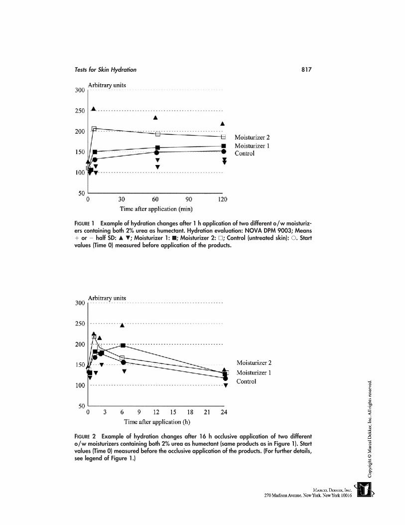
Tests for Skin Hydration 817
FIGURE 1 Example of hydration changes after 1 h application of two different o/w moisturiz-ers containing both 2% urea as humectant. Hydration evaluation: NOVA DPM 9003; Means� or � half SD: � �; Moisturizer 1: ■; Moisturizer 2: �; Control (untreated skin): �. Startvalues (Time 0) measured before application of the products.
FIGURE 2 Example of hydration changes after 16 h occlusive application of two differento/w moisturizers containing both 2% urea as humectant (same products as in Figure 1). Startvalues (Time 0) measured before the occlusive application of the products. (For further details,see legend of Figure 1.)
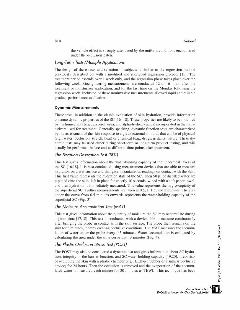
818 Gabard
the vehicle effect is strongly attenuated by the uniform conditions encounteredunder the occlusion patch.
Long-Term Tests/Multiple Applications
The design of these tests and selection of subjects is similar to the regression methodpreviously described but with a modified and shortened regression protocol [15]. Thetreatment period extends over 1 week only, and the regression phase takes place over thefollowing week. Bioengineering measurements are conducted 12 to 16 hours after thetreatment or moisturizer application, and for the last time on the Monday following theregression week. Inclusion of these noninvasive measurements allowed rapid and reliableproduct-performance evaluation.
Dynamic Measurements
These tests, in addition to the classic evaluation of skin hydration, provide informationon some dynamic properties of the SC [16–18]. These properties are likely to be modifiedby the humectants (e.g., glycerol, urea, and alpha-hydroxy acids) incorporated in the mois-turizers used for treatment. Generally speaking, dynamic function tests are characterizedby the assessment of the skin response to a given external stimulus that can be of physical(e.g., water, occlusion, stretch, heat) or chemical (e.g., drugs, irritants) nature. These dy-namic tests may be used either during short-term or long-term product testing, and willusually be performed before and at different time points after treatment.
The Sorption-Desorption Test (SDT)
This test gives information about the water-binding capacity of the uppermost layers ofthe SC [16,18]. It is best conducted using measurement devices that are able to measurehydration on a wet surface and that give instantaneous readings on contact with the skin.This first value represents the hydration state of the SC. Then 50 µl of distilled water arepipetted onto the skin, left in place for exactly 10 seconds, wiped with a soft paper towel,and then hydration is immediately measured. This value represents the hygroscopicity ofthe superficial SC. Further measurements are taken at 0.5, 1, 1.5, and 2 minutes. The areaunder the curve from 0.5 minutes onwards represents the water-holding capacity of thesuperficial SC (Fig. 3).
The Moisture-Accumulation Test (MAT)
This test gives information about the quantity of moisture the SC may accumulate duringa given time [17,18]. This test is conducted with a device able to measure continuouslyafter bringing the probe in contact with the skin surface. The probe then remains on theskin for 3 minutes, thereby creating occlusive conditions. The MAT measures the accumu-lation of water under the probe every 0.5 minutes. Water accumulation is evaluated bycalculating the area under the time curve until 3 minutes (Fig. 4).
The Plastic Occlusion Stress Test (POST)
The POST may also be considered a dynamic test and gives information about SC hydra-tion, integrity of the barrier function, and SC water-holding capacity [19,20]. It consistsof occluding the skin with a plastic chamber (e.g., Hilltop chamber or a similar occlusivedevice) for 24 hours. Then the occlusion is removed and the evaporation of the accumu-lated water is measured each minute for 30 minutes as TEWL. This technique has been
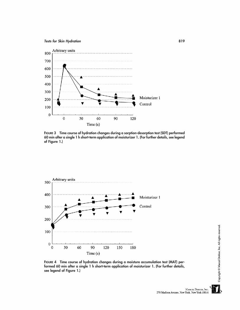
Tests for Skin Hydration 819
FIGURE 3 Time course of hydration changes during a sorption-desorption test (SDT) performed60 min after a single 1 h short-term application of moisturizer 1. (For further details, see legendof Figure 1.)
FIGURE 4 Time course of hydration changes during a moisture accumulation test (MAT) per-formed 60 min after a single 1 h short-term application of moisturizer 1. (For further details,see legend of Figure 1.)
820 Gabard
thoroughly described in recent guidelines [21,22]. The measurement is called skin surfacewater loss (SSWL) and not TEWL, because it does not represent the true TEWL but thesum of the TEWL and the evaporation of water trapped within and over the SC underthe occlusive equipment, at least at the beginning of the measurement period. During thesefirst minutes of evaporation, the SSWL is proportional to SC hydration. At the end of thedehydration time, SSWL is greatly reduced and mainly TEWL is measured. Therefore,changes induced in the last part of the curve reflects the barrier function of SC.
Other Suitable Tests
Some well-defined properties of the skin are more or less dependent on SC hydration andmay be evaluated with the following bioengineering methods:
• Mechanical or viscoelastic properties (elasticity, extensibility) [23]• Skin-surface roughness [24]• Skin-surface scaling [25]
Some other techniques are also indicated for evaluating SC hydration, but they are notavailable for routine experimentation at the present moment. They have been criticallyreviewed and evaluated in a recent publication to which the reader is referred [8].
CONCLUSION
During the evaluation of SC hydration in vivo, it must be kept in mind that no absolutedetermination of a water content or concentration is possible. This holds for clinical evalu-ation and for bioengineering measurements as well. For this reason, several measurementtechniques should be used simultaneously during a study. Not only is the informationgained from these different experimental approaches complementary, and of great benefitif they are integrated in a clinical evaluation, but one should remember that moisturizersmay influence skin hydration in different ways. Thus, different aspects of hydrationchanges need to be investigated, such as water binding, water retention, or emolliency,which is also a further part of a moisturizer’s action. Last, it should be remembered that,in order to obtain meaningful results, proper design of the study, inclusion of a suitablenumber of subjects, strict standardization of measurement conditions, and all other relevantfactors need to be tightly controlled. Only by assuring the best quality level will resultsbe obtained that will help to design and use optimal moisturizers.
REFERENCES
1. Seidenschnur EK. FDA and EEC regulations related to skin: documentation and measuringdevices. In: Serup J, Jemec GBE, eds. Handbook of Non-Invasive Methods and the Skin. BocaRaton: CRC Press, 1995:653–665.
2. COLIPA (The European Cosmetic, Toiletry and Perfumery Association). Guidelines for theevaluation of the efficacy of cosmetic products, 1997.
3. Davis JB, McNamara SH. Regulatory aspects of cosmetic claims substantiation. In: Aust LB,ed. Cosmetic Claims Substantiation. New York: Marcel Dekker, 1998:1–20.
4. Serup J. EEMCO guidance for the assessment of dry skin (xerosis) and ichthyosis: clinicalscoring systems. Skin Res Technol 1995; 1:109–114.
5. Berardesca E. EEMCO guidance for the assessment of stratum corneum hydration: electricalmethods. Skin Res Technol 1997; 3:126–132.
Tests for Skin Hydration 821
6. Wilhelm KP. Possible pitfalls in hydration measurements. In: Elsner P, Barel AO, BerardescaE, Gabard B, Serup J, eds. Skin Bioengineering: Techniques and Applications in Dermatologyand Cosmetology. Current Problems in Dermatology, Vol. 26. Basel: Karger, 1998:223–234.
7. Kligman AM. Regression method for assessing the efficacy of moisturizers. Cosmet & Toilet1978; 93:27–35.
8. Barel AO, Clarys P, Gabard B. In vivo evaluation of the hydration state of the skin: measure-ments and methods for claim support. In: Elsner P, Merk HF, Maibach HI, eds. In: Cosmetics:Controlled Efficacy Studies and Regulations. Berlin: Springer, 1999:57–80.
9. Tagami H, Ohi M, Iwatsuki K, Kanamaru Y, Yamada M, Ichijo B. Evaluation of the skinsurface hydration in vivo by electrical measurements. J Invest Dermatol 1980; 75:500–507.
10. Levêque JL, De Rigal J. Impedance methods for studying skin moisturization. J Soc CosmetChem 1983; 34:419–428.
11. Levêque JL. Cutaneous Investigation in Health and Disease: Noninvasive Methods and Instru-mentation. New York: Marcel Dekker, 1989.
12. Elsner P, Berardesca E, Maibach HI. Bioengineering of the Skin: Water and the Stratum Cor-neum. Boca Raton: CRC Press, 1994.
13. Serup J, Jemec GBE. (1995) Handbook of Non-Invasive Methods and the Skin. Boca Raton:CRC Press, 1995:159–170.
14. Serup J. A three-hour test for rapid comparison of effects of moisturizers and active constit-uents (urea). Acta Derm Venereol (Stockh) 1992; (suppl. 177):29–33.
15. Grove G. Skin surface hydration changes during a mini-regression test as measured in vivoby electrical conductivity. Curr Therap Res 1992; 52:1–6.
16. Tagami H, Kanamaru Y, Inoue K, Suehisa S, Inoue F, Iwatsuki K, Yoshikuni K, Yamada M.Water sorption-desorption test of the skin in vivo for functional assessment of the stratumcorneum. J Invest Dermatol 1982; 78:425–428.
17. Van Neste D. In vivo evaluation of unbound water accumulation in stratum corneum. Dermato-logica 1990; 181:197–201.
18. Treffel P, Gabard B. Stratum corneum dynamic function measurements after moisturizer orirritant application. Arch Dermatol Res 1995; 287:474–479.
19. Berardesca E, Maibach HI. Effect of nonvisible damage on the water-holding capacity of thestratum corneum, utilizing the plastic occlusion stress test (POST). In: Frosch PJ, Doom-Goos-sens A, Lachapelle JM, Rycroft RJG, Scheper RJ, eds. Current Topics in Contact Dermatitis.Berlin: Springer, 1989:554–559.
20. Berardesca E, Elsner P. Dynamic measurements: the plastic occlusion stress test (POST) andthe moisture accumulation test (MAT). In: Elsner P, Berardesca E, Maibach HI, eds. Bioengin-eering of the Skin: Water and the Stratum Corneum. Boca Raton: CRC Press, 1994:97–102.
21. Pinnagoda J. Hardware and measuring principles: evaporimeter. In: Elsner P, Berardesca E,Maibach HI, eds. Bioengineering of the Skin: Water and the Stratum Corneum. Boca Raton:CRC Press, 1994:51–58.
22. Pinnagoda J, Tupker RA, Agner T, Serup J. Guidelines for transepidermal water loss measure-ment: a report from the standardization group of the European Society of Contact Dermatitis.Contact Dermatitis 1990; 22:164–168.
23. Barel AO, Lambrecht R, Clarys P. Mechanical function of the skin: state of the art. In: ElsnerP, Barel AO, Berardesca E, Gabard B, Serup J, eds. Skin Bioengineering: Techniques andApplications in Dermatology and Cosmetology. Current Problems in Dermatology, Vol. 26.Basel: Karger, 1998:69–83.
24. Marks R. How to measure the effects of emollients. J Dermatol Treat 1997; 8:S15–S18.25. Schatz H, Altmeyer PJ. Dry skin and scaling evaluated by D-Squames and image analysis.
In: Serup J, Jemec GBE, eds. Handbook of Non-Invasive Methods and the Skin. Boca Raton:CRC Press, 1995:153–157.

68
Tests for Skin Protection: Barrier Effect
Hongbo Zhai and Howard I. MaibachUniversity of California at San Francisco School of Medicine,San Francisco, California
Barrier creams (BC) may play an important role in the prevention of contact dermatitis[1–6], and various in vitro and in vivo methods have been developed to evaluate theirefficacy. In practice, their use remains the subject of lively debate; some reports suggestthat inappropriate BC application may exacerbate, rather than prevent, irritation [1–3, 6–9]. The accuracy of measurements depends on the use of proper methodology. This chapterreviews the investigative details of pertinent scientific literature, and summarizes the meth-odology and efficacy of BC.
IN VITRO METHODS
In 1946, Sadler and Marriott [10] introduced some facile tests to evaluate the efficiencyof BC. One method used the fluorescence of a dyestuff and eosin as an indicator to measurepenetration and the rates of penetration of water through BC; this is rapid and simple, butprovides only a qualitative estimate.
Suskind [11] used a simple method to measure the relative efficacy or repellencyof several formulations with film-immersion tests in a specific exposure. Results showedtwo formulations (containing 52.5% silicone in bentonite and 30% silicone in petrolatum)were both effective against a range of aqueous irritants or sensitizers.
Langford [12] conducted in vitro studies to determine the efficacy of the formulatedfluorochemical (FC)–resin complex included against solvent penetration through treatedfilter paper, solvent repellency on treated pigskin, and penetration of radio-tagged sodiumlauryl sulfate through treated hairless mouse skin. He also conducted an in vivo study on75 persons who had all previously experienced irritation on their hands because of contin-ued contact with solvents. Eighty-three percent of the panelists stated the cream was effec-tive in protecting their hands.
Reiner et al. [13] examined the protective effect of ointments both on guinea pigskin in vitro and on guinea pigs in vivo. The permeation values of a toxic agent throughunprotected and protected skin within 10 h as a function of time was determined radiologi-cally and enzymatically. Permeation of the toxic agent was markedly reduced by polyeth-ylene-glycol ointment base and ointments containing active substances. In in vivo experi-
823
824 Zhai and Maibach
ments on guinea pigs, mortality was greater after applying the toxic agent to unprotectedskin. All formulations with nucleophilic substances markedly reduced the mortality rate.
Loden [14] evaluated the effect of BC on the absorption of (3H)-water, (14C)-ben-zene, and (14C)-formaldehyde into excised human skin. The control and BC-treated skinswere exposed to the test substance for 0.5 hours, whereupon absorption was determined.The experimental cream ‘‘water barrier’’ reduced the absorption of water and benzenebut not formaldehyde. One cream slightly reduced benzene and formaldehyde absorption.The other two creams did not affect the absorption of any of the substances studied.
Treffel et al. [15] measured in vitro on human skin the effectiveness of BC againstthree dyes (eosin, methylviolet, and oil red O) with varying n-octanol/water partition coef-ficients (0.19, 29.8, and 165, respectively). BC efficacy was assayed by measurements ofthe dyes in the epidermis of protected skin samples after 30 minutes of application. Theefficacy of BC against the three dyes showed in several cases data contrary to manufactur-er’s information. There was no correlation between the galenic parameters of the assayedproducts and the protection level, indicating that neither water content nor consistency ofthe formulations influenced the protection effectiveness.
Fullerton and Menne [16] tested that the protective effect of various ethylenedi-aminetetraacetate (EDTA) barrier gels against nickel contact allergy using in vitro and invivo methods. In an in vitro study, about 30 mg of barrier gel were applied on the epider-mal side of the skin and a nickel disc was applied above the gel. After 24-hours application,the nickel disc was removed and the epidermis separated from the dermis. Nickel contentin epidermis and dermis was quantified by absorption differential pulse voltammetry(ADPV). The amount of nickel in the epidermal skin layer on barrier gel–treated skinwas significantly reduced compared with the untreated control. In vivo patch testing ofnickel-sensitive patients was performed using nickel discs with and without barrier gels.Test preparations and nickel discs were removed 1 day after application, and the test siteswere evaluated. Reduction in positive test reactions was highly significant on barrier gel–treated sites.
Zhai et al. [17] used an in vitro diffusion system to measure the protective efficacyof Quaternium-18 bentonite (Q18B) gels to prevent 1% concentration of [35S] sodiumlauryl sulfate (SLS) penetration on human cadaver skin. The accumulated amount of [35S]-SLS in receptor-cell fluid were counted to evaluate the efficacy of the Q-18B gels overa 24-hour period. These test gels significantly decreased SLS absorption when comparedwith unprotected-skin control samples. The percent protection effect of three test gelsagainst SLS percutaneous absorption was 88%, 81%, and 65%, respectively.
IN VIVO METHODS
In 1940, Schwartz et al. [18] introduced an in vivo method to evaluate the efficacy of avanishing cream against poison ivy extract using visual erythema on human skin. The testcream was an effective prophylaxis against poison ivy dermatitis when compared withunprotected skin.
Lupulescu and Birmingham [19] observed the ultrastructural and relief changes ofhuman epidermis after exposure to a protective gel, acetone, and kerosene on humans.Unprotected skin produced cell damage and a disorganized pattern in the upper layers ofepidermis. Application of a protective agent before to solvent exposure substantially re-duced the ultrastructural and relief changes of epidermis cells.
Lachapelle and co-workers [3, 20–23] used a guinea pig model to evaluate the pro-
Barrier Effect 825
tective value of BC and/or gels by laser Doppler flowmetry and histological assessment.The histopathological damage after 10 minutes of contact to toluene was mostly confinedto the epidermis, whereas the dermis was almost normal. The dermal blood-flow changeswere relatively high on the control site compared with the gel-pretreated sites.
Frosch et al. [1, 8, 9, 24, 25] developed the repetitive irritation test (RIT) in theguinea pig and in humans to evaluate the efficacy of BC by using a series of bioengineeringtechniques. The cream-pretreated and -untreated test skin (guinea pig or humans) wasexposed daily to the irritants for 2 weeks. The resulting irritation was scored on a clinicalscale and assessed by biophysical techniques’ parameters. Some test creams suppressedirritation with all test parameters, some failed to show such an effect, and some evenexacerbated the irritation [9].
Zhai [2] used an in vivo human model to measure the effectiveness of BC againstdye-indicator solutions: methylene blue in water and oil red O in ethanol, which are repre-sentative of model hydrophilic and lipophilic compounds. Solutions of 5% methylene blueand 5% oil red O were applied to untreated and BC-pretreated skin with the aid of alumi-num occlusive chambers for 0 and 4 hours. At the end of the application time, the materialswere removed, and consecutive skin-surface biopsies (SSB) obtained. The amount of dyepenetrating into each strip was determined by colorimetry. Two creams exhibited effec-tiveness, but one cream enhanced the cumulative amount of dye.
Zhai et al. [5] introduced a facile approach to screening protectants in vivo in humansubjects. Two acute irritants and 1 allergen were selected: 1) sodium lauryl sulfate (SLS),representative of irritant household and occupational contact dermatitis, 2) the com-bination of ammonium hydroxide (NH4OH) and urea to simulate diaper dermatitis, and3) Rhus to evaluate the effect of model protective materials. Test materials were spreadonto test area, massaged, allowed to dry for 30 minutes, and reapplied with another 30-minute drying period. The model irritants and allergen were applied with an occlusivepatch for 24 hours. Inflammation was scored with an expanded 10-point scale at 72 hoursafter application. Most test materials statistically suppressed the SLS irritation and Rhusallergic reaction rather than NH4OH and urea-induced irritation.
Wigger-Alberti et al. [26] determined which areas of the hands were likely to beskipped on self-application of BC by fluorescence technique at the workplace. Resultsshowed the application of BC was incomplete, especially on the dorsal aspects of thehands. Brief data of recent experiments of BC are summarized in Table 1.
CONCLUSIONS
Some BC reduce CD under experimental conditions. But, inappropriate BC applicationmay enhance irritation rather than benefit. To achieve the optimal protective effects, BCshould be used with careful consideration based on specific exposure conditions; also, theproper use of BC should be instructed.
In vitro methods are simple, rapid, safe, and are recommended in the screeningprocedure for BC candidates. With radiolabeled methods, we may determine the accurateprotective and penetration results even in the lower levels of chemicals because of thesensitive radiolabeled counting when BCs are to be evaluated. Animal experiments maybe used to generate kinetic data because of a closer similarity between humans and someanimals (e.g., pigs and monkeys) in percutaneous absorption and penetration for somecompounds. But no one animal, with its complex anatomy and biology, will simulate thepenetration in humans for all compounds. Therefore, the best estimate of human percutane-
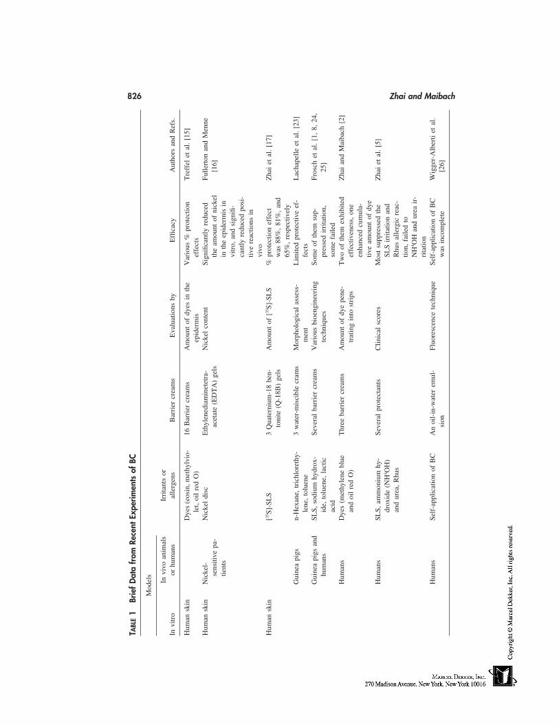
826 Zhai and Maibach
TABL
E1
Brie
fD
ata
from
Rece
ntEx
perim
ents
ofBC
Mod
els
Invi
voan
imal
sIr
rita
nts
orIn
vitr
oor
hum
ans
alle
rgen
sB
arri
ercr
eam
sE
valu
atio
nsby
Effi
cacy
Aut
hors
and
Ref
s.
Hum
ansk
inD
yes
(eos
in,m
ethy
lvio
-16
Bar
rier
crea
ms
Am
ount
ofdy
esin
the
Var
ious
%pr
otec
tion
Tre
ffel
etal
.[1
5]le
t,oi
lre
dO
)ep
ider
mis
effe
cts
Hum
ansk
inN
icke
l-N
icke
ldi
scE
thyl
ened
iam
inet
etra
-N
icke
lco
nten
tSi
gnifi
cant
lyre
duce
dFu
llert
onan
dM
enne
sens
itive
pa-
acet
ate
(ED
TA
)ge
lsth
eam
ount
ofni
ckel
[16]
tient
sin
the
epid
erm
isin
vitr
o,an
dsi
gnifi
-ca
ntly
redu
ced
posi
-tiv
ere
actio
nsin
vivo
Hum
ansk
in[35
S]-S
LS
3Q
uate
rniu
m-1
8be
n-A
mou
ntof
[35S]
-SL
S%
prot
ectio
nef
fect
Zha
iet
al.
[17]
toni
te(Q
-18B
)ge
lsw
as88
%,
81%
,an
d65
%,
resp
ectiv
ely
Gui
nea
pigs
n-H
exan
e,tr
ichl
oret
hy-
3w
ater
-mis
cibl
ecr
ams
Mor
phol
ogic
alas
sess
-L
imite
dpr
otec
tive
ef-
Lac
hape
lleet
al.
[23]
lene
,to
luen
em
ent
fect
sG
uine
api
gsan
dSL
S,so
dium
hydr
ox-
Seve
ral
barr
ier
crea
ms
Var
ious
bioe
ngin
eeri
ngSo
me
ofth
emsu
p-Fr
osch
etal
.[1
,8,
24,
hum
ans
ide,
tolu
ene,
lact
icte
chni
ques
pres
sed
irri
tatio
n,25
]ac
idso
me
faile
dH
uman
sD
yes
(met
hyle
nebl
ueT
hree
barr
ier
crea
ms
Am
ount
ofdy
epe
ne-
Tw
oof
them
exhi
bite
dZ
hai
and
Mai
bach
[2]
and
oil
red
O)
trat
ing
into
stri
psef
fect
iven
ess,
one
enha
nced
cum
ula-
tive
amou
ntof
dye
Hum
ans
SLS,
amm
oniu
mhy
-Se
vera
lpr
otec
tant
sC
linic
alsc
ores
Mos
tsu
ppre
ssed
the
Zha
iet
al.
[5]
drox
ide
(NH
4 OH
)SL
Sir
rita
tion
and
and
urea
,R
hus
Rhu
sal
lerg
icre
ac-
tion,
faile
dto
NH
4 OH
and
urea
ir-
rita
tion
Hum
ans
Self
-app
licat
ion
ofB
CA
noi
l-in
-wat
erem
ul-
Fluo
resc
ence
tech
niqu
eSe
lf-a
pplic
atio
nof
BC
Wig
ger-
Alb
erti
etal
.si
onw
asin
com
plet
e[2
6]
Barrier Effect 827
ous absorption is determined by in vivo studies in humans. The histological assessmentsmay define what layers of skin are damaged or protected, and may provide the insightmechanism of BC. Noninvasive bioengineering techniques may provide accurate, highlyreproducible, and objective observations in quantifying the inflammation response to vari-ous irritants and allergens when BC are to be evaluated that could assess subtle differencesto supplement traditional clinical studies.
To validate these models, well-controlled field trials are required to define the rela-tionship of the model to the occupational setting. Finally, the clinical efficacy of BC shouldbe assessed in the workplace rather than in experimental circumstances.
REFERENCES
1. Frosch PJ, Schulze-Dirks A, Hoffmann M, Axthelm I, Kurte A. Efficacy of skin barrier creams.(I). The repetitive irritation test (RIT) in the guinea pig. Contact Dermatitis 28:94, 193.
2. Zhai H, Maibach, HI. Effect of barrier creams: human skin in vivo. Contact Dermatitis 35:92, 1996.
3. Lachapelle JM. Efficacy of protective creams and/or gels. In: Elsner P, Lachapelle JM, Wahl-berg JE, Maibach HI, eds. Prevention of Contact Dermatitis, Current Problems in Dermatol-ogy. Basel: Karger, 1996; 182.
4. Zhai H, Maibach HI. Percutaneous penetration (Dermatopharmacokinetics) in evaluating bar-rier creams. In: Elsner P, Lachapelle JM, Wahlberg JE, Maibach HI. eds. Prevention of ContactDermatitis, Current Problems in Dermatology. Basel: Karger, 1996; 193.
5. Zhai H, Willard P, Maibach HI. Evaluating skin-protective materials against contact irritantsand allergens. An in vivo screening human model. Contact Dermatitis 38:155, 1998.
6. Wigger-Alberti W, Elsner P. Do barrier creams and gloves prevent or provoke contact derma-titis? Am J Contact Dermatitis 9:100, 1998.
7. Goh CL. Cutting oil dermatitis on guinea pig skin. (I). Cutting oil dermatitis and barrier cream.Contact Dermatitis 24:16, 1991.
8. Frosch PJ, Schulze-Dirks A, Hoffmann M, Axthelm I. Efficacy of skin barrier creams. (II).Ineffectiveness of a popular ‘‘skin protector’’ against various irritants in the repetitive irritationtest in the guinea pig. Contact Dermatitis 29:74, 1993.
9. Frosch PJ, Kurte A, Pilz B. Biophysical techniques for the evaluation of skin protective creams.In: Frosch PJ, Kligman AM, eds. Noninvasive Methods for the Quantification of Skin Func-tions. Berlin: Springer-Verlag, 1993; 214.
10. Sadler CGA, Marriott RH. The evaluation of barrier creams. Br Med J 23:769, 1946.11. Suskind RR. The present status of silicone protective creams. Ind Med Surg 24:413, 1955.12. Langford NP. Fluorochemical resin complexes for use in solvent repellent hand creams. Am
Ind Hyg Assoc J 39:33, 1978.13. Reiner R, Roßmann K, Hooidonk CV, Ceulen BI, Bock J. Ointments for the protection against
organophosphate poisoning. Arzneim-Forsch/Drug Res 32:630, 1982.14. Loden M. The effect of 4 barrier creams on the absorption of water, benzene, and formaldehyde
into excised human skin. Contact Dermatitis 14:292, 1986.15. Treffel P, Gabard B, Juch R. Evaluation of barrier creams: an in vitro technique on human
skin. Acta Derm Venereol 74:7, 1994.16. Fullerton A, Menne T. In vitro and in vivo evaluation of the effect of barrier gels in nickel
contact allergy. Contact Dermatitis 32:100, 1995.17. Zhai H, Buddrus DJ, Schulz AA, Wester RC, Hartway T, Serranzana S, Maibach HI. In vitro
percutaneous absorption of sodium lauryl sulfate (SLS) in human skin decreased by Quater-nium-18 bentonite gels. In vitro & Molecular Toxicol 12:11, 1999.
18. Schwartz L, Warren LH, Goldman FH. Protective ointment for the prevention of poison ivydermatitis. Public Health Rep 55:1327, 1940.
828 Zhai and Maibach
19. Lupulescu AP, Birmingham DJ. Effect of protective agent against lipid-solvent–induced dam-ages. Ultrastructural and scanning electron microscopical study of human epidermis. ArchEnviron Health 31:29, 1976.
20. Mahmoud G, Lachapelle JM, Neste DV. Histological assessment of skin damage by irritants:its possible use in the evaluation of a ‘barrier cream’. Contact Dermatitis 11:179, 1984.
21. Mahmoud G, Lachapelle JM. Evaluation of the protective value of an antisolvent gel by laserDoppler flowmetry and histology. Contact Dermatitis 13:14, 1985.
22. Mahmoud G, Lachapelle J. Uses of a guinea pig model to evaluate the protective value ofbarrier creams and/or gels. In: Maibach HI, Lowe NJ, eds. Models of Dermatology. Basel:Karger, 1987; 112.
23. Lachapelle JM, Nouaigui H, Marot L. Experimental study of the effects of a new protectivecream against skin irritation provoked by the organic solvents n-hexane, trichlorethylene andtoluene. Dermatosen 38:19, 1990.
24. Frosch PJ, Kurte A, Pilz B. Efficacy of skin barrier creams. (III). The repetitive irritation test(RIT) in humans. Contact Dermatitis 29:113, 1993.
25. Frosch PJ, Kurte A. Efficacy of skin barrier creams. (IV). The repetitive irritation test (RIT)with a set of 4 standard irritants. Contact Dermatitis 31:161, 1994.
26. Wigger-Alberti W, Maraffio B, Wernli M, Elsner P. Self-application of a protective cream.Pitfalls of occupational skin protection. Arch Dermatol 133:861, 1997.
69
Objective Methods for Assessment of HumanFacial Wrinkles
Gary Grove and Mary Jo GroveKGL’s Skin Study Center, Broomall, and cyberDERM, inc., Media, Pennsylvania
INTRODUCTION
The skin, especially that of the face, undergoes very characteristic changes with advancingage. Although there are other overt morphological changes that can be considered as mark-ers of cutaneous aging, the degree of wrinkling in the ‘‘crow’s feet’’ area seems to havethe greatest impact. Thus, it is not surprising that considerable effort has been expandedto develop skincare products and cosmetic surgical procedures that can effectively restorea more youthful appearance.
Wrinkles can be easily visualized and many clinical studies have involved the useof ranking scales that rely on subjective assessments by expert graders. To improve thevalidity and reproducibility of this approach, more complex ordinal scales with semiquan-tive word descriptors and reference photographs have been devised by several investiga-tors [1–3]. For example, Daniell [1] devised a set of reference photographs that illustrateshis six-point grading scheme for evaluating crow’s-feet wrinkles in the lateral periorbitalarea. Such reference photographs can be used to train inexperienced graders and periodi-cally review the competency of all evaluators. Nevertheless, the major drawback to thistype of approach is that it provides no permanent records that fully describe the skin-surface features or allow retrospective analysis. Instead, we must rely on the subjectivejudgements of trained graders and their ability to recall from memory the full range ofchanges in skin-surface features that might occur in each situation.
This problem can be overcome by taking standardized photographs before treatmentand at various intervals during the treatment period. This provides a series of photographsthat not only documents the study but can also be used to quantify the therapeutic response.This can be done by a panel of blinded, independent readers who are remote from thestudy environment as was done for photodamaged skin treated with isotretinoin [4] oralpha-hydroxy acids [5]. Although clinical assessments and photography are useful meth-ods for assessing such changes in photodamaged skin, we are concerned that they mightnot be appropriate for studying wrinkles. This is especially true for photography in whichchanges in lighting or facial expression can greatly influence the appearance of lines andwrinkles. We are also concerned that concurrent improvements in other facial features,such as a decrease in mottled pigmentation or increased dermal blood flow, might partially
829
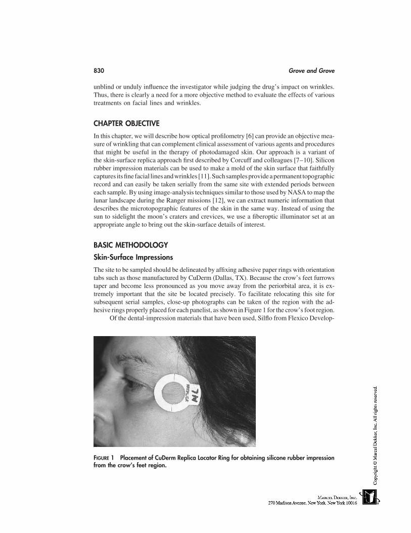
830 Grove and Grove
unblind or unduly influence the investigator while judging the drug’s impact on wrinkles.Thus, there is clearly a need for a more objective method to evaluate the effects of varioustreatments on facial lines and wrinkles.
CHAPTER OBJECTIVE
In this chapter, we will describe how optical profilometry [6] can provide an objective mea-sure of wrinkling that can complement clinical assessment of various agents and proceduresthat might be useful in the therapy of photodamaged skin. Our approach is a variant ofthe skin-surface replica approach first described by Corcuff and colleagues [7–10]. Siliconrubber impression materials can be used to make a mold of the skin surface that faithfullycaptures itsfine facial linesandwrinkles [11].Suchsamplesprovide apermanent topographicrecord and can easily be taken serially from the same site with extended periods betweeneach sample. By using image-analysis techniques similar to those used by NASA to map thelunar landscape during the Ranger missions [12], we can extract numeric information thatdescribes the microtopographic features of the skin in the same way. Instead of using thesun to sidelight the moon’s craters and crevices, we use a fiberoptic illuminator set at anappropriate angle to bring out the skin-surface details of interest.
BASIC METHODOLOGY
Skin-Surface Impressions
The site to be sampled should be delineated by affixing adhesive paper rings with orientationtabs such as those manufactured by CuDerm (Dallas, TX). Because the crow’s feet furrowstaper and become less pronounced as you move away from the periorbital area, it is ex-tremely important that the site be located precisely. To facilitate relocating this site forsubsequent serial samples, close-up photographs can be taken of the region with the ad-hesive rings properly placed for each panelist, as shown in Figure 1 for the crow’s foot region.
Of the dental-impression materials that have been used, Silflo from Flexico Develop-
FIGURE 1 Placement of CuDerm Replica Locator Ring for obtaining silicone rubber impressionfrom the crow’s feet region.
Assessment of Human Facial Wrinkles 831
ments Ltd. (Potters Bar, UK) is the best choice for skin-surface replicas. It not only offersa very high degree of fidelity, but the white opaque surface is ideal for viewing underreflected light used in image analysis. Moreover, these types of samples could be storedfor at least 2 years without any fear of alteration in microtopography.
To make a impression, a thin layer of freshly prepared Silflo is gently spread overthe bounded area of the ring and allowed to flow into the various furrows, creases, andfine lines that mark the surface. Within a few minutes, the material will polymerize andthe replica is removed by gently lifting away from the skin using the orientation tab ofthe paper ring. It is important that the panelist remains calm with eyes closed and facerelaxed during the polymerization phase. Because of the hydrophobic properties of siliconrubber, holes will form if the panelist is sweating from being too warm or emotionallystressed. Other artifacts such as bubbles can be created if the mixture is stirred too vigor-ously, causing it to froth. Alternatively, if the resin is not adequately mixed with thehardener or the mixture is allowed to partially polymerize before application, the speci-mens will lack detail.
Image Analysis
The general principles of image analysis for measuring the microtopography of the skinsurface as captured in replica specimens have been previously described [6,7]. Briefly, theseinstruments consist of a high-resolution, black-and-white digital camera that is interfacedinto a computer that contains specially designed image-processing hardware and software.The resulting image consists of a 640 � 480 pixel matrix with 256 gray levels. By selectingproper thresholds based on gray-level values, the image can be segmented into features ofinterest, such as wrinkles, and subsequently analyzed. One of the advantages of using replicasover photographs is that only topographic features are captured in the white replicas, whichcan be studied in all three dimensions by taking lighting angles into consideration. In strikingcontrast, not only are the photographs limited to two dimensions, but color variations attribut-able to mottled pigmentation greatly complicate the analysis.
In this application, the replica specimen is sidelighted using a fiberoptic illuminatorset at a precisely defined angle to bring out the surface details of interest. In general, thelower the light source the greater the detail will be. In the case of a child, an angle of15° to 20° will enable the observer to see a large number of fine lines, whereas for deeperfurrows and creases such as crow’s feet in an adult, an angle of 38° to 45° is optimal. Inboth cases, lines and wrinkles will cast shadows that are contrasted against the whitebackground of the replica. Figure 2 illustrates that differences in the degree of wrinklingin the crow’s foot region can be readily appreciated in this manner.
Because of the extreme anisotrophy of the skin, it is extremely important to takenote of the position of the light source relative to the orientation of the specimen. Indeed,the major furrows and lines that are recognized as crow’s feet are highly directional with180° symmetry. For technical reasons, it is far simpler to rotate the sample than to havethe lighting system revolve to simulate the movement of the sun. This is accomplishedby a using a lazy Susan as a revolving sample holder, and great care is taken to ensurethat the replica is held perfectly flat and centered with regard to both the light source andvideo camera during this movement. In the automated system of Corcuff and Leveque[10], the specimen is rotated at 9° steps through 360°, giving a series of 40 values. Whenplotted as polar coordinates according to the angle of rotation, one obtains a ‘‘rose ofdirection.’’ A min-max at 180° is readily apparent, and taking measurements in both orien-
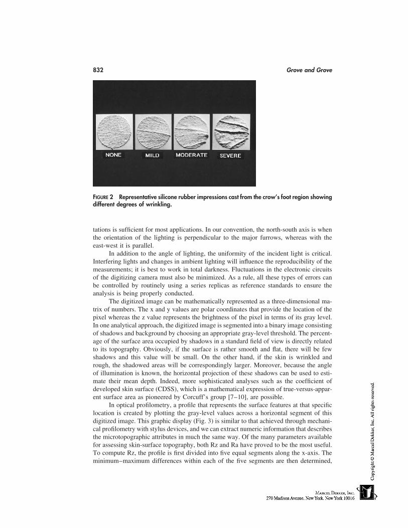
832 Grove and Grove
FIGURE 2 Representative silicone rubber impressions cast from the crow’s foot region showingdifferent degrees of wrinkling.
tations is sufficient for most applications. In our convention, the north-south axis is whenthe orientation of the lighting is perpendicular to the major furrows, whereas with theeast-west it is parallel.
In addition to the angle of lighting, the uniformity of the incident light is critical.Interfering lights and changes in ambient lighting will influence the reproducibility of themeasurements; it is best to work in total darkness. Fluctuations in the electronic circuitsof the digitizing camera must also be minimized. As a rule, all these types of errors canbe controlled by routinely using a series replicas as reference standards to ensure theanalysis is being properly conducted.
The digitized image can be mathematically represented as a three-dimensional ma-trix of numbers. The x and y values are polar coordinates that provide the location of thepixel whereas the z value represents the brightness of the pixel in terms of its gray level.In one analytical approach, the digitized image is segmented into a binary image consistingof shadows and background by choosing an appropriate gray-level threshold. The percent-age of the surface area occupied by shadows in a standard field of view is directly relatedto its topography. Obviously, if the surface is rather smooth and flat, there will be fewshadows and this value will be small. On the other hand, if the skin is wrinkled andrough, the shadowed areas will be correspondingly larger. Moreover, because the angleof illumination is known, the horizontal projection of these shadows can be used to esti-mate their mean depth. Indeed, more sophisticated analyses such as the coefficient ofdeveloped skin surface (CDSS), which is a mathematical expression of true-versus-appar-ent surface area as pioneered by Corcuff’s group [7–10], are possible.
In optical profilometry, a profile that represents the surface features at that specificlocation is created by plotting the gray-level values across a horizontal segment of thisdigitized image. This graphic display (Fig. 3) is similar to that achieved through mechani-cal profilometry with stylus devices, and we can extract numeric information that describesthe microtopographic attributes in much the same way. Of the many parameters availablefor assessing skin-surface topography, both Rz and Ra have proved to be the most useful.To compute Rz, the profile is first divided into five equal segments along the x-axis. Theminimum–maximum differences within each of the five segments are then determined,
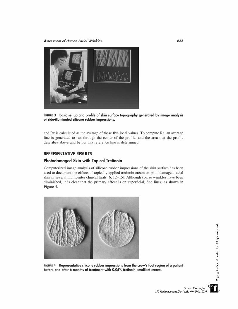
Assessment of Human Facial Wrinkles 833
FIGURE 3 Basic set-up and profile of skin surface topography generated by image analysisof side-illuminated silicone rubber impressions.
and Rz is calculated as the average of these five local values. To compute Ra, an averageline is generated to run through the center of the profile, and the area that the profiledescribes above and below this reference line is determined.
REPRESENTATIVE RESULTS
Photodamaged Skin with Topical Tretinoin
Computerized image analysis of silicone rubber impressions of the skin surface has beenused to document the effects of topically applied tretinoin cream on photodamaged facialskin in several multicenter clinical trials [6, 12–15]. Although coarse wrinkles have beendiminished, it is clear that the primary effect is on superficial, fine lines, as shown inFigure 4.
FIGURE 4 Representative silicone rubber impressions from the crow’s foot region of a patientbefore and after 6 months of treatment with 0.05% tretinoin emollient cream.
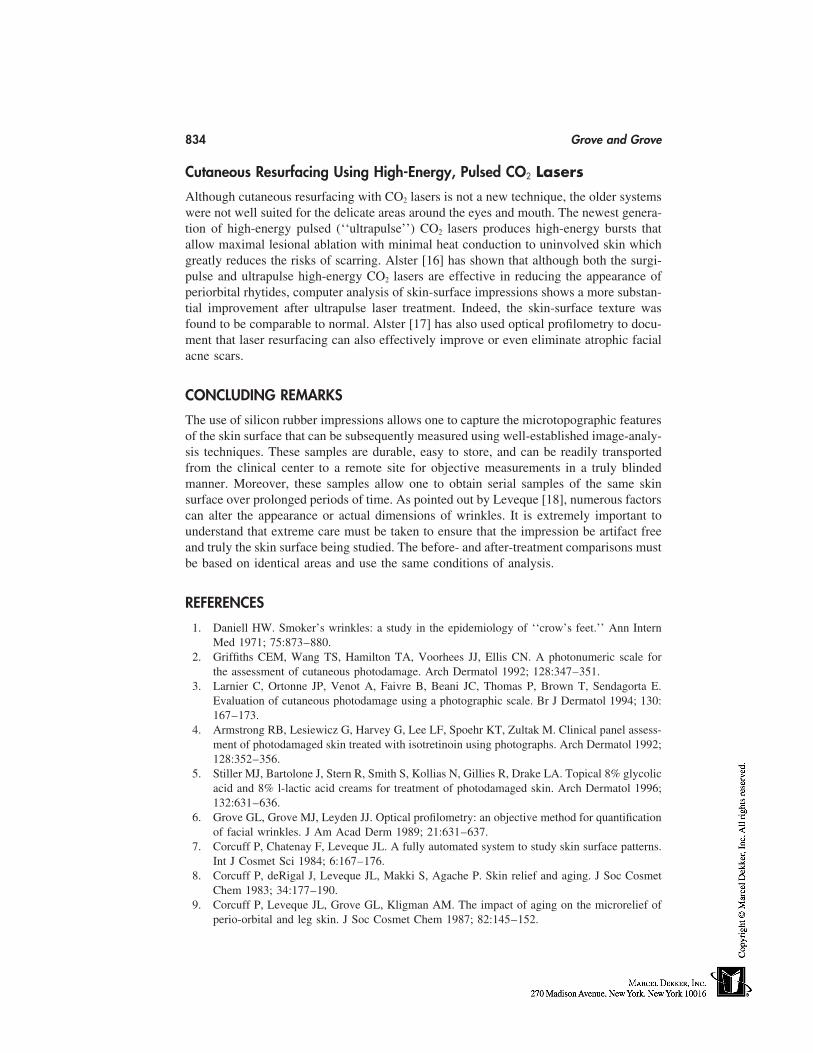
834 Grove and Grove
Cutaneous Resurfacing Using High-Energy, Pulsed CO2 Lasers
Although cutaneous resurfacing with CO2 lasers is not a new technique, the older systemswere not well suited for the delicate areas around the eyes and mouth. The newest genera-tion of high-energy pulsed (‘‘ultrapulse’’) CO2 lasers produces high-energy bursts thatallow maximal lesional ablation with minimal heat conduction to uninvolved skin whichgreatly reduces the risks of scarring. Alster [16] has shown that although both the surgi-pulse and ultrapulse high-energy CO2 lasers are effective in reducing the appearance ofperiorbital rhytides, computer analysis of skin-surface impressions shows a more substan-tial improvement after ultrapulse laser treatment. Indeed, the skin-surface texture wasfound to be comparable to normal. Alster [17] has also used optical profilometry to docu-ment that laser resurfacing can also effectively improve or even eliminate atrophic facialacne scars.
CONCLUDING REMARKS
The use of silicon rubber impressions allows one to capture the microtopographic featuresof the skin surface that can be subsequently measured using well-established image-analy-sis techniques. These samples are durable, easy to store, and can be readily transportedfrom the clinical center to a remote site for objective measurements in a truly blindedmanner. Moreover, these samples allow one to obtain serial samples of the same skinsurface over prolonged periods of time. As pointed out by Leveque [18], numerous factorscan alter the appearance or actual dimensions of wrinkles. It is extremely important tounderstand that extreme care must be taken to ensure that the impression be artifact freeand truly the skin surface being studied. The before- and after-treatment comparisons mustbe based on identical areas and use the same conditions of analysis.
REFERENCES
1. Daniell HW. Smoker’s wrinkles: a study in the epidemiology of ‘‘crow’s feet.’’ Ann InternMed 1971; 75:873–880.
2. Griffiths CEM, Wang TS, Hamilton TA, Voorhees JJ, Ellis CN. A photonumeric scale forthe assessment of cutaneous photodamage. Arch Dermatol 1992; 128:347–351.
3. Larnier C, Ortonne JP, Venot A, Faivre B, Beani JC, Thomas P, Brown T, Sendagorta E.Evaluation of cutaneous photodamage using a photographic scale. Br J Dermatol 1994; 130:167–173.
4. Armstrong RB, Lesiewicz G, Harvey G, Lee LF, Spoehr KT, Zultak M. Clinical panel assess-ment of photodamaged skin treated with isotretinoin using photographs. Arch Dermatol 1992;128:352–356.
5. Stiller MJ, Bartolone J, Stern R, Smith S, Kollias N, Gillies R, Drake LA. Topical 8% glycolicacid and 8% l-lactic acid creams for treatment of photodamaged skin. Arch Dermatol 1996;132:631–636.
6. Grove GL, Grove MJ, Leyden JJ. Optical profilometry: an objective method for quantificationof facial wrinkles. J Am Acad Derm 1989; 21:631–637.
7. Corcuff P, Chatenay F, Leveque JL. A fully automated system to study skin surface patterns.Int J Cosmet Sci 1984; 6:167–176.
8. Corcuff P, deRigal J, Leveque JL, Makki S, Agache P. Skin relief and aging. J Soc CosmetChem 1983; 34:177–190.
9. Corcuff P, Leveque JL, Grove GL, Kligman AM. The impact of aging on the microrelief ofperio-orbital and leg skin. J Soc Cosmet Chem 1987; 82:145–152.
Assessment of Human Facial Wrinkles 835
10. Corcuff P, Leveque JL. Skin surface replica image analysis of furrows and wrinkles. In: ScrupJ and Jemec GBE, eds. Handbook of Non-invasive Methods and the Skin. Boca Raton: CRC,1995:89–96.
11. Grove GL, Grove MJ. Objective methods for assessing skin surface topography noninvasively.In: Leveque JL, ed. Cutaneous Investigations in Health and Disease. New York: Marcel Dek-ker, 1989: 1–32.
12. Grove GL, Grove MJ, Leyden JJ, Lufrano L, Schwab B, Perry BH, Thorne EG. Skin replicaanalysis of photodamaged skin after therapy with tretinoin emollient cream. J Am Acad Der-matol 1991; 25:231–237.
13. Olsen EA, Katz HI, Levine N, et al. Tretinoin emollient cream: a new therapy for photodam-aged skin. J Am Acad Dermatol 1992; 26:215–224.
14. Weinstein GD, Migra TP, Poch PE, et al. Topical tretinoin for treatment of photodamagedskin. A multicenter study. Arch Dermatol 1991; 127(5):659–665.
15. Gilchrest BA. Treatment of photodamage with topical tretinoin: an overview. J Am AcadDermatol 1997; 36(?)S27- 36.
16. Alster TS. Comparison of two high-energy, pulsed carbon dioxide lasers in the treatment ofperiorbital rhytides. Dermatol Surg 1996; 22:541–545.
17. Alster TS, West TB. Resurfacing of atrophic facial acne scars with a high-energy, pulsedcarbon dioxide laser. Dermatol Surg 1996; 22:151–155.
18. Leveque JL, EEMCO guidance for the assessment of skin topography. J Eur Acad DermatolVenereol 1999; 12(2):103–114.

70
Acnegenicity and Comedogenicity Testingfor Cosmetics
F. Anthony SimionThe Andrew Jergens Company, Cincinnati, Ohio
INTRODUCTION
Many people experience facial acne, especially in their teens and early 20s. It typicallycauses distress, and in some individuals contributes to a lowered self-image [1]. As con-sumers age the prevalence of acne decreases, although it can be triggered by factors suchas stress, medications, and the use of cosmetics [2–4]. Indeed, there are many reports ofcosmetics causing comedones, or acneform eruptions. These adverse reactions are of greatconcern to consumers, many of whom look for products that will not cause such problems.Hence, cosmetics manufacturers strive to develop products that do not cause comedonesor acne. Products are frequently labeled noncomedogenic and/or nonacnegenic. Consum-ers use these terms interchangeably, although comedone formation and acnegenicity arenot the same. To consumers, the key issue is that the cosmetics they use do not causebreakouts.
COMEDOGENICITY
Comedone formation occurs when the pattern of keratinization inside the sebaceous folli-cle changes. Within the keratinocytes, these changes include the production of differentkeratins and a reduction in the number of lamellar granules [5]. There is also an increasein mitotic activity [6]. As a result the keratinocytes do not desquamate properly and thefollicular duct is blocked. It is not known what causes these changes, but the result is amicrocomedone. As further keratinized material accumulates, the follicle becomes visiblefrom the surface as a closed comedone, or whitehead (Fig. 1). As more material accumu-lates, the follicle distends and open comedones, also know as blackheads are formed. Theblack color is attributable to the oxidation of lipids as they reach the skin’s surface. Thetest methods to assess comedogenesis are designed to quantify the hyperkeratotic plugs.
ACNEGENICITY
The hyperkeratotic plug results in sebum accumulating in the follicilar duct and the seba-ceous gland. This enables the anaerobic bacteria, P. acnes, to proliferate. The follicular
837
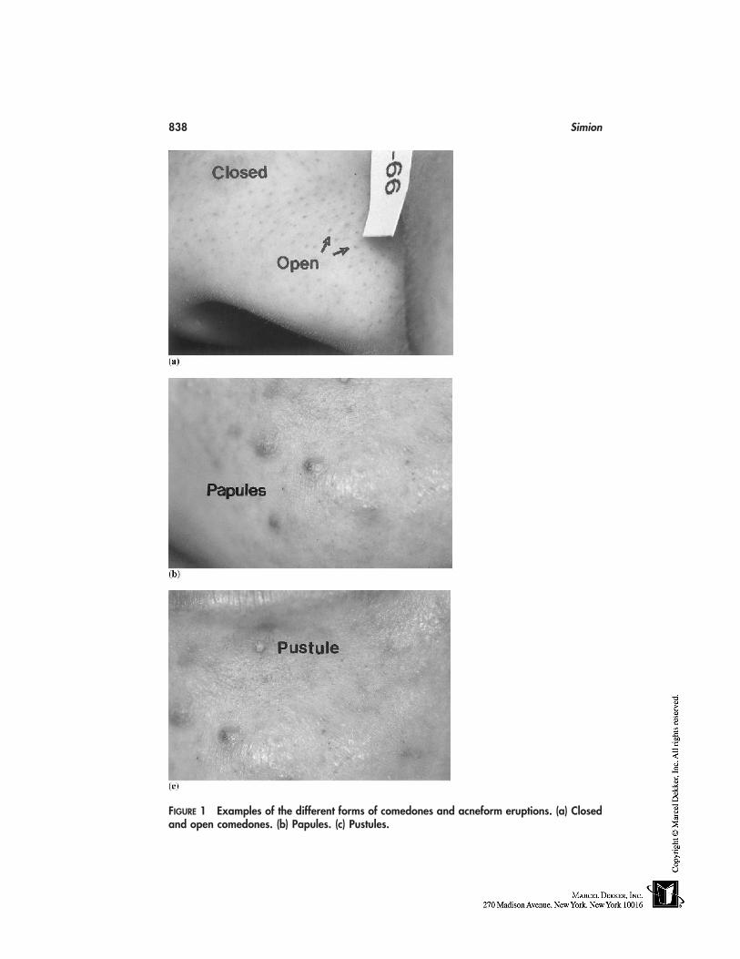
838 Simion
FIGURE 1 Examples of the different forms of comedones and acneform eruptions. (a) Closedand open comedones. (b) Papules. (c) Pustules.
Acnegenicity and Comedogenicity Testing 839
duct will expand until it ruptures, releasing the bacteria and their metabolic products intothe surrounding dermis. Both immunological and irritant reactions occur. In severe acne,elevated levels of anti–P. acnes antibodies are detected. Irritation may come from thefatty acids that are the result of sebaceous triglyceride digestion by bacteria. Certainly atthe histological level, the classic signs of inflammation such as a neutrophilic infiltrateare observed. The consumer recognizes this as acne pustules and papules.
PUSTULOGENIC POTENTIAL
Upon first using a new cosmetic, papules and pustules are sometimes observed after afew days. To the consumer this is acne, although this type of papulopustular reaction maybe a form of follicular irritation. Certainly, as Mills and Berger pointed out, it occurs morequickly than can be accounted for by the formation of hyperkeratotic plugs and its sequalae[7]. Thus, Mills and Berger suggested that pustulogenic potential—the ability to causeinflammatory lesions—should be differentiated from comedogenic potential—the abilityto cause the formation of hyperkeratotic plugs.
TEST METHODS
Human and animal models have been used to assess the comedogenic potential of cosmeticproducts and their ingredients. Both models require repeated applications of the materialsto the skin for 2 to 4 weeks. The number of hyperkeratotic impactions produced is com-pared with positive and negative controls.
Animal Models
The rabbit ear is the most commonly used animal model. The rabbit’s ear follicle hasmany structural similarities to the human sebaceous follicle. In 1941, Adams et al. [8]showed that the rabbit ear would respond similarly to human skin when exposed to chlori-nated hydrocarbons, the most common cause of acne in industrial accidents. Until the riseof the animal rights movement in the late 1980s, cosmetic products were routinely screenedusing rabbits. Briefly, the method entails that the test product or ingredient is applied dailyto the inner surface of one ear. This site is left open. The other ear is used as the negativecontrol. At the end of either 2 or 4 weeks, the animal was sacrificed, and the degree offollicular hyperkeratinization is assessed. Frequently this was done by taking histologicalsections and giving an overall score based on the number and degrees of compacted folli-cles. Occasionally the impactions were removed from the ear using cyanoacrylate glueon a glass slide. This method is now frequently used in human testing.
The results from the rabbit-ear studies show that some cosmetic ingredients havecomedogenic potential. These include branched chain esters and compounds that havesolubility in both oil and water (hydrophile-lipophile balance (HLB) of 10–12) [9]. How-ever, if these materials are chemically modified, or included at low levels with other ingre-dients in cosmetics, then their comedogenic potential is nullified. This was shown byFulton [9] as well as Kligman and Mills [2]. Fulton showed that chemically modifyingcosmetic ingredients can greatly effect their comedogenic potential. For instance, PEG-16 Lanolin gives a severe comedogenicity score of 4 on a 0 to 5 scale, whereas the higher–molecular weight and more water-soluble PEG-75 Lanolin yields a score of 0 under the
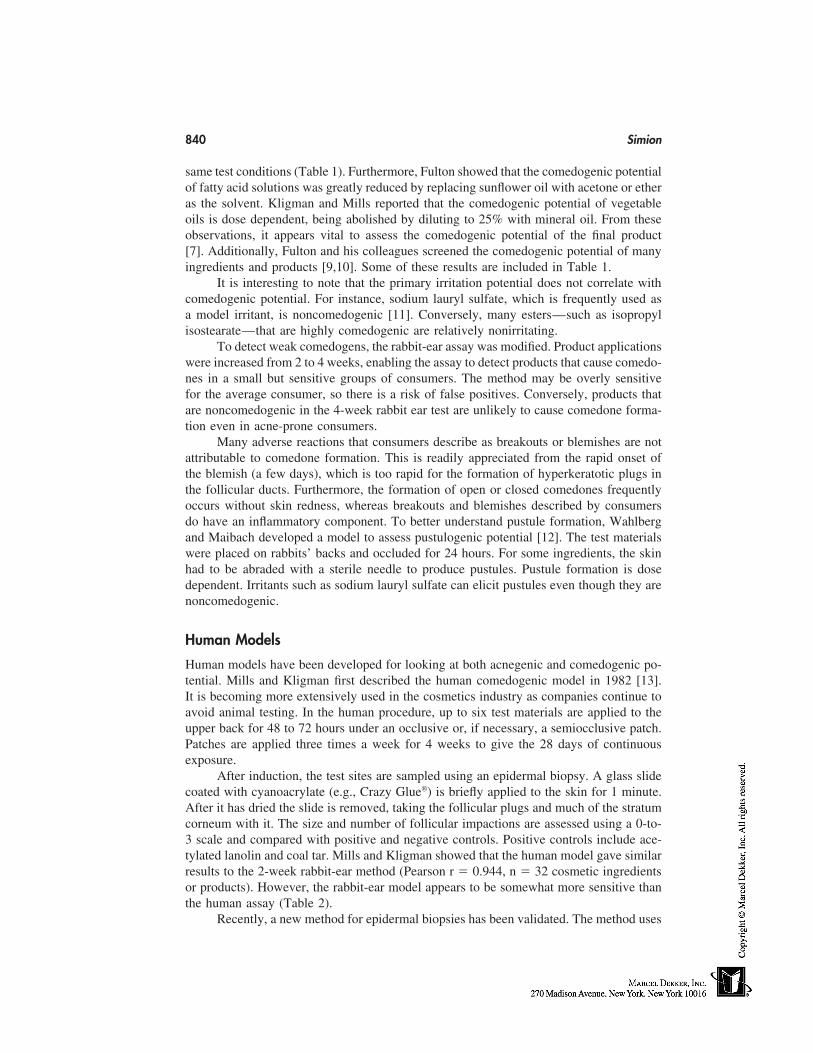
840 Simion
same test conditions (Table 1). Furthermore, Fulton showed that the comedogenic potentialof fatty acid solutions was greatly reduced by replacing sunflower oil with acetone or etheras the solvent. Kligman and Mills reported that the comedogenic potential of vegetableoils is dose dependent, being abolished by diluting to 25% with mineral oil. From theseobservations, it appears vital to assess the comedogenic potential of the final product[7]. Additionally, Fulton and his colleagues screened the comedogenic potential of manyingredients and products [9,10]. Some of these results are included in Table 1.
It is interesting to note that the primary irritation potential does not correlate withcomedogenic potential. For instance, sodium lauryl sulfate, which is frequently used asa model irritant, is noncomedogenic [11]. Conversely, many esters—such as isopropylisostearate—that are highly comedogenic are relatively nonirritating.
To detect weak comedogens, the rabbit-ear assay was modified. Product applicationswere increased from 2 to 4 weeks, enabling the assay to detect products that cause comedo-nes in a small but sensitive groups of consumers. The method may be overly sensitivefor the average consumer, so there is a risk of false positives. Conversely, products thatare noncomedogenic in the 4-week rabbit ear test are unlikely to cause comedone forma-tion even in acne-prone consumers.
Many adverse reactions that consumers describe as breakouts or blemishes are notattributable to comedone formation. This is readily appreciated from the rapid onset ofthe blemish (a few days), which is too rapid for the formation of hyperkeratotic plugs inthe follicular ducts. Furthermore, the formation of open or closed comedones frequentlyoccurs without skin redness, whereas breakouts and blemishes described by consumersdo have an inflammatory component. To better understand pustule formation, Wahlbergand Maibach developed a model to assess pustulogenic potential [12]. The test materialswere placed on rabbits’ backs and occluded for 24 hours. For some ingredients, the skinhad to be abraded with a sterile needle to produce pustules. Pustule formation is dosedependent. Irritants such as sodium lauryl sulfate can elicit pustules even though they arenoncomedogenic.
Human Models
Human models have been developed for looking at both acnegenic and comedogenic po-tential. Mills and Kligman first described the human comedogenic model in 1982 [13].It is becoming more extensively used in the cosmetics industry as companies continue toavoid animal testing. In the human procedure, up to six test materials are applied to theupper back for 48 to 72 hours under an occlusive or, if necessary, a semiocclusive patch.Patches are applied three times a week for 4 weeks to give the 28 days of continuousexposure.
After induction, the test sites are sampled using an epidermal biopsy. A glass slidecoated with cyanoacrylate (e.g., Crazy Glue) is briefly applied to the skin for 1 minute.After it has dried the slide is removed, taking the follicular plugs and much of the stratumcorneum with it. The size and number of follicular impactions are assessed using a 0-to-3 scale and compared with positive and negative controls. Positive controls include ace-tylated lanolin and coal tar. Mills and Kligman showed that the human model gave similarresults to the 2-week rabbit-ear method (Pearson r � 0.944, n � 32 cosmetic ingredientsor products). However, the rabbit-ear model appears to be somewhat more sensitive thanthe human assay (Table 2).
Recently, a new method for epidermal biopsies has been validated. The method uses
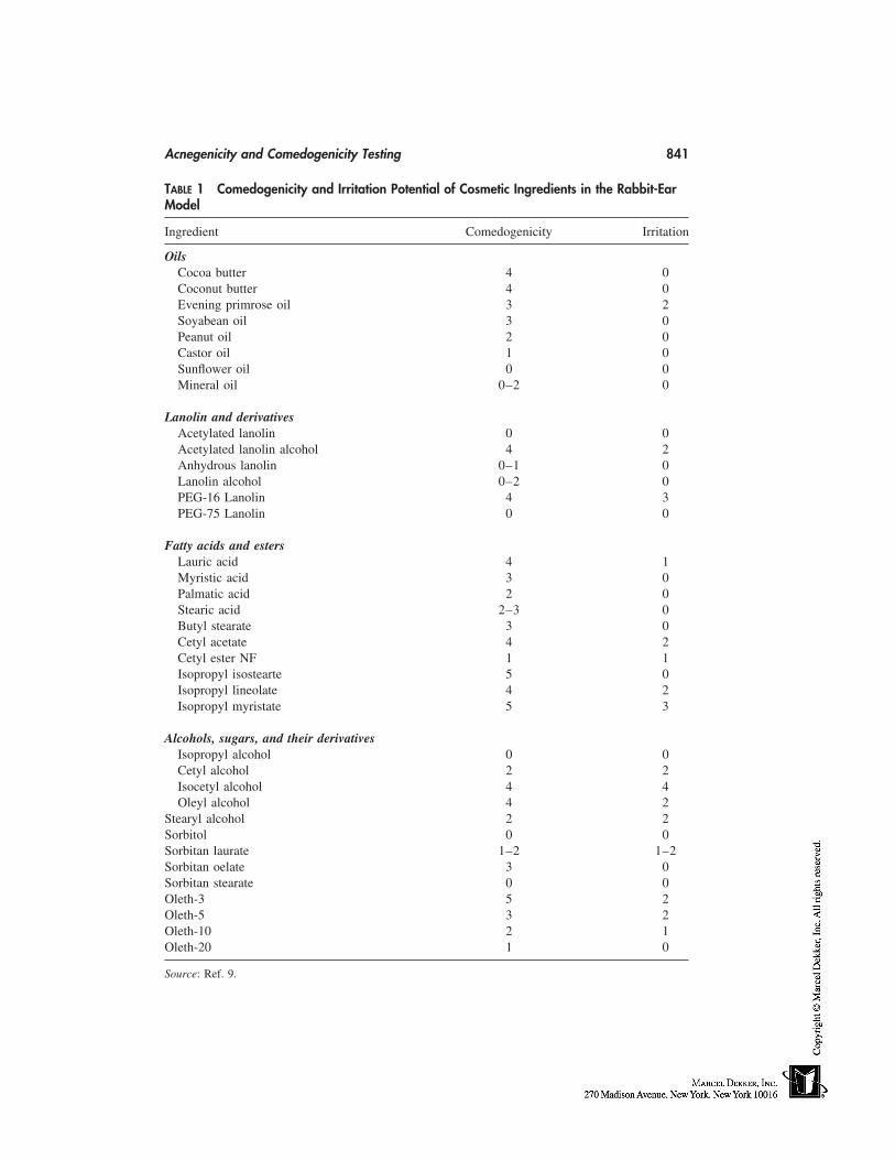
Acnegenicity and Comedogenicity Testing 841
TABLE 1 Comedogenicity and Irritation Potential of Cosmetic Ingredients in the Rabbit-EarModel
Ingredient Comedogenicity Irritation
OilsCocoa butter 4 0Coconut butter 4 0Evening primrose oil 3 2Soyabean oil 3 0Peanut oil 2 0Castor oil 1 0Sunflower oil 0 0Mineral oil 0–2 0
Lanolin and derivativesAcetylated lanolin 0 0Acetylated lanolin alcohol 4 2Anhydrous lanolin 0–1 0Lanolin alcohol 0–2 0PEG-16 Lanolin 4 3PEG-75 Lanolin 0 0
Fatty acids and estersLauric acid 4 1Myristic acid 3 0Palmatic acid 2 0Stearic acid 2–3 0Butyl stearate 3 0Cetyl acetate 4 2Cetyl ester NF 1 1Isopropyl isostearte 5 0Isopropyl lineolate 4 2Isopropyl myristate 5 3
Alcohols, sugars, and their derivativesIsopropyl alcohol 0 0Cetyl alcohol 2 2Isocetyl alcohol 4 4Oleyl alcohol 4 2
Stearyl alcohol 2 2Sorbitol 0 0Sorbitan laurate 1–2 1–2Sorbitan oelate 3 0Sorbitan stearate 0 0Oleth-3 5 2Oleth-5 3 2Oleth-10 2 1Oleth-20 1 0
Source: Ref. 9.
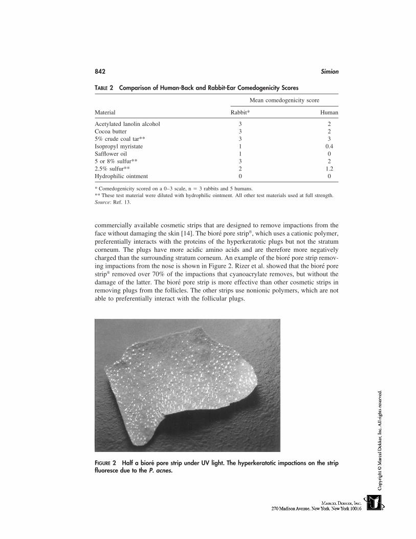
842 Simion
TABLE 2 Comparison of Human-Back and Rabbit-Ear Comedogenicity Scores
Mean comedogenicity score
Material Rabbit* Human
Acetylated lanolin alcohol 3 2Cocoa butter 3 25% crude coal tar** 3 3Isopropyl myristate 1 0.4Safflower oil 1 05 or 8% sulfur** 3 22.5% sulfur** 2 1.2Hydrophilic ointment 0 0
* Comedogenicity scored on a 0–3 scale, n � 3 rabbits and 5 humans.** These test material were diluted with hydrophilic ointment. All other test materials used at full strength.Source: Ref. 13.
commercially available cosmetic strips that are designed to remove impactions from theface without damaging the skin [14]. The bioré pore strip, which uses a cationic polymer,preferentially interacts with the proteins of the hyperkeratotic plugs but not the stratumcorneum. The plugs have more acidic amino acids and are therefore more negativelycharged than the surrounding stratum corneum. An example of the bioré pore strip remov-ing impactions from the nose is shown in Figure 2. Rizer et al. showed that the bioré porestrip removed over 70% of the impactions that cyanoacrylate removes, but without thedamage of the latter. The bioré pore strip is more effective than other cosmetic strips inremoving plugs from the follicles. The other strips use nonionic polymers, which are notable to preferentially interact with the follicular plugs.
FIGURE 2 Half a bioré pore strip under UV light. The hyperkeratotic impactions on the stripfluoresce due to the P. acnes.
Acnegenicity and Comedogenicity Testing 843
One advantage that the bioré strips have over the cyanoacrylate glue is that it is farless damaging and irritating to the skin. Therefore, it can be used to measure comedoneformation on the face at the end of usage studies. Cyanoacrylate is too damaging to beacceptable to most test panelists for use on their face. The bioré pore strip has been shownto be acceptable to panelists; indeed, this is its intended use.
Human Usage Tests
Ultimately, all predictive models must be related to back to the consumers’ experiencein the marketplace. Consumers who experience an adverse reaction will report it in termsmost familiar to them. Most consumers do not differentiate between acne and comedoneformation, or blemishes and breakouts.
One approach to assessing the rate of adverse reactions is to have a group of consum-ers use a product for several weeks [15–18]. Test subjects should be evaluated for comedo-nes, pustules and papules at the beginning of the study, and then at set intervals. A 1-week evaluation will reveal any propensity to cause irritation, including follicular irritationthat panelists may recognize as breakouts. Any sensory irritation will become evidentduring the first week. Three and six-week evaluations are used to detect comedogenicityand acnegenicity. This design is consistent with the recommendation of the AmericanAcademy of Dermatology’s consensus panel on acnegenicity testing [19].
A sizable proportion of adverse reactions is experienced by vulnerable subgroups. Asubgroup for irritation may include panelists whose skin is readily irritated by surfactants.Another subgroup will include panelists with acne-prone skin. Both subgroups should beidentified and form a significant part of the test panel. This will enable the investigatorto identify potential problems before the product reaches the marketplace.
SUMMARY
The induction of comedones and acneform eruptions is a significant concern to manyconsumers, especially those with acne-prone skin. Any product that has a propensity toproduce these eruptions will be unsuccessful in the marketplace. Indeed, many consumersexpressly look for products that are labeled noncomedogenic and/or nonacnegenic.
Cosmetics manufacturers are meeting this consumer demand by showing that theirproducts do not cause comedones and/or acne breakouts, and label their products accord-ingly. Consumers judge a facial cosmetic on whether it causes breakouts, blemishes,bumps, or blackheads. There are multiple causes for the reaction, including comedoneformation and follicular irritation. Consumers do not differentiate between the biologicalmechanisms; they are only concerned with the results they produce.
Today, human models have replaced animals for testing both comedogenicity andacnegenicity. Comedone formation is determined by continuously patching the materialon the human back for 28 consecutive days. Comedones are quantified by extracting theplugs from the follicle using cyanoacrylate glue on a glass slide or a bioré pore strip
containing cationic polymers. The degree of the impactions is compared with positive andnegative controls.
Acnegenicity is assessed by human-use testing, where a panel of consumers usesthe product under normal conditions. The skin is evaluated by a trained observer for come-dones, papules, and pustules at the beginning of the study, and then after 1, 3, and 6 weeksof usage. Cosmetic pore strips such as bioré can be used to assess comedone formation.
844 Simion
These can remove follicular plugs from the face without the skin damage associated withcyanoacrylate.
REFERENCES
1. S MacDonald Hull, WJ Cunliffe, BR Hughes. Treatment of the depressed and dysmorphopho-bic acne patient. Clin Exp Dermatol 16: 210–211, 1991.
2. AM Kligman, OH Jr Mills. Acne Cosmetica. Arch Dermatol 106: 843–850, 1972.3. G Plewig, JE Jr Fulton, AM Kligman. Pomade Acne. Arch Dermatol 101: 580–584, 1970.4. C Berlin. Acne comedo in children due to paraffin oil applied on the head. Arch Dermatol
69: 683–687, 1954.5. JS Strauss. Sebaceous Glands in ‘Dermatology in General Medicine’ Eds. Fitzpatrick TB,
Eisen AZ. Wolff K. et al., 4th Edition vol 1, p 709–726 (1993).6. G Plewig, JE Jr Fulton, AM Kligman. Cellular dynamics of comedo formation in acne vulgaris.
Arch Dermatol 102: 12–29, 1971.7. OH Jr Mills, RS Berger. Defining the susceptibility of acne-prone and sensitive skin popula-
tions to extrinsic factors. Dermatol Clinics 9: 93–98, 1991.8. EH Adams, DD Irish, HC Spencer, et al. The reponse of rabbit skin to compounds reported
to have caused anceform dermatitis Ind Med 2: 1–4, 1941.9. JE Jr Fulton, Comedogenicity and irritancy of commonly used ingredients in skin care prod-
ucts. J Soc Cosmet Chem 40: 321–333, 1989.10. JE Jr Fulton, SR Pay, JE III Fulton. Comedogenicity of current therapeutic products, cosmetics,
and ingredients in the rabbit ear. J Amer Acad Dermatol 10: 96–105, 1984.11. WE Morris, SC Kwan. Use of the rabbit ear model in evaluating the comedogenic potential
of cosmetic ingredients. J Soc Cosmet Chem 34: 215–225, 1983.12. JE Walhberg, HI Maibach. Sterile Cutaneous Pustules: a manifestation of primary irritancy?
In: Models in Dermatology 2: 297–302 Maibach HI and Lowe N, eds. Pub Krager (1985).13. OH Jr Mills, AM Kligman. A human model for assessing comedogenic substances. Arch Der-
matol 118: 903–905, 1982.14. RL Rizer, JK Woodford, FA Simion et al. ‘The follicular bioposy for assessing comedo-
genicity: a comparison of a pore strip and cyanoacrylate.’ Poster at the International Societyof Bioengineering and the Skin, June 1998.
15. EM Jackson, NF Robillard. The controlled use test in a cosmetic product safety substantiationprogram. J Toxicol Cut & Ocular Toxicol 1: 117–132, 1982.
16. OH Jr Mills, RS Berger, TJ Stephens et al. Assessing acnegenic and acne aggravating potential.J Toxicol Cut & Ocular Toxical 8: 353–360, 1989.
17. EM Jackson. Clinical assessments of acnegenicity. J Toxicol Cut & Ocular Toxicol 8: 389–393, 1989.
18. A Ghassemi, R Osborne, KA Korman, et al. Demonstrating the ocular safety of an eye cosmeticproduct using alternatives to animal eye irritation tests. Poster at the Society of ToxicologyMeeting in Cincinnati, OH, March 1997.
19. JS Strauss, EM Jackson et al. American Academy of Dermatology Invitational Symposiumon Comedogenicity, J Amer Acad Dermatol 20: 272–277, 1989.
71
Sensory Testing
Linda P. Oddo and Kathy ShannonHill Top Research, Inc., Scottsdale, Arizona
Although most individuals don’t realize it, they conduct sensory tests on a daily basis.Throughout the day, personal sensory assessments are made about the taste acceptabilityor liking for different foods for many different attributes. Individuals evaluate haircareproducts for various properties, not only while using the product but also for feel of thehair after shampooing. Every time a cosmetic product, moisturizer, or any other skincareproduct is applied to the skin, sensory assessments are made.
In very simple terms, the field of sensory testing applies controls, data-collectionskills, and reproducible methodology to these types of assessments for the purpose ofcollecting not only viable but valuable information about the test materials. Civille [1]states that ‘‘[t]he primary function of sensory testing is to conduct valid and reliable tests,which provide data on which sound decisions can be made.’’
Although the roots of sensory testing as a discipline exist in the food industry, itsapplications have steadily earned respect in the consumer and pharmaceutical industries.Science now plays an important role in the field of cosmetology by providing guidanceto the product formulator in predicting consumer response and by supporting or definingproduct claims.
Currently, trained sensory judges are routinely used in the development of manycosmetic products. Trained panels are used to evaluate the skin-feel properties of not onlytopical cosmetic products but any product that is applied to the skin. Trained judges areused to evaluate haircare products, and sensory judges are critical to determining oral andaxillary malodor.
Using carefully controlled sensory applications, sensitive-skin subjects can be identi-fied and selected. The response of these subjects can then be trusted to aid in the develop-ment of and/or to identify acceptable sensitive-skin cosmetic products. Finally, byapplying sensory scaling techniques and controls to the design of self-assessment question-naires, they are often added to clinical studies to provide a potential insight to consumerresponse. The primary focus of this chapter is to describe some of the sensory methodsand tools that are currently being applied in these areas.
845
846 Oddo and Shannon
AXILLARY MALODOR EFFICACY
Generally, axillary malodor efficacy tests are conducted on three product types: antiperspi-rants, deodorants, and soaps. Although antiperspirants are primarily designed to inhibitsweat production, they are also considered deodorants because they inhibit sweat, whichacts as a culture medium for bacteria to produce, degrade, and form malodor. Deodorantsare formulated to control malodor only, through absorption, fragrance masking, and/orby reducing antibacterial activity. Some soap products may also reduce axillary malodorby fragrance masking and/or inhibiting bacterial growth.
The use of sensory testing applications is the primary methodology used to establishdeodorant efficacy. In 1987, the sensory testing division of The American Society forTesting and Materials (ASTM) published the Standard Practice for the Sensory Evaluationof Axillary Deodorancy [2]. This document recommends that a product meet the criteriapresented in the standard in order for it to qualify as an effective deodorant.
The basic design for conducting a deodorant study consists of selecting subjectswith high axillary malodor, applying or using the test material at selected intervals, andthen measuring the level of axillary malodor using a panel of trained odor judges. Thetest material is considered effective if there is a statistical difference between it and aplacebo or untreated control. Factors that are critical to the test design and consequentlya successful study are the subject selection, subject restrictions, odor-judge selection andtraining, selection of a suitable test location, and using appropriate scaling techniques.
Subjects should be selected from the user population and have a distinct axillaryodor. Those with extremely high or low odor and those with large differences in odorlevel between the right and left axillae are usually disqualified. It is important that potentialsubjects participate in a washout or conditioning period before selection to prevent a carry-over effect from the use of other products. For a minimum of 7 days, subjects are notallowed to use any axillary products and are instructed to wash the axillae only with amild, nondeodorant soap. Other restrictions that are known to interfere with sensory odorassessments require the subjects to abstain from swimming, excessive exercise, and fromusing any fragranced products. They must also avoid spicy foods, and before an odorevaluation they are restricted from smoking.
Perhaps the most crucial factor in a well-executed malodor efficacy test is the selec-tion and training of qualified odor judges. This process requires management support andcommitment, a sensory staff or analyst to conduct the training, and a pool of availableand interested candidates. Other factors to consider are the time commitments to not onlyselect and train the judges but to continually maintain and validate their performance.The odor–judge selection and training process involves four basic steps: 1) interviewingcandidates, 2) conducting screening tests, 3) training, and 4) validating performance.
During the interviewing process, candidates who have conflicting commitments orinterfering health problems should be discontinued. A description of the test and the odor-judge process must be explained to each individual. If possible, because of the unusualnature of the intended task—sniffing the axillary region of subjects—a video of the pro-cess should be shown. Through interaction and discussion, those candidates who show asincere interest and are willing to commit to the program are identified.
In vitro screening tests are administered to the potential odor judges to determinetheir olfactory acuity and ability to discriminate and reproduce results. Because it is possi-ble for some individuals to be insensitive to some of the odors generated by the humanbody, potential judges should also be screened for this inherent lack of sensitivity.
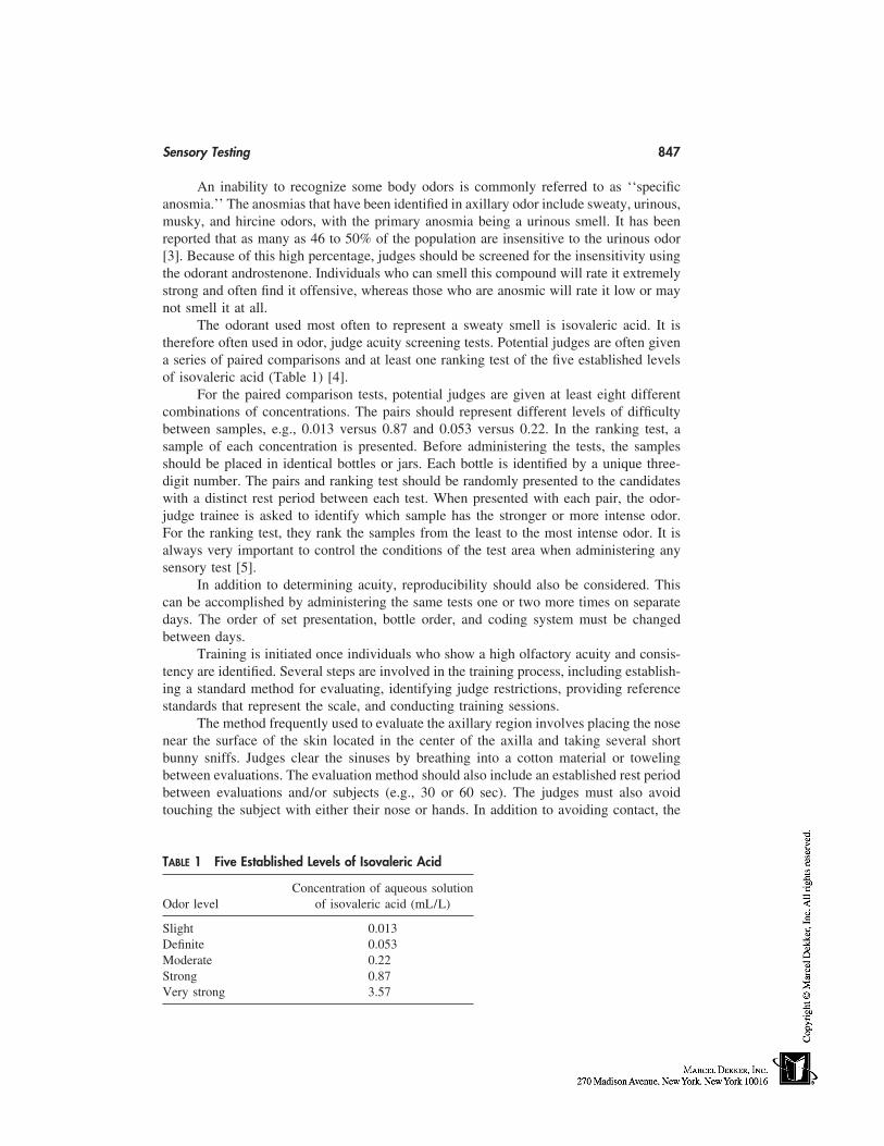
Sensory Testing 847
An inability to recognize some body odors is commonly referred to as ‘‘specificanosmia.’’ The anosmias that have been identified in axillary odor include sweaty, urinous,musky, and hircine odors, with the primary anosmia being a urinous smell. It has beenreported that as many as 46 to 50% of the population are insensitive to the urinous odor[3]. Because of this high percentage, judges should be screened for the insensitivity usingthe odorant androstenone. Individuals who can smell this compound will rate it extremelystrong and often find it offensive, whereas those who are anosmic will rate it low or maynot smell it at all.
The odorant used most often to represent a sweaty smell is isovaleric acid. It istherefore often used in odor, judge acuity screening tests. Potential judges are often givena series of paired comparisons and at least one ranking test of the five established levelsof isovaleric acid (Table 1) [4].
For the paired comparison tests, potential judges are given at least eight differentcombinations of concentrations. The pairs should represent different levels of difficultybetween samples, e.g., 0.013 versus 0.87 and 0.053 versus 0.22. In the ranking test, asample of each concentration is presented. Before administering the tests, the samplesshould be placed in identical bottles or jars. Each bottle is identified by a unique three-digit number. The pairs and ranking test should be randomly presented to the candidateswith a distinct rest period between each test. When presented with each pair, the odor-judge trainee is asked to identify which sample has the stronger or more intense odor.For the ranking test, they rank the samples from the least to the most intense odor. It isalways very important to control the conditions of the test area when administering anysensory test [5].
In addition to determining acuity, reproducibility should also be considered. Thiscan be accomplished by administering the same tests one or two more times on separatedays. The order of set presentation, bottle order, and coding system must be changedbetween days.
Training is initiated once individuals who show a high olfactory acuity and consis-tency are identified. Several steps are involved in the training process, including establish-ing a standard method for evaluating, identifying judge restrictions, providing referencestandards that represent the scale, and conducting training sessions.
The method frequently used to evaluate the axillary region involves placing the nosenear the surface of the skin located in the center of the axilla and taking several shortbunny sniffs. Judges clear the sinuses by breathing into a cotton material or towelingbetween evaluations. The evaluation method should also include an established rest periodbetween evaluations and/or subjects (e.g., 30 or 60 sec). The judges must also avoidtouching the subject with either their nose or hands. In addition to avoiding contact, the
TABLE 1 Five Established Levels of Isovaleric Acid
Concentration of aqueous solutionOdor level of isovaleric acid (mL/L)
Slight 0.013Definite 0.053Moderate 0.22Strong 0.87Very strong 3.57
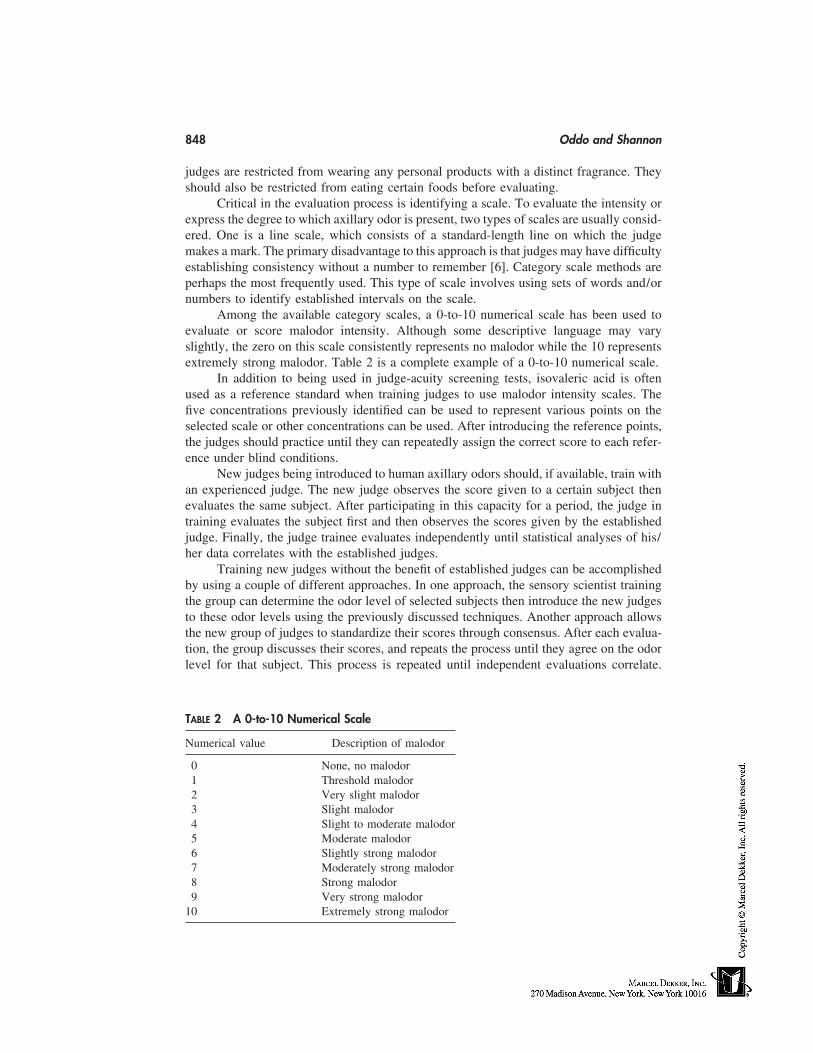
848 Oddo and Shannon
judges are restricted from wearing any personal products with a distinct fragrance. Theyshould also be restricted from eating certain foods before evaluating.
Critical in the evaluation process is identifying a scale. To evaluate the intensity orexpress the degree to which axillary odor is present, two types of scales are usually consid-ered. One is a line scale, which consists of a standard-length line on which the judgemakes a mark. The primary disadvantage to this approach is that judges may have difficultyestablishing consistency without a number to remember [6]. Category scale methods areperhaps the most frequently used. This type of scale involves using sets of words and/ornumbers to identify established intervals on the scale.
Among the available category scales, a 0-to-10 numerical scale has been used toevaluate or score malodor intensity. Although some descriptive language may varyslightly, the zero on this scale consistently represents no malodor while the 10 representsextremely strong malodor. Table 2 is a complete example of a 0-to-10 numerical scale.
In addition to being used in judge-acuity screening tests, isovaleric acid is oftenused as a reference standard when training judges to use malodor intensity scales. Thefive concentrations previously identified can be used to represent various points on theselected scale or other concentrations can be used. After introducing the reference points,the judges should practice until they can repeatedly assign the correct score to each refer-ence under blind conditions.
New judges being introduced to human axillary odors should, if available, train withan experienced judge. The new judge observes the score given to a certain subject thenevaluates the same subject. After participating in this capacity for a period, the judge intraining evaluates the subject first and then observes the scores given by the establishedjudge. Finally, the judge trainee evaluates independently until statistical analyses of his/her data correlates with the established judges.
Training new judges without the benefit of established judges can be accomplishedby using a couple of different approaches. In one approach, the sensory scientist trainingthe group can determine the odor level of selected subjects then introduce the new judgesto these odor levels using the previously discussed techniques. Another approach allowsthe new group of judges to standardize their scores through consensus. After each evalua-tion, the group discusses their scores, and repeats the process until they agree on the odorlevel for that subject. This process is repeated until independent evaluations correlate.
TABLE 2 A 0-to-10 Numerical Scale
Numerical value Description of malodor
0 None, no malodor1 Threshold malodor2 Very slight malodor3 Slight malodor4 Slight to moderate malodor5 Moderate malodor6 Slightly strong malodor7 Moderately strong malodor8 Strong malodor9 Very strong malodor
10 Extremely strong malodor
Sensory Testing 849
Although this approach is more time consuming, it often establishes a strong sense ofcommitment and involvement in the process for the new judges.
Once established, odor judges can be used to evaluate any personal-care productused in the axillae to control malodor. By using combinations of subject selection, product-treatment techniques, post-treatment evaluation times, and controlling environmental con-ditions, an almost endless number of possibilities can be evaluated by the judges. In addi-tion to directly evaluating human subjects, odor judges can also be used to evaluate axillaryodor that has been transferred to some other medium such as a t-shirt or a cloth wornagainst the axilla.
ORAL MALODOR EFFICACY
Currently, oral malodor efficacy studies are conducted with toothpastes, cleansers, pow-ders, mouth rinses, toothbrushes, breath mints, tongue scrapers, and any oral treatmentwhose primary or secondary function is to reduce or control halitosis. Oral treatments aredesigned to control, mask, or eliminate sulfur-producing bacteria, the primary componentof bad breath.
To accommodate the large variety of consumer products currently available for treat-ing halitosis, clinical studies vary in their design. Variables include the profile of the targetpopulation in their medical and dental history, current health conditions, and personalpractices. Other elements considered when designing oral-malodor clinical studies includethe number of treatments, evaluations, and post-treatment evaluation intervals. Evaluationsmay include any combination of professional examinations, microbiology sampling, oral-malodor assessments, and instrumental measurements.
Instruments that have been used to measure levels of malodor include gas chromato-graph (GC), which has been used to analyze oral volatile sulfur compounds. In a clinicalstudy comparing the GC with sensory odor judges, the instrumental measurements showedgood correlation with the organoleptic assessments. The GC, however, is considered large,cumbersome, and difficult to use in a clinical setting [7]. A portable sulfide monitor, easierto use in a clinical environment, has also been investigated and found to fall within therange observed with the GC. When compared with odor judges, the Halimeter alsosignificantly correlated (p � 0.001) with sensory ratings [8].
Although good correlation has been established, the manufacturer of the Halimeterstates that the data independently cannot confirm the existence of oral malodor becausevolatile sulfur compounds are not constant in any one person. They recommend using theinstrument with other procedures, such as bacterial cultures and organoleptic measure-ments to assess levels of oral malodor.
Organoleptic measurements or assessments are generally conducted by judges spe-cifically trained to evaluate oral malodor. The selection and training of these judges issimilar to the techniques used to select and train axillary malodor judges. Differencesinclude the use of reference standards more appropriate to oral malodor and training thejudges in a different process of evaluation. In the oral-malodor evaluation process, thejudge and subject are separated by a solid partition. The partition has a small circularopening in which the subject inserts a glass rod. During the actual assessment, the subjectplaces his/her mouth around the end of the glass rod and either holds his/her breath orexhales into the tube while the judge places his/her nose near the other end of the tube.
Currently, two very different types of sensory scales are being used to measure oralmalodor. One applies hedonic measurements whereas the other approach uses category
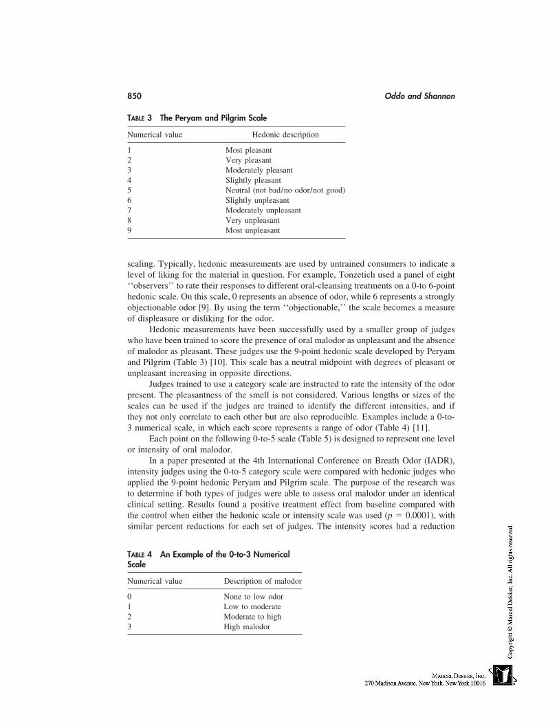
850 Oddo and Shannon
TABLE 3 The Peryam and Pilgrim Scale
Numerical value Hedonic description
1 Most pleasant2 Very pleasant3 Moderately pleasant4 Slightly pleasant5 Neutral (not bad/no odor/not good)6 Slightly unpleasant7 Moderately unpleasant8 Very unpleasant9 Most unpleasant
scaling. Typically, hedonic measurements are used by untrained consumers to indicate alevel of liking for the material in question. For example, Tonzetich used a panel of eight‘‘observers’’ to rate their responses to different oral-cleansing treatments on a 0-to 6-pointhedonic scale. On this scale, 0 represents an absence of odor, while 6 represents a stronglyobjectionable odor [9]. By using the term ‘‘objectionable,’’ the scale becomes a measureof displeasure or disliking for the odor.
Hedonic measurements have been successfully used by a smaller group of judgeswho have been trained to score the presence of oral malodor as unpleasant and the absenceof malodor as pleasant. These judges use the 9-point hedonic scale developed by Peryamand Pilgrim (Table 3) [10]. This scale has a neutral midpoint with degrees of pleasant orunpleasant increasing in opposite directions.
Judges trained to use a category scale are instructed to rate the intensity of the odorpresent. The pleasantness of the smell is not considered. Various lengths or sizes of thescales can be used if the judges are trained to identify the different intensities, and ifthey not only correlate to each other but are also reproducible. Examples include a 0-to-3 numerical scale, in which each score represents a range of odor (Table 4) [11].
Each point on the following 0-to-5 scale (Table 5) is designed to represent one levelor intensity of oral malodor.
In a paper presented at the 4th International Conference on Breath Odor (IADR),intensity judges using the 0-to-5 category scale were compared with hedonic judges whoapplied the 9-point hedonic Peryam and Pilgrim scale. The purpose of the research wasto determine if both types of judges were able to assess oral malodor under an identicalclinical setting. Results found a positive treatment effect from baseline compared withthe control when either the hedonic scale or intensity scale was used (p � 0.0001), withsimilar percent reductions for each set of judges. The intensity scores had a reduction
TABLE 4 An Example of the 0-to-3 NumericalScale
Numerical value Description of malodor
0 None to low odor1 Low to moderate2 Moderate to high3 High malodor
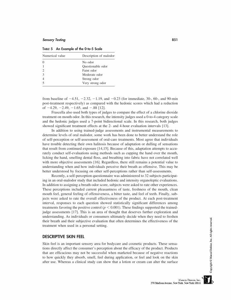
Sensory Testing 851
TABLE 5 An Example of the 0-to-5 Scale
Numerical value Description of malodor
0 No odor1 Questionable odor2 Faint odor3 Moderate odor4 Strong odor5 Very strong odor
from baseline of �4.51, �2.32, �1.19, and �0.23 (for immediate, 30-, 60-, and 90-minpost-treatment respectively) as compared with the hedonic scores which had a reductionof �4.29, �2.49, �1.65, and �.88 [12].
Frascella also used both types of judges to compare the effect of a chlorine dioxidetreatment on mouth odor. In this research, the intensity judges used a 0-to-4 category scaleand the hedonic judges used a 7-point bidirectional scale. In this research, both judgesshowed significant treatment effects at the 2- and 4-hour evaluation intervals [13].
In addition to using trained-judge assessments and instrumental measurements todetermine levels of oral malodor, some work has been done to better understand the roleof self-perception or self-assessment of oral-care treatments. Most agree that individualshave trouble detecting their own halitosis because of adaptation or dulling of sensationsthat result from continued exposure [14,15]. Because of this, adaptation attempts to accu-rately conduct self-evaluations using methods such as cupping the hand over the mouth,licking the hand, smelling dental floss, and breathing into fabric have not correlated wellwith more objective assessments [16]. Regardless, there still remains a potential value tounderstanding when and how individuals perceive their breath as offensive. This may bebetter understood by focusing on other self-perceptions rather than self-assessments.
Recently, a self-perception questionnaire was administered to 32 subjects participat-ing in an oral-malodor study that included hedonic and intensity organoleptic evaluations.In addition to assigning a breath-odor score, subjects were asked to rate other experiences.These perceptions included current pleasantness of taste, freshness of the mouth, cleanmouth feel, general feeling of offensiveness, a bitter taste, and feel of teeth. Finally, sub-jects were asked to rate the overall effectiveness of the product. At each post-treatmentinterval, responses to each question showed statistically significant differences amongtreatments favoring the positive control (p � 0.001). These findings supported the trained-judge assessments [17]. This is an area of thought that deserves further exploration andunderstanding. As individuals or consumers ultimately decide when they need to freshentheir breath and their subjective evaluation that often determines the effectiveness of thetreatment when used in a personal setting.
DESCRIPTIVE SKIN FEEL
Skin feel is an important sensory area for bodycare and cosmetic products. These sensa-tions directly affect the consumer’s perception about the efficacy of the product. Productsthat are efficacious may not be successful when marketed because of negative reactionsto how quickly they absorb, smell, feel during application, or feel and look on the skinafter use. Whereas a clinical study can show that a lotion or cream can alter the surface
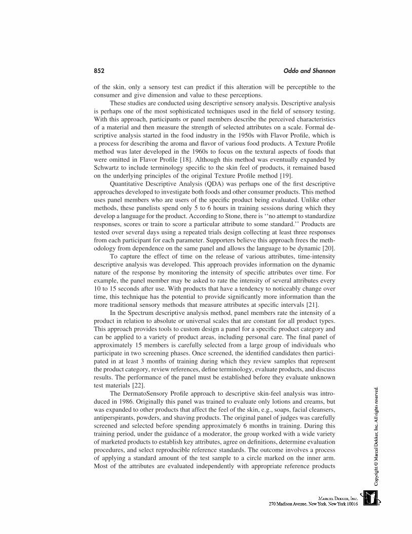
852 Oddo and Shannon
of the skin, only a sensory test can predict if this alteration will be perceptible to theconsumer and give dimension and value to these perceptions.
These studies are conducted using descriptive sensory analysis. Descriptive analysisis perhaps one of the most sophisticated techniques used in the field of sensory testing.With this approach, participants or panel members describe the perceived characteristicsof a material and then measure the strength of selected attributes on a scale. Formal de-scriptive analysis started in the food industry in the 1950s with Flavor Profile, which isa process for describing the aroma and flavor of various food products. A Texture Profilemethod was later developed in the 1960s to focus on the textural aspects of foods thatwere omitted in Flavor Profile [18]. Although this method was eventually expanded bySchwartz to include terminology specific to the skin feel of products, it remained basedon the underlying principles of the original Texture Profile method [19].
Quantitative Descriptive Analysis (QDA) was perhaps one of the first descriptiveapproaches developed to investigate both foods and other consumer products. This methoduses panel members who are users of the specific product being evaluated. Unlike othermethods, these panelists spend only 5 to 6 hours in training sessions during which theydevelop a language for the product. According to Stone, there is ‘‘no attempt to standardizeresponses, scores or train to score a particular attribute to some standard.’’ Products aretested over several days using a repeated trials design collecting at least three responsesfrom each participant for each parameter. Supporters believe this approach frees the meth-odology from dependence on the same panel and allows the language to be dynamic [20].
To capture the effect of time on the release of various attributes, time-intensitydescriptive analysis was developed. This approach provides information on the dynamicnature of the response by monitoring the intensity of specific attributes over time. Forexample, the panel member may be asked to rate the intensity of several attributes every10 to 15 seconds after use. With products that have a tendency to noticeably change overtime, this technique has the potential to provide significantly more information than themore traditional sensory methods that measure attributes at specific intervals [21].
In the Spectrum descriptive analysis method, panel members rate the intensity of aproduct in relation to absolute or universal scales that are constant for all product types.This approach provides tools to custom design a panel for a specific product category andcan be applied to a variety of product areas, including personal care. The final panel ofapproximately 15 members is carefully selected from a large group of individuals whoparticipate in two screening phases. Once screened, the identified candidates then partici-pated in at least 3 months of training during which they review samples that representthe product category, review references, define terminology, evaluate products, and discussresults. The performance of the panel must be established before they evaluate unknowntest materials [22].
The DermatoSensory Profile approach to descriptive skin-feel analysis was intro-duced in 1986. Originally this panel was trained to evaluate only lotions and creams, butwas expanded to other products that affect the feel of the skin, e.g., soaps, facial cleansers,antiperspirants, powders, and shaving products. The original panel of judges was carefullyscreened and selected before spending approximately 6 months in training. During thistraining period, under the guidance of a moderator, the group worked with a wide varietyof marketed products to establish key attributes, agree on definitions, determine evaluationprocedures, and select reproducible reference standards. The outcome involves a processof applying a standard amount of the test sample to a circle marked on the inner arm.Most of the attributes are evaluated independently with appropriate reference products
Sensory Testing 853
continually used to anchor the 0- to 10-point intensity scales [23]. Some of the lotion andcream attributes selected by the panel include the rate of absorption, shine, greasy oily,drag, stickiness, ease of spread, and residue at several intervals after application. Whenfive marketed products described as four oil-in-water (o/w) formulations and one water-in-oil (w/o) formulation were evaluated, the panel was capable of showing significantdifferences among all of the products for different attributes [24].
In 1992, the ASTM published the Standard Practice for Descriptive Analysis ofCreams and Lotions [25]. This practice identifies the elements of and the process fortraining a skin-feel panel. In addition to identifying the needed equipment, it presents aprocess for screening and selecting panel members. One section describes an evaluationprocedure that, in addition to explaining an application process, also discusses samplepreconditioning, conditioning aspects of the skin including skin temperature, and environ-mental conditions of the test area. Evaluation intervals, definitions, and suggested refer-ences are included for each listed attribute. The practice does state, however, that it shouldbe used by individuals who have become familiar with the process and have previousexperience with sensory testing.
Descriptive skin panels fill the gap between clinical and marketing data by providinginformation that can help predict or better understand consumer needs. It has been useddevelop a master profile of a product that is later used for quality-control purposes or toimprove the product. Panel information has also been frequently used to determine differ-ences in currently marketed products whereas the descriptive terms and results are oftenused to promote or market the product.
There were a number of product-performance trends in the skincare industry duringthe past decade that may have benefited from a descriptive profile, in either the product-development stage or in better understanding the competition. For example, hydratingagents were added to increase skin moisturization. Although the physiological improve-ment of these agents is established in clinical studies, descriptive sensory data is essentialto identifying potential consumer perceptibility. The move to silicone emulsion systemsto decrease the heavy, greasier feel created with oil systems was a natural application fordescriptive skin-feel data. Finally, industry responded to the increase in consumer aware-ness of the cumulative effect of sun exposure on the skin by adding sunscreens to manybodycare and cosmetic products. However, the addition of sunscreens often affects theskin-feel properties of the product. For example, they can increase the rate of absorption,add a greasy feeling, and create a heavier texture to the product. Descriptive skin-feelanalysis was and continues to be an appropriate tool to address and minimize the effectof these changes on consumer perception.
DESCRIPTIVE HAIRCARE
The competitive world of haircare products is very dependent on consumer perception.The success of a product often depends on whether the user perceives a positive changeor believes the claims being presented. Regardless of what can be shown clinically, it isultimately the consumer who decides if his/her hair is shinier, easier to comb, has morebody, or holds a curl longer. For these reasons, descriptive sensory analysis plays anessential role in the product-development stage. When appropriate sensory tools are used,these characteristics can be confidently assessed in a controlled environment. Formulachanges as well as new ingredients and ideas can be screened before substantiating prod-uct-performance claims with large-scale consumer studies.
854 Oddo and Shannon
Currently, the majority of descriptive sensory analysis with haircare products isbeing conducted internally. Most manufacturers use licensed cosmetologists, trained pan-els, or groups of semitrained consumers. Some companies will use each of these toolsdepending on the stage of development or the product type.
Cosmetologists are perhaps the most sophisticated tool used, and sometimes themost challenging to the sensory scientist. Regardless of experience, these individuals mustbe screened and carefully selected. Once selected they must be trained to follow estab-lished procedures such as those for washing the hair, combing the hair, and touching thehair. They are also trained in what characteristics or attributes to evaluate and when.Because these individuals often have many years of experience working in a salon environ-ment, they are often faced with the challenge of changing old habits. At the same time,the cosmetologist typically provides a certain level of knowledge or experience that evenwell-trained consumers don’t possess.
Panelists for a descriptive haircare panel are selected and trained using techniquessimilar to those identified for developing skin-feel panels. However, these panelists aretrained to evaluate hair rather than their own skin. Consequently, providing samples duringthe training process becomes perhaps the biggest challenge to developing the panel. Hairswatches are often used during the training process because using actual subjects canbecome costly and burdensome. They also provide a distinct advantage because the typeand condition of the hair can be carefully controlled. This introduces one of the primaryconsiderations of evaluating haircare products; subject selection. Because products willreact differently on different hair types, subjects must be carefully screened and selectedbased on the type of hair they have. Some of the things that must be considered are hairtexture, thickness, length, color, and amount of natural curl. The condition of the hair(e.g., dry or oily) and pretreatments, (e.g., permed or colored) must also be considered.A well-defined profile of the subject should be established before testing, and a methodfor selecting subjects using the trained panel, cosmetologist, or an independent personshould be built into the program.
Depending on local laws and regulations, unlicensed trained panelists may not beallowed to handle the subjects. Because they may be restricted from shampooing, theyare often only used to evaluate the feel or appearance of the hair. Some panels are trainedonly to evaluate hair swatches or a combination of both.
As mentioned earlier, because the majority of descriptive sensory analyses withhaircare products is conducted internally, very little has been published in this area. Toprovide the industry with information, ASTM committee E18.0 on sensory testing is inthe process of finalizing a standard practice for the descriptive analysis of shampoo perfor-mance [26]. This practice will present an overview of several options, some of whichwere previously described, that the sensory associate can follow to develop a hair descrip-tive program. Similar to the skin-feel standard practice, it will identify necessary equip-ment, a process for screening panel members and/or cosmetologist(s), as well as evaluationand application procedures. Although this document will focus on shampoo performance,it has the potential to provide an excellent panel foundation that can be expanded to theother haircare products.
ANTI-IRRITANT APPLICATIONS
Manufacturers fully understand the necessity of thorough safety testing before introducinga topically applied product. A battery of standard safety tests to determine the irritancy
Sensory Testing 855
potential of the product to contact sensitization and photosensitization are routinely con-ducted. However, often missing are tests to determine potential subjective discomfort tothe product, such as stinging, burning, and itching. In 1977, Frosch acknowledged thatproducts that meet standard safety parameters may still be rejected by the consumer ifdisagreeable subjective discomfort develops after application [27].
Early work in this area investigated subjective response to substances applied toskin that had been damaged by either blisters [28] or scotch tape–stripped skin [29]. How-ever, these were considered measurements of pain rather than measurements of more tran-sient subjective discomfort. In response to concerns that some substances, such as sun-screens, may cause delayed stinging, Frosch and Kligman developed a method foridentifying potential ‘‘stingers.’’ This method involved applying lactic acid to the nasolab-ial fold and cheek area of subjects brought to a profuse state of sweating. The intensityof stinging was then measured by the subject using a 4-point scale at 2.5, 5.0, and 8.0minutes after application. It was also established that a stinging response could be inducedin nonsweating subjects by increasing the concentration of lactic acid. An arbitrary methodfor classifying the irritancy potential of substances was also developed that identifies ifthe substance has a slight, moderate, or severe potential to cause stinging [27]. Althoughthis method was established over 20 years ago, modified versions of it remain the basisfor identifying subjects that are unusually sensitivity to stinging.
Grove improved the method by defining the demographic profile of the subjects andrecommending the exclusion of males and older individuals. He also established criterialimiting the frequency of use and determined that sensitive subjects often reported a historyof problems with soaps, cosmetics, and other personal-care products. Subjects who repeat-edly reported a stinging response to lactic acid applied under ambient conditions werealso tested for a burning and itching response. A method for evaluating burning sensationsusing a 20:80 mixture of chloroform:methanol pipetted into a greased aluminum cylindercovered and placed against the skin was used. To elicit itching, a 4% histamine base wasalso loaded into a grease-ringed cylinder and placed against the skin. Results found goodcorrelation between burning and stinging, but individual response variability was ratherhigh. A distinct correlation between itching and stinging was not observed [30].
A new interest in subjective sensory responses was renewed with the impact ofalpha-hydroxy acids (AHAs) in the marketplace. When applied to the skin, these acidsoften cause a burning, stinging, and/or itching response, often without a visible sign oftypical irritation. Draelos states that users have been conditioned to believe that stingingor burning sensations are an indication that the product is working, whereas ‘‘in fact, theyfeel pain because the acid has penetrated the dermis and is interacting with the dermalnerve endings [31].’’ Manufacturers have responded to these concerns by introducing asecond generation of AHAs that do not penetrate the skin to the same degree. Althoughthe FDA has found AHAs safe at low concentrations, the need to routinely include inde-pendent sensory assessments in the standard battery of safety studies is apparent.
Obviously missing from the available literature is a clear understanding of the sen-sory principles that were followed, which opens the door to certain questions that need tobe addressed. Were the testing environments controlled? How were the unknown materialspresented to the subjects? Were different scales explored? How was the scale that wasused presented to the subjects?
For example, it may be advantageous to screen individuals for current use of certainmedications that may affect their response, such as cortisones and other anti-inflammatorymedications. Subjects should be screened for obvious skin pathology or irritation as well
856 Oddo and Shannon
as a history of allergic reactions. When conducting the screening test or evaluating un-knowns, the subjects should be preconditioned for several days. During this period, theyshould be provided with instructions informing them of, e.g., which cleanser to use, whenmales can or can’t shave, what cosmetics are acceptable the day of the study, and whento cleanse the face. If the test is to be conducted at ambient conditions, the subjects shouldbe preconditioned in a quiet, climate-controlled room. If the subjects will be brought toa ‘‘profuse’’ state of sweating, this state should be carefully defined to ensure that all arebrought to the same level.
The actual screening probe should be administered in isolated areas to avoid subjectinteraction and influence. The subjects should not be told what the appropriate responseis. For that reason, it would be wise to either eliminate the word ‘‘stinging’’ from thescale or not let the subject see the scale and ask them to verbalize their response. The‘‘purest’’ way to approach it would be to administer the test and ask the subject to reportany sensation they experienced. To increase the sensitivity of the results and decreasevariability it may be worthwhile to explore a 0- to 7-point scale. Finally, the frequencyof subject use should be limited with a distinct rest period (�48 hours) between evalua-tions. With these considerations incorporated with the methods previously developed, itwould be possible to quantitatively assess the intensity of facial stinging. Once a reliablemethod is established, a database of responses to known ingredients can be collected thatwill allow unknown substances to be tested for subjective discomfort with confidence.
REFERENCES
1. Meilgaared M, Civille G, Carr B. Sensory Evaluation Techniques. Vol. 1. Boca Raton: CRCPress, Inc., 1987:1.
2. American Society for Testing and Materials. ASTM Designation E1207-87, ASTM AnnualBook of Standards, Vol. 15.07. Standard Practice for the Sensory Evaluation of Axillary Deod-orancy. 1987.
3. Labows J, Leyden J, Preti G. Axillary Odor Determination, Formation, and Control. CosmeticScience and Technology. Vol. 20, 71
4. Wild J, Bowman J, Oddo L. Clinical Evaluation of Antiperspirants and Deodorants. CosmeticScience and Technology. Vol. 20, 1999:318.
5. Chambers E, Wolf M. Sensory Testing Methods. ASTM Manual: MNL 26. General Require-ments for Sensory Testing. 1996:3–5.
6. Meilgaared M, Civille G, Carr B. Sensory Evaluation Techniques. Vol. 2. Boca Raton: CRCPress, Inc., 1987:3.
7. Niles H, Gaffer A. Relationship between sensory and instrumental evaluations of mouth odor.J Soc Cosmet Chem 1993; 44:101–107.
8. Rosenberg M, Kulkarni G, Bosy A, McCulloch C. Reproducibility and sensitivity of oralmalodor measurements with a portable sulphide monitor. J Dental Res 1991; 70(11):1436–1440.
9. Tonzetich J, Ng SK. Reduction of malodor by oral cleansing procedures. Oral Surg 1976; 42:172–181.
10. Peryam D, Pilgrim F. Food Technol 1957; 11:9–14.11. Schmidt N, Tarbet W. The effect of oral rinses on organoleptic mouth odor rating and levels
of volatile sulfur compounds. Oral Surg 1978; 45:876–882.12. Borden L, Oddo L, Bowman J. Correlation between Hedonic and Intensity Measurements for
the Evaluation of Oral Malodor. Presented at 4th International Conference on Breath Odor,UCLA, Los Angeles, CA, Aug. 1999.
Sensory Testing 857
13. Frascella J, Gilbert R, Femandex P, Gorden J. Effect on oral malodor of chlorine dioxideassessed by a hedonic panel. TKL Research Abstract.
14. Rosenberg M. Clinical assessment of bad breath: current concepts. JADA 1996; 127:475–481.
15. Tonzetich J. Production and origin of oral malodor: a review of mechanisms and methods ofanalysis. J Peridontal 1977; 48:13–20.
16. Rosenberg M, Kozlovsky A, Gelemter L, Cheriak O, Gabbay J, Baht, R, Eli I. Self-estimationof oral malodor. J Dental Res, 1995; pp. 1577–1582.
17. Oddo L, Borden L, Bowman J. Comparison of trained judge measurements to self-perceptionof oral malodor. Abstract, 4th, International Conference on Breath Odor, UCLA, Aug. 1999.
18. Chambers E, Wolf M. Sensory Testing Methods, ASTM Manual: MNL, General Requirementsfor Sensory Testing. 1996; 26:58–63.
19. Schwartz N. Method to skin care products. J Texture Studies, 1975; 6:33.20. Stone H, Side J. Sensory evaluation for skin care products. Cosmet Toilet.21. Lee W. Single-point versus time-intensity sensory measurements. J Sensory Studies 1989; 4:
19–30.22. Meilgaard C. Descriptive Analysis Techniques, Designing a Descriptive Procedure: The Spec-
trum Method. Boca Raton CRC Press, 1991: 196–199.23. Oddo L, Aust L. Applications of sensory science within the personal care business, J Sensory
Studies 1989; 3:187–191.24. Aust L, Oddo L, Wild J, Mills O. The descriptive analysis of skin care products by a trained
panel of Judges. J Soc Cosmet Chem 1987; 38:443–449.25. Standard Practice for Descriptive Analysis of Creams and Lotions, ASTM E1490, Annual
Book of ASTM Standards, Vol. 15.07.26. Standard practice for descriptive analysis of shampoo performance. ASTM Committee E18.0,
publication pending.27. Frosch P, Kligman A. A method for appraising the stinging capacity of topically applied sub-
stances. J Soc Cosmet Chem 1977: 197–209.28. Armstrong D, Dry M, Keele C, Markham J. Methods for studying chemical excitants of cutane-
ous pain in man. J Physiol 1951; 115:59.29. Laden K, Studies on irritancy and stinging potential. J Soc Cosmet Chem 1973; 24:385–383.30. Grove G, Soschin D, Kligman A. Adverse Subjective Reactions to Topical Agents, Cutaneous
Toxicity. New York: Raven Press 1984, pp. 203–211.31. Brewster B. MD’s address sensory irritation from AHA’s. Cosmet Toilet 1998; 113:9–10.

859
Index
Abrasion, 413Absorption, 853Accidental exposure, 124–125Acclimatization, 815Acetylated lanolin, 840Achromobacter, 782Acid, 70, 102, 112, 352Acidic milieu, 717Acidic rinse, 418Acne, 13, 50, 72, 196, 206, 234, 287,
292, 300, 352, 725, 837, 843Acnegenic potential, 840, 843Acnegenic testing, 843Acne-pore strips, 843Acrylic acid/alkyl acrylate copolymer,
776Active agent (see Ingredient, active)Active preservation, 248–249Active substance (see Ingredient, active)Activity spectrum, 248Acyl
amino acid, 418, 437–438amino alkane sulfonate, 436ethylenediamine, 441–442glutamate, 413, 418, 437peptide, 437sarcoside, 437–438
Adhesion, 68Adhesive layer, 239–240
dry, 240pressure-sensitive, 239
Adipose tissues, 533Adsorption, 240
isotherm, 425Adulteration, source of, 741Adverse effect, 47–50, 69, 77, 80, 89,
114, 254, 459Advertising, 798
self-regulation, 753Advisory opinion, 737Aeromonas, 782Aerosol, 397, 779
can, 226emulsion, 226foam (see Foam, aerosol)
Aesthetics, 394, 770Aflatoxin, 372Afterfeel, 400Aftershave, 261Agar, 385
Baird Parker, 784, 792Cetrimide, 784, 791Mac Conkey, 784, 791Sabouraud Chloramphenicol, 784, 790tryptic soy, 784, 789
Age, 69, 715, 716, 808Aging of the skin
accelerated, 770histological changes, 723morphological changes, 723physiological changes, 723–724process, 463
860 Index
AHA (see Alpha hydroxy acid)Air conditioning, 782Air quality, 782Alachlor, 60Alcohol, 150, 227, 246, 247, 261
alkoxylated, 443–444ethoxy sulfate, 433
Alginate, 385Alkali, 70, 102, 112Alkane sulfonate, 435Alkanolamide, 418, 445, 585
ethoxylated, 445Alkoxylated polysiloxane, 449Alkyl
amine-oxide, 418aryl sulfonate, 435betaine, 440
carbohydrate ester, 446, 448chain, 336–337dimethicone, 391, 392dimethyl betaine, 440ether phosphate, 437ether sulfate, 417, 433–434, 585glucoside, 418phosphate, 437polyglucoside, 444–445, 725sulfate, 417, 433, 585sulfonate, 434–435sulfosuccinate, 417
Alkylamido betaine, 440Alkylamine, 438
ethoxylated, 440–441Alkylimidazoline, 438–439Allergen, 48, 77–84, 89–93, 829, 833Allergenicity, 77–84Allergic reaction (see also Contact der-
matitis, allergic), 459Allergy, 77–84, 718
contact (see Contact dermatitis, al-lergic)
diagnosing of, 83reaction, 259
Aloe barbadensis, 373Alopecia, 40, 41, 43, 304, 578Alphabisabolol, 266, 277–284, 287
healing power of, 281isomers of, 279natural, 279
[Alphabisabolol]protective properties of, 281substantivity, 283synthetic, 279–284
Alpha hydroxy acid, 14–15, 261, 287,295, 311–314, 352, 362, 364,546, 726, 855
Alpha-olefin sulfonate, 417, 434Alpha-sulfo methyl ester, 436Alpha-tocopherol, 63Alternative methods, 95–103, 119–137Aluminum
chlorhydrate ACH, 690–692chloride, 292–293salt, 246, 690–691zirconium chlorhydrate, 691
Aluminum-magnesium hydroxide, 384Alveolar bone, 623Amide ether sulfate, 434Amido guanidine, 419Amine oxide, 448Amino acid, 347, 355Aminobenzoic acid, 457Ammonia, 356Ammonium hydroxyde, 831Amodimethicone, 341–342, 391, 395Amphiphile, 161Anagen (see Hair)Analytical methods/techniques, 370, 372Androsterone, 847Anesthesia, 215Animal, 123–125, 475–476
testing, 731welfare, 124
Anisotropy, 161, 831Anosmia, 704Antagonism, 264Anthralin, 71Antiaging, 12, 206Antibacterial, 7, 10, 13, 354
agent, 245–250for plaque, 637product, 245–247triclosan, 639–641
Anti-calculus dentifrices, 632–635Anticaries, 10,Anticellulite treatments, 536–539Antidandruff, 10, 579
Index 861
Antifungal, 78, 354Antigen, 48Anti-inflammatory, 855
constituent, 373effect, 273, 277, 304, 373, 463
Anti-irritant, 253–298, 394, 854, 855antisensory, 285–297ingredients, 264, 277, 403, 408–411strategy of making cosmetics, 263–265studies, 265–267for surfactant-based products, 271–
274systems, 271
Antimicrobialactivity, 195agent, 78, 162, 163, 248–250, 259,
490deodorant, 704, 705efficacy, 247–248in vitro tests, 247–248products, 245–247
Antioxidant, 82, 163, 204, 259, 292,299–306, 314, 463, 468, 546
activity, 473defense, 469lipophilic, 299network, 304–306system, 464, 465thiol, 302–303
Antiperspirant, 10, 11, 89–90, 246,292–293, 396, 397, 689–691,770, 846
aerosols, 692deodorants, 704polymers, 691roll-ons, 692sticks, 692–693
Antiphlogistic, 277, 304Antiseborrehic, 579
preparation, 435Antisensory anti-irritant (see Anti-irri-
tant, antisensory)Antiseptic, 248
effect, 247hand wash, 246
Anti-whitening, 397Anti-wrinkle, 12, 543–549, 482, 802APG (see Alkyl polyglucoside)
Apigenin, 304Apigenin-7-glucoside, 373Apocrine sweat, 690Apoptosis, 68Appearance, 165, 770, 773Aqueous humor, 122–123Arachidonic acid cascade, 277–278Arbutin, 475Artificial tanning, 551Ascorbic acid, 292, 301–302, 468, 570Ascorbyl palmitate, 469Aspergillus niger, 786Assay
cumulative irritation, 110–112enzyme-release, 99genotoxicity, 570immersion, 112, 115interleukin-1MTT viability, 99mutagenic, 602protective barrier, 116repeat application, 112tape stripping, 181topical application, 13421-day cumulative irritation, 113–114
Atopic, 115, 355Atopy, 557ATR-FTIR, 272Authorities, 798Avenanthamide, 372Axilla, 689, 696, 703, 846Axillae bacterial flora, 246Axillary odor malodor, 845, 849Azulene, 266, 371
Baby lotion, 719–720Baby shampoo, 442, 719Baby skin
care of, 719–720cleansing of, 719development of, 715–716physiology of, 716–717problems with, 717–718protection of, 720
Bacteria, 245anaerobic, 837bad breath, 849counts, 784

862 Index
[Bacteria]gram-negative, 786gram-positive, 786growth inhibition, 846growth, 249sulfur-producing, 849
Bacterial metabolism, 788Bactericidal activity, 195, 245, 438, 786Bacteriostatic activity, 245, 786Bag-in-can, 230Balance, 64Bar feel, 496Barrier, 22, 53, 68, 71, 97, 127–128,
211, 256, 272, 273, 393cream, 110, 116, 557–566, 829, 833function, 256, 264, 314, 356, 363,
466, 479,716, 717, 816protective, 557repair, 281restorative, 557
Barrier cream efficacy, 829, 830, 831Bath additive, 719Bead, 177, 777Beef tallow, 486Beeswax, 403Benzalkonium chloride, 71, 439Benzophenone, 82, 457Benzoyl peroxide, 195–196, 300Benzyl cinnamate, 452Benzyl salicylate, 452Beta hydroxy acids, 546Beta-carotene, 470Betaine, 413, 725Beta-ionone, 465Binding, 58, 272Binding site, 272, 427Bioavailability, 63Biodegradability, 338Biodegradable carrier, 177Bioengineering methods, 72, 116, 255–
257, 816Bioerosion, 177Biometric method, 802–803Bioré pore strip, 842, 843Biotin, 470Birbeck granules, 22Bird’s nest hair, 578Bisabolol (see also Alphabisabolol), 373
Black heads, 837, 843Blemish, 840, 843Blister, 855Blood flow, 115, 255, 301Body wash, 504Botanical handbook, 373–374Bowman’s layer, 122Breakout, 840, 843Broth, 784, 790, 791Brush, 12Buffers, 789–791Build-up, 337, 339Bullae, 67, 71Burning, 50, 71, 72, 273, 285, 295, 807,
809, 811, 855Butane, 221Butylene glycol, 347–348Butylmethoxydibenzoylmethane,
458
Calcitonin, 285Calcium, 274Calculus, 625, 632CAM vascular assay (CAMVA), 134Camphor derivative, 457Can, 226Candida albicans, 718, 783, 784, 786Capacitance, 115, 116, 256Capryloyl salicylic acid, 292Capsaicin, 809Capsule, 776Carbohydrate, 407Carbon black, 378Carbon chain length, 274Carboxylate, 432Carboxymethylcellulose, 380, 381, 386Carcinogen, 60Carcinogenesis, 304Care, 146Caries, 625Carrageenan, 384, 385, 386, 413Carrier, 146–147, 157, 171, 179, 185Castor oil derivative, 445Catagen (see Hair)Catalase, 299Cationic guar gum, 410Cationic macromolecule, 399Cationic moieties, 409
Index 863
Cavities, 627, 629CBBB (Council of Better Business Bu-
reau), 753Cells
basal, 19, 576clear, 26cornified, 21culture, 97–103, 466cuticle, 332dark, 26diffusion, 55fat, 716granular, 21immune, 285inflammatory, 295Langerhans, 22–23, 32, 103, 121,
301, 724mast, 285membrane lipids of, 464Merkel, 23myoepithelial, 26nail plate, 31neurosensory, 123proliferation of, 466spinous, 20squamous, 121squamous carcinoma, 451
Cellulite, 531–541Cellulose derivative, 411Cellulose ether derivative, 380, 381,
384, 385, 386Cementum, 621Cer[OSE], 362–365Ceramides, 201, 264, 314, 361–366
commercially available, 363–366from epidermis, 362–365fractions, 362–365future of, 366from stratum corneum, 364–365
Ceresin, 403CERP (Cosmetic Establishment Registra-
tion Program), 751Cetrimonium chloride, 335–337Chain length, 390, 403Challenge test, 785–788Challenge, 798Chamazulene, 277Chamomile, 266, 277, 371, 373
Chapping, 71Charge density, 333, 407Chelating agent, 249Chelator, 474Chemical absorber, 453Chemicals, 98–102Chemotaxin, 68Children, cosmetics not allowed for, 730Chitosan, 407Chitosan-PCA, 407Chloasma, 571Chlorexhidine, 247Chlorine dioxide, 851Chlorphenesin, 82Cholesterol, 183, 361–362
ester, 361sulfate, 361, 365
Chorio-allantoic membrane (CAM), 129Choroid, 123Chromameter, 255Chromametry, 116, 280Ciliary body, 123Cinnamate, 455–456Cinnamic, 62, 80, 90, 91CIP (cleaning in place), 782CIR (Cosmetic Ingredient Review), 752Citric acid, 311Claim, 2, 7–15, 63, 84, 233
absolute, 801antiwrinkle, 544categories of 799–801comparative, 801cultural, 800efficacy, 800juxtaposition, 800medically-oriented, 799noncomparative, 800oil-free type, 392on ingredients, 801proof of, 797–784regional requirements, 797–799related to endorsement, 800safety-related, 800, 802sensory, 803subjective, 800substantiation of,substantiation, 373support of, 801–804

864 Index
Classification, 95, 124, 127, 761–762of colorants, 327Hamilton’s, 41ingredient, 1, 13physical-chemical, 149
Clay, 377, 380, 384, 386, 414Cleansing, 146, 394, 719Clearance premarket, 762Clinical
assessment, 254, 815picture, 90study, 802trial, 690
Cloud point, 380, 420, 433, 444, 448Coal tar, 840Coal-tar hair-dye exemption, 741Coalescence, 775Cocamidopropylbetaine, 82, 83Coco amphocarboxy glycinate, 442Cocoa butter, 403Coefficient of developed skin surface,
831Co-emulsifier, 153Co-enzyme A, 467Cohesion, 257Cold, 809COLIPA, 136, 730, 731Collagen, 355, 407, 466Collagen synthesis, 292, 302, 312, 469Colloidal dispersion, 223Colloidal solution, 150Color, 259, 769, 773
additive, 745–746law, 317
Colorant, 317–329, 489application area, 318–324approved for cosmetics, 325–327classification, 327cosmetics products and, 327–329natural, 325semi-permanent, 599substantive, 329
Colorimetry, 255Coloring agent, 83, 152, 317Combar, 491–493Combo bar, 740Comedogenesis, 837Comedogenic potential, 839, 840, 843
Comedone, 837, 843Compatibility, 769Complaint, 296Composition, 150Concentration, 63, 162, 283, 372Conditioner, 287, 586, 612Conditioning agent, 331, 335–341, 413,
437Conditioning effect, 202, 395, 409Conductance, 256Conjunctiva, 121–122, 124Connective tissue, 533Consistency, 97–98, 769Consumer testing, 527Contact dermatitis, 47–49, 717, 829
airborne, 79, 80allergic, 48–49, 67, 77–84, 89–93
contributing factors to, 78–80diagnosis of, 91–92
irritant, 313, 519, 557 (see also Irrita-tion)
acneiform, 72acute, 48, 70–71, 107cumulative, 48, 71–72, 107, 114delayed acute, 48, 71, 107inhibitor of, 297nonerythematous, 72pustular, 72subclinical, 115traumatic, 72traumiterative, 72
photoallergic, 49photoirritant (see Photoirritation)protein, 355
Contact thermography, 533Container, 225–226Contamination, 787–788
source of, 781–782Cooling, 779Copolymer, 192, 199, 409, 412Cornea, 120–122, 124, 351
isolated, 127opacity, 127permeability, 127
Corneocyte, 256, 312, 807cohesion, 311
Corneosurfametry, 811Cornification, 467
Index 865
Cornified envelope, 365Corrosion, 225–226, 438Corrositex, 97–98Corrosive material, 67, 70Corrosivity, 95–98Corticosteroid, 314, 718Cortisone, 855Coryneforme bacteria, 703, 704Cosmeceutic(al), 14–15, 63–64, 84,
299, 480, 511, 763Cosmetic intolerance syndrome, 50Cosmetics, 1–3, 480, 739, 761–763
adulterated, 741categories of 762–763control of, 740–752definition of, 5–15, 729delivery, 215–216deregulation, 764directive, 249, 798formulation, 389future of, 764–766history of, 5–6,label warning, 749–750labeling, 729, 746–747misbranded, 742percutaneous absorption of, 60–63perfumed, 90–92
professional, 747registration program, 751–752safety, 743–744therapeutic, 15,vehicles (see Vehicles)
Cosmetic ingredient,label declaration, 747–749prohibited, 744restricted, 744
Cosmetic patch, 233–243application of, 234–235components of, 238–240development of, 235–236differences with classical forms, 235functional, 237future of, 242–243history of, 233production, 240–241regulation, 241–242types, 236–238
Cosolvent, 151
Co-surfactant, 156–157, 175Counterirritant, 272CPIS (Cosmetic Product Ingredient State-
ments), 751CPSC (Consumer Product Safety Com-
mission), 737Cracking, 71Cream, 151–155
protective, 718baby, 719–720
Creaminess, 400Creaming, 775Criminal prosecution, 743Critical micelle concentration (CMC),
134, 419Cross-sensitivity, 79Croton oil, 808Crow’s feet, 829
furrows, 830Culture media, 789–791Cumene sulfonate, 435Cutaneous aging, 829Cyanoacrylate, 840, 842, 843Cyclodextrin, 777Cyclomethicone, 341–342, 390–397, 405Cyclo-oxygenase, 304Cyclosiloxane (see Cyclomethicone)Cytokine, 68, 101, 137–138Cytotoxicity, 128
Dandruff, 352Dansyl chloride, 291Deciduous (milk) teeth, 619Decontamination, 58–60Decoration, 146Decorative cosmetic, 325, 391Defense system, 306Deformation, 377Degradation, 443Dehydroascorbic acid, 302, 305Delivery, 777
active, 147, 160–161, 171–186, 211–216, 396
system, 171, 177, 233, 235Dental plaque, 627, 629Dental pulp, 621Dentifrice, 627–631
bad breath, 629
866 Index
Dentin, 621Dentinal hypersensitivity, 635Dentinal tubuli, 636Deodorancy, 802Deodorant, 10, 89–90, 246, 396, 703–
713, 846aerosols, 709delivery systems, 708odor-neutralizing, 704odor-quenching, 704odor-masking, 704pump sprays, 710sticks, 708
Depigmentation, 467, 476Depilatory, 293Deposition, 336Dermal drug transport, 183Dermaspectrometer, 255Dermatitis
atopic, 69, 718connubial, 79, 80contact (see Contact dermatitis)diaper, 717–718ectopic, 79, 80irritant, 107neuro (see Atopic)photocontact, 80
Dermatoheliosis, 724Dermis, 715
de-epidermized, 101Descemet’s layer, 122Descriptive analysis, 852, 853, 854Desmosome, 312, 350
hemi-, 19, 20Desquamation, 60, 256, 311, 347, 467,
723Detention, 742Detergent, 259, 584DGIII, 730, 731Dialkyl ammonium salt, 419Diaper, 717–718
candidiasis, 718Dibenzoyl methane, 82, 459Dicetyldimonium chloride, 335–337Diffusion, 177, 178Digital camera, 831Diglyceride, 403Dihydrosphingosine, 362
Dihydroxyacetone (DHA), 551–556,746
Dimethicone, 339–342, 390, 391, 392,395, 397, 401, 405, 449
copolyol, 341–342, 395, 396, 449gum, 391
Dimethiconol, 391, 392, 395, 397Dimethyl diallylammonium chloride,
411Dimethylsulfoxide (DMSO), 808, 809Dioxane, 741Dipalmitoyl-ethyl hydroxyethylmonium
methosulfate, 338Discomfort, 855Disease, 50Disinfectant, 248, 439, 718Disperse system, 158Dispersion, 150
hydrolipid, 156phase, 173
Dissolution, 159Distribution, 166Dithranol, 71Divalent cation, 274DNA photodamage, 301DOPA, 567Dorsal root ganglia, 285Dossier, 798Drag, 853Droplet size, 175Drug, 7–15, 63, 84, 234, 453, 467, 704,
740, 761delivery, 177–179, 184, 211–216release, 177, 179, 181transport, 184
Dryness, 71, 72, 114, 723D-squame, 256, 519Dye, 830, 831Dynamic light scattering, 166Dynamic surface tension, 422–423
Ectoderm, 715ECVAM, 102, 136Edema, 67, 71, 532, 533EEC Cosmetic Directive, 729–734
annexes, 730implementation of, 731–734
18-methyleicosanoic acid, 365
Index 867
Einstein relation, 377Elastic
component, 385modulus, 385, 386stress, 378, 379
Elasticity, 312, 377, 379–381, 386, 466Elastin, 407Elderly skin, 723–726
skin care for, 724–726skin cleansing for, 725–726
Electrical conductivity, 773Electrical methods, 816Electrolyte, 162, 382, 414, 437Electron transport chain, 305Electrorepulsion, 211Electrostatic attachment, 339Electrostatic interaction, 425, 427Ellagic acid, 473–477Ellagitannin, 473Emolliency, 391Emollient, 152, 261, 264, 399, 400–406,
413, 448, 718, 726dry, 405lipophilic, 401–403occlusive, 405
Emulgator, 259Emulsification, 174, 401Emulsifier, 79, 83, 151–155, 343Emulsion, 151–155
micro- (see Microemulsion)multiple, 154–155, 173–175, 178,
181, 512nano- (see Nanoemulsion)oil-in-water, 152–153oil-in-water-in-oil, 174submicron (see Nanoemulsion)water-in-oil, 153–155, 432water-in-oil-in–water, 174
Emulsion micellar, 774Emulsion multiple, 774Enamel, 619, 629, 630Encapsulation, 171–186, 191–200, 204
future of, 185–186use of, 179–185
Enterobacterium, 782, 783, 784Environmental pollutant, 299Environmental safety, 338Enzyme, 55, 299
EPA (Environmental ProtectionAgency), 737
EpiDerm, 99Epidermis, 19–26, 715
biopsy, 840, 842cells, 20–23cyst, 361derivatives, 23–26proliferation, 312renewal, 466stratified culture, 99thickening, 466thickness, 312, 352turnover, 723
Epidermolysis, 311Epiocular culture, 128Episkin, 98Epithelial crest, 715Epithelium, 19, 121Equivalent
epidermal, 97skin, 97–98, 101
Erosion, 71Erythema, 61, 67, 71, 72, 255, 293, 313,
724, 809, 830Erythemameter, 255Escherichia coli, 784Esculoside, 266Ester carboxylate, 432Esterase inhibitors, 704Esterified quaternary, 441Esterquat (see Esterified quaternary)Esters, 445–448Estradiol, 56Estrogen, 547, 725Ethanol, 229Ether carboxylate, 432–433Ethers, 443–445Ethical frames, 815Ethoxylation level, 274, 403, 423, 434,
440, 448Ethylenediaminetetra-acetic acid
(EDTA), 249Eucalyptol, 249Eumelanin, 576European Directive, 2, 241, 452European Norm, 247Euxyl K400, 81
868 Index
Evaporimeter, 256Excipient, 83, 154Excoriation, 67Excretion, 53Exfoliant, 14Experimental error, 57Expert evaluation, 802Expiration date, 730Exposure time, 113Exsiccation eczematid, 72Extraction method, 370Eye, 719
anatomy, 120–123injury measurement, 126irritation testing (see test)isolated, 127
Eyelid, 90Eyeliners, 668–669Eyeshadows, 661–658
Facial cleanser, 504, 505Facial cleansing powder, 499Facial lines, 830, 831Facial masque, 236False negative, 79Farnesol, 82Fat, 401, 486
lobules, 532micro/macronodules, 531, 533septa, 532subcutaneous, 361, 532, 537–539
Fatty acid, 201, 362, 409, 486alkanolamide, 413ester, 362, 401ethoxylated, 446isethionate, 436saturated, 486unsaturated, 486
Fatty alcohol, 404, 442Fatty carbon chain, 409Fatty ester sulfonate, 436FD&C Act, 6–7, 740–746FDA (Food and Drug Administration),
5–15, 246, 248, 352, 452, 455,737–756, 798
Feel (see also Skin feel, Hair feel,Mouth feel, Foam feel), 392,396
Feeling, 50Fibers
beta, 808C-, 809, 810delta, 808nerve, 285, 808sensory, 809
Fibrillar network, 533Fibroblast, 312Fibrosclerosis, 531Film
backing, 238hydrolipidic, 717
Filmogen compound, 264, 351, 355,392
Firmness, 312Fissuring, 72, 114Flaking, 114Flaky scales, 312Flavobacterium, 782Flavonoid, 277, 299, 303–305Flexibility, 389, 394Flocculation, 775Flow characteristics, 162Fluid
cutting, 112Newtonian, 377–378non-Newtonian, 378–379preliposomal, 206receptor, 55
Fluoride, 14, 247, 630, 631Fluorosurfactant, 449Foam (see also Mousse; Surfactant,
effect on foam), 342, 394,499
aerosol, 221assessment methods, 422–425booster, 221, 396density of, 226effect of additives on, 401–
406feel, 400height, 423moisturizing, 223shaving, 225, 226stability of, 226, 422, 423–425structure, 221, 227temperature of, 228

Index 869
Foamability, 423–425Follicle
rabbit’s ear, 839sebaceous, 50
Follicularduct, 840impaction, 839, 840, 841irritation, 839, 843plug, 844
Food additive, 473Food grade, 448Formaldehyde, 81, 82, 245Formula
simplicity, 78Formulation, 163, 769Foundations, 662–664Fragrance, 80–81, 89–93, 259, 489,
769, 770, 771masking, 846mix, 81, 89–93
Freckles, 567Free radical, 463, 464Free samples, 747Freeze/thaw, 770, 772Frequency, 78, 80Friction, 717FTC (Federal Trade Commission), 737,
798Fungal counts, 784Fungi, 245, 785Fungicidal action, 786Fungicide, 372Fungistatic action, 786
Galactomannane polysaccharide, 410Galenical form, 782Gamma linoleic acid, 264Gas, 221Gas chromatography, 372, 849Gel, 155, 384, 419
hydrogel, 155hydrophobic, 155liquid crystalline, 386strong, 385weak, 385
Gelatine, 385Gelling agent, 155, 157, 380, 384–
386
Geneexpression, 306transcription, 306
Genistein, 304Geraniol, 249Germ, 437, 717Germicidal, 439Gibbs elasticity, 422Gingiva, 622Gingivitis, 625Gland
apocrine, 36, 689conjunctival, 122eccrine, 24, 689holocrine, 24salivary, 624sebaceous, 24, 36, 300, 583, 689sweat, 24–26, 716
Gluconic acid, 311Glutathione (GSH), 299, 302–305Glutathione peroxidase, 299, 465Gluten, 407Glyceraldehyde, 552Glyceride, ethoxylated, 403Glycerin, 249, 348–351, 406, 486, 489Glycerol ester, 46Glycerylmonothioglycolate, 82Glycol ester, 446Glycolic acid, 288–292, 295, 311, 313Glycolic extract, 266Glycolysis, 311Glycosaminoglycan, 351Glycyrrhizic acid, 287Good Manufacturing Practice, 751GRAS (generally regarded as safe), 691Green tea, 287Gray levels, 831Guar gum, 410Guidance document, 737Guidelines for biophysical methods, 481Guinea pig skin, 829, 830, 831Gums, 622
Hair, 35–45, 467, 468African, 606–607anagen, 37–44, 581appearance, 575body, 853

870 Index
[Hair]care, 853catagen, 37, 40caucasian and Asian, 608–615conditioner, 331–343, 355, 394–395,
438, 439hydrophobicity, 336–337lipophilic, 338needs and preferences, 343properties, 336–337
bleaching, 577, 601blushers, 658catagen, 581charge, 333, 338, 339chemically treated, 334–335color, 854coloring, 329, 396, 598–601, 854combing, 331, 332, 335, 338, 341,
394, 396, 853conditioners, 588cortex, 23, 331, 426, 575, 576, 582,
606cosmetics, 36, 581–603count, 42–43curls, 853cuticle, 23, 331–335, 396, 426, 575,
582, 606damage, 331–335, 396, 596density, 40, 41, 43diameter, 41, 44, 353dry, 347drying aid, 396dye, 82feel, 331, 335, 340, 342, 395fiber, 331–335, 426fixative, 396flyaway, 396follicle, 23–24, 36–37,follicle proliferation, 304friction, 331, 335function, 35–36graying, 575growth evaluation methods, 40–45isoelectric point, 333length, 41, 854lipids, 365loss, 41, 42matrix, 576
[Hair]medulla, 575, 576oxidation color, 329oxidative colorants, 600oxidative damage, 334oxidative dyeing, 577oxidative dyes, 599permanent colorants, 600permanent dyeing, 578permanent waving products, 592perming, 577, 854photography, 41, 43, 44protection, 332rinses, 588root sheath, 23shaft, 365, 576shine, 396, 802, 853static electricity, 576straighteners, 607straightening, 597–598structure, 37–39styling gels, 589styling mousses, 589styling products, 584styling sprays, 589substantivity to, 335, 339, 341, 426swatches, 854telogen, 579, 582temporary colorants, 599terminal, 37texture, 854thickness, 854trichoptilosis, 578vellus, 37virgin, 333–334weight, 42whitening products, 567–573
Haircare, 853, 854Haircare product, 394–396Hairdressers, 558Hairdressings, 589Hairless mouse skin, 829Halitosis, 849, 851Hand eczema, 69Hardening, 71Hazard, 95, 110Heat, 437Heat separation, 361
Index 871
Heating, 779Heavy metal, 372Heliodermatitis, 724Hemostase, 533Hen’s egg-CAM (HET-CAM), 129, 136Henle’s layer, 24, 37Henna plant, 555Herb, 370Heterocyclic ammonium compound, 439High performance liquid chromatogra-
phy, 372Histamine, 293, 724, 810, 855HLB, 443, 446, 839Hormone, 10, 14,Horny layer, 202, 413, 467Human health, 730Humectant, 14, 152, 221, 249, 244, 261,
343, 347–357, 406–407, 468,724, 725
Huxley’s layer, 24, 37Hyaluronic acid, 351, 726Hydrating agents, 347–357, 853Hydration, 146, 155, 167, 256, 717, 815Hydration tests, 818Hydrocarbon chlorinated, 839Hydrogenated tallow octyl dimonium
chloride, 338Hydrolytic enzyme, 350Hydrophilic hydrophobic balance, 420,
513Hydrophilicity, 333, 336–337Hydrophobic coating, 336, 340Hydrophobic interaction, 407, 409, 427Hydrophobic surface, 365Hydrophobicity, 272, 333, 393, 407Hydroquinone, 53–55, 62, 63, 476, 567Hydrotrope, 435Hydroxyapatite, 633Hydroxyethyl cellulose, 343, 380Hydroxypropylcellulose, 380, 3826-hydroxysphingosine, 362Hygiene cosmetic, 271Hygroscopic substance, 347, 352, 356,
406Hyper and hypothermic zones, 534Hyperhydrosis, 215Hyperirritability, 73Hyperkeratosis, 67, 71, 554
Hyperkeratotic plug, 839, 842Hyperkeratotic skin disorder, 356Hypermelanosis, 568Hyperpigmentation, 469, 567Hyperreactivity, 807Hypersensitive, 84, 724Hypertrophy adipose tissues, 533Hypoallergenic, 77–84, 263Hypodermis interface, 535Hypodermis, 534Hyponychium, 29, 32
Ichtyosiform, 72Ichtyosis, 352, 354, 356IFRA (International Fragrance Associa-
tion), 752IL (see Interleukin)Image analysis, 257, 830, 831Imino diacid, 442Immune
response, 23responsiveness, 724
Immunological, 47–49Impactions, 842Impedance, 255, 256Impurity, 78, 372In vitro data, 274INCI (International Nomenclature for
Cosmetic Ingredients), 730Inflammation, 47, 49, 50, 67, 68, 101,
112, 128, 215, 253, 277–284,292–297
response, 833Inflammatory disorder, 468Infrared absorption, 816Infrared thermal imaging, 535Infusion, 371Ingredient
active, 13–14, 63, 82–83, 146, 152,159, 180, 191, 195, 202, 234,371, 372, 770
extraction of, 371–272release of, 178, 195
antimicrobial, 248antistatic, 438, 439, 442category-specific, 82–83classification (see Classification)coloring, 730

872 Index
[Ingredient]concentration, 78, 80cosmetic, 234exfoliating, 413film-forming, 405–411forbidden, 730fragrance (see Fragrance)gelling, 384–386labeling, 766lipophilic, 153natural, 82negative list, 765new, 765occlusive, 401–402positive list, 766purity, 78skin-whitening, 473–477superfatting, 725, 726suspending, 381–382thixotropic, 321–384
Inhalation risk, 222Injunction, 742Inoculum, 784Inoculum, preparation of, 787Inorganic pigment, 458, 649–653Instability identification, 773Instrumental measurement, 255–257,
803Interaction, 272Intercorneocyte cohesion, 311Interface
liquid-liquid, 223–225liquid-gas, 223–225
Interfacial gelation, 382Interfacial tension, 175, 224, 390Interleukin (IL), 68, 128, 295Internet, 374Interviewing process, 846Intolerance, 70Invasive, 40
non-, 41–45semi-, 40–41
Involucrin, 22Iontophoresis, 211–216
effect on stratum corneum, 214effect on the skin, 214safety issues, 214–215topical delivery by, 215–216
Iontophoretic transport,applications of, 215–216mechanism of, 212–213parameters affecting, 213–214
Iris, 123, 124Irritancy (see Irritation)Irritant, 67–73, 253, 271, 808, 829, 833
acidic, 287–293basic, 293–294cosmetic, 259–261effect, 183neutral, 294occupational, 259ocular, 294–295primary, 70reaction, 71, 253weak, 112
Irritation, (see also Contact dermatitis, ir-ritant)
chronic, 253cumulative, 253, 291depressor, 273epidemiology, 69–70evaluation of, 254–257factors influencing, 257–259
age, 258climate, 258ethnic groups, 258exposure, 258external, 257gender, 258intrinsic, 257regional, 258
invisible, 809in vitro tests, 95–103in vivo tests, 107–116, 123–126,
261–263mechanism, 67–68mild, 99model, 271objective, 253ocular, 123–137potential, 107predisposing factors, 68–69primary, 109, 840sensory, 48, 50, 72–73, 285–297
clinical evaluation, 286on arms, 288–293

Index 873
[Irritation]on face, 287–288, 292on legs, 293inhibitors of, 297scale, 286–287to axilla, 292–293
severe, 291skin, 47–48, 67–73, 156, 183, 195,
196, 253–298, 854, 855SLS-induced, 280, 314sodium hydroxide-induced, 280by surfactants, 431–449
subclinical, 253suberythematous, 50subjective, 48, 273, 459, 807symptoms of, 254–257
Isethionate, 413Isoelectric point, 407, 410Iso-eugenol, 80, 90Isopropyl myristate, 61Isotretinoin, 829, 832Isovaleric acid, 847Itch-inducing chemicals, 294Itching, 50, 71, 72, 273, 285, 294, 295,
807, 811, 855
Japan, 761–767Jojoba oil, 403
Keratin, 20, 29, 31, 33, 407, 466, 837Keratinization, 466, 582Keratinocyte, 20–22, 67, 101, 295, 312,
466Keratinocyte detachment, 311Keratohyalin, 21Keratolytic, 314, 354Keratoplastic agent, 356Keratosis, 352, 451, 553Kinase, 306Kitchen workers, 558Klotz equation, 426Kojic acid, 475, 570Krafft point, 419Krebs cycle, 311
Labeling, 84, 95, 798Lacrymal drainage system, 122Lactate, 347
Lactic acid, 73, 116, 254, 287–288,295, 311, 351–352, 809, 810,855
Lamellar bodies, 21Lamellar granules, 837Lamina fuschia, 122Laminometer, 423Langmuir equation, 426Language, 730Lanham Act, 753Lanolin, 403, 404
derivative, 401, 404, 445oil, 401
Laser diffraction, 166Laser Doppler, 534, 831
flowmetry, 116, 255imaging, 535
Laser high energy pulsed CO2, 832Laser pulsed CO2, 832Lather, 488, 583Lauryl sulfate, 433Laurylmethicone copolyol, 391Law, 1, 737
case, 12,Lawsone, 555Laxity, 723Leakage, 776Leave in products, 588Lecithin (see Phosphatidylcholine)Legislation, 95, 729–734, 761–767Lentigines, 567Leukocyte, 68Leukotriene, 277, 810License, 762, 799Lichenification, 67, 71Licorice, 266Lidocaine, 58, 287Light exposure, 772Light-conditioning formulation, 337Linear alkylbenzene sulfonate,435Linoleic acid, 201, 205, 363Lipase, 717Lipids, 24, 68, 151, 153, 156, 261, 264,
350, 361–366, 726covalently bound, 362, 365from hair, 365from nail, 365from oral stratum corneum, 365

874 Index
[Lipids]hydrophilic, 403, 404lamellae, 363peroxidation, 301, 305, 464, 468
Lipodystrophy, 531–541Lipogel, 155Lipoic acid, 305Lipoid particle, 1815-lipooxygenase, 304Liposome, 176, 186, 201–208, 726, 776
application of, 206–208compatibility of, 204dispersion, 208future of, 208hydrolysis of, 206
Liposphere, 180Lipstick, 499, 670–672Liquid crystal, 161, 350, 419–420, 534Liquid crystalline phase, 419–420Liquid film movement, 423Loading, 194, 199Lobuli, 716Location, 80Long-lasting benefit, 392Long-term tests, 818Loss modulus, 385Lotion, 151–155Lubricant, 405Lubricity, 340, 392, 409Lunula, 29Lymph vessels, 533
Maceration, 371Macroemulsion, 774Magnesium, 274
ascorbyl palmitate, 469ascorbyl-2-phosphate, 302lauryl sulfate, 274sulfate, 274
Maillard reaction, 552Makeup remover, 80Malic acid, 311Manicuring instrument, 12Manufacture, 730Manufacturing date, 730Manufacturing process, 782Marangoni effect, 422Mascara, 664–667
Mass spectrometry, 372Massage, 536Matrix, 171, 173, 191Mechanical profilometry, 832Mechanical properties, 576Mediator, 67, 101, 128, 285Medicinal plant handbooks, 373Melanin, 22, 469, 474, 567, 575, 576, 582Melanocyte, 32, 103, 121, 475, 567, 576MelanoDerm, 103Melanogenesis, 473Melanoidins, 552Melanosomes, 567, 576Melasma, 567Membrane
basement, 19, 24, 121–122biological, 201, 305, 464glassy, 24polymeric, 173
Menthyl anthranilate, 457–458Mercaptans, 593Mesoderm, 715Mesophase, 161Metabolism, 56Metastable state, 151, 156, 204Methacrylate, 82Methyl(chloro)isothiazolinone, 78, 81Methylcellulose, 380Methyldibromoglutaronitrile, 81Methylhydroxypropylcellulose, 380, 382Mexameter, 255Micelle, 151, 156, 272, 419Microbial
contamination, 775growth, 162health hazard, 781limits, 782–783
Microbiological information, 731Microbiological strains (maintenance
of), 787Microcapsule, 171, 191, 777–778Microcomedone, 50, 837Microemulsion, 156, 175, 178, 182, 420,
774Microorganism, 162, 717Microparticles, 171–172, 177, 179Micropigment, 459Microscopy, 166

Index 875
Microsphere, 171, 177, 179, 191, 777biodegradabable, 191porous, 191–200
application of, 195–199loading, 194–195preparation of, 192–194visualization of, 194
Microsponge, 177Microtopography features, 830, 832Mildness, 273, 400Milk crust, 718Milk protein, 407Minimal erythemal dose (MED), 300,
301, 451, 808Minimal inhibitory concentration (MIC),
246Mitotic activity, 466Mixing, 779Models, 95–103, 17–116
arm immersion, 114cornea, 127–128eye, 127mouse ear, 112–113
Moisture, 343, 347, 406accumulation test, 818content, 503sensing device, 788
Moisturization, 14, 154, 392, 400, 463,725
Moisturizer, 152, 366, 401, 468, 546,720
Moisturizing properties, 405, 407, 409,820
Monocyte, 277Monoglyceride, 403Monograph, 13,Monomer, 272, 419Monophasic system, 159Mould, 784Mousses, 221–232
aerosol, 25–226appearance of, 221application characteristics, 222attributes, 221–223disinfectant, 225emulsion, 226–227future of, 213–232hair-setting, 228–229
[Mousses]hairstyling, 222, 225metastable, 223moisturizing, 221physical quality of, 223post foaming, 230–231quick-break, 227–228shave, 221, 222technology, 223types of, 226–231unstable, 221
Mouth rinse, 246–247, 641Mouthfeel, 383Mouthwashes, 641Multi-approach, 803–804Multilayer cell culture, 127–128Multiphasic system, 159Multiple application, 818Muscle, arrector, 36–37Mutagenicity, 570
N-acetyl-cysteine, 302N-acyl amidopropyl betaine, 419NAD (National Advertising Division),
753, 798Nail, 29–34
aesthetics, 34anatomy, 29apparatus, 32artificial, 686bed, 29, 32cuticle removers, 687cuticle, 29elasticity, 31film former, 685film modifier, 685fold, 29, 32histology, 29–33lacquers, 672lipids, 365matrix, 32permeability, 33
physicochemistry, 33physiology, 33–34plate, 29, 31, 33surgery, 34top coatings, 685whitener, 687
876 Index
N-alkyl amino acid, 442Nanocapsule, 172, 178, 180Nanoemulsion, 156–157, 175, 178,
183–184, 204, 208Nanoparticle, 156–157, 172–173, 177, 180Nanosphere, 172, 177NARB (National Advertising Review
Board), 753Nasolabial fold, 809, 855Natural acid mantle, 717Natural extracts
categorizing, 370constituents to avoid in, 372definition, 369–370production of, 370–372quality of, 370–372standardization of, 372–373
Natural Moisturizing Factor (see NMF)Necrosis, 67, 71Neoplasm, 724Neurokinin-A, 285Neurological sensation, 285Neuron, 295Neuronal depolarization, 295Neurosensory perception, 73, 480Neurotransmitter, 73, 295Neutralizing solution, 595, 783, 788,
789–791Neutrophilic infiltrate, 839Newborn, 716, 717Newtonian system, 162Niacinamide, 470Nickel contact allergy, 830Niosome, 176, 776Niotenside, 201Nitrosamine, 741NMF, 14, 256, 261, 347, 353, 355, 406,
501N-nitroso compound, 354Non comedogenic, 837, 843Nonacnegenic, 837, 843Noncooling, 397Noniceptor (type-C), 285, 294, 295Noninvasive techniques, 211, 255–257,
815Non-Newtonian system, 162, 411Nuclear magnetic resonance, 816Nylon particle, 778
Oak moss, 90Occlusion, 61, 79, 113, 262, 717Occlusive effect, 273Occlusive material, 399Occupational, 69Ochronosis, 569Octocrylene, 457Octyl methoxycinnamate, 455, 459Ocular instillation, 295Odland bodies, 21Oenothein B, 373Oil, 61, 156, 157, 173–176, 401, 403, 486
baby, 719bath, 399, 448, 726coconut, 487essential, 90, 91, 246, 247, 249, 266,
277, 371ethoxylated, 445impregnated towelet, 719mineral, 281, 401, 403, 719, 840palm, 487palm kernel, 487vegetable, 401
Oleochemistry, 432Oleogel, 155Omega-hydroxyacid, 362Omega-hydroxyceramid, 362Onychodermal band, 29Onychomycosis, 356Oozing, 67Opacifier, 443, 586Opaque formulation, 458Open epicutaneous application, 262Oral flora, 247Oral mucosa, 625, 627Oral pellicle, 634Organic pigments, 649- 654Organosilicone, 449Osmotic gradient, 185OTC, 10, 13–15, 452, 690Overconditioning, 339Oxidant, 299Oxidation, 163, 443Oxidative damage, 465Oxidative stress, 299–302, 306, 465Oxybenzone, 457Oxygen radical, 277Ozone, 222, 300, 306

Index 877
PABA, 452, 455, 459ester, 459
Packaging, 225, 249, 241Palate, 623Palm stearin, 487Panthenol, 352–353, 467–468Panthotenic acid, 353, 467Papilla, 19Papule, 50Paraben, 82Paraffin, 469, 716Paraffin sulfonate, 434Particle size, 165, 397Partition coefficient, 160, 181Partitioning, 53, 58, 177, 178Pathological changes of eye injury, 137PCA, 347, 353–354, 407Pearlescent agent, 446Pearlescent pigments, 655Pectin, 385Peel solution, 291PEG-2 cocomonium chloride, 337PEG-15 stearmonium chloride, 337Pencils, 669–670Penetration, 67, 158, 160, 154, 156,
178, 273enhancing, 79, 202percutaneous, 182
Peptide, 272–273Perception, 274Percutaneous absorption, 53–64, 716
methods, 55–57steps, 53–55variations, 57–58
Perfume, 79, 89–93, 152, 704bottom note, 704encapsulated, 182middle note, 704top note, 704
Periderm, 715Periodontal ligament, 621Permanent teeth, 619Permeability, 392, 716, 718, 720
barrier, 366skin, 211, 273, 356
Permeation, 156, 202, 235Peroxide, 299, 302Peroxyl radical, 469
Perspiration, 690, 716Persulfate, 82Pesticide, 372Petrolatum, 116, 719pH, 102, 134, 151, 166, 246, 249, 287,
312, 352, 437, 440, 441, 717Pharmaceutical patch, 233–234Pharmaceutical technology, 233Pharmacopoeial handbook, 373Phase inversion, 161, 175, 420Phase separation, 161Phenyl salicylate, 452Phenyl trimethicone, 391, 396Phenylbenzimadazole sulfonic acid, 457Pheomelanin, 576Phosphate ester, 437Phosphatidylcholine, 183, 201–203, 205,
405bilayer of, 204hydrogenated, 203, 205unsaturated, 203, 205
Phospholipid, 176, 201, 361–362, 405Photoaging, 287, 352, 451, 467, 543,
724Photoallergy, 720Photocontact dermatitis, 460Photographic pictures, 534, 536Photoirritation, 49Photolysis, 458Photoprotection, 303Photoreactivity, 458Photoreceptor, 123Photosensitivity, 313, 458Photosensitizer, 83Phototoxicity, 49Physical blocker, 453–454Physiotherapeutic treatments, 536Phytosphingosine, 362Pigmentation, 724
disorder, 80Plant
collecting, 370cultivation, 370drying, 370–371exudate, 377protein, 407
Plasmatic exsudate, 532Poloxamer plasticization, 312
878 Index
Plasticizer, 396, 403, 686Poison Information Center, 730
addresses of, 731–734Polarity, 150, 159, 444Polyacrylate, 162, 386Polyacrylic acid, 382, 384, 385Polyamide, 343Polycarboxylate, 442Polydimethylsiloxane, (see Dimethicone)Polyethylene glycol ether, 443Polyethylene glycol, 406, 516Polyglycerol, 205Polymer, 155, 156, 171–172, 173, 178,
192, 272–273, 378, 380, 385,389, 407–412
cationic, 338–339, 343, 399charge of, 272EO/PO block, 444molecular weight of, 407natural, 377, 407–411quaternary, 395–396quaternized, 272, 411–412size of, 272synthetic, 411–412
Polymeric emulsifiers, 512Polymerization, 194Polymerizing coating, 686Polyphenol, 303Polypropylene glycol ether, 443Polyquat (see Polyquaternium)Polyquaternium, 338–339, 342, 411Polysaccharide, 351, 385Polyvinyl pyrrolidone (see PVP)Pore cleaner, 238Porogen, 194Porosity, 192Positive list, 731Post-shampoo formulation, 335Pouching material, 240Pouch-on-valve, 230Powders, 658–660Prediction, 95–116, 130Preservation, 162, 248–250Preservative, 78–82, 152, 163, 249–250,
259, 342, 490, 517, 730Pre-shampoo formulation, 335Presurgical preparation, 246Process, 249
Procyanidin, 304Product
body-cleansing, 399causative, 90composition, 78dishwashing, 247labeling, 1, 11,leave-on, 79occlusive, 563occupational, 558oral care, 246–247perfumed, 90–92pharmaceutical, 78quality, 163rinse-off, 79, 335, 399scented, 92skin-cleansing, 399strontium-containing, 296suncare, 465sun-protection, 157
Profilometry, 545Proliferation, 68Pro-oxidant effect, 302Propane, 221Propellant, 222, 224, 229Propyl gallate, 82Propylene glycol, 406Prostaglandin, 180, 277Protease, 717Protection, 154, 167, 300, 350, 393, 464
active (of), 177from cold, 720from dehydration, 392from micro-organisms, 717from percutaneous absorption, 716–717skin, 203, 205sun, 206, 264, 720UVB, 103by vehicle, 146against water loss, 716
Protein, 272–273, 355, 407derivative, 409–410hydrolyzate, 409–410, 437hydrolyzed, 355quaternized, 409–410
Proteinases, 544Protocol, 130Pseudoplastic rheology, 380
Index 879
Pseudoplasticity, 383Psoriasis, 313Psyche, 482Pupil, 123Purity, 370Pustule, 50, 71Pustulogenicity, 50PVP, 409, 412Pyrollidone carboxylic acid, (see PCA)Pyruvic acid, 311
QSAR (quantitative structure activity re-lationship), 130–132
Quasi-drug, 84, 473, 799future of, 766
Quat (see Quaternary ammonium com-pound)
Quaternary ammonium compound, 335–338, 419, 439
alkyl chain, 336, 337compatibility with anionics, 337ethoxylated, 337
Quercetin, 304Questionnaire, 257
Ra parameter, 832Rabbit’s ear, 839, 840Radicals, 302, 304
ascorbyl, 305, 306chromanoxyl, 305free, 306hydroxyl, 302scavenging, 303tocopheryl, 306
Radiolabeled, 56Raw material, 2
from biological origin, 782microbiological control of, 781–785microbiological quality of, 781–782
Razor blade, 12Reactive oxygen species, 299Recall, 741, 743Red vetinary petrolatum, 452Redness (see Erythema)Refattener, 273, 400–406Refattening agent (see Refattener)Reflectance, 255Regression method, 815
Regulation, 1, 95, 241, 249, 737–756,761–766
of color additive, 648–649Regulatory, 10–15, 84, 95, 124, 452–453Release liner, 239Relevance, 92, 101, 124Relief, 256Removal of nail coatings, 687Repetitive irritation test, 831Reservoir, 79, 171, 178, 195Resistance, 249Retina, 123Retinal, 467Retinoic acid, 196–199, 313, 467, 546, 569Retinoid, 311, 313, 465, 467Retinol, 467, 546Rheological additive, 377–386Rheology, 150, 162, 165, 377–386, 394Rhus, 831, 832RIFM (Research Institute for Fragrance
Materials), 752Rinsing, 400Risk, 63, 80Robinson-Patman Act, 753Rosacea, 568Ross-Miles test, 423Rough sensation, 273Roughness, 400, 725Rz parameter, 832
Safety, 14–15, 47–50, 119, 731, 770assessment program, 130hydrating substances (of), 347–356
Salicylate, 457Saliva, 624, 627Sallowness, 725Salt, 151, 225Sanguinarine extract, 247Saponification, 491Saponin, 448Saprophyte, 717Sarcosinate, 413, 437Saturation solubility, 160Scale
categories, 848hedonic, 849, 850line, 848numerical, 848

880 Index
Scaliness, 724Scaling, 67, 71, 72, 816Scaling techniques, 846, 851Scalp
biopsy, 40proxigraphy, 44
Scavenger, 468Sclera, 122Screening, 96Scrub agent, 413Scurvy, 301Sebaceous follicle, 837Sebaceous gland, 837Sebaceous lipids, 361Seborrheic dermatitis, 353Sebum, 583, 724, 837
follicles, 581, 582plug, 238
Sedimentation, 157, 775Seizure, 742Self-assessment, 803, 810Self-emulsifying, 515Self-perception, 257, 851Self-preserving formula, 248–249, 342Self-regulation, 752–753Sensation, 285Sensitive-skin products, 845Sensitive skin, 50, 73, 79, 257, 263–
264, 285, 296, 468, 499, 807–812, 845
clinical parameters for, 808epidemiological studies for, 807–808tests for, 807–812
Sensitive teeth, 635Sensitivity, 97–98Sensitization, 77–84, 156, 353, 720, 855Sensory, 23, 50, 155, 167, 395
analysis, 852, 853assessment, 496, 845, 855, 856feeling, 499panel, 527profile, 852properties, 154, 399tests, 845, 856triggers, 399–414
Serine biosynthesis, 311Sesquiterpene, 277Sex, 68, 808
Shampoo, 578, 583–588, 607–610baby, 586, 719,daily use, 611dandruff, 586greasy hair, 611special, 6112-in-1, 332, 342, 612
Shampooing, 854Shaving, 579Shear
rate, 165, 378, 379, 383stress, 162, 165, 383thinning, 379, 386
Shelf stability, 380Shielding product, 558Shine, 853Short-term tests, 816Shower product, 726Silflo adhesive, 830Silica, 378, 382, 384, 386Silicon rubber, 830Silkiness, 395Silicone, 154, 155, 157, 222, 339–342,
389–397, 409, 413conditioning properties of, 340–
341dioxide, 384organic group of, 389blend, 391derivative, 405–406
Siloxane backbone, 389Silymarin, 304Single application, 816Site, 79, 149Size of particulate, 150Skin, 19–26, 467
after feeling, 499aging, 723–724atopic, 79, 499baby, (see Baby skin)barrier, 563, 717, 808biopsy, 831cadaver, 830cancer, 451cleansing liquids, 499–510cleansing, 58–60color, 479, 480compatibility, 271

Index 881
[Skin]compromised, 114, 115condition, 79conditioners, 490conditioning, 399, 400, 409, 410, 413conductance, 522corrosivity, 95–98damaged, 79desquamation index, 520dry, 79, 152, 347, 352, 356, 724, 802,
815dryness, 481elasticity, 481elderly, 723–726equivalent, 97–98, 101excised, 361exfoliation, 553extensibility, 352fair, 69feel spectrum descriptive analysis,
527feel, 154, 167, 257, 386, 391, 399–
414, 444, 468, 845, 851, 853feeling, 151flexibility, 347frame, 815full-thickness, 97human, 830irritants, 557–566lightening, 469lipids, 501, 502models, 97–103moisturization, 853orange-peel, 531pH, 482, 501, 503photodamaged, 467phototypes, 544, 554pig, 361–365, 829pigmentation, 476protectant, 393, 411, 452regeneration, 206repair, 563replicas, 536, 545roughness, 815scaling, 479, 480, 481sensitive (see Sensitive skin)sensitization, 720shedding, 579
[Skin]squames, 815susceptibility, 499texture, 312type, 808tolerability, 720turnover, 291whitening, 473–477, 802
Skin2 system, 97–98SkinEthic culture, 128Skin feel agent, 273–274, 399–414Skin-feel panel, 853Skin surface
surface topography, 534, 535impression, 830rating, 829replica, 830roughness, 820scaling, 820water loss, 820
Slaughterhouse workers, 558Slimming/anticellulite ingredients, 537Slipperiness, 400Slough/mush, 495SLS, 808, 840Smell, 303Smoothing effect, 407Smoothness, 257, 273, 400Soap, 114, 404, 432, 584, 585–497,
739–740additives, 488alkaline, 261bar, 245, 485–497, 493, 499, 501,
506cracking, 495crystalline phases, 488liquid, 246wear rate, 495
Sodium dodecyl sulfate, 425Sodium hydroxide, 110, 116, 280Sodium lactate, 14, 352Sodium lauryl sulfate, 68, 110, 113,
115, 116, 265, 271, 280, 356,433, 831
Softening effect, 202Softness, 257, 395Solar elastosis, 451Solid partition, 849

882 Index
Solubility, 150–151, 303Solution, 150–151, 158Solvent, 79, 159, 261, 686
extraction, 371type cleansers, 507–509
Sorbitan ester, 446, 447Sorbitol, 249, 355, 406
ester, 447Sorption-desorption test, 818Specific anosmia, 847Specificity, 97–98Spectroscopy, 116SPF, 13, 392, 451–460, 554Sphingolipid, 176Sphingosine, 362Sphingosome, 176Spider veins, 554Split end, 395Spore, 785Spray, 397
drying, 372Spread, ease of, 853Spreadability, 162, 167, 222, 395Spreading, 153, 390, 392, 400Squamometry, 115, 255, 256Squeaky feel, 400Stability, 162
control, 781–792testing, 769–780
definition of, 769–770design for, 761–763of emulsion, 774–775formula-related reasons for, 774–775information provided by, 770–771of molecular carrier, 777non–formula-related reasons for,
779–780of particulate systems, 777–779raw material substitution, 778situations requiring, 773–774storage for, 772of vesicular system, 776–777
Stabilizer, 377–386Standardization, 372, 815Staphylococcus, 782
aureus, 783, 784, 786epidermidis, 703
State of matter, 150
Static control, 396Status cosmeticus, 50, 72Steam distillation, 371–372Stearalkonium chloride, 335–337, 396Stearamidopropyl dimethylamine, 338Stearate derivative, 447Stick, 157, 394Stickiness, 400Sticky tape, 256Stinger, 73, 254, 855Stinging, 50, 72–73, 254, 273, 285, 295,
807, 809, 811, 855Storage temperature, 775Stratum
corneum, 22, 53–56, 68, 72, 97, 146,160, 184, 300, 311, 347, 350,353, 807, 811, 842
lipids from, 361–365, 502oral, 365
granulosum, 21lucidum, 21spinosum, 21, 22
Streptococcus, 782Stripping, 55, 56, 236, 350, 855Stroma, 122Strontium
chloride, 287–295mechanism of action, 295–296nitrate, 287–295pretreatment, 287–288safety, 295salt, 285–297
Suberythrogenic, 543–544Subjective, 72, 257Substance P, 285, 294, 810Substantivity, 160, 272, 392, 394, 397,
399–414Sucrose ester, 448Sugar ester, 448, 777Sulfate, 433–434Sulfide monitor, 849Sulfite, 595Sulfonate, 434–436Sulfosuccinamate, 436Sulfosuccinate, 435–436, 725Sun
damage, 723, 725exposure, 724
Index 883
Sun protection factor (see SPF)Sunblock, 452Sunburn, 465, 473, 476Sunflower oil, 840Sunless/self-tanners, 551Sunscreen, 13, 82, 160, 451, 452, 453,
554, 720, 853Suntanning, 10,Superfatting agent, 404Superficial epidermis, 479Superoxide, 299, 468Superoxide dismutase, 299, 465Surface
activity, 390elasticity, 422microscopy, 479property, 389tension, 340, 390, 396topography, 816
Surfactant, 151, 175, 223–224, 229,399, 417–428, 499–500, 584,719, 808, 811
adsorptionhair (on), 426–428solid surface (on), 425–426
aggregation, 425, 426amphoteric, 272, 412, 441–442, 501,
585anionic, 271, 417–418, 431–438, 501,
584cationic, 335–338, 419, 438–441classification of, 431–449combination of, 271–272compatibility with, 411containing-formula, 100effect on foam of, 431–449foaming properties of, 42–424irritancy, 100mildness characteristics of, 431–
449mixture, 272–273nature of, 407nonhydrocarbon specialty, 449nonionic, 176, 272, 413, 442–448,
501, 585phase behavior of, 420–421
polyethoxylated, 585salt of, 274
[Surfactant]secondary, 431skin feel of, 412–413solubility of, 419solution properties of, 417–421synthetic, 417, 493–494
Surgical hand scrub, 246Suspending agent, 381–382Suspension, 157Sweat, 26
apocrine glands, 689apocrine secretion, 703eccrine glands, 689eccrine secretion, 704gland duct, 690production, 846reduction, 690
Sweating, 724Syndet bar, 436, 491–493, 740Systemic, 53, 62–63, 350, 716
Tactile evaluation, 815Tannin, 304, 373Tartaric acid, 311Tattoo, 83Taurate, 413, 436Tauride, 436t-butyl hydroquinone, 82Tea tree oil, 229–231, 249Telangiectasia, 568, 724Telogen, 37–44Temperature, 115, 161, 716TER (see Transcutaneous electrical resis-
tance)Teratogenicity, 313Test
animal, 107, 109–113, 119–126cell culture, 800consumer, 803corrosivity, 95–98cumulative irritation, 113–114Draize, 96, 109–110Draize eye irritation, 123–124ethical, 103exaggerated use, 262–263eye irritation, 119–137flex wash, 263fluorescein leakage, 128
884 Index
[Test]forearm immersion, 263forearm wash, 2634-hour patch, 113hand immersion, 263home-use, 263hydration, 818immersion, 115, 801in vitro, 95–103, 126–137, 247–248,
801–802choice of, 135
in vivo, 107–116, 123–126, 800,801
in-use, 263low volume eye, 124MIC, 247microbiological, 781–792neutral red release, 128neutral red uptake, 128, 136occlusive patch, 262open, 49organism, 247patch, (see also Cosmetic patch), 47,
62, 69, 83, 91–92, 115, 808,810
photopatch, 83prick, 724protein denaturation, 274pull, 43quality control, 166red blood cell lysis, 128repeat insult patch, 720repeat irritation, 110, 116repeat patch, 112ROAT, 83, 92scarification, 114semiopen, 83sensory, 800, 803single-application patch, 113skin compatibility, 96skin irritation, 98–103, 261–263soap chamber, 114–115stinging, 809–810stripping, 2563-dimensional tissues, 127–128time-kill, 248wash, 115–116, 811zone inhibition, 247–248
Test article (see also Test material)compatibility of, 133dilution of, 133
Test material, 129–130, 133Tetra-alkyl (-aryl) ammonium salt, 439Tetraphtalydine dicamphor sulfonic acid,
458Tewameter, 256, 280TEWL (see Transepidermal water loss)Thermal sensation, 808–809Thermodynamic activity, 160Thermodynamic parameter, 427Thermographic pattern, 534Thermoregulation, 24, 716Thickener, 343, 377–381, 414, 443, 444,
514, 586Thickness of fatty layer, 535Thin-layer chromatography, 364, 372Thin-layer distillation, 372Thioglycolate, 293Thioglycolic acid, 593–595Thiol, 299, 302–303Thixotropic agent, 154, 382–384Thixotropy, 162, 377, 386Threshold, 809Thymidine, 723Tightness, 72, 273, 274, 807, 811Tinea versicolor, 354Titanium dioxide, 378, 452, 458, 459TNF-alpha, 68, 295Tocopherol, 300, 301, 464
sorbate ester of, 301Tocopheryl radical, 464Tocotrienol, 300, 301Toilet paper, 80Toiletries, 271, 389, 390Toluene, 110, 116
sulfonate, 435Toner, 287Tongue, 623Toothpaste, 10, 246–247, 383, 627–631Topical application, 301Topical delivery, 211–216Topical pharmaceuticals, 480Topographic record, 830Toxic compound, 372Toxicity, 60–63, 95, 130, 459Toxicological profile, 731
Index 885
Trade correspondence, 11–12,Transcutaneous electrical resistance
(TER), 96–97Transdermal delivery, 211–216Transdermal system, 242Transepidermal water loss (TEWL), 68,
116–5, 116, 146, 201, 203, 255,256, 273, 280–283, 350, 356,401, 481, 546, 563, 715, 716,808, 810, 816, 820
Transfersome, 176, 185Transglutaminase, 22Trends, 800Tretinoin, 71, 180, 546, 725Trichogram, 40
photo-, 43unit area, 41
Triclocarban (TCC), 245Triclosan, 246, 247, 706Triglyceride, 401, 403, 485, 486Trimethylsiloxysilicate, 391Trimethylsilylamodimethicone, 391,
395–396Trisodium ascorbyl palmitate, 4692-hexyldecanoxyoctane, 612-in-1 shampoo (see Shampoo)2-in-1 emulsion, 399Tyrosinase, 22, 473, 474, 567
inhibitor, 473, 476
Ubiquinol, 305, 465Ubiquinone, 465Ulceration, 67Ultrasonic echography, 534Ultrasonic imaging, 535Ultrasound, 116Ultraviolet (ir)radiation (see UVR)Underarm microflora, 703Underarm odor, 703–713Urea, 204, 314, 347, 356, 831Uric acid, 299Urinous odor, 847Urinous smell, 847Ursolic acid, 266Urticaria, 49, 80, 355, 724, 810Use tests, 843USTR (Office of the U.S. Trade Repre-
sentative), 755
UV damage, 334, 464UV filter, 259, 451–460, 554, 720, 730
mechanism of action, 453–455nomenclature, 455regulatory, 452–453toxicity, 459–460
UV light, 463, 466, 475UVA, 451, 457–459, 545UVA I, 451, 458
blocker, 452UVA II, 451, 457, 458UVB, 301, 451, 455–457, 469, 545
absorber, 455, 457Uvea, 123UV-induced erythema, 372UVR, 299, 300, 306, 333, 451–460, 543UVR-induced inflammation, 304
Validation, 97–98, 129, 784, 786of biophysical methods, 481
Valve, 222, 397Van der Waals forces, 336Vasodilatation, 255VCRP (Voluntary Cosmetic Registration
Program), 739, 751Vector
design aspect, 171–177properties of, 177
Vehicle, 58, 63, 145–167, 171, 178,195, 264, 512
appearance of, 147application of, 147biological characterization, 167characterization of, 165–167chemical characterization, 166classification of, 147–150composition of, 158–165definition of, 150–157description of, 150–157effect of, 145, 158–165function of, 145–147functional design of, 158–168interactions, 160metamorphosis of, 161physical characterization, 165–166preparation of, 165selection of, 157–161target profile of, 158

886 Index
Velocimetry, 810Venous network, 533Vesicle, 67, 71, 72, 176, 179, 184–185,
204Viability, 128Virucidal action, 786Virus, 785Viscoelastic property, 256, 820Viscosity, 150, 153, 157, 161, 162, 165,
343, 377–386, 406, 422, 431,446, 719, 771, 773
agent, 152Viscous compound, 385Visual evaluation, 815Vitamin A, 180, 465–467
acetate, 467ester, 467palmitate, 466, 467
Vitamin B5, 353, 467–468Vitamin B6, 470Vitamin C, 292, 299, 301–302, 304,
305, 465, 468–470Vitamin D, 470Vitamin E, 63, 180, 299–301, 304, 305,
463–465, 469acetate, 63, 301, 464, 465delivery of, 300
Vitamin K, 470Vitamin P, 303Vitamins, 463–470Vitamins A, C, D, and E, 546Volatile compound, 371Volatile odor molecules, 703Volatile Organic Compound (VOC),
391, 699
Warning letter, 742Warning system, 285Water, 151–155, 347–357
activity, 163, 249availability, 788–789
[Water]binding, 820content, 406extraction, 371feel, 413hardness, 414, 496holding capacity, 352, 354, 356, 366in oil, 853in silicone emulsions, 516loss, 716microbiological control, 782, 784–
785quality, 225repellant products, 558resistant, 451retention, 820
Wax, 157, 394, 401, 403Wheal, 809, 810Wheat, 355Whitehead, 837Whitening, 459Whitening toothpastes, 627Willow herb, 373Winter, 69, 257Wool, 426Wound healing, 304, 463, 468, 469Wrinkles, 312, 463, 480, 543
coarse, 832Wrinkling, 723, 725, 829
ranking scales, 829
Xanthan gum, 384, 386, 413Xerosis, 71, 352, 519, 816Xylene sulfonate, 435
Yeast, 245, 784Yield stress, 377, 386
Zinc oxide, 452, 458, 459, 718Zinc pyrithione, 14Zirconium-aluminum, 246, 293
Related Documents