COMPUTATIONAL STUDIES ON TETRAHYDROBIOPTERIN AND TETRAHYDRONEOPTERIN _______________ A Thesis Presented to the Faculty of San Diego State University _______________ In Partial Fulfillment of the Requirements for the Degree Master in Science in Biology with a Concentration in Molecular Biology _______________ by Perry Chen-Che Shieh Fall 2012

Welcome message from author
This document is posted to help you gain knowledge. Please leave a comment to let me know what you think about it! Share it to your friends and learn new things together.
Transcript
COMPUTATIONAL STUDIES ON TETRAHYDROBIOPTERIN AND
TETRAHYDRONEOPTERIN
_______________
A Thesis
Presented to the
Faculty of
San Diego State University
_______________
In Partial Fulfillment
of the Requirements for the Degree
Master in Science in Biology
with a Concentration in
Molecular Biology
_______________
by
Perry Chen-Che Shieh
Fall 2012

iii
Copyright © 2012
by
Perry Chen-Che Shieh
All Rights Reserved

iv
DEDICATION
This thesis is dedicated to my family.
v
ABSTRACT OF THE THESIS
Computational Studies on Tetrahydrobiopterin and Tetrahydroneopterin
by Perry Chen-Che Shieh
Master of Science in Biology with a Concentration in Molecular Biology
San Diego State University, 2012
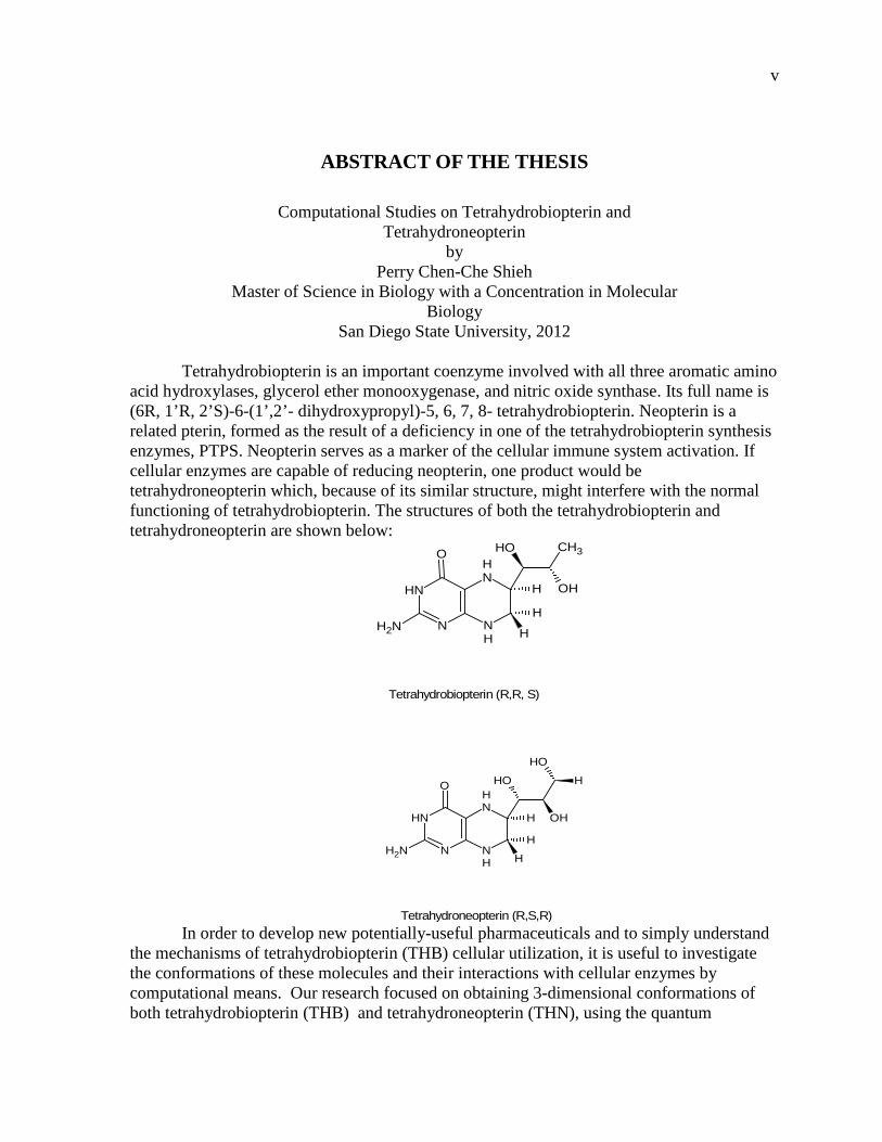
Tetrahydrobiopterin is an important coenzyme involved with all three aromatic amino acid hydroxylases, glycerol ether monooxygenase, and nitric oxide synthase. Its full name is (6R, 1’R, 2’S)-6-(1’,2’- dihydroxypropyl)-5, 6, 7, 8- tetrahydrobiopterin. Neopterin is a related pterin, formed as the result of a deficiency in one of the tetrahydrobiopterin synthesis enzymes, PTPS. Neopterin serves as a marker of the cellular immune system activation. If cellular enzymes are capable of reducing neopterin, one product would be tetrahydroneopterin which, because of its similar structure, might interfere with the normal functioning of tetrahydrobiopterin. The structures of both the tetrahydrobiopterin and tetrahydroneopterin are shown below:
In order to develop new potentially-useful pharmaceuticals and to simply understand
the mechanisms of tetrahydrobiopterin (THB) cellular utilization, it is useful to investigate the conformations of these molecules and their interactions with cellular enzymes by computational means. Our research focused on obtaining 3-dimensional conformations of both tetrahydrobiopterin (THB) and tetrahydroneopterin (THN), using the quantum
H
OH
NH2
O
NH
NH
N
NH
CH3
OH
H
H
OH
H
OH
NH2
O
NH
NH
N
NH OH
H
H
H
Tetrahydrobiopterin (R,R, S)
Tetrahydroneopterin (R,S,R)
vi
mechanical computational program Gaussian 09, to find the lowest energy state conformations of the molecules studied. We began by performing an overall scan of possible configurations of THB and THN molecules, obtaining the lowest energy conformations for each molecule. Next, we ran the DFT feature of the Gaussian 09 program for further study of the conformations showing the lowest energy conformations. Our work is the first to consider effects of water on THB and THN conformations; we used two approaches. In the first, we added one or two water molecules to THB molecules to observe their effects on hydrogen bonding and structure. In the second, we simulated the effects of solvating our THB and THN molecules using the computer program COSMOtherm.
Using the program Discovery Studio 2.5, we tested the ability of THB and THN to fit into the active sites defined by x-ray crystallographic studies of the enzyme structures (docking the THB and THN onto the enzyme). We studied their binding to each of the three aromatic amino acid hydroxylases, The H-bonding patterns were very comparable to the original H-bonding patterns of the THB in the aromatic amino acid hydroxylase structures. Nitric Oxide Synthase (NOS) interacts with THB in a different manner than that with the three aromatic amino acid hydroxylases. In the reaction catalyzed by NOS, THB gets converted into a radical form, either ·BH3 or ·BH4
+. We studied both ·BH3 and ·BH4+ using
Gaussian 09 and obtained their lowest energy forms. We obtained partial charge and spin density distributions that showed the electron of the ·BH4
+ radical was much more delocalized than that of ·BH3, and therefore more stable than ·BH3. Docking of the neutral THB to NOS yielded much better results than docking either ·BH3 or ·BH4
+ onto the enzyme active site.
We conclude that THB and THN exist in a equilibrium of conformations, varying between the keto and enol tautomers and the axial and equatorial side chain shifts. In both the neutral and the radical form of THB, the keto-axial is the lowest energy form and thus is the predominant conformation. The THB is in its neutral form before binding fully to the active site in the NOS. This is supported from the multiple docking studies showing better overlapping of THB than that of either ·BH3 or ·BH4
+. In addition to the above studies, we examined aspects of the recycling pathway,
specifically the destruction of THB through the non-enzymatic reaction of the quinoid form of 7,8 dihydrobiopterin (qDHB) formed in the normal recycling reaction to give 7,8 dihydrobiopterin (DHB). We further studied the reaction of DHB to give pterin and 2-hydroxypropanal. Theoretical intermediates were proposed for both reactions. ΔGs were calculated for DHB and intermediates in both the solvated and the unsolvated forms. The results indicated that the two destructive reactions are thermodynamically favored.
vii
TABLE OF CONTENTS
PAGE
ABSTRACT ...............................................................................................................................v
LIST OF TABLES ................................................................................................................... ix
LIST OF FIGURES ...................................................................................................................x
CHAPTER
1 INTRODUCTION .........................................................................................................1
Review of the Literature ..........................................................................................2
Earlier Computer Studies of Tetrahydrobiopterin and Tetrahydroneopterin ...........4
NMR Determinations of Tetrahydrobiopterin Structure..........................................8
Crystallographic Studies of Tetrahydrobiopterin Structure in Aromatic Amino Acid Hydrolases and Nitric Oxide Synthase ...............................................8
Discovery of Tyrosine Hydroxyase (TyrOH) Crystal Structure .......................8
Discovery of Tryptophan Hydroxyase (TrpOH) Crystal Structure ...................9
Discovery of Phenylalanine Hydroxyase (PheOH) Crystal Structure .............11
Comparisons Between the Three Hydroxylases ..............................................14
Discovery of Nitric Oxide Synthase (NOS) Crystal Structure ........................15
iNOS (Inducible Form) ..............................................................................15
eNOS (Endothelial Form) ..........................................................................16
nNOS (Neuronal Form) .............................................................................18
THB as a Radical Bound in NOS ....................................................................19
2 METHODS ..................................................................................................................24
The COSMOtherm Program ..................................................................................25
Density Functional Theory (DFT) .........................................................................27
Basis Sets ...............................................................................................................28
Gaussian Program ..................................................................................................29
Background on Gaussian .......................................................................................29
3 RESULTS ....................................................................................................................32
4 CONCLUSION / DISCUSSION .................................................................................49
5 STUDIES OF THE THB RECYCLING PATHWAY ................................................53

viii
ACKNOWLEDGEMENTS .....................................................................................................62
REFERENCES ........................................................................................................................63
APPENDIX
EXTRA GRAPHS AND TABLES ..............................................................................67
ix
LIST OF TABLES
PAGE
Table 1. Thermodynamic Properties for Unhydrated and Hydrated Forms of Tetrahydrobiopterin and Tetrahydroneopterin in Various Ionization States ...............35
Table 2. Shows the Predominant Conformations and the Equilibrium Constants ...................44
Table 3. Table Showing the Spin Densities on Atoms Ranging from N1 to C8α ...................45
Table 4. Table Showing the qDHB, 7,8 DHB, Pterin, Side Chains, and All Their Intermediates in Solvated Form ...................................................................................59
Table 5. Table Showing the Energy Differences Between the qDHB and 7,8 DHB, and the Proposed Pterin and Side Chain, All in Unsolvated Form. See Appendix for Tables 6-8 with All the Intermediates ...................................................60
Table 6. Table of Keq for BH3 and BH4 .................................................................................69
Table 7. Table of Solvated qDHB, 7,8 DHB, Intermediates, Pterin and Their Side Chains ..........................................................................................................................70
Table 8. Table of Unsolvated qDHB, 7,8 DHB, Intermediates, Pterin and Their Side Chains. .........................................................................................................................71
x
LIST OF FIGURES
PAGE
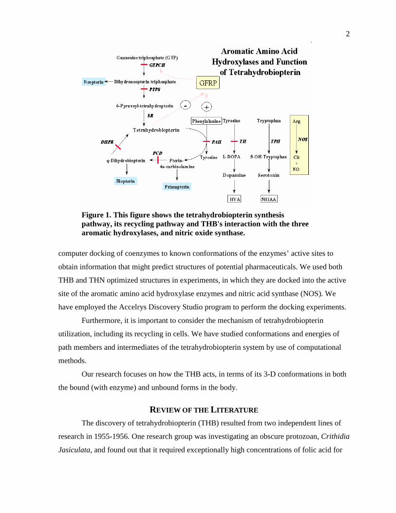
Figure 1. This figure shows the tetrahydrobiopterin synthesis pathway, its recycling pathway and THB's interaction with the three aromatic hydroxylases, and nitric oxide synthase. .....................................................................................................2
Figure 2. The structure of tetrahydrobiopterin (THB, BH4) and its accepted atom numbering system. .........................................................................................................5
Figure 3. Conformations of tetrahydrobiopterin calculated by Katoh, Sueoka, and Kurihara. They define axial and equatorial based on the rotational position of carbon 2’ with respect to carbon 1’. ...............................................................................6
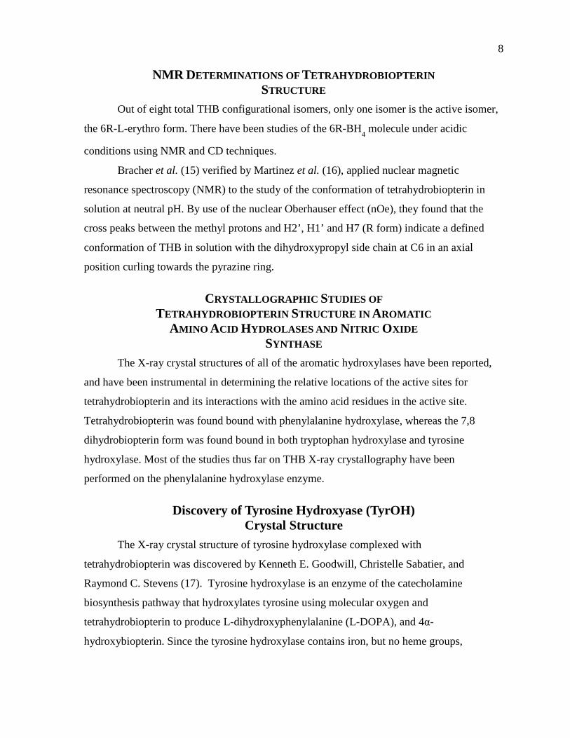
Figure 4. The catalytic site of tyrosine hydroxylase. Figure b is a close-up of figure a. For explanation, see the text. In figure a, the residues are His331, His336, and Glu376, as well as the hydroxylated Phe300. “The secondary structure portions containing the iron coordinating residues are shown in blue, His336 helix is shown in green. The helix which contains Phe300 and the adjacent loop region which participates in pterin binding is also shown in green.” ..................10
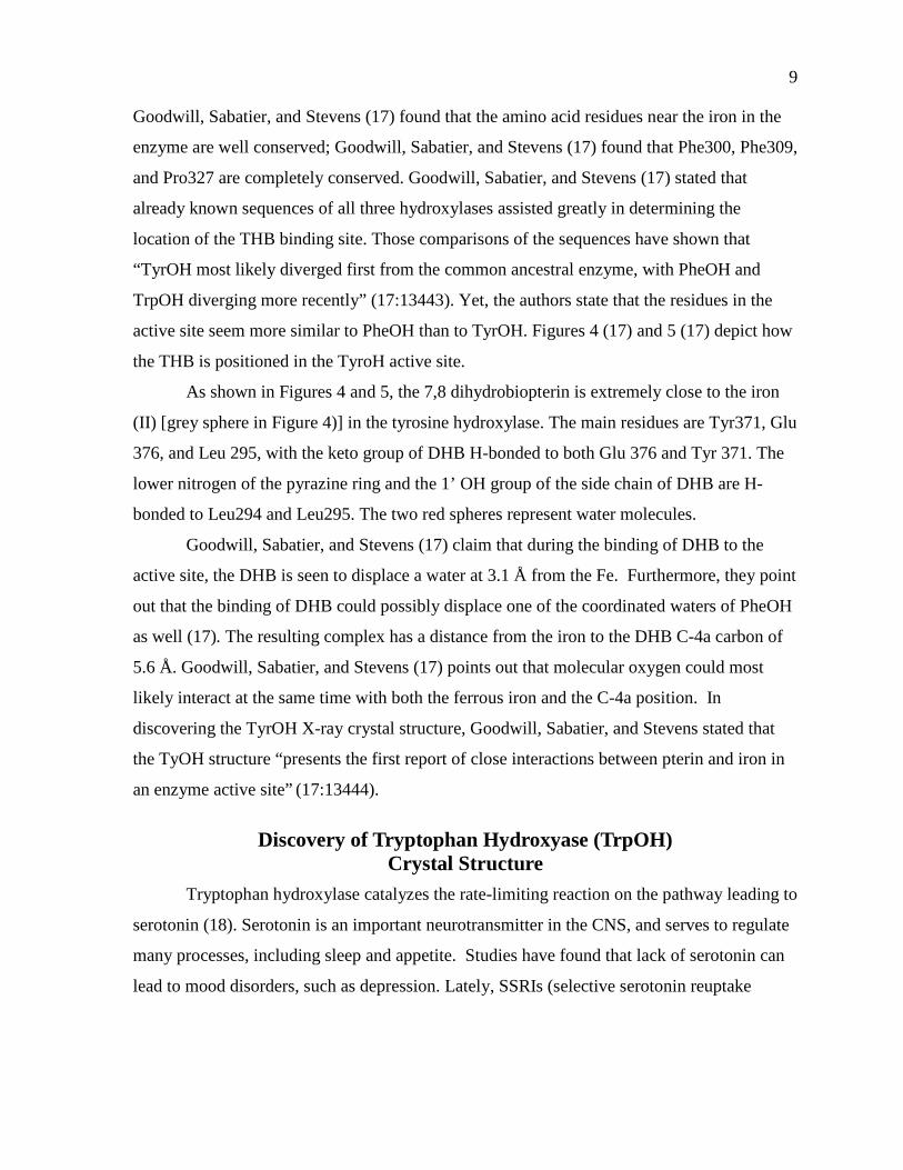
Figure 5. A better representation of the catalytic active site, taken from pdb (PDB ID: 2TOH) in ligand explorer. ............................................................................................10
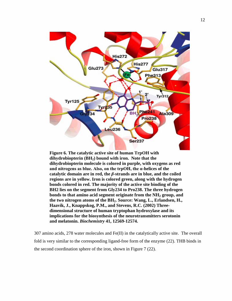
Figure 6. The catalytic active site of human TrpOH with dihydrobiopterin (BH2) bound with iron. Note that the dihydrobiopterin molecule is colored in purple, with oxygens as red and nitrogens as blue. Also, on the trpOH, the α-helices of the catalytic domain are in red, the β-strands are in blue, and the coiled regions are in yellow. Iron is colored green, along with the hydrogen bonds colored in red. The majority of the active site binding of the BH2 lies on the segment from Gly234 to Pro238The three hydrogen bonds to that amino acid segment originate from the NH2 group, and the two nitrogen atoms of the BH2. ..............................................................................................................................12
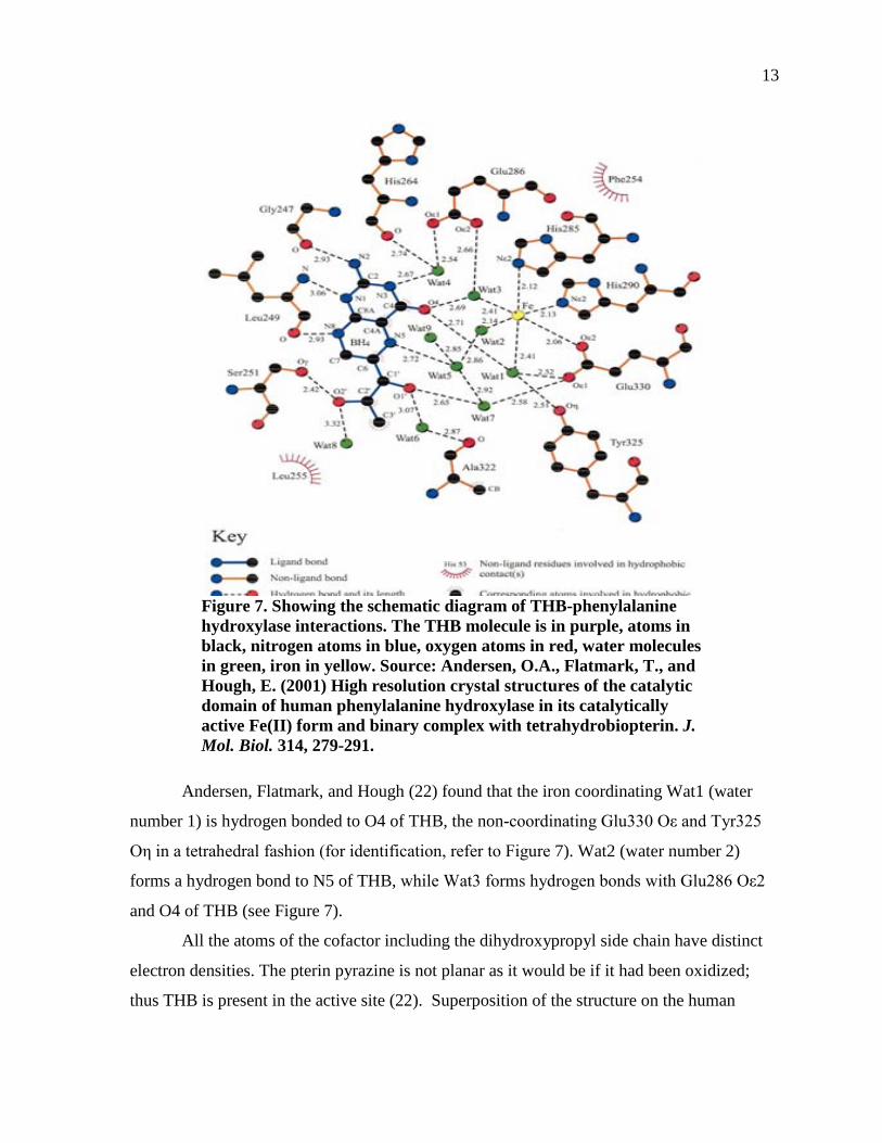
Figure 7. Showing the schematic diagram of THB-phenylalanine hydroxylase interactions. The THB molecule is in purple, atoms in black, nitrogen atoms in blue, oxygen atoms in red, water molecules in green, iron in yellow. .........................13
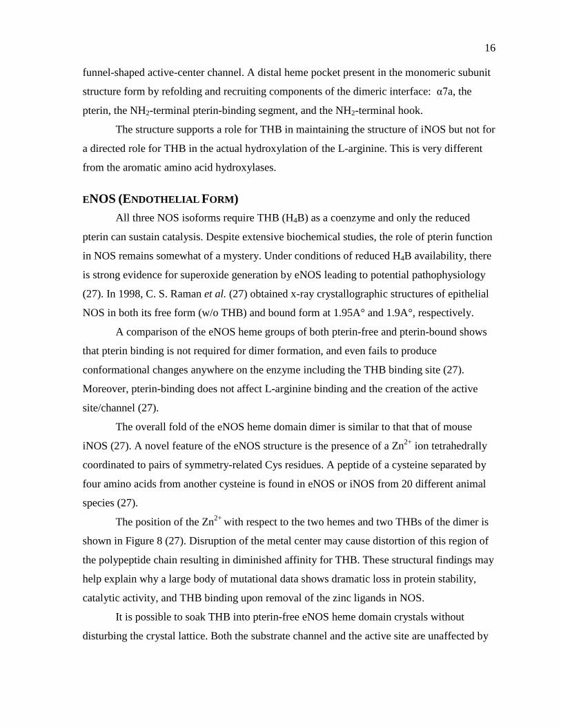
Figure 8. (A) Stereo view of the 2Fo-2Fc 1.9 A° omit electron density map around the zinc metal center, (B) “The ZnS4 metal center and its relationship to THB. Ser-104 is part of the loop containing the cysteine ligands and H-bonds to the C6 side chain of pterin. The stereospecific recognition of THB by NOS is dictated by the substitution at the C6 position.”. .........................................................17
Figure 9. (A) Cross talk between THB and L-Arg mediated by the heme propionate The guanidinium and amino groups of L-Arginine are held in place by H-bonding with the Glu-363. In these representations, the amino group and the
xi
THB hydrogen-bond with a heme while the pteridine ring is sandwiched between Phe-462 in one monomer and Trp-449 in another, respectively, and (B) L-Arginine binds at the THB binding site when the eNOS inhibitor SEITU is bound at the active site .Two water molecules bridge between the inhibitor and heme propionate. The ethyl group of the inhibitor forms nonbonded contacts with Val-338 and Phe-355. ............................................................................18
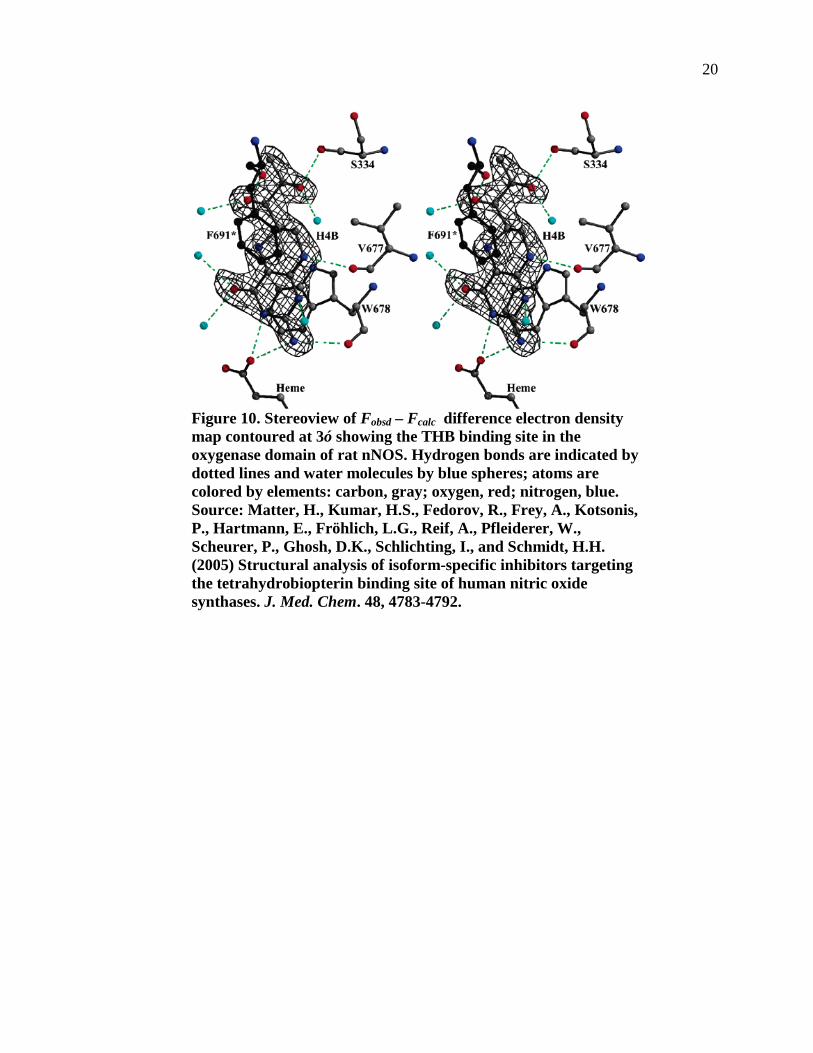
Figure 10. Stereoview of Fobsd – Fcalc difference electron density map contoured at 3ó showing the THB binding site in the oxygenase domain of rat nNOS. Hydrogen bonds are indicated by dotted lines and water molecules by blue spheres; atoms are colored by elements: carbon, gray; oxygen, red; nitrogen, blue. ..............................................................................................................................20
Figure 11. Schematic of the interaction of THB in the rat NOS-I binding site and the amino acid differences among all three NOS isoforms. The rat NOS-I residue numbering is given in black with NOS-III numbering (grey) in comparison..............21
Figure 12. Tetrahydrobiopterin in its binding pocket in NOS, with substrate arginine bound (PDB 1nod) and two structural waters W1 and W2. ........................................22
Figure 13. Shows how COSMO-RS works, starting from a molecule to the end solvation result. ............................................................................................................25
Figure 14. Illustration of molecular cavities and their contact interactions. ...........................26
Figure 15. The four basic conformations of tetrahydrobiopterin: (A) keto/axial; (B) keto/equatorial; (C) enol/axial; (D) enol/equatorial. In A and C, the solid triangle denotes a position above the plane of the ring system; in all four conformers, the hydrogens of carbon-6 are shown below the plane of the ring system by dashed triangles, meaning that the chirality of carbon-6 is R. ....................32
Figure 16. A scan showing different conformations for tetrahydrobiopterin in a keto-equatorial conformation. Note that the lowest energy point is around -851.493 Hartrees and the highest is around -851.473 Hartrees. Scans for the keto-axial conformations for THB and keto-axial and keto-equatorial conformations for THN were also run (see Figures 30 and 31 in the Appendix). ....................................34
Figure 17. Tetrahydrobiopterin and Tetrahydroneopterin in all four conformations and at the lowest energy states............................................................................................37
Figure 18. Keto-equatorial and enol-axial THB with one or two waters added. Our results suggest that several conformations of both THB and THN exist in an equilibrium both in the unhydrated and in the hydrated systems. The water molecules were found to hydrogen bond with side chain alcohol groups and with the enol hydrogen.................................................................................................40
Figure 19. Lowest energy form of keto-equatorial THB that has been docked, using the Discovery Studio program, to the active site of phenylalanine hydroxylase. ........42
Figure 20. The binding site of Figure 19 enlarged in order to show the hydrogen bonding of THB to the amino residues of the active site. ............................................42
xii
Figure 21.·BH3 and ·BH4+ keto-axial and keto-equatorial in lowest energy
conformations. .............................................................................................................43
Figure 22. Numbering of the different atoms on both the aromatic ring and the pyrazine ring of THB. ..................................................................................................45
Figure 23. Keto-axial docked to NOS for ·BH3, ·BH4+, and THB. ..........................................47
Figure 24. ·BH3, ·BH4+, and THB docked to NOS each 10 times. ..........................................48
Figure 25. Full cycle of tetrahydrobiopterin recycling pathway..............................................54
Figure 26. Showing both keto-axial and keto-equatorial conformations for quinoid 7,8 dihydrobiopterin. ..........................................................................................................55
Figure 27. Proposed mechanism of quinoid 7,8 dihydrobiopterin conversion into 7,8 dihydrobiopterin. This will be the first step of the dihydrobiopterin breakdown pathway. .......................................................................................................................56
Figure 28. The proposed mechanism by which 7,8 dihydrobiopterin breaks down and forms the intermediates. These intermediates will break down further into pterin and a three carbon diol or hydroxyaldehyde derivative. Several possible forms of the liberated side chain group are shown in Figure 29. This is the second and final step of the dihydrobiopterin breakdown pathway. ...........................57
Figure 29. Finally, here are the proposed individual pterin molecule and its corresponding side chain cleaved off. ..........................................................................58
Figure 30. THB keto axial scan. ..............................................................................................68
Figure 31. THN keto-equatorial scan.......................................................................................68
1
CHAPTER 1
INTRODUCTION
Tetrahydrobiopterin (THB), (6R, 1’R, 2’S)-6-(1’,2’- dihydroxypropyl)-5, 6, 7, 8-
tetrahydrobiopterin, is a naturally occurring compound that, so far, has been identified in
bacteria and mammals (1). It is an important coenzyme for phenylalanine hydroxylase (L-
phenylalanine + O2 to L-tyrosine + H2O) and tyrosine hydroxylase (L-tyrosine + O2 to 3,4-
dihydroxy-L-phenylalanine + H2O), both involved in the pathways leading to L-3,4-
dihydroxylphenylalanine (L-DOPA) and epinephrine, and for tryptophan hydroxylase (L-
tryptophan + O2 to 5-hydroxy-L-trytophan + H2O), which is on the pathway for serotonin and
melatonin formation. THB is also a coenzyme for reactions catalyzed by glycerol ether
monoxygenase and nitric oxide synthase.
Normally, all the THB synthesis pathways proceed without difficulty, and the
products created by the pathways (serotonin, epinephrine) remain at required levels.
Occasionally, one of the enzymes in THB synthesis, namely 6-pyruvoyltetrahydropterin
synthase (PTPS) (see Figure 1) becomes deficient; hence, tetrahydrobiopterin synthesis is
blocked or slowed as a consequence of inflammatory reactions. As a result, the intermediate
dihydroneopterin triphosphate becomes converted into the side product, neopterin. That
pathway is shown in Figure1.
Reports suggest that neopterin levels in serum are correlated, and can predict patient
mortality (2). Increased neopterin concentrations were discovered in patients with viral
infections that human monocytes and macrophages produce neopterin when stimulated by
interferon-γ. Therefore, measurement of neopterin concentrations in body fluids provides
information about activation of T helper cell-derived cellular immune activation. Neopterin
concentrations in humans reflect the degree of T helper 1 type immune activation (2).
In order to investigate both tetrahydrobiopterin (THB) and tetrahydroneopterin
(THN) further, we utilized Gaussian 09 to run computational simulations, obtaining their
optimized three dimensional structures in the lowest energy states, both alone and hydrated
by the computer program COSMOtherm.. This work allows us to do further studies involving
2
Figure 1. This figure shows the tetrahydrobiopterin synthesis pathway, its recycling pathway and THB's interaction with the three aromatic hydroxylases, and nitric oxide synthase.
computer docking of coenzymes to known conformations of the enzymes’ active sites to
obtain information that might predict structures of potential pharmaceuticals. We used both
THB and THN optimized structures in experiments, in which they are docked into the active
site of the aromatic amino acid hydroxylase enzymes and nitric acid synthase (NOS). We
have employed the Accelrys Discovery Studio program to perform the docking experiments.
Furthermore, it is important to consider the mechanism of tetrahydrobiopterin
utilization, including its recycling in cells. We have studied conformations and energies of
path members and intermediates of the tetrahydrobiopterin system by use of computational
methods.
Our research focuses on how the THB acts, in terms of its 3-D conformations in both
the bound (with enzyme) and unbound forms in the body.
REVIEW OF THE LITERATURE The discovery of tetrahydrobiopterin (THB) resulted from two independent lines of
research in 1955-1956. One research group was investigating an obscure protozoan, Crithidia
Jasiculata, and found out that it required exceptionally high concentrations of folic acid for
3
survival (3). They deduced from this that the protozoan actually required another type of
pteridine, which they named “biopterin”, which could be formed from folic acid. The second
group found a pteridine through structural studies of the eye color pigments in Drosophila
melanogaster (4). Yet, during this time, all scientists could establish was that biopterin was a
pigment of some kind, and they could only hint at any possible functions that this mystery
molecule might have.
In 1957 Dr. Seymour Kaufman, a researcher at the NIH, during investigations of the
phenylalanine to tyrosine conversion pathway, found that NADPH (TPNH in the older
notation), a known coenzyme, and another unknown coenzyme had to be involved in the
reaction. Through chemical and enzymatic analysis, Kaufman concluded that the unknown
coenzyme was an unconjugated pteridine (5). It took until 1963 before Kaufman’s structural
studies on the unknown coenzyme isolated from rat liver, proved it to be tetrahydrobiopterin
(6), THB (or BH4, full name (6R)-2-Amino-6-[(1R,2S)-1,2-dihydroxypropyl]-5,6,7,8-
tetrahydropteridin-4(1H)-one).
In 1974 Kaufman and Fisher (7) discovered that tetrahydrobiopterin was an essential
coenzyme not only for phenylalanine hydroxylase, but also the other aromatic amino acid
hydroxylase enzymes, tyrosine and tryptophan hydroxylases, as well. All three are involved
in the production of critical neurotransmitters, of either serotonin and dopamine (and
epinephrine) (7). Kaufman also played a part in the discovery that THB was a coenzyme in
the oxidative cleavage of glyceryl ethers (8). In 1995, THB was found to be an essential
coenzyme in the Nitric Oxide Synthase pathway (9).
Presently, it is well established that THB is synthesized from GTP (guanosine
triphosphate) in a three-step pathway. The three enzymes are: GTP cyclohydrolase I
(GTPCH), 6-pyruvoyltetrahydropterin synthase (PTPS), and sepiapterin reductase (SR).
Another pteridine, tetrahydroneopterin is a side product of the THB synthetic pathway is
formed, as already discussed, because of a lack of control of tetrahydrobiopterin synthesis
(cased by deficient levels of PTPS). When there is a deficient quantity of PTPS,
dihydroneopterin triphosphate becomes converted into dihydroneopterin (THN).
Dihydroneopterin is released by macrophages and is an immunologic marker for the
activation of the cell-mediated immune system (10). Measurement of neopterin (the
4
dihydroneopterin oxidation product) concentrations in body fluids such as serum or urine are
elevated in infections, cardiovascular disease, rheumatoid arthritis, and certain malignant
tumor diseases. Levels of neopterin well above control values are predictive of patient
mortality (2).
Interferon γ is an immunologic cytokine that correlates directly with the production of
neopterin. Interferon γ is produced by T-lymphocytes in response to foreign particles /
invaders. Then the interferon γ stimulates monocytes and macrophages to start producing
neopterin. GTP cyclohydrolase I converts guanosine triphosphate (GTP) into the
intermediate dihydroneopterin triphosphate. In normal circumstances, the enzyme that
converts dihydroneopterin into 6-pyruvoyl tetrahydropterin, 6-pyruvoyltetrahydropterin
synthase (PTPS), is abundant and GTP ultimately becomes tetrahydrobiopterin in the
synthesis pathway. Yet, when there is a dearth of PTPS enzyme available, the cell
coincidentally converts the dihydroneopterin triphosphate into “neopterin and 7,8-
dihydroneopterin, after dephosphorylation and oxidation at the expense of
biopterin derivatives” (11:2). Indeed, human monocytes / macrophages only have a small
constitutive activity of the biopterin-forming enzyme pyruvoyl-tetrahydropterin synthase
(PTPS), so that almost exclusively neopterin and 7,8-dihydroneopterin become synthesized
and released (11).
EARLIER COMPUTER STUDIES OF TETRAHYDROBIOPTERIN AND TETRAHYDRONEOPTERIN
Currently, with the modern understanding of the importance of 3-dimensional
structure in the components of cells, it became clear that the 3-dimensional structure of
tetrahydrobiopterin and other pterins were needed in order to examine how these pterins
interacted with enzymes and other cell components. Earlier efforts to predict 3-D structures
of THB and THN involved less powerful computers and molecular dynamics / semi-
empirical programs than those available today. Their use led to structures with conflicting
results. While using molecular dynamics simulation software, Estelberger, Mlekusch, and
Reibnegger (12) stated that although he did find that weak intramolecular hydrogen bonds
stabilized one of the conformers of THB, he concluded that both semi-empirical and
molecular dynamic methods had severe limitations in accuracy as compared to that of
5
Density Functional Theory. These program inaccuracies have led to major discrepancies in
reported results; for example, different authors perceptions of the existence or nonexistence
of hydrogen bonding in THB. Even Estelberger, Mlekusch, and Reibnegger (12), as well as
Katoh, Sueoka, and Kurihara (13) and Ziegler et al. (14), was unclear whether or not he truly
found any hydrogen bonds in his THB molecules. Katoh, Sueoka, and Kurihara (13) used the
molecular dynamics method to determine the full configuration of THB in neutral format.
Katoh, Sueoka, and Kurihara stated that “it is difficult to find any certain hydrogen bonds in
these forms” (13:30) , by which they meant forms A and B of THB that they modeled with
molecular dynamics (see Figure 2). Ziegler et al. (14) pointed out that they also failed to find
significant hydrogen bonding in the lowest energy conformations of THB that they ran using
a molecular dynamics program.
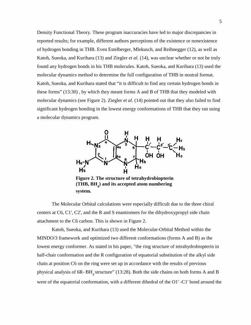
Figure 2. The structure of tetrahydrobiopterin (THB, BH4) and its accepted atom numbering system.
The Molecular Orbital calculations were especially difficult due to the three chiral
centers at C6, C1', C2', and the R and S enantiomers for the dihydroxypropyl side chain
attachment to the C6 carbon. This is shown in Figure 2.
Katoh, Sueoka, and Kurihara (13) used the Molecular-Orbital Method within the
MINDO/3 framework and optimized two different conformations (forms A and B) as the
lowest energy conformer. As stated in his paper, "the ring structure of tetrahydrobiopterin in
half-chair conformation and the R configuration of equatorial substitution of the alkyl side
chain at position C6 on the ring were set up in accordance with the results of previous
physical analysis of 6R-·BH4 structure” (13:28). Both the side chains on both forms A and B
were of the equatorial conformation, with a different dihedral of the O1' -C1' bond around the
6
C1'-C6' bond. Furthermore, in both forms the hydroxyl groups around the C1'-C2' bonds
were in trans positions. Katoh, Sueoka, and Kurihara (13) stated that forms A and B, shown
in Figure 2, were compared with the theoretical structures of tetrahydropterins involved in
the biosynthesis of 6R-BH4. The hydroxy keto intermediates, 6-lactoyl tetrahydropterin and
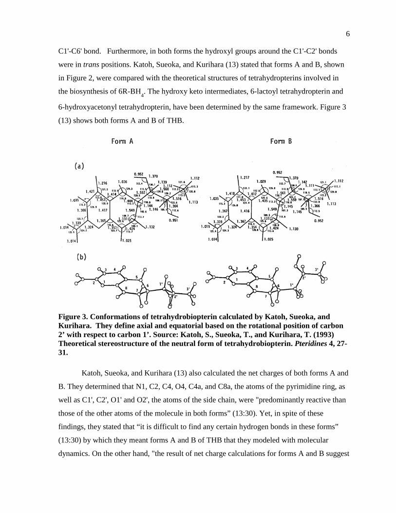
6-hydroxyacetonyl tetrahydropterin, have been determined by the same framework. Figure 3
(13) shows both forms A and B of THB.
Figure 3. Conformations of tetrahydrobiopterin calculated by Katoh, Sueoka, and Kurihara. They define axial and equatorial based on the rotational position of carbon 2’ with respect to carbon 1’. Source: Katoh, S., Sueoka, T., and Kurihara, T. (1993) Theoretical stereostructure of the neutral form of tetrahydrobiopterin. Pteridines 4, 27-31.
Katoh, Sueoka, and Kurihara (13) also calculated the net charges of both forms A and
B. They determined that N1, C2, C4, O4, C4a, and C8a, the atoms of the pyrimidine ring, as
well as C1', C2', O1' and O2', the atoms of the side chain, were "predominantly reactive than
those of the other atoms of the molecule in both forms” (13:30). Yet, in spite of these
findings, they stated that “it is difficult to find any certain hydrogen bonds in these forms”
(13:30) by which they meant forms A and B of THB that they modeled with molecular
dynamics. On the other hand, "the result of net charge calculations for forms A and B suggest
7
the possibility of hydrogen bonding between 1'-OH, 2'-OH, or N1' and some atom contained
in the immediate environment such as the BH4-fitting domain of enzymes"(13:30).
Estelberger, Mlekusch, and Reibnegger (12) researched both tetrahydrobiopterin and
tetrahydroneopterin conformational structures. In an earlier study that was done, the authors
concluded that significant conformational differences exist between both compounds; the
side chain at carbon atom C6, according to their results, was predicted to be in axial
orientation in tetrahydrobiopterin but in equatorial orientation in tetrahydroneopterin (14).
One of the original papers used both semi-empirical and molecular mechanics calculations to
arrive at their conclusion (14). Yet, as Estelberger, Mlekusch, and Reibnegger (12) pointed
out, the authors had many holes in their studies. He pointed out “First, as others have
indicated, the possibility of intramolecular hydrogen bonds in the molecules under
consideration cannot be ruled out with certainty, and secondly, all the studies cited above
have investigated the structural features of the molecules in vacuo at zero temperature, i.e.
only the energetic ground state, neglecting the possibility of internal rotations and vibrations
which are to be expected at realistic temperatures of, say, 310 K and in an aqueous
environment” (12:37).
Estelberger, Mlekusch, and Reibnegger (12) attempted to fill in the gaps from the
previous study by applying molecular dynamics simulations on both tetrahydrobiopterin and
tetrahydroneopterin, running at a time scale of 20ps. They established that the
stereochemistry of the N5 atom in both molecules could be either pseudoaxial or
pseudoequatorial, as well as the side chain being either axial or equatorial. Estelberger,
Mlekusch, and Reibnegger generated 40 lowest energy conformations from their molecular
mechanics program and discovered that “the most stable structures showed the E'A
orientations; on average, they were more stable than E'E by about 2.5 kcal/mol, and more
stable than A'A and A'E orientations by about 3.5 kcal/mol” (12:39). The amount of side
chain axial conformations far exceeded the amount of side chain equatorial conformations for
both molecules. It is interesting to note that Estelberger, Mlekusch, and Reibnegger (12), in
spite of his criticism of the first paper (14), did not study the hydrated molecules.
8
NMR DETERMINATIONS OF TETRAHYDROBIOPTERIN STRUCTURE
Out of eight total THB configurational isomers, only one isomer is the active isomer,
the 6R-L-erythro form. There have been studies of the 6R-BH4 molecule under acidic
conditions using NMR and CD techniques.
Bracher et al. (15) verified by Martinez et al. (16), applied nuclear magnetic
resonance spectroscopy (NMR) to the study of the conformation of tetrahydrobiopterin in
solution at neutral pH. By use of the nuclear Oberhauser effect (nOe), they found that the
cross peaks between the methyl protons and H2’, H1’ and H7 (R form) indicate a defined
conformation of THB in solution with the dihydroxypropyl side chain at C6 in an axial
position curling towards the pyrazine ring.
CRYSTALLOGRAPHIC STUDIES OF TETRAHYDROBIOPTERIN STRUCTURE IN AROMATIC
AMINO ACID HYDROLASES AND NITRIC OXIDE SYNTHASE
The X-ray crystal structures of all of the aromatic hydroxylases have been reported,
and have been instrumental in determining the relative locations of the active sites for
tetrahydrobiopterin and its interactions with the amino acid residues in the active site.
Tetrahydrobiopterin was found bound with phenylalanine hydroxylase, whereas the 7,8
dihydrobiopterin form was found bound in both tryptophan hydroxylase and tyrosine
hydroxylase. Most of the studies thus far on THB X-ray crystallography have been
performed on the phenylalanine hydroxylase enzyme.
Discovery of Tyrosine Hydroxyase (TyrOH) Crystal Structure
The X-ray crystal structure of tyrosine hydroxylase complexed with
tetrahydrobiopterin was discovered by Kenneth E. Goodwill, Christelle Sabatier, and
Raymond C. Stevens (17). Tyrosine hydroxylase is an enzyme of the catecholamine
biosynthesis pathway that hydroxylates tyrosine using molecular oxygen and
tetrahydrobiopterin to produce L-dihydroxyphenylalanine (L-DOPA), and 4α-
hydroxybiopterin. Since the tyrosine hydroxylase contains iron, but no heme groups,
9
Goodwill, Sabatier, and Stevens (17) found that the amino acid residues near the iron in the
enzyme are well conserved; Goodwill, Sabatier, and Stevens (17) found that Phe300, Phe309,
and Pro327 are completely conserved. Goodwill, Sabatier, and Stevens (17) stated that
already known sequences of all three hydroxylases assisted greatly in determining the
location of the THB binding site. Those comparisons of the sequences have shown that
“TyrOH most likely diverged first from the common ancestral enzyme, with PheOH and
TrpOH diverging more recently” (17:13443). Yet, the authors state that the residues in the
active site seem more similar to PheOH than to TyrOH. Figures 4 (17) and 5 (17) depict how
the THB is positioned in the TyroH active site.
As shown in Figures 4 and 5, the 7,8 dihydrobiopterin is extremely close to the iron
(II) [grey sphere in Figure 4)] in the tyrosine hydroxylase. The main residues are Tyr371, Glu
376, and Leu 295, with the keto group of DHB H-bonded to both Glu 376 and Tyr 371. The
lower nitrogen of the pyrazine ring and the 1’ OH group of the side chain of DHB are H-
bonded to Leu294 and Leu295. The two red spheres represent water molecules.
Goodwill, Sabatier, and Stevens (17) claim that during the binding of DHB to the
active site, the DHB is seen to displace a water at 3.1 Å from the Fe. Furthermore, they point
out that the binding of DHB could possibly displace one of the coordinated waters of PheOH
as well (17). The resulting complex has a distance from the iron to the DHB C-4a carbon of
5.6 Å. Goodwill, Sabatier, and Stevens (17) points out that molecular oxygen could most
likely interact at the same time with both the ferrous iron and the C-4a position. In
discovering the TyrOH X-ray crystal structure, Goodwill, Sabatier, and Stevens stated that
the TyOH structure “presents the first report of close interactions between pterin and iron in
an enzyme active site” (17:13444).
Discovery of Tryptophan Hydroxyase (TrpOH) Crystal Structure
Tryptophan hydroxylase catalyzes the rate-limiting reaction on the pathway leading to
serotonin (18). Serotonin is an important neurotransmitter in the CNS, and serves to regulate
many processes, including sleep and appetite. Studies have found that lack of serotonin can
lead to mood disorders, such as depression. Lately, SSRIs (selective serotonin reuptake
10
Figure 4. The catalytic site of tyrosine hydroxylase. Figure b is a close-up of figure a. For explanation, see the text. In figure a, the residues are His331, His336, and Glu376, as well as the hydroxylated Phe300. “The secondary structure portions containing the iron coordinating residues are shown in blue, His336 helix is shown in green. The helix which contains Phe300 and the adjacent loop region which participates in pterin binding is also shown in green.” Source: Goodwill, K.E., Sabatier, C., and Stevens, R.C. (1998) Crystal structure of tyrosine hydroxylase with bound cofactor analogue and iron at 2.3 Å resolution: Self-hydroxylation of phe300 and the pterin-binding site. Biochemistry 37, 13439-13445.
Figure 5. A better representation of the catalytic active site, taken from pdb (PDB ID: 2TOH) in ligand explorer. Source: Goodwill, K.E., Sabatier, C., and Stevens, R.C. (1998) Crystal structure of tyrosine hydroxylase with bound cofactor analogue and iron at 2.3 Å resolution: Self-hydroxylation of phe300 and the pterin-binding site. Biochemistry 37, 13439-13445.
11
inhibitors) have been used in treatment to upregulate TrOH expression. These studies give
rise to the connection between antidepressant effects and TrpOH activity (18). Interestingly,
THB has been tested as an antidepressant with inconclusive results, since THB cannot pass
through the blood-brain barrier and is unstable.
Lin Wang et al. (18) were the first to obtain the X-ray crystallographic structure of
tryptophan hydroxylase and thus the first to elaborate on the catalytic domain of the enzyme,
shown in Figure 6 (18). From their results, they determined that the human TrpOH active site
consists of an approximately 9 Å deep and 10 Å wide cavity. There is a ~12 Å long and ~7 Å
wide channel where tryptophan most likely binds (18).
As predicted in previous studies (19), it was found that human TrpOH contains a
catalytic Fe(III) atom about 13 Å from the keto group of bound THB, and intersection of the
channel and the opening to the active site. As shown in Figure 6, "the iron III is coordinated
to His272, His277 and one carboxyl oxygen atom of Glu3171. Three water molecules have
been observed coordinated to the iron III: wat1, wat2 , and wat3. Wat1 is axial to His272 and
has a distance of 2.2 Å to the iron, Wat2 is axial to His277 and has a distance of 2.3 Å to the
iron, and Wat3 is axial to Glu317 and has a distance of 2.2 Å to the iron” (18:12572).
Discovery of Phenylalanine Hydroxyase (PheOH) Crystal Structure
A deficiency in human L-phenylalanine hydroxylase activity is linked to the disease
phenylketonuria (PKU). In most cases, the lack of phenylalanine hydroxylase activity is a
result of lowered tetrahydrobiopterin levels due to mutations of enzymes involved in its
synthesis (20).
THB donates electrons in the hydroxylation reaction and inhibits the activation of the
enzyme by L-Phe. It was found that dihydrobiopterin (DHB) at higher concentrations than
THB also hinders the L-Phe activation of the enzyme. Previous studies have found that the
dihydroxypropyl side-chain of both THB and DHB is vital for this inhibitory effect (21).
In 2001 Ole Andreas Andersen, Torgeir Flatmark, and Edward Hough (22)
successfully obtained a X-ray crystallographic picture of tetrahydrobiopterin bound to the
catalytically active Fe(II) form of human phenylalanine hydroxylase. The final model has
12
Figure 6. The catalytic active site of human TrpOH with dihydrobiopterin (BH2) bound with iron. Note that the dihydrobiopterin molecule is colored in purple, with oxygens as red and nitrogens as blue. Also, on the trpOH, the α-helices of the catalytic domain are in red, the β-strands are in blue, and the coiled regions are in yellow. Iron is colored green, along with the hydrogen bonds colored in red. The majority of the active site binding of the BH2 lies on the segment from Gly234 to Pro238. The three hydrogen bonds to that amino acid segment originate from the NH2 group, and the two nitrogen atoms of the BH2. Source: Wang, L., Erlandsen, H., Haavik, J., Knappskog, P.M., and Stevens, R.C. (2002) Three-dimensional structure of human tryptophan hydroxylase and its implications for the biosynthesis of the neurotransmitters serotonin and melatonin. Biochemistry 41, 12569-12574.
307 amino acids, 278 water molecules and Fe(II) in the catalytically active site. The overall
fold is very similar to the corresponding ligand-free form of the enzyme (22). THB binds in
the second coordination sphere of the iron, shown in Figure 7 (22).
13
Figure 7. Showing the schematic diagram of THB-phenylalanine hydroxylase interactions. The THB molecule is in purple, atoms in black, nitrogen atoms in blue, oxygen atoms in red, water molecules in green, iron in yellow. Source: Andersen, O.A., Flatmark, T., and Hough, E. (2001) High resolution crystal structures of the catalytic domain of human phenylalanine hydroxylase in its catalytically active Fe(II) form and binary complex with tetrahydrobiopterin. J. Mol. Biol. 314, 279-291.
Andersen, Flatmark, and Hough (22) found that the iron coordinating Wat1 (water
number 1) is hydrogen bonded to O4 of THB, the non-coordinating Glu330 Oε and Tyr325
Oη in a tetrahedral fashion (for identification, refer to Figure 7). Wat2 (water number 2)
forms a hydrogen bond to N5 of THB, while Wat3 forms hydrogen bonds with Glu286 Oε2
and O4 of THB (see Figure 7).
All the atoms of the cofactor including the dihydroxypropyl side chain have distinct
electron densities. The pterin pyrazine is not planar as it would be if it had been oxidized;
thus THB is present in the active site (22). Superposition of the structure on the human
14
PheOH-Fe(III) BH2 complex shows that the reduced cofactor is displaced about 0.5 Å in the
direction away from Ser251, and that the pterin ring is rotated about 10 Å (along the C4a-
C8a bond) with the pyrimidine ring rotated towards Phe254 (22). The dihydroxypropyl side
chain is predominantly equatorial.
Comparisons Between the Three Hydroxylases All three of the aromatic amino acid hydroxylases utilize non-heme iron and
molecular oxygen to hydroxylate their amino acid substrates using a tetrahydrobiopterin
coenzyme. The enzymes from eukaryotic sources share a three domain structure (22). There
is an N- terminal regulatory domain of 100-170 residues, which has a pairwise homology of
about 25%. This is followed by a 270 residue catalytic domain exhibiting 80% sequence
homology and a 65% pairwise sequence identity. The C-terminal 40 residues in the family
form a tetramerization domain with a 60% pairwise homology. Deletion experiments with
the eukaryotic enzymes have demonstrated that the highly conserved catalytic domain can
promote the reaction independently of the other domains (22).
The biggest difference between the catalytic active sites of the three hydroxylases lie
with the two loops (residues 263-269 and residues 363-372). “ Phe313 and Ile366, can be
superimposed with the positions of the corresponding residues in human PheOH (Trp326 and
Val379, respectively) and rTyrOH (Trp372 and Asp425, respectively)” (18:12572).
Tyr235 is conserved in all known TrpOHs, whereas in both PheOH and TyrOH, it is a
smaller hydrophobic leucine; the leucine side chains in human PheOH and rat TyrOH are
located on the opposite side of the pterin as compared to those of Phe241
Two residues are noticeably different in TrpOH compared to the other two family
members. Trp372 is preserved in all TyrOH and PheOH sequences, but is a much smaller
tyrosine in TrpOH. Leu294 is also conserved in all TyrOH and PheOH sequences, but is a
larger tyrosine residue in TrpOH (17). Those differences in TrpOH may allow that enzyme to
hydroxylate tryptophan.
Second, in the region of residues 123-129 of human TrpOH, Tyr125 ð-stacks onto
Tyr235, forming an angle of 90° with respect to the aromatic rings (3.8 Å) (17). The
corresponding residue is a tyrosine in most TrpOHs and PheOHs (and is a phenylalanine in
most TyrOHs), but in the human PheOH structure, this tyrosine is positioned toward the
15
surface, instead of into the active site. These differences in the amino acid hydroxylase
cofactor binding sites are consistent with the distinct cofactor structural preferences
displayed by the three enzymes, even though they share a common reaction mechanism.
Discovery of Nitric Oxide Synthase (NOS) Crystal Structure
Since Seymour Kaufman was the ‘founding father of tetrahydrobiopterin,’ it would
only seem fitting that he was one of the first scientists that linked THB to one of the essential
coenzymes for nitric oxide synthase (NOS). In 1992, White and Marletta (23) were actually
the first to discover that THB played a role as the coenzyme for NOS.
NOS is an enzyme that catalyzes the NADPH-dependent conversion of L-arginine to
L-citrulline and nitric oxide. By 1993, the year of a major review article by Kaufman (24), it
had already been discovered that NOS-mediated roles, included a endothelium-derived
relaxing factor, a neurotransmitter, platelet aggregation inhibitor, and a generator of a cell-
mediated immune response which slowed the growth of some tumor and bacterial cells. In
1993, scientists knew only two distinct NOS forms, the constitutive form and the cytokine-
inducible form (24). Currently, NOS is known to have three distinct forms: the inducible,
Ca2+-independent form (iNOS), the neuronal form (nNOS) and the endothelial form (eNOS).
NOS is unique in that the enzymes requires five cofactors: flavin mononucleotide (FMN),
flavin adenine dinucleotide (FAD), NADPH, THB, and calmodulin (23).
Each of the Nitric Oxide Synthases have a much more complicated mechanism of
oxidation than the aromatic amino acid hydroxylases. It involves two sequential,
mechanistically distinct, heme-based oxidations in the five-electron oxidation of L-Arg to L-
citrulline (L-Cit) and NO. L-Arginine is first hydroxylated to N- hydroxy-L-arginine (NOH-
L-Arg) and a proposed oxo-iron porphyrin radical intermediate [P-Fe(IV)=O]. NOH-L-Arg is
then converted to L-Citruline and NO by means of one-electron oxidation (25).
Below I have listed all three of the NOS isoforms in more detail and how they were
discovered.
INOS (INDUCIBLE FORM) It took until 1998 before the first X-ray crystallographic structure of iNOS was
obtained by Brian R. Crane et al. (26). The enzyme proved to be a dimer with a ~30 A deep,
16
funnel-shaped active-center channel. A distal heme pocket present in the monomeric subunit
structure form by refolding and recruiting components of the dimeric interface: α7a, the
pterin, the NH2-terminal pterin-binding segment, and the NH2-terminal hook.
The structure supports a role for THB in maintaining the structure of iNOS but not for
a directed role for THB in the actual hydroxylation of the L-arginine. This is very different
from the aromatic amino acid hydroxylases.
ENOS (ENDOTHELIAL FORM) All three NOS isoforms require THB (H4B) as a coenzyme and only the reduced
pterin can sustain catalysis. Despite extensive biochemical studies, the role of pterin function
in NOS remains somewhat of a mystery. Under conditions of reduced H4B availability, there
is strong evidence for superoxide generation by eNOS leading to potential pathophysiology
(27). In 1998, C. S. Raman et al. (27) obtained x-ray crystallographic structures of epithelial
NOS in both its free form (w/o THB) and bound form at 1.95A° and 1.9A°, respectively.
A comparison of the eNOS heme groups of both pterin-free and pterin-bound shows
that pterin binding is not required for dimer formation, and even fails to produce
conformational changes anywhere on the enzyme including the THB binding site (27).
Moreover, pterin-binding does not affect L-arginine binding and the creation of the active
site/channel (27).
The overall fold of the eNOS heme domain dimer is similar to that that of mouse
iNOS (27). A novel feature of the eNOS structure is the presence of a Zn2+ ion tetrahedrally
coordinated to pairs of symmetry-related Cys residues. A peptide of a cysteine separated by
four amino acids from another cysteine is found in eNOS or iNOS from 20 different animal
species (27).
The position of the Zn2+ with respect to the two hemes and two THBs of the dimer is
shown in Figure 8 (27). Disruption of the metal center may cause distortion of this region of
the polypeptide chain resulting in diminished affinity for THB. These structural findings may
help explain why a large body of mutational data shows dramatic loss in protein stability,
catalytic activity, and THB binding upon removal of the zinc ligands in NOS.
It is possible to soak THB into pterin-free eNOS heme domain crystals without
disturbing the crystal lattice. Both the substrate channel and the active site are unaffected by
17
Figure 8. (A) Stereo view of the 2Fo-2Fc 1.9 A° omit electron density map around the zinc metal center, (B) “The ZnS4 metal center and its relationship to THB. Ser-104 is part of the loop containing the cysteine ligands and H-bonds to the C6 side chain of pterin. The stereospecific recognition of THB by NOS is dictated by the substitution at the C6 position.” Source: Raman, C.S., Li, H., Martasek, P., Kral, V., Masters, B.S., and Poulos, T.L. (1998) Crystal structure of constitutive endothelial nitric oxide synthase: A paradigm for pterin function involving a novel metal center. Cell 95, 939-950.
18
the lack of pterin at the THB-binding site. L-Arginine can also bind at the THB site in pterin-
free eNOS with a structure identical to that seen in the THB-bound form (28). There also is
biochemical evidence for substrate analog (Nω-nitro- L-arginine) recognition by eNOS even
in the absence of THB (27). Figure 9 shows THB interacting with all the residues in the
eNOS active site.
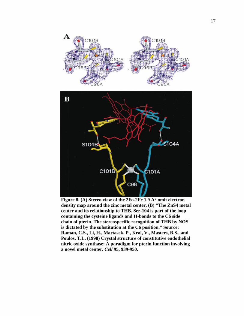
Figure 9. (A) Cross talk between THB and L-Arg mediated by the heme propionate The guanidinium and amino groups of L-Arginine are held in place by H-bonding with the Glu-363. In these representations, the amino group and the THB hydrogen-bond with a heme while the pteridine ring is sandwiched between Phe-462 in one monomer and Trp-449 in another, respectively, and (B) L-Arginine binds at the THB binding site when the eNOS inhibitor SEITU is bound at the active site .Two water molecules bridge between the inhibitor and heme propionate. The ethyl group of the inhibitor forms nonbonded contacts with Val-338 and Phe-355.
The specific recognition of L-Arginine at the THB site (Figure 9A) in eNOS suggests
the ability of this site to stabilize a positively charged pterin or pterin radical (27). In order
for NOS to utilize a pterin radical, extensive protonation of the bound THB is necessary.
Cycling between the pterin radical and THB may be achieved via electron transfer from the
reductase domain while the pterin remains bound to NOS.
NNOS (NEURONAL FORM) Crystal structural studies of neuronal nitric oxide synthase (nNOS) done before 2001
were restricted to the PDZ domain (28). The first x-ray crystal structure of the entire neuronal
19
nitric oxide synthase was obtained by Jian Zhang el al. in 2001 (29). Zhang et al. (29)
examined the crystal structure of the FAD/ NADPH domain of rat nNOS. It was not until
2005 for the tetrahydrobiopterin binding site was successfully determined in nNOS.
Hans Matter et al. (30) obtained a 2.0 Å x-ray crystal structure of the rat NOS-I
oxygenase dimer with bound THB and at 2.5 Å with bound THB and L-arginine substrate.
These studies provided a model for the dimeric oxygenase domain of the human NOS-I
isoform. Matter et al. (30) applied two strategies to identify and validate selective inhibitors
targeting NOS-I using ligand and protein structure-based approaches. First, the structure
activity relationship of a focused set of 41 pteridine counterparts were tested on three
recombinant human NOS isozymes. “Systematic variations at positions 4, 5, 6, and 7 of these
analogues revealed substitutions with up to 58-fold selectivity for NOS-I compared to eNOS
and iNOS” (30:4784). Importantly, this selectivity was especially evident with bulky,
hydrophobic substituents at 5 or 6 and alkylation of the 4-amino group with hydrophobic
groups. Specific 4-amino modifications and some changes at position 6 led to the most
selective inhibitors nNOS, of which alkylated 4-amino-tetrahydropteridines were especially
potent and selective.
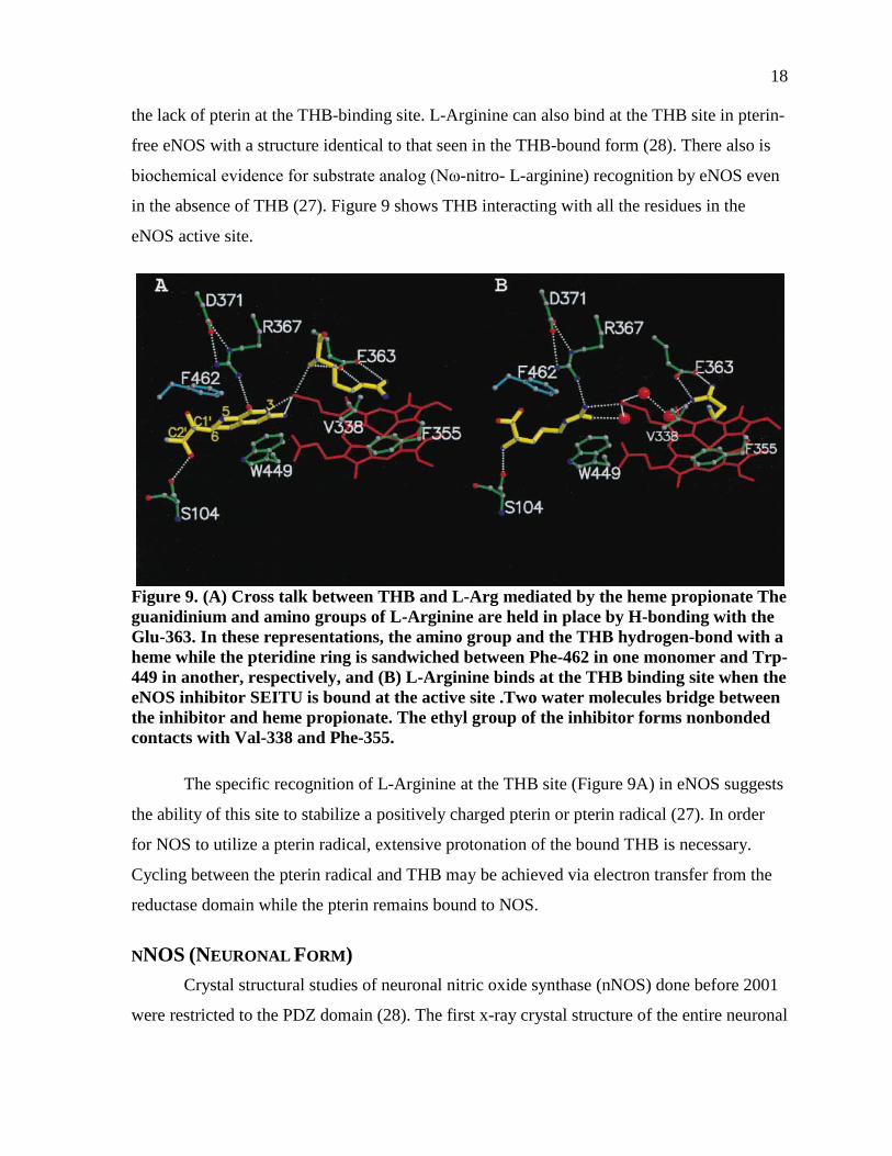
As depicted in Figure 10 (30), the cofactor THB is deeply buried in the cavity and not
accessible to bulk solvent, it is oriented proximal and perpendicular to the heme (30). The
main protein-ligand interaction as in other pterin-protein complexes, occurs between the
planar THB ring and the Trp678 indole, stacked at 3.6 Å distance In general, the hydrogen
bond pattern corresponds to H4B bound to NOS-II or –III. The 5,6,7,8-tetrahydropteridine
interacts with heme carboxylate (O4 via solvent, N3 directly); the structurally conserved
water is present in related X-ray structures. The C4-carbonyl oxygen is hydrogen-bonded to
Arg596 guanidine from the substrate binding helix. Figure 11 shows the THB interacting
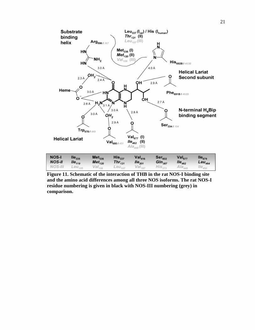
with different amino acid residues in each of the three NOS isoforms.
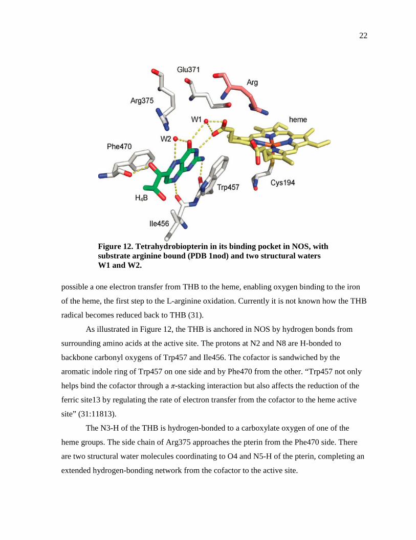
THB as a Radical Bound in NOS Figure 12 shows that each NOS monomer contains a non-covalently bound THB near
the dimer interface and in close proximity to the heme. This configuration seems to make
20
Figure 10. Stereoview of Fobsd – Fcalc difference electron density map contoured at 3ó showing the THB binding site in the oxygenase domain of rat nNOS. Hydrogen bonds are indicated by dotted lines and water molecules by blue spheres; atoms are colored by elements: carbon, gray; oxygen, red; nitrogen, blue. Source: Matter, H., Kumar, H.S., Fedorov, R., Frey, A., Kotsonis, P., Hartmann, E., Fröhlich, L.G., Reif, A., Pfleiderer, W., Scheurer, P., Ghosh, D.K., Schlichting, I., and Schmidt, H.H. (2005) Structural analysis of isoform-specific inhibitors targeting the tetrahydrobiopterin binding site of human nitric oxide synthases. J. Med. Chem. 48, 4783-4792.
21
Figure 11. Schematic of the interaction of THB in the rat NOS-I binding site and the amino acid differences among all three NOS isoforms. The rat NOS-I residue numbering is given in black with NOS-III numbering (grey) in comparison.
22
Figure 12. Tetrahydrobiopterin in its binding pocket in NOS, with substrate arginine bound (PDB 1nod) and two structural waters W1 and W2.
possible a one electron transfer from THB to the heme, enabling oxygen binding to the iron
of the heme, the first step to the L-arginine oxidation. Currently it is not known how the THB
radical becomes reduced back to THB (31).
As illustrated in Figure 12, the THB is anchored in NOS by hydrogen bonds from
surrounding amino acids at the active site. The protons at N2 and N8 are H-bonded to
backbone carbonyl oxygens of Trp457 and Ile456. The cofactor is sandwiched by the
aromatic indole ring of Trp457 on one side and by Phe470 from the other. “Trp457 not only
helps bind the cofactor through a π-stacking interaction but also affects the reduction of the
ferric site13 by regulating the rate of electron transfer from the cofactor to the heme active
site” (31:11813).
The N3-H of the THB is hydrogen-bonded to a carboxylate oxygen of one of the
heme groups. The side chain of Arg375 approaches the pterin from the Phe470 side. There
are two structural water molecules coordinating to O4 and N5-H of the pterin, completing an
extended hydrogen-bonding network from the cofactor to the active site.
23
THB bound in NOS undergoes a one electron chemistry which appears to be unique.
Stoll et al. (31) suggest that the NOS controls the protonation state of THB and through this
regulates proton and electron transfers at the heme center and at the coenzyme active site.
Stoll et al. (31) have successfully deduced the protonation state of the
tetrahydrobiopterin radical in NOS, from electron paramagnetic resonance spectroscopy
combined with DFT calculations. Their experimental magnetic parameters and their
comparison to quantum-chemical predictions show that the radical is a cation, ·BH4+
protonated at N3 and N5. The proton on N5 was directly observed in the 1H ENDOR
spectrum. In contrast, the chemically relevant proton at N3 cannot be resolved in the EPR
and ENDOR spectra, as its hyperfine coupling is small and one of many similar sizes.
However, the N3 protonation state was determined by its effect on the spin density
distribution in the radical, and hyperfine couplings of ring nitrogens and protons.
24
CHAPTER 2
METHODS
Two related families of compounds, are studied in this research. The heavy atoms of
the first set of compounds, the biopterin group, consist of 9 carbons, 5 nitrogens and 4
oxygens. The second family, by-products of biopterin family biosynthesis but of
undetermined physiological effect is the neopterin group, consists of 9 carbons, 5 nitrogens
and 5 oxygens. Each compound relative to this investigation was studied by computational
chemistry, specifically using density functional theory found on Gaussian 03 (32) and later
Gaussian 09 (33), installed on PC computers equipped with Pentium 4, and later with Lenovo
desktops equipped with Intel core i5 processors. .
The main focus of this work was on the compounds 5,6,7,8-tetrahydrobiopterin
(THB) and 5,6,7,8-tetrahydroneopterin (THN). Appropriate structures were entered into the
scanning program of the Gaussian program in order to determine the energies and other
thermodynamic data of the different conformations. Selected conformations of lower energy
were then studied by means of the Density Functional Theory program of Gaussian 09. These
data represent the conformations in vacuo. The effects of hydration on selected
conformations of THB and THN were studied in the hydrated state by two different methods.
The first method was by use of the COSMOtherm program (34). The second method was to
use Density Functional Theory settings with either THB or THN and one or two water
molecules, respectively.
Four different conformations of THB and THN having the lowest energies were
chosen as representatives, for studies of binding to enzymes for which THB was a coenzyme.
Appropriate enzyme crystal structures were obtained from the protein data bank. Coenzymes
associated with the crystal structures were removed and our computer modeled THB and
THN, or other related compounds of the two families, were docked into the protein structure
by means of Discovery Studio 2.5 of Accelrys (35). We also selected a low energy group for
docking experiments, for both THB and THN. Then we docked the four lowest energy
conformations (keto-axial, keto-equatorial, enol-axial, enol-equatorial) to phenylalanine
25
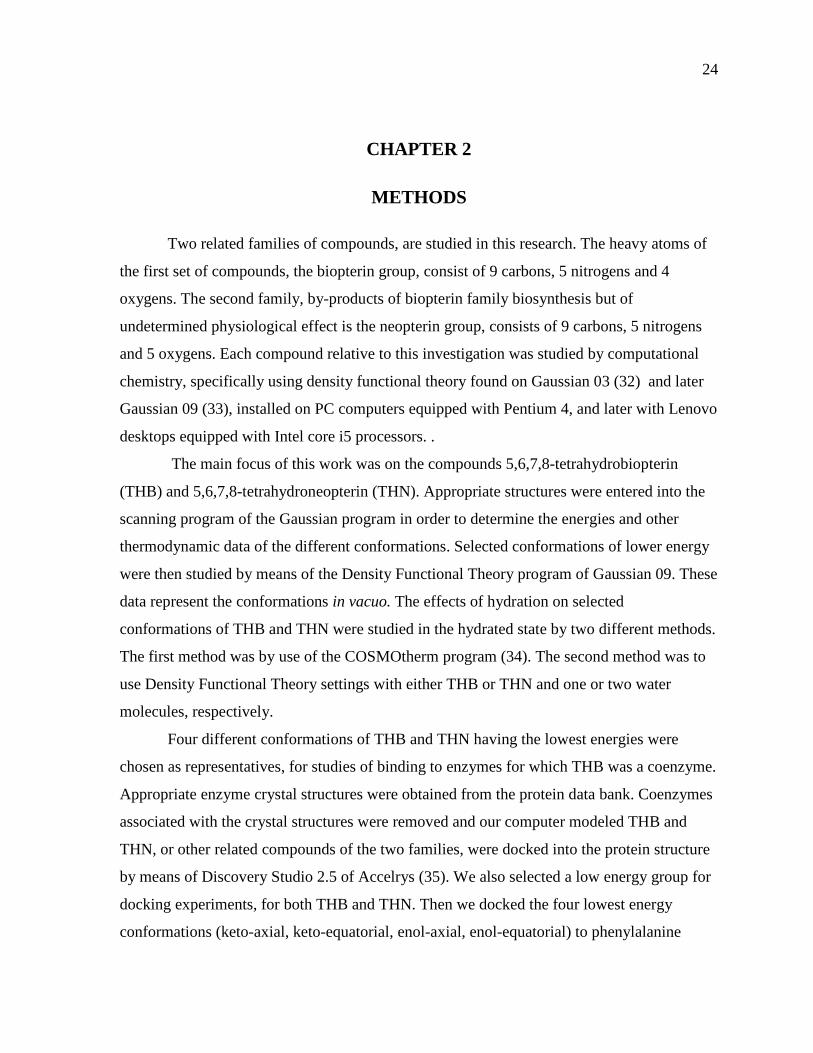
hydroxylase and NOS. Figure 13 (36) shows the flow chart of COSMO-RS, the theory upon
which the COSMOtherm program is based on.
Figure 13. Shows how COSMO-RS works, starting from a molecule to the end solvation result. Source: Klamt, A., and Eckert, F. (2000) COSMO-RS: A novel and efficient method for the a priori prediction of thermophysical data of liquids. Flu. Phase Equi. 172, 43-72.
THE COSMOTHERM PROGRAM “To directly calculate a molecule in solution is very complicated due to the large
number of solvent molecules required for a realistic representation” (37:1). Several different
solvent models have been developed to study the interactions between solute and solvent.
The most popular solvent models have been Continuum Solvation Models (CSM) and the
Self- Consistent Reaction Field Method (SCRF), which is usually combined with CSM.
CSMs describe a molecule in solution through a quantum chemical calculation of the solute
26
molecule with an approximate representation of the surrounding solvent as a continuum (37).
CSMs generally extend basic quantum mechanics methods to describe solutes dissolved in
aqueous solution.
SCRF simulates the solvent as a polarizable range with a given dielectric constant, ε.
The solute is located in a cavity inside the continuum medium. The overall solvent free
energy is ΔG(solvation) = ΔG(cavity) + ΔG(dispersion) + ΔG(polarization), where
ΔG(cavity) = cavity energy, ΔG(dispersion) = dispersion energy, and ΔG(polarization) =
electrostatic energy (38).
The COSMO-RS theory exceeds simple CSMs in that it integrates concepts from
quantum chemistry, dielectric continuum models, electrostatic surface interactions and
statistical thermodynamics. It is a fast and accurate method for extending the COSMO
method to predict solute-solvent interaction energies. COSMOtherm is the software that uses
the COSMO-RS method in its computation (34). We used this software to simulate the
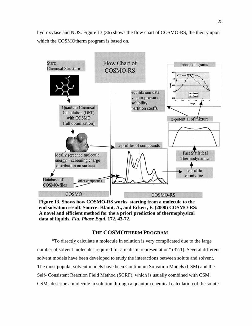
solvation of H2O around both our THB and THN molecules. Figure 14 (36) shows an
illustration of a molecular cavity in COSMO-RS.
Figure 14. Illustration of molecular cavities and their contact interactions. Source: Klamt, A., and Eckert, F. (2000) COSMO-RS: A novel and efficient method for the a priori prediction of thermophysical data of liquids. Flu. Phase Equi. 172, 43-72.
27
DENSITY FUNCTIONAL THEORY (DFT) Density Functional Theory (DFT) is a quantum mechanical modeling method used to
describe the ground state properties of inorganic metals, as well as organic molecules (39). In
recent years, one of the most widely used techniques in computational chemistry has been
density functional theory (DFT). In comparison to Hartree-Fock, DFT is not much more
demanding in computational effort and time, with much more accurate results.
DFT focuses on the electron density ρ, rather than the wavefunction ψ. The term
‘functional’ in the term DFT is derived from a mathematical function, since the “energy of
the molecule is a function of the electron density, written E[ρ], and the electron density is
itself a function of position, ρ (r)” (40:395). Thus, the position is a function of a function of
the energy; functionals are a function of a function.
From the equation ρ(r) = Σ | Ψm (r)|2, the occupied orbitals are used to construct the
electron density and are calculated from the Kohn - Sham equations. The Kohn-Sham
equations are like the Hartree-Fock equations except for a term Vxc, called the exchange-
correlation potential (40).
The Kohn-Sham equations are solved for the electron density first. Next, the Kohn-
Sham equations are solved to obtain an initial set of orbitals. This is a set of orbitals is used
to obtain a better approximation to the electron density and the process is repeated until the
density and the exchange-correlation energy are constant to within some tolerance. It is
important to note that the results of molecular orbital calculations are only approximate, with
deviations from experimental values increasing with the size of the molecule. Therefore, one
goal of computational chemistry is to gain insight into trends in properties of molecules,
without necessarily striving for ultimate accuracy.
The DFT method we used, B3LYP, is based partly on the Hartree-Fock method.
The Hartree Fock equation is F1Ψm (1) = εm Ψm (1). For each molecular orbital Ψm, The Fock operator f1 has terms that express mathematically: 1. The kinetic energy of the electron in Ψm. 2. The potential energy of interaction between the electron in Ψm and the nuclei
in the molecule. 3. Repulsive interactions between the electron in Ψm and other electrons in the
molecule. 4. The effects of spin correlation between electrons in the molecule. (40:395)
28
BASIS SETS Basis sets are mainly used to describe molecular orbitals in quantum mechanics
calculations. Generally, minimal basis set calculations are not very reliable because they are
so small, but very large basis sets are only feasible for small molecules. The basis-set size
must be limited for medium sized molecules, which provides one source of error in the
calculations. Orbitals called Slater orbitals are often used as basis sets; yet, if Slater orbitals
are used in polyatomic molecules, the calculations can become very time-consuming on a
computer.
Presently, Gaussian functions are used instead of Slater orbitals as the main basis
functions. The difference between a Gaussian function and a Slater function lie in its
equation: Gaussian functions contain the factor e-ζr2 instead of the e-ζr factor in Slater
functions. Overall, the computer calculations for Gaussian functions are much quicker than
that for Slater functions. The term STO stand for Slater Type Orbital, and they are actually
Gaussian orbitals. Some Gaussian basis sets of increasing size include "STO-3G, 3-21G, 3-
21G*, 6-31G*, and 6-31G**, where the numbers and symbols are related to the number of
basis functions on each atom" (41:714). STO-3G is essentially an obsolete basis set (41).
The next level of sophistication is the double-zeta basis sets. Here, the set of functions
is doubled; thus, there are two functions for each orbital. The basis sets that we chose for our
experiments were cc-pVDZ and cc-pVTZ, DZ for double zeta and TZ for triple zeta. For the
1st and 2nd row atoms, the cc-pVDZ (correlation consistent-polarized valence double zeta)
basis set adds 1s, 1p, and 1d function. The cc-pVTZ set adds another s, p, d, and an f
function, etc. Correlation Consistent Basis Sets were first discovered by Dunning in 1989
(42).
In the last 15 years, the hybrid functional B3LYP has been the most popular
functional that was used. The B in B3LYP is a term devised from Becke, and LYP indicates
a term devised from Lee, Yang, and Parr (43). LYP indicates three parameters that together
optimize the performance (43).
In the late 1980s, Becke discovered the gradient-corrected functional; use of the
gradient-corrected functional gives the generalized gradient approximation (GGA) (41). In
1993, Becke proposed a further improvement in ExcGGA , that included a contribution from
ExHF . That combination gave rise to hybrid functions, which give the best performance (41).
29
GAUSSIAN PROGRAM Gaussian 03 (32) and Gaussian 09 (33), the latter an improved version of the former,
is a program which is able to calculate structures and thermodynamic data for molecules of
interest using quantum mechanical theory. Classical physics, including mechanics, optics,
thermodynamics, electricity and magnetism, are not able to account for very small objects or
objects moving at very high velocities. Quantum mechanics is based on the discovery by de
Broglie that all particles move as waves (41). The de Broglie discovery as used by
Schrodinger to develop a wave mechanical model of the atom which is the basis of the
quantum mechanical calculations used in Gaussian 03, Gaussian 09 and similar programs.
The Gaussian program can run both single point calculations and whole geometry
optimizations. A single point calculation is performed only at a single fixed molecular
geometry. In a geometry-optimization, however, the program will try to locate an overall
minimum in the electronic energy. A geometry optimization calculation is composed of
many single point calculations, with each single point energy calculation followed by an
energy-gradient calculation. The geometry-optimization continues until the minimum energy
has been found from an energy surface (41). "In a vibrational – frequency calculation, the
program calculates the molecular vibration frequencies; a vibrational frequency calculation
must be preceded by a geometry optimization, since vibrational frequencies calculated for a
geometry that is not at an energy minimum are meaningless” (41:725). A transition-state
optimization aims to discover both the geometry and electronic energies of the transition
state in a chemical reaction.
The geometry optimization process locates the energy minimum that lies closest to
the starting geometry. The global minimum has the lowest energy of all the conformers,
whereas the local minimum is the minimum energy for all the geometries in the same region.
In our conformation calculations on Gaussian, we used mainly the basis set cc-pVDZ,
and some of the cc-pVTZ. We ran DFT with the hybrid functional B3LYP. All our
calculations were performed without any solvent present.
BACKGROUND ON GAUSSIAN We used the computational chemistry software Gaussian mainly to obtain the lowest
structure optimizations, frequencies, and single point energies of our THB and THN
30
molecules. In obtaining the lowest energy conformations and frequency calculations,
Gaussian employs the quantum mechanics method to carry the out the complex calculations.
Quantum Mechanics (QM) integrates the disciplines in Chemistry, Physics, and Math to
extend beyond the scope of Classical Physics. Instead of focusing on the tangible elements
such as mass, velocity, force, etc…, QM is more theory based and emphasizes more on
particles and waves. Newtonian mechanics, thermodynamics, and Maxwell's theory of
electromagnetism are all examples of classical physics. Many theories in classical physics
break down when applied to extremely small objects such as atoms or to objects moving near
the speed of light. QM’s mathematical foundation is based on the Schrodinger equation (41).
“QM is the exact mathematical representation that describes the behavior of a
particle. In principle, QM can predict any property of an individual atom or molecule
exactly” (38:6). Density Functional Theory is a quantum mechanical modeling method used
in physics and chemistry to investigate the electronic structure (principally the ground state)
of many-body systems, in particular atoms, molecules, and the condensed phases. Using
DFT, the properties of a multiple electron system can be determined by using functionals
(functions of another function) which in this case is the spatially dependent electron density.
Hence the name density functional theory comes from the use of functionals of the electron
density. DFT is among the most popular and versatile methods available in condensed-matter
physics, computational physics, and computational chemistry.
B3LYP is one of the energy functionals of the density functional methods. It is
actually the most popular hybrid functional that is used by computational chemists. Besides
choosing B3LYP as the energy functional for our DFT calculations, Gaussian also requires a
basis set in order to run the QM calculations. A basis set can be defined as a set of functions
used to create molecular orbitals, whether the orbitals are centered around atoms, bonds or
lone pairs. There are generally two categories for basis sets, minimal and extended basis sets.
A typical representative of the minimal basis set would be STO-3G. Yet, according to
Wolfram Koch and Max C. Holthausen (44), “One should expect no more than only
qualitative results from minimal sets and nowadays they are hardly used anymore” (44:100).
The next level of sophistication are the double-zeta basis sets. Here, the set of functions is
doubled; thus, there are two functions for each orbital. The basis sets that we chose for our
experiments were cc-pVDZ and cc-pVTZ, DZ for double zeta and TZ for triple zeta.

31
For the 1st and 2nd row atoms, the cc-pVDZ (correlation consistent-polarized valence
double zeta) basis set adds 1s, 1p, and 1d function. The cc-pVTZ set adds another s, p, d, and
an f function, etc.
32
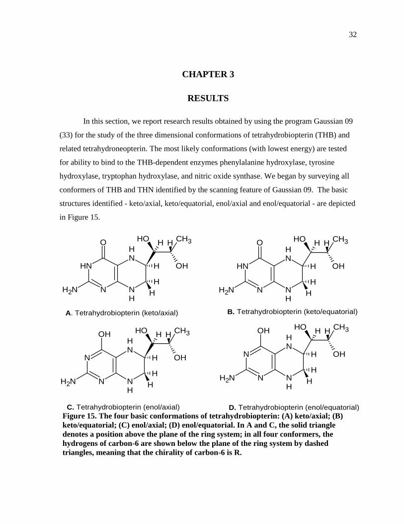
CHAPTER 3
RESULTS
In this section, we report research results obtained by using the program Gaussian 09
(33) for the study of the three dimensional conformations of tetrahydrobiopterin (THB) and
related tetrahydroneopterin. The most likely conformations (with lowest energy) are tested
for ability to bind to the THB-dependent enzymes phenylalanine hydroxylase, tyrosine
hydroxylase, tryptophan hydroxylase, and nitric oxide synthase. We began by surveying all
conformers of THB and THN identified by the scanning feature of Gaussian 09. The basic
structures identified - keto/axial, keto/equatorial, enol/axial and enol/equatorial - are depicted
in Figure 15.
Figure 15. The four basic conformations of tetrahydrobiopterin: (A) keto/axial; (B) keto/equatorial; (C) enol/axial; (D) enol/equatorial. In A and C, the solid triangle denotes a position above the plane of the ring system; in all four conformers, the hydrogens of carbon-6 are shown below the plane of the ring system by dashed triangles, meaning that the chirality of carbon-6 is R.
A. Tetrahydrobiopterin (keto/axial)
H
OH
NH2
O
NH
NH
N
NH
CH3
OH
HH
H H
H
OH
NH2
O
NH
NH
N
NH
CH3
OH
HH
H H
B. Tetrahydrobiopterin (keto/equatorial)
H
OH
NH2
OH
NH
NH
N
N
CH3
OH
HH
H H
C. Tetrahydrobiopterin (enol/axial) D. Tetrahydrobiopterin (enol/equatorial)
H
OH
NH2
OH
NH
NH
N
N
CH3
OH
HH
H H
33
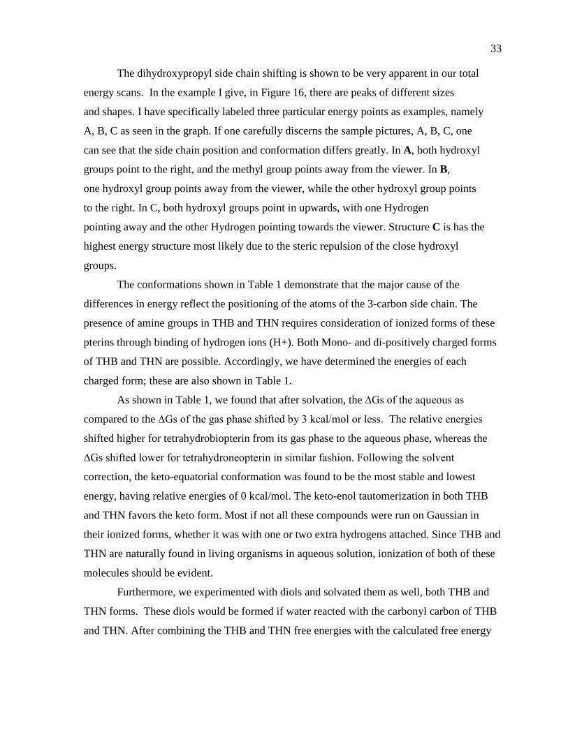
The dihydroxypropyl side chain shifting is shown to be very apparent in our total
energy scans. In the example I give, in Figure 16, there are peaks of different sizes
and shapes. I have specifically labeled three particular energy points as examples, namely
A, B, C as seen in the graph. If one carefully discerns the sample pictures, A, B, C, one
can see that the side chain position and conformation differs greatly. In A, both hydroxyl
groups point to the right, and the methyl group points away from the viewer. In B,
one hydroxyl group points away from the viewer, while the other hydroxyl group points
to the right. In C, both hydroxyl groups point in upwards, with one Hydrogen
pointing away and the other Hydrogen pointing towards the viewer. Structure C is has the
highest energy structure most likely due to the steric repulsion of the close hydroxyl
groups.
The conformations shown in Table 1 demonstrate that the major cause of the
differences in energy reflect the positioning of the atoms of the 3-carbon side chain. The
presence of amine groups in THB and THN requires consideration of ionized forms of these
pterins through binding of hydrogen ions (H+). Both Mono- and di-positively charged forms
of THB and THN are possible. Accordingly, we have determined the energies of each
charged form; these are also shown in Table 1.
As shown in Table 1, we found that after solvation, the ∆Gs of the aqueous as
compared to the ∆Gs of the gas phase shifted by 3 kcal/mol or less. The relative energies
shifted higher for tetrahydrobiopterin from its gas phase to the aqueous phase, whereas the
∆Gs shifted lower for tetrahydroneopterin in similar fashion. Following the solvent
correction, the keto-equatorial conformation was found to be the most stable and lowest
energy, having relative energies of 0 kcal/mol. The keto-enol tautomerization in both THB
and THN favors the keto form. Most if not all these compounds were run on Gaussian in
their ionized forms, whether it was with one or two extra hydrogens attached. Since THB and
THN are naturally found in living organisms in aqueous solution, ionization of both of these
molecules should be evident.
Furthermore, we experimented with diols and solvated them as well, both THB and
THN forms. These diols would be formed if water reacted with the carbonyl carbon of THB
and THN. After combining the THB and THN free energies with the calculated free energy
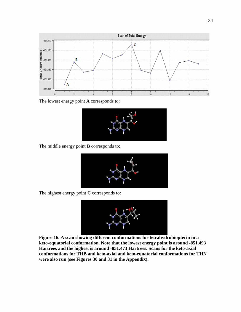
34
The lowest energy point A corresponds to:
The middle energy point B corresponds to:
The highest energy point C corresponds to:
Figure 16. A scan showing different conformations for tetrahydrobiopterin in a keto-equatorial conformation. Note that the lowest energy point is around -851.493 Hartrees and the highest is around -851.473 Hartrees. Scans for the keto-axial conformations for THB and keto-axial and keto-equatorial conformations for THN were also run (see Figures 30 and 31 in the Appendix).
35
Table 1. Thermodynamic Properties for Unhydrated and Hydrated Forms of Tetrahydrobiopterin and Tetrahydroneopterin in Various Ionization States
Name and
Configuration A b c R
∆E
kcal/mol
∆G (gas)
kcal/mol
∆G (aqueous)
kcal/mol
Tetrahydrobiopterin NH2 NH NH H
Keto-equatorial NH3 NH2 NH H 0 0 0
Keto-axial NH3 NH2 NH H 0.2 0.1 0.2
Enol-equatorial NH3 NH2 NH H 4.5 3.5 5
Enol-axial NH3 NH2 NH H 0.5 1 3.1
Tetrahydroneopterin NH2 NH NH OH
Keto-equatorial NH3 NH NH2 OH 0 0 0
Keto-axial NH3 NH NH2 OH 0.5 1.5 1.4
Enol-equatorial NH3 NH NH2 OH 8.3 7.4
Enol-axial NH3 NH NH2 OH 1.9 3.6
of water, the hydration of THB or THN to form the diol is found to be highly unfavorable,
with ΔG's of 31 and 33 kcal/mol, respectively.
After eventually discovering that ionized forms of THB did not dock as well in active
sites obtained from x-ray crystallographic studies of THB-dependent enzymes, the lowest
energy forms of the non-ionized form of THB (which did bind well in docking studies) were
chosen for more detailed examination.
We chose four lower energy configurations of tetrahydrobiopterin (THB) and
tetrahydroneopterin (THN) for detailed studies of their conformations and thermodynamic
H
OH
a
O
b
cN
NH
CH2R
OH
HH
H H
Tetrahydropterin base structure
36
properties. Figure 17 shows four structures for THB and four additional structures for THN.
As seen, THB and THN may exist in either the keto or enol form with the hydroxylated side
chain either axial or equatorial to the tetrahydropyrazine ring; all possible combinations are
shown. All of the tetrahydrobiopterin and tetrahydroneopterin conformations are in their
neutral, unsolvated forms. In the Gaussian program, calculated energy values are given
in units of Hartrees, which is why I have listed the energies in this unit. However, examining
Hartrees by itself is useless and irrelevant to the scientist. In order to find the lowest energy,
one must denote the lowest Hartree energy as zero, so it is relative to all the other energy
values to be significant.
Note that the below conformations of THB and THN have very small deviations in
their respective energies expressed in Hartrees. THB structures have energies around -
851.490 Hartrees and THN have energies around -926.705 Hartrees. A Hartree (or the
Hartree energy) is the atomic unit of energy equal to 4.3597439422x10⁻¹⁸ J. (45). Taking the
lowest energy value (-851.493 Hartrees) for THB shows that the keto axial and the enol
equatorial conformations are the most stable forms. For THN, the same analysis shows that
the keto-axial is the most stable form, followed by the enol-equatorial conformation. This
information is important because molecular shape will determine the ability for these
tetrahydropterins to bind to enzyme active sites.
Since these unbound pterins in the cell are found in an aqueous environment, it would
not be representative of the real organismal atmosphere if we only ran our THB and THN
molecules in a vacuum. We approached the solvation problem in two distinct ways: first, we
added one or two water molecules directly around our THB and THN molecules and ran the
optimizations on the Gaussian program; second, we used a COSMOtherm program to
approximate solvation of our THB and THN molecules as a uniform water shell. The
program applied to THB and one or two water molecules yielded the results shown in Figure
18.
As for the hydration studies with COSMOtherm, we estimated the energies of
hydrated keto-enol tautomers of THB and THN with side chains equatorial or axial to the
pyrazine ring and with variable positions of their alcohol groups. From these results, we
chose the lowest energy conformers for further study; the results are shown in Table 1. We
37
Figure 17. Tetrahydrobiopterin and Tetrahydroneopterin in all four conformations and at the lowest energy states.
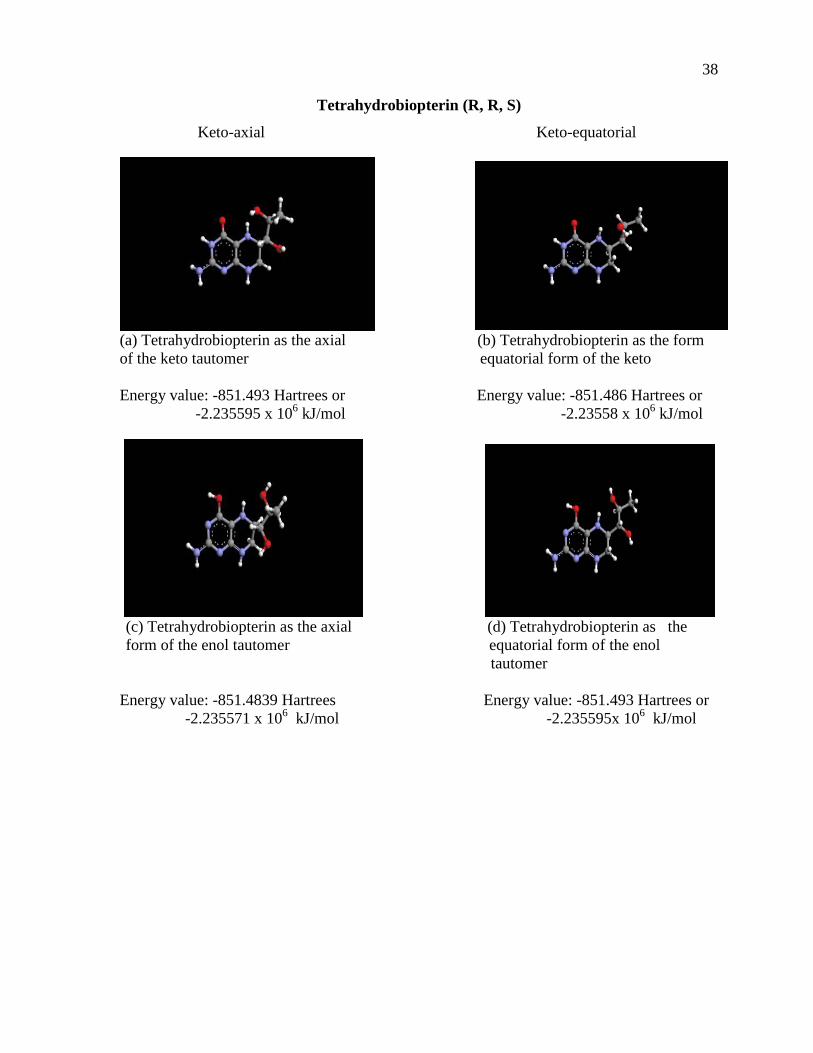
38
Tetrahydrobiopterin (R, R, S)
Keto-axial Keto-equatorial
(a) Tetrahydrobiopterin as the axial (b) Tetrahydrobiopterin as the form of the keto tautomer equatorial form of the keto Energy value: -851.493 Hartrees or Energy value: -851.486 Hartrees or -2.235595 x 10⁶ kJ/mol -2.23558 x 10⁶ kJ/mol
(c) Tetrahydrobiopterin as the axial (d) Tetrahydrobiopterin as the form of the enol tautomer equatorial form of the enol
tautomer Energy value: -851.4839 Hartrees Energy value: -851.493 Hartrees or
-2.235571 x 10⁶ kJ/mol -2.235595x 10⁶ kJ/mol
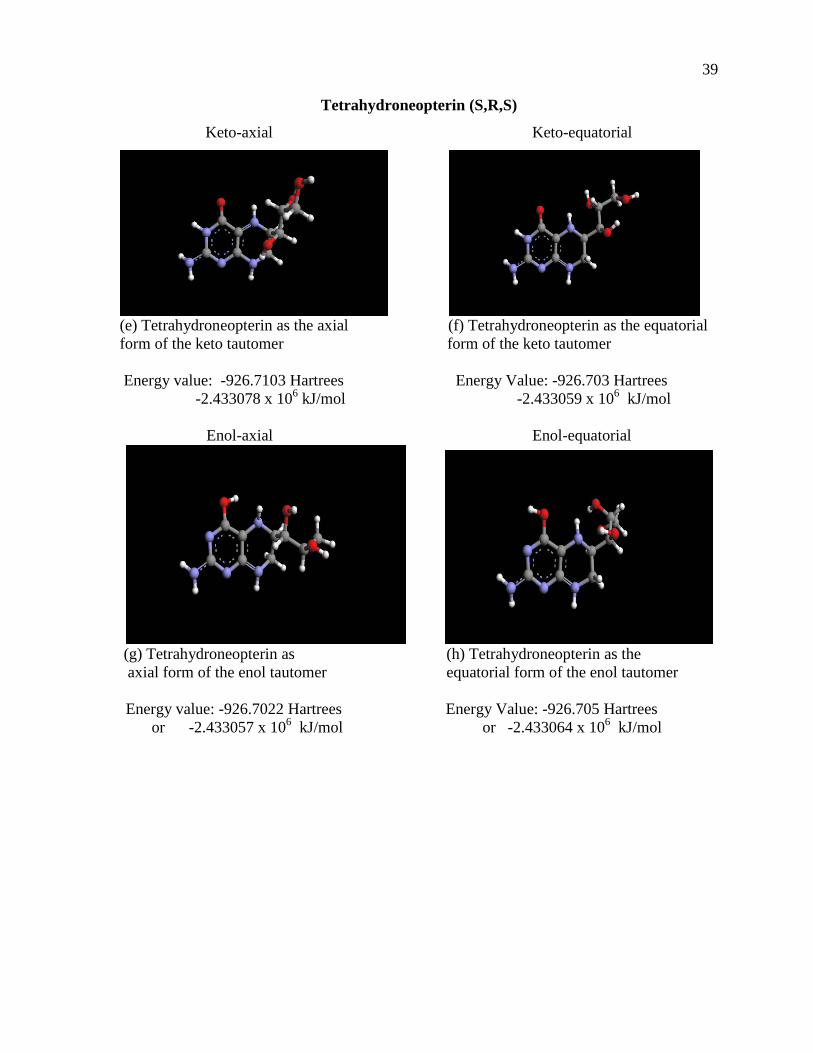
39
Tetrahydroneopterin (S,R,S)
Keto-axial Keto-equatorial
(e) Tetrahydroneopterin as the axial (f) Tetrahydroneopterin as the equatorial form of the keto tautomer form of the keto tautomer Energy value: -926.7103 Hartrees Energy Value: -926.703 Hartrees -2.433078 x 10⁶ kJ/mol -2.433059 x 10⁶ kJ/mol
Enol-axial Enol-equatorial
(g) Tetrahydroneopterin as (h) Tetrahydroneopterin as the axial form of the enol tautomer equatorial form of the enol tautomer Energy value: -926.7022 Hartrees Energy Value: -926.705 Hartrees
or -2.433057 x 10⁶ kJ/mol or -2.433064 x 10⁶ kJ/mol
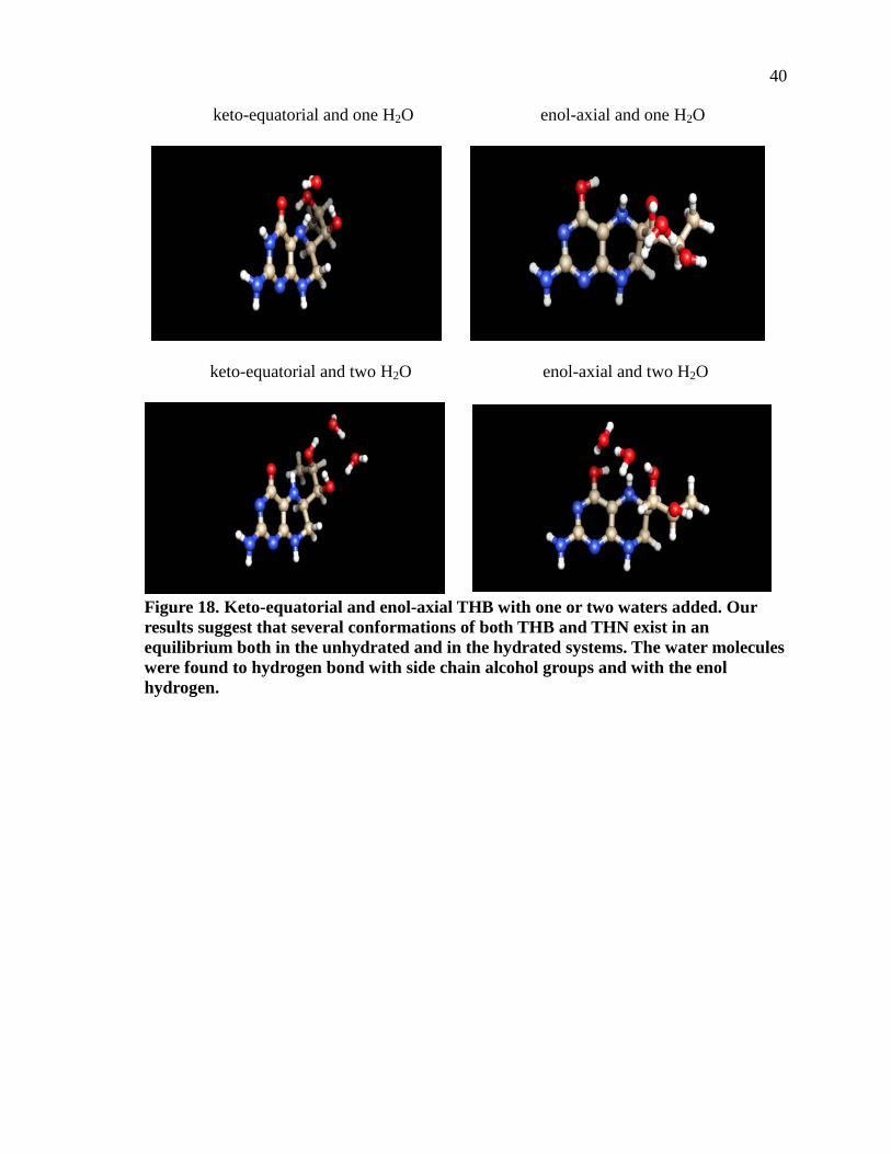
40
keto-equatorial and one H2O enol-axial and one H2O
keto-equatorial and two H2O enol-axial and two H2O
Figure 18. Keto-equatorial and enol-axial THB with one or two waters added. Our results suggest that several conformations of both THB and THN exist in an equilibrium both in the unhydrated and in the hydrated systems. The water molecules were found to hydrogen bond with side chain alcohol groups and with the enol hydrogen.
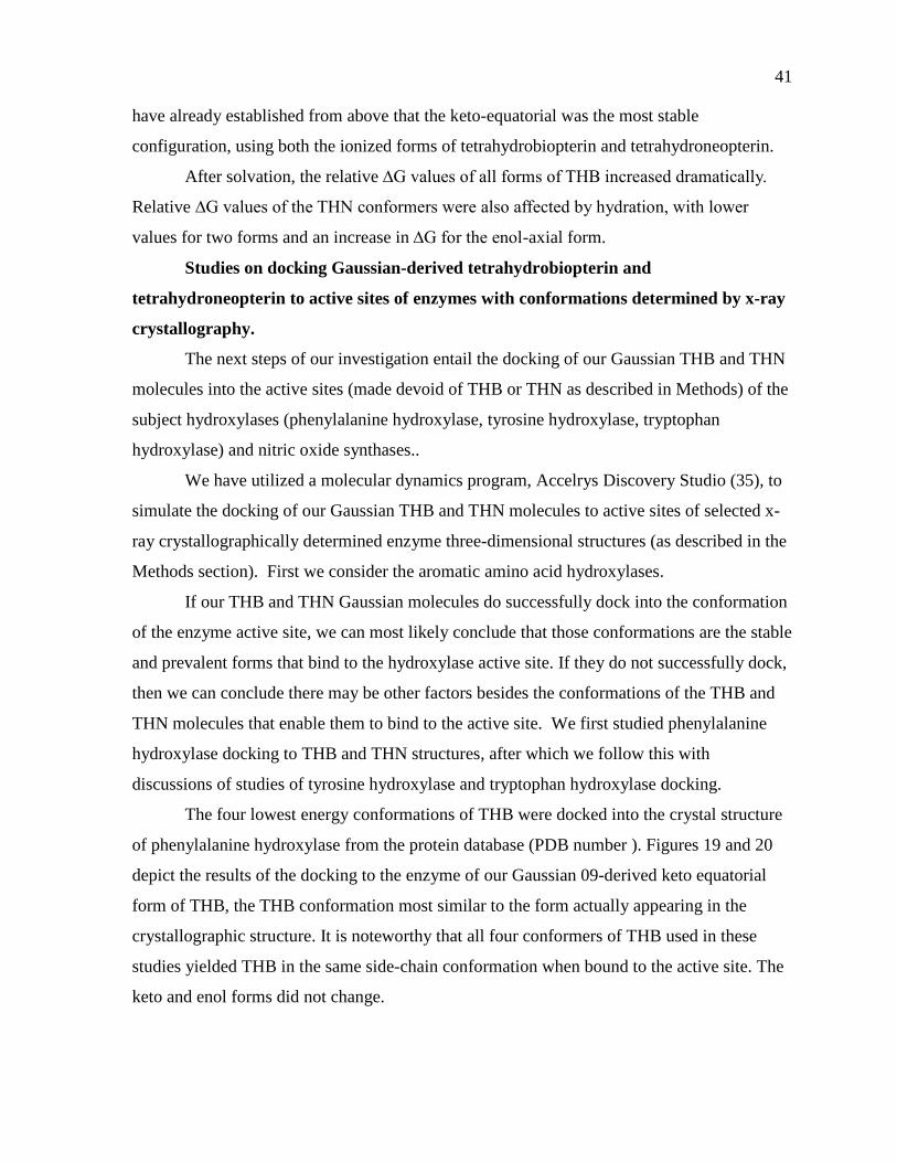
41
have already established from above that the keto-equatorial was the most stable
configuration, using both the ionized forms of tetrahydrobiopterin and tetrahydroneopterin.
After solvation, the relative ∆G values of all forms of THB increased dramatically.
Relative ∆G values of the THN conformers were also affected by hydration, with lower
values for two forms and an increase in ∆G for the enol-axial form.
Studies on docking Gaussian-derived tetrahydrobiopterin and
tetrahydroneopterin to active sites of enzymes with conformations determined by x-ray
crystallography.
The next steps of our investigation entail the docking of our Gaussian THB and THN
molecules into the active sites (made devoid of THB or THN as described in Methods) of the
subject hydroxylases (phenylalanine hydroxylase, tyrosine hydroxylase, tryptophan
hydroxylase) and nitric oxide synthases..
We have utilized a molecular dynamics program, Accelrys Discovery Studio (35), to
simulate the docking of our Gaussian THB and THN molecules to active sites of selected x-
ray crystallographically determined enzyme three-dimensional structures (as described in the
Methods section). First we consider the aromatic amino acid hydroxylases.
If our THB and THN Gaussian molecules do successfully dock into the conformation
of the enzyme active site, we can most likely conclude that those conformations are the stable
and prevalent forms that bind to the hydroxylase active site. If they do not successfully dock,
then we can conclude there may be other factors besides the conformations of the THB and
THN molecules that enable them to bind to the active site. We first studied phenylalanine
hydroxylase docking to THB and THN structures, after which we follow this with
discussions of studies of tyrosine hydroxylase and tryptophan hydroxylase docking.
The four lowest energy conformations of THB were docked into the crystal structure
of phenylalanine hydroxylase from the protein database (PDB number ). Figures 19 and 20
depict the results of the docking to the enzyme of our Gaussian 09-derived keto equatorial
form of THB, the THB conformation most similar to the form actually appearing in the
crystallographic structure. It is noteworthy that all four conformers of THB used in these
studies yielded THB in the same side-chain conformation when bound to the active site. The
keto and enol forms did not change.
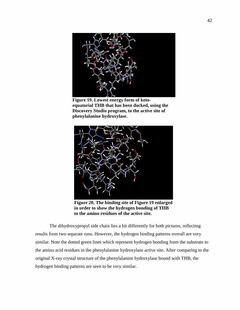
42
Figure 19. Lowest energy form of keto-equatorial THB that has been docked, using the Discovery Studio program, to the active site of phenylalanine hydroxylase.
Figure 20. The binding site of Figure 19 enlarged in order to show the hydrogen bonding of THB to the amino residues of the active site.
The dihydroxypropyl side chain lies a bit differently for both pictures, reflecting
results from two separate runs. However, the hydrogen binding patterns overall are very
similar. Note the dotted green lines which represent hydrogen bonding from the substrate to
the amino acid residues in the phenylalanine hydroxylase active site. After comparing to the
original X-ray crystal structure of the phenylalanine hydroxylase bound with THB, the
hydrogen binding patterns are seen to be very similar.
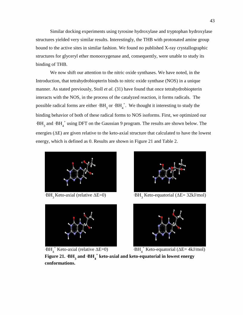
43
Similar docking experiments using tyrosine hydroxylase and tryptophan hydroxylase
structures yielded very similar results. Interestingly, the THB with protonated amine group
bound to the active sites in similar fashion. We found no published X-ray crystallographic
structures for glyceryl ether monooxygenase and, consequently, were unable to study its
binding of THB.
We now shift our attention to the nitric oxide synthases. We have noted, in the
Introduction, that tetrahydrobiopterin binds to nitric oxide synthase (NOS) in a unique
manner. As stated previously, Stoll et al. (31) have found that once tetrahydrobiopterin
interacts with the NOS, in the process of the catalyzed reaction, it forms radicals. The
possible radical forms are either ·BH3 or ·BH4+. We thought it interesting to study the
binding behavior of both of these radical forms to NOS isoforms. First, we optimized our
·BH3 and ·BH4+ using DFT on the Gaussian 9 program. The results are shown below. The
energies (ΔE) are given relative to the keto-axial structure that calculated to have the lowest
energy, which is defined as 0. Results are shown in Figure 21 and Table 2.
·BH3 Keto-axial (relative ΔE=0) ·BH3 Keto-equatorial (ΔE= 32kJ/mol)
·BH4
+ Keto-axial (relative ΔE=0) ·BH4+ Keto-equatorial (ΔE= 4kJ/mol)
Figure 21. ·BH3 and ·BH4+ keto-axial and keto-equatorial in lowest energy
conformations.

44
Table 2. Shows the Predominant Conformations and the Equilibrium Constants
In Table 2, note I have highlighted in blue the significant values, namely examining
the keto-axial conformation equilibrium constants. As seen from Table 2, when keto-axial is
on the reactant side, the Keq is a very low number. In contrast, when keto-axial is on the
product side of the equation, the Keq is a very high number. Thus, keto-axial is the
predominant form for both the ·BH3 and ·BH4+ radicals.
As seen in Table 2, the conformation of the THB dihydropropane group is
predominantly axial when the radicals are the keto tautomers. Conversely, the conformation
of the dihydropropane group is equatorial when the radicals are the enol tautomer. The ΔE
values indicate that the keto tautomer is favored over the enol tautomer. Hydration studies
were not performed on the radicals since the radicals are likely to be stabilized on the surface
of the enzyme.
It was observed that the relative ΔE differences from the conformation of the lowest
energy state (keto-axial) was much greater for the ·BH3 than the ·BH4+. Besides examining
the changes of the relative energies, we also studied the spin densities and partial charges.
Figure 22 shows the numbering system of the THB.
Referring to Figure 22, I have constructed Table 3 that shows the spin densities for all
the atoms, starting from N1 to C8α. Note the position of the dihydroxypropyl side chain did
BH3 (radical) Hartrees Joules Change Δ G (J) K(eq)Enol-axial -850.85552 -3.70952E-15 Enol (axial->equatorial) -1.03326E-20 0.081230566Enol-equatorial -850.85315 -3.70951E-15 Keto (axial->equatorial) -5.34069E-20 2.31517E-06Keto-axial -850.86485 -3.70956E-15 Axial (enol->keto) 4.06765E-20 19593.72425Keto-equatorial -850.8526 -3.7095E-15 Equatorial (enol->keto) -2.39786E-21 0.558446166
Enol-axial->keto-equatoria -1.27305E-20 0.045362898Enol-equatorial-> keto-ax 5.10091E-20 241211.2246
BH4+ (radical) Hartrees Joules Change Δ G (J) K(eq)Enol-axial -851.27244 -3.71134E-15 Enol (axial->equatorial) 2.91231E-20 1183.114989Enol-equatorial -851.27912 -3.71136E-15 Keto (axial->equatorial) -4.88292E-21 0.305324706Keto-axial -851.28352 -3.71138E-15 Axial (enol->keto) 4.8306E-20 125076.7099Keto-equatorial -851.2824 -3.71138E-15 Equatorial (enol->keto) 1.43E-20 32.27835843
Enol-axial->keto-equatoria 4.34231E-20 38189.00968Enol-equatorial-> keto-ax 1.91829E-20 105.7181348
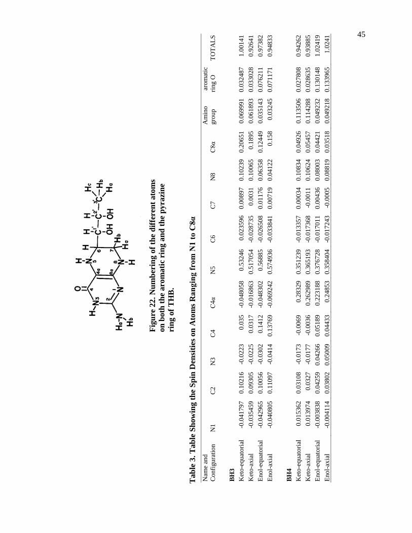
45
Fi
gure
22.
Num
beri
ng o
f the
diff
eren
t ato
ms
on b
oth
the
arom
atic
rin
g an
d th
e py
razi
ne
ring
of T
HB
.
Tab
le 3
. Tab
le S
how
ing
the
Spin
Den
sitie
s on
Ato
ms R
angi
ng fr
om N
1 to
C8α
Nam
e an
d C
onfig
urat
ion
N1
C2
N3
C4
C4α
N
5 C
6 C
7 N
8 C
8α
Am
ino
grou
p a
rom
atic
rin
g O
TO
TALS
BH
3
Ket
o-eq
uato
rial
-0.0
4179
7 0.
1021
6 -0
.022
3 0.
035
-0.0
4805
8 0.
5324
6 0.
0235
96
0.00
897
0.10
239
0.20
651
0.06
9991
0.
0324
87
1.00
141
Ket
o-ax
ial
-0.0
3545
9 0.
0930
5 -0
.022
5 0.
0317
-0
.016
863
0.51
7054
-0
.028
735
0.00
31
0.10
065
0.18
95
0.06
1893
0.
0330
28
0.92
641
Enol
-equ
ator
ial
-0.0
4296
5 0.
1005
6 -0
.030
2 0.
1412
-0
.048
302
0.56
885
-0.0
2650
8 0.
0117
6 0.
0635
8 0.
1244
9 0.
0351
43
0.07
6211
0.
9738
2 En
ol-a
xial
-0
.040
805
0.11
097
-0.0
414
0.13
769
-0.0
6924
2 0.
5749
36
- 0.0
3384
1 0.
0071
9 0.
0412
2 0.
158
0.03
245
0.07
1171
0.
9483
3
BH
4
Ket
o-eq
uato
rial
0.01
5362
0.
0310
8 -0
.017
3 -0
.006
9 0.
2832
9 0.
3512
39
-0.0
1335
7 0.
0003
4 0.
1083
4 0.
0492
6 0.
1135
06
0.02
7808
0.
9426
2 K
eto-
axia
l 0.
0139
74
0.03
27
-0.0
177
-0.0
036
0.26
2989
0.
3651
93
-0.0
1736
8 -0
.001
1 0.
1062
4 0.
0545
7 0.
1142
88
0.02
8635
0.
9388
5 En
ol-e
quat
oria
l -0
.003
838
0.04
259
0.04
266
0.05
189
0.22
3188
0.
3767
28
-0.0
1701
1 0.
0043
6 0.
0800
3 0.
0442
1 0.
0492
32
0.13
0148
1.
0241
9 En
ol-a
xial
-0
.004
114
0.03
802
0.05
009
0.04
433
0.24
853
0.35
8404
-0
.017
243
-0.0
005
0.08
819
0.03
518
0.04
9218
0.
1339
65
1.02
41
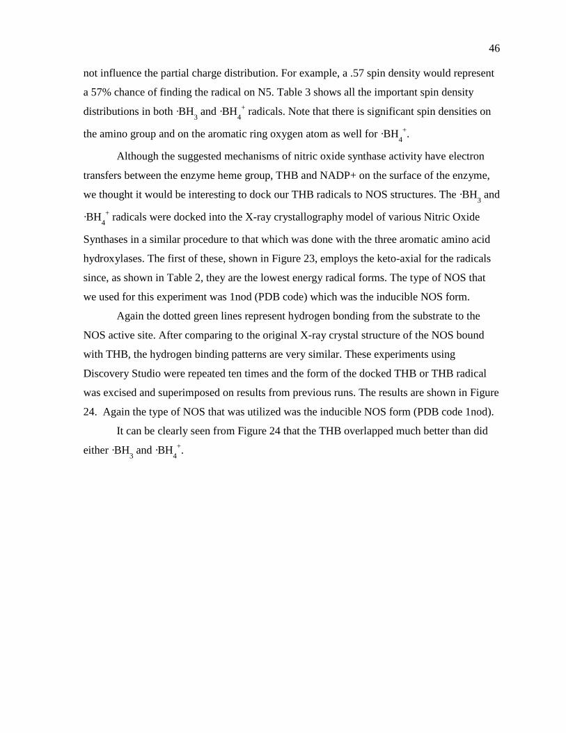
46
not influence the partial charge distribution. For example, a .57 spin density would represent
a 57% chance of finding the radical on N5. Table 3 shows all the important spin density
distributions in both ·BH3 and ·BH4+ radicals. Note that there is significant spin densities on
the amino group and on the aromatic ring oxygen atom as well for ·BH4+.
Although the suggested mechanisms of nitric oxide synthase activity have electron
transfers between the enzyme heme group, THB and NADP+ on the surface of the enzyme,
we thought it would be interesting to dock our THB radicals to NOS structures. The ·BH3 and
·BH4+ radicals were docked into the X-ray crystallography model of various Nitric Oxide
Synthases in a similar procedure to that which was done with the three aromatic amino acid
hydroxylases. The first of these, shown in Figure 23, employs the keto-axial for the radicals
since, as shown in Table 2, they are the lowest energy radical forms. The type of NOS that
we used for this experiment was 1nod (PDB code) which was the inducible NOS form.
Again the dotted green lines represent hydrogen bonding from the substrate to the
NOS active site. After comparing to the original X-ray crystal structure of the NOS bound
with THB, the hydrogen binding patterns are very similar. These experiments using
Discovery Studio were repeated ten times and the form of the docked THB or THB radical
was excised and superimposed on results from previous runs. The results are shown in Figure
24. Again the type of NOS that was utilized was the inducible NOS form (PDB code 1nod).
It can be clearly seen from Figure 24 that the THB overlapped much better than did
either ·BH3 and ·BH4+.
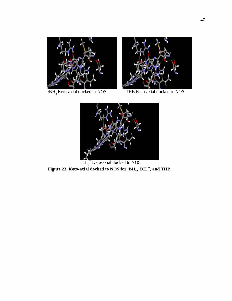
47
·BH3 Keto-axial docked to NOS THB Keto-axial docked to NOS
·BH4
+ Keto-axial docked to NOS Figure 23. Keto-axial docked to NOS for ·BH3, ·BH4
+, and THB.
48
·BH3 docked 10 times to NOS THB docked 10 times to NOS
·BH4
+ docked 10 times to NOS Figure 24. ·BH3, ·BH4
+, and THB docked to NOS each 10 times.
49
CHAPTER 4
CONCLUSION / DISCUSSION
Both tetrahydrobiopterin and tetrahydroneopterin exist in an equilibrium of
conformations, varying between the keto and enol tautomers and the axial and equatorial side
chain shifts. These molecules are never static, but are always fluid and shifting from one
conformation to another and back. Free energies do show that some conformations should be
more abundant than others at equilibrium. As deduced from our results, it is clear that the
structure of a molecule is extremely crucial in determining the chemical characteristics of a
particular molecule. For instance, the chemical characteristics of both tetrahydrobiopterin and
tetrahydroneopterin are quite different, due to their distinct conformations.
Tetrahydrobiopterin has the R,R,S stereochemistry, whereas tetrahydroneopterin has
the S, R,S stereochemistry. The stereochemistry of both molecules denotes the positions of
the hydroxyl groups on carbon 1 and carbon 2 of the hydroxypropyl side chain. Since the
first ring of both pterins are rigid and aromatic, the second ring has a bit of flexibility with
carbon 6 shifting either upwards or downwards, compared to the plane of the molecule. Most
of the shifting occurs in the highly flexible side chain. The dihydroxypropyl side chain
shifting is very apparent in our total energy scans.
Next, we ran the ionized versions of THB and THN, with either one or two positive
charges. Since these pterins are naturally found in the living cell, their real environment is an
aqueous environment. It is highly probable that these pterins will be protonated by either one
or two hydrogens in the aqueous solution. The program that was used to simulate the aqueous
solution was COSMOtherm (34), which adds a shell of uniform water molecules around the
molecule of interest. Thus, each of these pterins were run under Gaussian first to optimize
their structures, then run on COSMOtherm to obtain the free energies of the solvated species.
After analyzing the data, it was apparent that the free energies had increased for
tetrahydrobiopterin, from ∆G of the vacuum to ∆G of solvent. In contrast, the free energies
for tetrahydroneopterin had decreased, from ∆G of the vacuum to ∆G of solvent. Overall, the
∆Gs of the aqueous as compared to the ∆Gs of the gas phase shifted by 3 kcal/mol or less.
50
For both tetrahydrobiopterin and tetrahydroneopterin, the lowest energy structure was the
keto-equatorial conformation. Diols were solvated as well, but were discarded because their
∆Gs were deemed highly unfavorable at around 31 and 33 kcal/mol.
Most if not all our research was performed using the non-ionized version of the
pterins, since the ionized versions docked poorly to our enzymes We eventually chose the
four lowest energy configurations of tetrahydrobiopterin (THB) and for tetrahydroneopterin
(THN) for detailed studies of their conformations and thermodynamic properties, and their
lowest energies.
In addition to running experiments with the pterins in a uniform water solvent shell
with COSMOtherm, we also ran the pterins with one or two water molecules to investigate
any presence of hydrogen bonding. We chose structures had the best potential for hydrogen
bonding, which were the keto-equatorial and enol-equatorial conformations. When one water
molecule was added to the keto-equatorial forms, it hydrogen bonded to the carboxyl group
and the nitrogen and hydroxyl groups. When one water molecule was added to the enol-
equatorial forms, it hydrogen bonded to the hydroxyl groups on the side chain. However,
when two water molecules were added, the water molecules both hydrogen bonded to
themselves and to the hydroxyl groups, forming a cage-like structure. The water molecules
were originally set up far apart from each other to eliminate any random-binding results.
As discussed previously, THB binds distinctly to all three different hydroxylases,
since the all the active sites have dissimilar residues. After binding the Gaussian-derived
pterins and observing that the binding locations and hydrogen bonding patterns are very
similar to that found in their x-ray crystallographic structures, we concluded that the binding
has successfully mimicked the actual binding.
The pterins, specifically tetrahydrobiopterin, bind to Nitric Oxide Synthase (NOS) in
a unique manner. As stated previously, Stoll et al. (31) have found that once
tetrahydrobiopterin interacts with the NOS it undergoes reactions to become a radical form.
The radical form can be either ·BH3 or ·BH4+ but is most likely the latter (31). We wanted to
test the binding of the radicals to the NOS active site, so we performed docking experiments
with our radicals. First, we optimized our ·BH3 and ·BH4+ under Gaussian. I chose
specifically Keto-axial and Keto-equatorial conformations to the radicals in our experiment.
51
Once we obtained the optimized radical structures, we compared the relative energies of the
radicals and determined that overall, the ·BH3 and ·BH4+ keto-axial conformation were the
lowest energy forms.
We also observed the spin densities to determine on which atoms the radical would
most likely be. From Table 2, it can be clearly seen that radicals are more likely to be found
on N5 of ·BH3, than that of ·BH4+, having 20% higher probability. For ·BH3, it appears that
the radical lies in the vicinity of N5 for about 50%-55% of the time, and shifts to the C8α
atom for about 15-20%. As for the rest, the radical shifts from C2 to C7 to N8 for about the
same time, 10%. As for ·BH4+, the radicals lie around N5 for about 35% of the time, shifting
to the C4α atom for around 25%. The radical lies around N8 for the rest of the 10% of the
time. Another interesting observation is that both the ·BH3 keto tautomers have very similar
spin densities, as well as the ·BH3 enol tautomers. That observation applied to the ·BH4+ keto
and enol tautomers as well. Apparently, the tautomerization plays an important role in
determining the location of the radical, more than the position of the side chain. After data
analysis, we conclude that there is more delocalization of the radicals on the ·BH4+ than the
·BH3. Radicals lie on N5 on ·BH3 for about half the time, in contrast for radicals having a 30-
35% probability on the same atom.
Since keto-axial was found to be the lowest energy form for both ·BH3 and ·BH4+, we
used the keto-axial conformation of both forms to dock to the X-ray crystallographic NOS
enzyme. After docking our keto-axial radicals to the active site of the NOS enzyme, we
compared our docking results with that of the hydrogen-bonding patterns in the original X-
ray crystallographic NOS. After the comparison, we concluded that the hydrogen binding
pattern were very similar and thus the docking was a success.
More importantly, we docked our ·BH3 and ·BH4+ radicals ten times and isolated the
docked radical, observing the quality of the 10 overlapping molecules; the better the
overlapping, the more accurate the docking was. Thus, ·BH3 docked the poorest, having 4 out
of 10 molecules with decent overlapping. ·BH4+ had better overlapping, with 6 out of 10
being good overlaps. I even ran the neutral THB molecule, docking it 10 times in similar
fashion. Neutral tetrahydrobiopterin actually had the best overlapping result, with nearly 8 or
52
9 out of 10 for good overlaps. We believe the reason is that the THB is in its neutral form
before binding fully to the active site in the NOS, shifting it from neutral to the radical forms.
Thus, right when the THB binds to the NOS, it is still in the neutral conformation. Again,
these results are supported from the multiple docking studies showing better overlapping of
THB than that of either ·BH3 or ·BH4+. In conclusion, in both the neutral and the radical form
of THB, the keto-axial is the lowest energy and thus is the predominant conformation.
Overall, these docking experiments are important because they may lead to the
development of competitive inhibitors with therapeutic effects for certain pathological
conditions. There are many implications for these computational studies, for instance, for a
drug company desiring to create a drug that inhibits the binding of the original ligand. As we
have seen, there are certain hydrogen bonding patterns specific for THB bound to
phenylalanine hydroxylase or to nitric oxide synthase.
If one designs a drug that mimics the hydrogen bonding patterns of THB, and then
synthesizes certain parts that makes the binding even stronger than the THB, then that drug
will probably be a successfully inhibitor. Upon close examination, effective substitutions
could probably be made of the oxygen group on the C4, the amino group, and the two
hydroxyl groups on the side chain. One possible substitution could be another hydroxyl
group, for example, to replace the amino NH2 group. Other substitutions could be a strong
electronegative group, such as fluorine, to take the place of the side chain hydroxyl groups.
The main problem would be that the substituents cannot take up more space than the THB in
the active site, otherwise the new molecule may not bind properly. However, if those any of
the substitutions on the new molecule induce strong hydrogen bonding and the bonds are
even stronger than that of THB to the enzyme, than the new molecule can possibly be a new
inhibitor (R. Metzger, personal communication).
53
CHAPTER 5
STUDIES OF THE THB RECYCLING PATHWAY
Since dihydrobiopterin is formed during the oxygenation of various substrates in the
hydroxylation pathway, it must be regenerated to THB, This involves a dehydration reaction
in which 4-hydroxytetrahydrobiopterin is transformed to quinoid 7,8 dihydrobiopterin, which
can undergo non-enzymatic isomerization to 7,8-dihydrobiopterin, as shown in Figure 25 (R.
Metzger, personal communication).
We intend to study details of the two forms of dihydrobiopterin in greater detail. We
will run the two forms of dihydrobiopterin (quinoid dihydrobiopterin and 7,8
dihydrobiopterin) under Gaussian DFT to determine their lowest energy conformations.
Their 3-dimensional conformations will enable us to better understand the mechanism of the
pathway and this form was used for some of the x-ray crystallographic studies of pterins
bound to the active sites of hydroxylase enzymes. Figure 25 shows the difference in structure
of the dihydrobiopterins and the tetrahydrobiopterin.
Figure 25 shows the full cycle of the tetrahydrobiopterin recycling pathway, starting
from tetrahydrobiopterin. This reacts with oxygen to form the intermediate with the oxygen
adduct on the carbon 4 of the intermediate. Next, the intermediate serves as a coenzyme for
L-tryptophan to form 5-hydroxy-L-tryptophan. In that process, the intermediate is thereby
converted into 4α-hydroxytetrahydrobiopterin. The 4α-hydroxytetrahydrobiopterin then
interacts with pterin-4α-carbinolamine dehydratase to form the quinoid form of 7,8
dihydrobiopterin, shown in Figure 25. In order for the original tetrahydrobiopterin to be
properly reformed again, NADPH dehydrobiopteridine reductase converts the quinoid 7,8
dihydrobiopterin back into tetrahydrobiopterin. Figure 26 show some examples of
conformations for qDHB run under Gaussian. Figure 27 shows a proposed mechanism with
our postulated intermediates through which the quinoid form of 7,8 dihydrobiopterin double
bond changes from a C-4:N-5 to the N-5:C-6 of 7,8-dihydrobiopterin.
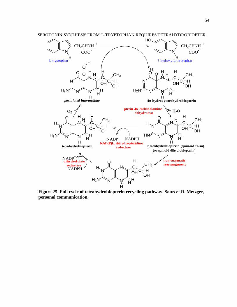
54
Figure 25. Full cycle of tetrahydrobiopterin recycling pathway. Source: R. Metzger, personal communication.
NH
COO-CH2CHNH3
+
NH
COO-CH2CHNH3
+HO
SEROTONIN SYNTHESIS FROM L-TRYPTOPHAN REQUIRES TETRAHYDROBIOPTERI
L-tryptophan 5-hydroxy-L-tryptophan
NADP+
NADPH
H2O
N
N
OH
N
N CC
CH3
OH
HH
HH2N
H
OHH
HH
N
N
OH
N
N CC
CH3
OH
HH2N
H
OHH
HH
N
N
O
N
N CC
CH3
OH
HH
HH2N
H
OHH
HH
OH
N
N
O
N
N CC
CH3
OH
HH
HH2N
H
OHH
HH
OO
H
pterin-4α-carbinolamine dehydratase
NAD(P)H dehydropteridine reductase 7,8-dihydrobiopterin (quinoid form)
N
N
OH
N
N CC
CH3
OH
H
HHN
H
OHH
HH
O2
4α-hydroxytetrahydrobiopterinpostulated intermediate
tetrahydrobiopterin
dihydrofolate reductase
NADPHNADP+
(or quinoid dihydrobiopterin)
non-enzymaticrearrangement
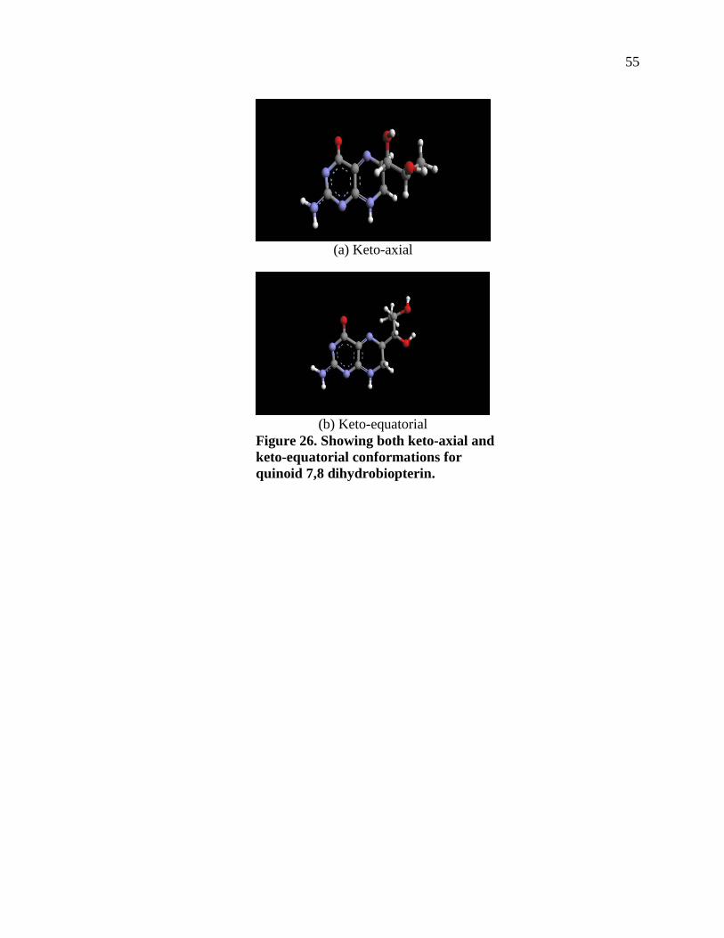
55
(a) Keto-axial
(b) Keto-equatorial
Figure 26. Showing both keto-axial and keto-equatorial conformations for quinoid 7,8 dihydrobiopterin.
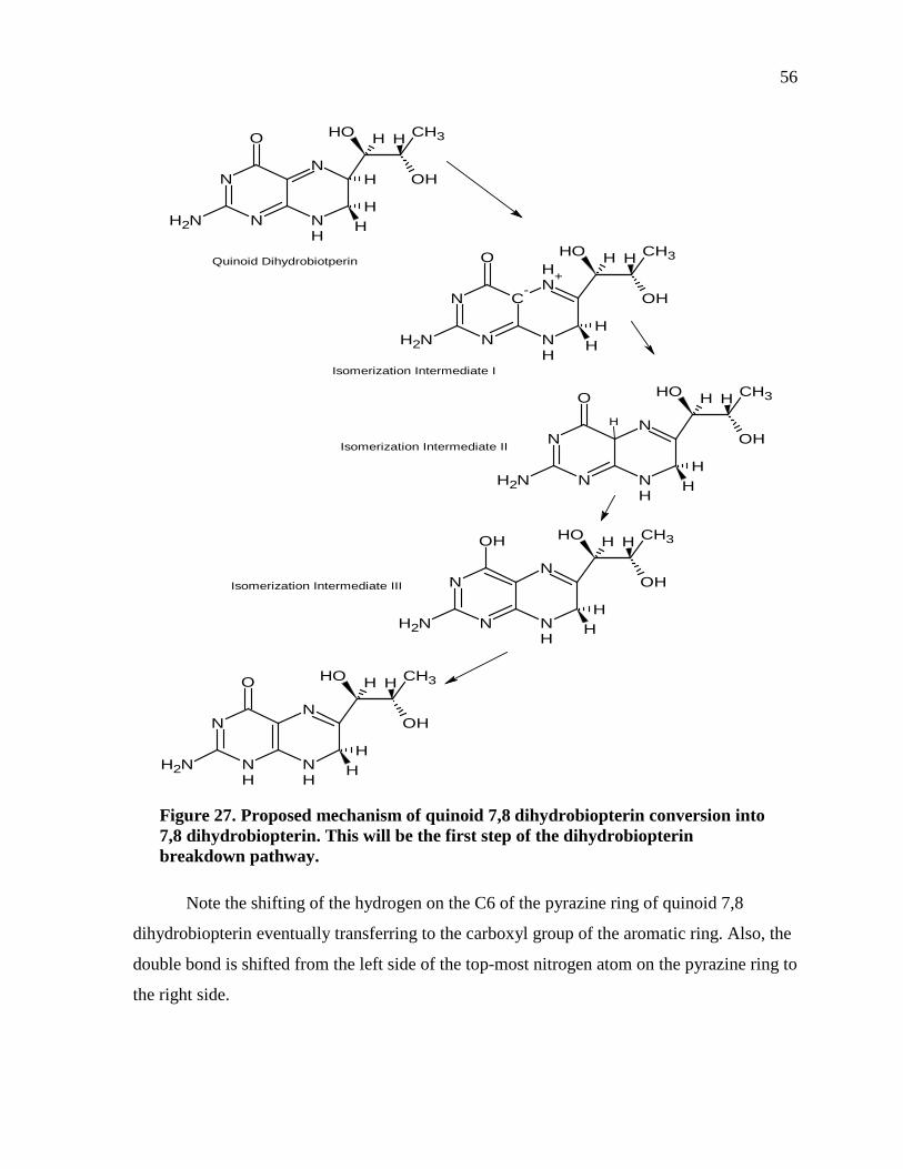
56
Figure 27. Proposed mechanism of quinoid 7,8 dihydrobiopterin conversion into 7,8 dihydrobiopterin. This will be the first step of the dihydrobiopterin breakdown pathway.
Note the shifting of the hydrogen on the C6 of the pyrazine ring of quinoid 7,8
dihydrobiopterin eventually transferring to the carboxyl group of the aromatic ring. Also, the
double bond is shifted from the left side of the top-most nitrogen atom on the pyrazine ring to
the right side.
H
OH
NH2
O
N
NH
N
N
CH3
OH
HH
H H
OH
NH2
O
N
NH
NH
N
CH3
OH
HH
H H
OH
NH2
O
NH+
NH
C-
N
N
CH3
OH
HH
H H
H
OH
NH2
O
N
NH
N
N
CH3
OH
HH
H H
OH
NH2
OH
N
NH
N
N
CH3
OH
HH
H H
Quinoid Dihydrobiotperin
Isomerization Intermediate I
Isomerization Intermediate II
Isomerization Intermediate III
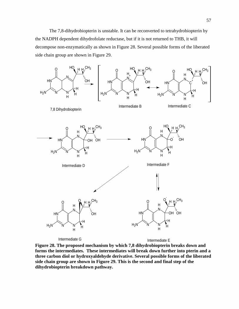
57
The 7,8-dihydrobiopterin is unstable. It can be reconverted to tetrahydrobiopterin by
the NADPH dependent dihydrofolate reductase, but if it is not returned to THB, it will
decompose non-enzymatically as shown in Figure 28. Several possible forms of the liberated
side chain group are shown in Figure 29.
Figure 28. The proposed mechanism by which 7,8 dihydrobiopterin breaks down and forms the intermediates. These intermediates will break down further into pterin and a three carbon diol or hydroxyaldehyde derivative. Several possible forms of the liberated side chain group are shown in Figure 29. This is the second and final step of the dihydrobiopterin breakdown pathway.
OH
NH2
O
N
NH
N
NH
CH3
OH
HH
H H OH
NH2
O
NH+
NH
N
NH
CH3
OH
HH
H H
7,8 Dihydrobiopterin
OH
NH2
O
C+N
H
NH
N
NH
CH3
OH
HH
H H
OH
NH2
O
NH
NH
N
NH
CH3
OH
HH
H H
OH
OH
NH2
O
NH
NH
N
NH
CH3
OH
HH
H H
O-
O-
NH2
O
NH
NH
N
NH
CH3
OH
HH
H H
OH
O
NH2
O
NH
NH
N
NH
CH3
OH
HH
H H
Intermediate B Intermediate C
Intermediate D Intermediate F
Intermediate EIntermediate G
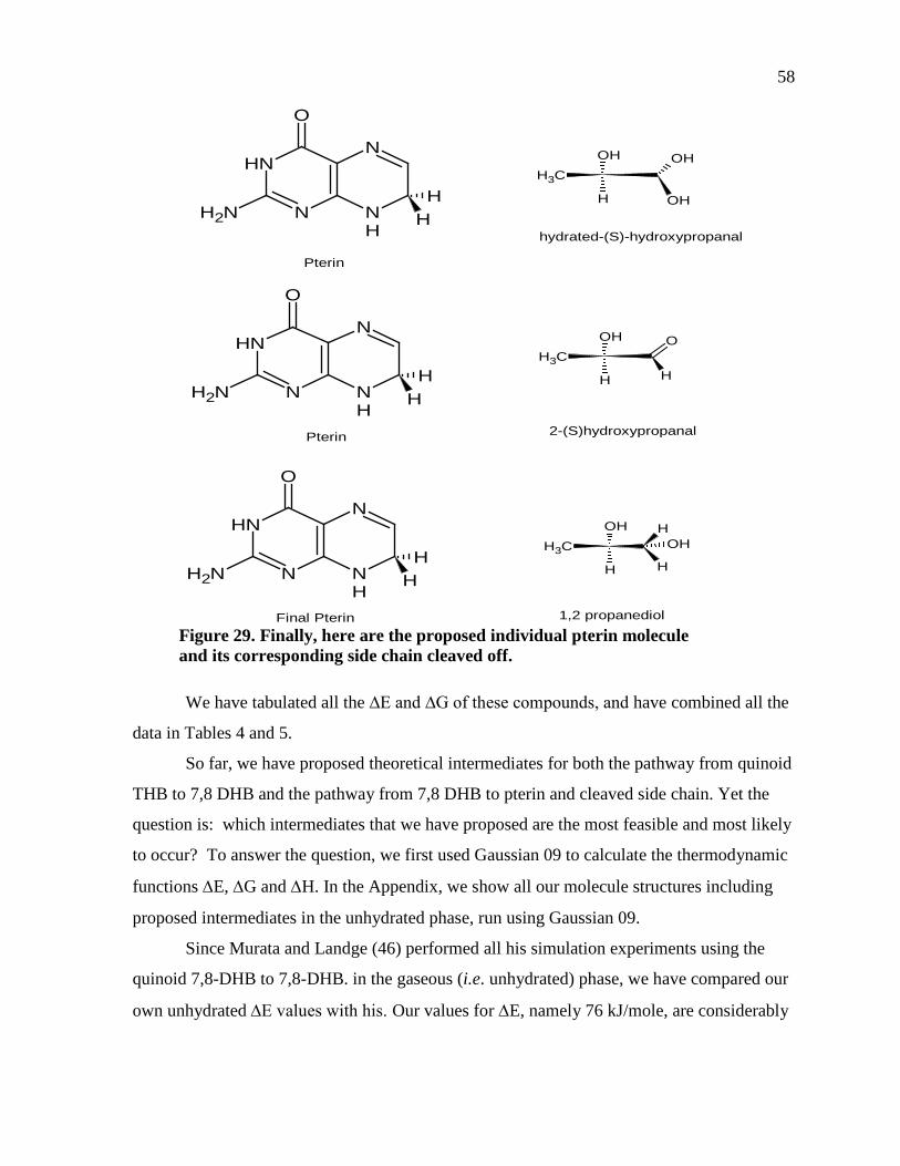
58
Figure 29. Finally, here are the proposed individual pterin molecule and its corresponding side chain cleaved off.
We have tabulated all the ∆E and ∆G of these compounds, and have combined all the
data in Tables 4 and 5.
So far, we have proposed theoretical intermediates for both the pathway from quinoid
THB to 7,8 DHB and the pathway from 7,8 DHB to pterin and cleaved side chain. Yet the
question is: which intermediates that we have proposed are the most feasible and most likely
to occur? To answer the question, we first used Gaussian 09 to calculate the thermodynamic
functions ∆E, ∆G and ∆H. In the Appendix, we show all our molecule structures including
proposed intermediates in the unhydrated phase, run using Gaussian 09.
Since Murata and Landge (46) performed all his simulation experiments using the
quinoid 7,8-DHB to 7,8-DHB. in the gaseous (i.e. unhydrated) phase, we have compared our
own unhydrated ∆E values with his. Our values for ∆E, namely 76 kJ/mole, are considerably
Pterin
NH2
O
N
NH
N
NH
HH
NH2
O
N
NH
N
NH
HH
Pterin
Final Pterin
NH2
O
N
NH
N
NH
HH
CH3
OH
H OH
OH
CH3
OH
H H
O
CH3
OH
H
H
H
OH
hydrated-(S)-hydroxypropanal
2-(S)hydroxypropanal
1,2 propanediol
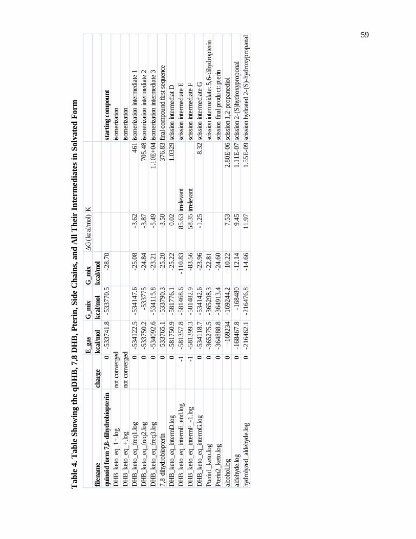
59
Tab
le 4
. Tab
le S
how
ing
the
qDH
B, 7
,8 D
HB
, Pte
rin,
Sid
e C
hain
s, an
d A
ll T
heir
Inte
rmed
iate
s in
Solv
ated
For
m
E_ga
sG
_mix
G_m
ix∆G
(kca
l/mol
)K
filen
ame
char
gekc
al/m
olkc
al/m
olkc
al/m
olqu
inoi
d fo
rm 7
,8-di
hydr
obio
pter
in0
-533
741.
8-5
3377
0.5
-28.
70st
artin
g co
mpo
unt
DH
B_ke
to_e
q_1+
.log
not c
onve
rged
isom
eriza
tion
DH
B_ke
to_e
q_+.
log
not c
onve
rged
isom
eriza
tion
DH
B_ke
to_e
q_fre
q1.lo
g0
-534
122.
5-5
3414
7.6
-25.
08-3
.62
461
isom
eriza
tion
inter
med
iate
1D
HB_
keto
_eq_
freq2
.log
0-5
3375
0.2
-533
775
-24.
84-3
.87
705.
48iso
mer
izatio
n int
erm
edia
te 2
DH
B_ke
to_e
q_fre
q3.lo
g0
-534
092.
6-5
3411
5.8
-23.
21-5
.49
1.10
E+04
isom
eriza
tion
inter
med
iate
37,
8-di
hydr
obio
pter
in0
-533
765.
1-5
3379
0.3
-25.
20-3
.50
376.
83fin
al co
mpo
und
first
sequ
ence
DH
B_ke
to_e
q_int
erm
D.lo
g0
-581
750.
9-5
8177
6.1
-25.
220.
021.
0329
sciss
ion in
term
edia
t DD
HB_
keto
_eq_
inter
mE_
end.
log
-1-5
8135
7.8
-581
468.
6-1
10.8
385
.63
irrele
vant
sciss
ion in
term
edia
te E
DH
B_ke
to_e
q_int
erm
F_-1
.log
-1-5
8139
9.3
-581
482.
9-8
3.56
58.3
5irr
eleva
ntsc
ission
inte
rmed
iate
FD
HB_
keto
_eq_
inter
mG
.log
0-5
3411
8.7
-534
142.
6-2
3.96
-1.2
58.
32sc
ission
inte
rmed
iate
GPt
erin1
_ket
o.lo
g0
-365
275.
5-3
6529
8.3
-22.
81sc
issio
n int
erm
eidat
e: 5,
6-di
hydr
opte
rinPt
erin2
_ket
o.lo
g0
-364
888.
8-3
6491
3.4
-24.
60sc
ission
fina
l pro
duct
: pte
rinalc
ohol
.log
0-1
6923
4-1
6924
4.2
-10.
227.
532.
80E-
06sc
issio
n 1,
2-pr
opan
edio
lald
ehyd
e.lo
g0
-168
467.
8-1
6848
0-1
2.14
9.45
1.11
E-07
sciss
ion
2-(S
)hyd
roxy
prop
onal
hydr
olyz
ed_a
ldeh
yde.
log
0-2
1646
2.1
-216
476.
8-1
4.66
11.9
71.
55E-
09sc
issio
n hy
drat
ed 2
-(S)
-hyd
roxy
prop
anal
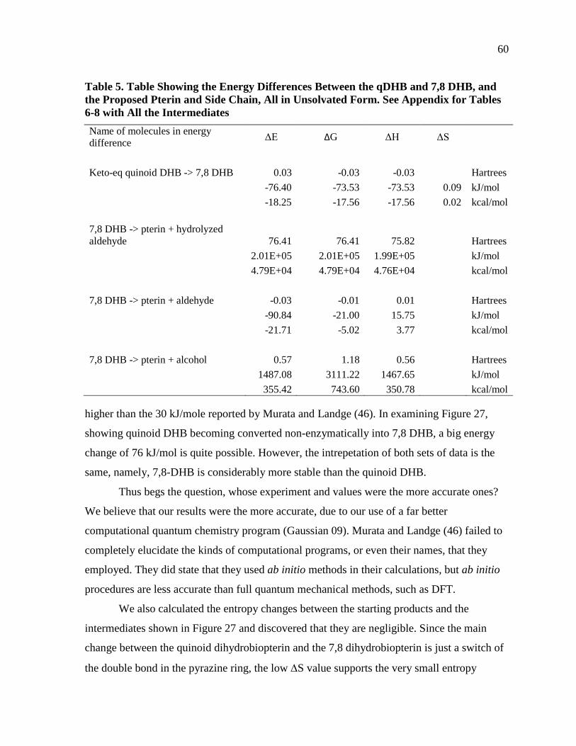
60
Table 5. Table Showing the Energy Differences Between the qDHB and 7,8 DHB, and the Proposed Pterin and Side Chain, All in Unsolvated Form. See Appendix for Tables 6-8 with All the Intermediates Name of molecules in energy difference ΔE ΔG ΔH ΔS
Keto-eq quinoid DHB -> 7,8 DHB 0.03 -0.03 -0.03
Hartrees
-76.40 -73.53 -73.53 0.09 kJ/mol
-18.25 -17.56 -17.56 0.02 kcal/mol
7,8 DHB -> pterin + hydrolyzed aldehyde 76.41 76.41 75.82
Hartrees
2.01E+05 2.01E+05 1.99E+05
kJ/mol
4.79E+04 4.79E+04 4.76E+04
kcal/mol
7,8 DHB -> pterin + aldehyde -0.03 -0.01 0.01
Hartrees
-90.84 -21.00 15.75
kJ/mol
-21.71 -5.02 3.77
kcal/mol
7,8 DHB -> pterin + alcohol 0.57 1.18 0.56
Hartrees
1487.08 3111.22 1467.65
kJ/mol
355.42 743.60 350.78
kcal/mol
higher than the 30 kJ/mole reported by Murata and Landge (46). In examining Figure 27,
showing quinoid DHB becoming converted non-enzymatically into 7,8 DHB, a big energy
change of 76 kJ/mol is quite possible. However, the intrepetation of both sets of data is the
same, namely, 7,8-DHB is considerably more stable than the quinoid DHB.
Thus begs the question, whose experiment and values were the more accurate ones?
We believe that our results were the more accurate, due to our use of a far better
computational quantum chemistry program (Gaussian 09). Murata and Landge (46) failed to
completely elucidate the kinds of computational programs, or even their names, that they
employed. They did state that they used ab initio methods in their calculations, but ab initio
procedures are less accurate than full quantum mechanical methods, such as DFT.
We also calculated the entropy changes between the starting products and the
intermediates shown in Figure 27 and discovered that they are negligible. Since the main
change between the quinoid dihydrobiopterin and the 7,8 dihydrobiopterin is just a switch of
the double bond in the pyrazine ring, the low ∆S value supports the very small entropy
61
change (shown in Table 5). Because the reactions of importance occur in aqueous solution,
we have used the program COSMOtherm to simulate solvation of all the molecules,
including quinoid DHB, 7,8 DHB, proposed reaction intermediates, pterins and their
corresponding side chains,
We have calculated the ∆G of the differences between the intermediates and both the
quinoid DHB and the 7,8-DHB. ∆G results still demonstrate the greater stability of the 7.8-
DHB compared to quinoid DHB. The intermediates in their solvated states yielded ∆G values
that were higher than that of the starting reactant, viz. the quinoid dihydrobiopterin.
From these data, we have calculated estimated rate constants for both reactions studied using
the equation k = e -∆G/RT, where the constant R is 0.00198 kcal/mole and T is 298°K.
The ∆Gs for the decomposition of 7,8-DHB were calculated from the difference
between 7,8 DHB and the products pterin and 2-hydroxypropanal. The results were
converted from units of Hartrees to those of both kJ/mol and kcal/mol. After obtaining the
results, it was clear that both the ∆Gs for the hydrolyzed aldehyde and the alcohol with pterin
were unrealistic (range in the thousands). We conclude that the most feasible side chain
product was most likely the aldehyde, for which ∆Gs values were more reasonable (lower
than 21kJ/mol).

62
ACKNOWLEDGEMENTS
I would like to thank my research advisor, Dr. Robert Metzger, for his endless
guidance and patience, along with my thesis committee members Dr. Tom Huxford and Dr.
Andrew Cooksy. I would also like to thank Dr. James Otto for the use of the Accelrys
Discovery Studio program. Lastly, I would like to thank the Department of Computer
Science for providing the Intel core i5 processors in GMSC 425 to run the Gaussian program.
63
REFERENCES
1. De Crécy-Lagard, V., and Hanson, A. (2012) Subsystem: Pterin Biosynthesis: Tetrahydrobiopterin (BH4) Biosynthesis and Regeneration, University of Florida, Gainesville, FL, http://www.theseed.org/SubsystemStories/Pterin_biosynthesis/ story.pdf
2. Feldman, D.M. (2004) Neopterin a novel marker of immune function in Hepatitis C. Indian J. Occup. Environ. Med. 12, 107-111
3. Cowperthwaite, J., Weber, M.M., Packer L., and Hutner, H.H. (1953) Nutrition of herpetomonas (strigomonas) culicidarum. Annals NY Acad. Sci. 56, 972-981
4. Fuchs, D., Hausen, A., Knosp, O., Reibnegger, G., Wachter, H., Hofler, M., Kosanowski, H., Huber, C., and Niederwieser, D. (1983) Neopterin evaluation in patients with pulmonary tuberculosis. Bioch. Clin. Asp. Pteridines 2, 281-291
5. Kaufmann, S., and Levenberg, B. (1959) Further studies in the phenylalanine-hydroxylation cofactor. J. Biol. Chem. 234, 2683-2688
6. Kaufman, S. (1963) The structure of the phenylalanine-hydroxylation cofactor. Proc. Nat. Acad. Sci. 5, 1085-1093
7. Kaufman, S., and Fisher, D.B. (1974) Molecular Mechanisms of Oxygen Activation (Hayaishi, O. ed.), Academic, New York.
8. Kaufman, S., Pollock, R.J., Summer, G.K., Das, A.K., and Hajra, A.K. (1990) Dependence of an alkyl glycol-ether monooxygenase activity upon tetrahydropterins. Biochim. Biophys. Acta. 1040, 19-27
9. Mayer, B., and Werner, E.R. (1995) In search of a function for tetrahydrobiopterin in the biosynthesis of nitric oxide. Naunyn. Schmiedebergs Arch. Pharmacol. 351, 453-463
10. Metametrix. (2008) Neopterin and Biopterin, Metametrix, Atlanta, GA, http://www. metametrix.com/files/learning-center/articles/neopterin-biopterin.pdf
11. Fuchs, D. (n.d.) Neopterin, Innsbruck Medical University, Innsbruck, Austria, http://www.neopterin.net/neopterin_e.pdf
12. Estelberger, W., Mlekusch, W., and Reibnegger, G. (1995) The conformational flexibility of 5,6,7,8-tetrahydrobiopterin and 5,6,7,8-tetrahydroneopterin: A molecular dynamical simulation. FEBS Letters 357, 37-40
13. Katoh, S., Sueoka, T., and Kurihara, T. (1993) Theoretical stereostructure of the neutral form of tetrahydrobiopterin. Pteridines 4, 27-31
14. Ziegler, I., Borchert, M., Heaney, F., Davis, A.P., and Boyle, P.H. (1992) Structural requirements for the modulatory effect of 6-substituted pterins on interleukin 2 receptor binding. Biochim. Biophys. Acta. 1135, 330-334
64
15. Bracher, A., Eisenreich,W., Schramek, N., Ritz, H., Götze, E., Herrmann, A., Gutlich, M., and Bacher, A. (1998) Biosynthesis of pteridines. J. Biol. Chem. 273, 28132-28141
16. Martinez, A., Dao, K.K., McKinney, J., Teigen, K., and Frøystein, N.Ǻ. (2000) The conformation of 5,6,7,8 tetrahydrobiopterin and 7,8-dihydrobiopterin in solution: A H1 NMR study. Pteridines 11, 32-33
17. Goodwill, K.E., Sabatier, C., and Stevens, R.C. (1998) Crystal structure of tyrosine hydroxylase with bound cofactor analogue and iron at 2.3 Å resolution: Self-hydroxylation of phe300 and the pterin-binding site. Biochemistry 37, 13439-13445
18. Wang, L., Erlandsen, H., Haavik, J., Knappskog, P.M., and Stevens, R.C. (2002) Three-dimensional structure of human tryptophan hydroxylase and its implications for the biosynthesis of the neurotransmitters serotonin and melatonin. Biochemistry 41, 12569-12574
19. Martinez, A., Knappskog, P.M., and Haavik, J. (2001) Structural approach into human tryptophan hydroxylase and its implications for the regulation of serotonin biosynthesis. Cur. Med. Chem. 8, 1077-1091
20. Genetics Home Reference. (2012) Phenylketonuria, U.S. National Library of Medicine, Bethesda, MD, http://ghr.nlm.nih.gov/condition/phenylketonuria
21. Erlandsen, H., Bjørgo, E., Flatmark, T., and Stevens, R.C. (2000) Crystal structure and site-specific mutagenesis of pterin-bound human phenylalanine hydroxylase. Biochemistry 39, 2208-2217
22. Andersen, O.A., Flatmark, T., and Hough, E. (2001) High resolution crystal structures of the catalytic domain of human phenylalanine hydroxylase in its catalytically active Fe(II) form and binary complex with tetrahydrobiopterin. J. Mol. Biol. 314, 279-291
23. White K.A., and Marletta, M.A. (1992) Nitric oxide synthase is a cytochrome P-450 type hemoprotein. Biochemistry 31, 6627-6631
24. Kaufman, S. (1993) New tetrahydrobiotperin-dependent systems. Annu. Rev. Nutr. 13, 261-286
25. Groves, J.T., and Wang, C.Y. (2000) Nitric oxide synthase: Models and mechanisms. Cur. Opin. Chem. Biol. 4, 687-695
26. Crane, B.R., Arvai, A.S., Ghosh, D.K., Wu, C., Getzoff, E.D., Stuehr, D.J., and Tainer, J.A. (1998) Structure of nitric oxide synthase oxygenase dimer with pterin and substrate. Science 279, 2121-2126
27. Raman, C.S., Li, H., Martasek, P., Kral, V., Masters, B.S., and Poulos, T.L. (1998) Crystal structure of constitutive endothelial nitric oxide synthase: A paradigm for pterin function involving a novel metal center. Cell 95, 939-950
28. Hillier, B.J., Christopherson, K.S., Prehoda, K.E., Bredt, D.S., and Lim, W.A. (1999) Unexpected modes of PDZ domain scaffolding revealed by structure of nNOS-syntrophin complex. Science 284, 812-815
65
29. Zhang, J., Martàsek, P., Paschke, R., Shea, T., Siler Masters, B.S., and Kim J.J. (2001) Crystal structure of the FAD/NADPH-binding domain of rat neuronal nitric-oxide synthase. J. Biol. Chem. 276, 37506-37513
30. Matter, H., Kumar, H.S., Fedorov, R., Frey, A., Kotsonis, P., Hartmann, E., Fröhlich, L.G., Reif, A., Pfleiderer, W., Scheurer, P., Ghosh, D.K., Schlichting, I., and Schmidt, H.H. (2005) Structural analysis of isoform-specific inhibitors targeting the tetrahydrobiopterin binding site of human nitric oxide synthases. J. Med. Chem. 48, 4783-4792
31. Stoll, S., Nejaty Jahromy, Y., Woodward, J.J., Ozarowski, A., Marletta, M.A., and Britt, R.D. (2010) Nitric oxide synthase stabilizes the tetrahydrobiopterin cofactor radical by controlling its protonation state. J. Amer. Chem. Soc. 132, 11812-11823
32. Frisch, M.J., Trucks, G.W., Schlegel, H.B., Scuseria, G.E., Rob, M.A., Cheeseman, J.R., Montgomery, J.A., Vreven, T., Kudin, K.N., Burant, J.C., Millam, J.M., Iyengar, S.S., Tomasi, J., Barone, V., Mennucci, B., Cossi, M., Scalmani, G., Rega, N., Petersson, G.A., Nakatsuji, H., Hada, M., Ehara, M., Toyota, K., Fukuda, R., Hasegawa, J., Ishida, M., Nakajima, T., Honda, Y., Kitao, O., Nakai, H., Klene, M., Li, X., Knox, J.E., Hratchian, H.P., Cross, J.B., Bakken, V., Adamo, C., Jaramillo, J., Gomperts, R., Stratmann, R.E., Yazyev, O., Austin, A.J., Cammi, R., Pomelli, C., Ochterski, J.W., Ayala, P.Y., Morokuma, K., Voth, G.A., Salvador, P., Dannenberg, J.J., Zakrzewski, V.G., Dapprich, S., Daniels, A.D., Strain, M.C., Farkas, O., Malick, D.K., Rabuck, A.D., Raghavachari, K., Foresman, J.B., Ortiz, J.V., Cui, Q., Baboul, A.G., Clifford, S., Cioslowski, J., Stefanov, B.B., Liu, G., Liashenko, A., Piskorz, P., Komaromi, I., Martin, R.L., Fox, D.J., Keith, T., Al-Laham, M.A., Peng, C.Y., Nanayakkara, A., Challacombe, M., Gill, P.M.W., Johnson, B., Chen, W., Wong, M.W., Gonzalez, C., and Pople, A.J. (2003) Gaussian 03, Gaussian, Inc., Wallingford, CT
33. Frisch, M.J., Trucks, G.W., Schlegel, H.B., Scuseria, G.E., Robb, M.A., Cheeseman, J.R., Scalmani, G., Barone, V., Mennucci, B., Petersson, G.A., Nakatsuji, H., Caricato, M., Li, X., Hratchian, H.P., Izmaylov, A.F., Bloino, J., Zheng, G., Sonnenberg, J.L., Hada, M., Ehara, M., Toyota, K., Fukuda, R., Hasegawa, J., Ishida, M., Nakajima, T., Honda, Y., Kitao, O., Nakai, H., Vreven, T., Montgomery, Jr., J.A., Peralta, J.E., Ogliaro, F., Bearpark, M., Heyd, J.J., Brothers, E., Kudin, K.N., Staroverov, V.N., Kobayashi, R., Normand, J., Raghavachari, K., Rendell, A., Burant, J.C., Iyengar, S.S., Tomasi, J., Cossi, M., Rega, N., Millam, J.M., Klene, M., Knox, J.E., Cross, J.B., Bakken, V., Adamo, C., Jaramillo, J., Gomperts, R., Stratmann, R.E., Yazyev, O., Austin, A.J., Cammi, R., Pomelli, C., Ochterski, J.W., Martin, R.L., Morokuma, K., Zakrzewski, V.G., Voth, G.A., Salvador, P., Dannenberg, J.J., Dapprich, S., Daniels, A.D., Farkas, Ö., Foresman, J.B., Ortiz, J.V., Cioslowski, J., Fox, D.J. (2009) Revision A.1, Gaussian, Inc., Wallingford, CT
34. COSMOlogic. (2007) COSMOtherm 1.05, COSMOlogic GmbH & Co. KG, Leverkeusen Germany
35. Accelrys Software. (2009) Discovery Studio Modeling Environment, Release 2.5, Accelrys Software Inc., San Diego, CA
66
36. Klamt, A., and Eckert, F. (2000) COSMO-RS: A novel and efficient method for the a priori prediction of thermophysical data of liquids. Flu. Phase Equi. 172, 43-72
37. COSMOlogic. (2012) COSMOtherm, COSMOlogic GmbH & Co. KG, Leverkeusen Germany, http://www.cosmologic.de/index.php?cosId=4203&crId=4
38. Lui, R. (2005) Computational Study of Aqueous Reactions in Vitamin E Regeneration, San Diego State University, San Diego, CA
39. Giannozzi, P. (2012) Density Functional Theory, University of Udine, Udine, Italy, http://www.fisica.uniud.it/~giannozz/Corsi/MetNum/LectureNotes/metnum-cap2.pdf
40. Atkins, P., and Paula, J.D. (2009) Physical Chemistry, 9th Ed., Oxford University Press, New York, NY
41. Levine, I.N. (2002) Physical Chemistry, 5th Ed., McGraw Hill, New York, NY
42. Dunning, Jr., T.H. (1989) Gaussian basis sets for use in correlated molecular calculations. I. The atoms boron through neon and hydrogen. J. Chem. Phys. 90, 1007-1023
43. Lee, C., Yang, W., and Parr, R.G. (1988) Development of the Cole-Salvetti correlation energy formula into a functional of the electron density. Phys. Rev. A. 37, 785-789
44. Koch, W., and Holthausen M.C. (2000) A Chemist’s Guide to Density Functional Theory, Wiley-VCH Verlag, Weinheim, Germany
45. McCammon Group. (2012) Physical Constant Table, University of California, San Diego, CA, http://users.mccammon.ucsd.edu/~blu/Research-Handbook/physical-constant.html
46. Murata, S., and Landge, S.S. (2006) A computational study on stabilities of dihydropterins. Heterocycles 68, 1705-1708

67
APPENDIX
EXTRA GRAPHS AND TABLES
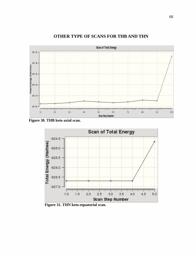
68
OTHER TYPE OF SCANS FOR THB AND THN
Figure 30. THB keto axial scan.
Figure 31. THN keto-equatorial scan.
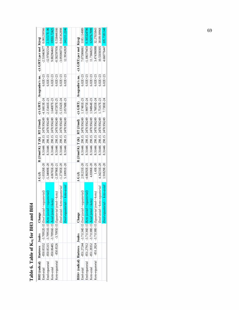
69 T
able
6. T
able
of K
eq fo
r B
H3
and
BH
4
BH
3 (r
adic
al)
Har
tree
sJo
ules
Cha
nge
Δ G
(J)
R (J
/mol
K)
T (K
)R
T (J
/mol
) -
(Δ G
/RT)
Avo
grad
ro's
no.
-(Δ
G/R
T) p
er m
olK
(eq)
Enol-
axial
-850
.855
52-3
.709
52E-
15En
ol (a
xial
->eq
uato
rial)
-1.0
3326
E-20
8.31
446
298.
1524
78.9
5624
9-4
.168
13E-
246.
02E+
23-2
.510
4636
770.
0812
3056
6En
ol-eq
uato
rial
-850
.853
15-3
.709
51E-
15Ke
to (a
xial
->eq
uato
rial)
-5.3
4069
E-20
8.31
446
298.
1524
78.9
5624
9-2
.154
41E-
236.
02E+
23-1
2.97
6025
342.
3151
7E-0
6K
eto-
axial
-850
.864
85-3
.709
56E-
15Ax
ial (
enol
->ke
to)
4.06
765E
-20
8.31
446
298.
1524
78.9
5624
91.
6408
7E-2
36.
02E+
239.
8829
6460
219
593.
7242
5K
eto-
equa
toria
l-8
50.8
526
-3.7
095E
-15
Equa
toria
l (en
ol->
keto
)-2
.397
86E-
218.
3144
629
8.15
2478
.956
249
-9.6
7287
E-25
6.02
E+23
-0.5
8259
7056
0.55
8446
166
Enol
-axi
al->
keto
-equ
ator
ial
-1.2
7305
E-20
8.31
446
298.
1524
78.9
5624
9-5
.135
42E-
246.
02E+
23-3
.093
0607
330.
0453
6289
8En
ol-e
quat
oria
l-> k
eto-
axia
l5.
1009
1E-2
08.
3144
629
8.15
2478
.956
249
2.05
768E
-23
6.02
E+23
12.3
9342
828
2412
11.2
246
BH
4+ (r
adic
al)
Har
tree
sJo
ules
Cha
nge
Δ G
(J)
R (J
/mol
K)
T (K
)R
T (J
/mol
) -
(Δ G
/RT)
Avo
grad
ro's
no.
-(Δ
G/R
T) p
er m
olK
(eq)
Enol-
axial
-851
.272
44-3
.711
34E-
15En
ol (a
xial
->eq
uato
rial)
2.91
231E
-20
8.31
446
298.
1524
78.9
5624
91.
1748
1E-2
36.
02E+
237.
0759
0606
111
83.1
1498
9En
ol-eq
uato
rial
-851
.279
12-3
.711
36E-
15Ke
to (a
xial
->eq
uato
rial)
-4.8
8292
E-21
8.31
446
298.
1524
78.9
5624
9-1
.969
75E-
246.
02E+
23-1
.186
3794
590.
3053
2470
6K
eto-
axial
-851
.283
52-3
.711
38E-
15Ax
ial (
enol
->ke
to)
4.83
06E-
208.
3144
629
8.15
2478
.956
249
1.94
864E
-23
6.02
E+23
11.7
3668
251
1250
76.7
099
Ket
o-eq
uato
rial
-851
.282
4-3
.711
38E-
15Eq
uato
rial (
enol
->ke
to)
1.43
E-20
8.31
446
298.
1524
78.9
5624
95.
7685
5E-2
46.
02E+
233.
4743
9698
832
.278
3584
3En
ol-a
xial
->ke
to-e
quat
oria
l4.
3423
1E-2
08.
3144
629
8.15
2478
.956
249
1.75
167E
-23
6.02
E+23
10.5
5030
305
3818
9.00
968
Enol
-equ
ator
ial->
ket
o-ax
ial
1.91
829E
-20
8.31
446
298.
1524
78.9
5624
97.
7383
E-24
6.02
E+23
4.66
0776
447
105.
7181
348
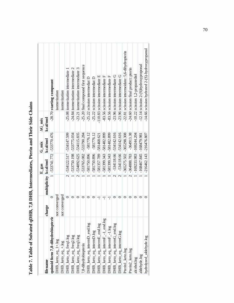
70 T
able
7. T
able
of S
olva
ted
qDH
B, 7
,8 D
HB
, Int
erm
edia
tes,
Pter
in a
nd T
heir
Sid
e C
hain
s
E_g
asG
_mix
ΔG_m
ixfil
enam
ech
arge
mul
tiplic
itykc
al/m
olkc
al/m
olkc
al/m
olqu
inoi
d fo
rm 7
,8-d
ihyd
robi
opte
rin
01
-533
741.
772
-533
770.
476
-28.
70st
artin
g co
mpo
unt
DH
B_k
eto_
eq_1
+.lo
gno
t con
verg
ediso
mer
izatio
nD
HB
_ket
o_eq
_+.lo
gno
t con
verg
ediso
mer
izatio
nD
HB
_ket
o_eq
_fre
q1.lo
g0
2-5
3412
2.51
7-5
3414
7.59
9-2
5.08
isom
eriza
tion
inte
rmed
iate
1D
HB
_ket
o_eq
_fre
q2.lo
g0
1-5
3375
0.19
8-5
3377
5.03
4-2
4.84
isom
eriza
tion
inte
rmed
iate
2D
HB
_ket
o_eq
_fre
q3.lo
g0
2-5
3409
2.62
5-5
3411
5.83
5-2
3.21
isom
eriza
tion
inte
rmed
iate
37,
8-di
hydr
obio
pter
in0
1-5
3376
5.08
2-5
3379
0.28
4-2
5.20
final
com
poun
d fir
st s
eque
nce
DH
B_k
eto_
eq_i
nter
mD
_end
.log
01
-581
750.
896
-581
776.
12-2
5.22
sciss
ion
inte
rmed
iat D
DH
B_k
eto_
eq_i
nter
mD
.log
01
-581
750.
896
-581
776.
12-2
5.22
sciss
ion
inte
rmed
iat D
DH
B_k
eto_
eq_i
nter
mE_
end.
log
-11
-581
357.
789
-581
468.
621
-110
.83
sciss
ion
inte
rmed
iate
ED
HB
_ket
o_eq
_int
erm
F_-1
_end
.log
-11
-581
399.
343
-581
482.
899
-83.
56sc
issio
n in
term
edia
te F
DH
B_k
eto_
eq_i
nter
mF_
-1.lo
g-1
1-5
8139
9.34
3-5
8148
2.89
9-8
3.56
sciss
ion
inte
rmed
iate
FD
HB
_ket
o_eq
_int
erm
G_e
nd.lo
g0
2-5
3411
8.66
-534
142.
616
-23.
96sc
issio
n in
term
edia
te G
DH
B_k
eto_
eq_i
nter
mG
.log
02
-534
118.
66-5
3414
2.61
6-2
3.96
sciss
ion
inte
rmed
iate
GPt
erin
1_ke
to.lo
g0
1-3
6527
5.50
2-3
6529
8.30
8-2
2.81
sciss
ion
inte
rmei
date
: 5,6
-dih
ydro
pter
inPt
erin
2_ke
to.lo
g0
2-3
6488
8.78
3-3
6491
3.38
-24.
60sc
issio
n fin
al p
rodu
ct: p
terin
alco
hol.l
og0
1-1
6923
3.98
3-1
6924
4.20
4-1
0.22
sciss
ion
1,2-
prop
aned
iol
alde
hyde
.log
01
-168
467.
845
-168
479.
981
-12.
14sc
issio
n 2-
(S)h
ydro
xypr
opon
alhy
drol
yzed
_ald
ehyd
e.lo
g0
1-2
1646
2.14
3-2
1647
6.80
7-1
4.66
sciss
ion
hydr
ated
2-(
S)-h
ydro
xypr
opan
al
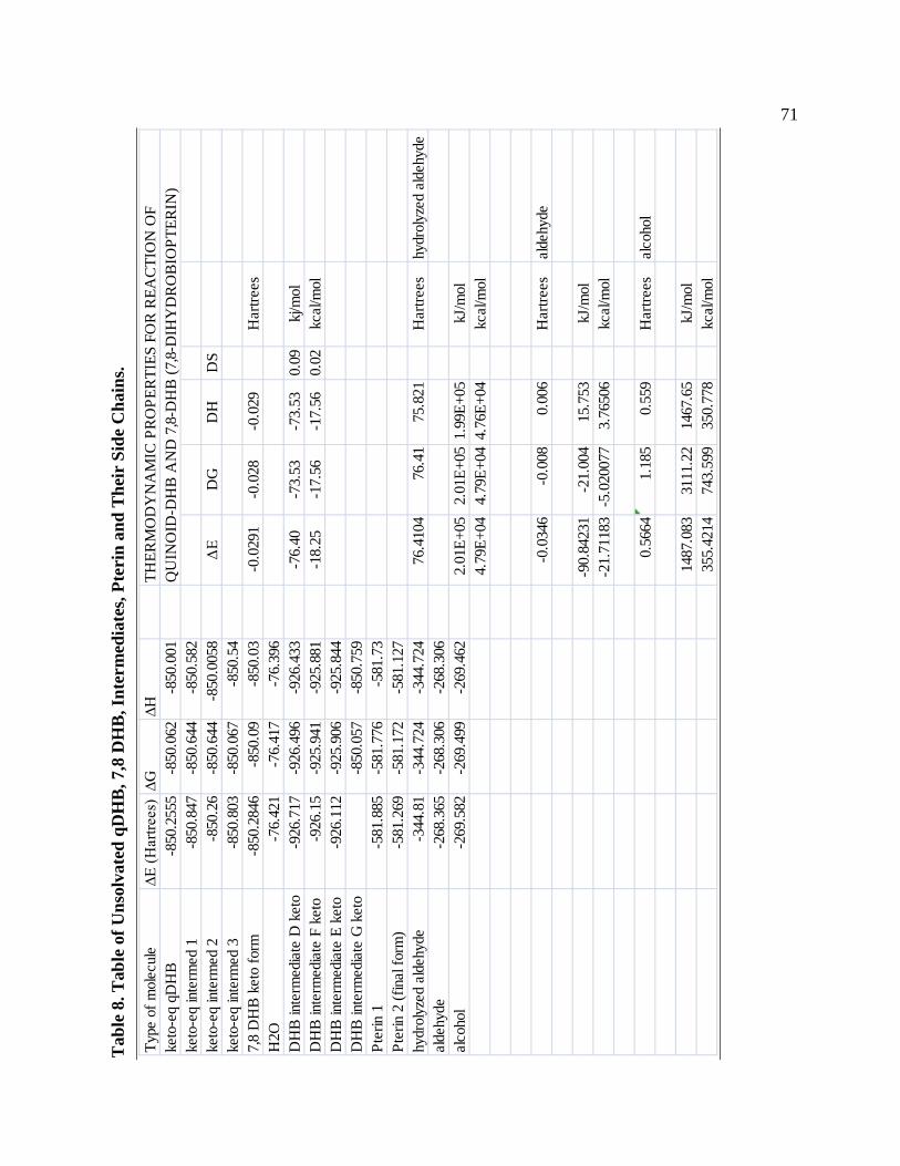
71 T
able
8. T
able
of U
nsol
vate
d qD
HB
, 7,8
DH
B, I
nter
med
iate
s, Pt
erin
and
The
ir S
ide
Cha
ins.
Ty
pe o
f mol
ecul
e∆E
(Har
trees
)∆G
∆H
THER
MO
DY
NA
MIC
PR
OPE
RTI
ES F
OR
REA
CTI
ON
OF
keto
-eq
qDH
B-8
50.2
555
-850
.062
-850
.001
QU
INO
ID-D
HB
AN
D 7
,8-D
HB
(7,8
-DIH
YD
RO
BIO
PTER
IN)
keto
-eq
inte
rmed
1-8
50.8
47-8
50.6
44-8
50.5
82ke
to-e
q in
term
ed 2
-850
.26
-850
.644
-850
.005
8Δ
ED
GD
HD
Ske
to-e
q in
term
ed 3
-850
.803
-850
.067
-850
.54
7,8
DH
B k
eto
form
-850
.284
6-8
50.0
9-8
50.0
3-0
.029
1-0
.028
-0.0
29H
artre
esH
2O-7
6.42
1-7
6.41
7-7
6.39
6D
HB
inte
rmed
iate
D k
eto
-926
.717
-926
.496
-926
.433
-76.
40-7
3.53
-73.
530.
09kj
/mol
DH
B in
term
edia
te F
ket
o-9
26.1
5-9
25.9
41-9
25.8
81-1
8.25
-17.
56-1
7.56
0.02
kcal
/mol
DH
B in
term
edia
te E
ket
o-9
26.1
12-9
25.9
06-9
25.8
44D
HB
inte
rmed
iate
G k
eto
-850
.057
-850
.759
Pter
in 1
-581
.885
-581
.776
-581
.73
Pter
in 2
(fin
al fo
rm)
-581
.269
-581
.172
-581
.127
hydr
olyz
ed a
ldeh
yde
-344
.81
-344
.724
-344
.724
76.4
104
76.4
175
.821
Har
trees
hydr
olyz
ed a
ldeh
yde
alde
hyde
-268
.365
-268
.306
-268
.306
alco
hol
-269
.582
-269
.499
-269
.462
2.01
E+05
2.01
E+05
1.99
E+05
kJ/m
ol4.
79E+
044.
79E+
044.
76E+
04kc
al/m
ol
-0.0
346
-0.0
080.
006
Har
trees
alde
hyde
-90.
8423
1-2
1.00
415
.753
kJ/m
ol-2
1.71
183
-5.0
2007
73.
7650
6kc
al/m
ol
0.56
641.
185
0.55
9H
artre
esal
coho
l
1487
.083
3111
.22
1467
.65
kJ/m
ol35
5.42
1474
3.59
935
0.77
8kc
al/m
ol
Related Documents