J. Exp. Med. The Rockefeller University Press • 0022-1007/2002/05/1233/13 $5.00 Volume 195, Number 10, May 20, 2002 1233–1245 http://www.jem.org/cgi/doi/10.1084/jem.20011930 1233 CD8 CD11b Dendritic Cells Present Exogenous Virus-like Particles to CD8 T Cells and Subsequently Express CD8 and CD205 Molecules Gabriel Morón, 1 Paloma Rueda, 2 Ignacio Casal, 2 and Claude Leclerc 1 1 Unité de Biologie des Régulations Immunitaires, Institut Pasteur, 75724 Paris, France 2 Immunología y Génetica Aplicada, SA, 28037 Madrid, Spain Abstract Recombinant porcine parvovirus virus-like particles (PPV-VLPs) are particulate exogenous anti- gens that induce a strong, specific cytotoxic T lymphocyte (CTL) response in the absence of adju- vant. In the present report, we demonstrate in vivo that dendritic cells (DCs) present PPV-VLPs to CD8 T cells after intracellular processing. PPV-VLPs are captured by DCs with a high effi- cacy, which results in the delivery of these exogenous antigens to 50% of the whole spleen DC population. In vivo, a few hours after injection, PPV-VLPs are presented exclusively to CD8 T cells by CD8 DCs, whereas 15 hours later they are presented mainly by CD8 DCs. Af- ter PPV-VLPs processing, a fraction of CD11b DCs undergo phenotypic changes, i.e., the up-regulation of CD8 and CD205 and the loss of CD4 molecules on their surface. The fail- ure to detect mRNA coding for CD8 in CD11b DCs suggests that CD8 expression by these cells is not due to de novo synthesis. In recombination-activating gene knockout mice (Rag / ), CD11b DCs did not express CD8 and PPV-VLPs presentation by CD8 DCs was severely diminished. These results indicate that both CD8 and CD8 DCs play an im- portant role in the induction of CTL responses by exogenous antigens, such as VLP. Key words: virus-like particles • dendritic cells • cross-priming • CTL • exogenous antigens Introduction The induction of CTL responses requires the presentation of antigen-derived peptides associated to MHC class I mol- ecules on the surface of APCs to specific CD8 T cells. These peptides essentially derive from antigens processed in the cytosol of APCs. Thus, antigens that do not reach the cytosol of APCs should not elicit a CTL response. How- ever, it is now well established that host APCs can process exogenous cell-associated antigens and present them associ- ated to MHC class I molecules through a process called cross-priming (1, 2). In this process, cell-associated antigens gain access to the MHC class I pathway by the transfer of these antigens from cells that expressed or carried them to APCs. Soluble exogenous antigens can also gain access to the cytosol of APCs through an alternative pathway exclu- sive for macrophage and dendritic cells (DCs) * (3), al- though cell-associated antigens are much more efficiently presented than soluble antigens (4, 5). Both macrophages (6) and DCs (7) have been reported to cross-present anti- gens, but only DCs are able to stimulate naive CD8 T cells (8). Two routes of cross-priming have been proposed. One route involves the passage of antigens from endosomes to cytosol and in the other route the antigens do not escape from endosomes and are processed inside these vesicles (9). The first route seems to be mostly used by DCs whereas the second one is specific to macrophages (10). DCs do not constitute a homogeneous cell population. On the basis of the expression of CD8 homodimer and CD4 molecules, these three subpopulations of DCs have been described in murine spleen: CD4 CD8 , CD4 CD8 (both are CD11b high ), and CD8 CD11c (CD11b low ) (11, 12). However, so far the attention has Address correspondence to Claude Leclerc, Institut Pasteur, 25 rue du Docteur Roux, 75724 Paris Cedex 15, France. Phone: 33 1 45 68 86 18; Fax: 33 1 45 68 85 40; E-mail: [email protected] I. Casal’s present address is Programa de Biotecnología, Centro Nacio- nal de Investigaciones Oncológicas, 28029 Madrid, Spain. *Abbreviations used in this paper: 2M, 2-microglobulin; DC, den- dritic cell; LCMV, lymphocytic choriomeningitis virus; MACS, mag- netic-activated cell sorting; OVA 257–264 , peptide corresponding to the se- quence of amino acids 257–264 of the chicken egg albumin; PPV, porcine parvovirus; PPV-VLPss-OVA, PPV virus-like particles carrying OVA 257–264 ; Rag, recombination-activating gene; RT, reverse transcrip- tion; TAP, transporter associated with antigen processing; VLP, virus- like particle; VP, viral protein.

Welcome message from author
This document is posted to help you gain knowledge. Please leave a comment to let me know what you think about it! Share it to your friends and learn new things together.
Transcript
J. Exp. Med.
The Rockefeller University Press • 0022-1007/2002/05/1233/13 $5.00Volume 195, Number 10, May 20, 2002 1233–1245http://www.jem.org/cgi/doi/10.1084/jem.20011930
1233
CD8
�
�
CD11b
�
Dendritic Cells Present Exogenous Virus-like Particles to CD8
�
T Cells and Subsequently Express CD8
�
and CD205 Molecules
Gabriel Morón,
1
Paloma Rueda,
2
Ignacio Casal,
2
and Claude Leclerc
1
1
Unité de Biologie des Régulations Immunitaires, Institut Pasteur, 75724 Paris, France
2
Immunología y Génetica Aplicada, SA, 28037 Madrid, Spain
Abstract
Recombinant porcine parvovirus virus-like particles (PPV-VLPs) are particulate exogenous anti-gens that induce a strong, specific cytotoxic T lymphocyte (CTL) response in the absence of adju-vant. In the present report, we demonstrate in vivo that dendritic cells (DCs) present PPV-VLPs
to CD8
�
T cells after intracellular processing. PPV-VLPs are captured by DCs with a high effi-cacy, which results in the delivery of these exogenous antigens to 50% of the whole spleen DC
population. In vivo
,
a few hours after injection, PPV-VLPs are presented exclusively to CD8
�
T cells by CD8
�
�
DCs, whereas 15 hours later they are presented mainly by CD8
�
�
DCs. Af-ter PPV-VLPs processing, a fraction of CD11b
�
DCs undergo phenotypic changes, i.e., theup-regulation of CD8
�
and CD205 and the loss of CD4 molecules on their surface. The fail-ure to detect mRNA coding for CD8
�
in CD11b
�
DCs suggests that CD8
�
expression bythese cells is not due to de novo synthesis. In recombination-activating gene knockout mice
(Rag
�
/
�
), CD11b
�
DCs did not express CD8
�
and PPV-VLPs presentation by CD8
�
�
DCs
was severely diminished. These results indicate that both CD8
�
�
and CD8
�
�
DCs play an im-portant role in the induction of CTL responses by exogenous antigens, such as VLP.
Key words: virus-like particles • dendritic cells • cross-priming • CTL • exogenous antigens
Introduction
The induction of CTL responses requires the presentationof antigen-derived peptides associated to MHC class I mol-ecules on the surface of APCs to specific CD8
�
T cells.These peptides essentially derive from antigens processed inthe cytosol of APCs. Thus, antigens that do not reach thecytosol of APCs should not elicit a CTL response. How-ever, it is now well established that host APCs can processexogenous cell-associated antigens and present them associ-ated to MHC class I molecules through a process calledcross-priming (1, 2). In this process, cell-associated antigensgain access to the MHC class I pathway by the transfer ofthese antigens from cells that expressed or carried them toAPCs. Soluble exogenous antigens can also gain access tothe cytosol of APCs through an alternative pathway exclu-
sive for macrophage and dendritic cells (DCs)
*
(3), al-
though cell-associated antigens are much more efficientlypresented than soluble antigens (4, 5). Both macrophages(6) and DCs (7) have been reported to cross-present anti-gens, but only DCs are able to stimulate naive CD8
�
Tcells (8). Two routes of cross-priming have been proposed.One route involves the passage of antigens from endosomesto cytosol and in the other route the antigens do not escapefrom endosomes and are processed inside these vesicles (9).The first route seems to be mostly used by DCs whereasthe second one is specific to macrophages (10).
DCs do not constitute a homogeneous cell population.On the basis of the expression of CD8
�
homodimer andCD4 molecules, these three subpopulations of DCs havebeen described in murine spleen: CD4
�
CD8
�
�
, CD4
�
CD8
�
�
(both are CD11b
high
), and CD8
�
�
CD11c
�
(CD11b
low
) (11, 12). However, so far the attention has
Address correspondence to Claude Leclerc, Institut Pasteur, 25 rue duDocteur Roux, 75724 Paris Cedex 15, France. Phone: 33 1 45 68 86 18;Fax: 33 1 45 68 85 40; E-mail: [email protected]
I. Casal’s present address is Programa de Biotecnología, Centro Nacio-nal de Investigaciones Oncológicas, 28029 Madrid, Spain.
*
Abbreviations used in this paper:
�
2M,
�
2-microglobulin; DC, den-dritic cell; LCMV, lymphocytic choriomeningitis virus; MACS, mag-
netic-activated cell sorting; OVA
257–264
, peptide corresponding to the se-
quence of amino acids 257–264 of the chicken egg albumin; PPV,porcine parvovirus; PPV-VLPss-OVA, PPV virus-like particles carryingOVA
257–264
; Rag, recombination-activating gene; RT, reverse transcrip-tion; TAP, transporter associated with antigen processing; VLP, virus-like particle; VP, viral protein.
1234
Presentation of Exogenous Particles by CD8
�
�
Dendritic Cells to CD8
�
T Cells
been focused mainly on the study of CD8
�
�
and CD8
�
�
DCs, which until recently have been considered to be de-rived from myeloid and lymphoid progenitors, respectively(13, 14). It has recently been reported that CD8
�
�
but notCD8
�
�
DCs can cross-prime CD8
�
T cells in vivo exclud-ing a role for CD8
�
�
DCs in CTL induction (15).Virus-like particles (VLPs) have clearly revealed an ex-
ceptional capacity to trigger CTL responses (3, 16–19).However, the mechanisms of uptake, processing, and pre-sentation of these exogenous particles remain unclear. Inparticular, the APC involved in the induction of CTL re-sponse by VLPs is still unknown. Indeed, DCs and mac-rophages have been shown to be involved in the processingof VLPs (16, 20–22), but no direct in vivo evidence hasbeen obtained identifying the APC that can uptake, pro-cess, and present VLPs.
We have developed an antigen delivery system based onnonreplicative, recombinant porcine parvovirus (PPV)-VLPs formed by the self-assembly of the viral protein(VP)2 of PPV (19, 23). The VP2 protein, the most abun-dant structural VP of the PPV capsid (24) and carrying for-eign CD8
�
T cell epitopes, self-assembles into 25-nMpseudo viral particles after expression in insect cells (19).Mice immunized with these PPV-VLPs, carrying a CD8
�
T cell epitope from the lymphocytic choriomeningitis virus(LCMV) nucleoprotein and in the absence of adjuvant, de-veloped a CTL response against LCMV that protectedmice against a lethal intracerebral injection of LCMV basedon the induction of high frequency of CTLs of high avidity(19, 25). This cytotoxic response was restricted to MHCclass I molecules and mediated by CD8
�
T cells (19).We have studied the mechanisms of in vivo presentation
of particulate exogenous antigens using PPV-VLPs as amodel to determine whether particulate antigens can becaptured and processed directly by DCs or if they induceCTL response by cross-priming after capture by other cells.In this report, we demonstrate that PPV-VLPs target DCswith a very high efficiency and directly induce a specificCTL response without cross-priming. CD8
�
�
and CD8
�
�
DCs capture and process these particles. We also establishthat CD8
�
�
DCs play an important role in CTL inductionby these exogenous antigens. Furthermore, this study dem-onstrates for the first time that stimulation by VLPs inducesphenotypic changes on CD8
�
�
DCs, which leads to the ac-quisition of several surface molecules and the loss of others.
Materials and Methods
Mice.
6–8-wk-old female C57BL/6 (H-2
b
) mice were pur-chased from Janvier. Transporter associated with antigen process-ing (TAP)1 female knockout mice (TAP
�
/
�
) were a gift from A.Bandeira (Institut Pasteur, Paris, France). Recombination-activat-ing gene (Rag)2 knockout mice (Rag2
�
/
�
) and
�
2-microglobu-lin (
�
2M
�
/
�
)
knockout mice were obtained from the Centre deDéveloppement des Techniques Avancées pour l’Expérimenta-tion Animale (Orléans, France). All animals were bred onto aC57BL/6 background. The mice were maintained under specificpathogen-free conditions.
PPV-VLPs.
The construction, characterization, and purifica-tion of recombinant and control PPV-VLPs were previously de-scribed in detail (19). In brief, the VP2 gene was expressed withthe 257–264 peptide plus natural flanking sequences (LEQLESI-INFEKLTE) from chicken egg ovalbumin (OVA
257–264
) in its 5
�
end (PPV-VLPs carrying the OVA
257–264
epitope [PPV-VLPs-OVA]) or without this sequence (PPV-VLPs) using a baculovirusvector system. After the infection of Sf9 insect cells, the recom-binant VLPs were purified by salt precipitation with 20% am-monium sulfate followed by dialysis. Characterization of PPV-VLPs-OVA and PPV-VLPs by CsCl sedimentation analysis andelectron microscopy revealed identical properties to those ofnative PPV virions. In some experiments, PPV-VLPs-OVA werelabeled with the fluorochrome Alexa 488, using the AlexaFluor™ 488 Protein Labeling Kit (Molecular Probes) accordingto the manufacturer’s instructions.
The concentration of PPV-VLPs-OVA was determined bydensitometry and by double-antibody sandwich ELISA. Thedensitometric assay was performed with 1D Image Analysis Soft-ware 2.0.1 (Eastman Kodak Co.) using BSA as reference. Thedouble-antibody sandwich ELISA was performed as previouslydescribed (26), using as capture antibody the anti-PPV mAb15C5 and as detection antibody the anti-PPV biotinylated mAb13C6 (27). Highly purified PPV-VLPs from size exclusion chro-matography were used as standard reference. PPV-VLPs are25-nM particles formed by 60 copies of VP2 (64 kD), and there-fore the molecular mass of the particles is 3,840 kD.
Endotoxin values were determined in each sample of VLPs,using the Limulus amebocyte lysate test (BioWhittaker Inc.). ForPPV-VLPs, endotoxin values were
�
500 pg/mg of protein andfor PPV-VLPs-OVA,
�
10 ng/mg.
Peptides and Cell Lines.
The OVA
257–264
peptide (SIINFEKL)that corresponds to an immunodominant H-2
b
–restricted CTLepitope of OVA was purchased from Neosystem. B3Z, a CD8
�
Tcell hybridoma (28), specific for OVA
257–264
epitope in the con-text of K
b
, was a gift from N. Shastri (University of California,Berkeley, CA). The thymoma EL-4 was obtained from AmericanType Culture Collection.
Preparation of DCs and Other APCs.
Spleens from either nor-mal or immunized mice were removed and treated for 45 min at37
�
C with 400 U/ml collagenase type IV and 50
g/ml DNase I(Boehringer) in RPMI 1640. After inhibition of collagenase ac-tivity with 6 mM EDTA in PBS, spleens were dissociated inCa
2
�
- and Mg
2
�
-free PBS in the presence of 2.5 mM EDTA and0.5% FCS (GIBCO BRL) for in vitro and ex vivo assays or BSA(Sigma-Aldrich) for immunization with DCs. In all assays involv-ing DCs, the same lot of endotoxin-free FCS (as determined byLimulus amebocyte lysate test) was used (batch 3013260S). Allsolutions were also tested for endotoxins with the same test. Sin-gle spleen cell suspensions were prepared and blocked with anti-CD16/32 (2.4G2 clone; BD PharMingen) and with colloidal su-per-paramagnetic microbeads, conjugated to anti-CD11c mAb(magnetic-activated cell sorting [MACS]–anti-CD11c, N418clone; Miltenyi Biotec), according to the manufacturer’s instruc-tions. CD11c
�
cells were positively selected with high speedMACS (AutoMACS; Miltenyi Biotec). The purified DC prepa-rations contained 3–10% autofluorescent cells (defined as doublepositive cells in a FL2 vs. FL3 dot plot without antibody labeling).The purity of DC preparations (excluding autofluorescent cells)was always 95–99%. CD11c
�
cells were H-2 K
b
�
, I-A
b low
,CD40
low
, CD80
low
, and CD86
�
. 25–30% were CD8
�
�
and 60–70% were CD8
�
�
CD11b
�
.B220
�
spleen cells were obtained according to the same pro-
1235
Morón et al.
tocol (but without collagenase/DNase treatment) using ratanti–mouse B220 mAb (MACS–anti-B220, clone RA3-6B2).CD11b
�
CD11c
�
spleen cells were obtained from the CD11c
�
fraction after CD11c
�
cell purification by magnetic sorting. TheCD11c
�
cells were stained with anti–mouse CD11b mAb (Mac-1
�
, MACS–anti-CD11b, clone M1/70) and purified with Au-toMACS as well.
For the purification of CD8
�
�
and CD8
�
�
DCs or ofCD11b
high
DCs, spleen cells were stained with MACS–anti-CD11c, PE–anti-CD11c (HL-3 clone) and anti–CD16-32 anti-bodies and FITC–anti-CD8� (53-6.7 clone) or FITC–anti-CD11b(M1/70 clone; all three clones from BD PharMingen). After sort-ing by AutoMACS, CD11c� cells were immediately sorted out ina FACScan™ Plus (BD Biosciences) or in a MoFlo® (Cytoma-tion, Inc.) cell sorter. Autofluorescent cells were gated out duringcell sorting. In all cases, the purity of both subpopulations was be-tween 96–99%.
Antigen Presentation Assay. For in vitro assays, CD11c� spleencells (105 cells/well) were first pulsed with antigen (PPV-VLPs-OVA, PPV-VLPs, or OVA257–264 peptide) for 4 h in 96-well cul-ture microplates in a final volume of 0.2 ml of RPMI 1640Glutamax-I, plus 5 10�5 M 2-ME, 100 IU/ml penicillin, 100g/ml streptomycin, and 10% FCS (10% RPMI; all fromGIBCO BRL). Subsequently, APCs were washed with 10%RPMI and incubated overnight with 105 B3Z cells/well in a finalvolume of 0.2 ml 10% RPMI at 37�C. The stimulation of B3Zcells was monitored by IL-2 release in supernatants, which wasmeasured using the classic CTLL-2 bioassay. 104 cells/well of theCTLL-2 cell line were cultured with 100-l supernatant in a finalvolume of 0.2 ml. 2 d later, [3H]thymidine (ICN Biomedicals)was added and the cells were harvested 6 h later with an auto-mated cell harvester (Skatron). Incorporated thymidine was de-tected by cell scintillation counting. All experiments were donein duplicate. Results are expressed in counts per minute.
For ex vivo assays, PPV-VLPs-OVA or PPV-VLPs were in-jected into the retro-orbital venous sinus of mice. APCs were iso-lated and incubated with B3Z hybridoma overnight in the sameconditions as previously described.
CTL Assay. Spleen cells of immunized or control mice wereobtained 7 d after immunization and were stimulated in vitro for5 d with 1 M OVA257–264 peptide in the presence of syngeneicirradiated naive spleen cells. The cytotoxic activity of these effec-tor cells was tested on 51Cr-labeled EL-4 target cells pulsed with50 M OVA257–264 at different effector/target ratios. The releasedradioactivity was measured in the supernatant. The percentage ofspecific lysis was calculated as 100 (experimental release �spontaneous release)/(maximum release � spontaneous release).Maximum release was determined by adding 1% Triton X-100 toEL-4 cells. Spontaneous release was obtained with target cells in-cubated without effector cells. Nonpulsed EL-4 cells were used ascontrol of specificity.
Flow Cytometry. Cells were preincubated with a rat anti–CD16/32 mAb (2.4G2 clone; BD PharMingen) for 15 min toblock unspecific binding of primary antibody and then stainedwith the primary antibodies for 30 min. Cells were washed twiceand propidium iodide was added to label dead cells. A minimumof 2 104 events were acquired for each sample on FACScan™or FACSCalibur® cytometers and analyzed using CELLQuest™software (all from BD Biosciences). The following mAbs wereused: anti-CD3� (145-2C11 clone), anti-CD4 (L3T4, RM4-5clone), anti-CD8� (Ly-2, 53-6.7 clone), anti-CD8� (Ly-3.2, 53-5.8 clone), anti-CD11b (Mac-1�, M1/70 clone), anti-CD11c(HL-3 clone), anti-CD45R (B220, RA3-6B2 clone), anti-CD86
(B7.2, GL1 clone), and CD90 (Thy1.2, 30-H12), all purchasedfrom BD PharMingen. Anti-CD8� (CT-CD8a clone; Caltag)and anti-CD205 (NLDC-145 clone; Cedarlane LaboratoriesLtd.) were also used.
Reverse Transcription (RT)-PCR. Total RNA was extractedusing RNA Plus solution (Quantum Appligene Société Anonyme),from 5�10 105 purified DCs subpopulations (before or afterovernight culture). cDNA was synthesized from total RNA inthe presence of random primer p(dN)6 (Boehringer) using Molo-ney murine leukemia virus reverse transcriptase SuperScript™(GIBCO BRL). For all samples, synthesis of cDNA was con-trolled by RT-PCR using �-actin primers for 30 cycles. CD8�chain mRNA was analyzed using these primers: CAC GAA TAATAA GTA CGT TCT CAC C (sense) and ATG TAA ATATCA CAG GCG AAG TCC A (antisense). PCR were per-formed using 1 IU of Goldstar DNA Taq polymerase (AdvancedBiotechnologies), 50 pmol of appropriate primers, 250 M ofeach dNTP except for dCTP (3,000 Ci/mmol; NEN Life Sci-ence Products) in 96-well polycarbonate Costar Thermowell™strips (Corning) in a PTC-100™ programmable thermal control-ler (MJ Research, Inc.). 40 cycles of amplifications were per-formed as follows: 1 min at 94�C, 45 s at 48�C, 1 min at 72�C,followed by 10 min of elongation at 72�C. Samples were sepa-rated in 2% agarose gels and stained with ethidium bromide.
ResultsIn Vivo Induction of OVA257–264-specific Cytotoxic Response
by PPV-VLPs-OVA. In a previous report (19), we estab-lished that PPV-VLPs carrying a CD8� T cell epitope ofLCMV (PPV-VLPs-LCMV) induced a strong LCMV-spe-cific CTL response when injected without adjuvant. In thepresent study, PPV-VLPs carrying an H-2b–restricted CD8�
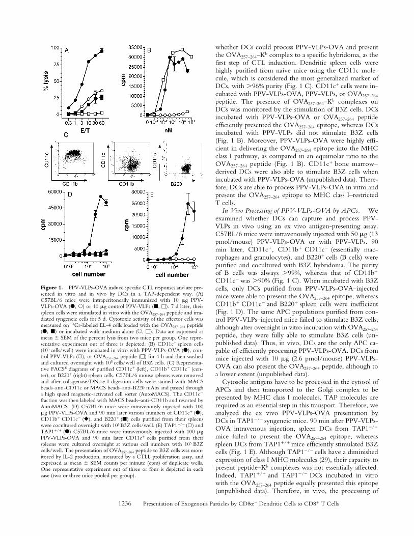
T cell epitope from OVA257–264 (PPV-VLPs-OVA) wereused. We first tested the ability of PPV-VLPs-OVA to in-duce a CTL response against OVA257–264 peptide–coatedcells. C57BL/6 mice were immunized intraperitoneallywith a single injection of 10 g PPV-VLPs-OVA or controlPPV-VLPs in PBS. As shown in Fig. 1 A, 7 d after immuni-zation the mice immunized with PPV-VLPs-OVA devel-oped a strong and specific CTL response against theOVA257–264 epitope, whereas, as expected, mice injectedwith control PPV-VLPs did not show a significant CTL re-sponse. Similar CTL responses were obtained after two in-traperitoneal injections (with a 21-d interval) or one intrave-nous injection of PPV-VLPs-OVA (unpublished data). Thisresult confirmed our earlier demonstration of the high im-munogenicity of PPV particles in the absence of adjuvant.As previously demonstrated (19), the CTL response inducedby PPV-VLPs was MHC class I restricted and mediated byCD8� T cells. Furthermore, inhibition by lactacystin andN-acetyl-leucinyl-leucinyl-norleucinal of PPV-VLPs-OVApresentation by purified DCs demonstrated that processingPPV-VLPs-OVA requires proteasome (unpublished data).These results confirm that PPV-VLPs can effectively deliveran exogenous peptide into the MHC class I pathway.
In Vitro Processing of PPV-VLPs-OVA by DCs. DCshave been clearly recognized as being the only APC capa-ble of stimulating naive T cells. Therefore, we wondered
1236 Presentation of Exogenous Particles by CD8�� Dendritic Cells to CD8� T Cells
whether DCs could process PPV-VLPs-OVA and presentthe OVA257–264–Kb complex to a specific hybridoma, as thefirst step of CTL induction. Dendritic spleen cells werehighly purified from naive mice using the CD11c mole-cule, which is considered the most generalized marker ofDCs, with �96% purity (Fig. 1 C). CD11c� cells were in-cubated with PPV-VLPs-OVA, PPV-VLPs, or OVA257–264
peptide. The presence of OVA257–264–Kb complexes onDCs was monitored by the stimulation of B3Z cells. DCsincubated with PPV-VLPs-OVA or OVA257–264 peptideefficiently presented the OVA257–264 epitope, whereas DCsincubated with PPV-VLPs did not stimulate B3Z cells(Fig. 1 B). Moreover, PPV-VLPs-OVA were highly effi-cient in delivering the OVA257–264 epitope into the MHCclass I pathway, as compared in an equimolar ratio to theOVA257–264 peptide (Fig. 1 B). CD11c� bone marrow–derived DCs were also able to stimulate B3Z cells whenincubated with PPV-VLPs-OVA (unpublished data). There-fore, DCs are able to process PPV-VLPs-OVA in vitro andpresent the OVA257–264 epitope to MHC class I–restrictedT cells.
In Vivo Processing of PPV-VLPs-OVA by APCs. Weexamined whether DCs can capture and process PPV-VLPs in vivo using an ex vivo antigen-presenting assay.C57BL/6 mice were intravenously injected with 50 g (13pmol/mouse) PPV-VLPs-OVA or with PPV-VLPs. 90min later, CD11c�, CD11b� CD11c� (essentially mac-rophages and granulocytes), and B220� cells (B cells) werepurified and cocultured with B3Z hybridoma. The purityof B cells was always �99%, whereas that of CD11b�
CD11c� was �90% (Fig. 1 C). When incubated with B3Zcells, only DCs purified from PPV-VLPs-OVA–injectedmice were able to present the OVA257–264 epitope, whereasCD11b� CD11c� and B220� spleen cells were inefficient(Fig. 1 D). The same APC populations purified from con-trol PPV-VLPs–injected mice failed to stimulate B3Z cells,although after overnight in vitro incubation with OVA257–264
peptide, they were fully able to stimulate B3Z cells (un-published data). Thus, in vivo, DCs are the only APC ca-pable of efficiently processing PPV-VLPs-OVA. DCs frommice injected with 10 g (2.6 pmol/mouse) PPV-VLPs-OVA can also present the OVA257–264 peptide, although toa lower extent (unpublished data).
Cytosolic antigens have to be processed in the cytosol ofAPCs and then transported to the Golgi complex to bepresented by MHC class I molecules. TAP molecules arerequired as an essential step in this transport. Therefore, weanalyzed the ex vivo PPV-VLPs-OVA presentation byDCs in TAP1�/� syngeneic mice. 90 min after PPV-VLPs-OVA intravenous injection, spleen DCs from TAP1�/�
mice failed to present the OVA257–264 epitope, whereasspleen DCs from TAP1�/� mice efficiently stimulated B3Zcells (Fig. 1 E). Although TAP1�/� cells have a diminishedexpression of class I MHC molecules (29), their capacity topresent peptide–Kb complexes was not essentially affected.Indeed, TAP1�/� and TAP1�/� DCs incubated in vitrowith the OVA257–264 peptide equally presented this epitope(unpublished data). Therefore, in vivo, the processing of
Figure 1. PPV-VLPs-OVA induce specific CTL responses and are pre-sented in vitro and in vivo by DCs in a TAP-dependent way. (A)C57BL/6 mice were intraperitoneally immunized with 10 g PPV-VLPs-OVA (�, �) or 10 g control PPV-VLPs (�, �). 7 d later, theirspleen cells were stimulated in vitro with the OVA257–264 peptide and irra-diated syngeneic cells for 5 d. Cytotoxic activity of the effector cells wasmeasured on 51Cr-labeled EL-4 cells loaded with the OVA257–264 peptide(�, �) or incubated with medium alone (�, �). Data are expressed asmean SEM of the percent lysis from two mice per group. One repre-sentative experiment out of three is depicted. (B) CD11c� spleen cells(105 cells/well) were incubated in vitro with PPV-VLPs-OVA (�), con-trol PPV-VLPs (�), or OVA257–264 peptide (�) for 4 h and then washedand cultured overnight with 105 cells/well of B3Z cells. (C) Representa-tive FACS® diagrams of purified CD11c� (left), CD11b� CD11c� (cen-ter), or B220� (right) spleen cells. C57BL/6 mouse spleens were removedand after collagenase/DNase I digestion cells were stained with MACSbeads–anti-CD11c or MACS beads–anti-B220 mAbs and passed througha high speed magnetic-activated cell sorter (AutoMACS). The CD11c�
fraction was then labeled with MACS beads–anti-CD11b and resorted byAutoMACS. (D) C57BL/6 mice were intravenously injected with 100g PPV-VLPs-OVA and 90 min later various numbers of CD11c� (�),CD11b� CD11c� (�), and B220� (�) cells purified from their spleenswere cocultured overnight with 105 B3Z cells/well. (E) TAP1�/� (�) andTAP1�/� (�) C57BL/6 mice were intravenously injected with 100 gPPV-VLPs-OVA and 90 min later CD11c� cells purified from theirspleens were cultured overnight at various cell numbers with 105 B3Zcells/well. The presentation of OVA257–264 peptide to B3Z cells was mon-itored by IL-2 production, measured by a CTLL proliferation assay, andexpressed as mean SEM counts per minute (cpm) of duplicate wells.One representative experiment out of three or four is depicted in eachcase (two or three mice pooled per group).
1237 Morón et al.
PPV-VLPs-OVA required TAP molecules, i.e., a cytosolicprocessing in DCs.
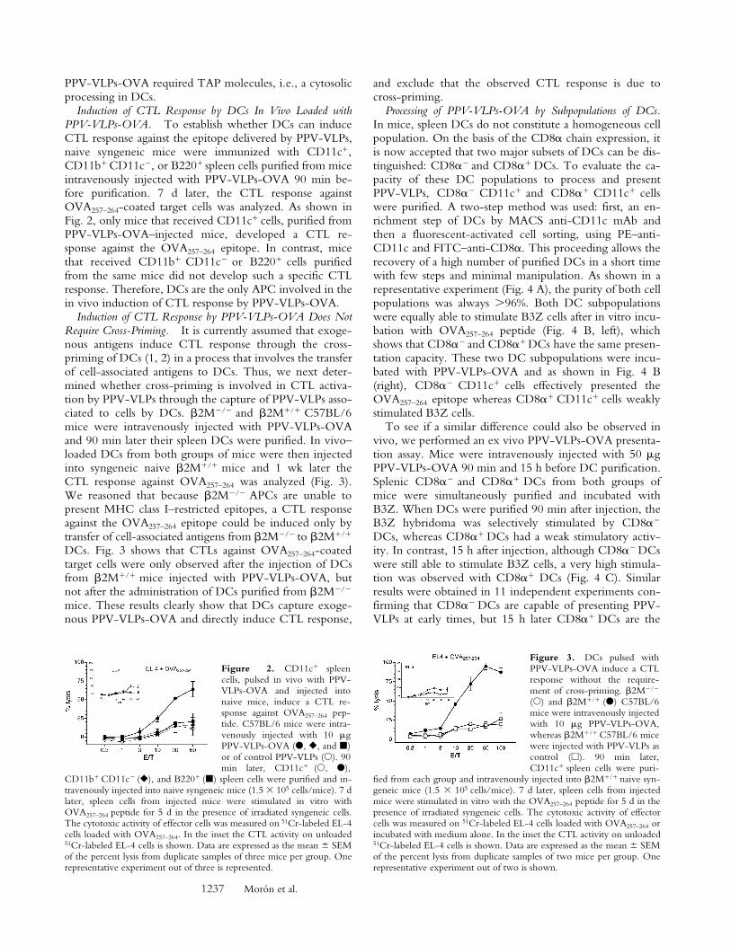
Induction of CTL Response by DCs In Vivo Loaded withPPV-VLPs-OVA. To establish whether DCs can induceCTL response against the epitope delivered by PPV-VLPs,naive syngeneic mice were immunized with CD11c�,CD11b� CD11c�, or B220� spleen cells purified from miceintravenously injected with PPV-VLPs-OVA 90 min be-fore purification. 7 d later, the CTL response againstOVA257–264-coated target cells was analyzed. As shown inFig. 2, only mice that received CD11c� cells, purified fromPPV-VLPs-OVA–injected mice, developed a CTL re-sponse against the OVA257–264 epitope. In contrast, micethat received CD11b� CD11c� or B220� cells purifiedfrom the same mice did not develop such a specific CTLresponse. Therefore, DCs are the only APC involved in thein vivo induction of CTL response by PPV-VLPs-OVA.
Induction of CTL Response by PPV-VLPs-OVA Does NotRequire Cross-Priming. It is currently assumed that exoge-nous antigens induce CTL response through the cross-priming of DCs (1, 2) in a process that involves the transferof cell-associated antigens to DCs. Thus, we next deter-mined whether cross-priming is involved in CTL activa-tion by PPV-VLPs through the capture of PPV-VLPs asso-ciated to cells by DCs. �2M�/� and �2M�/� C57BL/6mice were intravenously injected with PPV-VLPs-OVAand 90 min later their spleen DCs were purified. In vivo–loaded DCs from both groups of mice were then injectedinto syngeneic naive �2M�/� mice and 1 wk later theCTL response against OVA257–264 was analyzed (Fig. 3).We reasoned that because �2M�/� APCs are unable topresent MHC class I–restricted epitopes, a CTL responseagainst the OVA257–264 epitope could be induced only bytransfer of cell-associated antigens from �2M�/� to �2M�/�
DCs. Fig. 3 shows that CTLs against OVA257–264-coatedtarget cells were only observed after the injection of DCsfrom �2M�/� mice injected with PPV-VLPs-OVA, butnot after the administration of DCs purified from �2M�/�
mice. These results clearly show that DCs capture exoge-nous PPV-VLPs-OVA and directly induce CTL response,
and exclude that the observed CTL response is due tocross-priming.
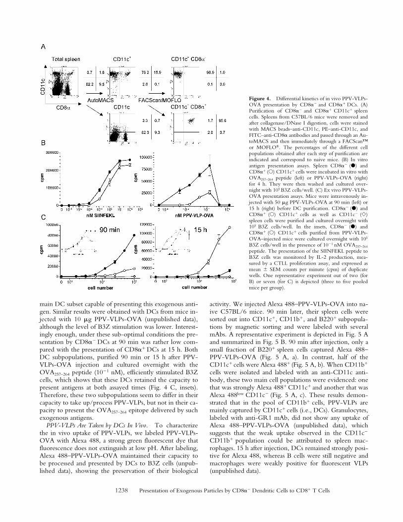
Processing of PPV-VLPs-OVA by Subpopulations of DCs.In mice, spleen DCs do not constitute a homogeneous cellpopulation. On the basis of the CD8� chain expression, itis now accepted that two major subsets of DCs can be dis-tinguished: CD8�� and CD8�� DCs. To evaluate the ca-pacity of these DC populations to process and presentPPV-VLPs, CD8�� CD11c� and CD8�� CD11c� cellswere purified. A two-step method was used: first, an en-richment step of DCs by MACS anti-CD11c mAb andthen a fluorescent-activated cell sorting, using PE–anti-CD11c and FITC–anti-CD8�. This proceeding allows therecovery of a high number of purified DCs in a short timewith few steps and minimal manipulation. As shown in arepresentative experiment (Fig. 4 A), the purity of both cellpopulations was always �96%. Both DC subpopulationswere equally able to stimulate B3Z cells after in vitro incu-bation with OVA257–264 peptide (Fig. 4 B, left), whichshows that CD8�� and CD8�� DCs have the same presen-tation capacity. These two DC subpopulations were incu-bated with PPV-VLPs-OVA and as shown in Fig. 4 B(right), CD8�� CD11c� cells effectively presented theOVA257–264 epitope whereas CD8�� CD11c� cells weaklystimulated B3Z cells.
To see if a similar difference could also be observed invivo, we performed an ex vivo PPV-VLPs-OVA presenta-tion assay. Mice were intravenously injected with 50 gPPV-VLPs-OVA 90 min and 15 h before DC purification.Splenic CD8�� and CD8�� DCs from both groups ofmice were simultaneously purified and incubated withB3Z. When DCs were purified 90 min after injection, theB3Z hybridoma was selectively stimulated by CD8��
DCs, whereas CD8�� DCs had a weak stimulatory activ-ity. In contrast, 15 h after injection, although CD8�� DCswere still able to stimulate B3Z cells, a very high stimula-tion was observed with CD8�� DCs (Fig. 4 C). Similarresults were obtained in 11 independent experiments con-firming that CD8�� DCs are capable of presenting PPV-VLPs at early times, but 15 h later CD8�� DCs are the
Figure 2. CD11c� spleencells, pulsed in vivo with PPV-VLPs-OVA and injected intonaive mice, induce a CTL re-sponse against OVA257–264 pep-tide. C57BL/6 mice were intra-venously injected with 10 gPPV-VLPs-OVA (�, �, and �)or of control PPV-VLPs (�). 90min later, CD11c� (�, �),
CD11b� CD11c� (�), and B220� (�) spleen cells were purified and in-travenously injected into naive syngeneic mice (1.5 105 cells/mice). 7 dlater, spleen cells from injected mice were stimulated in vitro withOVA257–264 peptide for 5 d in the presence of irradiated syngeneic cells.The cytotoxic activity of effector cells was measured on 51Cr-labeled EL-4cells loaded with OVA257–264. In the inset the CTL activity on unloaded51Cr-labeled EL-4 cells is shown. Data are expressed as the mean SEMof the percent lysis from duplicate samples of three mice per group. Onerepresentative experiment out of three is represented.
Figure 3. DCs pulsed withPPV-VLPs-OVA induce a CTLresponse without the require-ment of cross-priming. �2M�/�
(�) and �2M�/� (�) C57BL/6mice were intravenously injectedwith 10 g PPV-VLPs-OVA,whereas �2M�/� C57BL/6 micewere injected with PPV-VLPs ascontrol (�). 90 min later,CD11c� spleen cells were puri-
fied from each group and intravenously injected into �2M�/� naive syn-geneic mice (1.5 105 cells/mice). 7 d later, spleen cells from injectedmice were stimulated in vitro with the OVA257–264 peptide for 5 d in thepresence of irradiated syngeneic cells. The cytotoxic activity of effectorcells was measured on 51Cr-labeled EL-4 cells loaded with OVA257–264 orincubated with medium alone. In the inset the CTL activity on unloaded51Cr-labeled EL-4 cells is shown. Data are expressed as the mean SEMof the percent lysis from duplicate samples of two mice per group. Onerepresentative experiment out of two is shown.
1238 Presentation of Exogenous Particles by CD8�� Dendritic Cells to CD8� T Cells
main DC subset capable of presenting this exogenous anti-gen. Similar results were obtained with DCs from mice in-jected with 10 g PPV-VLPs-OVA (unpublished data),although the level of B3Z stimulation was lower. Interest-ingly enough, under these sub-optimal conditions the pre-sentation by CD8�� DCs at 90 min was rather low com-pared with the presentation of CD8�� DCs at 15 h. BothDC subpopulations, purified 90 min or 15 h after PPV-VLPs-OVA injection and cultured overnight with theOVA257–264 peptide (10�1 nM), efficiently stimulated B3Zcells, which shows that these DCs retained the capacity topresent antigens at both assayed times (Fig. 4 C, insets).Therefore, these two subpopulations seem to differ in theircapacity to take up/process PPV-VLPs, but not in their ca-pacity to present the OVA257–264 epitope delivered by suchexogenous antigens.
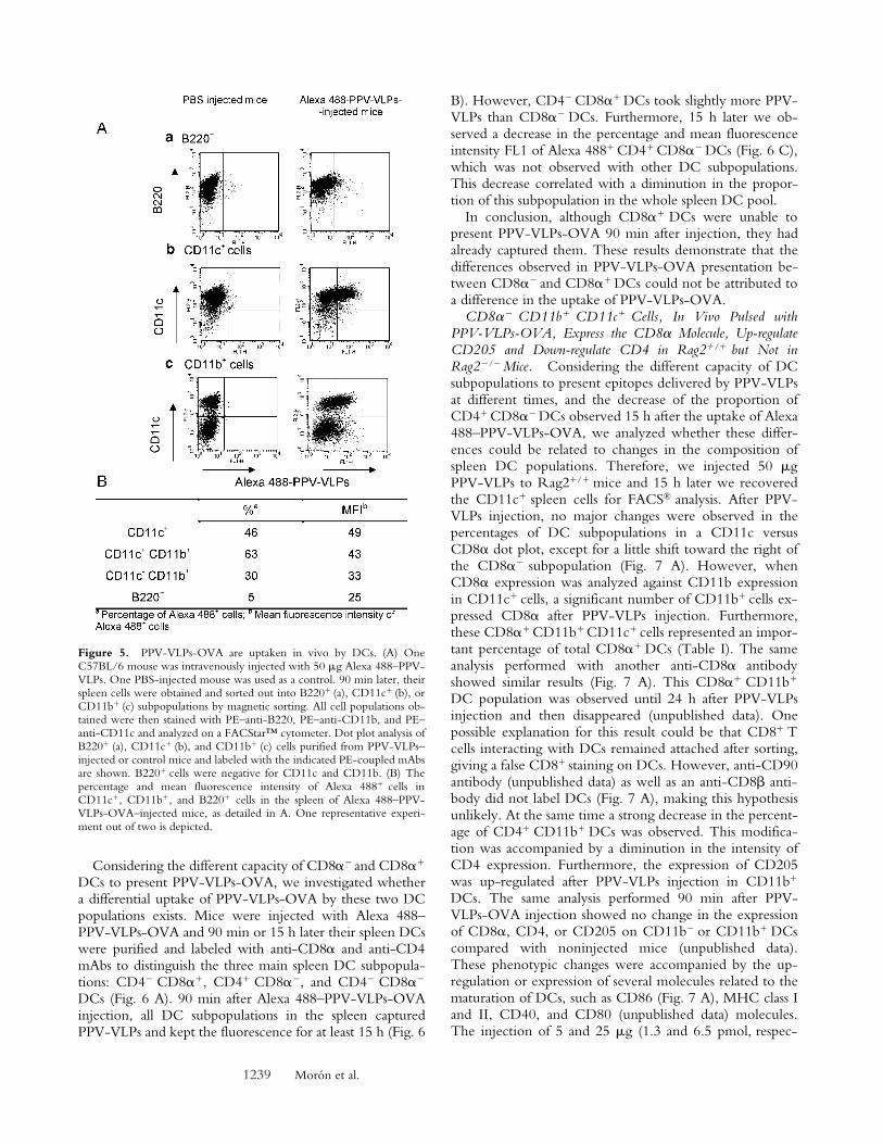
PPV-VLPs Are Taken by DCs In Vivo. To characterizethe in vivo uptake of PPV-VLPs, we labeled PPV-VLPs-OVA with Alexa 488, a strong green fluorescent dye thatfluorescence does not extinguish at low pH. After labeling,Alexa 488–PPV-VLPs-OVA maintained their capacity tobe processed and presented by DCs to B3Z cells (unpub-lished data), showing the preservation of their biological
activity. We injected Alexa 488–PPV-VLPs-OVA into na-ive C57BL/6 mice. 90 min later, their spleen cells weresorted out into CD11c�, CD11b�, and B220� subpopula-tions by magnetic sorting and were labeled with severalmAbs. A representative experiment is depicted in Fig. 5 Aand summarized in Fig. 5 B. 90 min after injection, only asmall fraction of B220� spleen cells captured Alexa 488–PPV-VLPs-OVA (Fig. 5 A, a). In contrast, half of theCD11c� cells were Alexa 488� (Fig. 5 A, b). When CD11b�
cells were isolated and labeled with an anti-CD11c anti-body, these two main cell populations were evidenced: onethat was strongly Alexa 488� CD11c� and another that wasAlexa 488low CD11c� (Fig. 5 A, c). These results demon-strated that in the pool of CD11b� cells, PPV-VLPs aremainly captured by CD11c� cells (i.e., DCs). Granulocytes,labeled with anti-GR1 mAb, did not show any uptake ofAlexa 488–PPV-VLPs-OVA (unpublished data), whichsuggests that the weak uptake observed in the CD11c�
CD11b� population could be attributed to spleen mac-rophages. 15 h after injection, DCs remained strongly posi-tive for Alexa 488, whereas B cells were still negative andmacrophages were weakly positive for fluorescent VLPs(unpublished data).
Figure 4. Differential kinetics of in vivo PPV-VLPs-OVA presentation by CD8�� and CD8�� DCs. (A)Purification of CD8�� and CD8�� CD11c� spleencells. Spleens from C57BL/6 mice were removed andafter collagenase/DNase I digestion, cells were stainedwith MACS beads–anti-CD11c, PE–anti-CD11c, andFITC–anti-CD8� antibodies and passed through an Au-toMACS and then immediately through a FACScan™or MOFLO®. The percentages of the different cellpopulations obtained after each step of purification areindicated and correspond to naive mice. (B) In vitroantigen presentation assays. Spleen CD8�� (�) andCD8�� (�) CD11c� cells were incubated in vitro withOVA257–264 peptide (left) or PPV-VLPs-OVA (right)for 4 h. They were then washed and cultured over-night with 105 B3Z cells/well. (C) Ex vivo PPV-VLPs-OVA presentation assays. Mice were intravenously in-jected with 50 g PPV-VLPs-OVA at 90 min (left) or15 h (right) before DC purification. CD8�� (�) andCD8�� (�) CD11c� cells as well as CD11c� (�)spleen cells were purified and cultured overnight with105 B3Z cells/well. In the insets, CD8�� (�) andCD8�� (�) CD11c� cells purified from PPV-VLPs-OVA–injected mice were cultured overnight with 105
B3Z cells/well in the presence of 10�1 nM OVA257–264
peptide. The presentation of the SIINFEKL peptide toB3Z cells was monitored by IL-2 production, mea-sured by a CTLL proliferation assay, and expressed asmean SEM counts per minute (cpm) of duplicatewells. One representative experiment out of two (forB) or seven (for C) is depicted (three to five pooledmice per group).
1239 Morón et al.
Considering the different capacity of CD8�� and CD8��
DCs to present PPV-VLPs-OVA, we investigated whethera differential uptake of PPV-VLPs-OVA by these two DCpopulations exists. Mice were injected with Alexa 488–PPV-VLPs-OVA and 90 min or 15 h later their spleen DCswere purified and labeled with anti-CD8� and anti-CD4mAbs to distinguish the three main spleen DC subpopula-tions: CD4� CD8��, CD4� CD8��, and CD4� CD8��
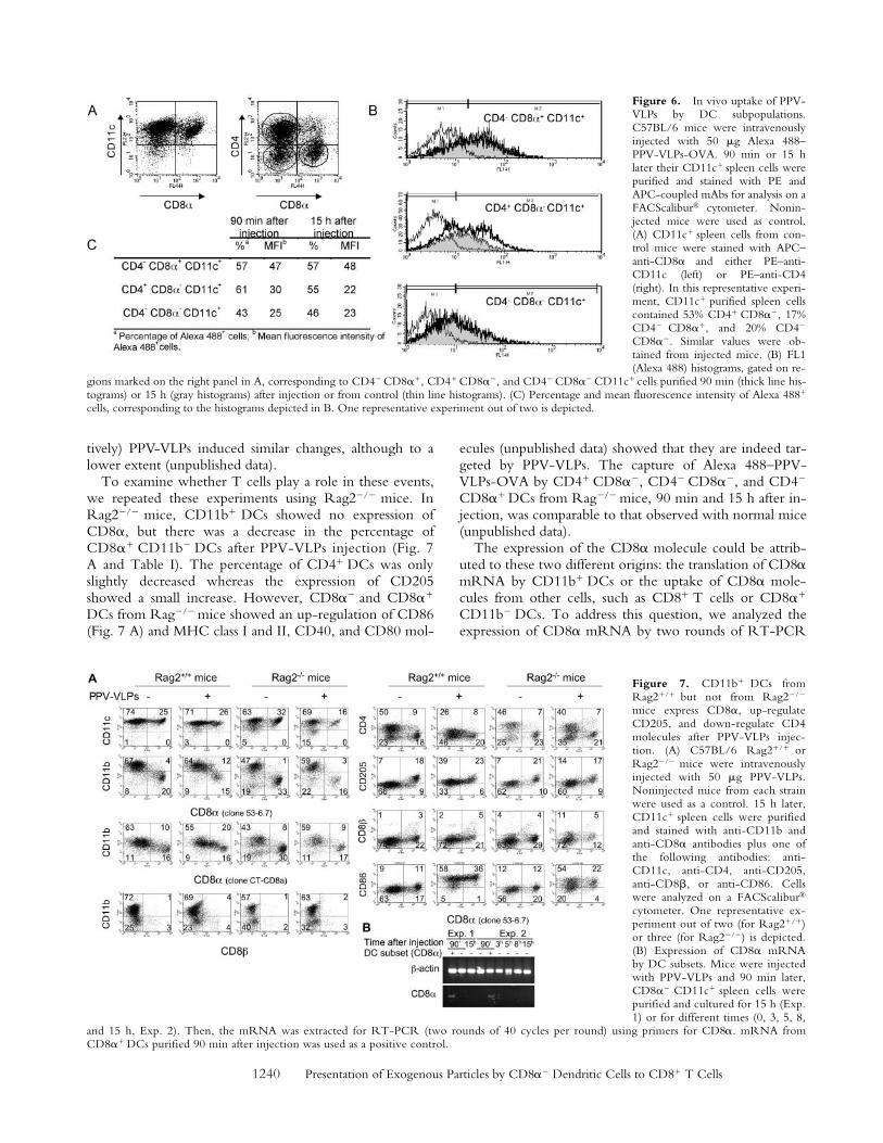
DCs (Fig. 6 A). 90 min after Alexa 488–PPV-VLPs-OVAinjection, all DC subpopulations in the spleen capturedPPV-VLPs and kept the fluorescence for at least 15 h (Fig. 6
B). However, CD4� CD8�� DCs took slightly more PPV-VLPs than CD8�� DCs. Furthermore, 15 h later we ob-served a decrease in the percentage and mean fluorescenceintensity FL1 of Alexa 488� CD4� CD8�� DCs (Fig. 6 C),which was not observed with other DC subpopulations.This decrease correlated with a diminution in the propor-tion of this subpopulation in the whole spleen DC pool.
In conclusion, although CD8�� DCs were unable topresent PPV-VLPs-OVA 90 min after injection, they hadalready captured them. These results demonstrate that thedifferences observed in PPV-VLPs-OVA presentation be-tween CD8�� and CD8�� DCs could not be attributed toa difference in the uptake of PPV-VLPs-OVA.
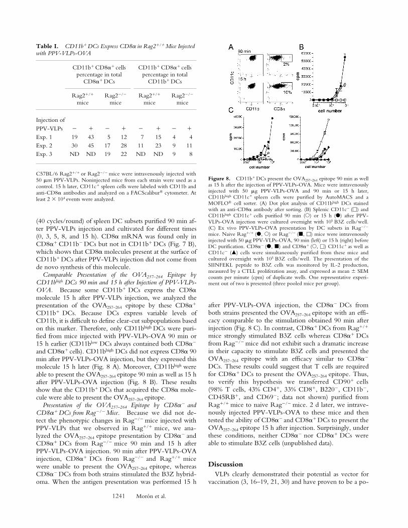
CD8�� CD11b� CD11c� Cells, In Vivo Pulsed withPPV-VLPs-OVA, Express the CD8� Molecule, Up-regulateCD205 and Down-regulate CD4 in Rag2�/� but Not inRag2�/� Mice. Considering the different capacity of DCsubpopulations to present epitopes delivered by PPV-VLPsat different times, and the decrease of the proportion ofCD4� CD8�� DCs observed 15 h after the uptake of Alexa488–PPV-VLPs-OVA, we analyzed whether these differ-ences could be related to changes in the composition ofspleen DC populations. Therefore, we injected 50 gPPV-VLPs to Rag2�/� mice and 15 h later we recoveredthe CD11c� spleen cells for FACS® analysis. After PPV-VLPs injection, no major changes were observed in thepercentages of DC subpopulations in a CD11c versusCD8� dot plot, except for a little shift toward the right ofthe CD8�� subpopulation (Fig. 7 A). However, whenCD8� expression was analyzed against CD11b expressionin CD11c� cells, a significant number of CD11b� cells ex-pressed CD8� after PPV-VLPs injection. Furthermore,these CD8�� CD11b� CD11c� cells represented an impor-tant percentage of total CD8�� DCs (Table I). The sameanalysis performed with another anti-CD8� antibodyshowed similar results (Fig. 7 A). This CD8�� CD11b�
DC population was observed until 24 h after PPV-VLPsinjection and then disappeared (unpublished data). Onepossible explanation for this result could be that CD8� Tcells interacting with DCs remained attached after sorting,giving a false CD8� staining on DCs. However, anti-CD90antibody (unpublished data) as well as an anti-CD8� anti-body did not label DCs (Fig. 7 A), making this hypothesisunlikely. At the same time a strong decrease in the percent-age of CD4� CD11b� DCs was observed. This modifica-tion was accompanied by a diminution in the intensity ofCD4 expression. Furthermore, the expression of CD205was up-regulated after PPV-VLPs injection in CD11b�
DCs. The same analysis performed 90 min after PPV-VLPs-OVA injection showed no change in the expressionof CD8�, CD4, or CD205 on CD11b� or CD11b� DCscompared with noninjected mice (unpublished data).These phenotypic changes were accompanied by the up-regulation or expression of several molecules related to thematuration of DCs, such as CD86 (Fig. 7 A), MHC class Iand II, CD40, and CD80 (unpublished data) molecules.The injection of 5 and 25 g (1.3 and 6.5 pmol, respec-
Figure 5. PPV-VLPs-OVA are uptaken in vivo by DCs. (A) OneC57BL/6 mouse was intravenously injected with 50 g Alexa 488–PPV-VLPs. One PBS-injected mouse was used as a control. 90 min later, theirspleen cells were obtained and sorted out into B220� (a), CD11c� (b), orCD11b� (c) subpopulations by magnetic sorting. All cell populations ob-tained were then stained with PE–anti-B220, PE–anti-CD11b, and PE–anti-CD11c and analyzed on a FACStar™ cytometer. Dot plot analysis ofB220� (a), CD11c� (b), and CD11b� (c) cells purified from PPV-VLPs–injected or control mice and labeled with the indicated PE-coupled mAbsare shown. B220� cells were negative for CD11c and CD11b. (B) Thepercentage and mean fluorescence intensity of Alexa 488� cells inCD11c�, CD11b�, and B220� cells in the spleen of Alexa 488–PPV-VLPs-OVA–injected mice, as detailed in A. One representative experi-ment out of two is depicted.
1240 Presentation of Exogenous Particles by CD8�� Dendritic Cells to CD8� T Cells
tively) PPV-VLPs induced similar changes, although to alower extent (unpublished data).
To examine whether T cells play a role in these events,we repeated these experiments using Rag2�/� mice. InRag2�/� mice, CD11b� DCs showed no expression ofCD8�, but there was a decrease in the percentage ofCD8�� CD11b� DCs after PPV-VLPs injection (Fig. 7A and Table I). The percentage of CD4� DCs was onlyslightly decreased whereas the expression of CD205showed a small increase. However, CD8�� and CD8��
DCs from Rag�/� mice showed an up-regulation of CD86(Fig. 7 A) and MHC class I and II, CD40, and CD80 mol-
ecules (unpublished data) showed that they are indeed tar-geted by PPV-VLPs. The capture of Alexa 488–PPV-VLPs-OVA by CD4� CD8��, CD4� CD8��, and CD4�
CD8�� DCs from Rag�/� mice, 90 min and 15 h after in-jection, was comparable to that observed with normal mice(unpublished data).
The expression of the CD8� molecule could be attrib-uted to these two different origins: the translation of CD8�mRNA by CD11b� DCs or the uptake of CD8� mole-cules from other cells, such as CD8� T cells or CD8��
CD11b� DCs. To address this question, we analyzed theexpression of CD8� mRNA by two rounds of RT-PCR
Figure 6. In vivo uptake of PPV-VLPs by DC subpopulations.C57BL/6 mice were intravenouslyinjected with 50 g Alexa 488–PPV-VLPs-OVA. 90 min or 15 hlater their CD11c� spleen cells werepurified and stained with PE andAPC-coupled mAbs for analysis on aFACScalibur® cytometer. Nonin-jected mice were used as control.(A) CD11c� spleen cells from con-trol mice were stained with APC–anti-CD8� and either PE–anti-CD11c (left) or PE–anti-CD4(right). In this representative experi-ment, CD11c� purified spleen cellscontained 53% CD4� CD8��, 17%CD4� CD8��, and 20% CD4�
CD8��. Similar values were ob-tained from injected mice. (B) FL1(Alexa 488) histograms, gated on re-
gions marked on the right panel in A, corresponding to CD4� CD8��, CD4� CD8��, and CD4� CD8�� CD11c� cells purified 90 min (thick line his-tograms) or 15 h (gray histograms) after injection or from control (thin line histograms). (C) Percentage and mean fluorescence intensity of Alexa 488�
cells, corresponding to the histograms depicted in B. One representative experiment out of two is depicted.
Figure 7. CD11b� DCs fromRag2�/� but not from Rag2�/�
mice express CD8�, up-regulateCD205, and down-regulate CD4molecules after PPV-VLPs injec-tion. (A) C57BL/6 Rag2�/� orRag2�/� mice were intravenouslyinjected with 50 g PPV-VLPs.Noninjected mice from each strainwere used as a control. 15 h later,CD11c� spleen cells were purifiedand stained with anti-CD11b andanti-CD8� antibodies plus one ofthe following antibodies: anti-CD11c, anti-CD4, anti-CD205,anti-CD8�, or anti-CD86. Cellswere analyzed on a FACScalibur®
cytometer. One representative ex-periment out of two (for Rag2�/�)or three (for Rag2�/�) is depicted.(B) Expression of CD8� mRNAby DC subsets. Mice were injectedwith PPV-VLPs and 90 min later,CD8�� CD11c� spleen cells werepurified and cultured for 15 h (Exp.1) or for different times (0, 3, 5, 8,
and 15 h, Exp. 2). Then, the mRNA was extracted for RT-PCR (two rounds of 40 cycles per round) using primers for CD8�. mRNA fromCD8�� DCs purified 90 min after injection was used as a positive control.
1241 Morón et al.
(40 cycles/round) of spleen DC subsets purified 90 min af-ter PPV-VLPs injection and cultivated for different times(0, 3, 5, 8, and 15 h). CD8� mRNA was found only inCD8�� CD11b� DCs but not in CD11b� DCs (Fig. 7 B),which shows that CD8� molecules present at the surface ofCD11b� DCs after PPV-VLPs injection did not come fromde novo synthesis of this molecule.
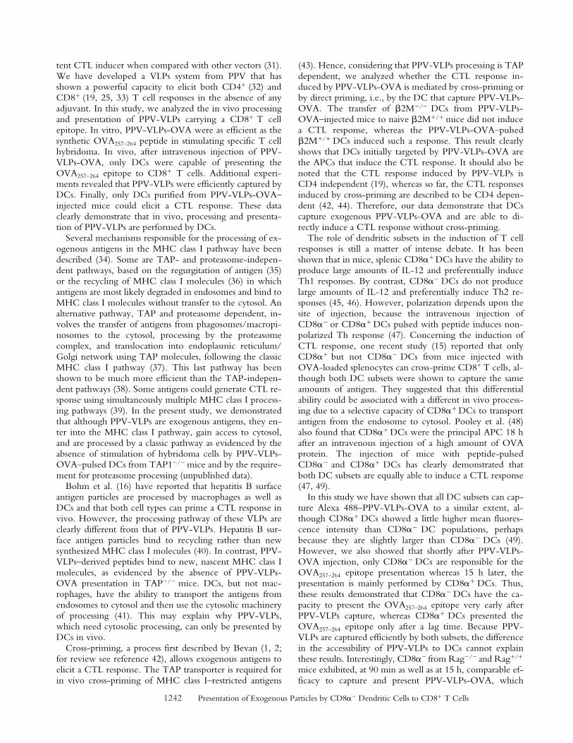
Comparable Presentation of the OVA257–264 Epitope byCD11bhigh DCs 90 min and 15 h after Injection of PPV-VLPs-OVA. Because some CD11b� DCs express the CD8�molecule 15 h after PPV-VLPs injection, we analyzed thepresentation of the OVA257–264 epitope by these CD8��
CD11b� DCs. Because DCs express variable levels ofCD11b, it is difficult to define clear-cut subpopulations basedon this marker. Therefore, only CD11bhigh DCs were puri-fied from mice injected with PPV-VLPs-OVA 90 min or15 h earlier (CD11blow DCs always contained both CD8��
and CD8�� cells). CD11bhigh DCs did not express CD8� 90min after PPV-VLPs-OVA injection, but they expressed thismolecule 15 h later (Fig. 8 A). Moreover, CD11bhigh wereable to present the OVA257–264 epitope 90 min as well as 15 hafter PPV-VLPs-OVA injection (Fig. 8 B). These resultsshow that the CD11b� DCs that acquired the CD8� mole-cule were able to present the OVA257–264 epitope.
Presentation of the OVA257–264 Epitope by CD8�� andCD8�� DCs from Rag�/� Mice. Because we did not de-tect the phenotypic changes in Rag�/� mice injected withPPV-VLPs that we observed in Rag�/� mice, we ana-lyzed the OVA257–264 epitope presentation by CD8�� andCD8�� DCs from Rag�/� mice 90 min and 15 h afterPPV-VLPs-OVA injection. 90 min after PPV-VLPs-OVAinjection, CD8�� DCs from Rag�/� and Rag�/� micewere unable to present the OVA257–264 epitope, whereasCD8�� DCs from both strains stimulated the B3Z hybrid-oma. When the antigen presentation was performed 15 h
after PPV-VLPs-OVA injection, the CD8�� DCs fromboth strains presented the OVA257–264 epitope with an effi-cacy comparable to the stimulation obtained 90 min afterinjection (Fig. 8 C). In contrast, CD8�� DCs from Rag�/�
mice strongly stimulated B3Z cells whereas CD8�� DCsfrom Rag�/� mice did not exhibit such a dramatic increasein their capacity to stimulate B3Z cells and presented theOVA257–264 epitope with an efficacy similar to CD8��
DCs. These results could suggest that T cells are requiredfor CD8�� DCs to present the OVA257–264 epitope. Thus,to verify this hypothesis we transferred CD90� cells(98% T cells, 43% CD4�, 33% CD8�, B220�, CD11b�,CD45RB�, and CD69�; data not shown) purified fromRag�/� mice to naive Rag�/� mice. 2 d later, we intrave-nously injected PPV-VLPs-OVA to these mice and thentested the ability of CD8�� and CD8�� DCs to present theOVA257–264 epitope 15 h after injection. Surprisingly, underthese conditions, neither CD8�� nor CD8�� DCs wereable to stimulate B3Z cells (unpublished data).
DiscussionVLPs clearly demonstrated their potential as vector for
vaccination (3, 16–19, 21, 30) and have proven to be a po-
Table I. CD11b� DCs Express CD8� in Rag2�/� Mice Injected with PPV-VLPs-OVA
CD11b� CD8�� cellspercentage in total
CD8�� DCs
CD11b� CD8�� cellspercentage in total
CD11b� DCs
Rag2�/�
miceRag2�/�
miceRag2�/�
miceRag2�/�
mice
Injection ofPPV-VLPs � � � � � � � �
Exp. 1 19 43 5 12 7 15 4 4Exp. 2 30 45 17 28 11 23 9 11Exp. 3 ND ND 19 22 ND ND 9 8
C57BL/6 Rag2�/� or Rag2�/� mice were intravenously injected with50 m PPV-VLPs. Noninjected mice from each strain were used as acontrol. 15 h later, CD11c� spleen cells were labeled with CD11b andanti-CD8� antibodies and analyzed on a FACScalibur® cytometer. Atleast 2 104 events were analyzed.
Figure 8. CD11b� DCs present the OVA257–264 epitope 90 min as wellas 15 h after the injection of PPV-VLPs-OVA. Mice were intravenouslyinjected with 50 g PPV-VLPs-OVA and 90 min or 15 h later,CD11bhigh CD11c� spleen cells were purified by AutoMACS and aMOFLO® cell sorter. (A) Dot plot analysis of CD11bhigh DCs stainedwith an anti-CD8� antibody after sorting. (B) Splenic CD11c� (�) andCD11bhigh CD11c� cells purified 90 min (�) or 15 h (�) after PPV-VLPs-OVA injection were cultured overnight with 105 B3Z cells/well.(C) Ex vivo PPV-VLPs-OVA presentation by DC subsets in Rag�/�
mice. Naive Rag�/� (�, �) or Rag�/� (�, �) mice were intravenouslyinjected with 50 g PPV-VLPs-OVA, 90 min (left) or 15 h (right) beforeDC purification. CD8�� (�, �) and CD8�� (�, �) CD11c� as well asCD11c� () cells were simultaneously purified from these mice andcultured overnight with 105 B3Z cells/well. The presentation of theSIINFEKL peptide to B3Z cells was monitored by IL-2 production,measured by a CTLL proliferation assay, and expressed as mean SEMcounts per minute (cpm) of duplicate wells. One representative experi-ment out of two is presented (three pooled mice per group).
1242 Presentation of Exogenous Particles by CD8�� Dendritic Cells to CD8� T Cells
tent CTL inducer when compared with other vectors (31).We have developed a VLPs system from PPV that hasshown a powerful capacity to elicit both CD4� (32) andCD8� (19, 25, 33) T cell responses in the absence of anyadjuvant. In this study, we analyzed the in vivo processingand presentation of PPV-VLPs carrying a CD8� T cellepitope. In vitro, PPV-VLPs-OVA were as efficient as thesynthetic OVA257–264 peptide in stimulating specific T cellhybridoma. In vivo, after intravenous injection of PPV-VLPs-OVA, only DCs were capable of presenting theOVA257–264 epitope to CD8� T cells. Additional experi-ments revealed that PPV-VLPs were efficiently captured byDCs. Finally, only DCs purified from PPV-VLPs-OVA–injected mice could elicit a CTL response. These dataclearly demonstrate that in vivo, processing and presenta-tion of PPV-VLPs are performed by DCs.
Several mechanisms responsible for the processing of ex-ogenous antigens in the MHC class I pathway have beendescribed (34). Some are TAP- and proteasome-indepen-dent pathways, based on the regurgitation of antigen (35)or the recycling of MHC class I molecules (36) in whichantigens are most likely degraded in endosomes and bind toMHC class I molecules without transfer to the cytosol. Analternative pathway, TAP and proteasome dependent, in-volves the transfer of antigens from phagosomes/macropi-nosomes to the cytosol, processing by the proteasomecomplex, and translocation into endoplasmic reticulum/Golgi network using TAP molecules, following the classicMHC class I pathway (37). This last pathway has beenshown to be much more efficient than the TAP-indepen-dent pathways (38). Some antigens could generate CTL re-sponse using simultaneously multiple MHC class I process-ing pathways (39). In the present study, we demonstratedthat although PPV-VLPs are exogenous antigens, they en-ter into the MHC class I pathway, gain access to cytosol,and are processed by a classic pathway as evidenced by theabsence of stimulation of hybridoma cells by PPV-VLPs-OVA–pulsed DCs from TAP1�/� mice and by the require-ment for proteasome processing (unpublished data).
Bohm et al. (16) have reported that hepatitis B surfaceantigen particles are processed by macrophages as well asDCs and that both cell types can prime a CTL response invivo. However, the processing pathway of these VLPs areclearly different from that of PPV-VLPs. Hepatitis B sur-face antigen particles bind to recycling rather than newsynthesized MHC class I molecules (40). In contrast, PPV-VLPs–derived peptides bind to new, nascent MHC class Imolecules, as evidenced by the absence of PPV-VLPs-OVA presentation in TAP�/� mice. DCs, but not mac-rophages, have the ability to transport the antigens fromendosomes to cytosol and then use the cytosolic machineryof processing (41). This may explain why PPV-VLPs,which need cytosolic processing, can only be presented byDCs in vivo.
Cross-priming, a process first described by Bevan (1, 2;for review see reference 42), allows exogenous antigens toelicit a CTL response. The TAP transporter is required forin vivo cross-priming of MHC class I–restricted antigens
(43). Hence, considering that PPV-VLPs processing is TAPdependent, we analyzed whether the CTL response in-duced by PPV-VLPs-OVA is mediated by cross-priming orby direct priming, i.e., by the DC that capture PPV-VLPs-OVA. The transfer of �2M�/� DCs from PPV-VLPs-OVA–injected mice to naive �2M�/� mice did not inducea CTL response, whereas the PPV-VLPs-OVA–pulsed�2M�/� DCs induced such a response. This result clearlyshows that DCs initially targeted by PPV-VLPs-OVA arethe APCs that induce the CTL response. It should also benoted that the CTL response induced by PPV-VLPs isCD4 independent (19), whereas so far, the CTL responsesinduced by cross-priming are described to be CD4 depen-dent (42, 44). Therefore, our data demonstrate that DCscapture exogenous PPV-VLPs-OVA and are able to di-rectly induce a CTL response without cross-priming.
The role of dendritic subsets in the induction of T cellresponses is still a matter of intense debate. It has beenshown that in mice, splenic CD8�� DCs have the ability toproduce large amounts of IL-12 and preferentially induceTh1 responses. By contrast, CD8�� DCs do not producelarge amounts of IL-12 and preferentially induce Th2 re-sponses (45, 46). However, polarization depends upon thesite of injection, because the intravenous injection ofCD8�� or CD8�� DCs pulsed with peptide induces non-polarized Th response (47). Concerning the induction ofCTL response, one recent study (15) reported that onlyCD8�� but not CD8�� DCs from mice injected withOVA-loaded splenocytes can cross-prime CD8� T cells, al-though both DC subsets were shown to capture the sameamounts of antigen. They suggested that this differentialability could be associated with a different in vivo process-ing due to a selective capacity of CD8�� DCs to transportantigen from the endosome to cytosol. Pooley et al. (48)also found that CD8�� DCs were the principal APC 18 hafter an intravenous injection of a high amount of OVAprotein. The injection of mice with peptide-pulsedCD8�� and CD8�� DCs has clearly demonstrated thatboth DC subsets are equally able to induce a CTL response(47, 49).
In this study we have shown that all DC subsets can cap-ture Alexa 488–PPV-VLPs-OVA to a similar extent, al-though CD8�� DCs showed a little higher mean fluores-cence intensity than CD8�� DC populations, perhapsbecause they are slightly larger than CD8�� DCs (49).However, we also showed that shortly after PPV-VLPs-OVA injection, only CD8�� DCs are responsible for theOVA257–264 epitope presentation whereas 15 h later, thepresentation is mainly performed by CD8�� DCs. Thus,these results demonstrated that CD8�� DCs have the ca-pacity to present the OVA257–264 epitope very early afterPPV-VLPs capture, whereas CD8�� DCs presented theOVA257–264 epitope only after a lag time. Because PPV-VLPs are captured efficiently by both subsets, the differencein the accessibility of PPV-VLPs to DCs cannot explainthese results. Interestingly, CD8�� from Rag�/� and Rag�/�
mice exhibited, at 90 min as well as at 15 h, comparable ef-ficacy to capture and present PPV-VLPs-OVA, which
1243 Morón et al.
shows that the processing by this DC subset is T cell inde-pendent. In contrast, 15 h after PPV-VLPs-OVA injection,CD8�� DCs from Rag�/� mice were unable to present theOVA257–264 epitope with the high efficacy of CD8�� DCsfrom Rag�/� mice. This could suggest that T cells play arole in the licensing of CD8�� DCs for exogenous antigenpresentation. Therefore, our results could suggest thatCD8�� and CD8�� DCs have differential requirements forMHC class I–restricted presentation of exogenous antigens.
Using two different anti-CD8� antibodies, we demon-strated that 15 h after the injection of PPV-VLPs, a signifi-cant percentage of CD11b� DCs expressed CD8�. Thesecells also expressed CD205. Moreover, the proportion ofCD4� CD8�� DCs showed an important decrease at thattime, associated with a diminution in the intensity of ex-pression of the CD4 molecule on those cells. Our study isthe first in vivo report showing the apparition of a CD8��
and CD205� CD11b� DC population in the spleen afterthe injection of an antigen. A very recent report suggeststhat CD8�� DCs could originate from the CD8�� DCsubset by a maturation process involving CD8�, DEC-205,and CD24 up-regulation 18 h after CD8�� DC transfer(50). However, in that study, the phenotypic change wasobserved after cell transfer without any stimulation,whereas in our case it was induced by the injection of anantigen. An association of some T cells with DCs, whichcould eventually explains our results, is excluded because:(a) all purification steps included EDTA, which disrupts in-teractions between cells, (b) no CD8� mRNA was de-tected in sorted CD8�� DCs, and (c) anti-CD3�, anti-CD8�, and anti-CD90 antibodies did not specificallybind to purified CD11b� DCs. A population ofCD11bdullCD11c� that expressed CD8� in mice treatedwith Flt3L was described in two previous reports (51, 52).However, in this study the CD8�� population is CD11b�,indicating that these populations are different.
The absence of CD8� mRNA in CD11b� DCs har-vested at various times after PPV-VLPs injection suggeststhat CD8� expression on these cells is not due to de novosynthesis. The lack of expression of the CD8� molecule onCD11b� DCs from Rag2�/� mice injected with PPV-VLPs, as well as the slight reduction in CD4 expression andthe small increase of CD205, also suggests that T cells mayplay a role in these phenotypic changes. Indeed, the similaruptake of Alexa 488–PPV-VLPs-OVA in Rag�/� andRag�/� mice (unpublished data), as well as the up-regula-tion of CD86 in DCs of Rag�/� mice, excluded the possi-bility that these effects were due to a lack of accessibility ofVLPs to DCs from Rag�/� mice. It remains to be deter-mined if the dramatic increase of CD8�� presentation ob-served 15 h after PPV-VLPs-OVA injection was due to theCD11b� CD8�� or to the CD11b� CD8�� population,which was inefficient at 90 min and therefore requiredlonger times than CD8�� DCs for VLPs processing.
This study clearly demonstrates that CD8�� DCs havethe capacity to transfer the VLPs to the cytosolic pathwayand present these exogenous antigens without cross-prim-ing almost immediately after antigen uptake and indepen-
dently of T cells. In contrast, CD8�� DCs cannot presentPPV-VLPs immediately after capture, but exhibited a verystrong capacity to present the OVA257–264 epitope carriedby VLPs at longer times after VLPs capture. The fact that inRag�/� mice the lack of expression of CD8� by CD11b�
DCs was accompanied by the inability of CD8�� DCs toacquire the same antigen-presenting capacity than in Rag�/�
mice suggests that both events are closely linked.In conclusion, this study highlighted the specialization
of the various DC subsets. Our results, which show thatCD8�� DCs can acquire molecules such as CD8� andCD205 after activation, strongly support the view that invivo studies addressing the functions of DC subsets mustdefine DC subsets carefully, based on the analysis of vari-ous markers.
We are grateful to Laleh Majlessi for her help in PCR analysis,Catherine Fayolle for endotoxin dosage, Anne Louise, Anne-Marie Balazuc, and Javier Sarraseca for their technical assistance,and Laleh Majlessi and Richard Loman for their critical reading ofthe manuscript.
G. Morón was supported by a Postdoctoral Fellowship of theConsejo Nacional de Investigaciones Científicas y Tecnológicasand by the European Economic Community grant QLK2-CT-1999-00429. This project is a collaborative work between the Pas-teur Institute and INGENASA and is supported by the EuropeanCommission grant QLK2-CT-1999-00318.
Submitted: 19 November 2001Revised: 13 March 2002Accepted: 25 March 2002
References1. Bevan, M.J. 1976. Minor H antigens introduced on H-2 dif-
ferent stimulating cells cross-react at the cytotoxic T cell levelduring in vivo priming. J. Immunol. 117:2233–2238.
2. Bevan, M.J. 1976. Cross-priming for a secondary cytotoxicresponse to minor H antigens with H-2 congenic cells whichdo not cross-react in the cytotoxic assay. J. Exp. Med. 143:1283–1288.
3. Reimann, J., and R. Schirmbeck. 1999. Alternative pathwaysfor processing exogenous and endogenous antigens that cangenerate peptides for MHC class I-restricted presentation. Im-munol. Rev. 172:131–152.
4. Li, M., G.M. Davey, R.M. Sutherland, C. Kurts, A.M. Lew,C. Hirst, F.R. Carbone, and W.R. Heath. 2001. Cell-associ-ated ovalbumin is cross-presented much more efficiently thansoluble ovalbumin in vivo. J. Immunol. 166:6099–6103.
5. Carbone, F.R., and M.J. Bevan. 1990. Class I–restricted pro-cessing and presentation of exogenous cell-associated antigenin vivo. J. Exp. Med. 171:377–387.
6. Bellone, M., G. Iezzi, P. Rovere, G. Galati, A. Ronchetti,M.P. Protti, J. Davoust, C. Rugarli, and A.A. Manfredi.1997. Processing of engulfed apoptotic bodies yields T cellepitopes. J. Immunol. 159:5391–5399.
7. Albert, M.L., B. Sauter, and N. Bhardwaj. 1998. Dendriticcells acquire antigen from apoptotic cells and induce classI-restricted CTLs. Nature. 392:86–89.
8. Ronchetti, A., P. Rovere, G. Iezzi, G. Galati, S. Heltai, M.P.Protti, M.P. Garancini, A.A. Manfredi, C. Rugarli, and M.Bellone. 1999. Immunogenicity of apoptotic cells in vivo:
1244 Presentation of Exogenous Particles by CD8�� Dendritic Cells to CD8� T Cells
role of antigen load, antigen-presenting cells, and cytokines.J. Immunol. 163:130–136.
9. den Haan, J.M., and M.J. Bevan. 2001. Antigen presentationto CD8(�) T cells: cross-priming in infectious diseases. Curr.Opin. Immunol. 13:437–441.
10. Yrlid, U., M. Svensson, C. Johansson, and M.J. Wick. 2000.Salmonella infection of bone marrow-derived macrophagesand dendritic cells: influence on antigen presentation and ini-tiating an immune response. FEMS Immunol. Med. Microbiol.27:313–320.
11. Grabbe, S., E. Kampgen, and G. Schuler. 2000. Dendriticcells: multi-lineal and multi-functional. Immunol Today. 21:431-433.
12. Vremec, D., J. Pooley, H. Hochrein, L. Wu, and K. Short-man. 2000. CD4 and CD8 expression by dendritic cell sub-types in mouse thymus and spleen. J. Immunol. 164:2978–2986.
13. Anjuere, F., P. Martin, I. Ferrero, M.L. Fraga, G.M. delHoyo, N. Wright, and C. Ardavin. 1999. Definition of den-dritic cell subpopulations present in the spleen, Peyer’spatches, lymph nodes, and skin of the mouse. Blood. 93:590–598.
14. Vremec, D., and K. Shortman. 1997. Dendritic cell subtypesin mouse lymphoid organs: cross-correlation of surface mark-ers, changes with incubation, and differences among thymus,spleen, and lymph nodes. J. Immunol. 159:565–573.
15. den Haan, J.M., S.M. Lehar, and M.J. Bevan. 2000. CD8�
but not CD8� dendritic cells cross-prime cytotoxic T cells invivo. J. Exp. Med. 192:1685–1696.
16. Bohm, W., R. Schirmbeck, A. Elbe, K. Melber, D. Di-minky, G. Kraal, N. van Rooijen, Y. Barenholz, and J. Rei-mann. 1995. Exogenous hepatitis B surface antigen particlesprocessed by dendritic cells or macrophages prime murineMHC class I-restricted cytotoxic T lymphocytes in vivo. J.Immunol. 155:3313–3321.
17. Oliveira-Ferreira, J., Y. Miyahira, G.T. Layton, N. Savage,M. Esteban, D. Rodriguez, J.R. Rodriguez, R.S. Nussen-zweig, F. Zavala, and Y. Myahira. 2000. Immunogenicity ofTy-VLP bearing a CD8(�) T cell epitope of the CS proteinof P. yoelii: enhanced memory response by boosting with re-combinant vaccinia virus. Vaccine. 18:1863–1869.
18. Chackerian, B., D.R. Lowy, and J.T. Schiller. 2001. Conju-gation of a self-antigen to papillomavirus-like particles allowsfor efficient induction of protective autoantibodies. J. Clin.Invest. 3:415–423.
19. Sedlik, C., M. Saron, J. Sarraseca, I. Casal, and C. Leclerc.1997. Recombinant parvovirus-like particles as an antigencarrier: a novel nonreplicative exogenous antigen to elicitprotective antiviral cytotoxic T cells. Proc. Natl. Acad. Sci.USA. 94:7503–7508.
20. Bachmann, M.F., M.B. Lutz, G.T. Layton, S.J. Harris, T.Fehr, M. Rescigno, and P. Ricciardi-Castagnoli. 1996. Den-dritic cells process exogenous viral proteins and virus-likeparticles for class I presentation to CD8� cytotoxic T lym-phocytes. Eur. J. Immunol. 26:2595–2600.
21. Rudolf, M.P., S.C. Fausch, D.M. Da Silva, and W.M. Kast.2001. Human dendritic cells are activated by chimeric hu-man papillomavirus type-16 virus-like particles and induceepitope-specific human T cell responses in vitro. J. Immunol.166:5917–5924.
22. Lenz, P., P.M. Day, Y.Y. Pang, S.A. Frye, P.N. Jensen, D.R.Lowy, and J.T. Schiller. 2001. Papillomavirus-like particlesinduce acute activation of dendritic cells. J. Immunol. 166:
5346–5355.23. Sedlik, C., J. Sarraseca, P. Rueda, C. Leclerc, and I. Casal.
1995. Immunogenicity of poliovirus B and T cell epitopespresented by hybrid porcine parvovirus particles. J. Gen. Vi-rol. 76:2361–2368.
24. Ranz, A.I., J.J. Manclus, E. Diaz-Aroca, and J.I. Casal. 1989.Porcine parvovirus: DNA sequence and genome organiza-tion. J. Gen. Virol. 70:2541–2553.
25. Sedlik, C., G. Dadaglio, M.F. Saron, E. Deriaud, M. Rojas,S.I. Casal, and C. Leclerc. 2000. In vivo induction of a high-avidity, high-frequency cytotoxic T-lymphocyte response isassociated with antiviral protective immunity. J. Virol. 74:5769–5775.
26. Rueda, P., J. Fominaya, J.P. Langeveld, C. Bruschke, C.Vela, and J.I. Casal. 2000. Effect of different baculovirus in-activation procedures on the integrity and immunogenicityof porcine parvovirus-like particles. Vaccine. 19:726–734.
27. Casal, J.I., E. Cortés, J.A. López de Turiso, and C. Vela.1992. Production of porcine and canine parvovirus-like par-ticles using recombinant baculovirus. In Baculovirus and Re-combinant Protein Production Processes. J.M. Vlak, E.S.Schlaeger, and A. Bernard, editors. Editiones Roches, Basel.76–91.
28. Karttunen, J., S. Sanderson, and N. Shastri. 1992. Detectionof rare antigen-presenting cells by the lacZ T-cell activationassay suggests an expression cloning strategy for T-cell anti-gens. Proc. Natl. Acad. Sci. USA. 89:6020–6024.
29. Van Kaer, L., P.G. Ashton-Rickardt, H.L. Ploegh, and S.Tonegawa. 1992. TAP1 mutant mice are deficient in antigenpresentation, surface class I molecules, and CD4–8� T cells.Cell. 71:1205–1214.
30. Ball, J.M., D.Y. Graham, A.R. Opekun, M.A. Gilger, R.A.Guerrero, and M.K. Estes. 1999. Recombinant Norwalk vi-rus-like particles given orally to volunteers: phase I study.Gastroenterology. 117:40–48.
31. Allsopp, C.E., M. Plebanski, S. Gilbert, R.E. Sinden, S. Har-ris, G. Frankel, G. Dougan, C. Hioe, D. Nixon, E. Paoletti,et al. 1996. Comparison of numerous delivery systems for theinduction of cytotoxic T lymphocytes by immunization. Eur.J. Immunol. 26:1951–1959.
32. Lo-Man, R., P. Rueda, C. Sedlik, E. Deriaud, I. Casal, andC. Leclerc. 1998. A recombinant virus-like particle systemderived from parvovirus as an efficient antigen carrier to elicita polarized Th1 immune response without adjuvant. Eur. J.Immunol. 28:1401–1407.
33. Sedlik, C., A. Dridi, E. Deriaud, M.F. Saron, P. Rueda, J.Sarraseca, J.I. Casal, and C. Leclerc. 1999. Intranasal deliveryof recombinant parvovirus-like particles elicits cytotoxicT-cell and neutralizing antibody responses. J. Virol. 73:2739–2744.
34. Rock, K.L. 1996. A new foreign policy: MHC class I mole-cules monitor the outside world. Immunol. Today. 17:131–137.
35. Pfeifer, J.D., M.J. Wick, R.L. Roberts, K. Findlay, S.J. Nor-mark, and C.V. Harding. 1993. Phagocytic processing ofbacterial antigens for class I MHC presentation to T cells.Nature. 361:359–362.
36. Gromme, M., F.G. Uytdehaag, H. Janssen, J. Calafat, R.S.van Binnendijk, M.J. Kenter, A. Tulp, D. Verwoerd, and J.Neefjes. 1999. Recycling MHC class I molecules and endo-somal peptide loading. Proc. Natl. Acad. Sci. USA. 96:10326–10331.
37. Norbury, C.C., L.J. Hewlett, A.R. Prescott, N. Shastri, and
1245 Morón et al.
C. Watts. 1995. Class I MHC presentation of exogenous sol-uble antigen via macropinocytosis in bone marrow macro-phages. Immunity. 3:783–791.
38. Sigal, L.J., and K.L. Rock. 2000. Bone marrow–derived anti-gen-presenting cells are required for the generation of cyto-toxic T lymphocyte responses to viruses and use transporterassociated with antigen presentation (TAP)-dependent and-independent pathways of antigen presentation. J. Exp. Med.192:1143–1150.
39. Liu, T., X. Zhou, C. Orvell, E. Lederer, H.G. Ljunggren,and M. Jondal. 1995. Heat-inactivated Sendai virus can entermultiple MHC class I processing pathways and generate cy-totoxic T lymphocyte responses in vivo. J. Immunol. 154:3147–3155.
40. Schirmbeck, R., W. Bohm, K. Melber, and J. Reimann.1995. Processing of exogenous heat-aggregated (denatured)and particulate (native) hepatitis B surface antigen for classI-restricted epitope presentation. J. Immunol. 155:4676–4684.
41. Rodriguez, A., A. Regnault, M. Kleijmeer, P. Ricciardi-Castagnoli, and S. Amigorena. 1999. Selective transport ofinternalized antigens to the cytosol for MHC class I presenta-tion in dendritic cells. Nat. Cell Biol. 1:362–368.
42. Heath, W.R., and F.R. Carbone. 2001. Cross-presentation,dendritic cells, tolerance and immunity. Annu. Rev. Immunol.19:47–64.
43. Huang, A.Y., A.T. Bruce, D.M. Pardoll, and H.I. Levitsky.1996. In vivo cross-priming of MHC class I-restricted anti-gens requires the TAP transporter. Immunity. 4:349–355.
44. Bennett, S.R., F.R. Carbone, F. Karamalis, J.F. Miller, andW.R. Heath. 1997. Induction of a CD8� cytotoxic T lym-phocyte response by cross-priming requires cognate CD4� Tcell help. J. Exp. Med. 186:65–70.
45. Moser, M., and K.M. Murphy. 2000. Dendritic cell regula-
tion of TH1-TH2 development. Nat. Immunol. 1:199–205.46. Pulendran, B., J. Banchereau, E. Maraskovsky, and C. Mal-
iszewski. 2001. Modulating the immune response with den-dritic cells and their growth factors. Trends Immunol. 22:41–47.
47. Schlecht, G., C. Leclerc, and G. Dadaglio. 2001. Induction ofCTL and nonpolarized Th cell responses by CD8alpha(�)and CD8alpha(�) dendritic cells. J. Immunol. 167:4215–4221.
48. Pooley, J.L., W.R. Heath, and K. Shortman. 2001. Cuttingedge: intravenous soluble antigen is presented to CD4 T cellsby CD8� dendritic cells, but cross-presented to CD8 T cellsby CD8� dendritic cells. J. Immunol. 166:5327–5330.
49. Ruedl, C., and M.F. Bachmann. 1999. CTL priming byCD8(�) and CD8(�) dendritic cells in vivo. Eur. J. Immunol.29:3762–3767.
50. del Hoyo, G.M., P. Martin, C.F. Arias, A.R. Marin, and C.Ardavin. 2002. CD8alpha(�) dendritic cells originate fromthe CD8alpha(�) dendritic cell subset by a maturation pro-cess involving CD8alpha, DEC-205, and CD24 up-regula-tion. Blood. 99:999–1004.
51. Maraskovsky, E., K. Brasel, M. Teepe, E.R. Roux, S.D. Ly-man, K. Shortman, and H.J. McKenna. 1996. Dramatic in-crease in the numbers of functionally mature dendritic cells inFlt3 ligand-treated mice: multiple dendritic cell subpopula-tions identified. J. Exp. Med. 184:1953–1962.
52. Pulendran, B., J. Lingappa, M.K. Kennedy, J. Smith, M.Teepe, A. Rudensky, C.R. Maliszewski, and E. Mara-skovsky. 1997. Developmental pathways of dendritic cells invivo: distinct function, phenotype, and localization of den-dritic cell subsets in FLT3 ligand-treated mice. J. Immunol.159:2222–2231.
Related Documents