CASE STUDY Prajjwal Malla MDGP Resident 1 st yr

Welcome message from author
This document is posted to help you gain knowledge. Please leave a comment to let me know what you think about it! Share it to your friends and learn new things together.
Transcript

CASE STUDY
Prajjwal Malla MDGP Resident 1st yr
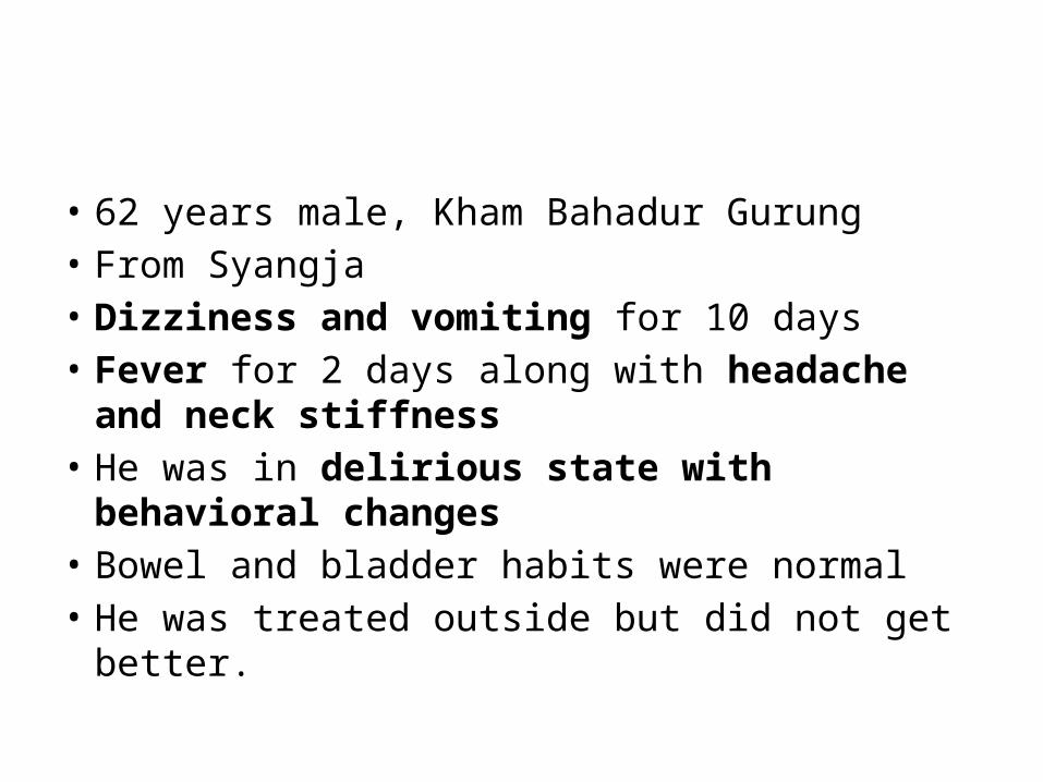
• 62 years male, Kham Bahadur Gurung• From Syangja• Dizziness and vomiting for 10 days• Fever for 2 days along with headache and neck
stiffness• He was in delirious state with behavioral changes• Bowel and bladder habits were normal• He was treated outside but did not get better.
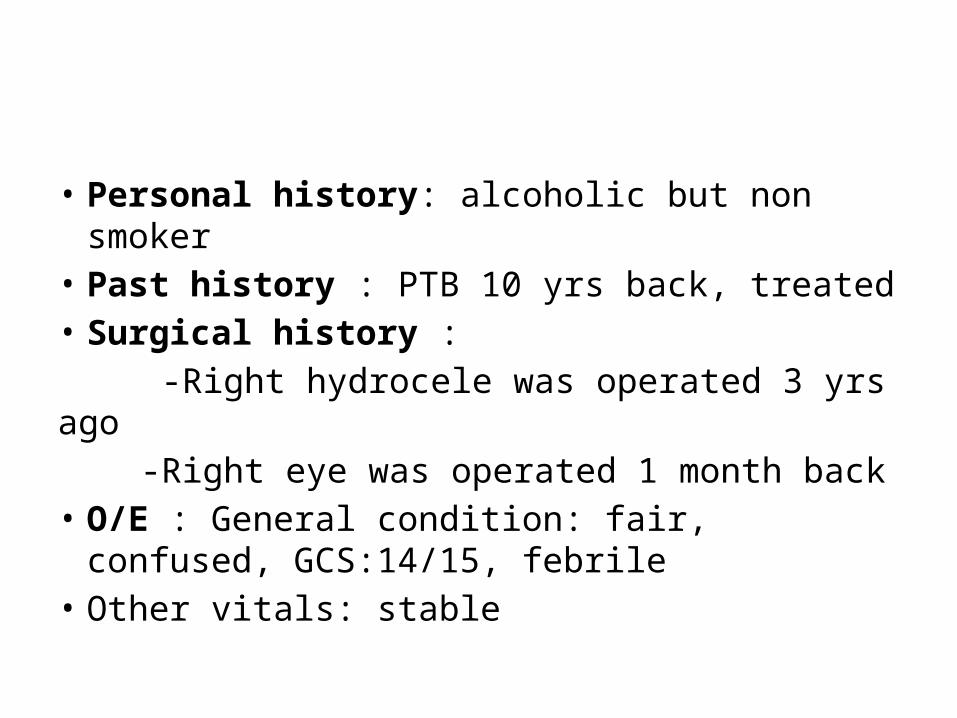
• Personal history: alcoholic but non smoker• Past history : PTB 10 yrs back, treated• Surgical history : -Right hydrocele was operated 3 yrs ago -Right eye was operated 1 month back• O/E : General condition: fair, confused,
GCS:14/15, febrile• Other vitals: stable
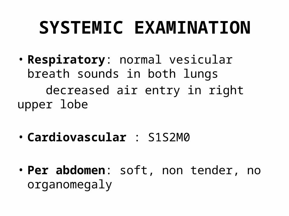
SYSTEMIC EXAMINATION
• Respiratory: normal vesicular breath sounds in both lungs
decreased air entry in right upper lobe
• Cardiovascular : S1S2M0
• Per abdomen: soft, non tender, no organomegaly
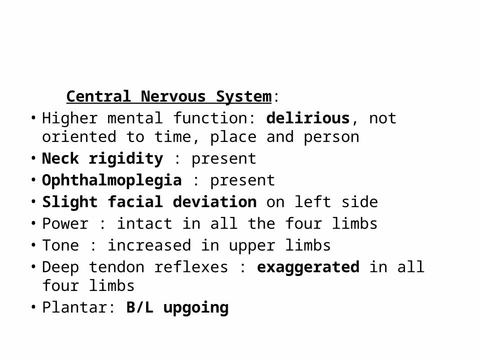
Central Nervous System: • Higher mental function: delirious, not oriented to time,
place and person• Neck rigidity : present• Ophthalmoplegia : present• Slight facial deviation on left side• Power : intact in all the four limbs• Tone : increased in upper limbs• Deep tendon reflexes : exaggerated in all four limbs• Plantar: B/L upgoing
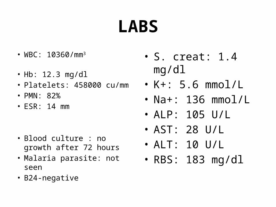
LABS• WBC: 10360/mm3
• Hb: 12.3 mg/dl• Platelets: 458000 cu/mm• PMN: 82%• ESR: 14 mm
• Blood culture : no growth after 72 hours
• Malaria parasite: not seen• B24-negative
• S. creat: 1.4 mg/dl• K+: 5.6 mmol/L• Na+: 136 mmol/L• ALP: 105 U/L• AST: 28 U/L• ALT: 10 U/L• RBS: 183 mg/dl
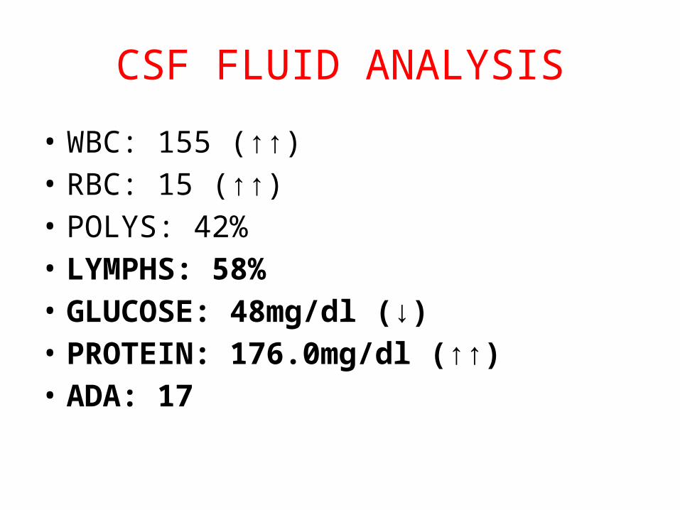
CSF FLUID ANALYSIS
• WBC: 155 (↑↑)• RBC: 15 (↑↑)• POLYS: 42% • LYMPHS: 58%• GLUCOSE: 48mg/dl (↓)• PROTEIN: 176.0mg/dl (↑↑)• ADA: 17
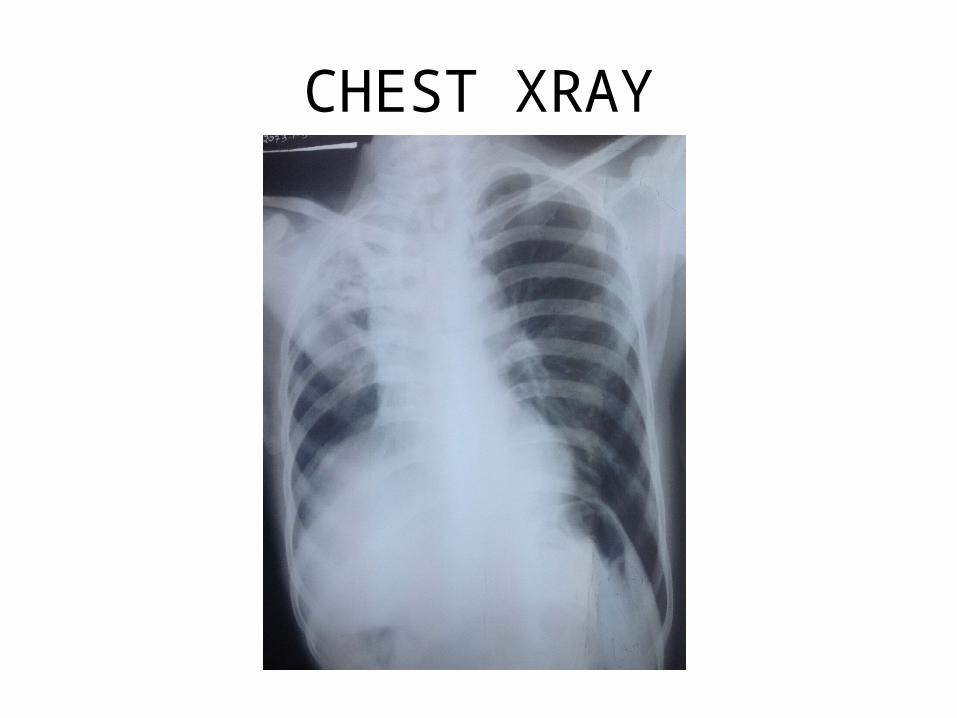
CHEST XRAY
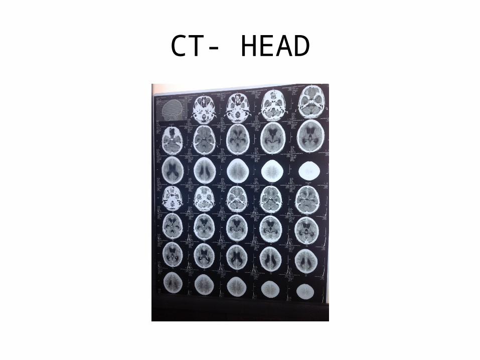
CT- HEAD
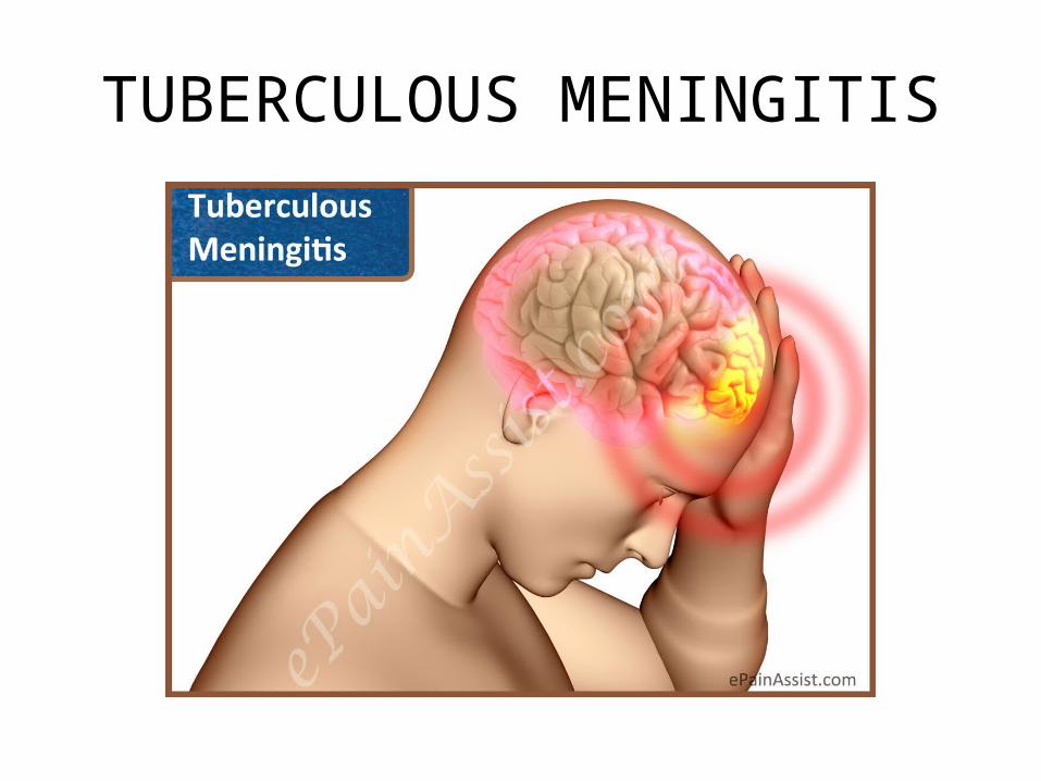
TUBERCULOUS MENINGITIS
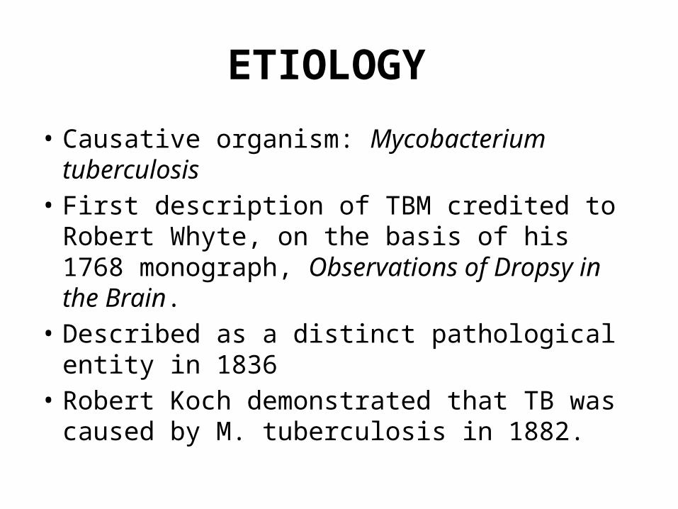
ETIOLOGY
• Causative organism: Mycobacterium tuberculosis• First description of TBM credited to Robert
Whyte, on the basis of his 1768 monograph, Observations of Dropsy in the Brain.
• Described as a distinct pathological entity in 1836
• Robert Koch demonstrated that TB was caused by M. tuberculosis in 1882.
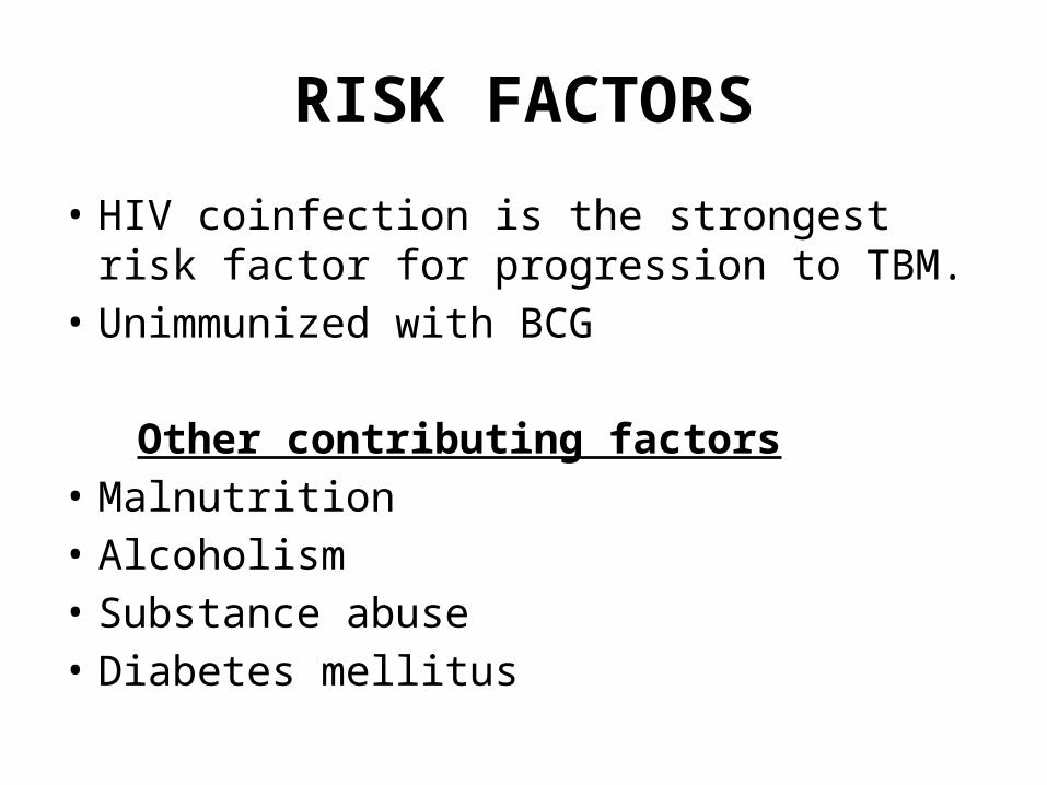
RISK FACTORS
• HIV coinfection is the strongest risk factor for progression to TBM.
• Unimmunized with BCG Other contributing factors• Malnutrition• Alcoholism• Substance abuse• Diabetes mellitus
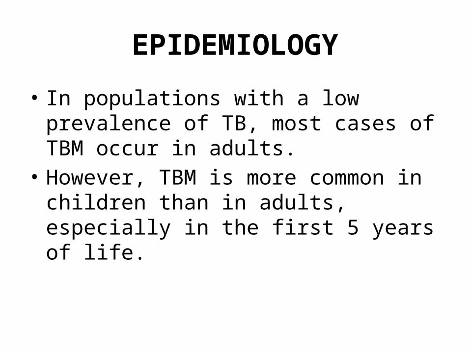
EPIDEMIOLOGY
• In populations with a low prevalence of TB, most cases of TBM occur in adults.
• However, TBM is more common in children than in adults, especially in the first 5 years of life.
PATHOPHYSIOLOGY• Following primary infection or late reactivation TB elsewhere in
the body, scattered tubercles are established in the brain, meninges, or adjacent bone.
• Subcortical or meningeal focus from which bacilli gained access to the subarachnoid space is the critical event for development of tuberculous meningitis .
• Due to chronic reactivation bacillemia occurs in older adults due to immune deficiency caused by aging, alcoholism, malnutrition, malignancy, or human immunodeficiency virus (HIV) infection
• Head trauma may also lead to destabilization of an established quiescent focus resulting in meningitis
• The spillage of tubercular protein into the subarachnoid space produces an intense hypersensitivity reaction due to a dense gelatinous exudate, giving rise to inflammatory changes.
• Proliferative arachnoiditis, most marked at the base of the brain, produces a fibrous mass involving cranial nerves and penetrating vessels.
• Vasculitis with resultant thrombosis and infarction involves vessels that traverse the basilar or spinal exudate or are located within the brain substance itself.
• Variety of stroke syndromes may result, involving the basal ganglia, cerebral cortex, pons, and cerebellum.
• Communicating hydrocephalus results from extension of the inflammatory process to the basilar cisterns and impedance of CSF circulation and resorption.
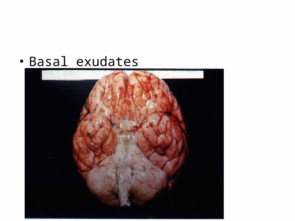
• Basal exudates
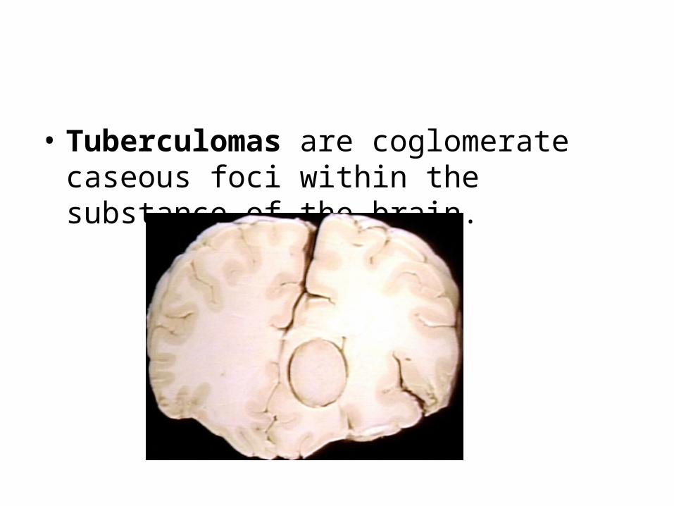
• Tuberculomas are coglomerate caseous foci within the substance of the brain.
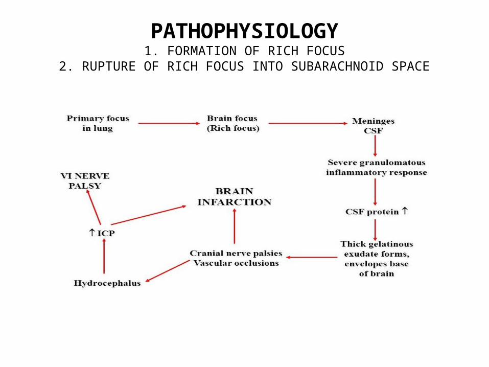
PATHOPHYSIOLOGY1. FORMATION OF RICH FOCUS
2. RUPTURE OF RICH FOCUS INTO SUBARACHNOID SPACE
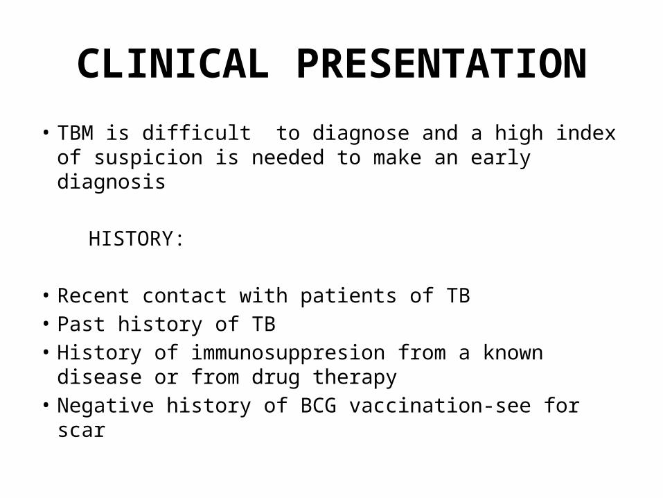
CLINICAL PRESENTATION• TBM is difficult to diagnose and a high index of suspicion
is needed to make an early diagnosis
HISTORY:
• Recent contact with patients of TB• Past history of TB• History of immunosuppresion from a known disease or
from drug therapy• Negative history of BCG vaccination-see for scar
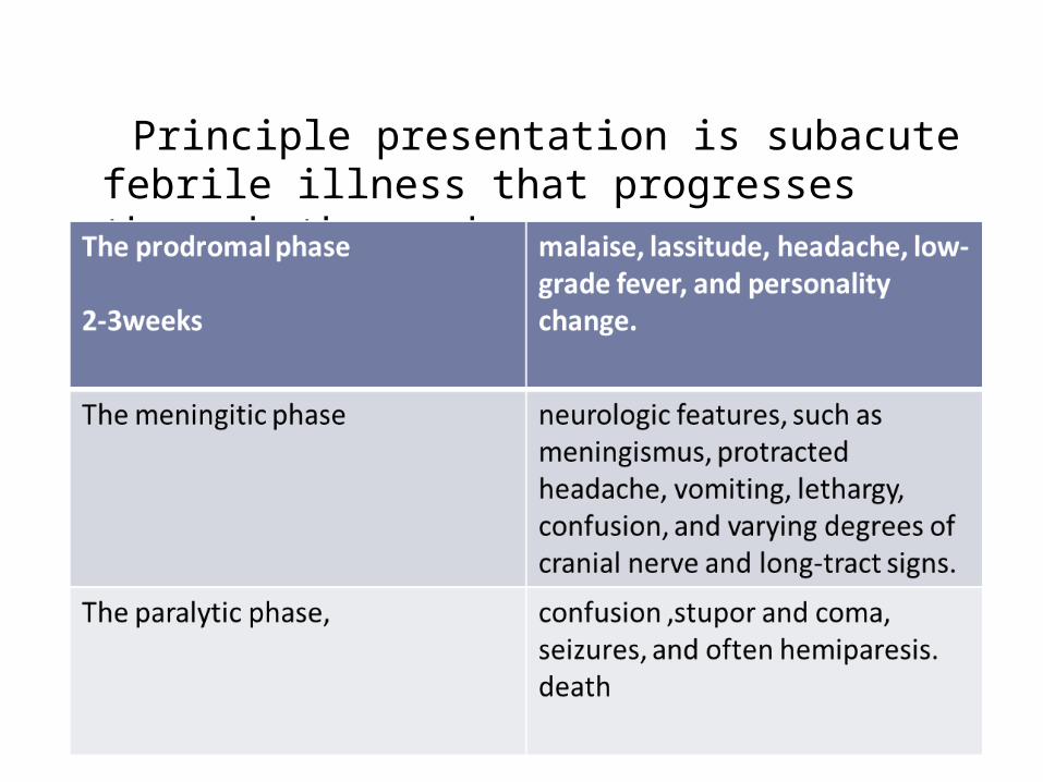
Principle presentation is subacute febrile illness that progresses through three phases:
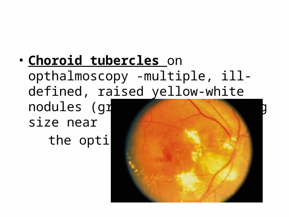
• Choroid tubercles on opthalmoscopy -multiple, ill-defined, raised yellow-white nodules (granulomas) of varying size near
the optic disc
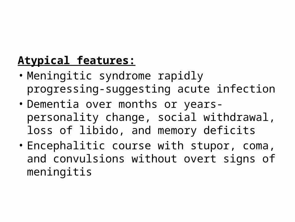
Atypical features:• Meningitic syndrome rapidly progressing-
suggesting acute infection• Dementia over months or years- personality
change, social withdrawal, loss of libido, and memory deficits
• Encephalitic course with stupor, coma, and convulsions without overt signs of meningitis
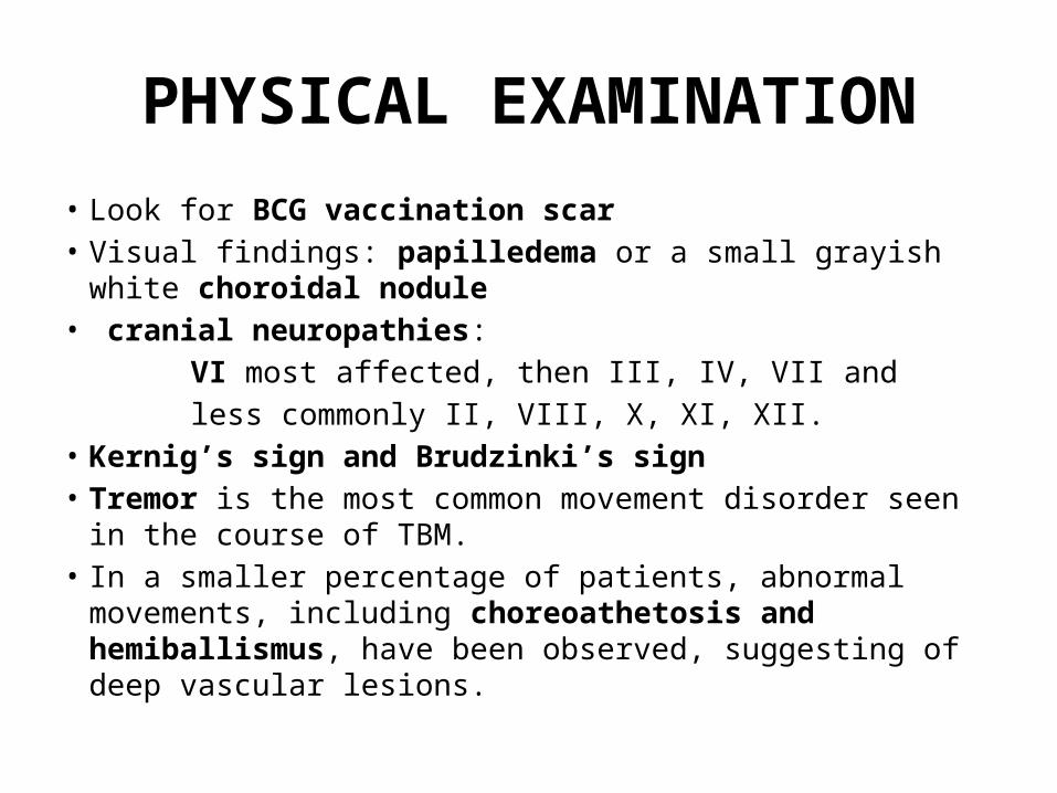
PHYSICAL EXAMINATION• Look for BCG vaccination scar• Visual findings: papilledema or a small grayish white choroidal
nodule• cranial neuropathies: VI most affected, then III, IV, VII and less commonly II, VIII, X, XI, XII.• Kernig’s sign and Brudzinki’s sign • Tremor is the most common movement disorder seen in the course
of TBM. • In a smaller percentage of patients, abnormal movements,
including choreoathetosis and hemiballismus, have been observed, suggesting of deep vascular lesions.
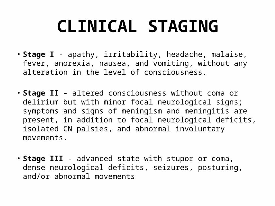
• Stage I - apathy, irritability, headache, malaise, fever, anorexia, nausea, and vomiting, without any alteration in the level of consciousness.
• Stage II - altered consciousness without coma or delirium but with minor focal neurological signs; symptoms and signs of meningism and meningitis are present, in addition to focal neurological deficits, isolated CN palsies, and abnormal involuntary movements.
• Stage III - advanced state with stupor or coma, dense neurological deficits, seizures, posturing, and/or abnormal movements
CLINICAL STAGING

DIFFERENTIAL DIAGNOSES Based on CSF findings of ↓Glucose, ↑Protein & lymphocytic
pleocytosis
• Subacute or chronic meningitis syndrome caused by Cryptococcosis, Granulomatous fungal infections, Brucellosis, and Neurosyphilis.
• Parameningeal suppurative infection, eg.brain abscess, or spinal epidural space infection.
• Herpes encephalitis
WORK UP
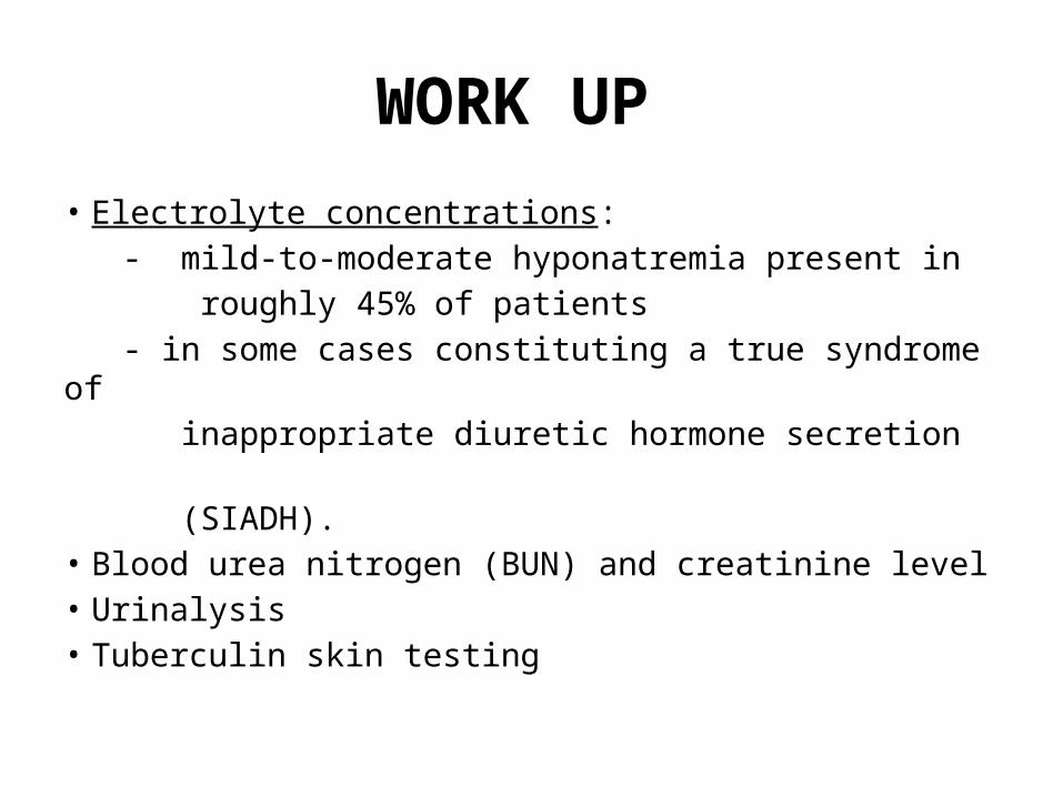
• Electrolyte concentrations: - mild-to-moderate hyponatremia present in roughly 45% of patients - in some cases constituting a true syndrome of inappropriate diuretic hormone secretion (SIADH). • Blood urea nitrogen (BUN) and creatinine level• Urinalysis• Tuberculin skin testing
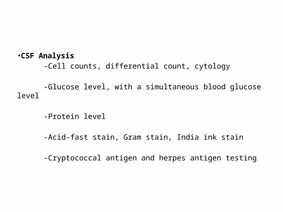
• CSF Analysis -Cell counts, differential count, cytology
-Glucose level, with a simultaneous blood glucose level
-Protein level
-Acid-fast stain, Gram stain, India ink stain
-Cryptococcal antigen and herpes antigen testing
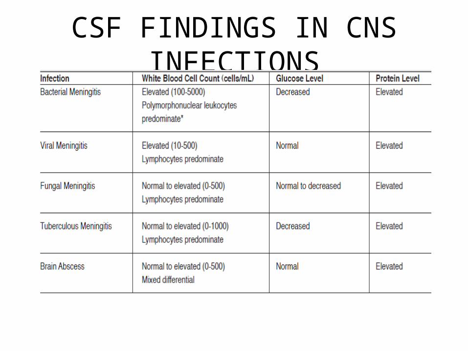
CSF FINDINGS IN CNS INFECTIONS
• Culture: (87% diagnostic) - CSF specimens for M. tuberculosis. - The demonstration of acid-fast bacilli (AFB) in the CSF is the effective means for an early diagnosis. - Minimum of 3 lumbar punctures be performed at daily intervals.
• Polymerase chain reaction: - 60% sensitive in rapid detection of M. tuberculosis in CSF. - Recommended whenever clinical suspicion is sufficiently high for empirical therapy or AFB is negative.
• Neuroimaging: - CT & MRI are helpful in detection. - CT can present the extent of basilar arachnoiditis, cerebral edema and infarction, and the presence and course of hydrocephalus.
• Hydrocephalus combined with marked basilar enhancement is indicative of advanced meningitic disease and carries a poor prognosis.
• Marked basilar enhancement correlates well with vasculitis and, therefore, with a risk for basal ganglia infarction.
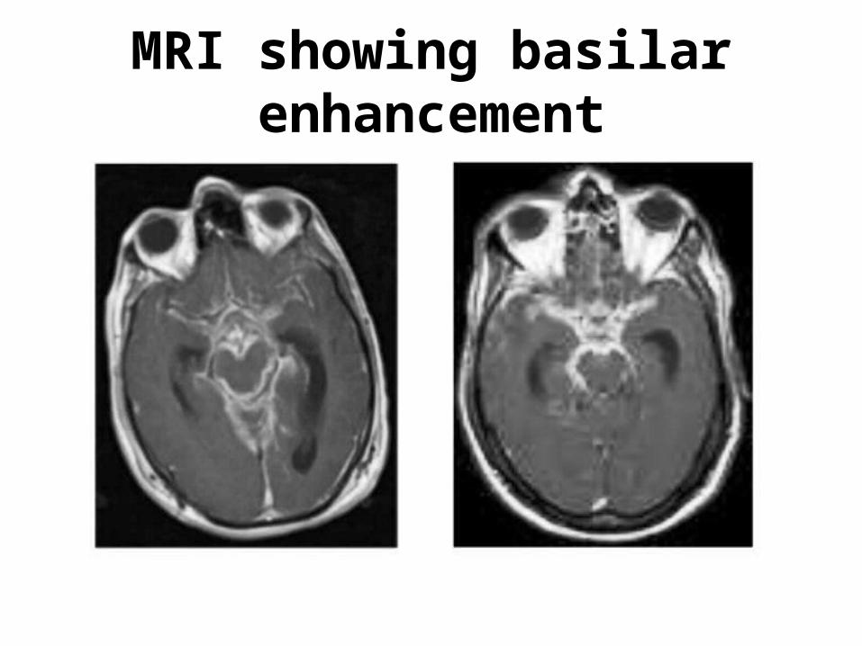
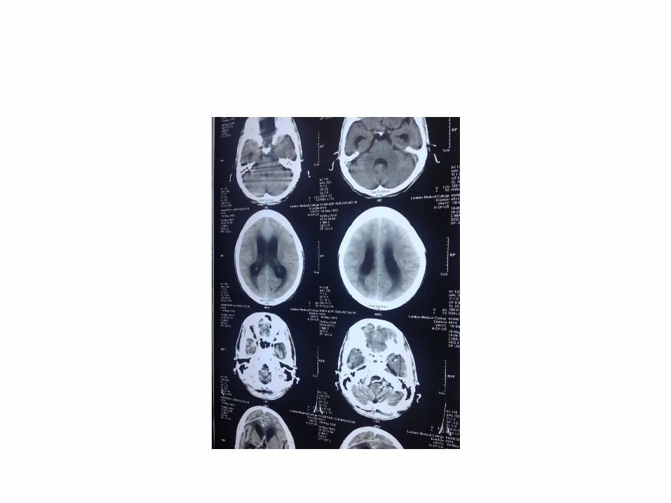
MRI showing basilar enhancement
• Interferon-gamma release assay (IGRA) using specific tuberculous antigens is a rapid, specific and sensitive method for the detection of tuberculous infection.
OTHERS• Angiography- for narrowing of the arteries
especially the small vessels at the base of the brain
• Electroencephalopathy-abnormal if meninigitis has progressed to advanced stage
• Brainstem Auditory Evoked Response Testing-abnormal in advanced stage of meningitis
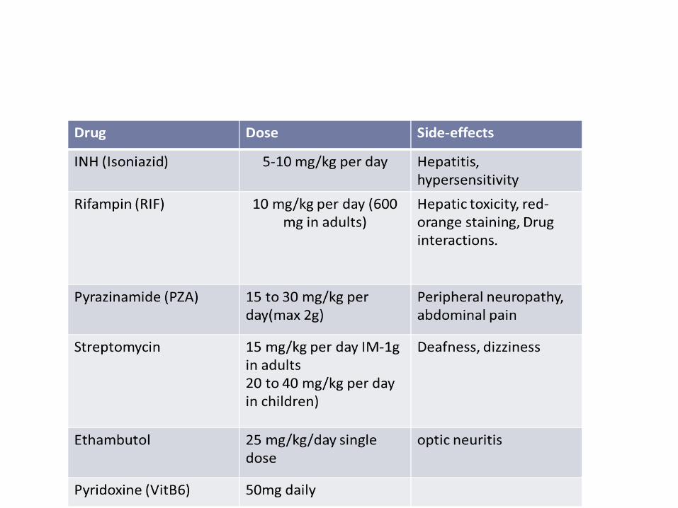
TREATMENT
• The mainstay of treatment for TB is clinical suspicion & starting of empirical therapy.
• First line drugs — Isoniazid (INH), rifampin (RIF), and pyrazinamide (PZA) are bactericidal, can be administered orally all having good meningeal penetration.
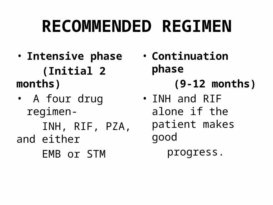
RECOMMENDED REGIMEN
• Intensive phase (Initial 2 months)• A four drug regimen- INH, RIF, PZA, and either EMB or STM
• Continuation phase (9-12 months) • INH and RIF alone if the
patient makes good progress.
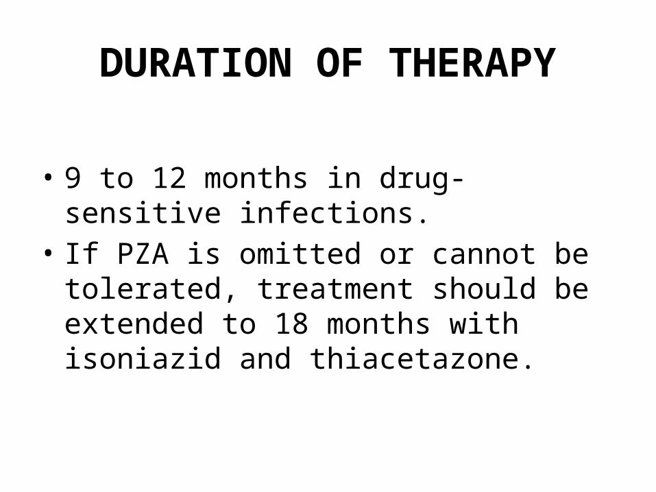
DURATION OF THERAPY
• 9 to 12 months in drug-sensitive infections. • If PZA is omitted or cannot be tolerated,
treatment should be extended to 18 months with isoniazid and thiacetazone.
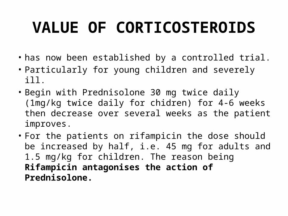
VALUE OF CORTICOSTEROIDS
• has now been established by a controlled trial.• Particularly for young children and severely ill. • Begin with Prednisolone 30 mg twice daily (1mg/kg
twice daily for chidren) for 4-6 weeks then decrease over several weeks as the patient improves.
• For the patients on rifampicin the dose should be increased by half, i.e. 45 mg for adults and 1.5 mg/kg for children. The reason being Rifampicin antagonises the action of Prednisolone.
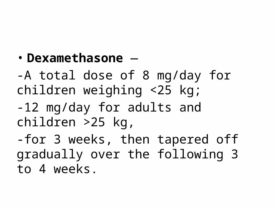
• Dexamethasone — -A total dose of 8 mg/day for children weighing <25 kg; -12 mg/day for adults and children >25 kg, -for 3 weeks, then tapered off gradually over the following 3 to 4 weeks.
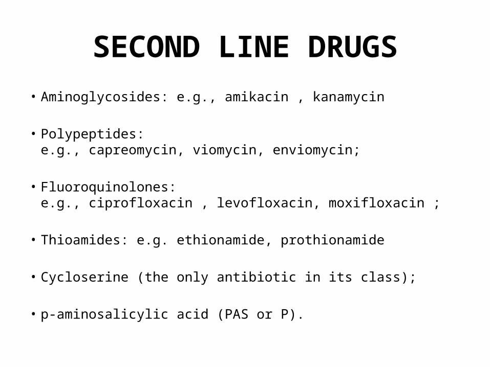
SECOND LINE DRUGS• Aminoglycosides: e.g., amikacin , kanamycin
• Polypeptides: e.g., capreomycin, viomycin, enviomycin;
• Fluoroquinolones: e.g., ciprofloxacin , levofloxacin, moxifloxacin ;
• Thioamides: e.g. ethionamide, prothionamide
• Cycloserine (the only antibiotic in its class);
• p-aminosalicylic acid (PAS or P).
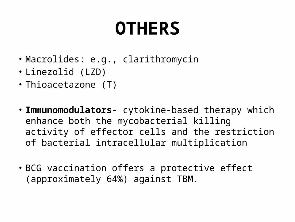
OTHERS• Macrolides: e.g., clarithromycin• Linezolid (LZD)• Thioacetazone (T)
• Immunomodulators- cytokine-based therapy which enhance both the mycobacterial killing activity of effector cells and the restriction of bacterial intracellular multiplication
• BCG vaccination offers a protective effect (approximately 64%) against TBM.
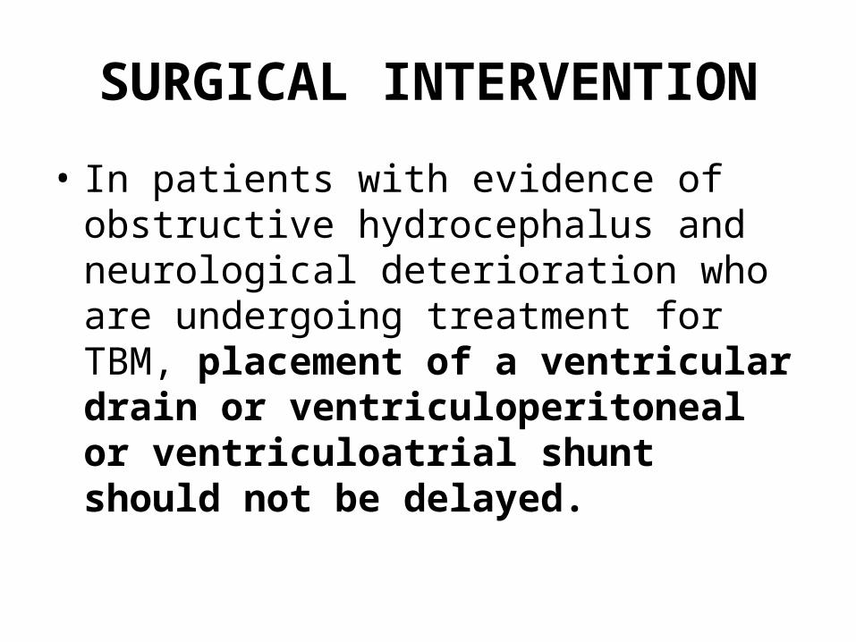
SURGICAL INTERVENTION
• In patients with evidence of obstructive hydrocephalus and neurological deterioration who are undergoing treatment for TBM, placement of a ventricular drain or ventriculoperitoneal or ventriculoatrial shunt should not be delayed.
COMPLICATIONS
• Hydrocephalus• Infarctions• Coma/stupor• Motor deficits- CN palsies, hemiparesis• Seizures• Mental impairment• Abnormal behavior• Brain damage• High morbidity and mortality
PROGNOSIS• Very critical disease in terms of fatal outcome and
permanent sequelae, requiring rapid diagnosis and treatment.
• Prognosis is directly related to the clinical stage at diagnosis.
• Kumar et al reported that children with TBM who have been vaccinated with BCG appear to maintain better mentation and have superior outcomes.
• Coexisting HIV encephalopathy and diminished immune competence contribute to the more severe clinical and neuroradiological features.
TAKE HOME MESSAGE
• Start ATT empirically when suspicion of TB• See for the BCG scar in suspected case• Counsel the patient for medication/side
effects• Complete the course• Follow up
REFERENCES
• Harrison’s Principle of Internal Medicine• Clinical Tuberculosis: John Crofton, Norman
Horne, Fred Miller• Medscape• Uptodate
Related Documents